Note: Descriptions are shown in the official language in which they were submitted.
CA 02871736 2019-10-27
WO 2013/162773
PCT/US2013/032526
QUANTITATION OF BIOMARKERS FOR THE DETECTION OF PROSTATE
CANCER
RELATED APPLICATIONS
100011 This application claims the benefit under 35 U.S.C. 119(e) of U.S.
Provisional
Application Nos. 61/639,768 filed on April 27, 2012, 61/764,288 filed on
February 13, 2013,
and 61/772,226 filed on March 4, 2013, which are hereby incorporated by
reference herein in
their entirety.
FIELD OF THE INVENTION
100021 This invention relates to novel biomarkers for treating, diagnosing,
and preventing
prostate cancer. The invention also relates to methods of identifying,
characterizing, and using
such prostate cancer biomarkers. This invention also relates to multiplexed
assays for
quantitating biomarkers.
BACKGROUND
100031 Prostate cancer is one of the most common malignancies in the US
(1). It is
clinically heterogeneous, with a highly variable natural history (2). The
discovery and
widespread utilization of serum prostate specific antigen (PSA) monitoring for
early detection
has greatly changed the way prostate cancer is diagnosed and treated. However,
PSA lacks
specificity as a screening tool for prostate cancer, and there is really no
lower limit of PSA that
entirely excludes cancer (3). Thus, clinical decision making in prostate
cancer places a
significant burden upon biopsy results. Ultrasound guided needle biopsy is the
standard for
diagnosis however, a negative result does not exclude the presence of cancer.
Both sampling
and analytical variables account for false negative results. In practice,
false negative results
engender a need for repeat biopsies which can delay diagnosis and treatment or
unnecessarily
subject cancer-free men to repeat biopsies and their attendant anxiety and
risk (4, 5). The
heterogeneity of prostate cancer is also a significant problem and while the
incidence is high,
the death rates from prostate cancer are relatively low as compared to those
from the other
major cancers such as lung, pancreas, and colon. The Gleason grading system
which has been
widely adopted for prostate cancer is a predictor of outcome (6). However, a
major limitation
of this grading system, and a result of aggressive screening procedures, is
that a majority of
newly diagnosed prostate cancer cases are Gleason score 6 or 7 tumors. These
moderately
differentiated tumors can either be indolent or aggressive (7).
100041 A.s referenced above, a determination of the prognosis of prostate
cancer is guided
by the Gleason grading system. In this system, a biopsy of the prostate tissue
is harvested,
fixed in formalin, embedded in paraffin, and then sliced and stained for
viewing. The
- 1 -
CA 02871736 2014-10-27
WO 2013/162773
PCT/US2013/032526
pathologist will then give the particular sample of tissue a grade or pattern
based on the
appearance of the tissue. The grade will range from 1 to 5, with a higher
number indicating a
more aggressive cancer. The pathologist gives a grade to the most common tumor
pattern and
then a grade to the second most common tumor pattern. These grades are added
to provide the
overall Gleason score. The Gleason score ranges from 2 to 10, with 10 having
the worst
prognosis.
100051 The Gleason score is only one component of prostate cancer staging.
The most
common method of prostate cancer staging is promulgated by the American joint
Committee
on Cancer and is known as the wINM" system. There are two types of staging,
the clinical
stage and the pathologic stage. The clinical stage is determined prior to
treatment such as
surgical prostatectomy, and includes five key elements:
= The extent of the primary tumor (T category)
= Whether the cancer has spread to nearby lymph nodes (N category)
= The absence or presence of distant metastasis (M category)
= The PSA. level at the time of diagnosis
= The Gleason score, based on the prostate biopsy (or surgery)
T describes the size of the primary tumor, N describes whether nearby lymph
nodes are
involved in the cancer and M describes metastasis or spread of the cancer.
00061 Following surgical prostatectomy, the prostate is carefully examined
for assignment
of pathologic stage. The "T" scale for prostate cancer is as follows:
Ti: tumor present, but not detectable clinically or with imaging
T 1 a: tumor was incidentally found in less than 5% of prostate tissue
resected
(for other reasons)
Tib: tumor was incidentally found in greater than 5% of prostate tissue
resected
Tic: tumor was found in a needle biopsy performed due to an elevated serum
PSA
T2: the tumor can be felt (palpated) on examination, but has not spread
outside the
prostate
T2a: the tumor is in half or less than half of one of the prostate gland's two
lobes
T2b: the tumor is in more than half of one lobe, but not both
12c: the tumor is in both lobes but within the prostafic capsule
T3: the tumor has spread through the prostatic capsule (if it is only part-way
through, it
is still T2)
- 2 -
CA 02871736 2014-10-27
WO 2013/162773
PCT/US2013/032526
T3a: the tumor has spread through the capsule on one or both sides
T3b: the tumor has invaded one or both seminal vesicles
T4: the tumor has invaded other nearby structures
This ranking, coupled with the N and M, is combined with the histological
assessment from the
Gleason score to determine whether definitive treatment for the cancer should
be taken or
watchful waiting should be chosen.
100071 A variety of nomograms are available to assess the risk of
aggressive prostate
cancer including the d'Amico system (8). This assigns the following risk
scores: Low-risk:
PSA less than or equal to 10, Gleason score less than or equal to 6, and
clinical stage T1-2a;
intermediate risk: PSA between 10 and 20, Gleason score 7, or clinical stage
T2b; High-risk:
PSA more than 20, Gleason score equal or larger than 8, or clinical stage T2c-
3a. Definitive
treatment entails radiation therapy, or prostatectomy. Therapy may also be
deferred in an
attempt to balance expected life span, the likelihood of treatment side
effects, and quality of
life. Watchful waiting (periodic clinical visits and PSA measurements) or
active surveillance
(periodic clinic visits and PSA measurements combined with scheduled repeat
biopsies) are
used when patients are comfortable with postponement of definitive therapy.
100081 The current methods for diagnosing and making prognostic decision-
making for
prostate cancer have limitations in that interobserver variability occurs,
especially in the setting
of small tumors. This is where quantitative information would be of value to
patient and
physician. Therefore, new quantitative methods to assist clinicians and
pathologists in both
diagnostic and prognostic decision making are needed to aid in the detection
and treatment of
prostate cancer.
100091 The majority of men with prostate cancer will die with their disease
rather than of it
(67), and there is a strong argument that screening has increased the
detection of indolent
tumors (68). Unfortunately, we lack clinical tools to distinguish indolent
from aggressive
prostate cancer (69), and it is estimated that over 1400 men need to be
screened and nearly 50
men treated to prevent one prostate cancer death (70). Prostate cancers with
similar
microscopic features have variable clinical outcomes, reflecting genetic and
biological
variables of individual patients not recognized by microscopy alone (71).
Unfortunately, tissue
architecture is often destroyed by extraction methods required for detection
of molecular tissue
biomarkers. Therefore, emerging methods for biomarker discovery and validation
compete
with histology for the same tissue are needed. This is especially so when
small needle biopsies
are utilized, which is the standard of care for diagnosis of suspected
prostate cancer.
- 3 -
CA 02871736 2014-10-27
WO 2013/162773
PCT/US2013/032526
100101 Pathologic examination remains a gold standard for diagnosis,
classification, and
staging of tumors. This requires intact tissue while implementation of
molecular tests often
requires extraction methods that disrupt tissue. Metabolomics is a newer area
of biospecimen
analysis in which small molecules (--2kD) (e.g., metabolites), present in a
biological sample,
are extracted, detected and measured. The method has been employed in the
study of the
biochemical basis and mechanisms for diverse biological processes such as
cancer diagnosis
and monitoring progression, drug mechanism of action, drug toxicity,
industrial bio-processing,
etc.
100111 In order to analyze the metabolites of a biological sample, they
must be extracted
completely from the sample. Existing methods of biological extraction involve
destroying the
sample such that it can no longer be used for other analysis (e.g.,
histology). Fixation of tissue
samples is usually done with formalin (formaldehyde in water), followed by
histologic
processing and sectioning, and this is the usual work flow that produces a
slide for microscopic
examination by a pathologist. The drawback of this method is that formaldehyde
is not an
effective extractant for metabolites. Therefore, under commonly known
biological extraction
methods, in order to conduct histology analysis and perform metabolomics, two
tissue samples
are required, one for each analysis. The current state of the art is that the
second biopsy would
be used only for metabolomics and would not be examined histologically. Thus,
it cannot be
known with certainty whether the biopsy used for metabolomics contained
diseased tissue.
100121 Methods and reagents for performing metabolomics and subsequent
histology on
various tissue samples are described in detail in PCT Appl. No.
PCMS2011/037093
(W02011/146683), which is herein incorporated by reference in its entirety. A
preferred
embodiment of that method involves contacting a single biological sample (e.g.
a tissue biopsy)
with a solvent (e.g. ethanol or methanol) such that the extracted biochemical
can be analyzed
and the extracted tissue retains its cellular architecture so that it can be
subsequently analyzed
using standard histological methods (including cytological analysis). Using
this method, a
number of tissues have been extracted, their biological chemicals analyzed,
and subsequent
histology preformed. See, Shuster et al. "Molecular preservation by extraction
and fixation,
inPREF: a method for small molecule biomarker analysis and histology on
exactly the same
tissue." BMC Clinical Pathology 2011, 11:14, herein incorporated by reference
in its entirety.
100131 The term, molecular preservation by extraction and fixation ("mPREF"),
refers to a
process of preserving cellular structure in tissue or cell specimens whilst
extracting small
molecules by immersing the tissue in a solution containing an organic solvent,
then
- 4 -
CA 02871736 2014-10-27
WO 2013/162773
PCT/US2013/032526
subsequently processing the exact same portion of tissue using histological
methods. mPREF
enables both quantitation of biochemicals, including small molecule
metabolites, and
histological examination of the same tissue sample. mPREF permits quantitation
of
metabolites and histology in exactly the same tissue. This diminishes the
competition of new
molecular testing methods with standard histology for small amounts of tumor
containing
biopsy tissue. mPREF treated tissues can be used for all existing methods that
use paraffin
embedded tissue. The aqueous alcohol in mPREF selectively extracts small
molecules from
tissue, leaving macromolecules such as proteins, RNA, and DNA in place.
Existing powerful
in-situ methods for detecting proteins (immunohistochemistry, IHC) and RNA and
DNA
(fluorescence in situ hybridization, FISH) in intact tissue can continue to be
used in mPREF
processed tissue.
100141 A biomarker is an organic biomolecule, the presence of which in a
sample is used to
determine the phenotypic status of the subject (e.g., cancer patient v. normal
patient or
prognosis of cancer patient). In order for the biomarker to be biologically
relevant it should be
differentially present in a sample taken from a subject of one phenotypic
status (e.g., having a
disease) as compared with another phenotypic status (e.g., not having the
disease). Biomarkers,
alone or in combination, provide measures of relative risk that a subject
belongs to one
phenotypic status or another. Therefore, they are useful as markers for
disease (diagnostics),
prognosis (i.e., state of the disease), therapeutic effectiveness of a drug
(theranostics), drug
toxicity, and predicting and identifying the immune response.
100151 There remains a need for multiplex assays to quantitate diagnostic
cancer
metabolites that are isolated from biological samples in a manner that allows
such samples to
be further analyzed using standard histological methods. The metabolites that
are identified
and characterized can then be used as cancer biomarkers.
100161 Unless otherwise defined, all technical and scientific terms used
herein have the
same meaning as commonly understood by one of ordinary skill in the art to
which this
disclosure belongs. Although methods and materials similar or equivalent to
those described
herein can be used in the practice or testing of the present disclosure,
suitable methods and
materials are described below. All publications, patent applications, patents,
and other
references mentioned herein are incorporated by reference in their entirety.
In addition, the
materials, methods, and examples are illustrative only and not intended to be
limiting.
BRIEF SUMMARY OF THE INVENTION
-5...
CA 02871736 2014-10-27
WO 2013/162773
PCT/US2013/032526
100171 In one embodiment, the invention provides a method for screening for
prostate
cancer in a subject by: (a) providing a biological sample from a subject; (b)
detecting at least
one biomarker in said sample, said biomarker selected from the group
consisting of betaine,
malate, proline, N-acetylaspartate, uracil, xanthine, cysteine, alanine, and N-
acetylglucosamine; and (c) correlating said detection with a status of
prostate cancer or no
prostate cancer.
100181 In a further embodiment, the invention provides a method for
screening for prostate
cancer in a subject by: (a) providing a biological sample from a subject; (b)
detecting at least
one biomarker in said sample, said biomarker selected from the group
consisting of betaine,
malate, proline, uracil, xanthine, cysteine, alanine, and N-acetylglucosamine;
and (c)
correlating said detection with a status of prostate cancer or no prostate
cancer.
100191 In a further embodiment the detecting at least one biomarker is
performed by mass
spectrometry.
100201 In a still further embodiment, the biological sample is selected
from the group
consisting of biological fluid and tissue.
100211 In a further embodiment, the biological fluid is whole blood, serum,
plasma, or
urine.
100221 In a further embodiment, the tissue is a prostate tissue sample.
100231 In a further embodiment, the biological sample is contacted with a
solvent capable
of extracting the at least one biomarker.
100241 In a further embodiment, the solvent is methanol or ethanol.
100251 In another embodiment, the invention provides a method of diagnosing
prostate
cancer in a subject by: (a) obtaining one or more test samples from a subject;
(b) detecting at
least one biomarker in the one or more test samples, wherein the biomarker is
selected from:
betaine, malate, proline, N-acetylaspartate, uracil, xanthine, cysteine,
alanine, and N-
acetylglucosamine; (c) quantitating the amount of the at least one biomarker;
and (d)
correlating the quantitation of the at least one biomarker with a diagnosis of
prostate cancer.
100261 In yet another embodiment, the invention provides a method of
diagnosing prostate
cancer in a subject by: (a) obtaining one or more test samples from a subject;
(b) detecting at
- 6 -
CA 02871736 2019-10-27
WO 2013/162773
PCT/US2013/032526
least one biomarker in the one or more test samples, wherein the biomarker is
selected from:
betaine, malate, prolinc, uracil, xanthine, cysteine, alanine, and N-
acetylglucosamine; (c)
quantitating the amount of the at least one biomarker; and (d) correlating the
qua3ntitation of the
at least one biomarker with a diagnosis of prostate cancer, wherein the
correlation takes into
account the amount of the at least one biomarker in the one or more test
samples compared to a
control amount of the at least one biomarker.
100271 In a further embodiment, the correlation takes into account the
amount of the at
least one biomarker in the one or more test samples compared to a control
amount of the at
least one biomarker.
100281 In a further embodiment, the test sample is selected from the group
consisting of
urine, whole blood, serum, plasma, and prostate tissue.
100291 In another embodiment, the invention provides a method of monitoring
the effect of
a prostate cancer drug or therapy on a subject by: (a) providing a biological
sample from the
subject; (b) contacting the biological sample with a solvent capable of
extracting at least one
prostate cancer biomarker selected from the group consisting of betaine,
malate, proline, N-
acetylaspartate, uracil, xanthine, cysteine, alanine, and N-
acetylglucosarnine; (c) quantitating
the amount of the at least one prostate cancer biomarker; (d) providing the
subject with an anti-
prostate cancer drug or therapy; (e) quantitating the amount of the at least
one prostate cancer
biomarker using steps (a) and (h); and (f) corrlating the two measurements
with a diagnosis that
the prostate cancer is regressing or progressing.
100301 In another embodiment, the invention provides a multiplexed assay
for screening for
prostate cancer in a subject by: (a) providing a biological sample from a
subject; (b)
quantitating at least two or more biomarkers in said sample, said biomarkers
selected from the
group consisting of betaine, malate, proline, N-acetylaspartate, uracil,
xanthine, cysteine,
alanine, and N-acetylglucosamine; (c) correlating said quantitation with a
status of prostate
cancer or no prostate cancer.
100311 In a further embodiment, the quantitating at least two or more
biomarkers is
performed by liquid chromatography in tandem with mass spectrometry.
100321 In a further embodiment, the biological sample is selected from the
group consisting
of biological fluid and tissue.
-7.-
CA 02871736 2014-10-27
WO 2013/162773
PCT/US2013/032526
100331 In a further embodiment, the biological fluid is whole blood, serum,
plasma, or
urine.
100341 In a further embodiment, the tissue is a prostate tissue sample.
100351 In a further embodiment, the biomarkers betaine, malate, proline, N-
acetylaspartate,
uracil, xanthine, cysteine, alanine, and N-acetylglucosamine are quantitated
in the same assay.
100361 In a further embodiment, the biomarkers betaine, malate, proline,
uracil, xanthine,
cysteine, alanine, and N-acetylglucosamine are quantitated in the same assay.
100371 In another embodiment, the invention provides a multiplexed method
for detecting
prostate cancer in a subject by: (a) providing a biological sample from the
subject; (b)
contacting the biological sample with a solvent capable of extracting two or
more prostate
cancer biomarkers selected from the group consisting of betaine, malate,
proline, N-
acetylaspartate, uracil, xanthine, cysteine, alanine, and N-acetylglucosamine;
(c) quantitating
the amount of the two or more biomarkers present in the biological sample; and
(d) correlating
the amount of the two or more biomarkers with the presence or absence of
prostate cancer.
100381 In a further embodiment, the quantitating differentiates between
different stages of
prostate cancer.
100391 In a further embodiment, the quantitating is part of a diagnosis or
prognosis of
prostate cancer in the subject.
100401 In a further embodiment, the solvent is methanol or ethanol.
100411 In a further embodiment, the step of performing additional
histological analysis on
the extracted biological sample.
100421 In another embodiment, the invention provides a method of diagnosing
prostate
cancer in a subject by: (a) obtaining one or more test samples from a subject;
(b) detecting at
least one biomarker in the one or more test samples, wherein the biomarker is
selected from the
group consisting of betaine, malate, proline, uracil, xanthine, cysteine,
alanine, and N-
acetylglucosamine; (c) quantitating the amount of the at least one biomarker;
(d) determining
the Gleason score of the one or more test samples; (e) correlating the
quantitation of the at least
one biomarker and the Gleason score with a relative risk of T2 versus T3
prostate cancer.
- 8 -
CA 02871736 2014-10-27
WO 2013/162773
PCT/US2013/032526
100431 In another embodiment, the invention provides a kit for diagnosing
prostate cancer
in a subject with (a) a vial for collecting a biological sample from the
subject; (b) a solvent for
extacfing biomarkers from the biological sample, the biomarkers selected from
the group
consisting of betaine, malate, proline, N-acetylaspartate, uracil, xanthine,
cysteine, alanine, and
N-acetylglucosamine; (c) instructions for performing the extraction of the
biomarkers; (d)
instructions for quantitating one or more of the biomarkers; (f) instructions
for correlating the
quantitation of the one or more biomarkers to a diagnosis of prostate cancer
or normal.
BRIEF DESCRIPTION OF THE DRAWINGS
100441 The following figures are provided for the purpose of illustration
only and are not
intended to be limiting.
100451 FIGURE 1: A quantitation curve of uracil.
100461 FIGURE 2: A quantitation curve of N-acetylaspartate.
100471 FIGURE 3: A quantitation curve of xanthine.
100481 FIGURE 4: A quantitation curve of alanine.
100491 FIGURE 5: A quantitation curve of proline.
100501 FIGURE 6: A quantitation curve of betaine.
100511 FIGURE 7: A quantitation curve of cysteine.
100521 FIGURE 8: A quantitation curve of malate.
100531 FIGURE 9: Quantitation results of targeted biomarker compounds are
provided in
Figure 9.
100541 FIGURE 10: Figure 10 shows the concentration ranges where the
measured values
of the biomarkers of the present invention fell on the concentration standard
curves. These are
shaded gray.
100551 FIGURE 11: Figure 11 shows the actual values from the 29 prostate
samples (15
tumor and 14 non-tumor) that were analyzed by the current method.
100561 FIGURE 12: Figure 12 shows the concentration ranges where the
measured values
of the biomarkers of the present invention fell on the concentration standard
curves. These are
shaded gray.
100571 FIGURE 13: A quantitation curve of uracil.
100581 FIGURE 14: A quantitation curve of N-acetylaspartate.
100591 FIGURE 15: A quantitation curve of xanthine.
100601 FIGURE 16: A quantitation curve of alanine.
100611 FIGURE 17: A quantitation curve of proline.
- 9 -
CA 02871736 2014-10-27
WO 2013/162773
PCT/US2013/032526
100621 FIGURE 18: A quantitation curve of betaine.
100631 FIGURE 19: A quantitation curve of cysteine.
100641 FIGURE 20: A quantitation curve of malate.
100651 FIGURE 21: A quantitation curve of N-acetylglucosamine.
100661 FIGURE 22: A graphical flow chart that represents the process of
performing the
extraction and metabolomics as described by the current invention and the
subsequent
histology of the tissue samples.
100671 FIGURE 23: A graph showing the difference between the concentration
of the
biomarkers uracil, N-acetylaspartate, proline, xanthine, betaine, malate, and
N-
acetylglucosamine in non-tumor tissue as compared to tumor tissue.
100681 FIGURE 24: A graph showing the difference between the concentration
of the
biomarkers alanine and cysteine in non-tumor tissue as compared to tumor
tissue.
DETAILED DESCRIPTION OF THE INVENTION
100691 The invention is directed to biomarkers for prostate cancer. The
invention is also
directed to methods of detecting the presence of one or more biomarkers in
order to make a
diagnosis or prognosis of prostate cancer. The measurement of these markers,
alone or in
combination, in patient samples, provides information that the diagnostician
can correlate with
a diagnosis of prostate cancer, risk of developing prostate cancer, and/or
prognosis of a subject
with prostate cancer. In some embodiments, the biomarkers are betaine, malate,
proline, N-
acetylaspartate, uracil, xanthine, cysteine, alanine, and N-acetylglucosamine.
All nine of these
biomarkers can be measured and quantitated at the same time in a single
multiplex assay,
which could provide very valuable information to the clinician or pathologist.
The assay can
be varied to quantitate a smaller number of biomarkers if desired.
[0070] In one embodiment of the invention, mPREF was used to demonstrate
that a subset
of metabolites can be quantitated in 18 gauge core needle biopsies of prostate
tissue. These
metabolites can be used as clinically useful biomarkers. In another
embodiment, the
quantitation of these biomarkers can be used in the diagnosis or prognosis of
prostate cancer.
Methods of the present invention can be used independently or in conjunction
with currently
accepted (or later developed) methods for diagnosing prostate cancer and/or
determining the
prognosis of a subject with prostate cancer. For example, methods of the
present invention
may be combined with histology methods.
100711 One aspect of this invention is an assay for quantitating select
candidate diagnostic
metabolites from cancer needle biopsy extracts using ultra-performance liquid
chromatography
-10-
CA 02871736 2014-10-27
WO 2013/162773
PCT/US2013/032526
coupled to a tandem mass spectrometry system (UPLC-MS/MS). In one embodiment,
the
cancer is prostate cancer. A subset of metabolites from prostate needle
biopsies taken from
surgical prostatectomy specimens prepared using molecular preservation by
extraction and
fixation ("mPREF") were identified as candidate prostate cancer diagnostic
biomarkers. (66)
In order to determine which metabolites would be the most useful, they were
ranked based on
the analytical technique that would be required for quantitation (e.g. GC/MS,
LC/MS, etc.); the
types of verification studies that could be used to confirm that the
metabolite was a biomarker
for cancer; and the likely ability that the metabolite could be used in a
diagnostic assays. Based
on this analysis, nine metabolites were identified as potential biomarkers for
prostate cancer. In
one aspect of the invention, UPLC-MS/MS can be used as the assay platform.
However, any
LC/MS configuration can be used.
100721 The assay of the present invention was developed and used to quantitate
the following
biomarkers: betaine, malate, proline, N-acetylaspartate, uracil, xanthine,
cysteine, alanine, and
N-acetylglucosamine in 29 human prostate biopsy extracts. These involve 8
biochemical
groups, which represent pathways of alanine and aspartate metabolism;
cysteine, methionine,
SAM, and taurine metabolism.; glycine, serine, and threonine metabolism; urea
cycle, arginine
and proline metabolism; aminosugars, glycolysis, pentose metabolism, Krebs
cycle, purine
(hypoxanthine/inosine containing) and pyiimidine metabolism, respectively.
100731 These biomarkers can be used in a multiplexed assay for aiding in the
prognosis and
diagnosis of cancers, for example prostate cancer. One aspect of the present
invention allows
for the assay of all nine identified metabolites in a single LC-MS run in a
quantitative manner
on 18 gauge core needle biopsies. This allows for immediate application in the
clinical setting.
Then, the tissue can be encased in paraffin and subjected to further
processing and histology.
A flow chart outlining this procedure is shown in Figure 22.
100741 One advantage of the current invention is that the biomarkers can.
be quantitated at
the time of a prostate biopsy rather than on prostate tissue samples procured
from
prostatectomy specimens. Prostatectomy results when the patient and physician
have already
made a decision to undergo definitive therapy, and have chosen radical
prostatectomy.
Prognostic information derived from a prostatectomy specimen is not without
value, however,
the greater value resides in prognostic information contained in the prostate
biopsy. The
decision point prior to definitive therapy is more crucial, and this is when
the patient has been
diagnosed with prostate cancer following a biopsy. Prognostic markers useful
at this point
must therefore be applied to biopsy tissue. The assays of the present
invention allow
-11-
CA 02871736 2014-10-27
WO 2013/162773
PCT/US2013/032526
biomarkers to be quantitated from prostate biopsies, and, therefore, can
provide information
prior to definitive therapy. The treatment options available to patients now
include watchful
waiting and active surveillance. Active surveillance protocols are
problematical since the
decision to undergo definitive therapy is substantially influenced by Gleason
grading. As
described above, this has limitations as interobserver variability occurs.
Therefore, quantitative
information would be of value to patient and physician.
100751 Quantitation of the biomarkers at the time of prostate biopsy rather
than on prostate
tissue samples procured from prostatectomy specimens may also be advantageous
because it
could reduce the effects of ischemia time on metabolites. Metabolite data on
human prostate
tissue has utilized cryopreserved tissue obtained from prostatectomy
specimens. These may be
subject to warm ischemia (intraoperafive) times of at least 40-60 minutes,
prior to any time
involved with specimen transport and processing.
100761 Another advantage of the current invention is that it allows for the
sampling of the
entire prostate when implemented in vivo. Biomarkers can be quantitated in
each core
regardless of whether histologic tumor is present, and multiple cores with
tumor can be
sampled. The capability to broadly sample the prostate could be very important
since prostate
cancer can be heterogeneous.
100771 A fluffier benefit of this invention is that the analysis can be
performed on a smaller
amount of tissue than the existing diagnostic/prognostic methods.
Specifically, this method can
be performed on a single 18 gauge needle biopsy that only removes ¨ 5 mg of
tissue. Previous
methods for tissue biopsy have required large tissue removal (1 gram or
greater) or multiple
biopsies of smaller volume (e.g. 18 gauge core biopsies that harvest about 5
mg of tissue). The
problem with these two biopsy methods is that they require a significant
quantity of tissue to be
removed causing greater discomfort and trauma to the subject.
100781 In the methods of the present invention, the fixation of the
biomarkers can be
performed on a tissue sample and that same tissue sample can then be sent on
for further
histological evaluation. This is an improvement over the traditional methods
of extraction, in
which tissue samples were fixed in formalin since formalin is not a suitable
extractant for
metabolites. Formalin is ineffective in extracting the metabolites and it is
reactive so it can
alter the metabolite chemistry. Therefore two separate samples (composed of
different tissues)
had to be harvested using the traditional methods. In the methods of the
present invention, a
single biological sample (e.g. a tissue biopsy) is contacted with a solvent
(e.g. ethanol or
methanol) such that the extracted metabolite can be analyzed and the extracted
tissue retains its
-12-
CA 02871736 2019-10-27
WO 2013/162773
PCT/US2013/032526
cellular architecture so that it can be subsequently analyzed using standard
histological
methods (including cytological analysis). The metabolites can then be
quantitated and
biomarkers may be identified.
100791 A further aspect of this invention is for the analysis to be a
multiplexed assay. A
multiplex assay is an assay that simultaneously measures multiple biomarkers
in a particular
sample. The biomarkers can be measured directly from a patient sample with
minimal
preparation. This allows for a real time assessment of the patient's health in
the clinical setting
as it relates to the state of the disease. For example, in one embodiment of
the invention,
samples of prostate cancer biopsy were extracted from various cancer patients.
The
metabolites were extracted and a series of biomarkers were discovered
(betaine, malate,
proline. N-acetylaspartate, uracil, xanthine, cysteine. N-acetylglucosamine,
and alanine).
These biomarkers can be used in a multiplexed analysis of that patient's
disease state.
100801 Another advantage of the present invention is that it provides a
high throughput
method for analyzing biomarkers. Alternatively, using intact tissue,
immunohistochemistry
(IHC) and fluorescence in situ hybridization (FISH) are used to measure
biomarkers in intact
tissue. IFIC and FISH are technically challenging, generally performed one at
a time, and
require microscopic interpretation, introducing inter-observer variability.
IHC still requires
optical detection and interpretation.
100811 Another advantage of the present invention is that the results are
quantitative.
Metabolite measurements as described herein can be expressed as absolute molar
amounts of
metabolites. This is a key distinction with IHC results which are notoriously
difficult to
quantitate.
100821 Betaine, malate, proline, N-acetylaspartate, uracil, xanthine,
cysteine, alanine, and
N-acetylglucosamine as biomarkers for prostate cancer, as well as methods and
uses thereof,
are disclosed. These biomarkers are overexpressed in patients with cancer,
specifically prostate
cancer. These biomarkers can, therefore, be utilized to diagnose patients with
cancer, or to
diagnose patients at risk for developing cancer. In some embodiments, the
invention provides a
method of diagnosing prostate cancer in a subject, comprising detecting the
differential
expression of at least one biomarker in the one or more test samples obtained
from the subject,
wherein the biomarker is betaine, malate, proline, N-acetylaspartate, uracil,
xanthine, cysteine,
alanine, and N-acetylglucosamine. In another embodiment, the invention
provides a method of
determining the prognosis of a subject with prostate cancer, comprising
detecting the
differential expression of at least one biomarker in the one or more test
samples obtained from
- 13 -
CA 02871736 2014-10-27
WO 2013/162773
PCT/US2013/032526
the subject, wherein the biomarker is betaine, malate, proline, N-
acetylaspartate, uracil,
xanthine, cysteine, alanine, and N-acetylglucosamine.
100831 In one embodiment of the present invention, a method of diagnosing
cancer,
specifically prostate cancer, or risk for developing cancer in a subject is
provided. This method
comprises the steps of (a) providing a biological sample from the subject; (b)
contacting the
biological sample with an extraction reagent capable of extracting the cancer
biomarkers
comprising betaine, malate, proline, N-acetylaspartate, uracil, xanthine,
cysteine, alanine, and
N-acetylglucosamine; (c) determining the amount of the biomarkers; and (d)
correlating the
amount biomakers to a prostate cancer diagnosis.
100841 The amount of cancer biomarkers (i.e., betaine, malate, proline, N-
acetylaspartate,
uracil, xanthine, cysteine, alanine, and N-acetylglucosamine) in normal
biological samples can
be assessed in a variety of ways as described herein. In one embodiment, the
normal or control
amount of biomarkers can be determined by assessing the amount of betaine,
malate, proline,
N-acetylaspartate, uracil, xanthine, cysteine, alanine, and N-
acetylglucosamine, in one or more
samples obtained from one or more individuals without cancer.
100851 Using the methods of the invention, levels of prostate cancer
biomarkers (i.e.,
betaine, malate, proline, N-acetylaspartate, uracil, xanthine, cysteine,
alanine, and N-
acetylglucosamine) are determined in a biological sample from a subject
suspected of having
prostate cancer and in one or more comparable biological samples from normal
or healthy
subjects (i.e., control samples). A level of prostate cancer biomarker (i.e.,
betaine, malate,
proline, N-acetylaspartate, uracil, xanthine, cysteine, alanine, and N-
acetylglucosamine)
detected in a biological sample from a subject suspected of having prostate
cancer that is higher
than the prostate cancer biomarker level detected in a comparable biological
sample from a
normal or healthy subject, indicates that the subject suspected of having
prostate cancer has or
is likely to have prostate cancer.
100861 The biomarkers of the present invention can also be quantitated and
correlated with
various stages of a disease. For example, the biomarkers can be used to
determine whether a
subject has stage pT2 disease or pT3 disease of prostate cancer. At present,
there is no way of
confidently distinguishing pT2 from pT3 disease unless a prostatectomy is
performed. Using
the methods of the invention, levels of prostate cancer biomarkers (i.e.,
betaine, malate, proline,
N-acetylaspartate, uracil, xanthine, cysteine, alanine, and N-
acetylglucosamine) could be
determined in a biological sample from a subject suspected of having prostate
cancer and in
one or more comparable biological samples from subjects with different stages
(i.e., pT2 or
-14-
CA 02871736 2014-10-27
WO 2013/162773
PCT/US2013/032526
pT3) of the diease. A level of prostate cancer biomarker (i.e., betaine,
malate, proline, N-
acetylaspartate, uracil, xanthine, cysteine, alanine, and N-acetylglucosamine)
detected in a
biological sample from a subject suspected of having prostate cancer that is
comparable to
prostate cancer biomarker level detected in a comparable biological sample
from a subject with
a pT2 or pT3 stage of the disease, would be instructive as to what stage of
prostate cancer is
present in the tested subject.
100871 In other aspects of the invention, the quantitation of biomarkers in
conjunction with
mPREF techniques can be done for biomarkers other than those for prostate
cancer. The
quantitation methods of the current invention are applicable to any tissue
biopsy and any
disease state, and are not limited to a single organ site. Other possible
applications of the
methods of the present invention include inflammatory skin diseases, diabetes,
allografts for
analysis of rejection, muscle and nerve biopsies, and cytologic specimens such
as fine needle
aspirates and smears.
100881 In accordance with the invention, at least one biomarker may be
detected. It is to be
understood, and is described herein, that one or more biomarkers may be
detected and
subsequently analyzed, including several or all of the biomarkers identified.
100891 It is to be understood that normal histology methods can also be
used in conjunction
with the methods of the invention. This is accomplished by imaging the slides
to estimate
tumor volume. The methods may combine automated feature recognition with
manual
(pathologist-assisted) feature recognition and assignment to optimize
workflow. One aspect of
the invention is to produce a high throughput system that can reasonably
approximate the
surface areas including 1) Total surface area of specimen, 2) Surface area of
benign epithelium,
3) Surface area of tumor epithelium, and 4) Surface area of stroma. This
information can then
be used with computer-assisted software to efficiently correlate metabolite
data with histology.
100901 The methods of the invention can also be used in conjunction with a
graphical user
interface ("GUI") that displays normalized metabolite values with standard
text based
pathology biopsy reports in a visually ergonomic fashion. Specifically, the
invention can be
used to display a pathological report that displays relative risk with each
positive core on the
"front sheet" readily visible to the clinician (urologist; oncologist) end
user. A more detailed
display with quantification would be on a "back sheet". Each specimen received
(each core) in
the laboratory requires generation of a pathology report which conveys in two
or three lines of
text, the diagnosis, Gleason score and grade, an estimate of the percent of
biopsy involved by
tumor, and the number of cores involved by tumor. The normalized metabolite
data can be
- 15-
CA 02871736 2014-10-27
WO 2013/162773
PCT/US2013/032526
combined with the traditional pathology report. It can be read quickly,
readily interpreted, and
can be expressed as a relative risk of T3 vs. T2 disease etc. Recent studies
have shown that
many methods of digital image processing can be used for prostate imaging (63,
64, 65). These
include computer-assisted tools and software for processing pathological
prostate images for
automatic classification and accurate grading of prostate tissues.
100911 Although mPREF techniques are described herein, the biomarkers of
the present
invention may be detected from any biological sample from a subject. The
biological sample
may be a biological fluid such as whole blood or serum. The biological sample
may also be
from tissue such as prostate tissue. Other examples of tissue specimen useful
to practice the
general methods of the present invention include samples taken from the
prostate, central
nervous system, bone, breast tissue, renal tissue, endometrium, head/neck,
gall bladder, parotid
tissue, brain, pituitary gland, kidney tissue, muscle, esophagus, stomach,
small intestine, colon,
urethra, liver, spleen, pancreas, thyroid tissue, heart, lung, bladder,
adipose tissue, lymph node
tissue, adrenal tissue, testis tissue, tonsils, and thymus.
100921 Biornarkers of the present invention may also be detected from
biological fluid such
as whole blood, serum, plasma, urine, tears, mucus ascites fluid, oral fluid,
saliva, semen,
seminal fluid, mucus, stool, sputum, cerebrospinal fluid, bone marrow, lymph,
and fetal fluid.
The biological fluid samples may include cells, proteins, or membrane extracts
of cells.
100931 "Subject" includes living and dead organisms. Examples of subjects
include
inainnials, e.g., humans, dogs, cows, horses, pigs, sheep, goats, cats, mice,
rabbits, rats, and
transgertic nonhuman animals. Most preferably the subject is a human.
100941 The biomarkers of this invention can be isolated and purified from
biological fluids,
such as urine or serum. They can be isolated by any method known in the art,
based on their
mass, their binding characteristics and their identity as betaine, malate,
proline, N-
acetylaspartate, uracil, xanthine, cysteine, alanine, and N-acetylglucosamine.
For example, a
biological sample comprising the biomarkers can be subject to chromatographic
fractionation
and subject to further separation.
100951 "Purified" means substantially pure and refers to biomarkers that
are substantially
free of other proteins, lipids, carbohydrates or other materials with which
they are naturally
associated.
Monitoring the Effect of an Anti-Prostate Cancer Drug or Therapy Administered
to a
Subject with Prostate Cancer
- 16
CA 02871736 2014-10-27
WO 2013/162773
PCT/US2013/032526
100961 In another embodiment of the present invention, a subject's prostate
cancer status is
determined as part of monitoring the effect of an anti-prostate cancer drug or
a therapy
administered to the subject diagnosed with prostate cancer. The effect of an
anti-prostate cancer
drug or a therapy administered to a subject with prostate cancer may include
the worsening or
improvement of prostate cancer processes.
100971 Using the methods of the invention, levels of betaine, malate,
proline, N-
acetylaspartate, uracil, xanthine, cysteine, alanine, or N-acetylglucosamine
are determined in a
biological sample from a subject at various times of having been given an anti-
prostate cancer
drug or a therapy. A prostate cancer biomarker level detected in a biological
sample from a
subject at a first time (ti; e.g., before giving an anti-prostate cancer drug
or a therapy) that is
higher than the prostate cancer biomarker level detected in a comparable
biological sample
from the same subject taken at a second time (t2; e.g., after giving an anti-
prostate cancer drug
or therapy), indicates that the cancer in the subject is regressing. Likewise,
a higher prostate
cancer biomarker level at a second time compared to a prostate cancer
biomarker level at a first
time, indicates that the cancer in the subject is progressing.
100981 In another embodiment, this method involves measuring one or more
prostate
cancer biomarkers, one of which may be betaine, malate, proline, N-
acetylaspartate, uracil,
xanthine, cysteine, alanine, or N-acetylglucosamine, in a subject at least at
two different time
points, e.g., a first time and a second time, and comparing the change in
amounts, if any. The
effect of the anti-prostate cancer drug or therapy on the progression or
regression of the cancer
is determined based on these comparisons. Thus, this method is useful for
determining the
response to treatment. If a treatment is effective, then the prostate cancer
biomarker will trend
toward normal, while if treatment is ineffective, the prostate cancer
biomarker will trend
toward disease indications.
100991 In another embodiment, the method involves measuring one or more
metastases of
prostate cancer biomarkers, one of which may be betaine, malate, proline, N-
acetylaspartate,
uracil, xanthine, cysteine, alanine, or N-acetylglucosamine, in a subject at
least at two different
time points, e.g., a first time and a second time, and comparing the change in
amounts, if any.
This is done to assess the state of the disease, the progression of the
disease and the likelihood
of response to a treatment.
Kits for Diagnosing and Assessing Prognosis of Prostate Cancer
CA 02871736 2019-10-27
WO 2013/162773
PCT/US2013/032526
101001 The methods, alone or in combination of the present invention, may
be provided in
the form of a kit. Kits may further comprise appropriate controls and/or
detection reagents. In
an embodiment, the kit may include tools and reagents for the analysis of a
tissue sample or
biopsy. The kit may comprise a sample collection element and a tool for
placing the biopsy or
tissue sample into the collection element. The collection element may contain
extraction
solvent, a tool to retrieve the tissue following incubation, and a tool to
place the collected tissue
sample into a collection receptacle for histological analyses. For example,
the kit may
comprise a sample collection element, an extraction solvent, a tissue
retrieval element, a
retrieved tissue collection receptacle, sample labels, sample barcodes, and
instruction protocol.
The instruction protocol may be provided as a printed form or booklet or on an
electronic
medium, such as, for example, a computer disk or other computer readable
medium.
EXAMPLES
101011 The following examples are presented for the purpose of illustration
only and are
not intended to be limiting.
101021 Methods for quantitating potential prostate cancer biomarkers are
provided below.
Materials and Methods
101031 Reference standards and stable isotope-labeled standards were
purchased from
Sigma-Aldrich (St. Louis, MO) including betaine, malate, proline, N-
acetylaspartate, uracil,
xanthine, cysteine, and alanine. Stable isotope- labeled chemicals including
betaine-trimethyl-
d5 hydrochloride, xanthine 1,3-N15, cysteine C13, malate-2,3,3-d3, and L-
proline 2,5,5-d3
were obtained from Cambridge Isotope Laboratories Inc. (Andover, MA) and
uracil-d4, N-
acetyl-aspartate 2,3,3-d3, and alanine-C13 were from CID/N ISOTOPES INC.
(Quebec,
Canada). Chemicals including methanol (Optima LC-MS), acetonitiile (Optima LC-
MS), and
formic acid (Optima LC-MS) were purchased from Thermo Fisher Scientific
(FairLawn, NJ).
Ammonium acetate and acetic acid (Glacial) were purchased from Sigma-Aldrich.
Sodium
formate solution (0.05 M NaOH + 0.5% formic acid in 90:10 2-propanol: water,
Waters Corp.,
Milford, MA) was used for instrument tuning and calibration. Ultrapure water
was produced by
Mill-Q Reference system equipped with a LC-MS Pak filter (Millipore,
Billerica, MA).
I. Quan t it at ion Experiments
101041 A total of 29 extract samples were obtained from surgical
prostatectomy specimens
of patients who elected prostatectomy as a primary treatment. The biopsies
were obtained ex
-18-
CA 02871736 2014-10-27
WO 2013/162773
PCT/US2013/032526
vivo and were stored at 4 C until sample preparation and analysis. A pooled
quality control
sample was prepared by mixing each aliquot of the test samples for assay
validation and
concentration estimation.
Sample preparation
101051 Each 1 mL of sample was transferred to a 5-mL glass vial and dried
under gentle
nitrogen at room temperature (Glas-Col Nitrogen Evaporator System, Terre
Haute, IN). The
residue was reconstituted with 100 'IL of acetonitrile plus 100 IA, of water.
The mixture was
centrifuged at 14,500 rpm for 20 min (Microfuge 22R centrifuge, Beckman
Coulter, Inc.,
Atlanta, GA). A. 150-p.L aliquot of the resulting supernatant was transferred
to a 2-mL glass
sample vial and analyzed on a UPLC-MS/MS system (Waters Corp., Milford, MA).
Quantitation Curve
101061 Both reference standards and stable isotope-labeled standards were
dissolved in
appropriate solvents as indicated below. As the quantitation curve range is
different for each
compound and the concentration of each metabolite present in real samples
varies, the pooled
sample was used to estimate the quantitation range. The designated
concentrations for each
compound/metabolite were obtained. The calibration/linearity equation and
corresponding
regression coefficients (R2) were calculated using the QuanLynx Application
Manager (Waters
Corp., Milford, MA) and the limit of quantitation was defined as the lowest
concentration in
the calibration curve in this study. The calibration curves generated in these
experiments for
biomarkers cysteine, malate, uracil, N-acetylaspartate, x.anthine, alanine,
betaine, and proline
are shown in Figures 1 to 8. Figure 9 contains all of the actual biomarker
measurements from
the various tissue samples in this set of experiments. Figure 10 demonstrates
that the
biomarkers of the present invention are capable of being quantitated because
the measured
value of a particular biomarker in the tissue fell within the range of the
calibration curve for
that biomarker. In the figure, the boxes in gray indicate the range on the
calibration curve
where the quantity of biomarker in the tissue samples fell. The numerical
values in the figure
are the actual values of the calibration curves.
101071 Several mobile phases were compared during these experiments
including I) A:
acetonitrile (0.2% formic acid, pH = 3), and B: water (0.2% formic acid, pH =
3); II) A:
acetonitrile, and B: water; III) A: acetonitrile (5 mM ammonium acetate, pH =
5.5), and B:
water (5 mM ammonium acetate, pH = 6); IV) A: acetonitrile (5 mM ammonium
acetate, pH =
3.8), and B: water (5 mM ammonium acetate, pH = 4); V) A: acetonitrile (5 mM
ammonium
-19-
CA 02871736 2014-10-27
WO 2013/162773
PCT/US2013/032526
acetate, pH = 3), and B: water (5 mM ammonium acetate, pH = 3). Based on
chromatographic
performance and overall sensitivity, the mobile phase I was finally selected.
The elution
gradient was optimized with the goal of separating as many as possible of the
targeted
compounds without significantly increasing analytical time.
101081 Different reconstitution solvents were compared to maximize the
extraction
efficiency without compromising the chromatographic performance. A total of
five
representative solvents were used including I) acetonitrile/methanol = 75:25
(v/v); II)
acetonitrile/water = 80:20 (v/v); III) acetonitrile/water = 65:35 (v/v); IV)
acetonitrile/water =
50:50 (v/v); V) water. The optimum dilution and reconstitution solvent was
acetonitrile/water =
50:50 (v/v) in terms of peak shapes and recovery. A maximal and consistent
recovery was
achieved by a two-step reconstitution with 100 }IL of acetonitrile followed by
100 1.tL of water.
101091 A UPLC-MS/MS system (ACQUITY 1JPI.,C-Quattro Premier XE M.S, Waters
Corp., Milford, MA) was used. The system was operated in electrospray
ionization (ESI)
positive mode. The optimized instrument settings are briefly described in
Table la-b. (can
probably leave as table but have to renumber) Gradient solvent B = 0.2% formic
acid in water
and gradient solvent A = 0.2% formic acid in acetonitrile.
101101 Tables la and lb below show the instrument settings for this set of
experiments:
Table la. UPLC-MS/MS instrument settings
13)11.02iMERgagagagagagagagagagag:
1.7 iM Van-GuarO pic-colimin (2.1x 5 nun) and
Column ACQUITY UPLC BE) l I IILIC 1.7 !AM analytical column (2.1
x 100 mm)
Column Temp (*C) 40th 0.5
Sample Manager Temp
( C) 4 0.5
0-1 min (5% B), 1-2.5 min (5-10% B), 2.5-5 mm (10-20% B), 5-7 min (20-
(31-adieu) Conditions 100%13), 7-8 min (100% B), 8-8.2 min (100-5% B), 8.2-
9 min (5% B).
Flow Rate (mUrnin) 0.40
Capillary (kV) 4.0
Sampling Cone (V) See Table lb ibr details
Collision Energy See Table lb for details
Extraction Cone (V) 4.0
Source Temp ( C) 120
Desolvation Temp (CC.) 350
Desolvation Gas Flow 1000
Cone Gas (L/Hr) 50
- 20 -
CA 02871736 2019-10-27
WO 2013/162 7 73
PCT/US2013/032526
Table lb. M.S/M.S parameters for compound detection
Compound Multiple reaction monitor ' Cone Collision
(MRM) transition voltage energy
ala3nine 90 >44 30 10
alanine-C13 91 >45 25 10
uracil 113 > 96 30 15
uracil-d4 115 > 98 35 20
proline 116 > 70 35 10
L-proline- 2,5,5-d3 119 > 73 40 15
cysteine 122 > 76 20 10
Cysteine-C13 123 >76 25 15
betaine 118 > 59 20 15
betaine-trimethyl-d5
hydrochloride 127 > 68 35 15
xanthine 153 > 110 30 15
xanthine 1,3-N15 155> 111 35 20
N-acetylaspartate 176> 134 20 10
N-acetyl-aspartate 2,3,3-d3 179> 137 20 10
malate 133 > 71a 25 15
malate 2,3,3-d3 136:> 117a 25 15
a These compounds were detected in negative mode.
101111 Using UPLC-MS/MS with MRM mode, the following metabolites were
within the
concentration range: betaine, malate, proline, uracil, xanthine, cysteine, and
alanine. Although
the concentration of N-acetylaspartate in the test sample was on the edge of
the LOQ, it is still
a viable biomarker because further refinement of the quantitation methods
cause it to be within
the linear range of the curve (see Figure 10).
101121 This first set of experiments (above) was used to determine which
metabolites
could be quantitated such that they could be viable biomarkers for prostate
cancer. The next
set of experiments (below) confirmed these metabolites as biomarkers for
prostate cancer.
Identification of Prostate Cancer Bioinarkers
10113] Single core needle biopsies obtained ex vivo from surgical
prostatectomy specimens
were immediately placed in 80% aqueous alcohol and transferred to formalin
after 12-24 hours.
The alcohol was retained, dried down and the residue reconstituted with 95 tiL
of acetonitiile
plus 95 !IL of water. Histology was performed on exactly the same tissue.
- 21 -
CA 02871736 2019-10-27
WO 2013/162773
PCT/US2013/032526
Standards and solvents
101141 Reference standards and stable isotope-labeled standards were
purchased from
Sigma-Aldrich (St. Louis, MO) including betaine, malate, proline, N-
acetylaspartate, N-
acetylglucosamine, uracil, xanthine, cysteine, alanine. Stable isotope-labeled
chemicals
including glucosamine CI3 dydrochloride, betaine-trimethyl-d5 hydrochloride,
xanthine 1,3-
N15, cysteine C13, malate-2,3,3-d3, and L-proline 2,5,5-d3 were obtained from
Cambridge
Isotope Laboratories Inc. (Andover, MA) and uracil-d4, N-acetyl-aspartate
2,3,3-d3, and
alanine-C13 were from C/D/N ISOTOPES INC. (Quebec, Canada). Chemicals
including
methanol (Optima LC-MS), acetonitrile (Optima LC-MS) and formic acid (Optima
LC-MS)
were purchased from Thermo Fisher Scientific (FairLawn, NJ). Ammonium acetate
and acetic
acid (Glacial) were purchased from Sigma-Aldrich. Sodium formate solution
(0.05 M NaOH
0.5% formic acid in 90:10 2-propanol: water, Waters Corp., Milford, MA) was
used for
instrument tuning and calibration. Ultrapure water was produced by a Mill-Q
Reference system
equipped with a LC-MS Pak filter (Millipore, Billerica, MA).
Source of Samples
101151 A total of 30 study samples extracted from prostate needle biopsies
were provided
and stored at 4 C prior to sample preparation and analysis. The sample
(CA5661_1) was not
analyzed due to instrument failure (over-pressurization) during injection. The
actual number of
samples analyzed was 29. The tissue samples from 12 patients used to
quantitate the
biomarkers were as follows: 15 Tumor samples (1 sample pT3a and 14 samples
pT2) and 14
Paired adjacent non-tumor sample.
Sample preparation
101161 Each 1.9 mL of sample was transferred to a 5raL glass vial and dried
under a gentle
nitrogen flow at room temperature (Glas-Col Nitrogen Evaporator System, Terre
Haute, IN).
The residue was reconstituted with 95 Lut of acetonitrile plus 95 1.tL of
water. The mixture was
centrifuged at 14,500 rpm for 20 mm (Microfuge 22R centrifuge, Beckman
Coulter, Inc.,
Atlanta, GA). A 150 lit aliquot of the resulting supernatant was transferred
to 2 mL glass
sample vial and analyzed on a UPLC-MS/MS system (Waters Corp., Milford, MA).
Quantitation curve
101171 Both reference standards and stable isotope-labeled standards were
dissolved in
appropriate solvents (methanol or water) based on their solubility. The stable
isotope-labeled
standards were used as internal standards for their corresponding analytes,
and thus were used
- 22 -
CA 02871736 2019-10-27
WO 2013/162773
PCT/US2013/032526
to compensate for possible variations during sample preparation, injection,
chromatography,
matrix effects, etc. The quantitation curve solutions were prepared by mixing
each aliquot of
reference standard stock solution followed by a series of dilution with a
mixture of methanol
and water (50:50, v/v). The designated concentrations for each compound were
obtained. The
calibration equation and corresponding regression coefficients (R2) were
calculated using the
QuanLynx Application Manager (Waters Corp., Milford, MA) and the limit of
quantitation was
defined as the lowest concentration in the calibration curve. These curves are
shown in figures
13-21.
101181 mPREF samples including tumor/non-tumor were analyzed using the
developed
assay method described above and the concentration ranges obtained. These
metabolites were
assayed on a single LC/MS run.
Tables 2a and 2b below show the instrument settings for this set of
experiments:
Table 2a. UPLC-MS/MS instrument settings
UPLC
AcQurry UPLC BEH HILIC 1.7 AM VanGuard pre-column (2.1 x5 mm) and
Column ACQUITY UPLC BEH HILIC 1.7 M analytical column (2.1 x 100
mm)
Column Temp ( C) 40 :1: 0.5
Sample Manager Temp
( C) 4 0.5
0-1 min (5% B), 1-2.5 min (5-10% B), 2.5-5 min (10-20% B), 5-7 min (20.
GradientConditions 100% B), 7-8 min (100% B), 8-8.2 min (100-5% B), 8.2-9
min (5% B)
Flow Rate (mLimin) 0.40
Quattre XE Premier MS
Capillary (kV) 4.0
Sampling Cone (V) See Table 2b for details
Collision Energy See Table 2b for details
Extraction Cone (V) 4.0
Source Tem2( C) 120
Desolvation Temp ( C) 350
Desolvation Gas Flow 1000
(L/Hr)
Cone Gas (L/Hr) 50
- 23 -
CA 02871736 2014-10-27
WO 2013/162773
PCT/US2013/032526
Table 2b. MS/MS parameters for compound detection
Compound Multiple reaction monitor Cone Collision
(MRM) transition voltage energy (V)
or)
alanine 90 > 44 30 10
alartine-C13 91 >45 25 10
uracil 113 > 96 30 15
uracil-d4 115 > 98 35 20
proline 116 > 70 35 10
L-proline-2,5,5-d3 119> 73 40 15
cysteine 122 > 76 20 10
cysteine-C13 123> 76 25 15
betaine 118> 59 20 15
betaine-trimethyl-d5
hydrochloride 127 > 68 35 15
xanthine 153 > 110 30 15
xauthine1,3-N15 155 > 111 35 20
N-acetylaspartate 176> 134 20 10
N-acetyl-aspartate 2,3,3-d3 179> 137 20 10
malate" 133 > 71 25 15
malate 2,3,3-d3 136 > 117 25 15
N-acetylglucosatnine 222> 138 20 15
glucosamine-C13 dydrochloride 181 > 73 30 15
' These compounds were detected in negative mode.
101191 Figures 13 to 21 are the calibration curves for each of the
biomarkers analyzed in
these experiments. Figure 11 contains the quantitation data for the biomarkers
of the present
invention in this set of experiments. Figure 12 demonstrates that the
quantitation range for
each biomarker (represented by the grey band) falls within the linear portion
of the calibration
curve for the biomarkers. Therefore, each of these biomarkers can be
quantitated in an 18
gauge core biopsy of prostate tissue.
111. Normalization of biomarkers to tumor surface area
101201 Matphometric determination of the amount of tumor, non-tumor, glands
and stroma
in a given biopsy was used to determine that the biomarkers of the present
invention
correspond with the amount of tumor present in a biopsy. The data was
normalized to the total
biopsy surface area for each biopsy core. Biopsy surface area was obtained by
scanning glass
slides on a high resolution flatbed scanner. Dark pixels were converted to
surface area using
- 24 -
CA 02871736 2014-10-27
WO 2013/162773 PCT/US2013/032526
the program "Imager. The resulting surface area was then used to convert data
to p1/1/cm2.
The uM/cm2 values for samples without tumor and with tumor are provided below
in Table 3.
Table 3
Biomarker No Tumor No Tumor Tumor Lo Std Hi Std
Tumor uM Tumor uM/cm2 Corrected uM uM
uM uM/cm2 uM
betaine 0.079 0.189 2.041 4.712 0.46 0.226 231
malate 1.017 1.248 30.233 33.92 3.47 1.25 1281
proline 1.472 1.763 42.943 47.543 4.99 0.262 269
N-acetylaspartate 0.171 0.153 2.794 2.992 0.312 1.11 1133
Mei! 1.443 2.030 41.127 50.555 5.15 3.74 3830 ,
xanthine 0.127 0.154 3.898 4.377 0.766 0.0833 85.3
cysteine 28.39 44.71 877.901 1209.76 127.94 1.46 1497
alanine 31.51 _____ 35.27 948.749 927.435 113.7 0.835 855
N- 0.698 1.195 20.859 28.970 2.57 0.387 396
acetylglucosamine
101211 In table 3 above, No Tumor uM is the raw data; uM/Cm2 is the data
normalized to
surface area of the biopsy core; Lo Std/Hi Std are the low and high end of the
standard curves.
The calculation of the "Tumor Corrected" data is described below.
101221 The data was also normalized based upon percent surface area of
tumor. Percent
tumor was assessed by microscopic examination of the biopsies by a
pathologist. Raw data for
each biomarker was converted to the equivalent of 100% tumor by expressing
percent in
decimal form and multiplying by the appropriate factor to equal one (e.g. 10%
= 0.1; 0.1 x 10=
1). This is referred to as "tumor corrected" in Table 3. The no tumor values
were then
compared to the tumor corrected values to demonstrate the difference between
the amount of
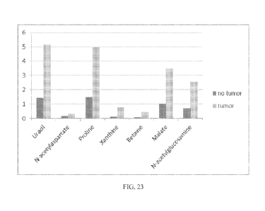
the biomarker in normal tissue and the amount in tumorous tissue. Figure 23
shows the
comparison of the uracil, N-acetylaspartate, proline, xanthine, betaine,
malate, and N-
acetylglucosamine biomarkers in normal tissue (black) versus tumor tissue
(gray). Figure 24
shows the comparison of the cysteine and alanine levels in normal tissue
(black) versus tumor
(gray). These data indicate that the biomarkers of the present invention can
be used to
quantitatively determine the amount of cancer tissue in a particular prostate
biopsy. Therefore,
these biomarkers can be used to detect prostate cancer, determine prostate
cancer prognosis,
monitor the treatment of the disease, and screen possible new treatments for
prostate cancer.
101231 While the invention has been illustrated and described in the
figures and foregoing
description, the same is to be considered as illustrative and not restrictive
in character, it being
understood that only the preferred embodiments have been shown and described
and that all
- 25 -
CA 02871736 2019-10-27
WO 2013/162773
PCT/US2013/032526
changes and modifications that come within the spirit of the invention are
desired to be
protected. In addition, all references and patents cited herein are indicative
of the level of skill
in the art and hereby incorporated by reference in their entirety.
References
1. Ries L AG MD, Krapcho M, Stinchcomb D G, Howlader N, Homer M J, Marion A.
Miller B A, Feuer E J, Altekruse S F, Lewis D R, Clegg L, Eisner M P, Reichman
M,
Edwards B K (eds). SEER Cancer Statistics Review, 1975-2005. National Cancer
Institute
Bethesda, Md. 2008; http://seer.cancer.govicsr/1975--2005/, based on
November 2007
SEER data submission, posted to the SEER web site.
2. Nelen V. Epidemiology of prostate cancer. Recent Results in Cancer Research
2007; 175:
1-8.
3. Thompson I M, Pauler D K, Goodman P J, et al. Prevalence of prostate cancer
among men
with a prostate-specific antigen level <or =4.0 ng per milliliter. [see
comment] [erratum
appears in N Engl J. Med. 2004 Sep. 30; 351(14):1470]. New England Journal of
Medicine
2004; 350: 2239-46.
4. Djavan B, Remzi M, Schulman C C, Marberger M, Zlotta A R. Repeat prostate
biopsy:
who, how and when? A review. Eur Urol 2002; 42: 93-103.
5. Epstein J I, Herawi M. Prostate needle biopsies containing prostatic
intraepithelial
neoplasia or atypical foci suspicious for carcinoma: implications for patient
care. J Urol
2006; 175: 820-34.
6. Egevad L, Grantors T, Karlberg L, Bergh A, Stank! P. Prognostic value of
the Gleason
score in prostate cancer. BJU International 2002; 89: 538-42.
7. Pinthus J H, Witkos M, Fieshner N E, et al. Prostate cancers scored as
Gleason 6 on
prostate biopsy are frequently Gleason 7 tumors at radical prostatectomy:
implication on
outcome. Journal of Urology 2006; 176: 979-84.
8. D'Amico AV, Whittington R, Malkowicz SB, Fondurulia J, Chen MB, Kaplan I,
Beard CJ,
Tomaszewski jE, Renshaw AA, Wein A., Coleman CN. Pretreatment nomogram for
prostate-specific antigen recurrence after radical prostatectomy or external-
beam radiation
therapy for clinically localized prostate cancer. J Clin Oncol. 1999
Jan;17(1):168-72.
9. Parilch K, Peppelenbosch MP: Kinome profiling of clinical cancer
specimens. Cancer Res,
70(7):2575-2578.
10. Henson DE: Back to the drawing board on immunohistochemistiy and
predictive factors. J
Nati Cancer Inst 2005, 97(24):1796-1797.
- 26 -
CA 02871736 2019-10-27
WO 2013/162773
PCT/US2013/032526
11. Rudiger T, Holler H, Kreipe HH, Nizze H, Pfeifer U, Stein H, Dallenbach
FE, Fischer HP,
Mengel M, von Wasielewski R et al: Quality assurance in immunohistochemistry:
results of
an interlaboratory trial involving 172 pathologists. Am J Surg Pathol 2002,
26(7):873-882.
12. Rhodes A, Jasani B, Balaton AJ, Miller KD: Immunohistochemical
demonstration of
estrogen and progesterone receptors: correlation of standards achieved on in
house tumours
with that achieved on external quality assessment material in over 150
laboratories from 26
countries. J Clin Pathol 2000, 53(4):292-301.
13. Rhodes A, Borthwick D, Sykes R, Al-Sam S, Paradiso A: The use of cell line
standards to
reduce HER-2/neu assay variation in multiple European cancer centers and the
potential of
automated image analysis to provide for more accurate cut points for
predicting clinical
response to trastuzumab. Am J Clin Pathol 2004, 122(1):51-60.
14. Fox CH, Johnson FB, Whiting J, Roller PP: Formaldehyde fixation. J
Histochem Cytochem
1985, 33(8):845-853.
15. Dimenstein 1B: A Pragmatic Approach to Formalin Safety in Anatomical
Pathology.
Labmed 2009,40:740-746.
16. Gillespie JW, Best CJ, Bichsel VE, Cole ICA, Greenhut SF, Hewitt SM,
Alvan' M,
Gathright YB, Merino MJ, Strausberg RL et al: Evaluation of non-fonnalin
tissue fixation
for molecular profiling studies. Am J Pathol 2002, 160(2):449-457.
17. Ahram M, Flaig MJ, Gillespie JW, Duray PH, Linehan W'M, Ornstein DK, Niu
5, Zhao Y,
Petricoin EF, 3rd, Emmert-Buck MR: Evaluation of ethanol-fixed, paraffin-
embedded
tissues for proteomic applications. Proteomics 2003, 3(4):413-421.
18. Cox ML, Schray CL, Luster CN, Stewart ZS, Korytko PJ, KN MK, Paulauskis
JD, Dunstan
RW: Assessment of fixatives, fixation, and tissue processing on morphology and
RNA
integrity. Exp Mol Pathol 2006, 80(2):183-191.
19. Jemal A, Siegel R, Xu J, Ward E: Cancer statistics, 2010. CA Cancer J
Clin, 60(5):277-300.
20. Hernandez J, Thompson IM: Prostate-specific antigen: a review of the
validation of the
most commonly used cancer biomarker. Cancer 2004, 101(5):894-904.
21. Schroder FH, Hugosson J, Roobol MJ, Tammela TL, Ciafto S, Nelen V,
Kwiatkowski M,
Lujan M, Lilja Fl, Zappa M et al: Screening and prostate-cancer mortality in a
randomized
European study. N Engl J Med 2009, 360(13):1320-1328.
22. Andriole GL, Crawford ED, Grubb RL, 3rd, Buys SS, Chia D, Church TR, Fouad
MN,
Gelmann EP, Kvale PA, Reding DJ et al: Mortality results from a randomized
prostate-
cancer screening trial. N Engl J Med 2009, 360(13):1310-1319.
- 27 -
CA 02871736 2014-10-27
WO 2013/162773
PCT/US2013/032526
23. Bastian PJ, Carter BH, Bjartell A. Seitz M, Stanislaus P, Montorsi F,
Stief CG, Schroder F:
Insignificant prostate cancer and active surveillance: from definition to
clinical
implications. Eur Urol 2009, 55(6):1321-1330.
24. Miocinovic R, Jones JS, Pujara AC, Klein EA, Stephenson AJ: Acceptance and
Durability
of Surveillance as a Management Choice in Men with Screen Detected, Low Risk
Prostate
Cancer: Improved Outcomes with Stringent Enrollment Criteria.
Urology 2011.
25. O'Toole SA, Selinger CI, Millar EK, Lum T, Beith JM: Molecular assays in
breast cancer
pathology. Pathology, 43(2):116-127.
26. Kelley RK, Van Bebber SL, Phillips KA, Venook AP: Personalized medicine
and oncology
practice guidelines: a case study of contemporary biomarkers in colorectal
cancer. J Nati
Compr Cane Netw, 9(1):13-25.
27. Turaga K, Acs G, Laronga C: Gene expression profiling in breast cancer.
Cancer Control,
17(3):177-182.
28. Troyer DA, Lucia MS, de Bruine AP, Mendez Meza R, Baldewijns MM, Dunscomb,
Van Engeland M, McAskill T, Bierau K, Louwagie J et al: Prostate cancer
detected by
methylated gene markers in histopathologically cancer negative tissues from
men with
subsequent positive biopsies. Cancer Epidemiol Biomarkers Prey 2009,
18(10):2717-2722.
29. Baden J, Green G, Painter J, Curtin K, Markiewicz J, Jones J, Astacio T.
Canning S.
Quijano J, Guinto W et al: Multicenter evaluation of an investigational
prostate cancer
methylation assay. J Urol 2009, 182(3):1186-1193.
30. Hogue MO: DNA methylation changes in prostate cancer: current developments
and future
clinical implementation. Expert Rev Mol Diagn 2009, 9(3):243-257.
31. Mucci LA, Pawitan Y, Demichelis F, Fall K, Stark JR, Adami HO, Andersson
SO, Andren
0, Eisenstein A, Holmberg L et al: Testing a multigene signature of prostate
cancer death
in the Swedish Watchful Waiting Cohort. Cancer Epidemiol Biomarkers Prey 2008,
17(7):1682-1688.
32. Donovan MJ, Khan FM, Fernandez G, Mesa Tejada R, Sapir M, Zubek VB, Powell
D,
Fogarasi 5, Vengrenyuk Y, Teverovskiy M et al: Personalized prediction of
tumor response
and cancer progression on prostate needle biopsy. J Urol 2009, 182(1):125-132.
33. Donovan MJ, Hamann S, Clayton M, Khan FM, Sapir M, Bayer Zubek V,
Fernandez G,
Mesa Tejada R, Teverovskiy M, Reuter VE et al: Systems pathology approach for
the
prediction of prostate cancer progression after radical prostatectomy. J Clin
Oncol 2008,
26(24):3923-3929.
- 28 -
CA 02871736 2019-10-27
WO 2013/162773
PCT/US2013/032526
34. Sreekurnar A, Poisson LM, Rajendiran TM, Khan AP, C'ao Q, Yu J, Laxman B,
Mehra R,
Lonigro RJ, Li Y et al: Metabolomic profiles delineate potential role for
sarcosine in
prostate cancer progression. Nature 2009, 457(7231):910 914.
35. Tessem MB, Swanson MG, Keshari KR, Albers MJ, Joun D, Tabatabai ZL, Simko
JP,
Shinohara K, Nelson SJ, Vigneron DB et al: Evaluation of lactate and alanine
as metabolic
biomarkers of prostate cancer using 1H HR-MAS spectroscopy of biopsy tissues.
Magn
Reson Med 2008, 60(3):510-516.
36. Swanson MG, Keshari KR, Tabatabai ZL, Simko JP, Shinohara K, Carroll PR,
Zektzer AS,
Kurhanewicz J: Quantification of choline- and ethanolamine-containing
metabolites in
human prostate tissues using 1H HR-MAS total correlation spectroscopy. Magn
Reson Med
2008, 60(1):33-40.
37. Serkova NJ, Gamito EJ, Jones RH, O'Donnell C, Brown JL, Green 5, Sullivan
H, Hedlund
T, Crawford ED: The metabolites citrate, myo-inositol, and spermine are
potential age-
independent markers of prostate cancer in human expressed prostafic
secretions. Prostate
2008, 68(6):620-628.
38. Mueller-Lisse UG, Scherr MK: Proton MR spectroscopy of the prostate. Eur J
Radiol 2007,
63(3):351 -360.
39. Jordan KW, Cheng LL: NMR-based metabolomics approach to target biomarkers
for
human prostate cancer. Expert Rev Proteomics 2007, 4(3):389-400.
40. Costello LC, Franklin RB: The clinical relevance of the metabolism of
prostate cancer; zinc
and tumor suppression: connecting the dots. Mol Cancer 2006, 5:17.
41. Dotan E, Cohen SJ, Alpaugh KR, Meropol NJ: Circulating tumor cells:
evolving evidence
and future challenges. Oncologist 2009, 14(11):1070-1082.
42. Eschwege P, Moutereau 5, Droupy S, Douard R, Gala :11.õ Benoit G, Conti M,
M.a3nivet P.
Lone 5: Prognostic value of prostate circulating cells detection in prostate
cancer patients:
a prospective study. Br J Cancer 2009, 100(4):608-610.
43. Okegawa T, Nutahara K, Higashihara E: Prognostic significance of
circulating tumor cells
in patients with hormone refractory prostate cancer. J Urol 2009, 181(3):1091-
1097.
44. Davis JW, Nakanishi H, Kumar VS, Bhadkamkar VA, McCormack R, Fritsche HA,
Handy
B, Gomet T, Babaian RJ: Circulating tumor cells in peripheral blood samples
from patients
with increased serum prostate specific antigen: initial results in early
prostate cancer. J Urol
2008, 179(6):2187-2191; discussion 2191.
- 29 -
CA 02871736 2019-10-27
WO 2013/162773
PCT/US2013/032526
45. Danila DC, Heller G, Gignac GA, Gonzalez-Espinoza R, Arland A, Tanaka E.
Lilja H,
Schwartz L, Larson S, Fleisher M et al: Circulating tumor cell number and
prognosis in
progressive castration-resistant prostate cancer. Clin Cancer Res 2007,
13(23):7053-7058.
46. Evans AM, DeFIaven CD, Barrett T, Mitchell M, Milgram E: Integrated,
nontargeted
ultrahigh performance liquid chromatographylelectrospray ionization tandem
mass
spectrometry platform for the identification and relative quantification of
the small-
molecule complement of biological systems. Anal Chem 2009, 81(16):6656-6667.
47. Dehaven CD, Evans AM, Dai H, Lawton KA: Organization of GC/MS and LC/MS
metabolomics data into chemical libraries. .1 Cheminform 2010, 2(1):9.
48. Crehange G, Parfait S, Liegard M, Maingon P, Ben Salem D, Cochet A, Funes
de la Vega
M, Cormier L, Bomietain F, Mirjolet C et al: Tumor Volume and Metabolism of
Prostate
Cancer Determined by Proton Magnetic Resonance Spectroscopic Imaging at 3T
Without
Endorectal Coil Reveal Potential Clinical Implications in the Context of
Radiation
Oncology. Int .1 Radiat Oncol Biol Phys 2010.
49. Zakian KL, Shukla Dave A, Ackerstaff E, Hricak H, Koutcher JA: 1H magnetic
resonance
spectroscopy of prostate cancer: biomarkers for tumor characterization. Cancer
Biomark
2008, 4(45):263-276.
50. Swanson MG, Zektzer AS, Tabatabai ZL, Simko J, Jarso 5, Keshari KR,
Schmitt L, Carroll
PR, Shinohara K, Vigneron DB et al: Quantitative analysis of prostate
metabolites using 1H
HR MAS spectroscopy. Magn Reson Med 2006, 55(6):1257-1264.
51. Schipper RU, Romijn jC, Cuijpers VM, Verhofitad AA: Polyamines and
prostatic cancer.
Biochem Soc Trans 2003, 31(2):375-380.
52. Oyama T, Allsbrook WC, Jr., Kurokawa K, Matsuda H, Segawa A, Sano T,
Suzuki K,
Epstein 11: A comparison of interobserver reproducibility of Gleason grading
of prostatic
carcinoma in Japan and the United States. Arch Pathol Lab Med 2005,
129(8):1004-1010.
53. Netto GJ, Eisenberger M, Epstein jl: Interobserver Variability in
Histologic Evaluation of
Radical Prostatectomy Between Central and Local Pathologists: Findings of TAX
3501
Multinational Clinical Trial. Urology 2010.
54. Allsbrook WC, Jr., Mangold KA, Johnson MU, Lane RB, Lane CU, Epstein JI:
Interobserver reproducibility of Gleason grading of prostatic carcinoma:
general
pathologist. Hum Pathol 2001, 32(1):81-88.
55. Allsbrook WC, Jr., Mangold KA, Johnson MI1, Lane RB, Lane CG, Amin MB,
Bostwick
DG, Humphrey PA, Jones EC, Reuter VE et al: Interobserver reproducibility of
Gleason
grading of prostatic carcinoma: urologic pathologists. Hum Pathol 2001,
32(1):74-80.
-30-
CA 02871796 2014-10-27
WO 2013/162773
PCT/US2013/032526
56. Paik S, Shak S, Tang G, Kim C, Baker J, Cronin M, Baehner FL, Walker MG,
Watson D,
Park T et al: A multigene assay to predict recurrence of tamoxifen treated,
node negative
breast cancer. N Engl J Med 2004, 351(27):2817-2826.
57. Hewitt SM, Lewis FA, Cao Y, Conrad RC, Cronin M, Dartenberg KD, Goralski
TJ,
Langmore JP, Raja RG, Williams PM et al: Tissue handling and specimen
preparation in
surgical pathology: issues concerning the recovery of nucleic acids from
formalin fixed,
paraffin embedded tissue. Arch Pathol Lab Med 2008, 132(12):1929-1935.
58. Tuck MK, Chan DW, Chia D, Godwin AK, Grizzle WE, Krueger KE, Rom W, Sanda
M,
Sorbara L, Stass S et al: Standard operating procedures for serum and plasma
collection:
early detection research network consensus statement standard operating
procedure
integration working group. J Proteome Res 2009, 8(1):113-117.
59. McLeffan D, Grizzle WE, Feng Z, Thompson IM, Bigbee WL, Cazares LH, Chan
DW,
Dahlgren J, Diaz j, Kagan J et al: SELDI TOF MS whole serum proteomic
profiling with
IMAC surface does not reliably detect prostate cancer. Clin Chem 2008,
54(1):53-60.
60. Vandromme MJ, Umphrey H, Krontiras H: Image guided methods for biopsy of
suspicious
breast lesions. J Surg Oncol, 103(4):299-305.
61. Solomon SB, Zakowski MF, Pao W, Thornton RH, Lada3nyi M, Kris MG, Rusch
VW,
Rizvi NA: Core needle lung biopsy specimens: adequacy for EGFR and KRAS
mutational
analysis. AJR Am J Roentgenol, 194(1):266-269.
62. Piccart Gebhart MJ, Procter M, Leyland Jones B, Goldhirsch A, Untch M,
Smith I, Gianni
L, Baselga J, Bell R, Jackisch C et al: Trastuz,umab after adjuvant
chemotherapy in HER2
positive breast cancer. N Engl J Med 2005, 353(16):1659-1672.
63. Diamond J, Anderson N, Bartels P, Montironi R, and Hamilton P. (2004) The
use of
morphological characteristics and texture analysis in the identification of
tissue
composition in prostatic neoplasia, Human Pathology 35: 1121-1131
64. Tabesh A, Teverovskiy M, Pang H-Y, Kumar VP, Verbel D, Kotsianti A, Saidi
0, (2007)
Multifeature prostate cancer diagnosis and Gleason grading of histological
images, IEEE
Trans. Med. Imag. 26: 1366-1378
65. Huang P-W, Lee C-H. (2009) Automatic Classification for Pathological
Prostate Images
Based on Fractal Analysis," IEEE Trans on Medical Imaging 28: 1037-1050
66. Shuster et al. "Molecular preservation by extraction and fixation, mPREF:
a method for
small molecule biomarker analysis and histology on exactly the same tissue."
BMC
Clinical Pathology 2011, 11:14
-31 -
CA 02871736 2014-10-27
WO 2013/162773
PCT/US2013/032526
67. Sandblom G, Dufinats M, Varenhorst E. (2000) Longterm survival in a
Swedish population
based cohort of men with prostate cancer. Urology 56: 442-447
68. Kramer BS, Croswell 1M. Cancer screening: the clash of science and
intuition. (2009)
Annu Rev Me,d 60:125-137
69. Welch HG, Black WC. (2010) Overdiagnosis in cancer. J Nat! Cancer Inst.
102:605-613.
70. Eckersberger E, Finkelstein J. Sadri H, Margreiter M, Taneja. SS, Lepor H,
Djavan B.
(2009) Screening for Prostate Cancer: A review of the ERSPC, and PLCO Trials.
Rev Urol
2009 11:127-133
71, Berger MF, et al. (2011) The genomic complexity of primary human prostate
cancer.
Nature 470:214-220
- 32 -