Note: Descriptions are shown in the official language in which they were submitted.
STEERABLE AND CURVABLE CAVITY CREATION SYSTEM
[0001] This paragraph intentionally removed.
BACKGROUND OF THE INVENTION
Field of the Invention
[0002] The invention relates, in some embodiments, to bone
augmentation devices and
procedures. In particular, the present invention relates to steerable and
curvable injection devices and
systems for introducing conventional or novel bone cement formulations such as
in performing
vertebroplasty.
- 1 -
CA 2872107 2019-06-04
CA 02872107 2014-10-30
WO 2013/166209
PCMJS2013/039149
Description of the Related Art
[0003] According to the National Osteoporosis Foundation ten million
Americans have osteoporosis (OSP), and an estimated 34 million with low bone
mass are
at risk of developing osteoporosis (http://www.nof
org/osteoporosis/diseasefacts.htm).
Called the "silent disease," OSP develops slowly over a number of years
without
symptoms. Eighty percent of those affected are women, particularly petite
Caucasian and
Asian women, although older men and women of all races and ethnicities are at
significant risk.
[0004] In the United States, 700,000 people are diagnosed with
vertebral
compression fractures as a result of OSP each year. Morbidity associated with
vertebral
fractures includes severe back pain, loss of height and deformity, all of
which negatively
affect quality of life.
[0005] Once microfracture of the vertebra begins, there is little the
clinician
can do except palliative medical treatment using analgesics, bed rest and/or
restriction of
activity. With time, the microfractures widen at one level and without
surgical
intervention, the fractures cascade downward with increasing kyphosis or
"hunching" of
the back. Once a mechanical lesion develops, surgery is often the only
practical option.
Vertebroplasty or kyphoplasty are the primary minimally-invasive surgical
procedures
performed for the treatment of compression-wedge fractures due to OSP.
[00061 Vertebroplasty stabilizes the collapsed vertebra by injecting
polymethylmethacrylate (PMMA) or a substantially equivalent bone cement into
cancellous bone space of the vertebrae. Besides providing structural support
to the
vertebra, the exothermic reaction of PMMA polymerization is said to kill off
the
nociceptors or pain receptors in the bone, although no proof of this
hypothesis has been
provided in the literature. This procedure is typically performed as an
outpatient
procedure and requires only a short-acting local or general anesthetic. Once
the surgical
area of the spine is anesthetized, the physician inserts one or two needles
through small
skin incisions into either the pedicle (uni-transpedicular) or the pedicles of
the vertebral
body i.e., bi-transpedicular. Polymethylmethacrylate (PMMA) is injected
through the
needle and into the cancellous-bone space of the vertebra.
[0007] Kyphoplasty mirrors the vertebroplasty procedure but has the
additional step of inserting and expanding a nylon or polyurethane balloon in
the interior
of the vertebral body. Expansion of the balloon under pressure reduces the
compression
-2-
CA 02872107 2014-10-30
WO 2013/166209
PCT/US2013/039149
fracture and creates a cavity. After withdrawal of the balloon, PMMA is
injected into the
cavity to stabilize the reduction. The kyphoplasty procedure may restore the
vertebral
body height. Kyphoplasty is an in-patient surgery that requires
hospitalization and a
general anesthetic. Kyphon Inc. claims over 275,000 spinal fractures have been
treated
using their PMMA derivative and their "balloon" kyphoplasty procedure
worldwide
(Sunnyvale, Calif., September 5, 2006, (PR NEWS WIRE) Kyphon study 2006).
[0008] Bone cement for both vertebroplasty and kyphoplasty procedures
currently employ variations of standard PMMA in a powder and a methyl
methacrylate
monomer liquid. When the powder and liquid monomer are mixed, an exothermic
polymerization takes place resulting in the formation of a "dough-like"
material, which is
then inserted into the cancellous bone space. The dough, when hardened,
becomes either
the reinforcing structure or the grout between the bone and prosthesis in the
case of total
joint replacement.
[0009] The average clinical in vivo life of the PMMA grout is
approximately
years due to corrosion fatigue of either the bone-cement/prosthesis and/or the
bone
cement/bone interfaces. Jasty et al. (1991) showed that in cemented total hip
replacements: "Fractures in the cement mantle itself were found on cut
sections around all
prostheses which had been in use for over three years." Jasty et al. also
noted: "In
general, specimens less than 10 years in situ showed small incomplete
fractures while the
specimens in place more than 10 years all showed large complete cement mantle
fractures."
[0010] When an implant fails, a revision becomes almost mandatory.
After
removal of the cement and hardware, a cemented arthroplasty can be repeated if
enough
cancellous bone matrixes exist to grip the new PMMA. Alternatively, cement-
less
prostheses can be installed. Such a revision, however, can only be applied to
total joint
replacement failures. For vertebroplasty and/or kyphoplasty, a classical screw
and plate
internal fixation with autograft fusion is necessary.
[0011] Despite advances in the foregoing procedures, there remains a
need for
improved bone cement delivery systems which enable rapid and controllable
deployment
of bone cement for the treatment of conditions such as vertebral compression
fractures.
-3-
CA 02872107 2014-10-30
WO 2013/166209
PCMJS2013/039149
SUMMARY OF THE INVENTION
[0012] There is provided in accordance with one aspect of the present
invention, a steerable and curvable vertebroplasty device having a cavity
creation
element. The vertebroplasty device comprises an elongate tubular body, having
a
proximal end, a distal end, and a central lumen extending therethrough. A
deflectable
zone is provided on the distal end of the tubular body, for deflection through
an angular
range. A handle is provided on the proximal end of the tubular body, having a
deflection
controller thereon. A cavity creating element may be carried by the
deflectable zone. In
one embodiment, the cavity creating element is an inflatable balloon, in
communication
with a proximal inflation port by way of an elongate inflation lumen extending
throughout the length of the tubular body.
[0013] The deflection controller may comprise a rotatable element,
such as a
knob rotatable about the longitudinal axis of the handle.
[0014] The distal end of the tubular body is provided with at least
one exit
port in communication with the central lumen. The exit port may open in a
lateral
direction, an axial direction, or along an inclined surface positioned
distally of a transition
point between the longitudinal side wall of the tubular body and the distal
end of the
distal tip.
[0015] In another aspect of the invention, disclosed is a steerable
and curvable
vertebroplasty device having a plurality of cavity creation elements. The
device can
include an elongate, tubular body, having a proximal end, a distal end, and a
central
lumen extending therethrough; a deflectable zone on the distal end of the
tubular body,
deflectable through an angular range; a handle on the proximal end of the
tubular body;
and a deflection controller on the handle; a first cavity creating element
carried by the
deflectable zone; and a second cavity creating element on the elongate tubular
body. The
second cavity creating element can be carried at least partially by the
deflectable zone.
The first and/or second cavity creating element can be a balloon. The first
and second
cavity creating elements can share a common inflation lumen, or have separate
lumens.
The first cavity creating element and/or second cavity creating element could
be
positioned proximal to, or distal to one or more exit ports on the tubular
body. The first
and/or cavity creating element could include a filament layer, such as a
braided layer.
[0016] A method of performing vertebroplasty is also disclosed
herein,
according to some embodiments. The method can include the steps of: creating a
-4-
CA 02872107 2014-10-30
WO 2013/166209
PCMJS2013/039149
pedicular access channel in a pedicle to access the interior of a vertebral
body; inserting
an introducer cannula into the pedicle; inserting a steerable and curvable
injection needle
through the introducer cannula into the interior of a vertebral body, the
steerable and
curvable injection needle having a proximal end and a distal end, the distal
end having a
first configuration substantially coaxial with a long axis of the proximal
end, the steerable
and curvable injection needle also having a first cavity creating element and
a second
cavity creating element; rotating a control to deflect the distal end of the
steerable and
curvable injection needle to a second configuration that is not substantially
coaxial with
the long axis of the proximal end; actuating the first cavity creating element
to create a
first cavity within the interior of the vertebral body; actuating a second
cavity creating
element to create a second cavity within the interior of the vertebral body;
and flowing
bone cement through the steerable and curvable injection needle into the
interior of the
vertebral body.
[0017] In some embodiments, flowing bone cement through the steerable
and
curvable injection needle into the interior of the vertebral body comprises
releasing a first
particle-containing bone cement within the interior of the vertebral body, the
bone cement
comprising at least 30%, 35%, 40%, 45%, 50%, or more particles by weight, and
additionally comprises releasing a second particle-containing bone cement
within the first
bone cement, the second particle-containing bone cement comprising less than
about
35%, 30%, 25%, 20%, or less particles by weight.
[0018] In another embodiment, disclosed herein is a steerable and
curvable
vertebroplasty device, that can include an elongate, tubular body, having a
proximal end,
a distal end, and a central lumen extending therethrough; a deflectable zone
on the distal
end of the tubular body, deflectable through an angular range; a handle on the
proximal
end of the tubular body; a deflection controller on the handle; and a cavity
creating
element carried by the deflectable zone, wherein the cavity creating element
comprises a
filament layer.
[0019] In still another embodiment, disclosed is a steerable and
curvable
vertebroplasty device that includes an elongate, tubular body, having a
proximal end, a
distal end, and a central lumen extending therethrough; a deflectable zone on
the distal
end of the tubular body, deflectable through an angular range; a handle on the
proximal
end of the tubular body; a deflection controller on the handle; and a cavity
creating
-5-
CA 02872107 2014-10-30
WO 2013/166209
PCMJS2013/039149
element carried by the deflectable zone, wherein the cavity creating element
comprises a
plurality of concentric balloons.
[0020] Also disclosed herein is a steerable vertebroplasty device,
comprising
an elongate, tubular body having a proximal end, a distal end, and a central
lumen
extending therethrough. The distal end can include a closed distal-facing
surface and a
lateral-facing surface comprising an exit aperture in connection with the
central lumen.
The exit port is defined by at least a first angled surface. Some apertures
can include a
first angled surface and a second angled surface, the first angled surface
opposing and
being non-parallel to the second angled surface. The device can also include a
deflectable
zone on the distal end of the tubular body, deflectable through an angular
range; the
deflectable zone having a proximal portion and a distal portion. The elongate
tubular
body has a longitudinal axis extending from the proximal end to the proximal
portion of
the deflectable zone. The deflectable zone is movable from a first
configuration coaxial
with the first longitudinal axis in an unstressed state to a second deflected
configuration.
The device can also have a handle on the proximal end of the tubular body, a
deflection
control on the handle, and an input port for receiving bone cement. The first
angled
surface and the second angled surface can have longitudinal axes that
intersect and form
an angle of between about, for example, 30 degrees and 150 degrees, 60 degrees
and 120
degrees, 75 degrees and 105 degrees, or about 90 degrees in some embodiments.
The
distal end can include an end cap operably attached to the tubular body, and
in some
embodiments have a zone having a radially inwardly tapering diameter. The
first radial
surface can include a proximal radial termination, and the second radial
surface can
include a distal radial termination. The proximal radial termination can be
radially offset
from the distal radial termination by at least about 0.01 inches, 0.05 inches,
0.10 inches,
or more. The exit aperture can include a rippled zone.
[0021] Also disclosed herein is a steerable vertebroplasty device
having an
elongate, tubular body having a proximal end, a distal end, and a central
lumen extending
therethrough. The distal end can include a closed distal-facing surface and a
lateral-facing
surface comprising an exit port in connection with the central lumen. The exit
port can be
defined by a first wall and a second wall that is not parallel to the first
wall. The exit port
can have a first inner axial or circumferential dimension at a junction with
the central
lumen and a second outer axial or circumferential dimension where bone cement
exits the
device. The second dimension can be less than, equal to, or greater than the
first
-6-
CA 02872107 2014-10-30
WO 2013/166209
PCMJS2013/039149
dimension, such as by about 5%, 10%, 15%, 20%, 25%, 35%, 40%, 50%, 75%, 100%,
or
more. The device can also include a deflectable zone on the distal end of the
tubular
body, deflectable through an angular range. The deflectable zone can have a
proximal
portion and a distal portion. The elongate tubular body has a first
longitudinal axis
extending from the proximal end to the proximal portion of the deflectable
zone. The
deflectable zone is movable from a first substantially straight configuration
in an
unstressed state to a second deflected configuration. The device also can
include a handle
on the proximal end of the tubular body, a deflection control on the handle,
and an input
port for receiving bone cement, the input port having a second longitudinal
axis spaced
apart from and at an angle with respect to the first longitudinal axis, the
input port
positioned distally on the elongate, tubular body relative to the deflection
control.
[0022] In another aspect, disclosed herein is a method for treating a
bone. The
method can include the steps of creating a pedicular access channel in a
pedicle to access
the interior of a vertebral body; inserting an introducer cannula into the
pedicle; inserting
a steerable injection needle through the introducer cannula into the interior
of a vertebral
body, the steerable injection needle having a proximal end, a tubular body
having a
longitudinal axis, and a distal end, a control for controlling deflection of
the distal end,
and an input port having a longitudinal axis and configured to receive bone
cement,
wherein the control is positioned proximally to the input port, wherein the
longitudinal
axis of the input port is not coaxial with the longitudinal axis of the
tubular body, wherein
the distal end has a first configuration substantially coaxial with the
longitudinal axis of
the tubular body, wherein the distal end comprises a closed distal-facing
surface and a
lateral-facing surface comprising an exit aperture in connection with a
central lumen, the
exit aperture defined by a first angled surface and a second angled surface,
the first
angled surface opposing and being non-parallel to the second angled surface;
adjusting
the control to deflect the distal end of the steerable injection needle to a
second
configuration that is not substantially coaxial with the longitudinal axis of
the tubular
body; and flowing bone cement through the steerable injection needle, out the
exit
aperture and into the interior of the vertebral body. In some embodiments, the
exit
aperture has a first inner axial or circumferential dimension at a junction
with the central
lumen and a second outer axial or circumferential dimension where bone cement
exits the
needle, wherein the second dimension is greater than, equal to, or less than
the first width.
-7-
CA 02872107 2014-10-30
WO 2013/166209
PCMJS2013/039149
[00231 In some embodiments, disclosed is a steerable cavity creation
device.
The device can include an elongate, tubular body having a proximal end, a
distal end, a
lumen extending therethrough, a first hypotube, and a second hypotube disposed
within
the first hypotube. The device can also include a deflectable zone on the
distal end of the
tubular body, deflectable through an angular range, the deflectable zone
having a
proximal portion and a distal portion. The first hypotube can have a distal
zone having a
first plurality of slots, and the second hypotube has a distal zone having a
second plurality
of slots. The first plurality of slots can be oriented 180 degrees
circumferentially apart
from the second plurality of slots. The elongate tubular body can have a first
longitudinal
axis extending from the proximal end to the proximal portion of the
deflectable zone. The
deflectable zone can be movable from a first substantially straight
configuration in an
unstressed state to a second deflected configuration. The device can include a
handle on
the proximal end of the tubular body, as well as a deflection control on the
handle
actuated by rotation about the first longitudinal axis of the tubular body.
Upon rotation of
the deflection control, a proximally directed force is exerted on a movable
actuator
attached to the tubular body to actively change the curvature of the
deflectable zone. The
device can also include an input port. The input port can have a second
longitudinal axis
spaced apart from and at an angle with respect to the first longitudinal axis.
The input
port can be positioned distally on the elongate, tubular body relative to the
deflection
control. The device can also include a cavity creating element carried by the
deflectable
zone. Further features and advantages of the present invention will become
apparent to
those of skill in the art in view of the detailed description of preferred
embodiments
which follows, when considered together with the attached drawings and claims.
BRIEF DESCRIPTION OF THE DRAWINGS
[00241 Figure 1 is a perspective view of a steerable and curvable
injection
needle in accordance with one aspect of the present invention.
[00251 Figure 2 is a perspective view of an introducer in accordance
with one
aspect of the present invention.
[0026] Figure 3 is a perspective view of a stylet in accordance with
one aspect
of the present invention.
-8-
CA 02872107 2014-10-30
WO 2013/166209
PCMJS2013/039149
[0027] Figure 4 is a side elevational view of the steerable and
curvable
injection needle moveably coaxially disposed within the introducer, in a
substantially
linear configuration.
[0028] Figure 5 is a side elevational view of the assembly of Figure
4,
showing the steerable and curvable injection needle in a curved configuration.
[0029] Figure 6 is a side elevational schematic view of another
steerable and
curvable injection needle in accordance with the present invention.
[0030] Figure 7 A is a schematic view of a distal portion of the
steerable and
curvable needle of Figure 6, shown in a linear configuration.
[0031] Figure 7B is a schematic view as in Figure 7A, following
proximal
retraction of a pull wire to laterally deflect the distal end.
[0032] Figure 8 is a schematic view of a distal portion of a
steerable and
curvable needle, having a side port.
[0033] Figure 9A is a schematic view of a distal portion of a
steerable and
curvable needle, positioned within an outer sheath.
[0034] Figure 9B is an illustration as in Figure 9A, with the distal
sheath
partially proximally retracted.
[0035] Figure 9C is an illustration as in Figure 9B, with the outer
sheath
proximally retracted a sufficient distance to fully expose the deflection
zone.
[0036] Figures 10A-10C illustrate various aspects of an alternative
deflectable
needle in accordance with the present invention.
[0037] Figures 11A through 11C illustrate various aspects of a
further
deflectable needle design in accordance with the present invention.
[0038] Figures 12 and 13 illustrate a further variation of the
deflectable needle
design in accordance with the present invention.
[0039] Figure 14 is a side elevational cross section through the
proximal
handle of the deflectable needle illustrated in Figure 13.
[0040] Figure 15 is a cross sectional detail view of the distal tip
of the
steerable and curvable needle illustrated in Figure 13.
[0041] Figures 15A through 15X illustrate various views of
alternative distal
tip designs.
[0042] Figure 15Y illustrates schematically an injector with an anti-
coring and
clog-preventing obturator within the central lumen of the injector.
-9-
CA 02872107 2014-10-30
WO 2013/166209
PCMJS2013/039149
[0043] Figures 16A and 16B are schematic illustrations of the distal
end of a
steerable and curvable injection device in accordance with the present
invention, having a
cavity creating element thereon.
[0044] Figures 16C and 16D are alternative cross sectional views
taken along
the line 16C-16C in Figure 16A, showing different inflation lumen
configurations.
[0045] Figures 16E-16G illustrate cross-sections of further
alternative
inflation lumen configurations.
[0046] Figure 16H schematically illustrates the distal end of a
steerable and
curvable injection device having a cavity creation element with a braided
layer.
[0047] Figure 161 illustrates a cross-section through line 161-161 of
Figure
16H, which some elements omitted for clarity.
[0048] Figure 16J illustrates a cross-section similar to that of
Figure 161 with
an additional exterior layer.
[0049] Figures 16K-16M illustrate various views of an asymmetrical
cavity
creation element, according to some embodiments of the invention.
[0050] Figures 160 and 16P schematically illustrate views of a
catheter with a
plurality of coaxial balloons, according to some embodiments of the invention.
[0051] Figures 17A and 17B illustrate an alternative steerable and
curvable
injection device having a cavity creation element thereon.
[0052] Figures 17C and 17D illustrate an alternative steerable and
curvable
injection device having a plurality of cavity creation elements thereon.
[0053] Figures 17E and 17F are alternative cross sectional views
showing
different inflation lumen configurations.
[0054] Figures 17G-17J illustrate further alternative steerable and
curvable
injection devices having a plurality of cavity creation elements thereon.
[0055] Figures 18A and 18B are schematic views of a bone cement
delivery
system in accordance with the present invention.
[0056] Figures 19A through 19F show stages m the method of
accomplishing
vertebroplasty in accordance with the invention.
[0057] Figures 20A-20C show stages in a method of creating a cavity
using a
steerable and curvable injector with a plurality of cavity creation elements
during a
vertebroplasty procedure in accordance with the invention.
-10-
=
[0058] FIGS. 21A-21E illustrate various views of an embodiment
of a cavity creation
device.
DETAILED DESCRIPTION OF THE PREFERRED EMBODIMENT
[0059] The present invention provides improved delivery systems
for delivery of a
bone cement or bone cement composite for the treatment of vertebral
compression fractures due to
osteoporosis (OSP), osteo-trauma, and benign or malignant lesions such as
metastatic cancers and
myeloma, and associated access and deployment tools and procedures. U.S.
patent application Ser. No.
11/941,764 filed Nov. 16, 2007, U.S. patent application Ser. No. 12/029,428
filed Feb. 11, 2008, and
U.S. patent application Ser. No. 12/469,654 filed May 20, 2009 describe
various systems and methods
for performing verterbroplasty including steerable, curvable vertebroplasty
devices.
[0060] The primary materials in the preferred bone cement
composite are methyl
methacrylate and inorganic cancellous and/or cortical bone chips or particles.
Suitable inorganic bone
chips or particles are sold by Allosource, Osteotech and LifeNet (K053098);
all have been cleared for
marketing by FDA. The preferred bone cement also may contain the additives:
barium sulfate for radio-
opacity, benzoyl peroxide as an initiator, N,N-dimethyl-p-toluidine as a
promoter and hydroquinone as
a stabilizer. Other details of bone cements and systems are disclosed in U.S.
patent application Ser. No.
11/626,336, filed Jan. 23, 2007.
[0061] One preferred bone cement implant procedure involves a
two-step injection
process with two different concentrations of the bone particle impregnated
cement. To facilitate the
implant procedure the bone cement materials are packaged in separate
cartridges containing specific
bone cement and inorganic bone particle concentrations for each step. Tables 1
and 2, infra, list one
example of the respective contents and concentrations in Cartridges 1A and 1B
for the first injection
step, and Cartridges 2A and 2B for the second injection step.
[0062] The bone cement delivery system generally includes at
least three mam
components: 1) stylet; 2) introducer cannula; and 3) steerable and curvable
injection needle. See FIGS.
1-3. Packaged with the system or packaged separately is a cement
- 11 -
CA 2872107 2019-06-04
CA 02872107 2014-10-30
WO 2013/166209
PCMJS2013/039149
dispensing pump. The complete system also preferably includes at least one
cement
cartridge having at least two chambers therein, and a spiral mixing nozzle.
[0063] The stylet is used to perforate a hole into the pedicle of the
vertebra to
gain access to the interior of the vertebral body.
[0064] The introducer cannula is used for bone access and as a guide
for the
steerable and curvable injection needle. The introducer cannula is sized to
allow
physicians to perform vertebroplasty or lcyphoplasty on vertebrae with small
pedicles
such as the thoracic vertebra T5 as well as larger vertebrae L5. In addition,
this system is
designed for uni-transpedicular access and/or bi-pedicular access.
[0065] Once bone access has been achieved, the steerable and curvable
injection needle can be inserted through the introducer cannula into the
vertebra. The
entire interior vertebral body may be accessed using the steerable and
curvable injection
needle. The distal end of the needle can be manually shaped to any desired
radius within
the product specifications. The radius is adjusted by means of a knob on the
proximal end
of the device.
[0066] The hand-held cement dispensing pump may be attached to the
steerable and curvable injection needle by a slip-ring luer fitting. The pre-
filled 2-
chambered cartridges (1A and 1B, and 2A and 2B) are loaded into the dispensing
pump.
As the handle of the dispensing pump is squeezed, each piston pushes the
cartridge
material into the spiral mixing tube. The materials are mixed in the spiral
mixing nozzle
prior to entering the steerable and curvable injection needle. The ratio of
diameters of the
cartridge chambers determines the mixing ratio for achieving the desired
viscosity.
[0067] The bone cement implant procedures described herein use
established
vertebroplasty and kyphoplasty surgical procedures to stabilize the collapsed
vertebra by
injecting bone cement into cancellous bone.
[0068] The preferred procedure is designed for uni-transpedicular
access and
may be accomplished under either a local anesthetic or short-duration general
anesthetic.
Once the area of the spine is anesthetized, an incision is made and the stylet
is used to
perforate the vertebral pedicle and gain access to the interior of the
vertebral body. The
introducer cannula is then inserted and acts as a guide for the steerable and
curvable
injection needle.
[0069] Injection of the preferred bone cement involves a two-step
procedure.
The pre-filled Cartridges lA and 1B are loaded into the dispensing pump. As
the
-12-
CA 02872107 2014-10-30
WO 2013/166209
PCT/1JS2013/039149
dispensing pump handle is squeezed, each piston pushes material into the
spiral mixing
tube. The diameter of each chamber may be utilized to determine the mixing
ratio for
achieving the desired viscosity.
[0070] The first step involves injecting a small quantity of PMMA
with more
than about 35%, e.g., 60% inorganic bone particles, onto the outer periphery
of the
cancellous bone matrix, i.e., next to the inner wall of the cortical bone of
the vertebral
body. The cement composite is designed to harden relatively quickly, forming a
firm but
still pliable shell. This shell is intended to prevent bone marrow/PMMA
content from
being ejected through any venules or micro-fractures in the vertebral body
wall. The
second step of the procedure involves a second injection of PMMA with an
approximately 30% inorganic bone particles to stabilize the remainder of the
weakened,
compressed cancellous bone.
[0071] Alternatively, the steerable and curvable needle disclosed
herein and
discussed in greater detail below, can be used in conventional vertebroplasty
procedures,
using a single step bone cement injection.
[0072] Injection control for the first and second steps is provided
by a 2 mm
ID flexible injection needle, which is coupled to the hand operated bone
cement injection
pump. The 60% (> 35%) and 30% ratio of inorganic bone particle to PMMA
concentrations may be controlled by the pre-filled cartridge sets lA and B,
and 2A and
2B. At all times, the amount of the injectate is under the direct control of
the surgeon or
intervention radiologist and visualized by fluoroscopy. The introducer cannula
is slowly
withdrawn from the cancellous space as the second injection of bone cement
begins to
harden, thus preventing bone marrow/PMMA content from exiting the vertebral
body.
The procedure concludes with closure of the surgical incision with bone
filler. In vitro
and in vivo studies have shown that the 60% (> 35%) bone-particle impregnated
bone
cement hardens in 2-3 minutes and 30% bone-particle impregnated bone cement
hardens
between 4 to 10 minutes.
[0073] Details of the system components will be discussed below.
[0074] There is provided in accordance with the present invention a
steerable
and curvable injection device that can be used to introduce any of a variety
of materials or
devices for diagnostic or therapeutic purposes. In one embodiment, the system
is used to
inject bone cement, e.g., PMMA or any of the bone cement compositions
disclosed
elsewhere herein. The injection system most preferably includes a tubular body
with a
-13-
CA 02872107 2014-10-30
WO 2013/166209
PCT/1JS2013/039149
steerable and curvable (i.e., deflectable) distal portion for introducing bone
cement into
various locations displaced laterally from the longitudinal axis of the device
within a
vertebral body during a vertebroplasty procedure.
[0075] Referring to Figure 1, there is illustrated a side perspective
view of a
steerable and curvable injection needle 10 in accordance with one aspect of
the present
invention. The steerable and curvable injection needle 10 comprises an
elongate tubular
body 12 having a proximal end 14 and a distal end 16. The proximal end 14 is
provided
with a handle or manifold 18, adapted to remain outside of the patient and
enable
introduction and/or aspiration of bone cement or other media, and control of
the distal
end as will be described herein. In general, manifold 18 is provided with at
least one
injection port 20, which is in fluid communication with a central lumen (not
illustrated)
extending through tubular body 12 to at least one distal exit port 22.
[0076] The manifold 18 is additionally provided with a control 26
such as a
rotatable knob, slider, or other moveable control, for controllably deflecting
a deflection
zone 24 on the distal end 16 of the tubular body 12. As is described elsewhere
herein, the
deflection zone 24 may be advanced from a relatively linear configuration as
illustrated in
Figure 1 to a deflected configuration throughout an angular range of motion.
[0077] Referring to Figure 2, there is illustrated an elongate
tubular introducer
30, having a proximal end 32, a distal end 34 and an elongate tubular body 36
extending
there between. A central lumen 38 (not shown) extends between a proximal
access port
40 and a distal access port 42.
[0078] The central lumen 38 has an inside diameter which is adapted
to slide
axially to receive the steerable and curvable injection needle 10
therethrough. This
enables placement of the distal end 34 adjacent a treatment site within the
body, to
establish an access pathway from outside of the body to the treatment site. As
will be
appreciated by those of skill in the art, the introducer 30 enables procedures
deep within
the body such as within the spine, through a minimally invasive and/or
percutaneous
access. The steerable and curvable injection needle 10 and/or other procedure
tools may
be introduced into port 40, through lumen 38 and out of port 42 to reach the
treatment
site.
[0079] The proximal end 32 of introducer 30 may be provided with a
handle
44 for manipulation during the procedure. Handle 44 may be configured in any
of a
-14-
CA 02872107 2014-10-30
WO 2013/166209
PCMJS2013/039149
variety of ways, such as having a frame 46 with at least a first aperture 48
and a second
aperture 50 to facilitate grasping by the clinician.
[0080] Referring to Figure 3, there is illustrated a perspective view
of stylet
60. Stylet 60 comprises a proximal end 62, a distal end 64 and an elongate
body 66
extending there between. The proximal end 62 may be provided with a stop 68
such as a
grasping block, manifold or other structure, to facilitate manipulation by the
clinician. In
the illustrated embodiment, block 68 is configured to nest within a recess 70
on the
proximal end of the introducer 30.
[0081] As will be appreciated by those of skill in the art, the
stylet 60 has an
outside diameter which is adapted to coaxially slide within the central lumen
on
introducer 30. When block 68 is nested within recess 70, a distal end 64 of
stylet 60 is
exposed beyond the distal end 34 of introducer 30. The distal end 64 of stylet
60 may be
provided with a pointed tip 72, such as for anchoring into the surface of a
bone.
[0082] Referring to Figure 4, there is illustrated a side elevational
view of an
assembly in accordance with the present invention in which a steerable and
curvable
injection needle 10 is coaxially positioned within an introducer 30. The
introducer 30 is
axially moveably carried on the steerable and curvable injection needle 10. In
the
illustration of Figure 4, the introducer 30 is illustrated in a distal
position such that it
covers at least a portion of the deflection zone 24 on injection needle 10.
[0083] Figure 5 illustrates an assembly as in Figure 4, in which the
introducer
30 has been proximally retracted along the injection needle 10 to fully expose
the
deflection zone 24 on injection needle 10. In addition, the control 26 has
been
manipulated to deflect the deflection zone 24 through an angle of
approximately 90 .
Additional details of the steerable and curvable needle will be discussed
below.
[0084] Figure 6 illustrates a schematic perspective view of an
alternate
steerable and curvable vertebroplasty injector, according to one embodiment of
the
invention. The steerable and curvable injector 700 includes a body or shaft
portion 702
that is preferably elongate and tubular, input port 704, adjustment control
706, and handle
portion 708. The elongate shaft 702 preferably has a first proximal portion
710 and a
second distal portion 712 which merge at a transition point 714. Shaft 702 may
be made
of stainless steel, such as 304 stainless steel, Nitinol, Elgiloy, or other
appropriate
material. Alternatively, the tubular body 702 may be extruded from any of a
variety of
polymers well known in the catheter arts, such as PEEK, PEBAX, nylon and
various
-15-
CA 02872107 2014-10-30
WO 2013/166209
PCMJS2013/039149
polyethylenes. Extruded tubular bodies 702 may be reinforced using metal or
polymeric
spiral wrapping or braided wall patterns, as is known in the art.
[0085] The shaft 702 defines at least one lumen therethrough that is
preferably
configured to carry a flowable bone cement prior to hardening. Proximal
portion 710 of
shaft 702 is preferably relatively rigid, having sufficient column strength to
push through
cancellous bone. Distal portion 712 of shaft 702 is preferably flexible and/or
deflectable
and reversibly actuatable between a relatively straight configuration and one
or more
deflected configurations or curved configurations as illustrated, for example,
in Figure 5,
as will be described in greater detail below. The distal portion 712 of shaft
702 may
include a plurality of transverse slots 718 that extend partially
circumferentially around
the distal portion 712 of the shaft 702 to provide a plurality of flexion
joints to facilitate
bending.
[0086] Input port 704 may be provided with a Luer lock connector
although a
wide variety of other connector configurations, e.g., hose barb or slip fit
connectors can
also be used. Lumen 705 of input port 704 is fluidly connected to central
lumen 720 of
shaft 702 such that material can flow from a source, through input port 704
into central
lumen 720 of the shaft 702 and out the open distal end or out of a side
opening on distal
portion 712. Input port 704 is preferably at least about 20 gauge and may be
at least about
18, 16, 14, or 12 gauge or larger in diameter.
[0087] Input port 704 advantageously allows for releasable connection
of the
steerable and curvable injection device 700 to a source of hardenable media,
such as a
bone cement mixing device described herein. In some embodiments, a plurality
of input
ports 704, such as 2, 3, 4, or more ports are present, for example, for
irrigation, aspiration,
introduction of medication, hardenable media precursors, hardenable media
components,
catalysts or as a port for other tools, such as a light source, cautery,
cutting tool,
visualization devices, or the like. A first and second input port may be
provided, for
simultaneous introduction of first and second bone cement components such as
from a
dual chamber syringe or other dispenser. A mixing chamber may be provided
within the
injection device 700, such as within the proximal handle, or within the
tubular shaft 702
[0088] A variety of adjustment controls 706 may be used with the
steerable
and curvable injection system, for actuating the curvature of the distal
portion 712 of the
shaft 702. Preferably, the adjustment control 706 advantageously allows for
one-handed
operation by a physician. In one embodiment, the adjustment control 706 is a
rotatable
-16-
CA 02872107 2014-10-30
WO 2013/166209
PCMJS2013/039149
member, such as a thumb wheel or dial. The dial can be operably connected to a
proximal
end of an axially movable actuator such as pull wire 724. See Figure 7 A. When
the dial
is rotated in a first direction, a proximally directed tension force is
exerted on the pull
wire 724, actively changing the curvature of the distal portion 712 of the
shaft 702 as
desired. The degree of deflection can be observed fluoroscopically, and/or by
printed or
other indicia associated with the control 706. Alternative controls include
rotatable knobs,
slider switches, compression grips, triggers such as on a gun grip handle, or
other
depending upon the desired functionality.
[0089] In some embodiments, the adjustment control 706 allows for
continuous adjustment of the curvature of the distal portion 712 of shaft 702
throughout a
working range. In other embodiments, the adjustment control is configured for
discontinuous (i.e., stepwise) adjustment, e.g., via a ratcheting mechanism,
preset slots,
deflecting stops, a rack and pinion system with stops, ratcheting band
(adjustable zip-tie),
adjustable cam, or a rotating dial of spring loaded stops. In still other
embodiments, the
adjustment control 706 may include an automated mechanism, such as a motor,
hydraulic
or compressed air system to facilitate adjustment.
[0090] The adjustment control may be configured to allow deflection
of the
distal portion 712 through a range of angular deviations from 0 degrees (i.e.,
linear) to at
least about 15 , and often at least about 250, 35 , 60 , 90 , 120 , 150 , or
more degrees
from linear.
[0091] In some embodiments, the length X of the flexible distal
portion 712 of
shaft 702 is at least about 10%, in some embodiments at least about 15%, 25%,
35%,
45%, or more of the length Y of the entire shaft 702 for optimal delivery of
bone cement
into a vertebral body. One of ordinary skill in the art will recognize that
the ratio of
lengths X: Y can vary depending on desired clinical application. In some
embodiments,
the maximum working length of needle 702 is no more than about 15", 10", 8",
7", 6", or
less depending upon the target and access pathway. In one embodiment, when the
working length of needle 702 is no more than about 8", the adjustable distal
portion 712
of shaft has a length of at least about 1" and preferably at least about 1.5"
or 2".
[0092] Figures 7 A-B are schematic perspective views of a distal
portion of
shaft 702 of a steerable and curvable vertebroplasty injector, according to
one
embodiment of the invention. Shown is the preferably rigid proximal portion
710 and
deflectable distal portion 712. The distal portion 712 of shaft 702 includes a
plurality of
-17-
transverse slots 718 that extend partially circumferentially around the distal
portion 712 of the shaft 702,
leaving a relatively axially non-compressible spine 719 in the form of the
unslotted portion of the tubular
wall.
[0093] In some embodiments, the slots 718 can be machined or laser
cut out of the tube
stock that becomes shaft 702, and each slot may have a linear, chevron or
other shape. In other
embodiments, the distal portion 712 of shaft 702 may be created from an
elongate coil rather than a
continuous tube.
[0094] Slots 718 provide small compression hinge joints to assist
in the reversible
deflection of distal portion 712 of shaft 702 between a relatively
straightened configuration and one or
more curved configurations. One of ordinary skill in the art will appreciate
that adjusting the size, shape,
and/or spacing of the slots 718 can impart various constraints on the radius
of curvature and/or limits of
deflection for a selected portion of the distal portion 712 of shaft 702. For
example, the distal portion
712 of shaft 702 may be configured to assume a second, fully deflected shape
with a relatively constant
radius of curvature throughout its length. In other embodiments, the distal
portion 712 may assume a
progressive curve shape with a variable radius of curvature which may, for
example, have a decreasing
radius distally. In some embodiments, the distal portion may be laterally
displaced through an arc having
a radius of at least about 0.5", 0.75", 1.0", 1.25", or 1.5" minimum radius
(fully deflected) to 00 (straight)
to optimize delivery of bone cement within a vertebral body. Wall patterns and
deflection systems for
bendable slotted tubes are disclosed, for example, in U.S. Patent Publication
No. 2005/0060030 Al to
Lashinski et al.
[0095] Still referring to FIGS. 7A-B, a pull wire 724 resides
within the lumen 720 of
shaft 702. The distal end 722 of the pull wire 724 is preferably operably
attached, such as by adhesive,
welding, soldering, crimping or the like, to an inner side wall of the distal
portion 712 of the shaft 702.
Preferably, the attachment point will be approximately 180 offset from the
center of the axially
extending spine 719. Proximal portion of pull wire 724 is preferably operably
attached to adjustment
control 706. The adjustment control 706 may be configured to provide an axial
pulling force in the
proximal direction toward the proximal end of pull wire 724. This in turn
exerts a proximal traction on
the distal portion 712 of shaft 702 operably attached to distal end 722 of
pull wire 724. The slotted side
of the tubular body shortens under compression,
- 18 -
CA 2872107 2019-06-04
CA 02872107 2014-10-30
WO 2013/166209
PCMJS2013/039149
while the spine side 719 retains its axial length causing the distal portion
712 of shaft 702
to assume a relatively curved or deflected configuration. In some embodiments,
a
plurality of pull wires, such as two, three, four, or more pull wires 724 may
be present
within the lumen 720 with distal points of attachment spaced axially apart to
allow the
distal portion 712 of shaft 702 to move through compound bending curves
depending on
the desired bending characteristic. Distal axial advance of the actuator will
cause a
deflection in an opposite direction, by increasing the width of the slots 718.
[0096] A distal opening 728 is provided on shaft 702 in communication
with
central lumen 720 to permit expression of material, such as bone cement, from
the
injector 700. Some embodiments may include a filter such as mesh 812. Mesh
structure
812 can advantageously control cement output by controlling air bubbles and/or
preventing undesired large or unwieldy aggregations of bone cement from being
released
at one location and thus promote a more even distribution of bone cement
within the
vertebral body. The mesh 812 may be created by a laser-cut criss-crossing
pattern within
distal end as shown, or can alternatively be separately formed and adhered,
welded, or
soldered on to the distal opening 728. Referring to Figure 8, the distal shaft
portion 712
may also include an end cap 730 or other structure for occluding central lumen
720, and a
distal opening 728 on the sidewall of shaft 702.
[0097] In some embodiments, the distal shaft 712 can generate a
lateral force
of at least about 0.125 pounds, 0.25 pounds, 0.5 pounds, 1 pound, 1.5 pounds,
2 pounds, 3
pounds, 4 pounds, 5 pounds, 6 pounds, 7 pounds, 8 pounds, 9 pounds, 10 pounds,
or more
by activating control 706. This can be advantageous to ensure that the distal
portion 712
is sufficiently navigable laterally through cancellous bone to distribute
cement to the
desired locations. In some embodiments, the distal shaft 712 can generate a
lateral force
of at least about 0.125 pounds but no more than about 10 pounds; at least
about 0.25
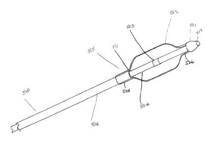
pounds but no more than about 7 pounds; or at least about 0.5 pounds but no
more than
about 5 pounds.
[0098] In some embodiments, the distal portion 712 of shaft 702 (or
end cap
730) has visible indicia, such as, for example, a marker visible via one or
more imaging
techniques such as fluoroscopy, ultrasound, CT, or MRI.
[0099] Figures 9A-C illustrate in schematic cross-section another
embodiment
of a distal portion 734 of a steerable and curvable injection device 740. The
tubular shaft
736 can include a distal portion 734 made of or containing, for example, a
shape memory
-19..
CA 02872107 2014-10-30
WO 2013/166209
PCT/1JS2013/039149
material that is biased into an arc when in an unconstrained configuration.
Some materials
that can be used for the distal curved portion 734 include Nitinol, Elgiloy,
stainless steel,
or a shape memory polymer. A proximal portion 732 of the shaft 736 is
preferably
relatively straight as shown. Also shown is end cap 730, distal lateral
opening 728 and
mesh 812.
[0100] The distal curved portion 734 may be configured to be axially
movably
received within an outer tubular sheath 738. The sheath 738 is preferably
configured to
have sufficient rigidity and radial strength to maintain the curved distal
portion 734 of
shaft 732 in a relatively straightened configuration while the outer tubular
sheath 738
coaxially covers the curved distal portion 734. Sheath 738 can be made of, for
example, a
metal such as stainless steel or various polymers known in the catheter arts.
Axial
proximal withdrawal of the sheath 738 with respect to tubular shaft 736 will
expose an
unconstrained portion of the shape memory distal end 734 which will revert to
its
unstressed arcuate configuration. Retraction of the sheath 738 may be
accomplished by
manual retraction by an operator at the proximal end, retraction of a pull
wire attached to
a distal portion of the sheath 738, or other ways as known in the art. The
straightening
function of the outer sheath 738 may alternatively be accomplished using an
internal
stiffening wire, which is axially movably positioned within a lumen extending
through
the tubular shaft 736. The length, specific curvature, and other details of
the distal end
may be as described elsewhere herein.
[0101] In another embodiment, as shown in Figures 10A-C, tubular
shaft 802
of a steerable and curvable vertebroplasty injector may be generally
substantially straight
throughout its length in its unstressed state, or have a laterally biased
distal end. A
distally facing or side facing opening 810 is provided for the release of a
material, such as
bone cement. In this embodiment, introducer 800 includes an elongate tubular
body 801
with a lumen 805 therethrough configured to receive the tubular shaft (also
referred to as
a needle) 802. Introducer 800 can be made of any appropriate material, such
as, stainless
steel and others disclosed elsewhere herein. Needle 802 may be made of a shape
memory
material, such as Nitinol, with superelastic properties, and has an outside
diameter within
the range of between about 1 to about 3 mm, about 1.5-2.5 mm, or about 2.1 mm
in some
embodiments.
[0102] Introducer 800 includes a needle-redirecting element 804 such
as an
inclined surface near its distal end. Needle-redirecting element 804 can be,
for example, a
-20-
CA 02872107 2014-10-30
WO 2013/166209
PCMJS2013/039149
laser-cut tang or a plug having a proximal surface configured such that when
needle 802
is advanced distally into introducer 800 and comes in contact with the needle-
redirecting
element 804, a distal portion 814 of needle 802 is redirected out an exit port
806 of
introducer 800 at an angle 808, while proximal portion 816 of needle 802
remains in a
relatively straightened configuration, as shown in Figure 10B. Bone cement can
then be
ejected from distal opening 810 on the end or side of needle 802 within bone
1000. Distal
opening 810 may be present at the distal tip of the needle 802 (coaxial with
the long axis
of the needle 802) or alternatively located on a distal radial wall of needle
802 as shown
in Figure 10C. In some embodiments, the angle 808 is at least about 15 degrees
and may
be at least about 30, 45, 60, 90, 105 degrees or more with respect to the long
axis of the
introducer 800.
[0103] The illustrated embodiment of Figures 10A-C and other
embodiments
disclosed herein are steerable and curvable through multiple degrees of
freedom to
distribute bone cement to any area within a vertebral body. For example, the
introducer
800 and needle 802 can both rotate about their longitudinal axes with respect
to each
other, and needle 802 can move coaxially with respect to the introducer 800,
allowing an
operator to actuate the injection system three dimensionally. The distal
portion 814 of
needle 802 can be deflected to a position that is angularly displaced from the
long axis of
proximal portion 816 of needle without requiring a discrete curved distal
needle portion
as shown in other embodiments herein.
[0104] Figures 11A-C illustrate another embodiment of a steerable and
curvable vertebroplasty injector. Figure 11A schematically shows handle
portion 708,
adjustment control 706, and elongate needle shaft 702, including proximal
portion 710,
distal portion 712, and transition point 714. Figure 11B is a vertical cross-
section through
line A-A of Figure 11A, and shows adjustment control 706 operably connected to
pull
wire 724 such as through a threaded engagement. Also shown is input port 704,
and
proximal portion 710 and distal portion 712 of needle shaft 702. Figure 11C
illustrates a
cross-sectional view of distal portion 712 of shaft 702. The distal end 722 of
pull wire
724 is attached at an attachment point 723 to the distal portion 712 of shaft
702. Proximal
retraction on pullwire 724 will collapse transverse slots 718 and deflect the
injector as has
been discussed. Also shown is an inner tubular sleeve 709, which can be
advantageous to
facilitate negotiation of objects or media such as bone cement, through the
central lumen
of the needle shaft 702.
-21-
[0105] The interior sleeve 709 is preferably in the form of a
continuous, tubular
flexible material, such as nylon or polyethylene. In an embodiment in which
the needle 702 has an
outside diameter of 0.095 inches (0.093 inch coil with a 0.001 inch thick
outer sleeve) and an inside
diameter of 0.077 inches, the interior tubular sleeve 709 may have an exterior
diameter in the area of
about 0.074 inches and an interior diameter in the area of about 0.069 inches.
The use of this thin walled
tube 705 on the inside of the needle shaft 702 is particularly useful for
guiding a fiber through the needle
shaft 702. The interior tube 705 described above is additionally preferably
fluid-tight, and can be used
to either protect the implements transmitted therethrough from moisture, or
can be used to transmit bone
cement through the steerable and curvable needle.
[0106] In some embodiments, an outer tubular coating or sleeve (not
shown) is
provided for surrounding the steerable and curvable needle shaft at least
partially throughout the distal
end of the needle. The outer tubular sleeve may be provided in accordance with
techniques known in
the art and, in one embodiment, is a thin wall polyester (e.g., ABS) heat
shrinks tubing such as that
available from Advanced Polymers, Inc. in Salem, N.H. Such heat shrink tubing
have a wall thickness
of as little as about 0.0002 inches and tube diameter as little as about 0.010
inches. The outer tubular
sleeve enhances the structural integrity of the needle, and also provides a
fluid seal and improved
lubricity at the distal end over embodiments with distal joints 718.
Furthermore, the outer tubular sleeve
tends to prevent the device from collapsing under a proximal force on a pull
wire. The sleeve also
improves lubricity of the tubular members, and improves torque transmission.
[0107] In other embodiments, instead of a slotted tube, the needle
shaft of a
vertebroplasty injection system may include a metal or polymeric coil.
Steerable and curvable helical
coil-type devices are described, for example, in U.S. Pat. No. 5,378,234 or
5,480,382 to Hammerslag et
al.
[0108] An interior tubular sleeve (not illustrated) may be provided
to facilitate flow of
media through the central lumen as described elsewhere in the application. In
some embodiments, a
heat-shrunk outer tubular sleeve as described elsewhere in the application is
also provided to enhance
the structural integrity of the sheath, provide a fluid seal across the
chevrons or slots, as well as improve
lubricity.
- 22 -
CA 2872107 2019-06-04
CA 02872107 2014-10-30
WO 2013/166209
PCMJS2013/039149
[0109] The steerable and curvable injection needle (also referred to
as the
injection shaft) may have an outside diameter of between about 8 to 24 gauge,
more
preferably between about 10 to 18 gauge, e.g., 12 gauge, 13 gauge (0.095" or
2.41 mm),
14 gauge, 15 gauge, or 16 gauge. In some embodiments, the inside diameter
(luminal
diameter) of the injection needle is between about 9 to 26 gauge, more
preferably
between about 11 to 19 gauge, e.g., 13 gauge, 14 gauge, 15 gauge, 16 gauge, or
17 gauge.
In some embodiments, the inside diameter of the injection needle is no more
than about 4
gauge, 3 gauge, 2 gauge, or 1 gauge smaller than the outside diameter of the
injection
needle.
[0110] The inside luminal diameter of all of the embodiments
disclosed herein
is preferably optimized to allow a minimal exterior delivery profile while
maximizing the
amount of bone cement that can be carried by the needle. In one embodiment,
the outside
diameter of the injection needle is 13 gauge (0.095" or 2.41 mm) with a 0.077"
(1.96 mm)
lumen. In some embodiments, the percentage of the inside diameter with respect
to the
outside diameter of the injection needle is at least about 60%, 65%, 70%, 75%,
80%,
85%, or more.
[0111] Referring to Figures 12 and 13, there is illustrated a
modification of the
steerable and curvable injection needle 10, in accordance with the present
invention. The
injection needle 10 comprises an elongate tubular shaft 702, extending between
a
proximal portion 710 and a distal portion 712. The proximal portion 710 is
carried by a
proximal handle 708, which includes a deflection controller 706 such as a
rotatable knob
or wheel. Rotation of the control 706 causes a lateral deflection or curvature
of the distal
steering region 24 as has been discussed.
[0112] Input port 704 is in fluid communication with a distal opening
728 on a
distal tip 730, by way of an elongate central lumen 720. Input port 704 may be
provided
with any of a variety of releasable connectors, such as a Luer or other
threaded or
mechanically interlocking connector known in the art. Bone cement or other
media
advanced through lumen 720 under pressure may be prevented from escaping
through the
plurality of slots 718 in the steering region 24 by the provision of a thin
flexible tubular
membrane carried either by the outside of tubular shaft 702, or on the
interior surface
defining central lumen 720.
[0113] Referring to Figure 14, the handle 708 is provided with an
axially
oriented central bore 732 having a first, female thread 733 thereon. A slider
734 having a
-23-
CA 02872107 2014-10-30
WO 2013/166209
PCMJS2013/039149
second complementary male thread 735, is thread-engaged with the central bore
732.
Rotation of the knob 706 relatively to the slider 734 thus causes the slider
734 to distally
advance or proximally retract in an axial direction with respect to the handle
708. The
slider 734 is mechanically linked to the pull wire 724, such as by the use of
one or more
set screws or other fastener 740.
[0114] Slider 734 is provided with at least one axially extending
keyway or
spline 742 for engaging a slide dowel pin 744 linked to the handle 708. This
allows
rotation of the rotatable control 706, yet prevents rotation of the slider 734
while
permitting axial reciprocal movement of the slider 734 as will be apparent to
those of
skill in the art. One or more actuating knob dowel pins 746 permits rotation
of the
rotatable control 706 with respect to the handle 708 but prevents axial
movement of the
rotatable control 706 with respect to the handle 708.
[0115] Referring to Figure 15, the distal end of the shaft 702 may be
provided
with any of a variety of distal opening 728 orientations or distal tip 730
designs,
depending upon the desired functionality. In the illustrated embodiment, the
distal tip 730
is provided with an annular flange 748 which may be slip fit into the distal
end of the
tubular body 702, to facilitate attachment. The attachment of the distal tip
730 may be
further secured by welding, crimping, adhesives, or other bonding technique.
[0116] In general, the distal tip 730 includes a proximal opening 750
for
receiving media from the central lumen 720, and advancing media through distal
opening
728. Distal opening 728 may be provided on a distally facing surface, on a
laterally facing
surface, or on an inclined surface of the distal tip 730.
[0117] Referring to Figures 15A and 15B, there is illustrated a
distal tip 30
having a single inclined opening 728 thereon. In any of the designs disclosed
herein, one
or two or three or four or more distal ports 728 may be provided, depending
upon the
desired clinical performance. In the illustrated embodiment, the distal tip
includes a
rounded distal end 750 which transitions either smoothly or through an angular
interface
with an inclined portion 752. The distal opening 728 is positioned distally of
a transition
754 at the proximal limit of the inclined surface 752. This configuration
enables the distal
opening 728 to have a distal axially facing component, as compared to an
embodiment
having a side wall opening. See, for example, Figure 8.
[0118] Referring to Figure 15B, the tip 730 can be considered to have
a
central longitudinal axis 770. The aperture 728 may be considered as residing
on an
-24-
CA 02872107 2014-10-30
WO 2013/166209
PCMJS2013/039149
aperture plane 772, which intersects the distal most limit and the proximal
most limit of
the aperture 728. Aperture plane 772 intersects the longitudinal axis at an
angle, O. In an
embodiment having a side wall aperture, the aperture plane 772 and
longitudinal axis 770
would be parallel. In an embodiment having a completely distally facing
aperture, the
aperture plane 772 would intersect the longitudinal axis 770 at an angle of 90
.
[0119] In the illustrated embodiment, the inclined aperture 728 is
defined by
an aperture plane 772 intersecting the longitudinal axis 770 at an angle 0,
which is at least
about 5 , often at least about 15 , and in many embodiments, at least about 25
or more.
Intersection angles within the range of from about 15 to about 450 may often
be used,
depending upon the desired clinical performance.
[0120] Referring to Figures 15C and 15D, an alternate distal tip 730
is
illustrated. In this configuration, the distal opening 728 is in the form of a
sculpted recess
756 extending axially in alignment with at least a portion of the central
lumen 720.
Sculpted recess 756 may be formed in any of a variety of ways, such as by
molding, or by
drilling an axial bore in an axial direction with respect to the tip 730. The
sculpted recess
756 cooperates with the tubular body 702, as mounted, to provide a distal
opening 728
having an inclined aspect as well as an axially distally facing aspect with
respect to the
longitudinal axis of the steerable and curvable needle.
[0121] Referring to Figures 15E and 15F, there is illustrated a
distal tip 730
having a plurality of distally facing apertures 728. In the illustrated
embodiment, four
distal apertures are provided. The distal apertures 728 may be provided on the
rounded
distal end 750, or on an inclined surface 752 as has been discussed.
[0122] Referring to Figures 15G and 15H, there is illustrated an
alternative
distal tip 730. In this configuration, an opening 728 is oriented in a
distally facing
direction with respect to the longitudinal axis of the needle. The distal
opening of the
central lumen is covered by at least one, preferably two, and, as illustrated,
four leaflets
758 to provide a collet-like configuration. Each of the adjacent leaflets 758
is separated
by a slot 760 and is provided with a living hinge or other flexible zone 762.
[0123] In use, the distal tip 730 may be distally advanced through
soft tissue
or cancellous bone, with the distal opening 728 being maintained in a closed
orientation.
Following appropriate positioning of the distal tip 30, the introduction of
bone cement or
other media under pressure through the central lumen 720 forces the distal
opening 728
open by radially outwardly inclining each leaflet 758 about its flexion point
762. This
-25-
CA 02872107 2014-10-30
WO 2013/166209
PCT/1JS2013/039149
configuration enables introduction of the needle without "coring" or occluding
with bone
or other tissue, while still permitting injection of bone cement or other
media in a distal
direction.
[0124] Referring to Figure 151, there is illustrated yet another
distal tip, this
time comprising a "pop-up" or deployable cap 730 in its deployed state. The
injection
needle 10 includes a shaft 702 having a distal shaft end 714. Any of the
foregoing or
other tip configurations may be separately formed and secured to the distal
end of the
tubular body 702, or may be machined, molded or otherwise formed integrally
with the
tube 702. Distal aperture 728 can be occluded by a plug or cap 730 with,
preferably, an
atraumatic tip, which minimizes coring during distal advance of the injection
needle. The
cap 730 includes a flange 748 and cap extensions 776 having optional slots
760. In its
undeployed state, the cap flange 748 is slip fitted within the needle injector
shaft 702 and
retained only by friction or by a reversible bond to the distal end 714 of the
shaft, which
is sufficient to retain the cap 730 in the distal end 714 during injection,
but insufficient to
resist the force of injected bone cement in some embodiments. In its
undeployed state, the
cap extensions 776 are not exposed and covered by the injection needle shaft
702. The
deployable cap 730 can be popped-up or deployed distally from the distal end
714 of the
shaft under pressure, thereby exposing the distal aperture 728 for cement
release.
[0125] The deployable cap 730 may take any of a variety of forms
depending
upon the injector design. The deployable cap 730 may be made from any of a
variety of
materials, such as stainless steel, Nitinol, or other implantable metals; any
of a wide
variety of implantable polymers such as PEEK, nylon, PTFE; or of bone cement
such as
PMMA. Alternatively, any of a variety of bioabsorbable polymers may be
utilized to
form the deployable cap 730, including blends and polymers in the PLA-PGLA
absorbable polymer families.
[0126] In operation, once the injection needle 10 is positioned in a
desired
location, the distal cap 730 may be pushed or popped-open from the distal end
of the
injector, such as by applying pressure from the injected bone cement. For
example, the
injected bone cement can apply a fluidic pressure that forces the deployable
cap 730 to
pop-open distally to its deployed state, as shown in Figure 151. In some
embodiments, the
cap can have at least two, three, or more successively longer distal
deployment positions,
thereby adjusting the size of the distal aperture 728 for variable control on
the flow of
media through distal aperture 728. In some embodiments, the minimum amount of
-26-
CA 02872107 2014-10-30
WO 2013/166209
PCMJS2013/039149
pressure required to pop-open the deployable cap 730 can be set at a certain
pressure
threshold. Once the deployable cap 730 is popped-open and placed in its
deployed state,
the aperture 728 is exposed and bone cement can be released and injected into
a target
location. The bone cement can flow out of the injection needle 10, past the
distal aperture
728, and through any of the slots 760 or open regions of the deployable cap
730. In some
embodiments, the deployable cap 730 is configured to be retractable back to
its
undeployed state, such as via a pullwire or other actuating mechanism, thereby
reducing
or inhibiting the flow of bone cement and advantageously reducing the risk of
overflow
and clogging of the injection needle.
101271 Referring to Figure 15J, there is illustrated yet another
distal tip, this
time including a check valve 783 that can block the release of bone cement
from a
sidewall aperture 728 of an injection needle 10. The distal tip 730 includes a
blunt
rounded distal end 750 and a check valve 783 coupled to an interior surface of
the
injection needle 10. The check valve 783 is capable of covering one or more
apertures
formed on the injection needle, such as on its rounded distal end or sidewalls
(as shown
in Figures 15J and 15K), that exposes the interior of the shaft 702. In some
embodiments,
the check valve 783 is moveable or capable of gliding along a longitudinal
axis of the
shaft 702. With the gliding check valve 783, the distal tip 730 can assume
three different
states: a blocked state (not shown), in which the check valve 783 completely
covers the
aperture 728; a partially blocked state (shown in Figures 15J and 15K), in
which the
check valve 783 partially covers the aperture 728; and an unblocked state (not
shown), in
which the aperture 728 is completely exposed.
[0128] In its blocked state, the distal tip 730 includes a check
valve 783 that
serves as a plug to completely cover the aperture 728 such that no bone cement
will flow
through the aperture 728. The check valve 783 can be moved to expose the
aperture 728,
in whole or in part, by using a mechanical or electrical mechanism. In some
embodiments, the check valve 783 can be moved to expose the aperture 728 by
using
fluidic pressure, e.g., from flowing bone cement, that forces the check valve
783 to slide
along the longitudinal direction of the injection needle 10, thereby exposing
the sidewall
aperture 728. In some embodiments, a lock or mechanical stopper can be
provided that
limits the movement of the check valve 783, such that the size of the exposed
aperture
728 can be controlled. For example, the mechanical stopper can lock the check
valve 783
in place once approximately half of the aperture 728 is exposed, thereby
restricting the
-27-
CA 02872107 2014-10-30
WO 2013/166209
PCMJS2013/039149
amount of bone cement that can be released from the injection needle 10. The
check
valve 783 advantageously allows for greater control over the injected volume
and flow
rate of the bone cement material, thereby reducing the risk of overflow and
clogging of
the injection needle.
[0129] Referring to Figure 15L, there is illustrated yet another
distal tip, this
time comprising a single inclined aperture 728 serving as an exit port along
the sidewall
751 of the distal tip 730. Unlike the distal tip in Figures 15A and 15B that
includes a
single inclined aperture that resides on the rounded distal end 750, the
distal tip in Figure
15L includes a single inclined aperture that resides on the sidewall 751. The
single
inclined aperture 728 may be considered as residing on an aperture plane 772,
which
intersects a plane along the longitudinal axis 770. While in some embodiments,
the
aperture plane 772 is viewed as being parallel to the plane along the
longitudinal axis
770, in other embodiments, the aperture plane 772 is at an angle that is at
least about 50,
often at least about 15 , and in many embodiments, at least about 25 or more.
Intersection angles within the range of from about 15 to about 450 may often
be used,
depending upon the desired clinical performance.
[0130] As the aperture 728 resides in a plane 772 that is at a non-
parallel
angle to the plane 770 along the longitudinal axis of the distal tip 730, the
aperture 728 is
also angulated with respect to the surface of the distal tip 730. Angled
surfaces 789 (best
shown in Figures 15M and 15N) reside adjacent to the aperture 728. Figure 15M1
is a
cross-section across line A-A of Figure 15M. The angled surfaces 789 provide a
sloped
passage upon which bone cement from the injection needle 10 can pass through.
Providing angled surfaces 789 on the sidewall of the distal tip 730 from which
bone
cement is injected allows for greater control of the bone cement relative to
conventional
injection needles, as the angled surfaces assist in breaking the flow of the
bone cement
exiting from the injection needle 10, thereby reducing the risk of overflow.
The advantage
of this design is that the aperture yields a smooth transition, which allows
better outflow
of the cement against cancellous bone fragments, blood and bone marrow that
may have
become lodged in the aperture. While the angled surfaces 789 appear planar, as
shown in
Figure 15N, in some embodiments, the surfaces may be non-planar e.g., it may
include
ridges, to assist in controlling the flow rate of the bone cement from the
injection needle
to a target site.
-28-
CA 02872107 2014-10-30
WO 2013/166209
PCMJS2013/039149
[0131] Referring to Figure 150, there is illustrated a distal tip
similar to the
distal tip in Figure 15N having a single inclined aperture 728 residing
adjacent to angled
surfaces 789; however, the distal tip 730 in Figure 150 includes a single
inclined aperture
728 that is narrower than the aperture in Figure 15N. While the aperture 728
is still
formed in the sidewall of the distal tip 730, the aperture is formed from an
angled surface
789 that is narrowed to a restricting neck 792 having a reduced width or
diameter. In
some embodiments, the width or diameter of the restricting neck 792 is at
least about 5%,
10%, 15%, 20%, 25%, 30%, 35%, 40%, 45%, 50%, or more narrower than the width
or
diameter of the lumen of the distal end 730 proximal to the restricting neck
792. The
restricting neck 792 helps to both control the flow rate of the bone cement
out of the
injection needle 10 and to reduce the volume of bone cement flowing into a
target site,
thereby reducing the likelihood of overflow and clogging.
[0132] Referring to FIGS. 15P-15P2, there is illustrated an aperture
728
having angled surfaces 789 that are located at right angles to the
corresponding angled
surfaces 789 shown in Figure 15N, e.g., the aperture is inclined proximally
and laterally.
FIG. 15P1 is a cross-section through line C-C of FIG. 15P. FIG. 15P2 is a
perspective
view of the tip 730 shown in FIG. 15P. This allows injectable material to be
dispensed in
a direction that is inward and proximal, as opposed to distal as in FIG. 15N.
The merit of
this positioning is to minimize clogging during the insertion of the steerable
and curvable
needle. In some embodiments, the angled surface 789 of the inclined aperture
728 forms
an angle with the longitudinal axis of the tip 730 (as illustrated in FIG.
15B). In some
embodiments, the angle can be between about 0 and 90 degrees, such as between
about
15 and 75 degrees, between about 30 and 60 degrees, between about 15 and 45
degrees,
between about 20 and 40 degrees, between about 45 and 75 degrees, or about 30
degrees
or about 45 degrees. In some embodiments, the angled surface can have a
distally facing
component as illustrated in FIGS. 15M-N, or a proximally facing component as
illustrated in FIGS. 15P-15P2. Where the aperture 728 is not on the distal tip
730 but
more proximally on the distal end cap 750 as illustrated, the distal end of
the aperture 728
can be, in some embodiments, separated by about 0.10, 0.09, 0.08, 0.07, 0.06,
0.05, 0.04,
0.03, 0.02 or less inches from the inclined distal tip 730 portion of the
distal end cap 750.
Any of the foregoing or other tip configurations may be separately formed and
secured to
the distal end of the tubular body 702, or may be machined, molded or
otherwise formed
integrally with the tube 702. In some embodiments, the aperture 728 can have a
diameter
-29-
CA 02872107 2014-10-30
WO 2013/166209
PCMJS2013/039149
of between about .060 and .010 inches, such as between about .070 and .090
inches, or
between about .075 and .085 inches. In some embodiments, the distal end 730
can have
an outside diameter (OD) of between about 0.05 and 0.20 inches, such as
between about
0.10 and 0.12 inches, or between about 0.107 and 0.111 inches. In some
embodiments,
the distal end 730 can have an inside diameter (ID) for flow of cement media
of between
about 0.04 and 0.19 inches, such as between about 0.05 and 0.10 inches, or
between about
0.072 and 0.078 inches in some embodiments. The length of the distal end cap
730 can
be, in some embodiments, between about 0.10 and 0.50 inches, such as between
about
0.10 inches and 0.30 inches, or between about 0.15 inches and 0.25 inches.
[0133] Referring to Figures 15Q-15Q2, there is illustrated one
embodiment of
a distal tip 730 having an aperture 728 with angled surfaces 789 that allow
injectable
material to be dispensed in a direction that is outward and distal. Figure
15Q1 is a cross-
sectional view through line A-A of Figure 15Q. Figure 15Q2 is a perspective
view of the
distal tip 730 of Figure 15Q. The body of the distal tip 730 in Figure 15Q is
somewhat
different from other distal tips disclosed herein. Whereas the distal tip 730
in some
embodiments (e.g., Figure 15P) include a generally cylindrical body with a
section
having a generally constant cross-sectional diameter that transitions into a
dome-like
distal end cap 750, in Figure 15Q, the body of the distal tip 730 having a
wall that has a
first radially inwardly tapering surface 773 (going from the proximal to
distal end of the
distal tip) that transitions into a second radially outwardly tapering surface
774 that
transitions into the distal end cap 750. In some embodiments, the length of
the first
radially inwardly tapering surface (starting from the proximal end to the
distal end of the
distal tip) is more than about 50%, 60%, 70%, 80%, 90%, or more of the second
radially
outwardly tapering surface. In other embodiments, the length of the first
radially inwardly
tapering surface 773 is less than about 50%, 40%, 30%, 20%, 10%, or less of
the length
of the second radially outwardly tapering surface 774. The radially inwardly
tapering
surface 773 could be proximal to (as illustrated in Figure 15T), or distal to
the radially
outwardly tapering surface 774, or a distal tip could have two, three, or more
radially
inwardly tapering surface 773 and/or radially outwardly tapering surfaces 774
(e.g., in a
sinusoidal pattern).
[0134] Referring to Figures 15R-15R2, there is illustrated a distal
tip 730
having an aperture 728 with angled surfaces 789 that allow injectable material
to be
dispensed in a direction that has a proximally facing component. Figure 15R1
is a cross-
-30-
CA 02872107 2014-10-30
WO 2013/166209
PCMJS2013/039149
sectional view through line A-A of Figure 15R, while Figure 15R2 is a
perspective view.
The body of the distal tip 730 in Figures 15R-15R2 has a wall having a
generally
transversely symmetrical, concave curvilinear surface 774 that transitions
into the dome-
like distal end cap 750.
[0135] Referring to Figure 15S-15S2, there is illustrated a distal
tip 730
having an aperture 728 with angled surfaces 789 that allow injectable material
to be
dispensed in a direction that has a proximally facing component. Figure 15S1
is a cross-
sectional view through line A-A of Figure 15S, while Figure 15S2 is a
perspective view.
The body of the distal tip 730 in Figure 15S has a wall having a linear bow-
tie shaped
radially inwardly tapered zone 773 and a linear radially outwardly tapered
zone 774 from
a proximal to distal direction (in contrast to the more curved taper of the
wall of Figure
15R) before forming the dome-like distal end cap 750.
[0136] Referring to Figure 15T, there is illustrated a distal tip 730
having an
aperture 728 with angled surfaces 789 that allow injectable material to be
dispensed in a
direction that is inward and proximal. Figure 15T1 is a cross-sectional view
through line
A-A of Figure 15T, while Figure 15T2 is a perspective view. The body of the
distal tip
730 in Figure 15T includes a wall having a proximal radially inwardly tapering
zone 774
followed by a radially outwardly tapering zone 773 from a proximal to distal
direction,
which transitions into the dome-like distal end cap 750.
[0137] Referring to Figures 15U-15U2, there is illustrated a "double
angle"
distal tip 730 having an aperture 728 with opposing angled surfaces 789a and
789b
(angled relative to an axis normal to the longitudinal axis of the distal tip)
that define an
outflow path, or exit port for dispensation of injectable material. Figure
15U1 is a cross-
sectional view through line A-A of Figure 15U, while Figure 15U2 is a
perspective view.
As illustrated, the angled surfaces 789a, 789b are configured such that the
aperture 728
can become larger in an axial direction, circumferential direction, or both as
the media
flows out of the central lumen, through the exit port, and out of the device
into the
intended anatomical location. Other embodiments could include a plurality of
apertures
728, such as 2, 3, 4, or more. In some embodiments, the exit port has a first
inner axial or
circumferential dimension at a junction with the central lumen and a second
axial or
circumferential dimension where bone cement exits the device, where the second
dimension is greater than the first dimension, such as by at least about 5%,
10%, 15%,
20%, 25%, 50%, or more. The increase in axial or circumferential direction of
the exit
-31-
CA 02872107 2014-10-30
WO 2013/166209
PCMJS2013/039149
port from the junction with the central lumen to the location in which the
bone cement
exits the device can be in a linear fashion, follow an accelerated curve, or a
decelerated
curve in some embodiments. Also as illustrated, the outer wall of the distal
tip 730 has a
first portion 799 that has a sidewall that is generally parallel to the
longitudinal axis of the
injector when the injector is in a nondeflected configuration, followed by a
second
radially inwardly tapering portion 774 that is not generally parallel to the
longitudinal
axis of the injector when the injector is in a nondeflected configuration, and
ending
distally in the distal cap 750, which can be dome-shaped or another atraumatic
shape. The
first portion 799 can have a cross-sectional diameter that is larger than a
cross-sectional
diameter of the radially inwardly tapering portion 774, which in turn has a
cross-sectional
diameter that is larger than a cross-sectional diameter of the end cap 750.
While the taper
of the second portion 774 illustrated in Figure 15U is generally constant, an
accelerating,
decelerating, undulating, or other taper could be employed as well. The exit
port can span
one, two, or more of the first portion 799, second portion 774, or third cap
portion 750. In
some embodiments, the angled surfaces 789a and 789b have intersecting
longitudinal
axes that form an angle of between about 30 degrees and 150 degrees, between
about 60
degrees and about 120 degrees, between about 75 degrees and 115 degrees, or
about 90
degrees. In some embodiments, angled surface 789b has an axial length that is
greater or
less than the axial length of angled surface 789a, such as by at least about
5%, 10%, 15%,
20%, 25%, or more. Angled surface 789b can have the same axial length as
angled
surface 789a in other embodiments.
[0138] Referring to Figures 15V -15V2, there is illustrated a distal
tip 730
similar to that of Figure 15U, but also including one, two, three, or more
rippled zones
777. Figure 15V1 is a cross-sectional view through line A-A of Figure 15V,
while Figure
15V2 is a perspective view. In some embodiments, the rippled zones 777 may
help slow
the flow of injectable material to allow for greater control over the
dispensation of fluid.
[0139] Referring to Figures 15W-15W2, there is illustrated a
schematic
diagram including non-limiting examples of particular dimensions for a distal
tip similar
to that illustrated in Figures 15U and 15V according to one embodiment. Figure
15W1 is
a perspective view, and Figure 15W2 is a side view. For example, in some
embodiments,
the distal tip could have an overall length of between about 0.15 and 0.25
inches, such as
between about 0.17 and 0.23 inches, or about 0.193 inches as shown. The
aperture 728
could in some embodiments, have a maximal linear dimension of between about
0.05 and
-32-
CA 02872107 2014-10-30
WO 2013/166209
PCMJS2013/039149
0.15 inches, such as between about 0.08 and 0.12 inches, or about 0.094 inches
in some
embodiments. In accordance with Figures 15W-15W2, a distal tip 730 is provided
having
an aperture 728 with non-parallel angled surfaces 789a, 789b that allow
dispensing of
injectable material. In some embodiments, the distal-most angled surface 789b
of the
aperture 728 has an axis P4 that intersects both the longitudinal axis of the
distal tip P3 or
an axis normal to the longitudinal axis of the distal tip P5 at an angle of
about 45 degrees.
In other embodiments, the angle could be between about 0 and 90 degrees, such
as
between about 15 and 75 degrees, between about 15 and 45 degrees, or between
about 30
and 60 degrees. The angle formed between an axis of the proximal-most angled
surface
789a could be as described above, and could be the same, less, or greater than
the angle
formed between an axis of the distal-most angled surface 789b and the
longitudinal axis
P3 of the distal tip. The distal tip 730 includes a radially inwardly (from
proximal to
distal) tapering wall 773 that transitions into a dome-like distal end cap
750. In some
embodiments, opposing zones of tapered wall 773, in some embodiments, resides
in
planes Pl, P2 that intersect at an angle approximately 15 degrees to 45
degrees, such as
about 15 to 25 degrees, or about 20.5 in some embodiments as illustrated,
although other
angles between 0 and 90 degrees are also possible.
[0140] Figure 15X illustrates a side schematic view of the distal tip
730
illustrated in Figure 15W, also illustrating a radially asymmetric offset 997A
of the
aperture 728 (e.g., from proximal radial termination of wall 789a and distal
radial
termination of wall 789b) from its proximal end to its distal end. In part due
to the offset
997 A, a cement flow out of the aperture 728 could be prevented from easily
and
prematurely severing at the aperture 728, for example, when the injector is
rotated while
the distal tip 730 is positioned near cancellous bone. In some embodiments,
the offset
distance 997A could be between about 0.01 and 0.05 inches, such as between
about 0.01
and 0.03 inches. In some embodiments, the offset distance 997A is at least
about 2%, 3%,
5%, 7%, 10%, 12%, 15%, or more of the distance from line 15X-15X (connecting
the
midpoints of the width of the distal tip 730 from its proximal end to its
distal end) to the
section of the tip 730 that extends the farthest radially outward, illustrated
as distance
997B. In other embodiments, the offset distance 997A is no more than about
15%, 12%,
10%, 7%, 5%, 3%, 2%, or less of the distance 997B. Other embodiments,
including that
of Figures 15U-15U2, can also be configured to have angled surfaces with an
offset as
described.
-33-
CA 02872107 2014-10-30
WO 2013/166209
PCMJS2013/039149
[0141] As a further alternative, coring during insertion of an
injector having a
distal opening 728 may be prevented by positioning a removable obturator 999
in the
distal opening, as illustrated schematically in Figure 15Y. The obturator 999
comprises an
elongate body, extending from a proximal end throughout the length of the
injector to a
blunt distal tip. The obturator 999 is advanced axially in a distal direction
through the
central lumen, until the distal tip of the obturator extends slightly distally
of the distal
opening 728 in the injector. This provides a blunt atraumatic tip for distal
advance of the
injector through tissue. Following positioning of the injector, the obturator
999 may be
proximally withdrawn from the central lumen, and discarded. The obturator 999
may be
provided with any of a variety of structures for securing the obturator 999
within the
central lumen during the insertion step, such as a proximal cap for threadably
engaging a
complementary Luer connector on the proximal opening of the central lumen.
[0142] In accordance with another aspect of the present invention,
there is
provided a combination device in which a steerable and curvable injector is
additionally
provided with one or two or more cavity formation elements. Thus, the single
device may
be advanced into a treatment site within a bone, expanded to form a cavity,
and used to
infuse bone cement or other media into the cavity. Either or both of the
expansion step
and the infusion step may be accomplished following or with deflection of the
distal
portion of the injector.
[0143] Referring to Figures 16A and 16B, the distal portion 302 of a
steerable
and curvable injector 300 having a cavity formation element 320 thereon is
schematically
illustrated. The steerable and curvable injector 300 includes a relatively
rigid proximal
section 304 and a deflectable section 306 as has been discussed elsewhere
herein. The
lateral flexibility of distal section 306 may be accomplished in any of a
variety of ways,
such as by the provision of a plurality of transverse chevrons or slots 308.
Slots 308 may
be machined or laser cut into appropriate tube stock, such as stainless steel
or any of a
variety of rigid polymers.
[0144] The slots 308 oppose a column strength element such as an
axially
extending spine 310, for resisting axial elongation or compression of the
device. A pull
wire 312 axially moveably extends throughout the length of the tubular body,
and is
secured with respect to the tubular body distally of the transverse slots 308.
The proximal
end of the pull wire is operatively connected to a control on a proximal
handpiece or
manifold. The control may be any of a variety of structures, such as a lever,
trigger, slider
-34-
CA 02872107 2014-10-30
WO 2013/166209
PCMJS2013/039149
switch or rotatable thumb wheel or control knob. Axial proximal traction (or
distal
advance) of the pull wire 312 with respect to the tubular body causes a
lateral deflection
of the distal steering section 306, by axial compression or expansion of the
transverse
slots 308 relative to the spine 310.
[0145] A distal aperture 314 is in communication via a central lumen
316 with
the proximal end of the steerable and curvable injector 300. Any of a variety
of tip
configurations may be used such as those disclosed elsewhere herein. The
proximal end
of the central lumen 316 may be provided with a Luer connector, or other
connection port
to enable connection to a source of media such as bone cement to be infused.
In the
illustrated embodiment, the aperture 314 faces distally from the steerable and
curvable
injector 302, although other exit angles may be used as will be discussed
below.
[0146] The steerable and curvable injector 300 is optionally provided
with a
cavity forming element 320, such as an inflatable balloon 322. In the
illustrated
embodiment, the inflatable balloon 322 is positioned in the vicinity of the
steerable and
curvable distal section 306. Preferably, the axial length of a distal leading
segment 307 is
minimized, so that the balloon 322 is relatively close to the distal end of
the steerable and
curvable injector 300. In this embodiment, the plurality of transverse slots
308 are
preferably occluded, to prevent inflation media from escaping into the central
lumen 316
or bone cement or other injective media from escaping into the balloon 322.
Occlusion of
the transverse slots 308 may be accomplished in a variety of ways, such as by
positioning
a thin tubular membrane coaxially about the exterior surface of the tubular
body and heat
shrinking or otherwise securing the membrane across the openings. Any of a
variety of
heat shrinkable polymeric sleeves, comprising high density polyethylene,
polyvinyl
chloride, ethylvinyl acetate, polyethylene terephthalate, polyurethane,
mixtures, and
block or random copolymers, or other materials, are well known in the catheter
arts.
Alternatively, a tubular liner may be provided within the central lumen 316,
to isolate the
central lumen from the transverse slots 308.
[0147] The balloon 322 is secured at a distal neck 309 to the leading
segment
307 as is understood in the balloon catheter arts. The distal neck 309 may
extend distally
from the balloon, as illustrated, or may invert and extend proximally along
the tubular
body. In either event, the distal neck 309 of the balloon 322 is preferably
provided with
an annular seal 324 either directly to the tubular body 301 or to a polymeric
liner
positioned concentrically about the tubular body, depending upon the
particular device
-35-
CA 02872107 2014-10-30
WO 2013/166209
PCMJS2013/039149
design. This will provide an isolated chamber within balloon 322, which is in
fluid
communication with a proximal source of inflation media by way of an inflation
lumen
326.
[0148] In the illustrated embodiment, the balloon 322 is provided
with an
elongate tubular proximal neck which extends throughout the length of the
steerable and
curvable injector 300, to a proximal port or other site for connection to a
source of
inflation media. This part can be blow molded within a capture tube as is well
understood
in the balloon catheter arts, to produce a one piece configuration.
Alternatively, the
balloon can be separately formed and bonded to a tubular sleeve. During
assembly, the
proximal neck or outer sleeve 328 may conveniently be proximally slipped over
the
tubular body 301, and secured thereto, as will be appreciated by those of
skill in the
catheter manufacturing arts. In some embodiments, the balloon 322 has a
lubricous
coating that can be chemically bonded or physically coated.
[0149] Referring to Figure 16C, the inflation lumen 326 may occupy an
annular space between the outer sleeve 328 and the tubular body 301. This may
be
accomplished by sizing the inside dimension of the outer sleeve 328 slightly
larger than
the outside dimension of the tubular body 301, by an amount sufficient to
enable the
desired inflation flow rate as understood in catheter art. Alternatively,
referring to Figure
16D, a discrete inflation lumen 326 may be provided while the remainder of the
outer
sleeve 328 is bonded or snuggly fit against the tubular body 301. This may be
accomplished by positioning an elongate mandrel (not illustrated) between the
outer
sleeve 328 and the tubular body 301, and heat shrinking or otherwise reducing
the outer
sleeve 328, thereafter removing the mandrel to leave the discrete inflation
lumen 326 in
place. In another embodiment, a cross-section of a catheter with a balloon
having an
inflation lumen 326 with outer layer 350 coextensive with the outer surface of
the balloon
coaxial with sleeve 328 and tubular body 301 is shown in Figure 16E. Figure
16F
illustrates a cross-section of another embodiment with an inflation lumen 326
external to
the tubular body 301. Figure 16G illustrates a cross-section of another
embodiment with
an inflation lumen 326 with a lumen internal to the tubular body 301. In some
embodiments, the internal inflation lumen 326 can be integrally formed with
the tubular
body 301 as shown. Alternatively, any of a variety of other inflation lumen
326
configurations can be used.
-36-
CA 02872107 2014-10-30
WO 2013/166209
PCT/1JS2013/039149
[0150] In some embodiments, the cavity-creating element could include
a
reinforcing layer that may be, for example, woven, wrapped or braided
(collectively a
"filament" layer), for example, over the liner of a balloon. The filament
layer can
advantageously protect the balloon from damage while in the working space, for
example
from jagged cancellous bone fragments within the interior of the vertebral
body. The
filament layer can also significantly elevated the burst pressure of the
balloon, such that it
exceeds about 20 atmospheres (ATM), in some embodiments exceeds about 25 ATM,
and
in a preferred embodiment, is at least about 30 ATM.
[0151] The filament layer can also be configured to control the
compliance of
the balloon depending on the desired clinical result, either symmetrically or,
if the
filaments are asymmetric, to constrain expansion of the balloon in one or more
directions.
In some embodiments, the balloon can be said to have a first compliance value
when
inflated to a first volume at a given first pressure when the balloon expands
without being
mechanically constrained by the constraining element such as the filament
layer. The
balloon can have a second compliance value when further inflated to a second
volume
(greater than the first volume) at a given second pressure (greater than the
first pressure)
when the balloon expands while being mechanically constrained by the
constraining
element. The second compliance value is, in some embodiments, less than the
first
compliance value due to the effect of the constraining element on the balloon.
The second
compliance value can be, for example, at least about 5%, 10%, 15%, 20%, 25%,
30%, _
40%, 50%, 60%, or 70% less than the first compliance value. In other
embodiments, the
second compliance value can be, for example, no more than about 70%, 60%, 50%,
40%,
30%, 25%, 20%, 15%, 10%, or 5% less than the first compliance value. In
embodiments
with a plurality of braided layers, the balloon could have an additional
third, fourth, etc.
progressively lower compliance values.
[0152] Figure 16H schematically illustrates a vertebroplasty catheter
300 with
a cavity creation element, namely a balloon 322 with a filament layer 340
carried by the
balloon. Figure 161 illustrates a cross-section of the filament reinforced
balloon 322
through line 161-161 of Figure 16H, with filaments 340 surrounding the
sidewall 350 of
the balloon 322. Figure 16J illustrates a cross-section of an alternative
embodiment with
filaments 340 over balloon sidewall 350 and also another layer 342 exterior to
the braided
layer 340. Other features have not been illustrated in Figures 161 and 16J for
clarity. The
exterior layer 342 could be made of, for example, a material discussed with
respect to
-37-
CA 02872107 2014-10-30
WO 2013/166209
PCT/1JS2013/039149
polymeric sleeve construction noted above, nylon, urethane, PET, or a
thermoplastic. In
some embodiments, there may be multiple layers, such as made of a polymer,
exterior to
the filament layer 340 and/or multiple liner layers interior to the filament
340, as well as
multiple braided or other filament layers between or amongst the various
layers. In some
embodiments, the filament 340 is co-molded within a wall 350 of the balloon
322 itself.
[0153] The filament 340 may comprise any of a variety of metallic
ribbons,
although wire-based braids could also be used. In some embodiments, the
ribbons can be
made at least in part of wires in braids or made of strips of a shape memory
material such
as Nitinol or Elgiloy, or alternatively stainless steel, such as AISI 303,
308, 310, and 311.
When using a braid 340 containing some amount of a super-elastic alloy, an
additional
step may be desirable in some embodiments to preserve the shape of the
stiffening braid
340. For instance, with a Cr-containing Ni/Ti superelastic alloy which has
been rolled
into 1 mm x 4 mm ribbons and formed into a 16-member braid 340, some heat
treatment
is desirable. The braid 340 may be placed onto a, e.g., metallic, mandrel of
an appropriate
size and then heated to a temperature of 600 degrees Fahrenheit to 750 degrees
Fahrenheit for a few minutes, to set the appropriate shape. After the heat
treatment step is
completed, the braid 340 retains its shape and the alloy retains its super-
elastic properties.
[0154] In some embodiments, metallic ribbons can be any of a variety
of
dimensions, including between about 0.25 mm and 3.5 mm in thickness and 1.0 mm
and
5.0 mm in width. Ribbons can include elongated cross-sections such as a
rectangle, oval,
or semi-oval. When used as ribbons, these cross-sections could have an aspect
ratio of
thickness-width of at least 0.5 in some embodiments.
[0155] In some embodiments, the braid 340 may include a minor amount
of
fibrous materials, both synthetic and natural, may also be used. In certain
applications,
particularly in smaller diameter catheter sections, more malleable metals and
alloys, e.g.,
gold, platinum, palladium, rhodium, etc., can be used. A platinum alloy with a
few
percent of tungsten is sometimes could be used partially because of its radio-
opacity.
[0156] Nonmetallic ribbons or wires can also be used, including, for
example,
materials such as those made of polyaramides (Kevlar), polyethylene
terephthalate
(Dacron), polyamides (nylons), polyimide carbon fibers, or a shape memory
polymer.
[0157] In some embodiments, the braids 340 can be made using
commercial
tubular braiders. The term "braid" when used herein includes tubular
constructions in
which the wires or ribbons making up the construction are woven in an in-and-
out fashion
-38-
as they cross, so as to form a tubular member defining a single lumen. The
braid members may be woven
in such a fashion that 2-4 braid members are woven together in a single
weaving path, although single-
strand weaving paths can also be used. In some embodiments, the braid 340 has
a nominal pitch angle
of 45 degrees. Other braid angles, e.g., from 20 degrees to 60 degrees could
also be used.
[0158] In some embodiments, the cavity creation element includes
two or more coaxial
balloons, including an inner balloon 322 and an outer balloon 370 as
illustrated schematically in FIG.
160. Inner balloon 322 can be oriented in a first direction, such as more
axially, while outer balloon 370
is oriented in a second direction, such as more radially. Balloon wall
orientation, such as by stretching,
is well understood in the art. The coaxial balloon configuration
advantageously provides improved
strength and burst resistance while minimizing the wall thickness of each
balloon. Thus, two or more
relatively thin-walled balloons can be utilized rather than a single thick-
walled balloon to achieve both
higher burst pressure and lower crossing profile. FIG. 16P illustrates a
schematic cross-section of a
section of the inner balloon wall 322 and outer balloon wall 370 that can be
separated by a slip plane
372 that may have a friction-reducing lubricious coating or the like. In some
embodiments, two, three,
four, or more coaxially arranged balloons can be used in the same fashion. In
some embodiments, one
or more coaxial balloons is interspersed or integrated with one or more
braided or other filament layers
as described above. In some embodiments, each balloon could have a thickness
of between about 0.0005
inches to 0.008 inches, or between about 0.001 inches to about 0.005 inches in
other embodiments.
[0159] In some embodiments, the cavity creation element could be
asymmetrical, for
example, as with the balloon 344 offset from the longitudinal axis of the
tubular body 301 illustrated
schematically in FIG. 16K. Such a balloon configuration can be advantageous,
for example, if the
vertebral fracture is generally more anterior, so that the balloon 344 can be
positioned to expand away
from the anterior area to reduce the risk of balloon expansion causing a
rupture all the way through the
cortical bone of the vertebrae. A cross-sectional schematic view through the
inflated offset balloon 344
is illustrated in FIG. 16L, also illustrating the tubular body 301. Other
components such as guidewire
312 have been omitted for clarity purposes. In some embodiments, various
balloons as described in
FIGS. 1-20 and the accompanying disclosure of U.S. Pat. No. 6,066,154 to
Reiley et al. can also be used
in connection with the injector 300 described herein. A schematic illustration
of an offset balloon 344
on the catheter 300 when the distal segment 306 is deflected is illustrated in
FIG. 16M.
[0160] Referring to FIGS. 17A and 17B, there is illustrated an
alternative embodiment
in which the distal aperture 314 is provided on a side wall of the tubular
body. One or two or three or
more distal apertures 314 may be provided in any of the embodiments disclosed
herein, depending upon
the desired clinical performance. In the illustrated embodiment, the distal
aperture 314 is provided on
- 39 -
CA 2872107 2019-06-04
the inside radius of curvature of the steerable and curvable section 306, as
illustrated in FIG. 17B. The
aperture 314 may alternatively be provided on the opposite, outside radius of
curvature, depending upon
the desired clinical performance.
[0161] As a further alternative, the distal aperture or apertures
314 may be provided in
any of a variety of configurations on a distal cap or tip, adapted to be
secured to the tubular body.
[01621 In some embodiments, it may be advantageous to have multiple
cavity-creation
elements on a steerable and curvable injector in order to, for example, more
quickly and efficiently move
sclerotic cancellous bone to better facilitate cavity formation and the
subsequent introduction of cement
media. Referring to FIGS. 17C and 17D, there is an illustrated another
embodiment of a steerable and
curvable injector with a plurality of cavity creation elements thereon
schematically illustrated, such as
at least two, three, four, or more cavity creation elements. The cavity
creation elements can be, for
example, a first balloon 330 and a second balloon 332 as shown. As
illustrated, both the first balloon
330 and the second balloon 332 are positioned in the vicinity of the steerable
and curvable distal section
306. In other embodiments, as illustrated in FIGS. 17G and 17H, the first
balloon 330 is positioned in
the vicinity of the steerable and curvable distal section 306 while the second
balloon 332 is positioned
more proximally on the more rigid proximal section 304. In still other
embodiments, as illustrated in
FIGS. 171 and 17J, the first balloon 330 is positioned in the vicinity of the
steerable and curvable distal
section 306 while the second balloon 332 is positioned partially on the
proximal section 304 and partially
on the steerable and curvable distal section 306. In other embodiments, both
the first balloon 330 and
the second balloon 332 can be positioned in the vicinity of the proximal
section 306.
- 40 -
CA 2872107 2019-06-04
CA 02872107 2014-10-30
WO 2013/166209
PCMJS2013/039149
[0163] In some embodiments, the first balloon 330 and the second
balloon
332 share a common inflation lumen 326 (such as illustrated in Figures 16C or
D) and
thus can be simultaneously inflatable from a common source of inflation media.
In other
embodiments, the first balloon 330 and the second balloon 332 have separate
respective
first inflation lumen 326 and second inflation lumen 327 and thus can be
inflated
according to the desired clinical result, e.g., simultaneously or the second
balloon 332
inflated before or after the first balloon 330. Figures 17E and 17F are
alternative cross
sectional views showing different inflation lumen configurations. As
illustrated in Figure
17E, in some embodiments the first inflation lumen 328 can be positioned
concentrically
around the second inflation lumen 329, both of which can occupy annular spaces
between
the outer sleeve 328 and the tubular body 301. Figure 17F illustrates an
alternative
embodiment where first 326 and second 327 discrete inflation lumens may be
provided
while the remainder of the outer sleeve 328 is bonded or snuggly fit against
the tubular
body 301.
[0164] The first balloon 330 and the second balloon 332 can have
substantially the same properties or differing properties, such as thickness,
material,
inflation diameter, burst strength, compliance, or symmetry (or lack thereof)
depending
on the desired clinical result. In some embodiments, the distal aperture 314
could be
distally facing, positioned on a side wall, or on an inclined surface; or 2,
3, 4, 5, or more
apertures could be presented as previously described. Furthermore, while the
aperture 314
is illustrated in Figures 17C-17D, and 17G-17J as positioned on the distal end
of the
catheter 300 as being distal to both first balloon 330 and second balloon 332
in some
embodiments the aperture 314 or additional aperture(s) can be positioned in
between first
balloon 330 and second balloon 332 and/or proximal to second balloon 332. In
embodiments with one or more cavity creating elements having multiple
apertures, the
apertures could be fluidly communicate with each other, or be fluidly isolated
in other
embodiments.
[0165] The steerable and curvable injection systems described above
are
preferably used in conjunction with a mixing and dispensing pump for use with
a multi-
component cement. In some embodiments, a cement dispensing pump is a hand-held
device having an interface such as a tray or chamber for receiving one or more
cartridges.
In one embodiment, the pump is configured to receive a double-barreled
cartridge for
simultaneously dispensing first and second bone cement components. The system
-41-
additionally includes a mixing chamber, for mixing the components sufficiently
and reproducibly to
fully automate the mixing and dispensing process within a closed system. In
some embodiments, the
cavity creation element(s) such as balloons described above can be coated or
impregnated with particles
such as those described in U.S. Pat. Pub. No. 2007/0185231 to Liu et al. The
particles can be released
within the vertebral cavity upon expansion or other transformation of the
cavity-creating element in
order to promote bone ingrowth into the bone cement or improve the crack arre
station properties of the
composite bone cement.
[0166] Bone cement components have conventionally been mixed, such
as by hand,
e.g., in mixing bowls in the operating room, which can be a time-consuming and
inelegant process. The
devices disclosed herein may be used with conventional bone cement
formulations, such as manually
mixed liquid-powder PMMA formulations. The mixed bone cement can then be
transferred to an
infusion device, such as a syringe connectable to the input port of the
steerable vertebroplasty device,
such that bone cement can be delivered through the steerable vertebroplasty
device to a desired
anatomical location within the body. In one embodiment, a first bone cement
component, such as a
cement powder, can be placed into a mixing bowl. A second bone cement
component such as a liquid
monomer, can be poured over the cement powder. The first and second bone
cement components can
then be mixed. The bone cement is then moved from the mixing bowl into a
cement reservoir. The
cement reservoir can have a distal opening connectable to the input port of
the steerable vertebroplasty
device, and a proximal cap having an opening connectable to a pump, such as a
hydraulic pump. When
the pump is connected to the cement reservoir, actuation of a pump control
(e.g., turning a control, such
as a knob) on the pump can urge the bone cement within the cement reservoir
into the input port of the
steerable vertebroplasty device for delivery to a desired anatomical location.
Alternatively, the use of a
closed mixing device such as a double-barreled dispensing pump as disclosed
herein is highly
advantageous in reducing bone cement preparation time, preventing escape of
fumes or ingredients,
ensuring that premature cement curing does not occur (i.e., the components are
mixed immediately prior
to delivery into the body), and ensuring adequate mixing of components.
101671 Two separate chambers contain respective materials to be
mixed in a specific
ratio. Manual dispensing (e.g., rotating a knob or squeezing a handle) forces
both materials into a mixing
nozzle, which may be a spiral mixing chamber within or in communication with a
nozzle. In the spiral
mixing nozzle, all or substantially all mixing preferably occurs prior to the
bone cement entering the
steerable and curvable injection needle and, subsequently, into the vertebra.
The cement dispensing hand
pump may be attached to the steerable and curvable injection needle
permanently, or removably via a
connector, such as slip-ring Luer fittings. A wide range of dispensing pumps
can be modified for use
- 42 -
CA 2872107 2019-06-04
with the present invention, including dispensing pumps described in, for
example, U.S. Pat, Nos.
5,184,757, 5,535,922, 6,484,904, and Patent Publication No. 2007/0114248.
[0168] Currently favored bone cement compositions are normally
stored as two
separate components or precursors, for mixing at the clinical site shortly
prior to implantation. As has
been described above, mixing of the bone cement components has traditionally
been accomplished
manually, such as by expressing the components into a mixing bowl in or near
the operating room. In
accordance with the present invention, the bone cement components may be
transmitted from their
storage and/or shipping containers, into a mixing chamber, and into the
patient, all within a closed
system. For this purpose, the system of the present invention includes at
least one mixing chamber
positioned in the flow path between the bone cement component container and
the distal opening on the
bone cement injection needle. This permits uniform and automated or semi-
automated mixing of the
bone cement precursors, within a closed system, and thus not exposing any of
the components or the
mixing process at the clinical site.
[0169] Thus, the mixing chamber may be formed as a part of the
cartridge, may be
positioned downstream from the cartridge, such as in-between the cartridge and
the proximal manifold
on the injection needle, or within the proximal manifold on the injection
needle or the injection needle
itself, depending upon the desired performance of the device. The mixing
chamber may be a discrete
component which may be removably or permanently coupled in series flow
communication with the
other components of the invention, or may be integrally formed within any of
the foregoing components.
[0170] In general, the mixing chamber includes an influent flow
path for
accommodating at least two bone cement components. The first and second
incoming flow path is
combined, and mixing structures for facilitating mixing of the components are
provided. This may
include any of a variety of structures, such as a helical flow path, baffles
and or additional turbulence
inducing structures.
- 43 -
CA 2872107 2019-06-04
CA 02872107 2014-10-30
WO 2013/166209
PCMJS2013/039149
[0171] Tables 1-2 below depict the contents and concentrations of one
exemplary embodiment of bone cement precursors. Chambers 1A and 1B contain
precursors for a first cement composition for distribution around the
periphery of the
formed in place vertebral body implant with a higher particle concentration to
promote
osteoconduction and/or osteoinduction, as discussed previously in the
application.
Chambers 2A and 2B contain precursors for a second cement composition for
expression
more centrally within the implanted mass within the vertebral body, for
stability and
crack arresting, as discussed previously in the application.
[0172] One of ordinary skill in the art will recognize that a wide
variety of
chamber or cartridge configurations, and bone cements, can be used with the
present
injection system. For example, in one embodiment, a first cartridge includes
pre-
polymerized PMMA and a polymerization catalyst, while a second cartridge
includes a
liquid monomer of MMA as is common with some conventional bone cement
formulations. In some embodiments, the contents of two cartridges can be
combined into
a single cartridge having multiple (e.g., four) chambers. Chambers may be
separated by a
frangible membrane (e.g., 1A and 2A in a first cartridge and 1B and 2B in a
second
cartridge, each component separated by the frangible membrane or other
pierceable or
removable barrier). In other embodiments, contents of the below cartridges can
be
manually pre-mixed and loaded into the input port of the injection system
without the use
of a cement mixing dispenser.
Table 1.
Chamber 1A
Methyl methacrylate (balance) Hydroquinone (-75 ppm)(stabilizer)
N,N-dimethyl-p-toluidine (-0.9%)(catalyst Sterile bone particles 35 wt. %)
for polymerization)
Barium sulfate (-20 wt. %)(radio-opacifier)
Chamber 1B
Benzoyl peroxide (-2%)(activator for Physiological saline or poppy seed oil
polymerization) (balance)
Table 2.
-44-
CA 02872107 2014-10-30
WO 2013/166209
PCMJS2013/039149
Chamber 2A
Methyl methacrylate (balance) Hydroquinone (-75 ppm)(stabilizer)
N,N-dimethyl-p-toluidine (-0.9%)(catalyst Sterile bone particles (-30 wt.
%)
for polymerization)
Barium sulfate (-20 wt. %)(radio-opacifier)
Chamber 2B
Benzoyl peroxide (-2%)(activator for Physiological saline or poppy seed oil
polymerization) (balance)
[0173] As illustrated in Figures 18A and 18B, in one embodiment, a
system or
kit for implanting bone cement includes at least some of the following
components: a
stylet configured to perforate a hole into the pedicle of the vertebral body;
an
introducer/cannula 800 for providing an access pathway to the treatment site,
a steerable
and curvable injection needle 700 to deliver bone cement to a desired
location, and, a
cement dispensing pump 910 preferably configured to accommodate one or two or
more
dual chamber cartridges 1200 as well as a mixing nozzle 995.
[0174] The stylet may have a diameter of between about .030" to
.300", .050"
to about .200" and preferably about .100" in some embodiments. The
introducer/cannula
800 is between about 8-14 gauge, preferably between about 10-12 gauge, more
preferably
11 gauge in some embodiments. The introducer/cannula 800, which may be made of
any
appropriate material, such as stainless steel (e.g., 304 stainless steel) may
have a
maximum working length of no more than about 12", 8", or 6" in some
embodiments.
One or two or more bone cement cartridges, each having one or two or more
chambers,
may also be provided. Various other details of the components have been
described above
in the application.
[0175] One embodiment of a method for delivering bone cement into a
vertebral body is now described, and illustrated in Figures 19A-F. The method
involves
the general concept of vertebroplasty and kyphoplasty in which a collapsed or
weakened
vertebra is stabilized by injecting bone cement into cancellous bone.
[0176] The cement implantation procedure is designed for uni-
transpedicular
access and generally requires either a local anesthetic or short-duration
general anesthetic
for minimally invasive surgery. Once the area of the spine is anesthetized, as
shown in
Figures. 19A-B, the physician inserts a stylet 1302 to perforate a lumen 1304
into the
pedicle wall 1300 of the vertebra 1308 to gain access to the interior of the
vertebral body
1310. As illustrated in Figure 19C, the introducer/cannula 800 is then
inserted through the
-45-
CA 02872107 2014-10-30
WO 2013/166209
PCMJS2013/039149
lumen 1304 for bone access as well as acting as the guide for the steerable
and curvable
injection needle 700. The introducer/cannula 800 is sized to allow physicians
to perform
vertebroplasty or kyphoplasty on vertebrae with small pedicles 1300 such as
the thoracic
vertebra (e.g., T5) as well as larger vertebrae (e.g., L5). In addition, this
system and
method is advantageously designed to allow uni-transpedicular access as
opposed to bi-
pedicular access, resulting in a less invasive surgical procedure.
[0177] Once bone access has been achieved, as shown in Figure 19C the
steerable and curvable injection needle 700 such as any of the devices
described above
can be inserted through the introducer/cannula 800 and into the vertebra 1308.
The entire
interior 1310 of the target vertebral body may be accessed using the steerable
and
curvable injection needle 800. The distal end 712 of the needle 700 can be
laterally
deflected, rotated, and/or proximally retracted or distally advanced to
position the bone
cement effluent port at any desired site as previously described in the
application. The
radius can be adjusted by means of an adjustment control, such as a knob on
the proximal
end of the device as previously described.
[0178] The actual injection procedure may utilize either one or two
basic
steps. In a one step procedure, a conventional bone cement is introduced as is
done in
simple vertebroplasty. The first step in the two step injection involves
injection of a small
quantity of PMMA with more than about 35%, e.g., 60% particles (such as
inorganic
bone particles) onto the periphery of the treatment site, i.e., next to the
cortical bone of
the vertebral body as shown in Figure 19D. This first cement composite 1312
begins to
harden rather quickly, forming a firm but still pliable shell, which is
intended to minimize
or prevent any blood/bone marrow/PMMA content from being ejected through any
venules or micro-fractures in the vertebral body wall. The second step in the
procedure
involves an injection of a bolus of a second formulation of PMMA with a
smaller
concentration such as approximately 30% (inorganic bone) particles (second
cement
composite 1314) to stabilize the remainder of the weakened, compressed
cancellous bone,
as illustrated in Figure 19E.
[0179] Injection control for the first and second steps is provided
by an
approximately 2 mm inside diameter flexible introducer/cannula 800 coupled to
a bone
cement injection pump (not shown) that is preferably hand-operated. Two
separate
cartridges containing respective bone cement and (inorganic bone) particle
concentrations
that are mixed in the 60% and 30% ratios are utilized to control (inorganic
bone) particle
-46-
CA 02872107 2014-10-30
WO 2013/166209
PCMJS2013/039149
to PMMA concentrations. The amount of the injectate is under the direct
control of the
surgeon or interventional radiologist by fluoroscopic observation. The
introducer/cannula
800 is slowly withdrawn from the cancellous space as the bolus begins to
harden, thus
preventing bone marrow/PMMA content from exiting the vertebral body 1308. The
procedure concludes with the surgical incision being closed, for example, with
bone void
filler 1306 as shown in Figure 19F. Both the high and low bone cement particle
concentration cement composites 1312, 1314 harden after several minutes. In
vitro and in
vivo studies have shown that the 60% bone-particle impregnated bone cement
hardens in
2-3 minutes and 30% bone-particle impregnated bone cement hardens between 4 to
10
minutes.
[0180] The foregoing method can alternatively be accomplished
utilizing the
combination steerable and curvable needle of Figure 16A, having a cavity
formation
structure 320 thereon. Once the steerable and curvable injector 300 has been
positioned as
desired, such as either with deflection as illustrated in Figure 19C, or
linearly, the cavity
forming element 320 is enlarged, such as by introducing inflation media under
pressure
into the inflatable balloon 322. The cavity forming element 320 is thereafter
reduced in
cross sectional configuration, such as by aspirating inflation media from the
inflatable
balloon 322 to produce a cavity in the adjacent cancellous bone. The steerable
and
curvable injector 300 may thereafter by proximally withdrawn by a small
distance, to
position the distal opening 314 in communication with the newly formed cavity.
Bone
cement or other media may thereafter be infused into the cavity, as will be
appreciated by
those skilled in the art.
[0181] At any time in the process, whether utilizing an injection
needle having
a cavity formation element or not, the steerable and curvable injector may be
proximally
withdrawn or distally advanced, rotated, and inclined to a greater degree or
advanced into
its linear configuration, and further distally advanced or proximally
retracted, to position
the distal opening 314 at any desired site for infusion of additional bone
cement or other
media. More than one cavity, such as two, or three or more, may be
sequentially created
using the cavity formation element, as will be appreciated by those of skill
in the art.
[0182] The aforementioned bone cement implant procedure process
eliminates
the need for the external mixing of PMMA powder with MMA monomer. This mixing
process sometimes entraps air in the dough, thus creating porosity in the
hardened
PMMA in the cancellous bone area. These pores weaken the PMMA. Direct mixing
and
-47-
CA 02872107 2014-10-30
WO 2013/166209
PCMJS2013/039149
hardening of the PMMA using an implant procedure such as the above eliminates
this
porosity since no air is entrapped in the injectate. This, too, eliminates
further weakening,
loosening, or migration of the PMMA.
[0183] A method of using the steerable and curvable injection system
described, for example, in Figures 17C-17D will now be described. Various
components
of the injector 300 are not illustrated for clarity purposes. The interior of
the vertebral
body 1310 can be first accessed via a unipedicular approach as described and
illustrated
in connection with Figures 19A-B. Next, the steerable and curvable injector
300 having
first balloon 330 and second balloon 332 thereon is inserted through an
introducer 800
into the interior of the vertebral body 1310 with the distal deflectable
section 306 in a
relatively straightened configuration, as shown schematically in Figure 20A.
In some
embodiments, the injector 300 also has a retractable outer sheath 340
actuatable by a
controller 350 on the handpiece 360 to protect the balloons 330, 332 from
damage during
introduction of the injector 300 into the interior of the vertebral body 1310.
The injector
300 can then be laterally deflected, rotated, and or proximally retracted or
distally
advanced to position the injector at any desired site as previously described
in the
application, and illustrated schematically in Figure 20B. The radius can be
adjusted by
means of an adjustment control, such as a knob on the proximal end of the
device as
previously described. The first balloon 330 and second balloon 332 can then be
inflated
simultaneously as illustrated in Figure 20C or sequentially as previously
described. In
some embodiments, only one of the balloons may need to be inflated depending
on the
size of the cavity desired to be created. Injection of the cement media can
proceed at any
desired time as previously described, such as, for example, following
deflation of one or
both balloons.
[0184] Figure 21A illustrates an embodiment of a steerable cavity
creation
device 500. The device 500 includes a proximal handle 508, a shaft portion
520, and a
steerable and curvable distal end 505 including a distal tip 501. The proximal
handle 508
includes a deflection control 510, such as a rotatable knob, that when
actuated in an
appropriate direction causes a tensile or compression force to be applied on
the distal end
505, causing it to move in an appropriate direction (e.g., opposite or toward
the injection
port 509). One, two, or more input ports 509 extend from the proximal handle
508 for
example, to inject a fluid (e.g., a liquid or gas) to expand the expandable
member 502,
such as inflating a balloon. Input port 509 is operably connected to a lumen
within the
-48-
CA 02872107 2014-10-30
WO 2013/166209
PCMJS2013/039149
shaft portion 520, which is in turn operably connected to balloon 502. The
input port 509
could be spaced apart distally with respect to the deflection control 510 as
illustrated, or
proximally with respect to the deflection control 510 with respect to a
longitudinal axis of
the device 500 in some embodiments. The input port 509 could be coaxial with,
or have a
longitudinal axis that is offset from the longitudinal axis of the device 500,
such as at an
angle of between about 00 and 90 , between about 15 and 60 , between about 15
to 45 ,
about 90 , or about 30 in some embodiments. The device 500 can include an
inner
member 504 such as a tubing, and an outer member 506, such as a shaft. Outer
shaft 506
can include indicia, such as an insertion marker 507 in shaft section 520 that
may be
radiopaque. Marker 507 can assist in indicating when the balloon 502 has
distally cleared
the access introducer/cannula and is in a space conducive for expansion of the
balloon
502. The distal end of the device 505 includes one, two, or more cavity
creation structures
502, which in some embodiment is an expandable member 502. Other non-
expandable
cavity creation structures such as one or a combination of cutting elements,
or energy-
based cavity creation structures involving RF, microwave, optical, thermal, or
cryoablation elements could also be utilized.
[0185] The expandable member 502 could be a balloon in some
embodiments.
A radiopaque marker 503 may assist in confirming the position of the balloon
prior to
expansion at the appropriate target location to create a cavity. The
radiopaque marker 503
could be, for example, a marker band partially or completely circumferentially
surrounding a portion of the shaft at the distal end 505 of the device 500,
such as at the
axial midpoint of the balloon 502 for example. Distal tip 501 could be either
blunt or
sharp, and in some embodiments can include a cutting element to further assist
in cavity
creation.
[0186] Figure 21B illustrates a close-up view of the distal end 505
of the
device. As shown, the proximal neck 524 of the balloon 502 is bonded or
otherwise
attached to the outer shaft 506. The distal neck 526 of the balloon 502 is
bonded or
otherwise attached to inner tubing 504. In order to expand the balloon, a
fluid is injected
through the input port 509 (shown in Figure 50A) through an annular space 511
between
the inner tubing 504 and the outer shaft 506. The effect of the balloon bond
configuration
is that the balloon 502 will expand generally radially outwardly without or
substantially
without expansion in the axial direction. The balloon could be either
constrained or
unconstrained, and have features, for example, as previously described.
-49-
CA 02872107 2014-10-30
WO 2013/166209
PCMJS2013/039149
[0187] Also shown is the distal tip 501, which could be a welded tip
512
having a closed distal end in some embodiments. The welded tip 512 can
advantageously
hold the distal neck 526 of the balloon 502 to improve bond strength and holds
the
balloon bond stationary when inflated.
[0188] In some embodiments, instead of a closed distal end the distal
tip could
include one, two, or more distal or side-facing exit ports connected via a
lumen, e.g., a
central lumen of the inner tubing 504, to a media input port for delivery of a
media, such
as a bone cement, to the interior of a bone, such as a cavity. Alternatively,
the cavity
creating device 500 can be withdrawn after cavity creation, and a separate
steerable
injection device then inserted for delivery of the media as described further
below.
[0189] Figure 21C is a cross-section of the distal end 505 of the
device 500
illustrated in Figure 21B. Shown is an inner hypotube 515 having slots 519
present within
an inner lumen of an outer hypotube 514, which also has slots 518. In some
embodiments,
the length of the inner hypotube 515 zone having slots 519 is the same or
substantially the
same as the length of the outer hypotube 514 zone having slots 518. The slots
519 of the
inner hypotube 515 can, in some embodiments, be spaced apart, such as between
about
120 and about 240 , or about 180 apart from the slots 518 of the outer
hypotube 514.
The distal ends of the inner hypotube 515 and the outer hypo tube 514 can be
attached,
such as by welding at 512 at the distal tip 501 of the device 500. The inner
hypotube 515
and outer hypotube 514 can be made of any appropriate material with sufficient
column
strength to navigate cancellous bone, such as a metal such as stainless steel,
or nitinol for
example. The inner hypotube 515 and outer hypotube 514 could be made of the
same or
different materials. Either or both hypotubes 515, 514 includes slots 519, 518
that could
be, for example, laser-cut. The slots 519, 518 could be any desired wall
pattern, such as
simply transverse to the longitudinal axis of the hypotube, or in a chevron
pattern in other
embodiments. An inner liner 513 at least partially circumscribing the outer
hypotube 514
serves to seal the balloon chamber. Radiopaque marker band 503 can be
positioned either
inside or outside of the balloon chamber in some embodiments, for
visualization during
fluoroscopy. The proximal end 528 of the distal tip 501 of the device 500 is
positioned
over the distal neck 526 of the balloon 502 to prevent or substantially
prevent the balloon
502 from elongating distally when inflated. The proximal end 528 of the distal
tip 501 of
the device also constrains the bond section to improve bond integrity.
-50-
CA 02872107 2014-10-30
WO 2013/166209
PCMJS2013/039149
[0190] Figure 21D illustrates schematically the distal end 505 of the
device
500, highlighting the inner hypotube 515-outer hypotube 514 configuration in
an
undeflected configuration where a longitudinal axis of the distal end 505 is
coaxial with,
or at least parallel to the longitudinal axis of the device 500. The number of
in.per
hypotube slots 519 can be less than, equal to, or greater than the number of
outer
hypotube slots 518. In some embodiments, the number of inner hypotube slots
519 is at
least about 5%, 10%, 15%, 20%,25%, 30%, 40%, 50%, 75%, 100%, or more than the
number of outer hypotube slots 518. A dimension, such as the axial width 516
of the
outer hypotube slots 518 can be, for example, less than, equal to, or greater
than the axial
width 517 of the inner hypotube slots 519. In some embodiments, the axial
width 516 of
outer hypotube slots 518 is at least about 5%, 10%, 15%, 20%, 25%, 30%, 40%,
50%,
75%, 100%, 150%, 200%, 250%, 300% greater than the axial width 517 of the
inner
hypotube slots 519. In some embodiments, the axial width 516 of the outer
hypotube slots
518 is between about 1.5x and 3x, or about 2x of the axial width 517 of the
inner
hypotube slots 519. In some embodiments, the width 517 of the inner hypotube
slot 519 is
between about .001" and about .005", between about .002" and about .006", or
about or at
least about .002". The width 517 of the inner hypotube is in some embodiments
sufficiently large to allow for the desired degree of deflection, but
sufficiently small such
that the structural integrity of the inner hypotube 515 is not sufficiently
impaired.
[0191] Figure 21E illustrates distal end 505 of the device 500,
highlighting the
inner hypotube 515-outer hypotube 514 configuration in a deflected
configuration. In
some embodiments, the device can be configured to deflect from the
longitudinal axis of
the device 500 by at least about 50 , 60 , 70 , 80 , 90 , 100 , or more, or
between about
70 and about 1000 in some embodiments. When a tensile force is applied to the
inner
hypotube 515 while the outer hypotube 514 is immobilized or held stationary, a
compression force will thus be applied to the outer hypo tube 514 which will
bend both
the inner hypo tube 515 and the outer hypo tube 514 in the direction of the
openings of
the outer hypotube slots 518 as shown. As the deflectable distal end 505 of
the device
deflects, the axial width 516 of the outer hypotube slots 518 decreases and
the axial width
517 of the inner hypotube slots 519 increases in the deflected zone. When a
compression
force is applied to the inner hypotube 515 the axial width 517 of the inner
hypotube slots
519 decreases and the axial width 516 of the outer hypotube slots 518
increases, causing
the distal end 505 of the device 500 to deflect in a direction opposite of the
direction of
-51-
CA 02872107 2014-10-30
WO 2013/166209
PCMJS2013/039149
the openings of the outer hypotube slots 518. The distal end of either or both
of the inner
hypotube 515 and outer hypotube 514 could be operably connected via one, two,
or more
pullwires (e.g., two pullwires positioned 180 circumferentially apart along a
sidewall
(such as oriented against or opposite the slots) of either the inner hypotube
515 and/or
outer hypotube 514 to the deflection control 510 proximally) to create the
desired tension
or compression forces when the deflection control is moved in an appropriate
direction
(e.g., clockwise or counterclockwise). In some embodiments, distal pullwires
are not
required, and a tensile or compression force can be transmitted via a
mechanism to the
proximal end of one or more of the inner hypotube 515 or outer hypotube 514.
[01921 In other words, the device could have a distal end 505 having
a first
hypotube having a first plurality of slots and a second hypotube having a
second plurality
of slots, the first hypotube coaxially aligned with the second hypotube, the
second
plurality of slots oriented in a direction opposite to that of the first
plurality of slots. The
axial width of the first plurality of slots can be greater than, e.g., about
2x greater than,
the axial width of the second plurality of slots. The number of slots of the
second
plurality of slots can be greater than the number of slots of the first
plurality of slots.
[0193] Embodiments of the cavity creation device 500 described in
connection with Figures 21A-21E above can be utilized with methods similar to
those
described and illustrated, for example, in connection with Figures 19A-20C
above.
Access to a bone, such as a vertebral body, can be achieved as described
above, e.g., by
utilizing a stylet to perforate a lumen, and then inserting an
introducer/cannula through
the lumen as described in connection with Figures 19A-B above. The cavity
creation
device 500 can then be inserted through the introducer/cannula and into the
vertebrae.
The entire interior of the target bone (e.g., the vertebral body) can be
laterally deflected,
rotated, and/or proximally retracted or distally advanced to position the
cavity creation
structure at any desired site. The radius of the distal end 505 can be
adjusted by means of
a deflection control, such as a knob at the proximal end of the device as
previously
described. Once the steerable and curvable cavity creation device 500 has been
positioned
as desired, either linearly or deflected, such as illustrated in Figure 19C
for example, the
cavity creation structure is used to form or enlarge a cavity, such as by
introducing
inflation media under pressure into an inflatable balloon. The balloon is
thereafter
reduced in cross-sectional configuration, such as by aspirating inflation
media from the
inflatable balloon 322, and the cavity creation device 500 is withdrawn from
the cavity.
-52-
CA 02872107 2014-10-30
WO 2013/166209
PCT/1JS2013/039149
Cavity creation device 500 can be used to form one, two, three, or more
cavities. An
injector, such as a steerable curvable injector as previously described, for
example, can be
inserted through the introducer/cannula and a media such as bone cement
injected into the
cavity as described, for example, in connection with Figures 19C-19E above.
[0194] The hypotube and slot configurations, distal tip
configurations, and
other features of a steerable device as described and illustrated in
connection with Figures
21A-21E can be applied, for example to any of the steerable and curvable
injectors shown
in any of the preceding figures, including those without a closed distal end,
such as
injectors having distally, side-facing, or angled exit ports for delivery of
media, such as a
bone cement, to a cavity of a bone. For example, features of embodiments of
Figures
21A-21E or variations thereof can be utilized with a combination steerable and
curvable
injector having a cavity forming structure, such as described in connection
with Figure
16A above.
[0195] While described herein primarily in the context of
vertebroplasty, one
of ordinary skill in the art will appreciate that the disclosed injection
system can be used
or modified in a wide range of clinical applications, such as, for example,
other
orthopedic applications such as kyphoplasty, treatment of any other bones,
pulmonary,
cardiovascular, gastrointestinal, gynecological, or genitourinary
applications. While this
invention has been particularly shown and described with references to
embodiments
thereof, it will be understood by those skilled in the art that various
changes in form and
details may be made therein without departing from the scope of the invention.
For all of
the embodiments described above, the steps of the methods need not be
performed
sequentially and the individual components of the devices may be combined
permanently
or be designed for removable attachment at the clinical site. Additionally,
the skilled
artisan will recognize that any of the above-described methods can be carried
out using
any appropriate apparatus. Further, the disclosure herein of any particular
feature in
connection with an embodiment can be used in all other disclosed embodiments
set forth
herein. Thus, it is intended that the scope of the present invention herein
disclosed should
not be limited by the particular disclosed embodiments described above.
-53-