Note: Descriptions are shown in the official language in which they were submitted.
81783303
PLANT WITH TARGETED MODIFICATION OF THE ENDOGENOUS MALATE
DEHYDROGENASE GENE
CROSS-REFERENCE TO RELATED APPLICATIONS
[0001] The present application claims the benefit of U.S. Provisional
Application No. 61/641,776, filed May 2, 2012 and U.S. Provisional Application
No.
61/780,512, filed March 13, 2013.
STATEMENT OF RIGHTS TO INVENTIONS
MADE UNDER FEDERALLY SPONSORED RESEARCH
[0002] Not applicable.
11,CHNICAL FIELD
[0003] The present disclosure is in the field of genomic engineering,
particularly altered expression and/or targeted modification of an endogenous
plant
malate dehydrogenase (1VIDH) gene.
BACKGROUND
[0004] Biotechnology has emerged as an essential tool in efforts to
meet the
challenge of increasing global demand for food production. Conventional
approaches
to improving agricultural productivity, e.g. enhanced yield or engineered pest
resistance, rely on either mutation breeding or introduction of novel genes
into the
genomes of crop species by transformation. Both processes are inherently
nonspecific
and relatively inefficient. For example, conventional plant transformation
methods
deliver exogenous DNA that integrates into the genome at random locations.
Thus, in
order to identify and isolate transgenic lines with desirable attributes, it
is necessary to
generate thousands of unique random-integration events and subsequently screen
for
the desired event. As a result, conventional plant trait engineering is a
laborious,
time-consuming, and unpredictable undertaking. Furthermore the random nature
of
these integrations makes it difficult to predict whether pleiotropic effects
due to
unintended genome disruption have occurred. As a result, the generation,
isolation
and characterization of plant lines with engineered transgenes or traits has
been an
extremely labor and cost-intensive process with a low probability of success.
1
CA 2872124 2019-06-13
81783303
100051 Targeted gene modification overcomes the logistical challenges
of
conventional practices in plant systems, and as such has been a long-standing
but
elusive goal in both basic plant biology research and agricultural
biotechnology.
However, with the exception of "gene targeting" via positive-negative drug
selection
in rice or the use of pre-engineered restriction sites, targeted genome
modification in
all plant species, both model and crop, has until recently proven very
difficult. Terada
et al. (2002) Nat Biotechnol 20(10):1030; Terada et al. (2007) Plant Physiol
144(2):846; D'Halluin et al. (2008) Plant Biotechnology J. 6(1):93.
[0006] Recently, methods and compositions for targeted cleavage of
genomic
DNA have been described. Such targeted cleavage events can be used, for
example,
to induce targeted mutagenesis, induce targeted mutations (e.g., deletions,
substitutions and/or insertions) of cellular DNA sequences, and facilitate
targeted
recombination and integration at a predetermined chromosomal locus. See, for
example, Umov et al. (2010) Nature 435(7042):646-51; United States Patent
Publications 20030232410; 20050208489; 20050026157; 20050064474;
20060188987; 20090263900; 20090117617; 20100047805; 20110207221;
20110301073; 2011089775 and International Publication WO 2007/014275.
Cleavage can occur through the use of specific nucleases such as engineered
zinc
finger nucleases (ZFNs), transcription-activator like effector nucleases
(TALENs),
homing endonucleases, or using the CR1SPR/Cas system with an engineered
crRNA/tracr RNA ('single guide RNA') to guide specific cleavage. U.S. Patent
Publication No. 20080182332 describes the use of non-canonical zinc finger
nucleases (ZFNs) for targeted modification of plant genomes; U.S. Patent
Publication
No. 20090205083 describes ZFN-mediated targeted modification of a plant EPSPS
locus; U.S. Patent Publication No. 20100199389 describes targeted modification
of a
plant Zp15 locus and U.S. Patent Publication No. 20110167521 describes
targeted
modification of plant genes involved in fatty acid biosynthesis. In addition,
Moehle et
al. (2007) Proc. Natl. Acad, Sci. USA 104(9):3055-3060 describes using
designed
ZFNs for targeted gene addition at a specified locus.
[0007] Carbon assimilation is central to the metabolic functioning of all
living
organisms. The ability to synthesize ATP and utilize its energy for
homeostasis,
growth and reproduction is conserved across kingdoms and impacts a majority of
known biological processes. A fundamental component of ATP synthesis in
2
CA 2872124 2019-06-13
CA 02872124 2014-10-30
WO 2013/166315
PCT/US2013/039309
eukaryotes is the tricarboxylic acid (TCA) cycle, also known as the citric
acid or
Krebs cycle, which moves electrons from organic acids to the oxidized redox
cofactors NAD+ and FAD, forming NADI', FADII2 and carbon dioxide. The TCA
cycle takes place within mitochondria; in plants, intermediates produced
during its
reactions serve as substrates for numerous biosynthetic pathways; primary
inputs for
the production of aspartate, glutamate, nucleic acids, porphyrins and fatty
acids
originate from the TCA cycle. In addition, TCA cycle intermediates play a key
role
in the energetic processes of photorespiration and photosynthesis. Therefore,
the
TCA cycle is thought to act as a link between chloroplastie, mitochondrial and
cytosolic redox functions.
[0008] Malate is one of the intermediates of the TCA cycle and acts as
a
substrate for both malic enzyme, which generates pyruvate, and malate
dehydrogenase (MDH). MDH catalyzes the reversible reduction of oxaloacetate
(OAA) to malate via NADH and is involved in the malate/aspartate shuttle. Most
plants contain multiple isofolins of MDH, including mitochondrial and
cytosolic
enzymes, which are encoded by nuclear genes. The plant mitochondrial MDII
(mMDH) participates in 3 types of reactions: conversion of malate to OAA,
reduction
of OAA to malate, and C4-pathway reduction of OAA. In maize (a C4 grass),
there
are 5 distinct MDII loci on 5 independent chromosomes, 2 of which encode
cytosolic
isoforms while the other 3 encode mitochondrial enzymes. Using classical
mutant
analyses, it was demonstrated that complete loss of function of the 2
cytosolic founs
of MDH had no deleterious effects on plant growth and reproduction ¨ the
cytosolic
function appeared to be dispensable. In contrast, complete loss of the 3
mitochondrial
enzymes resulted in lethality ¨ the plants needed at least one functional
allele in order
to be viable (Goodman et at. (1981) PrOC. Nat. Acad. Sci. USA 78:1783-1785).
Similarly, observations of naturally occurring spontaneous null alleles of
mitochondrial MDH-1 (Mdhl-n) in soybean showed that there was no obvious plant
phenotype as long as the mitochondrial Mdh2 gene remained stable (Imsande et
at.
(2001)1 Heredity 92:333-338).
[0009] Despite its fundamental role in plant metabolism, the functions of
malate in the TCA cycle are still not completely understood. MDII-mutant
plants
exhibit slower growth rates and altered photorespiratory characteristics. See,
e.g.,
Tomaz et al. (2010) Plant Physiol. 154(3):1143-1157. Anti-sense and RNAi
studies
in whole plants or fruit have shown contradictory results, including plants
with
3
CA 02872124 2014-10-30
WO 2013/166315
PCT/US2013/039309
increased dry (not fresh) fruit weights as well as plants having higher
ascorbate levels
in their leaves than wild-type controls but, when grown under short-day light
conditions (which favor photorespiration), the plants displayed a dwarf
phenotype and
had reduced biomass in leaves, stems and roots. Nunes-Nesi et al. (2005) Plant
Physiol. 137: 611-622); Nunes-Nesi et al. (2007) Physiol. Plant. 129:45-56);
Nunes-
Nesi (2008)1 Exp. Bot. 59:1675-1684; Finkmeier and Sweetlove (2009) F1000
Biology Reports 1:47; doi:10.3410/B1-47. Furthermore, mMDH anti-sense lines
with
reduced mMDH expression exhibited reduced activity (39% of wildtype) of this
enzyme resulted in decreased root area and stunted root growth. Van der Merwe
et at.
(2009) Plant Physiol. 149:653-669); Van Der Merwe et at. (2010) Plant Physiol.
153:611-621). Furthermore, mMDH anti-sense lines showed an increase in fruit
desiccation (more H20 loss) and increased susceptibility to fungal infection.
Centeno
et at. (2011) Plant Cell 23:162-184. U.S. Patent Publication No. 20090123626
describes the use of MDH RNAi to reduce asparagine levels, which in turn
lowers the
level of acrylamide that accumulates upon processing-associated heating of the
plant
and plant products.
[0010] Thus, there remain needs for compositions and methods for
altering
expression of MDH genes, for example by targeted genomic modification of MDH
genes, in plants for establishing stable, heritable genetic modifications in
the plant and
its progeny.
SUMMARY
[0011] The present disclosure provides methods and compositions for
targeted
modification of MDH gene(s) as well as cells (e.g., seeds), cell lines,
organisms (e.g.,
plants), etc. comprising one or more targeted mutations in MDH. The MDH gene,
may be, for example, a mitochondrial MDH gene (mMDH). As noted above, studies
showing reduction of MDH enzymatic function by anti-sense and RNA-interference
technology provide conflicting results on the effect(s) of MDH inhibition, for
example
on fruit yield. Based on these studies it would be expected that inhibition of
TCA
cycle flux would be have a negative effect on photosynthesis. Thus, it is
surprising
and unexpected that the present inventors have shown that plants (and plant
cells)
comprising targeted mutations in MDH that reduce MDH function (activity)
result in
increased crop yield from plants including the MDH-modified cells. Increased
yield
can include, for example, increased amount of fruit yield, increased biomass
of the
4
81783303
plant (or fruit of the plant), higher content of fruit flesh, larger plants,
increased dry
weight, increased solids context, higher total weight at harvest, enhanced
intensity
and/or uniformity of color of the crop, altered chemical (e.g,, oil, fatty
acid,
carbohydrate, protein) characteristics, etc.
[0012] Thus, in one aspect, disclosed herein are plant cells or plants
comprising plant
cells in which expression of an endogenous MDH gene is modified such that
expression of
MDH is reduced and in which the plant exhibits increased crop yield. In
certain
embodiments, expression of the endogenous MDH gene is altered using a fusion
protein comprising a DNA-binding protein (e.g., zinc finger protein, TAL
effector
domain) and a functional domain. In certain embodiments, the plant cells
contain a
targeted modification of an MDH gene (e.g., mMDH), wherein the targeted
modification that reduces MDH expression is induced by a nuclease, for example
fusion protein comprising a DNA-binding domain and a functional domain (e.g.,
a
zinc finger nuclease) that cleaves the endogenous gene and reduces its
expression.
The modification (e.g., deletion, substitution and/or insertion) may be, for
example, to
one or more amino acids in a NADH binding region of the MDH gene (e.g., first
and/or second NADH binding regions of the gene). In certain embodiments, the
modification comprises changing one or more amino acids in the first andlor
second
NADH binding region of an endogenous MDH gene in a plant cell, for example one
or more amino acids at positions 104-136 and/or 171-220, numbered relative and
aligned (e.g., Figure 10) to a wild-type MDH amino acid sequence (e.g., SEQ ID
NO:1 (wild-type tomato MDH sequence), SEQ ID NO:126 (wild-type corn MDH
sequence) and/or SEQ ID NO:125 (wild-type soybean MDII sequence)).
[0013] The zinc finger protein may include the recognition helix
regions show
in a single row of Table IA and/or bind to a target sequence as shown in Table
TB. In
other embodiments, the nuclease comprises a TAL effector domain, a homing
endonuclease and/or a Crispr/Cas single guide RNA. The targeted alteration of
MDH
expression (e.g., targeted genomic modification) may enhance or reduce MDH
activity, for example reducing MDH activity by making a mutation that results
in
aberrant transcription of the gene product (e.g., via a frame-shift, novel
stop codon or
other mutation). In certain embodiments, the targeted modification using a
nuclease
comprises a small insertion and/or deletion, also known as an indel, for
example an
indel as shown in Table 4. The modification in the cell may be to one or more
alleles
(e.g., homozygotes, heterozygotes, in paralogous genes). Any of the plant
cells
5
CA 2872124 2019-06-13
CA 02872124 2014-10-30
WO 2013/166315
PCT/US2013/039309
described herein may be within a plant or plant part (e.g., seeds, flower,
fruit), for
example, any variety of: tomato (e.g., M82 or Moneymaker), soy, maize, potato,
alfalfa or the like.
[0014] In another aspect, described herein is a DNA-binding domain
(e.g.,
zinc finger protein (ZFP)) that specifically binds to an MDH gene. The zinc
finger
protein can comprise one or more zinc fingers (e.g., 2, 3, 4, 5, 6, 7, 8, 9 or
more zinc
fingers), and can be engineered to bind to any sequence within any MDH gene.
Any
of the zinc finger proteins described herein may bind to a target site within
the coding
sequence of MDH or within adjacent sequences (e.g., promoter or other
expression
elements), so long as modification of MDH expression is achieved. In certain
embodiments, the zinc finger protein binds to a target site in an mMDH gene,
for
example, a target sequence as shown in Table 1B. In other embodiments, the
recognition helix regions of the component zinc fingers are ordered finger 1
to finger
5 (F1 to F5) or finger 1 to finger 6 (F1 to F6) as shown in a single row of
Table 1A.
One or more of the component zinc finger binding domains of the zinc finger
protein
can be a canonical (C2H2) zinc finger or a non-canonical (e.g., C3H) zinc
finger (e.g.,
the N-terminal and/or C-telminal zinc finger can be a non-canonical finger).
[0015] In another aspect, disclosed herein are fusion proteins, each
fusion
protein comprising a DNA-binding domain (e g., a zinc finger protein) that
specifically binds to one or more MDH genes. In certain embodiments, the
proteins
are fusion proteins comprising a MDH-binding zinc finger protein and a
functional
domain, for example a transcriptional activation domain, a transcriptional
repression
domain and/or a cleavage domain (or cleavage half-domain). In certain
embodiments,
the fusion protein is a zinc finger nuclease (ZFN). Cleavage domains and
cleavage
half domains can be obtained, for example, from various restriction
endonucleases
and/or homing endonueleases. In one embodiment, the cleavage half-domains are
derived from a Type IIS restriction endonuclease (e.g., Fok I).
[0016] In other aspects, provided herein are polynucleotides encoding
any of
the DNA-binding domains (e.g., zinc fmger proteins) and/or fusion proteins
described
herein. In certain embodiments, described herein is a ZFP expression vector
comprising a polynucleotidc, encoding one or more ZFPs described herein,
operably
linked to a promoter. In one embodiment, one or more of the ZFPs are ZFNs.
[0017] The ZFPs and fusion proteins comprising these ZFPs may bind to
and/or cleave one or more MDH genes (e.g., an mIVIDH gene) within the coding
6
CA 02872124 2014-10-30
WO 2013/166315
PCT/US2013/039309
region of the gene or in a non-coding sequence within or adjacent to the gene,
such as,
for example, a leader sequence, trailer sequence or intron, or promoter
sequence, or
within a non-transcribed region, either upstream or downstream of the coding
region.
In certain embodiments, the ZFPs or 'LPN's bind to and/or cleave a coding
sequence or
a regulatory sequence of an MDH gene.
[0018] In another aspect, described herein are compositions comprising
one or
more proteins, fusion proteins and/or polynucleotides as described herein.
Plant cells
may contain one unique MDH gene target or multiple paralogous MDH targets.
Thus, compositions described herein may comprise one or more ZFP-containing
proteins (and polynueleotides encoding same) that target one or more MDH genes
in a
plant cell. The ZFPs may target all paralogous or homologous genes and
selected
particular paralogous or homologous genes in a plant cell or a combination of
some
paralogous and some homologous genes.
[0019] In another aspect, provided herein is a method for altering
expression
.. of one or more MDH genes (e.g., an endogenous mMDH gene) in a plant cell,
the
method comprising, expressing one or more DNA-binding domain containing
proteins
(e.g, zinc finger proteins) in the cell such that expression of MDH is
altered. in
certain embodiments, the methods comprise using a pair of zinc finger
nucleases
(proteins and/or polynucicotides encoding the proteins) to create a small
insertion
and/or deletion ("indel") that disrupts MDH expression. In other embodiments,
the
methods comprise using a pair of zinc finger nucleases to enhance MDH
expression,
for example via targeted insertion of a transgene or expression enhancing
element. In
other embodiments, the methods of altering MDH expression comprise using one
or
more zinc finger transcription factors (fusion proteins comprising MDH-binding
zinc
finger proteins and a functional domain that is a transcriptional regulatory
domain,
such as an activation or repression domain). In certain embodiments, the
altered
MDH expression/function results in increased photosynthesis within plant
cells. In
certain embodiments, the altered MDH expression/function results in
modifications to
the citric acid cycle within plant cells. In certain embodiments, the altered
MDFI
.. expression/function results in higher levels of malate in the plant cell.
In other
embodiments, the altered expression/function of MDH results in reduced OAA
levels
in the cell. In one embodiment, the altered MINI expression/function in plant
cells
results in plants having increased yield. In certain embodiments, the increase
in yield
7
81783303
results in greater fresh weight of each fruit obtained and the total fresh
weigh of all fruit
harvested from the first truss of the mutant plants.
[0020] In another aspect, provided herein are nucleic acids and
antibodies, and
methods of using the same, for detecting and/or measuring altered expression
of and
modifications to MDH genes.
[0021] In another aspect, described herein is a method for modifying
one or more
MDH genes in a cell. In certain embodiments, the method comprising: (a)
introducing, into
the plant cell, one or more nucleases in protein form and/or one or more
expression vectors
encoding one or more nucleases (e.g., ZFNs) that bind to a target site in the
one or more MDH
genes under conditions such that the nucleases (e.g., ZFN(s)) is (are)
expressed and the one or
more MDH genes are cleaved, thereby modifying the one or more MDH genes. In
certain
embodiments, at least one target site is in an mMDH gene. In other
embodiments, more than
one MDH gene is cleaved. Furthermore, in any of the methods described herein,
cleavage of
the one or more genes may result in deletion, addition and/or substitution of
nucleotides in the
cleaved region, for example such that MDH activity is altered (e.g., enhanced
or reduced).
[0022] In yet another aspect, described herein is a method for
introducing an
exogenous sequence (transgene) into the genome of a plant cell such that MDH
activity in the
plant cell is altered, the method comprising the steps of: (a) contacting the
cell with an
exogenous sequence (donor vector); and (b) expressing one or more nucleases
(e.g., zinc
finger nucleases) as described herein in the cell, wherein the one or more
nucleases cleave
chromosomal DNA; such that cleavage of chromosomal DNA in step (b) stimulates
incorporation of the donor vector into the genome by homologous recombination.
In certain
embodiments, the exogenous sequence is introduced within an MDH gene. In other
embodiments, the exogenous sequence is introduced near an MDH gene. MDH
activity may
be increased or reduced. In any of the methods described herein, the one or
more nucleases
may be fusions between the cleavage domain of a Type Hs restriction
endonuclease and an
engineered zinc finger binding domain. In other embodiments, the nuclease
comprises a
homing endonuclease, for example a homing endonuclease with a modified DNA-
binding
domain. In any of the methods described herein, the exogenous sequence may
encode a
protein product.
8
CA 2872124 2019-06-13
81783303
[0023] In a still further aspect, a plant cell obtained according to
any of the methods
described herein is also provided, wherein the plant cell is a plant cell in
which the sequence
of an endogenous mitochondrial malate dehydrogenase (mMDH) gene is modified by
a
nuclease that binds to a target sequence within any of SEQ ID Nos:3-10 and
cleaves the
mMDH gene within or between SEQ ID NO:3 and 4, within or between SEQ ID NO:5
and 6,
within or between SEQ ID NO:7 and 8 or within or between SEQ ID NO:9 and 10,
such that
activity of an expressed mMDH protein is reduced compared to an unmodified
mMDH
protein.
[0024] In another aspect, provided herein is a plant comprising a
plant cell as
described herein, and further use of said plant for producing a crop, for
producing seed, and/or
for crossing with a second plant.
[0025] In another aspect, provided herein is a seed from a plant
comprising the plant
cell that is obtained as described herein.
[0026] In another aspect, provided herein is fruit obtained from a
plant comprising
plant cell obtained as described herein.
[0026a] In an embodiment, there is provided a plant cell in which the
sequence of an
endogenous mitochondrial malate dehydrogenase (mMDH) gene is modified by a
zinc finger
nuclease that binds to a target sequence within any of SEQ ID NOs:3-10 and
cleaves the
mMDH gene within or between SEQ ID NO:3 and 4, within or between SEQ ID NO:5
and 6,
within or between SEQ ID NO:7 and 8 or within or between SEQ ID NO:9 and 10,
such that
activity of an expressed mMDH protein is reduced compared to an unmodified
mMDH
protein, wherein the zinc finger nuclease comprises a pair of zinc finger
nucleases, each zinc
finger nuclease comprising a cleavage domain and five or six zinc finger
domains ordered
finger 1 to finger 5 or finger 1 to finger 6, each zinc finger domain
comprising a recognition
helix region, wherein the zinc finger protein comprises the recognition helix
regions ordered
and shown in a single row of the following Table:
9
Date Recue/Date Received 2021-03-23
81783303
ZFN Finger 1 Finger 2 Finger 3 Finger 4 Finger 5 Finger 6
Number/s (F1) (F2) (F3) (F4)
(F5) (F6)
ubunft
107830R/ RSDDLSE TNSNRKR RSDHLST TNSNRIT RREDLIT TSSNLS
28492 (SEQ ID (SEQ ID (SEQ ID (SEQ ID (SEQ ID
NO:11) NO:12) NO:13) NO:14) NO:15) (SEQ
ID
NO: 16)
107830L/ QSSDLSR TSGNLTR RSDYLSK TSSVRTT TSGNLTR QRSHLS
(SEQ ID (SEQ ID D (SEQ
28491 (SEQ ID (SEQ ID (SEQ ID
NO:20) NO:18) ID
NO:17) NO:18) NO:19)
NO:22)
107832R/ RSDTLSV DNSTRIK RSDHLSE TSGSLTR RSDALSR TSGNLT
(SEQ ID (SEQ ID (SEQ ID (SEQ ID
28536 (SEQ ID
NO:23) NO:24) NO:25) NO:26)
NO:27) (SEQ
ID
NO:18)
107832L/ RSDNLAR QRGNRNT DSSDRKK DRSNLSR LRHHLTR
(SEQ ID (SEQ ID (SEQ ID
28535 (SEQ ID (SEQ ID
NO:29) NO:30) NO:32)
NO:31) NO:33)
107833R/ DRSNLSR LRQNLIM RSDALSE RSSTRKT DRSALSR RSDALA
(SEQ ID (SEQ ID (SEQ ID (SEQ ID (SEQ ID R (SEQ
28550
NO:32) NO:35) NO:36) NO:37) NO:38) ID
NO: 39)
107833L/ QSGNLAR SEQ ID DRSNLSR LRFARDA RSDNLAR RSDHLT
NO:41 (SEQ ID (SEQ ID (SEQ ID Q (SEQ
28549 (SEQ ID
NRYDLHK NO:32) NO:43) NO:29) ID
NO: 40)
NO: 45)
9a
Date Recue/Date Received 2021-03-23
81783303
107835R/ DRSDLSR QAGNLKK QSGSLTR RSDNLRE DSSDRKK --
28564 (SEQ ID (SEQ ID (SEQ ID (SEQ ID (SEQ ID
NO:46) NO:47) NO:48) NO:49) NO:31)
107835L/ DRSNLSR LKQHLTR QSSDLSR QSGNLAR RSDHLSQ QNAHRI
28563
(SEQ ID (SEQ ID (SEQ ID (SEQ ID (SEQ ID T (SEQ
NO:32) NO:52) NO:17) NO:40) NO:55) ID
NO: 56)
and further wherein, the pair of zinc finger nucleases is selected from the
group consisting of
107830R and 107830L; 107832R and 107832L; 107833R and 107833L; and 107835R and
107835L.
10026b] In an embodiment, there is provided use of a plant for producing a
crop,
wherein the plant comprises plant cells as defined herein, and wherein crop
yield from the
plant is increased as compared to a wild-type plant.
[0026c] In an embodiment, there is provided use of a plant for
producing seed, wherein
the plant comprises plant cells as defined herein, and wherein crop yield from
the plant is
increased as compared to a wild-type plant.
[0026d] In an embodiment, there is provided use of a first plant for
crossing with a
second plant, wherein the first plant comprises plant cells as defined herein,
and wherein crop
yield from the plant is increased as compared to a wild-type plant.
[0026e] In an embodiment, there is provided a method for producing a
plant cell as
defined herein, the method comprising introducing the pair of zinc finger
nucleases into a
plant cell such that the mMDH gene is modified in the plant cell, wherein the
pair of zinc
finger nucleases dimerize and cleave the mMDH such that the mMDH protein
activity is
reduced compared to an unmodified mMDH protein.
[0027] In any of the compositions (cells or plants) or methods
described herein, the
plant cell can comprise a monocotyledonous or dicotyledonous plant cell. In
certain
embodiments, the plant cell is a crop plant, for example, tomato (or other
fruit crop), potato,
maize, soy, alfalfa, etc.
9b
Date Recue/Date Received 2021-03-23
81783303
BRIEF DESCRIPTION OF THE DRAWINGS
[0028] Figure 1 is a schematic depicting various structural elements
of the
mitochondrial MDH (mMDH) gene from Solanum lyocpersicum (v. M82).
[0029] Figure 2, panels A and B, depict the genomic organization and
sequence of
the tomato mitochondrial malate dehydrogenase (mMDH) gene. Figure 2A shows the
target
sites for the ZFNs (short left and right pointing arrows above exons) in exons
1, 3, 4 and 6.
The labeled ZFN binding sequence number of Figure 2A corresponds with the ZFN
number
described in Table 1; 107830L in Table 1 is described in Figure 2A as 830L,
107830R in
Table 1 is described in Figure 2A as 830R, 107832L in Table 1 is described in
Figure 2A as
.. 832L, 107832R in Table 1 is described in Figure 2A as 832R, 107833L in
Table 1 is described
in Figure 2A as 833L, 107833R in Table 1 is described in Figure 2A as 833R,
107835L in
Table 1 is described in Figure 2A as 835L, 107835R in Table 1 is described in
Figure 2A as
835R. Figure 2B (SEQ ID NO:2) shows the sequence of the mMDH locus; the exons
are
underlined and the ZFN target sites are indicated in bold type.
[0030] Figure 3 is a schematic of showing a plasmid map of pKG7479.
[0031] Figure 4 is a schematic of showing a plasmid map of pKG7480.
[0032] Figure 5 is a schematic of showing a plasmid map of pKG7481.
[0033] Figure 6 is a schematic of showing a plasmid map of pKG7482.
[0034] Figure 7 depicts sequence analysis of small insertions or
deletions ("indels")
induced in the tomato mMDH gene by ZFN activity in protoplasts. The
9c
Date Recue/Date Received 2021-03-23
CA 02872124 2014-10-30
WO 2013/166315
PCT/US2013/039309
ZFNs were expressed transiently in tomato protoplasts and indels detected
using
HRM analysis. The mMDH target sites of each ZFN are shown with the binding
sites
underlined. Amplified products containing deletions (shown as -) or insertions
(bold)
are shown under each target sequence.
[0035] Figure 8, panels A and B, are graphs showing mMDH activity in F2
plants. Figure 8A is a graph which shows the measurement of mMDH activity in
F2
plants derived from the line 107832_9-6 (-3 bps indel). "126 9-6 WT"
identifies the
biochemical assay results of F2 plant lacking the indel mutation, "115 9-6 M"
identifies the F2 plants homozygous for the indel mutation, and "132 9-6 H"
identifies
.. F2 plant heterozygous for the indel mutation. Figure 8B is a graph which
shows the
measurement of mMDH activity in F2 plants derived from the line 107832_10-2 (-
2
bps indel). WT48 identifies F2 plants lacking the indel mutation, "M60"
identifies F2
plants homozygous for the indel mutation, and "H 32" identifies F2 plant
heterozygous for the indel mutation.
[0036] Figure 9 is a graph depicting tomato fruit yield of line 107832 9-6.
The average tomato weight (g) is shown on the Y axis for the 3 classes of F2
plants
segregating for the -3 bps mutation in the mMDH locus. "WT" indicates the F2
plants
lacking the indel, "Het" indicates the F2 plants heterozygous for the indel,
and
"Homo" indicates the F2 plants homozygous for the indel.
[0037] Figure 10 is a sequence alignment of the soybean (SEQ ID NO:125),
corn (SEQ ID NO:126), and tomato (SEQ ID NO:1) mMDH enzymes.
[0038] Figure 11 is a sequence alignment of mMDH mutations occurring
in
the tomato genome.
[0039] Figure 12 is a schematic showing the biochemical reaction
catalyzed
by the mMDH enzyme.
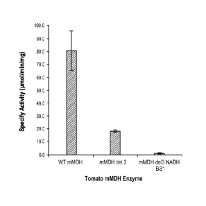
[0040] Figure 13 is a table which depicts the specific activities of
wild-type
and two mutant tomato mMDH enzymes that were measured spectrophotometrically
monitoring for NADH oxidation. The "mMDH del 3" mutation retains about 23 % of
the activity of the wild-type enzyme and the activity of the "mMDH de13 NADH
BS1" mutation is significantly diminished at about 1.5 % of the wild-type
enzyme.
DETAILED DESCRIPTION
[0041] The present disclosure relates to methods and compositions for
altered
expression of one or more malate dehydrogenase (MDH) genes in a plant cell or
CA 02872124 2014-10-30
WO 2013/166315
PCT/US2013/039309
plant, for example targeted genomic modification of a MDH gene such as a
mitochondrial malate dehydrogenase (mMDH) gene in a plant cell (e.g., maize,
tomato, soy, etc.). In particular, expression of MDH is altered via use of
fusion
proteins comprising a DNA-binding domain (e.g., zinc finger protein) and
functional
domain (e.g., transcriptional regulatory domain and/or nuclease). In certain
embodiments, targeted modification is achieved by cleaving an MDH gene using
one
or more nucleases (e.g., ZFNs) to produce modifications (e.g., mutations) at
the MDH
locus. Cleavage is targeted through the use of fusion proteins comprising a
DNA-
binding domain, such as a meganuclease DNA-binding domain, a leueine zipper
DNA-binding domain, a TAL DNA-binding domain, a zinc finger protein (ZFP), a
Crispr/Cas system or chimeric combinations of the aforementioned. In certain
embodiments, the modification comprises mutation (substitutions, deletions
and/or
insertions) of the MDH gene such that one or more amino acids in the first
and/or
second NADH binding region of an endogenous MDH gene are altered, for example
one or more amino acids at positions 104-136 and/or 171-220, numbered relative
and
aligned to SEQ ID NO:1 (wild-type tomato MDH sequence). SEQ ID NO:125 (wild-
type corn MDH sequence) and/or SEQ ID NO:126 (wild-type soybean MDH
sequence).
[0042] In certain embodiments, the nuclease(s) comprise one or more
ZFNs.
ZFNs typically comprise a cleavage domain (or a cleavage half-domain) and a
zinc
finger binding domain that binds to a target site in the endogenous MDH gene.
The
ZFNs may be introduced as proteins, as polynucleotides encoding these proteins
and/or as combinations of polypeptides and polypeptide-encoding
polynueleotides.
Zinc finger nucleases typically function as dimerie proteins following
dimerization of
the cleavage half-domains and may final homodimers and/or heterodimers.
Obligate
heterodimeric ZFNs, in which the ZFN monomers bind to the "left" and "right"
recognition domains can associate to fon-n an active nuclease have been
described.
See, e.g., U.S. Patent Publication No. 2008/0131962. Thus, given the
appropriate
target sites, a "left" monomer could form an active ZF nuclease with any
"right"
monomer. This significantly increases the number of useful nuclease sites
based on
proven left and right domains that can be used in various combinations. For
example,
recombining the binding sites of 4 homodimeric ZF nucleases yields an
additional 12
heterodimeric ZF nucleases. More importantly, it enables a systematic approach
to
transgenic design such that every new introduced exogenous sequence
(transgene)
11
CA 02872124 2014-10-30
WO 2013/166315
PCT/US2013/039309
becomes flanked with a unique ZFN site that can be used to excise the gene
back out
or to target additional genes next to it. Additionally, this method can
simplify
strategies of stacking into a single locus that is driven by ZFN-dependent
double-
strand breaks.
[00431 A zinc finger binding domain can be a canonical (C2H2) zinc finger
or
a non-canonical (e.g.. C3H) zinc finger. Furthermore, the zinc finger binding
domain
can comprise one or more zinc fingers (e.g., 2, 3, 4, 5, 6, 7, 8, 9 or more
zinc fingers),
and can be engineered to bind to any sequence within any MDH gene. The
recognition helix regions of exemplary MDH-binding zinc finger proteins for
use in
binding to an MDH gene are shown in Table lA and exemplary target sites within
an
MDH gene are shown in Table 1B. The presence of such a fusion protein (or
proteins
and/or polynucleotides encoding these fusion proteins) in a cell results in
binding of
the fusion protein(s) to its (their) binding site(s) and cleavage within the
MDH
gene(s).
General
[00441 Practice of the methods, as well as preparation and use of the
compositions disclosed herein employ, unless otherwise indicated, conventional
techniques in molecular biology, biochemistry, chromatin structure and
analysis,
computational chemistry, cell culture, recombinant DNA and related fields as
are
within the skill of the art. These techniques are fully explained in the
literature. See,
for example, Sambrook et al. MOLECULAR CLONING: A LABORATORY MANUAL,
Second edition, Cold Spring Harbor Laboratory Press, 1989 and Third edition,
2001;
Ausubel et al., CURRENT PROTOCOLS IN MOLECULAR BIOLOGY, John Wiley & Sons,
New York, 1987 and periodic updates; the series METHODS IN ENZYMOLOGY,
Academic Press, San Diego; Wolfe, CHROMATIN STRUCTURE AND FUNCTION, Third
edition, Academic Press, San Diego, 1998; METHODS IN ENZYMOLOGY, Vol. 304,
"Chromatin" (P.M. Wassannan and A. P. Wolffe, eds.), Academic Press, San
Diego,
1999; and METHODS IN MOLECULAR BIOLOGY, Vol. 119, "Chromatin Protocols"
(P.B. Becker. ed.) Humana Press, Totowa, 1999.
Definitions
[0045] The teims "nucleic acid," "polynucleotide," and
"oligonucleotide" are used
interchangeably and refer to a deoxyribonucleotide or ribonucleotide polymer,
in linear or
circular conformation, and in either single- or double-stranded farm. For the
purposes of
12
CA 02872124 2014-10-30
WO 2013/166315
PCT/US2013/039309
the present disclosure, these telins are not to be construed as limiting with
respect to the
length of a polymer. The temis can encompass known analogues of natural
nucleotides, as
well as nucleotides that are modified in the base, sugar and/or phosphate
moieties (e.g.,
phosphorothioate backbones). In general, an analogue of a particular
nucleotide has the
same base-pairing specificity; i.e., an analogue of A will base-pair with T.
[0046] The tein "polypeptide," "peptide" and "protein" are used
interchangeably
to refer to a polymer of amino acid residues. The term also applies to amino
acid
polymers in which one or more amino acids are chemical analogues or modified
derivatives of a corresponding naturally-occurring amino acids.
[0047] "Binding" refers to a sequence-specific, non-covalent interaction
between macromolecules (e.g., between a protein and a nucleic acid). Not all
components of a binding interaction need be sequence-specific (e.g., contacts
with
phosphate residues in a DNA backbone), as long as the interaction as a whole
is
sequence-specific. Such interactions are generally characterized by a
dissociation
constant (Kd) of 10-6 M-1 or lower. "Affinity" refers to the strength of
binding:
increased binding affinity being correlated with a lower IQ.
[0048] A "binding protein" is a protein that is able to bind to
another molecule. A
binding protein can bind to, for example, a DNA molecule (a DNA-binding
protein), an
RNA molecule (an RNA-binding protein) and/or a protein molecule (a protein-
binding
protein), in the case of a protein-binding protein, it can bind to itself (to
foini
homodimers, homotrimers, etc.) and/or it can bind to one or more molecules of
a different
protein or proteins. A binding protein can have more than one type of binding
activity.
For example, zinc finger proteins have DNA-binding, RNA-binding and protein-
binding
activity.
[0049] A "zinc finger DNA binding protein" (or binding domain) is a
protein, or a
domain within a larger protein, that binds DNA in a sequence-specific manner
through one
or more zinc fingers, which are regions of amino acid sequence within the
binding domain
whose structure is stabilized through coordination of a zinc ion. The term
zinc finger
DNA binding protein is often abbreviated as zinc finger protein or ZFP.
100501 Zinc finger binding domains can be "engineered" to bind to a
predetermined nucleotide sequence. Non-limiting examples of methods for
engineering zinc finger proteins are design and selection. A designed zinc
finger
protein is a protein not occurring in nature whose design/composition results
principally from rational criteria. Rational criteria for design include
application of
13
CA 02872124 2014-10-30
WO 2013/166315
PCT/US2013/039309
substitution rules and computerized algorithms for processing infounation in a
database storing information of existing ZFP designs and binding data. See,
for
example, US Patents 6,140,081; 6,453,242; and 6,534,261; see also WO 98/53058;
WO 98/53059; WO 98/53060; WO 02/016536 and WO 03/016496.
[0051] A "selected" zinc finger protein is a protein not found in nature
whose
production results primarily from an empirical process such as phage display,
interaction
trap or hybrid selection. See e.g., US 5,789,538; US 5,925,523; US 6,007,988;
US 6,013,453; US 6,200,759; WO 95/19431; WO 96/06166; WO 98/53057;
WO 98/54311; WO 00/27878; WO 01/60970 WO 01/88197 and WO 02/099084.
[0052] The term "sequence" refers to a nucleotide sequence of any length,
which can be DNA or RNA; can be linear, circular or branched and can be either
single-stranded or double stranded. The term "donor sequence" refers to a
nucleotide
sequence that is inserted into a genome. A donor sequence can be of any
length, for
example between 2 and 10,000 nucleotides in length (or any integer value
therebetween or thereabove), preferably between about 100 and 1,000
nucleotides in
length (or any integer therebetween), more preferably between about 200 and
500
nucleotides in length.
[0053] A "homologous, non-identical sequence" refers to a first
sequence
which shares a degree of sequence identity with a second sequence, but whose
sequence is not identical to that of the second sequence. For example, a
polynucleotide comprising the wild-type sequence of a mutant gene is
homologous
and non-identical to the sequence of the mutant gene. In certain embodiments,
the
degree of homology between the two sequences is sufficient to allow homologous
recombination therebetween, utilizing normal cellular mechanisms. Two
homologous
non-identical sequences can be any length and their degree of non-homology can
be
as small as a single nucleotide (e.g., for correction of a genomic point
mutation by
targeted homologous recombination) or as large as 10 or more kilobases (e.g.,
for
insertion of a gene at a predeteiinined ectopic site in a chromosome). Two
polynucleotides comprising the homologous non-identical sequences need not be
the
same length. For example, an exogenous polynucleotide (i.e., donor
polynucleotide)
of between 20 and 10,000 nucleotides or nucleotide pairs can be used.
[0054] Techniques for deteunining nucleic acid and amino acid sequence
identity are known in the art. Typically, such techniques include determining
the
nucleotide sequence of the mRNA for a gene and/or detei mining the amino
acid
14
CA 02872124 2014-10-30
WO 2013/166315
PCT/US2013/039309
sequence encoded thereby, and comparing these sequences to a second nucleotide
or
amino acid sequence. Genomic sequences can also be determined and compared in
this fashion. In general, identity refers to an exact nucleotide-to-nucleotide
or amino
acid-to-amino acid correspondence of two polynucleotides or polypeptide
sequences,
respectively. Two or more sequences (polynucleotide or amino acid) can be
compared by determining their percent identity. The percent identity of two
sequences, whether nucleic acid or amino acid sequences, is the number of
exact
matches between two aligned sequences divided by the length of the shorter
sequences and multiplied by 100. An approximate alignment for nucleic acid
.. sequences is provided by the local homology algorithm of Smith and
Waterman,
Advances in Applied Mathematics 2:482-489 (1981). This algorithm can be
applied
to amino acid sequences by using the scoring matrix developed by Dayhoff,
Atlas of
Protein Sequences and Structure, M.O. Dayhoff ed., 5 suppl. 3:353-358,
National
Biomedical Research Foundation, Washington, D.C., USA, and normalized by
Gribskov, Nucl. Acids Res. 14(6):6745-6763 (1986). An exemplary implementation
of this algorithm to determine percent identity of a sequence is provided by
the
Genetics Computer Group (Madison, WI) in the "BestFit" utility application.
Suitable programs for calculating the percent identity or similarity between
sequences
are generally known in the art, for example, another alignment program is
BLAST,
used with default parameters. For example, BLASTN and BLASTP can be used
using the following default parameters: genetic code = standard; filter =
none; strand
= both; cutoff= 60; expect ¨ 10; Matrix ¨ BLOSUM62; Descriptions = 50
sequences;
sort by = HIGH SCORE; Databases = non-redundant, GenBank + EMBL + DDBJ +
PDB + GenBank CDS translations + Swiss protein + Spupdate + PIR. Details of
these programs can be found on the internet. With respect to sequences
described
herein, the range of desired degrees of sequence identity is approximately 80%
to
100% and any integer value therebetween. Typically the percent identities
between
sequences are at least 70-75%, preferably 80-82%, more preferably 85-90%, even
more preferably 92%, still more preferably 95%, and most preferably 98%
sequence
identity.
[0055] Alternatively, the degree of sequence similarity between
polynucleotides can be determined by hybridization of polynucleotides under
conditions that allow formation of stable duplexes between homologous regions,
followed by digestion with single-stranded-specific nuclease(s), and size
CA 02872124 2014-10-30
WO 2013/166315
PCT/US2013/039309
determination of the digested fragments. Two nucleic acid, or two polypeptide
sequences are substantially homologous to each other when the sequences
exhibit at
least about 70%-75%, preferably 80%-82%, more preferably 85%-90%, even more
preferably 92%, still more preferably 95%, and most preferably 98% sequence
identity over a defined length of the molecules, as determined using the
methods
above. As used herein, substantially homologous also refers to sequences
showing
complete identity to a specified DNA or polypeptide sequence. DNA sequences
that
are substantially homologous can be identified in a Southern hybridization
experiment
under, for example, stringent conditions, as defined for that particular
system.
Defining appropriate hybridization conditions is known to those with skill of
the art.
See, e.g., Sambrook et al., supra; Nucleic Acid Hybridization: A Practical
Approach,
editors B.D. Hames and S.J. Higgins, (1985) Oxford; Washington, DC; IRE
Press).
[0056] Selective hybridization of two nucleic acid fragments can be
determined as follows. The degree of sequence identity between two nucleic
acid
molecules affects the efficiency and strength of hybridization events between
such
molecules. A partially identical nucleic acid sequence will at least partially
inhibit the
hybridization of a completely identical sequence to a target molecule.
Inhibition of
hybridization of the completely identical sequence can be assessed using
hybridization assays that are well known in the art (e.g., Southern (DNA)
blot,
Northern (RNA) blot, solution hybridization, or the like, see Sambrook, et
al.,
Molecular Cloning- A Laboratory Manual, Second Edition, (1989) Cold Spring
Harbor, N.Y.). Such assays can be conducted using varying degrees of
selectivity, for
example, using conditions varying from low to high stringency. If conditions
of low
stringency are employed, the absence of non-specific binding can be assessed
using a
secondary probe that lacks even a partial degree of sequence identity (for
example, a
probe having less than about 30% sequence identity with the target molecule),
such
that, in the absence of non-specific binding events, the secondary probe will
not
hybridize to the target.
[0057] When utilizing a hybridization-based detection system, a
nucleic acid
probe is chosen that is complementary to a reference nucleic acid sequence,
and then
by selection of appropriate conditions the probe and the reference sequence
selectively hybridize, or bind, to each other to form a duplex molecule. A
nucleic
acid molecule that is capable of hybridizing selectively to a reference
sequence under
moderately stringent hybridization conditions typically hybridizes under
conditions
16
CA 02872124 2014-10-30
WO 2013/166315
PCT/US2013/039309
that allow detection of a target nucleic acid sequence of at least about 10-14
nucleotides in length having at least approximately 70% sequence identity with
the
sequence of the selected nucleic acid probe. Stringent hybridization
conditions
typically allow detection of target nucleic acid sequences of at least about
10-14
nucleotides in length having a sequence identity of greater than about 90-95%
with
the sequence of the selected nucleic acid probe. Hybridization conditions
useful for
probe/reference sequence hybridization, where the probe and reference sequence
have
a specific degree of sequence identity, can be detennined as is known in the
art (see,
for example, Nucleic Acid Hybridization: A Practical Approach, editors B.D.
Hames
and Si. Higgins, (1985) Oxford; Washington, DC; IRL Press).
[0058] Conditions for hybridization are well-known to those of
skill in the art.
Hybridization stringency refers to the degree to which hybridization
conditions
disfavor the formation of hybrids containing mismatched nucleotides, with
higher
=
stringency correlated with a lower tolerance for mismatched hybrids. Factors
that
affect the stringency of hybridization are well-known to those of skill in the
art and
include, but are not limited to, temperature, plI, ionic strength, and
concentration of
organic solvents such as, for example, formamide and dimethylsulfoxide. As is
known to those of skill in the art, hybridization stringency is increased by
higher
temperatures, lower ionic strength and lower solvent concentrations.
[0059] With respect to stringency conditions for hybridization, it is well
known in the art that numerous equivalent conditions can be employed to
establish a
particular stringency by varying, for example, the following factors: the
length and
nature of the probe sequences, base composition of the various sequences,
concentrations of salts and other hybridization solution components, the
presence or
absence of blocking agents in the hybridization solutions (e.g., dextran
sulfate, and
polyethylene glycol), hybridization reaction temperature and time parameters,
as well
as, varying wash conditions. The selection of a particular set of
hybridization
conditions is selected following standard methods in the art (see, for
example,
Sambrook, et al., Molecular Cloning: A Laboratory Manual, Second Edition,
(1989)
Cold Spring Harbor, N.Y.).
[0060] "Recombination" refers to a process of exchange of genetic
information between two polynucleotides. For the purposes of this disclosure,
"homologous recombination (HR)" refers to the specialized font' of such
exchange
that takes place, for example, during repair of double-strand breaks in cells.
This
17
81783303
process requires nucleotide sequence homology, that uses a "donor" molecule to
template repair of a "target" molecule (i.e., the one that experienced the
double-strand
break), and is variously known as "non-crossover gene conversion" or "short
tract
gene conversion," because it leads to the transfer of genetic information from
the
donor to the target. Without wishing to be bound by any particular theory,
such
transfer can involve mismatch correction of heteroduplex DNA that forms
between
the broken target and the donor, and/or "synthesis-dependent strand
annealing," in
which the donor is used to resynthesize genetic information that will become
part of
the target, and/or related processes. Such specialized HR often results in an
alteration
of the sequence of the target molecule such that part or all of the sequence
of the
donor polynucleotide is incorporated into the target polynucleotide.
[0061] "Cleavage" refers to the breakage of the covalent backbone of
a DNA
molecule. Cleavage can be initiated by a variety of methods including, but not
limited
to, enzymatic or chemical hydrolysis of a phosphodiester bond. Both single-
stranded
cleavage and double-stranded cleavage are possible, and double-stranded
cleavage
can occur as a result of two distinct single-stranded cleavage events. DNA
cleavage
can result in the production of either blunt ends or staggered ends. In
certain
embodiments, fusion polypeptides are used for targeted double-stranded DNA
cleavage.
[0062] A "cleavage domain" comprises one or more polypeptide sequences
which possesses catalytic activity for DNA cleavage. A cleavage domain can be
contained in a single polypeptide chain or cleavage activity can result from
the
association of two (or more) polypeptides.
[0063] A "cleavage half-domain" is a polypeptide sequence which, in
conjunction with a second polypeptide (either identical or different) forms a
complex
having cleavage activity (preferably double-strand cleavage activity).
[0064] An "engineered cleavage half-domain" is a cleavage half-domain
that
has been modified so as to form obligate heterodimers with another cleavage
half-
domain (e.g., another engineered cleavage half-domain). See, also, U.S. Patent
Publication Nos. 2005/0064474, 20070218528 and 2008/0131962.
[0065] "Chromatin" is the nucleoprotein structure comprising the
cellular
genome. Cellular chromatin comprises nucleic acid, primarily DNA, and protein,
including histones and non-histone chromosomal proteins. The majority of
18
CA 2872124 2019-06-13
CA 02872124 2014-10-30
WO 2013/166315
PCT/US2013/039309
eukaryotic cellular chromatin exists in the form of nucleosomes, wherein a
nucleosome core comprises approximately 150 base pairs of DNA associated with
an
octamer comprising two each of histones H2A, H2B, 113 and 1-14; and linker DNA
(of
variable length depending on the organism) extends between nucleosome cores. A
molecule of histone Ill is generally associated with the linker DNA. For the
purposes
of the present disclosure, the term "chromatin" is meant to encompass all
types of
cellular nucleoprotein, both prokaryotic and eukaryotic. Cellular chromatin
includes
both chromosomal and episomal chromatin.
[00661 A "chromosome," is a chromatin complex comprising all or a
portion
of the genome of a cell. The genome of a cell is often characterized by its
karyotype,
which is the collection of all the chromosomes that comprise the genome of the
cell.
The genome of a cell can comprise one or more chromosomes.
[00671 An "episome" is a replicating nucleic acid, nucleoprotein
complex or
other structure comprising a nucleic acid that is not part of the chromosomal
karyotype of a cell. Examples of episomes include plasmids and certain viral
genomes.
[00681 An "accessible region" is a site in cellular chromatin in which
a target
site present in the nucleic acid can be bound by an exogenous molecule which
recognizes the target site. Without wishing to be bound by any particular
theory, it is
believed that an accessible region is one that is not packaged into a
nueleosomal
structure. The distinct structure of an accessible region can often be
detected by its
sensitivity to chemical and enzymatic probes, for example, nucleases.
[00691 A "target site" or "target sequence" is a nucleic acid sequence
that
defines a portion of a nucleic acid to which a binding molecule will bind,
provided
sufficient conditions for binding exist. For example, the sequence 5'-GAATTC-
3' is
a target site for the Eco RI restriction endonuclease.
[00701 An "exogenous" molecule is a molecule that is not normally
present in
a cell, but can be introduced into a cell by one or more genetic, biochemical
or other
methods. "Normal presence in the cell" is deteimined with respect to the
particular
developmental stage and environmental conditions of the cell. Thus, for
example, a
molecule that is present in cells only during the early stages of development
of a
flower is an exogenous molecule with respect to the cells of a fully developed
flower..
Similarly, a molecule induced by heat shock is an exogenous molecule with
respect to
a non-heat-shocked cell. An exogenous molecule can comprise, for example, a
19
CA 02872124 2014-10-30
WO 2013/166315
PCT/US2013/039309
coding sequence for any polypeptide or fragment thereof, a functioning version
of a
malfunctioning endogenous molecule or a malfunctioning version of a normally-
functioning endogenous molecule. Additionally, an exogenous molecule can
comprise
a coding sequence from another species that is an ortholog of an endogenous
gene in
the host cell.
[0071] An exogenous molecule can be, among other things, a small
molecule,
such as is generated by a combinatorial chemistry process, or a macromolecule
such
as a protein, nucleic acid, carbohydrate, lipid, glycoprotein, lipoprotein,
polysaccharide, any modified derivative of the above molecules, or any complex
comprising one or more of the above molecules. Nucleic acids include DNA and
RNA, can be single- or double-stranded; can be linear, branched or circular;
and can
be of any length. Nucleic acids include those capable of foiming duplexes, as
well as
triplex-forming nucleic acids. See, for example, U.S. Patent Nos. 5,176,996
and
5,422,251. Proteins include, but are not limited to, DNA-binding proteins,
transcription factors, chromatin remodeling factors, methylated DNA binding
proteins, polymerases, methylases, demethylases, acetylases, deacetylases,
kinases,
phosphatases, integrases, recombinases, ligases, topoisomerases, gyrases and
helieases. Thus, the term includes "transgenes" or "genes of interest" which
are
exogenous sequences introduced into a plant cell, e.g., into an MDH gene in a
plant
cell.
[0072] An exogenous molecule can be the same type of molecule as an
endogenous molecule, e.g., an exogenous protein or nucleic acid. For example,
an
exogenous nucleic acid can comprise an infecting viral genome, a plasmid or
episome
introduced into a cell, or a chromosome that is not normally present in the
cell.
Methods for the introduction of exogenous molecules into cells are known to
those of
skill in the art and include, but are not limited to, protoplast
transfoimation, silicon
carbide (e.g., WHISKERSTm), Agrobacterium-mediated transfoimation, lipid-
mediated transfer (i.e., liposomes, including neutral and cationic lipids),
electroporation, direct injection, cell fusion, particle bombardment (e.g.,
using a "gene
gun"), calcium phosphate co-precipitation, DEAE-dextran-mediated transfer and
viral
vector-mediated transfer.
[0073] By contrast, an "endogenous" molecule is one that is normally
present
in a particular cell at a particular developmental stage under particular
environmental
conditions. For example, an endogenous nucleic acid can comprise a chromosome,
CA 02872124 2014-10-30
WO 2013/166315
PCT/US2013/039309
the genome of a mitochondrion, chloroplast or other organelle, or a naturally-
occurring episomal nucleic acid. Additional endogenous molecules can include
proteins, for example, transcription factors and enzymes.
[0074] As used herein, the tem' "product of an exogenous nucleic acid"
includes both polynucleotide and polypeptide products, for example,
transcription
products (polynucleotides such as RNA) and translation products
(polypeptides).
[0075] A "fusion" molecule is a molecule in which two or more subunit
molecules are linked, preferably covalently. The subunit molecules can be the
same
chemical type of molecule, or can be different chemical types of molecules.
Examples of the first type of fusion molecule include, but are not limited to,
fusion
proteins (for example, a fusion between a ZFP DNA-binding domain and a
cleavage
domain) and fusion nucleic acids (for example, a nucleic acid encoding the
fusion
protein described supra). Examples of the second type of fusion molecule
include,
but are not limited to, a fusion between a triplex-forming nucleic acid and a
polypeptide, and a fusion between a minor groove binder and a nucleic acid.
[0076] Expression of a fusion protein in a cell can result from
delivery of the
fusion protein to the cell or by delivery of a polynucleotide encoding the
fusion
protein to a cell, wherein the polynucleotide is transcribed, and the
transcript is
translated, to generate the fusion protein. Trans-splicing, polypeptide
cleavage and
polypeptide ligation can also be involved in expression of a protein in a
cell. Methods
for polynucleotide and polypeptide delivery to cells are presented elsewhere
in this
disclosure.
[0077] A "gene," for the purposes of the present disclosure, includes
a DNA
region encoding a gene product (see infra), as well as all DNA regions which
regulate
the production of the gene product, whether or not such regulatory sequences
are
adjacent to coding and/or transcribed sequences. Accordingly, a gene includes,
but is
not necessarily limited to, promoter sequences, teaninators, translational
regulatory
sequences such as ribosome binding sites and internal ribosome entry sites,
enhancers,
silencers, insulators, boundary elements, replication origins, matrix
attachment sites
and locus control regions.
100781 "Gene expression" refers to the conversion of the infounation,
contained in a gene, into a gene product. A gene product can be the direct
transcriptional product of a gene (e.g., mRNA, tRNA, rRNA, antisense RNA,
ribozyme, structural RIXA or any other type of RNA) or a protein produced by
21
CA 02872124 2014-10-30
WO 2013/166315
PCT/US2013/039309
translation of a mRNA. Gene products also include RNAs which are modified, by
processes such as capping, polyadenylation, methylation, and editing, and
proteins
modified by, for example, methylation, acetylation, phosphorylation,
ubiquitination,
ADP-ribosylation, myristilation, and glycosylation.
[0079] "Modulation" of gene expression refers to a change in the activity
of a
gene. Modulation of expression can include, but is not limited to, Rene
activation and
gene repression.
[0080] "Plant" cells include, but are not limited to, cells of
monocotyledonous
(monocots) or dicotyledonous (dicots) plants. Non-limiting examples of
monocots
.. include cereal plants such as maize, rice, barley, oats, wheat, sorghum,
rye, sugarcane,
pineapple, onion, banana, and coconut. Non-limiting examples of dicots include
tobacco, tomato, sunflower, cotton, sugarbeet, potato, lettuce, melon, soy,
canola
(rapeseed), and alfalfa. Plant cells may be from any part of the plant and/or
from any
stage of plant development.
[0081] A "region of interest" is any region of cellular chromatin, such as,
for
example, a gene or a non-coding sequence within or adjacent to a gene, in
which it is
desirable to bind an exogenous molecule. Binding can be for the purposes of
targeted
DNA cleavage and/or targeted recombination. A region of interest can be
present in a
chromosome, an episome, an organellar genome (e.g., mitochondrial,
chloroplast), or
.. an infecting viral genome, for example. A region of interest can be within
the coding
region of a gene, within transcribed non-coding regions such as, for example,
leader
sequences, trailer sequences or introns, or within non-transcribed regions,
either
upstream or downstream of the coding region. A region of interest can be as
small as
a single nucleotide pair or up to 2,000 nucleotide pairs in length, or any
integral value
of nucleotide pairs.
[0082] The terms "operative linkage" and "operatively linked" (or
"operably
linked") are used interchangeably with reference to a juxtaposition of two or
more
components (such as sequence elements), in which the components are arranged
such
that both components function normally and allow the possibility that at least
one of
the components can mediate a function that is exerted upon at least one of the
other
components. By way of illustration, a transcriptional regulatory sequence,
such as a
promoter, is operatively linked to a coding sequence if the transcriptional
regulatory
sequence controls the level of transcription of the coding sequence in
response to the
presence or absence of one or more transcriptional regulatory factors. A
22
CA 02872124 2014-10-30
WO 2013/166315
PCT[US2013/039309
transcriptional regulatory sequence is generally operatively linked in cis
with a coding
sequence, but need not be directly adjacent to it. For example, an enhancer is
a
transcriptional regulatory sequence that is operatively linked to a coding
sequence,
even though they are not contiguous.
[0083] With respect to fusion polypeptides, the term "operatively linked"
can
refer to the fact that each of the components performs the same function in
linkage to
the other component as it would if it were not so linked. For example, with
respect to
a fusion polypeptide in which a ZFP DNA-binding domain is fused to a cleavage
domain, the ZFP DNA-binding domain and the cleavage domain are in operative
linkage if, in the fusion polypeptide, the ZFP DNA-binding domain portion is
able to
bind its target site and/or its binding site, while the cleavage domain is
able to cleave
DNA in the vicinity of the target site.
[0084] A "functional fragment" of a protein, polypeptide or nucleic
acid is a
protein, polypeptide or nucleic acid whose sequence is not identical to the
full-length
.. protein, polypeptide or nucleic acid, yet retains the same function as the
full-length
protein, polypeptide or nucleic acid. A functional fragment can possess more,
fewer,
or the same number of residues as the corresponding native molecule, and/or
can
contain one or more amino acid or nucleotide substitutions. Methods for
determining
the function of a nucleic acid (e.g., coding function, ability to hybridize to
another
nucleic acid) are well-known in the art. Similarly, methods for determining
protein
function are well-known. For example, the DNA-binding function of a
polypeptide
can be determined, for example, by filter-binding, electrophoretic mobility-
shift, or
immunoprecipitation assays. DNA cleavage can be assayed by gel
electrophoresis.
See Ausubel et al., supra. The ability of a protein to interact with another
protein can
be determined, for example, by co-immunoprecipitation, two-hybrid assays or
complementation, both genetic and biochemical. See, for example, Fields et at.
(1989) Nature 340:245-246; U.S. Patent No. 5,585.245 and PCT WO 98/44350.
DNA-binding domains
[0085] Any DNA-binding domain can be used in the methods disclosed
herein. In certain embodiments, the DNA binding domain comprises a zinc finger
protein. A zinc finger binding domain comprises one or more zinc fingers.
Miller et
at. (1985) EMBO J. 4:1609-1614; Rhodes (1993) Scientific American Feb.:56-65;
US
23
CA 02872124 2014-10-30
WO 2013/166315
PCT/US2013/039309
Patent No. 6,453,242. The zinc finger binding domains described herein
generally
include 2, 3, 4, 5, 6 or even more zinc fingers.
[0086] Typically, a single zinc finger domain is about 30 amino acids
in
length. Structural studies have demonstrated that each zinc finger domain
(motif)
.. contains two beta sheets (held in a beta turn which contains the two
invariant cysteine
residues) and an alpha helix (containing the two invariant histidine
residues), which
are held in a particular confoimation through coordination of a zinc atom by
the two
cysteines and the two histidines.
[0087] Zinc fingers include both canonical C2H2 zinc fingers (i.e.,
those in
which the zinc ion is coordinated by two cysteine and two histidine residues)
and non-
canonical zinc fingers such as, for example, C3H zinc fingers (those in which
the zinc
ion is coordinated by three cysteine residues and one histidine residue) and
C4 zinc
fingers (those in which the zinc ion is coordinated by four cysteine
residues). See also
WO 02/057293 and also U.S. Patent Publication No. 20080182332 regarding non-
.. canonical ZFPs for use in plants.
[0088] An engineered zinc finger binding domain can have a novel
binding
specificity, compared to a naturally-occurring zinc finger protein.
Engineering
methods include, but are not limited to, rational design and various types of
selection.
Rational design includes, for example, using databases comprising triplet (or
quadruplet) nucleotide sequences and individual zinc finger amino acid
sequences, in
which each triplet or quadruplet nucleotide sequence is associated with one or
more
amino acid sequences of zinc fingers which bind the particular triplet or
quadruplet
sequence.
[0089] Exemplary selection methods, including phage display and two-
hybrid
.. systems, are disclosed in US Patents 5,789,538; 5,925,523; 6,007,988;
6,013,453;
6,410,248; 6,140,466; 6,200,759; and 6,242,568; as well as WO 98/37186;
WO 98/53057; WO 00/27878; WO 01/88197 and GB 2,338,237.
[0090] Enhancement of binding specificity for zinc fmger binding
domains
has been described, for example, in WO 02/077227.
[0091] Since an individual zinc finger binds to a three-nucleotide (i.e.,
triplet)
sequence (or a four-nucleotide sequence which can overlap, by one nucleotide,
with
the four-nucleotide binding site of an adjacent zinc finger), the length of a
sequence to
which a zinc finger binding domain is engineered to bind (e.g., a target
sequence) will
deteimine the number of zinc fmgers in an engineered zinc finger binding
domain.
24
CA 02872124 2014-10-30
WO 2013/166315
PCT/US2013/039309
For example, for ZFPs in which the finger motifs do not bind to overlapping
subsites,
a six-nucleotide target sequence is bound by a two-finger binding domain; a
nine-
nucleotide target sequence is bound by a three-finger binding domain, etc. As
noted
herein, binding sites for individual zinc fingers (i.e., subsites) in a target
site need not
be contiguous, but can be separated by one or several nucleotides, depending
on the
length and nature of the amino acids sequences between the zinc fingers (i e,,
the
inter-finger linkers) in a multi-finger binding domain.
[0092] In a multi-finger zinc finger binding domain, adjacent zinc
fingers can
be separated by amino acid linker sequences of approximately 5 amino acids (so-
called "canonical" inter-finger linkers) or, alternatively, by one or more non-
canonical
linkers. See, e.g., US Patent Nos. 6,453,242 and 6,534,261. For engineered
zinc
finger binding domains comprising more than three fingers, insertion of longer
("non-
canonical") inter-finger linkers between certain of the zinc fingers may be
desirable in
some instances as it may increase the affinity and/or specificity of binding
by the
binding domain. See, for example, U.S. Patent No. 6,479,626 and WO 01/53480.
Accordingly, multi-finger zinc finger binding domains can also be
characterized with
respect to the presence and location of non-canonical inter-finger linkers.
For
example, a six-finger zinc finger binding domain comprising three fingers
(joined by
two canonical inter-finger linkers), a long linker and three additional
fingers (joined
by two canonical inter-finger linkers) is denoted a 2x3 configuration.
Similarly, a
binding domain comprising two fingers (with a canonical linker therebetween),
a long
linker and two additional fingers (joined by a canonical linker) is denoted a
2x2
configuration. A protein comprising three two-finger units (in each of which
the two
fingers are joined by a canonical linker), and in which each two-finger unit
is joined
to the adjacent two finger unit by a long linker, is referred to as a 3x2
configuration.
[0093] The presence of a long or non-canonical inter-finger linker
between
two adjacent zinc fingers in a multi-finger binding domain often allows the
two
fingers to bind to subsites which are not immediately contiguous in the target
sequence. Accordingly, there can be gaps of one or more nucleotides between
subsites in a target site; i.e., a target site can contain one or more
nucleotides that are
not contacted by a zinc finger. For example, a 2x2 zinc finger binding domain
can
bind to two six-nucleotide sequences separated by one nucleotide, i.e., it
binds to a
13-nucleotide target site. See also Moore et al. (2001a) Proc. Natl. Acad.
Sci. USA
CA 02872124 2014-10-30
WO 2013/166315
PCT/US2013/039309
98:1432-1436; Moore etal. (2001b) Proc. Natl. Acad. Sci. USA 98:1437-1441 and
WO 01/53480.
[0094] As discussed previously, a target subsite is a three- or four-
nucleotide
sequence that is bound by a single zinc finger. For certain purposes, a two-
finger unit
.. is denoted a "binding module." A binding module can be obtained by, for
example,
selecting for two adjacent fingers in the context of a multi-finger protein
(generally
three fingers) which bind a particular six-nucleotide target sequence.
Alternatively,
modules can be constructed by assembly of individual zinc fingers. See also
WO 98/53057 and WO 01/53480.
[0095] Alternatively, the DNA-binding domain may be derived from a
nuclease. For example, the recognition sequences of homing endonucleases and
meganucleases such as I-SceI,I-CeuI,PI-PspI,PI-Sce,I-SceIV,I-CsmI,I-PanI, I-
Scell, I-PpoI, I-SceIII, 1-CreI. I-TevI,I-TevII and I-TevIII are known. See
also U.S.
Patent No. 5,420,032; U.S. Patent No. 6,833,252; Belfort et al. (1997) Nucleic
Acids
Res. 25:3379-3388; Dujon etal. (1989) Gene 82:115-118; Perler et a/. (1994)
Nucleic Acids Res. 22, 1125-1127; Jasin (1996) Trends Genet. 12:224-228;
Gimble
et al. (1996).1 MoL Biol. 263:163-180; Argast et cd. (1998) Mal. Biol. 280:345-
353 and the New England Biolabs catalogue. In addition, the DNA-binding
specificity of homing endonucleases and meganucleases can be engineered to
bind
non-natural target sites. See, for example, Chevalier et al. (2002) Molec.
Cell 10:895-
905; Epinat et al. (2003) Nucleic Acids Res. 31:2952-2962; Ashworth et al.
(2006)
Nature 441:656-659; Paques et al. (2007) Current Gene Therapy 7:49-66; U.S.
Patent Publication No. 20070117128.
[0096] As another alternative, the DNA-binding domain may be derived
from
.. a leucine zipper protein. Leucine zippers are a class of proteins that are
involved in
protein-protein interactions in many eukaryotic regulatory proteins that are
important
transcriptional factors associated with gene expression. The leucine zipper
refers to a
common structural motif shared in these transcriptional factors across several
kingdoms including animals, plants, yeasts, etc. The leucine zipper is folioed
by two
polypeptides (homodimer or heterodimer) that bind to specific DNA sequences in
a
manner where the leucine residues are evenly spaced through an a-helix, such
that the
leucine residues of the two polypeptides end up on the same face of the helix.
The
DNA binding specificity of leucine zippers can be utilized in the DNA-binding
domains disclosed herein.
26
81783303
[0097] In some embodiments, the DNA-binding domain is an engineered
domain from a TAL effector derived from the plant pathogen Xanthomonas (see,
Miller et al. (2011) Nature Biotechnology 29(2):143-8; Boch et al, (2009)
Science 29
Oct 2009 (10.1126/science.117881) and Moscou and Bogdanove, (2009) Science 29
Oct 2009 (10.1126/science.1178817; and U.S. Patent Publication Nos.
20110239315,
20110145940 and 20110301073).
[0098] The CRISPR (lustered Regularly Interspaced Short Pafindromic
Repeats)/Cas (cRISPR Associated) nuclease system is a recently engineered
nuclease
system based on a bacterial system that can be used for genome engineering. It
is
based on part of the adaptive immune response of many bacteria and Archea.
When a
virus or plasmid invades a bacterium, segments of the invader's DNA are
converted
into CRISPR RNAs (crRNA) by the 'immune' response. This crRNA then associates,
through a region of partial complementarity, with another type of RNA called
tracrRNA to guide the Cas9 nuclease to a region homologous to the crRNA in the
target DNA called a "protospacer". Cas9 cleaves the DNA to generate blunt ends
at
the DSB at sites specified by a 20-nucleotide guide sequence contained within
the
crRNA transcript. Cas9 requires both the crRNA and the tracrRNA for site
specific
DNA recognition and cleavage. This system has now been engineered such that
the
crRNA and tracrRNA can be combined into one molecule (the "single guide RNA"),
and the crRNA equivalent portion of the single guide RNA can be engineered to
guide
the Cas9 nuclease to target any desired sequence (see Jinek et al (2012)
Science 337,
p. 816-821, Jinek et al, (2013), eLife 2:e00471, and David Segal, (2013) eLife
2:e00563). Thus, the CRISPRICas system can be engineered to create a double-
stranded break (DSB) at a desired target in a genome, and repair of the DSB
can be
influenced by the use of repair inhibitors to cause an increase in error prone
repair.
Cleavage Domains
[0099] As noted above, the DNA-binding domain may be associated with
a
cleavage (nuclease) domain. For example, homing endonucleases may be modified
in
their DNA-binding specificity while retaining nuclease function. In addition,
zinc
finger proteins may also be fused to a cleavage domain to form a zinc finger
nuclease
(ZFN). The cleavage domain portion of the fusion proteins disclosed herein can
be
obtained from any endonuclease or exonuclease. Exemplary endonucleases from
which a cleavage domain can be derived include, but are not limited to,
restriction
27
CA 2872124 2019-06-13
CA 02872124 2014-10-30
WO 2013/166315
PCT/US2013/039309
endonucleases and homing endonucleases. See, for example, 2002-2003 Catalogue,
New England Biolabs, Beverly, MA; and Belfort et al. (1997) Nucleic Acids Res.
25:3379-3388. Additional enzymes which cleave DNA are known (e.g., S1
Nuclease;
mung bean nuclease; pancreatic DNase I; micrococcal nuclease; yeast HO
endonuclease; see also Linn et al. (eds.) Nucleases, Cold Spring Harbor
Laboratory
Press,1993). Non limiting examples of homing endonucleases and meganucleases
include I-SceI,I-CeuI, PI-PspI, PI-Sce,I-SceIV I-PpoI, I-
SceIII,I-CreI,I-TevI,I-TevII and I-TevIII are known. See also U.S. Patent No.
5,420,032; U.S. Patent No. 6,833,252; Belfort et al. (1997) Nucleic Acids Res.
25:3379-3388; Duj on et al. (1989) Gene 82:115-118; Perler et al. (1994)
Nucleic
Acids Res. 22, 1125-1127; Jasin (1996) Trends Genet. 12:224-228; Gimble et al.
(1996)1 MoL Biol. 263:163-180; Argast et aL (1998)1 Mot Biol. 280:345-353 and
the New England Biolabs catalogue. One or more of these enzymes (or functional
fragments thereof) can be used as a source of cleavage domains and cleavage
half-
domains.
[0100] Restriction endonucleases (restriction enzymes) are present in
many
species and are capable of sequence-specific binding to DNA (at a recognition
site),
and cleaving DNA at or near the site of binding. Certain restriction enzymes
(e.g.,
Type IIS) cleave DNA at sites removed from the recognition site and have
separable
binding and cleavage domains. For example, the Type ITS enzyme FokI catalyzes
double-stranded cleavage of DNA, at 9 nucleotides from its recognition site on
one
strand and 13 nucleotides from its recognition site on the other. See, for
example, US
Patents 5,356,802; 5,436,150 and 5,487,994; as well as Li et al. (1992) Proc.
Natl.
Acad. Sci. USA 89:4275-4279; Li et al. (1993) Proc. Natl. Acad. Sci. USA
90:2764-
2768; Kim et al. (1994a) Proc. Natl. Acad. Sci. USA 91:883-887; Kim et al.
(1994b)
Biol. Chem. 269:31,978-31,982. Thus, in one embodiment, fusion proteins
comprise the cleavage domain (or cleavage half-domain) from at least one Type
ITS
restriction enzyme and one or more zinc finger binding domains, which may or
may
not be engineered.
[0101] An exemplary Type IIS restriction enzyme, whose cleavage domain is
separable from the binding domain, is FokI. This particular enzyme is active
as a
dimer. Bitinaite et al. (1998) Proc. Natl. Acad. Sci. USA 95: 10,570-10,575.
Accordingly, for the purposes of the present disclosure, the portion of the
Fold
enzyme used in the disclosed fusion proteins is considered a cleavage half-
domain.
28
81783303
Thus, for targeted double-stranded cleavage and/or targeted replacement of
cellular
sequences using zinc finger-FokI fusions, two fusion proteins, each comprising
a FokI
cleavage half-domain, can be used to reconstitute a catalytically active
cleavage
domain. Alternatively, a single polypeptide molecule containing a zinc finger
binding
domain and two FokI cleavage half-domains can also be used. Parameters for
targeted cleavage and targeted sequence alteration using zinc finger-FokI
fusions are
provided elsewhere in this disclosure.
[0102] A cleavage domain or cleavage half-domain can be any portion
of a
protein that retains cleavage activity, or that retains the ability to
multimcrize (e.g.,
dimerize) to form a functional cleavage domain.
[0103] Exemplary Type IIS restriction enzymes are described in
International
Publication WO 2007/014275.
[0104] To enhance cleavage specificity, cleavage domains may also be
modified. In certain embodiments, variants of the cleavage half-domain are
employed
these variants minimize or prevent homodimerization of the cleavage half-
domains.
Non-limiting examples of such modified cleavage half-domains are described in
detail
in WO 2007/014275. See, also,
Examples. In certain embodiments, the cleavage domain comprises an engineered
cleavage half-domain (also referred to as dimerization domain mutants) that
minimize
or prevent homodimerization are known to those of skill the art and described
for
example in U.S. Patent Publication Nos. 20050064474; 20060188987; 20070305346
and 20080131962. Amino acid residues at positions 446, 447, 479, 483, 484,
486, 487, 490, 491, 496, 498, 499, 500, 531, 534, 537, and 538 of FokI are all
targets
for influencing dimerization of the FokI cleavage half-domains.
[0105] Additional engineered cleavage half-domains of FokI that form
obligate heterodimers can also be used in the ZFNs described herein. Exemplary
engineered cleavage half-domains of Fok I that form obligate heterodimers
include a
pair in which a first cleavage half-domain includes mutations at amino acid
residues
at positions 490 and 538 of Fok I and a second cleavage half-domain includes
mutations at amino acid residues 486 and 499.
[0106] Thus, in one embodiment, a mutation at 490 replaces Glu (E)
with Lys
(K); the mutation at 538 replaces Iso (I) with Lys (K); the mutation at 486
replaced
Gin (Q) with Glu (E); and the mutation at position 499 replaces Iso (I) with
Lys (K).
29
CA 2872124 2019-06-13
81783303
Specifically, the engineered cleavage half-domains described herein were
prepared by
mutating positions 490 (E¨>K) and 538 (I¨>K) in one cleavage half-domain to
produce an engineered cleavage half-domain designated "E490K:I538K" and by
mutating positions 486 (Q¨E) and 499 (I--->L) in another cleavage half-domain
to
produce an engineered cleavage half-domain designated "Q486E:I499L". The
engineered cleavage half-domains described herein are obligate heterodimer
mutants
in which aberrant cleavage is minimized or abolished. See, e.g., U.S. Patent
Publication No. 2008/0131962. In certain embodiments, the engineered cleavage
half-
domain comprises mutations at positions 486, 499 and 496 (numbered relative to
wild-type Fold), for instance mutations that replace the wild type Gin (Q)
residue at
position 486 with a Glu (E) residue, the wild type Iso (I) residue at position
499 with a
Leu (L) residue and the wild-type Asn (N) residue at position 496 with an Asp
(D) or
Glu (E) residue (also referred to as a "ELD" and "ELE" domains, respectively).
In
other embodiments, the engineered cleavage half-domain comprises mutations at
positions 490, 538 and 537 (numbered relative to wild-type Fold), for instance
mutations that replace the wild type Glu (E) residue at position 490 with a
Lys (K)
residue, the wild type Iso (I) residue at position 538 with a Lys (K) residue,
and the
wild-type His (H) residue at position 537 with a Lys (K) residue or a Arg (R)
residue
(also referred to as "KKK" and "KKR" domains, respectively). In other
embodiments, the engineered cleavage half-domain comprises mutations at
positions
490 and 537 (numbered relative to wild-type FokI), for instance mutations that
replace the wild type Glu (E) residue at position 490 with a Lys (K) residue
and the
wild-type His (H) residue at position 537 with a Lys (K) residue or a Arg (R)
residue
(also referred to as "KIK" and "KW" domains, respectively). (See US Patent
Publication No. 20110201055). In other embodiments, the engineered cleavage
half
domain comprises the "Sharkey" and/or "Sharkey' "mutations (see Guo et al,
(2010)
.1. Mot Biol. 400(1):96-107).
101071 Engineered cleavage half-domains described herein can be
prepared
using any suitable method, for example, by site-directed mutagenesis of wild-
type
cleavage half-domains (Fok I) as described in U.S. Patent Publication Nos.
20050064474; 20080131962; and 20110201055.
[0108] Alternatively, nucleases may be assembled in vivo at the
nucleic acid
target site using so-called "split-enzyme" technology (see e.g. U.S. Patent
Publication
CA 2872124 2019-06-13
81783303
No. 20090068164). Components of such split enzymes may be expressed either on
separate expression constructs, or can be linked in one open reading frame
where the
individual components are separated, for example, by a self-cleaving 2A
peptide or
TRES sequence. Components may be individual zinc finger binding domains or
domains of a meganuclease nucleic acid binding domain.
[0109] Nucleases can be screened for activity prior to use, for
example in a
yeast-based chromosomal system as described in WO 2009/042163 and
20090068164. Nuclease expression constructs can be readily designed using
methods
known in the art. See, e.g., United States Patent Publications 20030232410;
20050208489; 20050026157; 20050064474; 20060188987; 20060063231; and
International Publication WO 07/014275. Expression of the nuclease may be
under
the control of a constitutive promoter or an inducible promoter, for example
the
galactolcinase promoter which is activated (de-repressed) in the presence of
raffinose
and/or galactose and repressed in presence of glucose.
Fusion proteins
[0110] Methods for design and construction of fusion proteins (and
polynucleotides encoding same) are known to those of skill in the art. For
example,
methods for the design and construction of fusion proteins comprising DNA-
binding
domains (e.g., zinc finger domains) and regulatory or cleavage domains (or
cleavage
half-domains), and polynucleotides encoding such fusion proteins, are
described in
U.S. Patents 6,453,242 and 6,534,261 and U.S. Patent Application Publications
2007/0134796; 2005/0064474; 20080182332; 20090205083; 20100199389;
20110167521, 20110239315, 20110145940 and 20110301073.
In certain embodiments, polynucleotides encoding the
fusion proteins are constructed. These polynucleotides can be inserted into a
vector
and the vector can be introduced into a cell (see below for additional
disclosure
regarding vectors and methods for introducing polynucleotides into cells).
[0111] In certain embodiments of the methods described herein, a zinc
finger
nuclease comprises a fusion protein comprising a zinc finger binding domain
and a
cleavage half-domain from the Fokl restriction enzyme, and two such fusion
proteins
are expressed in a cell. Expression of two fusion proteins in a cell can
result from
delivery of the two proteins to the cell; delivery of one protein and one
nucleic acid
encoding one of the proteins to the cell; delivery of two nucleic acids, each
encoding
31
CA 2872124 2019-06-13
CA 02872124 2014-10-30
WO 2013/166315
PCT/US2013/039309
one of the proteins, to the cell; or by delivery of a single nucleic acid,
encoding both
proteins, to the cell. In additional embodiments, a fusion protein comprises a
single
polypeptide chain comprising two cleavage half domains and a zinc finger
binding
domain. In this case, a single fusion protein is expressed in a cell and,
without
.. wishing to be bound by theory, is believed to cleave DNA as a result of
fottnation of
an intramolecular dimer of the cleavage half-domains.
[0112] In certain embodiments, the components of the fusion proteins
(e.g.,
ZFP-FokI fusions) are arranged such that the zinc finger domain is nearest the
amino
terminus of the fusion protein, and the cleavage half-domain is nearest the
carboxy-
terminus. This mirrors the relative orientation of the cleavage domain in
naturally-
occurring dimerizing cleavage domains such as those derived from the FokI
enzyme,
in which the DNA-binding domain is nearest the amino terminus and the cleavage
half-domain is nearest the carboxy terminus. In these embodiments,
dimerization of
the cleavage half-domains to form a functional nuclease is brought about by
binding
of the fusion proteins to sites on opposite DNA strands, with the 5' ends of
the
binding sites being proximal to each other.
[0113] In additional embodiments, the components of the fusion
proteins (e.g.,
ZFP-Fokl fusions) are arranged such that the cleavage half-domain is nearest
the
amino terminus of the fusion protein, and the zinc finger domain is nearest
the
carboxy-tetminus. In these embodiments, dimerization of the cleavage half-
domains
to foun a functional nuclease is brought about by binding of the fusion
proteins to
sites on opposite DNA strands, with the 3' ends of the binding sites being
proximal to
each other.
[0114] In yet additional embodiments, a first fusion protein contains
the
cleavage half-domain nearest the amino teiminus of the fusion protein, and the
zinc
finger domain nearest the carboxy-terminus, and a second fusion protein is
arranged
such that the zinc finger domain is nearest the amino terminus of the fusion
protein,
and the cleavage half-domain is nearest the carboxy-terminus. In these
embodiments,
both fusion proteins bind to the same DNA strand, with the binding site of the
first
fusion protein containing the zinc finger domain nearest the carboxy terminus
located
to the 5' side of the binding site of the second fusion protein containing the
zinc finger
domain nearest the amino terminus.
[0115] In certain embodiments of the disclosed fusion proteins, the
amino acid
sequence between the zinc finger domain and the cleavage domain (or cleavage
half-
32
CA 02872124 2014-10-30
WO 2013/166315
PCT/US2013/039309
domain) is denoted the "ZC linker." The ZC linker is to be distinguished from
the
inter-finger linkers discussed above. See, e.g., U.S. Patent Publications
20050064474
and 20030232410, and International Patent Publication W005/084190, for details
on
obtaining ZC linkers that optimize cleavage.
[0116] In one embodiment, the disclosure provides a nuclease (e.g., ZFN)
comprising a zinc finger protein having one or more of the recognition helix
amino
acid sequences shown in Table 1A (e.g, a zinc finger protein made up of
component
zinc finger domains with the recognition helices as shown in a single row of
Table
1A). In another embodiment, provided herein is a ZFP expression vector
comprising
a nucleotide sequence encoding a ZFP having one or more recognition helices
shown
in Table 1A, for example a ZFP having the recognition helix regions ordered
and
shown in a single row of Table 1A. In another embodiment, provided herein is a
DNA-binding domain (e.g., ZFP) that binds to a target site as shown in Table
1B or a
polynucleotide encoding a DNA-binding domain (e.g., ZFP) that binds to a
target site
shown in Table 1B.
Target Sites
[0117] The disclosed methods and compositions include fusion proteins
comprising a DNA-binding domain (e.g., ZFP) and a regulatory domain or
cleavage
(e.g., nuclease) domain (or a cleavage half-domain), in which the DNA-binding
domain (e.g., zinc finger domain), by binding to a sequence in cellular
chromatin in
one or more plant MDH genes, directs the activity of the transcriptional
regulatory
domain or cleavage domain (or cleavage half-domain) to the vicinity of the
sequence
and, hence, modulates transcription or induces cleavage in the vicinity of the
target
sequence.
[0118] As set forth elsewhere in this disclosure, a DNA-binding domain
such
as zinc finger domain can be engineered to bind to virtually any desired
sequence.
Accordingly, after identifying a region of interest containing a sequence at
which
gene regulation, cleavage, or recombination is desired, one or more zinc
finger
binding domains can be engineered to bind to one or more sequences in the
region of
interest. In certain embodiments, the ZFPs as described herein bind to a
target site as
shown in Table 1B.
[0119] Selection of a target site in a genomic region of interest in
cellular
chromatin of any MDH gene for binding by a DNA-binding domain (e.g, a target
33
CA 02872124 2014-10-30
WO 2013/166315
PCT/US2013/039309
site) can be accomplished, for example, according to the methods disclosed in
US
Patent No. 6,453,242 and/or U.S. Patent Publication Nos. 20110239315,
20110145940 and 20110301073, which also discloses methods for designing ZFPs
to
bind to a selected sequence. It will be clear to those skilled in the art that
simple
visual inspection of a nucleotide sequence can also be used for selection of a
target
site. Accordingly, any means for target site selection can be used in the
claimed
methods.
[0120] Target sites are generally composed of a plurality of adjacent
target
subsites. A target subsite refers to the sequence (usually either a nucleotide
triplet, or
a nucleotide quadruplet that can overlap by one nucleotide with an adjacent
quadruplet) bound by an individual zinc finger. See, for example, WO
02/077227. If
the strand with which a zinc finger protein makes most contacts is designated
the
target strand "primary recognition strand," or "primary contact strand," some
zinc
finger proteins bind to a three base triplet in the target strand and a fourth
base on the
non-target strand. For zinc finger proteins, a target site generally has a
length of at
least 9 nucleotides and, accordingly, is bound by a zinc finger binding domain
comprising at least three zinc fingers. However binding of, for example, a 4-
finger
binding domain to a 12-nucleotide target site, a 5-finger binding domain to a
15-
nucleotide target site or a 6-finger binding domain to an 18-nucleotide target
site, is
also possible. As will be apparent, binding of larger binding domains (e.g., 7-
, 8-, 9-
finger and more) to longer target sites is also possible.
[0121] It is not necessary for a target site to be a multiple of three
nucleotides.
For example, in cases in which cross-strand interactions occur (see, e.g, US
Patent
6,453,242 and WO 02/077227), one or more of the individual zinc fmgers of a
multi-
finger binding domain can bind to overlapping quadruplet subsites. As a
result, a
three-finger protein can bind a 10-nucleotide sequence, wherein the tenth
nucleotide is
part of a quadruplet bound by a terminal fmger, a four-finger protein can bind
a 13-
nucleotide sequence, wherein the thirteenth nucleotide is part of a quadruplet
bound
by a terminal finger, etc.
[0122] The length and nature of amino acid linker sequences between
individual zinc fingers in a multi-finger binding domain also affects binding
to a
target sequence. For example, the presence of a so-called "non-canonical
linker,"
"long linker" or "structured linker" between adjacent zinc fingers in a multi-
finger
binding domain can allow those fingers to bind subsites which are not
immediately
34
CA 02872124 2014-10-30
WO 2013/166315
PCT[US2013/039309
adjacent. Non-limiting examples of such linkers are described, for example, in
US
Patent No. 6,479,626 and WO 01/53480. Accordingly, one or more subsites, in a
target site for a zinc finger binding domain, can be separated from each other
by 1, 2,
3, 4, 5 or more nucleotides. To provide but one example, a four-finger binding
domain can bind to a 13-nucleotide target site comprising, in sequence, two
contiguous 3-nucleotide subsites, an intervening nucleotide, and two
contiguous
triplet subsites. See, also, U.S. Patent Publication No. 20090305419 for
compositions
and methods for linking artificial nucleases to bind to target sites separated
by
different numbers of nucleotides. Distance between sequences (e.g., target
sites)
refers to the number of nucleotides or nucleotide pairs intervening between
two
sequences, as measured from the edges of the sequences nearest each other.
[0123] In certain embodiments, ZFPs with transcription factor function
are
designed, for example by constructing fusion proteins comprising a ZFP and a
transcriptional regulatory domain (e.g., activation or repression domain). Non-
limiting examples of suitable transcriptional regulatory domains that can be
fused to
DNA-binding domains for modulation of gene expression are described, for
example,
in U.S. Patent Nos. 6,534,261; 6,824,978; 6,933,113 and 8,399,218 and U.S.
Patent
Publication No. 20080182332. For transcription factor function, simple binding
and
sufficient proximity to the promoter are all that is generally needed. Exact
positioning
relative to the promoter, orientation, and within limits, distance does not
matter
greatly. This feature allows considerable flexibility in choosing target sites
for
constructing artificial transcription factors. The target site recognized by
the ZFP
therefore can be any suitable site in the target gene that will allow
activation or
repression of gene expression by a ZFP, optionally linked to a regulatory
domain.
Preferred target sites include regions adjacent to, downstream, or upstream of
the
transcription start site. In addition, target sites that are located in
enhancer regions,
repressor sites, RNA polymerase pause sites, and specific regulatory sites
(e.g., SP-1
sites, hypoxia response elements, nuclear receptor recognition elements, p53
binding
sites), sites in the cDNA encoding region or in an expressed sequence tag
(EST)
coding region.
[0124] In other embodiments. \ nucleases are designed, for example
ZENs
and/or TALENs. Expression of a nuclease comprising a fusion protein comprising
a
DNA-binding domain and a cleavage domain (or of two fusion proteins, each
comprising a zinc finger binding domain and a cleavage half-domain), in a
cell,
CA 02872124 2014-10-30
WO 2013/166315
PCT/US2013/039309
effects cleavage in the vicinity of the target sequence. In certain
embodiments,
cleavage depends on the binding of two zinc finger domain/cleavage half-domain
fusion molecules to separate target sites. The two target sites can be on
opposite
DNA strands, or alternatively, both target sites can be on the same DNA
strand.
Regulation of Gene Expression
[0125] A variety of assays can be used to determine whether a DNA-
binding
protein such as a ZFP modulates gene expression. The activity of a particular
protein
can be assessed using a variety of in vitro and in vivo assays, by measuring,
e.g.,
protein or mRNA levels, product levels, enzyme activity, transcriptional
activation or
repression of a reporter gene, using, e.g., immunoassays (e.g., ELISA and
immunohistochemical assays with antibodies), hybridization assays (e.g., RNase
protection, northems, in situ hybridization, oligonucleotide array studies),
colorimetric assays, amplification assays, enzyme activity assays, phenotypic
assays,
and the like.
[0126] DNA-binding proteins are typically first tested for activity in
vitro
using ELISA assays and then using a yeast expression system. For example, the
ZFP
is often first tested using a transient expression system with a reporter
gene, and then
regulation of the target endogenous gene is tested in cells and in whole
plants, both in
vivo and ex vivo. The DNA-binding protein can be recombinantly expressed in a
cell,
recombinantly expressed in cells transplanted into a plant, or recombinantly
expressed
in a transgenic plant, as well as administered as a protein to plant or cell
using
delivery vehicles described below. The cells can be immobilized, be in
solution, be
injected into a plant, or be naturally occurring in a transgenic or non-
transgenic plant.
[0127] Modulation of gene expression is tested using one of the in vitro or
in
vivo assays described herein. Samples or assays are treated with a DNA-binding
protein (e.g., ZFP) and compared to control samples without the test compound,
to
examine the extent of modulation. For regulation of endogenous gene
expression, the
DNA-binding protein typically has a IQ of 200 nM or less, more preferably 100
nM
or less, more preferably 50 nM, most preferably 25 nM or less.
[0128] The effects of the binding proteins can be measured by
examining any
of the parameters described above. Any suitable gene expression, phenotypic,
or
physiological change can be used to assess the influence of a DNA-binding
protein
(e.g., ZFP). When the functional consequences are determined using intact
cells or
36
CA 02872124 2014-10-30
WO 2013/166315
PCT/US2013/039309
plants, one can also measure a variety of effects such as plant growth,
transcriptional
changes to both known and uncharacterized genetic markers (e.g., northern
blots or
oligonucleotide array studies), changes in cell metabolism such as cell growth
or pH
changes, and changes in intracellular second messengers such as cGMP.
[0129] Preferred assays for regulation of endogenous gene expression can be
perfoHned in vitro. In one preferred in vitro assay format, ZFP regulation of
endogenous gene expression in cultured cells is measured by examining protein
production using an ELISA assay. The test sample is compared to control cells
treated
with an empty vector or an unrelated ZFP that is targeted to another gene.
[0130] In another embodiment, regulation of endogenous gene expression is
determined in vitro by measuring the level of target gene mRNA expression. The
level of gene expression is measured using amplification, e.g., using PCR
(such as
real-time PCR), LCR, or hybridization assays, e.g., northern hybridization,
RNase
protection, dot blotting. RNase protection is used in one embodiment. The
level of
protein or mRNA is detected using directly or indirectly labeled detection
agents, e.g.,
fluorescently or radioactively labeled nucleic acids, radioactively or
enzymatically
labeled antibodies, and the like, as described herein.
[0131] Alternatively, a reporter gene system can be devised using the
target
gene promoter operably linked to a reporter gene such as luciferase, green
fluorescent
protein, CAT, or 13-gal. The reporter construct is typically co-transfected
into a
cultured cell. After treatment with the DNA-binding protein (e.g., ZFP) of
choice, the
amount of reporter gene transcription, translation, or activity is measured
according to
standard techniques known to those of skill in the art.
[0132] Transgenic and non-transgenic plants are also used as a
preferred
embodiment for examining regulation of endogenous gene expression in vivo.
Transgenic plants can stably express the DNA-binding protein (e.g., ZFP) of
choice.
Alternatively, plants that transiently express the ZFP of choice, or to which
the ZFP
has been administered in a delivery vehicle, can be used. Regulation of
endogenous
gene expression is tested using any one of the assays described herein.
Methods for targeted cleavage
[0133] The disclosed methods and compositions can be used to cleave
DNA at
a region of interest in cellular chromatin (e.g., at a desired or
predeteimined site in a
37
CA 02872124 2014-10-30
WO 2013/166315
PCT/US2013/039309
genome, for example, within or adjacent to a MDH gene). For such targeted DNA
cleavage, a DNA-binding domain such as zinc finger binding domain is
engineered to
bind a target site at or near the predetermined cleavage site, and a fusion
protein
comprising the engineered DNA-binding domain and a cleavage domain is
introduced
and/or expressed from a polynucleotide in a cell. Upon binding of the zinc
finger
portion of the fusion protein to the target site, the DNA is cleaved near the
target site
by the cleavage domain.
[0134] Alternatively, two fusion proteins, each comprising a zinc
finger
binding domain and a cleavage half-domain, are expressed in a cell, and bind
to target
sites which are juxtaposed in such a way that a functional cleavage domain is
reconstituted and DNA is cleaved in the vicinity of the target sites. In one
embodiment, cleavage occurs between the target sites of the two zinc finger
binding
domains. One or both of the zinc finger binding domains can be engineered.
[0135] For targeted cleavage using a zinc finger binding domain-
cleavage
.. domain fusion polypeptide, the binding site can encompass the cleavage
site, or the
near edge of the binding site can be l , 2, 3, 4, 5, 6, 10, 25, 50 or more
nucleotides (or
any integral value between 1 and 50 nucleotides) from the cleavage site. The
exact
location of the binding site, with respect to the cleavage site, will depend
upon the
particular cleavage domain, and the length of the ZC linker. For methods in
which
two fusion polypeptides, each comprising a zinc finger binding domain and a
cleavage half-domain, are used, the binding sites generally straddle the
cleavage site.
Thus the near edge of the first binding site can be 1, 2, 3, 4, 5, 6, 10, 25
or more
nucleotides (or any integral value between 1 and 50 nucleotides) on one side
of the
cleavage site, and the near edge of the second binding site can be 1, 2, 3, 4,
5, 6, 10,
25 or more nucleotides (or any integral value between 1 and 50 nucleotides) on
the
other side of the cleavage site. Methods for mapping cleavage sites in vitro
and in
vivo are known to those of skill in the art.
[0136] Thus, the methods described herein can employ an engineered
zinc
finger binding domain fused to a cleavage domain. In these cases, the binding
domain
is engineered to bind to a target sequence, at or near where cleavage is
desired. The
fusion protein, or a polynucleotide encoding same, is introduced into a plant
cell.
Once introduced into, or expressed in, the cell, the fusion protein binds to
the target
sequence and cleaves at or near the target sequence. The exact site of
cleavage
depends on the nature of the cleavage domain and/or the presence and/or nature
of
38
CA 02872124 2014-10-30
WO 2013/166315
PCT/US2013/039309
linker sequences between the binding and cleavage domains. In cases where two
fusion proteins, each comprising a cleavage half-domain, are used, the
distance
between the near edges of the binding sites can be 1, 2, 3, 4, 5, 6, 7, 8, 9,
10, 25 or
more nucleotides (or any integral value between 1 and 50 nucleotides). Optimal
.. levels of cleavage can also depend on both the distance between the binding
sites of
the two fusion proteins (see, for example, Smith et at. (2000) Nucleic Acids
Res.
28:3361-3369; Bibikova et at. (2001) Ala Cell. Biol. 21:289-297) and the
length of
the ZC linker in each fusion protein. See, also, U.S. Patent No. 7,888,121;
U.S.
Patent Publication 20050064474 and International Patent Publications
W005/084190,
W005/014791 and W003/080809.
[0137] In certain embodiments, the cleavage domain comprises two
cleavage
half-domains, both of which are part of a single polypeptide comprising a
binding
domain, a first cleavage half-domain and a second cleavage half-domain. The
cleavage half-domains can have the same amino acid sequence or different amino
acid
.. sequences, so long as they function to cleave the DNA.
[0138] Cleavage half-domains may also be provided in separate
molecules.
For example, two fusion polypeptides may be introduced into a cell, wherein
each
polypeptide comprises a binding domain and a cleavage half-domain. The
cleavage
half-domains can have the same amino acid sequence or different amino acid
sequences, so long as they function to cleave the DNA. Further, the binding
domains
bind to target sequences which are typically disposed in such a way that, upon
binding
of the fusion polypeptides, the two cleavage half-domains are presented in a
spatial
orientation to each other that allows reconstitution of a cleavage domain
(e.g., by
dimerization of the half-domains), thereby positioning the half-domains
relative to
each other to form a functional cleavage domain, resulting in cleavage of
cellular
chromatin in a region of interest. Generally, cleavage by the reconstituted
cleavage
domain occurs at a site located between the two target sequences. One or both
of the
proteins can be engineered to bind to its target site.
[0139] The two fusion proteins can bind in the region of interest in
the same
or opposite polarity, and their binding sites (i.e., target sites) can be
separated by any
number of nucleotides, e.g., from 0 to 200 nucleotides or any integral value
therebetween. In certain embodiments, the binding sites for two fusion
proteins, each
comprising a zinc finger binding domain and a cleavage half-domain, can be
located
between 5 and 18 nucleotides apart, for example, 5-8 nucleotides apart, or 15-
18
39
CA 02872124 2014-10-30
WO 2013/166315
PCT/US2013/039309
nucleotides apart, or 6 nucleotides apart, or 16 nucleotides apart, as
measured from
the edge of each binding site nearest the other binding site, and cleavage
occurs
between the binding sites.
[0140] The site at which the DNA is cleaved generally lies between the
binding sites for the two fusion proteins. Double-strand breakage of DNA often
results from two single-strand breaks, or "nicks," offset by 1, 2, 3, 4, 5, 6
or more
nucleotides, (for example, cleavage of double-stranded DNA by native Fok I
results
from single-strand breaks offset by 4 nucleotides). Thus, cleavage does not
necessarily occur at exactly opposite sites on each DNA strand. In addition,
the
structure of the fusion proteins and the distance between the target sites can
influence
whether cleavage occurs adjacent a single nucleotide pair, or whether cleavage
occurs
at several sites. However, for many applications, including targeted
recombination
and targeted mutagenesis (see infra) cleavage within a range of nucleotides is
generally sufficient, and cleavage between particular base pairs is not
required.
[0141] As noted above, the fusion protein(s) as described herein can be
introduced as polypeptides and/or polynucleotides. For example, two
polynucleotides, each comprising sequences encoding one of the aforementioned
polypeptides, can be introduced into a cell, and when the polypeptides are
expressed
and each binds to its target sequence, cleavage occurs at or near the target
sequence.
Alternatively, a single polynucleotide comprising sequences encoding both
fusion
polypeptides is introduced into a cell. Polynucleotides can be DNA, RNA or any
modified forms or analogues or DNA and/or RNA.
[0142] To enhance cleavage specificity, additional compositions may
also be
employed in the methods described herein. For example, single cleavage half-
domains can exhibit limited double-stranded cleavage activity. In methods in
which
two fusion proteins, each containing a three-finger zinc finger domain and a
cleavage
half-domain, are introduced into the cell, either protein specifies an
approximately 9-
nucleotide target site. Although the aggregate target sequence of 18
nucleotides is
likely to be unique in a mammalian and plant genomes, any given 9-nucleotide
target
site occurs, on average, approximately 23,000 times in the human genome. Thus,
non-specific cleavage, due to the site-specific binding of a single half-
domain, may
occur. Accordingly, the methods described herein contemplate the use of a
dominant-
negative mutant of a cleavage half-domain such as Fok I (or a nucleic acid
encoding
same) that is expressed in a cell along with the two fusion proteins. The
dominant-
CA 02872124 2014-10-30
WO 2013/166315
PCT/US2013/039309
negative mutant is capable of dimerizing but is unable to cleave, and also
blocks the
cleavage activity of a half-domain to which it is dimerized. By providing the
dominant-negative mutant in molar excess to the fusion proteins, only regions
in
which both fusion proteins are bound will have a high enough local
concentration of
functional cleavage half-domains for dimerization and cleavage to occur. At
sites
where only one of the two fusion proteins is bound, its cleavage half-domain
forms a
dimer with the dominant negative mutant half-domain, and undesirable, non-
specific
cleavage does not occur.
Expression vectors
[0143] A nucleic acid encoding one or more fusion proteins (e.g.,
ZFNs) as
described herein can be cloned into a vector for transfoiniation into
prokaryotic or
eukaryotic cells for replication and/or expression. Vectors can be prokaryotic
vectors
(e.g., plasmids, or shuttle vectors, insect vectors) or eukaryotic vectors. A
nucleic
acid encoding a fusion protein can also be cloned into an expression vector,
for
administration to a cell.
[0144] To express the fusion proteins (e.g., ZFNs), sequences encoding
the
fusion proteins are typically subcloned into an expression vector that
contains a
promoter to direct transcription. Suitable prokaryotic and eukaryotic
promoters are
well known in the art and described, e.g., in Sambrook et al., Molecular
Cloning, A
Laboratory Manual (2nd ed. 1989; 3rd ed., 2001); Kriegler, Gene Transfer and
Expression: A Laboratory Manual (1990); and Current Protocols in Molecular
Biology (Ausubel et al., supra. Bacterial expression systems for expressing
the
proteins are available in, e.g. E. coli, Bacillus sp., and Salmonella (Palva
et al., Gene
22:229-235 (1983)). Kits for such expression systems are commercially
available.
Eukaryotic expression systems for mammalian cells, yeast, and insect cells are
well
known by those of skill in the art and are also commercially available.
[0145] The promoter used to direct expression of a fusion protein-
encoding
nucleic acid depends on the particular application. For example, a strong
constitutive
promoter suited to the host cell is typically used for expression and
purification of
fusion proteins.
[0146] In contrast, when a fusion protein is administered in vivo for
regulation
of a plant gene (see, "Nucleic Acid Delivery to Plant Cells" section below),
either a
constitutive, regulated (e.g., during development, by tissue or cell type, or
by the
41
CA 02872124 2014-10-30
WO 2013/166315
PCT/US2013/039309
environment) or an inducible promoter is used, depending on the particular use
of the
fusion protein. Non-limiting examples of plant promoters include promoter
sequences derived from A. thaliana ubiquitin-3 (ubi-3) (Callis, et aL, 1990,1
Biol.
Chem. 265-12486-12493); A. tumifaciens mannopine synthase (Amas) (Petolino et
al.,
U.S. Patent No. 6,730,824); and/or Cassava Vein Mosaic Virus (CsVMV)
(Verdaguer
et al., 1996, Plant Molecular Biology 31:1129-1139). See, also, Examples.
[0147] In addition to the promoter, the expression vector typically
contains a
transcription unit or expression cassette that contains all the additional
elements
required for the expression of the nucleic acid in host cells, either
prokaryotic or
eukaryotic. A typical expression cassette thus contains a promoter (comprising
ribosome binding sites) operably linked, e.g., to a nucleic acid sequence
encoding the
fusion protein, and signals required, e.g., for efficient polyadenylation of
the
transcript, transcriptional termination, or translation teunination.
Additional elements
of the cassette may include, e.g., enhancers, heterologous splicing signals,
the 2A
sequence from Thosea asigna virus (Mattion et al. (1996)1 Virol. 70:8124-
8127),
and/or a nuclear localization signal (NLS).
[0148] The particular expression vector used to transport the genetic
information into the cell is selected with regard to the intended use of the
fusion
proteins, e.g., expression in plants, animals, bacteria, fungus, protozoa,
etc. (see
expression vectors described below). Standard bacterial and animal expression
vectors are known in the art and are described in detail, for example, U.S.
Patent
Publication 20050064474A1 and International Patent Publications W005/084190,
W005/014791 and W003/080809.
[0149] Standard transfection methods can be used to produce bacterial,
plant,
mammalian, yeast or insect cell lines that express large quantities of
protein, which
can then be purified using standard techniques (see, e.g., Colley et al., .1
Biol. Chem.
264:17619-17622 (1989); Guide to Protein Purification, in Methods in
Enzymology,
vol. 182 (Deutscher, ed., 1990)). Transformation of eukaryotic and prokaryotic
cells
are performed according to standard techniques (see, e.g., Morrison, I Bact.
132:349-
351 (1977); Clark-Curtiss & Curtiss, Methods in Enzymology 101:347-362 (Wu et
al.,
eds., 1983).
[0150] Any of the well-known procedures for introducing foreign
nucleotide
sequences into such host cells may be used. These include the use of calcium
phosphate transfection, polybrene, protoplast fusion, electroporation,
ultrasonic
42
81783303
methods (e.g., sonoporation), liposomes, microinjection, naked DNA, plasmid
vectors, viral vectors, Agrobacterium-mediated transformation, silicon carbide
(e.g.,
WHISKERSTM) mediated transformation, both episomal and integrative, and any of
the other well-known methods for introducing cloned genomic DNA, cDNA,
synthetic DNA or other foreign genetic material into a host cell (see, e.g.,
Sambrook
et al., supra). It is only necessary that the particular genetic engineering
procedure
used be capable of successfully introducing at least one gene into the host
cell capable
of expressing the protein of choice.
Nucleic Acid Delivery to Plant Cells
[0151] As noted above, DNA constructs may be introduced into (e.g.,
into the
genome of) a desired plant host by a variety of conventional techniques. For
reviews
of such techniques see, for example, Weissbach & Weissbach Methods for Plant
Molecular Biology (1988, Academic Press, N.Y.) Section VIII, pp. 421-463; and
Grierson & Corey, Plant Molecular Biology (1988, 2d Ed.), Biocide, London, Ch.
7-9.
See, also, U.S. Patent Publication Nos. 20090205083; 20100199389; 20110167521
and 20110189775.
[0152] For example, the DNA construct may be introduced directly into
the
genomic DNA of the plant cell using techniques such as electroporation and
naicroinjection of plant cell protoplasts, or the DNA constructs can be
introduced
directly to plant tissue using biolistic methods, such as DNA particle
bombardment
(see, e.g., Klein et al. (1987) Nature 327:70-73). Alternatively, the DNA
construct
can be introduced into the plant cell via nanoparticle transformation (see,
e.g., U.S.
Patent Publication No. 20090104700).
Alternatively, the DNA constructs may be combined with suitable T-DNA
border/flanking regions and introduced into a conventional Agrobacterium
tumefaciens host vector. Agrobacterium tumefaciens-mediated transformation
techniques, including disarming of oncogenes and the development and use of
binary
vectors, are well described in the scientific literature. See, for example
Horsch et al.
(1984) Science 233:496-498, and Fraley et al. (1983) Proc. Nat'l. Acad Sci.
USA
80:4803.
[0153] In addition, gene transfer may be achieved using non-
Agrobacterium
bacteria or viruses such as Rhizobium sp. NGR234, Sinorhizoboium rneliloti,
Mesorhizobium loti, potato virus X, cauliflower mosaic virus and cassava vein
mosaic
43
CA 2872124 2019-06-13
CA 02872124 2014-10-30
WO 2013/166315
PCT/US2013/039309
virus and/or tobacco mosaic virus, See, e.g., Chung et al. (2006) Trends Plant
Sci.
11(1):1-4.
10154] The virulence functions of the Agrobacterium tumefaciens host
will
direct the insertion of a T-strand containing the construct and adjacent
marker into the
plant cell DNA when the cell is infected by the bacteria using binary T-DNA
vector
(Bevan (1984) Nuc. Acid Res. 12:8711-8721) or the co-cultivation procedure
(Horsch
et al. (1985) Science 227:1229-1231). Generally, the Agrobacterium
transformation
system is used to engineer dicotyledonous plants (Bevan et al. (1982) Ann.
Rev. Genet
16:357-384; Rogers et al. (1986) Methods Enzymol. 118:627-641). The
Agrobacterium transformation system may also be used to transform, as well as
transfer, DNA to monocotyledonous plants and plant cells. See U.S. Patent No.
5,
591,616; Hemalsteen et al. (1984) EMBO J3:3039-3041; Hooykass-Van Slogteren et
al. (1984) Nature 311:763-764; Grimsley et al. (1987) Nature 325:1677-179;
Boulton
et al. (1989) Plant MoL Biol. 12:31-40; and Gould et al. (1991) Plant Physiol.
.. 95:426-434.
[0155] Alternative gene transfer and transformation methods include,
but are
not limited to, protoplast transformation through calcium-, polyethylene
glycol
(PEG)- or electroporation-mediated uptake of naked DNA (see Paszkowski et al.
(1984) EMBO J3:2717-2722, Potrykus et al. (1985) Malec. Gen. Genet.
199:169-177; Fromm et al. (1985) Proc. Nat. Acad. Sci. USA 82:5824-5828; and
Shimamoto (1989) Nature 338:274-276) and electroporation of plant tissues
(D'Halluin et al. (1992) Plant Cell 4:1495-1505). Additional methods for plant
cell
transformation include microinjection, silicon carbide (e.g., WHISKERSTM)
mediated
DNA uptake (Kaeppler et al. (1990) Plant Cell Reporter 9:415-418), and
.. mieroprojectile bombardment (see Klein et al. (1988) Proc. Nat. Acad. Sci.
USA
85:4305-4309; and Gordon-Kamm et al. (1990) Plant Cell 2:603-618).
[0156] The disclosed methods and compositions can be used to insert
exogenous sequences into an MDH gene. This is useful inasmuch as expression of
an
introduced transgene into a plant genome depends critically on its integration
site.
Accordingly, genes encoding, e.g., herbicide tolerance, insect resistance,
nutrients,
antibiotics or therapeutic molecules can be inserted, by targeted
recombination, into
regions of a plant genome favorable to their expression.
[0157] Transformed plant cells which are produced by any of the above
transformation techniques can be cultured to regenerate a whole plant which
44
CA 02872124 2014-10-30
WO 2013/166315
PCT/US2013/039309
possesses the transformed genotype and thus the desired phenotype. Such
regeneration techniques rely on manipulation of certain phytohonnones in a
tissue
culture growth medium, typically relying on a biocide and/or herbicide marker
which
has been introduced together with the desired nucleotide sequences. Plant
regeneration from cultured protoplasts is described in Evans, et al.,
''Protoplasts
Isolation and Culture" in Handbook of Plant Cell Culture, pp. 124-176,
Macmillian
Publishing Company, New York, 1983; and Binding, Regeneration of Plants. Plant
Protoplasts, pp. 21-73, CRC Press, Boca Raton, 1985. Regeneration can also be
obtained from plant callus, explants, organs, pollens, embryos or parts
thereof. Such
regeneration techniques are described generally in Klee et al. (1987) Ann.
Rev. of
Plant Phys. 38 :467-486.
[0158] Nucleic acids introduced into a plant cell can be used to
confer desired
traits on essentially any plant. A wide variety of plants and plant cell
systems may be
engineered for the desired physiological and agronomic characteristics
described
herein using the nucleic acid constructs of the present disclosure and the
various
transformation methods mentioned above. In preferred embodiments, target
plants
and plant cells for engineering include, but are not limited to, those
monocotyledonous and dicotyledonous plants, such as crops including grain
crops
(e.g., wheat, maize, rice, millet, barley), fruit crops (e.g., tomato, apple,
pear,
strawberry, orange), forage crops (e.g., alfalfa), root vegetable crops (e.g.,
carrot,
potato, sugar beets, yarn), leafy vegetable crops (e.g., lettuce, spinach);
flowering
plants (e.g., petunia, rose, chrysanthemum), conifers and pine trees (e.g.,
pine fir,
spruce); plants used in phytoremediation (e.g., heavy metal accumulating
plants); oil
crops (e.g., sunflower, rape seed) and plants used for experimental purposes
(e.g.,
.. Arabidopsis). Thus, the disclosed methods and compositions have use over a
broad
range of plants, including, but not limited to, species from the genera
Asparagus,
Avena, Brassica, Citrus, Citrullus, Capsicum, Cucurbita, Daucus, Erigeron,
Glycine,
Gossypium, Hordeum, Lactuca, Lolium, Lycopersicon, Malus, Manihot, Nicotiana,
Orychophragmus, Oryza, Persea, Phaseolus, Pisum, Pyrus, Prunus, Raphanus,
Secale,
Solanum, Sorghum, Triticum, Vitis, Vigna, and Zea.
[0159] The introduction of nucleic acids introduced into a plant cell
can be
used to confer desired traits on essentially any plant. In certain
embodiments, the
altered MDH expression/function in plant cells results in plants having
increased
amount of fruit yield, increased biomass of plant (or fruit of the plant),
higher content
CA 02872124 2014-10-30
WO 2013/166315
PCT/US2013/039309
of fruit flesh, concentrated fruit set, larger plants, increased fresh weight,
increased
dry weight, increased solids context, higher total weight at harvest, enhanced
intensity
and/or uniformity of color of the crop, altered chemical (e.g., oil, fatty
acid,
carbohydrate, protein) characteristics, etc.
[0160] One with skill in the art will recognize that an exogenous sequence
can
be transiently incorporated into a plant cell. The introduction of an
exogenous
polynucleotide sequence can utilize the cell machinery of the plant cell in
which the
sequence has been introduced. The expression of an exogenous polynucleotide
sequence comprising a ZFN that is transiently incorporated into a plant cell
can be
assayed by analyzing the genomic DNA of the target sequence to identify and
determine any indels, inversions, or insertions. These types of rearrangements
result
from the cleavage of the target site within the genomic DNA sequence, and the
subsequent DNA repair. In addition, the expression of an exogenous
polynucleotide
sequence can be assayed using methods which allow for the testing of marker
gene
expression known to those of ordinary skill in the art. Transient expression
of marker
genes has been reported using a variety of plants, tissues, and DNA delivery
systems.
Transient analyses systems include but are not limited to direct gene delivery
via
electroporation or particle bombardment of tissues in any transient plant
assay using
any plant species of interest. Such transient systems would include but are
not limited
to electroporation of protoplasts from a variety of tissue sources or particle
bombardment of specific tissues of interest. The present disclosure
encompasses the
use of any transient expression system to evaluate a site specific
endonuclease (e.g.,
ZFN) and to introduce mutations within an MDH target gene. Examples of plant
tissues envisioned to test in transients via an appropriate delivery system
would
include but are not limited to leaf base tissues, callus, cotyledons, roots,
endosperm,
embryos, floral tissue, pollen, and epidermal tissue.
[0161] One of skill in the art will recognize that an exogenous
polynucleotide
sequence can be stably incorporated in transgenic plants. Once the exogenous
polynucleotide sequence is confirmed to be operable, it can be introduced into
other
plants by sexual crossing. Any of a number of standard breeding techniques can
be
used, depending upon the species to be crossed.
[0162] A transformed plant cell, callus, tissue or plant may be
identified and
isolated by selecting or screening the engineered plant material for traits
encoded by
the marker genes present on the transforming DNA. For instance, selection can
be
46
81783303
performed by growing the engineered plant material on media containing an
inhibitory amount of the antibiotic or herbicide to which the transforming
gene
construct confers resistance. Further, transformed plants and plant cells can
also be
identified by screening for the activities of any visible marker genes (e.g.,
the
p-glueuronidase, luciferase, B or Cl genes) that may be present on the
recombinant
nucleic acid constructs. Such selection and screening methodologies are well
known
to those skilled in the art.
[0163] Physical and biochemical methods also may be used to identify
plant
or plant cell transformants containing stably inserted gene constructs, or
plant cell
containing target gene altered genomic DNA which results from the transient
expression of a site-specific endonuclease (e.g., ZFN). These methods include
but are
not limited to: 1) Southern analysis or PCR amplification for detecting and
determining the structure of the recombinant DNA insert; 2) Northern blot, Si
RNase
protection, primer-extension or reverse transcriptase-PCR amplification for
detecting
and examining RNA transcripts of the gene constructs; 3) enzymatic assays for
detecting enzyme or ribozyme activity, where such gene products are encoded by
the
gene construct; 4) protein gel electrophoresis, Western blot techniques,
immunoprecipitation, or enzyme-linked immunoassays (ELISA), where the gene
construct products are proteins. Additional techniques, such as in situ
hybridization,
enzyme staining, and immtmostaining, also may be used to detect the presence
or
expression of the recombinant construct in specific plant organs and tissues.
The
methods for doing all these assays are well known to those skilled in the art.
[0164] Effects of gene manipulation using the methods disclosed
herein can
be observed by, for example, northern blots of the RNA (e.g., raRNA) isolated
from
the tissues of interest. Typically, if the mRNA is present or the amount of
mRNA has
increased, it can be assumed that the corresponding transgene is being
expressed.
Other methods of measuring gene and/or encoded polypeptide activity can be
used.
Different types of enzymatic assays can be used, depending on the substrate
used and
the method of detecting the increase or decrease of a reaction product or by-
product.
In addition, the levels of polypeptide expressed can be measured
immunochernically,
i.e., ELISA, RIA, ETA and other antibody based assays well known to those of
skill in
the art, such as by electrophoretic detection assays (either with staining or
western
blotting). As one non-limiting example, the detection of the AAD-1 and PAT
proteins
using an ELISA assay is described in U.S. Patent Publication No. 20090093366.
47
CA 2872124 2019-06-13
81783303
A transgene may be selectively expressed in some tissues of the plant or at
some
developmental stages, or the transgene may be expressed in substantially all
plant
tissues, substantially along its entire life cycle. However, any combinatorial
expression mode is also applicable.
[0165] The present disclosure also encompasses seeds of the transgenic
plants
described above wherein the seed has the transgene or gene construct. The
present
disclosure further encompasses the progeny, clones, cell lines or cells of the
transgenic plants described above wherein said progeny, clone, cell line or
cell has the
transgene or gene construct.
[0166] Fusion proteins (e.g., ZFNs) and expression vectors encoding fusion
proteins can be administered directly to the plant for gene regulation,
targeted
cleavage, and/or recombination. In certain embodiments, the plant contains
multiple
paralogous MDH target genes. Thus, one or more different fusion proteins or
expression vectors encoding fusion proteins may be administered to a plant in
order to
target one or more of these paralogous genes in the plant.
[0167] Administration of effective amounts is by any of the routes
normally
used for introducing fusion proteins into ultimate contact with the plant cell
to be
treated. The ZFPs are administered in any suitable manner, preferably with
acceptable carriers. Suitable methods of administering such modulators are
available
and well known to those of skill in the art, and, although more than one route
can be
used to administer a particular composition, a particular route can often
provide a
more immediate and more effective reaction than another route.
[0168] Carriers may also be used and are determined in part by the
particular
composition being administered, as well as by the particular method used to
administer the composition. Accordingly, there is a wide variety of suitable
formulations of carriers that are available.
EXAMPLES
Example 1: Characterization of the mitochondrial Malate Dehydrogenase
(mMD11) Target Gene from Tomato (Solanum lycopersicum)
[0169] The malate dehydrogenase (MDH) gene is commonly present as
multiple paralogous copies within an organism. Despite the presence of similar
paralogous malate dehydrogenase gene sequences within an organism, a single
48
CA 2872124 2019-06-13
CA 02872124 2014-10-30
WO 2013/166315
PCT/US2013/039309
mitochondrial malate dehydrogenase (mMDH) gene sequence was identified and
isolated from Solanum lycopersicum. Initially, the mitochondrial malate
dehydrogenase (rnMDH) gene sequence was identified from several polynucleotide
sequence databases by comparing portions of known malate dehydrogenase gene
sequences, which were described in the literature, with the many different
polynucleotide sequences contained in the polynucleotide sequence databases.
After
numerous comparisons of sequence alignments, the complete sequences of two
different malate dehydrogenase gene sequences were identified; the mMDH
sequence
(described below) and the glycosomal MDH (gMDH) sequence (Accession Number:
AY725476). The sequence database screening efforts identified the mMDH locus
as
Accession Number: Solyc07g062650 (SEQ ID NO:1) from the tomato genomic
sequence database available online at Solgenomics (see, also, Bombarely et al.
(2011)
Nue Acids Res. (Database issue):D1149-55).
[0170] The mMDH gene sequence identified in silico was used to confirm
the
mMDH gene sequence from two distinct tomato genotypes, M82 and Moneymaker.
Polymerase Chain Reaction (PCR) primers were designed based on the identified
in
silico mMDH gene sequence. A PCR fragment of approximately 6 kb was isolated
from each tomato genotype, cloned and sequenced. Surprisingly, as minor
differences
are typically expected, the sequenced mMDH genes from the tomato genotypes
showed no difference with the mMDH locus which was originally identified and
isolated in silico.
[0171] The isolated novel mMDH gene sequences were used for further
zinc
finger reagent design.
Example 2: Production of Zinc Finger Proteins Designed to bind the
mitochondrial Malate Dehydrogenase (mMDH) gene
[0172] Zinc fmger proteins directed against DNA sequences encoding
various
functional domains in the S. lycopersieum v. MU mMDTI gene coding region (see,
Figure 1) were designed as previously described. See, e.g., Umov et al. (2005)
Nature
435:646-651. Exemplary target sequence and recognition helices are shown in
Tables
1A (recognition helix regions designs) and Table 1B (target sites). In Table
1B,
nucleotides in the target site that are contacted by the ZFP recognition
helices are
indicated in uppercase letters; non-contacted nucleotides indicated in
lowercase.
49
CA 02872124 2014-10-30
WO 2013/166315
PCT/US2013/039309
Table 1A: Tomato mMDH-binding zinc finger designs
ZFN Finger 1 Finger 2 Finger 3 Finger 4 Finger 5 Finger 6
Number/ (F1) (F2) (F3) (F4) (F5) (F6)
subunit
107830R RSDDLSE TNSNRKR RSDHLST TNSNRI RREDLI TSSNLS
/ (SEQ ID (SEQ ID (SEQ ID
28492 NO:11) NO:12) NO:13) (SEQ (SEQ (SEQ
ID ID ID
NO:14) NO:15) NO:16)
107830L QSSDLSR TSGNLTR RSDYLSK TSSVRT TSGNLT QRSHLS
/ (SEQ ID (SEQ ID (SEQ ID T (SEQ R (SEQ D (SEQ
28491 NO:17) NO:18) NO:19) ID ID ID
NO:20) NO:18) NO:22)
107832R RSDTLSV DNSTRIK RSDHLSE TSGSLT RSDALS TSGNLT
(SEQ ID (SEQ ID (SEQ ID R (SEQ
28536 NO:23) NO:24) NO:25) ID (SEQ (SEQ
NO:26) ID ID
NO:27) NO:18)
107832L RS DNLAR QRGNRNT DSSDRKK DRSNLS LRHHLT
/ (SEQ ID (SEQ ID (SEQ ID R (SEQ P.
28535 NO:29) NO:30) NO:31) ID (SEQ
NO:32) ID
NO: 33)
107833R , DRSNLSR LRQNLIM RSDALSE RSSTRK DRSALS RSDALA
/ (SEQ ID (SEQ ID (SEQ ID T (SEQ R (SEQ R (SEQ
28550 NO:32) NO: 35) NO: 36) ID ID ID
NO:37) NO:38) NO:39)
107833L ' QSGNLAR SEQ ID DRSNLSR LRFARD RSDNLA RSDHLT
(SEQ ID NO: 41 (SEQ ID A (SEQ R (SEQ Q (SEQ
28549 NO : 40 ) NRYDLHK NO : 32) ID ID ID
NO:43) NO:29) NO:45)
107835R DRS DLSR QAGNLKK QS GSLTR RS DNLR DS S DRK
(SEQ ID (SEQ ID (SEQ ID E (SEQ K (SEQ
28564 NO:46) NO:47) NO:48) ID ID
NO:49) NO:31)
107835L DRSNLSR LKQHLTR QS S DLS R QS GNLA RSDHLS QNAHRI
( SEQ ID (SEQ ID (SEQ ID P. Q (SEQ T (SEQ
28563 NO:32) NO:52) NO:17) (SEQ ID ID
ID NO: 55) NO: 56)
NO: 40)
Table 1B: Target Sequences for zinc finger proteins
Zinc Target Sequence
Finger
Number
107830R agcatcctatgtatctcgccgtgGATTCGCATCGGGATCCG (SEQ ID NO:3)
107830L cggatcccgatgcgaatccacggCGAGATACATAGGATGCT (SEQ ID NO:4)*
107832R ctctttggagttaccatgcttGATGTGGTTAGGGCCAAG (SEQ ID NO:5)
107832L cttggccctaaccacatcaagcatGGTAACTCCAAAGAG (SEQ ID NO:6)*
CA 02872124 2014-10-30
WO 2013/166315
PCT/US2013/039309
107833R ttcagaggicaatacccagtagttgGTGGTCATGCTGgCATAAC (SEQ ID NO:7)
107833L gttatgccagcatgaccaccaactacTGGGAGATTGACcTCTGAA (SEQ ID NO: 8)*
107835R gtcaccgagetteccttettcgcaTCCAAGGTAaTAAGCC (SEQ ID NO:9)
107835L ggettattaccttggatgegaAGAAGGGAAGCTcGGTGAC (SEQ ID NO:10)*
* indicates the binding sequences of the complement or reverse orientation of
SEQ ID
NO:2
[0173] The mMD1I zinc finger designs were incorporated into vectors
encoding a protein having at least one finger with a CCHC structure. See, U.S.
Patent
Publication No. 2008/0182332. In particular, the last finger in each protein
had a
CCHC backbone for the recognition helix. The non-canonical zinc finger-
encoding
sequences were fused to the nuclease domain of the type IIS restriction enzyme
Fold
(amino acids 384-579 of the sequence of Wah et al. (1998) Proc. Natl. Acad.
Sci.
USA 95:10564-10569) via a four amino acid ZC linker and an opaque-2 nuclear
localization signal derived from Zea mays to form mMDH zinc-finger nucleases
(ZFNs). Expression of the fusion proteins in a bicistronic expression
construct
utilizing a 2A ribosomal stuttering signal as described in Shulda et al.
(2009) Nature
459:437-441, and was driven by a relatively strong, constitutive and ectopic
promoter
such as the CsVMV promoter or a promoter derived from the Solanum lycopersicum
AA6 (AA6) promoter.
[0174] The optimal zinc fingers were verified for cleavage activity
using a
budding yeast based system previously shown to identify active nucleases. See,
e.g.,
U.S. Patent Publication No. 20090111119; Doyon et al. (2008) Nat Biotechnol.
26:702-708; Geurts et al. (2009) Science 325:433. Zinc fingers for the various
functional domains were selected for in-vivo use. Of the numerous ZFNs that
were
designed, produced and tested to bind to the putative frEMDH genomic
polynucleotide
target sites, four ZFNs were identified as having in vivo activity at high
levels, and
selected for further experimentation. See, Table 1A. These four ZFNs were
characterized as being capable of efficiently binding and cleaving the four
unique
mMDH genomic polynucleotide target sites in planta.
[0175] Figure 2A and 2B (SEQ ID NO:2) show the genomic organization of
the mMDEI locus in relation to the ZFN polynucleotide binding/target sites of
the four
51
CA 02872124 2014-10-30
WO 2013/166315
PCT/US2013/039309
ZFN pairs. The first ZFN pair (107830L/ 107830R; referred to respectively as
"830L" and "830R" in Figure 2A) binds within exon 1, the second ZFN pair
(107832L/107832R; referred to respectively as "832L" and "832R" in Figure 2A)
binds within exon 3, the third ZFN pair (107833L/107833R; referred to
respectively
as "833L" and "833R" in Figure 2A) binds within exon 4, and the fourth ZFN
pair
(107835L/107835R; referred to respectively as "835L" and "835R" in Figure 2A)
binds within exon 6.
Example 3: Zinc Finger Nuclease Constructs for Expression in Tomato
[0176] Plasmid vectors containing ZFN expression constructs of the four
exemplary zinc finger nucleases, which were identified using the yeast assay,
and
described in Example 2, were designed and completed using skills and
techniques
commonly known in the art. Each zinc finger-encoding sequence was fused to a
sequence encoding an opaque-2 nuclear localization signal (Maddaloni et al.
(1989)
Nuc. Acids Res. 17(18):7532), that was positioned upstream of the zinc finger
nuclease.
[0177] Next, the opaque-2 nuclear localization signal :: zinc finger
nuclease
fusion sequence was paired with the complementary opaque-2 nuclear
localization
signal :: zinc finger nuclease fusion sequence. As such, each construct
included a
.. single open reading frame comprised of two opaque-2 nuclear localization
signal::
zinc finger nuclease fusion sequences separated by the 2A sequence from Thosea
asigna virus (Mattion et at. (1996)J Virol. 70:8124-8127). Expression of the
ZFN
coding sequence was driven by the highly expressing constitutive AA6 promoter
(U.S. Patent Publication No. 2009/0328248) and flanked by the nos 3' polyA
untranslated region (Bevan et al. (1983) Nucl. Acid Res. 11:369-385).
[0178] The resulting four plasmid constructs, pKG7479 (containing the
ZFN
107830L/R construct), pKG7480 (containing the ZFN 107832L/R construct),
pKG7481 (containing the ZFN 107833L/R construct) and pKG7482 (containing the
ZFN 107835L/R construct) were confirmed via restriction enzyme digestion and
via
DNA sequencing. See, Figures 3 to 6.
Example 4: Large Scale Plasmid Isolation
[0179] A large scale plasmid DNA isolation protocol was utilized to
produce
large quantities of DNA for protoplast transfection using the following
protocol.
52
CA 02872124 2014-10-30
WO 2013/166315
PCT/US2013/039309
First, 250 mL of LB medium containing 100 g/mL of carbenicillin was
inoculated
with a strain of Escherichia coli TOP10 (Invitrogen, Carlsbad, CA) containing
one of
the zinc finger nuclease constructs described above in Example 3 at 37 C
overnight.
The culture was then centrifuged at 6,000 rpm for 15 minutes and the resulting
pellet
was resuspended in 20 mL of sterile GTE buffer (per liter: 10 g glucose, 25 mL
of 1M
tris at pH 8.0, and 20 mL of 0.5M EDTA at pH 8.0) and 20 mg lysozyme (Duchefa,
St. Louis, Mo). Next, 30 mL of NaOH-SDS buffer (200mM NaOH, 1% SDS (w/v))
was added, mixed thoroughly without vortexing and incubated on ice for 3
minutes.
Finally, 22.5 mL of potassium acetate buffer (294.5 g/L) was added and the
sample
was incubated for 10 minutes on ice. The resulting slurry was centrifuged
(6,000 rpm
for 25 minutes at 5 C) and the supernatant was collected after passing it
through
sterile filter paper.
[0180] Next, 60 mL of isopropanol (1 volume) was added to the filtrate
supernatant and incubated at room temperature for 10 minutes. The mixture was
then
centrifuged (6,000 rpm for 30 minutes at room temperature), the supernatant
discarded and the pellet washed in 70% ethanol. After a second round of
centrifugation (6,000 rpm for 5 minutes at room temperature) the supernatant
was
removed and the pellet was dissolved in 4 mL of TE (10mM Tris, lmM EDTA at pH
8.0), 4 pl of RNase solution was added and the solution was incubated for 20
minutes
at room temperature. The samples were then transferred to a 12 mL tube and 400
I of
3M Na0Ac at pH 5.2, and 4 rriL phenol was added, vortexed, and then
centrifuged
(4,000 rpm for 10 minutes at room temperature). The upper phase of the
centrifuged
sample was collected in a new tube and 4 mL of chloroform/isoamyl alcohol
(24:1)
was added, vortexed and centrifuged (4,000 rpm for 10 minutes at room
temperature).
Again, the upper phase of the centrifuged sample was collected, and 8 mL of
absolute
ethanol was added and incubated for 30 minutes at -20 C.
[0181] After a further round of centrifugation, (4,000 rpm for 30
minutes at
5 C) the pellet was rinsed with 70% ethanol and centrifuged (4,000 rpm for 5
minutes
at room temperature) and then air dried in a flow cabinet. The pellet was
dissolved in
4 mL of MILLIQTM purified water and aliquoted into 1.5 mL eppendorf tubes.
Next,
0.5 mL of a PEG solution (40% polyethylene glycol 6,000 (w/v), MgC12:6H20,
filter
(0.2 m)) was added and after a 30 minute incubation at room temperature, the
tubes
were centrifuged (14,000 rpm for 10 minutes). The pellet was washed with 70%
ethanol and then centrifuged (14,000 rpm for 5 minutes); this step was
repeated twice.
53
CA 02872124 2014-10-30
WO 2013/166315
PCT/US2013/039309
The supernatant was discarded and the pellet was dissolved in a final volume
of 0.5
mL of MILLIQTM water. The plasmid DNA concentration was determined using a
NANO DROPTM apparatus (Thermo Scientific, Wilmington, DE).
Example 5: Tomato Protoplast Isolation and Transfection
[0182] Isolation and regeneration of tomato leaf protoplasts has been
previously described (Shahin (1985) TheorAppl.Genet. 69:235-240; Tan et at.
(1987)
Theor. Appl. Genet. 75:105-108; Tan et at. (1987) Plant Cell Rep. 6:172-175)
and
exemplary solutions and medium that can be utilized for this protocol are
found in
these publications. Briefly, Solanum lycopersicum seeds were sterilized with
0.1%
sodium-hypochlorite and grown in vitro on sterile MS20 medium with a
photoperiod
of 16/8 hours at 2,000 lux at 25 C and 50-70% relative humidity. Next, 1 g of
freshly
harvested leaves was placed in a dish with 5 mL of CPW9M liquid medium. Using
a
scalpel blade the harvested leaves were cut perpendicular to the main stem.
Sections
of leaf were ¨1 mm in width.
[0183] The leaf sections were transferred to a fresh plate containing
25 mL
enzyme solution (CPW9M containing 2% cellulose ONOZUKA RSTM, 0.4%
macerozyme ONOZUKA R10Tm, 2,4-D (2 mg/mL), NAA (2 mg/mL), BAP (2
mg/mL) at pH5.8) and digestion proceeded overnight at 25 C in the dark. The
protoplasts were then freed by placing them on an orbital shaker (40-50 rpm)
for 1
hour. Protoplasts were separated from cellular debris by passing them through
a 50
gm sieve, and washing the sieve twice with CPW9M. Next, the protoplasts were
centrifuged at 85 times gravity (x g), the supernatant discarded, and the
pellet was
taken up in half the volume of CPW9M solution.
[0184] Finally, the protoplasts were taken up in 3 mL of CPW9M solution,
and 3m1 of CPW18S was then added carefully so that a layered interface was
created
and there was no mixing between the two solutions. The protoplasts were spun
at 85
times gravity for 10 minutes and the viable protoplasts floating at the
interphase layer
were collected using a long Pasteur pipette. The protoplast volume was
increased to
10 mL by adding more CPW9M medium and the number of recovered protoplasts
was determined in a haemocytometer. The protoplast suspension was centrifuged
at
85 times gravity for 10 minutes at 5 C. The supernatant was discarded and the
protoplast pellet was resuspended to a final concentration of 106.mL-1 in
CPW9M
wash medium.
54
CA 02872124 2014-10-30
WO 2013/166315
PCT/US2013/039309
[0185] Transfection of the isolated protoplasts with ZFN constructs
was then
performed. In a 10 mL tube, 250 [II of protoplast suspension, and plasmid DNA
(used at concentrations of 20 i_tg or 30 ps), and 250 il of PEG solution (40%
PEG4000, 0.1M Ca(NO3)2, 0.4M mannitol) were gently, but thoroughly mixed.
After
20 minutes of incubation at room temperature, 5 mL of cold 0.275 M Ca(NO3)2
was
added dropwise. The protoplast suspension was centrifuged for 10 minutes at 85
times
gravity at 4'C. After the centrifugation, the supernatant was discarded and
the
protoplasts were resuspended in 4 mL of liquid K8p medium. The protoplasts
were
incubated for 48 hours at 28 C in the dark and then harvested by
centrifugation for
.. genomic DNA isolation.
[0186] The activity of the ZFNs in tomato cell protoplasts was tested
by
transfecting the tomato protoplasts using the above described protocol. The
tomato
protoplasts were isolated in large numbers and plasmid DNA was introduced into
the
cell of the protoplast using the PEG mediated transfection protocol.
Typically, when
using reporter constructs, such as a reporter construct which contains the
green
fluorescent protein (GFP) gene, transfection rates of up to 80% are observed.
The
large numbers of plasmid copies introduced into the tomato cells produce a
large
amount of the protein encoded by the plasmid. Resultantly, tomato cell
expressed
GFP protein is detectable up to 48 hours after transfection. However, the
expression
of the introduced construct is transient as within 48 hours the plasmid DNA in
the cell
is eliminated.
Example 6: ZFN Activity in Tomato Protoplasts
[0187] To measure the expression of the ZFN constructs in tomato
protoplasts
and the activity of the ZFNs to cleave the rnMDH locus of the tomato genomic
DNA,
a footprint analysis was completed as follows. Tomato protoplasts were
isolated and
transfected with plasmid DNA carrying one of the four exemplary ZFN
constructs;
pKG7479 (containing the ZFN 107830L/R construct); pKG7480 (containing the ZFN
107832L/R construct); pKG7481 (containing the ZFN 107833L/R construct); or
pKG7482 (containing the ZFN 107835L/R construct). After being introduced
inside
the cell, the ZEN was expressed and the ZFN enzyme induced DNA double strand
breaks (DSB) at the specific mMDH target site. The DNA DSB was repaired by
proteins involved in the non-homologous end joining (NHEJ) pathway, this
repair
process is sometimes error prone and results in small insertions or deletions
("indels")
CA 02872124 2014-10-30
WO 2013/166315
PCT/US2013/039309
at the DSB induction site. After ZEN treatment the mMDH target sites were PCR
amplified and the resulting indels were detected and quantified using high
resolution
melting (HRM) curve analysis and/or sequencing.
101881 Protoplasts (-250,000) from the tomato M82 variety were
isolated and
transfected with varying concentrations (20 or 30 ug) of the pKG7479, pKG7480,
pKG7481 and pKG7482 plasmids and the cells were collected after 48 hours. As a
negative control, a tomato protoplast transfection was performed using a
plasmid
containing the AA6::GFP construct. Genomic DNA was then isolated from each
sample using the DNEASY MINI KITTm (Qiagen, Valencia, CA) and the DNA
concentration determined. The relevant target sites, which correspond with the
introduced ZFN construct, were amplified using the HERCULASE II FUSION KITTm
per manufacturer's instructions (Agilent Technologies, Santa Clara, CA) from
each
transfected protoplast sample. All of the target sites were amplified from the
negative
control sample that had been transfected with the GFP control construct using
the
HERCULASE II FUSION KITTm per manufacturer's instructions. The primers used
for the PCR amplifications are described in Tables 2A (PCR analysis) and 2B
(HRM
analysis).
Table 2A: Target site specific primers for the PCR analysis
amplifies Primer Sequence fragment
target size
site in:
Exon 1 08G384 ACCACAACTCCTAATTTATTTTCTCCG (SEQ ID 312 bps
NO:57)
10A493 AAGGCTGGATACTAAAGGGT (SEQ ID NO:58)
Exon 3 10Q441 TAAGTACTGCCCCAATGTGAG (SEQ ID NO:59) 714 bps
10Q442 TTGGGTTCGCTTTGTGAGT (SEQ ID NO:60)
Exon 4 10Q441 TAAGTACTGCCCCAATGTGAG (SEQ ID NO:59) 714 bps
10Q442 TTGGGTTCGCTTTGTGAGT (SEQ ID NO:60)
Exon 6 10R044 ATACTGCCCCAAACCACT (SEQ ID NO:63) 391 bps
10R045 ACTATCCCTCAACACATCCAGAA (SEQ ID NO:64)
Table 2B: Primers used for the HRM analysis
Target site HRM Primer Sequence
Exon 1 10Q445 CGTAGCTTTTACCTTTTCCTC (SEQ ID
NO:65)
10Q446 ATAAAGGCTGTCCAATCCC (SEQ ID NO:66)
56
CA 02872124 2014-10-30
WO 2013/166315
PCT/US2013/039309
Exon 3 10Q443 CAATATGATAAGCAACCCAG (SEQ ID
NO :67)
10Q444 TTAGACAAGAAGACGCGCA (SEQ ID NO:68)
Exon 4 10Q451 GCGTCTTCTTGTCTAATTC (SEQ ID NO:69)
10Q452 CTTGAGAAAATAATGGGAGG (SEQ ID
NO:70)
Exon 6 10Q459 CGGTTCTAAGCAGTTAGTTT (SEQ ID NO:71)
10Q460 TGACGTCCAGTGTCTTTGT (SEQ ID NO:72)
[0189] The PCR amplified products were purified using the PCR
PURIFICATION KITTm (Qiagen) and 50 ng of purified DNA was used for cloning
into the ZERO BLUNT CLONING KITTm (Invitrogen) as per the manufacturer's
instructions. Next, 2 ill of the ligation mix was transformed into Escherichia
coli
ONE-SHOT TOP10 competent cells (Invitrogen) and colonies were selected on LB
medium supplemented with 100 lig/mL of kanamycin. A HERCULASE II FUSION
KITTm PCR reaction (50 [11 final volume) was perfonned per the manufacturer's
directions using the HRM primers described in Table 2. Each set or PCR primers
amplified a fragment of approximately 200 bps. The resulting PCR products were
used for the HRM analysis.
[0190] Completion of the HRM analysis was perfolined using art
recognized
procedures. Briefly, the PCR products were mixed at a 1:1 ratio with a wild
type PCR
product generated using the same primers on untreated (no ZFNs) genomic DNA.
SYBRg-Green dye was added and a melting curve profile of the PCR products was
measured in a ROCHE LIGHT CYCLERTM (Roche Diagnostics, Indianapolis, IN).
PCR products with melting curves that were significantly different from the
wild type
untreated PCR product were identified and saved. A PCR reaction using M13F and
M13R primers was then completed directly on the bacterial clones which were
identified using the HRM procedure, and the resulting PCR product was purified
and
sequenced. The results of this analysis are shown in Table 3 and Figure 7.
Table 3: Summary of the HR1VI and sequence analysis
Construct ZFN Number Total clones INDEL clones % INDEL
Number analyzed clones
pKG7479 107830 288 12 4.1
pKG7480 107832 288 16 5.5
57
CA 02872124 2014-10-30
WO 2013/166315
PCT/US2013/039309
pKG7481 107833 288 6 2.0
pKG7482 107835 288 2 0.7
[0191] As shown, the formation of indels was detected at all of the
ZFN target
sites, demonstrating that all four ZFNs are active in tomato protoplasts. An
estimation
of the efficiency of a given ZFN construct can be made by calculating the
number of
PCR products produced by a treatment that contain indels. The ZFNs 107830 and
107832 gave the highest number of indel PCR products (4.1% and 5.5%,
respectively). Whereas, for ZFNs 107833 and 107835 the number of indel PCR
products was significantly lower (2% and 0.7%, respectively). There was no
detection of any indels in the PCR products derived from the control tomato
protoplasts treated with the GFP plasmid.
Example 7: Isolation of Plants Containing Mutations at the mMDII Locus
[0192] Tomato protoplasts were isolated from the tomato varieties, M82
and
Moneymaker, and transfections with pKG7479, pKG7480, pKG7481 and pKG7482
were performed as described previously. For regeneration of tomato plants from
the
transfection the protoplasts were finally resuspended in 2 mL of alginate
solution
(mannitol 90 g/L, CaC12.2H20 140 mg/L, alginate-Na 20 g/L (Sigma-Aldrich, St.
Louis, MO)) and were mixed thoroughly by inversion. From this resuspension, 1
mL
of the cells were layered evenly on a Ca-agar plate (72.5 g/L mannitol, 7.35
g/L
CaC12.2H20, 8g/L agar) and allowed to polymerize. The alginate discs were then
transferred to 4 cm Petri dishes containing 4 mL of K8p culture medium and
grown at
28 C in the dark for 7 days. After the allotted incubation time, the alginate
discs were
sliced into thin strips and placed on plates containing GM-ZG medium for 3
weeks at
28 C in the dark to promote callus development. After this period of
incubation,
individual calli were then picked from the strips and arrayed on fresh GM-ZG
medium and grown at 25 C in the light. After 3 weeks the calli were
transferred to
fresh GM-ZG medium. This step was repeated twice until the calli had reached
approximately 2 cm in size.
[0193] Next, the calli were transferred to medium to promote shoot
formation
(MS20-ZI; MS20 + 2 mg/L zeatin + 0.1 mg/L IAA) and the transfer was repeated
until shoots formed. One leaf from each shoot was then removed for DNA
isolation
and the relevant mMDH target sites were amplified using the primers described
in
58
CA 02872124 2014-10-30
WO 2013/166315
PCT/US2013/039309
Table 2 and the PCR reactions previously described. The PCR products were then
purified and sequenced to identify shoots that contained mutations in the mMDH
locus. These shoots were transferred to rooting medium (MS20 + 0.5 mg /1 IBA)
and
finally to the greenhouse.
[0194] A summary of the mutations which were produced in the genome of
the tomato plants for these experiments is shown in Table 4. The ZFN binding
sites
are underlined and the deleted nucleotides (-) are indicated. The plant
identification
numbers marked with an asterisk indicate plants that had identical mutations
in both
copies of mMDH and were determined to be homozygous for the described
mutations.
Table 4: Summary of ZFN-induced mutations in mMDII
Plant Fertile Muta SE mMDH sequence
tion Q
ID
NO:
107830 - -6 -- (Deletion upstream ZFN binding site)
16-6
107830 - -6 99 AGCATCCTATGTATCT
13-4 GGATTCGCATCGGGATCCG
107830 + -2 100 AGCATCCTATGTATCTCGCC--
60-7 GGATTCGCATCGGGATCCG
107830 - -4 101 AGCATCCTATGTATCT----
86-4 GTGGATTCGCATCGGGATCCG
107830 - -8 102 AGCATCCTATGTA
60-3 TGGATTCGCATCGGGATCCG
107832 + +2 103 CTCTTTGGAGTTACCATGCTCTTGATGTGGTTAGG
12-6 GCCAAG
107832 + -9 104 CTCTTTGGAGTT GATGIGGTTAGGGCCAAG
76-5
107832 + -9 105 CTCTTTGGAGTTACCA
8-3 TGTGGTTAGGGCCAAG
107832 + -3 106 CTCTTIGGAGTTACCAT---
3-8 TGATGTGGTTAGGGCCAAG
107832 ¨ -3 107 CTCTTTGGAGTTACCAT---
34-3* TGATGTGGTTAGGGCCAAG
107832 - -3 108 CTCTTTGGAGTTACCAT---
3-6 TGATGTGGTTAGGGCCAAG
107832 ¨ -4 109 C TCTTTGGAGTTACCATGCT----
17-5 GTGGTTAGGGCCAAG
107832 ¨ -3 110 CTCTTTGGAGTTACCAT---
9-6 TGATGTGGTTAGGGCCAAG
107832 + -4 111 CTCTTTGGAGTTACCA----
76-6 TGATGTGGTTAGGGCCAAG
107832 - -4 112 CTCTTTGGAGTTACCA----
59
CA 02872124 2014-10-30
WO 2013/166315
PCT/US2013/039309
16-3 TGATGTGGTTAGGGCCAAG
107832 - -4 113 CTCTTTGGAGTTACC----
28-1 TTGATGTGGTTAGGGCCAAG
107832 - -5 114 CTCTTTGGAGTTACCATG _
53-1 ATGTGGTTAGGGCCAAG
107832 - -1 115 CTCTTTGGAGTTACCAT-
10-8 CTTGATGTGGTTAGGGCCAAG
107832 - -4 116 CTCTTTGGAGTTACCATG---:
2-7* ATGTGGTTAGGGCCAAG
107832 + -2 117 CTCTTTGGAGTTACCATGC--
10-2 GATGTGGTTAGGGCCAAG
107832 - -4 118 CTCTTTGGAGTTACCATGCT----
44-3 GTGGTTAGGGCCAAG
107832 - -4 119 CTCTTTGGAGTTAC----
32-3 CTTGATGTGGTTAGGGCCAAG
107832 + -3 120 CTCTTTGGAGTTACCAT---
9-8 TGATGTGGTTAGGGCCAAG
107832 + -3 121 CTCTTTGGAGTTACCA---
9-7 TTGATGTGGTTAGGGCCAAG
107835 + -22 122 CACCGAGCTTCCCF1
20-5
107835 - -7 123 CACCGAGCTTCCC 11 C CCAAGGTAATAAGCC
33-1
[0195] The mutant plant lines that were fertile were then crossed to
the parent
plants and the F 1 seed was collected. The F 1 seedlings were screened to
detect the
indel mutation that was present in the parent mutant plant and these plants
were
grown to maturity and self-fertilized to produce F2 seed. The tomato plants
which are
grown from the F2 seed can be used to determine the effects of the mutations
on
tomato yield.
Example 8: Molecular and Biochemical Confirmation of Mutations at the
mMDH Locus
[0196] F2 seed from each mutant plant line was germinated and 12
seedlings
were genotyped by amplifying the mutation site and sequencing the PCR products
to
determine whether the seedling possessed the wild type sequence. or was either
heterozygous or homozygous for the indel mutation. Two seedlings of each type
(6 in
total) were then allowed to grow to maturity and set fruit. Tissues samples
were
collected from each plant and the effects of the various mutations on inMDH
protein
levels and rriMDH activity were measured. m.MDH protein levels were measured
CA 02872124 2014-10-30
WO 2013/166315
PCT/US2013/039309
semi-quantitatively using Western blotting, and the mMDH activity was measured
by
determining the tomato plant fruit yield.
[0197] To determine the mMDH protein levels, a polyclonal anti-mMDH
antibody produced through inoculation of rabbits with an mMDH epitope
(RSEVAGFAGEEQLGQA, SEQ ID NO:62) and tested using recombinant mMDH
protein overexpressed in E. coli was used for detection of the plant derived
proteins.
mMDH was detected in leaf samples obtained from transgenic and control plants.
Plant extracts from transgenic and control plants were assayed with mMDH
protein
standards which were incubated with NUPAGE LDS sample buffer (Invitrogen,
Carlsbad, CA) containing DTT at 90 C for 10 minutes and electrophoretically
separated in an acrylamide precast gel with MES running buffer (Invitrogen).
Proteins
were then electro-transferred onto nitrocellulose membrane using the
Invitrogen
manufacturer's protocol. After blocking with the Superblock Blocking Mix
(Invitrogen) and washing with 0.1% PBST, the mMDH protein was detected by anti-
mMDH antiserum followed by goat anti-rabbit phosphatase. The detected protein
was
visualized via chemilurninescence using the SuperSignal West Pico Luminol
EnhancerTM and Stable Peroxide SolutionTM mixed at equal volumes (Pierce,
Rockford, IL).
[0198] A Western blot assay was completed on the F2 mutant plant
families
to detect the presence and determine the molecular weight of the mMDH protein.
The
full length mMDH protein could be detected in F2 control plants lacking an
indel
mutation, and F2 plants heterozyogous for the indel mutations. However, no
signal for
the mMDH protein could be detected in F2 plants that were homozygous for an
indel
mutation which disrupted the mMDH open reading frame. The F2 plants which
contained a homozygous indel produced a truncated mMDH protein sequence. The
mMDH protein sequence was truncated by introducing premature stop codons or
shortening the mMDH protein. The truncated mMDH proteins were expected to be
present on the Western blot as a band with a lower molecular weight. However,
no
such smaller bands were detected. These results suggest that the truncated
proteins
were degraded in the cell. F2 mutant plants, 1078328-3 and 1078329-6, that
contained homozygous indel mutations which resulted in a deletion or
alteration of
one, two or more amino acids (i.e., the mMDH protein was not truncated) within
the
conserved NAD binding domain produced a full length mMDH protein as indicated
via the Western blot assay.
61
CA 02872124 2014-10-30
WO 2013/166315
PCT/US2013/039309
[0199] A biochemical assay was completed to measure the mMDH activity
in
the F2 plants. The mMDH protein catalyzes the conversion of malate and NAD+
into
oxaloacetate and NADH and vice versa. The converted NADH exhibits an
absorbance
maxima at O.D. 340 nm, whereas at this wavelength NAD+ has negligible
absorbance. Hence, the mMDH activity of a sample can be measured by following
the
conversion rate of NADH to NAD+ in the presence of oxaloacetate. Five leaf
punches
of about 2 cm in diameter were taken from young leaves of individual F2 plants
and
crushed in extraction buffer (50mM imidazole-HCl buffer containing 10mM
dithiothreitol, 20mM MgC12 and 2mM EDTA) at 4 C. The leaf extract was
centrifuged at 10,000 g for 20 minutes in a refrigerated centrifuge; the
supernatant
was decanted and placed on ice until the enzyme preparation was utilized in
the assay.
For the assay, 1.5 ml of assay buffer (50mM Tris-HC1 pH8.0, 1mM EDTA) was
pipetted into a tube, 100 t1 of the enzyme extract was added and incubated at
37 C in
a water bath for 5 minutes. The cocktail was poured into a quartz cuvette and
50 ul of
NADH was added (6mM NADH in Tris-HC1, pH 8.0) and placed in a
spectrophotometer set at 340nm. The initial reading and the following decrease
in
absorbance was recorded at 15 second intervals up to 3 minutes and these
readings
served as the control. Next, 50 ul of oxaloacetate was added (0.3M
oxaloacetate
freshly prepared in distilled water) and the absorbance was recorded for 3
minutes.
The mMDH activity in F2 lines segregating for both in frame and null indel
mutations
are shown in Figure 8.
[0200] The F2 plants derived from line 107832_9-6 that segregated for
the
indel mutation (- 3bps) produced a full length mMDH protein on the Western
blot. In
the biochemical assay the mMDH activity was similar in all plants tested. The
results
of the biochemical assay followed the same trend for all plants tested. The
tested
plants included null, heterozygous, and homozygous plants which contained the
indel
mutation and which produced an mMDH protein lacking a single amino acid.
Therefore, the loss of the amino acid did not negatively impact mMDH activity
in the
biochemical assay. Nevertheless, the biochemical efficiency may still be
slightly
compromised as the biochemical assay may not be sensitive enough to detect a
moderate decrease in mMDH activity. F2 plants derived from line 107832_10-2
and
segregating for the indel mutation (- 2bps) were also tested using the
biochemical
assay. The plants that were null and heterozygous for this indel mutation
resulted in
similar levels of mMDH activity. The plants that were homozygous for the indel
62
CA 02872124 2014-10-30
WO 2013/166315
PCT/US2013/039309
mutation showed significantly reduced mMDH activity. The Western blot for the
mMDH protein was not detected in these plants.
Example 9: Effect of Mutations at the miVID11 Locus on Tomato Fruit Yield
[0201] To determine the effect of the previously described mutations on
tomato fruit yield, all the fruits of the first tomato truss were harvested
and the
number of tomatoes and the fresh weight of each tomato was determined using
the
method described in Centeno et al. (2011) Plant Cell 23:162-184.
[0202] In F2 plants derived from the indel mutant 107832_9-6 (-3 bps)
the
.. average fresh weight of each tomato obtained and the total fresh weigh of
all tomatoes
harvested from the first truss of the mutant plants (heterozygous or
homozygous) was
measured. The fresh weights of tomatoes obtained from the homozygous 107832_9-
6
tomato plants were greater than the fresh weights of tomatoes obtained from
the
control plants (which do not contain the mutation). See, Figure 9. The
mutations
which were produced at the mMDH locus in the 107832_9-6 tomato plants resulted
in
reduced protein activity that increased fruit yield. In contrast, tomato
plants that were
heterozygous for any indel mutation that led to disruption of the mMDH open
reading
frame showed reduced growth and a lower total fruit yield. Therefore, a
decrease in
mMDH activity which was measured in plants homozygous for the indel mutation
resulting in an increase in total tomato fruit yield.
Example 10: Effect of Mutations at the mMDH Locus on Enzyme Rates and
Activity
[0203] The mutations to the mMDH enzyme in the tomato genome which
resulted from ZFN cleavage as previously described are introduced into
isolated
polynucleotide sequences which encode the native mMDH enzymes from tomato
(SEQ ID NO:1), soybean (SEQ ID NO:125), and corn (SEQ ID NO:126).
[0204] An alignment of the native versions of these enzymes is shown
in
Figure 10, which illustrates mIVIDH enzymes from different plant species share
high
levels of sequence similarity and conserved protein motifs. The mutations
which
resulted in the tomato mMDH enzyme from ZFN cleavage are illustrated in Figure
11,
these peptide sequences correspond with the cleaved DNA sequences shown in
Table
4.
63
CA 02872124 2014-10-30
WO 2013/166315
PCT/US2013/039309
[0205] Further included as an embodiment of this disclosure are novel
mutations which are incorporated throughout the isolated polynucleotide
sequence
which encodes the mMDH enzymes. Both the first and second NADH binding sites
contain the majority of the introduced mutations. Exemplary mutations (e.g.,
deletions and/or insertions) of one or more amino acids, numbered relative to
the
wild-type (native) sequences include, but are not limited to mutations at one
or more
amino acid residues: 104-136, numbered and aligned relative to the tomato MDH
sequence shown in SEQ ID NO:1(V, V. I, I, P. A, G,V, P. R, K, P. G, M, T, R,
D, D,
L, F, N, I. N, A. G, I, V. K, S, L, C,T, A) and/or amino acid residues 171-
220,
.. numbered relative to the tomato MDH sequence shown in SEQ ID NO:1 (D, E, K,
K,
L, F, G, V, T, M, L, D, V. V, R, A, K, T, F, Y, A, G, K, A, K, V, N, V. A, E,
V, N, L,
P. V, V, G, G, H, A, G, 1, T, I, L, P, L, F, S, Q); one or more mutations at
amino acid
residues 104-136 (V, V, I, I, P, A, G, V, P, R, K, P, G, M, T, R, D, D, L, F,
N, 1, N, A,
G, I, V, K N, L, S, T, A) and/or amino acid residues 171-220 (D, E, K, K, L,
F, G, V,
T, T, L, D, V, V, R, A, K, T, F, Y, A, G, K, A, N, L, P, V, T, D, V, N, V, P,
V, V, G,
G, H, A, G, I, T, I, L, P, L, F, S, Q), numbered relative to the corn MDH
sequence
shown in SEQ ID NO:126 and aligned to SEQ ID NO:1; and mutations at one or
more
amino acid residues 104-136 (V, V, I, I, P, A, G, V, P, R, K, P, G, M, T, R,
D, D, L,
F, N, I, N, A, G, I, V, E, T, L, C,T, A) and/or amino acid residues 171-220
(D, E, K,
R, L, F, G, V, T, T, L, D, V, V, R, A, K, T, F, Y, A, G, K, A, N, V, P, V, A,
G, V, N,
V, P, V, V, G, G, H, A, G, I, T, I, L, P, L, F, S, Q), numbered relative to
the soybean
MDH sequence shown in SEQ ID NO:125 and aligned to SEQ ID NO: 1.
[0206] Polynucleotide sequences encoding such mutations in MDH are
shown
below (SEQ ID NO:132-137 and SEQ ID NO:124):
Corn mMDH mutation #1 (3 nucleotide deletion) (SEQ ID NO:132)
ATGAAGGCCGTCGCTGATGAGATCCACCTCCCAGCTCCTCCGCCGCCGGA
GCTACTCCTCCGCATCCGGGCAGCCCGAGCGGAAGGTGGCCATCCTCGGG
GCGGCGGGGGGCATCGGGCAGCCGCTGTCGCTGCTCATGAAGCTTAACCC
ACTCGTCTCCTCCCTCTCGCTCTACGATATCGCCGGCACCCCAGGTGTCGC
GGCCGACGICTCCCACATCAACTCCCCCGCCCIGGTGAAGGGITTCAIGG
GTGATGAGCAGCTTGGGGAAGCGCTAGAGGGCTCGGACGTGGTGATCATA
CCGGCCGGCGTCCCGAGGAAGCCCGGCATGACCAGGGACGACCTATTCAA
TATCAACGCTGGCATCGTTAAGAACCTCAGCACCGCCATCGCCAAGTACT
GCCCCAATGCCCTTGTCAACATGATCAGCAACCCTGTGAACTCAACTGTA
CCGATTGCTGCTGAGGTTTTCAAGAAGGCTGGGACATATGATGAGAAGAA
GTTGTTTGGCGTGACCACTGATGTTGTTCGTGCTAAGACTTTCTATGCTGG
GAAGGCTAATTTACCAGTTACCGATGTGAATGTCCCTGTTGTTGGTGGTCA
TGCGGGTATCACTATCCTGCCGTTGTTCTCACAGGCCACCCCTGCAACCAA
64
CA 02872124 2014-10-30
WO 2013/166315
PCT/US2013/039309
CGCATTGTCTGATGAAGACATCAAGGCTCTCACCAAGAGGACACAGGATG
GTGGAACTGAAGTTGTCGAGGCAAAGGCTGGGAAGGGCTCTGCAACCTTG
TCCATGGC GTATGCTGGTGCTGTTTTTGCAGATGCATGCTTGAAGGGTC TC
AATGGAGTTCCGGATATTGTTGAGTGCTCTTTTGTTCAATCAACTGTAACA
GAGCTTCCATTCTTTGCATCTAAGGTAAGGCTTGGGAAGAATGGAGTTGA
GGAAGTGCTTGGATTAGGTGAGCTGTCGGACTTTGAGAAAGAAGGGTIGG
AGAAGCTCAAGAGCGAGCTCAAGTCTTCGATTGAGAAGGGTATCAAGTTT
GCAAATGATAAC TAG
Corn mMDH mutation #2 (9 nucleotide deletion) (SEQ ID NO:133)
ATGAAGGCCGTCGCTGATGAGATCCACCTCCCAGCTCCTCCGCCGCC GGA
GCTACTCCTCCGCATCCGGGCAGCCCGAGCGGAAGGTGGCCATCCTCGGG
GCGGCGGGGGGCATCGGGCAGC CGCTGTCGCTGC TCATGAAGCTTAACCC
A CTCGTCTCCTCCCTCTCGCTCTACGATATCGCCGGCACCCCAGGTGTCGC
GGC C GAC GTCTC C CACATCAACTC C C CC GCC C TGGTGAAGGGTTTCATGG
GTGATGAGCAGCTTGGGGAAGCGCTAGAGGGCTCGGACGTGGTGATCATA
CCGGCCGGCGTCCCGAGGAAGCCCGGCATGACCAGGGACGACCTATTCAA
TATCAACGCTGGCATCGTTAAGAACCTCAGCACCGCCATCGCCAAGTACT
GCCCCAATGCCCTTGTCAACATGATCAGCAACCCTGTGAACTCAACTGTA
CCGATTGCTGCTGAGGTTTTCAAGAAGGCTGGGACATATGATGAGAAGAA
GTTG ______________________________________________________________ fl
TGGCGTGACCATTGTTCGTGCTAAGACTTTCTATGCTGGGAAGGC
TAATTTACCAGTTACCGATGTGAATGTCCCTGTTGTTGGTGGTCATGCGGG
TATCACTATCCTGCCGTTGTTCTCACAGGCCACCCCTGCAACCAACGCATT
GTCTGATGAAGACATCAAGGCTCTCACCAAGAGGACACAGGATGGTGGA
ACTGAAGTTGTC GAGGCAAAGGCTGGGAAGGGC TCTGCAACCTTGTC CAT
GGCGTATGCTGGTGCTGTTTTTGCAGATGCATGCTTGAAGGGTCTCAATGG
AGTTCCGGATATTGTTGAGTGCTCTTTTGTTCAATCAACTGTAACAGAGCT
TCCATTCTTTGCATCTAAGGTAAGGCTTGGGAAGAATGGAGTTGAGGAAG
TGCTTGGATTAGGTGAGCTGTCGGACTTTGAGAAAGAAGGGTTGGAGAAG
CTCAAGAGCGAGCTCAAGTCTTCGATTGA GAAGGGTATCAAG TTTGCAAA
TGATAACTAG
Soybean mMDH mutation #1(3 nucleotide deletion) (SEQ ID NO:134)
ATGAAGCCATCGATGCTCAGATCTCTTCACTCTGCCGCCACCCGCGGCGCC
TCCCACCTCTCCCGCCGTGGCTACGCCTCCGAGCCGGTGCCGGAGCGCAA
GGTGGCC GTTCTAGGTGCCGCCGGCGGGATC GGGCAAC C CC TCTCCC TTCT
CATGAAGCTCAAC CC CC TCGTTTCCAGCCTCTCC CTCTAC GATATCGCCGG
AAC TCC CGGTGTCGCCGCCGATGTCAGCCACATCAACACCGGATCTGAGG
TAGTGGGGTACCAAGGTGACGAAGAGCTCGGAAAAGCTTTGGAGGGTGC
AGATGTTGTTATAATTCCTGCTGGTGTGCCCAGAAAGCC TGGAATGACTCG
TGATGATCTTTTTAACATCAATGCTGGCATTGTTGAGACACTGTGTACTGC
TATTGCTAAGTACTGCCCTCATGCCC ________________________________________ 11
GTTAACATGATAAGCAATCCTGT
GAAC TCCAC TGTTCCTATTGC TGCTGAAGTTTTCAA GAAGGCAGGAAC GT
ATGATGAGAAGAGATTGTTTGGTGTTACCACTGATGTTGTTAGGGCAAAA
ACTTTCTATGCTGGGAAAGCCAATGTTCCAGTTGC TGGTGTTAATGTAC CT
GTTGTGGGTGGCCATGCAGGCATTACTATTCTGCCATTATTTTCTCAAGCC
ACACCAAAAGCCAATCTTGATGATGATGTCATTAAGGCTCTTACAAAGAG
GA CACAAGATGG A GG AACAGAAGTTGTAGAAGCTAAGGCTGGAAAGGGT
TC TGCAAC TTTGTCAATGGCC TATGCTGGTGCCCTATTTGCTGATGCTTGC
CTTAAGGGCCTCAATGGAGTCCCAGATGTTGTGGAGTGCTCATTCGTGCA
CA 02872124 2014-10-30
WO 2013/166315
PCT/US2013/039309
ATCCACTGTTACTGAACTTCCCTACTTTGCTTCCAAGGTGAG GCTTGGGAA
GAATGGAGTGGAGGAAGTTCTGGGCTTAGGACCTCTCTCAGATTTTGAGC
AACAAGGCCTCGAAAGCCTTAAGCCTGAACTCAAATCATCAATTGAGAAG
GGAATCAAATTTGCCAACCAGTAA
Soybean mMDH mutation #2 (9 nucleotide deletion) (SEQ ID NO:135)
ATGAAGC CATCGATGCTCAGATCTCTTCACTCTGCCGCCACCCGCGGCGCC
TCCCAC CTCTC CC GCCGTGGCTACGC CTCCGAGCCGGTGCCGGAGC GCAA
GGTGGCCGTTCTAGG TGCCGCCGGCGGGATCGGGCAACCCCTCTCCCTTCT
CATGAAGCTCAACCCCCTCGTTTCCAGCCTCTCCCTCTACGATATCGCCGG
AACTCC CGGTGTCGCCGCCGATGTCAGCCACATCAACACCGGATC TGAGG
TAGTGGGGTAC CAAGGTGACGAAGAGC TCGGAAAAGCTTTGGAGGGTGC
AGATGTTGTTATAATTC CTGCTGGTGTGCC CAGAAAGCCTGGAATGACTCG
TGATGATC Fl _______________________________________________________
TTTAACATCAATGCTGGCATTGTTGAGACACTGTGTACTGC
TATTGCTAAGTACTGCC CTCATGCCCTTGTTAACATGATAAGCAATCCTGT
GAACTCCAC TGTTCCTATTGCTGCTGAAGTTTTCAAGAAGGCAGGAAC GT
A TGATGA GAAGAGATTGTTTGG TG TTACCATTGTTAGGGCAAAAACTTTCT
ATGCTGGGAAAGCCAATGTTCCAGTTGCTGGTGTTAATGTACCTGTTGTGG
GTGGCCATGCAGGCATTACTATTCTGCCATTA __________________________________ Fl
TTCTCAAGCCACACCAA
AAGCCAATCTTGATGATGATGTCATTAAGGCTCTTACAAAGAGGACACAA
GATGGAGGAACAGAAGTTGTAGAAGCTAAGGCTGGAAAGGGTTCTGCAA
CTTTGTCAATGGC CTATGCTGGTGCCCTATTTGCTGATGCTTGC CTTAAGG
GC CTCAATGGAGTCCCAGATGTTGTGGAGTGCTCATTCGTGCAATCCACTG
TTACTGAACTTCCCTACTTTGCTTCCAAGGTGAGGCTTGGGAAGAATGGAG
TGGAGGAAGF1 ____________________________________________________
CTGGGCTTAGGACCTCTCTCAGATTTTGAGCAACAAGGC
CTCGAAAGCCTTAAGCCTGAACTCAAATCATCAATTGAGAAGGGAATCAA
ATTTGCCAACCAGTAA
Tomato mMDH del 3 (SEQ ID NO:136):
ATGTCAAGGACCTCCATGTTGAAATCCATCGTCCGCCG GAGCTCCACTGC
CGGAGCATCCTATGTATCTCGCCGTGGATTCGCATCGGGATCCGCGCCGG
AGAGGAAAGTTGCAGTTTTGGGGGCAGCCGGAGGGATTGGACAGCCTTTA
TC TCTTCTAATGAAGCTTAACC CTTTAG TATCCAGCCTTTCACTCTACGATA
TCGCCGGTACTCCCGGTGTTGC C GC C GATGTTAGTCACATCAACACCAGAT
CTGAGGTTGC CGGITTTGCAGGAGAAGAGCAGCTAGGGCAGGCACTGGAA
GGAGCTGATGTTGTTATCATTCCTGC TGGTGTGC CC CGAAAGCCTGGTATG
ACCCGAGATGATCTGTTCAACATTAATGCGGGTATTGTTAAATCTCTATGC
ACGGCCATTGCTAAGTACTGCCCCAATGCTCTGG TCAATA TGATAAGCAA
CCCAGTGAATTC CACTGTCCCTATTGCTGCTGAGGTGTTTAAGAAAGCTGG
AACTTATGATGAAAAGAAGCTCTTTGGAGTTACCATTGATGTGGTTAGGG
CCAAGACATTTTATGCTGGAAAAGCTAAAGTAAATGTTGCTGAGGTCAAT
CTCCCAGTAGTTGGTGGTCATGCTGGCATAACTATCCTCCCATTATTTTCTC
AAGCCACTCCAAAGGCAAATCTATCATATGAGGAAATTGTTGCACTCACA
AAGCGAACCCAAGATGGTGGGACAGAAGTTGTAGAAGCCAAAGCTGGAA
AGGGTTCAGCCACCCTCTCAATAGCCTATGCTGGGGCTATTTTTGCCGATG
CTTGCTTGAAGGGG TTGAA TGGAGTTCCCGATGTTGTTGAATGTGCTTTTG
TGCAGTCCAATGTCACCGAGCTTCCCTTCTTCGCATCCAAGGTAAGACTTG
GGAAAAATGGAGTGGAGGAAGTCCTAGGGTTGGGICCACTTAACGACTAC
GAGAAGCAAGGACTTGAGGCTCTTAAGCCAGAGCTGCTCTCCTCCATTGA
AAAGGGAATCAAGTTTGCCAAAGAAAACTAA
66
CA 02872124 2014-10-30
WO 2013/166315
PCT/US2013/039309
Tomato mMDH del 9 (SEQ ID NO:137)
ATGTCAAGGACCTCCATGTTGAAATCCATCGTCC GC CGGAGCTC CACTGC
CGGAGCATCCTATGTATCTCGCCG TG GATTCGCATCGGGATCCGCGCCGG
AGAGGAAAGTTGCAGTTTTGGGGGCAGCCGGAGGGATTGGACAGCCTTTA
TCTCTTCTAATGAAGCTTAACCCTTTAGTATCCAGC C TTTCAC TCTACGATA
TCGCC GGTAC TCCCGGTGTTGC C GCCGATGTTAGTCACATCAACACCAGAT
CTGAGGTTGC C GGTTTTGCAGGAGAAGAGCAGCTAGGGCAGGCACTGGAA
GGAGCTGATG TTG TTATCATTCCTGCTGGTGTGCCCCGAAAGCCTGGTATG
AC CC GAGATGATC TGTTCAACATTAATGC GGGTATTGTTAAATCTCTATGC
ACGGCCATTGCTAAGTACTGCCCCAATGCTCTGGTCAATATGATAAGCAA
C CCAGTGAATTCCACTGTC C CTATTGCTGCTGAGGTGTTTAAGAAAGCTGG
AACTTATGATGAAAAGAAGCTCTTTGGAGTTACCATGGTTAGGGCCAAGA
CATITTATGCTGGAAAAGCTAAAGTAAATGTTGCTGAGGTCAATCTCCCA
GTAGTTGGTGGTCATGCTGGCATAACTATCCTCCCATTATTTTCTCAAGCC
ACTCCAAAGGCAAATCTATCATATGAGGAAATTGTTGCACTCACAAAGCG
AA C CCAA G ATG GTGGGACAGAAGTTGTAGAAGCCAAA G CTGGAAAGGGT
TCAGCCACCCTCTCAATAGCCTATGCTGGGGCTATTTTTGCCGATGCTTGC
TTGAAGGGGTTGAATGGAGTTCCCGATGTTGTTGAATGTGC Fl ____________ TTGTGCAG
TCCAATGTCACCGAGCTTCCCTTCTTCGCATCCAAGGTAAGACTTGGGAAA
AATGGAGTGGAGGAAGTCCTAGGGTTGGGTCCACTTAACGACTACGAGAA
GCAAGGACTTGAGGCTCTTAAGCCAGAGCTGCTCTCCTCCA TTGAAAAGG
GAATCAAGTTTGCCAAAGAAAACTAA
Tomato mMDH del in NADH binding domain 1 (SEQ ID NO:124)
ATGTCAAGGACCTCCATGTTGAAATCCATCGTCCGCCGGAGCTCCACTGC
CGGAGCATCCTATGTATCTC GC C GTGGATTC GCATCGGGATCCGCGC CGG
AGAGGAAAGTTGCAGTTTTGGGGGCAGCCGGAGGGATTGGACAGCCTTTA
TCTCTTCTAATGAAGCTTAACCCTTTAGTATCCAGCCTTTCACTCTACGATA
TC GC CGGTACTCCCGGTGTTGCCGCCGATG TTAGTCACATCAACACCAGAT
CTGAGGTTGCCGGTTTTGCAGGAGAAGAGCAGCTAGGGCAGGCACTGGAA
GGAGCTGATGTTGTTATCATTCCTGCTGGTGTGC CCC GAAAGCC TGGTAC C
C GAGATGATCTGTTCAACATTAATGC GGGTATTGTTAAATCTC TATGCAC G
GCCATTGCTAAGTACTGCC C CAATGCTCTGGTCAATATGATAAGCAACC C
AGTGAACTCCACTGTCCC fATTGCTGCTGAGGTGTTTAAGAAAGCTGGAA
CTTATGATGAAAAGAAGCTCTTTGGAGTTACCATGCTTGATGTGGTTAGGG
C CAAGACATTTTATGCTGGAAAAGCTAAAGTAAATGTTGC TGAGGTCAAT
CTCCCAGTAGTTGGTGGTCATGCTGGCATAACTATCCTCCCATTATTTTCTC
AAGCCACTCCAAAGGCAAATCTATCATATGAGGAAATTGTTGCACTCACA
AAGCGAACCCAAGATGGTGGGACAGAAGTTGTAGAAGCCAAAGCTGGAA
AGGGTTCANCCACCCTCTCAATAGC C TATGC TGGGGCTATTTTTGCCGATG
CTTGCTTGAAGGGGTTGAATGGAGTTCCCGATGTTGTTGAATGTGCTTTTG
TGCAGTCCAATGTCACCGAGCTTCCCTTCTTCGCATCCAAGGTAAGA CTTG
GGAAAAATGGAGTGGAGGAAGTCCTAGGGTTGGGTCCAC TTAACGACTAC
GAGAAGCAAGGACTTGAGGCTCTTAAGCCAGAGCTGCTCTCCTCCATTGA
AAAGGGAA TCAAGTTTGCCAAAGAAAACTAA
67
CA 02872124 2014-10-30
WO 2013/166315
PCT/US2013/039309
Construction of plasmids containing the mutant mMDH open reading frames
[0207] The plasmid pKG7495 containing the tomato mMDH ORF fused to a
6x His tag was digested with HindlII and XhoI restriction enzymes and the 5.8
kbps
vector band was isolated from an agarose gel using the QIAGEN GEL ISOLATION
KIT Tm (Qiagen, Carlsbad, CA). Portions of the tomato mMDH sequence were
synthesized (GeneArt) flanked by HindIII / XhoI sites. These sequences were
missing
a 3 bp sequence (nucleotides 543 to 545 deleted; SEQ ID NO:136) in the second
NADH binding domain and corresponding to the mutations that were produced in
the
tomato plants by ZFN cleavage at these genomie locations. A second tomato mMDH
mutation was synthesized on a Hindill I XhoI fragment missing 3 bps
(nucleotides
353 to 355; SEQ ID NO:124) in the first NADH binding domain. All fragments
were
ligated into the pKG7495 vector and the clones were confirmed by sequencing.
These
constructs results in a series of tomato mutant mMDH clones with in-frame
deletions
in both of the NADH binding domains which were over expressed in a
heterologous
production system in E. coli.
Overexpression and Purification of Mitochondrial Malate Dehydrogenase (mMDH)
[0208] Wild type and mutant mMDH constructs were cloned into an E.
coli
expression vector. Vector DNA was transformed into ONE SHOT BL21(DE3)
chemically competent cells (Invitrogen). Target genes were cloned such that,
when
overexpressed, the resulting enzymes contained N-terminal hexahistine tags.
Colonies were grown overnight at 37 C on LB plates containing 100 ug/mL
carbenecillin. A single colony was used to inoculate a 50 mL LB seed culture
of the
same media. The culture was grown overnight at 37 C and was subsequently used
to
inoculate 1.2 L LB containing 100 100 uginiL carbenecillin. For the wild-type
mMDH enzymes the culture was induced with 1 mM IPTG at 0D600 of 0.6. Induction
was allowed to proceed at 37 C for 4 hours at which time the cells were
harvest via
centrifugation at 8,000 rpm for 15 minutes. All variants were place in an ice
bath for
10 minutes and then induced with 200 uM IPTG for 18 hours at low temperature
(19
C).
[0209] Cell pellets were resuspended in 15-20 mL Buffer A (50 mM Tris-
HC1
pH 8.2, 300 mM NaCl, 10 mM imidazole, and 5 % glycerol). Once solubilized
protease inhibitor (Roche Complete Mini tablets) and lysozyme (1 mg/mL) were
added. The solutions were allowed to stir for 20 minutes at 4 C and were
sonicated 4
68
CA 02872124 2014-10-30
WO 2013/166315
PCT/US2013/039309
x 1 minute bursts while on ice. Cell debris was removed by centrifugation
(16,500
rpm for 45 minutes) at which time the crude lysate was applied to a 1 mL His
Trap FF
column equilibrated in Buffer A. Protein targets were eluted using a linear
gradient
from 0-100 % Buffer B (50 mM Tris-HCl pH 8.2, 300 mM NaC1, 10 mM imidazole,
and 5 % glycerol) over 20 column volumes. Fractions containing mMDH were
identified by SDS-PAGE analysis and activity assays. Positive fractions were
pooled
together and concentrated with an Amicon Ultrafiltration device equipped with
a 10
kDa molecular weight cutoff membrane. The concentrated fractions were
exchanged
into Buffer A lacking imidazole using a PD10 gel filtration. Protein
concentrations
were determined by theoretical molar extinction coefficients calculated using
the
Expasy Bioinformatics Resource Portal ProtParam tool. The desalted enzymes
were
flash frozen in liquid nitrogen and stored at -80 C until further use. SDS-
PAGE
analysis demonstrated the molecular weights of the purified proteins
corresponded to
their calculated weights of ¨37 kDa.
Specific Activities of Wild-Type and Mutant mMDHs
[0210] Enzyme assays were perfoinied in a microplate reader at 35 C
and
contained 100 mM Tris-HCl pH 8.2, 400 l_tM NADH, 0.0075-0.045 iM mMDH or
mMDH mutant in a final volume of 200 L. Reactions were incubated at 35 C for
1
minute prior to initiation by the addition of oxaloacetate to a final
concentration of 3
mM. Initial rates were determined spectrophotometrically monitoring for NADH
oxidation (?340 nm). Rates were converted from mOD/min to 1..tM min-1 using
the
molar extinction coefficient 6220 M-1 cm-1. The results of the enzyme assays
are
depicted in Figure 13 and Table 5.
[0211] As shown, the specific activities of wild-type and two mutant tomato
mMDH enzymes were measured spectrophotometrically monitoring for NADH
oxidation. The mutation to the first NADH binding sequence encoded on SEQ ID
NO:124 (labeled as "mMDH del3 NADH BS1" in Table 5 and Figure 13) is
significantly diminished and produces about 1.5% of the activity of the wild-
type
enzyme. Comparatively, the mutation of the second NADH binding sequence
encoded on SEQ ID NO:136 (labeled as "mMDH del 3" in Table 5 and Figure 13)
retains about 23% of the activity of the wild-type enzyme. The mutations
introduced
within the NADH binding site in the mMDH enzyme result in reduced enzymatic
69
CA 02872124 2014-10-30
WO 2013/166315
PCT/US2013/039309
activity, as a result the tomato plants which contain these mutations within
the
mMDH enzyme produce fruit with greater amounts of fresh weight.
Table 5: Enzyme kinetic assays which were completed on the mutated mMDH
.. enzymes which are described as "mMDH de13" and "mMDH de13 NADH BS1".
The mutations resulted in enzymes which had lower enzyme activity as
compared to the wild type mMDH "WT mMDH".
Average Standard Deviation
WT mMDH 80.4 15.11
mMDH del 3 18.4 0.79
mMDH del3
1.1 0.46
NADH BS1
% WT Activity
mDH del 3 22.9
mMDH del3
1.4
NADH B S1
Incorporation of Additional Mutations within the mMDH Enzyme
Construction of Vectors for Heterologous Expression and Overexpression of
Mitochondrial Malate Dehydrogenase (mMDH)
[0212] The isolated polynucleotide sequences which encode the native
and
mutated mMDH enzymes from tomato, soybean, and corn are introduced into
vectors
and transformed into microbial organisms for heterologous expression.
Construction
of plasmids containing the mutant mMDH open reading frames can be fused to a
6x
His tag or other sequence used for isolation. Fragments of the mMDH
polynucleotide
sequence are synthesized. The fragments are flanked by restriction enzyme
sites and
can be readily cloned into a plasmid containing the mMDH open reading frame.
The
fragments are designed to contain mutations of about 3 bps, 4 bps, 5 bps, 6
bps, 7 bps,
8 bps, or 9 bps throughout the first and second NADH binding domain. In
addition to
these various mutations introduced into the second NADH binding domain of the
mMDH open reading frame, the mutations that were produced in the tomato plants
are
incorporated into the isolated polynucleotide sequences which encode mMDH
enzymes from tomato, soybean, and corn. As a result, a series of mutated mMDH
.. coding sequences comprising in-frame deletions in both of the NADH binding
domains are produced for the enzymes isolated from tomato, soybean, and corn.
CA 02872124 2014-10-30
WO 2013/166315
PCT/US2013/039309
Overexpression and Purification of Mitochondrial Malate Dehydrogenase (mMDH)
[0213] The plasmids containing the various mutations introduced within
the
coding sequence of tomato, soybean, and corn are transformed into chemically
.. competent Escherichia coli cells. Transformed bacterial colonies are grown.
A single
colony is used to inoculate a seed culture. The culture is grown and is
subsequently
used to inoculate media. The cultures are induced and the growth of the
culture is
monitored using a spectrophotometer. Induction is allowed to proceed for a
period of
time until the cells are harvested.
[0214] The cultures are centrifuged and the resulting cell pellets are
resuspended in a lysis buffer. The resuspended cell solutions are sonicated or
lysed
using other methods known to those with skill in the art. Cell debris is
removed by
centrifugation. Proteins are eluted. Fractions containing mMDH are identified
by
SDS-PAGE analysis and activity assays. Positive fractions are pooled together
and
concentrated. The concentrated fractions are desalted. Protein concentrations
are
determined using predicted molar extinction coefficients calculated using the
Expasy
Bioinformatics Resource Portal ProtParam tool. The desalted enzymes can be
flash
frozen in liquid nitrogen and stored at -80 C until further use or assayed
immediately.
.. Assessment of Wild-Type and Mutant mMDH activity
[0215] Enzyme assays are performed in a final a cocktail which
comprises a
buffer, NADH, and purified mMDH enzyme (additional reagents are included as
needed). The reaction is initiated by the addition of oxaloacetate (0-50 mM).
Initial
enzymatic rates are determined by spectrophotometrically monitoring for a
decrease
at 340 nm (NADH oxidation). Rates for the various mutations introduced into
the
mMDH enzyme are determined by converting to uM min-1 using the molar
extinction
coefficient 6220 M-1 cm-1. The alterations to the enzymes from the
incorporation of
the mutations results in modified substrate specificity of the mMDH enzyme.
The
mMDH enzyme catalyzes the biochemical reaction shown in Figure 12, wherein the
mMDH enzyme reversibly catalyzes the oxidation of malate to oxaloacetate using
the
reduction of NAD+ to NADH. Of the various mMDH enzymes tested, the mutations
which produce the most favorable overall kinetic parameters (k/Km values) are
identified. These mutations result in an mMDH enzyme with reduced activity, as
a
71
CA 02872124 2014-10-30
result the plants which contain mutations to these mMDH enzymes produce seed
and
fruit with greater amounts of fresh weight.
[0216]
[0217] Although disclosure has been provided in some detail by way of
illustration and example for the purposes of clarity of understanding, it will
be
apparent to those skilled in the art that various changes and modifications
can be
practiced without departing from the scope of the disclosure. Accordingly,
the foregoing descriptions and examples should not be construed as limiting.
SEQUENCE LISTING IN ELECTRONIC FORM
In accordance with Section 111(1) of the Patent Rules, this
description contains a sequence listing in electronic form in ASCII
text format (file: 54964-2 Seq 22-OCT-14 vl.txt).
A copy of the sequence listing in electronic form is available from
the Canadian Intellectual Property Office.
72