Note: Descriptions are shown in the official language in which they were submitted.
CA 02872942 2014-11-06
WO 2013/173436
PCT/US2013/041107
PRE-SELECTION OF SUBJECTS FOR THERAPEUTIC TREATMENT WITH AN HSP90
INHIBITOR BASED ON HYPDXIC STATUS
RELATED APPLICATIONS
[0001] This application claims the benefit of priority to U.S. Provisional
Patent
Application Nos. 61/647,845, filed on May 16, 2012; and 61/815,082, filed on
April 23, 2013.
The contents of each of the above applications are incorporated herein by
reference in their
entireties.
BACKGROUND OF THE INVENTION
[0002] As tumors grow, they begin to exceed their supply of oxygen. Hypoxia
occurs
when the growth of the tumor exceeds new blood vessel formation, and the tumor
must
undergo genetic and adaptive changes to allow it to survive and proliferate in
a less well-
oxygenated environment. In such a hypoxic microenvironment, tumors exhibit a
greater
dependency on certain signaling pathways, referred to as oxygen-sensitive
pathways, to
facilitate crucial adaptive mechanisms, such as angiogenesis, glycolysis,
growth-factor
signaling, immortalization, genetic instability, tissue invasion and
metastasis, apoptosis, and
pH regulation (see, e.g., Harris, Nature Reviews, 2:38-47, 2002).
[0003] A number of oxygen-sensitive pathways have been shown to be
regulated by
hypoxia, including hypoxia-inducible factor (HIF) pathways, vascular
endothelial growth
factor (VEGF) pathways, and mammalian target of rapamycin (mTOR) pathways. See
e.g.,
Melillo, Cancer Metastasis Rev 26: 341-352, 2007. Hypoxia has also been shown
to up-regulate
epidermal growth factor receptor (EGFR) expression in tumors (Franovic et al.,
PNAS
104:13092-13097, 2007), which then leads to phosphorylation of tyrosine
residues in the
kinase domain of the receptor and activation of the Ras/Maf/MAPK or
PI3K/Akt/mTOR
pathways. Activation of these oxygen-sensitive pathways results in the nuclear
activation of
genes related to angiogenesis, cell proliferation, growth, metastasis, and
adhesion (Langer
and Soria, Clin. Lung Cancer, 11(2) 82-90, 2010).
[0004] Therapeutic agents targeting these oxygen-sensitive pathways are
invaluable for
the treatment of diseases such as cancer. However, patient response to
currently available
therapeutic agents is not always predictable. Indeed, although research has
provided
- 1 -
CA 02872942 2014-11-06
WO 2013/173436
PCT/US2013/041107
physicians with ever more options for therapeutics for the treatment of
cancer, the ability to
match a therapeutic agent to a specific patient based not just on the site of
the tumor, but the
characteristic of the tumor, is lacking. Accordingly, a need exists for the
accurate prediction
of patient response to currently available therapeutic agents.
SUMMARY OF THE INVENTION
[0005] High levels of hypoxia in tumors, e.g., cells within a tumor, in a
subject can be
used to predict whether a patient will respond to treatment with an Hsp90
inhibitor, as
disclosed herein. Specifically, the present invention provides methods for the
pre-selection
of a subject for therapeutic treatment with an agent based on high levels of
hypoxia in
cancerous cells in the subject. In one embodiment, the invention provides
methods for the
pre-selection of a subject for therapeutic treatment with a selected agent
based on high levels
of lactate dehydrogenase (LDH) in a cell, e.g., a cancerous cell. The
invention also provides
methods for treating cancer in a subject by administering an effective amount
of an Hsp90
inhibitor to the subject, wherein the subject has been selected based on a
high level of
hypoxia. The invention further provides kits to practice the methods of the
invention.
[0006] The invention also provides compositions for use in methods of
treating a subject
having cancer, the composition comprising an Hsp90 inhibitor, wherein the
cancer
comprises a tumor with a high level of hypoxia.
[0007] The invention also provides methods and use of a level of hypoxia in
a tumor for
identifying a subject for treatment with an Hsp90 inhibitor by determining the
level of
hypoxia in a tumor from the subject, wherein a high level of hypoxia in the
sample indicates
the subject is likely to respond to therapy with an Hsp90 inhibitor.
[0008] The invention also provides methods and uses of an Hsp90 inhibitor
for
preparation of a medicament for treating a subject having cancer, wherein the
subject has a
tumor with a high level of hypoxia.
[0009] The invention also provides business methods for decreasing
healthcare costs by
determining the level of hypoxia in a biological sample from a tumor obtained
from a
subject; storing the information on a computer processor; determining if the
subject would
- 2 -
CA 02872942 2014-11-06
WO 2013/173436
PCT/US2013/041107
likely benefit from treatment with an Hsp90 inhibitor based on the level of
hypoxia; and
treating the subject only if the subject will likely benefit from treatment,
thereby decreasing
healthcare costs.
[0010] The invention provides methods for identifying a subject for
treatment with an
Hsp90 inhibitor, comprising obtaining a subject sample from the subject,
determining the
level of hypoxia in a tumor from the subject in vitro, wherein a high level of
hypoxia in the
sample indicates the subject is likely to respond to therapy with an Hsp90
inhibitor.
[0011] In certain embodiments, a subject having a low level of hypoxia in
the tumor is
not likely to respond to therapy with an Hsp90 inhibitor.
[0012] In certain embodiments, the cancer is a solid tumor. In certain
embodiments, the
cancer is a blood tumor, i.e., not a solid tumor. The type of cancer includes,
but is not
limited to, one or more of the cancer types provided herein.
[0013] In certain embodiments, the level of hypoxia in a tumor is
determined in a subject
sample. The subject sample can include, but is not limited to, one or more of
tumor tissue,
blood, urine, stool, lymph, cerebrospinal fluid, circulating tumor cells,
bronchial lavage,
peritoneal lavage, exudate, effusion, and sputum. In certain embodiments, the
tumor tissue
is in the subject. In certain embodiments, the tumor tissue is removed from
the subject.
[0014] In certain embodiments, the level of hypoxia is determined by
detecting the
activity level or expression level of one or more hypoxia-modulated
polypeptides. In certain
embodiments, the activity level or expression level of the one or more hypoxia-
modulated
polypeptides are up regulated in the sample. The level of hypoxia can be
determined by any
method known in the art including, but not limited to, detecting the activity
level or
expression level of one or more hypoxia-modulated polypeptides or using
detection
methods selected from the group consisting of detection of activity or
expression of at least
one isoform or subunit of lactate dehydrogenase (LDH), at least one isoform or
subunit of
hypoxia inducible factor (HIF), at least one pro-angiogenic form of vascular
endothelial
growth factor (VEGF), phosphorylated VEGF receptor (pKDR) 1, 2, and 3;
neurolipin 1
(NRP-1), pyruvate dehydrokinase (PDH-K), ornithine decarboxylase (ODC),
glucose
- 3 -
CA 02872942 2014-11-06
WO 2013/173436
PCT/US2013/041107
transporter-1 (GLUT-1), glucose transporter-2 (GLUT-2), tumor size, blood
flow, EF5
binding, pimonidazole binding, PET scan, and probe detection of hypoxia level.
[0015] In certain embodiments, the isoform or subunit of LDH comprises one
or more of
LDH5, LDH4, LDH3, LDH2, LDH1, LDHA and LDHB; or any combination thereof
including total LDH. In certain embodiments, the isoform of HIF comprises one
or more of
HIF-1oc, HIF-1p, HIF-2a, and HIF-213; or any combination thereof including
total HIF-1
and/or HIF-2. In certain embodiments, the pro-angiogenic isoform of VEGF is
any VEGF-A
isoform, or any combination of VEGF-A isoforms including total VEGF-A.
[0016] In certain embodiments, detection of a high level of activity or
expression of at
least one LDH isoform or subunit comprises detection of an LDH activity or
expression level
of an LDH that may be total LDH, LDH5, LDH4, LDH5 plus LDH4, LDH5 plus LDH4
plus
LDH3, or LDHA, wherein the activity level or expression level is 0.8 ULN or
more. In
certain embodiments, detection of a high level of activity or expression of at
least one LDH
isoform or subunit comprises detection of an LDH activity or expression level
of an LDH
that may be total LDH, LDH5, LDH4, LDH5 plus LDH4, LDH5 plus LDH4 plus LDH3,
or
LDHA, wherein the activity level or expression level is 1.0 ULN or more.
[0017] In certain embodiments, detection of a high level of hypoxia
comprises detection
of a change in a ratio or levels of activity or expression or a change in a
ratio of normalized
levels of activity or expression of hypoxia-modulated polypeptides. In certain
embodiments, a high level of hypoxia comprises a ratio or a normalized ratio
of 1.0 or more
of the ULN, wherein the ratio or normalized ratio may be LDHA to LDHB, LDH5 or
LDH4
to LDH1, LDH5 or LDH4 to total LDH, LDH5 and LDH4 to LDH1, LDH5 and LDH4 to
total
LDH, LDH5, LDH4, and LDH3 to LDH1, and LDH5, LDH4, or LDH3 to total LDH.
[0018] In certain embodiments, the subject was previously treated with
another
chemotherapeutic agent.
- 4 -
CA 02872942 2014-11-06
WO 2013/173436 PCT/US2013/041107
[0019] In certain embodiments, the HSP90 inhibitor may be one or more of a
0
:1
N,...:......,:i 0
..õ,
,L o11
1 \
--,0.--C.1õ,0
...,-, .... ..,-.L.,
geldanamycin (tanespimycin), e.g., IPI-493 o -0, macbecins, tripterins,
0
t b
`,,....,,,J-4,
tanespimycins, e.g., 17-AAG (alyespimycin) . , KF-55823
0 0 g
0 0 7
-=*---1: '-..R."0
I 0
0 -.. µ=11:"..' \ , --...! .ki . H
0 di 0 f 1
a ...,
li
,
.siµ ,=""s01;:``,...0s4
, radicicols, KF-58333 L , KF-58332
:0
0 0 n
...,. r4...A.,,....,,N
if
0
- . W.(
' 44 ".." I I,
\il
, 17-DMAG '.::: IPI-504
0 01
..."1,,,,,,,Ik, .,:s1,...õ.='' ,
r 1 N 11 NCI 19 N " .1:11-1,80,H
0
1 )1 'Th': "
41/47".."'N
eiN"'";;1.----µ'. =-'"ti Ci .,..,
0 0 , BIIB-021 , BIIB-028, PU-H64
N
B r .......,...",,:,,,,....... 0....
:1
N ' S' = ". --- N .-IJ .^-1. 1 0 '''' s:"Ik..s,-'''
NI, ,:k . ....4.... : =
: I L ---- -.
0 P 0 ''
.0 2
.. ::
i
N:5- - N'I N = M
= 0 :
,,,.s ........õ. ,,t,
L.,------ N -..I's=
, PU-H71 , PU-DZ8
- 5 -
CA 02872942 2014-11-06
WO 2013/173436
PCT/US2013/041107
N
t ).." \^,,, N . = :
N '..! \>,.....,./'-,:.-5 " 0 11
, ----,sr,',.N......o.)
. ,..1.µ:, ...:11,1,- = ,
: 1 = .>:
=
F ' N ' ri -^k-, ki
'. '''''''f--' ' .,'
',,,
,.
,..
c. N ee=-=,..
i.. N ,....e.7
. N...,.;.' N.,.,
, PU-HZ151 ... , SNX-2112
F F
.:,. F ..,..4õ, F ;?" Q F ,..i.õ F
, 0
, I N'sN \ /1 µ5 - =)? N, 'N
'---N-, .-=="-'-',' 'r,. e ...:& = '
NI... Y...... . 11 - .14 : ......, N
...
,...,,-.=,:,-......-\,,,...1),, ,z...-.. ....u.... ,,-,,,, '3
i N
..;=.:),, r N . '
0 N
, SNX-2321 .." , SNX-5422 o^ N
,
0
ii i P
r. 1s\
.--
. , N
j I
ri,....õ.1 i....,,y
r
,, 0
'. NI ,
1, : :i
.s. .õ...j
iN -`=.= r'.--i--4'-e"--.'
SNX-7081 c. N
, SNX-8891, SNX-0723 N ' ' 0
, SAR-567530,
N ......'" \-1.("kk-e"N ,..9 i 0 ..., 0
' i p :, ,N '''...õ,,..;.5,-
,,...õ.AN Nµ,--=
,-, N ===-,,,," ',......,<0",---/
1:1
ABI-287, ABI-328, AT-13387 , NSC-113497
4- = ' 0
.... ...4.1
EE T a N. =
I . i . .
..""e'-`,..k-,,,,---==\4 ;t:!' k)
:;=<.?.." \ 'Ir='-iN're='')i>=== 0 q 1 444, ,L
1 H 0 " L.,....f.---.....'
I \ H I Q
, PF-3823863 GI
, PF-4470296
0C"`.0
(3
, ---".-,,,,1 ;µ;,..,,, ..---="-kk....,-4,.. c 1
F -- .. ' 1,,,,,,,,.
F T ,.....
":õ...)
F
, EC-102, EC-154, ARQ-250-RP, BC-274
- 6 -
CA 02872942 2014-11-06
WO 2013/173436 PCT/US2013/041107
0
0 '..11'. N ''''''''' N.
'' `-
,...... 0 41/4 ....- N
1,,,..= .HC .14 :1 0 .--.., 0 = =,.õ .....,
...e,
. .--,-;'," ".., --
,..0,, .....r,,,, ....../.....1 ...,...-....õ,-_, ..- ,
I j / --- =Nc.µ. ...'7,1
''.`"...-"'"==:,:; .... IR CI, ....---,, .......'-' c..)
0 , N
.. .,
-,..: .....L.,
o ,
, VER-50589 o , KW-2478
L ...,
1
f...?
,, -........
, .e.- r;
õ==11, õ.-.k Lt. ...,:::1õ-====,. õ.... N . õ.)H
0 N CH
H .. , I ,----`¨"N
='") o '. Nsr,i ''''''''"'' -----' CH'
\ 0'
H ,C let
, BHI-001, AUY-922 HO OH , EMD-614684
r.. if .i. ..........õõ ,..........
_.,...:õ.õ_, o...,..._ ,,. N ...,..",
( M "11 =S-.i 7
k'?. '-` .N. .14,. U. _,,,..,-J l'IL
.,::..k., õ...-?-",
11, --== .:Iz'7-=.I.;:
....;:,-'',..õ.....,n,.....:..... ,,......=.,..k...., . :
r =::=I ...= 0
.= = :7= I :1: 'N-
1.='''''''' "3,---'
0...,,,L,.õ: ...0
. õ,.,....)A,..,
, EMD-683671, XL-888, VER-51047 o - f.-.,
, KOS-
N
i 8=1:',.e.?'''',7;;;;,,, .0
:(,,:l .-"!.......,.,,.. r:1;... µ.. .,...
,,....
.... , .
.r
.t!. j \,.. ---, --,--- = '
='=::0:-.? '.. l',1
1
':'='S 1..
N
1 '' -s-I:" .-"S'N'''. - 1 =
M `',.*"( ,s.. , õ=-=,,,,z5-... -..- o' :
11
'' )=''' t''
N' 19.=
k, N
..õõ
.,,.
2484, KOS-2539, CUDC-305 , MPC-3100 ,
.47:::=
: =M....< M:
.,,,k : li .=.>1 N ===.',.=
...;.==='µ..,,,, ,.i.j..
.4.-... "...^:,;=/,.' ='",'N,:=.--, =."'. 3. i, y= = ..--,F,--r, ..\
z 4
iii 1 ..= =54....' µ'; = .-al',..
,i,,,,.. ,".
,.....;µ,..i...:VP.:;:...-----.'''' . ''''''' ....''' ..ko
,-,,, ..r.::::;-.---.14. = =
0.,,....--t...,r:i ..-.7,,...K,,,-..-r:, il.,..f.z., )4 = ..
. .
=''' )
,,,,,=",,,,,,,i;"k_
CH-5164840 , PU-DZ13 = A , PU-HZ151
?I '';,.,...,...;,.... N ....4k..
= E = L'i.....v.,`=.',),===,:õ..,..C3: .
I) .I. t.==:. :: = ..= `,..,..
4 === 1 . .;µ... , ?.'"'",,,õ..:(,.'r - '0.: .N.-
, ..,,,,,...::,.....!..õ. . j..1..,õ ..,..,....)..... . ,,..
IL,..1 .-''''''.. = = LI .õ..i: \;;'''''''''' . = '''''= ... ..
C'. I,OI.
:'.1)1,;( ..' '..' :..7,:õ.x.=:µ,."...-N =
,.!4',..,,.:,?..:µ
, PU-DZ13 , VER-82576
- 7 -
CA 02872942 2014-11-06
WO 2013/173436
PCT/US2013/041107
o 01
i 1
0 'N..; ,..., , 0 ....õõõ...,-`,,
.1
,r 7
-L..0
......1...
1 c
N ,r
,..i! ...-J. ' ----..- N..---s--,
.....:, 1.,A---..,"
N ' =N ' , VER-82160 N " , VER-82576
IV
NN
N.....-4 .",, ,,,',.,.=
..1,õ
N . 17-\\ ....N N
V µ:',"-----C..
õ..... , t ''g'''''' A ,.;=4', c.' N \
ti A ,..14-, , $ N N '
, VER-82160 , NXD-30001
1 ji
ii.."4:-õT.A
....-:, ..--",:, ======õõ, 0 j , ,,,
,., ,...- ....;
,, 1 0- = 0
,0 , ..,,,,,,,,,...--
N I .'.
r
, I -1 ..
/ N 0
, NVP-HSP990 ,,..,-
, SST-0201CL1
/
....-^,.. -----,. ....,., n---4:
iPI T.-= 3: 0 .õ,. , N ..-- 14 .\;:t , to)
.õ4...... N ......õ...,...,
i ', .ii 0--r- '---
0,,,
L.,"--k.=-= .,:t,- 1 3 'N'I''. -
N
A , HI.:1 \'µt,. !
-- 0
0 ¨ 0 o --- 0
, SST-0115AA1 , SST-0221AA1
F
F...õ.._
F ,^1,..
N
===,.:). 1 k,.. _....,... .... On'
....."1",,..., -.7., ...". - '3
n " ,,:.--. )...`= ,
, SST-0223AA1 , and novobiocin (a C-
terminal Hsp90i). In certain embodiments, the HSP90 inhibitor may be
ganetespib,
geldamycin and its derivatives (e.g. 17-allyamino-geldanamycin, i.e.,
tanespimycin, and 17-
Dimethylaminoethylamino-17-demethoxygeldanamycin, i.e., alvespimycin), NVP-
AUY922
(VER-52296), AT13387, BIIB021, MPC-3100, NVP-BEP800, SNX-2112, PF-04929113
(SNX-
5422) herbinmycin A, radicicol, CCT018059, PU-H71, and celastrol. In certain
embodiments,
the agent is ganetespib. In certain embodiments, the Hsp90 inhibitor is not
ganetespib.
- 8 -
CA 02872942 2014-11-06
WO 2013/173436
PCT/US2013/041107
[0020] The invention further provides kits to practice the methods or uses
of diagnosis,
treatment, or any other method or use provided herein.
[0021] In certain embodiments, the kit includes an Hsp90 inhibitor and
instruction for
administration of an Hsp90 inhibitor to a subject having a tumor with a high
level of
hypoxia.
[0022] In certain embodiments, the kit includes at least one reagent
specifically for
detection of a level of hypoxia and instructions for administering an Hsp90
inhibitor to a
subject with cancer identified as having a high level of hypoxia. It is
understood that not all
of the components of the kit need to be in a single package.
[0023] In certain embodiments, the Hsp90 inhibitor may be ganetespib,
geldanamycin
(tanespimycin), e.g., IPI-493, macbecins, tripterins, tanespimycins, e.g., 17-
AAG
(alvespimycin), KF-55823, radicicols, KF-58333, KF-58332, 17-DMAG, IPI-504,
BI1B-021, BI1B-
028, PU-H64, 20 PU-H71, PU-DZ8, PU-HZ151, SNX-2112, SNX-2321, SNX-5422, SNX-
7081,
SNX-8891, SNX-0723, SAR-567530, ABI-287, ABI-328, AT-13387, NSC-113497, PF-
3823863,
PF-4470296, EC-102, EC-154, ARQ-250-RP, BC-274, VER-50589, KW-2478, BHI-001,
AUY-922,
EMD-614684, EMD-683671, XL-888, VER-51047, KOS-2484, KOS-2539, CUDC-305, MPC-
3100, CH-5164840, PU-DZ13, PU-HZ151, PU-25 DZ13, VER-82576, VER-82160, VER-
82576,
VER-82160, NXD-30001, NVP-HSP990, SST-0201CL1, SST-0115AA1, SST-0221AA1, SST-
0223AA1, novobiocin, herbinmycin A, radicicol, CCT018059, PU-H71, or
celastrol. In certain
embodiments, the Hsp90 inhibitor is not ganetespib.
[0024] More embodiments of the invention are provided infra.
BRIEF DESCRIPTION OF THE DRAWINGS
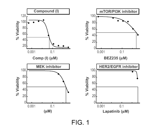
[0025] Figure 1 shows the activity of various chemotherapeutic agents in a
72 hr viability
assay using MDA-MB-231 breast cancer cells.
[0026] Figure 2 shows the activity of ganetespib in a 24 hr viability assay
using SUM149
inflammatory breast cancer (IBC) cells.
- 9 -
CA 02872942 2014-11-06
WO 2013/173436
PCT/US2013/041107
[0027] Figure 3 shows the activity of ganetespib in a viability assay in BT-
474 breast
cancer cells grown as mammospheres in Matrigel . The cells were treated for 72
hr and
analyzed by microscopy. IC50 was determined by AlamarBlue .
[0028] Figure 4A shows the activity of ganetespib in a single agent
viability assay
Detroit562 cells, a head and neck cancer cell line, exposed to various
chemotherapeutic
agents for 72 hr (left).
[0029] Figure 4B shows the expression of various Hsp90 client proteins as
determined by
western blot of cell extracts from Detroit562 cells exposed to ganetespib for
24 hr (right).
[0030] Figure 5 shows a western blot of protein expression in cell extracts
from Detroit
562 head and neck cancer cells treated with 100 nM of ganetespib 24 hours
prior to receiving
the DNA damaging agent bleomycin (5 n.M). Protein expression was measured at
the
indicated time points after bleomycin treatment. Bleomycin increased both Chk1
and Chk2
phosphorylation, which was blocked when cells were treated first with
ganetespib.
[0031] Figure 6 is a waterfall diagram showing the best percentage changes
in size of
target lesions responses according to ALK status after treatment with
ganetespib. The y axis
represents the percentage tumor volume change from baseline. For each patient
(each bar)
the percentage change in measurable tumor at best response was displayed by
the genotype
of the patient, i.e., ALK status. A subject was considered to be ALK+ (i.e.,
have an ALK
mutation) if a mutation in ALK was detected using any of the methods.
[0032] Figure 7 shows a western blot of Hsp90 client proteins in BT-474
cells after
treatment with ganetespib for 16 hours.
[0033] Figure 8 shows a graph of the average tumor volume over time in an
MDA-MB-
231 xenograft model in response to treatment with ganetespib.
[0034] Figure 9 is a waterfall diagram showing the best response in
patients with
metastatic breast cancer based on ER, PR, and HER2 marker status in a Phase II
clinical trial
of ganetespib.
[0035] Figure 10 shows a PET/CT scan of the lungs and bone before and after
19 days of
treatment with ganetespib in a female patient with metastatic triple negative
breast cancer.
Arrows indicate the tumor mass in the lung.
- 10 -
CA 02872942 2014-11-06
WO 2013/173436
PCT/US2013/041107
[0036] Figure 11 shows a table of IC50 values for ganetespib in NSCLC cell
lines with a
KRAS mutation after treatment with ganetespib for 72 hr.
[0037] Figure 12 shows a graph of the results of treatment of various NSCLC
cell lines
with ganetespib, camptothecin, or a combination thereof for 72 hours.
[0038] Figure 13 shows a graph of the results of treatment of various NSCLC
cell lines
with ganetespib, pemetrexed, or a combination thereof for 72 hours.
[0039] Figure 14 shows a graph of the results of treatment of various NSCLC
cell lines
with ganetespib, gemcitabine, or a combination thereof for 72 hours.
[0040] Figure 15 shows a graph of the results of treatment of various NSCLC
cell lines
with ganetespib, certain platins, or a combination thereof for 72 hours.
[0041] Figure 16 shows a graph of the results of treatment of various NSCLC
cell lines
with ganetespib, SN-38, or a combination thereof for 72 hours.
[0042] Figure 17 shows a graph of the results of treatment of various NSCLC
cell lines
with ganetespib, docetaxel, or a combination thereof for 72 hours.
[0043] Figure 18 shows a graph of the results of treatment of various NSCLC
cell lines
with ganetespib, AZD6244, or a combination thereof for 72 hours.
[0044] Figure 19 shows a graph of the results of treatment of various NSCLC
cell lines
with ganetespib, BEZ235, or a combination thereof for 72 hours.
[0045] Figure 20 shows a graph of the results of treatment of mice with
A549 NSCLC
xenografts with ganetespib, BEZ-235, or a combination thereof.
[0046] Figures 21A and B show the activity of LDH5 as a percent of total
LDH activity in
serum samples from nude mice with (A) HCT116 tumors or (B) 786-0 tumors
relative to
tumor volume. Figures 21C and D show the protein levels of LDH5 as a percent
of total
LDH activity in serum samples from nude mice with (C) HCT116 tumors or (D) 786-
0
tumors relative to tumor volume.
[0047] Figure 22 shows treatment with ganetespib for 24 hours decreases
proliferation of
Mia-PaCa2, HPAC and PANC-1 cells (p<0.001, one way ANOVA). These results were
further confirmed by XTT assay.
- 11 -
CA 02872942 2014-11-06
WO 2013/173436
PCT/US2013/041107
[0048] Figure 23 shows Western blot for Mia-PaCa2, PANC-1 and HPAC cell
lines
treated with ganetespib for 24 hours. Results indicate decreased levels of HIF-
la and VEGF
levels in pancreatic cancer cell lines.
[0049] Figure 24 shows ELISA assay demonstrates significant (p<0.001, one
way
ANOVA) down-regulation of VEGF secretion after treatment with ganetespib.
[0050] Figure 25 shows Egg CAM assay-treatment with ganetespib for 24 hours
in
conditioned medium in three pancreatic cell lines. The conditioned medium was
collected
from control and treated cells. 10Oul of conditioned medium, either control or
treated, was
injected into fertilized chicken eggs. Eggs were incubated at 37 C for 15
days, then dissected
and the membrane was photographed.
[0051] Figure 26 shows treatment with ganetespib significantly inhibits
tumor growth
and decreases angiogenesis in in vivo models of pancreatic cancer.
DETAILED DESCRIPTION OF THE INVENTION
[0052] Research has provided the physician with ever more options for
therapeutics for
the treatment of cancer. However, despite the availability of the new agents,
the ability to
match a therapeutic agent to a specific patient based not just on the type of
tumor or site of
the tumor, but the characteristic of the tumor, is lacking. The instant
invention provides
methods of identifying a subject who will likely respond favorably to
treatment with an
Hsp90 inhibitor by determining the level of hypoxia in a tumor, either by
looking directly at
markers within the tumor tissue or looking at markers in a peripheral sample
from the
subject, e.g., a bodily fluid such as blood, serum, plasma, lymph, urine,
cerebrospinal fluid,
fecal matter, circulating tumor cells, bronchial lavage, peritoneal lavage,
exudate, effusion,
and sputum for the presence of one or more indicators of the level of hypoxia
in the tumor.
[0053] Serum LDH level is well established as a prognostic factor
associated with poor
outcomes and large tumor burden in many tumor types. It is, therefore,
interesting to note
that a number of reports from large randomized phase 2 and phase 3 studies for
several anti-
cancer agents have shown a positive interaction between clinical outcomes and
high baseline
LDH levels. These include bevacizumab in pancreatic cancer (high LDH: OS
HR=0.59, 95%
CI 0.43-0.82; normal LDH: HR=0.98, 95% CI 0.78-1.24); bevacizumab in prostate
cancer (high
- 12 -
CA 02872942 2014-11-06
WO 2013/173436
PCT/US2013/041107
LDH: OS HR=0.80, P=0.029; normal LDH: OS HR=1.02, P=0.87); bevacizumab in
melanoma
(high LDH: OS HR=0.53, 95% CI 0.32-0.88; normal LDH: OS HR=1.25, 95% CI 0.73-
1.25);
temsirolimus in RCC (high LDH: OS HR=0.56, P=0.002; normal LDH: OS HR=0.90,
P=0.51);
vatalanib in colon cancer first line (high LDH: PFS HR=0.67, P=0.009; PFS
HR=0.88, P=0.188);
and vatalanib in colon cancer second line (high LDH: PFS HR=0.63, P<0.001; PFS
HR=0.83,
P=.01).
[0054] The VEGF and mTOR signaling pathways are regulated by hypoxia, both
at the
transcriptional and translational level. The oxygen-sensitive transcription
factor HIF-la is
one of the principal mediators of the hypoxic response in cancer cells,
including the
metabolic switch from oxidative phosphorylation to glycolysis. The hypoxia-
regulated LDH
A gene is under transcriptional control of HIF-1. Therefore, serum LDH levels
may in part
reflect tumor oxygenation and metabolic status. This connection between tumor
oxygenation
and serum LDH levels may explain the enhanced activity seen in patients with
high serum
LDH levels for drugs that affect hypoxia-mediated signaling pathways, such as
VEGF and
mTOR inhibitors.
[0055] There is therefore evidence that both VEGF/mTOR inhibitors are
sensitive to
tumor oxygenation and metabolic status. Both classes of drugs seem to
preferentially work
in anaerobic tumor cells.
[0056] Hsp90 inhibitors effect on hypoxia-driven pathways, including VEGF
and mTOR.
For example, Hsp90 inhibitors inhibit HIF1-a. Further, several key elements of
the VEGF
and mTOR pathways are client proteins VEGF, VEGFR1-3, IGF-1R, GLUT1-3, PI3K of
Hsp90. As demonstrated herein, ganetespib down-regulates the expression or
phosphorylation of Hsp90 client proteins. Therefore, Hsp90 inhibitors should
be useful in
the treatment of subjects with cancer wherein the tumor has a high level of
hypoxia.
[0057] In order that the present invention may be more readily understood,
certain terms
are first defined. In addition, it should be noted that whenever a value or
range of values of
a parameter are recited, it is intended that values and ranges intermediate to
the recited
values are also intended to be part of this invention.
- 13 -
CA 02872942 2014-11-06
WO 2013/173436
PCT/US2013/041107
I. Definitions
[0058] The articles "a", "an" and "the" are used herein to refer to one or
to more than one
(i.e., to at least one) of the grammatical object of the article unless
otherwise clearly indicated
by contrast. By way of example, "an element" means one element or more than
one
element.
[0059] The term "including" is used herein to mean, and is used
interchangeably with,
the phrase "including but not limited to".
[0060] The term "or" is used herein to mean, and is used interchangeably
with, the term
"and/or," unless context clearly indicates otherwise.
[0061] The term "such as" is used herein to mean, and is used
interchangeably, with the
phrase "such as but not limited to".
[0062] Unless specifically stated or obvious from context, as used herein,
the term
"about" is understood as within a range of normal tolerance in the art, for
example within 2
standard deviations of the mean. About can be understood as within 10%, 9%,
8%, 7%, 6%,
5%, 4%, 3%, 2%, 1%, 0.5%, 0.1 %, 0.05%, or 0.01% of the stated value. Unless
otherwise clear
from context, all numerical values provided herein can be modified by the term
about.
[0063] The recitation of a listing of chemical group(s) in any definition
of a variable
herein includes definitions of that variable as any single group or
combination of listed
groups. The recitation of an embodiment for a variable or aspect herein
includes that
embodiment as any single embodiment or in combination with any other
embodiments or
portions thereof.
[0064] Any compositions or methods provided herein can be combined with one
or more
of any of the other compositions and methods provided herein.
[0065] As used herein, the term "subject" refers to human and non-human
animals,
including veterinary subjects. The term "non-human animal" includes all
vertebrates, e.g.,
mammals and non-mammals, such as non-human primates, mice, rabbits, sheep,
dog, cat,
horse, cow, chickens, amphibians, and reptiles. In a preferred embodiment, the
subject is a
human and may be referred to as a patient.
- 14 -
CA 02872942 2014-11-06
WO 2013/173436
PCT/US2013/041107
[0066] As used herein, the terms "treat," "treating" or "treatment" refer,
preferably, to an
action to obtain a beneficial or desired clinical result including, but not
limited to, alleviation
or amelioration of one or more signs or symptoms of a disease or condition,
diminishing the
extent of disease, stability (i.e., not worsening) state of disease,
amelioration or palliation of
the disease state, diminishing rate of or time to progression, and remission
(whether partial
or total), whether detectable or undetectable. "Treatment" can also mean
prolonging
survival as compared to expected survival in the absence of treatment.
Treatment does not
need to be curative.
[0067] A "therapeutically effective amount" is that amount sufficient to
treat a disease in
a subject. A therapeutically effective amount can be administered in one or
more
administrations.
[0068] As used herein, an "Hsp90 inhibitor" is understood as a therapeutic
agent that
reduces the activity of Hsp90 either by directly interacting with Hsp90 or by
preventing the
formation of the Hsp90/CDC37 complex such that the expression and proper
folding of at
least one client protein of Hsp90 is inhibited. "Hsp90" includes each member
of the family
of heat shock proteins having a mass of about 90-kilodaltons. For example, in
humans the
highly conserved Hsp90 family includes cytosolic Hsp90 and Hsp90 isoforms, as
well as
GRP94, which is found in the endoplasmic reticulum, and HSP75/TRAP1, which is
found in
the mitochondrial matrix. As used herein, Hsp90 inhibitors include, but are
not limited to
ganetespib, geldanamycin (tanespimycin), e.g., IPI-493, macbecins, tripterins,
tanespimycins,
e.g., 17-AAG (alvespimycin), KF-55823, radicicols, KF-58333, KF-58332, 17-
DMAG, IPI-504,
BI1B-021, BI1B-028, PU-H64, PU-H71, PU-DZ8, PU-HZ151, SNX-2112, SNX-2321, SNX-
5422,
SNX-7081, SNX-8891, SNX-0723, SAR-567530, ABI-287, ABI-328, AT-13387, NSC-
113497,
PF-3823863, PF-4470296, EC-102, EC-154, ARQ-250-RP, BC-274, VER-50589, KW-
2478, BHI-
001, AUY-922, EMD-614684, EMD-683671, XL-888, VER-51047, KOS-2484, KOS-2539,
CUDC-
305, MPC-3100, CH-5164840, PU-DZ13, PU-HZ151, PU-25 DZ13, VER-82576, VER-
82160,
VER-82576, VER-82160, NXD-30001, NVP-HSP990, SST-0201CL1, SST-0115AA1, SST-
0221AA1, SST-0223AA1, novobiocin, herbinmycin A, radicicol, CCT018059, PU-H71,
and
celastrol. In certain embodiments, Hsp90 inhibitors do not include ganetespib.
- 15 -
CA 02872942 2014-11-06
WO 2013/173436
PCT/US2013/041107
[0069] By "diagnosing" and the like, as used herein, refers to a clinical
or other
assessment of the condition of a subject based on observation, testing, or
circumstances for
identifying a subject having a disease, disorder, or condition based on the
presence of at
least one indicator, such as a sign or symptom of the disease, disorder, or
condition.
Typically, diagnosing using the method of the invention includes the
observation of the
subject for multiple indicators of the disease, disorder, or condition in
conjunction with the
methods provided herein. Diagnostic methods provide an indicator that a
disease is or is
not present. A single diagnostic test typically does not provide a definitive
conclusion
regarding the disease state of the subject being tested.
[0070] The terms "administer", "administering" or "administration" include
any method
of delivery of a pharmaceutical composition or agent into a subject's system
or to a
particular region in or on a subject. In certain embodiments of the invention,
an agent is
administered intravenously, intramuscularly, subcutaneously, intradermally,
intranasally,
orally, transcutaneously, or mucosally. In a preferred embodiment, an agent is
administered
intravenously. Administering an agent can be performed by a number of people
working in
concert. Administering an agent includes, for example, prescribing an agent to
be
administered to a subject and/or providing instructions, directly or through
another, to take
a specific agent, either by self-delivery, e.g., as by oral delivery,
subcutaneous delivery,
intravenous delivery through a central line, etc.; or for delivery by a
trained professional,
e.g., intravenous delivery, intramuscular delivery, intratumoral delivery,
etc.
[0071] As used herein, the term "survival" refers to the continuation of
life of a subject
which has been treated for a disease or condition, e.g., cancer.
[0072] As used herein, the term "recur" refers to the re-growth of tumor or
cancerous
cells in a subject in whom primary treatment for the tumor has been
administered. The
tumor may recur in the original site or in another part of the body. In one
embodiment, a
tumor that recurs is of the same type as the original tumor for which the
subject was treated.
For example, if a subject had an ovarian cancer tumor, was treated and
subsequently
developed another ovarian cancer tumor, the tumor has recurred. In addition, a
cancer can
recur in or metastasize to a different organ or tissue than the one where it
originally
occurred.
- 16 -
CA 02872942 2014-11-06
WO 2013/173436
PCT/US2013/041107
[0073] As used herein, the terms "identify" or "select" refer to a choice
in preference to
another. In other words, to identify a subject or select a subject is to
perform the active step
of picking out that particular subject from a group and confirming the
identity of the subject
by name or other distinguishing feature. With respect to the instant
invention, it is
understood that identifying a subject or selecting a subject as having a
specific level of
hypoxia or a specific level of LDH can include any of a number of acts
including, but not
limited to, performing a test and observing a result that is indicative of a
subject having a
specific level of hypoxia; reviewing a test result of a subject and
identifying the subject as
having a specific level of hypoxia; reviewing documentation on a subject
stating that the
subject has a specific level of hypoxia and identifying the subject as the one
discussed in the
documentation by confirming the identity of the subject e.g., by an
identification card,
hospital bracelet, asking the subject for his/her name and/or other personal
information to
confirm the subject's identity.
[0074] As used herein, the term "benefit" refers to something that is
advantageous or
good, or an advantage. Similarly, the term "benefiting", as used herein,
refers to something
that improves or advantages. For example, a subject will benefit from
treatment if they
exhibit a decrease in at least one sign or symptom of a disease or condition
(e.g., tumor
shrinkage, decrease in tumor burden, inhibition or decrease of metastasis,
improving quality
of life ("QOL"), if there is a delay of time to progression ("TTP"), if there
is an increase of
overall survival ("OS"), etc.), or if there is a slowing or stopping of
disease progression (e.g.,
halting tumor growth or metastasis, or slowing the rate of tumor growth or
metastasis). A
benefit can also include an improvement in quality of life, or an increase in
survival time or
progression free survival.
[0075] The terms "cancer" or "tumor" are well known in the art and refer to
the
presence, e.g., in a subject, of cells possessing characteristics typical of
cancer-causing cells,
such as uncontrolled proliferation, immortality, metastatic potential, rapid
growth and
proliferation rate, decreased cell death/apoptosis, and certain characteristic
morphological
features. Cancer cells are often in the form of a solid tumor. However, cancer
also includes
non-solid tumors, e.g., blood tumors, e.g., leukemia, wherein the cancer cells
are derived
from bone marrow. As used herein, the term "cancer" includes pre-malignant as
well as
- 17 -
CA 02872942 2014-11-06
WO 2013/173436
PCT/US2013/041107
malignant cancers. Cancers include, but are not limited to, acoustic neuroma,
acute
leukemia, acute lymphocytic leukemia, acute myelocytic leukemia (monocytic,
myeloblastic,
adenocarcinoma, angiosarcoma, astrocytoma, myelomonocytic and promyelocytic),
acute T-
cell leukemia, basal cell carcinoma, bile duct carcinoma, bladder cancer,
brain cancer, breast
cancer, bronchogenic carcinoma, cervical cancer, chondrosarcoma, chordoma,
choriocarcinoma, chronic leukemia, chronic lymphocytic leukemia, chronic
myelocytic
(granulocytic) leukemia, chronic myelogenous leukemia, colon cancer,
colorectal cancer,
craniopharyngioma, cystadenocarcinoma, diffuse large B-cell lymphoma,
Burkitt's
lymphoma, dysproliferative changes (dysplasias and metaplasias), embryonal
carcinoma,
endometrial cancer, endotheliosarcoma, ependymoma, epithelial carcinoma,
erythroleukemia, esophageal cancer, estrogen-receptor positive breast cancer,
essential
thrombocythemia, Ewing's tumor, fibrosarcoma, follicular lymphoma, germ cell
testicular
cancer, glioma, heavy chain disease, hemangioblastoma, hepatoma,
hepatocellular cancer,
hormone insensitive prostate cancer, leiomyosarcoma, liposarcoma, lung cancer,
lymphagioendotheliosarcoma, lymphangiosarcoma, lymphoblastic leukemia,
lymphoma
(Hodgkin's and non-Hodgkin's), malignancies and hyperproliferative disorders
of the
bladder, breast, colon, lung, ovaries, pancreas, prostate, skin, and uterus,
lymphoid
malignancies of T-cell or B-cell origin, leukemia, lymphoma, medullary
carcinoma,
me dulloblastoma, melanoma, meningioma, mesothelioma, multiple myeloma,
myelogenous
leukemia, myeloma, myxosarcoma, neuroblastoma, non-small cell lung cancer,
oligodendroglioma, oral cancer, osteogenic sarcoma, ovarian cancer, pancreatic
cancer,
papillary adenocarcinomas, papillary carcinoma, pinealoma, polycythemia vera,
prostate
cancer, rectal cancer, renal cell carcinoma, retinoblastoma, rhabdomyosarcoma,
sarcoma,
sebaceous gland carcinoma, seminoma, skin cancer, small cell lung carcinoma,
solid tumors
(carcinomas and sarcomas), small cell lung cancer, stomach cancer, squamous
cell
carcinoma, synovioma, sweat gland carcinoma, thyroid cancer, Waldenstrom's
macroglobulinemia, testicular tumors, uterine cancer, and Wilms' tumor. Other
cancers
include primary cancer, metastatic cancer, oropharyngeal cancer,
hypopharyngeal cancer,
liver cancer, gall bladder cancer, bile duct cancer, small intestine cancer,
urinary tract
cancer, kidney cancer, urothelium cancer, female genital tract cancer, uterine
cancer,
gestational trophoblastic disease, male genital tract cancer, seminal vesicle
cancer, testicular
- 18 -
CA 02872942 2014-11-06
WO 2013/173436
PCT/US2013/041107
cancer, germ cell tumors, endocrine gland tumors, thyroid cancer, adrenal
cancer, pituitary
gland cancer, hemangioma, sarcoma arising from bone and soft tissues, Kaposi's
sarcoma,
nerve cancer, ocular cancer, meningial cancer, glioblastomas, neuromas,
neuroblastomas,
Schwannomas, solid tumors arising from hematopoietic malignancies such as
leukemias,
metastatic melanoma, recurrent or persistent ovarian epithelial cancer,
fallopian tube cancer,
primary peritoneal cancer, gastrointestinal stromal tumors, colorectal cancer,
gastric cancer,
melanoma, glioblastoma multiforme, non-squamous non-small-cell lung cancer,
malignant
glioma, epithelial ovarian cancer, primary peritoneal serous cancer,
metastatic liver cancer,
neuroendocrine carcinoma, refractory malignancy, triple negative breast
cancer, HER2
amplified breast cancer, nasopharageal cancer, oral cancer, biliary tract,
hepatocellular
carcinoma, squamous cell carcinomas of the head and neck (SCCHN), non-
medullary
thyroid carcinoma, recurrent glioblastoma multiforme, neurofibromatosis type
1, CNS
cancer, liposarcoma, leiomyosarcoma, salivary gland cancer, mucosal melanoma,
acral/
lentiginous melanoma, paraganglioma, pheochromocytoma, advanced metastatic
cancer,
solid tumor, triple negative breast cancer, colorectal cancer, sarcoma,
melanoma, renal
carcinoma, endometrial cancer, thyroid cancer, rhabdomysarcoma, multiple
myeloma,
ovarian cancer, glioblastoma, gastrointestinal stromal tumor, mantle cell
lymphoma, and
refractory malignancy.
[0076] "Solid tumor," as used herein, is understood as any pathogenic tumor
that can be
palpated or detected using imaging methods as an abnormal growth having three
dimensions. A solid tumor is differentiated from a blood tumor such as
leukemia.
However, cells of a blood tumor are derived from bone marrow; therefore, the
tissue
producing the cancer cells is a solid tissue that can be hypoxic.
[0077] "Tumor tissue" is understood as cells, extracellular matrix, and
other naturally
occurring components associated with the solid tumor.
[0078] As used herein, the term "isolated" refers to a preparation that is
substantially
free (e.g., 50%, 60%, 70%, 80%, 90% or more, by weight) from other proteins,
nucleic acids, or
compounds associated with the tissue from which the preparation is obtained.
[0079] The term "sample" as used herein refers to a collection of similar
fluids, cells, or
tissues isolated from a subject. The term "sample" includes any body fluid
(e.g., urine,
- 19 -
CA 02872942 2014-11-06
WO 2013/173436
PCT/US2013/041107
serum, blood fluids, lymph, gynecological fluids, cystic fluid, ascetic fluid,
ocular fluids, and
fluids collected by bronchial lavage and/or peritoneal rinsing), ascites,
tissue samples (e.g.,
tumor samples) or a cell from a subject. Other subject samples include tear
drops, serum,
cerebrospinal fluid, feces, sputum, and cell extracts. In one embodiment, the
sample is
removed from the subject. In a particular embodiment, the sample is urine or
serum. In
another embodiment, the sample does not include ascites or is not an ascites
sample. In
another embodiment, the sample does not include peritoneal fluid or is not
peritoneal fluid.
In one embodiment, the sample comprises cells. In another embodiment, the
sample does
not comprise cells. In certain embodiments, the sample can be the portion of
the subject that
is imaged (e.g., using a PET scan, a functional imaging method such as MRI to
detect blood
flow) or tested to determine level of hypoxia (e.g., tumor tissue assayed for
level of hypoxia
using a probe). Samples are typically removed from the subject prior to
analysis, however,
tumor samples can be analyzed in the subject, for example, using imaging or
other detection
methods.
[0080] In some embodiments, only a portion of the sample is subjected to an
assay for
determining the level of hypoxia or the level of the tumor using any method
provided
herein. In certain embodiments, the level of hypoxia is indicated by the level
of an isoform
or subunit of lactate dehydrogenase (LDH) or any combination of subunits or
isoforms
including total LDH, or various portions of the sample are subjected to
various assays for
determining the level of hypoxia or the level of an isoform or subunit of LDH.
Also, in
many embodiments, the sample may be pre-treated by physical or chemical means
prior to
the assay. For example, samples, e.g., blood samples, can be subjected to
centrifugation,
dilution and/or treatment with a solubilizing substance prior to assaying the
samples for the
level of hypoxia or LDH. Such techniques serve to enhance the accuracy,
reliability and
reproducibility of the assays of the present invention.
[0081] The term "control sample," as used herein, refers to any clinically
relevant
comparative sample, including, for example, a sample from a healthy subject
not afflicted
with cancer, a sample from a subject having a less severe or slower
progressing cancer than
the subject to be assessed, a sample from a subject having some other type of
cancer or
disease, a sample from a subject prior to treatment, a sample of non-diseased
tissue (e.g.,
- 20 -
CA 02872942 2014-11-06
WO 2013/173436
PCT/US2013/041107
non-tumor tissue), a sample from the same origin and close to the tumor site,
and the like. A
control sample can be a purified sample, protein, and/or nucleic acid provided
with a kit.
Such control samples can be diluted, for example, in a dilution series to
allow for
quantitative measurement of analytes in test samples. A control sample may
include a
sample derived from one or more subjects. A control sample may also be a
sample made at
an earlier time point from the subject to be assessed. For example, the
control sample could
be a sample taken from the subject to be assessed before the onset of the
cancer, at an earlier
stage of disease, or before the administration of treatment or of a portion of
treatment. The
control sample may also be a sample from an animal model, or from a tissue or
cell lines
derived from the animal model, of the cancer. The level of LDH in a control
sample that
consists of a group of measurements may be determined, e.g., based on any
appropriate
statistical measure, such as, for example, measures of central tendency
including average,
median, or modal values.
[0082] The term "control level" refers to an accepted or pre-determined
level of hypoxia
or LDH which is used to compare with the level of hypoxia or LDH in a sample
derived
from a subject. For example, in one embodiment, the control level of hypoxia
is based on the
level of hypoxia in sample(s) from a subject(s) having slow disease
progression. In another
embodiment, the control level of hypoxia is based on the level in a sample
from a subject(s)
having rapid disease progression. In another embodiment, the control level of
hypoxia is
based on the level of hypoxia in a sample(s) from an unaffected, i.e., non-
diseased, subject(s),
i.e., a subject who does not have cancer. In yet another embodiment, the
control level of
hypoxia is based on the level of hypoxia in a sample from a subject(s) prior
to the
administration of a therapy for cancer. In another embodiment, the control
level of hypoxia
is based on the level of hypoxia in a sample(s) from a subject(s) having
cancer that is not
contacted with a test compound. In another embodiment, the control level of
hypoxia is
based on the level of hypoxia in a sample(s) from a subject(s) not having
cancer that is
contacted with a test compound. In one embodiment, the control level of
hypoxia is based
on the level of hypoxia in a sample(s) from an animal model of cancer, a cell,
or a cell line
derived from the animal model of cancer. In another embodiment, the control
level of
hypoxia is listed in a chart.
- 21 -
CA 02872942 2014-11-06
WO 2013/173436
PCT/US2013/041107
[0083] In one embodiment, the control is a standardized control, such as,
for example, a
control which is predetermined using an average of the levels of hypoxia from
a population
of subjects having no cancer. In still other embodiments of the invention, a
control level of
hypoxia is based on the level of hypoxia in a non-cancerous sample(s) derived
from the
subject having cancer. For example, when a biopsy or other medical procedure
reveals the
presence of cancer in one portion of the tissue, the control level of hypoxia
may be
determined using the non-affected portion of the tissue, and this control
level may be
compared with the level of hypoxia in an affected portion of the tissue.
Similarly, when a
biopsy or other medical procedure reveals the presence of a cancer in one
portion of the
tissue, the control level of hypoxia may be determined using the non-affected
portion of the
tissue, and this control level may be compared with the level of hypoxia in an
affected
portion of the tissue.
[0084] As used herein, the term "obtaining" is understood herein as
manufacturing,
purchasing, or otherwise coming into possession of.
[0085] As used herein, the term "lactate dehydrogenase" refers to an enzyme
that
interconverts pyruvate and lactate with concomitant interconversion of NADH
and NAD+.
Under conditions of hypoxia, the reaction favors the conversion of pyruvate to
lactate.
Under conditions of normoxia, or low levels of hypoxia, the reaction favors
the conversion
of lactate to pyruvate. Functional lactate dehydrogenase are homo- or
heterotetramers
composed of M and H protein subunits encoded by the LDHA and LDHB genes
respectively: LDH-1 (4H) is the predominant form found, for example, in the
heart and red
blood cells (RBCs); LDH-2 (3H1M) is the predominant found, for example, in the
reticuloendothelial system; LDH-3 (2H2M) is the predominant form found, for
example, in
the lungs; LDH-4 (1H3M) is the predominant form found, for example, in the
kidneys,
placenta and pancreas; and LDH-5 (4M) is the predominant form found, for
example, in the
liver and striated muscle. Typically, multiple forms of LDH are found in these
tissues.
Lactate dehydrogenase is classified as (EC 1.1.1.27). The specific ratios
tested may be tumor-
type specific.
[0086] As used herein, the terms "hypoxia" and "hypoxic" refer to a
condition in which a
cancer or a tumor has a low oxygen microenvironment or a less well-oxygenated
- 22 -
CA 02872942 2014-11-06
WO 2013/173436
PCT/US2013/041107
microenvironment. Hypoxia occurs when tumor growth exceeds new blood vessel
formation, and a tumor must undergo genetic and adaptive changes to allow them
to
survive and proliferate in the hypoxic environment. The development of
intratumoral
hypoxia is a common sign of solid tumors. When a tumor microenvironment is
less well-
oxygenated, there is a greater dependency on oxygen-sensitive pathways,
including but not
limited to HIF1oc pathways, VEGF pathways, and mTOR pathways. These pathways
facilitate crucial adaptive mechanisms, such as angiogenesis, glycolysis,
growth-factor
signaling, immortalization, genetic instability, tissue invasion and
metastasis, apoptosis, and
pH regulation (see, e.g., Harris, Nature Reviews, 2:38-47, 2002). These
pathways may also
facilitate invasion and metastasis. Accordingly, the treatment of a subject
with a cancer or
tumor with a selected agent such as bevacizumab, ganetespib, temsirolimus,
erlotinib,
PTK787, BEZ235, XL765, pazopanib, cediranib, or axitinib is more effective
when the subject
has a tumor that exhibits a modulated level of hypoxia, e.g., a high level of
hypoxia. As the
level of hypoxia in the tumor can be determined by obtaining a sample from a
site other
than the tumor, as used herein, the subject can be stated to demonstrate a
modulated level of
hypoxia when it is the tumor present in the subject that demonstrates a
modulated level of
hypoxia. As used herein it is understood that the subject with a modulated
level of hypoxia
is typically not suffering from systemic oxygen imbalance or ischemic disease
at a site
remote from the tumor.
[0087] As used herein, the term "level of hypoxia" is understood as the
amount of one or
more markers indicative of a low oxygen level, or cells having characteristics
and/or
employing biological pathways characteristic of cells with a low oxygen level,
e.g., due to the
Warburg effect. Such markers include, but are not limited to, lactate
dehydrogenase (LDH),
at least one isoform or subunit of hypoxia inducible factor (HIF), at least
one pro-angiogenic
form of vascular endothelial growth factor (VEGF), phosphorylated VEGF
receptor (pKDR)
1, 2, or 3; neurolipin 1 (NRP-1), pyruvate dehydrokinase (PDH-K), and
ornithine
decarboxylase (ODC). Tumor size can also be correlated with a level of
hypoxia. A level of
hypoxia can also be determined by PET scan. LDH can be one or more isoforms or
subunits
of LDH such as LDH5, LDH4, LDH3, LDH2, LDH1, LDHM (also known as LDHA) and
LDHH (also known as LDHB). In one embodiment, LDH can be a total sample of all
LDH
isoforms or subunits. "Hypoxia inducible factors" or "HIFs" are transcription
factors which
- 23 -
CA 02872942 2014-11-06
WO 2013/173436
PCT/US2013/041107
respond to changes in available oxygen in a cellular environment. HIF1oc is a
master
regulator of hypoxic gene expression and oxygen homeostasis. HIF can be one or
more
subunits or isoforms of HIF including HIF-la, HIF-1B, HIF-2a, and HIF-213.
VEGF can be
one or more of the various splice forms of VEGF including pro-angiogenic VEGF-
A and
antiangiogenic VEGF-B.
[0088] As used herein, the term "level of LDH" refers to the amount of LDH
present in a
sample which can be used to indicate the presence or absence of hypoxia in the
tumor in the
subject from whom the sample was obtained. LDH enables the conversion of
pyruvate to
lactate and is a critical component of glycolysis under hypoxic conditions.
LDH can be total
LDH or one or more isoforms or subunits of LDH such as LDH5, LDH4, LDH3, LDH2,
LDH1, LDHM (also known as LDHA) and LDHH (also known as LDHB). A modulated
level of LDH can refer to a high level of LDH or a low level of LDH. In one
embodiment, a
PET scan (which is positive when aerobic glycolysis is active) is an indicator
of a high level
of LDH. In another embodiment, a PET scan (which is negative when aerobic
glycolysis is
inactive) is an indicator of a low level of LDH. In one embodiment, a high
level of LDH is at
least 1.1, 1.2, 1.3, 1.4, 1.5, 1.6, 1.7, 1.8, 1.9, 2.0, 2.1, 2.2, 2.3, 2.4,
2.5, 2.6, 2.7, 2.8, 2.9, 3, 4, 5, 6, 7,
8, 9, or 10 times the value of normal level of LDH. In another embodiment, a
low level of
LDH is 0.9, 0.8, 0.7, 0.6, 0.5, 0.4, 0.3, 0.2, or 0.1 times the value of a
normal level of LDH. A
normal level of LDH, or any other marker, can be defined as any value within
the range of
normal, or the upper limit of the normal value, or the lower limit of the
normal value.
Assays for determining the level of LDH in a sample are well known in the art
and provided
herein.
[0089] In another embodiment, the level of LDH can be understood to be a
change in the
relative levels of protein or activity of LDH isoforms or the ratio of LDH
isoforms.
Preferably, the ratios are the ratios of normalized values, e.g., the level of
the LDH subunit or
isoform is normalized to the ULN, the LLN, or a median value. A change of the
relative
levels of the isoforms can be indicative of the level of hypoxia. For example,
an increase in
the level of LDHA relative to LDHB can be indicative of an increase in
hypoxia.
Alternatively, an increase in the level of LDH5 and/or LDH4, either
individually or in total,
relative to the level of LDH1 or total LDH can be indicative of an increase in
hypoxia. The
- 24 -
CA 02872942 2014-11-06
WO 2013/173436
PCT/US2013/041107
relative levels can be compared to relative levels in an appropriate control
sample from
normal subjects, e.g., subjects without cancer or ischemic disease. That is,
the ratios are the
ratios of normalized values, e.g., the level of the LDH subunit or isoform is
normalized to the
ULN, the LLN, or a median value. The normal levels can be considered to be a
range with an
upper level of normal and a lower level of normal. In certain embodiments, a
high level of
LDH can be understood an increase in the normalized level of LDHA or LDH5
and/or LDH4
relative to the normalized level of LDHB or LDH1 or total LDH, respectively,
or to total
LDH of at least 1.1, 1.2, 1.3, 1.4, 1.5, 1.6, 1.7, 1.8, 1.9, 2.0, 2.1, 2.2,
2.3, 2.4, 2.5, 2.6, 2.7, 2.8, 2.9, 3,
4, 5, 6, 7, 8, 9, or 10 times the value of normalized level of LDHA or LDH5
and/or LDH4
relative to the normalized level of LDHB or LDH1 or total LDH, respectively.
In another
embodiment, a low level of LDH is a ratio of 0.9, 0.8, 0.7, 0.6, 0.5, 0.4,
0.3, 0.2, or 0.1 of the
normalized value of LDHA or LDH5 and/or LDH4 relative to the normalized level
of LDHB
or LDH1 or total LDH, respectively.
[0090] As used herein, a "normalized ratio" is understood as a proportion
of two values
that have been compared to a standard, either an external (e.g., population
control level) or
an internal (e.g., level from a normal tissue, level from an earlier time
point, level of one or
more isoforms) control to allow for comparison of samples between individuals.
For
example, the ratio of normalized levels of hypoxia-modulated polypeptides can
be
determined by determining a ratio of two normalized levels of two isoforms or
subunits of
LDH or total LDH by comparing the level of a first isoform or subunit of LDH
in the sample
relative to a control sample to provide a first normalized level, and the
level of a second
isoform or subunit of LDH or total LDH relative to a control sample to provide
a second
normalized level, and calculating a ratio of the first normalized level and
the second
normalized level to provide a normalized ratio of LDH isoforms or subunits,
wherein at
least one of the first level and the second level are not total LDH. In
certain embodiments, a
low level of hypoxia is a normalized ratio of the ULN of LDHA to LDHB of 1.0
or less, or a
normalized ratio of the ULN of LDH5 and/or LDH4 to LDH1 or total LDH of 1.0 or
less.
[0091] Assays for determining the level of LDH in a sample are well known
in the art.
See, e.g., U.S. Publication Nos. 2010/0178283 and 2008/0213744 and U.S. Patent
Nos. 4,250,255
and 6,242,208, the entire contents of each of which are expressly incorporated
herein by
- 25 -
CA 02872942 2014-11-06
WO 2013/173436
PCT/US2013/041107
reference. LDH sequences are further provided in public databases (e.g., at
htt42;fiblast.ncbi.nicrt.ratzly/Blast.cgi ).
[0092] It is also understood that levels of the various markers can include
the level of a
post-translationally modified marker, e.g., the total amount of an isoform of
HIF may remain
the same, but the amount of the hydroxylated version of the HIF may increase.
In addition,
it is noted that HIF and other hypoxia-modulated polypeptides can be up-
regulated by a
number of conditions other than hypoxia, e.g., pH change, changes in levels of
02 = or H202,
etc. Accordingly, although the term "level of expression," as used herein, is
intended to
encompass all hypoxia responsive factors, a change in their level of
expression may or may
not actually directly reflect the amount of oxygen available to the tumor.
[0093] Methods to detect the levels of markers of hypoxia are well known in
the art.
Antibodies against and kits for detection of hypoxia-modulated polypeptides
can be
purchased from a number of commercial sources. Alternatively, using routine
methods
known in the art (e.g., immunization of animals, phage display, etc.)
antibodies against one
or more hypoxia-modulated polypeptides or subunits or isoforms thereof can be
made and
characterized. Antibodies can be used for the detection of levels of hypoxia
using ELISA,
RIA, or other immunoassay methods, preferably automated methods, for the
quantitative
detection of proteins in samples of bodily fluids or homogenized solid
samples. Hypoxia
can be detected by enzyme activity assays (e.g., LDH activity, kinase
activity) including in
gel assays to resolve the activity of various isoforms of proteins.
Alternatively,
immunohistochemical methods can be used on tumor samples and tissue sections.
Antibodies against prodrugs that localize in hypoxic regions (e.g., EF5,
pimonidazole, etc.)
can also be used to detect hypoxia. Functional imaging measuring blood flow in
the tumor
can be used as an indicator of hypoxia in the tissue. Direct measurement of
hypoxia can be
performed by inserting a sensor into the tumor. Qualitative scoring methods
and scanning
methods to detect staining are known in the art. When qualitative scoring
methods are
used, it is preferred that two independent, blinded technicians, pathologists,
or other skilled
individuals analyze each sample with specific methods for resolving any
significant
disagreement in scoring, e.g., a third individual reviews the tissue sample.
- 26 -
CA 02872942 2014-11-06
WO 2013/173436
PCT/US2013/041107
[0094] Alternatively, nucleic acid-based methods of detection of levels of
hypoxia are
also well known in the art. Methods of designing primers and probes for
quantitative
reverse transcription real time (rt) PCR are known in the art. Methods for
performing
northern blots to detect RNA levels are known in the art. Nucleic acid
detection methods
can also include fluorescence in situ hybridization (FISH) and in situ PCR.
Qualitative
scoring methods and scanning methods to detect staining are known in the art.
When
qualitative scoring methods are used, it is preferred that two independent,
blinded
technicians, pathologists, or other skilled individuals analyze each sample
with specific
methods for resolving any significant disagreement in scoring, e.g., a third
individual
reviews the tissue sample.
[0095] "Baseline" refers to the level of hypoxia or the level of LDH upon
patient entrance
into the study and is used to distinguish from levels of hypoxia or levels of
LDH the patient
might have during or after treatment.
[0096] "Elevated" or "lower" refers to a patient's value relative to the
upper limit of
normal ("ULN") or the lower limit of normal ("LLN") which are based on
historical normal
control samples. As the level of the hypoxic marker present in the subject
will be a result of
the disease, and not a result of treatment, typically a control sample
obtained from the
patient prior to onset of the disease will not likely be available. Because
different labs may
have different absolute results, LDH values are presented relative to that
lab's upper limit of
normal value (ULN). LDH can be expressed in IU/ml (International Units per
milliliter). An
accepted ULN for LDH is 234 IU/ml, however, this value is not universally
accepted or
applicable to all methods of detection of LDH in all samples.
[0097] The specific value for ULN and LLN will also depend, for example, on
the type of
assay (e.g., ELISA, enzyme activity, immunohistochemistry, imaging), the
sample to be
tested (e.g., serum, tumor tissue, urine), and other considerations known to
those of skill in
the art. The ULN or LLN can be used to define cut-offs between normal and
abnormal. For
example, a low level of a marker (e.g., LDH) can be defined as a marker level
less than or
equal to the ULN for that marker, with a high level being all values greater
than the ULN.
Cut-offs can also be defined as fractional amounts of the ULN. For example, a
low level of a
marker can be understood to be a level of about 0.5 ULN or less, 0.6 ULN or
less, 0.7 ULN or
- 27 -
CA 02872942 2014-11-06
WO 2013/173436
PCT/US2013/041107
less, 0.8 ULN or less, 0.9 ULN or less, 1.0 ULN or less, 1.1 ULN or less, 1.2
ULN or less, 1.3
ULN or less, 1.4 ULN or less, 1.5 ULN or less, 1.6 ULN or less, 1.7 ULN or
less, 1.8 ULN or
less, 1.9 ULN or less, 2.0 ULN or less, 2.5 ULN or less, 3.0 ULN or less, or
4.0 ULN or less,
with the corresponding high level of the marker being a value greater than the
low level. In
certain embodiments, the presence of a low level of a marker in a subject
sample as defined
above can be indicative that a subject will or will not respond to a
particular therapeutic
intervention. In certain embodiments, the presence of a high level of a marker
in a subject
sample as defined above can be indicative that a subject will or will not
respond to a
particular therapeutic intervention.
[0098] Marker levels can also be further stratified, for example, into low,
intermediate,
and high based on the ULN value. For example, the presence of a low level of a
marker in a
subject sample as defined above can be indicative that a subject will or will
not respond to a
particular therapeutic intervention. An intermediate level of a marker, e.g.,
a range
bracketed by any range within the values of 0.5 ULN, 0.6 ULN, 0.7 ULN, 0.8
ULN, 0.9 ULN,
1.0 ULN, 1.1 ULN, 1.2 ULN, 1.3 ULN, 1.4 ULN, 1.5 ULN, 1.6 ULN, 1.7 ULN, 1.8
ULN, 1.9
ULN, and 2.0 ULN, can be considered an intermediate range wherein the level of
the marker
may be indeterminate that a subject will or will not respond to a particular
therapeutic
intervention. A high level, greater than the intermediate level, would be
indicative that a
subject will or will not respond to a particular therapeutic intervention.
[0099] Similarly, cut-offs of ratios of LDH subunits or isoforms comparing
the ULN, the
LLN, or the median values to differentiate between high and low levels of
hypoxia can be
defined as any value or range bracketed by the values 0.5, 0.6, 0.7, 0.8, 0.9,
1.0, 1.1, 1.2, 1.3,
1.4, 1.5, 1.6, 1.7, 1.8, 1.9, 2.0, 2.1, 2.2, 2.3, 2.4, 2.5, 2.6, 2.7, 2.8,
2.9, 3.0, or higher.
[00100] The "normal" level of expression of a marker is the level of
expression of the
marker in cells of a subject or patient not afflicted with cancer. In one
embodiment, a
"normal" level of expression refers to the level of expression of the marker
under normoxic
conditions.
[00101] An "over-expression" or "high level of expression" of a marker refers
to an
expression level in a test sample that is greater than the standard error of
the assay
employed to assess expression, and is preferably at least 1.1, 1.2, 1.3, 1.4,
1.5, 1.6, 1.7, 1.8, 1.9,
- 28 -
CA 02872942 2014-11-06
WO 2013/173436
PCT/US2013/041107
2.0, 2.1, 2.2, 2.3, 2.4, 2.5, 2.6, 2.7, 2.8, 2.9, 3, 4, 5, 6, 7, 8, 9, or 10
times the expression level of
the marker in a control sample (e.g., sample from a healthy subject not having
the marker
associated disease, i.e., cancer). In one embodiment, expression of a marker
is compared to
an average expression level of the marker in several control samples.
[00102] A "low level of expression" or "under-expression" of a marker refers
to an
expression level in a test sample that is less than at least 0.9, 0.8, 0.7,
0.6, 0.5, 0.4, 0.3, 0.2, or
0.1 times the expression level of the marker in a control sample (e.g., sample
from a healthy
subjects not having the marker associated disease, i.e., cancer). In one
embodiment,
expression of a marker is compared to an average expression level of the
marker in several
control samples.
[00103] As used herein, the term "identical" or "identity" is used herein in
relation to
amino acid or nucleic acid sequences refers to any gene or protein sequence
that bears at
least 30% identity, more preferably 40%, 50%, 60%, 70%, 75%, 80%, 81%, 82%,
83%, 84%,
85%, 86%, 87%, 88%, 89%, 90%, 91%, 92%, 93%, 94%, and most preferably 95%,
96%, 97%,
98%, 99% or more identity to a known gene or protein sequence over the length
of the
comparison sequence. Protein or nucleic acid sequences with high levels of
identity
throughout the sequence can be said to be homologous. A "homologous" protein
can also
have at least one biological activity of the comparison protein. In general,
for proteins, the
length of comparison sequences will be at least 10 amino acids, preferably 10,
20, 30, 40, 50,
60, 70, 80, 90, 100, 150, 175, 200, 250, or at least 300 amino acids or more.
For nucleic acids,
the length of comparison sequences will generally be at least 25, 50, 100,
125, 150, 200, 250,
300, 350, 400, 450, 500, 550, 600, 650, 700, 800, or at least 850 nucleotides
or more.
[00104] By "hybridize" is meant pairing to form a double-stranded molecule
between
complementary polynucleotide sequences, or portions thereof, under various
conditions of
stringency. (See, e.g., Wahl and Berger Methods Enzymol. 152:399, 1987;
Kimmel, Methods
Enzymol. 152:507, 1987.) For example, stringent salt concentration will
ordinarily be less than
about 750 mM NaC1 and 75 mM trisodium citrate, preferably less than about 500
mM NaC1
and 50 mM trisodium citrate, and most preferably less than about 250 mM NaC1
and 25 mM
trisodium citrate. Low stringency hybridization can be obtained in the absence
of organic
solvent, e.g., formamide, while high stringency hybridization can be obtained
in the presence
- 29 -
CA 02872942 2014-11-06
WO 2013/173436
PCT/US2013/041107
of at least about 35% formamide, and most preferably at least about 50%
formamide.
Stringent temperature conditions will ordinarily include temperatures of at
least about
30 C, more preferably of at least about 37 C, and most preferably of at
least about 42 C.
Varying additional parameters, such as hybridization time, the concentration
of detergent,
e.g., sodium dodecyl sulfate (SDS), and the inclusion or exclusion of carrier
DNA, are well
known to those skilled in the art. Various levels of stringency are
accomplished by
combining these various conditions as needed. In a preferred embodiment,
hybridization
will occur at 30 C in 750 mM NaC1, 75 mM trisodium citrate, and 1% SDS. In a
more
preferred embodiment, hybridization will occur at 37 C in 500 mM NaC1, 50 mM
trisodium
citrate, 1% SDS, 35% formamide, and 100 g/m1 denatured salmon sperm DNA
(ssDNA). In
a most preferred embodiment, hybridization will occur at 42 C in 250 mM NaC1,
25mM
trisodium citrate, 1% SDS, 50% formamide, and 200 g/m1 ssDNA. Useful
variations on
these conditions will be readily apparent to those skilled in the art.
[00105] As used herein, the term "oxygen-sensitive pathway" is a cellular
signaling
pathway which is activated by hypoxia. Oxygen-sensitive pathways may be up-
regulated
by hypoxia. Alternatively, an oxygen-sensitive pathway may be down-regulated
by
hypoxia. Oxygen-sensitive pathways include, but are not limited to, HIF
pathways (such as
HIF1oc pathways), VEGF pathways, and mTOR pathways. As used herein, the term
"hypoxia-modulated gene" or "hypoxia-modulated polypeptide" refers to a gene
or protein
which is up-regulated or down-regulated by hypoxia.
[00106] As used herein, the term "HIF pathway" and "HIF pathway members" as
used
herein, describe proteins and other signaling molecules that are regulated by
HIF-1 and HIF-
2. Hypoxia-Inducible Factor 1 (HIF-1) is a transcription factor that has been
shown to play
an essential role in cellular responses to hypoxia. Upon hypoxic stimulation,
HIF-1 has been
shown to activate genes that contain Hypoxic Response Elements (HREs) in their
promoters,
and thus up-regulate a series of gene products that promote cell survival
under conditions
of low oxygen availability. The list of known HIF-responsive genes includes
glycolytic
enzymes (such as lactate dehydrogenase (LDH), enolase-1 (ENO-I), and aldolase
A, glucose
transporters (GLUT 1 and GLUT 3), vascular endothelial growth factor (VEGF),
inducible
nitric oxide synthase (NOS-2), and erythropoietin (EPO). The switch of the
cell to anaerobic
- 30 -
CA 02872942 2014-11-06
WO 2013/173436
PCT/US2013/041107
glycolysis, and the up-regulation of angiogenesis by VEGF is geared at
maximizing cell
survival under conditions of low oxygen tension by reducing the requirement
for oxygen,
and increasing vasculature to maximize oxygen delivery to tissues. The HIF-1
transcription
complex has recently been shown to comprise a heterodimer of two basic helix-
loop-helix
proteins, HIF-la and HIF-113 (also known as ARNT, Aryl Hydrocarbon Receptor
Nuclear
Translocator).
[00107] HIF-la is a member of the basic-helix-loop-helix PAS domain protein
family and
is an approximately 120 kDa protein containing two transactivation domains
(TAD) in its
carboxy-terminal half and DNA binding activity located in the N -terminal half
of the
molecule. HIF-la is constitutively degraded by the ubiquitin-proteosome
pathway under
conditions of normoxia, a process that is facilitated by binding of the von
Hippel-Lindau
(VHL) tumor suppressor protein to HIF-la. Under conditions of hypoxia,
degradation of
HIF-la is blocked and active HIF-la accumulates. The subsequent dimerization
of HIF-la
with ARNT leads to the formation of active HIF transcription complexes in the
nucleus,
which can bind to and activate HREs on HIF-responsive genes.
[00108] As used herein, the term "VEGF pathway" and "VEGF pathway members" as
used herein, describe proteins and other signaling molecules that are
regulated by VEGF.
For example, VEGF pathway members include VEGFR1, 2, and 3; PECAM-1, LacCer
synthase, and PLA2.
[00109] As used herein, the term "mTOR pathway" and "mTOR pathway members" as
used herein, describe proteins and other signaling molecules that are
regulated by mTOR.
For example, mTOR pathway members include SK6, PDCD4, eIF4B, RPS6, eIF4, 4E-
BP1, and
eIF4E.
[00110] "Chemotherapeutic agent" is understood as a drug used for the
treatment of
cancer. Chemotherapeutic agents include, but are not limited to, small
molecules and
biologics (e.g., antibodies, peptide drugs, nucleic acid drugs). In certain
embodiments, a
chemotherapeutic agent does not include one or more of bevacizumab,
ganetespib,
temsirolimus, erlotinib, PTK787, BEZ235, XL765, pazopanib, cediranib, and
axitinib.
- 31 -
CA 02872942 2014-11-06
WO 2013/173436
PCT/US2013/041107
[00111] As used herein, an "Hsp90 inhibitor" is one or more of ganetespib,
geldanamycin
(tanespimycin), e.g., IPI-493, macbecins, tripterins, tanespimycins, e.g., 17-
AAG
(alvespimycin), KF-55823, radicicols, KF-58333, KF-58332, 17-DMAG, IPI-504,
BI1B-021, BI1B-
028, PU-H64, 20 PU-H71, PU-DZ8, PU-HZ151, SNX-2112, SNX-2321, SNX-5422, SNX-
7081,
SNX-8891, SNX-0723, SAR-567530, ABI-287, ABI-328, AT-13387, NSC-113497, PF-
3823863,
PF-4470296, EC-102, EC-154, ARQ-250-RP, BC-274, VER-50589, KW-2478, BHI-001,
AUY-922,
EMD-614684, EMD-683671, XL-888, VER-51047, KOS-2484, KOS-2539, CUDC-305, MPC-
3100, CH-5164840, PU-DZ13, PU-HZ151, PU-25 DZ13, VER-82576, VER-82160, VER-
82576,
VER-82160, NXD-30001, NVP-HSP990, SST-0201CL1, SST-0115AA1, SST-0221AA1, SST-
0223AA1, novobiocin, herbinmycin A, radicicol, CCT018059, PU-H71, and
celastrol. In
certain embodiments, the selected agent is bevacizumab. In certain
embodiments, the
selected agent is ganetespib.
[00112] As used herein, "detecting", "detection" and the like are understood
that an assay
performed for identification of a specific analyte in a sample, e.g., a
hypoxia-modulated
polypeptide or a hypoxia-modulated gene in a sample. The amount of analyte or
activity
detected in the sample can be none or below the level of detection of the
assay or method.
[00113] The terms "modulate" or "modulation" refer to up-regulation (i.e.,
activation or
stimulation), down-regulation (i.e., inhibition or suppression) of a level, or
the two in
combination or apart. A "modulator" is a compound or molecule that modulates,
and may
be, e.g., an agonist, antagonist, activator, stimulator, suppressor, or
inhibitor.
[00114] The term "expression" is used herein to mean the process by which a
polypeptide
is produced from DNA. The process involves the transcription of the gene into
mRNA and
the translation of this mRNA into a polypeptide. Depending on the context in
which used,
"expression" may refer to the production of RNA, or protein, or both.
[00115] The terms "level of expression of a gene" or "gene expression level"
refer to the
level of mRNA, as well as pre-mRNA nascent transcript(s), transcript
processing
intermediates, mature mRNA(s) and degradation products, or the level of
protein, encoded
by the gene in the cell.
- 32 -
CA 02872942 2014-11-06
WO 2013/173436
PCT/US2013/041107
[00116] As used herein, "level of activity" is understood as the amount of
protein activity,
typically enzymatic activity, as determined by a quantitative, semi-
quantitative, or
qualitative assay. Activity is typically determined by monitoring the amount
of product
produced in an assay using a substrate that produces a readily detectable
product, e.g.,
colored product, fluorescent product, or radioactive product. For example, the
isoforms of
LDH in a sample can be resolved using gel electrophoresis. Lactate,
nicotinamide adenine
dinucleotide (NAD+), nitroblue tetrazolium (NBT), and phenazine methosulphate
(PMS) can
be added to assess LDH activity. LDH converts lactate to pyruvate and reduces
NAD+ to
NADH. The hydrogens from NADH are transferred by PMS to NBT reducing it to a
purple
formazan dye. The percentage of each LDH isoenzyme activity as well as the
relative
amount of each isoform to the other isoforms or total LDH can be determined,
for example,
by densitometry.
[00117] As used herein, "changed as compared to a control" sample or subject
is
understood as having a level of the analyte or diagnostic or therapeutic
indicator (e.g.,
marker) to be detected at a level that is statistically different than a
sample from a normal,
untreated, or control sample. Control samples include, for example, cells in
culture, one or
more laboratory test animals, or one or more human subjects. Methods to select
and test
control samples are within the ability of those in the art. An analyte can be
a naturally
occurring substance that is characteristically expressed or produced by the
cell or organism
(e.g., an antibody, or a protein) or a substance produced by a reporter
construct (e.g., p-
galactosidase or luciferase). Depending on the method used for detection the
amount and
measurement of the change can vary. Changed as compared to a control reference
sample
can also include a change in one or more signs or symptoms associated with or
diagnostic
of disease, e.g., cancer. Determination of statistical significance is within
the ability of those
skilled in the art, e.g., the number of standard deviations from the mean that
constitute a
positive result.
[00118] As used herein, "binding" is understood as having at least a 102 or
more, 103 or
more, preferably 104 or more, preferably 105 or more, preferably 106 or more
preference for
binding to a specific binding partner as compared to a non-specific binding
partner (e.g.,
binding an antigen to a sample known to contain the cognate antibody).
- 33 -
CA 02872942 2014-11-06
WO 2013/173436
PCT/US2013/041107
[00119] "Determining" as used herein is understood as performing an assay or
using a
diagnostic method to ascertain the state of someone or something, e.g., the
presence,
absence, level, or degree of a certain condition, biomarker, disease state, or
physiological
condition.
[00120] "Prescribing" as used herein is understood as indicating a specific
agent or agents
for administration to a subject.
[00121] As used herein, the terms "respond" or "response" are understood as
having a
positive response to treatment with a therapeutic agent, wherein a positive
response is
understood as having a decrease in at least one sign or symptom of a disease
or condition
(e.g., tumor shrinkage, decrease in tumor burden, inhibition or decrease of
metastasis,
improving quality of life ("QOL"), delay of time to progression ("TTP"),
increase of overall
survival ("OS"), etc.), or slowing or stopping of disease progression (e.g.,
halting tumor
growth or metastasis, or slowing the rate of tumor growth or metastasis). A
response can
also include an improvement in quality of life, or an increase in survival
time or progression
free survival.
[00122] Ranges provided herein are understood to be shorthand for all of the
values
within the range. For example, a range of 1 to 50 is understood to include any
number,
combination of numbers, or sub-range from the group consisting 1, 2, 3, 4, 5,
6, 7, 8, 9, 10, 11,
12, 13, 14, 15, 16, 17, 18, 19, 20, 21, 22, 23, 24, 25, 26, 27, 28, 29, 30,
31, 32, 33, 34, 35, 36, 37, 38,
39, 40, 41, 42, 43, 44, 45, 46, 47, 48, 49, or 50.
[00123] Reference will now be made in detail to preferred embodiments of the
invention.
While the invention will be described in conjunction with the preferred
embodiments, it will
be understood that it is not intended to limit the invention to those
preferred embodiments.
To the contrary, it is intended to cover alternatives, modifications, and
equivalents as may
be included within the spirit and scope of the invention as defined by the
appended claims.
Agents for Treatment of Tumors with High Levels of Hypoxia with Selected
Agents
[00124] The invention provides methods of use of Hsp90 inhibitors that are
more effective
in treating disease, e.g., cancer, when administered to a patient with a
cancer or tumor
- 34 -
CA 02872942 2014-11-06
WO 2013/173436
PCT/US2013/041107
exhibiting high levels of hypoxia. In one embodiment, the Hsp90 inhibitor may
be
ganetespib, geldanamycin (tanespimycin), e.g., IPI-493, macbecins, tripterins,
tanespimycins,
e.g., 17-AAG (alvespimycin), KF-55823, radicicols, KF-58333, KF-58332, 17-
DMAG, IPI-504,
BI1B-021, BI1B-028, PU-H64, 20 PU-H71, PU-DZ8, PU-HZ151, SNX-2112, SNX-2321,
SNX-
5422, SNX-7081, SNX-8891, SNX-0723, SAR-567530, ABI-287, ABI-328, AT-13387,
NSC-
113497, PF-3823863, PF-4470296, EC-102, EC-154, ARQ-250-RP, BC-274, VER-50589,
KW-
2478, BHI-001, AUY-922, EMD-614684, EMD-683671, XL-888, VER-51047, KOS-2484,
KOS-
2539, CUDC-305, MPC-3100, CH-5164840, PU-DZ13, PU-HZ151, PU-25 DZ13, VER-
82576,
VER-82160, VER-82576, VER-82160, NXD-30001, NVP-HSP990, SST-0201CL1, SST-
0115AA1,
SST-0221AA1, SST-0223AA1, novobiocin (a C-terminal Hsp90i), herbinmycin A,
radicicol,
CCT018059, PU-H71, or celastrol. In an embodiment, the selected agent is
ganetespib. In
another embodiment, an Hsp90 inhibitor is more effective in treating disease,
e.g., cancer,
when administered to a patient with a cancer or tumor exhibiting high levels
of LDH.
Ganetespib
[00125] Ganetespib (also known as STA-9090) is a Heat Shock Protein 90 (Hsp90)
inhibitor
having the following structure:
1-0
zio
'"====
oq
N,
and the chemical name 3-2,4-dihydroxy-5-isopropyl-pheny1)-4-(1-methyl-indo1-5-
y1)-5-
hydroxy-[1,2,4]triazole (see, e.g., US Patent 7,825,148, incorporated herein
by reference).
[00126] Hsp90 is a chaperone protein required for the proper folding and
activation of
other cellular proteins, particularly kinases, such as AKT, BCR-ABL, BRAF,
KIT, MET,
EGFR, FLT3, HER2, PDGFRA and VEGFR. These proteins have been shown to be
critical to
cancer cell growth, proliferation, and survival. Ganetespib has shown potent
activity
against a wide range of cancer types, including lung, prostate, colon, breast,
gastric,
- 35 -
CA 02872942 2014-11-06
WO 2013/173436
PCT/US2013/041107
pancreatic, gastrointestinal stromal tumors (GIST), melanoma, AML, chronic
myeloid
leukemia, Burkitt's lymphoma, diffuse large B-cell lymphoma and multiple
myeloma in in
vitro and in vivo models. Ganetespib has also shown potent activity against
cancers resistant
to imatinib, sunitinib, erlotinib and dasatinib.
[00127] Ganetespib is more effective in treating disease, e.g., cancer, when
administered to
a patient with a cancer or tumor exhibiting high levels of hypoxia. In another
embodiment,
ganetespib is more effective in treating disease, e.g., cancer, when
administered to a patient
with a cancer or tumor exhibiting high levels of LDH.
II. Compositions, Dosages and Modes of Administration
[00128] In certain embodiments, the invention also provides compositions for
treating
subjects having cancer, wherein the cancer comprises a tumor with a high level
of hypoxia.
In certain embodiments, the composition comprising ganetespib, geldanamycin
(tanespimycin), e.g., IPI-493, macbecins, tripterins, tanespimycins, e.g., 17-
AAG
(alvespimycin), KF-55823, radicicols, KF-58333, KF-58332, 17-DMAG, IPI-504,
BI1B-021, BI1B-
028, PU-H64, 20 PU-H71, PU-DZ8, PU-HZ151, SNX-2112, SNX-2321, SNX-5422, SNX-
7081,
SNX-8891, SNX-0723, SAR-567530, ABI-287, ABI-328, AT-13387, NSC-113497, PF-
3823863,
PF-4470296, EC-102, EC-154, ARQ-250-RP, BC-274, VER-50589, KW-2478, BHI-001,
AUY-922,
EMD-614684, EMD-683671, XL-888, VER-51047, KOS-2484, KOS-2539, CUDC-305, MPC-
3100, CH-5164840, PU-DZ13, PU-HZ151, PU-25 DZ13, VER-82576, VER-82160, VER-
82576,
VER-82160, NXD-30001, NVP-HSP990, SST-0201CL1, SST-0115AA1, SST-0221AA1, SST-
0223AA1, novobiocin (a C-terminal Hsp90i), herbinmycin A, radicicol,
CCT018059, PU-H71,
and celastrol, In certain embodiments, the composition does not comprise
ganetespib.
[00129] In certain embodiments, the composition is for treating a subject
having a solid
tumor. In certain embodiments, the composition is for treating a subject
having primary
cancer, metastatic cancer, breast cancer, colon cancer, rectal cancer, lung
cancer,
oropharyngeal cancer, hypopharyngeal cancer, esophageal cancer, stomach
cancer,
pancreatic cancer, liver cancer, gallbladder cancer, bile duct cancer, small
intestine cancer,
urinary tract cancer, kidney cancer, bladder cancer, urothelium cancer, female
genital tract
cancer, cervical cancer, uterine cancer, ovarian cancer, choriocarcinoma,
gestational
trophoblastic disease, male genital tract cancer, prostate cancer, seminal
vesicle cancer,
- 36 -
CA 02872942 2014-11-06
WO 2013/173436
PCT/US2013/041107
testicular cancer, germ cell tumors, endocrine gland tumors, thyroid cancer,
adrenal cancer,
pituitary gland cancer, skin cancer, hemangiomas, melanomas, sarcomas arising
from bone
and soft tissues, Kaposi's sarcoma, brain cancer, nerve cancer, ocular cancer,
meningial
cancer, astrocytoma, glioma, glioblastoma, retinoblastoma, neuroma,
neuroblastoma,
Schwannoma, meningioma, solid tumors arising from hematopoietic malignancies,
leukemia, Hodgkin's lymphoma, non-Hodgkin's lymphoma, Burkitt's lymphoma,
metastatic
melanoma, recurrent or persistent ovarian epithelial cancer, fallopian tube
cancer, primary
peritoneal cancer, epithelial ovarian cancer, primary peritoneal serous
cancer, non-small cell
lung cancer, gastrointestinal stromal tumors, colorectal cancer, small cell
lung cancer,
melanoma, glioblastoma multiforme, non-squamous non-small-cell lung cancer,
malignant
glioma, primary peritoneal serous cancer, metastatic liver cancer,
neuroendocrine
carcinoma, refractory malignancy, triple negative breast cancer, HER2
amplified breast
cancer, squamous cell carcinoma, nasopharageal cancer, oral cancer, biliary
tract,
hepatocellular carcinoma, squamous cell carcinomas of the head and neck
(SCCHN), non-
medullary thyroid carcinoma, neurofibromatosis type 1, CNS cancer,
liposarcoma,
leiomyosarcoma, salivary gland cancer, mucosal melanoma, acral/ lentiginous
melanoma,
paraganglioma; pheochromocytoma, advanced metastatic cancer, solid tumor,
squamous
cell carcinoma, sarcoma, melanoma, endometrial cancer, head and neck cancer,
rhabdomysarcoma, multiple myeloma, gastrointestinal stromal tumor, mantle cell
lymphoma, gliosarcoma, bone sarcoma, or refractory malignancy.
[00130] In certain embodiments, the composition is for treating a subject with
tumor,
wherein the level of hypoxia in the tumor is determined in a subject sample.
In an
embodiment, the subject sample may be tumor tissue, blood, urine, stool,
lymph,
cerebrospinal fluid, circulating tumor cells, bronchial lavage, peritoneal
lavage, exudate,
effusion, or sputum. In an embodiment, the tumor tissue is in the subject. In
an
embodiment, the tumor tissue is removed from the subject.
[00131] In certain embodiments, the composition is for treating a subject with
tumor,
wherein the level of hypoxia in the tumor is determined by detecting the
activity level or
expression level of one or more hypoxia-modulated polypeptides. In an
embodiment, the
- 37 -
CA 02872942 2014-11-06
WO 2013/173436
PCT/US2013/041107
activity level or expression level of the one or more hypoxia-modulated
polypeptides may
be up regulated in the sample.
[00132] In certain embodiments, the composition is for treating a subject with
tumor,
wherein the level of hypoxia in the tumor is determined by detecting the
activity level or
expression level of one or more hypoxia-modulated polypeptides or using
detection
methods selected from the group consisting of detection of activity or
expression of at least
one isoform or subunit of lactate dehydrogenase (LDH), at least one isoform or
subunit of
hypoxia inducible factor (HIF), at least one pro-angiogenic form of vascular
endothelial
growth factor (VEGF), phosphorylated VEGF receptor (pKDR) 1, 2, and 3;
neurolipin 1
(NRP-1), pyruvate dehydrokinase (PDH-K), ornithine decarboxylase (ODC),
glucose
transporter-1 (GLUT-1), glucose transporter-2 (GLUT-2), tumor size, blood
flow, EF5
binding, pimonidazole binding, PET scan, and probe detection of hypoxia level.
In an
embodiment, the isoform or subunit of LDH comprises one or more LDH5, LDH4,
LDH3,
LDH2, LDH1, LDHA and LDHB; or any combination thereof including total LDH. In
an
embodiment, the isoform of HIF comprises one or more of HIF-1a, HIF-1B, HIF-
2a, and
HIF-213; or any combination thereof including total HIF-1 and/or HIF-2. In an
embodiment,
the pro-angiogenic isoform of VEGF is any VEGF-A isoform, or any combination
of VEGF-A
isoforms including total VEGF-A.
[00133] In certain embodiments, the composition is for treating a subject with
tumor,
wherein detection of a high level of activity or expression of at least one
LDH isoform or
subunit in the tumor comprises detection of an LDH activity or expression
level of an LDH
selected from the group consisting of total LDH, LDH5, LDH4, LDH5 plus LDH4,
LDH5
plus LDH4 plus LDH3, and LDHA, wherein the activity level or expression level
is 0.8 ULN
or more.
[00134] In certain embodiments, the composition is for treating a subject with
tumor,
wherein detection of a high level of activity or expression of at least one
LDH isoform or
subunit in the tumor comprises detection of an LDH activity or expression
level of an LDH
selected from the group consisting of total LDH, LDH5, LDH4, LDH5 plus LDH4,
LDH5
plus LDH4 plus LDH3, and LDHA, wherein the activity level or expression level
is 1.0 ULN
or more.
- 38 -
CA 02872942 2014-11-06
WO 2013/173436
PCT/US2013/041107
[00135] In certain embodiments, the composition is for treating a subject with
tumor,
wherein detection of a high level of hypoxia in the tumor comprises detection
of a change in
a ratio or levels of activity or expression or a change in a ratio of
normalized levels of
activity or expression of hypoxia-modulated polypeptides. In an embodiment, a
high level
of hypoxia comprises a ratio or a normalized ratio of 1.0 or more of the ULN,
wherein the
ratio or normalized ratio is selected from the group consisting of the LDHA to
LDHB, LDH5
or LDH4 to LDH1, LDH5 or LDH4 to total LDH, LDH5 and LDH4 to LDH1, LDH5 and
LDH4 to total LDH, LDH5, LDH4, and LDH3 to LDH1, and LDH5, LDH4, and LDH3 to
total LDH.
[00136] In certain embodiments, the invention also provides compositions for
treating
subjects having cancer with a high level of hypoxia, wherein the subjects were
previously
treated with another chemotherapeutic agent. In certain embodiments, the
composition
comprises an Hsp90 inhibitor, wherein the Hsp90 inhibitor may be ganetespib,
geldanamycin (tanespimycin), IPI-493, macbecins, tripterins, tanespimycins, 17-
AAG
(alvespimycin), KF-SS823, radicicols, KF-S8333, KF-S8332, 17-DMAG, IPI-SO4,
BIIB-021, BIIB-
028, PU-H64, PU-H71, PU-DZ8, PU-HZ1S1, SNX-2112, SNX-2321, SNX-S422, SNX-7081,
SNX-8891, SNX-0723, SAR-S67S30, ABI-287, ABI-328, AT-13387, NSC-113497, PF-
3823863,
PF-4470296, EC-102, EC-154, ARQ-250-RP, BC-274, VER-50589, KW-2478, BHr-001,
AUY-922,
EMD-614684, EMD-683671, XL-888, VER-51047, KOS-2484, KOS-2539, CUDC-305, MPC-
3100, CH-5164840, PU-DZ13, PU-HZ151, PU-DZ13, VER-82576, VER-82160, VER-82576,
VER-82160, NXD-30001, NVP-HSP990, SST-0201CL1, SST-0115AA1, SST-0221AA1, SST-
0223AA1, novobiocin (a C-terminal Hsp90i), herbinmycin A, radicicol,
CCT018059, PU-H71,
or celastrol. In some embodiments, the composition comprises an Hsp90
inhibitor wherein
the Hsp90 inhibitor is not ganetespib.
[00137] Techniques and dosages for administration vary depending on the type
of
compound (e.g., chemical compound, antibody, antisense, or nucleic acid
vector) and are
well known to those skilled in the art or are readily determined.
[00138] Therapeutic compounds of the present invention may be administered
with a
pharmaceutically acceptable diluent, carrier, or excipient, in unit dosage
form.
Administration may be parenteral, intravenous, subcutaneous, oral, or local by
direct
- 39 -
CA 02872942 2014-11-06
WO 2013/173436
PCT/US2013/041107
injection into the amniotic fluid. Administering an agent can be performed by
a number of
people working in concert. Administering an agent includes, for example,
prescribing an
agent to be administered to a subject and/or providing instructions, directly
or through
another, to take a specific agent, either by self-delivery, e.g., as by oral
delivery,
subcutaneous delivery, intravenous delivery through a central line, etc; or
for delivery by a
trained professional, e.g., intravenous delivery, intramuscular delivery,
intratumoral
delivery, etc.
[00139] The composition can be in the form of a pill, tablet, capsule, liquid,
or sustained
release tablet for oral administration; or a liquid for intravenous,
subcutaneous, or
parenteral administration; or a polymer or other sustained release vehicle for
local
administration.
[00140] Methods well known in the art for making formulations are found, for
example, in
"Remington: The Science and Practice of Pharmacy" (20th ed., ed. A. R.
Gennaro, 2000,
Lippincott Williams & Wilkins, Philadelphia, Pa.). Formulations for parenteral
administration may, for example, contain excipients, sterile water, saline,
polyalkylene
glycols such as polyethylene glycol, oils of vegetable origin, or hydrogenated
napthalenes.
Biocompatible, biodegradable lactide polymer, lactide/glycolide copolymer, or
polyoxyethylene-polyoxypropylene copolymers may be used to control the release
of the
compounds. Nanoparticulate formulations (e.g., biodegradable nanoparticles,
solid lipid
nanoparticles, liposomes) may be used to control the biodistribution of the
compounds.
Other potentially useful parenteral delivery systems include ethylene-vinyl
acetate
copolymer particles, osmotic pumps, implantable infusion systems, and
liposomes. The
concentration of the compound in the formulation varies depending upon a
number of
factors, including the dosage of the drug to be administered, and the route of
administration.
[00141] The compound may be optionally administered as a pharmaceutically
acceptable
salt, such as non-toxic acid addition salts or metal complexes that are
commonly used in the
pharmaceutical industry. Examples of acid addition salts include organic acids
such as
acetic, lactic, pamoic, maleic, citric, malic, ascorbic, succinic, benzoic,
palmitic, suberic,
salicylic, tartaric, methanesulfonic, toluenesulfonic, or trifluoroacetic
acids and the like;
polymeric acids such as tannic acid, carboxymethyl cellulose, and the like;
and inorganic
- 40 -
CA 02872942 2014-11-06
WO 2013/173436
PCT/US2013/041107
acid such as hydrochloric acid, hydrobromic acid, sulfuric acid phosphoric
acid, and the like.
Metal complexes include zinc, iron, and the like.
[00142] Formulations for oral use include tablets containing the active
ingredient(s) in a
mixture with non-toxic pharmaceutically acceptable excipients. These
excipients may be, for
example, inert diluents or fillers (e.g., sucrose and sorbitol), lubricating
agents, glidants, and
anti-adhesives (e.g., magnesium stearate, zinc stearate, stearic acid,
silicas, hydrogenated
vegetable oils, or talc).
[00143] Formulations for oral use may also be provided as chewable tablets, or
as hard
gelatin capsules wherein the active ingredient is mixed with an inert solid
diluent, or as soft
gelatin capsules wherein the active ingredient is mixed with water or an oil
medium.
[00144] The dosage and the timing of administering the compound depend on
various
clinical factors including the overall health of the subject and the severity
of the symptoms of
disease, e.g., cancer. In general, once a tumor is detected, administration of
the agent is used
to treat or prevent further progression of the tumor. Treatment can be
performed for a
period of time ranging from 1 to 100 days, more preferably 1 to 60 days, and
most preferably
1 to 20 days, or until the remission of the tumor. It is understood that many
chemotherapeutic agents are not administered daily, particularly agents with a
long half-life.
Therefore, an agent can be continually present without being administered
daily. Dosages
vary depending on each compound and the severity of the condition. Dosages can
be
titrated to achieve a steady-state blood serum concentration. Dosages can be
interrupted or
decreased in the presence of dose limiting toxicities.
III. Methods of the Invention
[00145] The instant invention provides methods of identifying a subject who
will likely
respond favorably to treatment with an Hsp90 inhibitor by determining the
level of hypoxia
in a tumor, either by looking directly at markers within the tumor tissue or
looking at
markers in a peripheral sample from the subject, e.g., a bodily fluid such as
blood, serum,
plasma, lymph, urine, cerebrospinal fluid, or fecal matter, for the presence
of one or more
indicators of the level of hypoxia in the tumor.
- 41 -
CA 02872942 2014-11-06
WO 2013/173436
PCT/US2013/041107
[00146] The specific subject sample analyzed will depend, for example, on the
site of the
tumor. It is known that hypoxia drives angiogenesis in tumors, resulting in
leaky blood
vessels resulting in the presence of markers in circulation. Further, tumor
growth and
hypoxia are typically associated with necrosis and cell breakdown, resulting
in cellular
material in other bodily fluids or wastes. These readily accessible subject
samples allow for
the monitoring of the subject for the presence, or absence, of markers for
hypoxia prior to
and during the course of treatment.
[00147] Biopsies are routinely obtained for the purpose of cancer diagnosis,
and solid
tumors are frequently further resected prior to initiation of chemotherapy
which also can be
used for analysis to determine the level of hypoxia. Biopsy samples and
resected tumor
samples typically include at least some normal tissue adjacent to the tumor
that can be used
as a control.
[00148] In one embodiment of the invention, the modulated level of hypoxia is
a high
level of hypoxia. In one embodiment of the invention, the modulated level of
hypoxia is a
high level of LDH.
[00149] In one embodiment, the level of hypoxia is determined by detecting the
level of
one or more hypoxia-modulated polypeptides or using one or more methods such
as
imaging methods. In one embodiment, a hypoxia-modulated polypeptide is at
least one
isoform or subunit of lactate dehydrogenase (LDH), at least one isoform or
subunit of
hypoxia inducible factor (HIF), at least one pro-angiogenic form of vascular
endothelial
growth factor (VEGF), phosphorylated VEGF receptor (pKDR), neurolipin 1 (NRP-
1),
pyruvate dehydrokinase (PDH-K), and omithine decarboxylase (ODC). In one
embodiment,
the isoform or subunit of LDH is LDHH, LDH5, LDH4, LDH3, LDH2, LDH1 or LDHM,
or
any combination thereof. In another embodiment, the isoform or subunit of LDH
is LDH5.
In another embodiment, the level of hypoxia is determined by determining the
ratio of two
or more forms of LDH, e.g., the ratio of LDH5:LDH1. In another embodiment, the
isoform of
HIF is HIF-la, HIF-1B, HIF-2a, and HIF-213. In another embodiment, the pro-
angiogenic
isoform of VEGF is any one or a combination of VEGF-A splice variants.
Antibodies against
prodrugs that localize in hypoxic regions (e.g., EF5, pimonidazole, etc.) can
also be used to
detect hypoxia. Tumor size can also be correlated with a level of hypoxia. A
level of
- 42 -
CA 02872942 2014-11-06
WO 2013/173436
PCT/US2013/041107
hypoxia can also be determined by PET scan. Functional imaging measuring blood
flow in
the tumor can be used as an indicator of hypoxia in the tissue. Direct
measurement of
hypoxia can be performed by inserting a sensor into the tumor.
[00150] Methods to detect the protein or activity levels of markers of
hypoxia, or hypoxia-
modulated polypeptides, are well known in the art. Antibodies against and kits
for
detection of hypoxia-modulated polypeptides can be purchased from a number of
commercial sources. Alternatively, using routine methods known in the art
(e.g.,
immunization of animals, phage display, etc.) antibodies against one or more
hypoxia-
modulated polypeptides or subunits or isoforms thereof can be made and
characterized.
Antibodies can be used for the detection of levels of hypoxia using ELISA,
RIA, or other
immunoassay methods, preferably automated methods, for the quantitative
detection of
proteins in samples of bodily fluids or homogenized solid samples.
Alternatively,
immunohistochemical methods can be used on tumor samples and tissue sections.
Qualitative scoring methods and scanning methods to detect staining are known
in the art.
When qualitative scoring methods are used, it is preferred that two
independent, blinded
technicians, pathologists, or other skilled individuals analyze each sample
with specific
methods for resolving any significant disagreement in scoring, e.g., a third
individual
reviews the tissue sample. Many markers of hypoxia, including LDH, are
enzymes.
Enzymatic activity can be assayed in total, or for individual isoforms, for
example, using in
gel assays.
[00151] Alternatively, nucleic acid based methods of detection of levels of
hypoxia are
also well known in the art. Methods of designing primers and probes for
quantitative
reverse transcription real time (rt) PCR are known in the art. Methods for
performing
northern blots to detect RNA levels are known in the art. Nucleic acid
detection methods
can also include fluorescence in situ hybridization (FISH) and in situ PCR.
Qualitative
scoring methods and scanning methods to detect staining are known in the art.
[00152] In another aspect, the present invention provides methods for the pre-
selection of
a subject for therapeutic treatment with an anti-cancer agent, wherein the
subject has
previously been found to have a high level of hypoxia. The invention also
provides
methods for the pre-selection of a subject for therapeutic treatment with an
agent by
- 43 -
CA 02872942 2014-11-06
WO 2013/173436
PCT/US2013/041107
evaluating the results of an assessment of a sample from the subject for a
high level of
hypoxia.
[00153] Such determinations can be made based on a chart review of the level
of hypoxia
of the tumor of the subject. Inclusion criteria can include information being
available
regarding the cancer type, the specific treatment regimen with the agent, and
the outcome to
death or for a meaningful follow-up period which varies depending on the
cancer type, e.g.,
metastatic or refractile cancers with poor prognoses requiring follow-up of
weeks to months
whereas cancers with less poor prognoses preferably having months to years of
follow-up
with subjects. In addition to information related to survival, information
related to quality
of life, side effects, and other relevant information can be considered when
available.
Exclusion criteria can include the presence of other diseases or conditions
that could result
in alteration of levels of hypoxia-modulated peptides, e.g., ischemic heart or
vascular
disease, poor circulation, diabetes, macular degeneration, recent stroke, or
other ischemic
events or conditions. Other exclusion criteria can be selected based on the
available samples
and patient population, e.g., prior treatment with specific agents.
[00154] The subjects can be sorted into groups based on various criteria.
Subjects who
were treated with an agent for whom no levels of hypoxic markers were
determined can be
used as an unstratified control group to understand the efficacy of the agent
on a treatment
population not selected based on the level of hypoxia in the subject.
Alternatively, the
population analyzed in the study can be compared to historical control samples
in which an
unstratified population was analyzed for response to the agent.
[00155] Subjects for whom hypoxic levels were obtained can be divided into two
or more
groups having high and low level of hypoxia, optionally with a group of
subjects with
moderate levels of hypoxia, depending on the distribution of subjects. It is
understood that
subjects and samples can also be divided into other groups, e.g., survival
time, treatment
regimen with the agent, cancer type, previous failed treatments, etc. for
analysis. Preferably,
the same marker(s) of hypoxia is measured in each of the subjects, e.g., at
least one isoform
or subunit of lactate dehydrogenase (LDH) or hypoxia inducible factor (HIF);
at least one
pro-angiogenic form of vascular endothelial growth factor (VEGF),
phosphorylated VEGF
receptor (pKDR) 1, 2, or 3; GLUT-1, GLUT-2, neurolipin 1 (NRP-1), pyruvate
dehydrokinase
- 44 -
CA 02872942 2014-11-06
WO 2013/173436
PCT/US2013/041107
(PDH-K), and ornithine decarboxylase (ODC). Tumor size can also be a marker
correlated
with a level of hypoxia. A marker of a level of hypoxia can also be determined
by PET scan.
A level of hypoxia can also be determined by PET scan. Further, it is
preferred that the same
type of subject sample, e.g., blood, serum, lymph, tumor tissue, etc., is
tested for the presence
of the marker for the level of hypoxia. It is understood that the level of
hypoxia can be
measured directly in the tumor sample, using quantitative, semi-quantitative,
or qualitative
immunohistochemical methods, immunological assays (e.g., ELISA assay); reverse
transcription PCR assays, particularly quantitative PCR methods, e.g., real
time PCR;
northern blot assays, enzyme activity assays (e.g., for lactate dehydrogenase
activity, for
kinase activity); and in situ hybridization assay (e.g., fluorescence in situ
hybridization
(FISH) assay). Antibodies against prodrugs that localize in hypoxic regions
(e.g., EF5,
pimonidazole, etc.) can also be used to detect hypoxia. Functional imaging
measuring blood
flow in the tumor can be used as an indicator of hypoxia in the tissue. Direct
measurement
of hypoxia can be performed by inserting a sensor into the tumor. Antibodies
against
prodrugs that localize in hypoxic regions (e.g., EF5, pimonidazole, etc.) can
also be used as
markers to detect hypoxia. Functional imaging measuring blood flow in the
tumor can be
used as a marker of hypoxia in the tissue. Direct measurement of hypoxia can
be performed
to provide a marker for hypoxia by inserting a sensor into the tumor. Again,
it is preferred
that the same method of determining the level of the marker of hypoxia is used
for all
samples, particularly when qualitative assessment methods are used.
[00156] Outcomes of subjects based on the level of hypoxia can be analyzed to
determine
if the outcome between the two groups is different. Outcomes can further be
compared to a
non-stratified group treated with the Hsp90 inhibitor. Methods for statistical
analysis and
determination of statistical significance are within the ability of those of
skill in the art. The
analysis demonstrates that subjects with a high level of hypoxia have a better
response, e.g.,
one or more of longer time to failure, longer survival time, better quality of
life, decreased
tumor size, better tolerance of the agent, etc., as compared to subjects with
a low level of
hypoxia.
[00157] In another aspect, the present invention provides methods for the pre-
selection of
a subject for therapeutic treatment with an Hsp90 inhibitor, wherein the
subject has
- 45 -
CA 02872942 2014-11-06
WO 2013/173436
PCT/US2013/041107
previously been found to have a high level of hypoxia. The invention also
provides
methods for the pre-selection of a subject for therapeutic treatment with an
Hsp90 inhibitor
by evaluating the results of an assessment of a sample from the subject for a
modulated level
of hypoxia wherein the subject is found to have a high level of hypoxia. Such
determinations can be made based on the level of hypoxia observed in
historical samples.
An analysis using samples collected from subjects during treatment can be
performed to
determine the efficacy of a selected agent for the treatment of cancer based
on the level of
hypoxia of the tumor based on markers assessed during the treatment of the
subjects.
Inclusion criteria are information being available regarding the cancer type,
the specific
treatment regimen with the selected agent, and the outcome to death or for a
meaningful
follow-up period which varies depending on the cancer type, e.g., metastatic
or refractile
cancers with poor prognoses requiring follow-up of weeks to months whereas
cancers with
less poor prognoses preferably having months to years of follow-up with
subjects. In
addition to information related to survival, information related to quality of
life, side effects,
and other relevant information is considered when available. Exclusion
criteria can include
the presence of other diseases or conditions that could result in alteration
of levels of
hypoxia-modulated peptides, e.g., ischemic heart or vascular disease, poor
circulation,
diabetes, macular degeneration, recent stroke, or other ischemic events or
conditions. Other
exclusion criteria can be selected based on the available samples and patient
population, e.g.,
prior treatment with specific agents.
[00158] The samples can be analyzed for the level of hypoxia. Preferably, all
of the
samples are the same type or types, e.g., blood, plasma, lymph, or tumor
tissue. Depending
on the availability of subject samples, the analysis can be performed using
two (or more)
subject sample types, e.g., serum and tumor tissue. Various portions of the
tumor tissue can
also be analyzed when sufficient material is available, e.g., adjacent to the
necrotic core, in
the center of the tumor, adjacent to or including tumor vasculature, adjacent
to normal
tissue, etc. One or more markers of hypoxia can be measured in each of the
subjects, e.g., at
least one isoform or subunit of lactate dehydrogenase (LDH) or hypoxia
inducible factor
(HIF); at least one pro-angiogenic form of vascular endothelial growth factor
(VEGF),
phosphorylated VEGF receptor (pKDR) 1, 2, or 3, GLUT-1, GLUT-2, neurolipin 1
(NRP-1),
pyruvate dehydrokinase (PDH-K), and omithine decarboxylase (ODC). Enzymatic
assays of
- 46 -
CA 02872942 2014-11-06
WO 2013/173436
PCT/US2013/041107
markers can be performed. Tumor size can also be a marker correlated with a
level of
hypoxia. A marker of a level of hypoxia can also be determined by PET scan.
Antibodies
against prodrugs that localize in hypoxic regions (e.g., EF5, pimonidazole,
etc.) can also be
used as markers to detect hypoxia. Functional imaging measuring blood flow in
the tumor
can be used as a marker of hypoxia in the tissue. Direct measurement of
hypoxia can be
performed to provide a marker for hypoxia by inserting a sensor into the
tumor. Further, it
is preferred that the same type of subject sample, e.g., blood, serum, lymph,
tumor tissue,
etc., is tested for the presence of the marker for the level of hypoxia. It is
understood that the
level of hypoxia could have been measured directly in the tumor sample, using
quantitative,
semi-quantitative, or qualitative immunohistochemical methods, immunological
assays (e.g.,
ELISA assay); reverse transcription PCR assays, particularly quantitative PCR
methods, e.g.,
real time PCR; northern blot assays, enzyme activity assays (e.g., for lactate
dehydrogenase
activity, for kinase activity); and in situ hybridization assay (e.g.,
fluorescence in situ
hybridization (FISH) assay). Again, it is preferred that the same method of
determining the
level of the marker of hypoxia is used for all samples, particularly when
qualitative
assessment methods are used.
[00159] In another aspect, the present invention provides methods for treating
a cancer
with an Hsp90 inhibitor in a subject having a high level of hypoxia. The
methods include
not administering to the subject having a cancer or susceptible to a cancer
who further has a
low level of hypoxia, an Hsp90 inhibitor, thereby treating the cancer. Other
methods
include administering to the subject having a cancer or susceptible to a
cancer an Hsp90
inhibitor, and at least one chemotherapeutic agent, thereby treating the
cancer. In certain
embodiments, the subject has previously been treated with a chemotherapeutic
agent.
[00160] Other methods include methods of treating a subject who has cancer by
prescribing to the subject an effective amount of an Hsp90 inhibitor, wherein
the subject has
previously been found to have a high level of hypoxia. As used herein, the
term
"prescribing" is understood as indicating a specific agent or agents for
administration to a
subject. Furthermore, the present invention also includes methods of
increasing the
likelihood of effectively treating a subject having cancer by administering a
therapeutically
- 47 -
CA 02872942 2014-11-06
WO 2013/173436
PCT/US2013/041107
effective amount of a composition comprising an Hsp90 inhibitor, to the
subject, wherein the
subject has previously been found to have a modulated level of hypoxia.
[00161] Cancers that may be treated or prevented using the methods of the
invention
include, for example, acoustic neuroma, acute leukemia, acute lymphocytic
leukemia, acute
myelocytic leukemia (monocytic, myeloblastic, adeno carcinoma, angiosarcoma,
astrocytoma, myelomonocytic and promyelocytic), acute T-cell leukemia, basal
cell
carcinoma, bile duct carcinoma, bladder cancer, brain cancer, breast cancer,
bronchogenic
carcinoma, cervical cancer, chondrosarcoma, chordoma, choriocarcinoma, chronic
leukemia,
chronic lymphocytic leukemia, chronic myelocytic (granulocytic) leukemia,
chronic
myleogeneous leukemia, colon cancer, colorectal cancer, craniopharyngioma,
cystadenocarcinoma, diffuse large B-cell lymphoma, dysproliferative changes
(dysplasias
and metaplasias), embryonal carcinoma, endometrial cancer, endotheliosarcoma,
ependymoma, epithelial carcinoma, erythroleukemia, esophageal cancer, estrogen-
receptor
positive breast cancer, essential thrombocythemia, Ewing's tumor,
fibrosarcoma, follicular
lymphoma, germ cell testicular cancer, glioma, heavy chain disease,
hemangioblastoma,
hepatoma, hepatocellular cancer, hormone insensitive prostate cancer,
leiomyosarcoma,
liposarcoma, lung cancer, lymphagioendotheliosarcoma, lymphangiosarcoma,
lymphoblastic leukemia, lymphoma (Hodgkin's and non-Hodgkin's), malignancies
and
hyperproliferative disorders of the bladder, breast, colon, lung, ovaries,
pancreas, prostate,
skin and uterus, lymphoid malignancies of T-cell or B-cell origin, leukemia,
lymphoma,
medullary carcinoma, medulloblastoma, melanoma, meningioma, mesothelioma,
multiple
myeloma, myelogenous leukemia, myeloma, myxosarcoma, neuroblastoma, non-small
cell
lung cancer, oligodendroglioma, oral cancer, osteogenic sarcoma, ovarian
cancer, pancreatic
cancer, papillary adenocarcinomas, papillary carcinoma, pinealoma,
polycythemia vera,
prostate cancer, rectal cancer, renal cell carcinoma, retinoblastoma,
rhabdomyosarcoma,
sarcoma, sebaceous gland carcinoma, seminoma, skin cancer, small cell lung
carcinoma,
solid tumors (carcinomas and sarcomas), small cell lung cancer, stomach
cancer, squamous
cell carcinoma, synovioma, sweat gland carcinoma, thyroid cancer,
Waldenstrom's
macroglobulinemia, testicular tumors, uterine cancer and Wilms' tumor. Other
cancers
include primary cancer, metastatic cancer, oropharyngeal cancer,
hypopharyngeal cancer,
liver cancer, gallbladder cancer, small intestine cancer, urinary tract
cancer, kidney cancer,
- 48 -
CA 02872942 2014-11-06
WO 2013/173436
PCT/US2013/041107
urothelium cancer, female genital tract cancer, uterine cancer, gestational
trophoblastic
disease, male genital tract cancer, seminal vesicle cancer, testicular cancer,
germ cell tumors,
endocrine gland tumors, thyroid cancer, adrenal cancer, and pituitary gland
cancer,
hemangiomas, sarcomas arising from bone and soft tissues; Kaposi's sarcoma,
nerve cancer,
ocular cancer, and meningial cancer, glioblastomas, neuromas, Schwannomas,
solid tumors
arising from hematopoietic malignancies such as leukemias, metastatic
melanoma, recurrent
or persistent ovarian epithelial cancer, fallopian tube cancer, primary
peritoneal cancer,
gastrointestinal stromal tumors, colorectal cancer, gastric cancer, melanoma,
glioblastoma
multiforme, non-squamous non-small-cell lung cancer, malignant glioma,
epithelial ovarian
cancer, primary peritoneal serous cancer, metastatic liver cancer,
neuroendocrine carcinoma,
refractory malignancy, triple negative breast cancer, HER2 amplified breast
cancer,
squamous cell carcinoma of the head and neck (SCCHN), nasopharageal cancer,
oral cancer,
biliary tract, hepatocellular carcinoma, non-medullary thyroid carcinoma,
recurrent
glioblastoma multiforme, neurofibromatosis type 1, CNS cancer, liposarcoma;
leiomyosarcoma; salivary gland cancer, mucosal melanoma; acral/ lentiginous
melanoma,
paraganglioma, and pheochromocytoma.
[00162] It is understood that diagnosis and treatment of a complex disease
such as cancer
is not performed by a single individual, test, agent, or intervention. For
example, a subject
may meet with a primary care physician to express a concern and be referred to
an
oncologist who will request tests that are designed, carried out, and analyzed
by any of a
number of individuals, but not limited to, radiologists, radiology
technicians, physicists,
phlebotomists, pathologists, laboratory technicians, and radiation, clinical,
and surgical
oncologists. Selection, dosing, and administration of agents to a subject
diagnosed with
cancer will be performed by any of a number of individuals including, but not
limited to,
radiologists, radiology technicians, physicists, pathologists, infusion
nurses, pharmacists,
and radiation, clinical, and surgical oncologists. Therefore, it is understood
that within the
terms of the invention, identifying a subject as having a specific level of
hypoxia can include
any of a number of acts including, but not limited to, performing a test and
observing a
result that is indicative of a subject having a specific level of hypoxia;
reviewing a test result
of a subject and identifying the subject as having a specific level of
hypoxia; reviewing
documentation on a subject stating that the subject has a specific level of
hypoxia and
- 49 -
CA 02872942 2014-11-06
WO 2013/173436
PCT/US2013/041107
identifying the subject as the one discussed in the documentation by
confirming the identity
of the subject, e.g., by an identification card, hospital bracelet, asking the
subject for his/her
name and/or other personal information to confirm the subjects identity.
[00163] Similarly, administering an Hsp90 inhibitor can be performed by a
number of
people working in concert. Administering an agent includes, for example,
prescribing an
agent to be administered to a subject and/or providing instructions, directly
or through
another, to take a specific agent, either by self-delivery, e.g., as by oral
delivery,
subcutaneous delivery, intravenous delivery through a central line, etc; or
for delivery by a
trained professional, e.g., intravenous delivery, intramuscular delivery,
intratumoral
delivery, etc.
[00164] As discussed extensively, above, the terms "administer",
"administering" or
"administration" include any method of delivery of a pharmaceutical
composition or agent
into a subject's system or to a particular region in or on a subject. In
certain embodiments of
the invention, an Hsp90 inhibitor is administered intravenously,
intramuscularly,
subcutaneously, intradermally, intranasally, orally, transcutaneously, or
mucosally. In a
preferred embodiment, an agent is administered intravenously. Administering an
Hsp90
inhibitor can be performed by a number of people working in concert.
Administering an
agent includes, for example, prescribing an agent to be administered to a
subject and/or
providing instructions, directly or through another, to take a specific agent,
either by self-
delivery, e.g., as by oral delivery, subcutaneous delivery, intravenous
delivery through a
central line, etc.; or for delivery by a trained professional, e.g.,
intravenous delivery,
intramuscular delivery, intratumoral delivery, etc.
[00165] In certain embodiments, the invention further provides a business
method for
decreasing healthcare costs comprising:
determining the level of hypoxia in a biological sample from a cancer obtained
from
a subject;
storing the information on a computer processor;
determining if the subject would likely benefit from treatment with an Hsp90
inhibitor based on the level of hypoxia; and
- 50 -
CA 02872942 2014-11-06
WO 2013/173436
PCT/US2013/041107
treating the subject only if the subject will likely benefit from treatment,
thereby decreasing healthcare costs.
[00166] In certain embodiments of the business method, the subject has a solid
tumor. In
certain embodiments of the business method, the subject has primary cancer,
metastatic
cancer, breast cancer, colon cancer, rectal cancer, lung cancer, oropharyngeal
cancer,
hypopharyngeal cancer, esophageal cancer, stomach cancer, pancreatic cancer,
liver cancer,
gallbladder cancer, bile duct cancer, small intestine cancer, urinary tract
cancer, kidney
cancer, bladder cancer, urothelium cancer, female genital tract cancer,
cervical cancer,
uterine cancer, ovarian cancer, choriocarcinoma, gestational trophoblastic
disease, male
genital tract cancer, prostate cancer, seminal vesicle cancer, testicular
cancer, germ cell
tumors, endocrine gland tumors, thyroid cancer, adrenal cancer, pituitary
gland cancer, skin
cancer, hemangiomas, melanomas, sarcomas arising from bone and soft tissues,
Kaposi's
sarcoma, brain cancer, nerve cancer, ocular cancer, meningial cancer,
astrocytoma, glioma,
glioblastoma, retinoblastoma, neuroma, neuroblastoma, Schwannoma, meningioma,
solid
tumors arising from hematopoietic malignancies, leukemia, Hodgkin's lymphoma,
non-
Hodgkin's lymphoma, Burkitt's lymphoma, metastatic melanoma, recurrent or
persistent
ovarian epithelial cancer, fallopian tube cancer, primary peritoneal cancer,
epithelial
ovarian cancer, primary peritoneal serous cancer, non-small cell lung cancer,
gastrointestinal
stromal tumors, colorectal cancer, small cell lung cancer, melanoma,
glioblastoma
multiforme, non-squamous non-small-cell lung cancer, malignant glioma, primary
peritoneal serous cancer, metastatic liver cancer, neuroendocrine carcinoma,
refractory
malignancy, triple negative breast cancer, HER2 amplified breast cancer,
squamous cell
carcinoma, nasopharageal cancer, oral cancer, biliary tract, hepatocellular
carcinoma,
squamous cell carcinomas of the head and neck (SCCHN), non-medullary thyroid
carcinoma, neurofibromatosis type 1, CNS cancer, liposarcoma, leiomyosarcoma,
salivary
gland cancer, mucosal melanoma, acral/ lentiginous melanoma, paraganglioma;
pheochromocytoma, advanced metastatic cancer, solid tumor, squamous cell
carcinoma,
sarcoma, melanoma, endometrial cancer, head and neck cancer, rhabdomysarcoma,
multiple myeloma, gastrointestinal stromal tumor, mantle cell lymphoma,
gliosarcoma,
bone sarcoma, or refractory malignancy.
- 51 -
CA 02872942 2014-11-06
WO 2013/173436
PCT/US2013/041107
[00167] In certain embodiments of the business method, the level of hypoxia in
a tumor is
determined in a subject sample. In certain embodiments, the subject sample may
be tumor
tissue, blood, urine, lymph, cerebrospinal fluid, circulating tumor cells,
bronchial lavage,
peritoneal lavage, exudate, effusion, or sputum. In certain embodiments, the
tumor tissue is
in the subject. In certain embodiments, the tumor tissue is removed from the
subject.
[00168] In certain embodiments of the business method, the level of hypoxia is
determined by detecting the level of one or more hypoxia-modulated
polypeptides. In
certain embodiments, the hypoxia-modulated polypeptides are up regulated in
the sample.
In certain embodiments, the level of hypoxia is determined by detecting the
activity level or
expression level of one or more hypoxia-modulated polypeptides or using
detection
methods selected from the group consisting of detection of activity or
expression of at least
one isoform or subunit of lactate dehydrogenase (LDH), at least one isoform or
subunit of
hypoxia inducible factor (HIF), at least one pro-angiogenic form of vascular
endothelial
growth factor (VEGF), phosphorylated VEGF receptor (pKDR) 1, 2, and 3;
neurolipin 1
(NRP-1), pyruvate dehydrokinase (PDH-K), ornithine decarboxylase (ODC),
glucose
transporter-1 (GLUT-1), glucose transporter-2 (GLUT-2), tumor size, blood
flow, EF5
binding, pimonidazole binding, PET scan, and probe detection of hypoxia level.
[00169] In certain embodiments of the business method, the isoform or subunit
of LDH
comprises one or more of LDH5, LDH4, LDH3, LDH2, LDH1, LDHA and LDHB; or any
combination thereof including total LDH. In certain embodiments, the isoform
of HIF may
be HIF-la, HIF-1B, HIF-2a, or HIF-213; or any combination thereof including
total HIF-1 and
HIF-2. In certain embodiments, the pro-angiogenic isoform of VEGF is VEGF-A,
or any
combination thereof including total VEGF-A.
[00170] In certain embodiments of the business method, the detection of a high
level of
activity or expression of at least one LDH isoform or subunit comprises
detection of an LDH
activity or expression level of an LDH selected from the group consisting of
total LDH,
LDH5, LDH4; LDH5 plus LDH4; LDH5 plus LDH4 plus LDH3; and LDHA, wherein the
activity level or expression level is 0.8 ULN or more.
[00171] In certain embodiments of the business method, the detection of a high
level of
activity or expression of at least one LDH isoform or subunit comprises
detection of an LDH
- 52 -
CA 02872942 2014-11-06
WO 2013/173436
PCT/US2013/041107
activity or expression level of an LDH selected from the group consisting of
total LDH,
LDH5, LDH4; LDH5 plus LDH4; LDH5 plus LDH4 plus LDH3; and LDHA, wherein the
activity level or expression level is 1.0 ULN or more.
[00172] In certain embodiments of the business method, the high level of
hypoxia is a
change in a ratio or a ratio of normalized levels of hypoxia-modulated
polypeptides. In
certain embodiments, the high level of hypoxia comprises a ratio or a
normalized ratio of 1.0
or more of the ULN, wherein the ratio or normalized ratio may be LDHA to LDHB,
LDH5 or
LDH4 to LDH1, LDH5 or LDH4 to total LDH, LDH5 and LDH4 to LDH1, LDH5 and LDH4
to total LDH, LDH5, LDH4, and LDH3 to LDH1, and LDH5, LDH4, or LDH3 to total
LDH.
[00173] In certain embodiments of the business method, the subject was
previously
treated with another chemotherapeutic agent.
[00174] In certain embodiments of the business method, the Hsp90 inhibitor may
be
ganetespib, geldanamycin (tanespimycin), IPI-493, macbecins, tripterins,
tanespimycins, 17-
AAG (alvespimycin), KF-SS823, radicicols, KF-S8333, KF-S8332, 17-DMAG, IPI-
SO4, BIIB-021,
BBB-028, PU-H64, PU-H71, PU-DZ8, PU-HZ1S1, SNX-2112, SNX-2321, SNX-S422, SNX-
7081,
SNX-8891, SNX-0723, SAR-S67S30, ABI-287, ABI-328, AT-13387, NSC-113497, PF-
3823863,
PF-4470296, EC-102, EC-154, ARQ-250-RP, BC-274, VER-50589, KW-2478, BHr-001,
AUY-922,
EMD-614684, EMD-683671, XL-888, VER-51047, KOS-2484, KOS-2539, CUDC-305, MPC-
3100, CH-5164840, PU-DZ13, PU-HZ151, PU-DZ13, VER-82576, VER-82160, VER-82576,
VER-82160, NXD-30001, NVP-HSP990, SST-0201CL1, SST-0115AA1, SST-0221AA1, SST-
0223AA1, novobiocin (a C-terminal Hsp90i), herbinmycin A, radicicol,
CCT018059, PU-H71,
or celastrol. In certain embodiments of the business method, the Hsp90
inhibitor is not
ganetespib.
IV. Kits of the Invention
[00175] The invention also provides for kits to practice the methods of the
invention. For
example, a kit can include an Hsp90 inhibitor, and an instruction for
administration of the
selected agent to a subject having cancer with a high level of hypoxia. In
another
embodiment, the subject has cancer with a high level of lactate dehydrogenase
(LDH). In
- 53 -
CA 02872942 2014-11-06
WO 2013/173436
PCT/US2013/041107
one embodiment, the instruction provides that the Hsp90 inhibitor is a second
line therapy.
In another embodiment, the kits of the invention may contain reagents for
determining the
level of LDH in a sample from a subject.
EXAMPLES
Example 1 -- A Phase I dose escalation study of ganetespib in twice-weekly
administration in patients with solid tumors
[00176] An open-label Phase 1 dose-escalation study in subjects with solid
tumors was
performed. The first cohort consisted of three subjects who received 2 mg/m2
of ganetespib
during a 1-hour infusion 2 times per week (e.g., [Monday, Thursday] or
[Tuesday, Friday])
for three consecutive weeks followed by a 1 week dose-free interval. The first
infusion for
the first three subjects was staggered by a minimum of 5 days between
subjects. This
staggered enrollment scheme was followed for the first cohort only. Subjects
tolerating
ganetespib continued treatment past week 8 until disease progression as long
as the re-
treatment criteria continued to be met.
[00177] Subsequent cohorts were originally planned to receive escalating doses
of 4, 7, 10,
14, 19, 25, 33, 40, and 48 mg/m2, provided that the previous dose was well-
tolerated during
cycle 1 (week 1-4). The dose escalation scheme was updated to 25, 50, 75 and
100 mg/m2,
provided that the previous dose was well-tolerated during cycle 1. Following
the
completion of enrollment at 50 mg/m2 twice per week, subsequent cohorts were
to be treated
at 100 mg/m2 (100% increase above prior dose) and 120 mg/m2 (20% increase
above prior
dose), with further dose increments to be approximately 20% over the previous
dose level,
until the maximum tolerated dose (MTD) was determined. Enrollment was
completed at
100 mg/m2 and the next doses planned were 120 mg/m2 and 144 mg/m2.
[00178] There had to be at least three evaluable subjects in a cohort before
dose escalation
could occur. An evaluable subject was defined as one who had received at least
5 of 6 doses
of ganetespib during cycle 1 and had a subsequent follow up visit or
experienced a dose
limiting toxicity (DLT) after any dose. Once a subject experienced a DLT the
cohort was
expanded to six subjects. If only 1 of 6 subjects experienced a DLT, further
dose escalation
- 54 -
CA 02872942 2014-11-06
WO 2013/173436
PCT/US2013/041107
was allowed. However, if 2 of 3 or 2 of 6 subjects experienced a DLT, dose
escalation
terminated.
[00179] A subject's duration of participation included a 2-week screening
period and two
4-week treatment cycles totaling approximately 10 weeks. However, at the
investigator's
discretion, subjects tolerating ganetespib continued treatment past week 8
until disease
progression.
[00180] The subjects in this study had histologically- or cytologically-
confirmed non-
hematological malignancy that was metastatic or unresectable. The subjects
were
documented to be refractory to, or were not candidates for, current standard
therapy.
[00181] Ganetespib was formulated using 90%v/v PEG 300 and 10% v/v Polysorbate
80 at
a concentration of 8 mg/mL and was packaged in a Type I glass amber vial,
stoppered with a
Flurotec -coated stopper, and sealed. Each vial had a deliverable volume of
12.5 mL
(equivalent to 100 mg/vial). The formulation was further diluted with 5%
dextrose for
injection in infusion container (DEHP-free 500mL) to a concentration range of
0.02 to 1.2
mg/mL and administered via infusion tubing (DEHP-free) with a 0.22 micron end
filter over
an hour to the patient. The dosing solution once prepared was administered
within 3 hours.
Eligible subjects received the drug during a 1-hour infusion 2 times per week
for 3
consecutive weeks followed by a 1-week dose-free interval. The amount of
ganetespib
administered depended upon the cohort to which the subject was assigned and
the subject's
body surface area (BSA). This cycle was repeated for subjects tolerating
ganetespib who did
not experience disease progression.
[00182] Forty-one of 54 enrolled patients were assessable for response. A
total of 13
patients discontinued prior to the Week 8 response assessment. Confirmed
partial responses
included 1 patient with melanoma and 1 patient with triple negative breast
cancer. Fifteen
(15) patients achieved stable disease.
- 55 -
CA 02872942 2014-11-06
WO 2013/173436
PCT/US2013/041107
Example 2 ¨ Efficacy of ganetespib in the treatment of triple negative breast
cancer
subject from Example 1
[00183] Triple-negative breast cancer (TNBC) represents 10-20% of all
diagnosed breast
cancer cases and tests negative for the presence of estrogen receptor (ER),
progesterone
receptor (PR), and the human epidermal growth factor receptor 2 (HER2).
Therefore, this
breast cancer subtype does not respond to hormonal therapy used to treat
breast cancer,
such as tamoxifen or aromatase inhibitors, or therapies that target HER2
receptors, such as
Herceptin . Triple-negative breast cancer is characterized as more aggressive
than other
breast cancer subtypes, disproportionately affects younger women, and is
associated with a
poorer 5-year survival rate of 77%, as compared to the 93% survival rate for
other cancers.
Triple-negative breast cancer is typically treated with a combination of
therapies such as
surgery, radiation therapy, and chemotherapy, however, early relapse and
metastasis is
common.
[00184] A 39-year old white female with triple negative breast cancer (with
Stage III
invasive ductal carcinoma) from Example 1 enrolled in a Phase I dose
escalation study of
ganetespib. The patient's disease had progressed on 7 prior chemotherapeutic
regimens.
The patient was administered 144 mg/m2 of ganetespib twice-weekly for 3 weeks,
followed
by 1 week dose-free. After 2 cycles, she demonstrated stable disease per
Response
Evaluation Criteria in Solid Tumors (RECIST). After 4 cycles, there was a
documented 31%
reduction in target lesion size (partial response). The treatment was
interrupted due to brain
metastases treated with whole brain radiation, but treatment with ganetespib
resumed in
cycle 5. The patient tolerated the treatment well with mild/moderate
toxicities.
Example 3 ¨ Efficacy of ganetespib in the treatment of non-small cell lung
cancer
(NSCLC) by KRAS, EGFR, and ALK mutation status
[00185] A Phase 2 clinical trial was performed to determine the efficacy of
ganetespib in
the treatment of NSCLC.
[00186] Patients with advanced NSCLC who failed prior treatments received 200
mg/m2
of ganetespib as a 1-hr infusion once weekly for 3 of a 4-wk cycle in a Simon
two-stage study
- 56 -
CA 02872942 2014-11-06
WO 2013/173436
PCT/US2013/041107
design assessing primary endpoint of PFS rate at 16 wks. Initial cohorts were
defined by
mutation status: A) EGFR mutation, KRAS wild-type B) EGFR wild-type, KRAS
mutation C)
EGFR wild-type, KRAS wild-type (WT). If 2/14 patients in A, B or C were
progression-
free at 16 weeks, enrollment increased to 23 patients for that cohort. Tumor
response was
assessed every 8 weeks. Cohort D was added to include 35 additional EGFR wild-
type and
KRAS wild-type patients with adenocarcinoma histology. FISH analysis for ALK
translocation, was performed for Cohorts C and D.
[00187] There were 73 patients (31 M, 42 F; median age 62 years, range 28-82;
ECOG 0-1;
prior therapies range 1-10) that received a median of 2 cycles (range 1-12) of
ganetespib in
cohorts A (14), B (17), and C+D (42). Adverse events reported in 20% of
patients included
diarrhea, fatigue, nausea, anorexia, constipation, and dyspnea and were
generally grade 1-2.
[00188] In Cohort B, subjects with a wild-type EGFR and a KRAS mutation,
greater than
60% of patients with NSCLC exhibited tumor shrinkage at 8 weeks, indicating
that
ganetespib is useful in the treatment of NSCLC with a KRAS mutation.
[00189] Expansion criteria were achieved for cohort C, including a durable
partial
response (PR) and seven patients with prolonged stable disease (. 16 weeks).
[00190] Of the 23 patients of cohorts C and D (EGFR wild-type, KRAS wild-type)
in the
Phase 2 trial tested for ALK translocation or rearrangement (ALK+), eight
patients were
ALK+ in at least one assay. Six of these eight ALK+ patients (75%) showed
tumor shrinkage
in target lesions. One ALK+ patient showed no change in tumor size, and one
ALK+ patient
achieved stable disease (tumor growth < 20%). The disease control rate in this
population
was 7/8 (88%), and the objective response rate (complete response (CR) +
partial response
(PR)) was 4/8 (50%) (See Figure 6).
[00191] In summary, ganetespib administered as a single-agent was well-
tolerated in
patients with NSCLC at 200 mg/m2 once weekly without severe liver, ocular,
cardiovascular
or renal toxicity. Clinical activity was observed in patients with advanced
NSCLC tumors
with both a wild-type EGFR and a KRAS mutation; a wild-type EGFR and a wild-
type
KRAS. Clinical activity was observed in patients with ALK+ NSCLC tumors (i.e.,
tumors
- 57 -
CA 02872942 2014-11-06
WO 2013/173436
PCT/US2013/041107
with an ALK mutation). This demonstrates the utility of ganetespib for the
treatment of
NSCLC with various mutations.
Example 4¨ Efficacy of ganetespib in a Phase 2 study for the treatment of
gastrointestinal
stromal tumors (GIST)
[00192] A gastrointestinal stromal tumor (GIST) is a type of cancer that
occurs in the
gastrointestinal (GI or digestive) tract, including the esophagus, stomach,
gall bladder, liver,
small intestine, colon, and rectum. The American Cancer Society estimates
4,500 to 6,000
GIST cases are diagnosed each year in the United States. Although these tumors
can start
anywhere in the GI tract, they occur most often in the stomach (50% to 70%) or
the small
intestine (20% to 30%). Gastric cancer is second to lung cancer as the most
lethal cancer
worldwide, with 5-year survival rates in the range of 10% to 15%.
[00193] Patients with advanced (e.g., metastatic or unresectable) GIST
following failure of
prior therapy, e.g., imatinib or sunitinib, received ganetespib (200 mg/m2) as
a 1 hour IV
infusion once per week for 3 weeks of a 28 day cycle. GIST status was assessed
at 8 weeks
per RECIST, until progression. In this Simon's 2 stage study design, if 4/23
patients in
Stage 1 had clinical benefit (CR + PR + stable disease (SD) 16 weeks)
enrolment would
continue with Stage 2. Hsp90 client protein levels were analyzed in biopsies
pre-therapy and
24-48 h post-treatment with ganetespib in a subset of patients.
[00194] There were 26 patients (15 M, 11 F; median age 53 years, range 33-67;
ECOG status
0-1; median 5 prior therapy regimens, range 3-12, wild-type platelet derived
growth factor
receptor alpha (PDGFRA)) that received a median of 2 cycles of ganetespib
(range 1-8).
Adverse events reported in > 20% of patients were generally NCI CTC grade 1-2
and
included diarrhea, fatigue, nausea, vomiting, increased alkaline phosphatase,
headache,
insomnia, and abdominal pain. At the time of the analysis, 23 patients out of
26 patients in
the intent to treat (ITT) population were evaluable. 12/23 evaluable patients
had SD (4 SD
16 weeks, 8 SD 8 weeks), meeting formal criteria to enroll Stage 2. However,
analysis of
client proteins in paired tumor biopsies from 4 patients did not show
prolonged inhibition of
activated KIT or its downstream pathways.
- 58 -
CA 02872942 2014-11-06
WO 2013/173436
PCT/US2013/041107
[00195] In summary, ganetespib given by once-weekly dosing was well-tolerated
in
patients with heavily pre-treated advanced GIST, with no evidence of severe
liver, ocular,
cardiac or renal toxicity. Disease stabilization was seen in a subset of
patients. These results
demonstrate the utility of ganetespib in the treatment of GIST.
Example 5 -- Efficacy of ganetespib in a Phase 2 study for the treatment of
solid tumors
[00196] A phase 2 study of ganetespib was performed to determine its efficacy
in the
treatment of solid tumors.
[00197] Patients with solid tumors who had exhausted standard treatment
options
received ganetespib as a 1 hr infusion twice-weekly for 3 weeks (weeks) of a
28 day cycle
until disease progression. Serial PK and pharmacodynamic samples were obtained
during
cycle 1. Safety assessments included frequency and grade of adverse events
(AEs),
laboratory parameters and ECG changes.
[00198] Data were presented for 49 patients (22 M, 27 F; median age 55 years,
range 32-81;
ECOG status range 0-2) treated at doses from 2-144 mg/m2. Patients received a
median of 2
(range 1-12) cycles of ganetespib. AEs reported in 20% of patients treated at
doses from 2-
120 mg/m2 were fatigue, diarrhea, nausea, anemia, abdominal pain,
constipation, anorexia,
vomiting, and headache; the majority of events were mild to moderate in
severity with
absence of severe liver, ocular, cardiac and renal toxicity. Two DLTs
(elevated
transaminases) were reported in the 10 and 144 mg/m2 cohorts. Ganetespib
showed linear
PK, rapid distribution, a mean terminal half-life of 10-14 hours, a volume of
distribution
greater than total body water and no accumulation in plasma. A confirmed
durable PR by
RECIST was seen in a patient with metastatic melanoma. Additionally, 2 NSCLC
patients
who received 6 months of treatment had durable SD, with tumor shrinkage.
[00199] In summary, ganetespib was well-tolerated administered twice-weekly.
Preliminary safety profile, activity signals and differences in client protein
kinetics warrant
continued evaluation of ganetespib using a twice-weekly dosing regimen.
- 59 -
CA 02872942 2014-11-06
WO 2013/173436
PCT/US2013/041107
Example 6 -- A Phase 2 trial of ganetespib: Efficacy and safety in patients
with metastatic
breast cancer (MBC)
[00200] A phase 2 trial was performed to determine the safety and efficacy of
ganetespib
in the treatment of subjects with metastatic breast cancer.
[00201] Patients with locally advanced or MBC were treated with single agent
of
ganetespib at 200mg/m2 on a cycle of once weekly for 3 weeks, one week off, on
a 28 day
cycle. The primary endpoint of the trial was overall response rate using
RECIST 1.1.
Patients with HER2+ breast cancer were required to have received prior therapy
with
trastuzumab. No more than 3 lines of chemotherapy in the metastatic setting
were
permitted, but there was no limit on prior lines of hormone therapy. Patients
were
evaluated for response after 2 cycles. The trial used a Simon two-stage design
requiring at
least 3 responses among the first 22 patients, to allow expansion to a total
of 40 patients.
[00202] A total of 22 patients were treated with a median age of 51 years (38
to 70) and the
following subtypes: 13 HER2+ (10 ER+/HER2+; 3 ER-/HER2+), 6 ER+/HER2-, and 3
ER-/PR-
/HER2- (TNBC).
Prior treatment regimens are summarized as follows:
Prior lines of chemotherapy in metastatic setting Number of Subjects
1 8
2 9
3 5
Prior lines of trastuzumab in metastatic setting Number of Subjects
0 9
1 6
2 6
3 1
The responses of the subjects by ER, PR, and HER2 status are summarized in the
table below.
- 60 -
CA 02872942 2014-11-06
WO 2013/173436
PCT/US2013/041107
Regpouse. Total (N=22y" HER2+ INBC
(N= 1 4:
ORR 2 (9%. 2 (9%)
Ck 0 0
PR / (9%) 0
SD 7 (32q ) 0 6 (27%) 1 59
(iBR 02 M)
*(CR PR-f-S1)> 6 ranuths)
[00203] These were the first data showing an objective anti-tumor response
with single
agent Hsp90 inhibitor therapy in patients with advanced breast cancer.
Additionally, these
were the first data to show anti-tumor activity for an Hsp90 inhibitor in
TNBC. In this
study, the single agent of ganetespib was well tolerated, with expected GI
toxicity that was
mild in nature and manageable in all patients.
Example 7 -- Ganetespib displays activity across breast cancer subtypes
[00204] Breast cancer is a heterogeneous disease historically broken down into
4 subtypes.
Various compounds were tested for their effects in cell viability assays using
various breast
cancer cell lines. Cellular viability was assessed using the CellTiter-Glo
Luminescent Cell
Viability Assay (Promega, Madison, WI, USA) according to the manufacturer's
protocol.
KRAS mutant NSCLC cell lines were seeded into 96-well plates based on optimal
growth
rates determined empirically for each line. Twenty-four hours after plating,
cells were
dosed with graded concentrations of ganetespib for 72 h. CellTiter-Glo was
added (50%
v/v) to the cells, and the plates incubated for 10 min prior to luminescent
detection in a
SpectraMax Plus 384 microplate reader (Molecular Devices, Sunnyvale, CA,
USA). Data
were normalized to percent of control and IC50 values used to determine the
sensitivity of
each line. For the comparative analysis study with MEK and PI3K/mTOR
inhibitors, A549,
H2009, Calu-1, and H358 cells were treated with graded concentrations of
ganetespib,
AZD6244, or BEZ235 for 72 h and cell viability measured as above.
[00205] Shown in Figure 1, ganetespib showed potency across all 4 subtypes
(luminal
HER2 +, luminal HER2 -, Basal A, Basal B) of breast cancer cells, grown as a
monolayer in
- 61 -
CA 02872942 2014-11-06
WO 2013/173436
PCT/US2013/041107
vitro. The IC50s of the various compounds and the ER, PR, and HER2 status are
provided in
the table below.
Breast cancer, cell lines, marker status, and IC50 in response to ganetespib
at 72 hr (viability
by CTG).
Cell Line Subtype ER PR HER2 IC50, nM
OCUB-M Basal -- -- 39
MDA-MB-468 Basal A -- -- 27
HCC70 Basal A -- -- 114
MDA-MB-231 Basal B -- -- 24
SK-BR-3 Luminal -- -- + 10
BT-474 Luminal + + + 13
MCF7 Luminal + + 25
[00206] Basal breast cancer is a subtype believed to be more stem like and
less
differentiated than luminal breast cancer, and therefore more aggressive with
limited
treatment options. Comparison was made for the anticancer activity of
ganetespib versus
MEK and mTOR inhibitors in the basal line MDA-MB-231, using lapatinib as a
control since
these cells were HER2 negative. Shown in Figure 1, ganetespib was highly
potent, killing all
the cells as opposed to the weak activity of the mTOR and MEK inhibitors.
[00207] Ganetespib was assayed in inflammatory breast cancer (IBC), a rare but
aggressive form of breast cancer distinct from the subtypes presented above.
Shown in
Figure 2, ganetespib displayed considerable anticancer activity against 5UM149
cells 24 hr
after exposure.
[00208] BT-474 HER2+ luminal cells were cultured as mammospheres in Matrigel
and
exposed to ganetespib for 72 hr. As shown in Figure 3, ganetespib was fully
capable of
killing cells organized into spheroids, with an IC50 (20 nM) nearly identical
to that observed
in 2D (13 nM), demonstrating that ganetespib retained its activity in breast
cancer cells
grown in three dimensions.
-62 -
CA 02872942 2014-11-06
WO 2013/173436
PCT/US2013/041107
Example 8¨Expression of Hsp90 client proteins in BT-474 HER2+ luminal breast
cancer
cells after treatment with ganetespib
[00209] Expression of various proteins in the BT-474 HER2+ luminal breast
cancer cells
after exposure to ganetespib was assessed by western blot using routine
methods. Briefly,
following treatment, tumor cells were disrupted in lysis buffer (CST) on ice
for 10 min.
Lysates were clarified by centrifugation and equal amounts of proteins
resolved by SDS-
PAGE before transfer to nitrocellulose membranes (Invitrogen, Carlsbad CA).
Membranes
were blocked with 5% skim milk in TBS with 0.5% Tween and immunoblotted with
the
indicated antibodies. Antibody-antigen complexes were visualized using an
Odyssey
system (LI-COR, Lincoln, NE). Figure 7 is a western blot showing expression of
various
proteins in the BT-474 HER2+ cells at various time points after treatment with
ganetespib.
Example 9¨Treatment of breast cancer with ganetespib and BEZ235 in a mouse
xenograft
tumor model
[00210] Female immunodeficient CD-1 (nude) mice (Charles River Laboratories,
Wilmington, MA) were maintained in a pathogen-free environment, and all in
vivo
procedures were approved by the Synta Pharmaceuticals Corp. Institutional
Animal Care
and Use Committee. A549 NSCLC cells (7.5 x 10⁶) were subcutaneously implanted
into the
animals. Mice bearing established tumors (100-200 mm3) were randomized into
treatment
groups of 8 and i.v. dosed via the tail vein with either vehicle, ganetespib
formulated in
10/18 DRD (10% DMSO, 18% Cremophor RH 40, 3.6% dextrose, 68.4% water) or p.o.
dosed
with BEZ235 formulated in PEG300/NMP (90% PEG300, 10% N-Methylpyrrolidone).
Animals were treated with ganetespib at 50 mg/kg weekly or BEZ235 at 10 mg/kg
5 times a
week, either alone or in combination. Tumor growth inhibition was determined
as
described previously. See Proia et al, PLoS One. 2011;6(4):e18552. The results
are shown in
Figure 8.
[00211] As shown in Figure 8, average tumor volume was significantly reduced
in mice
treated with ganetespib and BEZ235 as compared to vehicle, particularly at
later time points.
The efficacy of ganetespib and BEZ235 were about the same, and average tumor
volume
-63 -
CA 02872942 2014-11-06
WO 2013/173436
PCT/US2013/041107
substantially reduced in mice treated with ganetespib and BEZ235 as compared
to vehicle
control, particularly at later time points.
[00212] In summary, ganetespib displayed anticancer activity in all four
breast cancer
subtypes, as well as inflammatory breast cancer. Importantly, ganetespib was
equally
effective in killing cells grown as three dimensional spheres compared to
cells grown in
monolayer, as well as in vivo.
Example 10 -- Ganetespib displays activity across GIST subtypes
[00213] Many of the oncoproteins associated with gastric cancer and Hsp90
client proteins
including HER2, MET, RAS and the FGFR family. Ganetespib was evaluated for its
effect on
the growth of AGS (wt-p53 and mut-KRAS) and MKN45 (wt-p53, wt-KRAS, MET
amplified)
gastric cancer cells. Cells were treated for 72 hr and viability determined by
CTG (upper) or
Syto60 (lower). Most gastric cell lines displayed low nanomolar IC50 with
ganetespib, as
shown in the Table below.
IC50 (nM) AGS MKN45
wt-p53, mut-KRAS wt-p53, wt-KRAS
Ganetespib 1.5 0.5
Docetaxel 0.3 0.1
17-AAG 50 1.0
Cell Line ganetespib IC50 (nM)
AGS 5
SNU-1 6
GT3TKB 6
MKN-45 8
GCIY 9
HGC-27 11
ECC12 12
SNU-5 31
Hs746T 384
[00214] Ganetespib was also evaluated for its effects on Hsp90 client proteins
in AGS
gastric cancer cells by western blot. Ganetespib abolished the expression of
EGFR, IGF-IR,
-64 -
CA 02872942 2014-11-06
WO 2013/173436
PCT/US2013/041107
C-RAF and their down-stream effectors PI3K/AKT and MAPK, resulting in PARP
cleavage
and increased levels of p-Histone H2X (Ser139), a marker for DNA fragmentation
during
apoptosis. Similar to the observation in melanoma cells, exposure to
ganetespib enhanced B-
RAF expression. Without being bound by mechanism, it is suggested that a
decrease in
phosphorylation of CDK1 by ganetespib may be due to the loss of WEE1
expression, an
important regulatory kinase for CDK1. In MKN45 cells, exposure of ganetespib
led to the
complete degradation of MET and IGF-IR, followed by inactivation of AKT.
[00215] In summary, ganetespib displayed potent anticancer activity with low
nanomolar
IC5Os in gastric cancer cell lines. Without being bound by mechanism, it is
suggested that
the activity is at least, in part, a result of widespread degradation of
client proteins essential
for cell growth, proliferation and survival including MET, IGF-1R, EGFR, WEE1
and CDK1.
Example 11-- Ganetespib displays efficacy in head and neck cancer subtypes
[00216] Head and neck (H/N) cancer refers to a group of biologically similar
cancers
originating from the upper autodigestive tract. First line therapies include
EGFR inhibitors
and platins. Modulation of EGFR and other client proteins by ganetespib was
investigated
in Detroit 562 H/N cancer cells. As shown in Figure 4B, ganetespib led to the
depletion of
EGFR and JAK2, resulting in the inactivation of several key effectors
including AKT, STAT3,
p70S6, and ERK followed by cleaved PARP. Single agent viability analyses were
then
performed and it was found that the IC50 of ganetespib (42 nM) correlated with
initiation of
client protein degradation (Figure 4A). A fraction of cells remained viable
after a 72 hr
exposure to ganetespib, in contrast to the platins which completely killed the
cells.
[00217] Western blot of protein expression is shown in Figure 5. Cell extracts
from Detroit
562 head and neck cancer cells were treated with 100 nM of ganetespib 24 hours
prior to
receiving the DNA damaging agent bleomycin (5 uM). Protein expression was
measured at
the indicated time points after bleomycin treatment. Bleomycin increased both
Chk1 and
Chk2 phosphorylation, which was blocked when cells were treated first with
ganetespib.
-65 -
CA 02872942 2014-11-06
WO 2013/173436
PCT/US2013/041107
Example 12 -- Ganetespib in combination with standard of care chemotherapies
displays
efficacy in NSCLC cancer subtypes with KRAS mutations
[00218] Mutant KRAS is detected in 20-25% of non-small cell lung carcinomas
(NSCLC)
and represents one of the most common oncogenic drivers of this disease. NSCLC
tumors
with oncogenic KRAS respond poorly to currently available therapies
necessitating the
pursuit of new treatment strategies. Recent results from a Phase 2 trial with
ganetespib
revealed that >60% of patients with NSCLC having a KRAS mutation exhibited
tumor
shrinkage at 8 weeks, indicating that ganetespib is useful in the treatment of
this disease.
[00219] To further understand the actions of ganetespib in NSCLC tumors having
a KRAS
mutation, studies were executed in a diverse panel of KRAS mutant NSCLC cell
lines to
investigate whether ganetespib is effective in suppressing critical cell
signaling nodes
responsible for KRAS-driven NSCLC cell survival and to assess whether
ganetespib can
synergize with both clinical agents targeted against these signaling nodes and
standard of
care chemotherapies.
[00220] For combinatorial analysis, cells were seeded in 96-well plates at a
predetermined,
optimum growth density for 24 h prior to the addition of drug or vehicle to
the culture
medium. Drug combinations were applied at a non-constant ratio over a range of
concentrations for 72 or 96 hours. For each compound tested, a 7 point dose
range was
generated based on 1.5 fold serial dilutions using IC50 values set as the mid-
point. Cell
viability was assessed by either AlamarBlue (Invitrogen, Carlsbad, CA) or
CellTiter-Glo
assays and normalized to vehicle controls. For each combination study, the
level of growth
inhibition (fraction affected) is plotted relative to vehicle control. Data
are presented as one
relevant combination point and the corresponding single agent data for each
cell line tested.
[00221] Ganetespib displayed potent anticancer activity across 15 KRAS mutant
NSCLC
cell lines assayed in vitro, with an average IC50 of 24 nM. Combining
ganetespib with anti-
mitotics, alkylating agents or topoisomerase inhibitors resulted in an
increase in cell death of
up to 44, 61 and 26%, respectively, versus monotherapy. At the molecular
level, ganetespib
induced the destabilization of several KRAS substrates, including BRAF and
CRAF, leading
to inactivation of their downstream effectors followed by programmed cell
death.
Ganetespib effectively suppressed the growth of human KRAS mutant NSCLC tumor
-66 -
CA 02872942 2014-11-06
WO 2013/173436
PCT/US2013/041107
xenografts in vivo; however, ganetespib did not induce tumor regression. In
light of this, we
sought to investigate whether inhibitors targeting KRAS driven signaling nodes
would
confer greater sensitivity to ganetespib. In vitro, combinations of low dose
of ganetespib
with either MEK or PI3K/mTOR inhibitors consistently resulted in greater
activity than
monotherapy, up to 77% and 42%, respectively. Furthermore, ganetespib
suppressed
activating feedback loops that occur in response to MEK and PI3K/mTOR
inhibition,
providing a rationale for the enhanced combinatorial activity. To validate
these results, in
vivo combinations were performed with ganetespib and a PI3K/mTOR inhibitor in
KRAS
mutant NSCLC xenografts. While both agents promoted tumor shrinkage on their
own,
considerable improvement in tumor growth inhibition was observed in the
combination
arm.
[00222] More particularly, ganetespib elicited promising activity against
mutant KRAS
NSCLC tumor cells (Figure 11). In order to further identify feasible
strategies to enhance the
anti-tumor activity of ganetespib, combination studies were performed with
standard of
care chemotherapies in mutant KRAS NSCLC cell lines. Combining low nanomolar
concentrations of ganetespib with the topoisomerase I inhibitor camptothecin
resulted in a
1.5, 3.4, and 1.4 fold increase in cytotoxicity for H2009, H2030, and H358
cells, respectively
(Figure 12). Similar results were observed for SN-38, another topoisomerase I
inhibitor
(Figure 16). It was also found that combining ganetespib with the
antimetabolite
pemetrexed enhanced cell death by 2.4 and 1.5 fold for H2030 and H2009 cells,
respectively,
while a marginal increase in cytotoxicity was observed for A549 and H358 cells
(Figure 13).
Ganetespib in combination with the nucleoside analog, gemcitabine, increased
cell death 2.3
and 1.4 fold for H2009 and A549 cells, respectively, and no benefit was
observed for H358
cells (Figure 14).
[00223] More combination data are presented in Figures 15-20. The results
highlight the
heterogeneity in response to various targeted agents and chemotherapies as
well as the
variability in benefit achieved when these agents are combined with
ganetespib. Taken
together, these results suggest that chemotherapies currently used for the
treatment of
NSCLC may enhance the antitumor activity of ganetespib.
- 67 -
CA 02872942 2014-11-06
WO 2013/173436
PCT/US2013/041107
[00224] Without being bound by mechanism, it is suggested that ganetespib
promotes
destabilization of multiple oncogenic signaling proteins and is potently
cytotoxic in KRAS
mutant NSCLC cells and simultaneously disrupts multiple nodes of KRAS driven
signaling
resulting in enhanced apoptosis compared to MEK or PI3K/mTOR inhibitors.
Combining
ganetespib with MEK or mTOR inhibitors blocks feedback induced accumulation of
activated MEK and ERK contributing to enhanced cytotoxicity in vitro and in
vivo. Common
standard of care chemotherapeutics utilized in the treatment of NSCLC enhance
the activity
of ganetespib.
[00225] In summary, ganetespib, a potent inhibitor of Hsp90, has shown
encouraging
evidence of clinical activity, including tumor shrinkage in patients with KRAS
mutant
NSCLC. In vitro, ganetespib exhibited potent anticancer activity in NSCLC
cells with a
diverse spectrum of KRAS mutations due in part to degradation and inactivation
of critical
KRAS signaling effectors. Combination with targeted therapies that overlap
with these
signaling nodes led to enhanced anticancer activity in vitro and in mouse
models of KRAS
mutant NSCLC. Taken together, these results demonstrate clinical utility of
ganetespib in
patients with KRAS mutant NSCLC.
[00226] Standard of care chemotherapeutics utilized in KRAS mutant NSCLC show
activity with ganetespib in vitro. Camptothecin, pemetrexed and gemcitabine
showed up to
4 fold increases in cell death when combined with ganetespib. None of the
agents
antagonized the anticancer activity of ganetespib.
Example 13¨Phase 1 trial of the combination of ganetespib and docetaxel in the
treatment of solid tumors
[00227] A Phase 1 study of ganetespib in combination with docetaxel in solid
tumors has
been studied in a broad range of clinical trials.
[00228] A trial to evaluate three dose-level combinations of docetaxel and
ganetespib,
administered on a three-week cycle, with the primary objective of determining
an optimal
dose for future clinical trials was performed. Docetaxel was administered as a
one hour IV
infusion on day 1 and ganetespib was administered as a one hour IV infusion on
days 1 and
-68 -
CA 02872942 2014-11-06
WO 2013/173436
PCT/US2013/041107
15. The dose level combinations evaluated were 150 mg/m2 and 60 mg/m2; 150
mg/m2 and
75 mg/m2; and 200 mg/m2 and 75 mg/m2 for ganetespib and docetaxel
respectively. The
standard of care dose level for docetaxel was 75 mg/m2. A total of 19 patients
received at
least one dose of study treatment at the cut-off time. The median number of
cycles of
treatment was 4, with a range of 1 to 11 cycles of treatment. No prophylactic
treatment for
neutropenia was used. The combination of ganetespib at 150 mg/m2 and docetaxel
at 75
mg/m2 was selected as the recommended dose.
[00229] It was observed that a patient responded with over 50% shrinkage of
target tumor
lesions on the trial diagnosed with cancer of the parotid gland, the largest
of the salivary
glands. The patient did not respond to prior treatment regimens including
carboplatin,
cetuximab, and methotrexate.
[00230] The most common adverse event was neutropenia (67%), including four
patients
(22%) who reported febrile neutropenia. Neutropenia, a known effect of
docetaxel
treatment, was commonly observed at approximately 8 days following dosing and
typically
resolved spontaneously within 7 days. Serious adverse events were reported in
a total of
nine patients (50%) including two reports of pneumonia and one report each of
chest pain,
chills, dyspnea, fatigue, mucosal inflammation, neutropenia, pneumothorax,
pulmonary
embolism, rib fracture, transient ischaemic attack, and vomiting.
[00231] Pharmacokinetic data indicate a pharmacokinetic similarity between
ganetespib
administered alone and ganetespib administered prior to docetaxel. There was
no effect of
ganetespib on docetaxel pharmacokinetics.
[00232] These results support the use of ganetespib at a dose of 150 mg/m2 in
combination
with docetaxel at a dose of 75 mg/m2 for treating NSCLC and other solid
cancers.
Example 14 -- Method of evaluating activity levels of LDH isoforms in subject
samples
[00233] Human tumor cell lines HCT116 (ATCC #CRL-247; Schroy PC, et al. Cancer
76:
201-209, 1995) and 786-0 (ATCC #CRL-1932; Williams RD, et al. In Vitro 12: 623-
627, 1976),
were obtained from the American Type Culture Collection (Manassus, Virginia,
USA) were
cultured using routine methods until a sufficient number of cells were
obtained for
-69 -
CA 02872942 2014-11-06
WO 2013/173436
PCT/US2013/041107
implantation. Studies were conducted on animals between 7 and 12 weeks of age
at
implantation. To implant HCT116 tumor cells into nude mice, the cells were
trypsinized,
washed in PBS and resuspended at a concentration of 75 x 106 cells/ml in
McCoy's modified
medium with 50% of BD Matrigel Basement Membrane Matrix (BD Biosciences ,
Bedford,
Massachusetts, USA). To implant 786-0 tumor cells into nude mice, the cells
were
trypsinized as above, washed in PBS and resuspended at a concentration of 75 x
106 cells/ml
in RPMI 1640 medium with 50% of BD Matrigel Basement Membrane Matrix. Using a
27
gauge needle and 1 cc syringe, 0.1 ml of the cell suspension was injected into
the corpus
adiposum of nude mice. The corpus adiposum is a fat body located in the
ventral abdominal
vicera in the right quadrant of the abdomen at the juncture of the os coxae
(pelvic bone) and
the os femoris (femur). The location permits palpation and measurement of the
tumors using
external calipers. Tumor volumes (V) were calculated by caliper measurement of
the width
(W), length (L) and thickness (T) of tumors using the following formula: V =
0.5236 x (L x W
x T). Animals were randomized into treatment groups so that the average tumor
volumes of
each group were similar at the start of dosing.
[00234] Blood was collected from the tumor bearing mice at appropriate time
points,
serum was prepared, and the serum frozen for later analysis. On the same days
as blood
collection, tumor volumes (V) were calculated by caliper measurement of the
width (W),
length (L) and thickness (T) of tumors using the following formula: V = 0.5236
x (L x W x T).
After collection of the serum samples was completed, serum samples were
resolved by gel
electrophoresis. Following electrophoresis, the bands for the five isoenzymes
were
visualized by an enzymatic reaction using an in-gel assay. Lactate,
nicotinamide adenine
dinucleotide (NAD+), nitroblue tetrazolium (NBT), and phenazine methosulphate
(PMS)
were added to assess LDH activity. LDH converts lactate to pyruvate and
reduces NAD+ to
NADH. The hydrogens from NADH are transferred by PMS to NBT reducing it to a
purple
formazan dye. The percentage of each LDH isoenzyme activity as well as the
relative
amount of LDH5 was determined by densitometry (Beckman Appraise densitometer,
Beckman Coulter Inc. or Sebia (GELSCAN, Sebia Inc). The percent of LDH5
protein and
LDH5 activity relative to the total LDH present (i.e., the amount of LDH5,
LDH5, LDH3,
LDH2, and LDH1 combined) was calculated and graphed against tumor volume. The
results are shown in Figures 21A-D.
- 70 -
CA 02872942 2014-11-06
WO 2013/173436
PCT/US2013/041107
[00235] Figures 21A and 21B show the amount of LDH5 activity as a percent of
total LDH
activity as determined by the in-gel assay. As shown, the HCT116 tumors had a
substantially greater percent to LDH5 activity relative to total LDH activity
as compared to
the 7860 tumors. Figures 21C and 21D demonstrate that despite the difference
in the
relative activity of LDH5 that is observed, the amount of LDH5 protein present
relative to
total LDH is about the same for both tumor types.
Example 15-- Selection of subjects for treatment with an Hsp90 inhibitor based
on a level
of hypoxia
[00236] Multiple clinical trials for the treatment of cancer have been
performed using
Hsp90 inhibitor compounds, e.g., ganetespib such as those described herein.
Such studies
can be performed to analyze the effect of the level of hypoxia on treatment
outcomes with
Hsp90 inhibitors such as ganetespib.
[00237] In such studies, a subject is diagnosed with a cancer based on a
series of clinically
accepted diagnostic criteria including imaging, immunohistochemistry,
hematological
analyses, and physical examination. The immunohistochemical analysis includes
staining
for the presence of one or more hypoxic markers in the biopsy sample. Further,
or
alternatively, a serum sample is tested for the presence of one or more
hypoxic markers.
[00238] A subject is identified as having a high level of a hypoxic marker in
serum and/or
in the tumor. The subject is selected for treatment with the Hsp90 inhibitor,
e.g., ganetespib,
known to be effective in treating cancer in a subject having a high level of
hypoxic marker.
The subject is treated with tan Hsp90 inhibitor, and monitored for therapeutic
response as
well as the presence of side effects. Therapy is continued as long as it is
sufficiently
tolerated and a benefit to the subject is observed as determined by the
subject, the treating
physician, the caregiver, and/or other qualified individual.
- 71 -
CA 02872942 2014-11-06
WO 2013/173436
PCT/US2013/041107
Example 16-- Selection of subjects not to be treated with an Hsp90 inhibitor
based on a
level of hypoxia
[00239] A subject is diagnosed with cancer based on a series of clinically
accepted
diagnostic criteria including imaging, immunohistochemistry, hematological
analyses, and
physical examination. The immunohistochemical analysis includes staining for
the presence
of one or more hypoxic markers in the biopsy sample. Further, or
alternatively, a serum
sample is tested for the presence of one or more hypoxic markers.
[00240] A subject is identified as having a low level of a hypoxic marker in
serum and/or
in the tumor. A treatment regimen not including an Hsp90 inhibitor known to be
effective
in treating cancer in a subject having a high level of hypoxic marker is
selected for the
subject.
Example 17-- Characterization of treatment outcomes with an Hsp90 inhibitor
based on
chart review
[00241] A chart review analysis is performed to determine the efficacy of an
Hsp90
inhibitor, e.g., ganetespib, for the treatment of a cancer based on the level
of hypoxia of the
tumor based on markers assessed during the treatment of the subjects.
Inclusion criteria are
information being available regarding the cancer type, the specific treatment
regimen with
the Hsp90 inhibitor, and the outcome over a meaningful follow-up period which
varies
depending on the cancer type, e.g., metastatic or refractile cancers with poor
prognoses
requiring follow-up of weeks to months (e.g., until death, until tumor
progression, until
administration of new therapeutic intervention) whereas cancers with less poor
prognoses
preferably having months to years of follow-up with subjects (e.g., until
tumor progression,
until administration of new therapeutic intervention, to an arbitrary end
point). In addition
to information related to survival, information related to quality of life,
side effects, and
other relevant information is considered when available. Exclusion criteria
can include the
presence of other diseases or conditions that could result in alteration of
levels of hypoxia-
modulated peptides, e.g., ischemic heart or vascular disease, poor
circulation, diabetes,
macular degeneration, recent stroke, recent surgery, or other ischemic events
or conditions.
- 72 -
CA 02872942 2014-11-06
WO 2013/173436
PCT/US2013/041107
Other exclusion criteria can be selected based on the available samples and
patient
population, e.g., prior treatment with specific agents.
[00242] The subjects can be sorted into groups based on various criteria.
Subjects who
were treated with an Hsp90 inhibitor, e.g., ganetespib, for whom no levels of
hypoxic
markers were determined can be used as an unstratified control group to
understand the
efficacy of the Hsp90 inhibitor on a treatment population not selected based
on the level of
hypoxia in the subject/ tumor. Alternatively, the population analyzed in the
study for which
hypoxia levels (e.g., LDH marker levels) can be compared to historical control
samples in
which an unstratified population was analyzed for response to the agent.
[00243] Subjects for whom hypoxic levels are available in chart records are
divided into
two or more groups having high and low level of hypoxia, optionally with a
group of
subjects with moderate levels of hypoxia, depending on the distribution of
subjects. It is
understood that subjects and samples can also be divided into other groups,
e.g., survival
time, treatment regimen with the selected agent, cancer type, previous failed
treatments, etc.
for analysis. Preferably, the same marker(s) of hypoxia is measured in each of
the subjects,
e.g., at least one isoform or subunit of lactate dehydrogenase (LDH) or
hypoxia inducible
factor (HIF); at least one pro-angiogenic form of vascular endothelial growth
factor (VEGF),
phosphorylated VEGF receptor (pKDR) 1, 2, or 3; neurolipin 1 (NRP-1), pyruvate
dehydrokinase (PDH-K), and ornithine decarboxylase (ODC). Antibodies against
prodrugs
that localize in hypoxic regions (e.g., EF5, pimonidazole, etc.) can also be
markers hypoxia.
Functional imaging measuring blood flow in the tumor can be used as a marker
of hypoxia
in the tissue. Direct measurement of hypoxia can be a marker and can be
performed by
inserting a sensor into the tumor. Tumor size can also be a marker correlated
with hypoxia.
Further, it is preferred that the same type of subject sample, e.g., blood,
serum, lymph, tumor
tissue, etc., is tested for the presence of the marker for the level of
hypoxia. It is understood
that the level of hypoxia can be measured directly in the tumor sample, using
quantitative,
semi-quantitative, or qualitative immunohistochemical methods, immunological
assays (e.g.,
ELISA assay); reverse transcription PCR assays, particularly quantitative PCR
methods, e.g.,
real time PCR; northern blot assays, enzyme activity assays (e.g., for lactate
dehydrogenase
activity, for kinase activity); and in situ hybridization assay (e.g.,
fluorescence in situ
- 73 -
CA 02872942 2014-11-06
WO 2013/173436
PCT/US2013/041107
hybridization (FISH) assay). Antibodies against prodrugs that localize in
hypoxic regions
(e.g., EF5, pimonidazole, etc.) can also be used to detect hypoxia. PET scans
can be used to
detect hypoxia. Functional imaging measuring blood flow in the tumor can be
used as an
indicator of hypoxia in the tissue. Direct measurement of hypoxia can be
performed by
inserting a sensor into the tumor. Tumor size can also be a marker for
hypoxia. Again, it is
preferred that the same method of determining the level of the marker of
hypoxia is used for
all samples, particularly when qualitative assessment methods are used.
[00244] Outcomes of subjects based on the level of hypoxia are analyzed to
determine if
the outcome between the two groups is different. Outcomes can further be
compared to a
non-stratified group treated with the Hsp90 inhibitor. Methods for statistical
analysis and
determination of statistical significance are within the ability of those of
skill in the art. For
the Hsp90 inhibitor, the analysis demonstrates that subjects with a high level
of hypoxia
have a better response, e.g., one or more of longer time to failure, longer
survival time, better
quality of life, decreased tumor size, better tolerance of the selected agent,
etc., as compared
to subjects with a low level of hypoxia, and that Hsp90 inhibitors should be
preferentially
used in subjects having high levels of markers of hypoxia.
Example 18-- Characterization of treatment outcomes with an Hsp90 inhibitor
based on
historical samples
[00245] An analysis using samples collected from subjects during treatment is
performed
to determine the efficacy of an Hsp90 inhibitor, e.g., ganetespib, for the
treatment of cancer
based on the level of hypoxia of the tumor based on markers assessed prior to
and/or during
the treatment of the subjects. Inclusion criteria are information being
available regarding the
cancer type, the specific treatment regimen with the selected agent, and the
outcome for a
meaningful follow-up period which varies depending on the cancer type, e.g.,
metastatic or
refractile cancers with poor prognoses requiring follow-up of weeks to months
(e.g., until
death, until tumor progression, until administration of new therapeutic
intervention)
whereas cancers with less poor prognoses preferably having months to years of
follow-up
(e.g., until tumor progression, until administration of new therapeutic
intervention, to an
arbitrary end point) with subjects. In addition to information related to
survival,
- 74 -
CA 02872942 2014-11-06
WO 2013/173436
PCT/US2013/041107
information related to quality of life, side effects, and other relevant
information is
considered when available. Exclusion criteria include the presence of other
diseases or
conditions that could result in alteration of levels of hypoxia-modulated
peptides, e.g.,
ischemic heart or vascular disease, poor circulation, diabetes, macular
degeneration, recent
stroke, or other ischemic events or conditions. Other exclusion criteria can
be selected based
on the available samples and patient population, e.g., prior treatment with
the Hsp90
inhibitors.
[00246] The samples are analyzed for the level of hypoxia. Preferably, all of
the samples
are the same type or types, e.g., blood, plasma, lymph, urine, tumor tissue.
Depending on
the availability of subject samples, the analysis can be performed using two
(or more) subject
sample types, e.g., serum and tumor tissue. Various portions of the tumor
tissue can also be
analyzed when sufficient material is available, e.g., adjacent to the necrotic
core, in the center
of the tumor, adjacent to or including tumor vasculature, adjacent to normal
tissue, etc. One
or more markers of hypoxia are measured in each of the subjects, e.g., at
least one isoform or
subunit of lactate dehydrogenase (LDH) or hypoxia inducible factor (HIF); at
least one pro-
angiogenic form of vascular endothelial growth factor (VEGF), phosphorylated
VEGF
receptor (pKDR) 1, 2, or 3, neurolipin 1 (NRP-1), pyruvate dehydrokinase (PDH-
K), and
omithine decarboxylase (ODC), tumor size. Antibodies against prodrugs that
localize in
hypoxic regions (e.g., EF5, pimonidazole, etc.) can also be markers hypoxia.
Functional
imaging measuring blood flow in the tumor can be used as a marker of hypoxia
in the tissue.
Direct measurement of hypoxia can be a marker and can be performed by
inserting a sensor
into the tumor. Tumor size can also be a marker correlated with hypoxia.
Further, it is
preferred that the same type of subject sample, e.g., blood, serum, lymph,
urine, tumor
tissue, etc., is tested for the presence of the marker for the level of
hypoxia. It is understood
that the level of hypoxia can be measured directly in the tumor sample, using
quantitative,
semi-quantitative, or qualitative immunohistochemical methods, immunological
assays (e.g.,
ELISA assay); reverse transcription PCR assays, particularly quantitative PCR
methods, e.g.,
real time PCR; northern blot assays, enzyme activity assays (e.g., for lactate
dehydrogenase
activity, for kinase activity); and in situ hybridization assay (e.g.,
fluorescence in situ
hybridization (FISH) assay). Antibodies against prodrugs that localize in
hypoxic regions
(e.g., EF5, pimonidazole, etc.) can also be used to detect hypoxia. PET scans
can be used to
- 75 -
CA 02872942 2014-11-06
WO 2013/173436
PCT/US2013/041107
detect hypoxia. Functional imaging measuring blood flow in the tumor can be
used as an
indicator of hypoxia in the tissue. Direct measurement of hypoxia can be
performed by
inserting a sensor into the tumor. Tumor size can also be a marker for
hypoxia. Again, it is
preferred that the same method of determining the level of the marker of
hypoxia was
determined using the same method in all samples, particularly when qualitative
assessment
methods are used.
[00247] Subjects are divided into two or more groups having high and low level
of
hypoxia, optionally with a group of subjects with moderate levels of hypoxia,
depending on
the distribution of subjects. It is understood that subjects and samples can
also be divided
into other groups, e.g., survival time, treatment regimen with an Hsp90
inhibitor, cancer
type, previous failed treatments, etc. for analysis.
[00248] Outcomes of subjects based on the level of hypoxia are analyzed to
determine if
the outcome between the two groups is different. Outcomes can further be
compared to a
non-stratified group treated with an Hsp90 inhibitor e.g., a historical group
provided by
another study. Methods for statistical analysis and determination of
statistical significance
are within the ability of those of skill in the art. For the Hsp90 inhibitor,
the analysis
demonstrates that subjects with a high level of hypoxia have a better
response, e.g., one or
more of longer time to failure, longer survival time, better quality of life,
decreased tumor
size, better tolerance of the selected agent, delayed time to progression,
etc., as compared to
subjects with a low level of hypoxia, and that such Hsp90 inhibitors should be
preferentially
used in subjects having high levels of markers of hypoxia.
Example 19-- Trial to demonstrate improved efficacy of an Hsp90 inhibitor in
subjects
with a modulated level of hypoxia
[00249] Subjects diagnosed with solid tumors are recruited for a study to
determine the
efficacy of an Hsp90 inhibitor, e.g., ganetespib, in the treatment of solid
tumors, preferably
tumors from the same tissue origin, e.g., breast, prostate, lung, liver,
brain, colorectal, etc.
Inclusion criteria include the presence of a solid tumor and at least 30 days
from surgery and
any incisions are fully closed. Exclusion criteria include the presence of an
ischemia related
- 76 -
CA 02872942 2014-11-06
WO 2013/173436
PCT/US2013/041107
disease or disorder including, e.g., ischemic heart or vascular disease, poor
circulation,
diabetes, macular degeneration, recent stroke, or other ischemic events or
conditions; or
surgery planned during the duration of the trial. Blood and tumor samples are
collected for
analysis of levels of hypoxia by determining the level of one or more markers
of hypoxia,
e.g., at least one isoform or subunit of lactate dehydrogenase (LDH) or
hypoxia inducible
factor (HIF); at least one pro-angiogenic form of vascular endothelial growth
factor (VEGF),
phosphorylated VEGF receptor (pKDR) 1, 2, or 3; neurolipin 1 (NRP-1), pyruvate
dehydrokinase (PDH-K), ornithine decarboxylase (ODC), tumor size. Antibodies
against
prodrugs that localize in hypoxic regions (e.g., EF5, pimonidazole, etc.) can
also be used to
detect hypoxia. PET scans can be used to detect hypoxia. Functional imaging
measuring
blood flow in the tumor can be used as an indicator of hypoxia in the tissue.
Direct
measurement of hypoxia can be performed by inserting a sensor into the tumor.
Tumor size
can also be a marker for hypoxia. Depending on the tumor site, other subject
samples can be
collected, e.g., fecal matter in subjects with colorectal cancer, urine for
subjects with kidney
or bladder cancer, cerebrospinal fluid in subjects with brain cancer, etc. by
assaying the same
markers. Additional samples for analysis can be collected during the course of
the study.
Complete medical histories are also obtained when not otherwise available.
[00250] All subjects are treated with the Hsp90 inhibitor, either alone or in
combination
with one or more additional chemotherapeutic agents. The number regimens used
will
depend on the size of the study, the number of subjects available, the time
frame of the
study, etc. The number of regimens is selected to allow the study to be
sufficiently powered
to provide meaningful results. Subjects are monitored for response to the
agent throughout
the trial, at the end of the trial, and at regular intervals after the
conclusion of the trial using
routine methods including, but not limited to, e.g., imaging, hematology, and
physical
examination. Treatment may be discontinued for non-responsive subjects or for
with
intolerable side effects. Preferably, the subjects continue to be monitored
for outcomes
beyond the formal end of the trial. Subjects with a positive response to the
treatment
regimen can be continued on the regimen beyond the predetermined treatment
window of
the trial at the discretion of the attending physician.
- 77 -
CA 02872942 2014-11-06
WO 2013/173436
PCT/US2013/041107
[00251] An analysis of the samples collected from subjects prior to and
optionally during
treatment is performed to determine the efficacy of the Hsp90 inhibitor for
the treatment of
cancer based on the level of hypoxia of the tumor based on markers assessed
prior to and
optionally during the treatment of the subjects. The analysis can be performed
at the
conclusion of the trial, or the analysis can be performed prior to the
conclusion of the trial
with the results being blinded or not disclosed to the treating physicians.
Preferably, the
analysis for hypoxia level is determined during the course of the trial to
insure that a
sufficient number of subjects with high and low hypoxia levels were enrolled
in the study to
allow for sufficient power of the study to provide a conclusive outcome.
[00252] Outcomes of subjects based on the level of hypoxia are analyzed to
determine if
the outcome between the two groups is different. Outcomes can further be
compared to a
non-stratified group treated with the agent, e.g., a historical group provided
by another
study. Samples can be analyzed to confirm the correlation of the level of
hypoxia in the
tumor to the level of hypoxia in the peripherally collected sample (e.g.,
blood, urine,
cerebrospinal fluid). Methods for statistical analysis and determination of
statistical
significance are within the ability of those of skill in the art. The analysis
demonstrates that
subjects with a high level of hypoxia have a better response, e.g., one or
more of longer time
to failure, longer survival time, better quality of life, decreased tumor
size, better tolerance of
the selected agent, etc., as compared to subjects with a low level of hypoxia,
and that such
agents should be preferentially used in subjects having high levels of markers
of hypoxia.
Example 20 -- Characterization of treatment outcomes to demonstrate improved
efficacy
of Hsp90 inhibitors in subjects with solid tumors with a high level of LDH
[00253] Clinical trials have been performed to demonstrate the efficacy of
Hsp90
inhibitors, e.g., ganetespib, in the treatment of cancer. A chart review is
performed to
determine if levels of one or more hypoxic markers, particularly LDH, is
analyzed for the
subjects prior to, and optionally during treatment with ganetespib. If no
information is
available regarding the levels of hypoxic markers, serum samples retained from
the study
subjects are analyzed for LDH level and outcomes are analyzed in view of the
LDH level.
- 78 -
CA 02872942 2014-11-06
WO 2013/173436
PCT/US2013/041107
[00254] Preliminarily, subjects within each of the groups, or at least the
groups in which
subjects were treated with ganetespib, are divided into high and low LDH level
based on the
upper limit of normal (ULN) for the site where the testing is done. A value
equal to or less
than the ULN is considered as low. Values greater than the ULN are considered
high.
Alternatively, low LDH can be considered as levels up to and including 0.8 ULN
with high
LDH being considered all values above 0.8 ULN. Alternatively, low LDH can be
considered
as levels up to and including 1.2 or 1.5 ULN with high LDH being considered
all values
above 1.2 or 1.5 ULN, respectively. It may be possible to further stratify the
high and low
ULN groups to provide further predictive power of the LDH level in predicting
the response
of a subject to treatment with ganetespib, e.g., assigning those with an LDH
level of 1 to <2
times, or 1 to <3 times, etc. the ULN as having an intermediate or slightly
elevated LDH
level. Ratios of LDH isoforms or subunits, e.g., ratios of the ULN values of
LDHA to LDHB
or LDH4 and/or LDH5 to LDH1 or total LDH can also be used to determine high
and low
levels of hypoxia. Other cut-off values such as those provided in the instant
application can
also be selected. Statistical analysis can be used to select appropriate cut-
offs. The outcome
of the analysis is further used to select treatment regimens for subjects
including or not
including Hsp90 inhibitors based on the ULN level. The outcome of the analysis
is further
used to allow for the selection of subjects likely to benefit from treatment
with Hsp90
inhibitors based on the ULN level. Subjects with a high level of LDH are
selected for
treatment with Hsp90 inhibitors as they are likely to benefit from such
treatment. Subjects
with a low level of LDH are selected against for treatment with Hsp90
inhibitors as they are
not likely to benefit from such treatment.
Example 21 -- Characterization of treatment outcomes to demonstrate improved
efficacy
of ganetespib in subjects with cancers with a high level of LDH
[00255] Multiple Phase 1 and 2 clinical trials have been and are being
performed to
demonstrate the efficacy of ganetespib in non-small cell lung cancer,
gastrointestinal stromal
tumors, colorectal cancer, gastric cancer, small cell lung cancer, and
melanoma as discussed
in the previous example.
- 79 -
CA 02872942 2014-11-06
WO 2013/173436
PCT/US2013/041107
[00256] A chart review is performed to determine if levels of one or more
hypoxic
markers, particularly LDH, is analyzed for the subjects prior to, and
optionally during
treatment with ganetespib. If no information is available regarding the levels
of hypoxic
markers, serum samples retained from the study subjects are analyzed for LDH
level and
outcomes are analyzed in view of the LDH level.
[00257] Preliminarily, subjects within each of the groups, or at least the
groups in which
subjects were treated with ganetespib, are divided into high and low LDH level
based on the
upper limit of normal (ULN) for the site where the testing is done. A value
equal to or less
than the ULN is considered as low. Values greater than the ULN are considered
high.
Alternatively, low LDH can be considered as levels up to and including 0.8 ULN
with high
LDH being considered all values above 0.8 ULN. Alternatively, low LDH can be
considered
as levels up to and including 1.2 or 1.5 ULN with high LDH being considered
all values
above 1.2 or 1.5 ULN, respectively. It may be possible to further stratify the
high and low
ULN groups to provide further predictive power of the LDH level in predicting
the response
of a subject to treatment with ganetespib, e.g., assigning those with an LDH
level of 1 to <2
times, or 1 to <3 times, etc. the ULN as having an intermediate or slightly
elevated LDH
level. Ratios of LDH isoforms or subunits, e.g., ratios of the ULN values of
LDHA to LDHB
or LDH4 and/or LDH5 to LDH1 or total LDH can also be used to determine high
and low
levels of hypoxia. Other cut-off values such as those provided in the instant
application can
also be selected. Statistical analysis can be used to select appropriate cut-
offs. The outcome
of the analysis is further used to select treatment regimens for subjects
including or not
including ganetespib based on the ULN level. The outcome of the analysis is
further used to
allow for the selection of subjects likely to benefit from treatment with
ganetespib based on
the ULN level. Subjects with a high level of LDH are selected for treatment
with ganetespib
as they are likely to benefit from such treatment. Subjects with a low level
of LDH are
selected against for treatment with ganetespib as they are not likely to
benefit from such
treatment.
-80 -
CA 02872942 2014-11-06
WO 2013/173436
PCT/US2013/041107
Example 22 -- Trial to demonstrate improved efficacy of Hsp90 inhibitors in
subjects with
various cancer types with a high level of LDH
[00258] Subjects are identified as having one of advanced solid tumor
malignancies
including metastatic or unresectable malignancy with evidence of progression,
non-small
cell lung cancer, gastrointestinal stromal tumors, colorectal cancer, gastric
cancer, small cell
lung cancer, melanoma, refractory malignancy. A subject is selected as being
candidate for
treatment with an Hsp90 inhibitor, e.g. ganetespib. Routine assessments are
made prior to
treatment to characterize the disease state of the subject including, but not
limited to,
imaging studies, hematological studies, and physical examination.
Additionally, coded
serum sample from the subject is tested to determine the LDH level. The
results from the
LDH level determination are not matched to the subject until the end of the
treatment
period. However, samples can be tested to allow sufficient numbers of subjects
with low
and high LDH levels to be recruited to provide sufficient power to the study.
[00259] Subjects are treated with the standard dose of an Hsp90 inhibitor,
either alone or
in combination with other agents, e.g., using the regimens presented in the
prior examples.
At predetermined regular or irregular intervals, subjects are assessed for
specific outcomes
including, but not limited to, overall survival, progression free survival,
time to progression,
and adverse events. Treatment is continued for as long as the subject responds
positively to
treatment with the Hsp90 inhibitor and there are no limiting adverse events.
[00260] Upon conclusion of the study, the results from the LDH level analysis
are
unblinded and matched to the subjects. As specific methods of testing are
available, the
amount of LDH is scored as being low or high based on the upper limit of
normal (ULN) for
the site where the testing is done. A value equal to or less than the ULN is
considered as
low. Values greater than the ULN are considered high. Alternatively, low LDH
can be
considered as levels up to and including 0.8 ULN with high LDH being
considered all
values above 0.8 ULN. Alternatively, low LDH can be considered as levels up to
and
including 1.2 or 1.5 ULN with high LDH being considered all values above 1.2
or 1.5 ULN,
respectively. It may be possible to further stratify the high and low ULN
groups to provide
further predictive power of the LDH level in predicting the response of a
subject to
treatment with ganetespib, e.g., assigning those with an LDH level of 1 to <2
times, or 1 to <3
- 81 -
CA 02872942 2014-11-06
WO 2013/173436
PCT/US2013/041107
times, etc. the ULN as having an intermediate or slightly elevated LDH level.
Ratios of LDH
isoforms or subunits, e.g., ratios of the ULN values of LDHA to LDHB or LDH4
and/or
LDH5 to LDH1 or total LDH can also be used to determine high and low levels of
hypoxia.
Other cut-off values such as those provided in the instant application can
also be selected.
Statistical analysis can be used to select appropriate cut-offs. The outcome
of the analysis is
further used to select treatment regimens for subjects including or not
including an Hsp90
inhibitor based on the ULN level. The outcome of the analysis is further used
to allow for
the selection of subjects likely to benefit from treatment with an Hsp90
inhibitor based on
the ULN level. Subjects with a high level of LDH are selected for treatment
with an Hsp90
inhibitor as they are likely to benefit from such treatment. Subjects with a
low level of LDH
are selected against for treatment with ganetespib as they are not likely to
benefit from such
treatment.
Example 23 -- Selection of subjects with lung cancer and a high level of LDH
for
treatment with ganetespib
[00261] Subject is identified as having lung cancer, either small cell or non-
small cell lung
cancer, or other cancer type known to be or suspected to be susceptible to
treatment with
ganetespib, and being candidate for treatment with ganetespib. A serum sample
from the
subject is tested to determine the LDH level. The amount of LDH is scored as
being low or
high based on the upper limit of normal (ULN) for the site where the testing
is done. A
value equal to or less than the ULN is considered as low. A value greater than
the ULN is
considered to be high. Alternatively, low LDH can be considered as levels up
to and
including 0.8 ULN with high LDH being considered all values above 0.8 ULN.
Alternatively, low LDH can be considered as levels up to and including 1.2 or
1.5 ULN with
high LDH being considered all values above 1.2 or 1.5 ULN, respectively. It
may be possible
to further stratify the high and low ULN groups to provide further predictive
power of the
LDH level in predicting the response of a subject to treatment with
ganetespib, e.g.,
assigning those with an LDH level of 1 to <2 times, or 1 to <3 times, etc. the
ULN as having
an intermediate or slightly elevated LDH level. Ratios of LDH isoforms or
subunits, e.g.,
ratios of the ULN values of LDHA to LDHB or LDH4 and/or LDH5 to LDH1 or total
LDH
-82 -
CA 02872942 2014-11-06
WO 2013/173436
PCT/US2013/041107
can also be used to determine high and low levels of hypoxia. Other cut-off
values such as
those provided in the instant application can also be selected.
[00262] If the subject has a low LDH level, treatment with compounds other
than
ganetespib is selected. If the subject has a high LDH level, treatment with
ganetespib,
optionally with other agents, is selected as the treatment regimen.
Example 24-- Antiangiogenic activity of ganetespib in pancreatic cancer models
[00263] Pancreatic cancer is the fourth most common cause of cancer related
mortality in
US. In the year 2012 alone, approximately 43,900 new cases of pancreatic
cancer are
estimated in the US.
[00264] It's postulated that functional inhibition of Hsp90 by ganetespib can
inhibit
angiogenesis and growth in vitro and in vivo models of pancreatic cancer. PANC-
1 and
HPAC cell lines were treated with vehicle or G (50nM) for 24h and lysates were
analyzed by
Western blot. Egg CAM and matrigel plug assays were performed to quantify the
effects of
ganetespib on angiogenesis. Efficacy of ganetespib (100mg/kg) was assessed in
mice bearing
HPAC and ASPC-1 xenograft. Western blot analyses demonstrated a significant
reduction
in intracellular HIF-la and VEGF protein levels in PANC-1 and HPAC cells
treated with G.
Results from ELISA assays showed that ganetespib reduced VEGF secretion in the
culture
medium from both pancreatic lines. Treatment of genetespib reduced
angiogenesis
compared to vehicle in all three models. Animals with human pancreatic tumor
xenografts
treated with ganetespib had significant tumor growth delay and inhibition of
angiogenesis.
The preclinical data demonstrates that ganetespib can inhibit pancreatic
cancer growth and
angiogenesis, suggesting that targeting Hsp90 is a rational new approach to
pancreatic
cancer therapy to be explored in clinical trials.
Materials and Methods
[00265] Cell lines: Mia-PaCa2, PANC-1, HPAC and ASPC-1 cell lines (ATCC,
Manassas,
VA) were cultured according to the ATCC manual. Medium was supplemented with
10%
fetal bovine serum (Invitrogen Corporation, Carlsbad, CA), 50units/m1
penicillin, and
-83 -
CA 02872942 2014-11-06
WO 2013/173436
PCT/US2013/041107
50 g/m1streptomycin (Life Technologies, Inc., Frederick, MD). Cells were
incubated at 37 C
in a humidified 5% CO2 atmosphere.
[00266] Chemicals and antibodies: Primary antibodies specific to Hsp90, HIF-
la, VEGF,
Actin and HRP conjugated secondary antibodies (Santa Cruz Biotechnology,
California and
Cell Signaling Technology, USA) were used for Western blot. Ganetespib was
provided by
Synta Pharmaceuticals, Lexington, MA and Matrigel was purchased from BD
Biosciences.
[00267] VEGF levels: VEGF concentration in the conditioned medium (from
control and
treated cells) was determined using a commercial Human VEGF Quantikine ELISA
kit (R&D
systems, Minneapolis, MN) as per manufacturer's instructions.
[00268] Western blotting: Cells were harvested at the end of treatment and
lysed in the
RIPA protein extraction buffer (Sigma-Aldrich, Saint Louis, Missouri, USA)
containing the
protease inhibitor cocktail. Equal amounts of protein fractions of lysates
were resolved over
SDS-PAGE and transferred on to PVDF membrane. Membranes were incubated with
primary antibodies followed by HRP-conjugated secondary antibodies. Bound
antibodies
were visualized using enhanced chemiluminescence. To confirm equal loading,
membranes
were verified and re-probed with an antibody specific for the housekeeping
gene, anti-f3-
actin.
[00269] Egg Cam assay: Mia-PaCa2, PANC-1 and HPAC cells were treated with
Ganetespib (50nM) or control for 24 hours, the conditioned medium was
harvested and
100 1 of conditioned or control medium was injected into fertilized chicken
eggs (Avian
Vaccine Service center, North Franklin, CT). Eggs were incubated at 37 C for
15 days and
dissected. Chorioallantoic membrane was photographed.
[00270] In vivo tumor growth delay and angiogenic assay: Five-week-old SCID
mice were
divided into 4 groups with 10 animals in each group. First two groups received
100 ?al of Ice
cold matrigel medium containing ASPC-1 (1 X 10⁶ cells/1000 and other two
groups
received HPAC cell lines subcutaneously. Once the tumor reached 100-120 min3,
the groups
2 and 4 received ganetespib (100 mg/kg body weight) IV once a week for three
weeks. None
of the animals died from the treatment. Every other day, tumor was measured
using vernier
caliper scale for a total of five weeks, when the animals were sacrificed.
Skin around the
-84 -
CA 02872942 2014-11-06
WO 2013/173436
PCT/US2013/041107
implanted matrigel tumor was removed carefully and the tumor with its
surrounding was
photographed under visible light.
Conclusions
[00271] Growth inhibition and anti-angiogenic effects of ganetespib was
observed in
pancreatic cancer cell lines, and ganetespib decreased HIF-la and VEGF
expression which
resulted in decrease in VEGF secretion and inhibition of angiogenesis in vivo.
Incorporation by Reference
[00272] All publications, patents, and patent applications mentioned in this
specification
are herein incorporated by reference to the same extent as if each independent
publication or
patent application was specifically and individually indicated to be
incorporated by
reference. Moreover, this application is related to PCT application Nos.
PCT/US2011/061440
and PCT/US2011/061446, both filed on November 18, 2011; and to PCT application
No.
PCT/US12/37564, filed on May 11, 2012. Each of the applications is
incorporated herein by
reference.
Equivalents
[00273] Those skilled in the art will recognize, or be able to ascertain using
no more that
routine experimentation, many equivalents to the specific embodiments of the
invention
described herein. Such equivalents are intended to be encompassed by the
following claims.
-85 -