Note: Descriptions are shown in the official language in which they were submitted.
CA 02873547 2014-11-12
WO 2013/173700
PCT/US2013/041556
DEVICES AND METHODS FOR KNEE ARTHROPLASTY
INCORPORATION BY REFERENCE TO ANY PRIORITY APPLICATIONS
[0001] Any and
all applications for which a foreign or domestic priority claim
is identified in the Application Data Sheet as filed with the present
application are hereby
incorporated by reference under 37 CFR 1.57.
BACKGROUND OF THE INVENTION
Field of the Invention
[0002] The
present application includes inventions that provide devices and/or
methods to assist in the distal femur resection and/or the proximal tibial
resection during
knee arthroplasty.
Description of the Related Art
[0003] The knee
joint often requires replacement in the form of prosthetic
components due to strain, stress, wear, deformation, misalignment, and/or
other conditions
in the joint. Prosthetic knee joint components are designed to replace a
distal portion or
portions of a femur and/or a proximal portion or portions of a tibia. Prior to
replacing the
knee joint with prosthetic components, surgical cuts commonly called
resections are
generally made with a cutting tool or tools along a portion or portions of
both the
proximal tibia and distal femur. These cuts are made to prepare the tibia and
femur for the
prosthetic components. After these cuts are made, the prosthetic components
can be
attached and/or secured to the tibia and femur.
[0004]
Resecting a portion or portions of the distal femur can provide a
location for placement and/or attachment of a femoral knee joint prosthetic
("distal
femoral resection"). The orientation of a cutting block, and/or cutting plane
or planes, can
be pre-operatively determined in order to provide a desired fit and/or
orientation for the
femoral knee joint prosthetic. Properly orientating the cutting plane or
planes along the
distal femur can facilitate alignment of the femoral knee joint prosthetic
with the tibial knee
joint prosthetic. This alignment can create a set of knee joint prosthetics
which function
smoothly, continuously, and/or without substantial wear during their life of
use.
[0005]
Similarly, resecting a portion or portions of the proximal tibia can
provide a location for placement and/or attachment of a femoral knee joint
prosthetic
("proximal tibial resection"). The orientation of a cutting block, and/or
cutting plane or
-1-
CA 02873547 2014-11-12
WO 2013/173700
PCT/US2013/041556
planes, can be pre-operatively determined in order to provide a desired fit
and/or
orientation for the tibial knee joint prosthetic. Properly orientating the
cutting plane or
planes along the proximal tibia can facilitate alignment of the tibial knee
joint prosthetic
with the femoral knee joint prosthetic. This alignment can create a set of
knee joint
prosthetics which function smoothly, continuously, and/or without substantial
wear during
their life of use.
[0006] Joint
replacement procedures described above often use a system or
systems of surgical tools and devices, including but not limited to cutting
guides (e.g.
cutting blocks) and surgical guides, to make surgical cuts along a portion or
portions of
the patient's bone. Current systems and methods often use expensive, complex,
bulky,
and/or massive computer navigation systems which require a computer or
computers, as
well as three dimensional imaging, to track a spatial location and/or movement
of a
surgical instrument or landmark in the human body. These systems are used
generally to
assist a user to determine where in space a tool or landmark is located, and
often require
extensive training, cost, and room.
[0007] Where
such complex and costly system are not used, simple methods
are used, such "eyeballing" the alignment of rods with anatomical features,
such as leg
bones. These simple methods are not sufficiently accurate to reliably align
and place
implant components and the bones to which such components are attached.
[0008]
Accordingly, there is a lack of devices, systems and methods that can be
used to accurately position components of prosthetic joints without overly
complicating
the procedures, crowding the medical personnel, and/or burdening the physician
of health-
care facility with the great cost of complex navigation systems.
[0009] During
conventional knee arthroplasty, the surgeon often visually aligns
the various components required for the femoral and tibial implants.
SUMMARY OF THE INVENTION
[0010] In one
embodiment, a system is provided for cutting a tibia of a leg of a
patient in a uni-condylar procedure. The system includes a guide pin and a
sagittal saw
guide. The guide pin has a first end configured to be embedded in a distal
aspect of a
femur and a second end configured to protrude from the femur when the first
end is so
placed. The sagittal saw guide has a first portion configured to couple with
the second
portion of the guide pin and a second portion comprising a saw registration
feature.
-2-
CA 02873547 2014-11-12
WO 2013/173700
PCT/US2013/041556
Wherein when the first portion of the sagittal saw guide is coupled with the
second portion
of the guide pin, the second portion of the sagittal saw guide projects
distally away from
the guide pin to position the saw registration feature over the tibia in a
generally sagittal
plane.
[0011] In
another embodiment, a method of cutting a tibia of a leg of a patient
in a uni-condylar procedure is provided. The mechanical axis of a femur is
located based
on output from at least one inertial sensor coupled with the leg. A pin is
placed in the
femur at an orientation corresponding to the mechanical axis of the femur
based on output
from at least one inertial sensor. A sagittal saw guide is coupled with the
pin such that a
saw registration feature is disposed over the tibia in a generally sagittal
plane. The tibia is
resected along the saw registration feature. Whereby the sagittal resection is
made based
on the orientation of the mechanical axis of the femur.
[0012] In
another embodiment, a system for preparing a femur for a femoral
cutting block is provided. The system includes a first guide and a second
guide. The first
guide has a first portion configured to contact a posterior condyle surface
and a second
portion extending away from the first portion. The second portion is
configured to be
disposed adjacent to a resected distal femoral surface. The second portion has
a drill guide
feature spaced from the first portion a distance to provide a mounting
position for a
femoral cutting block. The second guide has a first portion having a spike
member and a
second portion extending away from the first portion. The second portion
comprises a
drill guide feature. The second guide has a linear structure configured to be
aligned with
a tibial plateau. Whereby the system enables the formation of a plurality of
holes for
mounting a femoral cutting block to the femur.
[0013] In
another embodiment, a method of preparing a femur for a femoral
cutting block is provided. Resection planes are formed on a distal portion of
a femur and a
proximal portion of a tibia. A first portion of a first guide is contacted
with a posterior
condyle of the femur. A second portion of the first guide is positioned over
the resection
plane of the femur. A first hole is formed in the femur extending superiorly
(e.g., toward
the hip joint) from the resection plane of the femur through the second
portion of the first
guide. A first portion of a second guide is coupled with the first hole. A
second portion
of the second guide is positioned such that a feature of the second guide is
aligned with the
resection plane of the tibia. A second hole is formed in the femur extending
superiorly
-3-
CA 02873547 2014-11-12
WO 2013/173700
PCT/US2013/041556
(e.g., toward the hip joint) from the resection plane of the femur through the
second
portion of the second guide.
[0014] In
another embodiment, a system is provided for setting tibial implant
rotation. The system includes at least one orientation device and a plurality
of tibial trial
components. The orientation device(s) is or are configured to be coupled with
one or both
of a femur and a tibia. Each of the tibial trial components of the plurality
is configured to
be placed between the tibia and the femur. The system also includes a
processor
configured to perform one or more of the following functions:
[0015] (i) gathering measurements from one or more inertial sensors of the
orientation device(s);
[0016] (ii) performing calculations to convert the measurements from the
inertial sensors to tibio-femoral kinematic information;
[0017] (iii) comparing the tibio-femoral kinematic information to target
values of tibio-femoral kinematics; and
[0018] (iv) transmitting user output corresponding to one or both of the
tibio-femoral kinematic information and the target vales.
[0019] In
another embodiment, a method for setting tibial implant rotation is
provided. In the method, at least one inertial sensor is coupled with at least
one of a tibia
and a femur of a leg of a patient. An implant is positioned on a resected
surface of the
tibia of the patient. The leg is moved to position the tibia in a plurality of
positions
differing in flexion, axial rotation, and/or varus-valgus relative to the
femur. Values based
on output of the sensors indicative of tibio-femoral kinematics are compared
with tibio-
femoral kinematic target values for one or more of flexion, axial rotation,
and/or varus-
valgus.
-4-
CA 02873547 2014-11-12
WO 2013/173700
PCT/US2013/041556
BRIEF DESCRIPTION OF THE DRAWINGS
[0020] FIG. 1 shows a perspective view of an anterior-posterior
positioning
guide of one embodiment of the present invention;
[0021] FIG. 2 shows a perspective view of a human femur;
[0022] FIG. 3 shows a perspective view of the positioning guide shown
in FIG.
1 attached to the femur shown in FIG. 2, with a tibia in an anatomically
correct relative
location;
[0023] FIG. 4 shows a perspective view of a drill guide of one
embodiment of
the present invention;
[0024] FIG. 5 shows a perspective view the drill guide shown in FIG 4
positioned on the femur and tibia shown in FIG. 3;
[0025] FIG. 6 shows a perspective view of a reference device of one
embodiment of the present invention;
[0026] FIG. 7 shows a perspective view of a femur, tibia, and a guide
pin of
one embodiment of the present invention;
[0027] FIG. 8 shows a front view of a surgical orientation device of
one
embodiment of the present invention;
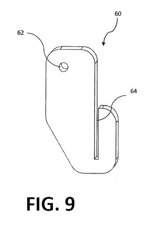
[0028] FIG. 9 shows a front view of a cutting block of one embodiment
of the
present invention; and
[0029] FIG. 10 shows a top view of a tibia including contact point
lines.
DETAILED DESCRIPTION OF THE PREFERRED EMBODIMENT
[0030] To overcome the problems described above, the certain
embodiments of
the present invention include devices and/or methods to assist in distal femur
resection and
proximal tibial resection during knee arthroplasty.
1. Devices for positioning and orienting femoral cutting block
[0031] The rotation of the femoral implant in total knee arthroplasty
(TKA) is
set by the placement of the 4-in-1 femoral cutting block, a standard component
of the knee
system's instrument set. This cutting block is used to guide the creation of
the anterior,
posterior, anterior chamfer, and posterior chamfer resections. The cutting
block usually
includes either two fixed spikes, or two holes for bone pins, which are used
to secure it to
the femur after the distal resection has been completed. Drilling or marking
two holes for
-5-
CA 02873547 2014-11-12
WO 2013/173700
PCT/US2013/041556
these features orients and locates the cutting block. The locations of these
holes are
typically defined by a drill guide device which the surgeon visually aligns
with anatomical
landmarks on the femur, but which does not account for the mechanical
alignment of the
femur with the tibia. A drill guide that references the tibia may improve
implant function.
[0032]
Following completion of the tibial resection 41 and the distal femoral
resection 22, an AP (anterior-posterior) positioning guide 10 is placed on the
distal surface
22 of the femur 20. Referring to FIGS. 1-3, this instrument includes a paddle
12 to
reference either the medial or the lateral posterior condyle 24, and a hole 14
to position a
drill at a fixed distance anterior to the paddle 12. The distance from the
paddle 12 to the
hole 14 is determined by the implant system's 4-in-1 cutting block dimensions:
The
distance is equal to the distance from the posterior cutting slot to the
cutting block spike,
plus the posterior thickness of the femoral implant. The surgeon drills a hole
in the femur
20 through the AP positioning guide 10.
[0033] Now
referring to FIGS. 4-5, a spike 32 on one end of a drill guide 30 is
placed in the hole in the distal femur 22. The drill guide 30 is rotated
around the spike 32
until its edge 36 is parallel to the tibial resection 41, then a second hole
is drilled into the
femur 20 through the hole 34 in the drill guide 30. The drill guide 30 is
configured to
space the two holes at the correct distance to accommodate the 4-in-1 cutting
block's
mounting pins.
[0034]
Preferably, the technique described would include the use of some
commonly-used tensioning instrument (e.g., laminar spreader) to hold the femur
20 in the
correct rotational alignment with the tibia 40 while aligning the drill guide
30 with the
tibial resection 41.
2. Devices for setting rotation of sagittal resection for UKA tibial
implant
[0035] In
unicompartmental knee arthroplasty (UKA), the tibial implant
replaces only the (usually) medial compartment of the tibia. Accordingly, two
tibial
resections are performed, one in a transverse plane, and one in a sagittal
plane. This
sagittal resection both defines the medial-lateral position of the implant,
and sets the
rotation of the implant relative to the tibia. The rotation of this sagittal
resection is
typically visually aligned according to surgeon preference and experience.
This visual
-6-
CA 02873547 2014-11-12
WO 2013/173700
PCT/US2013/041556
alignment does not account for the mechanical alignment of the femur with the
tibia. A
cutting guide that references the femur may improve implant function.
[0036]
Referring to FIGS. 6-7, the mechanical axis 28 of the femur 20 is
calculated by a reference device 100, which contains accelerometers and
gyroscopes to
sense its angular orientation and rate, and which is fixed to the femur 20.
The reference
device 100 incorporates generally the same components and basic measurement
functions
as described in U.S. Patent No. 8,118,815 for its reference device (e.g., 16),
and may be
identical to this device. A surgical orientation device 200, such as the
device shown in
FIG. 8, communicates with the reference device and displays the angle of the
surgical
orientation device relative to the calculated mechanical axis 28. The surgical
orientation
device 200 incorporates generally the same components and basic measurement
functions
as described in U.S. Patent No. 8,118,815 for its surgical orientation device
(e.g., 14).
U.S. Patent No. 8,118,815 is hereby incorporated by reference in its entirety.
[0037] With
reference to FIG. 7, a guide pin 50 is then placed in the distal
femur 20, with its axis parallel to the mechanical axis 28 of the femur 20, as
determined by
the reference device 100. Alternatively, the guide pin 50 may be placed to
align its axis
toward the femoral head 26. The surgical orientation device 200 may be used to
guide
placement of the pin 50.
[0038] This
guide pin 50 is used to position a cutting block 60, which
references the pin 50 by a mating hole 62 in the cutting block 60, and which
also includes a
cutting slot 64 for the sagittal resection on the tibia 40. The cutting slot
64 guides the saw
during resection of the tibia 40.
[0039]
Optionally, the cutting block 60 could be configured to allow medial-
lateral translation between the guide hole 62 and the cutting slot 64. This
would allow the
rotation and position of the sagittal resection to be set independently. Also
optionally, the
cutting block 60 could include a second cutting slot oriented in a transverse
plane. This
second cutting slot would provide guidance for the saw during resection of the
tibia 40 in
the transverse plane.
[0040] As an
alternative method, the surgical orientation device 200 could be
mounted on the cutting block 60 and used to align it relative to the
mechanical axis 26
without using the guide pin 50. The surgical orientation device 200 would
display real-
time orientation to the user during placement and pinning of the cutting block
60. If the
-7-
CA 02873547 2014-11-12
WO 2013/173700
PCT/US2013/041556
cutting block 60 included a second (transverse) cutting slot as described
above, the angular
display from the surgical orientation device could also be used to align this
second slot
relative to the mechanical axis of the tibia 40.
3. Methods
for setting the rotation of the tibial implant by kinematic
measurements
[0041] The
rotation of the tibial implant in TKA is set following completion of
the tibial resection. The tibial implant can be rotated in any direction on
the resected tibial
surface. Final rotation of the implant is typically determined by the surgeon
by one or
more of three methods: 1) visually maximizing coverage of the resected surface
in an
attempt to place the implant as nearly as possible on the outer rim of the
bone; 2) visually
aligning the anterior-posterior (AP) axis of the implant with an anatomic
landmark such as
the tibial tubercle; 3) allowing the implant to rotate freely, then fixing the
tibial implant in
the rotational alignment dictated by contact with the femur with the knee in
full extension
(hereinafter referred to as "traditional methods"). A more precise and/or
quantifiable
alignment method is likely to improve implant performance and patient
satisfaction. The
present invention provides, in certain embodiments, such more precise and/or
quantifiable
alignment methods to improve implant performance and patient satisfaction.
[0042] The
present invention provides, in one embodiment, a method for
setting the rotation of the tibial implant by kinematic measurements based
upon femur-tibia
contact points. In this method of the present invention and referring to FIG.
7, the femur
20 contacts the tibia 40 at two points: one medial, and one lateral. As the
knee flexes
through its range of motion, the location of these contact points on the tibia
40 are
recorded. At any instantaneous flexion angle, a line connecting the two
contact points can
be constructed, as shown in FIG. 10. Lines 42-45 represent the lines
connecting the
medial and lateral contact points throughout the range of motion, from line 42
at full
extension, to line 45 at full flexion. At any flexion angle, a line 46-49
perpendicular to the
instantaneous contact point lines 42-45 defines an AP axis that can be used as
a reference
for tibial component alignment.
[0043] The
contact points are identified using one of several art-disclosed
methods and devices including, without limitations, (i) pressure-sensitive
film (e.g.,
"Prescale" film manufactured by Fujifilm0 Corp.); and (ii) use of knee implant
-8-
CA 02873547 2014-11-12
WO 2013/173700
PCT/US2013/041556
measurement devices such as those described by D'Lima et al., "Tibial Forces
Measured In
Vivo After Total Knee Arthroplasty," Journal of Arthroplasty P. 255-262 (Vol.
21 No.2
February 2006), which contain load cells able to measure contact forces. Once
the contact
points and connecting line 42-45 are identified, the AP axis of the tibial
component is
aligned with any one of the perpendicular AP axes 46-49 chosen according to
surgeon
preference. Alternatively, an AP axis could be calculated as an average of all
axes
throughout the range of motion, or could be a weighted average with greater
weight given
to a specific range of flexion angles.
[0044] The
present invention also provides, in one embodiment, a method for
setting the rotation of the tibial implant by kinematic measurements based on
inertial
measurement of tibio-femoral kinematics. In this method of the present
invention and
referring to FIGS. 6-7, one reference device 100 is securely attached to each
of the femur
20 and tibia 40. The reference device 100 attached to the femur 20 is aligned
approximately with the femoral mechanical axis 28. The reference devices are
preferably
mounted in a manner which allows normal function of the patella to reproduce
normal
knee kinematics. A medialized attachment is preferred on both the tibia 40 and
femur 20
to better accommodate the typical surgical exposure. Optionally, the
orientation of the
mechanical axis 28 is calculated relative to the reference device 100
following the method
described in U.S. Patent No. 8,118,815. If desired, this offset angle can be
applied to the
reference device 100 for greater measurement accuracy.
[0045] In order
to establish the characteristics of the knee joint prior to
resection, the surgeon brings the knee into full extension and moves the leg
through a
short arc of motion, pivoting about the femoral head 26 in all directions and
rotating about
the long axis of the leg. During this motion, the two references devices 100,
stationary
relative to each other, perform a "transfer alignment" to calculate the
relative misalignment
between the two reference devices 100, allowing the orientation of the tibial
device to be
established in the frame of reference of the femoral device.
[0046] The knee
is then taken through a range of motion. Relative rotations
between the tibia 40 and femur 20 are measured by comparing the angular
changes
recorded by their respective reference devices 100 throughout the range of
motion. These
rotations are resolved into three directions corresponding to the flexion,
axial rotation, and
varus/valgus directions. The rotations are transmitted to the surgical
orientation device
-9-
CA 02873547 2014-11-12
WO 2013/173700
PCT/US2013/041556
200 as shown in FIG. 8, which graphically displays to the user plots of axial
rotation and
varus/valgus rotation vs. flexion angle. The surgical orientation device 200
may also
display numerical values for the rotation angle at various flexion angles of
interest, such as
90 degrees or 120 degrees.
[0047] During
trial reduction, the surgeon repeats the above procedure. The
surgical orientation device 200 then displays the aforementioned kinematic
data graphically
and superimposes the trial curves upon the pre-operative curves and/ or
calculates the
appropriate amount by which the tibial component should be rotated about the
tibial axis in
order to best approximate the pre-operative curves. An optimization algorithm
can be
employed for this purpose.
[0048] The
surgeon then adjusts the rotational alignment of the tibial implant
and repeats the measurements above until the rotations of the tibia 40
relative to the femur
20 match the target rotations. These target rotations may be based on
published averages
for healthy knees, or on kinematic measurements taken from the same patient
prior to
resection.
[0049] As an
additional optional step, the surgeon applies alternating varus and
valgus torque to the knee in order to gauge the tibio-femoral rotation allowed
in each
direction. This varus or valgus rotation is displayed on the surgical
orientation device 200,
supplementing the traditional visual estimation of knee laxity in the
varus/valgus direction.
This rotation information provides a means to quantitatively compare the varus
and valgus
laxity, towards the traditional goal of balancing the two by means of soft
tissue releases.
This measurement can be used to quantify the laxity of the knee joint in full
extension, 90
degrees flexion or any other angle to which the knee can be flexed.
[0050] The
present invention further provides, in one embodiment, a method
for setting the rotation of the tibial implant by kinematic measurements using
load cells to
measure contract forces between the tibial implant and the femoral implant. In
this method
of the present invention, the trial tibial implant is fitted with load cells
able to measure
contact forces between the tibial implant and the femoral implant. Such
devices have been
developed previously, and function similarly to the instrumented implant
described by
D'Lima et al., "Tibial Forces Measured In Vivo After Total Knee Arthroplasty,"
Journal of
Arthroplasty p. 255-262 (Vol. 21 No.2 February 2006).
-10-
CA 02873547 2014-11-12
WO 2013/173700
PCT/US2013/041556
[0051] This
instrumented trial tibial component is fixed to the tibia 40 in a
rotation determined by the traditional methods described above. As the knee is
taken
through a range of motion, the trial component transmits the measured contact
forces to a
surgical orientation device 200, which stores and displays the force data,
either as a peak
force number, a force vs. flexion angle history, or both. The surgeon then
iteratively
adjusts the alignment of the trial tibial component and repeats the force
measurement
steps. The tibial component alignment that provides the best fit with the soft
tissue
kinematic envelope will be identified as the configuration that produces the
minimum tibio-
femoral contact force.
[0052] The
present invention also provides, in one embodiment, a method for
setting the rotation of the tibial implant by kinematic measurements based
upon
measurement of tibial interface torque. In this method of the present
invention, the trial
tibial implant is fitted with a torque transducer able to measure axial torque
between the
tibial articular surface and the tibia 40. Such devices have been previously
demonstrated,
such as the instrumented implants described by Heinlein et al. in the Journal
of
Biomechanics (Vol. 41 No.10). For the purposes of the present invention, the
torque is
measured around an axis approximately parallel to the long axis of the tibia
40. This
instrumented trial tibial component is fixed to the tibia 40 in a rotation
determined by the
traditional methods described above. As the knee is taken through a range of
motion, the
trial component transmits the measured torque to a surgical orientation device
200, which
stores and displays the torque data, either as a peak torque number, a torque
vs. flexion
angle history, or both. The surgeon then iteratively adjusts the alignment of
the trial tibial
component and repeats the torque measurement steps. The tibial component
alignment
that provides the best fit with the soft tissue kinematic envelope will be
identified as the
configuration that produces the minimum axial torque.
[0053] Many
other variations than those described herein and/or incorporated
by reference will be apparent from this disclosure. For example, depending on
the
embodiment, certain acts, events, or functions of any of the algorithms
described herein
can be performed in a different sequence, can be added, merged, or left out
altogether
(e.g., not all described acts or events are necessary for the practice of the
algorithms).
Moreover, in certain embodiments, acts or events can be performed
concurrently, e.g.,
through multi-threaded processing, interrupt processing, or multiple
processors or
-11-
CA 02873547 2014-11-12
WO 2013/173700
PCT/US2013/041556
processor cores or on other parallel architectures, rather than sequentially.
In addition,
different tasks or processes can be performed by different machines and/or
computing
systems that can function together.
[0054] The
various illustrative logical blocks, modules, and algorithm steps
described in connection with the embodiments disclosed herein or incorporated
herein by
reference can be implemented as electronic hardware, computer software, or
combinations
of both. To clearly illustrate this interchangeability of hardware and
software, various
illustrative components, blocks, modules, and steps have been described above
generally in
terms of their functionality. Whether such functionality is implemented as
hardware or
software depends upon the particular application and design constraints
imposed on the
overall system. The described or incorporated functionality can be implemented
in varying
ways for each particular application, but such implementation decisions should
not be
interpreted as causing a departure from the scope of the disclosure.
[0055] The
various illustrative logical blocks and modules described in
connection with the embodiments disclosed herein or incorporated by reference
can be
implemented or performed by a machine, such as a general purpose processor, a
digital
signal processor (DSP), an application specific integrated circuit (ASIC), a
field
programmable gate array (FPGA) or other programmable logic device, discrete
gate or
transistor logic, discrete hardware components, or any combination thereof
designed to
perform the functions described herein. A general purpose processor can be a
microprocessor, but in the alternative, the processor can be a controller,
microcontroller,
or state machine, combinations of the same, or the like. A processor can also
be
implemented as a combination of computing devices, e.g., a combination of a
DSP and a
microprocessor, a plurality of microprocessors, one or more microprocessors in
conjunction with a DSP core, or any other such configuration. Although
described herein
primarily with respect to digital technology, a processor may also include
primarily analog
components. For example, any of the signal processing algorithms described
herein may
be implemented in analog circuitry. A computing environment can include any
type of
computer system, including, but not limited to, a computer system based on a
microprocessor, a mainframe computer, a digital signal processor, a portable
computing
device, a personal organizer, a device controller, and a computational engine
within an
appliance, to name a few.
-12-
CA 02873547 2014-11-12
WO 2013/173700
PCT/US2013/041556
[0056] The
steps of a method, process, or algorithm described in connection
with the embodiments disclosed herein can be embodied directly in hardware, in
a software
module executed by a processor, or in a combination of the two. A software
module can
reside in RAM memory, flash memory, ROM memory, EPROM memory, EEPROM
memory, registers, hard disk, a removable disk, a CD-ROM, or any other form of
non-
transitory computer-readable storage medium, media, or physical computer
storage known
in the art. An example storage medium can be coupled to the processor such
that the
processor can read information from, and write information to, the storage
medium. In the
alternative, the storage medium can be integral to the processor. The
processor and the
storage medium can reside in an ASIC. The ASIC can reside in a user terminal.
In the
alternative, the processor and the storage medium can reside as discrete
components in a
user terminal.
[0057]
Conditional language used herein, such as, among others, "can,"
"might," "may," "e.g.," and the like, unless specifically stated otherwise, or
otherwise
understood within the context as used, is generally intended to convey that
certain
embodiments include, while other embodiments do not include, certain features,
elements
and/or states. Thus, such conditional language is not generally intended to
imply that
features, elements and/or states are in any way required for one or more
embodiments or
that one or more embodiments necessarily include logic for deciding, with or
without
author input or prompting, whether these features, elements and/or states are
included or
are to be performed in any particular embodiment. The terms "comprising,"
"including,"
"having," and the like are synonymous and are used inclusively, in an open-
ended fashion,
and do not exclude additional elements, features, acts, operations, and so
forth. Also, the
term "or" is used in its inclusive sense (and not in its exclusive sense) so
that when used,
for example, to connect a list of elements, the term "or" means one, some, or
all of the
elements in the list. Further, the term "each," as used herein, in addition to
having its
ordinary meaning, can mean any subset of a set of elements to which the term
"each" is
applied.
[0058] While
the above detailed description has shown, described, and pointed
out novel features as applied to various embodiments, it will be understood
that various
omissions, substitutions, and changes in the form and details of the devices
or algorithms
illustrated can be made without departing from the spirit of the disclosure.
As will be
-13-
CA 02873547 2014-11-12
WO 2013/173700
PCT/US2013/041556
recognized, certain embodiments of the inventions described herein can be
embodied
within a form that does not provide all of the features and benefits set forth
herein, as some
features can be used or practiced separately from others.
-14-