Note: Descriptions are shown in the official language in which they were submitted.
= CA 02873655 2014-11-13
1
DESCRIPTION
RADIATION STERILIZATION-RESISTANT PROTEIN COMPOSITION
TECHNICAL FIELD
The present invention relates to a protein composition
which comprises specific amino acids and/or a cellulose ether
derivative and has resistance to radiation sterilization.
BACKGROUND ART
Natural and synthetic proteins are becoming more and more
important as drugs. When they are used for medical applications,
their products must be sterilized. As means of sterilization,
there are known heat sterilization in an autoclave,
sterilization with ionizing radiation such as ay ray or electron
beam, gas sterilization with an ethylene oxide gas, plasma
sterilization with hydrogen peroxide, and separate
sterilization using a chemical sterilant comprising a
glutaraldehyde formulation or a filter. However, the
activities of proteins such as bioactive proteins are reduced
by sterilization with heat or radiation. Sterilization with
ethylene oxide has possibilities that a by-product may be
produced by a chemical reaction and that a highly toxic residual
gas may adversely affect the human body. Sterilization with a
chemical sterilant has a problem that the resistance to a
sterilant of a protein and changes in pH, ion intensity and
temperature must be taken into consideration. Then, to
manufacture pharmaceuticals and medical products containing or
immobilizing a protein, their production processes must be
entirely made in sterile conditions and a huge amount of
production cost is required.
Although a solution containing a protein is subjected to
CA 02873655 2014-11-13
2
separate sterilization with a filter, it is difficult to apply
this separate sterilization to a composition containing large
particles or a solid or semisolid composition.
EP0437095 teaches that a neutralized oxidized cellulose
product combined with heparin or a heparin fragment (nORC) can
be sterilized by gamma-ray irradiation. However, this document
fails to teach the sterilization of ORC or n-ORC to which a
protein is bound.
EP0562864 discloses a composite wound care substance
containing a collagen sponge matrix, a second bioabsorbable
polymer (such as an oxidized regenerated cellulose (ORC)
dispersed fiber) and an activator (such as peptide). This
document teaches that the activator may be contained in the
matrix, the bioabsorbable polymer or both of them and that the
composite sponge substance can be sterilized while it is
packaged.
US5730933 discloses a method of sterilizing biologically
active peptide by gamma-ray or electron-beam irradiation without
the loss of the biological activity of the peptide. This method
is a technology comprising the steps of forming a mixture of
biologically active peptide and a foreign protein such as gelatin,
freezing or lyophilizing this mixture, and irradiating it. This
document teaches that the existence of the foreign protein
stabilizes peptide and prevents the reduction of the activity
of peptide.
W02000/033893 discloses a complex of therapeutic peptide
and a polysaccharide selected from the group consisting of
oxidized regenerated cellulose, neutralized oxidized
regenerated cellulose and mixtures thereof. This document
teaches that when peptide is formulated together with an
effective amount of the polysaccharide before sterilization with
ionizing radiation, the biological activity of the peptide
1
CA 02873655 2014-11-13
3
therapeutic agent is not lost and is stabilized if peptide is
sterilized with ionizing radiation.
However, these documents do not suggest that the
deactivation of a protein at the time of sterilizing with
ionizing radiation can be suppressed by making a cellulose ether
derivative and specific amino acids coexistent with the protein.
Meanwhile, JP-A 2011-47089 discloses a process for
producing an enzyme-containing nanofiber having excellent
enzyme activity. In this process, a spinning solution
containing an enzyme and a polymer dissolved in a nonaqueous
solvent is spun by an electrostatic spinning method to form a
zymogen nanofiber which is then imparted with water and dried.
However, this document is silent about the sterilization of the
enzyme-containing nanofiber.
DISCLOSURE OF THE INVENTION
It is an object of the present invention to provide a
protein composition having resistance to radiation
sterilization.
The inventors of the present invention conducted intensive
studies to solve the above problem and found that, surprisingly,
the resistance to radiation sterilization of a protein is
improved by making a mixture of glycine, phenylalanine and
histidine and/or a cellulose ether derivative coexistent with
the protein. The present invention was accomplished based on
this finding.
That is, the present invention is a protein composition
which comprises a mixture of glycine, phenylalanine and
histidine and/or a cellulose ether derivative as an additive.
BRIEF DESCRIPTION OF THE DRAWINGS
Fig. 1 shows the sterilization resisting effect for a
1
CA 02873655 2014-11-13
4
protein of a combination of a cellulose ether derivative and
specific additives of the present invention (axis of ordinate:
gel intensity relative value (before sterilization: 100)); and
Fig. 2 shows the sterilization resisting effect of a
combination of a cellulose ether derivative and specific
additives of the present invention (axis of ordinate: an increase
in the amount of a protein aggregate (%)).
BEST MODE FOR CARRYING OUT THE INVENTION
The present invention is a protein composition which
comprises a mixture of glycine, phenylalanine and histidine
and/or a cellulose ether derivative as an additive.
The protein used in the present invention is not
particularly limited. Examples of the protein include hemostat
proteins typified by fibrinogen and thrombin, enzymes typified
by asparaginase, catalase, superoxide dismutase and lipase,
transport proteins typified by hemoglobin, serum albumin and
low density lipoprotein, muscle proteins typified by actin and
myosin, defense proteins typified by antibodies and complements,
toxin proteins typified by diphtheria toxin, botulinum toxin
and snake venom, protein hormones typified by insulin, growth
factors and cytokine, storage proteins typified by ovalbumin
and ferritin, structural proteins typified by collagen and
keratin, and growth factors typified by epidermal growth factor
(EGF), insulin-like growth factor (IGF), transforming growth
factor (TGF), nerve growth factor (NGF), brain-derived
neurotrophic factor (BDNF), vascular endothelial growth factor
(VEGF), granulocyte-colony stimulating factor (G-CSF),
granulocyte-macrophage-colony stimulating factor (GM-CSF),
platelet-derived growth factor (PDGF), erythropoietin (EPO),
thrombopoietin (TP0), basic fibroblast growth factor(bFGF or
FGF2) and hepatocyte growth factor (HGF). Out of these,
CA 02873655 2014-11-13
hemostat proteins, enzymes, transport proteins, muscle proteins,
defense proteins, toxin proteins, protein hormones, storage
proteins, structural proteins and growth factors are preferred,
and fibrinogen is particularly preferred.
5 The
protein used in the present invention may be of animal
origin or manufactured by a genetic recombination technique.
If it is of animal origin, it is preferably of human origin.
The protein manufactured by the genetic recombination technique
may be a variant obtained by replacing the amino acid sequence
by another amino acid sequence if the essential bioactivity is
the same. Proteins obtained by modifying these proteins and
mixtures thereof may also be used.
To the protein used in the present invention, a stabilizer
and an additive which are pharmaceutically acceptable (to be
referred to as "stabilizer, etc." hereinafter and distinguished
from the additive which is made coexistent with the protein to
provide resistance to radiation sterilization in the present
invention) may be added. Preferred examples of the stabilizer,
etc. include arginine, isoleucine, glutamic acid, citric acid,
calcium chloride, sodium chloride, protease inhibitors (such
as aprotinin) , albumin, surfactants, phospholipids,
polyethylene glycol, sodium hyaluronate, glycerin, trehalose
and sugar alcohols (such as glycerol and mannitol) . At least
one selected from arginine, sodium chloride, trehalose, mannitol
and citric acid is preferred, and citric acid is particularly
preferred.
1
CA 02873655 2014-11-13
6
A mixture of the protein and the stabilizer, etc. used in
the present invention contains the protein in an amount of not
more than 35 parts by weight, preferably not more than 30 parts
by weight based on 100 parts by weight of the mixture.
When the additive used in the present invention is a
mixture of glycine, phenylalanine and histidine, the content
of glycine is generally 5 to 90 parts by weight, preferably 15
to 60 parts by weight, more preferably 20 to 40 parts by weight,
the content of phenylalanine is generally 1 to 80 parts by weight,
preferably 2 to 40 parts by weight, more preferably 4 to 20 parts
by weight, and the content of histidine is generally 2 to 70
parts by weight, preferably 5 to 40 parts by weight, more
preferably 8 to 20 parts by weight based on 100 parts by weight
of the total of the additive and the protein.
When the additive in the present invention is a cellulose
ether derivative, the protein or a mixture of the protein and
the stabilizer, etc. used in the present invention may be
supported on the cellulose ether derivative but preferably
contained in the cellulose ether derivative (the word
"contained" refers to a state that at least part of the protein
enters the inside of the cellulose ether derivative) . In this
case, the molecules of the protein and the stabilizer, etc. may
be dispersed in the cellulose ether derivative but preferably
as particles formed by the aggregation of the molecules of the
protein and the stabilizer, etc. (may be referred to as "protein
particles" including mixed particles with the stabilizer, etc.)
This preferred existence of the protein or the protein
particles in the cellulose ether derivative remains unchanged
when the additive is only the cellulose ether derivative and
also when the additive consists of a mixture of glycine,
phenylalanine and histidine and the cellulose ether derivative.
CA 02873655 2014-11-13
7
Examples of the cellulose ether derivative used in the
present invention include hydroxypropyl cellulose, methyl
cellulose, hydroxyethyl cellulose, hydroxypropylmethyl
cellulose, sodium carboxymethyl cellulose and mixtures thereof.
One selected from the group consisting of hydroxypropyl
cellulose, hydroxyethyl cellulose, hydroxypropylmethyl
cellulose and mixtures thereof is preferred, and hydroxypropyl
cellulose is most preferred.
Although the molecular weight of the cellulose ether
derivative used in the present invention is not particularly
limited, when viscosity measurement is carried out at a
concentration of 2 % and 20 C, a molecular weight which exhibits
a viscosity of 1 to 10,000 mPa.s, preferably 2 to 5,000 mPa.
s, more preferably 2 to 4,000 mPa.s is selected.
In the protein composition of the present invention,
another polymer or another compound may be used in combination
as long as the object of the present invention is not impaired.
The cellulose ether derivative used in the present
invention preferably has high purity. Especially, the contents
of additives and plasticizer contained in the cellulose ether
derivative and residues such as residual catalyst, residual
monomers and residual solvent used for molding and
post-processing are preferably as low as possible. Especially
when the composition is used for medical purposes, it is
necessary to reduce these contents to values below safety
standards.
The form of the protein composition of the present
invention is not limited to a particular form including an
indeterminate form, and the composition may be in the form of
a film, fiber, sheet, plate-like body, tube-like body, linear
body, rod-like body, cushion material, foam or porous body. The
molding method for producing a molded product is not particularly
CA 02873655 2014-11-13
8
limited if it is a method in which the activity of the protein
is not reduced. For example, suitable molding techniques such
as extrusion molding, injection molding, calender molding,
compression molding, blow molding, vacuum forming, powder
molding, cast molding and casting may be employed. The protein
composition of the present invention is suitable for the
production of films and fibers. The fiber form as used herein
refers to a 3-D molded body formed by the lamination, weaving,
knitting or another technique of one or a plurality of fibers.
The fiber form is, for example, a nonwoven fabric. Further, a
tube and a mesh obtained by processing the nonwoven fabric are
included in the fiber form.
For the production of these, any one of techniques which
have been employed for the production of films or plastic fibers
may be employed. For example, extrusion molding techniques such
as casting, electrospinning, inflation extrusion molding and
T die extrusion molding, and calendering technique may be used.
The above molding may be solution molding or melt molding, out
of which solution molding is preferred in order to facilitate
the dispersion of the protein or the protein particles so as
to prevent the functional deterioration of the protein.
The process for producing the protein composition having
a film form out of the present invention will be explained, taking
the casting technique as an example. Protein particles having
an average particle diameter (generally 0.1 to 200 i.tm, preferably
1 to 100 !..tm) suitable for dispersion in a solvent are prepared
by pounding lyophilized protein powders in a mortar. After the
protein particles are dispersed in one or more suitable solvents
(such as 2-propanol and ethanol) which can dissolve the cellulose
ether derivative, can form a suspension with the protein
particles and evaporate in the film forming step to form a film,
the cellulose ether derivative and further optionally a
CA 02873655 2014-11-13
9
plasticizer such as MACROGOL are dissolved in the resulting
dispersion so as to prepare a dope solution containing the
protein particles dispersed in the cellulose ether derivative
solution. A film is formed by the casting technique using the
obtained dope solution.
The protein composition having a film form out of the
present invention comprises the protein or the protein particles
in an amount of generally not less than 100 wt%, preferably not
less than 500 wt%, more preferably 800 to 950 wt% based on the
cellulose ether derivative though this depends on the type of
the protein and the type of the cellulose ether derivative. When
the content of the protein or the protein particles falls below
the above range, the function of the protein may not be obtained
fully and when the content exceeds the above range, film
moldability may become unsatisfactory.
The average thickness of a film of the protein composition
having a film form out of the present invention which differs
according to the intended use is preferably 10 to 1,000
The average fiber diameter of the protein composition
having a fiber form out of the present invention is, for example,
0.01 to 50 .tra and may be suitably determined by a person skilled
in the art according to the intended use. The protein
composition may be in the form of a long fiber. The long fiber
is a fiber formed without adding the step of cutting a fiber
in the course of transition from spinning to the processing of
a fiber molded body. It can be formed by electrospinning, span
bonding and melt blowing methods. Out of these, the
electrospinning method is preferred as the long fiber can be
molded without adding heat and the functional deterioration of
the protein can be suppressed.
The electrospinning method is a method in which a fiber
molded body is obtained on an electrode by applying a high voltage
CA 02873655 2014-11-13
to a solution containing a polymer. This process comprises the
steps of preparing a spinning solution containing a polymer,
applying a high voltage to the solution, jetting the solution,
forming a fiber molded body by evaporating the solvent from the
5 jetted solution, eliminating the charge of the formed fiber
molded body as an optional step, and accumulating the fiber
molded body by the charge loss.
A description is subsequently given of the process for
producing the protein composition having a fiber form or a
10 nonwoven fabric form out of the present invention, taking the
electrospinning method as an example.
The step of preparing a spinning solution in the
electrospinning method will be explained. A suspension of a
cellulose ether derivative solution and protein particles is
preferably used as the spinning solution in the present
invention.
The concentration of the cellulose ether derivative in the
suspension is preferably 1 to 30 wt%. When the concentration
of the cellulose ether derivative is lower than 1 wt%, it is
difficult to form a fiber molded body disadvantageously. When
the concentration is higher than 30 wt%, the fiber diameter of
the obtained fiber molded body becomes large and the viscosity
of the suspension becomes high disadvantageously. The
concentration of the cellulose ether derivative in the
suspension is more preferably 1.5 to 20 wt%.
The solvent for the cellulose ether derivative is not
particularly limited if it can dissolve the cellulose ether
derivative, forms a suspension with the protein particles and
evaporates in the spinning step so that a fiber can be formed.
Only one solvent or a combination of two or more solvents may
be used. Examples of the solvent include chloroform, 2-propanol,
toluene, benzene, benzyl alcohol, dichloromethane, carbon
CA 02873655 2014-11-13
11
tetrachloride, cyclohexane, cyclohexanone, trichloroethane,
methyl ethyl ketone, ethyl acetate, acetone, ethanol, methanol,
tetrahydrofuran, 1,4-dioxane, 1-propanol, phenol, pyridine,
acetic acid, formic acid, hexafluoro-2-propanol,
hexafluoroacetone, N,N-dimethylformamide,
N,N-dimethylacetamide, acetonitrile,N-methy1-2-pyrrolidinone,
N-methylmorpholine-N-oxide, 1,3-dioxolan, water and mixtures
thereof. Out of these, 2-propanol or ethanol is preferably used
from the viewpoints of handling ease and physical properties.
Although the method of preparing a suspension by mixing
together the cellulose ether derivative solution and the protein
particles is not particularly limited, ultraviolet waves or
stirring means may be used. As the stirring means, high-speed
stirring means such as a homogenizer or stirring means such as
an attriter or ball mill may be used. Out of these, dispersion
with ultrasonic waves is preferred.
Also, the spinning solution may be prepared by adding the
cellulose ether derivative after a suspension is formed from
a solvent and the protein particles.
Before the preparation of the suspension, protein
particles maybe microfabricated. For microfabrication, there
are dry milling and wet milling both of which may be employed
and also may be combined in the present invention.
Dry milling is carried out by milling with a ball mill,
planetary mill or oscillating mill, by pounding in a mortar with
a pestle, or by grinding with a medium stirring type pulverizer,
jet mill or stone mill.
Meanwhile, wet milling is carried out by stirring with a
stirrer or kneader having high shear force while the protein
particles are dispersed in a suitable dispersion medium, or by
using a ball mill or bead mill while the protein particles are
dispersed in a medium.
CA 02873655 2014-11-13
12
Further, protein particles produced by a spay drier may
also be used.
In the protein composition having a fiber form or a
nonwoven fabric form out of the present invention, the sizes
of the protein particles are not particularly limited but
preferably 0.01 to 100 1.1M. It is technically difficult to
manufacture protein particles having a particle size smaller
than 0.01 .tm, and when the particle size is larger than 100 p.m,
dispersibility degrades and the fiber molded body becomes
brittle disadvantageously.
The sterilization method used for the protein composition
of the present invention is radiation sterilization. Examples
of the radiation include alpha rays, beta rays, gamma rays,
neutron rays, electron beams and X-rays. Out of these, gamma
rays and electron beams are preferred, and electron beams are
most preferred. Although the sterilization method is not
particularly limited, the dose of the radiation is 10 to 80 kGy,
preferably 20 to 30 kGy. Although the temperature condition is
not particularly limited, it is -80 to 40 C, preferably -80 to
30 C.
The radiation such as alpha rays, positron, gamma rays,
neutron rays, electron beams or X-rays strips an electron off
from molecules or atoms constituting a substance when it is
applied to the substance. A molecular bond is broken upon this,
and a highly reactive radical is produced and chemically reacts
with a surrounding substance secondarily.
It is well known that a protein tends to lose its function
(activity) upon exposure to radiation. This is considered to
be due to the destruction of "a high-order structure" which is
a source of developing a function by the breakage of a molecular
bond by exposure. However, the functional deterioration of the
protein composition of the present invention is suppressed even
CA 02873655 2014-11-13
13
when it is exposed to radiation. This means that the high-order
structure of the protein is retained in the composition of the
present invention, which is a common effect regardless of the
type of the protein. It is not considered from the thickness
of the cellulose ether derivative through which the radiation
is transmitted that this effect is due to screening, and the
control mechanism is not known. The mechanism of a phenomenon
that the effect of the cellulose ether derivative is remarkably
improved by the addition of specific amino acids is not known
as well.
The protein composition of the present invention may
further comprise an electron/ion scavenger, energy transfer
agent, radical scavenger, antioxidant and plasticizer.
Examples of the electron/ion scavenger include N,N' -tetramethyl
phenylenediamine, diphenylenediamine, pyrene and quinone.
Examples of the energy transfer agent include acenaphthene.
Examples of the radical scavenger include mercaptans,
octahydrophenanthrene, monoalkyl diphenyl ethers, tocopherol,
citric acid, butylated hydroxyanisole, butylated
hydroxytoluene, t-butyl hydroquinone, propyl gallate and
ascorbic acid derivatives. Examples of the antioxidant include
BHT, phosphite triesters, phenolic antiaging agents and organic
thio acid salts. Additives that are generally accepted as safe
for use in foods and pharmaceuticals are preferred. The amount
of the additive which is not particularly limited is, for example,
0.01 to 10 wt% based on the cellulose ether derivative in the
protein composition.
The cellulose ether derivative containing the protein in
the sterilization step preferably contains no water. The water
content of the cellulose ether derivative is preferably not more
than 10 wt%, more preferably not more than 4 wt%, much more
preferably substantially 0 wt%.
CA 02873655 2014-11-13
14
The protein composition of the present invention may be
wrapped in a packaging material to be sterilized with radiation.
As the packaging material, a material having high gas barrier
properties such as aluminum is preferably used. The protein
composition may be hermetically sealed and packaged together
with a deoxidant or desiccant or while an inert gas is filled
into the package after degasification, or both methods may be
combined together. As the deoxidant and the desiccant, ones
which do no harm to the human body and are not deactivated upon
exposure to radiation are preferred.
The protein composition of the present invention may be
used as a medical material which requires the function and
sterility of a protein.
The present invention includes a sterile protein
composition obtained by sterilizing the protein composition of
the present invention with radiation.
EXAMPLES
The following examples are provided for the purpose of
further illustrating the present invention but are in no way
to be taken as limiting.
Measurement methods for Examples 1 to 4 and Comparative Examples
1 and 2
1. Average fiber diameter:
The diameters of fibers at 20 locations selected at random
from a photo of the surface of the obtained fiber molded body
taken by a scanning electron microscope (VE8800 of Keyence
Corporation) at 3,000-fold magnification to obtain the average
value of all the fiber diameters as average fiber diameter. N
= 20.
15
2. Average thickness:
The film thicknesses of 15 fiber molded bodies cut to a
size of 50 mm x 100 mm were measured with a measurement force
of 0.01 N by means of a high-resolution digimatic measuring unit
(LITEMATIC VL-50 of Mitutoyo Corporation) to calculate the
average value. This measurement was carried out with minimum
measurement force that could be used by the measuring unit.
3. ELISA measurement
(1) Fibrinogen
10 g/mL of an antihuman fibrinogen antibody (DAKO A0080)
was immobilized to an ELISA plate (NUNC 468667). After it was
washed with PBS containing 0.05% of Tween 20, Block Ace (UK-380
of DS Pharma Biomedical Co., Ltd.) was added to each well to
carry out masking. After washing with PBS containing 0.05 % of
Tween720, a test body was added. Human fibrinogen (No. FIB3 of
Enzyme Research Laboratories) was used as a standard to form
a calibration curve. After washing with PBS containing 0.05 %
of Tween'20, an HRP-labelled antihuman fibrinogen antibody
(CPL5523) was added. After a reaction, the reaction product was
washed with PBS containing 0.05 % of Tween 20, a TMB reagent
(KPL 50-76-02 50-65-02) was added, and the resulting mixture
was left for 6 minutes to develop color. 1 m H3PO4 was added
to stop color development so as to measure 0D450-650 nm with
a microplate reader.
(2) Thrombin
5 g/mL of an antihuman thrombin antibody (No. SAHT-AP of
Affinity Biologicals Inc.) was immobilized to an ELISA plate
(NUNC 468667). After it was washed with PBS containing 0.05 %
of Tween 20, Block Ace (UK-B80 of DS Pharma Biomedical Co., Ltd.)
was added to each well to carryout masking. After washing with
PBS containing 0.05 % of Tween 20, a test body was added. Human
CA 2873655 2019-06-03
CA 02873655 2014-11-13
16
thrombin (HCT-0020 of Haematologic Technologies, Inc.) was used
as a standard to form a calibration curve. After washing with
PBS containing 0.05 % of Tween 20, 0.1 g/mL of an HRP-labelled
antihuman thrombin antibody (No. SAHT-HRP of Affinity
Biologicals Inc.) was added. After a reaction, the reaction
product was washed with PBS containing 0.05 % of Tween 20, a
TMB reagent (DaKo S1599) was added, and the resulting mixture
was left for 10 minutes to develop color. 0.5M H2SO4 was added
to stop color development so as to measure 0D450-650 nm with
a microplate reader.
4. Measurement of thrombin activity
L of a sample and 80 L of a dilution solution for
activity measurement (0.01 % F-68, 50 mmol/L NaCl, 50 mmol/L
15 Tris-HCl, pH 8.4) were added to the polystyrene tube of BD to
be incubated at 37 C for 3 minutes. Recombinant thrombin (JPU
Thrombin Standard 400 U/mL or WHO/US Thrombin Standard 110 IU/mL:
prepared by their own companies) diluted with the above buffer
to 4, 2, 1, 0.5 and 0.25 U/mL in the case of JPU and to 6, 3,
20 1.5, 0.75 and 0.375 IU/mL in the case of IU was used as a standard.
100 L of the S-2238 test team chromogenic substrate (1 mM:
Daiichi Pure Chemicals Co., Ltd.) was added to and mixed with
the obtained reaction solution under agitation to carry out a
reaction at 37 C for 7minutes, and then 800 L of a 0.1M citric
acid solution was added to terminate the reaction. 200 L of
the reaction solution was transferred to 96 well plates to
measure OD405/650.
Example 1
After lyophilized fibrinogen powders (Bolheal,
(registered trademark, the same shall apply hereinafter) , tissue
adhesive: Vial 1) were dispersed in 2-propanol, hydroxypropyl
CA 02873655 2014-11-13
17
cellulose (6-10 mPa.s, manufactured by Wako Pure Chemical
Industries, Ltd.) was dissolved in the resulting dispersion to
a concentration of 16 wt% so as to prepare a spinning solution
having a fibrinogen-containing particle/hydroxypropyl
cellulose ratio of 20 (9.2 as fibrinogen)/100 (w/w). Spinning
was carried out by an electrospinning method at a temperature
of 22 C and a humidity of not more than 26% to obtain a sheet-like
fiber molded body. The inner diameter of a jet nozzle was 0.8
mm, the voltage was 11 kV, the flow rate of the spinning solution
was 1.2 mL/h, and the distance from the jet nozzle to a flat
plate was 15 cm. The obtained fiber molded body had an average
fiber diameter of 0.86 m and an average thickness of 137 m.
The obtained sheet was sterilized with a 20 kGy electron beam.
The sterilized sheet was cut to a size of 0.5 cm x 0.5 cm, and
the protein was extracted with 62.5 L of physiological saline
to carry out ELISA measurement. As a result, the amount of the
immobilized protein was 0.15 mg/cm2. Meanwhile, when ELISA
measurement was made on an unsterilized sheet likewise, the
amount of the immobilized protein was 0.16 mg/cm2. Therefore,
the recovery rate of the protein of the sterilized sheet was
94 % of that of the unsterilized sheet.
Example 2
After lyophilized fibrinogen powders (Bolheal tissue
adhesive: Vial 1) were dispersed in 2-propanol, hydroxypropyl
cellulose (6-10 mPa.s, manufactured by Wako Pure Chemical
Industries, Ltd.) was dissolved in the resulting dispersion to
a concentration of 16 wt% so as to prepare a spinning solution
having a lyophilized fibrinogen powder/hydroxypropyl cellulose
ratio of 40 (18 as fibrinogen)/100 (w/w). Spinning was carried
out by the electrospinning method at a temperature of 22 C and
a humidity of not more than 26% to obtain a sheet-like fiber
CA 02873655 2014-11-13
18
molded body. The inner diameter of the jet nozzle was 0.8 mm,
the voltage was 12.5 kV, the flow rate of the spinning solution
was 1.2 mL/h, and the distance from the jet nozzle to the flat
plate was 15 cm. The obtained fiber molded body had an average
fiber diameter of 0.43 p.m and an average thickness of 152 Rm.
The obtained sheet was sterilized with a 20 kGy electron beam.
The sterilized sheet was cut to a size of 0.5 cm x 0.5 cm, and
the protein was extracted with 62.5 RL of physiological saline
to carry out ELISA measurement. As a result, the amount of the
immobilized protein was 0.27 mg/cm2. Meanwhile, when ELISA
measurement was made on an unsterilized sheet likewise, the
amount of the immobilized protein was 0.30 mg/cm2. Therefore,
the recovery rate of the protein of the sterilized sheet was
90 % of that of the unsterilized sheet.
Example 3
After lyophilized fibrinogen powders (Bolheal tissue
adhesive: Vial 1) were dispersed in 2-propanol, hydroxypropyl
cellulose (6-10 mPa.s, manufactured by Wako Pure Chemical
Industries, Ltd.) was dissolved in the resulting dispersion to
a concentration of 16 wt% so as to prepare a spinning solution
having a lyophilized fibrinogen powder/hydroxypropyl cellulose
ratio of 100 (46 as fibrinogen)/100 (w/w). Spinning was carried
out by the electrospinning method at a temperature of 22 C and
a humidity of not more than 26% to obtain a sheet-like fiber
molded body. The inner diameter of the jet nozzle was 0.8 mm,
the voltage was 12.5 kV, the flow rate of the spinning solution
was 1.2 mL/h, and the distance from the jet nozzle to the flat
plate was 15 cm. The obtained fiber molded body had an average
fiber diameter of 0.35 p.m and an average thickness of 191 Rm.
The obtained sheet was sterilized with a 20 kGy electron beam.
The sterilized sheet was cut to a size of 0.5 cm x 0.5 cm, and
CA 02873655 2014-11-13
19
the protein was extracted with 62.5 )11, of physiological saline
to carry out ELISA measurement. As a result, the amount of the
immobilized protein was 0.78 mg/cm2. Meanwhile, when ELISA
measurement was made on an unsterilized sheet likewise, the
amount of the immobilized protein was 0.76 mg/cm2. Therefore,
the recovery rate of the protein of the sterilized sheet was
102 % of that of the unsterilized sheet.
Comparative Example 1
Lyophilized fibrinogen powders (Bolheal tissue adhesive:
Vial 1) were sterilized with a 20 kGy electron beam. The protein
was extracted with 1 mL of physiological saline to carry out
ELISA measurement. As a result, the ELISA measurement value was
31 vig/mL. Meanwhile, when ELISA measurement was made on
unsterilized lyophilized fibrinogen powders (Bolheal) likewise,
the ELISA measurement value was 90 [1g/mL. Therefore, the
recovery rate of the protein of the sterilized sheet was 34 %
of that of the unsterilized sheet.
Example 4
After thrombin-containing particles (prepared by
lyophilizing an aqueous solution containing 1 mg/mL of
recombinant thrombin, sodium chloride, sodium citrate, calcium
chloride and mannitol and having a pH of 7) were dispersed in
2-propanol, hydroxypropyl cellulose (2.0-2.9 mPa=s,
manufactured by Nippon Soda Co., Ltd.) was dissolved in the
resulting dispersion to a concentration of 13 wt% so as to prepare
a dope solution having a thrombin-containing
particle/hydroxypropyl cellulose ratio of 100/100 (w/w) .
Spinning was carried out by the electrospinning method to obtain
a sheet-like fiber molded body. The obtained fiber molded body
had a thickness of 204 pm, a weight of 2.08 mg/cm2 and a bulk
CA 02873655 2014-11-13
density of 101 mg/cm3. The obtained sheet was cut to a diameter
of 1 cm, and theprotein was extractedwith 200 }AL of physiological
saline to measure its activity. As a result, the activity
measurement value was 110.3 U/cm2. The obtained sheet was
5 sterilized by exposure to a 30 kGy electron beam to measure the
activity of thrombin. When the activity of thrombin before
sterilization was 100 %, the retention rate of the activity of
thrombin right after exposure to an electron beam was 68.4 %.
10 Comparative Example 2
After a 30 kGy electron beam was applied to
thrombin-containing particles (prepared by lyophilizing an
aqueous solution containing 1 mg/mL of recombinant thrombin,
sodium chloride, sodium citrate, calcium chloride and mannitol
15 and having a pH of 7) to sterilize them, the activity of thrombin
was measured. The activity of thrombin before exposure was
404.73 U/vial. When the activity of thrombin before
sterilization was 100 %, the retention rate of the activity of
thrombin right after exposure to an electron beam was 51.8 %.
Measurement methods for Examples 5 and 6 and Comparative Examples
3 and 4
1. Average thickness:
The film thicknesses of 9 fiber molded bodies obtained by
cutting the composition to a suitable size were measured with
a measurement force of 0.01 N by means of a high-resolution
digimatic measuring unit (LITEMATIC VL-50 of Mitutoyo
Corporation) to calculate the average value. This measurement
was carried out with minimum measurement force that could be
used by the measuring unit.
1
CA 02873655 2014-11-13
21
2. Measurement of enzyme activity
A continuous fluorometric lipase test kit (manufactured
by PROGEN BIOTECHNIK GMBH) was used to measure the activity of
lipase. The retention rate of activity was calculated from the
following equation. The amount of the active enzyme was
calculated in terms of concentration from the value of activity.
Retention rate of activity ( %) = (amount of active enzyme after
sterilization (mg/cm2) /amount of active enzyme before
sterilization (mg/cm2)1 x 100
Fluorescent measurement using Tokyogreen (registered
trademark, the same shall apply hereinafter)-plu (of Sekisui
Medical Co., Ltd.) was employed to measure the activity of
0-glucosidase. The recovery rate of activity was calculated
from the following equation. The theoretical weight of the
immobilized enzyme was calculated from wt% of the charged enzyme
powder and the weight of the composition.
Recovery rate of activity (%) = (amount of active enzyme
(mg)/theoretical weight of immobilized enzyme (mg)1 x 100
The retention rate of activity was calculated from the
following equation.
Retention rate of activity (%) = (recovery rate of activity
after sterilization (%)/recovery rate of activity before
sterilization (%))) x 100
Example 5
After lipase powders (derived from pig pancreas,
manufactured by Wako Pure Chemical Industries, Ltd., the same
shall apply hereinafter) were dispersed in 2-propanol,
hydroxypropyl cellulose (6-10 mPa-s, manufactured by Wako Pure
Chemical Industries, Ltd.) was dissolved in the resulting
CA 02873655 2014-11-13
22
dispersion to a concentration of 13 wt% so as to prepare a
spinning solution having a lipase powder/hydroxypropyl
cellulose ratio of 50/100 (w/w). Spinning was carried out by
the electrospinning method at a temperature of 27 C and a
.. humidity of not more than 27 % to obtain a sheet-like fiber molded
body. The inner diameter of the jet nozzle was 0.8 mm, the
voltage was 18 kV, the flow rate of the spinning solution was
1.2 mL/h, and the distance from the jet nozzle to the flat plate
was 16.5 cm. The obtained fiber molded body (10cm x 14 cm) had
an average thickness of 168 him. The obtained fiber molded body
was sterilized with a 20 kGy electron beam. After the sterilized
fiber molded body was cut to a size of 1 cm x 1 cm, lipase was
extracted with 1 mL of a lipase buffer contained in a kit to
measure its activity. As a result, the amount of the active
enzyme was 0.46 mg/cm2. Meanwhile, when activity measurement
was made on an unsterilized sheet likewise, the amount of the
active enzyme was 0.40 mg/cm2. It is understood from above that
the retention rate of the activity of the sterilized fiber molded
body was 115 % of that of the unsterilized fiber molded body
and that lipase was not deactivated by sterilization with an
electron beam.
Example 6
After lipase powders were dispersed in 2-propanol,
hydroxypropyl cellulose (6-10 mPa=s, manufactured by wako Pure
Chemical Industries, Ltd.) was dissolved in the resulting
dispersion to a concentration of 13 wt% so as to prepare a cast
solution having a lipase powder/hydroxypropyl cellulose ratio
of 50/100 (w/w) . Casting was carried out by using a doctor blade
.. (YBA-3 of YOSHIMITSU) at a coating width of 15 mil to obtain
a sheet. The obtained sheet (4 cm x 6 cm) had an average thickness
of 180 1.1m. The obtained sheet was sterilized with a 20 kGy
CA 02873655 2014-11-13
23
electron beam. After the sterilized sheet was cut to a size of
1 cm x 1 cm, lipase was extracted with 1 mL of a lipase buffer
contained in a kit to measure its activity. As a result, the
amount of the active enzyme was 0.69 mg/cm2. Meanwhile, when
activity measurement was made on an unsterilized sheet likewise,
the amount of the active enzyme was 0 64 mg/cm2 . It is understood
from above that the retention rate of the activity of the
sterilized sheet was 108 % of that of the unsterilized sheet
and that lipase was not deactivated by sterilization with an
electron beam.
Example 7
After P-glucosidase powders (derived from almond,
manufactured by Oriental Yeast Co., Ltd, the same shall apply
hereinafter) were dispersed in 2-propanol, hydroxypropyl
cellulose (6-10 mPa.s, manufactured by Wako Pure Chemical
Industries, Ltd.) was dissolved in the resulting dispersion to
a concentration of 13 wt% so as to prepare a spinning solution
having a 13-glucosidasepowder/hydroxypropyl cellulose ratio of
38/62 (w/w). Spinning was carried out by the electrospinning
method at a temperature of 27 C and a humidity of not more than
27 % to obtain a sheet-like fiber molded body. The inner diameter
of the jet nozzle was 0.9 mm, the voltage was 18 kv, the flow
rate of the spinning solution was 1.2 mL/h, and the distance
from the jet nozzle to the flat plate was 16.5 cm. The obtained
fiber molded body (10 cm x 10 cm) had an average thickness of
207 m. After the obtained fiber molded body was cut to a size
of 2 cm x 2 cm, it was sterilized with a 20 kGy electron beam.
P-glucosidase was extracted from the obtained sterilized sheet
with 1 mL of physiological saline to measure its activity with
Tokyogreen-3G1u. As a result, the recovery rate of activity was
42 %. meanwhile, when activity measurement was made on an
CA 02873655 2014-11-13
24
unsterilized sheet likewise, the recovery rate of activity was
46 %. It is understood from above that the retention rate of
the activity of the sterilized fiber molded body was 91 % of
that of the unsterilized fiber molded body and that the
deactivation of the enzyme can be suppressed by containing it
in the cellulose ether derivative.
Comparative Example 3
Lipase powders were sterilized with a 20 kGy electron beam.
1 mL of a lipase buffer was added to 1 mg of the powders to measure
its activity. As a result, the activity value was 0.25 pmol/mL=
min. Meanwhile, when activity measurement was made on
unsterilized lipase powders likewise, the activity value was
0.34 pmol/mL=min. Therefore, the retention rate of the activity
of the sterilized powders was 74 % of that of the unsterilized
powders.
Comparative Example 4
p-glucosidase powders were sterilized with a 20 kGy
electron beam. 2 mg of the powders was dissolved in 1 mL of
physiological saline to measure its activity with
Tokyogreen-1301u. As a result, the retention rate of activity
was 81 %
Measurement method for Examples 8 to 10
When an electron beam is applied to lyophilized fibrinogen
powders (fibrinogen-containing particles) , the changes of
fibrinogen (increase in the amount of its aggregate, reduction
in gel strength) occur. To investigate the effect of
suppressing the changes of fibrinogen by exposure to an electron
beam (sterilization resisting effect) , a bulk solution of
fibrinogen was prepared and 1 mL of the bulk solution was charged
I
CA 02873655 2014-11-13
into a 5 mL glass vial to be lyophilized. A30 kGy electron beam
was applied to part of the vial in which lyophilization was
completed to compare each lyophilized product before and after
sterilization.
5 Comparison evaluation was carried out by measuring gel
strength by means of the EZTest small-sized bench-top tester
(of Shimadzu Corporation) and the content of the aggregate by
means of BioSep-SEC-s4000 (of Phenomenex) (analyzing
conditions: fractionating with a 50 mM phosphoric acid buffer
10 solution (pH of 7.0) and 0.5 M arginine hydrochloride salt as
mobile phases at a flow rate of 1.0 ml/min, detecting a target
substance with a wavelength of 280 nm; and determining the
quantity of the aggregate from a peak detected earlier than a
monomer peak).
15 As for the procedure of preparing a sample for analysis
(analytical sample), an unsterilized lyophilized product vial
and a sterilized lyophilized product vial were each dissolved
in 1 mL of distilled water. The resulting solutions were
centrifuged by a centrifugal tube at 15,000 rpm for 5 minutes
20 and let pass through a 0.45 gm filter to be used as analytical
samples.
Example 8
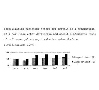
The sterilization resisting effect for a protein of a
25 combination of a cellulose ether derivative and specific
additives was investigated by the following method.
(method) The function of fibrinogen was evaluated by measuring
the gel strength of each of fibrinogen bulk solutions of
compositions comprising "a cellulose ether derivative + specific
additives" (compositions (1) shown in Nos. 1 to 6 in Table 1
below) and fibrinogen bulk solutions of compositions (2)
prepared by eliminating the cellulose ether derivative from the
CA 02873655 2014-11-13
26
compositions (1), and the gel strengths before and after
sterilization of these solutions were compared with each other
to investigate the sterilization resisting effect. The results
are shown in Table 2.
The compositions (1) (lyophilized powders and
hydroxypropyl cellulose were suspended in 2-propanol to form
a sheet) and the compositions (2) (lyophilized powders) were
dissolved in water to an Fbg concentration of 1 % and diluted
with a buffer solution containing 10 IttM arginine and 270 mM sodium
chloride and having a pH of 8.5 to a concentration of 2 mg/mL.
After 10 L of fibrogammin (240 units/mL) and 110 L of
a thrombin solution (containing 0.2 mg/mL of 100 mM calcium
chloride) were added to a 2 mL polypropylene tube and the
resulting solution was pipetted, 900 L of a 2 mg/mL fibrinogen
solution was added in such a manner that air bubbles were not
contained and left to stand at 37 C for 1 hour to measure the
gel strength by means of the EZTest small-size bench -top tester
(of Shimadzu Corporation).
= CA 02873655 2014-11-13
27
Table 1 compositions (1): compositions comprising
cellulose ether derivative + specific additives
Composition composition of bulk solution
No. 1 1 % of Fbg, 10 mM arginine, 110 mM sodium
chloride, 1.0 % of glycine, 0.1 % of mannitol,
0.4 % of hydroxypropyl cellulose
No. 2 1 % of Fbg, 10 mM arginine, 110 mM sodium
chloride, 1.0 % of glycine, 0.2 % of mannitol,
0.4 % of hydroxypropyl cellulose
No. 3 1 % of Fbg, 10 mM arginine, 110 mM sodium
chloride, 1.0 % of glycine, 0.25 % of
phenylalanine, 0.2 % of trehalose, 0.4 % of
histidine, 0.1 %of trisodium citrate, 0.4 % of
hydroxypropyl cellulose
No. 4 1 % of Fbg, 10 mM arginine, 110 mM sodium
chloride, 1.0 % of glycine, 0.1 % of mannitol,
0.25 % of phenylalanine, 0.2 % of trehalose,
0.4 % of histidine, 0.1 % of trisodium citrate,
0.4 % of hydroxypropyl cellulose
No. 5 1 % of Fbg, 10 mM arginine, 110 mM sodium
chloride, 1.0 % of glycine, 0.1 % of mannitol,
0.25 % of phenylalanine, 0.4 % of histidine,
0.1 % of trisodium citrate, 0.4 % of
hydroxypropyl cellulose
No. 6 1 % of Fbg, 10 mM arginine, 110 mM sodium
chloride, 1.0 % of glycine, 0.2 % of mannitol,
0.25 % of phenylalanine, 0.4 % of histidine,
0.1 % of trisodium citrate, 0.4 % of
hydroxypropyl cellulose
= CA 02873655 2014-11-13
28
Compositions (2): compositions comprising specific additives
These were prepared by eliminating the cellulose ether
derivative (hydroxypropyl cellulose: HPC) from the compositions
(1) .
(results)
The values of gel strength after sterilization are shown
in Table 2 and Fig. 1 when the values before sterilization are
100.
10 Table 2 sterilization
resisting effect for protein of a
combination of cellulose ether derivative and specific additives
composition Compositions (1) (cellulose compositions (2)
ether derivative + specific
(specific additives)
additives)
No.1 51.5 49.8
No.2 51.1 36.5
No.3 81.5 58.1
No.4 84.0 57.9
No.5 77.6 57.7
No.6 84.4 59.6
The sterilization resistance improving effect due to the
existence of the cellulose ether derivative was not observed
in the composition No. 1 whereas the above effect due to the
existence of the cellulose ether derivative was observed in the
compositions Nos. 2 to 6. This effect was marked especially in
the composition Nos. 3 to 6.
CA 02873655 2014-11-13
29
Example 9
The sterilization resisting effect of glycine was
investigated with the compositions (two) shown in Table 3 below
in the same manner as in Example 8. The results are shown in
Table 4.
Table 3 compositions for evaluating the sterilization
resisting effect of glycine
Arginine sodium
Composition fibrinogen
mannitol glycine
(pH 8.5) chloride
G(-) 1.0% 10 mm 110 mm 0.2% 0%
G(+) 1.0% 10 mm 110 mm 0.2% 1.0%
Table 4 results of evaluating the sterilization resisting
effect of glycine
s4000 (aggregate)
Before after
increase in
Composition
sterilization sterilization amount
(%) (%) (%)
G(-) 14.0 28.0 14.0
G(+) 14.0 21.6 7.6
An increase in the content of the fibrinogen aggregate was
suppressed by the addition of glycine.
Example 10
The sterilization resisting effect of a combination of a
cellulose ether derivative and specific additives was
investigated by the same method as in Example 8.
Hydroxypropyl cellulose (HPC) as the cellulose ether
I
CA 02873655 2014-11-13
derivative and eight different fibrinogen bulk solutions shown
in Table 5 below were used. 1.0 % of fibrinogen, 110 mM sodium
chloride, 1.0 % of glycine and 0.2 % of mannitol were used in
the following eight compositions. The results are shown in
5 Table 6 and Fig. 2.
Table 5 sterilization resisting effect of a combination of
cellulose ether derivative and specific additives
Arginine tris sodium
Composition histidine phenylalanine HPC
(pH8.5) (pH8.5) citrate
1HPC(-) 10m5 OmM 0% 0% 0% 0%
1HPC(+) 10mM OmM 0% 0% 0% 0.419%
2HPC(-) 10mM OmM 0.4% 0.25% 0% 0%
2HPC(+) 10mM OmM 0.4% 0.25% 0% 0.419%
,
3HPC(-) 10mM OmM 0.4% 0.25% 0.1% 0%
3HPC(+) 10mM OmM 0.4% 0.25% 0.1% 0.419%
4HPC(-) 0.4mM 10mM 0.4% 0.25% 0.1% 0%
..--,
4HPC(+) 0.4mM 10mM 0.4% 0.25% 0.1% 0.419%
'
CA 02873655 2014-11-13
31
Table 6 evaluation results of sterilization resisting effect
of a combination of cellulose ether derivative and specific
additives
s4000 (aggregate)
Composition Before after increase
sterilization sterilization in
amount
(%) (%) (%)
1HPC(-) 14.0 21.6 7.6
1HPC(+) 12.8 18.4 5.7
2HPC(-) 13.8 19.2 5.4
2HPC(+) 13.9 15.8 1.9
3HPC(-) 15.1 19.8 4.7
3HPC(+) 14.8 17.8 2.9
4HPC(-) 14.9 19.5 4.6
4HPC(+) 14.2 15.5 1.3
An increase in the content of the protein aggregate was
suppressed by the addition of the cellulose ether derivative.
The effect of suppressing an increase in the above content due
to the existence of the cellulose ether derivative was marked
in compositions 2 to 4 comprising phenylalanine and histidine.
It is understood from this that the reason that the effect of
improving sterilization resistance due to the existence of the
cellulose ether derivative is not observed in composition No.1
whereas the effect is observed and marked in composition Nos.
3 to 6 in Example 7 is considered to be due to the fact that
the coexistence of phenylalanine and histidine with the
cellulose ether derivative in composition Nos. 3 to 6 provides
a marked sterilization resisting effect for a protein.
CA 02873655 2014-11-13
32
Effect of the Invention
The protein composition of the present invention has
resistance to radiation sterilization. The sterile composition
of the present invention retains the structure and function of
a protein though it is sterilized.
Industrial Feasibility
The protein composition of the present invention is used
in the manufacturing industry of medical products which requires
the function and sterility of a protein.