Note: Descriptions are shown in the official language in which they were submitted.
CA 02873829 2014-11-17
WO 2014/009474
PCT/EP2013/064683
1
Method for the detection of a multispecific binder
The current invention is directed to a method for the detection of a
multispecific
binder in a sample, wherein the multispecific binder is detected using anti-
idiotypic
antibodies directed against the different binding specificities of the
multispecific
binder.
Background of the Invention
Standard solid-phase immunoassays with antibodies involve the formation of a
complex between an antibody adsorbed/immobilized on a solid phase (capture
antibody), the antigen, and an antibody to another epitope of the antigen
conjugated
with an enzyme or detectable label (tracer antibody). In the assay, a sandwich
is
formed: solid phase/capture antibody/antigen/tracer antibody. In the reaction
catalyzed by the sandwich among other things the activity of the antibody-
conjugated enzyme is proportional to the antigen concentration in the
incubation
medium. Anti-idiotypic antibody assays are mentioned, for example, in
US 5,219,730; WO 87/002778; EP 0 139 389; and EP 0 170 302. Wadhwa, M., et
al. (J. Immunol. Methods 278 (2003) 1-17) report strategies for the detection,
measurement and characterization of unwanted antibodies induced by therapeutic
biologicals. A method for producing anti idiotypic antibodies is reported in
EP 1 917 854.
In WO 2008/119353 bispecific antibodies and methods for producing thereof are
reported. Methods for determining the bivalency of protein and antibody
therapeutics are reported in WO 2006/096697. In WO 2008/134046 potent, stable
and non-immunosuppressive anti-CD4 antibodies are reported. Muller, K.M., et
al.
report that the first constant domain (CH1 and CL) of an antibody can be used
as
heterodimerization domain for bispecific miniantibodies.
Summary of the Invention
It has been found that by using two anti-idiotypic antibodies as capture and
as
tracer antibody in a sandwich immunoassay for the determination of the amount
of
a multispecific antibody in a sample, whereby each of the anti-idiotypic
antibodies
binds to a different binding specificity of the multispecific antibody,
influences due
to the sample matrix (e.g. serum or human plasma, antigens, etc.) can be
minimized. Additionally it is possible to use an assay setup that is more
sensitive,
CA 02873829 2014-11-17
WO 2014/009474
PCT/EP2013/064683
- 2 -
less influencable by the sample matrix, can be performed faster, requires
minimal
removal/dilution of sample matrix, and provides more flexibility for
labeling/derivatization/immobilization of capture and/or tracer antibody,
respectively. This is achieved by using two anti-idiotypic antibodies, one
directed
against the first binding specificity of the bispecific antibody and one
against the
second binding specificity of the bispecific antibody.
Thus, herein is reported a method for the (immunological) determination of the
amount of a multispecific binder in a sample comprising the step of:
- determining the amount of a complex formed between
i) an anti-idiotypic antibody that specifically binds to a first binding
specificity of the multispecific binder, and
ii) the multispecific binder
by incubating the complex with an anti-idiotypic antibody that specifically
binds to a second binding specificity of the multispecific binder, which is
different from the first binding specificity of the multispecific antibody,
and
thereby determining the amount of the multispecific binder in the sample.
In one embodiment the anti-idiotypic antibody that specifically binds to a
first
binding specificity of the multispecific binder is conjugated to a solid
phase.
In one embodiment the anti-idiotypic antibody that specifically binds to a
second
binding specificity of the multispecific binder is conjugated to a detectable
label.
In one embodiment the sample comprises (human) serum or (human) plasma,
and/or is a cell lysate, and/or comprises one or more antigens of the
multispecific
binder. In one embodiment the sample is (human) serum or (human) plasma.
In one embodiment the multispecific binder is selected from an antibody, a
fusion
polypeptide comprising an antibody or antibody fragment and a non-antibody
polypeptide, a fusion polypeptide comprising an antibody or antibody fragment
and
a soluble receptor, or a fusion polypeptide comprising an antibody or antibody
fragment and a peptidic binding molecule.
In one embodiment the multispecific binder is an antibody. In one embodiment
the
antibody is a bispecific antibody, or a trispecific antibody, or a
tetraspecific
CA 02873829 2014-11-17
WO 2014/009474
PCT/EP2013/064683
-3 -
antibody, or a pentaspecific antibody, or a hexaspecific antibody. In one
embodiment the antibody is a bispecific antibody.
In one embodiment the binding specificity is a binding site or a pair of an
antibody
heavy chain variable domain and an antibody light chain variable domain.
In one embodiment the anti-idiotypic antibody that specifically binds to a
first
binding specificity of the multispecific binder is biotinylated and the solid
phase is
streptavidin coated. In one embodiment the solid phase is a streptavidin
coated
paramagnetic bead or a streptavidin coated sepharose bead.
In one embodiment the anti-idiotypic antibody that specifically binds to the
second
binding specificity of the multispecific binder is digoxigenylated.
In one embodiment the method comprises the step of
- determining the amount of a complex formed between
i) an anti-idiotypic antibody that specifically binds to a first binding
specificity of the multispecific binder,
ii) the multispecific binder, and
iii) an anti-idiotypic antibody that specifically binds to a second
binding specificity of the multispecific binder and that comprises a
detectable label,
by determination the detectable label in the formed complex.
In one embodiment the conjugation of an anti-idiotypic antibody to its
conjugation
partner is performed by chemically binding via N-terminal and/or 8-amino
groups
(lysine), 8-amino groups of different lysins, carboxy-, sulfhydryl-, hydroxyl-
and/or
phenolic functional groups of the amino acid backbone of the drug antibody
and/or
sugar alcohol groups of the carbohydrate structure of the drug antibody.
In one embodiment the anti-idiotypic antibody is a mixture comprising the anti-
idiotypic antibody conjugated via at least two different amino groups to the
solid
phase. Such coupling via different amino groups can be performed by acylation
of
a part of the 8-amino groups with chemical protecting agents, e.g. by
citraconylation, in a first step. In a second step conjugation is performed
via the
remaining amino groups. Subsequently citraconylation is removed and the
antibody
is conjugated to the solid phase via remaining free amino groups, i.e. the
antibody
obtained is conjugated to the solid phase via amino groups that have not been
protected by citraconylation. Suitable chemical protecting agents form bonds
at
CA 02873829 2014-11-17
WO 2014/009474
PCT/EP2013/064683
- 4 -
unprotected side chain amines and are less stable than and different from
those
bonds at the N-terminus. Many such chemical protecting agents are known (see
for
example EP 0 651 761). In one embodiment the chemical protecting agents
include
cyclic dicarboxylic acid anhydrides like maleic or citraconylic acid
anhydride.
In one embodiment the anti-idiotypic antibody is conjugated to the solid phase
by
passive adsorption. Passive adsorption is, e. g., described by Butler, J.E.,
in "Solid
Phases in Immunoassay" (1996) 205-225 and Diamandis, E.P., and Christopoulos,
T.K. (Editors), in "Immunoassays" (1996) Academic Press (San Diego).
In one embodiment the anti-idiotypic antibody is conjugated (immobilized) via
a
specific binding pair. Such a binding pair (first component/second component)
is in
one embodiment selected from streptavidin or avidin/biotin, antibody/antigen
(see,
for example, Hermanson, G.T., et al., Bioconjugate Techniques, Academic Press
(1996), lectin/polysaccharide, steroid/steroid binding protein,
hormone/hormone
receptor, enzyme/substrate, IgG/Protein A and/or G, etc. In one embodiment the
anti-idiotypic antibody is conjugated to biotin and immobilization is
performed via
immobilized avidin or streptavidin.
Description of the Figures
Figure 1
Schematic pharmacokinetic ELISA assay principle for
determination of concentrations of bispecific antibodies in
serum and cell samples (anti-idiotypic antibody based
ELISA).
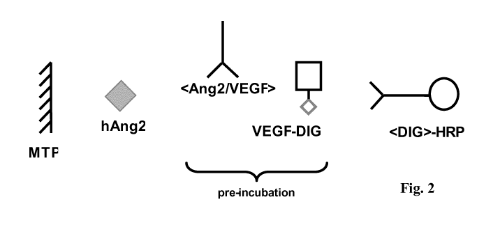
Figure 2
Antigen-based sandwich ELISA for detection of anti-
VEGF/ANG2 antibodies. Recombinant human angiopoietin
2 was directly coated to a micro titer plate. In parallel,
samples containing anti-VEGF/ANG2 antibodies were pre-
incubated with digoxigenin-labeled recombinant VEGF.
After coating, the plate was washed and incubated with the
pre-incubated mixture. The complexes of anti-
VEGF/ANG2 antibody and digoxigenin-labeled VEGF
bind to the ANG2 on the micro titer plate surface. Bound
digoxigenin-labeled VEGF was detected with anti-
digoxigenin antibody-HRP conjugate and ABTS.
CA 02873829 2014-11-17
WO 2014/009474
PCT/EP2013/064683
-5 -
Figure 3
Comparison of calibration curves obtained for antigen-
based ELISA (A) and anti-idiotype antibody based ELISA
(B) for detection of bispecific anti-VEGF/ANG2 antibody.
Figure 4
Comparison of calibration curves of the anti-idiotype
antibody based ELISA and the antigen-based ELISA in the
presence of 5 %, 10 % and 20 % human serum.
Detailed Description of the Invention
Herein is reported an in vitro method for the determination of the amount of a
multispecific binder, such as bispecific antibodies/drugs, in pre-clinical and
clinical
samples.
It has been found that a multispecific binder in a sample has to be detected
by using
two anti-idiotypic antibodies that bind to different binding specificities of
the
multispecific binder in order to minimize influences by the sample matrix and
to
allow a greater degree of flexibility in setting up and performing the
immunological
determination of the amount of the multispecific binder in the sample.
In the following the method as reported herein is exemplified with a
multispecific
antibody as embodiment of a multispecific binder.
The term "antibody" herein is used in the broadest sense and encompasses
various
antibody structures, including but not limited to monoclonal antibodies,
polyclonal
antibodies, multispecific antibodies (e.g., bispecific antibodies), and
antibody
fragments so long as they exhibit the desired antigen-binding activity.
In certain embodiments, the multispecific binder is a multispecific antibody,
e.g. a
bispecific antibody. Multispecific antibodies are monoclonal antibodies that
have
binding specificities for at least two different sites. In certain
embodiments, one of
the binding specificities is for a first antigen and the other is for a
different second
antigen. In certain embodiments, bispecific antibodies may bind to two
different
epitopes of the same antigen. Bispecific antibodies can be prepared as full
length
antibodies or antibody fragments. In one embodiment the antibody is a
bispecific
antibody which specifically binds to a first and a second antigen. In one
embodiment the bispecific antibody has i) a first binding specificity that
specifically binds to a first antigen or a first epitope on an antigen, and
ii) a second
binding specificity that specifically binds to a second antigen or a second
epitope
CA 02873829 2014-11-17
WO 2014/009474
PCT/EP2013/064683
- 6 -
on the same antigen. In one embodiment the second epitope on the same antigen
is
a non-overlapping epitope.
Multispecific antibodies are described in WO 2009/080251, WO 2009/080252,
WO 2009/080253, WO 2009/080254, WO 2010/112193, WO 2010/115589,
WO 2010/136172, WO 2010/145792, or WO 2010/145793.
An "antibody fragment" refers to a molecule other than an intact antibody that
comprises a portion of an intact antibody that binds the antigen to which the
intact
antibody binds. Examples of antibody fragments include but are not limited to
Fv,
Fab, Fab', Fab'-SH, F(ab')2; diabodies; linear antibodies; single-chain
antibody
molecules (e.g. scFv); and multispecific antibodies formed from antibody
fragments.
The "class" of an antibody refers to the type of constant domain or constant
region
possessed by its heavy chain. There are five major classes of antibodies: IgA,
IgD,
IgE, IgG, and IgM, and several of these may be further divided into subclasses
(isotypes), e.g., IgGi, IgG2, IgG3, Igai, IgAi, and IgA2. The heavy chain
constant
domains that correspond to the different classes of immunoglobulins are called
a,
8, E, 7, and , respectively.
The term "free antigen" denotes the antigen that can be specifically bound by
a
binding specificity of an antibody but which is currently not bound to this
binding
specificity. In one embodiment the free antigen is a not-antibody bound
antigen or
a non-antibody complexed antigen.
The term "Fc-region" herein is used to define a C-terminal region of an
immunoglobulin heavy chain that contains at least a portion of the constant
region.
The term includes native sequence Fc-regions and variant Fc-regions. In one
embodiment, a human IgG heavy chain Fc-region extends from Cys226, or from
Pro230, to the carboxyl-terminus of the heavy chain. However, the C-terminal
lysine (Lys447) of the Fc-region may or may not be present. Unless otherwise
specified herein, numbering of amino acid residues in the Fc-region or
constant
region is according to the EU numbering system, also called the EU index, as
described in Kabat, E.A. et al., Sequences of Proteins of Immunological
Interest,
5th ed., Public Health Service, National Institutes of Health, Bethesda, MD
(1991),
NIH Publication 91-3242.
CA 02873829 2014-11-17
WO 2014/009474
PCT/EP2013/064683
- 7 -
"Framework" or "FR" refers to variable domain residues other than
hypervariable
region (HVR) residues. The FR of a variable domain generally consists of four
FR
domains: FR1, FR2, FR3, and FR4. Accordingly, the HVR and FR sequences
generally appear in the following sequence in VH (or VL): FR1-H1(L1)-FR2-
H2(L2)-FR3-H3(L3)-FR4.
A "human antibody" is one which possesses an amino acid sequence which
corresponds to that of an antibody produced by a human or a human cell or
derived
from a non-human source that utilizes human antibody repertoires or other
human
antibody-encoding sequences. This definition of a human antibody specifically
excludes a humanized antibody comprising non-human antigen-binding residues.
A "humanized" antibody refers to a chimeric antibody comprising amino acid
residues from non-human HVRs and amino acid residues from human FRs. In
certain embodiments, a humanized antibody will comprise substantially all of
at
least one, and typically two, variable domains, in which all or substantially
all of
the HVRs (e.g., CDRs) correspond to those of a non-human antibody, and all or
substantially all of the FRs correspond to those of a human antibody. A
humanized
antibody optionally may comprise at least a portion of an antibody constant
region
derived from a human antibody. A "humanized form" of an antibody, e.g., a non-
human antibody, refers to an antibody that has undergone humanization.
The term "hypervariable region" or "HVR", as used herein, refers to each of
the
regions of an antibody variable domain which are hypervariable in sequence
and/or
form structurally defined loops ("hypervariable loops"). Generally, native
four-
chain antibodies comprise six HVRs; three in the VH (H1, H2, H3), and three in
the VL (L1, L2, L3). HVRs generally comprise amino acid residues from the
hypervariable loops and/or from the "complementarity determining regions"
(CDRs), the latter being of highest sequence variability and/or involved in
antigen
recognition. Exemplary hypervariable loops occur at amino acid residues 26-32
(L1), 50-52 (L2), 91-96 (L3), 26-32 (H1), 53-55 (H2), and 96-101 (H3)
(Chothia,
C. and Lesk, A.M., J. Mol. Biol. 196 (1987) 901-917). Exemplary CDRs (CDR-L1,
CDR-L2, CDR-L3, CDR-H1, CDR-H2, and CDR-H3) occur at amino acid residues
24-34 of Ll, 50-56 of L2, 89-97 of L3, 31-35B of H1, 50-65 of H2, and 95-102
of
H3 (Kabat, E.A. et al., Sequences of Proteins of Immunological Interest, 5th
ed.
Public Health Service, National Institutes of Health, Bethesda, MD (1991), NIH
Publication 91-3242). With the exception of CDR1 in VH, CDRs generally
comprise the amino acid residues that form the hypervariable loops. CDRs also
CA 02873829 2014-11-17
WO 2014/009474
PCT/EP2013/064683
- 8 -
comprise "specificity determining residues," or "SDRs," which are residues
that
contact antigen. SDRs are contained within regions of the CDRs called
abbreviated-CDRs, or a-CDRs. Exemplary a-CDRs (a-CDR-L1, a-CDR-L2, a-
CDR-L3, a-CDR-H1, a-CDR-H2, and a-CDR-H3) occur at amino acid residues 31-
34 of Ll, 50-55 of L2, 89-96 of L3, 31-35B of H1, 50-58 of H2, and 95-102 of
H3
(Almagro, J.C. and Fransson, J., Front. Biosci. 13 (2008) 1619-1633). Unless
otherwise indicated, HVR residues and other residues in the variable domain
(e.g.,
FR residues) are numbered herein according to Kabat et al., supra.
The term "monoclonal antibody" as used herein refers to an antibody obtained
from
a population of substantially homogeneous antibodies, i.e., the individual
antibodies comprising the population are identical and/or bind the same
epitope,
except for possible variant antibodies, e.g., containing naturally occurring
mutations or arising during production of a monoclonal antibody preparation,
such
variants generally being present in minor amounts. In contrast to polyclonal
antibody preparations, which typically include different antibodies directed
against
different determinants (epitopes), each monoclonal antibody of a monoclonal
antibody preparation is directed against a single determinant on an antigen.
Thus,
the modifier "monoclonal" indicates the character of the antibody as being
obtained
from a substantially homogeneous population of antibodies, and is not to be
construed as requiring production of the antibody by any particular method.
For
example, the monoclonal antibodies to be used in accordance with the present
invention may be made by a variety of techniques, including but not limited to
the
hybridoma method, recombinant DNA methods, phage-display methods, and
methods utilizing transgenic animals containing all or part of the human
immunoglobulin loci, such methods and other exemplary methods for making
monoclonal antibodies being described herein.
The term "variable region" or "variable domain" refers to the domain of an
antibody heavy or light chain that is involved in binding the antibody to
antigen.
The variable domains of the heavy chain and light chain (VH and VL,
respectively)
of a native antibody generally have similar structures, with each domain
comprising four conserved framework regions (FRs) and three hypervariable
regions (HVRs) (see, e.g., Kindt, T.J. et al. Kuby Immunology, 6th ed., W.H.
Freeman and Co., N.Y. (2007), page 91). A single VH or VL domain may be
sufficient to confer antigen-binding specificity. Furthermore, antibodies that
bind a
particular antigen may be isolated using a VH or VL domain from an antibody
that
binds the antigen to screen a library of complementary VL or VH domains,
CA 02873829 2014-11-17
WO 2014/009474
PCT/EP2013/064683
- 9 -
respectively (see, e.g., Portolano, S. et al., J. Immunol. 150 (1993) 880-887;
Clackson, T. et al., Nature 352 (1991) 624-628).
The term "anti-idiotypic antibody" denotes an antibody, which specifically
binds to
a binding specificity such as a binding site of a parent antibody, i.e. which
is
directed e.g. against an antigen binding site of a parent antibody. In one
embodiment the anti-idiotypic antibody specifically binds to one or more of
the
CDRs of the parent antibody. In one embodiment the parent antibody is a
therapeutic antibody. In one embodiment the parent antibody is a multispecific
antibody. In one embodiment the parent antibody is a bispecific antibody.
Two epitopes are overlapping if a signal reduction of 50 % or more, in one
embodiment of 75 % or more, is detected by a surface plasmon resonance (SPR)
assay using the immobilized antibody and soluble antigen, or vice versa, with
the
epitope in question at a concentration of 20-50 nM and the antibody for which
the
epitope overlap has to be detected at a concentration of 100 nM. Alternatively
a
method can be used in which epitope overlap of two antibodies binding to the
same
antigen is determined with the help of a competitive test system. For this
purpose,
for example with the help of a cell-based enzyme immunoassay (ELISA)
employing cells expressing recombinant antigen epitopes, it is tested if the
antibody for which the epitope overlap has to be detected competes with the
other
antibody for the binding to the immobilized antigen. For this purpose, the
immobilized antigen is incubated with the antibody in labeled form and an
excess
of the antibody for which the epitope overlap has to be determined. By
detection of
the bound labeling there can easily be ascertained the epitope overlap. If a
signal
reduction of more than 70 %, in one embodiment of more than 80 %, at the same
concentration, or a displacement of more than 80 %, in one embodiment of more
than 90 %, at higher concentrations, in one case with a 105-fold excess of the
antibody for which epitope overlap has to be determined, referred to the known
antibody is determined then epitope identity or overlap is present and both
antibodies bind to the same or an overlapping epitope on the same antigen.
The principles of different immunoassays are described, for example, by Hage,
D.S. (Anal. Chem. 71 (1999) 294R-304R). Lu, B., et al. (Analyst 121 (1996) 29R-
32R) report the orientated immobilization of antibodies for the use in
immunoassays. Avidin-biotin-mediated immunoassays are reported, for example,
by Wilchek, M., and Bayer, E.A., in Methods Enzymol. 184 (1990) 467-469.
CA 02873829 2014-11-17
WO 2014/009474
PCT/EP2013/064683
- 10 -
Monoclonal antibodies and their constant domains contain as proteins a number
of
reactive side chains for coupling to a binding partner, such as a surface, a
protein, a
polymer (e.g. PEG, cellulose or polystyrol), an enzyme, or a member of a
binding
pair. Chemical reactive groups of antibodies are, for example, amino groups
(lysins, alpha-amino groups), thiol groups (cystins, cysteines, and
methionins),
carboxylic acid groups (aspartic acids, glutamic acids), and sugar-alcoholic
groups.
Such methods are e.g. described by Aslam M., and Dent, A., in
"Bioconjugation",
MacMillan Ref. Ltd. 1999, pp. 50-100.
One of the most common reactive groups of proteins is the aliphatic 8-amine of
the
amino acid lysine. In general, nearly all antibodies contain abundant lysine.
Lysine
amines are reasonably good nucleophiles above pH 8.0 (plc = 9.18) and
therefore
react easily and cleanly with a variety of reagents to form stable bonds.
Amine-
reactive reagents react primarily with lysins and the a-amino groups of
proteins.
Reactive esters, particularly N-hydroxy-succinimide (NHS) esters, are among
the
most commonly employed reagents for modification of amine groups. The
optimum pH for reaction in an aqueous environment is pH 8.0 to 9Ø
Isothiocyanates are amine-modification reagents and form thiourea bonds with
proteins. They react with protein amines in aqueous solution (optimally at pH
9.0
to 9.5). Aldehydes react under mild aqueous conditions with aliphatic and
aromatic
amines, hydrazines, and hydrazides to form an imine intermediate (Schiff s
base).
A Schiff s base can be selectively reduced with mild or strong reducing agents
(such as sodium borohydride or sodium cyanoborohydride) to derive a stable
alkyl
amine bond. Other reagents that have been used to modify amines are acid
anhydrides. For example, diethylenetriaminepentaacetic anhydride (DTPA) is a
bifunctional chelating agent that contains two amine-reactive anhydride
groups. It
can react with N-terminal and 8-amine groups of proteins to form amide
linkages.
The anhydride rings open to create multivalent, metal-chelating arms able to
bind
tightly to metals in a coordination complex.
Another common reactive group in antibodies is the thiol residue from the
sulfur-
containing amino acid cystine and its reduction product cysteine (or half
cystine).
Cysteine contains a free thiol group, which is more nucleophilic than amines
and is
generally the most reactive functional group in a protein. Thiols are
generally
reactive at neutral pH, and therefore can be coupled to other molecules
selectively
in the presence of amines. Since free sulfhydryl groups are relatively
reactive,
proteins with these groups often exist with them in their oxidized form as
disulfide
groups or disulfide bonds. In such proteins, reduction of the disulfide bonds
with a
CA 02873829 2014-11-17
WO 2014/009474
PCT/EP2013/064683
- 11 -
reagent such as dithiothreitol (DTT) is required to generate the reactive free
thiol.
Thiol-reactive reagents are those that will couple to thiol groups on
proteins,
forming thioether-coupled products. These reagents react rapidly at slight
acidic to
neutral pH and therefore can be reacted selectively in the presence of amine
groups.
The literature reports the use of several thiolating crosslinking reagents
such as
Traut's reagent (2-iminothiolane), succinimidyl (acetylthio) acetate (SATA),
and
sulfosuccinimidyl 6-[3-(2-pyridyldithio) propionamido] hexanoate (Sulfo-LC-
SPDP) to provide efficient ways of introducing multiple sulfhydryl groups via
reactive amino groups. Haloacetyl derivatives, e.g. iodoacetamides, form
thioether
bonds and are also reagents for thiol modification. Further useful reagents
are
maleimides. The reaction of maleimides with thiol-reactive reagents is
essentially
the same as with iodoacetamides. Maleimides react rapidly at slight acidic to
neutral pH.
Another common reactive group in antibodies is the carboxylic acid. Proteins
contain carboxylic acid groups at the C-terminal position and within the side
chains
of aspartic acid and glutamic acid. The relatively low reactivity of
carboxylic acids
in water usually makes it difficult to use these groups to selectively modify
proteins
and other biomolecules. When this is done, the carboxylic acid group is
usually
converted to a reactive ester by the use of a water-soluble carbodiimide and
reacted
with a nucleophilic reagent such as an amine, hydrazide, or hydrazine. The
amine-
containing reagent should be weakly basic in order to react selectively with
the
activated carboxylic acid in the presence of the more highly basic 8-amines of
lysine to form a stable amide bond. Protein crosslinking can occur when the pH
is
raised above 8Ø
Sodium periodate can be used to oxidize the alcohol part of a sugar within a
carbohydrate moiety attached to an antibody to an aldehyde. Each aldehyde
group
can be reacted with an amine, hydrazide, or hydrazine as described for
carboxylic
acids. Since the carbohydrate moiety is predominantly found on the
crystallizable
fragment (Fc) region of an antibody, conjugation can be achieved through site-
directed modification of the carbohydrate away from the antigen-binding site.
A
Schiff s base intermediate is formed, which can be reduced to an alkyl amine
through the reduction of the intermediate with sodium cyanoborohydride (mild
and
selective) or sodium borohydride (strong) water-soluble reducing agents.
The term "sample" includes, but is not limited to, any quantity of a substance
from
a living thing or formerly living thing. Such living things include, but are
not
CA 02873829 2014-11-17
WO 2014/009474
PCT/EP2013/064683
- 12 -
limited to, humans, mice, monkeys, rats, rabbits, and other animals. In one
embedment the sample is obtained from a monkey, especially a cynomolgus
monkey, or a rabbit, or a mouse or rat. Such substances include, but are not
limited
to, in one embodiment whole blood, serum, or plasma from an individual, which
are the most widely used sources of sample in clinical routine.
The term "solid phase" denotes a non-fluid substance, and includes particles
(including microparticles and beads) made from materials such as polymer,
metal
(paramagnetic, ferromagnetic particles), glass, and ceramic; gel substances
such as
silica, alumina, and polymer gels; capillaries, which may be made of polymer,
metal, glass, and/or ceramic; zeolites and other porous substances;
electrodes;
microtiter plates; solid strips; and cuvettes, tubes or other spectrometer
sample
containers. A solid phase component is distinguished from inert solid surfaces
in
that a "solid phase" contains at least one moiety on its surface, which is
intended to
interact with a substance in a sample. A solid phase may be a stationary
component, such as a tube, strip, cuvette or microtiter plate, or may be non-
stationary components, such as beads and microparticles. A variety of
microparticles that allow either non-covalent or covalent attachment of
proteins and
other substances may be used. Such particles include polymer particles such as
polystyrene and poly (methylmethacrylate); gold particles such as gold
nanoparticles and gold colloids; and ceramic particles such as silica, glass,
and
metal oxide particles. See for example Martin, C.R., et al., Analytical
Chemistry-
News & Features, 70 (1998) 322A-327A, or Butler, J.E., Methods 22 (2000) 4-23.
From chromogens (fluorescent or luminescent groups and dyes), enzymes, NMR-
active groups, metal particles, or haptens, such as digoxygenin, the
detectable label
is selected in one embodiment. The detectable label can also be a
photoactivatable
crosslinking group, e.g. an azido or an azirine group. Metal chelates which
can be
detected by electrochemiluminescense are also in one embodiment signal-
emitting
groups, with particular preference being given to ruthenium chelates, e.g. a
ruthenium (bispyridy1)32 chelate. Suitable ruthenium labeling groups are
described, for example, in EP 0 580 979, WO 90/05301, WO 90/11511, and
WO 92/14138.
The term "therapeutic multispecific binder" denotes a multispecific binder
which is
intended for use in a human being. In one embodiment the multispecific binder
is a
multispecific antibody. In one embodiment the multispecific antibody is a
bispecific antibody. In one embodiment the multispecific or bispecific
antibody is a
CA 02873829 2014-11-17
WO 2014/009474
PCT/EP2013/064683
- 13 -
monoclonal antibody. In one embodiment the multispecific antibody or
bispecific
antibody is a human or humanized monoclonal antibody.
The term "experimental animal" denotes any mammal including domestic and farm
animals as well as higher primates, however, excluding humans. In one
embodiment the method as reported herein is performed with a sample obtained
from an experimental animal selected from the group comprising mouse, rat,
rabbit,
goat, sheep, dog, cat, and primates like lemurs, monkeys, marmosets, and apes.
If
the experimental animal is a lesser ape the closest relatives to mankind, the
great
apes, especially the group of chimpanzees, bonobos, gorillas and orangutans
are
excluded.
The term "total therapeutic multispecific binder" denotes any therapeutic
multispecific binder present in a sample of an experimental animal
irrespective of
whether the therapeutic multispecific binder is active (i.e., still reactive
with its one
or more binding partners), inactive, and/or binding partner complexed.
The term "active therapeutic multispecific binder" denotes the therapeutic
multispecific binder present in a sample of an experimental animal that still
is
capable of binding one or more of its binding partners.
The term "binding partner-complexed therapeutic multispecific binder" denotes
the
therapeutic multispecific binder present in a sample of an experimental animal
in
which at least one binding specificity specifically binds to its binding
partner.
Herein is reported an immunological determination method for the determination
of
the amount of a multispecific binder in a sample.
The immunological determination is performed as bridging assay using a capture
molecule, a tracer molecule, and a detection molecule.
The capture molecule is in one embodiment bound to a solid phase. The capture
molecule can be any of a binding partner of the multispecific binder (e.g. one
of the
antigens of a bispecific antibody), a general complexing agent of the
multispecific
binder (e.g. an Fc-receptor in case of a full length antibody, or an anti-Fc-
region
antibody in case of a full length antibody), or a first partner of a binding
pair if the
multispecific binder is derivatized with the second partner of a binding pair,
or an
anti-idiotypic antibody that specifically binds to a binding specificity of
the
multispecific binder.
CA 02873829 2014-11-17
WO 2014/009474
PCT/EP2013/064683
- 14 -
The tracer molecule can be any of a binding partner of the multispecific
binder (e.g.
one of the antigens of a bispecific antibody but if one antigen is used as
capture
molecule a different antigen has to be used as tracer molecule), a general
complexing agent of the multispecific binder (e.g. an Fc-receptor in case of a
full
length antibody with the proviso that this molecule is not already used as
capture
molecule, or an anti-Fc-region antibody in case of a full length antibody with
the
proviso that this antibody binds to a different epitope if the same kind of
antibody
is also used as capture molecule), or a first partner of a binding pair if the
multispecific binder is derivatized with the second partner of a binding pair
(with
the proviso that a different binding pair is used as that used to immobilized
the
capture molecule), or an anti-idiotypic antibody that specifically binds to a
binding
specificity of the multispecific binder (with the proviso that this binds to a
different
binding specificity that an anti-idiotypic antibody used as capture molecule).
It has now been found that for the determination of the amount of a
multispecific
binder in a sample it is advantageous to use an anti-idiotypic antibody as
capture
molecule and as tracer molecule whereby the anti-idiotypic antibodies bind to
different binding specificities of the multispecific binder.
By using an anti-idiotypic antibody as capture molecule and as tracer molecule
in
the immunological determination of a multispecific antibody in a sample the
method is i) more robust/experiences less interference with respect to
substances in
the sample matrix, ii) does require less sample dilution, iii) can be
performed
quicker, iv) is more flexible with respect to
the
labeling/derivatization/immobilization of capture and/or tracer antibody
compared
to a method using the antigens of the multispecific binder as capture and
tracer
molecule.
It has been found that the immunological determination of the amount of a
multispecific binder in a sample, especially in a serum or human plasma
containing
sample obtained from an experimental animal, is strongly influenced by the
sample
matrix if the antigens of the multispecific binder are used as capture and
tracer
molecule.
If the sample is obtained from an experimental animal in which, e.g. a
pharmacokinetic study is conducted, the sample will contain beside the
multispecific binder also other closely related components derived from the
experimental animal. For example if the sample is serum obtained from the
blood
CA 02873829 2014-11-17
WO 2014/009474
PCT/EP2013/064683
- 15 -
of an experimental animal it will also contain one or more of the binding
partners
(e.g. soluble ligands of a receptor or shed receptor molecules) of the
multispecific
binder (e.g. the antigens of a multispecific antibody), immunoglobulins of
different
subclasses in an amount higher or lower than that of the multispecific binder,
non-
specific complexing molecules etc. These molecules can all interfere in an
immunological determination method.
For example in case of a bispecific antibody the sample comprises beside the
bispecific antibody also one or both of the antigens of the bispecific
antibody. This
results in the presence of a mixture of free bispecific antibody, bispecific
antibody
complexed with one antigen, bispecific antibody complexed with two antigens
and
each of the afore listed molecules non-specifically complexed with other serum
or
human plasma components. The free bispecific antibody can be detected with any
possible combination of capture and tracer molecule as outlined above. The
antigen
complexed antibody can principally also be detected with any of the above
combinations but the sensitivity of the method will be reduced as some of the
bispecific antibody is withdrawn from the total amount of antibody present in
the
sample as the antigen-complexed antibody is in an equilibrium with the non-
complexed form. The amount of the antigen-complexed antibody is highly
variable
and, thus, influences the assay also in highly variable way. This can be
counteracted at least partly by using prolonged incubation times, which on the
other hand slows down the assay performance.
It has now been found that the sensitivity of an immunological determination
method of the amount of a bispecific antibody in a serum or plasma sample
obtained from an experimental animal can be improved by using as capture
molecule and as tracer molecule an anti-idiotypic antibody which specifically
binds
to the CDRs of the different binding specificities of the bispecific antibody.
Likewise the required time for performing the determination can be reduced as
among other things no prolonged incubation times are necessary in order to
shift
binding equilibrium. Additionally the required amount of capture
molecule/density
of the capture molecule on the solid surface can be reduced as the bispecific
antibody can be captured irrespective of the complexation with antigen and,
thus,
less capture molecule is required.
One aspect as reported herein is a method for determining the amount or
concentration of a therapeutic multispecific binder, in one embodiment a
CA 02873829 2014-11-17
WO 2014/009474
PCT/EP2013/064683
- 16 -
multispecific antibody, especially a bispecific antibody, in a sample obtained
from
an experimental animal comprising the following steps in the following order:
a) incubating a sample with a first anti-idiotypic antibody that specifically
binds to
a first binding specificity of the multispecific binder,
b) incubating the sample with a second anti-idiotypic antibody that
specifically
binds to a second binding specificity of the multispecific binder, whereby the
second binding specificity is different from the first binding specificity,
and
c) correlating the complex formed in step b) to the amount or concentration of
the
therapeutic multispecific binder.
In one embodiment the multispecific binder is a multispecific antibody.
In one embodiment the multispecific binder is a bispecific antibody.
In one embodiment the binding specificity is a pair of an antibody heavy chain
variable domain with its cognate antibody light chain variable domain.
In one embodiment the binding partner of one or more binding specificities of
the
multispecific binder is an antigen. In one embodiment the antigen is selected
independently of each for each binding specificity other from soluble antigens
and
membrane-bound antigens. In one embodiment the antigen is selected
independently of each other for each binding specificity from receptor ligands
and
cell surface receptors.
In one embodiment the method is an immunoassay. In one embodiment the
immunoassay is a sandwich immunoassay.
In one embodiment the conjugation of the multispecific binder to its
conjugation
partner is performed by chemically binding via N-terminal and/or 8-amino
groups
(lysine), 8-amino groups of different lysins, carboxy-, sulfhydryl-, hydroxyl-
and/or
phenolic functional groups of the amino acid backbone of the antibody and/or
sugar alcohol groups of the carbohydrate structure of the antibody.
In one embodiment the capture anti-idiotypic antibody is immobilized via a
specific binding pair. In one embodiment the capture anti-idiotypic antibody
is
conjugated to biotin and immobilization is performed via immobilized avidin or
streptavidin.
CA 02873829 2014-11-17
WO 2014/009474
PCT/EP2013/064683
- 17 -
In one embodiment the tracer anti-idiotypic antibody is conjugated to the
detectable
label via a specific binding pair. In one embodiment the tracer anti-idiotypic
antibody is conjugated to digoxygenin and linking to the detectable label is
performed via an antibody against digoxygenin.
In one embodiment the experimental animal is selected from the group
comprising
the members of the families of marmosets and tamarins, old world monkeys,
dwarf
and mouse lemurs, gibbons and lesser apes, true lemurs, as well as crossings
thereof
In one embodiment the therapeutic antibody is a human or a humanized antibody.
In one embodiment the human or humanized antibody is a monoclonal antibody. In
one embodiment the total therapeutic antibody is detected, in another the
active
therapeutic antibody is detected and in one embodiment the therapeutic
antibody is
detected which is bound to its antigen.
In one embodiment the anti-idiotypic antibody is conjugated to a paramagnetic
bead.
In one embodiment the anti-idiotypic antibody is conjugated to a solid phase.
Various aspects connected to the application of a therapeutic multispecific
binder
in an experimental animal may have to be assessed during pre-clinical studies.
In
certain settings it may be relevant to analyze the total amount of therapeutic
multispecific binder present in a sample, or it may be important to analyze
certain
fragments of a therapeutic multispecific binder in a sample, certain
modifications
of a therapeutic multispecific binder in a sample, the concentration of
therapeutic
multispecific binder in a sample bound to its one or more binding partners in
the
sample, or the fraction of therapeutic multispecific binder in a sample still
capable
of binding to one or more of its binding partners. In one embodiment the
method as
reported herein is for the detection of total therapeutic multispecific
binder, or
active therapeutic multispecific binder, or binding-partner complexed
therapeutic
multispecific binder, respectively.
Total, active, or binding partner-complexed therapeutic multispecific binder
can be
detected directly in a method as reported herein.
In addition, it is also possible to indirectly assess any "inactive"
therapeutic
multispecific binder. Such inactive therapeutic multispecific binder may be
for
CA 02873829 2014-11-17
WO 2014/009474
PCT/EP2013/064683
- 18 -
example a therapeutic bispecific antibody bound to one or both of its antigen,
or a
therapeutic bispecific antibody bound to a cross-reactive antigen, or a
therapeutic
bispecific antibody blocked by an auto antibody against the therapeutic
bispecific
antibody. In case the total multispecific binder amounts to more than the sum
of
active multispecific binder and binding partner-complexed multispecific
binder, an
additional fraction of multispecific binder comprising the inactive
multispecific
binder not bound to its corresponding binding partner will be present.
Various assay systems are at hand to analyze e.g., total, active or binding
partner-
complexed therapeutic multispecific binder.
Total multispecific binder for example can be detected in a so-called
competitive
immunoassay system or in a so-called sandwich type assay system.
Such assay may be performed without washing steps (homogeneous immunoassay)
or with washing steps (heterogeneous immunoassay).
In one embodiment the amount of total therapeutic multispecific binder is
detected
in a sandwich type immunoassay, wherein an anti-idiotypic antibody is used at
both
sides of such sandwich assay, whereby the two anti-idiotypic antibodies
specifically bind to different binding specificities of the multispecific
binder. The
anti-idiotypic antibody used at one side of such sandwich is bound or capable
of
binding to a solid phase (often referred to as capture anti-idiotypic
antibody),
whereas the anti-idiotypic antibody at the other side of such sandwich is
labeled in
such a manner that direct or indirect detection is facilitated (so-called
tracer anti-
idiotypic antibody). The amount of tracer anti-idiotypic antibody bound in
such
sandwich assay procedure is directly correlated to the amount of therapeutic
multispecific binder in the sample investigated.
In the art (e.g. US 2003/0068664) assay systems are known, which allow for the
detection of active therapeutic antibodies. Such systems require the binding
of the
antigen to a solid phase, binding of the therapeutic antibody to this bound
antigen
and detection of the therapeutic antibody bound via the antigen to the solid
phase.
Detection of active multispecific binder in a sample may be achieved by
convenient
state of the art procedures. However, the detection of total therapeutic
multispecific
binder or of the fraction of therapeutic multispecific binder complexed with
its
binding partner is rather complicated and requires quite different assay set-
ups and
especially requires tailor-made reagents for each of the different assays.
With the
CA 02873829 2014-11-17
WO 2014/009474
PCT/EP2013/064683
- 19 -
method as reported herein it is possible to assess the fraction of active
therapeutic
multispecific binder, total therapeutic multispecific binder, or binding
partner-
complexed therapeutic multispecific binder in test systems which are analogues
to
each other. By its very nature this kind of comparative assessment of total,
active,
or binding partner-complexed therapeutic multispecific binder should have
advantages once quantitative comparisons are made in between these various
fractions of therapeutic multispecific binder.
In one embodiment a sandwich type assay format is set up to detect the active
therapeutic multispecific binder. In one embodiment the anti-idiotypic
antibody
which is binding to one binding specificity of the therapeutic multispecific
binder
is used as a capture anti-idiotypic antibody and the detection side of such
sandwich
assay either makes use of an antigen, which is specifically bound by a
respective
other binding specificity of the multispecific binder, in a labeled form, or
alternatively after binding of an antigen, which is specifically bound by a
respective other binding specificity of the multispecific binder, makes use of
a
second antibody not binding to or competing with the epitope recognized by the
therapeutic multispecific binder, wherein the second antibody is specifically
detectable and/or is labeled in such a manner that direct or indirect
detection is
facilitated.
The binding partner-complexed therapeutic multispecific binder is in one
embodiment detected in a sandwich type assay format using an anti-idiotypic
antibody that specifically binds to one binding specificity of the
multispecific
binder as a capture anti-idiotypic antibody. In the detection a second
antibody is
used binding to the binding partner that is specifically bound by a different
binding
specificity of the multispecific binder at an epitope which does not compete
with
the epitope of the therapeutic multispecific binder. Said second antibody in
one
embodiment is labeled in such a manner that direct or indirect detection is
facilitated.
For direct detection the labeling group can be selected from any known
detectable
marker groups, such as dyes, luminescent labeling groups such as
chemiluminescent groups, e.g. acridinium esters or dioxetanes, or fluorescent
dyes,
e.g. fluorescein, coumarin, rhodamine, oxazine, resorufin, cyanine and
derivatives
thereof Other examples of labeling groups are luminescent metal complexes,
such
as ruthenium or europium complexes, enzymes, e.g. as used for ELISA or for
CEDIA (Cloned Enzyme Donor Immunoassay), and radioisotopes. Metal chelates
CA 02873829 2014-11-17
WO 2014/009474
PCT/EP2013/064683
- 20 -
which can be detected by electrochemiluminescense are also in one embodiment
signal-emitting groups used as detectable labels, with particular preference
being
given to ruthenium chelates. In one embodiment the labeling group is a
ruthenium
(bispyridy1)32 chelate.
Indirect detection systems comprise, for example, that the detection reagent,
e.g.,
the tracer anti-idiotypic antibody is labeled with a first partner of a
bioaffine
binding pair. Examples of suitable binding pairs are hapten or
antigen/antibody,
biotin or biotin analogues such as aminobiotin, iminobiotin or
desthiobiotin/avidin
or streptavidin, sugar/lectin, nucleic acid or nucleic acid
analogue/complementary
nucleic acid, and receptor/ligand, e.g., steroid hormone receptor/steroid
hormone.
In one embodiment the first binding pair members comprise hapten, antigen and
hormone. In one embodiment the haptens is selected from the group comprising
digoxigenin, theophylline, carborane, and biotin as well as analogues thereof
The
second partner of such binding pair, e.g. an antibody, streptavidin, etc.,
usually is
labeled to allow for direct detection, e.g., by the labels as mentioned above.
Immunoassays are well known to the skilled artisan. Methods for carrying out
such
assays as well as practical applications and procedures are summarized in
related
textbooks. Examples of related textbooks are Tijssen, P., Preparation of
enzyme-
antibody or other enzyme-macromolecule conjugates in "Practice and theory of
enzyme immunoassays" (1990) 221-278, Eds. R. H. Burdon and v. P. H.
Knippenberg, Elsevier, Amsterdam) and various volumes of "Methods in
Enzymology", Eds. S. P. Colowick, N. O. Caplan and S. P., Academic Press),
dealing with immunological detection methods, especially volumes 70, 73, 74,
84,
92 and 121.
In all the above immunological detection methods reagent conditions are chosen
which allow for binding of the reagents employed, e.g. for binding of an
antibody
to its corresponding antigen. The skilled artisan refers to the result of such
binding
event by using the term complex. The complex formed in an assay method
according to the present invention is correlated by state of the art
procedures to the
corresponding concentration of the therapeutic multispecific binder in the
sample.
Depending on the detection reagent employed this correlating step will result
in the
concentration of total, active or binding partner-complexed therapeutic
multispecific binder.
CA 02873829 2014-11-17
WO 2014/009474
PCT/EP2013/064683
- 21 -
Due to the use of one and the same reagent, the anti-idiotypic antibody in the
different assays the values obtained can be easily compared to each other and
even
ratios thereof assessed. In one embodiment the present invention relates to
the ratio
of active to total therapeutic multispecific binder. This ratio may well serve
as an
indicator for the efficacy of a therapeutic multispecific binder.
Herein is reported in one aspect a method for the immunological determination
of
the amount of a multispecific binder in a sample comprising the step of:
- determining the amount of a complex formed between
i) a first anti-idiotypic antibody that specifically binds to a first binding
specificity of the multispecific binder, and
ii) the multispecific binder
by incubating the complex with a second anti-idiotypic antibody that
specifically binds to a second binding specificity of the multispecific
binder, which is different from the first binding specificity of the
multispecific antibody, and
thereby determining the amount of the multispecific binder in the sample.
In one embodiment the determination of the amount of the multispecific binder
is
by a bridging immunoassay. In one embodiment the immunoassay comprises a
capture antibody and a tracer antibody, wherein the capture is conjugated to a
solid
phase, and the tracer antibody is conjugated to a detectable label. In one
embodiment the capture antibody and the tracer antibody are both anti-
idiotypic
antibody that bind to different binding specificities of the multispecific
binder.
In one embodiment the capture antibody are conjugated to a solid phase.
In one embodiment the tracer antibody comprises a detectable label.
The anti-idiotypic capture antibody useful in a method as reported herein can
be
conjugated to a solid phase. The conjugation is in one embodiment performed by
chemical binding via N-terminal and/or 8-amino groups (lysine), 8-amino groups
of
different lysins, carboxy-, sulfhydryl-, hydroxyl- and/or phenolic functional
groups
of the amino acid backbone of the antibody and/or sugar alcohol groups of the
carbohydrate structure of the antibody. The anti-idiotypic capture antibody is
in one
embodiment a mixture of at least two antibodies conjugated to a solid phase,
wherein the at least two antibodies conjugated to a solid phase differ in the
site at
which they are conjugated to the solid phase. For example, the mixture of at
least
CA 02873829 2014-11-17
WO 2014/009474
PCT/EP2013/064683
- 22 -
two antibodies conjugated to a solid phase may comprise an antibody conjugated
via an amino acid of the amino acid backbone of the antibody to the solid
phase
and an antibody conjugated via a sugar alcohol group of a carbohydrate
structure of
the antibody to the solid phase. Also, for example, the mixture of at least
two
antibodies conjugated to a solid phase may comprise antibodies conjugated to
the
solid phase via different amino acid residues of their amino acid backbone.
The
expression "different amino acid residue" denotes either two different kinds
of
amino acids, such as e.g. lysine and aspartic acid, or tyrosine and glutamic
acid, or
two amino acid residues of the amino acid backbone differing in their position
in
the amino acid sequence of the antibody. In the latter case the amino acid can
be of
the same kind or of different kind. The expression "differ in the antibody
site"
denotes a difference either in the kind of site, e.g. amino acid or sugar
alcohol
group, or in the number of the amino acid of the amino acid backbone, e.g. at
which the antibody is conjugated to the solid phase. The same applies vice
versa to
the tracer antibody useful in a method as reported herein.
In one embodiment of the method the immunoassay comprises a capture antibody,
a tracer antibody and a detection antibody, wherein the capture antibody is a
biotinylated anti-idiotypic antibody that specifically binds to a first
binding
specificity of the multispecific binder conjugated to a solid phase via
streptavidin,
the tracer antibody is an anti-idiotypic antibody that specifically binds to a
second
binding specificity of the multispecific binder that is different from the
first binding
specificity conjugated to digoxygenin, and the detection antibody is an
antibody
against digoxygenin conjugated to horseradish peroxidase.
In one embodiment the method comprises the following steps:
- incubating a
sample comprising a multispecific binder with a first anti-
idiotypic antibody that specifically binds to a first binding specificity of
the multispecific binder, whereby a complex between the first anti-
idiotypic antibody and the multispecific binder is formed,
-
incubating the complex between the anti-idiotypic capture antibody and
the multispecific binder with a second anti-idiotypic antibody that
specifically binds to a second binding specificity of the multispecific
binder that is not identical, i.e. that is different, to the binding
specificity
to which the first anti-idiotypic antibody binds, whereby a complex
between the first anti-idiotypic antibody, the multispecific binder, and the
second anti-idiotypic antibody is formed, and
CA 02873829 2014-11-17
WO 2014/009474
PCT/EP2013/064683
- 23 -
- determining the amount of the complex formed in the previous
step.
In one embodiment the determining the amount of the multispecific binder is by
using a calibration curve.
In one embodiment the second anti-idiotypic antibody is conjugated to a
detectable
label.
In one embodiment the determining the amount of the multispecific binder is by
determining the amount of immobilized detectable label.
In one embodiment the multispecific binder is a bispecific binder. In one
embodiment the bispecific binder is a bispecific antibody.
The following examples and figure are provided to aid the understanding of the
present invention, the true scope of which is set forth in the appended
claims. It is
understood that modifications can be made in the procedures set forth without
departing from the spirit of the invention.
Examples
Example 1
General method for the determination of the amount of a bispecific antibody
in a sample
The concentrations of the multispecific binder/bispecific antibody was
determined
with an enzyme linked immunosorbent assay (ELISA)
For quantification of bispecific antibodies in mouse serum or plasma samples
and
eye lysates, a solid-phase serial sandwich immunoassay with biotinylated and
digoxigenated monoclonal anti-idiotypic antibodies against the different
binding
specificities of the bispecific antibody as capture and detection antibodies
was
performed in order to verify the integrity of the bispecificity of the
bispecific
antibody. In this setup for a bispecific anti-VEGF/ANG2 antibody the
biotinylated
anti-idiotypic capture antibody specifically binds to the VEGF-binding site
whereas
the digoxigenated anti-idiotypic detection antibody specifically binds to the
ANG2
binding site. The bound immune complex of capture antibody, bispecific
antibody
and detection antibody on the solid phase of the streptavidin coated micro
titer
plate (SA-MTP) was detected with a horseradish-peroxidase coupled to an anti-
digoxigenin antibody. After washing unbound material from the SA-MTP and
CA 02873829 2014-11-17
WO 2014/009474
PCT/EP2013/064683
- 24 -
addition of ABTS-substrate, the gained signal was proportional to the amount
of
bispecific antibody bound on the solid phase of the SA-MTP. Quantification was
done by converting the measured signals of the samples into concentrations
referring to calibrators analyzed in parallel.
Setup for the detection of a bispecific anti-VEGF/ANG2 antibody
In a first step the SA-MTP is coated with 100 1/we11 of the biotinylated anti-
idiotypic capture antibody solution (anti-anti-VEGF antibody antibody M-
2.45.51-
Bi) with a concentration of 1 ug/m1 for one hour at 500 rpm on a MTP-shaker.
Meanwhile calibrators, QC-samples and samples were prepared. Calibrators and
QC-samples were diluted to 2 % serum matrix. Samples were diluted until the
signals are within the linear range of the calibrators.
After coating the SA-MTP with anti-idiotypic capture antibody, the plate was
washed three times with 300 1/we11 washing buffer. Subsequently 100 1/we11
of
the calibrators, QC-samples and samples were pipetted on the SA-MTP and
incubated for one hour at 500 rpm.
The bispecific anti-VEGF/ANG2 antibody was now bound with its VEGF binding
site via the anti-idiotypic anti-VEGF antibody capture antibody to the solid
phase
of the SA-MTP. After incubation unbound analyte was removed by washing the
plate. Thereafter 100 1/we11 of the anti-idiotypic detection antibody (anti-
anti-
ANG2 antibody antibody M-2.6.81-Dig) with a concentration of 250 ng/ml was
added to the wells of the SA-MTP. Afterwards, the plate was incubated for one
hour at 500 rpm on a shaker. After washing, 100 1/we11 of the second
detection
antibody (polyclonal anti-digoxigenin antibody-Fab-POD conjugate) at a
concentration of 50 mU/m1 was added to the wells of the SA-MTP and the plate
was incubated for one hour at 500 rpm. After a final washing step to remove
excess
of second detection antibody, 100 1/we11 substrate (ABTS) was added. The
antibody-POD conjugate catalyzes the color reaction of the ABTSO substrate.
The
signal was then measured by an ELISA reader at 405 nm wavelength (reference
wavelength: 490 nm ([405/490] nm)).
CA 02873829 2014-11-17
WO 2014/009474
PCT/EP2013/064683
- 25 -
Example 2
Use of an assay according to Example 1 for the pharmacokinetic
characterization of a bispecific antibody in FcRn mice transgenic for human
FcRn
In life phase
The study included female C57BL/6J mice (background), which are mouse FcRn
deficient and hemizygous transgenic for human FcRn (huFcRn, line 276 -/tg).
Part 1
All mice were injected once intravitreally into the right eye with 2 1/anima1
of the
appropriate solution (i.e. 21 iLig compound/animal (anti-VEGF/ANG2 antibody
without the mutations I253A, H310A, and H435A (numbering according to EU
Index of Kabat), see EP 12176299.1 for further details with respect to the
used
antibodies and amino acid sequences) or 23.6 iLig compound/animal (anti-
VEGF/ANG2 antibody comprising the mutations I253A, H310A, and H435A
(numbering according to EU Index of Kabat)).
Mice were allocated to 2 groups with 6 animals each. Blood samples were taken
from group 1 at 2, 24 and 96 hours and from group 2 at 7, 48 and 168 hours
after
application of the respective bispecific antibody.
Injection into the vitreous of the right mouse eye was performed by using the
NanoFil Microsyringe system for nanoliter injection from World Precision
Instruments, Inc., Berlin, Germany. Mice were anesthetized with 2.5 %
Isoflurane
and for visualization of the mouse eye a Leica MZFL 3 microscope with a 40
fold
magnification and a ring-light with a Leica KL 2500 LCD lightning was used.
Subsequently, 2 1 of the compound were injected using a 35-gauge needle.
Blood was collected via the retrobulbar venous plexus of the contralateral eye
from
each animal for the determination of the compound levels in serum.
Serum samples of at least 50 1 were obtained from blood after 1 hour at room
temperature by centrifugation (9,300 x g) at 4 C for 3 min. Serum samples
were
frozen directly after centrifugation and stored frozen at -80 C until
analysis.
Treated eyes of the animals of group 1 were isolated 96 hours after treatment
and
of treated eyes of the animals of group 2 were isolated 168 hours after
treatment.
CA 02873829 2014-11-17
WO 2014/009474
PCT/EP2013/064683
- 26 -
Samples are stored frozen at -80 C until analysis.
Part 2
All mice were injected once intravenously via the tail vein with 200 1/anima1
of
the appropriate solution (i.e. 21 iLig compound/animal (anti-VEGF/ANG2
antibody
without the mutations I253A, H310A, and H435A (numbering according to EU
Index of Kabat)) or 23.6 iLig compound/animal (anti-VEGF/ANG2 antibody
comprising the mutations I253A, H310A, and H435A (numbering according to EU
Index of Kabat)).
Mice were allocated to 2 groups with 5 animals each. Blood samples were taken
from group 1 at 1, 24 and 96 hours and from group 2 at 7, 48 and 168 hours
after
application of the respective bispecific antibody.
Blood was collected via the retrobulbar venous plexus from each animal for the
determination of the compound levels in serum.
Serum samples of at least 50 1 were obtained from blood after 1 hour at room
temperature by centrifugation (9,300 x g) at 4 C for 3 min. Serum samples
were
frozen directly after centrifugation and stored frozen at -80 C until
analysis.
Preparation of whole eye lysates (mice)
The eye lysates were gained by physico-chemical disintegration of the whole
eye
from laboratory animals. For mechanical disruption, each eye was transferred
into a
1.5 ml micro vial with conical bottom. After freeze and thawing, the eyes were
washed with 1 ml cell washing buffer once (Bio-Rad, Bio-Plex Cell Lysis Kit,
Cat.
No. 171-304011). In the following step, 500 1 of freshly prepared cell lysis
buffer
was added and the eyes were grinded using a 1.5 ml tissue grinding pestle
(Kimble
Chase, 1.5 ml pestle, Art. No. 749521-1500). The mixture was then frozen and
thawed five times and grinded again. To separate lysate from remaining tissue
the
samples were centrifuged for 4 min. at 4,500 g. After centrifuging the
supernatant
was collected and stored at -20 C until further analysis in the
quantification
ELISA.
CA 02873829 2014-11-17
WO 2014/009474
PCT/EP2013/064683
- 27 -
Analysis
The determination of the amount of bispecific antibody in the sample was
performed according to Example 1.
Pharmacokinetic Evaluation
The pharmacokinetic parameters were calculated by non-compartmental analysis,
using the pharmacokinetic evaluation program WinNonlinTM (Pharsight), version
5.2.1.
Results
A) Serum concentrations
Results for serum concentrations are shown in Tables 1 to 2.
Table 1: Comparison of serum concentrations after intravitreal and intravenous
application of anti-VEGF/ANG2 antibody without the mutations
I253A, H310A, and H435A.
serum concentration serum concentration
after intravitreal after intravenous
application application
ID average concentration average concentration
hag/m1] hag/m1]
1 h 17.7
2h 9.8
7 h 10.4 12.1
24 h 6.4 8.3
48 h 6.5 6.9
96h 3.4 4.1
168 h 2.9 2.7
CA 02873829 2014-11-17
WO 2014/009474
PCT/EP2013/064683
- 28 -
Table 2: Comparison of serum concentrations after intravitreal and intravenous
application of anti-VEGF/ANG2 antibody with the mutations I253A,
H310A, and H435A.
serum concentration serum concentration
after intravitreal after intravenous
application application
ID average concentration average concentration
hag/m1] hag/m1]
1 h 18.4
2h 7.0
7h 8.7 10.0
24 h 2.2 3.3
48 h 1.0 1.0
96h 0.1 0.1
168 h 0.0 0.0
B) Concentrations in eye-lysates of left and right eyes
Results for concentrations in eye lysates are shown in Tables 3 to 4.
Table 3a: Concentrations of anti-VEGF/ANG2 antibody without the mutations
I253A, H310A, and H435A in eye lysates after intravitreal application
into right eye.
mean concentration values from n=6 mice
ID mean concentration
ing/m1]
96h left eye 8.7
right eye 46.1
168h left eye 4.3
right eye 12.9
CA 02873829 2014-11-17
WO 2014/009474
PCT/EP2013/064683
- 29 -
Table 3b: Concentrations of anti-VEGF/ANG2 antibody without the mutations
I253A, H310A, and H435A in eye lysates after intravenous application.
mean concentration values from n=5 mice
ID mean concentration
ing/m1]
96h left eye 4.2
right eye 7.5
168h left eye 3.4
right eye 6.1
Table 4a: Concentrations of anti-VEGF/ANG2 antibody with the mutations
I253A, H310A, and H435A in eye lysates after intravitreal application
into right eye.
mean concentration values from n=5 mice
ID mean concentration
ing/m1]
left eye 0.3
96 h
right eye 34.5
168h left eye 0.1
right eye 9.0
Table 4b: Concentrations of anti-VEGF/ANG2 antibody with the mutations
I253A, H310A, and H435A in eye lysates after intravenous application.
mean concentration values from n=5 mice
ID mean concentration
ing/m1]
96h left eye 0.0
right eye 0.1
168h left eye 0.0
right eye 0.1
Example 3
Assay for the pharmacokinetic characterization of bispecific antibodies
The same set of samples comprising an anti-VEGF/ANG2 antibody in 10 % human
EDTA/plasma were analyzed with an antigen-based ELISA (A) and an anti-
idiotypic antibody-based ELISA (B).
CA 02873829 2014-11-17
WO 2014/009474
PCT/EP2013/064683
- 30 -
Antigen-based ELISA for detection of anti-VEGF/ANG2 antibody:
Recombinant human angiopoietin 2 (ANG2) was directly coated to a Maxisorb
micro titer plate for one hour, in the first step. In parallel, digoxigenin-
labeled
recombinant human VEGF was pre-incubated with the samples containing
unknown amounts of anti-VEGF/ANG2 antibody or reference standards,
respectively. Samples were diluted 10-fold prior to pre-incubation with
digoxigenin-labeled VEGF. After coating and washing of the micro titer plate,
the
pre-incubated solution of anti-VEGF/ANG2 antibody and digoxigenin-labeled
VEGF was pipetted to the micro titer plate and incubated for another hour.
Anti-
VEGF/ANG2 antibodies bound to digoxigenin-labeled VEGF from the pre-
incubation solution were bound to immobilized ANG2. After another washing
step,
a polyclonal HRP-labeled (horseradish peroxidase-labeled) anti-digoxigenin
antibody was added to the plate and incubated for another hour. Thereafter,
the
plate was washed and ABTS substrate solution was added to trigger a color
reaction (see Figure 2).
Anti-idiotypic antibody-based ELISA for detection of anti-VEGF/ANG2 antibody:
In a first step the SA-MTP (streptavidin-coated micro titer plate) was coated
with
biotinylated anti-idiotypic capture antibody solution (anti-anti-VEGF antibody
antibody (M-2.45.51-BI)). Meanwhile calibrators, control-samples (QC-samples)
and samples were prepared. Calibrators and QC-samples were diluted to 10 %
cynomolgus plasma matrix. Samples were diluted until the signals are within
the
linear range of the calibrators.
After coating the SA-MTP with anti-idiotypic capture antibody the plate was
washed three times. Subsequently QC-samples and samples were pipetted on the
SA-MTP and incubated for one hour at 500 rpm.
The bispecific anti-VEGF/ANG2 antibody was via its VEGF binding site to the
anti-idiotypic anti-VEGF antibody capture antibody to the solid phase of the
SA-
MTP. After incubation unbound analyte was removed by washing the plate.
Thereafter the anti-idiotypic detection antibody (anti-anti-ANG2 antibody
antibody
(M-2.6.81-DIG), 0.5 1.1g/m1) was added to the wells of the SA-MTP. Afterwards,
the plate was incubated for one hour at 500 rpm on a shaker. After washing,
the
second detection antibody (polyclonal anti-digoxigenin antibody-Fab-POD
conjugate (POD=peroxidase)) was added to the wells of the SA-MTP and the plate
was incubated for one hour at 500 rpm. After a final washing step to remove
excess
CA 02873829 2014-11-17
WO 2014/009474
PCT/EP2013/064683
- 31 -
of second detection antibody substrate (ABTS) was added. The antibody-POD
conjugate catalyzes the color reaction of the ABTSO substrate. The signal was
then
measured by an ELISA reader at 405 nm wavelength (reference wavelength: 490
nm ([405/490] nm)). OD-Signals of calibration standards from 0.1- 30 ng/ml in
10 % human plasma were measured at 405 nm.
Table 5: Comparison of assay (A) and assay (B) for detection of anti-
VEGF/ANG2 antibody.
anti-idiotype
antigen- antibody-
ng/mL based assay based assay
30 0.963 2.282
0.296 1.982
7.5 0.117 1.493
3.75 0.077 0.930
1.88 0.037 0.506
0.94 0.036 0.292
0.47 0.032 0.169
0.23 0.031 0.098
0.12 0.030 0.065
0.06 0.030 0.047
0.03 0.030 0.037
0.01 0.030 0.032
0 0.030 0.033
The calibration curves obtained with the different assays (A) and (B) are
shown in
Figure 3.
10 From Figure 4 can be seen that the anti-idiotype antibody based assay
provides
comparable results in the presence of 5 %, 10 % and 20 % human serum, whereas
the antigen-based assay shows large deviations.