Note: Descriptions are shown in the official language in which they were submitted.
CA 02874392 2014-11-21
WO 2013/175310 PCT/1B2013/001562
APPARATUSES AND METHODS FOR WOUND THERAPY
CROSS-REFERENCE TO RELATED APPLICATIONS
[0001] This application claims the benefit of U.S. Provisional
Application Nos.
61/650,391, filed May 22, 2012, entitled WOUND CLOSURE DEVICE, 61/663,405,
filed
June 22, 2012, entitled APPARATUSES AND METHODS FOR VISUALIZATION OF
TISSUE INTERFACE, 61/681,037, filed August 8, 2012, entitled WOUND CLOSURE
DEVICE, and 61/782,026, filed March 14, 2013, entitled WOUND CLOSURE DEVICE,
the
contents of which are hereby incorporated by reference in their entireties as
if fully set forth
herein. The benefit of priority to the foregoing applications is claimed under
the appropriate
legal basis including, without limitation, under 35 U.S.C. 119(e).
BACKGROUND OF THE DISCLOSURE
Field of the Disclosure
[0002] Embodiments or arrangements disclosed herein relate to methods
and
apparatuses for visualizing a position of a wound interface or a degree of
closure of a wound.
Such apparatuses and methods can be applied to a wide range of wounds, for
example an
abdominal wound or following fasciotomy procedures. The methods and
apparatuses for
visualizing a position of a wound interface or a degree of closure of a wound
may be used
with topical negative pressure (TNP) therapy dressings or kits, but are not
required to be.
Other embodiments disclosed herein relate to methods and apparatuses for
treating a wound
with negative pressure, and for detecting excessive compartment pressures and
adjusting
treatment to reduce such excessive pressures.
Description of the Related Art
[0003] A number of techniques have been developed for treatment (e.g.,
closure)
of wounds, including wounds resulting from accident and wounds resulting from
surgery.
CA 02874392 2014-11-21
WO 2013/175310 PCT/1B2013/001562
Often, for deeper wounds in the abdominal region, fasciotomy procedures on
deep tissue,
deep trauma wounds, or pressure ulcers, it is difficult or impossible to
determine whether the
deeper layers of tissue are being drawn together by the surgical or wound
therapy treatment
methods. It is particularly difficult to determine if the deeper layers of
tissue, such as the
subcutaneous, muscle and fascial layer, are closing or have closed during
wound closure
treatment after an open abdominal surgical procedure or fasciotomy.
[0004] The application of reduced or negative pressure to a wound site
has been
found to generally promote faster healing, increased blood flow, decreased
bacterial burden,
increased rate of granulation tissue formation, to stimulate the proliferation
of fibroblasts,
stimulate the proliferation of endothelial cells, close chronic open wounds,
inhibit burn
penetration, and/or enhance flap and graft attachment, among other things. It
has also been
reported that wounds that have exhibited positive response to treatment by the
application of
negative pressure include infected open wounds, decubitus ulcers, dehisced
incisions, partial
thickness burns, and various lesions to which flaps or grafts have been
attached.
Consequently, the application of negative pressure to a wound site can be
beneficial to a
patient.
[0005] Compartment syndrome can occur when excessive pressure builds up
inside an enclosed space in the body. Excessive pressures in the abdominal
compartment, for
example, can impede the flow of blood to and from the affected tissues, bodily
organs, or
even the lower extremities if excessive pressure is exerted on the abdominal
aorta. The
pressure buildup within the abdominal compartment can be the result of
excessive fluid
buildup in the abdominal compartment, in addition to or alternatively as a
result of the forces
exerted on the abdominal region from the application of negative pressure
wound therapy to
the abdominal compartment. Such excessive pressure can cause permanent injury
or damage
to the tissues, organs (such as the liver, bowels, kidneys, and other organs),
and other body
parts affected by the reduction of blood flow.
SUMMARY OF SOME EMBODIMENTS
[0006] Some embodiments of the present disclosure relate to
visualization
apparatuses and methods for visualizing a position of a wound interface during
negative
-2-
CA 02874392 2014-11-21
WO 2013/175310 PCT/1B2013/001562
pressure wound therapy. Some embodiments of the present disclosure relate to
pressure
sensing, feedback, and control systems for preventing compartment syndrome
during
application of negative pressure wound therapy or any therapeutic treatment of
wounds.
Other embodiments of the present disclosure relate to methods and apparatuses
for
controlling the rate of closure of a wound.
[0007] Any of the features, components, or details of any of the
arrangements or
embodiments disclosed in this application, including those disclosed below,
are
interchangeably combinable with any other features, components, or details of
any of the
arrangements or embodiments disclosed herein to form new arrangements and
embodiments.
With that, the following are examples of arrangements disclosed herein.
1. A visualization element to visualize a location of a wound surface,
comprising:
a radiopaque member that is configured to be positioned on or adjacent to a
surface of an open wound.
2. The visualization element of arrangement 1, wherein the visualization
element
comprises a radiopaque marker, the radiopaque marker being configured to be
attached to an
edge or the surface of the wound.
3. The visualization element of any one of the previous arrangements,
wherein
the visualization element comprises a radiopaque marker configured to be
attached to the
surface of the wound along at least one of subdermal layer, a fat layer, a
muscle layer, and a
fascia layer.
4. The visualization element of any one of the previous arrangements,
wherein
the visualization element comprises a radiopaque marker configured to be
attached to the
surface of the wound along the fascia layer and at least one of subdermal
layer, a fat layer,
and a muscle layer.
5. The visualization element of any one of the previous arrangements,
comprising a plurality of radiopaque markers configured to be attached to the
surface of the
wound.
6. The visualization element of any one of the previous arrangements,
wherein
the visualization element comprises a gold wire that is configured to be
advanced through
tissue at the surface of the wound.
-3-
CA 02874392 2014-11-21
WO 2013/175310 PCT/1B2013/001562
7. The visualization element of any one of the previous arrangements,
wherein
the visualization element comprises a suture wire that is configured to be
advanced through
tissue at the surface of the wound, the suture wire comprising at least one of
barium sulfate,
zirconium, gold, titanium, and tungsten oxide.
8. The visualization element of any one of the previous arrangements,
wherein
the visualization element comprises a suture wire that is configured to be
stitched through
tissue at the surface of the wound in a running stitch and/or a loop stitch.
9. The visualization element of any one of the previous arrangements,
wherein
the visualization element comprises a gold wire that is configured to be
advanced through the
peritoneum tissue at and/or adjacent to the surface of the wound.
10. The visualization element of any one of the previous arrangements,
wherein
the visualization element comprises a bioabsorbable material.
11. The visualization element of any one of the previous arrangements,
wherein
the visualization element comprises an adhesive configured to be applied to a
surface of the
wound, the adhesive comprising a radiopaque material.
12. The visualization element of arrangement 11, wherein the radiopaque
material
comprises at least one of least one of barium sulfate, zirconium, gold,
titanium, and tungsten
oxide.
13. A kit for use in the treatment of a wound using negative pressure wound
therapy, the kit comprising:
the visualization element of any one of the previous arrangements configured
to be positioned on or adjacent to a surface of an open wound;
a dressing configured to sealably cover the wound; and
a source of negative pressure configured to apply negative pressure to a space
between the dressing and the wound.
14. The kit of arrangements 13, further comprising a wound packing element
positioned in the wound.
15. A method of visualizing a position of a tissue interface in a wound,
comprising:
-4-
CA 02874392 2014-11-21
WO 2013/175310 PCT/1B2013/001562
positioning one or more visualization elements of any one of the previous
arrangements in or on a first side of the wound, the visualization element or
elements
being configured to contrast with tissue in the wound;
positioning one or more visualization elements of any one of the previous
arrangements on a second side of the wound, the visualization element being
configured to contrast with tissue in the wound; and
monitoring a position of the visualization element or elements on the first
side
of the wound relative to a position of the visualization element or elements
on the
second side of the wound to determine the distance between the first and
second sides
of the wound.
16. A method of visualizing a position of a tissue interface in a wound,
comprising:
positioning a first visualization element in or on a first side of a wound
interface, the visualization element being configured to contrast with a
tissue in the
wound;
positioning a second visualization element in or on a second side of a wound
interface, the visualization element being configured to contrast with a
tissue in the
wound; and
monitoring a position of the first visualization element relative to a
position of
the second visualization element to determine the distance between the first
and
second sides of the wound.
17. The method of visualizing a position of a tissue interface in a wound
of
arrangement 16, wherein positioning a first visualization element in or on a
first side of the
wound interface comprises advancing a suture comprising a radiopaque material
through at
least a portion of the tissue on the first side of the wound.
18. The method of visualizing a position of a tissue interface in a wound
of any
one of arrangements 16-17, wherein positioning a first visualization element
in or on a first
side of the wound interface comprises advancing a suture comprising a
radiopaque material
through at least a portion of a peritoneum layer of tissue on the first side
of the wound.
19. The method of visualizing a position of a tissue interface in a wound
of any
one of arrangements 16-18, wherein positioning a first visualization element
in or on a first
-5-
CA 02874392 2014-11-21
WO 2013/175310 PCT/1B2013/001562
side of the wound interface comprises applying an adhesive comprising a
radiopaque on at
least a portion of a peritoneum layer of tissue on the first side of the
wound.
20. The method of visualizing a position of a tissue interface in a wound
of any
one of arrangements 16-19, further comprising removing the first visualization
element
and/or the second visualization element when the distance between the first
side of the
wound and the second side of the wound meets or exceeds a threshold distance.
21. A method of treating a wound, comprising:
placing a wound packing member into the wound;
applying a cover over the wound packing member and sealing the cover to
skin surrounding the wound;
applying negative pressure to the wound through the cover;
monitoring the internal pressure in the wound; and
controlling the closure of the wound by controlling the amount that the wound
packing material collapses within the wound based on the monitored internal
pressure, wherein the wound packing material collapse is controlled to ensure
that the
monitored internal pressure does not exceed a threshold value.
22. The method of treating a wound of arrangement 21, wherein the internal
pressure is measured by monitoring at least one of a bladder pressure, an
aortic pressure, a
pressure within the colon, a pressure within the uterus, a limb pressure, and
a blood flow rate.
23. The method of treating a wound of any one of arrangements 21-22,
wherein
the wound is an abdominal wound.
24. The method of treating a wound of any one of arrangements 21-22,
wherein
the wound is a wound on a limb.
25. The method of treating a wound of any one of arrangements 21-24,
wherein
the wound packing member comprises an adjustable volume wound filler.
26. The method of treating a wound of any one of arrangements 21-25,
wherein
the wound packing member comprises an inflatable sealed member and controlling
the
closure of the wound comprises controllably removing fluid or air from the
inflatable
member
-6-
CA 02874392 2014-11-21
WO 2013/175310 PCT/1B2013/001562
27. The method of treating a wound of any one of arrangements 21-26,
comprising detecting blood flow rate adjacent to the treated region using
Laser Doppler
velocimetry.
28. The method of treating a wound of any one of arrangements 21-27,
further
comprising positioning one or more wound visualization elements in a wound
interface.
29. A method of treating a wound, comprising:
placing a wound packing member into the wound;
applying a cover over the wound packing member and sealing the cover to
skin surrounding the wound;
applying negative pressure to the wound through the cover; and
controlling collapse of the wound packing member as the wound closes under
negative pressure.
30. The method of treating a wound of arrangement 29, wherein the wound
packing member is an inflatable bladder, and controlling collapse of the wound
packing
member comprises controlling the pressure within the bladder.
31. The method of treating a wound of any one of arrangements 29-30,
comprising dynamically adjusting at least one of the volume, stiffness,
pressure and collapse
the wound packing member as the wound closes.
32. The method of treating a wound of arrangement 31, wherein the at least
one of
the volume, stiffness pressure and collapse of the wound packing member is
dynamically
adjusted based on internal pressure readings of the patient.
[0008] Other apparatuses, systems, methods and arrangements are also
contemplated, which may or may not include some or all of the features
described above.
For example, wound treatment systems are contemplated that may utilize or
perform one or
more of the elements, kits or methods described above.
[0009] For example, in another embodiment, an apparatus for providing
negative
pressure wound therapy to a wound is provided. The apparatus may comprise a
wound
packing member or wound filler that has an adjustable volume, a backing layer
for providing
a substantially air and liquid-tight seal over a wound when the wound packing
member or
wound filler is positioned in the wound, and a source of negative pressure for
providing
-7-
CA 02874392 2014-11-21
WO 2013/175310 PCT/1B2013/001562
negative pressure to a space beneath the backing layer. In some embodiments, a
pressure
sensor for measuring internal pressure is provided. Closure of the wound can
be controlled
by controlling the amount that the wound packing member or wound filler
collapses based on
the measured internal pressure.
[0010] In some embodiments, the pressure sensor for measuring internal
pressure
is configured to be placed in communication with a human organ. The wound
packing
member or wound filler may comprise a sealed member that can be controllably
inflatable
and deflatable from a pressure source. The apparatus may further comprise an
organ
protection layer configured to be positioned between the wound packing member
or wound
filler and the viscera or other organs. A further pressure sensor may be
provided configured
to monitor a pressure level within the sealed member. A pump may be configured
to control
a level of pressure within the sealed member. In further embodiments, a
pressure sensor may
also be provided for detecting pressure beneath the backing layer. A
controller may be
configured to adjust negative pressure based on one or more of the
aforementioned pressure
sensors.
[0011] In some embodiments, an apparatus may comprise at least three
pressure
sensors, the first pressure sensor configured to monitor a pressure level
within the sealed
member, the second pressure sensor being for detecting pressure beneath the
backing layer,
and the third pressure sensor being for measuring internal pressure within or
on an organ. A
controller may be configured to adjust one or more pressure levels under the
backing layer
and/or within the sealed member to reduced pressure exerted on the organ.
BRIEF DESCRIPTION OF THE DRAWINGS
[0012] Embodiments of the present disclosure will now be described
hereinafter,
by way of example only, with reference to the accompanying drawings in which:
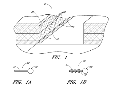
[0013] Figure 1 is a schematic illustration of an embodiment of a wound
interface
visualization apparatus.
[0014] Figures 1A and 1B illustrate embodiments of visualization
elements.
-8-
CA 02874392 2014-11-21
WO 2013/175310 PCT/1B2013/001562
[0015] Figure 2 is an image of an experimental setup of a wound having
an
embodiment or arrangement of a wound interface visualization apparatus,
looking vertically
down through the wound wherein no negative pressure has been applied to the
wound.
[0016] Figure 3 is an image of the experimental setup of the wound
having the
embodiment or arrangement of a wound interface visualization element shown in
Figure 2,
looking vertically down through the wound wherein negative pressure has been
applied to
the wound at a level of -120 mmHg.
[0017] Figure 4 is an image of an experimental setup of a wound having
the
embodiment or arrangement of the wound interface visualization element shown
in Figure 2,
looking vertically down through the wound wherein negative pressure has been
applied to
the wound at a level of -200 mmHg.
[0018] Figure 5 is an image of an experimental setup of a wound having
another
embodiment or arrangement of a wound interface visualization element, looking
vertically
down through the wound wherein no negative pressure has been applied to the
wound.
[0019] Figure 6 is an image of an experimental setup of a wound having
the
embodiment or arrangement of a wound interface visualization element shown in
Figure 5,
looking vertically down through the wound wherein negative pressure has been
applied to
the wound at a level of -120 mmHg.
[0020] Figure 7 is an image of an experimental setup of a wound having
the
embodiment or arrangement of a wound interface visualization element shown in
Figure 5,
looking vertically down through the wound wherein negative pressure has been
applied to
the wound at a level of -200 mmHg.
[0021] Figure 8 is an image of an experimental setup of a wound having
the
embodiment or arrangement of a wound interface visualization element shown in
Figure 5,
looking vertically down through the wound wherein negative pressure has been
applied to
the wound at a level of -200 mmHg, then released to -120 mmHg and held at -120
mmHg for
the image.
[0022] Figure 9 is an image of an experimental setup of a wound having
another
embodiment or arrangement of a wound interface visualization element, looking
vertically
down through the wound wherein negative pressure has been applied to the wound
at a level
of -120 mmHg.
-9-
CA 02874392 2014-11-21
WO 2013/175310 PCT/1B2013/001562
[0023] Figure 10 is an image of an experimental setup of a wound having
the
embodiment or arrangement of a wound interface visualization element shown in
Figure 9,
looking vertically down through the wound wherein negative pressure has been
applied to
the wound at a level of -200 mmHg.
[0024] Figure 11 is a split image showing, on the left hand side, an
experimental
setup of a wound having an embodiment or arrangement of a wound interface
visualization
element without application of negative pressure and looking vertically down
through the
wound, and on the right hand side, the same experimental setup of a wound
shown in the left
side, having the embodiment or arrangement of the wound interface
visualization element
shown in the left hand side of Figure 11, having a vacuum applied to the wound
at a level of -
200 mmHg, and looking vertically down through the wound.
[0025] Figure 12 is a schematic representation of an embodiment of an
apparatus
used to provide negative pressure wound therapy to a wound.
[0026] Figure 13 is a schematic representation of another embodiment of
an
apparatus used to provide negative pressure wound therapy to a wound, showing
the wound
in first state of contraction.
[0027] Figure 14 is a schematic representation of another embodiment of
an
apparatus used to provide negative pressure wound therapy to a wound, showing
the wound
in second state of contraction.
[0028] Figures 15A-15F illustrate further embodiments of wound fillers.
DETAILED DESCRIPTION OF THE PREFERRED EMBODIMENTS
[0029] Some of the embodiments disclosed herein relate to apparatuses
and
methods of treating a wound with reduced pressure, including pump and wound
dressing
components and apparatuses. Generally, the embodiments including the wound
fillers
described herein may be used in combination with a negative pressure system
comprising a
drape or wound cover placed over the filler. A vacuum source, such as a pump,
may be
connected to the cover, for example, through one or more tubes connected to an
aperture or
port made in or under the cover. The apparatuses and components comprising the
wound
overlay and packing materials, if any, are sometimes collectively referred to
herein as
-10-
CA 02874392 2014-11-21
WO 2013/175310 PCT/1B2013/001562
dressings. Further details of methods and apparatuses, such as dressing
components and
inflatable bladders, that are usable with the embodiments described herein are
found in the
following applications, which are hereby incorporated by reference in their
entireties: U.S.
Patent No. 8,235,955, titled "Wound treatment apparatus and method," issued
August 7,
2012; U.S. Patent No. 7,753,894, titled "Wound cleansing apparatus with
stress," issued July
13, 2010; Application Serial No. 12/886,088, titled "Systems And Methods For
Using
Negative Pressure Wound Therapy To Manage Open Abdominal Wounds," filed
September
20, 2010, published as US 2011/0213287; Application Serial No. 13/092,042,
titled "Wound
Dressing And Method Of Use," filed April 21, 2011, published as US
2011/0282309; and
Application Serial No. 13/365,615, titled "Negative Pressure Wound Closure
Device," filed
February 3, 2012, published as US 2012/0209227.
[0030] It will be appreciated that throughout this specification
reference is made
to a wound or wounds. It is to be understood that the term wound is to be
broadly construed
and encompasses open and closed wounds in which skin is torn, cut or
punctured, or where
trauma causes a contusion, or any other superficial or other conditions or
imperfections on
the skin of a patient or otherwise that benefit from reduced pressure
treatment. A wound is
thus broadly defined as any damaged region of tissue where fluid may or may
not be
produced. Examples of such wounds include, but are not limited to, acute
wounds, chronic
wounds, surgical incisions and other incisions, subacute and dehisced wounds,
traumatic
wounds, flaps and skin grafts, lacerations, abrasions, contusions, burns,
diabetic ulcers,
pressure ulcers, stoma, surgical wounds, trauma and venous ulcers or the like.
In some
embodiments, the components of the negative pressure treatment system
described herein
can be particularly suited for incisional wounds that exude a small amount of
wound exudate.
Thus, while some embodiments and methods disclosed herein are described in the
context of
treating abdominal wounds, the apparatuses and methods disclosed herein are
applicable to
any wound in a body.
[0031] As is used herein, reduced or negative pressure levels, such as
¨X mmHg,
represent pressure levels that are below standard atmospheric pressure, which
corresponds to
760 mmHg (or 1 atm, 29.93 inHg, 101.325 kPa, 14.696 psi, etc.). Accordingly, a
negative
pressure value of ¨X mmHg reflects absolute pressure that is X mmHg below 760
mmHg or,
in other words, an absolute pressure of (760¨X) mmHg. In addition, negative
pressure that is
-11-
CA 02874392 2014-11-21
WO 2013/175310 PCT/1B2013/001562
"less" or "smaller" than X mmHg corresponds to pressure that is closer to
atmospheric
pressure (e.g., ¨40 mmHg is less than ¨60 mmHg). Negative pressure that is
"more" or
"greater" than ¨X mmHg corresponds to pressure that is further from
atmospheric pressure
(e.g., ¨80 mmHg is more than ¨60 mmHg).
[0032] The negative pressure range for some embodiments of the present
disclosure can be approximately -80 mmHg, or between about -20 mmHg and -200
mmHg.
Note that these pressures are relative to normal ambient atmospheric pressure.
Thus, -200
mmHg would be about 560 mmHg in practical terms. In some embodiments, the
pressure
range can be between about -40 mmHg and -150 mmHg. Alternatively a pressure
range of
up to -75 mmHg, up to -80 mmHg or over -80 mmHg can be used. Also in other
embodiments a pressure range of below -75 mmHg can be used. Alternatively, a
pressure
range of over approximately -100 mmHg, or even 150 mmHg, can be supplied by
the
negative pressure apparatus. Unless stated otherwise, the term approximately
is meant to
represent a range of +/- 10% of the stated value.
[0033] Figure 1 is a schematic illustration of an embodiment of a wound
interface
visualization element. As illustrated in Figure 1, in some embodiments and
methods for the
visualization of a surface of a wound interface, one or more first
visualization elements 10
and/or second visualization elements 20 can be positioned along a surface of a
wound W.
The visualization elements can be positioned in fascia, peritoneum, fat,
muscle, and/or any
other tissue in the body.
[0034] In some embodiments, the first visualization elements 10 and/or
second
visualization elements 20 can comprise any suitable and biocompatible
radiopaque material
or a radiopaque marker for visualization during any suitable procedure,
including, for
example and without limitation, fluoroscopy, computerized tomography (CT)
scan, x-ray,
magnetic resonance imaging (MRI), or any other suitable visualization
procedures or
techniques applied during or after wound treatment, including without
limitation negative
pressure wound treatment. Any number or variety of radiopaque markers or
visualization
elements can be used.
[0035] The visualization element(s) can be sutures, a powder or solid
material
(such as a barium sulfate powder), or an adhesive comprising a radiopaque or
contrasting
material or element that can be applied at or adjacent to a surface of a
wound. For example,
-12-
CA 02874392 2014-11-21
WO 2013/175310 PCT/1B2013/001562
in some arrangements, the visualization element(s) can be applied at or
adjacent to an
interface or wound surface of a fascia layer of tissue, the peritoneum, and/or
any other
suitable tissue layer.
[0036] As illustrated in Figure 1, the visualization elements 10 can be
radiopaque
sutures (such as, without limitation, gold wire) applied in running or
loopstitch along the
length of a particular tissue layer in a wound interface. In the embodiment
illustrated in
Figure 1, the tissue layer through which the visualization elements 10 are
passed can be a
fascia layer 12. However, any of the visualization element embodiments
disclosed herein
can be positioned in any desired tissue or tissue layer during surgery.
[0037] The sutures can comprise a radiopaque material. Additionally,
though not
required, the sutures can comprise a bioabsorbable material so that the
sutures need not be
removed from the wound after the target layer of tissue has progressed to a
threshold or
sufficient level of closure.
[0038] Additionally, in some embodiments, the visualization element can
be an
adhesive material, such as cyanoacrylate, or a powder comprising a radiopaque
material such
as barium sulfate, zirconium, gold, titanium, iodine, isohexol, iodixanol and
tungsten oxide
(any one or combination of which can be used with any other material or
substance disclosed
herein), can be applied to the surface of the subject layer of tissue to
provide the contrast
desired for visualization under fluoroscopy, computerized tomography (CT)
scan, x-ray,
magnetic resonance imaging (MRI), or during any other suitable visualization
procedure or
technique described herein or otherwise. The chosen materials for the
visualization element
should be biocompatible and also compatible with the chosen medical and
visualization
procedures and equipment.
[0039] Further, with reference to Figure 1, any disclosed embodiments
of the
visualization elements, such as without limitation visualization elements 20,
can be
positioned in a fatty layer 22. In any embodiments, as illustrated in Figures
lA and 1B, the
visualization elements can have a body portion 30 and a shaft portion 32. In
some
embodiments, the body portion can comprise one or more radiopaque studs,
balls, or other
radiopaque objects 30, which can be positioned on an end portion of a shaft
portion 32.
Additionally, in any embodiments, the visualization elements 20 can have one
or more barbs
-13-
CA 02874392 2014-11-21
WO 2013/175310 PCT/1B2013/001562
or protrusions 34 extending in a transverse direction away from the shaft
portion 32 for more
secure engagement with the target tissue.
[0040] In the embodiment illustrated in Figure 1, the tissue layer
through which
the visualization elements 20 are passed can be a fatty layer 22. However, any
of the
visualization element embodiments disclosed herein can be positioned in any
desired tissue
or tissue layer during surgery.
[0041] Any of the embodiments or details of the visualization elements
described
herein can be used with, or adapted for use with, negative pressure wound
therapy dressings
or components, surgical dressings or components used for closing wounds, or
any other
dressings or dressing components.
[0042] A number of experiments were conducted, using as the
visualization
element a gold wire used for jewellery making. The gold wire was very
flexible, making it
easy to position and pull though the tissue thus minimising trauma to the
tissue. A further
advantage of the being flexible is the fact that it does not impact on or
hinder the contraction
of the wound. Thus, in any embodiments disclosed herein, the visualization
element can be
flexible.
[0043] Other materials that may be suitable are titanium, tantalum,
stainless
steels, and corrosive resistant alloys, for example and without limitation
Inconel, monel,
hastelloy. Other materials that can be used include polymers filled with
powdered
radiopaque materials e.g. Barium Sulphate, titanium, zirconium oxide, iodine,
iohexol,
iodixanol. Additionally, the following non-resorbable polymers that can be
used in
conjunction with a contrast agent include, without limitation, nylon and
polypropylene.
Further improvements of some embodiments disclosed herein may be made by
making the
visualization elements from bioresorbable polymers. Suitable bioresorbable
materials
include polyglycolic acid, polylactic acid and caprolactan. Other suitable
radiopaque
materials may be so called radiopaque dyes e.g. low-osmolality contrast agents
or less
preferably high osmolality contrast agents. Use of bioresorbable polymers
allows the
visualization elements to be left within the tissue such that they will slowly
dissolve over
time without trace or substantially no trace.
[0044] Monofilaments of the above polymers may be produced by extrusion
of
the polymer premixed with a desired contrast agent. This will ensure the
contrasting agent is
-14-
CA 02874392 2014-11-21
WO 2013/175310 PCT/1B2013/001562
spread uniformly throughout the visualization element and remain in contact
with the
substrate or other materials of the visualization element. Alternatively a
master batch of the
polymer containing the contrast agent may be made and then extruded or molded
into the
desired visualization element.
[00451 In further improvements, it may be desirable to utilize two or
more
visualization elements that include contrast agents of different contrast
levels to show the
movement of different layers of tissue in the body or different areas of the
same tissue in the
body. Closure of the thseial layer is one of the primary objectives of open
abdominal
treatments. So, monitoring the closure of the fascial layer is very important
to ensuring a
successful abdominal wound treatment. For example and without limitation, a
metal may be
used to give a contrast close to black whereas an inorganic salt (e.g., barium
sulphate) may
be used to give a contrast closer to white. Additionally, for example and
without limitation,
two or more visualization elements that define a different shape or which are
similar in all
regards but which are stitched in a different pattern can be used to show the
movement of
different layers of tissue in the body or different areas of the same tissue
in the body. In this
way the movement of different layers of tissue or different areas of the same
tissue can be
visualized and easily distinguished by a user in a 2-dimensional or other
image.
[00461 Alternatively the contrasting agent maybe printed, sprayed,
sputtered or
coated onto the visualization element, In this way it may be possible to apply
the contrasting
agents at different spacings along the length or patterns on the visualization
element thus
allowing for differentiation of 2 or more different visualization elements in
the body.
[0047] In the examples, the wire was looped through the fascia tissue
in a running
stitch format right around the wound such that the majority of the wire was
not imbedded in
the tissue, but ran along the surface of the tissue. This was achieved by
threading the wire
onto a standard curved surgical suture needle and passing through the tissue
to create a
running stitch.
[0048] The fluoroscope was positioned vertically over the centre of the
wound.
The contraction of the wound was watched in real time on the screen of the
fluoroscope and
still images were captured once movement had stopped. Using the fluoroscope in
real time
video mode, it is possible to see the tissue move and therefore visually
determine the degree
of closure being achieved in the deep tissue
-15-
CA 02874392 2014-11-21
WO 2013/175310 PCT/1B2013/001562
[0049] Other embodiments may include staples comprising a radiopaque
material
but minimising the number of parts used, which reduces the risk of any devices
being left in
the body after closure, unless the parts are bioresorbable. An alternative
embodiment may
include sprinkling radiopaque powder on the wound edge. In a further
embodiment a
radiopaque liquid or gel maybe painted or sprayed or spread on to the wound
edge. Suitable
liquids/gels may preferably be glues, advantageously their adherence to tissue
prevents their
movement after application. E.g. cyanoacrylate or fibrin glues filled with
radiopaque powder.
Such glues may also be used to help attach the main tissue closure device to
the tissue.
[0050] In some embodiments, there can be a separate device attached to
the tissue
rather than the main closure device in case the main closure devices movement
does not
follow the movement of the tissue. Making the main closure device radiopaque
also would
help show differential movement.
[0051] Additionally, a surgeon or medical practitioner can place one or
more
visualization elements in a patient's dermis to detect a position of the
dermis during the
course of treatment. The one or more visualization elements in a patient's
dermis can have a
different level of contrast or a different shape as compared to the
visualization elements that
can be positioned in other layers of tissue. Or, the visualization elements
can be positioned
in the various tissue layers following a different pattern. For example, a
radiopaque wire
sutured through a first layer or fascial layer can have a first shape, a first
pattern, and/or a
first contrast level, and a radiopaque wire sutured through second layer or
dermal layer can
have a second shape, a second pattern, and/or a second contrast level. A first
stitch pattern
can have a wave-like stitch pattern having a first frequency and amplitude. A
second stitch
pattern can have a wave-like stitch pattern having a second frequency and
amplitude so as to
be discernible from the first stitch pattern.
[0052] A series of images were taken during experiments conducted
having
different embodiments of visualization element or arrangements, as shown in
Figures 2-11.
[0053] Figure 2 is an image of an experimental setup of a wound having
an
embodiment or arrangement of a wound interface visualization element, looking
vertically
down through the wound wherein no negative pressure has been applied to the
wound. The
wound shown in Figure 2 is packed with foam and covered with a drape. The
visualization
element 50 is a gold wire sewn through the fascia in a wave form pattern or
path.
-16-
CA 02874392 2014-11-21
WO 2013/175310 PCT/1B2013/001562
[0054] Figure 3 is an image of the experimental setup of the wound
having the
embodiment or arrangement of a wound interface visualization element shown in
Figure 2,
looking vertically down through the wound wherein negative pressure has been
applied to
the wound at a level of -120 mmHg. In this experimental setup, as mentioned
above, the
wound is packed with foam and covered with a drape. Again, the visualization
element 50 is
a gold wire sewn through the fascia in a wave form pattern or path. In Figure
3, a negative
pressure of -120 mmHg. has been applied to the wound. The visualization
element 50
provides a clear image and identification of the location of the interface of
the facia layer to a
surgeon or medical practitioner. Figure 4 has the same experimental setup and
visualization
element embodiment as shown in Figure 3, with a negative pressure level of -
200 mmHg
applied to the wound.
[0055] Figures 5-7 are images of an experimental setup of a wound
having
another embodiment or arrangement of a wound interface visualization element
60, which
again is a gold wire, sewn through the fascia in a wave form. In Figure 5, the
wound is
subject only to atmospheric pressure. In Figure 6, the level of negative
pressure applied to
the wound is -120 mmHg. In Figure 7, the level of negative pressure applied to
the wound is
-200 mmHg. Figure 8 is an image of the experimental setup of the wound having
the
embodiment or arrangement of the wound interface visualization element 60
shown in Figure
5, looking vertically down through the wound wherein negative pressure has
been applied to
the wound at a level of -200 mmHg, then released to -120 mmHg and held at -120
mmHg for
the image.
[0056] Figure 9 is an image of an experimental setup of a wound having
another
embodiment or arrangement of a wound interface visualization element 70,
looking vertically
down through the wound wherein negative pressure has been applied to the wound
at a level
of -120 mmHg. In this experimental setup, the wound has been packed with wound
packing
and foam inserts in cells and covered with drape. The visualization element
70, which was a
gold wire in this experimental setup, is sewn through the fascia in a wave
form. Figure 10 is
an image of the same experimental setup as shown in Figure 9, showing the
wound with
negative pressure level of -200 mmHg applied to the wound.
[0057] Figure 11 is a split image showing, on the left hand side, an
experimental
setup of a wound having an embodiment or arrangement of a wound interface
visualization
-17-
CA 02874392 2014-11-21
WO 2013/175310 PCT/1B2013/001562
element 80 without application of negative pressure and looking vertically
down through the
wound, and on the right hand side, the same experimental setup of a wound
shown in the left
side, having the embodiment or arrangement of the wound interface
visualization element 80
shown in the left hand side of Figure 11, having a vacuum applied to the wound
at a level of -
200 mmHg, and looking vertically down through the wound. Split image comparing
the
wound packing closure device at rest and after application of 200 mmHg of
reduced pressure.
[0058] Compartment syndrome can occur when excessive pressure builds up
inside an enclosed space in the body. Excessive pressures in the abdominal
compartment, for
example, can impede the flow of blood to and from the affected tissues, bodily
organs, or
even the lower extremities if excessive pressure is exerted on the abdominal
aorta. The
pressure buildup within the abdominal compartment can be the result of
excessive fluid
buildup in the abdominal compartment, in addition to or alternatively as a
result of the forces
exerted on the abdominal region from the application of negative pressure
wound therapy to
the abdominal compartment.
[0059] Such excessive pressure can cause permanent injury or damage to
the
tissues, organs (such as the liver, bowels, kidneys, and other organs), and
other body parts
affected by the reduction of blood flow. Therefore, preventing the buildup of
excessive
pressures in the abdominal compartment is beneficial for the treatment of
abdominal injuries.
[0060] Internal abdominal pressure may also be measured and/or
monitored
indirectly using intragastric, intracolonic, intravesical (bladder), inferior
vena cava catheters,
or by other suitable methods, such as via the uterus. In some arrangements,
for example, the
internal pressure may be measured by inserting a catheter into the patient's
bladder. Aortic
blood pressure can also be monitored using techniques known in the field. For
limb-based
compartment syndrome, the internal pressure can be measured by a needle
inserted into the
affected limb, and preferably, the pressure measured there should be within 20-
30mmHg of
the patient's diastolic blood pressure. The clinician can also monitor for a
pulse distal of the
affected extremity.
[0061] In addition to any of the foregoing methods or devices for
measuring
internal pressure, or any combination of such, in some embodiments, negative
pressure
wound therapy can be applied to the wound of a patient in a manner to minimize
or prevent
the build-up of excessive pressure that causes compartment syndrome. For
example, any of
-18-
CA 02874392 2014-11-21
WO 2013/175310 PCT/1B2013/001562
the negative pressure wound therapy dressing components disclosed herein can
be
configured to support or contain one or more pressure sensors configured to
permit a
clinician to monitor the internal pressure within the compartment, wound
cavity, or
abdominal cavity. In some embodiments, the negative pressure dressing
components may
include a wound filler that may have an adjustable volume, such as an
inflatable bladder or
other wound fillers as described below, which when placed within a wound can
control how
much the wound can close. In one example, one or more pressure sensors can be
added to
the dressing components, including without limitation positioning one or more
pressure
sensors on the surface of and/or inside any inflatable bladder embodiment
disclosed herein
(such as described with respect to Figure 12) that can be positioned in the
abdominal cavity.
The pressure sensors can be supported on, embedded within, or be integral with
an outer
and/or inner surface of any inflatable bladder embodiments disclosed herein,
and can be used
to monitor the pressure exerted on the inflatable bladder from the adjacent
tissues and organs
within the abdominal cavity to alert the patient or caregiver when a threshold
or potentially
harmful pressure is present within the abdominal cavity.
[0062] Additionally or alternatively, one or more pressure sensors can
be
positioned on or supported by a portion of any wound packing or wound filler
components
positioned within or adjacent to the wound cavity, or embedded within a
portion of the
wound filler and/or the dressing overlay or cover, including being supported
by the overlay
itself, and/or any conduit components of the dressing. The pressure sensors
can therefore be
positioned on, supported by, or embedded within any combination of the
dressing
components disclosed herein.
[0063] Furthermore, in addition or alternatively to any of the sensor
positions
located herein, one or more pressure sensors can also be positioned adjacent
to one or more
of the organs in the cavity being treated, for example the bladder, one or
more kidneys,
and/or any other organs or proximally located tissue surfaces.
[0064] Some embodiments can have one or more pressure sensors supported
by
or on or embedded within the wound packing layer or wound filler, one or more
pressure
sensors supported by or on or embedded within one or more of the organs (such
as the
bladder) or tissue layers in the cavity, and one or more pressure sensors
supported by or on or
embedded within one or more inflatable bladders positioned within the wound
cavity.
-19-
CA 02874392 2014-11-21
WO 2013/175310 PCT/1B2013/001562
[0065] Monitoring the pressure in one, some or all of these three
locations can
permit the caregiver to optimize or control the level of negative pressure
applied to the
wound cavity, optimize or control a level of inflation or pressure of an
inflatable bladder
placed within the wound, optimize or control the collapse, stiffness or volume
of a wound
filler placed within the wound, and/or monitor a level of pressure exerted on
one or more
organs, tissue layers, blood vessels, or other body parts affected by the
closure pressures. A
caregiver can then adjust a level of pressure in the inflatable bladder by
either adding fluid to
the bladder or releasing fluid from within the bladder to a receptacle or
container positioned
outside the body, adjust the collapse, stiffness or volume of the wound
filler, adjust a level of
negative pressure exerted on the wound cavity, and/or adjust any other closure
forces applied
to the wound to either increase or decrease the closure forces. In some
embodiments, these
adjustments can be made dynamically or automatically by a computer controller
that receives
one or more pressure readings or other data indicative of excessive pressure,
and that sends a
control signal to a pump or other device to make the adjustments.
[0066] A clinician may monitor the internal pressure as vacuum is
slowly
increased to the wound dressing, or as air is slowly released from the
inflatable member. In
one embodiment, human bladder pressure is controlled below approximately 40
mmHg, or
below approximately 30 mmHg, approximately 20 mmHg, or approximately 15 mmHg.
In
some embodiments, the measurement of internal pressure and control of the
vacuum and air
release can be controlled automatically. This way as the oedema decreases the
wound can be
slowly closed further over, for example, a period of hours to days (e.g.,
closure by seven
days). It will be appreciated that systems can be employed where the vacuum
can be slowly
applied with pressure feedback being provided based on vital signs of the
patient or other
monitoring described herein or in htip://www.uptodate.comicontentslabdominal-
compartment-syndrome.
[0067] Figure 12 is a schematic representation of an apparatus 120 used
to
provide negative pressure wound therapy to a wound and to control the level of
therapy
and/or closure of the wound based on pressure sensors positioned within the
wound cavity to
minimize the risk of compartment syndrome. For example and without limitation,
in some
embodiments, the apparatus 120 can have a backing layer 122 for providing a
substantially
air and liquid-tight seal over a wound. Under the overlay, the apparatus 120
can have a
-20-
CA 02874392 2014-11-21
WO 2013/175310 PCT/1B2013/001562
wound packing member or wound filler 124 that can have an adjustable volume
and/or
internal pressure. For example, some embodiments of the wound packing member
124 can
have a sealed member 126 (such as a sealed bag) that can be controllably
inflatable and
deflatable from a pressure source such as a pump via a conduit 128 in
communication with a
sealed space within the sealed member 126. The sealed member 126 can be
positioned in the
wound in contact with the wound tissue interface. For example, in any
embodiments used
for abdominal wounds, the sealed member 126 can be configured and can be
positioned in
the wound cavity so as to engage all tissue layers above the organs in the
body. For
example, in some embodiments, the sealed member 126 can be positioned in the
wound so as
to contact any or all of the layers that can be present in an abdominal wound,
such as (from
deepest to most superficial) the peritoneum, extraperitoneal fascia (deep
fascia), muscle,
superficial fascia, subcutaneous tissue, and skin. However, the presence or
absence of
various layers is location dependent, so not all of these layers may be
present in every
abdominal wound treatable with the apparatuses of the present disclosure.
[0068] In some embodiments, an organ protection layer 127, such as any
embodiments of the wound contact layer disclosed in U.S. Application
Publication No.
2011/0213287, Serial No. 12/886,088, titled SYSTEMS AND METHODS FOR USING
NEGATIVE PRESSURE WOUND THERAPY TO MANAGE OPEN ABDOMINAL
WOUNDS, filed on 09/20/2010, which application is hereby incorporated by
reference
herein as if fully set forth herein, can be positioned between the sealed
member 126 and the
viscera or other organs. Embodiments of the apparatus 120 disclosed herein can
comprise
any of the other components, materials, features, or details of any of the
embodiments or
components of the negative pressure systems disclosed in U.S. Application No.
12/886,088.
As mentioned, all embodiments or components of the negative pressure systems
disclosed in
U.S. Application No. 12/886,088 are hereby incorporated by reference as if
fully set forth
herein.
[0069] A pressure sensor 130 (also referred to herein as a first
pressure sensor)
can be used to monitor a pressure level within the sealed member 126. The
pressure sensor
130 can provide a visual reading of the level of pressure within the sealed
member 126,
and/or can provide a signal to a controller 132 based on the level of pressure
within the
sealed member.
-21-
CA 02874392 2014-11-21
WO 2013/175310 PCT/1B2013/001562
[0070] The level of pressure within the sealed member 126, as
mentioned, can be
controlled in part by the pump 127 (also referred to herein as the first pump)
and can be
adjusted to be a positive or a negative pressure. Additionally, in some
embodiments, the
pump 127 can be configured to cycle the pressure level between any desired
positive or
negative pressure levels or to apply intermittent pressure to the sealed
member 126. Positive
pressures within some embodiments of the sealed member 126 or any sealed
member
embodiment disclosed herein can range from 0 mmHg to 60 mmHg or more. Negative
pressures within some embodiments of the sealed member 126 or any sealed
member
embodiment disclosed herein can range from 0 mmHg to -180 mmHg or more.
[0071] In any embodiments disclosed herein, the pressure level within
the sealed
member 126 can be controlled independently of the pressure in a space 134
beneath the
backing layer 122. The pressure beneath the backing layer 122 can be detected
by a pressure
sensor (such as pressure sensor 138, which is also referred to herein as a
second pressure
sensor) in communication with the space 134 beneath the backing layer 122. The
second
pressure sensor 138 can be configured to provide a signal to the controller
132. In any
embodiments disclosed herein, a second pump, such as pump 136, can be used to
provide a
source of negative pressure to a space 134 beneath the backing layer 122.
Alternatively, the
apparatus can be configured to have only one pump (not illustrated) having
multiple conduits
and multiple valves to independently control a level of pressure within the
sealed member
126 and the space 134 beneath the backing layer 122.
[0072] In some embodiments, the level of pressure within the sealed
member 126
can be adjusted independent of the level of reduced pressure in the space 134
to increase or
decrease a volume of the sealed member 126, which can be beneficial in terms
of controlling
a level of pressure exerted on one or more organs in the abdominal area and,
hence, can be
beneficial in terms of controlling or minimizing a risk of compartment
syndrome. A pressure
sensor 140 (which is also referred to herein as a third pressure sensor) can
be placed in
communication with a human organ, for example the human bladder to monitor
pressure
within the human bladder. The third pressure sensor 140 can also be configured
to provide a
signal to the controller based on the pressure reading detected by the third
pressure sensor
140.
-22-
CA 02874392 2014-11-21
WO 2013/175310 PCT/1B2013/001562
[0073] If a pressure detected in one or more organs, such as the human
bladder,
as detected by a pressure sensor 140, exceeds a threshold value, the
controller 132 can adjust
one or more pressure levels to reduce the pressure exerted on the organ or
organs. In some
embodiments, the threshold value of pressure measurements for organs in the
abdominal
region can be 10 mmHg (or approximately 10 mmHg), or 12 mmHg (or about
approximately
12 mmHg), or 15 mmHg (or about 15 mmHg) but such values may be organ specific
and/or
patient specific. Additionally, in some applications, wherein any of the
dressings disclosed
herein are used to treat a wound on the thigh, for example, compartment
pressures can reach
as high as 120 mmHg, such that the threshold value of compartment pressure in
that region
may be much higher than for abdominal wounds, such as approximately 60 mmHg or
less to
approximately 80 mmHg, or approximately 100 mmHg. In the leg, generally, the
threshold
value of pressure which can trigger such pressure and dressing adjustments can
be
approximately 40 mmHg, or from approximately 40 mmHg to approximately 60 mmHg.
Some embodiments of the apparatus can configured such that a medical
practitioner can set
the level of the threshold value, since a different value may be applicable to
each patient.
For younger patients or children, or patients that are at a higher risk for
developing
compartment syndrome, for example, a lower threshold value can be set. In some
embodiments, the threshold value can be set at from approximately 8 mmHg to
approximately 12 mmHg.
[0074] For example, in abdominal negative pressure wound therapy kits,
to
reduce the pressure buildup, the apparatus can be configured to decrease the
level of closure
forces applied to the wound. This can be achieved in some embodiments by
increasing a
level of pressure in the sealed member 126, thereby limiting the amount of
closure in the
walls of the wound interface even when an elevated level of reduced pressure
applied to the
space 134 in the wound is maintained to ensure an appropriate level of fluid
removal. This
can be done until the level of pressure in one or more of the organs, such as
the bladder, or
blood flow rate measurements, reach a safe or below-threshold value once
again. In some
embodiments, the pressure level within the sealed member 126 can be a positive
value (i.e.,
above atmospheric) to exert a spreading force on the tissue interface, while
the pressure level
within the space 134 but outside of the sealed member 126 is at a negative
pressure level.
This arrangement wherein the sealed member 126 can independently control the
level of
-23-
CA 02874392 2014-11-21
WO 2013/175310 PCT/1B2013/001562
closure of the wound interface, can also permit a medical practitioner to
exceed the normal
negative pressure levels in the space 134 beyond the typical therapeutic
ranges that might
otherwise have been limited by excessive interabdominal pressure levels.
[0075] In some embodiments or arrangements, a sealed member 126 can be
sized
and configured to contact the peritoneum, extraperitoneal fascia (deep
fascia), muscle,
superficial fascia, subcutaneous tissue, and skin when placed in the abdominal
wound. When
the level of closure of the wound interface is desired to be limited, such as
when excessive
levels of pressure are present in or adjacent to the wound area, a level of
pressure within the
sealed member 126 can be increased to limit the contraction in one or more of
the
peritoneum, extraperitoneal fascia (deep fascia), muscle, superficial fascia,
subcutaneous
tissue, and skin, thereby increasing the volume of space that the viscera can
occupy and
reducing the level of pressure exerted on the various organs and blood
vessels. Again,
because the level of pressure within the sealed member 126 can be adjusted
independently of
the level of pressure within the space 134 beneath the backing layer 122 but
outside of the
sealed member 126, a therapeutic level of reduced pressure can be applied to
the wound to
remove excessive liquid exuded in the abdominal compartment and improve the
healing
conditions.
[0076] In any of embodiments disclosed herein, the apparatus can gather
pressure
readings from one or more pressure sensors positioned throughout the body to
monitor
compartment pressures. For interabdominal compartment pressures, readings can
be
gathered in the abdominal region or adjacent thereto. For example, any
apparatus disclosed
herein can have one or more blood flow meters (such as a laser Doppler blood
flow meter)
configured to measure a flow rate of blood through target blood vessels,
arteries, capillaries,
and/or muscles. Any embodiments of the laser Doppler can be permanently
mounted to the
patient's skin near the wound cavity. In some embodiments, for example, one or
more blood
flow meters can be used to measure a flow rate of blood through the femoral
arteries or
through musculature at or near to the abdominal region and provide a feedback
signal to the
controller 132.
[0077] Additionally, in some embodiments, pressure levels in, for
example, the
abdominal compartment can be measured using the vesicular technique, which can
involve
the use of an indwelling urinary catheter, a pressure transducer, and a
syringe or similar
-24-
CA 02874392 2014-11-21
WO 2013/175310 PCT/1B2013/001562
device capable of infusing fluid. Additionally, pressure levels in the
abdominal compartment
can be measured by catheterizing the inferior vena cava through either the
left or right
femoral artery. See F. Lui, A. Sangosanya, and L.J. Kaplan, "Abdominal
Compartment
Syndrome: Clinical Aspects and Monitoring," Critical Care Clinics, vol. 23,
no. 3, pp. 415-
433, 2007 for more information about monitoring techniques for suitable for
monitoring
abdominal compartment syndrome.
[0078] Further, any embodiments of the sealed member 126 disclosed
herein can
be formed from a substantially sealed impermeable membrane 148, that seals
around or to
the conduit 128 that provides the fluid (e.g., air, nitrogen, or argon, or
saline, water, or other
liquids) into and out of the impermeable membrane 148, which can be formed
from any
suitable, biocompatible polymer film, sheet, bag, pouch, chamber, or
otherwise, similar to
any of the inflatable membranes disclosed in U.S. Patent No. 7,753,894, which
is Application
No. 12/886,088, titled WOUND CLEANSING APPARATUS WITH STRESS, filed on
12/17/2007.
[0079] In some embodiments, the sealed member 126 can have a foam layer
150
around some or all of the outside surface of the impermeable membrane 148. In
some
embodiments, the foam layer 150 can surround the entire surface of the
impermeable
membrane 148. The foam 150 can help cushion any pressure points exerted on the
tissue by
the sealed member 126, and can assist with the distribution of negative
pressure across the
wound cavity.
[0080] Additionally, though not required, any embodiments disclosed
herein can
have a structural member 160 positioned inside the impermeable membrane 148.
In some
embodiments, the structural member 160 can be configured to be more rigid in a
vertical
direction (i.e., transverse to the backing layer, as indicated by arrow Al in
Figure 12), than in
a lateral direction (i.e., in the direction of wound closure of the tissue
interfaces, as indicated
by arrow A2 in Figure 12). Examples of structural members that can be used are
found in
Application Serial No. 13/365,615, titled "Negative Pressure Wound Closure
Device," filed
February 3, 2012, published as US 2012/0209227, the entirety of which is
hereby
incorporated by reference.
[0081] In some embodiments, the sealed member 126 can have multiple,
independently controllable (e.g., inflatable or deflatable) chambers. One or
more manifolds
-25-
CA 02874392 2014-11-21
WO 2013/175310 PCT/1B2013/001562
can control the inflation and deflation of the various compartments or
chambers to control
the size and/or shape of the bladder member as desired to suit the particular
wound size and
application.
[0082] Additionally, in any embodiments disclosed herein, the sealed
member
126 can be used with a vertically rigid but laterally collapsible structure
positioned either
inside or outside of the sealed member 126. For example, with reference to
Figure 13,
another embodiment of an apparatus 200 is illustrated. The apparatus 200 can
have any of
the same features, components, or details of any other embodiments disclosed
herein,
including any of the visualization elements and the pressure sensors disclosed
above.
Additionally, as shown in Figure 13, a sealed member 206 can be positioned in
the wound
cavity and have any of the same features, materials, or other details of the
sealed member
126 disclosed herein, including but not limited to the foam layer or interface
208 surrounding
the impermeable layer 210.
[0083] The apparatus 200 can also have a support member 216 positioned
under a
backing layer 218. Some embodiments of the support member 216 can have one or
more
legs (also referred to herein as a body portion) 220 attached to a top portion
226 (also
referred to herein as a first portion) of the support member 216. In some
embodiments, the
top portion 226 of the support member 216 can be along an apex of the support
member 216
and define a longitudinal axis Al of the support structure. The legs 220 can
be rotatably
supported by the top portion 226 so that the legs 220 can rotate about axis Al
defined
through the axial centerline of the top portion 226. The sealed member 206 can
be coupled
with, connected to, adhered to, or otherwise attached the legs 220 such that
contracting or
expanding the sealed member 206 will correspondingly contract or expand the
legs 22 and
support member 216. In some embodiments, the legs 220 can be positioned within
molded
pockets formed in the sealed member 206. In some embodiments, one or more foam
pockets
positioned at the bottom of the legs 220 can be adhered to the sealed member
206.
[0084] In this configuration, as the sealed member 206 is contracted
from a first
volume, such as volume V1 shown in Figure 13, to a second, larger volume, such
as volume
V2 shown in Figure 14, the support member 216 (or any other suitable support
member
having greater vertical than lateral rigidity) can also laterally contract.
Additionally, the
sealed member 206 can be configured to expand from a smaller volume, such as
volume V2
-26-
CA 02874392 2014-11-21
WO 2013/175310 PCT/1B2013/001562
shown in Figure 14, to a larger volume, such as volume V1 shown in Figure 13,
the so as to
urge the support member 216 and the legs 220 thereof, laterally outward
against the walls of
the wound interface, thereby potentially reducing the pressure on the organs
within the
abdominal compartment. As the wound closes during the course of healing, the
legs 220 can
rotate closer together so that the closure of the wound is not inhibited by
the dressing
backing layer 218.
[0085] Further, some embodiments of the wound closure apparatuses, such
as
embodiments 120 and 200, can have one or more tissue engaging elements
supported by the
sealed member or the support member in communication with the sealed member.
The tissue
engaging elements can be configured to engage one or more layers of the wound
interface,
including any one or combination of the peritoneum, extraperitoneal fascia
(deep fascia),
muscle, superficial fascia, subcutaneous tissue, and skin. The tissue engaging
elements 164
(schematically represented in Figure 12) of the embodiment of the apparatus
120 shown in
Figure 12, or the tissue engaging elements 264 of the embodiment of the
apparatus 200 can
comprise any one or combination of tissue connectors, tissue anchors, hook
shaped members,
balls on the ends of rods, and/or any other suitable engaging mechanisms
available for use
with the various layers of tissue. Some embodiments of the sealed member 126
can have any
combination of different tissue engaging elements desired to engage the
various different
tissue layers in the wound site.
[0086] In any embodiments of the sealed member disclosed herein, a
level of the
volume of fluid within the sealed member can be controlled automatically by
the control
system, as discussed. Additionally, in any embodiments, the level of the
volume of fluid
within the sealed member can be changed manually by adding or removing fluid
into the
sealed member through a tube and a hand operated pump system, or through a
syringe and
cannula device inserted into a sealed receptacle such as one or more syringe
ports on the
sealed member, in response to pressure readings acquired by any of the
plurality of pressure
sensors in the apparatus.
[0087] In some embodiments, the sealed member can itself be more rigid
in a
vertical direction than in a lateral direction. For example, any embodiments
of the sealed
member can have corrugations or an undulating surface that causes the sealed
member to be
-27-
CA 02874392 2014-11-21
WO 2013/175310 PCT/1B2013/001562
more flexible in a lateral direction than in a vertical direction. In some
embodiments, the
sealed member can have, for example, an accordion-like shape.
[0088] It will be appreciated that in some embodiments, it is not
necessary to take
any measurements indicative of excessive pressure within the patient. Rather,
it may simply
be desired to control the closure of a wound by controlling the volume,
stiffness, pressure,
and/or collapse of any of the wound fillers described above. Such closure can
be controlled
based on visual inspection, use of the wound visualization methods and
apparatus described
above, or can be controlled based on a desired predetermined schedule. The
control over
such closure can be performed manually by a health practitioner, or may be
performed
automatically or based on inputs by a controller as described above. For
example, where an
inflatable bladder is placed in the wound, the pressure in the bladder may be
manually or
automatically controlled to limit and/or allow a certain amount of wound
closure for a given
period of time. This concept may similarly be applied to wound fillers such as
described in
Figure 13 by including a mechanism (such as the adjustable bladder between the
legs) where
the angle between the legs can be controlled over time.
[0089] Other embodiments of wound fillers whose volume, stiffness,
pressure
and/or collapse may be controlled, can be used with any of the components of
any of the
embodiments disclosed herein. Examples of such additional wound fillers that
can be used
with any of the components of any of the embodiments disclosed herein are
found in
Application Serial No. 13/365,615, titled "Negative Pressure Wound Closure
Device," filed
February 3, 2012, published as US 2012/0209227, incorporated by reference
herein, the
entirety of which is hereby incorporated by reference. Figures 15A-F
illustrate further
embodiments of suitable wound fillers, and examples of these and other
suitable wound
fillers may be found in U.S. Patent No. 7,754,937, titled "WOUND PACKING
MATERIAL
FOR USE WITH SUCTION," the entirety of which is hereby incorporated by
reference.
Other embodiments of negative pressure therapy apparatuses, dressings, wound
fillers and
methods of using the same that may also be utilized alone or in combination
with the
embodiments described herein, and further description of the embodiments found
above, for
example with respect to Figures 13 and 14 above, are found in U.S. Provisional
Application
No. 61/681,037, filed August 8, 2012, entitled WOUND CLOSURE DEVICE, and PCT
Application No. PCT/US13/42064 filed May 21, 2013, entitled WOUND CLOSURE
-28-
CA 02874392 2014-11-21
WO 2013/175310 PCT/1B2013/001562
DEVICE, the entireties of which are hereby incorporated by reference. It will
be appreciated
that any of these embodiments of wound fillers may also be used in combination
with or
instead of the inflatable bladder in the system and method of Figure 12.
[0090] Figure 15A illustrates a wound filler comprising a corrugated
unit 450
comprising two sheets. A first sheet layer 410 is provided, and a second sheet-
like layer 420
having an essentially sinusoidal cross section, for example, is coupled to the
surface of the
first sheet layer at locations 430. The coupling can be achieved by use of an
adhesive or heat
sealing. A two part silicone adhesive has been found suitable to provide
coupling between
sheet 410 and sheet 420. A bead of silicone material 440 may be optionally
added to adjust
the resiliency of the corrugated unit 450. A suitable sheet material is
polyester or polyester
fibers. Although sheet layer 420 is illustrated as having a sinusoidal cross-
section, it is also
contemplated that other cross-sections, such as pleated, may be used.
[0091] Corrugated unit 450 can be used as a wound packing without
further
modification. It can also be used to form more complex three-dimensional
structures. Spiral
wound packing 500 illustrated in Figures. 15B and 15C is formed by rolling
corrugated unit
450 to expose a portion of the first flat sheet 410 along the circumference.
More elaborate
structures can also be formed from a plurality of corrugated units. For
example, individual
corrugated units may be coupled to each other by adhesive or heat sealing
means to form
multi-corrugated wound packing 550, as shown in Figure 15D. One or more beads
of
silicone material 552 can serve dual purposes of coupling the corrugated units
450, and
improving the resiliency of the structure. It should be noted that, while FIG.
15D illustrates
this embodiment with the peaks of each adjacent corrugation unit in alignment,
a staggered
configuration is also contemplated. Multi-corrugated wound packing 550 can
also be sliced
along a cross section at a suitable thickness to produce cut corrugated wound
packing.
Alternatively, wound packing 550 can be sliced at a bias to produce biased-cut
corrugated
wound packing.
[0092] Spiral wound packing 500, cut corrugated wound packing, and
biased-cut
corrugated wound packing have the benefit of being highly compressible and
highly resilient.
Preferably, these wound packing structures are sufficiently compressible to
reduce to less
than 50% of their original volume when subjected to the approximately 2 psi
(pounds per
square inch) compression force commonly encountered with the application of
suction. More
-29-
CA 02874392 2014-11-21
WO 2013/175310 PCT/1B2013/001562
preferably, the wound packing is sufficiently compressible to reduce to less
than 25% of its
original volume. Most preferably, the wound packing is sufficiently
compressible to reduce
to less than 10% of its original volume.
[0093] Figures 15D-15F illustrate another embodiment of a wound filler.
As
shown in Figure 15D, wound packing material 1000 has a generally spiral shape
exhibiting
open areas 1020 along the longitudinal axis of the fiber spiral and open areas
1040 between
adjacent segments of a particular spiral. Wound packing material 1000 is
generally
constructed from polymer fibers 1060, such as spandex.
[0094] To form wound packing material 1000, fibers 1060 are wrapped
around
mandrels, such as a steel tube (not shown). The steel tubes with the spandex
wrap are stacked
in rows and a polyurethane film (not shown) is placed between each row.
Desirably, the
polyurethane film is about 0.003 inch thick. The stack of tubes is then
clamped together and
heated to about 320 degrees F. The polyurethane film melts and adheres to the
spandex
fibers, thus coupling the adjacent spirals to one another. After cooling, the
steel tubes are
removed. Wound packing material 1000, as illustrated in Figures 15D-15F,
remains.
[0095] Features, materials, characteristics, or groups described in
conjunction
with a particular aspect, embodiment, or example are to be understood to be
applicable to
any other aspect, embodiment or example described herein unless incompatible
therewith.
All of the features disclosed in this specification (including any
accompanying claims,
abstract and drawings), and/or all of the steps of any method or process so
disclosed, may be
combined in any combination, except combinations where at least some of such
features
and/or steps are mutually exclusive. The protection is not restricted to the
details of any
foregoing embodiments. The protection extends to any novel one, or any novel
combination,
of the features disclosed in this specification (including any accompanying
claims, abstract
and drawings), or to any novel one, or any novel combination, of the steps of
any method or
process so disclosed.
[0096] While certain embodiments have been described, these embodiments
have
been presented by way of example only, and are not intended to limit the scope
of protection.
Indeed, the novel methods and systems described herein may be embodied in a
variety of
other forms. Furthermore, various omissions, substitutions and changes in the
form of the
methods and systems described herein may be made. Those skilled in the art
will appreciate
-30-
CA 02874392 2014-11-21
WO 2013/175310 PCT/1B2013/001562
that in some embodiments, the actual steps taken in the processes illustrated
and/or disclosed
may differ from those shown in the figures. Depending on the embodiment,
certain of the
steps described above may be removed, others may be added. Furthermore, the
features and
attributes of the specific embodiments disclosed above may be combined in
different ways to
form additional embodiments, all of which fall within the scope of the present
disclosure.
[0097] Although the present disclosure includes certain embodiments,
examples
and applications, it will be understood by those skilled in the art that the
present disclosure
extends beyond the specifically disclosed embodiments to other alternative
embodiments
and/or uses and obvious modifications and equivalents thereof, including
embodiments
which do not provide all of the features and advantages set forth herein.
Accordingly, the
scope of the present disclosure is not intended to be limited by the specific
disclosures of
preferred embodiments herein, and may be defined by claims as presented herein
or as
presented in the future.
-31-