Note: Descriptions are shown in the official language in which they were submitted.
A
CA 02874716 2014-11-25
4
- 1 -
Description
Title of Invention: METHOD FOR ASSESSING ENDOMETRIOSIS
Related Application
[0001]
The present application claims a priority right based
on Japanese Patent Application No. 2012-124595 (filed May
31, 2012), the content of which is incorporated herein by
reference.
Technical Field
[0002]
The present invention relates to a novel endometriosis
diagnostic marker and _diagnosis of endometriosis using the
marker.
Background of the Invention
[0003]
Endometriosis is presently diagnosed by transvaginal
ultrasonography and magnetic resonance imaging and
confirmed by laparoscopic inspection. As a diagnostic
marker for endometriosis, a blood-protein marker such as
CA125 is known.
[0004]
,
CA 02874716 2014-11-25
,
- 2 -
In addition to CA125, serum CA19-9 has been studied on
availability for a diagnostic marker (FERTILITY AND
STERILITY 78 (2002) 733-739: Non Patent Literature 1).
Furthermore, a diagnostic method using CA-125 in
combination with various inflammation markers (all are
protein markers) has been proposed (FERTILITY AND STERILITY
89 (2008) 1073-1081: Non Patent Literature 2).
[0005]
Moreover, miRNA expression analysis performed in the
endometrial membrane of endometriosis patients has been
reported (Mol Endocrinol 25 (2011) 821-832: Non Patent
Literature 3). In the Literature, it is described that
expression of 10 miRNAs increases; whereas expression of 12
miRNAs decreases in the endometrial tissue of patients.
However, the literature is silent about expression of these
miRNAs in the blood.
[0006]
The "miRNA (microRNA)" refers to a single-stranded RNA
molecule consisting of 19 to 23 bases and endogenously
present in a living body. Up to present, 700 types or more
of human miRNAs have been identified and found to act on
mRNA (messenger RNA) of a target gene in a sequence
dependent manner to control gene expression; however, the
detailed mechanism of the action and the physiological role
=
CA 02874716 2014-11-25
= , .
- 3 -
of the human miRNAs have not yet been sufficiently
elucidated.
[0007]
Literatures cited for reference herein are as follows.
The contents of these Literatures are incorporated herein
in their entirety by reference.
Citation List
Non Patent Literature
[0008]
Non Patent Literature 1: FERTILITY AND STERILITY 78
(2002) 733-739
Non Patent Literature 2: FERTILITY AND STERILITY 89
(2008) 1073-1081
Non Patent Literature 3: Mol Endocrinol 25 (2011) 821-
832
Summary of Invention
Technical Problem
[0009]
An object of the present invention is to provide an
endometriosis diagnostic marker in a blood-derived sample,
which is simpler and less invasive and has higher
sensitivity and specificity than the markers used in
conventional methods. Another object of the present
k
CA 02874716 2014-11-25
k ,
- 4 -
invention is to provide endometriosis diagnostic markers in
a blood-derived sample which are easily used in combination,
and provide a diagnostic method using the markers.
Solution to Problem
[0010]
The present inventors checked expression of about 600
types of miRNAs in samples derived from the blood of
endometriosis patients. As a result, they found that
expression of three types of miRNAs: hsa-miR-708, hsa-miR-
127-3p and hsa-miR-518d-3p increased. Based on the finding,
the present inventors conducted further intensive studies
and accomplished the present invention.
[0011]
More specifically, the present invention provides
[1] A diagnostic agent for endometriosis for measuring
the concentration of at least one miRNA selected from the
group consisting of hsa-miR-708, hsa-miR-127-3p and hsa-
miR-518d-3p in a sample derived from the blood of a subject,
the agent comprising an amplification primer for the miRNA
as a main component;
[2] A kit for diagnosing endometriosis comprising the
diagnostic agent according to [1];
[3] The diagnostic agent for endometriosis according
to [1], in which the miRNA is hsa-miR-708;
N
CA 02874716 2014-11-25
- 5 -
[4] The diagnostic agent for endometriosis according
to [1], in which the miRNA is hsa-miR-127-3p;
[5] The diagnostic agent for endometriosis according
to [1], in which the miRNA is hsa-miR-518d-3p;
[6] The diagnostic agent for endometriosis according
to any one of [1] and [3] to [5], wherein the sample
derived from the blood is blood plasma;
[7] A method of detecting endometriosis, comprising a
step of measuring the concentration of at least one miRNA
selected from the group consisting of hsa-miR-708, hsa-miR-
127-3p and hsa-miR-518d-3p in a sample derived from the
blood of a subject;
[8] The method according to [7], comprising a step of
determining whether the concentration of the miRNA is
higher than the concentration of the miRNA in a normal
control;
[9] The method according to [7] or [8], comprising a
step of determining whether the concentration of the miRNA
is a cutoff value or more;
[10] The method according to any one of [7] to [9],
wherein the miRNA is hsa-miR-708;
[11] The method according to any one of [7] to [9],
wherein the miRNA is hsa-miR-127-3p;
[12] The method according to any one of [7] to [9],
wherein the miRNA is hsa-miR-518d-3p;
,
CA 02874716 2014-11-25
. . ,
- 6 -
[13] The method according to any one of [7] to [12],
wherein the sample derived from the blood is blood plasma;
[14] A method for diagnosing endometriosis in a
subject, comprising a step of measuring the concentration
of at least one miRNA selected from the group consisting of
hsa-miR-708, hsa-miR-127-3p and hsa-miR-518d-3p in a sample
derived from the blood of the subject;
[15] The method according to [14], comprising a step
of determining whether the concentration of the miRNA is
higher than the concentration of the miRNA in a normal
control; and
[16] The method according to [14] or [15], comprising
a step of determining whether the concentration of the
miRNA is a cutoff value or more.
Advantageous Effects of Invention
[0012]
The diagnostic agent and method for endometriosis
according to the present invention are simpler and less
invasive and have higher sensitivity and specificity than a
conventional method (transvaginal ultrasonography and
magnetic resonance imaging, laparoscopic inspection, blood-
protein marker, etc.). Furthermore, in the diagnostic
agent and method for endometriosis of the present invention,
measurement can be easily made in combination with plural
CA 02874716 2014-11-25
- 7 -
markers. Owing to the combination of plural markers,
endometriosis can be diagnosed with a high sensitivity and
specificity.
Brief Description of Drawings
[0013]
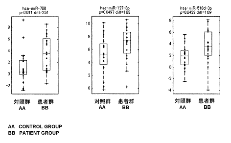
[Figure 1] Figure 1 shows distribution of expression of
three-type miRNAs: hsa-miR-708, hsa-miR-127-3p and hsa-miR-
518d-3p in two groups: a patient group and a control group.
[Figure 2] Figure 2 shows detectability of endometriosis by
a single miRNA, evaluated by ROC analysis, in which the
vertical axis represents sensitivity; whereas the
transverse axis represents a false-positive rate (1-
Specificity).
[Figure 3] Figure 3 shows detectability evaluation by use
of a prediction model using plural miRNAs.
Description of Embodiments
[0014]
The present invention provides a diagnostic agent for
endometriosis (sometimes referred to as "the diagnostic
agent of the present invention" in this specification) for
measuring the concentration of at least one miRNA
(sometimes referred to as "miRNA of the present invention"
in this specification) selected from the group consisting
CA 02874716 2014-11-25
. ,
- 8 -
of hsa-miR-708, hsa-miR-127-3p and hsa-miR-518d-3p in a
sample derived from the blood of a subject, the agent
containing an amplification primer for the miRNA as a main
component.
[0015]
The subject in this specification refers to a human
having endometriosis or suspected to have endometriosis.
Examples of the sample derived from the blood of a subject
include whole blood, blood serum and blood plasma. The
sample is preferably blood serum or blood plasma and
further preferably blood plasma. Note that it is generally
believed that the serum miRNA concentration correlates with
the plasma miRNA concentration (for example, see Proc. Natl.
Acad. Sci. USA, 105 (2008) 10513-10518).
[0016]
In the present invention, miRNAs which have been found
to be overexpressed in a sample derived from the blood of
an endometriosis patient, are three types of miRNAs: hsa-
miR-708, hsa-miR-127-3p and hsa-miR-518d-3p. The base
sequences of these miRNAs are as follows and can be found
in miRBase (http: //www.mirbase.org/index.shtml).
hsa-miR-708 (aaggagcuuacaaucuagcuggg) SEQ ID No:1
hsa-miR-127-3p (ucggauccgucugagcuuggcu) SEQ ID No:2
hsa-miR-518d-3p (caaagcgcuucccuuuggagc) SEQ ID No:3
[0017]
CA 02874716 2014-11-25
- 9 -
The diagnostic agent for endometriosis of the present
invention contains a primer (sometimes referred to as "the
amplification primer of the present invention" in this
specification) capable of amplifying at least one of these
three types of miRNAs, as a main component. The
concentration of the miRNA of the present invention in a
blood-derived sample can be measured by amplifying the
miRNA of the present invention by a polymerase chain
reaction (PCR), etc. using these amplification primers,
with the result that endometriosis can be simply and
quickly examined.
[0018]
The amplification primer of the present invention is a
single-stranded oligonucleotide and preferably DNA. The
amplification primer includes a forward primer and a
reverse primer and they are used in combination. The
forward primer typically has a homologous sequence to the
sequence consisting of 6 to 10 bases, which are present on
the 5' side of the center nucleic acid of a miRNA sequence
(however, if the primer is DNA, T is contained in place of
U); whereas, the reverse primer has a complementary
sequence to the sequence consisting of 6 to 10 bases, which
are present on the 3' side of the center nucleic acid of a
miRNA sequence.
[0019]
CA 02874716 2014-11-25
- 10 -
The primer may optionally have an additional sequence
(for example, a sequence encoding His-tag and restriction
enzyme recognition sites) useful for recognizing, purifying,
and sub-cloning of an amplified product, on the side
opposite to the direction of extension. Such an additional
sequence preferably has a length of 1 to 15 bases. The
sequences of the amplification primers are selected such
that the primers each have high specificity, do not form a
secondary structure within their molecules and are not
mutually hybridized. Each amplification primer has a
length of preferably 12 to 30 nucleotides and more
preferably 15 to 25 nucleotides.
[0020]
To simply measure the amount of PCR product, one or
both of the forward primer and the reverse primer are
preferably tagged with a fluorescent label. Examples of
the fluorescent label include a fluorescent dye, a
quenching substance, a donor pigment and an acceptor
pigment.
[0021]
The amplification primer of the present invention can
be chemically synthesized by a general DNA synthesis
apparatus (for example, Model 394, manufactured by Applied
Biosystems); however, another method well known in the art
may be employed for synthesis.
CA 02874716 2014-11-25
- 11 -
[0022]
In another aspect, the present invention provides a
kit for diagnosing endometriosis. The kit may contain not
only the diagnostic agent of the present invention but also
e.g., pretreatment reagents for a sample, reagents and
enzyme for PCR, a buffer solution, a container and
instruction for use.
[0023]
In yet another aspect, the present invention provides
a method for detecting endometriosis (sometimes referred to
as "the method of the present invention" herein) including
a step of measuring the concentration of at least one miRNA
selected from the group consisting of hsa-miR-708, hsa-miR-
127-3p and hsa-miR-518d-3p, in a sample derived from the
blood of a subject.
[0024]
To measure the concentration of miRNA in a sample, RNA
is extracted from the sample, template cDNA is prepared and
PCR is performed using the template cDNA as a template and
the amplification primer of the present invention. In this
manner, at least one miRNA selected from the group
consisting of hsa-miR-708, hsa-miR-127-3p and hsa-miR-518d-
3p contained in the sample is amplified.
[0025]
CA 02874716 2014-11-25
. , .
- 12 -
A method of extracting RNA from a sample is well known
in the art. For example, guanidine-isothiocyanate and
phenol chloroform extraction can be used. Alternatively, a
commercially available RNA extraction reagent can be used.
Subsequently, the obtained total RNA is fractionated to
obtain miRNA. For example, RNA is extracted from a sample
by use of Trizol LS reagent (Life Technologies Inc.) and
fractionated by use of a filter cartridge of mirVana miRNA
isolation kit (Life Technologies Inc.) to obtain an RNA
fraction.
[0026]
A method of synthesizing cDNA using RNA as a template
is also well known in the art. In the method, cDNA can be
synthesized using a commercially available random primer or
Stem-loop reverse transcription primers as the primer in
the presence of dNTPs and a reverse transcriptase.
[0027]
Subsequently, PCR is performed using DNA or cDNA thus
prepared as a template and the amplification primer of the
present invention. Preferably, miRNA is quantified using
real time PCR amplification method. In a particularly
preferable aspect, the concentration of miRNA in a blood-
derived sample can be measured by a quantitative RT-PCR
using a TaqMan (trade name) probe.
[0028]
CA 02874716 2014-11-25
- 13 -
To correct variation of measurement values in
measuring miRNA concentration, an appropriate internal
standard is preferably included. Examples of the internal
standard include RNAs, which are known not to change in
expression level, such as miRNA, snRNA and mRNA. Specific
examples thereof include P-actin, glyceraldehyde J3-
phosphate dehydrogenase and ribosomal protein Pl. In the
case of measuring a great number (for example 200 or more)
of miRNAs at the same time, medium values of expression
levels of all the miRNAs can be used as the internal
standard.
[0029]
The concentration of the miRNA of the present
invention may be measured by a hybridization method using a
microarray on which a probe having a homologous sequence or
a complementary sequence to the sequence of a part of hsa-
miR-708, hsa-miR-127-3p or hsa-miR-518d-3p is immobilized.
Alternatively, the concentration of the miRNA can be
directly measured by use of a next-generation sequencing.
[0030]
It is determined whether the concentration of the
miRNA of the present invention obtained as mentioned above
in a sample derived from the blood of a subject is higher
than the concentration of the miRNA of the present
invention in a normal control. If the concentration of the
CA 02874716 2014-11-25
- 14 -
miRNA of the present invention in the subject's sample as
mentioned above is higher than that of a normal control, it
is determined that the subject has endometriosis or
suspected to have endometriosis. The "normal control"
refers to a sample derived from the blood of a human who
was diagnosed not to have endometriosis. It is preferable
that concentrations of each miRNA in plural normal controls
were measured in advance to obtain an average expression
level of the control group.
[0031]
In a preferable aspect, the method of the present
invention includes a step of determining whether a
prediction model function value, which is constructed using
the concentration of miRNA in a sample derived from the
blood of a subject, is a cutoff value or more. The cutoff
value refers to a value, which is set in order to determine
and diagnose a disease. The cutoff value of the present
invention is a value of a prediction model function, which
is constructed using the concentration of the miRNA of the
present invention in a sample derived from the blood of a
subject, and set in order to find whether or not the
subject is suspected to have endometriosis. If the
concentration of the miRNA of the present invention in the
subject's sample as mentioned above is a cutoff value or
more, it is determined that the subject has endometriosis
CA 02874716 2014-11-25
- 15 -
or is suspected to have endometriosis. How to set such a
cutoff value is not particularly defined; however, the
cutoff value can be obtained by constructing a prediction
model function taking a value between 0 and 1 by logistic
regression and subjecting a predetermined subject group and
a control group to ROC curve analysis.
[0032]
In a specific aspect, the following values can be used
as the cutoff value:
When three miRNAs: has-miR-708, has-miR-127-3p and
has-miR-518d-3p, are used as miRNA in the sample, a value
of 0.1 or more and 0.9 or less, preferably 0.31 or more and
0.68 or less and more preferably 0.40 or more and 0.59 or
less can be used as the cutoff value.
When two miRNAs: has-miR-708 and has-miR-518d-3p, are
used as miRNA in the sample, a value of 0.1 or more and 0.9
or less, preferably 0.34 or more and 0.66 or less and more
preferably 0.38 or more and 0.54 or less can be used, as
the cutoff value.
When has-miR-708 is used as miRNA in the sample, a
value of 0.1 or more and 0.9 or less, preferably 0.42 or
more and 0.60 or less and more preferably 0.46 or more and
0.58 or less can be used as the cutoff value.
When has-miR-127-3p is used as miRNA in the sample, a
value of 0.1 or more and 0.9 or less, preferably 0.44 or
CA 02874716 2014-11-25
. . .
- 16 -
more and 0.55 or less and more preferably 0.49 or more and
0.55 or less can be used as the cutoff value.
When has-miR-518d-3p is used as miRNA in the sample, a
value of 0.1 or more and 0.9 or less, preferably 0.44 or
more and 0.56 or less and more preferably 0.47 or more and
0.53 or less can be used as the cutoff value.
[0033]
Furthermore, the method of the present invention can
be applied to the case of measuring the concentration of
the miRNA of the present invention in a sample derived from
the blood only taken from a single subject once in a
certain time point. Since the concentration of the miRNA
of the present invention increases with the passage of time
from the onset of endometriosis, the method of the present
invention can be applied to the case of measuring the
concentration of the miRNA of the present invention in
samples taken from a single subject with the passage of
time. This case is preferred since endometriosis can be
detected without comparing with the concentration of the
miRNA in a normal control. In this case, a sample is taken,
for example, at an interval of e.g., once per week during
the period from two weeks after a first medical examination
to full recovery. More specifically, the diagnostic method
of the present invention includes a method of monitoring
CA 02874716 2014-11-25
. . ,
- 17 -
progression of a disease and a method of monitoring
therapeutic process.
[0034]
The contents of the all patent literatures and
reference literatures expressly cited in the specification
are incorporated in their entirety in the specification by
reference. The aspect expressed by the phrase
i, ...comprising" used in the specification includes the
aspect expressed by the phrase "... essentially consisting
of" and an aspect expressed by the phrase "...consisting
of".
[0035]
Now, the present invention will be more specifically
described by way of Examples; however, the present
invention is not limited by these Examples.
Examples
[0036]
(Serum samples from patients and control subjects)
Serum samples of endometriosis patients (20 cases)
diagnosed by a transvaginal hysteroscope and control
subjects (20 cases, 9 cases of them had benign
gynecological diseases except endometriosis, 11 cases were
healthy persons) having the corresponding age composition
to the patient group, were purchased from Cureline Inc.
..
CA 02874716 2014-11-25
. i
'
- 18 -
(South San Francisco, California, U.S.A.). The serum
samples were stored at -80 C until use. Clinical
information of the subjects are shown in Table 1.
[0037]
[Table 1]
Clinical information of subject
subject ID Height Weight Estragial Progeateren I.SH
Days atter Age waenos,. Smoking
(cm) (kg) (pM) lnfil)
(mMU/isa.) last period (Nicer)
EM0) 156 70 601.03 3.95 6.87 6 33 stage II
EM02 159 74 58448 6.25 2.57 6 33 stage 11
EM03 160 79 235.00 1.26 4.58 5 32 stage 11
EM04 174 55 1025.40 5.26 60.20 27 34 stage 1
EM05 170 54 2146.10 5.92 8.64 9 32 stage 11
EM06 170 65 600.00 2.40 9.70 7 54 Genital endomettiosis,
adenomyosis
EM07 170 72 600.30 2B0 7.60 3 39 Endometious ovarian cyst
EM08 164 67 350.40 1.29 352.40 14 39
Adenomyosis 5
EM09 183 83 420.50 2.80 7.90 21 39 Genita endornetriosis.
adenornyosis
EM10 167 58 350.80 2.10 3.70 5 44 Genital endometriosis
EM11 158 50 250.30 1.20 18.30 5 39 Cervical endometrisis
EM12 170 62 350.40 1.80 6.50 4 45 Genital
encfornetriosis, adenomyosis
EM13 167 63 550.40 7.60 8.70 4 44 Genital endometriosis,
adenomyosis
EM14 170 64 78.60 2.70 5.80 7 42
Genital endometriosis, adenomyosis 10
EM15 170 60 227.96 1.15 5.69 13 44 Disseminated genital
endometriosis
EM16 165 54 258.70 1.40 6.50 3 42
Genital endometriosis, adenomyosis 5
EM17 165 67 490.00 4,55 3 43
EM18 170 70 540013 10.70 4 42
EM19 164 65 66000 8.40 6 46
. EM20 168 59 408.50 5.56 4 39
ECO1 170 78 206.82 2.56 6.30 2 33 -
ECO2 178 58 301.39 2,76 9.10 19 32 -
ECO3 176 90 300.25 7.08 5.50 2 33 -
E004 168 68 205.30 6.08 25.40 15 30 -
ECO5 158 58 270.80 4.72 25.40 7 34 -
ECM 163 55 350.40 2.30 4.80 5 54 Healthy
ECO7 158 48 650 30 10,30 6.50 8 54 ovarian polycystosis
EC013 158 47 1020.19 2.40 9.30 36 39 ovarian polycystosis
ECO9 160 65 52.40 1.70 4,60 5 45 endometrial polyp
EC10 165 49 273.40 1.80 2.60 3 39 Healthy
EC11 158 55 170.80 2.40 463.80 13 45 cervix erosion
E012 162 54 268.40 1.60 5.30 6 41 Healthy
E013 185 62 304_80 120 6.40 26 39 cervix erosion
E014 168 54 228.40 1.40 2.80 3 39 ovarian polycystosis
EC15 170 58 320.80 1.40 5.80 3 39 Healthy
EC16 169 65 540.30 1.60 4.70 6 45 uterine rnyoma. mixed
form
EC I 7 164 58 205.40 1,80 4.70 6 44 endometrial polyp
EC18 168 51 159.40 1,40 2.60 5 39 Healthy
EC19 158 67 350.80 1,70 4.20 10 45 uteral myoma,
subserosal form
EC20 165 52 174.80 1,80 5.72 4 39
Healthy ..
[0038]
(RNA extraction from serum samples)
CA 02874716 2014-11-25
- 19 -
RNA was extracted from serum samples by use of Trizol
LS reagent and mirVana miRNA isolation kit (both
manufactured by Life Technologies Inc.). Specific
procedure was as follows. To a serum sample (1 mL), 1 mL
of sterilized water was added. Further to the mixture, 6
mL of Trizol LS reagent was added and mixed. The solution
mixture was allowed to stand still at room temperature for
minutes, and then 1.6 mL of chloroform was added thereto
and vigorously stirred by hand. After stirring, the
solution was allowed to stand still at room temperature for
minutes, and centrifugally separated (4 C, 12,000 x g,
10 minutes) to obtain an organic solvent layer and a water
layer. From the obtained water layer, an aliquot (4 mL)
was taken. To the aliquoted water layer, 5 mL of 99.5%
ethanol was added and homogeneously stirred. The stirred
solution was added to a filter cartridge of the mirVana
miRNA isolation kit and fractionated in accordance with the
protocol attached to the kit to obtain an RNA fraction.
The RNA fraction was taken, lyophilized and resuspended
with 10 L of sterilized water to obtain a concentrated RNA
solution. The concentration of miRNA contained in the
obtained concentrated RNA solution was measured by use of
Agilent 2100 bioanalyzer and Small RNA kit (Agilent
Technologies) and represented in terms of the concentration
CA 02874716 2014-11-25
. . ,
- 20 -
of RNA having 10 to 40 base length contained in the
concentrated RNA solution.
[0039]
(Quantitative analysis of miRNA)
From the above concentrated RNA solution, a cDNA-
containing solution was obtained by use of Megaplex RT
primers (pool A or B v.2.0) and TaqManTm MicroRNA Reverse
Transcription Kit. Thereafter, cDNA contained in the
obtained solution was amplified by use of Megaplex PreAmp
primers (pool A or B v.2.0) and TaqManTm PreAmp Master Mix.
The amplified cDNA was mixed with TaqMan Universal PCR
Master Mix (No AmpErase UNG). The obtained sample was
loaded to TaqManTm Human MicroRNA Array v2.0 and subjected
to quantitative PCR analysis by use of 7900HT Fast Real-
Time PCR system. Note that each operation described in
this section was performed in accordance with the protocol
attached to the kit and system.
[0040]
(Data analysis)
In analyzing data, RQ Manager 1.2 software (Life
Technologies) was used. A CT (Threshold cycle) value was
calculated from an amplification curve in accordance with
the method reported by Liang et al. (Liang, Y. et al., BMC
Genomics, 8, 166-, 2007), and the threshold was set at 0.2.
If miRNA was not amplified in 80% or more of the total 40
CA 02874716 2014-11-25
- 21 -
specimens, the miRNA was eliminated from the analysis. The
median value of CT values of the remaining miRNAs was
calculated for each sample. Then, a value (ACT values) was
obtained by subtracting the medium value from each of the
CT values to normalize the expression difference between
the specimens. To identify miRNA whose expression level
significantly differs between the patient group and the
control group, Welch's t-test was carried out. Plural
miRNAs identified by using logistic regression analysis,
were used in combination to construct and evaluate a
diagnosis model. Furthermore, diagnostic ability by each
of the miRNAs or a combination of miRNAs was evaluated by
ROC (Receiver operating characteristic) analysis. For
these analyses, MATLAB software (The Mathworks Inc.) was
used.
[0041]
(Results)
Figure 1 shows distribution of expression of three-
type miRNAs in two groups: a patient group and a control
group. The vertical axis represents the relative
expression level of each of the miRNAs by a logarithm to
base 2. The expression level of each subject is plotted by
a blue dot and the median value of expression level in each
group is indicated by a red line. The upper limit and
lower limit of each box indicate 25 percentile and 75
CA 02874716 2014-11-25
. . .
- 22 -
percentile, respectively. Both ends of each whisker
represent the upper limit and lower limit of the values
from which outliers are eliminated. Red crosses represent
outliners. The P value obtained as a result of the Welch's
t-test and the difference in average expression level
(logarithm using 2 as the base) between both groups are
indicated under the name of miRNA in each Figure.
[0042]
The expression profile of miRNAs in the sera of the
endometriosis patient group (20 cases) was compared with
that of the control subject group (20 cases). As a result,
it was found that expression levels of three-type miRNAs:
hsa-miR-708, hsa-miR-127-3p, and hsa-miR-518d-3p,
significantly increased in the patient group (Figure 1).
From this, it was clearly demonstrated that the patient
group can be satisfactorily detected by measuring
expression levels of these three miRNAs in the serum.
[0043]
Table 2 shows the sequences of these miRNAs and the
expression-level increase rates (Fold Change) of these
miRNAs in the patient group compared to the control group,
calculated from average values of expression levels
(logarithm using 2 as the base) of both groups.
[0044]
[Table 2]
CA 02874716 2014-11-25
- 23 -
Name of miRNA Base sequence Expression increase Sequence
rate in patient group No.
hsa-miR-708 aaggagcuuacaaucuagcuggg 5.71 1
hsa-miR-127-3p ueggauccgueugagcuuggcu 3.56 2
hsa-miR-518d-3p caaagegcuucccuuuggage 3.24 3
[0045]
In the case where these upregulated three miRNAs were
used for detection of endometriosis, the detectability was
evaluated by ROC analysis.
[0046]
First, the detectability of each of the three types of
miRNAs: hsa-miR-708, hsa-miR-127-3p, hsa-miR-518d-3p, when
they were used singly, was compared based on the area under
the curve of the Roc curve (AUC). Further, on the ROC
curve, a point at which both groups can be most efficiently
distinguished was identified. The cutoff value (threshold)
corresponding to the point was regarded (expressed) as the
upregulated control rate (Fold Change) based on the
control-group average expression level. Next, specificity
true positive rate (Sensitivity) and specificity true
negative rate (Specificity) were calculated when the cutoff
value was used. These are shown in Figure 2.
[0047]
A prediction model was constructed by using plural
miRNAs in combination based on logistic regression analysis
and detectability of the model was evaluated by ROC
CA 02874716 2014-11-25
- 24 -
analysis in the same manner as above. Figure 3 shows, a
ROC curve (left) when a prediction model constructed of
hsa-miR-708 and hsa-miR-518d-3p was used, or a ROC curve
(right) when a prediction model constructed of all of hsa-
miR-708, hsa-miR-127-3p and hsa-miR-518d-3p (right) was
used. Detectability was evaluated based on the areas under
the curves (AUC).
[0048]
As a result, the area under the curve of the ROC curve
when using a model of two or three miRNAs in combination
was larger than when using a model of a single miRNA
(Figure 2), demonstrating that a patient can be detected
with a higher accuracy.
Industrial Applicability
[0049]
The present invention is useful for diagnosis of
endometriosis.