Note: Descriptions are shown in the official language in which they were submitted.
CA 02874918 2014-11-27
WO 2013/178736
PCT/EP2013/061173
1
TLR3 BINDING AGENTS
CROSS-REFERENCE TO RELATED APPLICATIONS
This application claims the benefit of U.S. Provisional Application Nos.
61/653,652
filed 31 May 2012, 61/670,289 filed 11 July 2012 and 61/679,923 filed 6 August
2012; all of
which are incorporated herein by reference in their entirety; including any
drawings.
REFERENCE TO SEQUENCE LISTING
The present application is being filed along with a Sequence Listing in
electronic
format. The Sequence Listing is provided as a file entitled "PCT Seq list TLR3-
4_5T25",
created May 29, 2013, which is 46 KB in size. The information in the
electronic format of the
Sequence Listing is incorporated herein by reference in its entirety.
FIELD OF THE INVENTION
The present invention relates to antibodies (e.g. monoclonal antibodies),
antibody
fragments, and derivatives thereof that bind and inhibit TLR3 signaling. The
invention also
relates to cells producing such antibodies; methods of making such antibodies;
fragments,
variants, and derivatives of the antibodies; pharmaceutical compositions
comprising the
same; methods of using the antibodies to diagnose, treat or prevent diseases,
e.g.
autoimmune diseases, inflammatory diseases and the like.
BACKGROUND
Drosophila toll proteins control dorsal-ventral patterning and are thought to
represent
an ancient host defense mechanism. In humans, TLRs are believed to be an
important
component of innate immunity. Human and Drosophila Toll protein sequences show
homology over the entire length of the protein chains. The family of human
Toll-like
receptors is comprised of ten highly conserved receptor proteins, TLR1-TLR10.
Like
Drosophila toll, human TLRs are type I transmembrane proteins with an
extracellular domain
consisting of a leucine-rich repeat (LRR) domain that recognizes pathogen-
associated
molecular patterns (PAMPs), and a cytoplasmic domain that is homologous to the
cytoplasmic domain of the human interleukin-1 (IL-1) receptor. Similar to the
signaling
pathways for both Drosophila toll and the IL-1 receptor, human Toll-like
receptors signal
through the NF-KB pathway.
Although the different mammalian TLRs share many characteristics and signal
transduction mechanisms, their biological functions are very different. This
is due in part to
CA 02874918 2014-11-27
WO 2013/178736
PCT/EP2013/061173
2
the fact that four different adaptor molecules (MyD88, TIRAP, TRIF and TRAF)
are
associated in various combinations with the TLRs and mediate different
signaling pathways.
In addition, different ligands for one TLR may preferentially activate
different signal
transduction pathways. Furthermore, the TLRs are differentially expressed in
various
hematopoietic and non-hematopoietic cells. Accordingly, the response to a TLR
ligand
depends not only on the signal pathway activated by the TLR, but also on the
nature of the
cells in which the individual TLR is expressed.
Toll-like receptor 3 (TLR3) has received considerable attention as a
therapeutic
target as TLR3 signaling has been implicated in inflammatory and autoimmune
conditions.
Patent application W098/50547 provides the nucleic acid and amino acid
sequence of the
hTLR3 protein. De Bouteiller et al. (2005) J. Biol. Chem. 280(46): 38133-
38145) disclose use
of an anti-TLR3 antibody to bind cell surface TLR3. Antibody 01130 is stated
to be activatory
toward TLR3 and has been described in WO 2007/051164. Polyclonal antibodies
that
inhibited TLR3 were described in Cavassani et al. (2008) J. Exp. Med. 205:
2609-2621. WO
03/106499 and Matsumoto et al. (2003) J. lmmunol. 171:3154-3162 describes an
antibody
corresponding to antibody clone TLR3.7 (eBioScience Inc., San Diego) reported
to bind and
inhibit cell surface TLR3 but not cell compartment TLR3 or in myeloid-lineage
DC. WO
06/060513 describes an antibody 01068 which is reported to inhibit cytokine
production in
epithelial cells, which are reported to express TLR3 on the cell surface. PCT
patent
application W02010/051470 provides further anti-TLR3 antibodies. Other
anti-TLR3
antibodies for research use include polyclonal anti-TLR3 antibodies from R&D
Systems
Corp., antibody 4001285 from Abcam and antibodies 619F7, 713E4, 716G10, IMG-
5631
and ¨IMG-5348, all from lmgenex Corp.
However, among currently available anti-TLR3 antibodies, they are not
optimally
suited for use as therapeutic agents, e.g. to modulate TLR3 in vivo. For
example, many
suffer from lack of efficacy or affinity to their epitopes. There is therefore
a need to provide
improved antibodies directed to TLR3.
SUMMARY OF THE INVENTION
The present invention arises from the discovery of novel compositions
comprising,
and methods of using monoclonal antibodies, including but not limited to
antibody fragments,
and derivatives that specifically bind to and inhibit the function of human
TLR3.
The present invention provides antibodies with new properties useful for
targeting
TLR3 in vivo. Since TLR3 binds its natural ligand (dsRNA) and signals
exclusively in the
endosome in macrophages and dendritic cells (DCs) (at acidic pH), antibodies
have
previously been selected based on high affinity at endosomal pH where
signalling occurs.
CA 02874918 2014-11-27
WO 2013/178736
PCT/EP2013/061173
3
However, little remains known about the mechanism by which anti-TLR3
antibodies enter
cells. The present invention provides antibodies that have strong binding to
TLR3 exposed at
the cell surface and in a pH neutral environment and which display improved
potency in
TLR3 inhibition. Optimizing binding of cell-surface expressed TLR3 may
therefore be an
important criteria for cellular (endosomal) uptake by a cell which may
condition downstream
(or overall) biological activity. The present antibodies show strong binding
to human cell
surface TLR3, as observed in an assay where TLR3 is expressed exclusively at
the cell
surface in neutral pH conditions. In this way, antibodies having improved cell
surface binding
were selected. The inhibitory activity of anti-TLR3 antibodies may therefore
be governed by
the cycling back to the cell surface of the endosomal TLR3 polypeptides
involved in
endocytosis once the receptors separated from their ligands.
In one aspect the invention provides an antibody that inhibits TLR3-mediated
signalling in a TLR3-expressing cell, wherein the antibody specifically binds
a human TLR3
polypeptide expressed solely at the surface of a cell, optionally at neutral
pH. Optionally, the
antibody has an EC50 of no more than 0.3 g/ml, optionally no more than 0.2
g/ml,
optionally no more than 0.1 g/ml, for binding to cells expressing TLR3 solely
at the cell
surface.
In one aspect the invention provides antibodies that bind the N-terminal
portion of the
TLR3 protein at least partly (or primarily or exclusively) on the glycan-free
lateral surface of
the TLR3 polypeptide (the face bound by dsRNA), and optionally furthermore at
least partly
within the N-terminal dsRNA binding site of a human TLR3 polypeptide.
Optionally, the
antibody further binds TLR3 at least partly within the backbone of the N-
terminal portion of
the TLR3 polypeptide.
While some previous epitopes on TLR3 have been shown to be useful for
efficacious
inhibition of TLR3, epitopes have not necessarily remained present in non-
human primates.
In one aspect the invention provides antibodies that inhibit TLR3 polypeptide
activity by
binding to the N-terminal portion of a human TLR3 protein, and that also bind
non-human
primate TLR3. In one aspect the invention provides antibodies that bind the N-
terminal
portion of the TLR3 protein and that do not compete with dsRNA for binding to
human TLR3,
wherein the antibodies also bind to a of non-human primate TLR3 polypeptide
(in a non-
human primate-TLR3-expressing cell). In one aspect the invention provides
antibodies that
bind the N-terminal portion of the TLR3 protein and that competes with dsRNA
for binding to
the N-terminal dsRNA binding site of a human TLR3 polypeptide, wherein the
antibodies
also bind to a non-human primate TLR3 polypeptide (in a non-human primate-TLR3-
expressing cell). In one embodiment, the antibodies at least partly (or
primarily or
exclusively) on the glycan-free lateral surface of the TLR3 polypeptide (the
face bound by
CA 02874918 2014-11-27
WO 2013/178736
PCT/EP2013/061173
4
dsRNA). In one embodiment, the antibodies bind to TLR3 at least partly within
the N-terminal
dsRNA binding site of a human TLR3 polypeptide. In one embodiment, the non-
human
primate is macaca fascicularis. In one embodiment, the non-human primate
TLR3
polypeptide comprises an amino acid sequence shown in NCB! accession number
BAG55033 (SEQ ID NO: 2).
In one aspect the invention provides antibodies that bind to the N-terminal
portion of
a human TLR3 protein, notably within residues 41 to 251, optionally at least
partly within
residues 41 to 139, 41 to 120 or residues 41 to 89, of human TLR3 of SEQ ID
NO: 1.
In one aspect the invention provides antibodies that bind to the N-terminal
portion of
a human TLR3 protein, wherein the antibody has reduced binding to a TLR3
polypeptide
having a mutation in its N-terminal portion in the segment corresponding to
residues 41-139
of SEQ ID NO: 1, relative to binding between the antibody and a wild-type TLR3
polypeptide
of SEQ ID NO: 1.
Optionally, the antibodies of the invention interfere with binding of dsRNA to
the N-
terminal dsRNA binding site of a human TLR3 polypeptide. Optionally, the
antibodies bind
one or more amino acid residues within the glycan-free lateral surface of the
TLR3
polypeptide that is involved in binding of the TLR3 polypeptide to dsRNA,
and/or residues
adjacent thereto.
Optionally, the antibodies bind an epitope comprising residues 64 and/or
residue 65
of SEQ ID NO: 1, and/or have reduced binding to a TLR3 polypeptide having a
mutation at
residues 64 and/or residue 65 of SEQ ID NO: 1. Optionally, the antibodies bind
an epitope
comprising residues 86 and/or residue 89 of SEQ ID NO: 1, and/or have reduced
binding to
a TLR3 polypeptide having a mutation at residues 86 and/or residue 89 of SEQ
ID NO: 1.
Optionally, the antibodies bind an epitope comprising residues 117 and/or
residue 120 of
SEQ ID NO: 1, and/or have reduced binding to a TLR3 polypeptide having a
mutation at
residues 117 and/or residue 120 of SEQ ID NO: 1. Optionally, the antibodies
bind an epitope
comprising residues 137 and/or residue 139 of SEQ ID NO: 1, and/or have
reduced binding
to a TLR3 polypeptide having a mutation at residues 137 and/or residue 139 of
SEQ ID NO:
1. Optionally, the antibodies bind an epitope comprising residues 112, 113
and/or 115 of
SEQ ID NO: 1, and/or have reduced binding to a TLR3 polypeptide having a
mutation at
residues 112, 113 and/or 115 of SEQ ID NO: 1.
In one embodiment, the antibodies have reduced binding to a TLR3 polypeptide
having a mutation at residues 117 and/or residue 120 of SEQ ID NO: 1, and
reduced binding
to a TLR3 polypeptide having a mutation at residues 137 and/or residue 139 of
SEQ ID NO:
1. Optionally, the antibodies have reduced binding to a TLR3 polypeptide
having a mutation
CA 02874918 2014-11-27
WO 2013/178736
PCT/EP2013/061173
at residues 117 and/or residue 120 of SEQ ID NO: 1, and do not have reduced
binding to a
TLR3 polypeptide having a mutation at residues 137 and/or residue 139 of SEQ
ID NO: 1.
In one embodiment, the antibodies have reduced binding to a TLR3 polypeptide
having a mutation at residues 64 and/or residue 65 of SEQ ID NO: 1, and
reduced binding to
5 a TLR3 polypeptide having a mutation at residues 137 and/or residue 139
of SEQ ID NO: 1.
Optionally, the antibodies have reduced binding to a TLR3 polypeptide having a
mutation at
residues 86 and/or residue 89 of SEQ ID NO: 1. and reduced binding to a TLR3
polypeptide
having a mutation at residues 137 and/or residue 139 of SEQ ID NO: 1.
Optionally, the
antibodies do not have reduced binding to a TLR3 polypeptide having a mutation
at residues
117 and/or residue 120 of SEQ ID NO: 1. Optionally, the antibodies do not have
reduced
binding to a TLR3 polypeptide having a mutation at residues 112, 113 and/or
115 of SEQ ID
NO: 1.
Optionally, the antibodies have reduced binding to a TLR3 polypeptide having a
mutation at residues 64 and/or residue 65 of SEQ ID NO: 1, a TLR3 polypeptide
having a
mutation at residues 86 and/or residue 89 of SEQ ID NO: 1 and a TLR3
polypeptide having
a mutation at residues 137 and residue 139 of SEQ ID NO: 1. As evidenced by
binding to
TLR3 mutants, the antibodies differ in their epitope from previously described
antibodies.
In one embodiment, the antibodies have reduced binding to a TLR3 polypeptide
having a mutation at residues 112, 113 and/or 115 of SEQ ID NO: 1, and reduced
binding to
a TLR3 polypeptide having a mutation at residues 137 and/or residue 139 of SEQ
ID NO: 1.
In one embodiment, the antibodies have reduced binding to a TLR3 polypeptide
having a
mutation at residues 112, 113 and/or 115 of SEQ ID NO: 1, and reduced binding
to a TLR3
polypeptide having a mutation at residues 117 and/or residue 120 of SEQ ID NO:
1. In one
embodiment, the antibodies have reduced binding to a TLR3 polypeptide having a
mutation
at residues 112, 113 and/or 115 of SEQ ID NO: 1, reduced binding to a TLR3
polypeptide
having a mutation at residues 117 and/or residue 120 of SEQ ID NO: 1, and
reduced binding
to a TLR3 polypeptide having a mutation at residues 137 and/or residue 139 of
SEQ ID NO:
1. In one embodiment, the antibodies have reduced binding to a TLR3
polypeptide having a
mutation at residues 112 and/or 113 of SEQ ID NO: 1, and reduced binding to a
TLR3
polypeptide having a mutation at residue 137 of SEQ ID NO: 1. Optionally, the
antibodies do
not have reduced binding to a TLR3 polypeptide having a mutation at residues
86 and/or
residue 89 of SEQ ID NO: 1. Optionally, the antibodies do not have reduced
binding to a
TLR3 polypeptide having a mutation at residues 64 and/or residue 65 of SEQ ID
NO: 1.
Optionally, in any of the embodiments herein, the antibodies maintain binding
(do not
have reduced binding) to a TLR3 polypeptide having a mutation at residues 116,
145, 182,
196 and/or residue 171 of SEQ ID NO: 1.
CA 02874918 2014-11-27
WO 2013/178736
PCT/EP2013/061173
6
In one aspect, the invention provides antibodies that interfere with binding
of dsRNA
to a human TLR3 polypeptide. In one aspect, the invention provides antibodies
that interfere
with binding of dsRNA to the N-terminal dsRNA binding site of a human TLR3
polypeptide.
Optionally, the antibodies compete with dsRNA for binding to human TLR3
polypeptide, e.g,
to the N-terminal dsRNA binding site of a human TLR3 polypeptide. Competition
can be
assessed using standard methods, e.g. Biacore assays to assess whether
antibodies bind to
immobilized TLR3 in the presence of dsRNA, and/or whether dsRNA binds to
immobilized
TLR3 in the presence of antibodies, under acidic conditions.
Optionally, the antibodies compete with dsRNA for binding to human TLR3
polypeptide in an in vitro assay comprising the steps of: (i) contacting an
anti-TLR3 antibody
with a TLR3 polypeptide so as to obtain antibodies bound to TLR3 polypeptide,
and (ii)
contacting the antibody bound TLR3 polypeptide of step (i) with dsRNA and
assessing
whether dsRNA decreases binding of TLR3 polypeptide ( to the antibody, wherein
a
decrease of binding indicates competition with dsRNA for human TLR3
polypeptide.
Optionally, the antibodies compete with dsRNA for binding to human TLR3
polypeptide in an
in vitro assay comprising the steps of: (i) contacting a TLR3 polypeptide with
dsRNA so as to
obtain dsRNA bound to TLR3 polypeptide, and (ii) contacting the dsRNA bound
TLR3
polypeptide of step (i) with an anti-TLR3 antibody and assessing whether the
TLR3
polypeptide binds the dsRNA-TLR3 polypeptide complex, wherein lack of
substantial binding
indicates competition with dsRNA for human TLR3 polypeptide. Optionally, the
antibodies
compete with dsRNA for binding to human TLR3 polypeptide in an in vitro assay
comprising
the steps of: (i) attaching an anti-TLR3 antibody to a solid support (e.g.,
via a constant
domain), (ii) contacting said antibody with a TLR3 polypeptide so as to obtain
antibodies
bound to TLR3 polypeptide, and (iii) contacting the antibody bound TLR3
polypeptide of step
(ii) with dsRNA and assessing whether dsRNA decreases binding of TLR3
polypeptide to the
antibody, wherein a decrease of binding indicates competition with dsRNA for
human TLR3
polypeptide.
In one aspect of any of the embodiments of the invention, the antibody binds
to a
human TLR3 polypeptide expressed at the surface of a cell, optionally as
assessed in a cell
expressing TLR3 exclusively at the cell surface under neutral pH, internalizes
into a cell that
expresses TLR3, and inhibits TLR3 signaling in a cell (e.g. a TLR3-expressing
human
dendritic cell).
In one aspect, the antibodies bind human TLR3 polypeptides under neutral
conditions, and in particular under conditions representative of that
encountered in the cell
cytosol. Such neutral conditions are generally characterized by a pH between
6.6 and 7.4,
for example a slightly alkaline pH of 7.2 found in the cell cytosol.
Optionally, the antibody has
CA 02874918 2014-11-27
WO 2013/178736
PCT/EP2013/061173
7
a KD of no more than 10-9M, optionally less than 10-19M, optionally less than
10-11M for
binding to a TLR3 polypeptide at neutral pH. Optionally, the binding at
neutral conditions is
of better affinity than under acid conditions, e.g. where the KD for binding
to TLR3 at neutral
compared to acidic conditions is lower by at least 0.5-, 1.0-, 1.5- or 2.0-
log10.
The present invention further provides specific antibodies have increased
activity
over previously reported antibodies. In one aspect of any of the embodiments
of the
invention, the antibody competes for binding to a TLR3 polypeptide (e.g. a
human TLR3
polypeptide comprising an amino acid sequence of SEQ ID NO: 1) with any one or
any
combination of monoclonal antibody 11E1, 7G11, 31F6, 3204 and 37B7, optionally
under
acid and/or neutral conditions. In one embodiment, an antibody of the
invention competes for
binding to a TLR3 polypeptide, optionally under acid and/or neutral
conditions, with an
antibody having respectively a VH and VL region of SEQ ID NOS: 3 and 4 (11E1
), a VH and
VL region of SEQ ID NOS: 14 and 15 (31F6), a VH and VL region of SEQ ID NOS:
25 and
26 (3204), a VH and VL region of SEQ ID NOS: 36 and 37 (37B7) or a VH and VL
region of
SEQ ID NOS: 47 and 48 (7G11). In one aspect of any of the embodiments of the
invention,
the antibody may have a heavy and/or light chain having one, two or three CDRs
of antibody
11E1 , 7G11, 31F6, 3204 or 37B7.
In one aspect, the antibody that specifically binds TLR3 has one or more
(including
any combination thereof, or all of) of the following properties:
a. binds to a TLR3 polypeptide comprising an amino acid sequence of SEQ ID NO:
1
and/or 2;
b. specifically binds a human TLR3 polypeptide expressed solely at the surface
of a
cell, wherein the antibody has an E050 of no more than 0.3 g/ml, optionally
no more
than 0.2 g/ml, optionally no more than 0.1 g/ml, for binding to cells
expressing
TLR3 solely at the cell surface;
c. internalizes into a cell that expresses TLR3 on its surface;
d. has a subnanomolar affinity for a TLR3 polypeptide at an neutral pH, e.g. a
pH of
about pH 7.2;
e. has a subnanomolar affinity for a TLR3 polypeptide at an acidic pH, e.g. a
pH less
than about 6.5, or between about 4.5 to 6.5 or about pH 5.6;
f. inhibits TLR3 signaling in the presence of a TLR3 ligand;
g. inhibits TLR3 signaling in an inflammatory background, e.g. in the presence
of
inflammatory cytokines such as IFNa;
h. competes for binding to a TLR3 polypeptide with antibody 11E1 , 7G11, 31F6,
3204
or 37B7;
CA 02874918 2014-11-27
WO 2013/178736
PCT/EP2013/061173
8
i. competes with dsRNA for binding to the N-terminal portion the TLR3
polypeptide;
j. has reduced binding to a TLR3 polypeptide having a mutation at residues
64 and/or
residue 65 of SEQ ID NO: 1, and/or reduced binding to a TLR3 polypeptide
having a
mutation at residues 86 and/or residue 89 of SEQ ID NO: 1;
k. has reduced binding to a TLR3 polypeptide having a mutation at residues 117
and/or
residue 120 of SEQ ID NO: 1, and/or has reduced binding to a TLR3 polypeptide
having a mutation at residues 137 and/or residue 139 of SEQ ID NO: 1, and/or
has
reduced binding to a TLR3 polypeptide having a mutation at residues 112, 113
and/or 115 of SEQ ID NO: 1; and/or
I. binds to at least one, two, three, four, five, six, seven or more residues
in the
segment corresponding to residues 41-251, 41-89, 41-120 or 41-139 of the TLR3
polypeptide of SEQ ID NO: 1.
In one aspect, the invention provides a monoclonal antibody that specifically
binds to
at least one, two, three, four, five, six, seven or more residues in the
segment corresponding
to residues 1-251, optionally 41-251, 41-89, 41-120 or 41-139 of the TLR3
polypeptide of
SEQ ID NO: 1. Optionally, the antibody inhibits signaling by the TLR3
polypeptide.
Optionally, the antibody does not bind residue 116, residue 145 and/or residue
182 of the
TLR3 polypeptide of SEQ ID NO: 1. Optionally, the antibody does not bind
residue 171,
and/or residue 196 of the TLR3 polypeptide of SEQ ID NO: 1. Optionally,
binding of the
antibody to a TLR3 polypeptide having a mutation at residues 116, 145, 182 196
and/or
residue 171 of the TLR3 polypeptide of SEQ ID NO: 1 is maintained (i.e., is
not substantially
reduced), in comparison to binding to a wild-type TLR3 polypeptide of SEQ ID
NO: 1;
preferably said mutation is a K145E, D116R, K182E, N196A and/or E171A
mutation. Such
antibodies can optionally further have any properties described herein, e.g.
subnanomolar
affinity for a TLR3 polypeptide at an acidic pH, inhibits TLR3 signaling in
the presence of a
TLR3 ligand or in an inflammatory background (e.g. in the presence of
inflammatory
cytokines such as IFNa), competes for binding to a TLR3 polypeptide with 11E1,
7G11,
31F6, 3204 or 37B7; does not compete with dsRNA for binding to C-terminal
portion the
TLR3 polypeptide; or inhibits IP-10 secretion on DC (e.g. in human myeloid
DC). Such
antibodies can furthermore be used in any of the methods of the invention.
In one embodiment, the invention provides an antibody that binds a TLR3
polypeptide, wherein the antibody comprises (i) the heavy chain CDR 1, 2 and 3
(HCDR1,
HCDR2, HCDR3) amino acid sequences as shown in SEQ ID NO: 5, 6 or 7 (HCDR1), 8
or 9
(HCDR2) and 10 (HCDR3), and (ii) the light chain CDR 1, 2 and 3 (LCDR1, LCDR2,
LCDR3)
amino acid sequences as shown in SEQ ID NO: 11, 12 and 13, respectively;
wherein one,
CA 02874918 2014-11-27
WO 2013/178736
PCT/EP2013/061173
9
two, three, four, or five or more of the amino acids in any of said sequences
may be
substituted by a different amino acid. In one embodiment, the antibody
comprises (i) the
heavy chain CDR 1, 2 and 3 (HCDR1, HCDR2, HCDR3) amino acid sequences as shown
in
SEQ ID NO: 16, 17 or 18 (HCDR1), 19 or 20 (HCDR2) and 21 (HCDR3), and (ii) the
light
chain CDR 1, 2 and 3 (LCDR1, LCDR2, LCDR3) amino acid sequences as shown in
SEQ ID
NO: 22, 23 and 24, respectively; wherein one, two, three, four, or five or
more of the amino
acids in any of said sequences may be substituted by a different amino acid.
In one
embodiment, the antibody comprises (i) the heavy chain CDR 1, 2 and 3 (HCDR1,
HCDR2,
HCDR3) amino acid sequences as shown in SEQ ID NO: 27, 28 or 29 (HCDR1), 30 or
31(HCDR2) and 32 (HCDR3), and (ii) the light chain CDR 1, 2 and 3 (LCDR1,
LCDR2,
LCDR3) amino acid sequences as shown in SEQ ID NO: 33, 34 and 35,
respectively;
wherein one, two, three, four, or five or more of the amino acids in any of
said sequences
may be substituted by a different amino acid. In one embodiment, the antibody
comprises (i)
the heavy chain CDR 1, 2 and 3 (HCDR1, HCDR2, HCDR3) amino acid sequences as
shown in SEQ ID NO: 38, 39 or 40 (HCDR1), 41 or 42 (HCDR2) and 43 (HCDR3), and
(ii)
the light chain CDR 1, 2 and 3 (LCDR1, LCDR2, LCDR3) amino acid sequences as
shown in
SEQ ID NO: 44, 45 and 46, respectively; wherein one, two, three, four, or five
or more of the
amino acids in any of said sequences may be substituted by a different amino
acid. In one
embodiment, the antibody comprises (i) the heavy chain CDR 1, 2 and 3 (HCDR1,
HCDR2,
HCDR3) amino acid sequences as shown in SEQ ID NO: 49, 50 or 51 (HCDR1), 52 or
53
(HCDR2) and 54 (HCDR3), and (ii) the light chain CDR 1, 2 and 3 (LCDR1, LCDR2,
LCDR3)
amino acid sequences as shown in SEQ ID NO: 55, 56 and 57, respectively;
wherein one,
two, three, four, or five or more of the amino acids in any of said sequences
may be
substituted by a different amino acid.
In another embodiment, the antibody of any of the embodiments herein is
capable of
being internalized by a cell that expresses TLR3 polypeptide on its surface.
In one embodiment, the antibody is chimeric, e.g. contains a non-murine,
optionally a
human, constant region. In one embodiment, the antibody is human or humanized.
In
another embodiment, the antibody is a mouse or rat antibody (e.g., comprises
CDRs derived
from a rat or rat gene or rat Ig locus gene segment). In another embodiment,
the antibody
does not substantially bind to other human TLRs (e.g. TLR4).
In one aspect of any of the embodiments of the invention, the isotype of the
antibody
is IgG, optionally IgG1 or IgG3. In one embodiment the antibody comprises an
Fc domain or
is of an isotype that is bound by FcyR.
CA 02874918 2014-11-27
WO 2013/178736
PCT/EP2013/061173
In one aspect of any of the embodiments of the invention, the antibody is an
antibody
fragment selected from Fab, Fab', Fab'-SH, F(ab')2, Fv, diabodies, single-
chain antibody
fragment, or a multispecific antibody comprising multiple different antibody
fragments. In one
aspect of any of the embodiments of the invention, the antibody does not
comprise an Fc
5 domain or is of an isotype that is not substantially bound by FcyR (e.g.
human CD16). In one
embodiment, the antibody is of human IgG4 or IgG2 isotype. Human IgG4 isotypes
or other
IgG isotypes modified to reduce their FcyR binding can be used for their
advantageous
pharmacological properties such as serum half-life, while modulating TLR3
signaling, in e.g.
a DC, without inducing the death of the cell. In one aspect of any of the
embodiments of the
10 invention, the anti-TLR3 antibody inhibits TLR3 signaling and comprises
a constant region of
human IgG4 or IgG2 isotype. In one aspect, of any of the embodiments of the
invention, the
anti-TLR3 antibody inhibits TLR3 signaling and comprises a constant region
(heavy chain
constant region) that does not substantially bind FcyRIlla.
In one preferred embodiment, the anti-TLR3 antibody comprises a heavy chain of
human IgG4 isotype. In one embodiment, the anti-TLR3 antibody comprises a
human IgG4
heavy chain constant region and comprising a serine to proline mutation in
residue 241,
corresponding to position 228 according to the EU-index (Kabat et al.,
"Sequences of
proteins of immunological interest", 5th e a .55
NIH, Bethesda, ML, 1991). Compositions
comprising such antibodies can be characterized as having less than about 15%,
such as
less than about 10% (e.g., about 5% or less, about 4% or less, about 3% or
less, or even
about 1% or less) of IgG4 "half-antibodies" (comprising a single heavy
chain/light chain pair).
Such IgG4 "half-antibody" by-products form due to heterogeneity of inter-heavy
chain
disulfide bridges in the hinge region in a proportion of secreted human IgG4
(see Angal et
al., Molecular Immunology, 30(1):105-108, 1993 for a description of IgG4 "half-
antibodies",
S241P mutation, and related principles). This effect is typically only
detectable under
denaturing, non-reducing conditions.
In another embodiment, the antibody is conjugated or covalently bound to a
detectable or toxic moiety.
In one aspect, the antibodies optionally inhibit TLR3 signaling without
blocking
binding of a dsRNA TLR3 ligand to the principal (i.e. C-terminal) dsRNA
binding site of the
TLR3 polypeptide.
In one aspect, the antibodies also bind human TLR3 under acidic conditions,
and in
particular under conditions representative of that encountered in an acidified
subcellular
compartment of a cell (e.g. compartments of the endocytic pathway endosomic,
lysosomal).
CA 02874918 2014-11-27
WO 2013/178736
PCT/EP2013/061173
11
Such acidic conditions are generally characterized by a pH lower than about pH
6.5, or
between about pH 4.5 to 6.5, or about pH 5.6.
In one aspect of any of the embodiments herein, the antibodies modulate,
optionally
inhibit, TLR3 signaling in an acidified subcellular compartment of a cell
(e.g. compartments
of the endocytic pathway endosomic, lysosomal).
In one aspect of any of the embodiments herein, the antibodies modulate,
optionally
inhibit, TLR3 signaling in a dendritic cell (DC) (e.g. a myeloid DC, monocyte
derived DC).
In other aspects of any of the embodiments herein, the antibodies' bivalent
binding
affinity for TLR3 under neutral and/or acidic conditions can optionally be
characterized by a
mean KD of no more than about (i.e. better affinity than) 100, 50, 10, 5, or 1
nanomolar,
preferably sub-nanomolar or optionally no more than about 500, 200, 100 or 10
picomolar.
In other aspects of any of the embodiments herein, the antibodies inhibit TLR3
signaling by at least partly (or fully) blocking the binding of a TLR3 ligand
to a TLR3
polypeptide. The TLR3 ligand will generally be a ligand other than an anti-
TLR3 antibody
and may be a naturally occurring or non-naturally occurring TLR3 ligand,
optionally a
dsRNA-based ligand such as polyAU (polyadenylic acid:polyuridylic acid) or
polyIC
(polyinosinic:polycytidylic acid).
In another aspect, the invention provides a method of identifying, screening
and/or
producing an antibody that specifically binds and inhibits a TLR3 polypeptide
in a
mammalian subject, said method comprising the steps of: a) providing a
plurality of
antibodies that bind human TLR3 polypeptide, optionally by a method comprising
immunizing a non-human mammal with an immunogen comprising a TLR3 polypeptide,
and
(b) assessing the binding affinity of said antibodies for the TLR3 polypeptide
in a cell which
expresses human TLR3 solely at the cell surface. Optionally, the method
further comprises
selecting an antibody from said plurality that has an EC50 of no more than 0.3
pg/ml,
optionally no more than 0.2 pg/ml, optionally no more than 0.1 pg/ml, for
binding to cells
expressing human TLR3 solely at the cell surface.
Any of the methods of the invention can further be characterized as comprising
any
step described in the application, including notably in the "Detailed
Description of the
Invention"). The invention further relates to an antibody obtainable by any of
present
methods. The invention further relates to pharmaceutical or diagnostic
formulations of the
antibodies of the present invention. The invention further relates to methods
of using
antibodies in methods of treatment or diagnosis, optionally in combination
with a second
therapeutic agent (e.g. a corticoid, a DMARD, an anti-cytokine or anti-
cytokine receptor
agent, an anti-TNFalpha agent, etc.).
CA 02874918 2014-11-27
WO 2013/178736
PCT/EP2013/061173
12
These and additional advantageous aspects and features of the invention may be
further described elsewhere herein.
BRIEF DESCRIPTION OF THE DRAWINGS
Figures 1A, 1B, 1C and 1D show in vitro dose effect for binding to HEK293T
cells
transiently transfected with a TLR3 ECD construct and expressing TLR3 solely
at their
surface for antibodies 11E1, 7G11, 32C4 and 31F6, respectively. Each antibody
is
compared with reference antibody 31C3; binding to cell surface TLR3 is
improved for
antibodies 11E1 , 7G11, 32C4 and 31F6.
Figures 2A, 2B, 2C, 2D and 2E show dose dependent inhibition of TLR3 signaling
using a 293T-human TLR3 luciferase assay with the human anti-TLR3 antibodies.
Each
antibody is compared with reference antibody 31C3; inhibition of dsRNA-induced
TLR3
signaling is improved for antibodies 11E1 , 7G11, 32C4, 31F6 and 37B7.
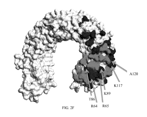
Figures 2F, 2G, and 2H show view of the N-terminal end of the TLR3
polypeptide,
showing amino acid residues mutated indicated in black and residues adjacent
to residues
which form part of the principle epitopes in grey. Figure 2F shows a view of
the glycan-free
lateral surface of the TLR3 polypeptide, with the N-terminal end of the TLR3
polypeptide at
the right of the image); Figure 2G shows a view of the glycan-containing
lateral surface of
the TLR3 polypeptide and the backbone, with the N-terminal end of the TLR3
polypeptide in
the foreground (at the left of the image). Figure 2H shows a view of the
glycan-free lateral
surface of the TLR3 polypeptide and the backbone, with the N-terminal end of
the TLR3
polypeptide in the foreground (at the right of the image).
Figure 3 shows results of a rheumatoid arthritis mouse models. Figure 3A shows
the
results of a preventive rheumatoid arthritis mouse model. Figure 3B shows the
results of a
curative rheumatoid arthritis mouse model. Figure 3C shows the results of a
curative
rheumatoid arthritis mouse model when mice are treated with PBS, a control
antibody, 28G7
and an anti-TNFa antibody (HumiraTm).
Figure 4 shows results of the mouse colitis model. Figure 4A shows the wall
thickness measurements for the mice treated with saline (black dots), with
TNBS only (black
squares) with an anti-TNFa antibody and TNBS (black triangles), with 28G7 and
TNBS
(open dots), and with a control Ab and TNBS (open squares). Figure 4B shows
the
macroscopic damage score for the mice treated with saline (black dots), with
TNBS only
(black squares) with an anti-TNFa antibody and TNBS (black triangles), with
28G7 and
TNBS (open dots), and with a control Ab and TNBS (open squares). The anti-TLR3
antibody
CA 02874918 2014-11-27
WO 2013/178736
PCT/EP2013/061173
13
according to the invention ameliorating the development of the disease, under
stringent
conditions. (* p<0.05, ' p<0.01 vs saline).
Figure 5 shows results of a COPD mouse model. Figure 5A shows BAL differential
cell counts for macrophages, eosinophils, neutrophils and lymphocytes. The
anti-TLR3
antibodies strongly decreased the infiltration of neutrophils into the
airways, while not
substantially affecting macrophages eosinophils or lymphocytes. Figure 5B
shows venous
blood saturated oxygen (in percent) for each of LPS/elastase alone and
LPS/elastase in
combination with anti-TLR3 antibodies or roflumilast. Figure 50 shows IL17A in
BAL fluid
(BALF), where anti-TLR3 antibodies decreased IL17A (pg/ml) substantially, and
as much as
roflumilast. Figure 5D shows IP-10 in BALF, where -TLR3 antibodies decreased
IP-10
(pg/ml) substantially. Figure 5E shows BAL differential cell counts for
macrophages,
neutrophils and lymphocytes for a second study comparing anti-TLR3 antibodies
(28G7),
roflumilast (Rofu) and the combination of roflumilast and anti-TLR3 antibodies
(combo).
Figure 6 shows results of a CLP (cecal ligation and puncture - sepsis) mouse
model.
In this acute model, mice experience an acute infection, mimicking septic
shock.
Figures 7A and 7B show results of drug combinations with anti-human TLR3 mAbs
(labeled IPH33.1) in combination with dexamethasone or Humira respectively.
Antibodies
each substantially reduce in vitro IP-10 production by PBMC in response to
polyIC further
when combined with dexamethasone or Humira .
DETAILED DESCRIPTION OF THE INVENTION
Introduction
The present invention is based, at least in part, on the discovery of high
affinity
monoclonal antibodies that specifically and efficiently inhibit the TLR3
signaling pathway.
The inventors also provide new epitopes present on human TLR3, including the
epitope
recognized by antibody 11E1, 7G11, 31F6, 3204 and/or 37B7, which are
particularly
efficient in inhibiting TLR3 signaling, and inhibiting cytokine release in
response to
stimulation with a TLR3 ligand.
The antibodies can be used for treating an autoimmune or inflammatory disease
in a
subject in need thereof. The present invention also provides methods for
treating relapses,
attacks, or acute phases, occurring during the course of an inflammatory or
autoimmune
disease in a subject in need thereof using an anti-TLR3 antibody which
inhibits TLR3
signaling. The present invention also provides novel methods for treating
established
inflammatory or autoimmune diseases in a subject in need thereof using an anti-
TLR3
antibody which inhibits TLR3 signaling. The invention also provides treatment
regimens and
treatment combinations that can be used for the treatment of inflammatory or
autoimmune
CA 02874918 2014-11-27
WO 2013/178736 PCT/EP2013/061173
14
disease in a subject in need thereof using an anti-TLR3 antibody which
inhibits TLR3
signaling.
The antibodies of the present invention that bind TLR3 under acidic and
neutral
conditions will generally bind both cell surface TLR3 and endosomic TLR3 at
high affinity,
such that the antibodies will be useful in any situation (e.g. treatment or
prevention of
disease) where targeting (e.g. modulating) TLR3 is useful. TLR3 has been found
in some
cases of inflammation the surface of macrophages and blocking TLR3 upon
chloroquine
neutralization of endosomal acidification nevertheless exhibited some anti-
inflammatory
activity (Cavassani et al. 2008, supra). However, the antibodies of the
invention will have
the greatest advantage over other antibodies in the treatment or prevention of
diseases
where the modulating (e.g. inhibiting) the signaling by TLR3 in the cytosolic
(e.g. endosomic)
compartments is useful or required, and the relative importance of modulating
signaling of
such compartments TLR3 may depend on the disease. One example of such as
disease is
rheumatoid arthritis; endosomic compartment¨expressed TLR3 is believed to play
an
important role in rheumatoid arthritis, since treatment with chloroquine, an
inhibitor of
endosomal acidification, inhibits TLR3 signaling and inhibits production of
inflammatory
cytokines from synovial cultures from patients having rheumatoid arthritis
(Sacre et al.
(2008) J. lmmunol. 181:8002-8009). Endosomic compartment¨expressed TLR3 is
believed
to play an important role in a number of other diseases where DC (e.g. myeloid
DC) are
involved in exacerbating disease, as mDC have a well-documented capacity to
take up
antigens from apoptotic or necrotic cells including during tissue necrosis
during acute
inflammation.
Since the present antibodies are specific for TLR3, they can also be used for
other
purposes, including purifying TLR3 or TLR3-expressing cells, modulating (e.g.
activating or
inhibiting) TLR3 receptors in vitro, ex vivo, or in vivo, targeting TLR3-
expressing cells for
destruction in vivo, or specifically labeling/binding TLR3 in vivo, ex vivo,
or in vitro, including
for methods such as immunoblotting, IHC analysis, i.e. on frozen biopsies,
FACS analysis,
and immunoprecipitation.
Definitions
As used herein, "TLR3 ligand" refers to any compound that can specifically
bind to
and alter the activity of TLR3 in vitro, ex vivo, or in vivo. The compound can
be a naturally
occurring ligand, e.g., generally dsRNA or viral dsRNA, or a synthetic ligand
such as polyIC
or polyAU. The compound can be any type of molecule, including inorganic or
organic
compounds or elements, including proteins (such as antibodies), nucleic acids,
carbohydrates, lipids, or any other molecular entity. Further, such compounds
can modulate
CA 02874918 2014-11-27
WO 2013/178736
PCT/EP2013/061173
TLR3 receptors in any way, including activating or inhibiting, and by any
mechanism,
including by binding to the receptor and triggering or shutting off activity
in a manner similar
to a naturally occurring ligand, or by binding to the receptor and blocking
access to other
ligands. Preferably, the ligand activates the receptor, and as such can be
used to induce the
5 production of cytokines by TLR3-expressing cells.
The term "antibody," as used herein, refers to polyclonal and monoclonal
antibodies.
Depending on the type of constant domain in the heavy chains, antibodies are
assigned to
one of five major classes: IgA, IgD, IgE, IgG, and IgM. Several of these are
further divided
into subclasses or isotypes, such as IgG1 , IgG2, IgG3, IgG4, and the like. An
exemplary
10
immunoglobulin (antibody) structural unit comprises a tetramer. Each tetramer
is composed
of two identical pairs of polypeptide chains, each pair having one "light"
(about 25 kDa) and
one "heavy" chain (about 50-70 kDa). The N-terminus of each chain defines a
variable
region of about 100 to 110 or more amino acids that is primarily responsible
for antigen
recognition. The terms variable light chain (VL) and variable heavy chain (VH)
refer to these
15
light and heavy chains respectively. The heavy-chain constant domains that
correspond to
the different classes of immunoglobulins are termed "alpha," "delta,"
"epsilon," "gamma" and
"mu," respectively. The subunit structures and three-dimensional
configurations of different
classes of immunoglobulins are well known. IgG and/or IgM are the preferred
classes of
antibodies employed in this invention, with IgG being particularly preferred,
because they are
the most common antibodies in the physiological situation and because they are
most easily
made in a laboratory setting. Preferably the antibody of this invention is a
monoclonal
antibody. Particularly preferred are humanized, chimeric, human, or otherwise-
human-
suitable antibodies. "Antibodies" also includes any fragment or derivative of
any of the herein
described antibodies.
The term "specifically binds to" means that an antibody can bind preferably in
a
competitive binding assay to the binding partner, e.g. TLR3, as assessed using
either
recombinant forms of the proteins, epitopes therein, or native proteins
present on the surface
of isolated target cells. Competitive binding assays and other methods for
determining
specific binding are further described below and are well known in the art.
When an antibody is said to "compete with" a particular monoclonal antibody
(e.g.
11E1, 7G11, 31F6, 32C4 or 37B7) or other TLR3 ligand (e.g., dsRNA, it means
that the
antibody competes with the monoclonal antibody (or other TLR3 ligand) in a
binding assay
using either recombinant TLR3 molecules or surface expressed TLR3 molecules.
For
example, if a test antibody reduces the binding of 11E1, 7G11, 31F6, 32C4 or
37B7 to a
TLR3 polypeptide or TLR3-expressing cell in a binding assay, the antibody is
said to
"compete" respectively with 11E1 , 7G11, 31F6, 32C4 or 37B7.
CA 02874918 2014-11-27
WO 2013/178736
PCT/EP2013/061173
16
The term "affinity", as used herein, means the strength of the binding of an
antibody
to an epitope. The affinity of an antibody is given by the dissociation
constant KD, defined as
[Ab] x [Ag] / [Ab-Ag], where [Ab-Ag] is the molar concentration of the
antibody-antigen
complex, [Ab] is the molar concentration of the unbound antibody and [Ag] is
the molar
concentration of the unbound antigen. The affinity constant Ka is defined by
1/Kd. Preferred
methods for determining the affinity of mAbs can be found in Harlow, et al.,
Antibodies: A
Laboratory Manual, Cold Spring Harbor Laboratory Press, Cold Spring Harbor,
N.Y., 1988),
Coligan et al., eds., Current Protocols in Immunology, Greene Publishing
Assoc. and Wiley
lnterscience, N.Y., (1992, 1993), and Muller, Meth. Enzymol. 92:589-601
(1983), which
references are entirely incorporated herein by reference. One preferred and
standard
method well known in the art for determining the affinity of mAbs is the use
of Biacore
instruments.
Within the context of this invention a "determinant" designates a site of
interaction or
binding on a polypeptide.
The term "epitope" is defined as an antigenic determinant, and is the area or
region
on an antigen to which an antibody binds. A protein epitope may comprise amino
acid
residues directly involved in the binding as well as amino acid residues which
are effectively
blocked by the specific antigen binding antibody or peptide, i.e., amino acid
residues within
the "footprint" of the antibody. It is the simplest form or smallest
structural area on a complex
antigen molecule that can combine with e.g., an antibody or a receptor.
Epitopes can be
linear or conformational/structural. The term "linear epitope" is defined as
an epitope
composed of amino acid residues that are contiguous on the linear sequence of
amino acids
(primary structure). The term "conformational or structural epitope" is
defined as an epitope
composed of amino acid residues that are not all contiguous and thus represent
separated
parts of the linear sequence of amino acids that are brought into proximity to
one another by
folding of the molecule (secondary, tertiary and/or quaternary structures). A
conformational
epitope is dependent on the 3-dimensional structure. The term 'conformational'
is therefore
often used interchangeably with 'structural'.
By "immunogenic fragment," it is herein meant any polypeptidic or peptidic
fragment
that is capable of eliciting an immune response such as (i) the generation of
antibodies
binding said fragment and/or binding any form of the molecule comprising said
fragment,
including the membrane-bound receptor and mutants derived therefrom, (ii) the
stimulation
of a T-cell response involving T-cells reacting to the bi-molecular complex
comprising any
MHC molecule and a peptide derived from said fragment, (iii) the binding of
transfected
vehicles such as bacteriophages or bacteria expressing genes encoding
mammalian
immunoglobulins. Alternatively, an immunogenic fragment also refers to any
construction
CA 02874918 2014-11-27
WO 2013/178736
PCT/EP2013/061173
17
capable of eliciting an immune response as defined above, such as a peptidic
fragment
conjugated to a carrier protein by covalent coupling, a chimeric recombinant
polypeptide
construct comprising said peptidic fragment in its amino acid sequence, and
specifically
includes cells transfected with a cDNA of which sequence comprises a portion
encoding said
fragment.
A "human-suitable" antibody refers to any antibody, derivatized antibody, or
antibody
fragment that can be safely used in humans for, e.g. the therapeutic methods
described
herein. Human-suitable antibodies include all types of humanized, chimeric, or
fully human
antibodies, or any antibodies in which at least a portion of the antibodies is
derived from
humans or otherwise modified so as to avoid the immune response that is
generally
provoked when native non-human antibodies are used.
For the purposes of the present invention, a "humanized" or "human" antibody
refers
to an antibody in which the constant and variable framework region of one or
more human
immunoglobulins is fused with the binding region, e.g. the CDR, of an animal
immunoglobulin. Such antibodies are designed to maintain the binding
specificity of the non-
human antibody from which the binding regions are derived, but to avoid an
immune reaction
against the non-human antibody. Such antibodies can be obtained from
transgenic mice or
other animals that have been "engineered" to produce specific human antibodies
in
response to antigenic challenge (see, e.g., Green et al. (1994) Nature Genet
7:13; Lonberg
et al. (1994) Nature 368:856; Taylor et al. (1994) Int lmmun 6:579, the entire
teachings of
which are herein incorporated by reference). A fully human antibody also can
be constructed
by genetic or chromosomal transfection methods, as well as phage display
technology, all of
which are known in the art (see, e.g., McCafferty et al. (1990) Nature 348:552-
553). Human
antibodies may also be generated by in vitro activated B cells (see, e.g.,
U.S. Pat. Nos.
5,567,610 and 5,229,275, which are incorporated in their entirety by
reference).
A "chimeric antibody" is an antibody molecule in which (a) the constant
region, or a
portion thereof, is altered, replaced or exchanged so that the antigen binding
site (variable
region) is linked to a constant region of a different or altered class,
effector function and/or
species, or an entirely different molecule which confers new properties to the
chimeric
antibody, e.g., an enzyme, toxin, hormone, growth factor, drug, etc.; or (b)
the variable
region, or a portion thereof, is altered, replaced or exchanged with a
variable region having a
different or altered antigen specificity.
The terms "Fe domain," "Fe portion," and "Fe region" refer to a C-terminal
fragment of
an antibody heavy chain, e.g., from about amino acid (aa) 230 to about aa 450
of human y
(gamma) heavy chain or its counterpart sequence in other types of antibody
heavy chains
CA 02874918 2014-11-27
WO 2013/178736
PCT/EP2013/061173
18
(e.g., a, 5, E and p for human antibodies), or a naturally occurring allotype
thereof. Unless
otherwise specified, the commonly accepted Kabat amino acid numbering for
immunoglobulins is used throughout this disclosure (see Kabat et al. (1991 )
Sequences of
Protein of Immunological Interest, 5th e a .55
United States Public Health Service, National
Institute of Health, Bethesda, MD).
The terms "isolated", "purified" or "biologically pure" refer to material that
is
substantially or essentially free from components which normally accompany it
as found in
its native state. Purity and homogeneity are typically determined using
analytical chemistry
techniques such as polyacrylamide gel electrophoresis or high performance
liquid
chromatography. A protein that is the predominant species present in a
preparation is
substantially purified.
The terms "polypeptide," "peptide" and "protein" are used interchangeably
herein to
refer to a polymer of amino acid residues. The terms apply to amino acid
polymers in which
one or more amino acid residue is an artificial chemical mimetic of a
corresponding naturally
occurring amino acid, as well as to naturally occurring amino acid polymers
and non-
naturally occurring amino acid polymer.
The term "recombinant" when used with reference, e.g., to a cell, or nucleic
acid,
protein, or vector, indicates that the cell, nucleic acid, protein or vector,
has been modified by
the introduction of a heterologous nucleic acid or protein or the alteration
of a native nucleic
acid or protein, or that the cell is derived from a cell so modified. Thus,
for example,
recombinant cells express genes that are not found within the native
(nonrecombinant) form
of the cell or express native genes that are otherwise abnormally expressed,
under
expressed or not expressed at all.
Within the context of this invention, the term antibody that "binds" a common
determinant designates an antibody that binds said determinant with
specificity and/or
affinity.
Producing Anti-TLR3 Antibodies
The antibodies suitable for the method of the invention specifically bind
TLR3.
Antibodies of the invention furthermore bind TLR3 with high affinity at
conditions
corresponding to that encountered in at the cell surface. Antibodies of the
invention are
furthermore capable of inhibiting the TLR3 signaling pathway. The ability of
the inhibitory
antibodies to specifically inhibit the TLR3 signaling pathway makes them
useful for
numerous applications, in particular for treating or preventing diseases
wherein the inhibition
of TLR3 signaling pathway is desirable, i.e. avoid further cytokine and
chemokine secretion
as well as cellular activation, as described herein.
CA 02874918 2014-11-27
WO 2013/178736
PCT/EP2013/061173
19
In one embodiment, the invention provides methods using an antibody that binds
human TLR3, and competes for binding to human TLR3 with monoclonal antibody
11E1,
7G11, 31F6, 3204 or 37B7.
"TLR3", "TLR3 polypeptide" and "TLR3 receptor", used interchangeably, are used
herein to refer to Toll-Like Receptor 3, a member of the Toll-like receptor
(TLRs) family. The
amino acid sequence of human TLR3 is shown in SEQ ID NO: 1 (NCB! accession
number
NP 003256, the disclosure of which is incorporated herein by reference). The
human TLR3
mRNA sequence is described in NCB! accession number NM 003265. Human TLR3
sequences are also described in PCT patent publication no. WO 98/50547, the
disclosure of
which is incorporated herein by reference. A non-human primate (macaca
fascicularis) TLR3
amino acid sequence is shown in NCB! accession number BAG55033 (SEQ ID NO: 2).
In one aspect, the invention provides an antibody that competes with
monoclonal
antibody 11E1, 7G11, 31F6, 3204 or 37B7 and recognizes, binds to, or has
immunospecificity for substantially or essentially the same, or the same,
epitope or "epitopic
site" on a TLR3 molecule as monoclonal antibody 11E1 , 7G11, 31F6, 3204 or
37B7. In other
embodiments, the monoclonal antibody consists of, or is a derivative or
fragment of,
antibody 11E1 , 7G11, 31F6, 3204 or 37B7.
Any fragment of TLR3, preferably but not exclusively human TLR3, or any
combination of TLR3 fragments, can be used as immunogens to raise antibodies,
and the
antibodies of the invention can recognize epitopes at any location within the
TLR3
polypeptide, so long as they can do so on TLR3 expressing cells such as MdDC
or MoDC as
described herein. In an embodiment, the recognized epitopes are present on the
cell
surface, i.e. they are accessible to antibodies present outside of the cell.
Most preferably, the
epitope is the epitope specifically recognized by antibody 11E1 , 7G11, 31F6,
3204 or 37B7.
Further, antibodies recognizing distinct epitopes within TLR3 can be used in
combination,
e.g. to bind to TLR3 polypeptides with maximum efficacy and breadth among
different
individuals.
The antibodies of this invention may be produced by a variety of techniques
known in
the art. Typically, they are produced by immunization of a non-human animal,
preferably a
mouse, with an immunogen comprising a TLR3 polypeptide, preferably a human
TLR3
polypeptide. The TLR3 polypeptide may comprise the full length sequence of a
human TLR3
polypeptide, or a fragment or derivative thereof, typically an immunogenic
fragment, i.e., a
portion of the polypeptide comprising an epitope exposed on the surface of
cells expressing
a TLR3 polypeptide, preferably the epitope recognized by the 11E1 , 7G11,
31F6, 3204 or
37B7 antibody. Such fragments typically contain at least about 7 consecutive
amino acids of
the mature polypeptide sequence, even more preferably at least about 10
consecutive amino
CA 02874918 2014-11-27
WO 2013/178736
PCT/EP2013/061173
acids thereof. Fragments typically are essentially derived from the extra-
cellular domain of
the receptor. In a preferred embodiment, the immunogen comprises a wild-type
human
TLR3 polypeptide in a lipid membrane, typically at the surface of a cell. In a
specific
embodiment, the immunogen comprises intact cells, particularly intact human
cells,
5 optionally treated or lysed. In another preferred embodiment, the
polypeptide is a
recombinant TLR3 polypeptide.
The step of immunizing a non-human mammal with an antigen may be carried out
in
any manner well known in the art for stimulating the production of antibodies
in a mouse
(see, for example, E. Harlow and D. Lane, Antibodies: A Laboratory Manual.,
Cold Spring
10 Harbor Laboratory Press, Cold Spring Harbor, NY (1988), the entire
disclosure of which is
herein incorporated by reference). The immunogen is suspended or dissolved in
a buffer,
optionally with an adjuvant, such as complete or incomplete Freund's adjuvant.
Methods for
determining the amount of immunogen, types of buffers and amounts of adjuvant
are well
known to those of skill in the art and are not limiting in any way on the
present invention.
15 These parameters may be different for different immunogens, but are
easily elucidated.
Similarly, the location and frequency of immunization sufficient to stimulate
the
production of antibodies is also well known in the art. In a typical
immunization protocol, the
non-human animals are injected intraperitoneally with antigen on day 1 and
again about a
week later. This is followed by recall injections of the antigen around day
20, optionally with
20 an adjuvant such as incomplete Freund's adjuvant. The recall injections
are performed
intravenously and may be repeated for several consecutive days. This is
followed by a
booster injection at day 40, either intravenously or intraperitoneally,
typically without
adjuvant. This protocol results in the production of antigen-specific antibody-
producing B
cells after about 40 days. Other protocols may also be used as long as they
result in the
production of B cells expressing an antibody directed to the antigen used in
immunization.
For polyclonal antibody preparation, serum is obtained from an immunized non-
human animal and the antibodies present therein isolated by well-known
techniques. The
serum may be affinity purified using any of the immunogens set forth above
linked to a solid
support so as to obtain antibodies that react with TLR3 polypeptides.
In an alternate embodiment, lymphocytes from a non-immunized non-human
mammal are isolated, grown in vitro, and then exposed to the immunogen in cell
culture. The
lymphocytes are then harvested and the fusion step described below is carried
out.
For preferred monoclonal antibodies, the next step is the isolation of
splenocytes
from the immunized non-human mammal and the subsequent fusion of those
splenocytes
with an immortalized cell in order to form an antibody-producing hybridoma.
The isolation of
splenocytes from a non-human mammal is well-known in the art and typically
involves
CA 02874918 2014-11-27
WO 2013/178736
PCT/EP2013/061173
21
removing the spleen from an anesthetized non-human mammal, cutting it into
small pieces
and squeezing the splenocytes from the splenic capsule through a nylon mesh of
a cell
strainer into an appropriate buffer so as to produce a single cell suspension.
The cells are
washed, centrifuged and resuspended in a buffer that lyses any red blood
cells. The solution
is again centrifuged and remaining lymphocytes in the pellet are finally
resuspended in fresh
buffer.
Once isolated and present in single cell suspension, the lymphocytes can be
fused to
an immortal cell line. This is typically a mouse myeloma cell line, although
many other
immortal cell lines useful for creating hybridomas are known in the art.
Preferred murine
myeloma lines include, but are not limited to, those derived from MOPC-21 and
MPC-11
mouse tumors available from the Salk Institute Cell Distribution Center, San
Diego, U. S. A.,
X63 Ag8653 and SP-2 cells available from the American Type Culture Collection,
Rockville,
Maryland U. S. A. The fusion is effected using polyethylene glycol or the
like. The resulting
hybridomas are then grown in selective media that contains one or more
substances that
inhibit the growth or survival of the unfused, parental myeloma cells. For
example, if the
parental myeloma cells lack the enzyme hypoxanthine guanine phosphoribosyl
transf erase
(HGPRT or HPRT), the culture medium for the hybridomas typically will include
hypoxanthine, aminopterin, and thymidine (HAT medium), which substances
prevent the
growth of HGPRT-deficient cells.
Hybridomas are typically grown on a feeder layer of macrophages. The
macrophages
are preferably from littermates of the non-human mammal used to isolate
splenocytes and
are typically primed with incomplete Freund's adjuvant or the like several
days before plating
the hybridomas. Fusion methods are described in Goding, "Monoclonal
Antibodies:
Principles and Practice," pp. 59-103 (Academic Press, 1986), the disclosure of
which is
herein incorporated by reference.
The cells are allowed to grow in the selection media for sufficient time for
colony
formation and antibody production. This is usually between about 7 and about
14 days.
The hybridoma colonies are then assayed for the production of antibodies that
specifically bind to TLR3 polypeptide gene products, optionally the epitope
specifically
recognized by antibody 11E1, 7G11, 31F6, 32C4 or 37B7. The assay is typically
a
colorimetric ELISA-type assay, although any assay may be employed that can be
adapted to
the wells that the hybridomas are grown in. Other assays include
radioimmunoassays or
fluorescence activated cell sorting. The wells positive for the desired
antibody production are
examined to determine if one or more distinct colonies are present. If more
than one colony
is present, the cells may be re-cloned and grown to ensure that only a single
cell has given
rise to the colony producing the desired antibody. Typically, the antibodies
will also be tested
CA 02874918 2014-11-27
WO 2013/178736
PCT/EP2013/061173
22
for the ability to bind to TLR3 polypeptides, e.g., TLR3-expressing cells, in
paraffin-
embedded tissue sections, as described below.
Hybridomas that are confirmed to produce a monoclonal antibody of this
invention
can be grown up in larger amounts in an appropriate medium, such as DMEM or
RPMI-
1640. Alternatively, the hybridoma cells can be grown in vivo as ascites
tumors in an animal.
After sufficient growth to produce the desired monoclonal antibody, the growth
media
containing monoclonal antibody (or the ascites fluid) is separated away from
the cells and
the monoclonal antibody present therein is purified. Purification is typically
achieved by gel
electrophoresis, dialysis, chromatography using protein A or protein G-
Sepharose, or an
anti-mouse Ig linked to a solid support such as agarose or Sepharose beads
(all described,
for example, in the Antibody Purification Handbook, Biosciences, publication
No. 18-1037-
46, Edition AC, the disclosure of which is hereby incorporated by reference).
The bound
antibody is typically eluted from protein A/protein G columns by using low pH
buffers (glycine
or acetate buffers of pH 3.0 or less) with immediate neutralization of
antibody-containing
fractions. These fractions are pooled, dialyzed, and concentrated as needed.
Positive wells with a single apparent colony are typically re-cloned and re-
assayed to
insure only one monoclonal antibody is being detected and produced.
Antibodies may also be produced by selection of combinatorial libraries of
immunoglobulins, as disclosed for instance in (Ward et al. Nature, 341 (1989)
p. 544, the
entire disclosure of which is herein incorporated by reference).
The identification of one or more antibodies that bind(s) to TLR3,
particularly
substantially or essentially the same epitope as monoclonal antibody 11E1,
7G11, 31F6,
32C4 or 37B7 can be readily determined using any one of a variety of
immunological
screening assays in which antibody competition can be assessed. Many such
assays are
routinely practiced and are well known in the art (see, e. g., U. S. Pat. No.
5,660,827, issued
Aug. 26, 1997, which is specifically incorporated herein by reference). It
will be understood
that actually determining the epitope to which an antibody described herein
binds is not in
any way required to identify an antibody that binds to the same or
substantially the same
epitope as the monoclonal antibody described herein.
For example, where the test antibodies to be examined are obtained from
different
source animals, or are even of a different Ig isotype, a simple competition
assay may be
employed in which the control (11E1, 7G11, 31F6, 32C4 or 37B7, for example)
and test
antibodies are admixed (or pre-adsorbed) and applied to a sample containing
TLR3
polypeptides. Protocols based upon western blotting and the use of BIACORE
analysis are
suitable for use in such competition studies.
CA 02874918 2014-11-27
WO 2013/178736
PCT/EP2013/061173
23
In certain embodiments, one pre-mixes the control antibodies (11E1, 7G11,
31F6,
3204 or 37B7, for example) with varying amounts of the test antibodies (e.g.,
about 1:10 or
about 1:100) for a period of time prior to applying to the TLR3 antigen
sample. In other
embodiments, the control and varying amounts of test antibodies can simply be
admixed
during exposure to the TLR3 antigen sample. As long as one can distinguish
bound from
free antibodies (e. g., by using separation or washing techniques to eliminate
unbound
antibodies) and 11E1 , 7G11, 31F6, 3204 or 37B7 from the test antibodies (e.
g., by using
species-specific or isotype-specific secondary antibodies or by specifically
labeling 11E1 ,
7G11, 31F6, 3204 or 37B7 with a detectable label) one can determine if the
test antibodies
reduce the binding of 11E1 , 7G11, 31F6, 3204 or 37B7 to the antigens,
indicating that the
test antibody recognizes substantially the same epitope as 11E1, 7G11, 31F6,
3204 or
37B7. The binding of the (labeled) control antibodies in the absence of a
completely
irrelevant antibody can serve as the control high value. The control low value
can be
obtained by incubating the labeled (11E1, 7G11, 31F6, 3204 or 37B7) antibodies
with
unlabelled antibodies of exactly the same type (11E1 , 7G11, 31F6, 3204 or
37B7), where
competition would occur and reduce binding of the labeled antibodies. In a
test assay, a
significant reduction in labeled antibody reactivity in the presence of a test
antibody is
indicative of a test antibody that recognizes substantially the same epitope,
i.e., one that
"cross-reacts" or competes with the labeled (11E1 , 7G11, 31F6, 3204 or 37B7)
antibody.
Any test antibody that reduces the binding of 11E1, 7G11, 31F6, 3204 or 37B7
to TLR3
antigens by at least about 50%, such as at least about 60%, or more preferably
at least
about 80% or 90% (e. g., about 65-100%), at any ratio of 11E1, 7G11, 31F6,
3204 or
37B7:test antibody between about 1:10 and about 1:100 is considered to be an
antibody that
binds to substantially the same epitope or determinant as 11E1 , 7G11, 31F6,
3204 or 37B7.
Preferably, such test antibody will reduce the binding of 11E1 , 7G11, 31F6,
3204 or 37B7 to
the TLR3 antigen by at least about 90% (e.g., about 95%).
Competition can also be assessed by, for example, a flow cytometry test. In
such a
test, cells bearing a given TLR3 polypeptide can be incubated first with 11E1
, for example,
and then with the test antibody labeled with a fluorochrome or biotin. The
antibody is said to
compete with 11E1 if the binding obtained upon preincubation with a saturating
amount of
11E1 is about 80%, preferably about 50%, about 40% or less (e.g., about 30%,
20% or 10%)
of the binding (as measured by mean of fluorescence) obtained by the antibody
without
preincubation with 11E1. Alternatively, an antibody is said to compete with
11E1 if the
binding obtained with a labeled 11E1 antibody (by a fluorochrome or biotin) on
cells
preincubated with a saturating amount of test antibody is about 80%,
preferably about 50%,
CA 02874918 2014-11-27
WO 2013/178736
PCT/EP2013/061173
24
about 40%, or less (e. g., about 30%, 20% or 10%) of the binding obtained
without
preincubation with the test antibody.
A simple competition assay in which a test antibody is pre-adsorbed and
applied at
saturating concentration to a surface onto which a TLR3 antigen is immobilized
may also be
employed. The surface in the simple competition assay is preferably a BIACORE
chip (or
other media suitable for surface plasmon resonance analysis). The control
antibody (e.g.,
11E1) is then brought into contact with the surface at a TLR3-saturating
concentration and
the TLR3 and surface binding of the control antibody is measured. This binding
of the control
antibody is compared with the binding of the control antibody to the TLR3-
containing surface
in the absence of test antibody. In a test assay, a significant reduction in
binding of the
TLR3-containing surface by the control antibody in the presence of a test
antibody indicates
that the test antibody recognizes substantially the same epitope as the
control antibody such
that the test antibody "cross-reacts" with the control antibody. Any test
antibody that reduces
the binding of control (such as 11E1 ) antibody to a TLR3 antigen by at least
about 30% or
more, preferably about 40%, can be considered to be an antibody that binds to
substantially
the same epitope or determinant as a control (e.g., 11E1). Preferably, such a
test antibody
will reduce the binding of the control antibody (e.g., 11E1) to the TLR3
antigen by at least
about 50% (e. g., at least about 60%, at least about 70%, or more). It will be
appreciated that
the order of control and test antibodies can be reversed: that is, the control
antibody can be
first bound to the surface and the test antibody is brought into contact with
the surface
thereafter in a competition assay. Preferably, the antibody having higher
affinity for the TLR3
antigen is bound to the surface first, as it will be expected that the
decrease in binding seen
for the second antibody (assuming the antibodies are cross-reacting) will be
of greater
magnitude. Further examples of such assays are provided in, e.g., Sauna!
(1995) J.
lmmunol. Methods 183: 33-41, the disclosure of which is incorporated herein by
reference.
Determination of whether an antibody binds within an epitope region can be
carried
out in ways known to the person skilled in the art. As one example of such
mapping/characterization methods, an epitope region for an anti-TLR3 antibody
may be
determined by epitope "foot-printing" using chemical modification of the
exposed
amines/carboxyls in the TLR3 protein. One specific example of such a foot-
printing
technique is the use of HXMS (hydrogen-deuterium exchange detected by mass
spectrometry) wherein a hydrogen/deuterium exchange of receptor and ligand
protein amide
protons, binding, and back exchange occurs, wherein the backbone amide groups
participating in protein binding are protected from back exchange and
therefore will remain
deuterated. Relevant regions can be identified at this point by peptic
proteolysis, fast
microbore high-performance liquid chromatography separation, and/or
electrospray
CA 02874918 2014-11-27
WO 2013/178736
PCT/EP2013/061173
ionization mass spectrometry. See, e. g., Ehring H, Analytical Biochemistry,
Vol. 267 (2) pp.
252-259 (1999) Engen, J. R. and Smith, D. L. (2001) Anal. Chem. 73, 256A-265A.
Another
example of a suitable epitope identification technique is nuclear magnetic
resonance epitope
mapping (NMR), where typically the position of the signals in two-dimensional
NMR spectra
5
of the free antigen and the antigen complexed with the antigen binding
peptide, such as an
antibody, are compared. The antigen typically is selectively isotopically
labeled with 15N so
that only signals corresponding to the antigen and no signals from the antigen
binding
peptide are seen in the NMR-spectrum. Antigen signals originating from amino
acids
involved in the interaction with the antigen binding peptide typically will
shift position in the
10
spectrum of the complex compared to the spectrum of the free antigen, and the
amino acids
involved in the binding can be identified that way. See, e. g., Ernst Schering
Res Found
Workshop. 2004; (44): 149-67; Huang et al., Journal of Molecular Biology, Vol.
281 (1) pp.
61-67 (1998); and Saito and Patterson, Methods. 1996 Jun; 9(3): 516-24.
Epitope mapping/characterization also can be performed using mass spectrometry
15
methods. See, e.g., Downard, J Mass Spectrom. (2000) 35 (4): 493-503 and
Kiselar and
Downard, Anal Chem. (1999) 71(9): 1792-801. Protease digestion techniques also
can be
useful in the context of epitope mapping and identification. Antigenic
determinant-relevant
regions/sequences can be determined by protease digestion, e.g. by using
trypsin in a ratio
of about 1:50 to TLR3 or o/n digestion at and pH 7-8, followed by mass
spectrometry (MS)
20
analysis for peptide identification. The peptides protected from trypsin
cleavage by the anti-
TLR3 binder can subsequently be identified by comparison of samples subjected
to trypsin
digestion and samples incubated with antibody and then subjected to digestion
by e.g.
trypsin (thereby revealing a footprint for the binder). Other enzymes like
chymotrypsin,
pepsin, etc., also or alternatively can be used in similar epitope
characterization methods.
25
Moreover, enzymatic digestion can provide a quick method for analyzing whether
a potential
antigenic determinant sequence is within a region of the TLR3 polypeptide that
is not surface
exposed and, accordingly, most likely not relevant in terms of
immunogenicity/antigenicity.
See, e. g., Manca, Ann 1st Super Sanita. 1991; 27: 15-9 for a discussion of
similar
techniques.
Site-directed mutagenesis is another technique useful for elucidation of a
binding
epitope. For example, in "alanine-scanning", each residue within a protein
segment is re-
placed with an alanine residue, and the consequences for binding affinity
measured. If the
mutation leads to a significant resuction in binding affinity, it is most
likely involved in binding.
Monoclonal antibodies specific for structural epitopes (i.e., antibodies which
do not bind the
unfolded protein) can be used to verify that the alanine-replacement does not
influence over-
CA 02874918 2014-11-27
WO 2013/178736
PCT/EP2013/061173
26
all fold of the protein. See, e.g., Clackson and Wells, Science 1995; 267:383-
386; and
Wells, Proc Natl Acad Sci USA 1996; 93:1-6.
Electron microscopy can also be used for epitope "foot-printing". For example,
Wang
et al., Nature 1992; 355:275-278 used coordinated application of cryoelectron
micros-copy,
three-dimensional image reconstruction, and X-ray crystallography to determine
the physical
footprint of a Fab-fragment on the capsid surface of native cowpea mosaic
virus.
Other forms of "label-free" assay for epitope evaluation include surface
plasmon
resonance (SPR, BIACORE) and ref lectometric interference spectroscopy (RifS).
See, e.g.,
Fagerstam et al., Journal Of Molecular Recognition 1990;3:208-14; Nice et al.,
J. Chroma-
togr. 1993; 646:159-168; Leipert et al., Angew. Chem. Int. Ed. 1998; 37:3308-
3311; Kroger
et al., Biosensors and Bioelectronics 2002; 17:937-944.
It should also be noted that an antibody binding the same or substantially the
same
epitope as an antibody of the invention can be identified in one or more of
the exemplary
competition assays described herein.
Once antibodies are identified that are capable of binding TLR3 and/or having
other
desired properties, they will also typically be assessed, using standard
methods including
those described herein, for their ability to bind to other polypeptides,
including unrelated
polypeptides and other TLR family members (e.g., human TLR1, 2, or 4-10).
Ideally, the
antibodies only bind with substantial affinity to TLR3, e.g., human TLR3, and
do not bind at a
significant level to unrelated polypeptides or to other TLR family members
(e.g., TLR2 or
TLR4; the amino acid sequence of human precursor TLR4 including a signal
peptide at
amino acid residues 1-23 is found in NCB! accession number NP 612564, the
disclosure of
which is incorporated herein by reference). However, it will be appreciated
that, as long as
the affinity for TLR3 is substantially greater (e.g., 5x, 10x, 50x, 100x,
500x, 1000x, 10,000x,
or more) than it is for other TLR family members (or other, unrelated
polypeptides), then the
antibodies are suitable for use in the present methods.
The binding of the antibodies to TLR3-expressing cells can also be assessed in
non-
human primates, e.g. rhesus or cynomolgus monkeys, or other mammals such as
mice. The
invention therefore provides an antibody, as well as fragments and derivatives
thereof,
wherein said antibody, fragment or derivative specifically binds TLR3, and
which furthermore
bind TLR3 from non-human primates, e.g., rhesus or cynomolgus monkeys, a TLR3
polypeptide of SEQ ID NO: 2. Optionally, cellular uptake or localization,
optionally
localization in a subcellular compartment such as the endocytic pathway, is
assessed in
order to select an antibody that is readily taken up into the cell and/or into
the cellular
compartment where it TLR3 is expressed. Cellular uptake or localization will
generally be
measured in the cells in which the antibody is sought or believed to exert its
activity, such as
CA 02874918 2014-11-27
WO 2013/178736
PCT/EP2013/061173
27
in DC. Cellular uptake or localization can be assessed by standard methods,
such as by
confocal staining using an antibody marked with a detectable moiety (e.g. a
fluorescent
moiety).
Upon immunization and production of antibodies in a vertebrate or cell,
particular
selection steps may be performed to isolate antibodies as claimed. In this
regard, in a
specific embodiment, the invention also relates to methods of producing
antibodies that
inhibit TLR3 signaling, comprising: (a) providing a plurality of antibodies;
and (b) selecting
antibodies from step (a) that are capable of binding with high affinity a TLR3
polypeptide
expressed solely at the cell surface. The antibodies can be tested for binding
to TLR3 under
neutral conditions. In one embodiment, step (a) comprises (i) immunizing a non-
human
mammal with an immunogen comprising a TLR3 polypeptide; and (ii) preparing
antibodies
from said immunized animal. In one embodiment, step (a) comprises generating
library of
antibody sequences (e.g. using phage-display).
The antibodies' bivalent binding affinity for human TLR3 under acidic
conditions can
determined. Antibodies can be characterized for example by a mean KD of no
more than
about (i.e. better affinity than) 100, 60, 10, 5, or 1 nanomolar, preferably
sub-nanomolar or
optionally no more than about 300, 200, 100 or 10 picomolar. KD can be
determined for
example for example by immobilizing recombinantly produced human TLR3 proteins
on a
chip surface, followed by application of the antibody to be tested in
solution, e.g. as shown in
the present Examples. To select antibodies that retain binding similar binding
under acidic
and neutral conditions, one can seek to minimize the difference observed
between binding at
neutral pH (e.g. 7.2) and acidic pH (e.g. a pH in the range of 4.5-6-5), for
example where
binding affinity at acidic pH is not substantially lower, e.g. where the KD
for binding to TLR3
decreases by no more than 0.2-, 0.3-, 0.5-, 1.0-, or 1.5- 10g10, than that
observed at non-
acid pH. In one embodiment, the method further comprises a step (d), selecting
antibodies
from (b) that are capable of competing for binding to TLR3 with antibody 11E1,
7G11, 31F6,
32C4 or 37B7, or that are capable of (or not capable of) competing for binding
to TLR3 with
dsRNA (e.g. polyAU).
In one aspect of any of the embodiments, the antibodies prepared according to
the
present methods are monoclonal antibodies. In another aspect, the non-human
animal used
to produce antibodies according to the methods of the invention is a mammal,
such as a
rodent (e.g. rat), bovine, porcine, fowl, horse, rabbit, goat, or sheep. The
antibodies of the
present invention encompass 11E1, 7G11, 31F6, 32C4 and 37B7. However, it will
be
appreciated that other antibodies can be obtained using the methods described
herein, and
thus antibodies of the invention can be antibodies other than 11E1 , 7G11,
31F6, 32C4 and
37B7. Additionally, antibodies of the invention can optionally be specified to
be antibodies
CA 02874918 2014-11-27
WO 2013/178736
PCT/EP2013/061173
28
other than any of antibodies 31C3, 29H3, 23C8, 28F11 or 34A3 disclosed in
W02011/004028 or as deposited Collection Nationale de Culture de
Microorganismes
(CNCM), Institut Pasteur, 25 rue de Docteur Roux, F-75724 Paris on 3 July
2009, under the
number CNCM 1-4187 (29H3.7) and CNCM 1-4186 (3103.1), antibody TLR3.7
(eBioScience
Inc., San Diego), antibody 01068 of WO 06/060513, antibody 01130 of WO
2007/051164,
any of the antibodies disclosed W02010/051470, e.g., antibodies 1-19 and F17-
F19, and
their variants such as 15EVQ and 12QVQ/QSV, antibody 4001285 (Abcam), or
antibodies
619F7, 713E4, 716G10, IMG-5631, IMG-315 or IMG-5348 (all from lmgenex. Corp.)
or
derivatives of the foregoing, e.g. that comprise the antigen binding region in
whole or in part.
Each of the above disclosures are incorporated herein by reference.
According to an alternate embodiment, the DNA encoding an antibody that binds
an
epitope present on TLR3 polypeptides is isolated from the hybridoma of this
invention and
placed in an appropriate expression vector for transfection into an
appropriate host. The host
is then used for the recombinant production of the antibody, or variants
thereof, such as a
humanized version of that monoclonal antibody, active fragments of the
antibody, chimeric
antibodies comprising the antigen recognition portion of the antibody, or
versions comprising
a detectable moiety.
DNA encoding the monoclonal antibodies of the invention can be readily
isolated and
sequenced using conventional procedures (e. g., by using oligonucleotide
probes that are
capable of binding specifically to genes encoding the heavy and light chains
of murine
antibodies). Once isolated, the DNA can be placed into expression vectors,
which are then
transfected into host cells such as E. coli cells, simian COS cells, Chinese
hamster ovary
(CHO) cells, or myeloma cells that do not otherwise produce immunoglobulin
protein, to
obtain the synthesis of monoclonal antibodies in the recombinant host cells.
As described
elsewhere in the present specification, such DNA sequences can be modified for
any of a
large number of purposes, e.g., for humanizing antibodies, producing fragments
or
derivatives, or for modifying the sequence of the antibody, e.g., in the
antigen binding site in
order to optimize the binding specificity of the antibody.
Recombinant expression in bacteria of DNA encoding the antibody is well known
in
the art (see, for example, Skerra et al., Curr. Opinion in Immunol., 5, pp.
256 (1993); and
Pluckthun, lmmunol. 130, p. 151 (1992).
Assessing the ability of antibodies to modulate TLR3 signaling
In certain embodiments, the antibodies of this invention are able to modulate,
e.g.,
inhibit signaling by, TLR3 polypeptides, and consequently to modulate the
activity or
behavior of TLR3-expressing cells. For example, antibodies may inhibit the
activation of
CA 02874918 2014-11-27
WO 2013/178736
PCT/EP2013/061173
29
TLR3-expressing cells, e.g. they can inhibit the TLR3 signaling pathway,
optionally by
blocking the binding to TLR3 of natural or endogenous ligands such as dsRNA;
optionally
they may block the ability of TLR3 protein to form homodimers in the presence
of a TLR3
ligand, thus blocking the initiation a signaling cascade. These antibodies are
thus referred to
as "neutralizing" or "inhibitory" or "blocking" antibodies. Such antibodies
are useful, inter alia,
for decreasing the activity of TLR3-expressing immune cells, e.g. for the
treatment or
prevention of conditions involving excess TLR3-expressing cell activity or
number, or where
decreased TLR3-expressing cell activity can ameliorate, prevent, eliminate, or
in any way
improve the condition or any symptom thereof.
A range of cellular assays can be used to assess the ability of the antibodies
to
modulate TLR3 signaling. Any of a large number of assays, including molecular,
cell-based,
and animal-based models can be used to assess the ability of anti-TLR3
antibodies to
modulate TLR3-expressing cell activity. For example, cell-based assays can be
used in
which cells expressing TLR3 are exposed to dsRNA, viral dsRNA, polyIC, or poly
AU, or
another TLR3 ligand and the ability of the antibody to disrupt the binding of
the ligand or the
stimulation of the receptor (as determined, e.g., by examining any of the TLR3
cell activities
addressed herein, such as interferon expression, NFkB activity, NK cell
activation, etc.) is
assessed. The TLR3 ligand used in the assays may be in any suitable form,
including but
not limited to as a purified ligand composition, in a mixture with non-TLR3
ligands, in a
naturally occurring composition, in a cell or on the surface of a cell, or
secreted by a cell (e.g.
a cell that produces ligand is used in the assay), in solution or on a solid
support.
The activity of TLR3-expressing cells can also be assessed in the absence of a
ligand, by exposing the cells to the antibody itself and assessing its effect
on any aspect of
the cells' activity or behavior. In such assays, a baseline level of activity
(e.g., cytokine
production, proliferation, see below) of the TLR3-expressing cells is obtained
in the absence
of a ligand, and the ability of the antibody or compound to alter the baseline
activity level is
detected. In one such embodiment, a high-throughput screening approach is used
to identify
compounds capable of affecting the activation of the receptor.
Any suitable physiological change that reflects TLR3 activity can be used to
evaluate
test antibodies or antibody derivatives. For example, one can measure a
variety of effects,
such as changes in gene expression (e.g., NFkB-responding genes), protein
secretion (e.g.,
interferon), cell growth, cell proliferation, pH, intracellular second
messengers, e.g., Ca2+,
IP3, cGMP, or cAMP, or activity such as ability to activate NK cells. In one
embodiment, the
activity of the receptor is assessed by detecting production of cytokines,
e.g. TLR3-
responsive cytokines, proinflammatory cytokines.
CA 02874918 2014-11-27
WO 2013/178736
PCT/EP2013/061173
TLR3 modulation can be assessed using any of a number of possible readout
systems, most based upon a TLR/IL-1R signal transduction pathway, involving,
e.g., the
MyD88-independent/TRIF dependent signal transduction pathway, involving, e.g.,
IRF3,
IRF7, IKKE and/or TBK1 (Akira and Takeda (2004) Nature Review lmmunol. 4:499-
511).
5 These pathways activate kinases including KB kinase complex. TLR3
activation can be
assessed by examining any aspect of TLR signaling. For example, activation of
TLR
signaling triggers alterations in protein-protein associations (e.g., TRIF
with TBK and/or
IKKE), in intracellular localization of proteins (such as movement of NK-kB
into the nucleus),
and in gene expression (e.g., in expression of NK-kB sensitive genes), and
cytokine
10 production (e.g., production and secretion of IFN-gamma, IL-6, IP10, MCP-
1). Any such
alteration can be detected and used to detect TLR3 activation. In one
embodiment, TLR3
stimulation is detected by collecting supernatants after 18-20 hr of culture
and measuring
levels of IFN-gamma, IL-6, IP-10 and/or MCP-1 by sandwich ELISA. In another
embodiment,
TLR3 stimulation is detected by collecting supernatants after 18-20 hr of
culture and
15 measuring levels of IFN-gamma, IL-6, IP-10 and/or MCP-1 by sandwich
ELISA.
In one embodiment, cells that naturally express TLR3 are used, such as DC
(e.g.
myeloid DC or monocyte derived DC. In another embodiment, cells are used that
contain a
reporter construct that causes the expression of a detectable gene product
upon TLR3
stimulation and consequent activation of the signal transduction pathway.
Reporter genes
20 and reporter gene constructs particularly useful for the assays include,
e.g., a reporter gene
operatively linked to a promoter sensitive to NF-kB or to signaling mediated
by, particularly
TRIF, IRF3, IRF7, IKKE, TBK1. Examples of such promoters include, without
limitation, those
for IL-1 alpha, IL-6, IL-8, IL-12 p40, IP-10, 0D80, 0D86, and TNF-alpha. The
reporter gene
operatively linked to the TLR-sensitive promoter can include, without
limitation, an enzyme
25 (e.g., luciferase, alkaline phosphatase,
beta-galactosidase, chloramphenicol
acetyltransferase (CAT), etc.), a bioluminescence marker (e.g., green-
fluorescent protein
(GFP, e.g., U.S. Pat. No. 5,491,084), blue fluorescent protein (BFP, e.g.,
U.S. Pat. No.
6,486,382), etc.), a surface-expressed molecule (e.g., 0D25, 0D80, 0D86), and
a secreted
molecule (e.g., IL-1, IL-6, IL-8, IL-12 p40, TNF-alpha). See, e.g., Hcker H et
al. (1999)
30 EMBO J. 18:6973-82; Murphy TL et al. (1995) Mol Cell Biol 15:5258-67,
the disclosures of
which are herein incorporated by reference. Reporter plasmids suitable for use
are
commercially available (InvivoGen, San Diego, CA). In one embodiment, the
assay includes
determining, in a host cell made to express a human TLR3 polypeptide, whether
a test
composition induces luciferase expression (or other reporter) under the
control of a promoter
responsive to TLR3 signaling (e.g. ISRE, IFN-stimulated response element).
CA 02874918 2014-11-27
WO 2013/178736
PCT/EP2013/061173
31
In assays relying on enzyme activity readout, substrate can be supplied as
part of the
assay, and detection can involve measurement of chemoluminescence,
fluorescence, color
development, incorporation of radioactive label, drug resistance, optical
density, or other
marker of enzyme activity. For assays relying on surface expression of a
molecule, detection
can be accomplished using flow cytometry (FACS) analysis or functional assays.
Secreted
molecules can be assayed using enzyme-linked immunosorbent assay (ELISA) or
bioassays. Many of these and other suitable readout systems are well known in
the art and
are commercially available. Preferably, the reporter system, whichever used,
is quantifiable.
In another embodiment, the effect of the antibodies on TLR3-expressing cells
is
assessed in non-human primates in vivo. For example, a pharmaceutical
composition
comprising an anti-TLR3 antibody of the present invention is administered to a
non-human
primate that is either healthy or affected by a condition, e.g. an autoimmune
disease or
inflammation and the effect of the administration on, e.g., the number or
activity of TLR3-
expressing cells in the primate, the presence and/or levels of cytokines, or
on the
progression of the condition is assessed. Any antibody or antibody derivative
or fragment
that effects a detectable change in any of these TLR3-related parameters is a
candidate for
use in the herein-described methods.
In any of the herein-described assays, an increase or decrease of 5%, 10%,
20%,
preferably 30%, 40%, 50%, most preferably 60%, 70%, 80%, 90%, 95%, or greater
in any
detectable measure of TLR3-stimulated activity in the cells indicates that the
test antibody is
suitable for use in the present methods.
When assessing inhibitory anti-TLR3 antibodies, the antibodies can be
advantageously selected to modify any parameter associated with inflammation
or
autoimmunity. For example, antibodies can be selected to reduce activation,
particularly
production of pro-inflammatory cytokines, in cells. The cells may be, for
example, cells
obtained from an individual suffering from an inflammatory or autoimmune
disorder.
Antibodies
The antibodies of the present invention bind human TLR3 polypeptides. In an
embodiment, the antibodies are antagonistic TLR3 antibodies. In another
embodiment, the
antibodies block the signaling induced through human TLR3.
The antibodies optionally have affinity (KD) at an acidic pH, i.e. a pH of
about 5.6, of
less than 10-9 M, preferably less than 10-10M. In another embodiment, the
antibodies have an
affinity (KD) at a neutral pH, i.e. a pH of about 7.2, of less than 10-9 M,
preferably less than
10-1 M. In another embodiment, the antibodies have an affinity (KD) at an
acidic pH, i.e. a pH
CA 02874918 2014-11-27
WO 2013/178736
PCT/EP2013/061173
32
of about 5.6, and at a neutral pH, i.e. a pH of about 7.2, of less than 10-9
M, preferably less
than 10-10M. Affinity may be, for example monovalent or bivalent binding to
TLR3.
In another embodiment, the antibodies are able to inhibit TLR3 signaling in
the
presence of a TLR3 ligand, i.e. dsRNA (polyAU, polyIC). In another embodiment,
the
antibodies are able to inhibit TLR3 signaling when administered after a TLR3
ligand. In
another embodiment, the antibodies are able to inhibit TLR3 signaling when
administered
before a TLR3 ligand. In another embodiment, the antibodies are able to
inhibit TLR3
signaling when administered simultaneously with a TLR3 ligand.
In another embodiment, the antibodies compete for binding with dsRNA to the N-
terminal dsRNA binding site of the TLR3 polypeptide. In another embodiment,
the antibodies
do not compete for binding with dsRNA to the C-terminal dsRNA binding site of
the TLR3
polypeptide.
Antibody epitopes
In another embodiment, the antibodies bind substantially the same epitope as
antibody 11E1, 7G11, 31F6, 32C4 or 37B7. In one embodiment, all key residues
of the
epitope is in a segment corresponding to residues 1 to 251 of the TLR3
polypeptide of SEQ
ID NO: 1. In one embodiment, the antibodies bind an epitope comprising 1, 2,
3, 4, 5, 6, 7 or
more residues in the segment corresponding to residues 1 to 251 (or 41-139, 41-
115, 41-
120 or 41-251) of the TLR3 polypeptide of SEQ ID NO: 1. In another embodiment,
the
antibodies bind one or more amino acids present on the surface of the TLR3
polypeptide
within the epitopes bound by the anti-TLR3 antibodies of the invention,
optionally, the
antibodies bind 1, 2, 3, 4, 5, 6, 7 or more residues selected from the group
consisting of: 41,
43, 60, 61, 62, 64, 65, 66, 67, 68, 86, 88, 89, 91, 92, 93, 96, 97, 108, 110,
112, 113, 114,
115, 117, 120, 121, 132, 134, 137 and residue 139 of SEQ ID NO: 1.
Optionally, the antibodies bind at least partly (or primarily) on the glycan-
free lateral
surface of the N-terminal portion of the human TLR3 polypeptide. Optionally,
the antibodies
bind at least partly (or primarily) on the backbone of the N-terminal portion
of the human
TLR3 polypeptide. Optionally, the antibodies bind at least partly (or
primarily) on the glycan-
free lateral surface of the N-terminal portion of the human TLR3 polypeptide
and partly on
the backbone of the N-terminal portion of the human TLR3 polypeptide.
Optionally, the
antibodies bind one or more amino acid residues within the glycan-free lateral
surface of the
TLR3 polypeptide that is involved in binding of the TLR3 polypeptide to dsRNA,
and/or
residues adjacent thereto. Optionally, the antibodies bind an epitope
comprising residues 41
and/or residue 43 of SEQ ID NO: 1. Optionally, the antibodies bind an epitope
comprising 1,
2, 3, 4, 5, 6 or 7 of residues 60, 61, 62, 64, 65, 67 and/or residue 68 of SEQ
ID NO: 1.
CA 02874918 2014-11-27
WO 2013/178736
PCT/EP2013/061173
33
Optionally, the antibodies bind an epitope comprising 1, 2 or 3 of residues
86, 88 and/or
residue 89 of SEQ ID NO: 1. Optionally, the antibodies bind an epitope
comprising 1, 2, 3, 4,
or 6 of residues 91, 92, 93, 96, 97 and/or residue 121 of SEQ ID NO: 1.
Optionally, the
antibodies bind an epitope comprising 1, 2, 3, 4, 5, 6, 7 or more of residues
108, 110, 112,
5
113, 114, 115, 116, 117, 120. Optionally, the antibodies bind an epitope
comprising 1, 2, 3 or
4, of residues 132, 134, 137 and/or residue 139 of SEQ ID NO: 1. Optionally,
the antibodies
bind an epitope comprising 1, 2, 3 or, 4 of the residues 112, 113, 115, 117,
120, 137 and
139. Optionally, the antibodies bind an epitope comprising residue 117 and
residues 137
and/or 139. Optionally, the antibodies bind an epitope comprising residue 120
and residues
137 and/or 139. Optionally, the antibodies bind an epitope comprising residue
117 and/or
120 but not residues 137 and/or 139. Optionally, the antibodies bind an
epitope comprising
1, 2, 3, 4, 5 or 6 of the residues R64, R65, T86 and K89, K137 and K139.
Optionally, the
antibodies bind an epitope comprising residue 64 and residues 137 and/or 139.
Optionally,
the antibodies bind an epitope comprising residue 65 and residues 137 and/or
139.
Optionally, the antibodies bind an epitope comprising residue 86 and residues
137 and/or
139. Optionally, the antibodies bind an epitope comprising residue 89 and
residues 137
and/or 139. In one embodiment, amino acid residues within the glycan-free
lateral surface of
the TLR3 polypeptide that is involved in binding of the TLR3 polypeptide to
dsRNA, and/or
residues adjacent thereto, are selected from the group consisting of residues
41, 43, 60, 61,
62, 64, 65, 67, 68, 86, 88, 89, 108, 110, 112, 113, 114, 132, 134, 137 and
residue 139 of
SEQ ID NO: 1. . Optionally, the antibodies bind an epitope comprising 1, 2, 3
or, 4 of the
residues 112, 113 and 137 of SEQ ID NO: 1. In one embodiment, amino acid
residues within
the backbone of the N-terminal portion of the human TLR3 polypeptide are
selected from the
group consisting of residues 91, 92, 93, 96, 97, 117, 120 and 121 of SEQ ID
NO: 1.
Optionally, the antibodies do not bind, or do not bind principally, on the
glycan-containing
lateral surface of the N-terminal portion of the human TLR3 polypeptide
Optionally, in any embodiment, the antibodies can optionally further be
characterized
by not substantially binding to one, two, three, or more residues in the
segment
corresponding to residues 174 to 191 residues 465-619, or to residues 116, 145
and/or 182,
of the mature TLR3 polypeptide of SEQ ID NO: 1. In another embodiment, the
antibodies
bind to, or optionally do not bind to, an epitope comprising one or more
residues in the
segment corresponding to residues 177 to 191, 224 to 243, 280 to 286, 295 to
374, 379 to
391, 428 to 459, 461 to 487, 524 to 529, 533 to 542, 546 to 569, 575 to 581,
583 to 605, 607
to 623, 641 to 657 and/or 670 to 705 of the TLR3 polypeptide of SEQ ID NO: 1.
The Examples section herein describes the construction of a series of mutant
human
TLR3 polypeptides. Binding of anti-TLR3 antibody to cells transfected with the
TLR3 mutants
CA 02874918 2014-11-27
WO 2013/178736
PCT/EP2013/061173
34
was measured and compared to the ability of anti-TLR3 antibody to bind wild-
type TLR3
polypeptide (SEQ ID NO:1). A reduction in binding between an anti-TLR3
antibody and a
mutant TLR3 polypeptide as used herein means that there is a reduction in
binding affinity
(e.g., as measured by known methods such FACS testing of cells expressing a
particular
mutant, or by Biacore testing of binding to mutant polypeptides) and/or a
reduction in the
total binding capacity of the anti-TLR3 antibody (e.g., as evidenced by a
decrease in Bmax
in a plot of anti-TLR3 antibody concentration versus
polypeptide concentration). A
significant reduction in binding indicates that the mutated residue is
directly involved in
binding to the anti-TLR3 antibody or is in close proximity to the binding
protein when the anti-
TLR3 antibody is bound to TLR3. An antibody epitope will thus preferably
include such
residue and may include additional residues adjacent to such residue.
In some embodiments, a significant reduction in binding means that the binding
affinity and/or capacity between an anti-TLR3 antibody and a mutant TLR3
polypeptide is
reduced by greater than 40 %, greater than 50 %, greater than 55 %, greater
than 60 %,
greater than 65 %, greater than 70 %, greater than 75 %, greater than 80 %,
greater than 85
%, greater than 90% or greater than 95% relative to binding between the
antibody and a wild
type TLR3 polypeptide (e.g., the polypeptide shown in SEQ ID NO:1). In certain
embodiments, binding is reduced below detectable limits. In some embodiments,
a
significant reduction in binding is evidenced when binding of an anti-TLR3
antibody to a
mutant TLR3 polypeptide is less than 50% (e.g., less than 45%, 40%, 35%, 30%,
25%, 20%,
15% or 10%) of the binding observed between the anti-TLR3 antibody and a wild-
type TLR3
polypeptide (e.g., the extracellular domain shown in SEQ ID NO:1). Such
binding
measurements can be made using a variety of binding assays known in the art. A
specific
example of one such assay is described in the Example section.
In some embodiments, anti-TLR3 antibodies are provided that exhibit
significantly
lower binding for a mutant TLR3 polypeptide in which a residue in a wild-type
TLR3
polypeptide (e.g., SEQ ID NO:1) is substituted. In the shorthand notation used
here, the
format is: Wild type residue: Position in polypeptide: Mutant residue, with
the numbering of
the residues as indicated in SEQ ID NO: 1.
Optionally, the antibodies have reduced binding to a TLR3 polypeptide having a
substitution at residues 41, 43, 60, 61, 62, 64, 65, 67, 68, 86, 88, 89, 91,
92, 93, 96, 97, 108,
110, 112, 113, 114, 115, 117, 120, 121, 132, 134,137 and/or residue 139 of SEQ
ID NO: 1.
In some embodiments, an anti-TLR3 antibody binds a wild-type TLR3 polypeptide
having a sequence of SEQ ID NO: 1 but has decreased binding to a mutant TLR3
polypeptide having any one or more (e.g., 1, 2, 3 or 4) of the following
mutations: R64Q,
R65Q, T865, K89Q, K1 17Q, A120V, K1375 and/or K139A (with reference to SEQ ID
NO:1).
CA 02874918 2014-11-27
WO 2013/178736
PCT/EP2013/061173
Preferably binding to the mutant TLR3 is significantly reduced compared to
binding to the
wild-type TLR3.
In another embodiment, the antibodies are able of inhibiting cytokine (e.g. IP-
10)
secretion in myeloid dendritic cells (MdDC). In another embodiment, the
antibodies are
5 capable of internalizing into a TLR3-expressing cell rapidly and
efficiently. In another
embodiment, the antibodies are capable of internalizing without inducing or
requiring hTLR3
down-modulation.
Antibody CDR sequences
10 In one aspect of any of the embodiments of the invention, an antibody
may comprise
a heavy and/or light chain having CDR1, 2 and/or 3 sequences according to the
respective
formula selected from Formulas (I) to (VII). In any embodiment herein, a
particular LCDR1 or
-2 or HCDR-1 or 2 may be specified as having a sequence of Formulas (I) to
(VII). In any
embodiment herein, a particular HCDR1-3 or LCDR-1-3 may be specified as having
a
15 sequence of Formulas (I) to (VII). In one preferred embodiment, the
antibody comprises a
light chain comprising the three LCDRs and a heavy chain comprising the three
HCDRs.
Optionally, provided is an antibody where any of the light and/or heavy chain
variable
regions are fused to an immunoglobulin constant region of the IgG type,
optionally a human
constant region, optionally an IgG1 or IgG4 isotype.
20 In one embodiment, LCDR1 comprises a sequence of Formula (I):
Xaai-A-S-E- Xaa2-l- Xaa3- Xaa4-Xaa9-L-A (I) (SEQ ID NO: 58),
wherein Xaai to Xaa9 may be a conservative or non-conservative substitution or
a
deletion or insertion, preferably, wherein Xaai may be Leu or Gln, and/or Xaa2
may be Asp
or Gly, and/or Xaa3 may be Ser or Tyr, and/or Xaa4 may be Asn or Ser, and/or
Xaa9 may be
25 Asp, Tyr or Gly.
In one embodiment, LCDR2 comprises a sequence of Formula (II):
A-A- Xaa6-R-L- XaarD (II) (SEQ ID NO: 59),
wherein Xaa6 to Xaa, may be a conservative or non-conservative substitution or
a
deletion or insertion, preferably, wherein Xaa6 may be Ser or Asn and/or Xaa,
may be Glu or
30 Gln.
In one embodiment, LCDR3 comprises a sequence of Formula (Ill):
Xaa8-Q-Xaa9-Xaa10-Xaa11-Xaa12-P-Xaa13-T (Ill) (SEQ ID NO: 60),
wherein Xaa8 to Xaa13 may be a conservative or non-conservative substitution
or a
deletion or insertion, preferably, wherein Xaa8 may be Leu or Gln, and/or Xaa9
may be Ser,
35 Asn or Gly, and/or Xaai, may be Tyr, Ser or Val, and/or Xaaii may be Lys
or Glu, and/or
Xaa12 may be Phe or Tyr, and/or Xaa13 may be Asn, Tyr, Leu or Val.
CA 02874918 2014-11-27
WO 2013/178736
PCT/EP2013/061173
36
In one embodiment, HCDR1 comprises a sequence of Formula (IV):
Xaa14- Xaa15-Y- Xaa16- Xaai, (IV) (SEQ ID NO: 61),
wherein Xaa14 to Xaai, may be a conservative or non-conservative substitution
or a
deletion or insertion, preferably, wherein Xaa14 may be Tyr or Ser, and/or
Xaa15 may be Thr
or Asn, and/or Xaa16 may be Met or Ile, and/or Xaai, may be Tyr or His.
In one embodiment, HCDR1 comprises a sequence of Formula (V):
G-Y-Xaa18-Xaa19-Xaa20-Xaa21 (V) (SEQ ID NO: 62),
wherein Xaa18 to Xaa21 may be a conservative or non-conservative substitution
or a
deletion or insertion, preferably, wherein Xaa18 may be Thr or Asn, and/or
Xaa19 may be Phe
or Ile, and/or Xaa20 may be Arg, Trp or Thr; and/or Xaa21 may be Tyr or Ser.
In one embodiment, HCDR2 comprises a sequence of Formula (VI):
Xaa22-l- Xaa23-P- Xaa24- Xaa25-G (VI) (SEQ ID NO: 63),
wherein Xaa22 to Xaa25 may be a conservative or non-conservative substitution
or a
deletion or insertion, preferably, wherein Xaa22 may be Arg or Trp, and/or
Xaa23 may be Asp
or Phe, and/or Xaa24 may be Ala or Gly, and/or Xaa25 may be Asn or Asp.
Optionally HCDR
of Formula VI further comprises a sequence ¨Xaa26-Xaa27-Xaa28, wherein Xaa26
to Xaa28
may be a conservative or non-conservative substitution or a deletion or
insertion, preferably,
wherein Xaa26 may be Asp or Asn, and/or Xaa27 may be Thr or Ser, and/or Xaa28
may be Asn
or Ile.
In one embodiment, HCDR3 comprises a sequence of SEQ ID NO: 21, 32, 43 or 54,
or of Formula (VII):
G-Xaa29-Xaa30-Xaa31-Y-F-D (VII) (SEQ ID NO: 64),
wherein Xaa29 to Xaa31 may be a conservative or non-conservative substitution
or a
deletion or insertion, preferably, wherein Xaa29 may be a deletion or Glu,
and/or Xaa30 may
be Phe or Asp, and/or Xaa31 may be Asp or Trp.
In one embodiment, an antibody of the invention may comprise a light chain
comprising:
a a light chain CDR1 (LCDR1) amino acid sequence selected from SEQ ID
NOS: 22, 33, 44, 55 and 58; and/or
b a light chain CDR2 (LCDR2) amino acid sequence selected from SEQ ID
NOS: 23, 34, 45, 56 and 59; and/or
c a light chain CDR3 (LCDR3) amino acid sequence selected from SEQ ID
NOS: 24, 35, 46, 57 and 60.
In one embodiment, an antibody of the invention may comprise a heavy chain
comprising:
CA 02874918 2014-11-27
WO 2013/178736
PCT/EP2013/061173
37
a. a heavy chain CDR1 (HCDR1) amino acid sequence selected from SEQ ID NOS: 16-
18, 27-29, 38-40, 49-51 and 61-62; and/or
b. a heavy chain CDR2 (HCDR2) amino acid sequence selected from SEQ ID NOS:
19,
20, 31, 31, 41, 42, 52, 53 and 63; and/or
c. a heavy chain CDR3 (HCDR3) amino acid sequence selected from SEQ ID NOS:
21,
32, 43, 54, and 64.
Antibody 11E1
The amino acid sequence of the heavy chain variable region of 11E1 is listed
as SEQ
ID NO:3, the amino acid sequence of the light chain variable region is listed
as SEQ ID NO:
4. In a specific embodiment, the invention provides an antibody that binds
essentially the
same epitope or determinant as monoclonal antibodies 11E1; optionally the
antibody
comprises an antigen binding region of antibody 11E1 . In any of the
embodiments herein,
antibody 11E1 can be characterized by its amino acid sequence and/or nucleic
acid
sequence encoding it. In one preferred embodiment, the monoclonal antibody
comprises the
Fab or F(ab')2 portion of 11E1 . Also provided is a monoclonal antibody that
comprises the
heavy chain variable region of 11E1. According to one embodiment, the
monoclonal
antibody comprises the three CDRs of the heavy chain variable region of 11E1.
Also
provided is a monoclonal antibody that further comprises the variable light
chain variable
region of 11E1 or one, two or three of the CDRs of the light chain variable
region of 11E1 .
Optionally any one or more of said light or heavy chain CDRs may contain one,
two, three,
four or five amino acid modifications (e.g. substitutions, insertions or
deletions). Optionally,
provided is an antibody where any of the light and/or heavy chain variable
regions
comprising part or all of an antigen binding region of antibody 11E1 are fused
to an
immunoglobulin constant region of the IgG type, optionally a human constant
region,
optionally a human IgG1 or IgG4 isotype. In another preferred embodiment the
antibody is
11E1.
In another aspect, the invention provides an antibody or a purified
polynucleotide
which encodes an antibody, wherein the antibody comprises: a VHCDR1 region
comprising
an amino acid sequence as set forth in SEQ ID NO: 5, 6 or 7, wherein one or
more amino
acids may be substituted by a different amino acid; a VHCDR2 region comprising
an amino
acid sequence as set forth in SEQ ID NO: 8 or 9, wherein one or more amino
acids may be
substituted by a different amino acid; a VHCDR3 region comprising an amino
acid sequence
as set forth in SEQ ID NO: 10, wherein one or more amino acids may be
substituted by a
different amino acid; a VLCDR1 region comprising an amino acid sequence as set
forth in
SEQ ID NO: 11, wherein one or more amino acids may be substituted by a
different amino
CA 02874918 2014-11-27
WO 2013/178736
PCT/EP2013/061173
38
acid; a VLCDR2 region comprising an amino acid sequence as set forth in SEQ ID
NO:12,
wherein one or more amino acids may be substituted by a different amino acid;
a VLCDR3
region comprising an amino acid sequence as set forth in SEQ ID NO: 13,
wherein one or
more amino acids may be substituted by a different amino acid.
In another aspect, the invention provides an antibody that binds human TLR3,
comprising:
a. the heavy chain variable region of SEQ ID NO: 3, wherein one, two, three or
more
amino acids may be substituted by a different amino acid; or
b. the light chain variable region of SEQ ID NO: 4, wherein one, two, three or
more
amino acids may be substituted by a different amino acid; or
c. the heavy chain variable region of SEQ ID NO: 3, wherein one, two, three or
more of
amino acids may be substituted by a different amino acid; and the light chain
variable
region of SEQ ID NO: 4, wherein one, two, three or more amino acids may be
substituted by a different amino acid; or
d. the heavy chain CDRs 1, 2 and 3 (HCDR1, HCDR2, HCDR3) amino acid sequences
as shown in SEQ ID NOS: 5-10, wherein one, two, three, four, five or more
amino
acids may be substituted by a different amino acid; or
e. the light chain CDRs 1, 2 and 3 (LCDR1, LCDR2, LCDR3) amino acid sequences
as
shown in SEQ ID NOS: 11-13, wherein one, two, three, four, five or more amino
acids may be substituted by a different amino acid; or
f. the heavy chain CDRs 1, 2 and 3 (HCDR1, HCDR2, HCDR3) amino acid
sequences
as shown in SEQ ID NOS: 5-10, wherein one, two, three, four, five or more
amino
acids may be substituted by a different amino acid; and the light chain CDRs
1, 2 and
3 (LCDR1, LCDR2, LCDR3) amino acid sequences as shown in SEQ ID NOS: 11-13,
wherein one, two, three, four, five or more amino acids may be substituted by
a
different amino acid; or
g. the heavy chain variable region having CDRs 1, 2 and 3 (HCDR1, HCDR2,
HCDR3)
amino acid sequences which are each at least 60%, 70%, 80%, 85%, 90% or 95%
identical to the CDRs 1, 2 and 3 of the variable region having an amino acid
sequence of SEQ ID NO: 3, wherein one, two, three, four, five or more amino
acids
may be substituted by a different amino acid; or
h. the light chain variable region having CDRs 1, 2 and 3 (LCDR1, LCDR2,
LCDR3)
amino acid sequences which are each at least 60%, 70%, 80%, 85%, 90% or 95%
identical to the CDRs 1, 2 and 3 of the variable region having an amino acid
sequence of SEQ ID NO: 4, wherein one, two, three, four, five or more amino
acids
may be substituted by a different amino acid.
CA 02874918 2014-11-27
WO 2013/178736
PCT/EP2013/061173
39
In another aspect of any of the embodiments herein, any of the CDRs 1, 2 and 3
of
the heavy and light chains may be characterized as having an amino acid
sequence that
shares at least 50%, 60%, 70%, 80%, 85%, 90% or 95% sequence identity with the
particular CDR or set of CDRs listed in the corresponding SEQ ID NO.
In another aspect, the invention provides an antibody that competes for TLR3
binding
with a monoclonal antibody of (a) to (h), above.
Antibody 31F6
The amino acid sequence of the heavy chain variable region of 31F6 is listed
as SEQ
ID NO: 14, the amino acid sequence of the light chain variable region is
listed as SEQ ID
NO: 15. In a specific embodiment, the invention provides an antibody that
binds essentially
the same epitope or determinant as monoclonal antibodies 31F6; optionally the
antibody
comprises an antigen binding region of antibody 31F6. In any of the
embodiments herein,
antibody 31F6 can be characterized by its amino acid sequence and/or nucleic
acid
sequence encoding it. In one preferred embodiment, the monoclonal antibody
comprises the
Fab or F(ab')2 portion of 31F6. Also provided is a monoclonal antibody that
comprises the
heavy chain variable region of 31F6. According to one embodiment, the
monoclonal
antibody comprises the three CDRs of the heavy chain variable region of 31F6.
Also
provided is a monoclonal antibody that further comprises the variable light
chain variable
region of 31F6 or one, two or three of the CDRs of the light chain variable
region of 31F6.
Optionally any one or more of said light or heavy chain CDRs may contain one,
two, three,
four or five amino acid modifications (e.g. substitutions, insertions or
deletions). Optionally,
provided is an antibody where any of the light and/or heavy chain variable
regions
comprising part or all of an antigen binding region of antibody 31F6 are fused
to an
immunoglobulin constant region of the IgG type, optionally a human constant
region,
optionally a human IgG1 or IgG4 isotype. In another preferred embodiment the
antibody is
31F6.
In another aspect, the invention provides an antibody or a purified
polynucleotide
which encodes an antibody, wherein the antibody comprises: a VHCDR1 region
comprising
an amino acid sequence as set forth in SEQ ID NO: 16, 17 or 18, wherein one or
more
amino acids may be substituted by a different amino acid; a VHCDR2 region
comprising an
amino acid sequence as set forth in SEQ ID NO: 19 or 20, wherein one or more
amino acids
may be substituted by a different amino acid; a VHCDR3 region comprising an
amino acid
sequence as set forth in SEQ ID NO: 21, wherein one or more amino acids may be
substituted by a different amino acid; a VLCDR1 region comprising an amino
acid sequence
as set forth in SEQ ID NO: 22, wherein one or more amino acids may be
substituted by a
CA 02874918 2014-11-27
WO 2013/178736
PCT/EP2013/061173
different amino acid; a VLCDR2 region comprising an amino acid sequence as set
forth in
SEQ ID NO: 23, wherein one or more amino acids may be substituted by a
different amino
acid; a VLCDR3 region comprising an amino acid sequence as set forth in SEQ ID
NO: 24,
wherein one or more amino acids may be substituted by a different amino acid.
5 In another aspect, the invention provides an antibody that binds human
TLR3,
comprising:
a. the heavy chain variable region of SEQ ID NO: 14, wherein one, two, three
or more
amino acids may be substituted by a different amino acid; or
b. the light chain variable region of SEQ ID NO: 15, wherein one, two, three
or more
10 amino acids may be substituted by a different amino acid; or
c. the heavy chain variable region of SEQ ID NO: 14, wherein one, two, three
or more
of amino acids may be substituted by a different amino acid; and the light
chain
variable region of SEQ ID NO: 15, wherein one, two, three or more amino acids
may
be substituted by a different amino acid; or
15 d. the heavy chain CDRs 1, 2 and 3 (HCDR1, HCDR2, HCDR3) amino acid
sequences
as shown in SEQ ID NOS: 16-21, wherein one, two, three, four, five or more
amino
acids may be substituted by a different amino acid; or
e. the light chain CDRs 1, 2 and 3 (LCDR1, LCDR2, LCDR3) amino acid sequences
as
shown in SEQ ID NOS: 22-24, wherein one, two, three, four, five or more amino
20 acids may be substituted by a different amino acid; or
f. the heavy chain CDRs 1, 2 and 3 (HCDR1, HCDR2, HCDR3) amino acid
sequences
as shown in SEQ ID NOS: 16-21, wherein one, two, three, four, five or more
amino
acids may be substituted by a different amino acid; and the light chain CDRs
1, 2 and
3 (LCDR1, LCDR2, LCDR3) amino acid sequences as shown in SEQ ID NOS: 22-24,
25 wherein one, two, three, four, five or more of amino acids may be
substituted by a
different amino acid; or
g. the heavy chain variable region having CDRs 1, 2 and 3 (HCDR1, HCDR2,
HCDR3)
amino acid sequences which are each at least 60%, 70%, 80%, 85%, 90% or 95%
identical to the CDRs 1, 2 and 3 of the variable region having an amino acid
30 sequence of SEQ ID NO: 14, wherein one, two, three, four, five or more
amino acids
may be substituted by a different amino acid; or
h. the light chain variable region having CDRs 1, 2 and 3 (LCDR1, LCDR2,
LCDR3)
amino acid sequences which are each at least 60%, 70%, 80%, 85%, 90% or 95%
identical to the CDRs 1, 2 and 3 of the variable region having an amino acid
35 sequence of SEQ ID NO: 15, wherein one, two, three, four, five or more
of these
amino acids may be substituted by a different amino acid.
CA 02874918 2014-11-27
WO 2013/178736
PCT/EP2013/061173
41
In another aspect of any of the embodiments herein, any of the CDRs 1, 2 and 3
of
the heavy and light chains may be characterized as having an amino acid
sequence that
shares at least 50%, 60%, 70%, 80%, 85%, 90% or 95% sequence identity with the
particular CDR or set of CDRs listed in the corresponding SEQ ID NO.
In another aspect, the invention provides an antibody that competes for TLR3
binding
with a monoclonal antibody of (a) to (h), above.
Antibody 3204
The amino acid sequence of the heavy chain variable region of 3204 is listed
as
SEQ ID NO: 25, the amino acid sequence of the light chain variable region is
listed as SEQ
ID NO: 26. In a specific embodiment, the invention provides an antibody that
binds
essentially the same epitope or determinant as monoclonal antibodies 3204;
optionally the
antibody comprises an antigen binding region of antibody 3204. In any of the
embodiments
herein, antibody 3204 can be characterized by its amino acid sequence and/or
nucleic acid
sequence encoding it. In one preferred embodiment, the monoclonal antibody
comprises the
Fab or F(ab')2 portion of 3204. Also provided is a monoclonal antibody that
comprises the
heavy chain variable region of 3204. According to one embodiment, the
monoclonal
antibody comprises the three CDRs of the heavy chain variable region of 3204.
Also
provided is a monoclonal antibody that further comprises the variable light
chain variable
region of 3204 or one, two or three of the CDRs of the light chain variable
region of 3204.
Optionally any one or more of said light or heavy chain CDRs may contain one,
two, three,
four or five amino acid modifications (e.g. substitutions, insertions or
deletions). Optionally,
provided is an antibody where any of the light and/or heavy chain variable
regions
comprising part or all of an antigen binding region of antibody 3204 are fused
to an
immunoglobulin constant region of the IgG type, optionally a human constant
region,
optionally a human IgG1 or IgG4 isotype. In another preferred embodiment the
antibody is
3204.
In another aspect, the invention provides an antibody or a purified
polynucleotide
which encodes an antibody, wherein the antibody comprises: a VHCDR1 region
comprising
an amino acid sequence as set forth in SEQ ID NO: 27, 28 or 29, wherein one or
more
amino acids may be substituted by a different amino acid; a VHCDR2 region
comprising an
amino acid sequence as set forth in SEQ ID NO: 30 or 31, wherein one or more
amino acids
may be substituted by a different amino acid; a VHCDR3 region comprising an
amino acid
sequence as set forth in SEQ ID NO: 32, wherein one or more amino acids may be
substituted by a different amino acid; a VLCDR1 region comprising an amino
acid sequence
as set forth in SEQ ID NO: 33, wherein one or more amino acids may be
substituted by a
CA 02874918 2014-11-27
WO 2013/178736
PCT/EP2013/061173
42
different amino acid; a VLCDR2 region comprising an amino acid sequence as set
forth in
SEQ ID NO: 34, wherein one or more amino acids may be substituted by a
different amino
acid; a VLCDR3 region comprising an amino acid sequence as set forth in SEQ ID
NO: 35,
wherein one or more amino acids may be substituted by a different amino acid.
In another aspect, the invention provides an antibody that binds human TLR3,
comprising:
a. the heavy chain variable region of SEQ ID NO: 25, wherein one, two, three
or more
amino acids may be substituted by a different amino acid; or
b. the light chain variable region of SEQ ID NO: 26, wherein one, two, three
or more
amino acids may be substituted by a different amino acid; or
c. the heavy chain variable region of SEQ ID NO: 25, wherein one, two, three
or more
amino acids may be substituted by a different amino acid; and the light chain
variable
region of SEQ ID NO: 26, wherein one, two, three or more amino acids may be
substituted by a different amino acid; or
d. the heavy chain CDRs 1, 2 and 3 (HCDR1, HCDR2, HCDR3) amino acid sequences
as shown in SEQ ID NOS: 27-32, wherein one, two, three, four, five or more
amino
acids may be substituted by a different amino acid; or
e. the light chain CDRs 1, 2 and 3 (LCDR1, LCDR2, LCDR3) amino acid sequences
as
shown in SEQ ID NOS: 33-35, wherein one, two, three, four, five or more amino
acids may be substituted by a different amino acid; or
f. the heavy chain CDRs 1, 2 and 3 (HCDR1, HCDR2, HCDR3) amino acid
sequences
as shown in SEQ ID NOS: 27-32, wherein one, two, three, four, five or more
amino
acids may be substituted by a different amino acid; and the light chain CDRs
1, 2 and
3 (LCDR1, LCDR2, LCDR3) amino acid sequences as shown in SEQ ID NOS: 33-35,
wherein one, two, three, four, five or more amino acids may be substituted by
a
different amino acid; or
g. the heavy chain variable region having CDRs 1, 2 and 3 (HCDR1, HCDR2,
HCDR3)
amino acid sequences which are each at least 60%, 70%, 80%, 85%, 90% or 95%
identical to the CDRs 1, 2 and 3 of the variable region having an amino acid
sequence of SEQ ID NO: 25, wherein one, two, three, four, five or more amino
acids
may be substituted by a different amino acid; or
h. the light chain variable region having CDRs 1, 2 and 3 (LCDR1, LCDR2,
LCDR3)
amino acid sequences which are each at least 60%, 70%, 80%, 85%, 90% or 95%
identical to the CDRs 1, 2 and 3 of the variable region having an amino acid
sequence of SEQ ID NO: 26, wherein one, two, three, four, five or more amino
acids
may be substituted by a different amino acid.
CA 02874918 2014-11-27
WO 2013/178736
PCT/EP2013/061173
43
In another aspect of any of the embodiments herein, any of the CDRs 1, 2 and 3
of
the heavy and light chains may be characterized as having an amino acid
sequence that
shares at least 50%, 60%, 70%, 80%, 85%, 90% or 95% sequence identity with the
particular CDR or set of CDRs listed in the corresponding SEQ ID NO.
In another aspect, the invention provides an antibody that competes for TLR3
binding
with a monoclonal antibody of (a) to (h), above.
Antibody 37B7
The amino acid sequence of the heavy chain variable region of 37B7 is listed
as SEQ
ID NO:36, the amino acid sequence of the light chain variable region is listed
as SEQ ID NO:
37. In a specific embodiment, the invention provides an antibody that binds
essentially the
same epitope or determinant as monoclonal antibodies 37B7; optionally the
antibody
comprises an antigen binding region of antibody 37B7. In any of the
embodiments herein,
antibody 37B7 can be characterized by its amino acid sequence and/or nucleic
acid
sequence encoding it. In one preferred embodiment, the monoclonal antibody
comprises the
Fab or F(ab')2 portion of 37B7. Also provided is a monoclonal antibody that
comprises the
heavy chain variable region of 37B7. According to one embodiment, the
monoclonal
antibody comprises the three CDRs of the heavy chain variable region of 37B7.
Also
provided is a monoclonal antibody that further comprises the variable light
chain variable
region of 37B7 or one, two or three of the CDRs of the light chain variable
region of 37B7.
Optionally any one or more of said light or heavy chain CDRs may contain one,
two, three,
four or five amino acid modifications (e.g. substitutions, insertions or
deletions). Optionally,
provided is an antibody where any of the light and/or heavy chain variable
regions
comprising part or all of an antigen binding region of antibody 37B7 are fused
to an
immunoglobulin constant region of the IgG type, optionally a human constant
region,
optionally a human IgG1 or IgG4 isotype. In another preferred embodiment the
antibody is
37B7.
In another aspect, the invention provides an antibody or a purified
polynucleotide
which encodes an antibody, wherein the antibody comprises: a VHCDR1 region
comprising
an amino acid sequence as set forth in SEQ ID NO: 38, 39 or 40, wherein one or
more
amino acids may be substituted by a different amino acid; a VHCDR2 region
comprising an
amino acid sequence as set forth in SEQ ID NO: 41 or 42, wherein one or more
amino acids
may be substituted by a different amino acid; a VHCDR3 region comprising an
amino acid
sequence as set forth in SEQ ID NO: 43, wherein one or more amino acids may be
substituted by a different amino acid; a VLCDR1 region comprising an amino
acid sequence
as set forth in SEQ ID NO: 44, wherein one or more amino acids may be
substituted by a
CA 02874918 2014-11-27
WO 2013/178736
PCT/EP2013/061173
44
different amino acid; a VLCDR2 region comprising an amino acid sequence as set
forth in
SEQ ID NO: 45, wherein one or more amino acids may be substituted by a
different amino
acid; a VLCDR3 region comprising an amino acid sequence as set forth in SEQ ID
NO: 46,
wherein one or more amino acids may be substituted by a different amino acid.
In another aspect, the invention provides an antibody that binds human TLR3,
comprising:
a. the heavy chain variable region of SEQ ID NO: 36, wherein one, two, three
or more amino acids may be substituted by a different amino acid; or
b. the light chain variable region of SEQ ID NO: 37, wherein one, two, three
or more amino acids may be substituted by a different amino acid; or
c. the heavy chain variable region of SEQ ID NO: 36, wherein one, two, three
or more amino acids may be substituted by a different amino acid; and the
light chain variable region of SEQ ID NO: 37, wherein one, two, three or
more amino acids may be substituted by a different amino acid; or
d. the heavy chain CDRs 1, 2 and 3 (HCDR1, HCDR2, HCDR3) amino acid
sequences as shown in SEQ ID NOS: 38-43, wherein one, two, three, four,
five or more amino acids may be substituted by a different amino acid; or
e. the light chain CDRs 1, 2 and 3 (LCDR1, LCDR2, LCDR3) amino acid
sequences as shown in SEQ ID NOS: 44-46, wherein one, two, three, four,
five or more amino acids may be substituted by a different amino acid; or
f. the heavy chain CDRs 1, 2 and 3 (HCDR1, HCDR2, HCDR3) amino acid
sequences as shown in SEQ ID NOS: 38-43, wherein one, two, three, four,
five or more amino acids may be substituted by a different amino acid; and
the light chain CDRs 1, 2 and 3 (LCDR1, LCDR2, LCDR3) amino acid
sequences as shown in SEQ ID NOS: 44-46, wherein one, two, three, four,
five or more amino acids may be substituted by a different amino acid; or
g. the heavy chain variable region having CDRs 1, 2 and 3 (HCDR1, HCDR2,
HCDR3) amino acid sequences which are each at least 60%, 70%, 80%,
85%, 90% or 95% identical to the CDRs 1, 2 and 3 of the variable region
having an amino acid sequence of SEQ ID NO: 36, wherein one, two,
three, four, five or more amino acids may be substituted by a different
amino acid; or
h. the light chain variable region having CDRs 1, 2 and 3 (LCDR1, LCDR2,
LCDR3) amino acid sequences which are each at least 60%, 70%, 80%,
85%, 90% or 95% identical to the CDRs 1, 2 and 3 of the variable region
having an amino acid sequence of SEQ ID NO: 37, wherein one, two,
CA 02874918 2014-11-27
WO 2013/178736
PCT/EP2013/061173
three, four, five or more amino acids may be substituted by a different
amino acid.
In another aspect of any of the embodiments herein, any of the CDRs 1, 2 and 3
of
the heavy and light chains may be characterized as having an amino acid
sequence that
5
shares at least 50%, 60%, 70%, 80%, 85%, 90% or 95% sequence identity with the
particular CDR or set of CDRs listed in the corresponding SEQ ID NO.
In another aspect, the invention provides an antibody that competes for TLR3
binding
with a monoclonal antibody of (a) to (h), above.
10 Antibody 7G11
The amino acid sequence of the heavy chain variable region of 7G11 is listed
as
SEQ ID NO: 47, the amino acid sequence of the light chain variable region is
listed as SEQ
ID NO: 48. In a specific embodiment, the invention provides an antibody that
binds
essentially the same epitope or determinant as monoclonal antibodies 7G11;
optionally the
15
antibody comprises an antigen binding region of antibody 7G11. In any of the
embodiments
herein, antibody 7G11 can be characterized by its amino acid sequence and/or
nucleic acid
sequence encoding it. In one preferred embodiment, the monoclonal antibody
comprises the
Fab or F(ab')2 portion of 7G11. Also provided is a monoclonal antibody that
comprises the
heavy chain variable region of 7G11. According to one embodiment, the
monoclonal
20
antibody comprises the three CDRs of the heavy chain variable region of 7G11.
Also
provided is a monoclonal antibody that further comprises the variable light
chain variable
region of 7G11 or one, two or three of the CDRs of the light chain variable
region of 7G11.
Optionally any one or more of said light or heavy chain CDRs may contain one,
two, three,
four or five amino acid modifications (e.g. substitutions, insertions or
deletions). Optionally,
25
provided is an antibody where any of the light and/or heavy chain variable
regions
comprising part or all of an antigen binding region of antibody 7G11 are fused
to an
immunoglobulin constant region of the IgG type, optionally a human constant
region,
optionally a human IgG1 or IgG4 isotype. In another preferred embodiment the
antibody is
7G11.
30
In another aspect, the invention provides an antibody or a purified
polynucleotide
which encodes an antibody, wherein the antibody comprises: a VHCDR1 region
comprising
an amino acid sequence as set forth in SEQ ID NO: 49, 50 or 51, wherein one or
more
amino acids may be substituted by a different amino acid; a VHCDR2 region
comprising an
amino acid sequence as set forth in SEQ ID NO: 52 or 53, wherein one or more
amino acids
35
may be substituted by a different amino acid; a VHCDR3 region comprising an
amino acid
sequence as set forth in SEQ ID NO: 54, wherein one or more amino acids may be
CA 02874918 2014-11-27
WO 2013/178736
PCT/EP2013/061173
46
substituted by a different amino acid; a VLCDR1 region comprising an amino
acid sequence
as set forth in SEQ ID NO: 55, wherein one or more amino acids may be
substituted by a
different amino acid; a VLCDR2 region comprising an amino acid sequence as set
forth in
SEQ ID NO: 56, wherein one or more amino acids may be substituted by a
different amino
acid; a VLCDR3 region comprising an amino acid sequence as set forth in SEQ ID
NO: 57,
wherein one or more amino acids may be substituted by a different amino acid.
In another aspect, the invention provides an antibody that binds human TLR3,
comprising:
a. the heavy chain variable region of SEQ ID NO: 47, wherein one, two, three
or more amino acids may be substituted by a different amino acid; or
b. the light chain variable region of SEQ ID NO: 48, wherein one, two, three
or more amino acids may be substituted by a different amino acid; or
c. the heavy chain variable region of SEQ ID NO: 47, wherein one, two, three
or more amino acids may be substituted by a different amino acid; and the
light chain variable region of SEQ ID NO: 48, wherein one, two, three or
more amino acids may be substituted by a different amino acid; or
d. the heavy chain CDRs 1, 2 and 3 (HCDR1, HCDR2, HCDR3) amino acid
sequences as shown in SEQ ID NOS: 49-54, wherein one, two, three, four,
five or more amino acids may be substituted by a different amino acid; or
e. the light chain CDRs 1, 2 and 3 (LCDR1, LCDR2, LCDR3) amino acid
sequences as shown in SEQ ID NOS: 55-57, wherein one, two, three, four,
five or more amino acids may be substituted by a different amino acid; or
f. the heavy chain CDRs 1, 2 and 3 (HCDR1, HCDR2, HCDR3) amino acid
sequences as shown in SEQ ID NOS: 49-54, wherein one, two, three, four,
five or more amino acids may be substituted by a different amino acid; and
the light chain CDRs 1, 2 and 3 (LCDR1, LCDR2, LCDR3) amino acid
sequences as shown in SEQ ID NOS: 55-57, wherein one, two, three, four,
five or more amino acids may be substituted by a different amino acid; or
g. the heavy chain variable region having CDRs 1, 2 and 3 (HCDR1, HCDR2,
HCDR3) amino acid sequences which are each at least 60%, 70%, 80%,
85%, 90% or 95% identical to the CDRs 1, 2 and 3 of the variable region
having an amino acid sequence of SEQ ID NO: 47, wherein one, two,
three, four, five or more amino acids may be substituted by a different
amino acid; or
h. the light chain variable region having CDRs 1, 2 and 3 (LCDR1, LCDR2,
LCDR3) amino acid sequences which are each at least 60%, 70%, 80%,
CA 02874918 2014-11-27
WO 2013/178736
PCT/EP2013/061173
47
85%, 90% or 95% identical to the CDRs 1, 2 and 3 of the variable region
having an amino acid sequence of SEQ ID NO: 48, wherein one, two,
three, four, five or more amino acids may be substituted by a different
amino acid.
In another aspect of any of the embodiments herein, any of the CDRs 1, 2 and 3
of
the heavy and light chains may be characterized as having an amino acid
sequence that
shares at least 50%, 60%, 70%, 80%, 85%, 90% or 95% sequence identity with the
particular CDR or set of CDRs listed in the corresponding SEQ ID NO.
In another aspect, the invention provides an antibody that competes for TLR3
binding
with a monoclonal antibody of (a) to (h), above.
CDR sequences for antibodies 7G11 and 3204 are derived from VL and VH gene
rearrangements of the rat IGKV12530*01 and IGHV156*01 genes for the light and
heavy
chains, respectively. CDR sequences for the heavy chain of antibody 31F6 are
also derived
from a VH gene rearrangement of the rat IGHV156*01 gene while the light chain
CDR
sequence are derived from a VL gene rearrangement the rat IGKV1258*0 gene. In
one
aspect of the invention, the light chain of one antibody according to the
present invention is
obtained from or encoded by a nucleic acid sequence derived from a VL gene
rearrangement selected from IGKV12530*01 and IGKV1258*0 for the V gene. In one
aspect
of the invention, the heavy chain of one antibody according to the present
invention is
obtained from or encoded by a nucleic acid sequence derived from a VH gene
rearrangement selected from IGHV1s6*01 for the V gene.
In any of the antibodies of the invention, e.g., 11E1 , 7G11, 31F6, 3204 or
37B7, the
specified variable region and CDR sequences may comprise conservative sequence
modifications. Conservative sequence modifications refers to amino acid
modifications that
do not significantly affect or alter the binding characteristics of the
antibody containing the
amino acid sequence. Such conservative modifications include amino acid
substitutions,
additions and deletions. Modifications can be introduced into an antibody of
the invention by
standard techniques known in the art, such as site-directed mutagenesis and
PCR-mediated
mutagenesis. Conservative amino acid substitutions are typically those in
which an amino
acid residue is replaced with an amino acid residue having a side chain with
similar
physicochemical properties. Specified variable region and CDR sequences may
comprise
one, two, three, four or more amino acid insertions, deletions or
substitutions. Where
substitutions are made, preferred substitutions will be conservative
modifications. Families
of amino acid residues having similar side chains have been defined in the
art. These
families include amino acids with basic side chains (e.g., lysine, arginine,
histidine), acidic
side chains (e.g., aspartic acid, glutamic acid), uncharged polar side chains
(e.g. glycine,
CA 02874918 2014-11-27
WO 2013/178736
PCT/EP2013/061173
48
asparagine, glutamine, serine, threonine, tyrosine, cysteine, tryptophan),
nonpolar side
chains (e.g., alanine, valine, leucine, isoleucine, proline, phenylalanine,
methionine), beta-
branched side chains (e.g. threonine, valine, isoleucine) and aromatic side
chains (e.g.,
tyrosine, phenylalanine, tryptophan, histidine). Thus, one or more amino acid
residues within
the CDR regions of an antibody of the invention can be replaced with other
amino acid
residues from the same side chain family and the altered antibody can be
tested for retained
function (i.e., the properties set forth herein) using the assays described
herein.
The term "identity" or "identical", when used in a relationship between the
sequences
of two or more polypeptides, refers to the degree of sequence relatedness
between
polypeptides, as determined by the number of matches between strings of two or
more
amino acid residues. "Identity" measures the percent of identical matches
between the
smaller of two or more sequences with gap alignments (if any) addressed by a
particular
mathematical model or computer program (i.e., "algorithms"). Identity of
related polypeptides
can be readily calculated by known methods. Such methods include, but are not
limited to,
those described in Computational Molecular Biology, Lesk, A. M., ed., Oxford
University
Press, New York, 1988; Biocomputing: Informatics and Genome Projects, Smith,
D. W., ed.,
Academic Press, New York, 1993; Computer Analysis of Sequence Data, Part 1,
Griffin, A.
M., and Griffin, H. G., eds., Humana Press, New Jersey, 1994; Sequence
Analysis in
Molecular Biology, von Heinje, G., Academic Press, 1987; Sequence Analysis
Primer,
Gribskov, M. and Devereux, J., eds., M. Stockton Press, New York, 1991; and
Carillo et al.,
SIAM J. Applied Math. 48, 1073 (1988).
Preferred methods for determining identity are designed to give the largest
match
between the sequences tested. Methods of determining identity are described in
publicly
available computer programs. Preferred computer program methods for
determining identity
between two sequences include the GCG program package, including GAP (Devereux
et al.,
Nucl. Acid. Res. 12, 387 (1984); Genetics Computer Group, University of
Wisconsin,
Madison, Wis.), BLASTP, BLASTN, and FASTA (Altschul et al., J. Mol. Biol. 215,
403-410
(1990)). The BLASTX program is publicly available from the National Center for
Biotechnology Information (NCB!) and other sources (BLAST Manual, Altschul et
al.
NCB/NLM/NIH Bethesda, Md. 20894; Altschul et al., supra). The well-known Smith
Waterman algorithm may also be used to determine identity.
The sequences of the CDRs of the antibodies according to the invention,
according
to AbM (Oxford Molecular's AbM antibody modelling software definition), Kabat
and Chothia
definitions systems, have been summarized in Tables 1 and 2 below. The amino
acids
sequences described herein are numbered according to Abm, Kabat and Chothia
numbering
systems. While any suitable numbering system may be used to designated CDR
regions, in
CA 02874918 2014-11-27
WO 2013/178736
PCT/EP2013/061173
49
the absence of any other indication, Abm numbering can be used. Such numbering
has
been established using the following indications: CDR-L1: Start: approx.
residue 24, residue
before: always a Cys, residue after: always a Trp (typically Trp-Tyr-Gln, but
also, Trp-Leu-
Gln, Trp-Phe-Gln, Trp-Tyr-Leu), length: 10 to 17 residues; CDR-L2: Start:
always 16
residues after the end of L1, Residues before: generally Ile-Tyr (but also,
Val-Tyr, Ile-Lys,
Ile-Phe), Length: always 7 residues; CDR-L3, Start: always 33 residues after
end of L2,
Residue before: always Cys, Residues after: always Phe-Gly-Xaa-Gly, Length: 7
to 11
residues; CDR-H1, Start: approx. residue 26 (always 4 after a Cys) (Chothia /
AbM
definition, the Kabat definition starts 5 residues later), Residues before:
always Cys-Xaa-
Xaa-Xaa, Residues after: always a Trp (typically Trp-Val, but also, Trp-Ile,
Trp-Ala), Length:
10 to 12 residues (AbM definition, Chothia definition excludes the last 4
residues); CDR-H2,
Start: always 15 residues after the end of Kabat / AbM definition of CDR-H1,
Residues
before: typically Leu-Glu-Trp-Ile-Gly (but a number of variations, Residues
after Lys/Arg-
Leu/Ile/Val/Phe/Thr/Ala-Thr/Ser/Ile/Ala), Length: Kabat definition 16 to 19
residues; AbM
(and Chothia) definition ends 7 residues earlier; CDR-H3, Start: always 33
residues after end
of CDR-H2 (always 2 after a Cys), Residues before: always Cys-Xaa-Xaa
(typically Cys-Ala-
Arg), Residues after: always Trp-Gly-Xaa-Gly, Length: 3 to 25 residues.
In an embodiment, the antibodies of the invention are of the human or mouse
IgG1
isotype. In another embodiment, the antibodies of the invention are of the
human IgG4
isotype. In an embodiment, the antibodies of the invention are antibody
fragments that retain
their binding and/or functional properties.
Table 1
mAb CDR HCDR1 HCDR2 HCDR3
defini- SE Sequence SEQ Sequence SEQ Sequence
tion Q ID ID
ID
11E1 Kabat 5 NYVVMN 8 MIDPSDSETHYNQMFK 10 GASSDYYYFDY
D
Chotia 6 GYTFTN 9 MIDPSDSETH GASSDYYYFDY
Abm 7 GYTFTNYWM MIDPSDSETH
GASSDYYYFDY
N
31F6 Kabat 16 YTYMH 19 RIDPANGDSIYGEDFKT 21 GEFDYFDY
Chotia 17 GYNIRY 20 RIDPANGDSI GEFDYFDY
Abm 18 GYNIRYTYMH RIDPANGDSI GEFDYFDY
3204 Kabat 27 STYIY 30 RIDPANGNTIYAEKFKT 32 GDHGGYVMDA
CA 02874918 2014-11-27
WO 2013/178736
PCT/EP2013/061173
Chotia 28 GYNIWS 31 RIDPANGNTI GDHGGYVMDA
Abm 29 GYNIWSTYIY RIDPANGNTI GDHGGYVMDA
37B7 Kabat 38 SNYMH 41 WIFPGDGDTNYNQKFN 43 SDGDWYFDF
G
Chotia 39 GYTFTS 42 WIFPGDGDTN SDGDWYFDF
Abm 40 GYTFTSNYMH WIFPGDGDTN SDGDWYFDF
7G11 Kabat 49 YTYMH 52 RIDPANGNTIYGEKFKN 54 GEFDYFDH
Chotia 50 GYNITY 53 RIDPANGNTI GEFDYFDH
Abm 51 GYNITYTYMH RIDPANGNTI GEFDYFDH
Table 2
mAb CDR LCDR1 LCDR2 LCDR3
defini- SEQ Sequence SEQ Sequence SEQ Sequence
tion ID ID ID
11E1 Abm 11 ITSTDIHDDIN 12 EGNTLRP LQSDNLPRT
Chotia 13
Kabat
31F6 Abm 22 LASEGISNDLA 23 AASRLQD QQNYKYPLT
Chotia 24
Kabat
3204 Abm 33 LASEDIYSYLA 34 AANRLED LQGSEFPYT
Chotia 35
Kabat
37B7 Abm 44 QASEDIYSGLA 45 AASRLQD
QQGVKYPNT
Chotia 46
Kabat
7G11 Abm 55 LASEDISNDLA 56 AASRLED QQSYKYPVT
Chotia 57
Kabat
Table 3
Antibody SEQ Variable region amino acid sequence
portion ID
NO:
11E1 VH 3
QVQLQQPGAELVRPGASVKLSCKASGYTFTNYWMN
WVKQRPGQGLEWIGMIDPSDSETHYNQMFKDKATL
TVDKSSSTAYMQLSSLTSEDSAVYYCARGGASSDYY
CA 02874918 2014-11-27
WO 2013/178736
PCT/EP2013/061173
51
YFDYWGQGTTLTVSSASTK
11E1 VL 4 ETTVTQSPASLSMAIGEKVTIRCITSTDIHDDINWYQ
QKPGEPPKLLISEGNTLRPGVPSRFSSSGYGTDFVF
TIENMLSEDVADYYCLQSDNLPRTFGGGTKLEIKRT
31F6 VH 14 EVQLQQSGAELGKPGTSVKLSCKVSGYNIRYTYMH
WVNQRPGKGLEWIGRIDPANGDSIYGEDFKTKATLT
ADTSSNTAYMQLSQLKSDDTAIYFCAMGEFDYFDYW
GQGVMVTVSSASTK
31F6 VL 15 DIQMTQSPPSLSASLGETVSIECLASEGISNDLAWYQ
QRSGKSPQLLIYAASRLQDGVPSRFSGSGSGTRYSL
KISGMQPEDEADYFCQQNYKYPLTFGSGTKLEIK
32C4 VH 25 EVQLQQYGAELGKPGTSVKLSCKVSGYNIWSTYIYW
VNQRPGKGLEWIGRIDPANGNTIYAEKFKTKATLTA
DTSSNTAYMQLSQLKSDDTAIYFCAMGDHGGYVMD
AWGQGASVTVSS
32C4 VL 26 DIQMTQSPGSLSASLGETVSIECLASEDIYSYLAWYQ
QKPGKSPQLLIYAANRLEDGVPSRFSGSGSGTQYSL
KISGMQPEDEGDYFCLQGSEFPYTFGTGTKLELK
3767 VH 36 QVQLQQSGTELVKPGSSVKISCKASGYTFTSNYMH
WIRQLPGNGLEWIGWIFPGDGDTNYNQKFNGKATLT
ADKSSSTAYMQLSSLTSEDYAVYFCARSDGDWYFD
FWGPGTMVTVSS
3767 VL 37 DIQMTQSPGSLSASLGETVTIQCQASEDIYSGLAWY
QQKPRKSPQLLISAASRLQDGVPSRFSGSGSGTQYS
LKISSMQTEDEGVYFCQQGVKYPNTFGPGTKLELK
7G11 VH 47 EVQLQQYGAELGKPGTSVKLSCKVSGYNITYTYMH
WVNQRPGKGLEWIGRIDPANGNTIYGEKFKNKATLT
ADTSSNTAYMQLSQLKSDDTAIYFCAMGEFDYFDHW
GQGVMVTVSS
7G11 VL 48 DIQMTQSPASLSASLGETVSIECLASEDISNDLAWYQ
QKSGKSPQVLIYAASRLEDGVPSRFSGSGSGTRYSL
KISGMQPEDEADYFCQQSYKYPVTFGSGTKLEIK
Fragments and Derivatives of the present Monoclonal Antibodies
Fragments and derivatives of antibodies of this invention (which are
encompassed by
the term "antibody" or "antibodies" as used in this application, unless
otherwise stated or
clearly contradicted by context), preferably a 11E1 , 7G11, 31F6, 3204 or 37B7-
like antibody,
can be produced by techniques that are known in the art. "Fragments" comprise
a portion of
the intact antibody, generally the antigen binding site or variable region.
Examples of
antibody fragments include Fab, Fab', Fab'-SH, F (ab') 2, and Fv fragments;
diabodies; any
antibody fragment that is a polypeptide having a primary structure consisting
of one
uninterrupted sequence of contiguous amino acid residues (referred to herein
as a "single-
chain antibody fragment" or "single chain polypeptide"), including without
limitation (1) single-
chain Fv molecules (2) single chain polypeptides containing only one light
chain variable
domain, or a fragment thereof that contains the three CDRs of the light chain
variable
domain, without an associated heavy chain moiety and (3) single chain
polypeptides
containing only one heavy chain variable region, or a fragment thereof
containing the three
CA 02874918 2014-11-27
WO 2013/178736
PCT/EP2013/061173
52
CDRs of the heavy chain variable region, without an associated light chain
moiety; and
multispecific antibodies formed from antibody fragments. Included, inter alia,
are a
nanobody, domain antibody, single domain antibody or a "dAb".
Fragments of the present antibodies can be obtained using standard methods.
For
instance, Fab or F (ab') 2 fragments may be produced by protease digestion of
the isolated
antibodies, according to conventional techniques. It will be appreciated that
immunoreactive
fragments can be modified using known methods, for example to slow clearance
in vivo and
obtain a more desirable pharmacokinetic profile the fragment may be modified
with
polyethylene glycol (PEG). Methods for coupling and site-specifically
conjugating PEG to a
Fab' fragment are described in, for example, Leong et al, 16 (3): 106-119
(2001) and
Delgado et al, Br. J. Cancer 73 (2): 175- 182 (1996), the disclosures of which
are
incorporated herein by reference.
Alternatively, the DNA of a hybridoma producing an antibody of the invention,
preferably a 11E1, 7G11, 31F6, 32C4 or 37B7-like antibody, may be modified so
as to
encode a fragment of the invention. The modified DNA is then inserted into an
expression
vector and used to transform or transfect an appropriate cell, which then
expresses the
desired fragment.
In certain embodiments, the DNA of a hybridoma producing an antibody of this
invention, preferably a 11E1 , 7G11, 31F6, 32C4 or 37B7-like antibody, can be
modified prior
to insertion into an expression vector, for example, by substituting the
coding sequence for
human heavy- and light-chain constant domains in place of the homologous non-
human
sequences (e.g., Morrison et al., PNAS pp. 6851 (1984)), or by covalently
joining to the
immunoglobulin coding sequence all or part of the coding sequence for a non-
immunoglobulin polypeptide. In that manner, "chimeric" or "hybrid" antibodies
are prepared
that have the binding specificity of the original antibody. Typically, such
non-immunoglobulin
polypeptides are substituted for the constant domains of an antibody of the
invention.
Thus, according to another embodiment, the antibody of this invention,
preferably a
11E1, 7G11, 31F6, 32C4 or 37B7-like antibody, is humanized. "Humanized" forms
of
antibodies according to this invention are specific chimeric immunoglobulins,
immunoglobulin chains or fragments thereof (such as Fv, Fab, Fab', F (ab') 2,
or other
antigen-binding subsequences of antibodies) which contain minimal sequence
derived from
the murine or rat immunoglobulin. For the most part, humanized antibodies are
human
immunoglobulins (recipient antibody) in which residues from a complementary-
determining
region (CDR) of the recipient are replaced by residues from a CDR of the
original antibody
(donor antibody) while maintaining the desired specificity, affinity, and
capacity of the original
antibody.
CA 02874918 2014-11-27
WO 2013/178736
PCT/EP2013/061173
53
In some instances, Fv framework residues of the human immunoglobulin may be
replaced by corresponding non-human residues. Furthermore, humanized
antibodies can
comprise residues that are not found in either the recipient antibody or in
the imported CDR
or framework sequences. These modifications are made to further refine and
optimize
antibody performance. In general, the humanized antibody will comprise
substantially all of
at least one, and typically two, variable domains, in which all or
substantially all of the CDR
regions correspond to those of the original antibody and all or substantially
all of the FR
regions are those of a human immunoglobulin consensus sequence. The humanized
antibody optimally also will comprise at least a portion of an immunoglobulin
constant region
(Fc), typically that of a human immunoglobulin. For further details see Jones
et al., Nature,
321, pp. 522 (1986); Reichmann et al, Nature, 332, pp. 323 (1988); Presta,
Curr. Op. Struct.
Biol., 2, pp. 593 (1992); Verhoeyen et Science, 239, pp. 1534; and U.S. Patent
No.
4,816,567, the entire disclosures of which are herein incorporated by
reference.) Methods for
humanizing the antibodies of this invention are well known in the art.
The choice of human variable domains, both light and heavy, to be used in
making
the humanized antibodies is very important to reduce antigenicity. According
to the so-called
"best-fit" method, the sequence of the variable domain of an antibody of this
invention is
screened against the entire library of known human variable-domain sequences.
The human
sequence which is closest to that of the mouse is then accepted as the human
framework
(FR) for the humanized antibody (Sims et al., J. lmmunol. 151, pp. 2296
(1993); Chothia and
Lesk, J. Mol. 196, 1987, pp. 901). Another method uses a particular framework
from the
consensus sequence of all human antibodies of a particular subgroup of light
or heavy
chains. The same framework can be used for several different humanized
antibodies (Carter
et al., PNAS 89, pp. 4285 (1992); Presta et al., J. Immunol., 151, p. 2623
(1993)).
It is further important that antibodies be humanized with retention of high
affinity for
TLR3 receptors and other favorable biological properties. To achieve this
goal, according to
a preferred method, humanized antibodies are prepared by a process of analysis
of the
parental sequences and various conceptual humanized products using three-
dimensional
models of the parental and humanized sequences. Three-dimensional
immunoglobulin
models are commonly available and are familiar to those skilled in the art.
Computer
programs are available which illustrate and display probable three-dimensional
structures of
selected candidate immunoglobulin sequences. Inspection of these displays
permits analysis
of the likely role of the residues in the functioning of the candidate
immunoglobulin
sequence, i.e., the analysis of residues that influence the ability of the
candidate
immunoglobulin to bind its antigen. In this way, FR residues can be selected
and combined
from the consensus and import sequences so that the desired antibody
characteristic, such
CA 02874918 2014-11-27
WO 2013/178736
PCT/EP2013/061173
54
as increased affinity for the target antigen (s), is achieved. In general, the
CDR residues are
directly and most substantially involved in influencing antigen binding.
Another method of making "human" monoclonal antibodies is to use a XenoMouse
(Abgenix, Fremont, CA) as the mouse used for immunization. A XenoMouse is a
murine host
according to this invention that has had its immunoglobulin genes replaced by
functional
human immunoglobulin genes. Thus, antibodies produced by this mouse or in
hybridomas
made from the B cells of this mouse, are human (or already humanized). The
XenoMouse is
described in United States Patent No. 6,162,963, which is herein incorporated
in its entirety
by reference.
Human antibodies may also be produced according to various other techniques,
such
as by using, for immunization, other transgenic animals that have been
engineered to
express a human antibody repertoire (Jakobovitz et Nature 362 (1993) 255), or
by selection
of antibody repertoires using phage display methods. Such techniques are known
to the
skilled person and can be implemented starting from monoclonal antibodies as
disclosed in
the present application.
The antibodies of the present invention, preferably a 11E1, 7G11, 31F6, 32C4
or
37B7-like antibody, may also be derivatized to "chimeric" antibodies
(immunoglobulins) in
which a portion of the heavy/light chain(s) is identical with or homologous to
corresponding
sequences in the original antibody, while the remainder of the chain (s) is
identical with or
homologous to corresponding sequences in antibodies derived from another
species or
belonging to another antibody class or subclass, as well as fragments of such
antibodies, so
long as they exhibit the desired biological activity and binding specificity
(Cabilly et al., supra;
Morrison et al., Proc. Natl. Acad. Sci. U. S. A., pp. 6851 (1984)).
Dosing regimens
Based on efficacy data collected during in vivo experiments using anti-TLR3
antibodies, the inventors have established that a dose as low as 100 g/mouse
produces a
therapeutic effect. Such dosage is equivalent to 4 mg/kg in the mouse and
therefore 0.5
mg/kg in a human subject. Therefore, in the methods of the invention, the anti
TLR3-
antibody can be administered at a dosage comprised between 0.05 and 20 mg/kg
in human,
preferably 0.1 and 10 mg/kg, further preferably between 0.5 and 5 mg/kg (for
example a unit
dose of between about 25 mg and 500 mg).
An exemplary treatment regime entails administration twice per week, once per
week, once every two weeks, once every three weeks, once every four weeks,
once a
month, once every 2 to 3 months, or once every 3 to 6 months. Exemplary dosage
regimens
for an anti-TLR3 antibody include between 0.05 and 20 mg/kg (preferably 0.1
and 10 mg/kg,
CA 02874918 2014-11-27
WO 2013/178736
PCT/EP2013/061173
further preferably between 0.5 and 5 mg/kg) body weight body weight via
intravenous
administration or subcutaneous injection, with the antibody being given using
one of the
following dosing schedules: (i) loading doses about every 1, 2, 3 of 4 weeks
(e.g., for 2-4
dosages), then every one to three months; (ii) once per month or once per two
month period;
5 (iii) every one to two weeks, or any other optimal dosing.
The anti-TLR3 antibody is optionally administered at a dose that is suitable
to induce
substantially full TLR3 receptor saturation (90%, optionally 95% receptor
saturation), e.g.
saturation of TLR3 polypeptide expressed in targeted cells. As the TLR3
receptor is thought
to dimerize before signaling, an inhibition of less than fully saturation by
at least 20%, 30%,
10 40%, 50% receptor saturation may be useful in the treatment of a
disease. In one
embodiment, a dose of anti-TLR3 antibody resulting in at least about 20%, 30%,
40%, 50%,
90% or 95% receptor saturation is administered from about 2 times per week to
about once
per month, or from about once per month to about once per 2 months. The dose
can be,
e.g., administered at least 3 times, at least 6 times, or more. For example,
the method may
15 comprise administering an anti-TLR3 antibody at a dose and a dosing
frequency achieving
at least about 20%, 30%, 40%, 50%, 90% or 95% TLR3 receptor saturation on
targeted cells
for at least about two weeks, one month, 6 months, 9 months or 12 months. In
one preferred
embodiment, a regimen results in sustained substantially full receptor
saturation. A dose of
anti-TLR3 antibody resulting in substantially full receptor saturation for a
period of at least
20 about 1 week, 2 weeks or 1 month is administered.
Receptor occupancy can be evaluated on human samples where target cells are
present (e.g. whole blood, any tissue which is the site of an inflammation,
synovial fluid).
Saturation percentage of the TLR3 receptor can be measured by FACS analysis
using
methods known in the art, via intracellular staining since the TLR3 receptor
is present in the
25 cells. Alternatively, saturation percentage can be determined using a
test of cytokine
inhibition secretion profile in response to a TLR3 ligand such as a dsRNA
(i.e. polyAU) in
mononuclear cells (preferable PBMCs) obtained from a patient. An efficient
cytokine
inhibition is correlated with an efficient therapeutic effect and the dosage
can then be
adapted for each patient. Cytokines that can be measured in this assay are for
instance IP-
30 10 or IL-6. In another embodiment, receptor saturation is assessed as
receptor occupancy,
for example by conducting free site and bound site assays. Briefly, free and
bound TLR3
receptor levels are assessed on target cells from a biological sample obtained
from an
individual treated with the anti-TLR3 antibody, where a free site assay
assesses unbound
TLR3 by staining with PE¨conjugated form of the anti-TLR3 antibody
administered to an
35 individual. A bound site assay assesses TLR3 polypeptides occupied by
anti-TLR3 antibody
by staining with a PE-conjugated mouse anti-human IgG4 monoclonal antibody
(when the
CA 02874918 2014-11-27
WO 2013/178736
PCT/EP2013/061173
56
anti-TLR3 antibody is of human IgG4 isotype) that recognizes the anti-TLR3
antibody bound
to the TLR3 polypeptides. In one embodiment, the invention further provides a
method for
treating an individual comprising: (a) administering an anti-TLR3 antibody to
an individual
and (b) determining TLR3 receptor saturation in the individual, optionally
further determining
a dosage of anti-TLR3 antibody to be administered to the individual.
Dosage forms
Therapeutic formulations of the antagonists used in accordance with the
present
invention are prepared for storage by mixing the antagonist having the desired
degree of
purity with optional pharmaceutically acceptable carriers, excipients, or
stabilizers in the form
of lyophilized formulations or aqueous solutions. For general information
concerning
formulations, see, e.g., Gilman et al. (eds.), The Pharmacological Bases of
Therapeutics, 8th
Ed. (Pergamon Press, 1990); Gennaro (ed.), Remington's Pharmaceutical
Sciences, 18th
Edition (Mack Publishing Co., Easton, Pa., 1990); Avis et al. (eds.),
Pharmaceutical Dosage
Forms: Parenteral Medications (Dekker, New York, 1993); Lieberman et al.
(eds.),
Pharmaceutical Dosage Forms: Tablets (Dekker, New York, 1990); Lieberman et
al. (eds.)
Pharmaceutical Dosage Forms: Disperse Systems (Dekker, New York, 1990); and
Walters
(ed.), Dermatological and Transdermal Formulations (Drugs and the
Pharmaceutical
Sciences), Vol 119 (Dekker, New York, 2002).
Acceptable carriers, excipients, or stabilizers are non-toxic to recipients at
the
dosages and concentrations employed, and include buffers such as phosphate,
citrate, and
other organic acids; antioxidants including ascorbic acid and methionine;
preservatives (such
as octadecyldimethylbenzyl ammonium chloride; hexamethonium chloride;
benzalkonium
chloride, benzethonium chloride; phenol, butyl or benzyl alcohol; alkyl
parabens such as
methyl or propyl paraben; catechol; resorcinol; cyclohexanol; 3-pentanol; and
m-cresol); low-
molecular-weight (less than about 10 residues) polypeptides; proteins such as
serum
albumin, gelatin, or immunoglobulins; hydrophilic polymers such as
polyvinylpyrrolidone;
amino acids such as glycine, glutamine, asparagine, histidine, arginine, or
lysine;
monosaccharides, disaccharides, and other carbohydrates including glucose,
mannose, or
dextrins; chelating agents such as ethylenediaminetetraacetic acid (EDTA);
sugars such as
sucrose, mannitol, trehalose, or sorbitol; salt-forming counter-ions such as
sodium; metal
complexes (e.g., Zn-protein complexes); and/or non-ionic surfactants such as
TWEENTm,
PLURONICSTM, or PEG.
Exemplary antibody formulations are described for instance in WO 1998/56418,
which describes a liquid multidose formulation for an anti-CD20 antibody,
comprising 40
mg/mL rituximab, 25 mM acetate, 150 mM trehalose, 0.9% benzyl alcohol, and
0.02%
CA 02874918 2014-11-27
WO 2013/178736
PCT/EP2013/061173
57
polysorbate2oTM at pH 5.0 that has a minimum shelf life of two years storage
at 2-8 C.
Another anti-CD20 formulation of interest comprises 10 mg/mL rituximab in 9.0
mg/mL
sodium chloride, 7.35 mg/mL sodium citrate dihydrate, 0.7 mg/mL
polysorbate8OTM, and
Sterile Water for Injection, pH 6.5.
Lyophilized formulations adapted for subcutaneous administration are
described, for
example, in U.S. Pat. No. 6,267,958 (Andya et al.). Such lyophilized
formulations may be
reconstituted with a suitable diluent to a high protein concentration and the
reconstituted
formulation may be administered subcutaneously to the mammal to be treated
herein.
Crystallized forms of the antagonist are also contemplated. See, for example,
US
2002/0136719A1 (Shenoy et al.).
The formulation herein may also contain more than one active compound (a
second
medicament as noted above), preferably those with complementary activities
that do not
adversely affect each other. The type and effective amounts of such
medicaments depend,
for example, on the amount and type of B-cell antagonist present in the
formulation, and
clinical parameters of the subjects. The preferred such second medicaments are
noted
above.
The active ingredients may also be entrapped in microcapsules prepared, e.g.,
by
coacervation techniques or by interfacial
polymerization, for example,
hydroxymethylcellu lose or gelatin-microcapsules and
poly-(methylmethacylate)
microcapsules, respectively, in colloidal drug delivery systems (for example,
liposomes,
albumin microspheres, microemulsions, nano-particles, and nanocapsules) or in
macroemulsions. Such techniques are disclosed in Remington's Pharmaceutical
Sciences,
supra, for example.
Sustained-release formulations may be prepared. Suitable examples of sustained-
release preparations include semi-permeable matrices of solid hydrophobic
polymers
containing the antagonist, which matrices are in the form of shaped articles,
e.g. films, or
microcapsules. Examples of sustained-release matrices include polyesters,
hydrogels (for
example, poly(2-hydroxyethyl-methacrylate), or poly(vinylalcohol)),
polylactides (U.S. Pat.
No. 3,773,919), copolymers of L-glutamic acid and y ethyl-L-glutamate, non-
degradable
ethylene-vinyl acetate, degradable lactic acid-glycolic acid copolymers such
as the Lupron
DepotTM (injectable microspheres composed of lactic acid-glycolic acid
copolymer and
leuprolide acetate), and poly-D-(-)-3-hydroxybutyric acid.
The formulations to be used for in vivo administration must be sterile. This
is readily
accomplished by filtration through sterile filtration membranes.
Pharmaceutically acceptable
carriers that may be used in these compositions include, but are not limited
to, ion
CA 02874918 2014-11-27
WO 2013/178736
PCT/EP2013/061173
58
exchangers, alumina, aluminium stearate, lecithin, serum proteins, such as
human serum
albumin, buffer substances such as phosphates, glycine, sorbic acid, potassium
sorbate,
partial glyceride mixtures of saturated vegetable fatty acids, water, salts or
electrolytes, such
as protamine sulfate, disodium hydrogen phosphate, potassium hydrogen
phosphate,
sodium chloride, zinc salts, colloidal silica, magnesium trisilicate,
polyvinyl pyrrolidone,
cellulose-based substances, polyethylene glycol, sodium
carboxymethylcellulose,
polyacrylates, waxes, polyethylene-polyoxypropylene- block polymers,
polyethylene glycol
and wool fat. The antibodies of this invention may be employed in a method of
modulating,
e.g. inhibiting, the activity of TLR3-expressing cells in a patient. This
method comprises the
step of contacting a patient with said composition (e.g. administering said
composition to the
patient). Such method will be useful for both prophylaxis and therapeutic
purposes.
Formulations may be adapted to nasal or inhalation routes. A formulation may
comprise a pharmaceutically acceptable nasal carrier. For nasal delivery, any
well-known
delivery methods such as drops, a nasal spray, a nasal liquid or powder
aerosol, a capsule
or a nasal insert can be used. For aerosol delivery, any well-known delivery
methods such
as a nebulizer, inhaler, atomizer, aerosolizer, mister, dry powder inhaler,
metered dose
inhaler, metered dose sprayer, metered dose mister, metered dose atomizer, or
other
suitable delivery device can be used.
Further aspects and advantages of this invention will be disclosed in the
following
experimental section, which should be regarded as illustrative and not
limiting the scope of
this application.
Treatment of disease
The present invention provides methods for the treatment of an individual
having an
autoimmune or inflammatory disease, comprising administering to the individual
an anti-
TLR3 antibody of the invention. The antibody may be comprised in a composition
that further
comprises a pharmaceutically acceptable carrier. Such compositions are also
referred to as
"antibody compositions" of the invention. In one embodiment, antibody
compositions of this
invention comprise an antibody disclosed in the antibody embodiments above.
The invention further provides a method of modulating TLR3-expressing cell
activity
in a patient in need thereof, comprising the step of administering to said
patient a
composition according to the invention. The method is more specifically
directed at
decreasing TLR3 cell activity in patients having a disease in which decreased
TLR3 cell
activity is beneficial (e.g., autoimmune diseases, inflammatory diseases,
infectious disease,
viral infection), or which is caused or characterized by excessive TLR3 cell
activity. In one
embodiment, the TLR3-expressing cell activity is inhibited, wherein the
patient has a disease
CA 02874918 2014-11-27
WO 2013/178736
PCT/EP2013/061173
59
or disorder wherein such inhibition may promote, enhance, and/or induce a
therapeutic
effect (or promotes, enhances, and/or induces such an effect in at least a
substantial
proportion of patients with the disease or disorder and substantially similar
characteristics as
the patient, as may be determined by, e. g., clinical trials).
Diseases and conditions in which the present methods can be used include all
diseases where modulating TLR3 can be beneficial, including for example
diseases
mediated or exacerbated partially or totally by TLR3 signaling or by cytokines
produced upon
said TLR3 signaling. In particular, where antibodies that inhibit TLR3
signaling are used,
such disorders include any disorders mediated or exacerbated partially or
totally by TLR3
signaling or by cytokines produced upon said TLR3 signaling, including inter
alia immune
disorders such as inflammatory diseases and autoimmune diseases. More
specifically, the
methods of the present invention are utilized for the treatment of a variety
of immune
disorders and other diseases including, but not limited to autoimmunity,
inflammation,
allergy, asthma, infections (e.g. chronic infection, viral infection) and
sepsis. Examples of
diseases which can be treated with the antibodies that inhibit TLR3 signaling
include, but are
not limited to arthritis, systemic lupus erythematosus, sepsis, asthma,
osteoporosis,
autoimmunity to central nervous system antigens, autoimmune diabetes,
inflammatory bowel
disease, autoimmune carditis and autoimmune hepatitis.
In a further embodiment, an anti-TLR3 antibody of the invention that
inhibitors
signalling by a TLR3 polypeptide is used for the treatment or prevention of
graft-versus-host
disease (GvHD), e.g. in transplantation or transfusions. Particularly, after
bone marrow
transplantation, T cells present in the graft, either as contaminants or
intentionally introduced
into the host, attack the tissues of the transplant recipient after perceiving
host tissues as
antigenically foreign. The T cells produce an excess of cytokines, including
TNF-a and
interferon-gamma (IFNy). Anti-TLR3 antibodies can be administered before,
during or
following a transplantation or transfusion, e.g. allogeneic bone marrow
transplantation,
particularly in the treatment of cancer, for example leukemias. The antibody
may be any anti-
TLR3 antibody that inhibits signalling of a TLR3 polypeptide. Since GvHD is
believed to be
largely driven by antigen presenting cells, anti-TLR3 antibodies that inhibit
TLR3 signalling in
DC are believed to be particularly useful.
Other immune disorders treatable using the antibodies that inhibit TLR3
signaling
according to the invention include, inter alia, autoimmune disorders and
inflammatory
disorders, including, but not limited to, Crohn's disease, Celiac disease,
ulcerative colitis,
irritable bowel syndrome, acute disseminated encephalomyelitis (ADEM),
Addison's disease,
antiphospholipid antibody syndrome (APS), aplastic anemia, autoimmune
hepatitis, Diabetes
mellitus, Goodpasture's syndrome, Graves' disease, Guillain-Barre syndrome
(GBS),
CA 02874918 2014-11-27
WO 2013/178736
PCT/EP2013/061173
Hashimoto's disease, lupus erythematosus, demyelinating conditions, Multiple
sclerosis,
Myasthenia gravis, opsoclonus myoclonus syndrome (OMS), optic neuritis, Ord's
thyroiditis,
pemphigus, cirrhosis, psoriasis, rheumatoid arthritis, Reiter's syndrome,
Takayasu's
arteritis, temporal arteritis, warm autoimmune hemolytic anemia, Wegener's
granulomatosis,
5 appendicitis, arteritis, arthritis, blepharitis, bronchiolitis,
bronchitis, bursitis, cervicitis,
cholangitis, cholecystitis, chorioamnionitis, colitis, conjunctivitis,
cystitis, dacryoadenitis,
dermatitis, dermatomyositis, encephalitis, endocarditis, endometritis,
enteritis, enterocolitis,
epicondylitis, epididymitis, fasciitis, fibrositis, gastritis,
gastroenteritis, gingivitis, hepatitis,
hidradenitis suppurativa, ileitis, iritis, laryngitis, mastitis, meningitis,
myelitis, myocarditis,
10 myositis, nephritis, omphalitis, oophoritis, orchitis, osteitis, otitis,
pancreatitis, parotitis,
pericarditis, peritonitis, pharyngitis, pleuritis, phlebitis, pneumonitis,
proctitis, prostatitis,
pyelonephritis, rhinitis, salpingitis, sinusitis, stomatitis, synovitis,
tendonitis, tonsillitis, uveitis,
vaginitis, vasculitis, and vulvitis.
One aspect of the invention is to provide a composition which is able to treat
a SIRS,
15 a sepsis, a severe sepsis or a septic shock. Another aspect of the
invention is to provide a
method for the prophylactic treatment of patients who is at risk of developing
a sepsis. For
instance, patients that have had their spleen surgically removed, patients
with an impaired
immune system (i.e. chemotherapy treatment, immunodepression) but also other
causes
such as long term steroids medication, diabetes, AIDS, or cirrhosis, large
burns or severe
20 injuries, infections such as pneumonia, meningitis, peritonitis,
appendicitis, cellulitis, urinary
tract infection or infections occurring after a major surgical act.
In one embodiment, the individual has an autoimmune or inflammatory disease
that
has been declared for an extended period of time (e.g. more than one year),
has signs of
ongoing or active inflammation, has physical signs of disease (e.g. joint
swelling, lesions,
25 neurological symptoms, etc.), has chronic disease, has severe disease
(as assessed by
applicable criteria, e.g. DAS or ACR criteria in rheumatoid arthritis) or has
progressing
disease.
In one embodiment, the present invention provides methods for the treatment of
an
individual having an established autoimmune or inflammatory disease,
comprising
30 administering to the individual an anti-TLR3 antibody. In one
embodiment, the present
invention provides methods for the treatment of acute phases, or of an attack,
crisis,
exacerbation or flare, of autoimmune or inflammatory diseases using a TLR3
antibody (or
related compositions), preferably wherein the antibody is administered to an
individual during
an acute phase or during an attack, crisis, exacerbation or flare of an
autoimmune or
35 inflammatory disease. In one embodiment, the disease is selected from
the group consisting
of rheumatoid arthritis, Juvenile idiopathic arthritis, multiple sclerosis,
Crohn's disease or
CA 02874918 2014-11-27
WO 2013/178736
PCT/EP2013/061173
61
rectocolitis, Lupus erythematosus, hepatitis, chronic obstructive pulmonary
disease (COPD)
or asthma, ankylosing spondylitis and related diseases. In one embodiment, the
disease is
characterized by the presence of a TLR3 ligand (e.g. extracellular dsRNA). In
one
embodiment, the disease is characterized by the presence of detectable levels
of a
proteolytic enzyme, an inflammatory mediator, a marker of ongoing inflammation
or a
proinflammatory cytokine (e.g. TNF-a and/or interleukin-1 (IL-1)). Preferably
the antibody
inhibits signaling by the TLR3 polypeptide, optionally further in acid
conditions and with high
binding affinity, optionally in a human dendritic cell.
Treatment generally involves the delivery of an effective amount of a
composition
comprising an anti-TLR3 antibody with the purpose of preventing any symptoms
or disease
state to develop or worsen, or with the purpose of preventing (e.g. preventing
or postponing
progression), easing, ameliorating, or eradicating (curing) such symptoms or
disease states
already developed. Disease diagnosis, evolution and rating (or staging) can be
defined by
standard medical criteria for the particular type of disease in order to
determine whether an
individual has disease that is established, is in an acute phase, is
progressing, is chronic,
has physical symptoms, or is of a certain level of severity. Likewise, attack,
crisis,
exacerbation or flares can be identified by any suitable medical criteria.
In one embodiment, the invention will comprises a step of conducting an
evaluation
or testing step to assess the presence, stage, evolution or rating of disease.
Thus, in one
aspect, the invention provides a method for the treatment of an autoimmune or
inflammatory
disease in a patient, comprising: (a) conducting an evaluation of disease in
the patient; and
(b) if said patient has a disease suitable for treatment with an anti-TLR3
antibody of the
invention, administering to said patient an effective dose of anti-TLR3
antibody. Optionally
such evaluation step may involve obtaining a biological sample from a patient
suspected of
having an autoimmune or inflammatory disease. Methods for evaluating disease
(e.g.
diagnosing, staging, etc.) can be achieved by any suitable technique known in
the art, for
example by performing a laboratory-based test. Examples of suitable techniques
include
conducting a PCR or RT-PCR based assay (e.g., to detect disease associated
nucleic acids
or genes, often referred to as "markers" or "biomarkers"), biopsy, endoscopy,
stool studies,
any noninvasive laboratory tests (e.g. anemia and infection, liver function
tests to screen for
liver and bile duct problems, tests for bacterial, viral and parasitic
infections), ultrasound, CT,
MRE, MRI and other imaging techniques, chromosomal
analysis,
immunoassay/immunocytochemical detection techniques (e.g. presence of
autoantibodies),
histological and/or histopathologic assays, serum protein electrophoresis,
flow cytometry
(e.g. detection of immune cells, T cells, etc.), arterial blood gas (ABG)
analysis (in asthma or
CA 02874918 2014-11-27
WO 2013/178736
PCT/EP2013/061173
62
COPD), and physical examination techniques (e.g., for physical symptoms,
numbers of joints
with synovitis, etc.). In one embodiment, the methods comprise detecting the
presence of
auto-antibodies, for example detecting rheumatoid factor (RhF), anti-cyclic
citrullinated
peptide antibodies, anti-ssRNA, anti-dsRNA, anti-Smith, anti-phospholipid,
anti-nuclear
and/or anti-actin antibodies. In one embodiment, the methods comprise
assessing levels of a
proteolytic enzyme, an inflammatory mediator, a marker of ongoing inflammation
or a
proinflammatory cytokine. In one embodiment, the methods comprise determining
c-reactive
protein (CRP) level and/or erythrocyte sedimentation rate. A determination
that an individual
has abnormal results (indicative of disease, exacerbation, ongoing
inflammation, etc.), for
example abnormal levels of ABG, autoantibodies, CRP, any proteolytic enzyme,
inflammatory mediator or marker of ongoing inflammation indicates the
individual is suitable
for treatment with an anti-TLR3 antibody.
Delivering anti-TLR3 antibodies to a subject (either by direct administration
or
expression from a nucleic acid therein, such as from a pox viral gene transfer
vector
comprising anti-TLR3 antibody-encoding nucleic acid sequence(s)) and
practicing the other
methods of the invention can be used to reduce, treat, prevent, or otherwise
ameliorate any
suitable aspect of disease or disease progression. The methods of the
invention can be
particularly useful in the reduction and/or amelioration of inflammation
and/or tissue damage,
and any parameter or symptom associated therewith (e.g. the presence of a
marker of
inflammation, number of pro-inflammatory cells in circulation or in a
particular tissue).
Anti-TLR3 antibodies can advantageously be used to treat established disease.
"Established disease" refers to an autoimmune or inflammatory disease which
has been
declared for an extended period of time, e.g. more than one year. Depending on
the specific
disease, established disease also means a disease which is not controlled e.g.
which is still
progressing or for which the patient does not experience remission, in the
presence or in the
absence of a treatment. In one aspect, the invention provides a method for the
treatment of
an autoimmune or inflammatory disease in a patient, comprising: (a)
determining whether
said patient has an established disease; and (b) if said patient has an
established diseases,
administering to said patient an effective dose of anti-TLR3 antibody.
Anti-TLR3 antibodies can also advantageously be used to treat chronic disease.
"Chronic disease" refers to a disease that persists for an extended period of
time. For
instance, a chronic disease can be a disease lasting 3 months or more, as
defined by the
U.S. National Center for Health Statistics. In one aspect, the invention
provides a method for
the treatment of an autoimmune or inflammatory disease in a patient,
comprising: (a)
determining whether said patient has chronic disease; and (b) if said patient
has chronic
diseases, administering to said patient an effective dose of anti-TLR3
antibody.
CA 02874918 2014-11-27
WO 2013/178736
PCT/EP2013/061173
63
Anti-TLR3 antibodies can also advantageously be used to treat individuals
having an
attack, crisis, exacerbation or flare. The terms "attack", "crisis",
"exacerbation" and "flare",
designate a more rapid evolution of new symptoms or worsening of old symptoms
related to
an inflammatory or an autoimmune disease. Such phases last over a period of
hours or
days, as opposed to a slow progression of the disease that occurs over months
and years.
During such attacks, the patient experiences fever, pain, inflammatory
syndrome (flu-like
syndrome). In RA, the joints of the patient are swollen and painful. The
patient can
experience flu-like syndromes. A crisis can last from a few hours to many
weeks. In Multiple
Sclerosis, flare-ups can feature a new symptom or the worsening of an existing
symptom but
must last at least 24 hours to be considered a true exacerbation, a flare up
denotes new
lesions forming in the brain or spinal cord that disrupt neural transmission.
Most flare-ups
last a few days or weeks but can last for several months. Effects can for
instance be:
movement difficulties or spasms, balance and coordination problems; vision
problems,
uncoordinated eye movements, blurred vision or double vision, partial
blindness during a
flare-up; bladder and bowel symptoms; sexual problems, changes in mental
function:
memory loss, inattention and poor judgment or depression. In COPD, an
exacerbation can
be defined as "an event in the natural course of the disease characterized by
a change in the
patient's baseline dyspnea, cough, and/or sputum that is beyond normal day-to-
day
variations, is acute in onset and may warrant a change in medication in a
patient with
underlying COPD". The patient experiencing an exacerbation has one of the
following
symptoms: increased cough and sputum production, change in the color and/or
thickness of
the sputum, wheezing, chest tightness, fever. In Crohn's disease or
rectocolitis, a flare up is
mainly the exacerbation of usual Crohn's disease symptoms: diarrhea, crampy
abdominal
pain, fever, loss of appetite. In one aspect, the invention provides a method
for the treatment
an autoimmune or inflammatory disease in a patient comprising: (a) determining
whether
said patient is experiencing an attack, crisis, exacerbation or flare; (b) if
said patient
experiences an attack, crisis, exacerbation or flare, administering to said
patient an effective
dose of anti-TLR3 antibody.
Anti-TLR3 antibodies can also advantageously be used to treat individuals
having a
relapse. The term "relapse" refers to improvement or stabilization in a
patient's symptoms. A
disease is relapsing when the health or condition of the patient improves. In
one aspect, the
invention provides a method for the treatment an autoimmune or inflammatory
disease in a
patient comprising: (a) determining whether said patient is experiencing a
relapse, crisis,
exacerbation or flare; (b) if said patient experiences a relapse,
administering to said patient
an effective dose of anti-TLR3 antibody.
CA 02874918 2014-11-27
WO 2013/178736
PCT/EP2013/061173
64
Optionally, an assessment step can be carried out, comprising assessing the
expression of a TLR3 polypeptide on cells (e.g. pro-inflammatory cells,
dendritic cells, T
cells, etc.) from a patient prior to treatment with an anti-TLR3 antibody.
Generally, in this
step, a sample of cells is taken from a patient, typically as a biopsy, and
tested, e.g., using
immunoassays, to determine the expression and optionally relative prominence
of the TLR3
polypeptide on the cells. In one aspect, a determination that a patient has
cells that
prominently express the TLR3 polypeptide indicates that the anti-TLR3 antibody
(and
optionally any further therapeutic agent) is suitable for said patient. In a
further step, the
patient can then be treated with the anti-TLR3 antibody.
Optionally, in one embodiment, a TLR3 ligand detection step can be carried
out,
comprising detecting the presence of a TLR3 ligand in a patient, prior to
treatment with an
anti-TLR3 antibody. Generally, in this step, biological sample is taken from a
patient, for
example a sample of synovial fluid, e.g. in a patient having rheumatoid
arthritis. The
biological sample is assessed for the presence of a TLR3 ligand, such as the
presence of
extracellular dsRNA. If the biological sample is positive for the presence of
a TLR3 ligand,
the patient can then advantageously be treated with the anti-TLR3 antibody,
preferably with
an antibody that inhibits TLR3 signaling in a TLR3-expressing cell in the
presence of a
dsRNA TLR3 ligand.
The anti-TLR3 antibody administered to an individual having a disease can be
any
monoclonal antibody that specifically binds a TLR3 polypeptide, preferably any
antibody
inhibits signaling by the TLR3 polypeptide, as described herein. For example,
the anti-TLR3
antibody is an antibody that specifically binds TLR3, wherein the antibody has
a KD for
binding to a human TLR3 polypeptide of less than 10-9M under acid conditions,
and
optionally further also a KD of less than 10-9M under neutral conditions.
In one embodiment, the anti-TLR3 antibody is used as monotherapy (the sole
therapeutic agent).
According to another embodiment, the treatment methods this invention may
further
comprise treatment an individual with an anti-TLR3 antibody and a second
therapeutic
agent, including agents normally utilized for the particular therapeutic
purpose for which the
antibody is being administered. The anti-TLR3 antibody and second therapeutic
agent can
be administered separately, together or sequentially, or in a cocktail. The
second therapeutic
agent will normally be administered in amounts typically used for that agent
in a
monotherapy for the particular disease or condition being treated. In one
embodiment, the
second therapeutic agent is administered in a dose less than the generally
accepted
efficacious dose; for example, in various embodiments, the composition
comprises a dosage
that is less than about 10% to 75% of the generally accepted efficacious dose
is
CA 02874918 2014-11-27
WO 2013/178736
PCT/EP2013/061173
administered. In one embodiment, the second therapeutic agent is a
corticosteroid, e.g. a
corticosteroid selected from the group consisting of dexamethasone,
hydrocortisone
(Cortisol), cortisone acetate, prednisone, prednisolone, methylprednisolone,
deflazacort,
betamethasone, triamcinolone, beclometasone, paramethasone, fluticasone,
fludrocortisone
5 acetate, deoxycorticosterone acetate (DOCA), fluprednisolone, fluticasone
propionate,
budesonide, beclomethasone dipropionate, flunisolide and triamcinolone
acetonide.
Preferably, the second therapeutic agent is an agent that reduces proteolytic
enzymes, an
inflammatory mediator, or a proinflammatory cytokine such as TNF-a and/or
interleukin-1
(IL-1). Preferably, the second therapeutic agent is DMARD or a DMD, optionally
further
10 wherein the second therapeutic agent is methotrexate (RheumatrexTM,
TrexallTm),
hydroxychloroquine (PlaquenilTm), sulfasalazine (Azulfidine ), leflunomide
(AravaTm), a
tumor necrosis factor inhibitor (e.g. etanercept (Enbrel , adalimumab
(HumiraTm), and
infliximab (RemicadeTm)),a T-cell costimulatory blocking agent (e.g. abatacept
(OrenciaTm)),
a B cell depleting agent (e.g. rituximab (RituxanTm)), an interleukin-4 (IL-4)
antagonist
15 therapy (e.g., anti-1L4 antibodies or anti-1L4 receptor antibodies), an
interleukin-5 (IL-5)
antagonist therapy (e.g., anti-1L5 antibodies or anti-1L5 receptor
antibodies), an interleukin-6
(IL-6) antagonist therapy (e.g., anti-1L6 antibodies or anti-1L6 receptor
antibodies), an
interleukin-1 (IL-1) receptor antagonist therapy (anakinra (KineretTm)), an
anti-BlyS antibody
(BenlystaTm), intramuscular gold, or another immunomodulatory or cytotoxic
agent (e.g.
20 azathioprine (ImuranTm), cyclophosphamide, or cyclosporine A (NeoralTM,
SandimmuneTm)).
In one embodiment, when treating respiratory disease the second therapeutic
agent is a
PDE-4 inhibitor.
In some embodiments, the anti-TLR3 antibody is administered prior to the
administration of the second therapeutic agent. For example, an anti-TLR3
antibody can be
25 administered approximately 0 to 30 days prior to the administration of
the second therapeutic
agent. In some embodiments, an anti-TLR3 antibody is administered from about
30 minutes
to about 2 weeks, from about 30 minutes to about 1 week, from about 1 hour to
about 2
hours, from about 2 hours to about 4 hours, from about 4 hours to about 6
hours, from about
6 hours to about 8 hours, from about 8 hours to 1 day, or from about 1 to 5
days prior to the
30 administration of the second therapeutic agent. In some embodiments, the
anti-TLR3
antibody is administered concurrently with the administration of the
therapeutic agents. In
some embodiments, the anti-TLR3 antibody is administered after the
administration of the
second therapeutic agent. For example, an anti-TLR3 antibody can be
administered
approximately 0 to 30 days after the administration of the second therapeutic
agent. In some
35 embodiments, an anti-TLR3 antibody is administered from about 30 minutes
to about 2
CA 02874918 2014-11-27
WO 2013/178736
PCT/EP2013/061173
66
weeks, from about 30 minutes to about 1 week, from about 1 hour to about 2
hours, from
about 2 hours to about 4 hours, from about 4 hours to about 6 hours, from
about 6 hours to
about 8 hours, from about 8 hours to 1 day, or from about 1 to 5 days after
the administration
of the second therapeutic agent.
For use in administration to a patient, the composition will be formulated for
administration to the patient. The compositions of the present invention may
be administered
orally, parenterally, by inhalation spray, topically, rectally, nasally,
buccally, vaginally or via
an implanted reservoir. The used herein includes subcutaneous, intravenous,
intramuscular,
intra-articular, intra-synovial, intrasternal, intrathecal, intrahepatic,
intralesional and
intracranial injection or infusion techniques.
The present antibodies can be included in kits. The kits may optionally
further contain
any number of antibodies and/or other compounds, e.g., 1, 2, 3, 4, or any
other number of
therapeutic antibodies and/or compounds (e.g. second therapeutic agent(s)). It
will be
appreciated that this description of the contents of the kits is not limiting
in any way. For
example, the kit may contain other types of therapeutic compounds. Preferably,
the kits also
include instructions for using the antibodies, e.g., detailing the herein-
described methods.
Further aspects and advantages of this invention will be disclosed in the
following
experimental section, which should be regarded as illustrative and not
limiting the scope of
this application.
EXAMPLES
Materials and Methods
Materials
293T Human Embryonic Kidney cells (#CRL-1573) was purchased from the ATCC.
Antibodies (antigen, supplier, reference): AP-coupled F(ab')2 Fragment Goat
Anti-Mouse IgG
(H+L), Jackson lmmunoresearch, ref. 115-056-003, PE-coupled goat anti-mouse
IgG Fc,
Beckman Coulter, IM0551 Instrumentation: FACSCanto II flow cytometer (BD
Biosciences). TLR3 ligands (name, supplier, reference): poly(IC) HMW,
InvivoGen, ref. tlrl-
pic. PolyAU, also referred to as IPH3102, is an at least partially double
stranded molecule
made of polyadenylic acid(s) and polyuridylic acid(s), prepared as described
in
W02009/130616 (Innate Pharma), the disclosure of which is incorporated herein
by
reference. PolyAU was a high molecular weight polyAU having an Mr, (also
referred to as
"number average molecular weight" or "mean molecular weight") above 2000 kD, a
PI of 1.4
- 1.6, and thermal stability: 62.3-63.2 C, hyperchromicity of 53-60%.
Antibody 3103 is
CA 02874918 2014-11-27
WO 2013/178736
PCT/EP2013/061173
67
described in PCT application No.W02011/004028, the disclosure of which is
incorporated
herein by reference.
Surface Plasmon resonance (SPR)
General Biacore T100 procedures. SPR measurements were performed on a Biacore
T100 apparatus (Biacore GE Healthcare) at 25 C. In all Biacore experiments HBS-
EP+ or
HBS-P buffer (Biacore GE Healthcare) served as running buffer and sensorgrams
were
analyzed with Biaevaluation 4.1 and Biacore T100 Evaluation software.
For bivalent affinity measurement, unless indicated otherwise, TLR3-His
protein was
immobilized on a Sensor Chip CM5 (carboxymethylated dextran layer) by amine
coupling.
Anti-TLR3 antibodies were diluted to a concentration series (0.01 to 100 nM)
in the running
buffer HBS-EP+ (for affinity at neutral pH) or 10mM acetate pH5.6, 150 mM NaCI
(for affinity
at acidic pH), injected over the immobilized antigen for two minutes at a flow
rate of 40
pl/min and allowed to dissociate for 3 minutes before regeneration by a 5 to
30s injection of
0.5M NaCI, 10mM NaOH buffer.. The resulting sensorgrams were analysed by
global fitting
using the appropriate model.
Luciferase reporter assay.
A reporter gene assay using as promoter ISRE (IFN-stimulated response element)
and as reporter gene and protein luciferase was set up. A 293T cell line
(ATCC, #CRL-1573)
was stably transfected with pISRE-luc plasmid (#219089 ¨ Stratagene), further
selected by
cloning as inducing optimal response to IFN-alpha stimulation and referred to
as control
293T-ISRE. This cell line was further stably transfected with pUNO-humanTLR3
plasmid
(#puno-ht1r3 ¨ InVivogen) and referred to as 293T-TLR3-ISRE.
Efficacy assay was performed as described: on day 0, cells are seeded at 4x10⁵
cells/mL in complete culture medium in 96-well culture plate (1000/well).
Cells are first
incubated at 37 C for 20 hours, then 50 I_ of medium are discarded and cells
are activated
with 25 pl/well final of fixed amounts of polyAU together with increasing
concentrations of
anti-TLR3 antibodies. Cells incubated with fresh medium will be used as
background
luciferase activity (500/well). Cells are incubated at 37 C for 6 hours. 100
I_ of freshly
thawed Steady Glo (Promega) are added to each well, plates were incubated 10
min at RT
in the dark and the light emitted in each well is quantified as Count Per
Second (CPS) on a
gamma-counter (TopCount) apparatus.
CA 02874918 2014-11-27
WO 2013/178736
PCT/EP2013/061173
68
Generation of mutant TLR3 constructs K145E, D116R, K182E, N196A and El 71A
The generation of the TLR3 mutants K145E, D116R, K182E, N196A and E171A was
performed using the Stratagene's QuikChange Site-Directed Mutagenesis Kit
according to
the manufacturer instructions. The oligonucleotides used are listed in Table
4A. Mutagenesis
was performed on wild type human TLR3 inserted into a pcDNA3.1 vector. After
sequencing,
the vectors containing the mutated sequences were prepared as Maxiprep using
the
Promega PureYieldTM Plasmid Maxiprep System. Vectors were then used for HEK-
293T cell
transfection using lnvitrogen's Lipofectamine 2000 according to the
manufacturer
instructions.
Table 4A
Oligonucleotide name Sequence
K145E forward 5' GAA AAT TAA AAA TAA TCC GTT TGT GGA GGA GAA
GAA TTT AAT CAC ATT AG 3' (SEQ ID NO: 65)
K145E reverse 5' CT AAT GTG ATT AAA TTG TTG TGC TCG ACA AAG
GGA TTA TTT TTA ATT TTG 3' (SEQ ID NO: 66)
D11 6R forward 5' CAA TGA GCT ATC TGA ACT TTG TCG TAA AAG GTT
TGC GTT GTG CAC 3' (SEQ ID NO: 67)
D11 6R reverse 5' GTG GAG AAG GGA AAG GTT TTA GGA GAA AGT TGA
GAT AGC TGA TTG 3' (SEQ ID NO: 68)
K182E forward 5' CAA GAG GTT CTA TTA TGA AAG AAT GAG ATT CAA
GCG CTA AAA AGT GAA G 3' (SEQ ID NO: 69)
K182E reverse 5' C TTG ACT TTT TAG CGC TTG AAT GTG ATT GTT
TGA
TAA TAG AAG GTG TTG 3' (SEQ ID NO: 70)
N196A forward 5' GAA GAA GTG GAT ATC TTT GCC GCT TGA TCT TTA
AAA AAA TTA GAG TTG 3' (SEQ ID NO: 71)
N196A reverse 5' CAA GTG TAA TTT TTT TAA AGA TGA AGC GGC AAA
GAT ATC GAG TTG TTG 3' (SEQ ID NO: 72)
E171A forward 5' GGA ACT GAG GTT GAG GTG GCC AAT GTG CAA GAG
GTT CTA TTA TGA 3' (SEQ ID NO: 73)
E171A reverse 5' TGA TAA TAG AAG GTG TTG GAG ATT GGC GAG GTG
AAG GTG AGT TCC 3' (SEQ ID NO: 74)
Generation of N-terminal mutant TLR3 constructs 1-19
The generation of the TLR3 mutants (V5-TLR3-CD32 constructs) were generated by
PCR (see Table 4B below). All the Mx-R primers were used with the following 5'
primer
ACCCAAGCTGGCTAGCATGAGACAGACTTTGCCTTG (SEQ ID NO: 121). All the Mx-F
primers were used with the following 3' primer
AGCACAGTGGCGGCCGCTTAGTTATTACTGTTGACATGG (SEQ ID NO: 122). The
sequences amplified were run on agarose gel then purified using the Qiagen Gel
Extraction
kit. The two PCR product generated for each mutant were then ligated into a
pcDNA3.1
vector, digested with the restriction enzyme Nhel and Notl, with the InFusion
system
(Clontech) according to the manufacturer's instructions.
After sequencing, the vectors containing the mutated sequences were prepared
as
Maxiprep using the Promega PureYieldTM Plasmid Maxiprep System. Vectors were
then
CA 02874918 2014-11-27
WO 2013/178736 PCT/EP2013/061173
69
used for HEK-293T cell transfection using lnvitrogen's Lipofectamine 2000
according to the
manufacturer instructions.
Table 4B
Mutants Reverse primers Forward primers
Number 1 M1- R M1-F
Q44H + V451 5'-gggtatatgagtcaacttcaggtggctg-3' 5'-
ttgactcatatacccgatgatctaccc-3'
(SEQ ID NO: 75) (SEQ ID NO:
76)
Number 2 M2-R M2-F
R64Q + R65Q 5'-taattgttggagttgattatgggtaagg-3' 5'-
caactccaacaattaccagccgccaacttc-3'
(SEQ ID NO: 77) (SEQ ID NO:
78)
Number 3 M3-R.2 M3-F.2
T865 + K89Q 5' ¨ ctccagttgtgagat ggagttaaatcctac 5' ¨
atctcacaactggagccagaattgtgccag
atccaagc ¨ 3' aaactt c - 3'
(SEQ ID NO: 79) (SEQ ID NO:
80)
Number 4 M4-R M4-F
K971 + M1 00L 5'-taacaagggaagtatctggcacaattctggctcc-3' 5'-
atacttcccttgttaaaagttttgaacctcc-3'
(SEQ ID NO 81) (SEQ ID NO
82)
Number 5 M5-R M5-F
K117Q + 5'- 5'-caaacctttgtcttctgcacgaatttgact-3'
A120V gaagacaaaggtttgatcagaaagttgagatagctc-3' (SEQ ID NO:
84)
(SEQ ID NO: 83)
Number 6 M6-R M6-F
Q1 36H 5'-gtggattgagttggacatgagatgg-3' 5'-
tccaactcaatccacaaaattaaaaataatccc-
(SEQ ID NO: 85) 3'
(SEQ ID NO: 86)
Number 7 M7-R M7-F
Ni 40S 5'-attagatttaattttctggattgagttgg-3' 5'-
aaaattaaatctaatccctttgtcaagcag-3'
(SEQ ID NO: 87) (SEQ ID NO:
88)
Number 8 M8-R M8-F
Vi 44K + 5'-
ctgattcttaaagggattatttttaattttc-3' 5'-ccctttaagaatcagaagaatttaatcacattag-
K145N (SEQ ID NO: 89) 3'
(SEQ ID NO: 90)
Number 9 M9-R M9-F
K147-Al 47 5'-tgtgattaaattcgcctgcttgacaaagggatt-3' 5'-
gcgaatttaatcacattagatctg -3'
(SEQ ID NO: 91) (SEQ ID NO:
92)
Number 10 M10-R M10-F
K163-A163 5'-agttcctaatgctgtagatgacaagcc-3' 5'-
acagcattaggaactcaggttcag-3'
(SEQ ID NO: 93) (SEQ ID NO:
94)
Number 11 M11-R M11-F
Q167G 5'-ttccagctgaaccccagttcctaattttgtag-3' 5'-
ggggttcagctggaaaatctcc-3'
(SEQ ID NO: 95) (SEQ ID NO:
96)
Number 12 M12-R M12-F
Q184L + 5'-
acttcgtagcgctagaattttattgtttgataatag-3' 5'-ctagcgctacgaagtgaagaactggatatc-
3'
K187R (SEQ ID NO: 97) (SEQ ID NO:
98)
Number 13 M13-R M13-F
Q184L + 5'-
actttgtagcgctagaattttattgtttgataatag-3' 5'-ctagcgctacaaagtgaagaactggatatc-
3'
K187Q (SEQ ID NO: 99) (SEQ ID NO:
100)
CA 02874918 2014-11-27
WO 2013/178736
PCT/EP2013/061173
Number 14 M14-R M14-F
D192 E + 5'-attgccaaagatctccagttcttcactttttagcg-3'
5'-gagatctttggcaattcatctttaaaaaaa-3'
A195G (SEQ ID NO: 101) (SEQ ID NO: 102)
Number 15 M15-R M15-F
Q208P + 5'-ctctttaagtggattcgatgacaactct-3' 5'-
aatccacttaaagagttttctccag-3'
1209L (SEQ ID NO: 103) (SEQ ID NO: 104)
Number 16 M16-R M16-F
H218Q 5'-tcttccaattgcctgaaaacaccctggagaaaactc- 5'-
caggcaattggaagattatttgg-3'
3' (SEQ ID NO: 106)
(SEQ ID NO: 105)
Number 17 M17-R M17-F
A219T 5'-tcttccaattgtgtgaaaacaccctggagaaaactc- 5'-
cacacaattggaagattatttgg-3'
3' (SEQ ID NO: 108)
(SEQ ID NO: 107)
Number 18 M18-R M18-F
G234N + 5'-aaggtggggattcagctggacattgttcag-3'
5'-ctgaatccccaccttacagagaagctatgtttg-3'
5236H (SEQ ID NO: 109) (SEQ ID NO: 110)
Number 19 M19-R M19-F
L243W + 5'-gtttgataattcccaacatagcttctctgtaagg-3'
5'-tgggaattatcaaacacaagcattcggaatctg-
A2465 (SEQ ID NO: 111) 3'
(SEQ ID NO: 112)
Number 23 M23-R: M23-F:
S11 2A + 5'- tagctcattgtgctggaggttc 5'-
aaaacctttgccttctgcac
Q1 13S+ -3' -3'
5115A (SEQ ID NO: 119) (SEQ ID NO: 120)
Number 24 M24a-R: M24a-F:
K1375 + 5'- 5'-
K1 39A
tgcaattgactggattgagttggacatgagatggag-3' atccagtcaattgcaaataatccctttgtcaagcag-
3'
(SEQ ID NO: 113) (SEQ ID NO: 114)
Generation of TLR3 cell surface constructs
The mature wild-type or mutant TLR3 sequence was fused to the CD32
transmembrane and intracellular domains as described by De Bouteiller et al.
(2005) J. Biol.
5
Chem. 280(46): 38133-38145. A first round of PCR was performed to introduce a
V5 tag in
N-terminal position of the TLR3-CD32 fusion protein and a second round ¨ using
the first
PCR as a template ¨ to introduce the TLR3 leader peptide in the construct.
Primers used for
these steps were summarized in the table below (Table 5). The PCR product was
TA-cloned
into the pGEMTeasy vector for sequencing and finally cloned into the pcDNA3.1
vector using
10 the Nhel and Notl restriction sites.
Table 5
Oligonucleotide Name Sequence
V5 Tag Forward 5' AA GGT AAG CCT ATC CCT AAC CCT CTC CTC GGT CTC
GAT TCT ACG AAG TGC ACT GTT AGC CAT GAA G (SEQ ID
NO: 115)
V5 Tag Reverse 3' AA GCG GCC GC TTA GTT ATT ACT GTT GAC ATG
GTC G
(SEQ ID NO: 116)
CA 02874918 2014-11-27
WO 2013/178736
PCT/EP2013/061173
71
TLR3 leader peptide Forward 5' AA GCT AGC ATG AGA GAG ACT TTG CCT TGT ATG TAG
TTT TGG GGG GGG GTT TTG CCC TTT GGG ATG CTG TGT
GCA TCC TCC ACC ACC GGT AAG CCT ATG CCT AAC CC
(SEQ ID NO: 117)
TLR3 leader peptide Reverse 3' AA GGG GGG GC TTA GTT ATT ACT GTT GAG ATG GTC G
(SEQ ID NO: 118)
Titration on cells expressing TLR3+ at their surface
HEK293T cell line was transiently transfected with the wild-type or mutant
TLR3
ECD-CD32 construct using lipofectamine 2000 and stained for 30 min at 4 C with
a dose-
range of 11E1 or 3103 Bound mAbs were revealed by addition of a PE-coupled
goat anti-
mouse IgG Fc antibody for 20 additional min at 4 C. Following two wash cycles,
cell MFI
was analyzed on a FACS Canto ll cytometer. With the aim to study the effect of
pH on mAb
affinity, staining was performed in staining buffer (0.2% BSA, 2mM EDTA, 0.02%
Sodium
Azoture) pH7.4 or in citric acid-acidified staining buffer pH5.6.
EXAMPLE 1 - GENERATION OF TLR3-SPECIFIC MONOCLONAL MOUSE AND
RAT ANTI-HUMAN ANTIBODIES
A series of immunization were carried out in order to generate different
antibodies
that block human TLR3 with improved efficacy over previous antibodies. The
primary and
secondary screens were as follows.
Immunization #1.
Primary screen. To obtain anti-human TLR3 antibodies, Balb/c mice (3 animals)
were
immunized with a recombinant human His-tagged TLR3 extracellular domain
recombinant
protein (R&D systems, #1487-TR-050). Mice were immunized, spleen cells were
fused and
cultured in the presence of irradiated spleen cells. Mice received one primo-
immunisation
with an emulsion of 50 pg TLR3 protein and Complete Freund Adjuvant,
intraperitoneally, a
2'd immunization with an emulsion of 50 pg TLR3 protein and Incomplete Freund
Adjuvant,
intraperitoneally, and a boost with 10 pg TLR3 protein, intravenously.
Hybridomas were
plated into culture plates and supernatants (SN) were evaluated in a first
screen for TLR3
binding using an ELISA developed for detection of binding to TLR3. Briefly,
His-tagged
recombinant TLR3 protein (R&D systems, #1487-TR) was coated on Maxisorp ELISA
plates (Nunc). Supernatant from hybridoma culture plates were harvested and
incubated
onto ELISA plates in the presence of an anti-TLR3 antibody that inhibits TLR3
and binds
within the C-terminal portion, and the presence of TLR3 binding Ig was
revealed with AP-
coupled F(ab')2 fragment Goat Anti-Mouse IgG (H+L). 358 hybridomas out of 3840
were
selected for the secondary screen.
CA 02874918 2014-11-27
WO 2013/178736
PCT/EP2013/061173
72
Secondary screen; selection of hybridomas of interest. 358 supernatants were
retained and tested in a further screen by FACS staining using a D116R-mutated
TLR3
transiently expressing 293T cell line to identify antibodies that do not lose
binding to the
mutant TLR3 at amino acid position 116. 151 hybridomas on the 358 did not lose
binding to
D11 6R. Antagonist activity of hybridomas was also tested in a ISRE-luciferase
gene reporter
assay on 293T-huTLR3 cells. Wells from supernatants having an inhibitory
effect superior to
60% were selected for further cloning. 28 hybridomas had antagonist activity
and 25 clones
had both antagonist activity and D11 6R binding.
Cloning of hybridomas of potential interest. Potentially interesting
hybridomas from
the initial screening were cloned by limiting dilution techniques in 96-wells
plates, and clones
were tested in the same series of secondary screens as for hybridomas. Clones
from 13
hybridomas had both antagonist activity and D116R binding.
With the aim to compare these 13 hybridomas to our reference anti-TLR3 mAbs
(notably 31C3), the 13 hybridomas were amplified and their supernatants were
purified.
Purified mAbs were tested in a gene-reporter assay. Hybridomas were selected
for further
characterization based on efficiency of blockade of TLR3 signaling in
comparison with
reference anti-TLR3 mAbs.
2 hybridomas were further assessed for epitope binding to mutant TLR3 K145E
and
K182E (see also Example 6). Both antibodies did not show any loss of binding
to the K145E
variant of TLR3 but one of them showed loss in binding to the mutant K182E.
Clone 11E1
bound TLR3 even in presence of the mutation K145E or K182E.
Immunization #2.
LOU/c rats were immunized with recombinant His-tagged human TLR3, carrier free
extracellular domain recombinant protein (R&D systems, #1487-TR). Rats
received, on day
0, one primo-immunisation with an emulsion of 50 pg of human TLR3 diluted in
PBS and
Complete Freund Adjuvant, intraperitoneally, a 2nd immunization on day 14 with
an emulsion
of 50 pg of human TLR3 diluted in PBS and Incomplete Freund Adjuvant,
intraperitoneally,
and one boost with 25 pg of human TLR3 diluted in PBS, intravenously. Immune
spleen
cells were fused with X63.Ag8.653 immortalized B cells, and cultured in the
presence of
irradiated spleen cells.
Secondary screening was performed as in immunization #1, above. Potentially
interesting hybridomas were cloned by limiting dilution techniques in 96-wells
plates, and
clones were tested in the same series of secondary screens as for hybridomas.
With the aim
to compare hybridomas to our reference anti-TLR3 mAbs, the hybridomas were
amplified
and their supernatants were purified. Purified mAbs were tested in a gene-
reporter assay.
CA 02874918 2014-11-27
WO 2013/178736
PCT/EP2013/061173
73
Hybridomas were selected for further characterization based on efficiency of
blockade of
TLR3 signaling in comparison with reference anti-TLR3 mAbs 3103. Hybridomas
were
further assessed for epitope binding to mutant TLR3 polypeptides having K1 45E
and K1 82E.
Clones 7G11, 31F6, 32C4 and 37B7 bound TLR3 even in presence of the mutation
K1 45E
or K182E.
EXAMPLE 2¨ GENERATION OF TLR3-SPECIFIC MONOCLONAL RAT ANTI-
MOUSE ANTIBODIES
Primary screen. To obtain anti-TLR3 antibodies, LOU/c rats were immunized with
a
recombinant His-tagged mouse TLR3, carrier free extracellular domain
recombinant protein
(R&D systems, #3005-TR) and recombinant His-tagged human TLR3, carrier free
extracellular domain recombinant protein (R&D systems, #1487-TR). Rats
received, on day
0, one primo-immunisation with an emulsion of 50 pg of mouse TLR3 + 50 pg of
human
TLR3 diluted in PBS and Complete Freund Adjuvant, intraperitoneally, a 2nd
immunization on
day 14 with an emulsion of 50 pg of mouse TLR3 + 50 pg of human TLR3 diluted
in PBS
and Incomplete Freund Adjuvant, intraperitoneally, and one boost with 25 pg of
mouse TLR3
+ 25 pg of human TLR3 diluted in PBS, intravenously. Immune spleen cells were
fused with
X63.Ag8.653 immortalized B cells, and cultured in the presence of irradiated
spleen cells.
40 culture plates were obtained and evaluated in a first screen for mouse TLR3
binding using an ELISA developed for detection of binding to TLR3. Briefly,
His-tagged
recombinant mouse TLR3 protein (R&D systems, #1487-TR-050) was coated on Ni-
NTA 96-
wells plates (Qiagen). Supernatant (SN) from hybridoma culture plates and
incubated in
TLR3-plates, and the presence of TLR3 binding Ig was revealed with goat anti-
mouse F(ab)
IgG-HRP.
Secondary screen: selection of hybridomas of interest. 181 supernatants were
retained and tested in a further screen in an inhibition test on 293T-mTLR3
cells. Wells from
supernatants having an inhibitory effect superior to 95% were selected for
further cloning by
limiting dilution.
Cloning of hybridomas of potential interest. 27 potentially interesting
hybridomas
selected from the initial screening were cloned by limiting dilution
techniques in 96-wells
plates, and 370 subclones were evaluated in a screen for mouse TLR3 binding
using an
ELISA as above. The 178 positive clones were tested in a further screen in an
inhibition test
on 293T-TLR3 cells as above. Among them was supernatant from well G7 from
plate 28
(28G7).
CA 02874918 2014-11-27
WO 2013/178736
PCT/EP2013/061173
74
EXAMPLE 3¨ BINDING TO CELL SURFACE TLR3
To investigate the possibility that TLR3 is cycled to the cell surface and
that cell
surface TLR3 contributes to internalization and efficacy of inhibitory anti-
TLR3 antibodies,
binding to cell surface TLR3 was tested, in comparison to reference antibody
31C3.
Additionally, binding (e.g. affinity) to cell surface TLR3 at neutral pH may
differ from that of
endosomally-expressed TLR3 at acidic pH. Antibodies were tested for binding to
cells
expressing human TLR3 solely at the cell surface (TLR3/CD32-expressing 293T
cells), at
neutral pH conditions since pH could potentially affect TLR3 conformation.
Results for
neutral pH are shown in Figure 1A, 1B, 1C and 1D for antibodies 11E1, 7G11,
32C4 and
31F6, respectively. EC50 values are shown in Table 6. Antibodies 11E1, 7G11,
31F6 and
32C4 each had strong binding to surface-expressed human TLR3 at neutral pH.
Affinity of
the antibodies for TLR3 was stronger than reference antibody 31C3 at neutral
pH.
Table 6
mAb EC50 ( g/m1)
31C3 0.413
11E1 0.016
7G11 0.021
32C4 0.037
31F6 0.026
EXAMPLE 4 - BIVALENT AFFINITY AT ENDOSOMAL AND NEUTRAL PH
Binding properties of the antibody 11E1 was determined using the methods
described for SPR, item c). Binding to TLR3 at neutral (pH 7.2) and acid (pH
5.6) conditions,
and KD values were calculated. At neutral pH, 11E1 showed strong bivalent
affinity (KD) for
recombinant human TLR3 (mean KD (M) at pH 7.2 of 8.75 * 1011). Binding
affinity at pH 5.6
was somewhat lower for antibody 11E1 than affinity at pH 7.2, as the mean
KD (M) at pH 5.6
was 1 * 109).
Similarly, binding of anti-mouse TLR3 antibodies to mouse TLR3 was determined
at
neutral (pH 7.2) and acid (pH 5.6) conditions, and KD values were calculated.
At neutral and
acid pH, mAb 32D4, 28G7 and 13D1 all showed strong and similar bivalent
affinity (KD) for
recombinant mouse TLR3 better than 500 picomolar. The affinity (mean of 2 or 3
experiments) of mAb 28G7 was 7.05 * 10 13 at neutral pH (mean KD (M) at pH
7.2) and 1.26 *
10-13 at acid pH (mean KD (M) at pH 5.6).
CA 02874918 2014-11-27
WO 2013/178736
PCT/EP2013/061173
EXAMPLE 5- HUMAN REPORTER ASSAY
Antibodies were tested for inhibition of TLR3 signaling in a lucif erase based
reporter
gene activity (293T-TLR3-ISRE). Engagement of TLR3 receptor using TLR3-
agonists such
as poly (I:C) has been reported to activate the type-IFN pathway including the
promoter
5 ISRE (Wietek et al. J. Biol. Chem., 278 (51), p50923, 2003). Briefly,
dsRNA TLR3 agonists
were used to induce TLR3 signaling in the reporter assay in the presence of
anti-TLR3
antibodies, and TLR3 signaling was assessed. Antibodies 11E1, 7G11, 31F6, 3204
and
37B7 were all more potent in this assay than antibody 3103. The results shown
in Figures
2A, 2B, 20, 2D and 2E for antibodies 11E1, 7G11, 3204, 31F6 and 37B7,
respectively,
10 showing dose dependent inhibition of TLR3 signaling using a 293T-human
TLR3 luciferase
assay with the human anti-TLR3 antibodies. Each antibody is compared with
reference
antibody 3103.
Similarly, rat anti-mouse TLR3 antibodies were assessed similarly in separate
experiments for their ability to inhibit TLR3 signaling in a murine TLR3
luciferase based
15 reporter gene activity (293T-mTLR3-ISRE). Antibody 28G7 showed dose-
dependent
inhibition of TLR3 signaling with an I050 of 2.6 pg/ml.
EXAMPLE 6 - EPITOPE MAPPING
Epitope mapping at neutral pH. Competition assays were conducted by FACS assay
20 on HEK293T-WT TLR3 ECD /0D32 cells. Antibodies 11E1, 7G11, 31F6, 3204
and 37B7
competed with previously obtained antibody 3103 for binding to TLR3 since
binding by one
antibody impaired the binding to TLR3 of the other antibody. Antibody 3103
binds at least in
part to a region of TLR3 corresponding to residues 102 to 204 of the mature
TLR3
polypeptide (particularly residues 174 to 191 and residue 182) and compete
with each other
25 for binding to human TLR3. Antibodies 11E1 , 7G11, 31F6, 3204 and 37B7
each competed
with 3103, but the degree of competition varied, suggesting that epitopes of
11E1, 7G11,
31F6, 3204 and 37B7 were in the same region but having differences between
each other.
Antibody 11E1 competed with each of antibodies 7G11, 31F6, 3204 and 37B7.
Based on
the profiles of competition between antibodies 11E1, 7G11, 31F6, 3204 and 37B7
and
30 antibody 3103, at least three different epitope bins were determined
since at least 11E1,
37B7 seemed to differ from each other and from 7G11, 31F6, 3204.
EXAMPLE 7 - EPITOPE MAPPING BY BINDING TO TLR3 MUTANTS
Antibodies 11E1, 7G11, 31F6, 3204 and 37B7 compete for binding to TLR3 with
35 antibody 3103. 3103 has been determined to bind within the N-terminal
end of human
TLR3, having a principal epitope of the antibody that includes residue 182 but
not residues
CA 02874918 2014-11-27
WO 2013/178736
PCT/EP2013/061173
76
K145, D116, K182, E171 or N196. Antibodies 11E1, 7G11, 31F6, 3204 and 37B7
were
therefore tested for binding to TLR3 mutants. TLR3 mutant polypeptides having
mutations
K145E, D116R, K182E, N196A and E171A (reference to SEQ ID NO: 1) were prepared
as
described herein in the Materials and Methods and anti-TLR3 antibody staining
to cells
expressing TLR3 mutant polypeptides was assessed by FACS. Antibodies 11E1,
7G11,
31F6, 3204 and 37B7 did not show any loss of binding to unmutated wild type
(WT) TLR3
nor to any of K145E, D116R, E171A, K182E or N196A. The principal epitope of
antibodies
11E1, 7G11, 31F6, 3204 and 37B7 while located in the N-terminal region
therefore not
include residues K145, D116, K182, El 71 or N196.
Antibodies were then tested for binding to further set of mutants in the N-
terminal
portion of TLR3 to identify new epitopes. Antibodies were therefore tested for
binding to
TLR3 mutants. TLR3 mutant polypeptides having mutations in positions Q44H and
V45I,
R64Q and R65Q, T865 and K89Q, K97I and M1 00L, K117Q and A120V, Q136H, N1405,
V144K and K145N, K147A, K163A, Q167G, Q184L and K187R, Q184L and K187Q, D192E
and A195G, Q208P and 1209L, H218Q, A219T, G234N and 5236H, L243W and A2465,
5112A. Q1135. and 5115A, and K1375 and K139A (reference to SEQ ID NO: 1) were
prepared as described herein in the Materials and Methods and anti-TLR3
antibody staining
to cells expressing TLR3 mutant polypeptides was assessed by FACS. Antibody
11E1
showed loss of binding to mutants having the substitutions R64Q and R65Q, and
to lesser
degree possibly adjacent mutants T865 and K89Q. Antibodies 7G11, 31F6, 3204
and 37B7
showed loss of binding to mutants having the substitutions K1 1 7Q and Al 20V
but not any of
the other mutants. Antibodies 11E1 and 37B7 showed complete loss of binding to
mutants
having the substitutions K1 37S and K139A, while antibodies 7G11, 31F6 and
3204 showed
partial loss of binding to mutants having the substitutions K1375 and K139A.
Furthermore,
all of the 7G11, 31F6, 3204 and 37B7 antibodies showed complete loss of
binding to
mutants having the substitutions S112, Q113 and S115. Antibodies 11E1 and 3103
did not
show loss of binding to mutants having the substitutions S112, Q113, and S115.
The
principal epitope of antibody 11E1 therefore includes residues R64 and R65 and
possibly (to
lesser degree) adjacent residues T86 and K89, as well as residues K137 and
K139. The
principal epitope of antibodies 7G11, 31F6, 3204 and 37B7 therefore includes
residues
K117 and/or A120, as well as S112, Q113 and/or S115. Additionally, for
antibody 37B7 the
principal epitope further includes residues K137 and K139 while for antibodies
7G11, 31F6
and 3204 the residues K137 and K139 may also be within the epitope. Residues
R64Q and
R65Q in particular are within the N-terminal dsRNA binding region on the
glycan-free lateral
surface of TLR3 and residues K117 and A120 although partly on the backbone of
the TLR3
molecule, are adjacent to the N-terminal dsRNA binding region. Surface-exposed
residues
CA 02874918 2014-11-27
WO 2013/178736
PCT/EP2013/061173
77
adjacent to these mutated residues may also contribute to the epitopes of the
antibodies,
including for example residues 41, 43, 60, 61, 62, 64, 65, 67, 68, 88, 91, 92,
93, 96, 97, 108,
110, 112, 113, 114, 115, 121, 132, 134, 137 and/or 139 (reference to SEQ ID
NO: 1) located
at the surface of TLR3 in the region of the N-terminal dsRNA binding region.
Antibodies may
as such compete with binding of dsRNA to TLR3. The antibodies may bind within
and/or
adjacent to the N-terminal dsRNA binding site, including optionally further
spanning residues
on the backbone of the TLR3 protein.
Figures 2F, 2G, and 2H show view of the N-terminal end of the TLR3
polypeptide,
showing amino acid residues mutated indicated in black (Q44, V45, R64, R65,
T86, K89,
K97, M100, K117, A120, Q136, N140, V144, K145, K147, K163, Q167, Q184, K187,
Q184,
K187, D192, A195, Q208, 1209, H218, A219, G234 and S236, L243 and A246). Also
shown
in grey are residues adjacent to residues 86, 89, 117 and 120 (residues 41,
43, 60, 61, 62,
67, 68, 88, 91, 92, 93, 96, 108, 110, 112, 113, 114, 115, 121, 132, 134, 137
and 139); such
adjacent residues may also be bound by antibodies indicated. Figure 2F shows a
view of the
glycan-free lateral surface of the TLR3 polypeptide; Figure 2G shows a view of
the glycan-
containing lateral surface of the TLR3 polypeptide and the backbone, with the
N-terminal
end of the TLR3 polypeptide in the foreground (at the left of the image).
Figure 2H shows a
view of the glycan-free lateral surface of the TLR3 polypeptide and the
backbone, with the N-
terminal end of the TLR3 polypeptide in the foreground (at the right of the
image).
EXAMPLE 8- IN VIVO EFFICACY MODEL FOR THE TREATMENT OF RA -
PREVENTIVE SETTING
Briefly 20 mice were immunized on day 0 with 100pg of collagen emulsified in
CFA
complemented with Mycocbater tuberculosis (2mg/m1) and injected intradermally
(ID) at the
base of the tail. At day 17, animals were scored (clinical signs often appear
prior to the
boost), randomized into 2 groups of 8 or 9 mice according to the sum of the 4
limbs clinical
score and treated. At days 21, the collagen immunization was boosted by ID
administration
of collagen alone (100pg in 500).
The following groups were constituted:
30- group 1 (PBS, n=9): treated 200 1 twice / week IP
- group 2 (28G7, n=8): treated 500 g/mice twice / week IP
Scoring of the four limbs of the animal was evaluated thrice a week for 3 to 4
weeks.
Scoring was evaluated according to Table 7.
CA 02874918 2014-11-27
WO 2013/178736
PCT/EP2013/061173
78
Table 7
Score Inflammation level
0 No evidence of erythema and swelling
1 Erythema and mild swelling confined to the tarsals or ankle joint
2 Erythema and mild swelling extending from the ankle to the
tarsals
3 Erythema and moderate swelling extending from the ankle to
metatarsal joints
Erythema and severe swelling encompass the ankle, foot and digits, or
ankylosis of
4 the limb
The results are reported in Figure 3A. The present experiment underlines that
the
anti-TLR3 antibodies are statistically effective (* p>0.05, ** p>0.005, in
Dunnett's test) in the
curative treatment of Rheumatoid Arthritis (RA) in comparison with PBS.
EXAMPLE 9¨ IN VIVO EFFICACY MODEL FOR THE TREATMENT OF RA ¨
CURATIVE SETTING
Experiment #1: 28G7, PBS, MTX
Briefly 30 mice were immunized on day 0 with intradermal injection of 10014 of
collagen emulsified in CFA complemented with Mycobater tuberculosis (2mg/m1)
at the base
of the tail. 21 days later, the collagen immunization is boosted by ID
administration of
collagen alone (100pg in 50 1/mice). At day 24, animals were randomized into 3
groups of
10 mice according to the sum of the 4 limbs clinical score and treatment
began.
Group 1 (PBS, n=10): treated 200 1 twice / week IP.
Group 2 (Methotrexate ¨ MTX, n=10): treated 2.5 mg/kg twice / week IP.
Group 3 (28G7, n=10): treated 50014/mice twice / week IP.
Scoring of the four limbs of the animal was evaluated thrice a week for 3 to 4
weeks.
Scoring was evaluated according to Table 7.
The results are reported in Figure 3B. The present experiment underlines that
the
anti-TLR3 antibodies are statistically effective in the treatment of an
established Rheumatoid
Arthritis (RA) in comparison with PBS and Methotrexate (* p>0.05, in Dunnett's
test). MTS
having a different mechanism of action than the anti-TLR3 antibody, a
combination of the
two drugs could be beneficial.
Experiment #2: 28G7, PBS, anti-TNFa HumiraTM
Briefly 35 mice were immunized on day 0 with intradermal injection of 10014 of
collagen emulsified in CFA complemented with Mycobater tuberculosis (2mg/m1)
at the base
CA 02874918 2014-11-27
WO 2013/178736
PCT/EP2013/061173
79
of the tail. 21 days later, the collagen immunization is boosted by ID
administration of
collagen alone (100pg in 501.11/mice). At day 24, animals were randomized into
4 groups
according to the sum of the 4 limbs clinical score and treatment began.
Group 1 (PBS, n=9): treated 2001.11 twice / week IP.
Group 2 (control Ig antibody, n=9): treated 5001.11 twice / week IP.
Group 3 (28G7, n=9): treated 500 g/mice twice / week IP.
Group 4 (HumiraTm, n=6): treated 100111 twice / week IP.
Scoring of the four limbs of the animal was evaluated thrice a week for 3 to 4
weeks.
Scoring was evaluated according to Table 7.
The results are reported in Figure 30. The present experiment underlines that
the
anti-TLR3 antibodies are effective in the treatment of an established
Rheumatoid Arthritis
(RA) in comparison with PBS and anti-TNFa antibody HumiraTM. The anti-TNFa
having a
different mechanism of action than the anti-TLR3 antibody, a combination of
the two drugs
could be beneficial.
EXAMPLE 10¨ IN VIVO EFFICACY MODEL FOR THE TREATMENT OF COLITIS
Four groups of 10 male mice (Balb/c) were used for the model of TNBS-induced
colitis and one extra group of 8 mice without colitis were used as control (no
dosage,
intracolonic instillation of saline).
The treated groups were divided as follows:
Group 1: 10 mice received antibody 28G7 (ip, 50014/mouse).
Group 2: 10 mice received a non TLR3-relevant antibody administration (ip,
50014/mouse).
Group 3: 10 mice received the rat anti-mouse TNF antibody (ip, 15mg/kg,
HumiraTm).
Group 4: 10 mice received a PBS (ip, 20014/mouse).
One hour after in injections, colitis was induced by intracolonic instillation
of 2, 4, 6-
trinitrobenzen-sulfonic acid (TNBS) (2 mg/mouse in 40 % ethanol of TNBS) in
male Balb/C
mice (5 to 6 weeks-old). In groups 1, 2 and 3, another injection of either
28G7 or non TLR3-
relevant antibody or the anti-TNF antibody was repeated 72 hours after the
first antibody
injection.
For all groups, several parameters of disease progression were assessed daily:
body
weight, presence of blood in the feces, presence and severity of diarrhea. All
animals were
sacrificed for tissue collection 7-days after the induction of colitis.
Macroscopic damage
score, wall thickness and myeloperoxydase activity (index of granulocyte
infiltration), were
CA 02874918 2014-11-27
WO 2013/178736
PCT/EP2013/061173
measured in colonic tissues. Macroscopic damage score is evaluated by
observing fecal
blood, diarrhea, haemorrhage, adhesion, mucus, erythema, edema, ulcer and
stricture.
Figure 4 shows the results of the experiment. Figure 4A shows the wall
thickness
measurements for the mice treated with saline (black dots), with TNBS only
(black squares)
5
with an anti-TNFa antibody and TNBS (black triangles), with 28G7 and TNBS
(open dots),
and with a control Ab and TNBS (open squares). Figure 4B shows the macroscopic
damage
score for the mice treated with saline (black dots), with TNBS only (black
squares) with an
anti-TNFa antibody and TNBS (black triangles), with 28G7 and TNBS (open dots),
and with
a control Ab and TNBS (open squares).
EXAMPLE 11 ¨ IN VIVO EFFICACY MODEL FOR THE TREATMENT OF COPD
(CHRONIC OBSTRUCTIVE PULMONARY DISEASE)
A. Single agent study
Three groups of 10 male mice were treated as follows:
- 1 group of 10 mice with a vehicle.
- 1 group of 10 mice with the anti-mouse TLR3 28G7 antibody administration
(i.p.,
500 g/per mouse) on days 0, 3, 7, 10, 14, 17, 21 and 24.
- 1 group of 10 mice with the positive control roflumilast (approved as
DaxasTM;
antagonist of PDE-4) (oral, 15mg/kg, five times per week).
All mice were treated at days 0, 7, 14 and 21 with Elastin and LPS (i.n.), to
induce
COPD. At day 28 mice were sacrificed for analysis.
The cellular infiltrates into airways was measured by analysis of
bronchoalveolar
lavage (BAL) fluid by differential cell count on day 28. The oxygenation of
venous blood was
measured by gasometry on day 28. The level inflammatory mediators in BALF,
through the
analysis for protein levels of TNF-alpha, IL-6, IL-17A and IP-10 was performed
by multiplex
assay. Results are shown in Figure 5. The figures show that the anti-TLR3
antibodies were
highly effective in treating COPD, including in decreasing neutrophil airway
infiltration,
venous blood oxygen saturation and pro-inflammatory cytokines. Statistical
analysis was
performed on all data utilizing a Student's t test: '"' p<0.05; '**' p<0.005;
'***'p<0.0005.
Figure 5A shows BAL differential cell counts for macrophages, eosinophils,
neutrophils and lymphocytes. The anti-TLR3 antibodies strongly decreased the
infiltration of
neutrophils into the airways, while not substantially affecting macrophages
eosinophils or
lymphocytes. COPD is primarily mediated by neutrophils rather than macrophages
or
eosinophils (nor lymphocytes, used here as a control).
CA 02874918 2014-11-27
WO 2013/178736
PCT/EP2013/061173
81
Figure 5B shows venous blood saturated oxygen (in percent) for each of
LPS/elastase alone and LPS/elastase in combination with anti-TLR3 antibodies
or
roflumilast. The percent saturation of oxygen in arterial blood is diminished
in COPD
patients compared to healthy individuals, while the venous blood saturation in
oxygen is
increased in COPD patients compared to healthy individuals. A decrease in
percent of 02 in
venous blood is therefore an important indicator of a favourable response in
disease
treatment (see O'Connor et al. Respiration 2011; 81:18-25). It can be seen
that anti-TLR3
antibodies decreased percent of 02 in venous blood substantially, almost as
much as
roflumilast. Anti-TLR3 antibodies are therefore effective in treating COPD. In
medical
practice, arterial blood gas (ABG) analysis is used both in the acute setting
and for
assessing patients' gas exchange status during periods of clinical stability.
Blood gas
analysis, including ABG analysis can therefore be a particularly useful tool
to assess
whether patients are suitable for treatment with anti-TLR3 antibodies and to
assess or
monitor whether treatment with anti-TLR3 antibodies is effective, during an
acute phase or a
phase of clinical stability.
Figure 50 shows IL17A in BAL fluid (BALF). Anti-TLR3 antibodies decreased
IL17A
(pg/ml) substantially, and as much as roflumilast. Figure 5D shows IP-10 in
BALF. Anti-
TLR3 antibodies decreased IP-10 (pg/ml) substantially.
B. Drug combination study
Four groups of mice were treated as follows:
- 1 group of mice with a vehicle (PBS, i.p., twice per week).
- 1 group of mice with the anti-mouse TLR3 28G7 antibody administration (i.p.,
500 g/per mouse, twice per week).
- 1 group of mice with the positive control roflumilast (approved as DaxasTM;
antagonist of PDE-4) (3 mg/kg, oral, five times per week).
- 1 group of mice with both roflumilast and 28G7 antibody, each agent
according to
the schedule used as single agent.
All mice were treated with Elastin and LPS (i.n.), to induce COPD and
sacrificed as in
study A.
The cellular infiltrates into airways was measured by analysis of
bronchoalveolar
lavage (BAL) fluid by differential cell count as in Study A.
Figure 5E shows BAL differential cell counts for macrophages, neutrophils and
lymphocytes. The anti-TLR3 antibodies strongly decreased the infiltration of
neutrophils into
the airways, with decreases in macrophages and lymphocytes also observed. COPD
is
primarily mediated by neutrophils rather than macrophages or eosinophils (nor
lymphocytes,
CA 02874918 2014-11-27
WO 2013/178736
PCT/EP2013/061173
82
used here as a control). Roflumilast also caused decreased infiltration of
neutrophils into the
airways, together with decreases in macrophages. Interestingly, the
combination of anti-
TLR3 antibodies and roflumilast caused decreased infiltration of neutrophils
into the airways
substantially to the level seen in wild-type mice, a decrease far beyond that
observed with
either agent alone.
EXAMPLE 12- IN VIVO EFFICACY MODEL FOR THE TREATMENT OF SEPSIS -
CiECAL LIGATURE AND PUNCTURE (CLP) - CURATIVE SETTING
Briefly 30 mice were operated: the surgery consists in cmcal ligature and
puncture.
By this way the content of the caecal lumen is draining of in the abdominal
cavity leading to
peritonitis and consequently a septic shock. The CLP is mid-grade, e.g.
ligature is performed
approximately in the middle of the cecum.
Mice were treated with 28G7 (100 g/mouse, ip), a control antibody with no TLR3
specificity ("control", 100 g/mouse, ip) or the PBS (3000/mouse, ip) 6 hours
and 24 hours
after operation. Survival was assessed at hours 24, 28, 32, 48, 52, 56, 72,
76, 80, 96, 100,
104, 120, 124, 128, 144, 148, 152, 168, 172, 176, 192, 196, 200, 216, 220,
224, 240, 244,
248, 264, 270, 274, 288, 292, 296, 312, 316, 320 and 336. After 336 hours, the
mice which
have survived have cleared the acute phase infection. The experiment was
stopped and
mice were sacrificed.
Figure 6 shows the results of the experiment. The 28G7 treated group has a 80%
survival whereas the non-treated group experiences a 40% survival. These
results
demonstrate that TLR3 antibodies are efficient for the treatment of mice in a
CLP mouse
model. In this acute model, mice experience an acute infection, mimicking
septic shock. The
mice which survive have efficiently cleared the acute infection. Therefore,
the anti-TLR3
antibodies are suitable agents to treat a patient experiencing a severe acute
infection such
as a SIRS, a sepsis, a severe sepsis or a septic shock.
EXAMPLE 13- IN VITRO IP-10 PRODUCTION BY DONOR PBMCS IN
RESPONSE TO DSRNA AND DRUG COMBINATIONS
IP-10 production was assessed in human donors in response to pIC. Fresh PBMC
were isolated from whole blood of donors and incubated in the presence of 0,
10 or 50 pg/m1
anti-human TLR3 mAbs and a dose range of dexamethasone (0.2, 2 and 20 pg/m1)
or
Humira (1, 0.1, 0.01 pg/m1). Cells were incubated 30 minutes at 37 C prior
addition of 30
pg/mIpoly(1:C). Cells were incubated for 24 additional hours at 37 C.
Supernatant were then
harvested to quantify IP10 production by ELISA.
CA 02874918 2014-11-27
WO 2013/178736
PCT/EP2013/061173
83
Results of drug combinations with anti-human TLR3 mAbs in combination with
dexamethasone or Humira are shown in Figures 7A and 7B respectively.
Antibodies each
substantially reduce IP-10 production in response to polyIC and that the
antibodies reduce
IP-10 further when combined with dexamethasone or Humira . Anti-TLR3
antibodies can
therefore provide an additional effect when used in combination with
dexamethasone or
Humira. In particular, in response to polyIC the antibodies potentiate the
effects of
dexamethasone, the treatment of reference in rheumatoid arthritis.
Furthermore, the anti-
TLR3 antibodies appeared to potentiate the effects Humira at 0.1 and 1 pg/m1
concentrations. The anti-TLR3 antibodies can operate by modulating a signaling
pathway
that is complementary to those modulated by dexamethasone or Humira , without
antagonistic effects, and can therefore be used advantageously in combination
with such
drugs.
All headings and sub-headings are used herein for convenience only and should
not
be construed as limiting the invention in any way. Any combination of the
above-described
elements in all possible variations thereof is encompassed by the invention
unless otherwise
indicated herein or otherwise clearly contradicted by context. Recitation of
ranges of values
herein are merely intended to serve as a shorthand method of referring
individually to each
separate value falling within the range, unless otherwise indicated herein,
and each separate
value is incorporated into the specification as if it were individually
recited herein. Unless
otherwise stated, all exact values provided herein are representative of
corresponding
approximate values (e. g., all exact exemplary values provided with respect to
a particular
factor or measurement can be considered to also provide a corresponding
approximate
measurement, modified by "about," where appropriate).
All methods described herein can be performed in any suitable order unless
otherwise indicated herein or otherwise clearly contradicted by context.
The use of any and all examples, or exemplary language (e.g., "such as")
provided
herein is intended merely to better illuminate the invention and does not pose
a limitation on
the scope of the invention unless otherwise indicated. No language in the
specification
should be construed as indicating any element is essential to the practice of
the invention
unless as much is explicitly stated.
The citation and incorporation of patent documents herein is done for
convenience
only and does not reflect any view of the validity, patentability and/or
enforceability of such
patent documents, The description herein of any aspect or embodiment of the
invention
using terms such as reference to an element or elements is intended to provide
support for a
CA 02874918 2014-11-27
WO 2013/178736
PCT/EP2013/061173
84
similar aspect or embodiment of the invention that "consists of'," "consists
essentially of" or
"substantially comprises" that particular element or elements, unless
otherwise stated or
clearly contradicted by context (e. g. , a composition described herein as
comprising a
particular element should be understood as also describing a composition
consisting of that
element, unless otherwise stated or clearly contradicted by context).
This invention includes all modifications and equivalents of the subject
matter recited
in the aspects or claims presented herein to the maximum extent permitted by
applicable
law.
All publications and patent applications cited in this specification are
herein
incorporated by reference in their entireties as if each individual
publication or patent
application were specifically and individually indicated to be incorporated by
reference.
Although the foregoing invention has been described in some detail by way of
illustration and example for purposes of clarity of understanding, it will be
readily apparent to
one of ordinary skill in the art in light of the teachings of this invention
that certain changes
and modifications may be made thereto without departing from the spirit or
scope of the
appended claims.