Note: Descriptions are shown in the official language in which they were submitted.
CA 02875170 2014-12-15
LOW-PROFILE LOCATION PAD FOR MAGNETIC-BASED INTRA-BODY PROBE
TRACKING SYSTEM
FIELD OF THE INVENTION
The present invention relates generally to intra-body
position tracking, and particularly to magnetic-based
position tracking of intra-body probes.
BACKGROUND OF THE INVENTION
The position of an intra-body probe, such as a catheter,
can be tracked in the body of a patient using magnetic
position tracking techniques. For example, U.S. Patent
Application 2007/0265526, whose disclosure is incorporated
herein by reference, describes a magnetic position tracking
system for performing a medical procedure on a patient. The
patient is positioned on an upper surface of a table includes
a location pad, which is positioned on the upper surface of
the table beneath the patient. The location pad includes one
or more field generators, which are operative to generate
respective magnetic fields and are arranged so that a
thickness dimension of the location pad is no greater than 3
centimeters. A position sensor is fixed to an invasive
medical device for insertion into a body of the patient, and
is arranged to sense the magnetic fields so as to measure a
position of the medical device in the body.
Magnetic resonance imaging (MRI) is an imaging technique
used for visualizing tissue, particularly soft tissue, of a
patient. The technique relies on exciting nuclei, typically
hydrogen nuclei, from their equilibrium state, and measuring
the resonant radio-frequency signals emitted by the nuclei as
they relax back to equilibrium. The measured resonant radio-
frequency signals are used to create high quality images of
the tissue. Medical practitioners may use MRI in conjunction
with other medical procedures.
1
CA 02875170 2014-12-15
SUMMARY OF THE INVENTION
An embodiment of the present invention provides a
location pad including a housing having a flat surface and
multiple field generators. The multiple field generators are
fixed to the housing and are configured to generate
respective magnetic fields having respective axes that are
perpendicular to the flat surface.
In some embodiments, the flat surface lies in a plane.
In other embodiments, the flat surface is curved. In yet
other embodiments, the housing has a thickness no greater
than 5 millimeters. In some embodiments, the field generators
include coils having windings that are parallel to the
surface. In other embodiments, the housing includes elastic
material that is configured to hold the field generators and
to dampen resonances in the field generators.
There is also provided, in accordance with an embodiment
of the present invention, a method for producing a location
pad, including providing a housing having a flat surface.
Multiple field generators are fixed to the housing, such that
the field generators generate respective magnetic fields
having respective axes that are perpendicular to the flat
surface.
There is additionally provided, in accordance with an
embodiment of the present invention, a method for position
tracking including driving multiple field generators that are
coupled to a flat surface in a vicinity of a patient body
with multiple respective drive signals, so as to cause the
field generators to generate respective magnetic fields
having respective axes that are perpendicular to the flat
surface. At least one electrical signal is measured that is
induced by the magnetic fields in a position sensor coupled
to an intra-body probe inserted into the patient body. A
position of the probe in the body is estimated based on the
electrical signal.
2
CA 02875170 2014-12-15
In some embodiments, the position sensor includes a
single-axis sensor. In other embodiments, estimating the
position of the probe includes computing an average magnitude
of the electrical signal, and estimating a distance of the
probe from the flat surface dependin on the average
magnitude.
In some embodiments, estimating the position of the
probe includes computing magnitudes of multiple components of
the electrical signal induced respectively by the magnetic
fields generated by the multiple field generators, and
estimating a lateral position of the probe relative to the
field generators depending on the average magnitude. In other
embodiments, driving the field generators includes generating
the multiple drive signals having different respective
frequencies, and computing the magnitudes includes
distinguishing among the components of the electrical signal
by discriminating among the different frequencies. In yet
other embodiments, estimating the position of the probe
includes refining the position of the probe by performing an
iterative position estimation process that uses at least the
lateral position as an initial condition.
The present invention will be more fully understood from
the following detailed description of the embodiments
thereof, taken together with the drawings in which:
BRIEF DESCRIPTION OF THE DRAWINGS
Fig. 1 is a schematic, pictorial illustration of a
magnetic catheter tracking system collocated with a magnetic
resonance imaging (MRI) system, in accordance with an
embodiment of the present invention;
Figs. 2A and 2B are schematic, pictorial illustrations
of location pads, in accordance with embodiments of the
present invention; and
3
CA 02875170 2014-12-15
Fig. 3 is a flow chart that schematically illustrates a
method for estimating the position of a catheter relative to
a location pad, in accordance with an embodiment of the
present invention.
DETAILED DESCRIPTION OF EMBODIMENTS
OVERVIEW
Intra-body probes, such as catheters, are used in
various therapeutic and diagnostic medical procedures. The
probe is inserted into the living body of a patient and
navigated to the target region in a body cavity to perform
the medical procedure. In magnetic field-based position
tracking systems, an external magnetic field is applied to
the patient's body. A sensor installed near the distal end of
the catheter responds to the field by producing an electric
signal. The signal is used by the tracking system to locate
the position and orientation of the catheter in the patient's
body. The magnetic field is typically produced by multiple
field generators, e.g., field-generating coils.
Embodiments of the present invention that are described
herein provide small and flat location pad configurations.
The disclosed location pads comprise multiple magnetic field
generators (e.g., planar coils) that are mounted on a
surface. The axes of the field generators are all
perpendicular to the surface. When the surface is entirely
flat, the axes of the field generators are parallel to one
another.
The resulting location pad has a low profile, and can be
easily placed under the patient. In some embodiments, the
location pad surface is slightly shaped, i.e., deviates
slightly from a flat plane, for example in order to fit into
an MRI scanner.
In some embodiments, the field generators in the
location pad are driven with Alternating-Current (AC) drive
4
CA 02875170 2014-12-15
=
signals having different frequencies, such that the signals
induced in the sensor at the catheter distal end can be
distinguished from one another. The use field generators
having parallel axes facilitates the mathematical modeling of
the resulting magnetic field, which simplifies the
computation of the position and orientation of the distal end
of the catheter based on the catheter sensor output.
In an example implementation, the probe position is
estimated in a two-stage process. In the first stage, the
height of the probe above the plane of the location pad is
estimated from the absolute magnitude of the composite signal
sensed by the position sensor in the probe. Then, the
transverse position of the probe relative to the location pad
can be determined by analyzing the relative amplitudes of the
different frequencies in the composite signal. This initial
estimate can be output per-se, or it can be used as the
starting point to a more accurate, iterative position
estimation process.
In some embodiments, the field generators comprise coils
that are embedded in silicone within a housing, so as to
dampen audio frequency resonance that may be generated by the
MRI scanner. In other embodiments, electrical transformers
are used for impedance matching between low impedance
amplifiers used to drive signals into the high impedance
magnetic coils.
In summary, the improved location pad configurations
described herein permit operating a magnetic probe tracking
system while the patient is within a second magnetic
environment of an MRI scanner. The disclosed location pads
are suitable for use with single-axis position sensors in the
probe, such that simpler and thinner probes may be used in
the medical procedure.
5
CA 02875170 2014-12-15
SYSTEM DESCRIPTION
Fig. 1 is a schematic, pictorial illustration of a
system 20 for magnetic catheter tracking collocated with
magnetic resonance imaging (MRI), in accordance with an
embodiment of the present invention. System 20 comprises an
MRI scanner 22, an intra-body probe 24, such as a catheter,
and a control console 26. Probe 24 comprises a sensor at a
distal end 34 of catheter 24 (as will be shown later in Fig.
2A), which is used for tracking the position of catheter 24
in the body of a patient 32.
Catheter 24 may be used, for example, for mapping
electrical potentials in a chamber of a heart 28 of patient
32 with multiple electrodes disposed near distal end 34 of
catheter 24 that contact the tissue of the heart cavity at
multiple points. In alternative embodiments, catheter 24 may
be used, mutatis mutandis, for other therapeutic and/or
diagnostic functions in the heart or other body organs.
An operator 30, such as a cardiologist, percutaneously
inserts probe 24 through the vascular system of patient 32 so
that distal end 34 of the probe enters a body cavity, herein
assumed to be the cardiac chamber. Distal end 34 is
illustrated and explained in more detail with respect to Fig.
2A.
Console 26 uses magnetic position sensing to determine
the orientation and position coordinates of distal end 34 of
catheter 24 inside heart 28. For the sensing, console 26
operates a driver circuit 36 that drives one or more magnetic
field generators 39 in a location pad 38 as shown in the
inset, and below in cross-section below the patient's torso
on a table 37. A position sensor installed in distal end 34
generates electrical signals in response to the magnetic
fields generated by location pad 38, thereby enabling console
26 to determine the position and orientation of distal end 34
6
CA 02875170 2014-12-15
=
with respect to location pad 38, and thus, the position and
orientation within the heart of patient 32.
MRI scanner 22 comprises magnetic field coils 29,
including field gradient coils, which together generate a
spatially variant magnetic field. The spatially variant
magnetic field provides spatial localization for radio
frequency (RF) signals generated by the scanner. In addition,
the scanner comprises transmit/receive coils 31. In a
transmit mode, coils 31 radiate RF energy to patient 32, the
RF energy interacting with the nuclear spins of the patient's
tissue and thereby realigning the magnetic moments of the
nuclei away from their equilibrium positions. In a receive
mode, coils 31 detect RF signals received from the patient's
tissue as the tissue nuclei relax to their equilibrium state.
A processor 40 has dual functionality in the embodiment
shown in Fig. 1. First, processor 40 has interface circuitry
(not shown) to receive electrical signals induced in the
sensor at catheter distal end 34 in response to the magnetic
field generated by location pad 38, and uses the received
electrical signal to locate the catheter in the patient's
body.
Secondly, processor 40 operates MRI scanner 22 by using
circuitry to control MRI coils 29, including forming required
magnetic field gradients, as well as other circuitry to
operate transmit/receive coils 31 around patient 32.
Processor 40 acquires MRI data of heart 28 of patient 32, or
at least of the cardiac chamber to be imaged, using signals
received by coils 31. Using the data, processor 40 displays
an image 44 of heart 28 to operator 30 on a display 42.
Alternatively, the functions of processor 40 may be split
between two processors, one managing the magnetic position
tracking system and one managing the MRI scanner.
In some embodiments, the position of the catheter
acquired by the magnetic tracking system can be super-imposed
7
CA 02875170 2014-12-15
on image 44 of heart 28 on display 42 acquired by MRI scanner
22. In yet other embodiments, operator 30 can manipulate
image 44 using one or more input devices 46.
Processor 40 may also be configured to reduce any
magnetic interference, or coexistence effects of the
respective MRI system and magnetic catheter tracking systems,
which may, for example, degrade system performance. Stated
differently, processor 40 is configured to compensate for any
coupling effects, for example, between the magnetic fields
generated by MRI coils 29 and 31 used in MRI scanner 22, and
the magnetic generators 39 in location pad 38 for the
magnetic catheter tracking system.
Processor 40 typically comprises a general-purpose
computer, which is programmed in software to carry out the
functions that are described herein. The software may be
downloaded to processor 40 in electronic form, over a
network, for example, or it may be provided on non-transitory
tangible media, such as optical, magnetic or electronic
memory media. Alternatively, some or all of the functions of
processor 40 may be carried out by dedicated or programmable
digital hardware components, or by using a combination of
hardware and software elements.
The magnetic catheter tracking system can be realized as
the CARTO XP EP Navigation and Ablation System, available
from Biosense Webster, Inc. (Diamond Bar, California),
suitably modified to execute the procedures described herein.
The embodiments shown in Fig. 1 are merely for
conceptual clarity, and not by way of limitation of the
embodiments of the present invention. MRI scanner 22 and the
magnetic catheter tracking system may have separate
processors for each system and not shared as in the
embodiment shown in system 20. Single or separate displays
may be used for the MRI scanner and the catheter tracking
system.
8
CA 02875170 2014-12-15
=
MRI COMPATIBLE LOCATION PAD
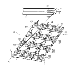
Fig. 2A is a schematic, pictorial illustration of
location pad 38, in accordance with an embodiment of the
present invention. Location pad 38 comprises multiple
magnetic field generators 39 arranged in an array shown in
the transverse XY plane of Fig. 2A. Twelve generators 39 of
equal sizes are shown in the embodiment of Fig. 2A. The array
is held in a housing which may be made from any suitable
material, such as from various plastics. X-Y-Z coordinate
axes are shown to the lower left side of the housing of
location pad 39, which has a thickness t.
Each generator 39 comprises a planar coil 100 whose
windings are parallel to the X-Y plane. In some embodiments,
coil 100 is surrounded by a trench 105. The coil may be
formed from any suitable material, such as copper. When a
signal, typically a current, is applied to coil 100, coil 100
generates a magnetic field B oriented along the Z-axis in
response to the applied signal and perpendicular to the plane
of the coil (the X-Y plane). In this example, the axes of all
the magnetic fields are parallel to one another and
perpendicular to the surface of the location pad. The
composite magnetic field in the region above the location pad
comprises the superposition of magnetic fields B from the
multiple field generators.
When patient 32 lies on location pad 38 as shown in the
inset in Fig. 1 and catheter 24 is navigated into the target
region within the patient's body above the location pad, a
magnetic sensor coil 120 near distal end 34 of the catheter
generates an electrical signal, typically a voltage, in
response to the composite magnetic field. Sensor coil 120 is
assumed here to be a single-axis sensor at distal end 34 of
catheter 24. (Alternatively, catheter 24 may comprise a
multiple-axis position sensor, e.g., a sensor comprising
three mutually-orthogonal coils.)
9
I
CA 02875170 2014-12-15
-
In the embodiments presented herein, the location pad is
configured to be placed between the patient and the top
surface of table 37, e.g., with the patient lying on top of
the location pad. The transverse dimensions of the location
pad are typically confined to the transverse dimensions of
patient table 37, which is moved into the MRI scanner. The
thickness t of the location pad is usually configured to be
no more than 5 mm. In this manner, the MRI scanner does not
collide or interfere with location pad 38 of the magnetic
tracking system, or vice versa.
Processor 40 in system 20 is configured to use the
electrical signal sensed by sensor 120 to compute a position
P vector and an orientation vector 0 of sensor 120 relative
to the origin of the X-Y-Z axes. Position vector P is the
vector from the origin to sensor 120. Orientation vector 0 is
the axial vector through catheter 24. The position of the
origin of the X-Y-Z coordinate system shown in Fig. 2A is
merely for conceptual clarity, and not by way of limitation
of the embodiments of the present invention. The origin may
be defined in any suitable position relative to the location
pad.
Fig. 2B is a schematic, pictorial illustration of an
alternative embodiment of the location pad, in accordance
with an embodiment of the present invention. In this
embodiment, each row 140 of coils 100 is planar, but the rows
lie on a slightly curved surface. In this configuration, too,
the axes of the magnetic fields generated by coils 100 are
perpendicular to the surface of the location pad. The curved
configuration of Fig. 2B is useful, for example, for fitting
into the chamber of MRI scanner 22.
On the right-most row shown in the Fig. 2B, magnetic
field generators 39 have a lid 150 covering coils 100, which
1
CA 02875170 2014-12-15
=
may be formed from any suitable material, such as a plastic,
covering the entire array.
In the configuration of Fig. 2B, the magnetic fields B
generated by coils 100 are nearly parallel to one another as
will be described below. Any small deviations of the magnetic
fields B from parallelism due to the curvature shown in Fig.
2B were found to have a negligible impact on the accuracy of
the catheter position tracking system.
When location pad 38 is used in a MRI environment, the
large magnetic MRI coils generate very large magnetic fields
such as in the range of 0.5-3 Tesla. Magnetic catheter
tracking systems, such as the CARTO system, use magnets with
AC frequencies in the audio frequency range. Hence when
magnetic coils 100 are driven with the audio frequencies in
the presence of the large MRI magnets, small magnetic coils
100 can resonate at audio frequencies, e.g., from 19-22 kHz.
Thus in some embodiments, the coils are potted in an elastic
material, such as silicone or any other suitable material, in
order to dampen or otherwise prevent this resonance. For
example, trench 105 and any other regions around coil 100 can
be filled with silicone, or any other suitable material,
which dampen the audio frequency resonances of coils 100 in
location pad 38 in an MRI environment.
Small magnetic coils 100 can also exhibit large
impedances on the order of 600 ohms, e.g., due to skin
effects at these frequencies as well as the small size of
coils 100. These coils are driven with driver amplifier 36,
which typically has an output characteristic impedance on the
order of 6 ohms. In some embodiments, to drive these high
impedance coils, transformers can be used in driver amplifier
36 with an impedance transformation ratio to overcome the
impedance mismatches from 6 ohms to 600 ohms.
The array configurations of Fig. 2A and 2B are shown
merely for visual clarity and not by way of limitation of the
11
CA 02875170 2014-12-15
embodiments of the present invention. Any suitable number of
magnetic coils 100 in any suitable configuration may be used.
Coils 100 are not limited to a flat circular shape, but may
be of any suitable shape.
COMPUTING THE POSITION AND ORIENTATION OF A CATHERTER WITH
THE MRI COMPATIBLE LOCATION PAD
As explained above, in the disclosed embodiments the
magnetic fields generated by coils 100 of location pad 38 are
parallel to one another and perpendicular to the surface of
the location pad. As a result, the magnitude of the composite
magnetic field varies with the Z coordinate but is
substantially constant as a function of X and Y. Therefore,
when using a single-axis sensor (e.g., sensor 120 in Fig.
2A), the magnitude of the composite signal sensed by the
sensor is strongly indicative of the height of distal end 34
above location pad 38, but is insensitive to the lateral
position of the distal tip relative to the location pad. This
insensitivity may cause inaccuracy or even lack of conversion
in the position and orientation estimation process performed
by processor 40.
One possible solution for this problem is to use a more
complex position sensor in the catheter, such as a three-axis
sensor. Such an arrangement is described in U.S. Patent
Application Number US 2007/0265526, cited above. This
solution, however, is complex and increases the catheter
diameter.
In some embodiments, processor 40 estimates the position
and orientation of catheter 24 is a two-stage process. This
process, which is described below, enables the use of a
single-axis sensor in conjunction with a low-profile location
pad. The disclosed process is computationally-simple and
converges quickly and efficiently. Typically, a total of five
coils 100 is sufficient for providing accurate location, but
12
CA 02875170 2014-12-15
a larger number of coils (e.g., twelve coils as shown in
Figs. 2A and 2B) is preferable for higher accuracy and
robustness.
In some embodiments, coils 100 are driven with AC
signals having different respective frequencies, so that the
signals induced in the single axis sensor coil can be
distinguished from one another.
Fig. 3 is a flow chart that schematically illustrates a
method for estimating the position of distal end 34 of
catheter 24 relative to location pad 38, in accordance with
an embodiment of the present invention. In a positioning step
200, location pad 38 is positioned under patient 32. In an
inserting step 210, catheter 24 is inserted into patient 32.
In a generating step 220, coils 100 are driven with
respective AC drive signals having different frequencies.
In measuring step 230, processor 40 measures the
electrical voltage signal that is induced in catheter sensor
120 in response to the magnetic field. In a first estimating
step 240, processor 40 estimates an initial Z-distance of the
sensor from the location pad by computing the average (e.g.,
RMS) intensity of the electrical voltage signal (which is
proportional to the average magnitude of the composite
magnetic field produced by coils 100).
In a second estimating step 250, processor 40 estimates
an initial X-Y position of the sensor relative to location
pad 38, by analyzing the relative amplitudes of the
individual different-frequency components in the induced
voltage signal. Processor 40 is able to discriminate among
the signal components induced by the different coils 100,
because each signal component has a different frequency.
Processor 40 may filter the signal sensed by sensor 120 with
suitable digital filtering for this purpose.
In an iterative estimation step 260, processor 40
refines the initial X-Y position estimate from step 250 and Z
13
CA 02875170 2014-12-15
position estimate from step 240 of the sensor. Typically,
processor 40 carries out an iterative position estimation
process that uses the initial X-Y-Z coordinate (output of
steps 240 and 250) as initial conditions. Due to the
relatively-accurate initial conditions, the iterative process
converges quickly and reliably to the accurate X-Y-Z
coordinate of the catheter distal end.
It will thus be appreciated that the embodiments
described above are cited by way of example, and that the
present invention is not limited to what has been
particularly shown and described hereinabove. Rather, the
scope of the present invention includes both combinations and
sub-combinations of the various features described
hereinabove, as well as variations and modifications thereof
which would occur to persons skilled in the art upon reading
the foregoing description and which are not disclosed in the
prior art. Documents incorporated by reference in the present
patent application are to be considered an integral part of
the application except that to the extent any terms are
defined in these incorporated documents in a manner that
conflicts with the definitions made explicitly or implicitly
in the present specification, only the definitions in the
present specification should be considered.
14