Note: Descriptions are shown in the official language in which they were submitted.
CA 02875346 2014-12-01
WO 2014/002095
PCT/1L2013/050549
FLOW-RELATED IMAGE PROCESSING IN LUMINAL ORGANS
CROSS REFERENCES TO RELATED APPLICATIONS
The present application claims the benefit of:
US Provisional Patent Application 61/690,393, entitled "Flow-related image
processing in luminal organs," filed June 26, 2012;
US Provisional Patent Application 61/741,105, entitled "Flow-related image
processing in luminal organs," filed July 12, 2012;
US Provisional Patent Application 61/692,280, entitled "Flow-related image
processing in luminal organs," filed August 23, 2012; and
US Provisional Patent Application 61/704,570, entitled "Flow-related image
processing in luminal organs," filed September 24, 2012.
The present application is related to the following patent applications:
International Patent Application PCT/IL2013/050438, entitled "Co-use of
endoluminal data and extraluminal imaging," filed May 21, 2013;
International Patent Application PCT/IL2012/000246 (published as WO
12/176191), entitled "Luminal background cleaning," filed June 21, 2012;
International Patent Application PCT/IL2011/000612 (published as WO
12/014212), entitled "Co-use of endoluminal data and extraluminal imaging,"
filed
July 28, 2011;
US Patent Application 13/228,229 (published as US 2012/0004537), entitled
"Co-use of endoluminal data and extraluminal imaging," filed September 08,
2011;
International Patent Application PCT/IL2011/000391 (published as WO
11/145094), entitled "Identification and presentation of device-to-vessel
relative
motion," filed May 17, 2011;
US Patent Application 12/781,260 to Blank (published as US 2010/0228076),
entitled "Controlled actuation and deployment of a medical device," filed May
17,
2010;
1
CA 02875346 2014-12-01
WO 2014/002095
PCT/1L2013/050549
US Patent Application 12/650,605 to Cohen (published as US
2010/0172556), entitled "Automatic enhancement of an image stream of a moving
organ," filed December 31, 2009;
International Patent Application No. PCT/IL2009/001089 (published as WO
10/058398), entitled "Image processing and tool actuation for medical
procedures,"
filed November 18, 2009;
US Patent Application 12/487,315 to Iddan (published as US 2009/0306547),
entitled "Stepwise advancement of a medical tool," filed June 18, 2009; and
US Patent Application 12/075,244 to Tolkowsky (published as US
2008/0221442), entitled "Imaging for use with moving organs," filed March 10,
2008.
All of the aforementioned references are incorporated herein by reference.
FIELD OF EMBODIMENTS OF THE INVENTION
Some applications of the present invention generally relate to medical
imaging.
Specifically, some applications of the present invention relate to determining
a luminal-flow-
related index, such as fractional flow reserve (FFR), based upon medical
imaging.
BACKGROUND
Fractional flow reserve (FFR) is physiological index that measures the
functional
severity of a coronary artery stenosis (i.e., a narrowing, and/or an occlusion
of the artery that
is usually due to atherosclerosis). FFR measures the severity of the stenosis
by determining
the maximal blood flow through the artery in the presence of the stenosis
relative to the
hypothetical level of blood flow through the artery, if the artery were
healthy. FFR provides
an indication of the likelihood that the stenosis is impeding and/or will
impede oxygen
delivery to the heart muscle (i.e., the likelihood that the stenosis is
causing and/or will cause
myocardial ischemia). Other luminal-flow-related indices that are used to
measure
conditions of the coronary circulatory system include instantaneous wave-free
ratio (iFR),
coronary flow reserve (CFR), index of microcirculatory resistance (IMR),
microvascular
resistance index (MVRI), TIMI myocardial perfusion grade (TMPG), relative
fractional flow
reserve (RFFR), and other related (e.g., other statistically correlated)
indices.
2
CA 02875346 2014-12-01
WO 2014/002095
PCT/1L2013/050549
FFR is typically utilized in coronary catheterizations, and is typically
calculated by
measuring pressure differences across a coronary artery stenosis. Assuming
that there is
single stenosis, the relationship between the pressure downstream of the
stenosis and the
pressure upstream of the stenosis approximates the relationship between the
flow of blood in
the currently-stenosed coronary artery and the normal flow of blood had the
artery been
healthy. Thus, measuring pressure differences across a coronary artery
stenosis provides an
approximation of the FFR.
Typically, FFR serves as a decision support tool for determining whether the
stenosis
should be treated, such as by means of inflating a balloon and implanting a
stent.
FFR is defined as the ratio between stenotic flow Qs and normal flow QN under
hyperemic conditions: FFR = Qs/QN
Using the flow equation Q = AP/R, where Q is the flow (mL/min), AP is the
pressure
difference (mm Hg), and R is resistance (mmHg x min / mL), and the assumption
that the
venous pressure Pvein is negligible, the FFR can be expressed as the ratio
between distal
pressure Pd to proximal pressure Pa of a stenosis:
FFR = (Qs/QN) = ((Pd ¨ Pvein)/R)/((Pa ¨ Pvein)/R) = Pd/Pa
This pressure ratio can be written as follows:
FFR= Pd/Pa = (Pa- AP,)/Pa
where AP, is the pressure drop along the axis of the lumen along a segment of
the
lumen from upstream of the stenosis to downstream of the stenosis.
The FFR result is an absolute number between zero and one; an FFR of 0.50
means
that a given stenosis causes a 50% drop in blood pressure. In other words, FFR
expresses
the maximal flow through a lumen in the presence of a stenosis compared to the
maximal
flow in the hypothetical absence of the stenosis.
Typically, FFR is measured in coronary vessels by means of inserting into such
vessels a wire equipped with sensors. The device analyzes pressure and flow
parameters
from inside of the vessel. Such wires are currently being produced by Volcano
Corp. (San
Diego, CA) and by St. Jude Medical (St. Paul, MN).
3
CA 02875346 2014-12-01
WO 2014/002095
PCT/1L2013/050549
SUMMARY OF EMBODIMENTS
For some applications of the present invention, flow-related image processing
is
performed on luminal organs. Typically, a set of angiographic images of a
lumen is
acquired, and the geometry of the lumen at a given location within the lumen
(typically, in a
vicinity of a stenosis within the lumen) is determined automatically by
performing image
processing on at least one of the angiographic images. Blood velocity along
the lumen is
determined automatically, by performing image processing on at least two of
the
angiographic images. Typically, the geometry of the lumen and the blood
velocity are
determined without generating a three dimensional model of the lumen. For some
applications, the geometry of the lumen and the blood velocity are determined
solely by
performing image-processing on two-dimensional angiographic images of the
lumen. Based
upon the geometry of the lumen and the blood velocity, the value of a current
flow-related
parameter of the lumen at the given location is determined. For example, the
current flow,
blood pressure, and/or blood velocity may be determined. An indication of a
value of a
second flow-related parameter of the subject is received. For example, an
indication of
blood pressure at an upstream location (e.g., aortic pressure) may be
received. Alternatively
or additionally, a historic angiographic image sequence that was acquired when
the lumen
was healthy may be received, and flow, blood pressure, and/or blood velocity
within the
lumen at the time when the lumen was healthy may be derived from the historic
angiographic image sequence. A value of a luminal-flow-related index of the
subject (such
as the FFR of the subject) at the location is determined by determining a
relationship
between the value of the current flow-related parameter and the value of the
second flow-
related parameter.
For some applications, the value of a luminal-flow-related index of the
subject is
determined by (a) automatically determining pressure at a site based upon the
automatically-
determined lumen geometry and the automatically-determined blood velocity at
the site, and
(b) determining a relationship between the automatically-determined pressure
at the site, and
the subject's aortic pressure. An output is typically generated in response to
the determined
index at the site. For example, a stabilized image stream that is based upon
the acquired
angiographic images may be displayed, and, at a location within the image
stream
corresponding to the site, an indication of the index at the site may be
displayed. For some
applications, an indication of the value of the flow-related index is
generated on an image of
4
CA 02875346 2014-12-01
WO 2014/002095
PCT/1L2013/050549
the lumen, using a color legend. Alternatively or additionally, in response to
the luminal-
flow-related index passing a first threshold value, an output is generated
indicating that
treatment of the subject is recommended, and in response to the luminal-flow-
related index
passing a second threshold value but not passing the first threshold value, an
output is
generated recommending that the luminal-flow-related index be measured using a
sensor that
is inserted into the lumen.
Typically, image processing described in the present application is performed
intra-
procedurally, though, for some applications, image processing is applied post-
procedurally.
Although some applications of the present invention are described with
reference to
coronary catheterizations, the scope of the present invention includes
applying the apparatus
and methods described herein to other medical procedures and to other luminal
organs in
which there is a flow of fluid. For example, for some applications, the
apparatus and
methods described herein are applied, mutatis mutandis, to renal
catheterization procedures,
subclavian procedures, and/or below-the-knee procedures. For some such
applications,
determining a luminal-flow-related index using angiographic data facilitates
determination
of such an index, even in cases in which determination of the index via
insertion of a wire
would be physiologically difficult.
Although some applications of the present invention are described with
reference to
determining a subject's fractional flow reserve, the scope of the present
invention includes
applying the apparatus and methods described herein to determine other luminal-
flow-related
indices, including but not limited to instantaneous wave-free ratio (iFR),
coronary flow
reserve (CFR), index of microcirculatory resistance (IMR), microvascular
resistance index
(MVRI), TIMI myocardial perfusion grade (TMPG), relative fractional flow
reserve (RFFR),
and/or other related (e.g., other statistically correlated) indices.
It is noted that the terms "vessel" and "lumen" are used interchangeably in
the present
application. Both of the aforementioned terms should be construed to mean
structures within
the body that are shaped as lumens, for example, arteries and veins.
It is noted that the term "proximal" is used in the present application to
denote a
location within a lumen that is upstream of a given reference location (such
as a stenosis)
within the lumen, and the term "distal" is used to denote a location within a
lumen that is
downstream of a given reference location.
5
CA 02875346 2014-12-01
WO 2014/002095
PCT/1L2013/050549
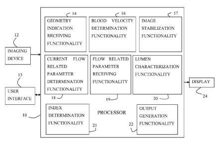
There is therefore provided, in accordance with some applications of the
present
invention, apparatus for use with an imaging device configured to acquire a
set of
angiographic images of a lumen of a subject's body, and a display, the
apparatus including:
at least one processor, including:
blood-velocity-determination functionality configured, via image processing,
to determine blood velocity within the lumen, by:
defining at least first and second regions of interest along the lumen in
one of the angiographic images;
identifying the regions of interest in at least some additional
angiographic images belonging to the set of angiographic images;
determining a distance between the regions of interest;
determining that a presence of a contrast agent appears at the first
region of interest in a first one of the angiographic images and that the
presence of contrast agent appears at the second region of interest in a
second
one of the angiographic images; and
determining the time that it took for the contrast agent to travel from
the first region of interest to the second region of interest, based upon an
interval between an acquisition of the first angiographic image and an
acquisition of the second angiographic image;
geometry-indication-receiving functionality configured to receive an
indication of geometry of the lumen at a given location within the lumen;
current-flow-related-parameter-determination functionality configured to
determine a value of a current flow-related parameter at the location based
upon the
determined blood velocity and the geometry of the lumen in the vicinity of the
location;
flow-related-parameter-receiving functionality configured to receive an
indication of a value of a second flow-related parameter of the subject;
index-determination functionality configured to determine a value of a
luminal-flow-related index of the subject at the location, by determining a
relationship between the value of the current flow-related parameter and the
value of
the second flow-related parameter; and
6
CA 02875346 2014-12-01
WO 2014/002095
PCT/1L2013/050549
output-generation functionality configured to generate an output on the
display in response to the determined value of the luminal-flow-related index.
For some applications, the given location includes a location in a vicinity of
a
stenosis within the lumen, and the index-determination functionality is
configured to
determine the value of the luminal-flow-related index of the subject at the
location, by
determining the value of the luminal-flow-related index in the vicinity of the
stenosis.
For some applications, the index-determination functionality is configured to
determine the value of the luminal-flow-related index of the subject at the
location, by
determining a value of functional flow reserve of the subject at the location.
For some applications, the index-determination functionality is configured to
determine the value of the luminal-flow-related index of the subject at the
location, by
determining a value of instantaneous wave-free ratio of the subject at the
location.
For some applications, the blood-velocity-determination functionality is
configured
to determine that the presence of the contrast agent appears at the first
region of interest in
the first one of the angiographic images and that the presence of contrast
agent appears at the
second region of interest in the second one of the angiographic images by
determining that a
given concentration of the contrast agent appears at the first region of
interest in the first one
of the angiographic images and that the given concentration of contrast agent
appears at the
second region of interest in the second one of the angiographic images.
For some applications, the blood-velocity-determination functionality is
configured
to determine that the presence of the contrast agent appears at the first
region of interest in
the first one of the angiographic images and that the presence of contrast
agent appears at the
second region of interest in the second one of the angiographic images by
determining that a
bolus of the contrast agent appears at the first region of interest in the
first one of the
angiographic images and that the bolus of contrast agent appears at the second
region of
interest in the second one of the angiographic images.
For some applications, the blood-velocity-determination functionality is
configured
to determine that the presence of the contrast agent appears at the first
region of interest in
the first one of the angiographic images and that the presence of contrast
agent appears at the
second region of interest in the second one of the angiographic images by
determining that a
given pattern of the contrast agent appears at the first region of interest in
the first one of the
7
CA 02875346 2014-12-01
WO 2014/002095
PCT/1L2013/050549
angiographic images and that the given pattern of contrast agent appears at
the second region
of interest in the second one of the angiographic images.
For some applications, the blood-velocity-determination functionality is
configured
to define at least first and second regions of interest along the lumen in one
of the
angiographic images by defining at least first and second regions of interest
along a center
line of the lumen in one of the angiographic images.
For some applications, the at least one processor further includes image-
stabilization
functionality configured to generate a stabilized image stream of the lumen
based upon the
acquired angiographic images, and the output-generation functionality is
configured to
generate the output by driving the display to display the stabilized image
stream, and by
generating, at a location that corresponds to the location and that is within
the displayed
image stream, an indication of the value of the flow-related index at the
location.
For some applications, the output-generation functionality is configured to
generate
the output by driving the display to display an indication of the value of the
flow-related
index, using a color legend, on an image of the lumen.
For some applications, the current-flow-related-parameter-determination
functionality is configured to determine the value of the current flow-related
parameter at the
location using a machine-learning classifier, based upon at least the
determined blood
velocity and the geometry of the lumen at the location.
For some applications, the index-determination functionality is configured to
determine the relationship between the value of the current flow-related
parameter and the
value of the second flow-related parameter by determining the relationship
between the
value of the current flow-related parameter and the value of the second flow-
related
parameter using a machine-learning classifier.
For some applications, the output-generation functionality is configured to
generate
the output by:
in response to the luminal-flow-related index passing a first threshold value,
generating an output indicating that treatment of the subject is recommended;
and
in response to the luminal-flow-related index passing a second threshold value
but
not passing the first threshold value, generating an output recommending that
the luminal-
flow-related index be measured using a sensor that is inserted into the lumen.
8
CA 02875346 2014-12-01
WO 2014/002095
PCT/1L2013/050549
For some applications:
the location includes a location in the vicinity of a stenosis;
the flow-related-parameter-receiving functionality is configured to receive
the
indication of the value of the second flow-related parameter of the subject by
receiving an
indication of a value of blood pressure of the subject at a location that is
upstream of the
stenosis;
the current-flow-related-parameter-determination functionality is configured
to
determine the value of the current flow-related parameter in the vicinity of
the stenosis by
determining a value of current blood pressure in the vicinity of the stenosis
based upon the
determined blood velocity and the geometry of the lumen in the vicinity of the
stenosis; and
the index-determination functionality is configured to determine the
relationship
between the value of the current flow-related parameter and the value of the
second flow-
related parameter by comparing the current blood pressure in the vicinity of
the stenosis to
the subject's blood pressure at the location that is upstream of the stenosis.
For some applications, the flow-related-parameter-receiving functionality is
configured to receive the indication of the value of the blood pressure of the
subject at the
location that is upstream of the stenosis by receiving an indication of a
value of aortic blood
pressure of the subject.
For some applications, the flow-related-parameter-receiving functionality is
configured to receive the indication of the value of the second flow-related
parameter of the
subject by receiving the indication of the value of the second flow-related
parameter of the
subject, based upon patient history of the subject.
For some applications:
the flow-related-parameter-receiving functionality is configured to receive
the
indication of the value of the second flow-related parameter of the subject by
receiving at
least one previously-acquired angiographic image of the subject's lumen,
the flow-related-parameter-receiving functionality is further configured to
derive a
value of flow within the lumen at a time of acquisition of the previously-
acquired
angiographic image,
the current-flow-related-parameter-determination functionality is configured
to
determine the value of the current flow-related parameter in the vicinity of
the stenosis by
9
CA 02875346 2014-12-01
WO 2014/002095
PCT/1L2013/050549
determining a value of current flow at the location based upon the determined
blood velocity
and the geometry of the lumen at the location; and
the index-determination functionality is configured to determine the
relationship
between the value of the current flow-related parameter and the value of the
second flow-
related parameter by determining a relationship between the value of the
current flow at the
location and the value of the derived flow within the lumen at the time of
acquisition of the
previously-acquired angiographic image.
For some applications:
the flow-related-parameter-receiving functionality is configured to receive
the
indication of the value of the second flow-related parameter of the subject by
receiving at
least one previously-acquired angiographic image of the subject's lumen,
the flow-related-parameter-receiving functionality is further configured to
derive a
value of blood velocity within the lumen at a time of acquisition of the
previously-acquired
angiographic image,
the current-flow-related-parameter-determination functionality is configured
to
determine the value of the current flow-related parameter in the vicinity of
the stenosis by
determining a value of current blood velocity at the location based upon the
determined
blood velocity and the geometry of the lumen at the location; and
the index-determination functionality is configured to determine the
relationship
between the value of the current flow-related parameter and the value of the
second flow-
related parameter by determining a relationship between the value of the
current blood
velocity at the location and the value of the derived blood velocity within
the lumen at the
time of acquisition of the previously-acquired angiographic image.
For some applications, the geometry-indication-receiving functionality is
configured
to determine geometry of the lumen at the location, based upon the received
indication of the
geometry of the lumen.
For some applications, the current-flow-related-parameter-determination
functionality is configured to determine the value of the current flow-related
parameter at the
location using a machine-learning classifier, based upon the determined lumen
geometry and
the determined blood velocity.
CA 02875346 2014-12-01
WO 2014/002095
PCT/1L2013/050549
For some applications, the geometry-indication-receiving functionality is
configured
to:
receive the indication of the geometry of the lumen by receiving at least one
of the
set of angiographic images, and
determine geometry of the lumen at the location by determining a cross-
sectional
area of the lumen by performing quantitative vessel analysis on the at least
one of the set of
angiographic images.
For some applications, the geometry-indication-receiving functionality is
configured
to:
receive the indication of the geometry of the lumen by receiving at least one
of the
set of angiographic images, and
determine geometry of the lumen at the location by determining a cross-
sectional
area of the lumen by performing densitometry on the at least one of the set of
angiographic
images.
For some applications, the flow-related-parameter-receiving functionality is
configured to receive the indication of a value of a second flow-related
parameter of the
subject by receiving an angiographic image of a second location within the
lumen, and the
flow-related-parameter-receiving functionality is configured to determine
geometry of the
lumen at the second location within the lumen, by performing image processing
on the
angiographic image of the second location within the lumen.
For some applications, the flow-related-parameter-receiving functionality is
configured to determine geometry of the lumen at the second location within
the lumen by
determining a cross-sectional area at the second location within the lumen by
performing
quantitative vessel analysis on the angiographic image of the second location
within the
lumen.
For some applications, the flow-related-parameter-receiving functionality is
configured to determine geometry of the lumen at the second location within
the lumen by
determining a cross-sectional area at the second location within the lumen by
performing
densitometry on the angiographic image of the second location within the
lumen.
For some applications, the flow-related-parameter-receiving functionality is
configured to determine a value of flow at the second location within the
lumen based upon
11
CA 02875346 2014-12-01
WO 2014/002095
PCT/1L2013/050549
the determined geometry at the second location within the lumen and the
determined blood
velocity.
For some applications, the flow-related-parameter-receiving functionality is
configured to determine the value of the flow at the second location within
the lumen based
upon the determined geometry at the second location within the lumen and the
determined
blood velocity, using a machine-learning classifier.
There is further provided, in accordance with some applications of the present
invention, a method for use with a set of angiographic images of a lumen of a
subject's body,
the method including:
via image processing, determining blood velocity within the lumen, by:
defining at least first and second regions of interest along the lumen in one
of
the angiographic images;
identifying the regions of interest in at least some additional angiographic
images belonging to the set of angiographic images;
determining a distance between the regions of interest;
determining that a presence of a contrast agent appears at the first region of
interest in a first one of the angiographic images and that the presence of
contrast
agent appears at the second region of interest in a second one of the
angiographic
images; and
determining the time that it took for the contrast agent to travel from the
first
region of interest to the second region of interest, based upon an interval
between an
acquisition of the first angiographic image and an acquisition of the second
angiographic image;
receiving an indication of geometry of the lumen at a given location within
the
lumen;
determining a value of a current flow-related parameter at the location based
upon the
determined blood velocity and the geometry of the lumen in the vicinity of the
location;
receiving an indication of a value of a second flow-related parameter of the
subject;
determining a value of a luminal-flow-related index of the subject at the
location, by
determining a relationship between the value of the current flow-related
parameter and the
value of the second flow-related parameter; and
12
CA 02875346 2014-12-01
WO 2014/002095
PCT/1L2013/050549
generating an output in response to the determined value of the luminal-flow-
related
index.
For some applications, the contrast agent is within the lumen due to an
injection of
contrast agent into the lumen, and the method further includes acquiring a
plurality of
endoluminal images of the lumen, the acquisition of the plurality of
endoluminal images
being facilitated by the injection of the contrast agent.
There is further provided, in accordance with some applications of the present
invention, apparatus for use with an imaging device configured to acquire a
set of
angiographic images of a lumen of a subject's body, and a display, the
apparatus including:
at least one processor including:
image-processing functionality configured to analyze temporal changes in a
density of a contrast agent at a given location within the lumen;
lumen-characterization functionality configured, in response to the analysis,
to determine a characteristic of the lumen at the location, the characteristic
being
selected from the group consisting of: a presence of a stenosis in a vicinity
of the
location, and a value of a luminal-flow-related index of the subject at the
location;
and
output-generation functionality configured to generate an output on the
display in response to the determined characteristic of the lumen.
For some applications, the lumen-characterization functionality is configured
to
determine the characteristic of the lumen at the location, using a machine
learning classifier.
For some applications, the at least one processor further includes geometry-
indication-receiving functionality configured to determine geometry of the
lumen at the
location, and the lumen-characterization functionality is configured to
determine the
characteristic of the lumen at the location by determining the characteristic
of the lumen at
the location in response to the geometry of the vessel at the location and the
analysis of the
temporal changes in the density of the contrast agent at the location.
For some applications, the lumen-characterization functionality is configured
to
determine the characteristic of the lumen at the location by determining the
value of the
luminal-flow-related index of the subject at the location.
13
CA 02875346 2014-12-01
WO 2014/002095
PCT/1L2013/050549
For some applications, the lumen-characterization functionality is configured
to
determine the characteristic of the lumen at the location by determining the
presence of the
stenosis in the vicinity of the location.
For some applications, the contrast agent includes contrast agent that is
administered
to the subject's lumen according to a given protocol, and the lumen-
characterization
functionality is configured to determine the characteristic of the lumen at
the location by
determining the characteristic of the lumen at the location based upon the
temporal changes
in the density of the contrast agent at the given location within the lumen
and the given
protocol.
For some applications, the lumen-characterization functionality is configured
to
determine the characteristic of the lumen at the location, using a machine
learning classifier.
For some applications, the contrast agent includes contrast agent that is
administered
to the subject's lumen according to a given time-density protocol, and the
lumen-
characterization functionality is configured to determine the characteristic
of the lumen at the
location by comparing the temporal changes in the density of the contrast
agent at the given
location within the lumen to the time-density protocol according to which the
contrast agent
was administered to the subject.
There is further provided, in accordance with some applications of the present
invention, a method for use with a set of angiographic images of a lumen of a
subject's body,
the method including:
via image processing, analyzing temporal changes in a density of a contrast
agent at a
given location within the lumen;
in response to the analysis, determining a characteristic of the lumen at the
location,
the characteristic being selected from the group consisting of: a presence of
a stenosis in a
vicinity of the location, and a value of a luminal-flow-related index of the
subject at the
location; and
in response thereto, generating an output.
For some applications, the contrast agent includes contrast agent that is
injected into
the lumen, and the method further includes acquiring a plurality of
endoluminal images of
the lumen, the acquisition of the plurality of endoluminal images being
facilitated by the
injection of the contrast agent.
14
CA 02875346 2014-12-01
WO 2014/002095
PCT/1L2013/050549
There is further provided, in accordance with some applications of the present
invention, apparatus for use with an imaging device configured to acquire a
set of two-
dimensional angiographic images of a lumen of a subject's body, and a display,
the apparatus
including:
at least one processor, including:
blood-velocity-determination functionality configured, without generating a
virtual three-dimensional model of the lumen, and by performing image
processing
on the two-dimensional angiographic images, to determine blood velocity within
the
lumen;
geometry-indication-receiving functionality configured, without generating a
virtual three-dimensional model of the lumen, and by performing image
processing
on the two-dimensional angiographic images, to determine geometry of the lumen
at
a given location within the lumen;
current-flow-related-parameter-determination functionality configured to
determine a value of a current flow-related parameter at the location based
upon the
determined blood velocity and the geometry of the lumen in the vicinity of the
location;
flow-related-parameter-receiving functionality configured to receive an
indication of a value of a second flow-related parameter of the subject;
index-determination functionality configured to determine a value of a
luminal-flow-related index of the subject at the location, by determining a
relationship between the value of the current flow-related parameter and the
value of
the second flow-related parameter; and
output-generation functionality configured to generate an output on the
display in response to the determined value of the luminal-flow-related index.
For some applications, the blood-velocity-determination functionality is
configured
to determine the blood velocity within the lumen by:
defining at least first and second regions of interest along the lumen in one
of the
angiographic images;
identifying the regions of interest in at least some additional angiographic
images
belonging to the set of angiographic images;
determining a distance between the regions of interest;
CA 02875346 2014-12-01
WO 2014/002095
PCT/1L2013/050549
determining that a presence of a contrast agent appears at the first region of
interest in
a first one of the angiographic images and that the presence of contrast agent
appears at the
second region of interest in a second one of the angiographic images; and
determining the time that it took for the contrast agent to travel from the
first region
of interest to the second region of interest, based upon an interval between
an acquisition of
the first angiographic image and an acquisition of the second angiographic
image.
For some applications, the given location includes a location in a vicinity of
a
stenosis within the lumen, and the index-determination functionality is
configured to
determine the value of the luminal-flow-related index of the subject at the
location, by
determining the value of the luminal-flow-related index in the vicinity of the
stenosis.
For some applications, the index-determination functionality is configured to
determine the value of the luminal-flow-related index of the subject at the
location, by
determining a value of functional flow reserve of the subject at the location.
For some applications, the index-determination functionality is configured to
determine the value of the luminal-flow-related index of the subject at the
location, by
determining a value of instantaneous wave-free ratio of the subject at the
location.
For some applications, the at least one processor further includes image-
stabilization
functionality configured to generate a stabilized image stream of the lumen
based upon the
acquired angiographic images, and the output-generation functionality is
configured to
generate the output by driving the display to display the stabilized image
stream, and by
generating, at a location that corresponds to the location and that is within
the displayed
image stream, an indication of the value of the flow-related index at the
location.
For some applications, the output-generation functionality is configured to
generate
the output by driving the display to display an indication of the value of the
flow-related
index, using a color legend, on an image of the lumen.
For some applications, the current-flow-related-parameter-determination
functionality is configured to determine the value of the current flow-related
parameter at the
location based upon at least the determined blood velocity and the geometry of
the lumen at
the location, using a machine-learning classifier.
16
CA 02875346 2014-12-01
WO 2014/002095
PCT/1L2013/050549
For some applications, the index-determination functionality is configured to
determine the relationship between the value of the current flow-related
parameter and the
value of the second flow-related parameter by determining the relationship
between the
value of the current flow-related parameter and the value of the second flow-
related
parameter using a machine-learning classifier.
For some applications, the output-generation functionality is configured to
generate
the output by:
in response to the luminal-flow-related index passing a first threshold value,
generating an output indicating that treatment of the subject is recommended;
and
in response to the luminal-flow-related index passing a second threshold value
but
not passing the first threshold value, generating an output recommending that
the luminal-
flow-related index be measured using a sensor that is inserted into the lumen.
For some applications, the geometry-indication-receiving functionality is
configured
to determine the geometry of the lumen at the given location within the lumen
determining a
cross-sectional area of the lumen by performing quantitative vessel analysis
on at least one
of the set of angiographic images.
For some applications, the geometry-indication-receiving functionality is
configured
to determine the geometry of the lumen at the given location within the lumen
by performing
densitometry on at least one of the set of angiographic images.
For some applications:
the location includes a location in the vicinity of a stenosis;
the flow-related-parameter-receiving functionality is configured to receive
the
indication of the value of the second flow-related parameter of the subject by
receiving an
indication of a value of blood pressure of the subject at a location that is
upstream of the
stenosis;
the current-flow-related-parameter-determination functionality is configured
to
determine the value of the current flow-related parameter in the vicinity of
the stenosis by
determining a value of current blood pressure in the vicinity of the stenosis
based upon the
determined blood velocity and the geometry of the lumen in the vicinity of the
stenosis; and
the index-determination functionality is configured to determine the
relationship
between the value of the current flow-related parameter and the value of the
second flow-
17
CA 02875346 2014-12-01
WO 2014/002095
PCT/1L2013/050549
related parameter by comparing the current blood pressure in the vicinity of
the stenosis to
the subject's blood pressure at the location that is upstream of the stenosis.
For some applications, the flow-related-parameter-receiving functionality is
configured to receive the indication of the value of the blood pressure of the
subject at the
location that is upstream of the stenosis by receiving an indication of a
value of aortic blood
pressure of the subject.
For some applications, the flow-related-parameter-receiving functionality is
configured to receive the indication of the value of the second flow-related
parameter of the
subject by receiving the indication of the value of the second flow-related
parameter of the
subject, based upon patient history of the subject.
For some applications:
the flow-related-parameter-receiving functionality is configured to receive
the
indication of the value of the second flow-related parameter of the subject by
receiving at
least one previously-acquired angiographic image of the subject's lumen,
the flow-related-parameter-receiving functionality is further configured to
derive a
value of flow within the lumen at a time of acquisition of the previously-
acquired
angiographic image,
the current-flow-related-parameter-determination functionality is configured
to
determine the value of the current flow-related parameter in the vicinity of
the stenosis by
determining a value of current flow at the location based upon the determined
blood velocity
and the geometry of the lumen at the location; and
the index-determination functionality is configured to determine the
relationship
between the value of the current flow-related parameter and the value of the
second flow-
related parameter by determining a relationship between the value of the
current flow at the
location and the value of the derived flow within the lumen at the time of
acquisition of the
previously-acquired angiographic image.
For some applications:
the flow-related-parameter-receiving functionality is configured to receive
the
indication of the value of the second flow-related parameter of the subject by
receiving at
least one previously-acquired angiographic image of the subject's lumen,
18
CA 02875346 2014-12-01
WO 2014/002095
PCT/1L2013/050549
the flow-related-parameter-receiving functionality is further configured to
derive a
value of blood velocity within the lumen at a time of acquisition of the
previously-acquired
angiographic image,
the current-flow-related-parameter-determination functionality is configured
to
determine the value of the current flow-related parameter in the vicinity of
the stenosis by
determining a value of current blood velocity at the location based upon the
determined
blood velocity and the geometry of the lumen at the location; and
the index-determination functionality is configured to determine the
relationship
between the value of the current flow-related parameter and the value of the
second flow-
related parameter by determining a relationship between the value of the
current blood
velocity at the location and the value of the derived blood velocity within
the lumen at the
time of acquisition of the previously-acquired angiographic image.
For some applications, the flow-related-parameter-receiving functionality is
configured to receive the indication of a value of a second flow-related
parameter of the
subject by receiving an angiographic image of a second location within the
lumen, and the
flow-related-parameter-receiving functionality is configured to determine
geometry of the
lumen at the second location within the lumen, by performing image processing
on the
angiographic image of the second location within the lumen.
For some applications, the flow-related-parameter-receiving functionality is
configured to determine geometry of the lumen at the second location within
the lumen by
determining a cross-sectional area at the second location within the lumen by
performing
quantitative vessel analysis on the angiographic image of the second location
within the
lumen.
For some applications, the flow-related-parameter-receiving functionality is
configured to determine geometry of the lumen at the second location within
the lumen by
determining a cross-sectional area at the second location within the lumen by
performing
densitometry on the angiographic image of the second location within the
lumen.
For some applications, the flow-related-parameter-receiving functionality is
configured to determine a value of flow at the second location within the
lumen based upon
the determined geometry at the second location within the lumen and the
determined blood
velocity.
19
CA 02875346 2014-12-01
WO 2014/002095
PCT/1L2013/050549
For some applications, the flow-related-parameter-receiving functionality is
configured to determine the value of the flow at the second location within
the lumen based
upon the determined geometry at the second location within the lumen and the
determined
blood velocity, using a machine-learning classifier.
There is further provided, in accordance with some applications of the present
invention, a method for use with a set of two-dimensional angiographic images
of a lumen of
a subject's body, the method including:
without generating a virtual three-dimensional model of the lumen, and by
performing image processing on the two-dimensional angiographic images:
determining blood velocity within the lumen; and
determining geometry of the lumen at a given location within the lumen;
determining a value of a current flow-related parameter at the location based
upon the
determined blood velocity and the geometry of the lumen at the location;
receiving an indication of a value of a second flow-related parameter of the
subject;
determining a value of a luminal-flow-related index of the subject at the
location, by
determining a relationship between the value of the current flow-related
parameter and the
value of the second flow-related parameter; and
generating an output in response to the determined value of the luminal-flow-
related
index.
There is further provided, in accordance with some applications of the present
invention, apparatus for use with a lumen of a subject, including:
a pressure sensor configured to measure pressure of the lumen;
a blood velocity sensor configured to measure blood velocity within the lumen;
and
at least one processor including:
lumen-dimension-derivation functionality configured to derive a dimension of
the lumen from the measured pressure and blood velocity; and
output-generation functionality configured to generate an out output in
response to the derived dimension.
For some applications, the apparatus further includes a tool configured to be
inserted
into the lumen, and the pressure sensor and the blood velocity sensor are both
coupled to the
tool.
CA 02875346 2014-12-01
WO 2014/002095
PCT/1L2013/050549
For some applications, the lumen-dimension-derivation functionality is
configured to
derive the dimension of the lumen by deriving a length of a portion of the
lumen.
For some applications, the lumen-dimension-derivation functionality is
configured to
derive the dimension of the lumen by deriving a cross-sectional area of the
lumen.
For some applications, the lumen-dimension-derivation functionality is
configured to
derive the dimension of the lumen by deriving a percentage occlusion of the
lumen.
For some applications, the lumen-dimension-derivation functionality is
configured to
derive the dimension of the lumen by deriving a diameter of the lumen.
For some applications, the lumen-dimension-derivation functionality is
configured to
derive the diameter of the lumen by deriving a minimum lumen diameter of the
lumen.
There is further provided, in accordance with some applications of the present
invention, a method for use with a lumen of a subject, including:
measuring pressure of the lumen;
measuring blood velocity within the lumen;
deriving from the measured pressure and blood velocity, a dimension of the
lumen;
and
generating an output in response thereto.
For some applications, measuring pressure of the lumen includes measuring
pressure
of the lumen using a pressure sensor that is coupled to a medical device while
the medical
device is inside the lumen, and measuring blood velocity includes measuring
blood velocity
using a blood velocity sensor that is coupled to the medical device while the
medical device
is inside the lumen.
There is further provided, in accordance with some applications of the present
invention, apparatus for use with (a) an endoluminal data-acquisition device
configured to be
moved through a lumen of a subject's body, and to acquire at least a first set
of endoluminal
data points of the lumen at a plurality of locations within the lumen, while
being moved
through the lumen, (b) and extraluminal imaging device configured to acquire
an
extraluminal image of the lumen, and (c) a display, the apparatus including:
at least one processor, including:
21
CA 02875346 2014-12-01
WO 2014/002095
PCT/1L2013/050549
endoluminal-geometry-derivation-functionality configured, for at least some
of the endoluminal data points, to derive from the endoluminal data point a
value of a
geometrical parameter of the lumen at a location within the lumen at which the
endoluminal data point was acquired;
extraluminal-geometry-derivation-functionality configured to derive values of
the geometrical parameter of the lumen at a plurality of locations along the
lumen, by
performing image processing on the at least one extraluminal image of the
lumen;
co-registration functionality configured to co-register at least some of the
endoluminal data points to locations along the lumen within the extraluminal
image
by correlating the values of the geometrical parameters corresponding to the
endoluminal data points with the values of the geometrical parameter derived
by
performing image processing on the at least one extraluminal image; and
output-generation functionality configured to generate an output on the
display based upon the co-registration.
For some applications, the output-generation functionality is configured to
generate
the output by generating an output indicating that a given endoluminal data
point
corresponds to a given location along the lumen.
For some applications:
the endoluminal-geometry-derivation-functionality is configured to derive the
value
of the geometrical parameter of the lumen by deriving a value of a geometrical
parameter of
the lumen selected from the group consisting of: a cross-sectional area of the
lumen, and a
diameter of the lumen; and
the extraluminal-geometry-derivation-functionality is configured to derive
values of
the geometrical parameter of the lumen, by deriving values of the selected
geometrical
parameter.
For some applications, the set of endoluminal data points includes a set of
blood
velocity data points that are indicative of blood velocity within the lumen at
locations at
which respective endoluminal data points belonging to the set of endoluminal
data points
were acquired, and the endoluminal-geometry-derivation-functionality is
configured to
derive from at least some of the blood velocity data points a value of a
geometrical
22
CA 02875346 2014-12-01
WO 2014/002095
PCT/1L2013/050549
parameter of the lumen at a location within the lumen at which the blood
velocity data point
was acquired.
For some applications, the set of endoluminal data points includes a set of
blood
pressure data points that are indicative of blood pressure within the lumen at
locations at
which respective endoluminal data points belonging to the set of endoluminal
data points
were acquired, and the endoluminal-geometry-derivation-functionality is
configured to
derive from at least some of the blood pressure data points a value of a
geometrical
parameter of the lumen at a location within the lumen at which the blood
pressure data point
was acquired.
For some applications, the set of endoluminal data points includes a set of
flow data
points that are indicative of flow within the lumen at locations at which
respective
endoluminal data points belonging to the set of endoluminal data points were
acquired, and
the endoluminal-geometry-derivation-functionality is configured to derive from
at least some
of the flow data points a value of a geometrical parameter of the lumen at a
location within
the lumen at which the flow data point was acquired.
For some applications, the set of endoluminal data points includes a set of
endoluminal images, and the endoluminal-geometry-derivation-functionality is
configured to
derive the value of the geometrical parameter of the lumen at the location
within the lumen
at which an endoluminal data point was acquired by deriving the value of the
geometrical
parameter of the lumen at the location within the lumen at which an
endoluminal image was
acquired by performing image processing on the endoluminal image.
For some applications:
the endoluminal data-acquisition device includes an endoluminal data-
acquisition
device that is further configured to acquire a second set of endoluminal data
points of the
lumen at a plurality of locations within the lumen, while being moved through
the lumen;
the co-registration functionality is configured, based upon the co-registering
of the
first set of endoluminal data points to locations along the lumen within the
extraluminal
image, to co-register the second set of endoluminal data points to locations
along the lumen
within the extraluminal image; and
23
CA 02875346 2014-12-01
WO 2014/002095
PCT/1L2013/050549
the output-generation functionality is configured to generate the output by
generating
an output indicating that a given endoluminal data point belonging to the
second set of
endoluminal data points corresponds to a given location along the lumen.
For some applications:
the first set of endoluminal data points includes a set of blood velocity data
points
that are indicative of blood velocity within the lumen at locations at which
respective
endoluminal data points belonging to the set of endoluminal data points were
acquired;
the endoluminal-geometry-derivation-functionality is configured to derive from
at
least some of the blood velocity data points a value of a geometrical
parameter of the lumen
at a location within the lumen at which the blood velocity data point was
acquired;
the second set of endoluminal data points includes a set of endoluminal
images; and
the output-generation functionality is configured to generate the output by
generating
an output indicating that a given endoluminal image corresponds to a given
location along
the lumen.
For some applications:
the first set of endoluminal data points includes a set of blood velocity data
points
that are indicative of blood velocity within the lumen at locations at which
respective
endoluminal data points belonging to the set of endoluminal data points were
acquired;
the endoluminal-geometry-derivation-functionality is configured to derive from
at
least some of the blood velocity data points a value of a geometrical
parameter of the lumen
at a location within the lumen at which the blood velocity data point was
acquired;
the second set of endoluminal data points includes a set of endoluminal
functional
data points; and
the output-generation functionality is configured to generate the output by
generating
an output indicating that a given endoluminal functional data point
corresponds to a given
location along the lumen.
For some applications, the co-registration functionality is configured to co-
register at
least some of the endoluminal data points to locations along the lumen within
the
extraluminal image by correlating a sequence of values of the geometrical
parameters
corresponding to the endoluminal data points with a sequence of values of the
geometrical
parameter derived by performing image processing on the at least one
extraluminal image.
24
CA 02875346 2014-12-01
WO 2014/002095
PCT/1L2013/050549
For some applications, the co-registration functionality is configured to co-
register at
least some of the endoluminal data points to locations along the lumen within
the
extraluminal image by correlating a variation of the sequence of values of the
geometrical
parameters corresponding to the endoluminal data points with a variation of
the sequence of
values of the geometrical parameter derived by performing image processing on
the at least
one extraluminal image.
For some applications, the co-registration functionality is configured to co-
register at
least some of the endoluminal data points to locations along the lumen within
the
extraluminal image by correlating a mathematical derivative of the sequence of
values of the
geometrical parameters corresponding to the endoluminal data points with a
mathematical
derivative of the sequence of values of the geometrical parameter derived by
performing
image processing on the at least one extraluminal image.
There is further provided, in accordance with some applications of the present
invention, a method for use with an endoluminal data-acquisition device
configured to be
moved through a lumen of a subject's body, the method including:
while the endoluminal data-acquisition device is being moved through the
lumen,
acquiring at least a first set of endoluminal data points of the lumen at a
plurality of locations
within the lumen, using the endoluminal data-acquisition device;
for at least some of the endoluminal data points, deriving from the
endoluminal data
point a value of a geometrical parameter of the lumen at a location within the
lumen at which
the endoluminal data point was acquired;
acquiring at least one extraluminal image of the lumen;
deriving values of the geometrical parameter of the lumen at a plurality of
locations
along the lumen, by performing image processing on the at least one
extraluminal image of
the lumen;
co-registering at least some of the endoluminal data points to locations along
the
lumen within the extraluminal image by correlating the values of the
geometrical parameters
corresponding to the endoluminal data points with the values of the
geometrical parameter
derived by performing image processing on the at least one extraluminal image;
and
in response thereto, generating an output.
CA 02875346 2014-12-01
WO 2014/002095
PCT/1L2013/050549
There is further provided, in accordance with some applications of the present
invention, apparatus for use with (a) an endoluminal data-acquisition device
configured to be
moved through a lumen of a subject's body, and to acquire at least a first set
of endoluminal
data points of the lumen at a plurality of locations within the lumen, while
being moved
through the lumen, (b) an extraluminal imaging device configured to acquire at
least one
two-dimensional angiographic image of the lumen, and (c) a display, the
apparatus
including:
at least one processor including:
index-determination functionality configured to non-invasively determine a
value of a luminal-flow-related index of the subject at a plurality of
locations along
the lumen, at least partially by performing image processing on the two-
dimensional
angiographic image;
co-registration functionality configured:
to determine that respective endoluminal data points correspond to
respective locations along the lumen, and
in response thereto, to determine that respective endoluminal data
points correspond to respective values of the luminal flow-related index; and
output-generation functionality configured to generate an output on the
display based upon determining that respective endoluminal data points
correspond
to respective values of the luminal flow-related index.
For some applications, the index-determination functionality is configured to
determine the value of the luminal-flow-related index of the subject at the
plurality of
locations along the lumen, by determining a value of functional flow reserve
of the subject at
the plurality of locations along the lumen.
For some applications, the index-determination functionality is configured to
determine the value of the luminal-flow-related index of the subject at the
plurality of
locations along the lumen, by determining a value of instantaneous wave-free
ratio of the
subject at the plurality of locations along the lumen.
For some applications, the output-generation functionality is configured to
generate
the output by generating an output indicating that a given endoluminal data
point
corresponds to a given value of the luminal flow-related index.
26
CA 02875346 2014-12-01
WO 2014/002095
PCT/1L2013/050549
For some applications, the set of endoluminal data points includes a set of
endoluminal images, and the output-generation functionality is configured to
generate the
output by generating an output indicating that a given endoluminal image
corresponds to a
given value of the luminal flow-related index.
For some applications, the set of endoluminal data points includes a set of
endoluminal functional data points, and the output-generation functionality is
configured to
generate the output by generating an output indicating that a given
endoluminal functional
data point corresponds to a given value of the luminal flow-related index.
There is further provided, in accordance with some applications of the present
invention, a method for use with an endoluminal data-acquisition device
configured to be
moved through a lumen of a subject's body, and at least one two-dimensional
angiographic
image of the lumen, the method including:
non-invasively determining a value of a luminal-flow-related index of the
subject at a
plurality of locations along the lumen, at least partially by performing image
processing on
the at least one two-dimensional angiographic image;
while the endoluminal data-acquisition device is being moved through the
lumen,
acquiring a set of endoluminal data points of the lumen at a plurality of
locations within the
lumen, using the endoluminal data-acquisition device;
determining that respective endoluminal data points correspond to respective
locations along the lumen;
in response thereto, determining that respective endoluminal data points
correspond
to respective values of the luminal flow-related index; and
generating an output in response thereto.
There is further provided, in accordance with some applications of the present
invention, apparatus for use with an imaging device configured to acquire a
plurality of
angiographic image frames of a moving lumen of a subject, and a display, the
apparatus
including:
at least one processor including:
blood-velocity-determination functionality configured to:
align the image frames with each other; and
27
CA 02875346 2014-12-01
WO 2014/002095
PCT/1L2013/050549
using the aligned image frames, determine a time it takes a contrast
agent to travel a known distance through the lumen;
lumen-characterization functionality configured at least partially in response
to the determined time it takes the contrast agent to travel the known
distance through
the lumen, to determine a characteristic of the lumen; and
output-generation functionality configured, in response to the determined
characteristic, to generate an output on the display.
For some applications, the lumen-characterization functionality is configured
to
determine the characteristic of the lumen by determining flow within the
lumen.
For some applications, the lumen-characterization functionality is configured
to
determine the characteristic of the lumen by determining a hemodynamic
characteristic of
the lumen.
For some applications, the lumen-characterization functionality is configured
to
determine the characteristic of the lumen by:
determining geometry of the lumen, and
determining a value of a current flow-related parameter of the lumen based
upon the
time it takes the contrast agent to travel the known distance through the
lumen and the
determined geometry of the lumen.
For some applications:
the at least one processor further includes flow-related-parameter-receiving
functionality configured to receive an indication of a value of a second flow-
related
parameter of the subject; and
the lumen-characterization functionality is configured to determine the
characteristic
of the lumen by determining a value of a luminal-flow-related index of the
subject at a given
location within the lumen, by determining a relationship between the value of
the current
flow-related parameter and the value of the second flow-related parameter.
For some applications, the given location includes a location in a vicinity of
a
stenosis within the lumen, and the lumen-characterization functionality is
configured to
determine the value of the luminal-flow-related index by determining the value
of the
luminal-flow-related index in the vicinity of the stenosis.
28
CA 02875346 2014-12-01
WO 2014/002095
PCT/1L2013/050549
For some applications, the lumen-characterization functionality is configured
to
determine the value of the luminal-flow-related index by determining a value
of functional
flow reserve of the subject at the location.
For some applications, the lumen-characterization functionality is configured
to
determine the value of the luminal-flow-related index by determining a value
of
instantaneous wave-free ratio of the subject at the location.
For some applications, the at least one processor is configured to generate a
stabilized
image stream of the lumen based upon the acquired angiographic images, and the
output-
generation functionality is configured to generate the output by driving the
display to display
the stabilized image stream, and by generating, at a location that corresponds
to the location
and that is within the displayed image stream, an indication of the value of
the flow-related
index at the location.
For some applications, the output-generation functionality is configured to
generate
the output by driving the display to display an indication of the value of the
flow-related
index, using a color legend, on an image of the lumen.
For some applications, the lumen-characterization functionality is configured
to
determine the value of the luminal-flow-related index based upon the
determined blood
velocity and geometry of the lumen at the location, using a machine-learning
classifier.
For some applications, the lumen-characterization functionality is configured
to
determine the relationship between the value of the current flow-related
parameter and the
value of the second flow-related parameter using a machine-learning
classifier.
For some applications, the output-generation functionality is configured to
generate
the output by:
in response to the luminal-flow-related index passing a first threshold value,
generating an output indicating that treatment of the subject is recommended;
and
in response to the luminal-flow-related index passing a second threshold value
but
not passing the first threshold value, generating an output recommending that
the luminal-
flow-related index be measured using a sensor that is inserted into the lumen.
29
CA 02875346 2014-12-01
WO 2014/002095
PCT/1L2013/050549
There is further provided, in accordance with some applications of the present
invention, a method for use with a plurality of angiographic image frames of a
moving
lumen of a subject, the method including:
aligning the image frames with each other;
using the aligned image frames, determining a time it takes a contrast agent
to travel
a known distance through the lumen;
at least partially in response thereto, determining a characteristic of the
lumen; and
in response to the determined characteristic, generating an output on a
display.
For some applications, determining the characteristic of the lumen includes
determining flow within the lumen.
For some applications, determining the characteristic of the lumen includes
determining a hemodynamic characteristic of the lumen.
For some applications, determining the characteristic of the lumen includes
determining geometry of the lumen, and determining a value of a current flow-
related
parameter of the lumen based upon the time it takes the contrast agent to
travel the known
distance through the lumen and the determined geometry of the lumen.
For some applications, the method further includes:
receiving an indication of a value of a second flow-related parameter of the
subject;
and
determining a value of a luminal-flow-related index of the subject at a given
location
within the lumen, by determining a relationship between the value of the
current flow-related
parameter and the value of the second flow-related parameter.
For some applications, the given location includes a location in a vicinity of
a
stenosis within the lumen, and determining the value of the luminal-flow-
related index
includes determining the value of the luminal-flow-related index in the
vicinity of the
stenosis.
For some applications, determining the value of the luminal-flow-related index
at the
location includes determining a value of functional flow reserve of the
subject at the
location.
CA 02875346 2014-12-01
WO 2014/002095
PCT/1L2013/050549
For some applications, determining the value of the luminal-flow-related index
of the
subject at the location includes determining a value of instantaneous wave-
free ratio of the
subject at the location.
For some applications, the method further includes generating a stabilized
image
stream of the lumen based upon the aligned angiographic images, and generating
the output
includes generating an indication of the value of the flow-related index on
the image stream.
For some applications, generating the output includes generating, on an image
of the
lumen, an indication of the value of the flow-related index, using a color
legend.
For some applications, determining the value of the current flow-related
parameter at
the location within the lumen includes, using a machine-learning classifier,
determining the
value of the current flow-related parameter at the location within the lumen,
based upon the
determined blood velocity and geometry of the lumen at the location.
For some applications, determining the relationship between the value of the
current
flow-related parameter and the value of the second flow-related parameter
includes
determining the relationship between the value of the current flow-related
parameter and the
value of the second flow-related parameter using a machine-learning
classifier.
For some applications, generating the output includes:
in response to the luminal-flow-related index passing a first threshold value,
generating an output indicating that treatment of the subject is recommended;
and
in response to the luminal-flow-related index passing a second threshold value
but
not passing the first threshold value, generating an output recommending that
the luminal-
flow-related index be measured using a sensor that is inserted into the lumen.
The present invention will be more fully understood from the following
detailed
description of embodiments thereof, taken together with the drawings, in
which:
BRIEF DESCRIPTION OF THE DRAWINGS
Fig. 1 is a schematic illustration of a processor that is used to calculate a
luminal-
flow-related index, by means of image processing, in accordance with some
applications of
the present invention;
31
CA 02875346 2014-12-01
WO 2014/002095
PCT/1L2013/050549
Fig. 2 is a flow chart, at least some of the steps of which are used to
calculate a
luminal-flow-related index, by means of image processing, in accordance with
some
applications of the present invention;
Fig. 3A shows regions of an angiographic image at which the progress of
contrast
agent through the lumen is measured, in accordance with some applications of
the present
invention;
Fig. 3B shows an illustrative example of time-density curves of a contrast
agent
measured at respective regions within a lumen, in accordance with some
applications of the
present invention;
Fig. 4 shows an angiogram image with an FFR value calculated and displayed
distally to a stenosis, in accordance with some applications of the present
invention;
Fig. 5 is a schematic illustration of a processor that is used to determine a
characteristic of a lumen by means of image processing, in accordance with
some
applications of the present invention;
Fig. 6 is a schematic illustration of a processor that is used to calculate
lumen
dimensions and/or lumen geometry using blood velocity and pressure
measurements, in
accordance with some applications of the present invention; and
Fig. 7 is a schematic illustration of a processor that is used to co-register
endoluminal
data points to locations along the lumen within an extraluminal image, in
accordance with
some applications of the present invention.
DETAILED DESCRIPTION OF EMBODIMENTS
= The term "contrast agent," when used in reference to its application in
conjunction
with imaging, refers to any substance that is used to highlight, and/or
enhance in
another manner, the anatomical structure, functioning, and/or composition of a
bodily
organ while the organ is being imaged.
= The term "stabilized," when used in the context of displayed images,
means a display
of a series of images in a manner such that periodic, cyclical, and/or other
motion of
the body organ(s) being imaged, and/or of a medical tool being observed, is
partially
or fully reduced, with respect to the entire image frame, or at least a
portion thereof.
32
CA 02875346 2014-12-01
WO 2014/002095
PCT/1L2013/050549
= The term "automatic," when used for describing the generation and
utilization of the
road map, means "without necessitating user intervention or interaction."
(Such
interaction or intervention may still however be optional in some cases.)
= The term "real time" means without a noticeable delay.
= The term
"near real time" means with a short noticeable delay (such as approximately
one or two motion cycles of the applicable organ, and, in the case of
procedures
relating to organs or lumens the motion of which are primarily as a result of
the
cardiac cycle, less than two seconds).
= The term "on-line," when used in reference to image processing, or to
measurements
being made on images, means that the image processing is performed, and/or the
measurements are made, intra-procedurally, for example, in real time or near
real
time.
= The term "luminal-flow-related index" includes fractional flow reserve
(FFR),
instantaneous wave-free ratio (iFR), coronary flow reserve (CFR), index of
microcirculatory resistance (IMR), microvascular resistance index (MVRI),
TIIVII
myocardial perfusion grade (TMPG), relative fractional flow reserve (RFFR),
and/or
other related indices (e.g., indices that are statistically correlated with
one or more of
the aforementioned indices).
Reference is now made to Fig. 1, which is a schematic illustration of a
processor 10
that is used to calculate a luminal-flow-related index, by means of image
processing, in
accordance with some applications of the present invention. Typically the
processor
calculates the luminal-flow-related index at a location within a lumen (e.g.,
a location in the
vicinity of a stenosis) of the subject based upon image processing of
angiographic images of
the lumen that are acquired by an imaging device 12. Processor 10 is typically
used to
perform the procedure described with respect to Fig. 2. Processor 10 typically
receives
inputs via the imaging device and via a user interface 13, and generates an
output on display
24. For some applications, the user interface includes a keyboard, a mouse, a
trackball, a
joystick, a touchscreen monitor, a touchpad, a voice-command interface, and/or
other types
of user interfaces that are known in the art. Typically, the display includes
a monitor. For
some applications, the display includes a head-up display and/or a head-
mounted display,
such as Google Glass. Processor 10 typically includes at least some of the
following
33
CA 02875346 2014-12-01
WO 2014/002095
PCT/1L2013/050549
functionalities, the functions of which are described in further detail
hereinbelow: geometry-
indication-receiving functionality 14, blood-velocity-determination
functionality 16, image-
stabilization functionality 17, current-flow-related-parameter-determination
functionality 18,
flow-related-parameter-receiving functionality 19, lumen-characterization
functionality 20,
index-determination functionality 21, and/or output-generation functionality
22. For some
applications, more than one processor is used to perform the aforementioned
functionalities.
For some applications, the at least one processor performs only a portion of
the
aforementioned functionalities.
For some applications, processor 10 includes geometry-indication-receiving
functionality 14 that receives an indication of the geometry of the lumen.
Typically, the
geometry-indication-receiving functionality receives at least one of the
angiographic images,
and automatically determines geometry of the lumen at a location within the
lumen (e.g., in a
vicinity of a stenosis within the lumen), by performing image processing on at
least one of
the angiographic images.
For some applications, the aforementioned geometric
measurements include quantitative vessel analysis, e.g., quantitative coronary
analysis
(QCA). For some applications, QCA is performed in an automated manner,
typically on
line, using techniques described in WO 10/058398 to Cohen, US 2010/0172556 to
Cohen,
and/or US 2010/0228076 to Blank, all of which applications are incorporated
herein by
reference. It is noted that, typically, geometry-indication-receiving
functionality determines
geometry of the lumen solely by performing image processing on two-dimensional
angiographic images.
Further typically, geometry-indication-receiving functionality
determines geometry of the lumen without generating a three-dimensional model
of the
lumen.
For some applications, and typically in order to account for potential
asymmetry in
the geometry of the lumen around its longitudinal axis, angiographic images of
the lumen are
acquired from two or more different viewing angles, and the lumen geometry is
determined
based upon the two or more angiographic images (e.g., by performing QCA on the
two or
more angiographic images). Typically, in the case of angiographic images of
the lumen
being acquired from two or more different viewing angles, the viewing angles
(or at least
two of the viewing angles) form an angle with one another of at least thirty
degrees. The
resulting two or more measured diameters, or two or more sets of measured
diameters, are
used to calculate the cross-sectional area of the lumen (e.g., the cross-
sectional area in the
34
CA 02875346 2014-12-01
WO 2014/002095
PCT/1L2013/050549
vicinity of the stenosis, and/or at other locations along the lumen (e.g.,
within a healthy
portion of the lumen)). For some applications, and typically in order to
facilitate
measurements, a two-dimensional model is generated for one or more cross-
sections of the
lumen, and the lumen geometry is determined based upon the two-dimensional
model. For
some applications and typically in order to facilitate measurements, a three-
dimensional
model of a lumen section is generated, and the lumen geometry is determined
based upon the
three-dimensional model. For some applications, typically for the purpose of
generating the
two-dimensional or the three-dimensional model, the lumen is assumed to be
symmetrical
around its longitudinal axis. For some applications, typically in order to
account for
potential foreshortening of the lumen as viewed from a single specific angle,
QCA is
performed on angiographic images acquired from two or more different viewing
angles, and
the resulting two or more measured lengths, or two or more sets of length
measurements, are
used to calculate the length of the lumen.
For some applications, geometry-indication-receiving functionality 14
determines the
cross-sectional area of the lumen in the vicinity of the stenosis, and/or at
other locations
along the lumen (e.g., within a healthy portion of the lumen) by performing
densitometry on
at least one of the angiographic images, in accordance with the techniques
described
hereinbelow.
Processor 10 typically includes blood-velocity-determination functionality 16
that
automatically determines blood velocity within the lumen, by performing image
processing
on the angiographic image sequence. It is noted that, typically, blood-
velocity-determination
functionality 16 automatically determines blood velocity within the lumen
solely by
performing image processing on two-dimensional angiographic images. Further
typically,
blood-velocity-determination functionality 16 automatically determines blood
velocity
within the lumen without generating a three-dimensional model of the lumen.
For some applications, image-stabilization functionality 17 of processor 10 is
configured to generate a stabilized image stream of the lumen. For some
applications of the
present invention, on-line geometric and/or hemodynamic measurements (e.g.,
size, flow,
ejection fraction) are determined by the processor, for example, by utilizing
the stabilized
image stream, in accordance with techniques described in US 2008/0221442 to
Tolkowsky,
which is incorporated herein by reference. For some applications, the
stabilized image
CA 02875346 2014-12-01
WO 2014/002095
PCT/1L2013/050549
stream is used for on-line measurement of the flow within a lumen, by
measuring the time it
takes contrast agent to travel a known distance, e.g., in accordance with
techniques described
in US 2008/0221442 to Tolkowsky, which is incorporated herein by reference.
For some applications, the aforementioned hemodynamic measurements include
measuring the time it takes contrast agent to travel a known distance, i.e.,
measuring the
velocity of the contrast agent, and thereby measuring the velocity of blood
flow through the
lumen (e.g., as described in further detail with reference to Fig. 2). For
some applications,
such measurements include, typically automatically, by means of image
processing,
measuring the movement and/or the concentration of contrast agent within the
lumen. For
some applications, such measurements include, typically automatically, by
means of image
processing, measuring the location and/or darkness of pixels corresponding to
contrast agent
within the lumen, which typically serves as a further indication of the
quantity and/or the
concentration of the contrast agent in the blood flow. For some applications,
such
measurements are performed, typically automatically, by means of image
processing,
proximally and/or distally to the stenosis.
For some applications, parameters associated with the injection of the
contrast agent
for the angiograms are known, which typically facilitates the aforementioned
calculations.
For example, the duration, quantity, concentration, pressure and/or flow of
the contrast agent
may be known. For some applications, the contrast agent is injected at a known
pattern of
known quantities and concentrations along a known time line, which typically
facilitates the
aforementioned calculations.
For some applications, the contrast agent is injected for the angiograms with
an
automated injection device such as the ACIST CVi injection system
manufactured by
ACIST Medical Systems (Minnesota, USA). Typically, the use of such an
automated device
facilitates determination and control of some or all of the aforementioned
parameters.
For some applications, the automated injection device is programmed to inject
contrast agent such that the contrast agent replaces all the blood in the
coronary blood
vessels for a period of time. For some applications, this facilitates
measurement of blood
flow by measuring the time the contrast agent is evacuated from a section of
known volume
of the blood vessel.
36
CA 02875346 2014-12-01
WO 2014/002095
PCT/1L2013/050549
For some applications, the automated injection device is programmed to inject
pulses
of contrast agent in a predetermined pattern. For some applications, a series
of pulses is used
to measure blood velocity in a more precise manner by using time-density
curves. For some
applications, a series of pulses is used to measure blood velocity throughout
the cardiac
cycle by using time-density curves.
For some applications, the aforementioned hemodynamic measurements are made
upon the aforementioned stabilized image stream. For some applications, the
stabilized
image stream is generated using techniques described in US 2008/0221442 to
Tolkowsky,
which is incorporated herein by reference. For some applications, the
stabilized image
stream is generated using techniques described in WO 10/058398 to Cohen, US
2010/0172556 to Cohen, and/or US 2010/0228076 to Blank, all of which
applications are
incorporated herein by reference. Typically, stabilization is performed by
aligning images
with one another with respect to a luminal section that contains the stenosis,
or with respect
to a location within the stenosis (such as the location of the minimal lesion
diameter of the
stenosis). Typically, automatic measurement of the progress of the contrast
agent along the
lumen is facilitated by aligning the angiographic images with each other,
and/or by
generating a stabilized image stream.
For example, blood-velocity determination-
functionality 16 may automatically align two of the angiographic images with
one another,
the times at which the respective images were acquired being separated by a
given time
period. The blood-velocity-determination functionality may then identify the
location of a
portion of the contrast agent in each of the two images (e.g., by identifying
a pixel
corresponding to the portion of the contrast agent that is furthest
downstream), and may
thereby determine a distance travelled by the contrast agent during the time
period that
separated the acquisition of the two images.
For some applications, the stabilized image stream is also enhanced. For some
applications, such enhancement is performed using techniques described in WO
10/058398
to Cohen, US 2010/0172556 to Cohen, and/or US 2010/0228076 to Blank, all of
which
applications are incorporated herein by reference.
For some applications, the stabilized image stream is displayed on display 24.
Hemodynamic measurements (such as the velocity of blood through the lumen) are
performed (e.g., in accordance with the techniques described hereinabove), and
the flow
37
CA 02875346 2014-12-01
WO 2014/002095
PCT/1L2013/050549
measurements are displayed upon the stabilized image stream. For some
applications, flow
measurements are displayed upon an image stream that has been both stabilized
and
enhanced.
In general, the scope of the present invention includes performing the
following
technique on a plurality of angiographic image frames of a moving lumen of a
body, based
upon techniques described in US 2008/0221442 to Tolkowsky, which is
incorporated herein
by reference:
1) aligning the image frames with each other, to reduce imaged motion of the
portion
of the subject's body, e.g., using image-stabilization functionality 17;
2) using the aligned image frames, determining a time it takes contrast agent
to travel
a known distance through the lumen, e.g., using blood-velocity-determination
functionality
16;
3) at least partially in response thereto, determining a characteristic of the
lumen,
e.g., using lumen-characterization functionality 20; and
4) in response to the determined characteristic, generating an output on a
display,
e.g., using output-generation functionality 22.
For some applications, flow and/or another hemodynamic characteristic of the
lumen
is determined. For some applications, geometry of the lumen is determined, and
the value of
a current flow-related parameter of the lumen in the vicinity of a stenosis is
determined
based upon the time it takes the contrast agent to travel the known distance
through the
lumen and the determined geometry of the lumen. For some applications, an
indication of
the value of a second flow-related parameter of the subject is received, e.g.,
using flow-
related-parameter receiving functionality 19, and the value of a luminal-flow-
related index of
the subject in the vicinity of the stenosis is determined, by determining a
relationship
between the current flow-related parameter and the second flow-related
parameter. For
some applications, techniques described herein for determining a luminal-flow-
related index
are combined with techniques described in US 2008/0221442 to Tolkowsky, which
is
incorporated herein by reference.
Typically, processor 10 includes current-flow-related-parameter-determination
functionality 18. The current-flow-related-parameter-determination
functionality uses the
38
CA 02875346 2014-12-01
WO 2014/002095
PCT/1L2013/050549
aforementioned geometrical measurements in conjunction with the aforementioned
hemodynamic measurements in order to compute the value of a current flow-
related
parameter (e.g., blood pressure, blood velocity, or flow) at a given location
in the lumen
(e.g., in the vicinity of a stenosis), as will be further detailed in
subsequent sections of the
description of embodiments of the current invention.
Further typically, processor 10 includes flow-related-parameter-receiving
functionality 19. In order to calculate the subject's luminal-flow-related
index, the processor
receives an indication of the value of a flow-related parameter (such as
pressure, flow, or
blood velocity) at a second location within a lumen of the subject, or an
indication of the
value of a flow-related parameter (such as pressure, flow, or blood velocity)
at the given
location within the lumen (e.g., in the vicinity of the stenosis) at a time
when the lumen was
healthy. For example, the processor may receive an indication of the subject's
aortic
pressure and may calculate the subject's luminal flow-related index by
assuming that the
pressure immediately upstream of the stenosis is equal to the subject's aortic
pressure. For
some applications, aortic pressure is measured via a pressure sensor that is
coupled to a
guiding catheter, and aortic pressure receiving functionality receives an
indication of the
subject's aortic pressure from the pressure sensor. For some applications, the
aortic pressure
serves as an input for the calculation of the pressure proximal to the
stenosis, typically, by
the pressure proximal to the stenosis being assumed to be equal to the aortic
pressure.
Alternatively or additionally, the value of a flow-related parameter (such as
pressure, flow,
or blood velocity) at the second location within the lumen may be determined
by performing
image-processing on an angiographic image of the second location. For example,
the
geometry of the lumen at the second location may be determined using the
techniques
described herein, and blood pressure, blood velocity and/or flow at the second
location may
thereby be determined, using the techniques described herein.
For some applications, the processor receives an indication of the value of a
flow-
related parameter within the subject's lumen at a time when the subject was
healthy, by
receiving data relating to the subject's patient history. For example, the
processor may
receive at least one angiographic image of the subject's lumen that was
acquired at a time
when the subject was healthy, as described hereinbelow. The processor may
derive flow or
blood velocity within the lumen at the time of the acquisition of the
previously-acquired
39
CA 02875346 2014-12-01
WO 2014/002095
PCT/1L2013/050549
image (i.e., at the time when the lumen was healthy), by performing image
processing on the
previously-acquired image.
Typically, processor 10 includes index-determination functionality 21, which
is
configured to determine the subject's luminal-flow-related index (e.g., FFR)
based upon
input from at least some of the other functionalities of the processor. As
described
hereinabove, the aforementioned geometrical measurements are used in
conjunction with the
aforementioned hemodynamic measurements to compute a current flow-related
parameter
(e.g., blood pressure, blood velocity, or flow) in the vicinity of the
stenosis, as will be further
detailed in subsequent sections of the description of embodiments of the
current invention.
The subject's luminal flow-related parameter is determined by comparing the
value of the
current flow-related parameter to the value of the flow-related parameter the
indication of
which was received by flow-related-parameter-receiving functionality 19, as
described
hereinabove. For some applications, such computations are made automatically.
For some
applications, such computations are made on line.
For some applications, the pressure drop induced by a stenosis is calculated
and is
then used to calculate a luminal-flow-related index (e.g., FFR). For example,
the pressure
drop induced by the stenosis may be determined by (a) determining the current
pressure in
the vicinity of the stenosis based upon the geometrical measurements and the
hemodynamic
measurements that are determined by the processor, and (b) comparing the
current pressure
in the vicinity of the stenosis to blood pressure at a location upstream of
the stenosis (e.g.,
the subject's aortic pressure). For some applications, a luminal-flow-related
index (e.g.,
FFR) is determined by (a) determining the current flow or blood velocity in
the vicinity of
the stenosis based upon the geometrical measurements and the hemodynamic
measurements
that are determined by the processor, and (b) comparing the current flow or
blood velocity in
the vicinity of the stenosis to historical flow or blood velocity within the
lumen, at a time
when the lumen was healthy.
Typically, in response to the FFR or another index being determined, output-
generation functionality 22 of the processor drives display 24 to display an
output, e.g., as
described hereinbelow with reference to Fig. 4.
Reference is now made to Fig. 2, which is a flow chart, at least some of the
steps of
which are used to calculate a luminal-flow-related index, by means of image
processing, in
CA 02875346 2014-12-01
WO 2014/002095
PCT/1L2013/050549
accordance with some applications of the present invention. It is noted that,
for some
applications, some of the steps shown in Fig. 2 may be practiced, without all
of the steps
shown in Fig. 2 necessarily being practiced in combination. Typically, at
least the steps that
are marked with asterisks in Fig. 2 are performed automatically.
In step 1, one or more angiographic image streams are acquired. For some
applications, processor 10 automatically determines that an angiogram has
commenced
and/or has ended, for example, in accordance with techniques described in WO
10/058398 to
Cohen, US 2010/0172556 to Cohen, and/or US 2010/0228076 to Blank, all of which
applications are incorporated herein by reference. For some applications, the
determination
that an angiogram has commenced and/or has ended is performed in real time or
near real
time, for example, in accordance with techniques described in WO 10/058398 to
Cohen, US
2010/0172556 to Cohen, and/or US 2010/0228076 to Blank, all of which
applications are
incorporated herein by reference.
In step 2, at least one suitable angiographic frame is selected from the
angiographic
sequence by processor 10. For some applications, the selection of the frame is
performed
automatically, and/or in real time or near real time, for example, in
accordance with
techniques described in WO 10/058398 to Cohen, US 2010/0172556 to Cohen,
and/or US
2010/0228076 to Blank, all of which applications are incorporated herein by
reference.
In step 3, the user indicates the location of interest, which is typically the
area of a
stenosis in the lumen. For some applications, processor 10 identifies the
location of a
stenosis at least partially automatically, for example, in accordance with
techniques
described in WO 10/058398 to Cohen, US 2010/0172556 to Cohen, and/or US
2010/0228076 to Blank, all of which applications are incorporated herein by
reference. For
example, a user may designate a single location in an image that is at or near
a given location
of a given blood vessel in the image (e.g., using user interface 13, the user
may click at or
near the given location). For some applications, in response to the user
designating the
single location, the system automatically detects a stenosis in the vicinity.
For example, the
system may identify edge lines and the reference diameters of the stenosis.
In step 4, quantitative measurements of the lumen geometry (e.g., QCA
measurements) are performed by geometry-indication-receiving functionality 14.
For some
applications, QCA measurements are performed automatically and/or in real time
or near
41
CA 02875346 2014-12-01
WO 2014/002095
PCT/1L2013/050549
real time, for example, in accordance with techniques described in WO
10/058398 to Cohen,
US 2010/0172556 to Cohen, and/or US 2010/0228076 to Blank, all of which
applications are
incorporated herein by reference. For some applications, in step 4 of the
procedure, the
cross-sectional area of the lumen in the vicinity of the stenosis, and/or at
other locations
along the lumen (e.g., within a healthy portion of the lumen), is determined
by performing
densitometry on at least one of the angiographic images, in accordance with
the techniques
described hereinbelow.
In step 5, additional image frames in the angiographic image stream are
aligned with
one another, for example, by aligning the image frames with each other with
respect to the
location of the stenosis within the image frames. For some applications,
alignment is
performed automatically and/or in real time or near real time, for example, in
accordance
with techniques described in US 2008/0221442 to Tolkowsky, WO 10/058398 to
Cohen, US
2010/0172556 to Cohen, and/or US 2010/0228076 to Blank, all of which
applications are
incorporated herein by reference. Typically, the alignment is performed such
as to generate
a stabilized angiographic image stream, for example, in accordance with
techniques
described in US 2008/0221442 to Tolkowsky, WO 10/058398 to Cohen, US
2010/0172556
to Cohen, and/or US 2010/0228076 to Blank, all of which applications are
incorporated
herein by reference. For some applications, the alignment is performed such as
to generate
an angiographic image stream that is both stabilized and enhanced, for
example, in
accordance with techniques described in WO 10/058398 to Cohen, US 2010/0172556
to
Cohen, and/or US 2010/0228076 to Blank, all of which applications are
incorporated herein
by reference.
For some applications, the QCA performed in step 4 on the suitable frame
selected in
step 2 is preceded by enhancement of the suitable frame selected in frame 2.
Such
enhancement is typically performed according to the techniques described with
reference to
step 5, e.g., in accordance with techniques described in WO 10/058398 to
Cohen, US
2010/0172556 to Cohen, and/or US 2010/0228076 to Blank, all of which
applications are
incorporated herein by reference.
In steps 6 and 7, which may be performed in combination with one another, the
progress and density of the contrast agent along the luminal section proximal
and/or distal to
the stenosis, and/or other hemodynamic parameters, are measured by blood-
velocity-
42
CA 02875346 2014-12-01
WO 2014/002095
PCT/1L2013/050549
determination functionality 16. For some applications, such measurements are
performed
automatically, for example, in accordance with techniques described
hereinabove with
reference to Fig. 1. For some applications, such measurements are performed in
real time or
near real time. For some applications, such measurements are performed for one
or more
regions located along the luminal section. For some applications, such regions
are selected
automatically. Typically, such regions are located along the center line of
the luminal
section. For some applications, the center line is determined automatically,
for example, in
accordance with techniques described in WO 10/058398 to Cohen, US 2010/0172556
to
Cohen, and/or US 2010/0228076 to Blank, all of which applications are
incorporated herein
by reference.
Reference is made to Fig. 3A, which shows regions of an angiographic image at
which the progress of contrast agent through the lumen is measured, in
accordance with
some applications of the present invention. Fig. 3A is a sample frame taken
from an
angiographic image stream. Regions 31 and 32 comprise a pair of regions along
the center
line of the lumen. Reference is also made to Fig. 3B, which shows an
illustrative example of
time-density curves of a contrast agent measured at region 31 (the solid
curve) and region 32
(the dashed curve). For some applications, blood-velocity-determination
functionality 16
determines the blood velocity at a region within the lumen by comparing the
contrast agent
time-density curves at proximal and distal locations within the region. The
blood-velocity-
determination functionality 16 thereby determines the time taken for the
contrast agent to
flow from the proximal location to the distal location. For example, blood-
velocity-
determination functionality 16 may determine that a given peak of the time-
density curve
appears at region 31 in a first angiographic image, and that the peak appears
at region 32 in a
second angiographic image. The blood-velocity-determination functionality may
thereby
determine the time that it took for the contrast agent to travel from the
first region of interest
to the second region of interest, based upon an interval (e.g., a time
interval and/or a number
of image frames) between an acquisition of the first angiographic image and an
acquisition
of the second angiographic image.
Typically, the blood-velocity-determination functionality is configured to
determine
blood velocity within the lumen by (a) defining at least first and second
regions of interest
along the lumen in one of the angiographic images, (b) identifying the regions
of interest in
at least some additional angiographic images belonging to the set of
angiographic images, (c)
43
CA 02875346 2014-12-01
WO 2014/002095
PCT/1L2013/050549
determining a distance between the regions of interest, and (d) determining
that a presence of
a contrast agent (e.g., a bolus of contrast agent, a given concentration of
contrast agent,
and/or a given pattern of contrast agent) appears at the first region of
interest in a first one of
the angiographic images and that the presence of contrast agent appears at the
second region
of interest in a second one of the angiographic images.
Reference is again made to Fig. 2. In step 8, the aforementioned lumen
geometry and
hemodynamic measurements are utilized to calculate a current flow-related
parameter of the
lumen in the vicinity of the stenosis, typically, by means of current-flow-
related-parameter-
determination functionality 18.
In step 9, the luminal-flow-related index is calculated in the vicinity of the
stenosis
(e.g., along the luminal section comprising the stenosis), typically by means
of index-
determination functionality 21. For some applications, the index is calculated
with respect to
a specific stenosis which was indicated by the user, and/or identified by the
processor, in
step 3. For some applications, the index is calculated for multiple locations
along a luminal
section.
As described hereinabove, for some applications, the pressure drop induced by
the
stenosis is determined and is then used to calculate a luminal-flow-related
index (e.g., FFR).
For example, the pressure drop induced by the stenosis may be determined by
(a)
determining the current pressure in the vicinity of the stenosis, based upon
the geometrical
measurements and the hemodynamic measurements that are determined by the
processor,
and (b) comparing the current pressure in the vicinity of the stenosis to
blood pressure at a
location upstream of the stenosis (e.g., the subject's aortic pressure). For
some applications,
a luminal-flow-related index (e.g., FFR) is calculated by (a) determining the
current flow or
blood velocity in the vicinity of the stenosis, based upon the geometrical
measurements and
the hemodynamic measurements that are determined by the processor, and (b)
comparing the
current flow or blood velocity in the vicinity of the stenosis to historical
flow or blood
velocity within the lumen at a time when the lumen was healthy. Alternatively
or
additionally, a flow-related parameter (such as pressure, flow, or blood
velocity) at a second
location within the lumen is determined by performing image-processing on an
angiographic
image of the second location. For example, the geometry of the lumen at the
second location
may be determined using the techniques described herein, and blood pressure,
blood velocity
44
CA 02875346 2014-12-01
WO 2014/002095
PCT/1L2013/050549
and/or flow at the second location may thereby be determined, using the
techniques
described herein. A luminal flow-related index is determined by comparing the
value of the
flow-related parameter at the location of interest to the value of the flow-
related parameter at
the second location.
In step 10, output-generation functionality 22 drives display 24 to display
the
luminal-flow-related index. For some applications, a single value
corresponding to the
specific stenosis is displayed. For some applications, multiple values are
displayed along the
luminal section comprising the stenosis. For some applications, the index is
displayed upon
an angiogram frame, such as the frame selected in step 2. For some
applications, the
parameter is displayed upon an image stream that is stabilized with respect to
the stenosis,
e.g., a stabilized image stream as described hereinabove.
Reference is made to Fig. 4, which shows an angiogram image 41 with an FFR
value
42 calculated and displayed distally to a stenosis, in accordance with some
applications of
the present invention. For some applications of the present invention, an FFR
value (or the
value of another luminal flow-related index) at a given site along a lumen is
displayed on an
image of the lumen (e.g., on a selected raw angiographic image, on a
stabilized angiographic
image stream, on an enhanced angiographic image frame, and/or on a stabilized
and
enhanced angiographic image stream) at a location within the image (or image
stream)
corresponding to the site. For example, in image 41, an FFR value of 0.7
corresponds to a
lumen location 43 that is downstream of stenotic section 44 in lumen 45. For
some
applications, the lumen is displayed in a manner that indicates the FFR values
of respective
locations along the lumen. For example, a legend 46, according to which
different FFR
values are assigned respective colors and/or patterns may be used, and the
lumen may be
displayed in a manner that indicates the FFR values of respective locations
along the lumen,
in accordance with the legend. For example, lumen 45, in the area of stenotic
section 44, is
colored with respect to calculated FFR values according to FFR color legend
46. (It is noted
that, since Fig. 4 is shown in black-and-white, the legend appears in black-
and-white.
However, a color legend is typically used to indicate FFR values of locations
along the
lumen.) For some applications, QCA parameters 47 for the stenotic section 44
are displayed
on the angiographic image and/or the angiographic image stream. For some
applications, an
enhanced image of stenotic section 44 is displayed in window 48, and/or a
stabilized clip of
lumen 45 is displayed in window 49. For some applications, the aforementioned
FFR
CA 02875346 2014-12-01
WO 2014/002095
PCT/1L2013/050549
calculations, QCA, enhancement and/or stabilization are all performed by
processor 10,
typically on line, in response to the user's indication (e.g., via user
interface 13) of the
location of the stenosis, or in response to the system automatically
identifying the stenosis,
e.g., in response to an input from the user.
For some applications, in response to determining that the subject's FFR
passes a first
threshold value, an output is generated on the display indicating that
treatment of the subject
(e.g., by deploying a stent at the stenosis) is recommended. For example, by
way of
illustration, in response to determining that the FFR of the stenosis is less
than 0.75, an
output may be generated indicating that treatment of the subject is
recommended. For some
applications, in response to determining that the subject's FFR passed a
second threshold
value but did not pass the first threshold value, an output is generated on
the display
recommending that the luminal-flow-related index be determined using a sensor
that is
inserted into the lumen (e.g., by inserting a wire equipped with pressure
sensors into the
lumen). For example, by way of illustration, in response to determining that
the FFR of the
stenosis is less than 0.8 but not less than 0.75 (i.e., in response to
determining that the
subject's FFR is between 0.8 and 0.75), an output may be generated
recommending that the
luminal-flow-related index be determined using a sensor that is inserted into
the lumen.
For some applications, Instantaneous wave-Free Ratio (iFR), or a parameter
that is
related to iFR (e.g., by being statistically correlated with iFR) is
determined by processor 10,
as an alternative to, or in addition to the processor determining FFR.
Typically, the
processor determines iFR using generally similar techniques to those described
herein for
determining FFR. iFR is a pressure-derived index of stenosis severity the
determination of
which, unlike typical FFR, does not typically require pharmacologic
vasodilation. iFR has
been described as providing a drug-free index of stenosis severity comparable
to FFR (Sian
Sen et al., "Development and Validation of a New, Adenosine-Independent Index
of
Stenosis Severity From Coronary Wave¨Intensity Analysis," Journal of the
American
College of Cardiology, Vol. 59 2012).
For some applications, another luminal-flow-related index, for example, one of
the
luminal-flow-related indices described hereinabove, is determined by processor
10, as an
alternative to, or in addition to the processor determining FFR. Typically,
the processor
determines the other index using generally similar techniques to those
described herein for
46
CA 02875346 2014-12-01
WO 2014/002095
PCT/1L2013/050549
determining FFR, mutatis mutandis. Further typically, the other index is
displayed in a
generally similar manner to that described with reference to FFR, mutatis
mutandis.
For some applications, a luminal-flow-related index of a subject is determined
based
upon an angiographic image stream of the subject's lumen, via a procedure that
includes at
least some of the following steps:
1. A healthcare professional induces a hyperemic condition within the
subject's lumen.
It is noted that this step is optional, since the determination of some
luminal-flow
related indices is not dependent on inducing a hyperemic condition within the
subject's lumen.
2. A healthcare professional initiates a cine angiogram of the lumen.
3. In response to the healthcare professional initiating the angiogram,
processor 10
simultaneously acquires an x-ray image stream of the lumen (e.g., a high-
resolution
x-ray image stream of the lumen) and the subject's ECG signal.
4. Angiogram-detecting functionality (not shown) of processor 10 automatically
determines that an angiogram has commenced and/or has ended, for example, in
accordance with techniques described in WO 10/058398 to Cohen, US 2010/0172556
to Cohen, and/or US 2010/0228076 to Blank, all of which applications are
incorporated herein by reference. For some applications, the identification
that an
angiogram has commenced and/or has ended is performed in real time or near
real
time, for example, in accordance with techniques described in WO 10/058398 to
Cohen, US 2010/0172556 to Cohen, and/or US 2010/0228076 to Blank, all of which
applications are incorporated herein by reference.
5. Processor 10 analyzes the subject's ECG signal.
6. Processor 10 selects a suitable angiographic image frame(s) for analysis,
typically in
response to the analysis of the ECG signal. For example, the processor may
select
the image with the highest contrast that is near a QRS complex. For some
applications, steps 5 and 6 are performed in accordance with techniques
described in
WO 10/058398 to Cohen, US 2010/0172556 to Cohen, and/or US 2010/0228076 to
Blank, all of which applications are incorporated herein by reference.
47
CA 02875346 2014-12-01
WO 2014/002095
PCT/1L2013/050549
7. A healthcare professional indicates a location of the guiding catheter on
the
angiographic image (e.g., using user interface 13).
8. The geometry-indication-receiving functionality 14 of the processor
utilizes the
known dimensions of the guiding catheter to calibrate dimensions that are
measured
in the angiographic image, for example, in accordance with techniques
described in
WO 10/058398 to Cohen, US 2010/0172556 to Cohen, and/or US 2010/0228076 to
Blank, all of which applications are incorporated herein by reference. For
some
applications, alternative techniques are used for calibrating the dimensions
that are
measured in the angiographic image. For some applications, techniques as
described
in International Patent Application PCT/IL2013/050438, which is incorporated
herein by reference, are used for calibrating the dimensions that are measured
in the
image.
9. A healthcare professional indicates a location of the stenosis on the
angiographic
image (e.g., using user interface 13). For some applications, processor 10
identifies
the location of a stenosis at least partially automatically, for example, in
accordance
with techniques described in WO 10/058398 to Cohen, US 2010/0172556 to Cohen,
and/or US 2010/0228076 to Blank, all of which applications are incorporated
herein
by reference. For example, a user may designate a single location in an image
that is
at or near a given location of a given blood lumen in the image (e.g., using
user
interface 13). In response to the user designating the single location, the
system
automatically detects a stenosis in the vicinity. For example, the system may
identify
edge lines and the reference diameters of the stenosis.
10. Quantitative measurements of the lumen geometry (e.g., QCA measurements)
are
performed by geometry-indication-receiving functionality 14. For some
applications,
QCA measurements are performed automatically and/or in real time or near real
time, for example, in accordance with techniques described in WO 10/058398 to
Cohen, US 2010/0172556 to Cohen, and/or US 2010/0228076 to Blank, all of which
applications are incorporated herein by reference. For some applications, one
or
more of the following steps are performed automatically by the geometry-
indication-
receiving functionality, in order to perform the QCA measurements:
48
CA 02875346 2014-12-01
WO 2014/002095
PCT/1L2013/050549
a. The lumen is enhanced, for example, in accordance with techniques described
in WO 10/058398 to Cohen, US 2010/0172556 to Cohen, and/or US
2010/0228076 to Blank, all of which applications are incorporated herein by
reference.
b. A vesselness index of pixels of the image is calculated, for example, in
accordance with techniques described in WO 10/058398 to Cohen, US
2010/0172556 to Cohen, and/or US 2010/0228076 to Blank, all of which
applications are incorporated herein by reference.
c. Centerlines of lumens are automatically determined, for example, in
accordance with techniques described in WO 10/058398 to Cohen, US
2010/0172556 to Cohen, and/or US 2010/0228076 to Blank, all of which
applications are incorporated herein by reference.
d. Edges of lumens are automatically detected, for example, in accordance with
techniques described in WO 10/058398 to Cohen, US 2010/0172556 to
Cohen, and/or US 2010/0228076 to Blank, all of which applications are
incorporated herein by reference.
e. Measurements of the lumen geometry are made automatically, for example,
in accordance with techniques described in WO 10/058398 to Cohen, US
2010/0172556 to Cohen, and/or US 2010/0228076 to Blank, all of which
applications are incorporated herein by reference.
11. Blood-velocity-determination functionality 16 of processor 10 defines at
least two
regions of interest, typically along the lumen center line. For some
applications,
three or more regions of interest are selected, the regions of interest
typically being
equidistant from each other along the center line.
12. The lumen is tracked through at least a portion of, and typically through
the entire,
angiographic sequence. For some applications, the lumen is automatically
identified
in the angiographic images and the images are aligned with respect to each
other by
aligning the lumen in the images, for example, in accordance with techniques
described in US 2008/0221442 to Tolkowsky, WO 10/058398 to Cohen, US
2010/0172556 to Cohen, and/or US 2010/0228076 to Blank, all of which
applications are incorporated herein by reference. For some applications, in
order to
49
CA 02875346 2014-12-01
WO 2014/002095
PCT/1L2013/050549
align the images with respect to each other, the shape of the lumen in some of
the
images is warped. For example, the warping may be applied by determining a
transformation function for transforming locations within the lumen (such as
the
regions of interest) in respective images with respect to each other, for
example, in
accordance with techniques described in WO 10/058398 to Cohen, US 2010/0172556
to Cohen, and/or US 2010/0228076 to Blank, all of which applications are
incorporated herein by reference. For some applications, a transformation
function is
determined using techniques as described in International Patent Application
PCT/IL2013/050438, which is incorporated herein by reference.
13. Blood-velocity-determination functionality 16 of processor 10 identifies
the regions
of interest within all of the image frames of the angiographic sequence.
Typically,
the alignment of the image frames with each other, and/or the determination of
transformation functions (for transforming locations within the lumen (such as
the
regions of interest) in respective images with respect to each other), as
performed in
step 12, facilitates the identification of the regions of interest within the
image frames
of the angiographic sequence.
14. Blood-velocity-determination functionality 16 of processor 10 estimates
the velocity
of the contrast agent through the coronary artery using time-density curves
and/or
contrast flow maps.
The blood-velocity-determination functionality typically
determines the blood velocity by determining that a point (e.g., a peak) of
the time-
density curve moved from a first region of interest to an adjacent region of
interest
between first and second (not necessarily adjacent) angiographic image frames.
The
time taken for the contrast agent to move from the first region of interest to
the
second region of interest may be determined by determining the period of time
that
separated the acquisition of the first image frame and the acquisition of the
second
image frame. For some applications, the time taken for a bolus of contrast
agent, a
given concentration of contrast agent, and/or a pattern of contrast agent to
move from
the first region of interest to the second region of interest is determined.
The distance
between the first region of interest and the second region of interest may be
determined by determining the distance between the first region of interest
and the
second region of interest in the image frame that was selected in step 6, the
distance
CA 02875346 2014-12-01
WO 2014/002095
PCT/1L2013/050549
being calibrated based upon the known dimensions of the guiding catheter, in
accordance with step 8.
15. Processor 10 calculates hyperemic coronary flow, based upon the QCA and
the blood
velocity calculations.
16. The pressure drop due to the stenosis is calculated, based upon the
determined
hyperemic flow, in accordance with the techniques described herein.
17. Aortic pressure is received by flow-related-parameter-receiving
functionality 19. For
some applications, a healthcare professional manually inputs the aortic
pressure, for
example, based upon the pressure detected by an aortic pressure sensor.
Alternatively or additionally, the processor automatically receives the aortic
pressure
from an aortic pressure sensor.
For some applications, as an alternative to receiving the subject's aortic
pressure, the
flow-related-parameter-determination functionality receives an indication of a
parameter that is indicative of a flow-related parameter within the subject's
lumen
while the subject was healthy, by receiving data relating to the subject's
patient
history. For example, the processor may receive at least one angiographic
image of
the subject's lumen that was acquired at a time when the subject was healthy,
as
described hereinbelow. The processor may derive flow within the lumen or blood
velocity within the lumen at the time of the acquisition of the previously-
acquired
image (i.e., at the time when the lumen was healthy), by performing image
processing on the previously-acquired image. Alternatively or additionally,
the flow-
related-parameter-determination functionality receives an angiographic image
of a
second location within the lumen, and a flow-related parameter (such as
pressure,
flow, or blood velocity) at the second location within the lumen is determined
by
performing image-processing on the angiographic image of the second location.
For
example, the geometry of the lumen at the second location may be determined
using
the techniques described herein, and blood pressure, blood velocity and/or
flow at the
second location may thereby be determined, using the techniques described
herein.
18. Index-determination functionality calculates FFR and/or another luminal-
flow-
related index based upon the aortic pressure and the calculated pressure drop,
in
accordance with the techniques described herein. Alternatively or
additionally,
51
CA 02875346 2014-12-01
WO 2014/002095
PCT/1L2013/050549
index-determination functionality calculates FFR and/or another luminal-flow-
related
index by comparing current flow or blood velocity in the vicinity of the
stenosis to
flow or blood velocity within the lumen at a time when the lumen was healthy,
in
accordance with the techniques described herein.
Further alternatively or
additionally, the luminal flow-related index is determined by comparing the
value of
the current flow-related parameter at the location of interest to the value of
the flow-
related parameter at the second location.
For some applications, the techniques described herein are applied to a lumen
that
defines a second stenosis that is downstream of a first stenosis. For some
such applications,
in order to determine a luminal-flow-related index of the second stenosis, the
processor
determines the luminal pressure at a site between the first stenosis and the
second stenosis,
and uses this pressure as the pressure to which the pressure downstream of the
second
stenosis is compared. Alternatively, in order to determine the luminal-flow-
related index of
the second stenosis, the processor uses the aortic pressure as the pressure to
which the
pressure downstream of the second stenosis is compared.
The following portion of the present application describes models according to
which
parameters that are derived from angiogram data are used in order to calculate
a luminal-
flow-related index (e.g., FFR), in accordance with some applications of the
present
invention. Typically such steps are performed by index-determination
functionality 21 of
processor 10.
For some applications of the current invention, FFR, and/or another luminal-
flow-
related index is deduced from data that are typically derived from the
angiogram. For some
applications, such parameters include the geometry of the lumen, the aortic
pressure, the
density of the contrast agent as observed in the angiogram images, the
hyperemic flow,
and/or the density and viscosity of blood. It is noted that typically, blood
velocity and lumen
geometry are determined solely by performing image processing on the two-
dimensional
angiographic images. Further typically, blood velocity and lumen geometry are
determined
without generating a three-dimensional model of the lumen. For some
applications, such
parameters are derived using one or more of the following techniques:
= For some
applications, the geometry of the lumen is determined, typically
online and typically in response to a single user click, at the area of the
stenosis, e.g.,
52
CA 02875346 2014-12-01
WO 2014/002095
PCT/1L2013/050549
by performing QCA. As described hereinabove, QCA may be performed using
images that were acquired from two or more viewing angles.
= For some applications, aortic pressure Pa is measured through the guiding
catheter, as described hereinabove.
= For some
applications, geometry-indication-receiving functionality 14
determines the cross-sectional area of the lumen in the vicinity of the
stenosis, and/or
at other locations along the lumen (e.g., within a healthy portion of the
lumen), by
performing densitometry on at least one of the angiographic images, in
accordance
with the techniques described hereinbelow. For some applications, densitometry
is
performed, typically automatically, by comparing the density of the contrast
agent in
a healthy section of the lumen (e.g., the section proximal to the stenosis) to
the
density of the contrast agent in other parts of the lumen (e.g., in the
vicinity of the
stenosis, or downstream of the stenosis). For some applications, such a
comparison
is made on an angiogram image after background subtraction is applied to the
angiogram image. For some applications, background subtraction is performed by
subtracting images acquired before the contrast injection from images acquired
after
the contrast injection. For some applications, the images acquired before the
contrast
injection and the images acquired after the contrast injection are gated to
the same
phase in the cardiac cycle. For some applications, the images acquired before
the
contrast injection and the images acquired after the contrast injection are
gated to the
end-diastolic phase.
= For some applications, the hyperemic flow is calculated by digital
subtraction
angiography, for example using techniques that are similar to those described
in one
or more of the following references, which are incorporated herein by
reference:
o Molloi, S., Ersahin, A., Tang, J., Hicks, J. & Leung, C. Y., 1996
"Quantification of volumetric coronary blood flow with dual-energy digital
subtraction angiography," Circulation 93,1919-1927;
o Molloi, S., Zhou, Y. & Kassab, G. S. 2004 "Regional volumetric coronary
blood flow measurement by digital angiography: in vivo validation," Acad.
Radiol. 11,757-766;
53
CA 02875346 2014-12-01
WO 2014/002095
PCT/1L2013/050549
o
Sabee Molloi, David Chalyan, Huy Le and Jerry T. Wong, 2012, "Estimation
of coronary artery hyperemic blood flow based on arterial lumen volume
using angiographic images," The International Journal Of Cardiovascular
Imaging, Volume 28, Number 1, 1-11; and
o Molloi S, Bednarz G, Tang J, Zhou Y, Mathur T (1998), "Absolute
volumetric coronary blood flow measurement with digital subtraction
angiography," Int J Card Imaging 14:137-145.
= For some applications, the hyperemic flow is calculated by performing
digital
subtraction on images of the stenosis or lumens, which have been stabilized
via
image tracking, with or without warping of the lumens in the images, e.g.,
using
techniques described hereinabove. For some applications, flow is determined in
accordance with techniques described in US 2008/0221442 to Tolkowsky, which is
incorporated herein by reference.
For example, on-line geometric and/or
hemodynamic measurements (e.g., size, flow, ejection fraction) may be made
and/or
displayed upon stabilized images, e.g., as described with reference to Fig. 4.
Alternatively or additionally, a stabilized image stream may be used for on-
line
measurement of the flow within a lumen, e.g., by measuring the time it takes a
presence of contrast agent (e.g., a bolus of contrast agent, a given
concentration of
contrast agent, and/or a pattern of contrast agent) to travel a known
distance, in
accordance with the techniques described hereinabove.
= For some applications, the hyperemic flow is calculated by multiplying
blood
velocity, by the lumen cross-sectional area, the blood velocity and the lumen
cross-
sectional area typically having been determined automatically by processor 10.
= For some applications, blood velocity is calculated from angiogram images
by comparing density curves, for example, as described hereinabove with
reference
to Fig. 3B, and/or as described in Gerhard Albert ten Brinke, 2011, "Automated
coronary flow reserve assessment using planar x-ray angiography", PhD
dissertation,
Universiteit Twente, chapter 3 (hereinafter "ten Brinke"), which is
incorporated
herein by reference.
54
CA 02875346 2014-12-01
WO 2014/002095
PCT/1L2013/050549
= For some applications, the blood velocity is calculated from angiogram
images by using contrast flow maps, e.g., using techniques that are similar to
those
described in ten Brinke, which is incorporated herein by reference.
= For some applications, the cross-sectional area is calculated from QCA
measurements of the artery and/or by densitometry.
= For some applications, the density and/or viscosity of blood is
determined, for
example, using techniques described in one or more of the following
references,
which are incorporated herein by reference:
o Gerald E. Miller, "Fundamentals of Biomedical Transport Processes, Morgan
& Claypool Publishers," chapter 2; and
o Buddy D. Ratner, "Biomaterials Science: An Introduction to Materials in
Medicine," Elsevier, chapter 7.
The following is a description of how FFR is calculated, utilizing at least
some of the
above-mentioned parameters, the parameters typically having been determined
automatically
from one or more angiographic images, in accordance with some applications of
the present
invention.
As described hereinabove, mathematically, FFR is defined as:
FFR= Pd/Pa = (Pa- APs)/Pa
Assuming there is no disease in the lumen proximal to the stenosis in
question, the
value of the proximal pressure Pa may be assumed to be the same as the aortic
pressure.
Therefore, typically, processor 10 assumes that the pressure proximal to the
stenosis is equal
to the measured aortic pressure. For some applications, in order to calculate
FFR, the
processor calculates the pressure drop in the stenotic part of the lumen,
i.e., APE.
For some applications, the calculation of APE is performed by using the
Bernoulli
equation, e.g., using generally similar techniques to those described in
Yunlong Huo, Mark
Svendsen, Jenny Susana Choy, Z.-D. Zhang and Ghassan S. Kassab, 2011, "A
validated
predictive model of coronary fractional flow reserve," J. R. Soc. Interface
(hereinafter
"Huo"), which is incorporated herein by reference. For some applications, the
system
applies the Bernoulli equation, while ignoring the effect of gravity in the
coronary
circulatory system, such that the Bernoulli equation can be written as
follows:
CA 02875346 2014-12-01
WO 2014/002095
PCT/1L2013/050549
APs ¨ APconvective + APconstriction + APdiffusive + APexpansion
Each element of the pressure drop in the above equation is a function of the
lumen
geometry (e.g., lengths and cross-sections), the hyperemic flow rate in the
lumen segment
and the density and viscosity of blood, all of which parameters may be
determined
automatically from angiographic images of the lumen, in accordance with
techniques
described herein. Thus, for some applications, the value of the pressure drop
is calculated
using the aforementioned parameters.
For some applications, the pressure drop is calculated in a generally similar
manner
to that described in Huo, but using parameters that are automatically
determined based upon
angiographic images of the lumen, as described hereinabove.
For some applications, FFR and/or another luminal-flow-related index, is
determined
by processor 10 generating a local model of a portion of the lumen, using a
combination of
QCA and densitometry data obtained from angiogram images.
The following is a description of how FFR may be calculated, utilizing the
above-
mentioned data.
FFR is defined as:
FFR= Pd/Pa
Assuming there is no disease in the lumen proximal to the stenosis in
question, the
value of the proximal pressure Pa may be assumed to be the same as the aortic
pressure. For
some applications, aortic pressure is measured through the guiding catheter,
as described
hereinabove.
What remains, in order to calculate FFR, is to calculate the pressure distal
to the
stenotic part of the lumen, i.e., Pd.
For some applications the pressure distal to the stenotic portion of the lumen
is
determined by the processor as follows:
1) An angiogram is performed under hyperemic conditions.
2) QCA and densitometry are performed on the stenotic portion and in the
vicinity
thereof. As described hereinabove, for some applications, QCA is performed
using images acquired from two or more viewing angles.
56
CA 02875346 2014-12-01
WO 2014/002095
PCT/1L2013/050549
3) One or more of the following boundary conditions are determined:
a. coronary blood flow;
b. proximal blood pressure; and
c. proximal blood velocity.
4) Computational fluid dynamics equations are solved, using the aforementioned
parameters as inputs, in order to obtain the pressure distal to the stenotic
part of
the lumen, i.e., Pd. For some applications, the Navier-Stokes equations listed
below are solved, using the aforementioned parameters as inputs, in order to
obtain the pressure distal to the stenotic part of the lumen:
Op/Ot + a/axj [puji = 0
Ht(pti) + Wai [ptixj + p6,j¨ = 0, i = 1, 2, 3
Ht(peo) + Wai [pujeo + ujp + qj ¨ ufrj,1 = 0
For some applications, FFR is deduced by solving the computational fluid
dynamics
equations, which are dependent on data that is typically available in the
angiogram. For
some applications, such parameters include the geometry of the coronary
vessel, the
geometry of the stenosis, the aortic pressure, the density of the contrast
agent as observed in
the angiogram images, the hyperemic flow, and the density and viscosity of
blood. For some
applications, such parameters are derived using one or more of the following
techniques:
= For some applications, the geometric model of the stenosis is obtained by
extrapolating lumen measurement data from QCA. For some applications, the
geometry of the lumen is determined, typically online and typically in
response to a
single user click, at the area of the stenosis, e.g., by performing QCA. As
described
hereinabove, QCA may be performed using images that were acquired from two or
more viewing angles.
= For some
applications, densitometry is used to determine or to enhance the
accuracy of the geometric model of the stenosis. For some applications,
geometry-
indication-receiving functionality 14 determines the cross-sectional area of
the lumen
in the vicinity of the stenosis, and/or at other locations along the lumen
(e.g., within a
healthy portion of the lumen) by performing densitometry on at least one of
the
angiographic images, in accordance with the techniques described hereinbelow.
For
some applications, densitometry is obtained, typically automatically, by
comparing
57
CA 02875346 2014-12-01
WO 2014/002095
PCT/1L2013/050549
the density of the contrast agent in a healthy section of the lumen (e.g., the
section
proximal to the stenosis) to its density in other parts of the lumen (e.g., in
the vicinity
of the stenosis, or downstream of the stenosis). For some applications, such a
comparison is made on an angiogram image after background subtraction is
applied
to the angiogram image. For some applications background subtraction is
performed
by subtracting images acquired before the contrast injection from images
acquired
after the contrast injection. For some applications, the images acquired
before the
contrast injection and the images acquired after the contrast injection are
gated to the
same phase in the cardiac cycle. For some applications, the images acquired
before
the contrast injection and the images acquired after the contrast injection
are gated to
the end-diastolic phase.
= For some applications, the hyperemic flow is calculated by digital
subtraction
angiography, for example, using techniques that are similar to those described
in one
or more of the following references, which are incorporated herein by
reference:
o Molloi, S., Ersahin, A., Tang, J., Hicks, J. & Leung, C. Y., 1996
"Quantification of volumetric coronary blood flow with dual-energy digital
subtraction angiography," Circulation 93,1919-1927;
o Molloi, S., Zhou, Y. & Kassab, G. S. 2004 "Regional volumetric coronary
blood flow measurement by digital angiography: in vivo validation," Acad.
Radiol. 11,757-766;
o Sabee Molloi, David Chalyan, Huy Le and Jerry T. Wong, 2012, "Estimation
of coronary artery hyperemic blood flow based on arterial lumen volume
using angiographic images," The International Journal Of Cardiovascular
Imaging, Volume 28, Number 1, 1-11; and
o Molloi S, Bednarz G, Tang J, Zhou Y, Mathur T (1998), "Absolute
volumetric coronary blood flow measurement with digital subtraction
angiography," Int J Card Imaging 14:137-145
= For some applications, the hyperemic flow is calculated by performing
digital
subtraction on images of the stenosis or lumens, which have been stabilized
via
image tracking, with or without warping of the lumens in the images, e.g.,
using
techniques described hereinabove. For some applications, flow is determined in
58
CA 02875346 2014-12-01
WO 2014/002095
PCT/1L2013/050549
accordance with techniques described in US 2008/0221442 to Tolkowsky, which is
incorporated herein by reference.
For example, on-line geometric and/or
hemodynamic measurements (e.g., size, flow, ejection fraction) may be made
and/or
displayed upon the stabilized images, e.g., as described with reference to
Fig. 4.
Alternatively or additionally, a stabilized image stream may be used for on-
line
measurement of the flow within a lumen, e.g., by measuring the time it takes a
presence of contrast agent (e.g., a bolus of contrast agent, a given
concentration of
contrast agent, and/or a pattern of contrast agent) to travel a known
distance, in
accordance with the techniques described hereinabove.
= For some
applications, the hyperemic flow is calculated by multiplying blood
velocity by the lumen cross-sectional area, the blood velocity and the lumen
cross-
sectional area typically having been determined automatically by processor 10.
= For some applications, the blood velocity is calculated from angiogram
images by comparing density curves, for example, as described hereinabove with
reference to Figs. 3A-B, and/or as described in ten Brinke, which is
incorporated
herein by reference.
= For some applications, the blood velocity is calculated from angiogram
images by using contrast flow maps, e.g., using techniques that are similar to
those
described in ten Brinke, which is incorporated herein by reference.
= For some
applications, the cross-sectional area is calculated from QCA
measurements of the artery and/or densitometry.
= For some applications, the density and viscosity of blood is determined,
for
example, using techniques that are similar to those described in one or more
of the
following references, which are incorporated herein by reference:
o Gerald E. Miller, "Fundamentals of Biomedical Transport Processes, Morgan
& Claypool Publishers," chapter 2; and
o Buddy D. Ratner, "Biomaterials Science: An Introduction to Materials in
Medicine," Elsevier, chapter 7.
As described hereinabove, typically, parameters relating to the geometry of
the
lumen, and/or flow within the lumen are determined from angiographic images of
the lumen.
59
CA 02875346 2014-12-01
WO 2014/002095
PCT/1L2013/050549
For some applications, a luminal-flow-related index (e.g., FFR) is calculated,
in whole or in
part, using a model which was previously created by means of a machine
learning classifier
(e.g., Support Vector Machine, Neural Network, etc.). Typically, in order to
train the
machine learning classifier, FFR or a similar luminal-flow-related index of a
blood vessel is
measured using conventional methods (e.g., using a pressure wire, and/or an
alternative
technique). Additionally, angiographic images of the blood vessel are
acquired, and are
analyzed such as to determine parameters such as lumen dimensions, blood
velocity, blood
flow, haziness, heart muscle flush, time of contrast dissipation,
densitometry, QCA, distance
from an ostium, number of bifurcations between an ostium and a lesion, and/or
anatomical
locations (e.g., distal left anterior descending artery, proximal right
coronary artery, 5 mm
along the circumflex branch, etc.). Feature vectors consisting of some, or all
of, the above
mentioned parameters are derived from the angiograms. Multiple sets of the
aforementioned
vectors, together with the corresponding measured FFR, and/or other measured
luminal-
flow-related indices, are provided as inputs to the machine learning
classifier. For some
applications, the aforementioned FFR and/or other luminal-flow-related index
is quantized,
such as to allow multiclass classification for each discrete level of FFR
and/or other luminal-
flow-related index. For some applications, a machine learning algorithm which
allows a
continuous result function (e.g. a machine learning regression algorithm) is
used to train a
machine learning classifier using the FFR or other luminal-flow-related index
inputted into
the algorithm as is, i.e., without the FFR or the other luminal-flow-related
index being
quantized.
After training the aforementioned machine learning classifier, a subject's FFR
and/or
other luminal-flow-related input parameter is derived, using the machine
learning classifier,
using an angiogram of a lumen of the subject. At least some of the parameters
that are
automatically derived from an angiogram of a lumen of the subject are provided
as inputs to
the machine learning classifier. Based on the training of the machine learning
classifier, the
classifier uses the parameters that are inputted to the classifier to predict
FFR or another
luminal-flow-related index. Typically, the classifier predicts FFR or another
luminal-flow-
related index, by determining one or more feature vectors of the blood vessel
based upon the
inputted parameters, and by utilizing the data collected and processed by the
classifier during
the aforementioned training phase to determine the luminal-flow-related index
based upon
the feature vector(s).
CA 02875346 2014-12-01
WO 2014/002095
PCT/1L2013/050549
For some applications, the value of the current flow-related parameter at a
location
within a lumen is determined using a machine-learning classifier, based upon
at least the
determined blood velocity and the geometry of the lumen at the location. For
some
applications, the value of the luminal-flow-related index is determined by
determining the
relationship between the value of a current flow-related parameter and the
value of a second
flow-related parameter, using a machine-learning classifier.
For some applications of the current invention, a luminal-flow-related index
(e.g.,
FFR) is deduced, using patient history as an input, in accordance with the
following
technique.
FFR is defined as the ratio between stenotic flow Qs and normal flow QN under
hyperemic conditions: FFR = Qs/QN
For some applications, patient history data (typically, data obtained using a
cine
angiogram injection post treatment of a stenosis) are analyzed in order to
determine the
subject's normal flow through the lumen (i.e., the subject's flow through the
lumen, when the
subject was healthy). For example, the subject's normal flow may be determined
by
analyzing a historical angiographic image sequence of the subject, using the
techniques
described hereinabove. The subject's stenotic flow through the lumen is
determined by
analyzing an angiographic sequence acquired while the subject had the stenosis
(e.g., a
current angiographic image sequence), in accordance with the techniques
described
hereinabove. A luminal-flow-related index (e.g., FFR), is determined by
comparing to each
other the normal and the stenotic flows.
For some applications, the coronary flow is calculated by applying
densitometry to
digital subtraction angiography images, for example, using techniques
described in one or
more of the following references, which are incorporated herein by reference:
o Molloi, S., Ersahin, A., Tang, J., Hicks, J. & Leung, C. Y., 1996
"Quantification of volumetric coronary blood flow with dual-energy digital
subtraction angiography," Circulation 93,1919-1927;
o Molloi, S., Zhou, Y. & Kassab, G. S. 2004 "Regional volumetric coronary
blood flow measurement by digital angiography: in vivo validation," Acad.
Radiol. 11,757-766;
61
CA 02875346 2014-12-01
WO 2014/002095
PCT/1L2013/050549
o
Sabee Molloi, David Chalyan, Huy Le and Jerry T. Wong, 2012, "Estimation
of coronary artery hyperemic blood flow based on arterial lumen volume
using angiographic images," The International Journal Of Cardiovascular
Imaging, Volume 28, Number 1,1-11; and
o Molloi S, Bednarz G, Tang J, Zhou Y, Mathur T (1998), "Absolute
volumetric coronary blood flow measurement with digital subtraction
angiography," Int J Card Imaging 14:137-145.
For some applications, the coronary flow is calculated by performing digital
subtraction on images of the stenosis or lumens, which have been stabilized
via image
tracking, with or without warping of the lumens in the images, e.g., using
techniques
described hereinabove. For some applications, flow is determined in accordance
with
techniques described in US 2008/0221442 to Tolkowsky, which is incorporated
herein by
reference. For example, on-line geometric and/or hemodynamic measurements
(e.g., size,
flow, ejection fraction) may be made and/or displayed upon the stabilized
images, e.g., as
described with reference to Fig. 4. Alternatively or additionally, a
stabilized image stream
may be used for on-line measurement of the flow within a lumen, e.g., by
measuring the time
it takes a presence of contrast agent (e.g., a bolus of contrast agent, a
given concentration of
contrast agent, and/or a pattern of contrast agent) to travel a known
distance, in accordance
with the techniques described hereinabove.
For some applications of the current invention, a luminal-flow-related index
(e.g.,
FFR) is deduced, using patient history as an input, in accordance with the
following
technique.
FFR is defined as the ratio of stenotic flow Qs and normal flow QN. In turn,
the flow
can be written as the product of mean velocity and volume, divided by length
L, of a lumen
segment.
FFR = (Qs/QN) = ((VELOCITYs)(VOLUMEs)/L)/((VELOCITYN)(VOLUMEN)/ L)
For some applications, patient history data (typically, data obtained using a
cine
angiogram injection post treatment of a stenosis), are analyzed in order to
determine the
subject's normal blood velocity within the lumen (i.e., the subject's blood
velocity within the
lumen, when the subject was healthy). For example, the subject's normal blood
velocity may
be determined by analyzing a historical angiographic image sequence, using the
techniques
62
CA 02875346 2014-12-01
WO 2014/002095
PCT/1L2013/050549
described hereinabove. The subject's stenotic blood velocity is determined by
analyzing an
angiographic sequence acquired while the subject had the stenosis (e.g., a
current
angiographic image sequence), in accordance with the techniques described
hereinabove.
This provides both normal and stenotic blood velocities, thus facilitating the
calculation of
the FFR.
The FFR is typically determined by identifying a segment of the lumen that is
currently healthy (even though the lumen currently contains a stenosis in a
different segment
thereof). A ratio is determined between the blood velocity in the segment of
the lumen at the
time of the acquisition of the historical angiographic image sequence (when
the whole lumen
was healthy), and blood velocity in a healthy segment of the stenotic lumen at
the time of the
acquisition of the current angiographic sequence. Assuming that the volume of
the segment
of the lumen being analyzed is substantially unchanged between the time of the
acquisition
of the historical angiographic image sequence and the time of the acquisition
of the current
angiographic sequence, the ratio of flows is equal to the ratio of the
velocities in this
segment. Thus:
FFR = (Qs/QN) = VELOCITYs/VELOCITYN
For some applications, the blood velocity is calculated from angiogram images
by
comparing density curves, for example, as described hereinabove with reference
to Figs. 3A-
B, and/or as described in ten Brinke, which is incorporated herein by
reference.
For some applications, the blood velocity is calculated from angiogram images
by
using contrast flow maps, for example, using techniques as described in ten
Brinke, which is
incorporated herein by reference.
Reference is now made to Fig. 5, which is a schematic illustration of a
processor 50
that is used to determine a characteristic of a lumen by means of image
processing, in
accordance with some applications of the present invention. Typically the
processor
determines the characteristic of the lumen based upon image processing of
angiographic
images of the lumen that are acquired by an imaging device 51. Processor 50
typically
receives inputs via the imaging device and via a user interface 52, and
generates an output on
display 53. The imaging device, the user interface, and the display are
generally similar to
those described with reference to Fig. 1. For some applications,
functionalities described
63
CA 02875346 2014-12-01
WO 2014/002095
PCT/1L2013/050549
with reference to processor 50 are performed in conjunction with
functionalities performed
with one or more of the other processors described herein.
For some applications of the present invention, image-processing functionality
54 of
processor 50 analyzes temporal changes in a density of a contrast agent at a
given location
within the lumen, within an angiographic sequence of the lumen. In response to
the analysis,
lumen-characterization functionality 55 determines a characteristic of the
lumen at the
location. For example, contrast agent may be administered to the lumen in
accordance with
a protocol. For example, as described hereinabove, an automated injection
device may be
programmed to inject pulses of contrast agent in a predetermined pattern,
e.g., in a pattern
having a given time-density curve. For some applications, the processor
compares (a) the
temporal changes in the density of the contrast agent at the location within
the lumen to (b)
the protocol in accordance with which the contrast agent was administered. The
processor
determines a characteristic of the lumen at the location in response to the
comparison. For
example, in response to seeing that there is a build-up of contrast agent at
the location, the
processor may determine that there is a stenosis in the vicinity of the
location, e.g., at the
location, upstream of the location, and/or downstream of the location. For
some
applications, based upon temporal changes in the density of a contrast agent
at the given
location, the lumen-characterization functionality determines a luminal-flow-
related index
(e.g., FFR) of the lumen at the location. For some applications, the lumen-
characterization
functionality determines the characteristic of the lumen, based upon the
temporal changes in
the density of the contrast agent, using a machine learning classifier. For
some applications,
the processor includes geometry-indication-receiving functionality 56, which
is configured
to determine the geometry of the lumen at the location in a generally similar
manner to that
described with respect to the geometry-indication-receiving functionality
described with
reference to Fig. 1. The luminal-flow-related index is determined at least
partially based
upon the geometry of the lumen at the location. Output-generation
functionality 57
generates an output on the display in response to the determined
characteristic of the lumen.
CALCULATING FLOW VELOCITIES FROM ANGIOGRAMS AND USING THE
FLOW VELOCITIES TO CALCULATE A CFR MEASURE
Coronary flow reserve (CFR) is defined as the ratio between hyperemic blood
velocity and resting blood velocity. For some applications, a first angiogram
is acquired
64
CA 02875346 2014-12-01
WO 2014/002095
PCT/1L2013/050549
under hyperemic conditions, and a second angiogram is acquired under resting
conditions.
The velocity of blood flow in the selected lumen is automatically determined
in the first and
second angiogram images (e.g., using techniques described hereinabove), and
the determined
velocities are used to calculate the CFR.
For some applications, the blood velocity is calculated from angiogram images
by
comparing density curves, for example, as described hereinabove with reference
to Figs. 3A-
B, and/or as described in ten Brinke, which is incorporated herein by
reference.
For some applications, the blood velocity is calculated from angiogram images
by
using contrast flow maps, for example, as described in ten Brinke, which is
incorporated
herein by reference.
CALCULATING LUMEN DIMENSIONS AND GEOMETRY (QCA) FROM ACTUAL
VELOCITY/PRESSURE READINGS
Reference is now made to Fig. 6, which is a schematic illustration of a
processor 60
that is used to calculate lumen dimensions and/or lumen geometry based upon
blood velocity
and pressure measurement, in accordance with some applications of the present
invention.
Typically, the processor calculates lumen dimensions based upon (a) pressure
within the
lumen measured by a pressure sensor 61, and (b) blood velocity within the
lumen, measured
by a blood velocity sensor 62. For some applications, the pressure sensor and
blood velocity
sensor are coupled to a tool 63 that is configured to be inserted into the
lumen. For some
applications, functionalities described with reference to processor 60 are
performed in
conjunction with functionalities performed with one or more of the other
processors
described herein. Lumen-dimension-derivation functionality 64 of the processor
derives a
dimension of the lumen from the measured pressure and blood velocity. Output-
generation
functionality 65 generates an output on a display 66 in response to the
derived dimension.
For some applications, the blood velocity and pressure readings are gathered
simultaneously, for example, using a device that is capable of measuring blood
pressure and
blood velocity simultaneously in a lumen, while the device is being moved
through the
lumen (e.g., during pullback of the device through the lumen). For example,
the
ComboWire manufactured by Volcano Corp. (San Diego, CA) may be used to
measure
blood pressure and blood velocity simultaneously.
CA 02875346 2014-12-01
WO 2014/002095
PCT/1L2013/050549
For some applications, the lumen cross-sectional areas and length are
automatically
calculated by solving computational fluid dynamics equations, which are
dependent on the
velocity and pressure values along the lumen segment. Alternatively or
additionally, a
length of a portion of the lumen, a diameter of the lumen, a minimal lumen
diameter of the
lumen, and/or a percentage occlusion of the lumen is determined.
For some applications, in a circular stenosis the length and cross-sections of
the
lumen are calculated based upon the following equations:
Q , dP Q ilt P'dt
¨ ¨ L = ¨
dR 87cri v2
Q = vA to
2
dR = 8 A=
ic v
e
where L is the length of at least a portion of a segment of a lumen along
which
pullback is performed, A is the cross-sectional area along the lumen, v is the
blood velocity
along the lumen as measured by the device, Q is the blood flow, 11 is the
blood viscosity, P' is
the time derivative of the pressure along the lumen as measured by the device,
r is the radius
of the lumen, and to and ti are the times at which the device is at respective
ends of the
luminal segment during the pullback.
CO-REGISTRATION OF ENDOLUMINAL IMAGES AND EXTRALUMINAL IMAGES
Reference is now made to Fig. 7, which is a schematic illustration of a
processor 70
that is used to co-register at least some of the endoluminal data points to
locations along the
lumen within an extraluminal image, in accordance with some applications of
the present
invention. Processor 70 typically receives inputs via imaging device 71, data-
acquisition
device 72, and a user interface 73, and generates an output on display 74.
Typically, the
processor receives extraluminal images of the lumen that are acquired by an
extraluminal
imaging device 71. Further typically, the processor receives endoluminal data
points of the
lumen that are acquired by an endoluminal data-acquisition device 72. The
extraluminal
imaging device, the user interface, and the display are typically generally
similar to those
described with reference to Fig. 1. For some applications, functionalities
described with
66
CA 02875346 2014-12-01
WO 2014/002095
PCT/1L2013/050549
reference to processor 70 are performed in conjunction with functionalities
performed with
one or more of the other processors described herein.
Typically, processor 70 includes endoluminal-geometry-derivation-functionality
75,
which is configured, for at least some of the endoluminal data points, to
derive from the
endoluminal data point a value of a geometrical parameter of the lumen (e.g.,
cross-sectional
area of the lumen, and/or a diameter of the lumen) at a location within the
lumen at which
the endoluminal data point was acquired. Further typically, processor 70
includes
extraluminal-geometry-derivation-functionality 76, which is configured to
derive values of
the geometrical parameter of the lumen (e.g., cross-sectional area of the
lumen, and/or a
diameter of the lumen) at a plurality of locations along the lumen, by
performing image
processing on the at least one extraluminal image of the lumen (e.g., using
techniques
described hereinabove). Co-registration functionality 77 of the processor is
configured to
co-register at least some of the endoluminal data points to locations along
the lumen within
the extraluminal image by correlating (a) the values of the geometrical
parameters (e.g., a
sequence of values of the geometrical parameters) corresponding to the
endoluminal data
points with (b) the values of the geometrical parameter (e.g., a sequence of
values of the
geometrical parameters) determined by performing image processing on the at
least one
extraluminal image. For some applications, the co-registration functionality
correlates (a) a
variation (e.g., a mathematical derivative) of the values of the geometrical
parameter
corresponding to a sequence of endoluminal data points with (b) a variation
(e.g., a
mathematical derivative) of the values of the geometrical parameter
corresponding to a
sequence of locations within the extraluminal image. Output-generation
functionality 78 of
the processor generates an output on the display based upon the co-
registration (e.g., an
output indicating that a given endoluminal data point corresponds to a given
location along
the lumen).
For some applications, the endoluminal data-acquisition device acquires
endoluminal
images, and endoluminal-geometry-derivation-functionality 75 derives the value
of the
geometrical parameter of the lumen at the location within the lumen at which
an endoluminal
image was acquired by performing image processing on the endoluminal image.
Alternatively or additionally, the endoluminal data-acquisition device
acquires blood
velocity, flow, and/or blood pressure data points, and endoluminal-geometry-
derivation-
functionality 75 derives the value of the geometrical parameter of the lumen
from the blood
67
CA 02875346 2014-12-01
WO 2014/002095
PCT/1L2013/050549
velocity, flow, and/or blood pressure data points, e.g., using techniques
described
hereinabove.
For some applications, processor 70 includes index-determination functionality
79
(and/or other functionalities described with reference to Fig. 1), and the
processor is
configured to determine a luminal-flow-related index of the subject in a non-
invasive
manner, e.g., using techniques described hereinabove. By performing the co-
registration, it
is determined that respective endoluminal data points correspond to respective
values of the
luminal flow-related index. The output-generation functionality generates an
output on the
display based upon determining that respective endoluminal data points
correspond to
respective values of the luminal flow-related index (e.g., by generating an
output indicating
that a given endoluminal data point corresponds to a given value of the
luminal flow-related
index).
For some applications, the endoluminal data-acquisition device, while being
moved
through the lumen, acquires endoluminal data points (e.g., endoluminal images
(such as
IVUS images or OCT images), or functional endoluminal data points) in addition
to
acquiring blood velocity data (e.g., using a velocity sensor that is coupled
to the endoluminal
data-acquisition device). Typically, the endoluminal data acquisition device,
while being
moved through the lumen, acquires a set of the endoluminal data points, and a
set of blood
velocity data points, the blood velocity data points being indicative of the
blood velocity
within the lumen (and therefore being indicative of the cross-sectional area
of the lumen) at
respective locations within the lumen. For some applications, the blood
velocity data points
from the endoluminal imaging device pullback are used to co-register the
endoluminal data
points to respective locations along the lumen within an extraluminal image
(such as an
angiogram) of the lumen. For example, the following technique may be used:
It is assumed that flow in the lumen is constant and that the blood velocity
within the
lumen is therefore inversely proportional to the cross-section of the lumen.
Cross-sectional
areas of the lumen at respective locations along the lumen are determined, by
performing
image processing on the extraluminal image of the lumen, e.g., by
automatically performing
QCA on the extraluminal image, and/or by performing densitometry on the
extraluminal
image. The blood velocity data points acquired by the endoluminal data-
acquisition device
are correlated with the cross-sectional areas determined from the extraluminal
image, such as
68
CA 02875346 2014-12-01
WO 2014/002095
PCT/1L2013/050549
to determine locations within the extraluminal image that correspond to the
location of the
endoluminal imaging device at the time of the acquisition of respective
endoluminal images
by the endoluminal imaging device.
For example, the pullback of the endoluminal imaging device may commence when
the endoluminal imaging device is at a known location with respect to the
lumen within the
extraluminal image. It may be determined, based upon the blood velocity data,
that when the
nth endoluminal image was acquired, the cross-section of the lumen at the
location of the
endoluminal imaging device was 50 percent of the cross-section of the lumen at
the location
of the endoluminal imaging device within the lumen when pullback commenced.
The
extraluminal image may then be analyzed to determine the location within the
extraluminal
image at which the cross-section of the lumen is 50 percent of the cross-
section of the lumen
at the location of the endoluminal imaging device when pullback commenced.
Based upon
this analysis, the processor determines the location within the extraluminal
image that
corresponds to the nth endoluminal image. In general, the co-registration
functionality
determines that a blood velocity data point acquired in temporal proximity to
a given
endoluminal data point is associated with a given location along the lumen. In
response
thereto, the co-registration functionality determines that the given
endoluminal data point is
associated with the given location along the lumen.
For some applications, techniques described in US 2012/0004537 and/or in
International Patent Application PCT/IL2013/050438, both of which application
are
incorporated herein by reference, are used in conjunction with the above-
described co-
registration technique. Typically, an output is generated in response to the
co-registration.
For some applications, the endoluminal data points include endoluminal images,
and, based
upon the co-registration, the endoluminal images are arranged in an image
stack. Typically,
the endoluminal image stack is generated by extracting endoluminal images at
locations
along the lumen. From each image, a cross section of the image (typically, one
line of
pixels) is extracted and placed in the stack at a location corresponding to
the determined
location of the endoluminal image along the lumen. Thus, the images are
positioned at
locations within the stack corresponding to relative locations along the lumen
at which the
images were acquired. For some applications, the endoluminal data points are
functional
endoluminal data points, and a display of the endoluminal data points is
generated, in which
the endoluminal data points are positioned at locations corresponding to
relative locations
69
CA 02875346 2014-12-01
WO 2014/002095
PCT/1L2013/050549
within the lumen at which the endoluminal data points were acquired.
Typically, the
functional endoluminal data points are displayed in the stack by displaying a
stack of
indications of the functional endoluminal data points, locations of the
indications within the
stack corresponding to relative locations within the lumen at which the
endoluminal data
points were acquired. For example, numerical indications of the functional
endoluminal data
points may be displayed and/or representations of the functional endoluminal
data points
(which may be based upon a color legend, for example) may be displayed. For
some
applications, indications of non-functional endoluminal data points are
displayed in the
described manner.
For some applications, while observing an extraluminal image of the lumen, one
or
more locations along the lumen are indicated by a user. In response thereto,
based upon the
co-registration, previously-acquired endoluminal data points (e.g., images)
corresponding to
the one or more locations are displayed. For some applications, user interface
73 is used to
select the one or more locations. Typically, the user designates a location
using the user
interface, and, in response thereto, typically automatically and on-line, the
system identifies
a location along the lumen as corresponding to the designated location, and
retrieves and
displays a corresponding endoluminal data point (e.g., image).
For some applications, data acquired by a first endoluminal modality (e.g.,
IVUS) are
co-registered with the extraluminal image, e.g., in accordance with the
techniques described
hereinabove. Subsequently, data acquired by a second endoluminal modality
(e.g., OCT) are
co-registered with the extraluminal image, e.g., in accordance with the
techniques described
hereinabove. Consequently, due to both data sets being co-registered with the
extraluminal
image, the two data sets are co-registered to one another. For some
applications, the two
endoluminal data sets are displayed overlaid or otherwise merged with one
another.
For some applications, movement (e.g., pullback) of the endoluminal data-
acquisition
device is performed in the course of a continuous injection of contrast agent
performed under
fluoroscopic imaging. For example, the endoluminal data-acquisition device may
be an
OCT probe, which typically requires concurrent flushing of the lumen during
image
acquisition, in order to remove blood from the lumen, since the blood
interferes with the
OCT imaging. Therefore, typically, during endoluminal imaging with an OCT
probe,
contrast agent is continuously injected into the lumen. As described
hereinabove, typically,
CA 02875346 2014-12-01
WO 2014/002095
PCT/1L2013/050549
extraluminal images of the lumen are acquired in the presence of contrast
agent, in order to
determine the cross-sectional area of the lumen (e.g., by performing QCA
and/or
densitometry on the extraluminal images). For some applications, a single
injection of
contrast agent is used (a) to facilitate the acquisition of a set of
endoluminal data points, and
(b) to facilitate determination of the cross-sectional area of the lumen. For
some
applications, based upon the determined cross-sectional area of the lumen, the
endoluminal
data points are co-registered to the extraluminal image, e.g., using the
techniques described
hereinabove.
In general, the scope of the present invention includes non-invasively
determining a
value of a luminal-flow-related index of the subject at a plurality of
locations along the
lumen, at least partially by performing image processing on the two-
dimensional
angiographic images, in accordance with the techniques described herein, and
co-registering
the luminal-flow-related index at the locations to a set of endoluminal data
points (e.g.,
endoluminal images, or endoluminal functional data points). Typically,
while an
endoluminal data-acquisition device is being moved through the lumen, the
device acquires a
set of endoluminal data points of the lumen at a plurality of locations within
the lumen. Co-
registration functionality 77 of the processor determines that respective
endoluminal data
points correspond to respective locations along the lumen, for example using
techniques
described in US 2012/0004537 and/or in International Patent Application
PCT/IL2013/050438, both of which application are incorporated herein by
reference. Thus,
the co-registration functionality determines that respective endoluminal data
points
correspond to respective values of the luminal flow-related index. Typically,
an output is
generated in response to the aforementioned co-registration. For example, an
endoluminal
image frame may be displayed together with an indication of the value of the
luminal-flow-
related index at the location along the lumen at which the endoluminal image
was acquired.
It will be appreciated by persons skilled in the art that the present
invention is not
limited to what has been particularly shown and described hereinabove. Rather,
the scope of
the present invention includes both combinations and subcombinations of the
various
features described hereinabove, as well as variations and modifications
thereof that are not in
the prior art, which would occur to persons skilled in the art upon reading
the foregoing
description.
71