Note: Descriptions are shown in the official language in which they were submitted.
DEMANDES OU BREVETS VOLUMINEUX
LA PRESENTE PARTIE DE CETTE DEMANDE OU CE BREVETS
COMPREND PLUS D'UN TOME.
CECI EST LE TOME 1 _______________________ DE 2
NOTE: Pour les tomes additionels, veillez contacter le Bureau Canadien des
Brevets.
JUMBO APPLICATIONS / PATENTS
THIS SECTION OF THE APPLICATION / PATENT CONTAINS MORE
THAN ONE VOLUME.
THIS IS VOLUME 1 OF 2
NOTE: For additional volumes please contact the Canadian Patent Office.
õ
CA 0287556') 2014-12-02
WO 2013/192240 PCT/US2013/046432
COMPOSITIONS AND METHODS OF INHIBITING MASP-1 AND/OR MASP-2
AND/OR MASP-3 FOR THE TREATMENT OF VARIOUS DISEASES AND
DISORDERS
BACKGROUND
The complement system provides an early acting mechanism to initiate, amplify
and orchestrate the immune response to microbial infection and other acute
insults
(M.K. Liszewski and J.P. Atkinson, 1993, in Fundamental Immunology, Third
Edition,
edited by W.E. Paul, Raven Press, Ltd., New York), in humans and other
vertebrates.
While complement activation provides a valuable first-line defense against
potential
pathogens, the activities of complement that promote a protective immune
response can
also represent a potential threat to the host (K.R. Kalli, et al., Springer
Semin.
Immunopathol. /5:417-431, 1994; B.P. Morgan, Ear. J. Clinical Investig. 24:219-
228,
1994). For example, C3 and C5 proteolytic products recruit and activate
neutrophils.
While indispensable for host defense, activated neutrophils are indiscriminate
in their
release of destructive enzymes and may cause organ damage. In addition,
complement
activation may cause the deposition of lytic complement components on nearby
host cells
as well as on microbial targets, resulting in host cell lysis.
The complement system has also been implicated in the pathogenesis of numerous
acute and chronic disease states, including: myocardial infarction, stroke,
ARDS,
reperfusion injury, septic shock, capillary leakage following thermal bums,
-1-
CA 2875567 2019-10-10
CA 02875567 2014-12-02
WO 2013/192240 PCT/US2013/046432
postcardiopulmonary bypass inflammation, transplant rejection, rheumatoid
arthritis,
multiple sclerosis, myasthenia gravis, and Alzheimer's disease. In almost all
of these
conditions, complement is not the cause but is one of several factors involved
in
pathogenesis. Nevertheless, complement activation may be a major pathological
mechanism and represents an effective point for clinical control in many of
these disease
states. The growing recognition of the importance of complement-mediated
tissue injury
in a variety of disease states underscores the need for effective complement
inhibitory
drugs. To date, Eculizumab (Solaris0), an antibody against C5, is the only
complement-
targeting drug that has been approved for human use. Yet, C5 is one of several
effector
molecules located "downstream" in the complement system, and blockade of C5
does not
inhibit activation of the complement system. Therefore, an inhibitor of the
initiation
steps of complement activation would have significant advantages over a
"downstream"
complement inhibitor.
Currently, it is widely accepted that the complement system can be activated
through three distinct pathways: the classical pathway, the lectin pathway,
and the
alternative pathway. The classical pathway is usually triggered by a complex
composed
of host antibodies bound to a foreign particle (i.e., an antigen) and thus
requires prior
exposure to an antigen for the generation of a specific antibody response.
Since
activation of the classical pathway depends on a prior adaptive immune
response by the
host, the classical pathway is part of the acquired immune system. In
contrast, both the
lectin and alternative pathways are independent of adaptive immunity and are
part of the
innate immune system.
The activation of the complement system results in the sequential activation
of
serine protease zymogens. The first step in activation of the classical
pathway is the
binding of a specific recognition molecule, Clq, to antigen-bound IgG and IgM
molecules. Clq is associated with the Clr and Cls serine protease proenzymes
as a
complex called Cl. Upon binding of C I q to an immune complex, autoproteolytic
cleavage of the Arg-Ile site of Clr is followed by Clr-mediated cleavage and
activation
of Cis, which thereby acquires the ability to cleave C4 and C2. C4 is cleaved
into two
fragments, designated C4a and C4b, and, similarly, C2 is cleaved into C2a and
C2b. C4b
fragments are able to form covalent bonds with adjacent hydroxyl or amino
groups and
generate the C3 convertase (C4b2a) through noncovalent interaction with the
C2a
fragment of activated C2. C3 convertase (C4b2a) activates C3 by proteolytic
cleavage
-2-
CA 02875567 2014-12-02
WO 2013/192240 PCT/US2013/046432
into C3a and C3b subcomponents leading to generation of the C5 convertase
(C4b2a3b),
which, by cleaving C5 leads to the formation of the membrane attack complex
(C5b
combined with C6, C7, C8 and C-9, also referred to as "MAC") that can disrupt
cellular
membranes resulting in cell lysis. The activated forms of C3 and C4 (C3b and
C4b) are
covalently deposited on the foreign target surfaces, which are recognized by
complement
receptors on multiple phagocytes.
Independently, the first step in activation of the complement system through
the
lectin pathway is also the binding of specific recognition molecules, which is
followed by
the activation of associated serine protease proenzymes. However, rather than
the
binding of immune complexes by Cl q, the recognition molecules in the lectin
pathway
comprise a group of carbohydrate-binding proteins (mannan-binding lectin
(MBL),
L-ficolin and C-type lectin CL-11), collectively referred to as
lectins. See J. Lu et al., Biochim. Biophys. Acta /572:387-400, (2002);
Holmskov et al.,
Annu. Rev. Immunol. 21:547-578 (2003); Teh et al., Immunology /01:225-232
(2000)).
See also J. Luet et al., Biochim Biophys Acta 1572:387-400 (2002); Holmskov et
al, Annu
Rev Immunol 21:547-578 (2003); Teh et al., Immunology 101:225-232 (2000);
Hansen et
al, J. Immunol 185(10):6096-6104 (2010).
Ikeda et al. first demonstrated that, like Cl q, MBL could activate the
complement
system upon binding to yeast mannan-coated erythrocytes in a C4-dependent
manner
(Ikeda et al., J. Biol. Chem. 262:7451-7454, (1987)). MBL, a member of the
collectin
protein family, is a calcium-dependent lectin that binds carbohydrates with 3-
and 4-
hydroxy groups oriented in the equatorial plane of the pyranosc ring.
Prominent ligands
for MBL are thus D-mannose and N-acetyl-D-glucosamine, while carbohydrates not
fitting this steric requirement have undetectable affinity for MBL (Weis et
al.,
Nature 360:127-134, (1992)). The interaction between MBL and monovalent sugars
is
extremely weak, with dissociation constants typically in the single-digit
millimolar range.
MBL achieves tight, specific binding to glycan ligands by avidity, i.e., by
interacting
simultaneously with multiple monosaccharide residues located in close
proximity to each
other (Lee et al., Archly. Biocheni. Biophys. 299:129-136, (1992)). MBL
recognizes the
carbohydrate patterns that commonly decorate microorganisms such as bacteria,
yeast,
parasites and certain viruses. In contrast, MBL does not recognize D-galactose
and sialic
acid, the penultimate and ultimate sugars that usually decorate "mature"
complex
glycoconjugates present on mammalian plasma and cell surface glycoproteins.
This
-3-
CA 02875567 2014-12-02
WO 2013/192240 PCT/US2013/046432
binding specificity is thought to promote recognition of "foreign" surfaces
and help
protect from "self-activation." However, MBL does bind with high affinity to
clusters of
high-mannose "precursor" glycans on N-linked glycoproteins and glycolipids
sequestered
in the endoplasmic reticulum and Golgi of mammalian cells (Maynard et al., J.
Biol.
Chem. 257:3788-3794, (1982)). In addition, it has been shown that MBL can bind
the
polynucleotides, DNA and RNA, which may be exposed on necrotic and apoptotic
cells
(Palaniyar et al., Ann. N.Y. Acad. Sc., 1010:467-470 (2003); Nakamura et al.,
J. Lel&
Biol. 86:737-748 (2009)). Therefore, damaged cells are potential targets for
lectin
pathway activation via MBL binding.
The ficolins possess a different type of lectin domain than MBL, called the
fibrinogen-like domain. Ficolins bind sugar residues in a Ca -independent
manner. In
humans, three kinds of ficolins (L-ficolin, M-ficolin and H-ficolin) have been
identified.
The two serum ficolins, L-ficolin and H-ficolin, have in common a specificity
for
N-acetyl-D-glucosamine; however, H-ficolin also binds N-acetyl-D-
galactosamine. The
difference in sugar specificity of L-ficolin, H-ficolin, CL-11, and MBL means
that the
different lectins may be complementary and target different, though
overlapping,
glycoconjugates. This concept is supported by the recent report that, of the
known lectins
in the lectin pathway, only L-ficolin binds specifically to lipoteichoic acid,
a cell wall
glycoconjugate found on all Gram-positive bacteria (Lynch et al., J. Immunol.
172:1198-1202, (2004)). In addition to acetylated sugar moieties, the ficolins
can also
bind acetylated amino acids and polypeptides (Thomsen et al., Hol. Immunol.
48(4):369-
81(2011)). The collectins (i.e., MBL) and the ficolins bear no significant
similarity in
amino acid sequence. However, the two groups of proteins have similar domain
organizations and, like C 1 q, assemble into oligomeric structures, which
maximize the
possibility of multisite binding.
The serum concentrations of MBL are highly variable in healthy populations and
this is genetically controlled by polymorphisms/mutations in both the promoter
and
coding regions of the MBL gene. As an acute phase protein, the expression of
MBL is
further upregulated during inflammation. L-ficolin is present in serum at
concentrations
similar to those of MBL. Therefore, the L-ficolin branch of the lectin pathway
is
potentially comparable to the MBL arm in strength. MBL and ficolins can also
function
as opsonins, which allow phagocytes to target MBL- and ficolin-decorated
surfaces (see
Jack et al., J Leukoc Biol., 77(3):328-36 (2004), Matsushita and Fujita,
Immunobiology,
-4-
CA 02875567 2014-12-02
WO 2013/192240 PCT/US2013/046432
205(4-5):490-7 (2002), Aoyagi et al., J Immunol, 174(0:418-25(2005). This
opsonization requires the interaction of these proteins with phagocyte
receptors (Kuhlman
et al., J. Exp. Med. /69:1733, (1989); Matsushita etal., J. Biol. Chem.
271:2448-54,
(1996)), the identity of which has not been established.
Human MBL forms a specific and high-affinity interaction through its
collagen-like domain with unique Clr/C1s-like serine proteases, termed MBL-
associated
serine proteases (MASPs). To date, three MASPs have been described. First, a
single
enzyme "MASP" was identified and characterized as the enzyme responsible for
the
initiation of the complement cascade (i.e., cleaving C2 and C4) (Matsushita et
al., J Exp
Med 176(6):1497-1502 (1992); Ji ct al., J. Immunol. 150:571-578, (1993)). It
was
subsequently determined that the MASP activity was, in fact, a mixture of two
proteases:
MASP-1 and MASP-2 (Thiel et al., Nature 386:506-510, (1997)). However, it was
demonstrated that the MBL-MASP-2 complex alone is sufficient for complement
activation (Vorup-Jensen et al., J. Immunol. /65:2093-2100, (2000)).
Furthermore, only
MASP-2 cleaved C2 and C4 at high rates (Ambrus et al., J. Immunol. 170:1374-
1382,
(2003)). Therefore, MASP-2 is the protease responsible for activating C4 and
C2 to
generate the C3 convertase, C4b2a. This is a significant difference from the
Cl complex
of the classical pathway, where the coordinated action of two specific serine
proteases
(Clr and Cls) leads to the activation of the complement system. In addition, a
third
novel protease, MASP-3, has been isolated (Dahl, M.R., et al., Immunity /5:127-
35,
2001). MASP-1 and MASP-3 are alternatively spliced products of the same gene.
MASPs share identical domain organizations with those of Clr and Cls, the
enzymatic components of the Cl complex (Sim et al., Biochem. Soc. Trans.
28:545,
(2000)). These
domains include an N-terminal Clr/C1s/sea urchin VEGF/bone
morphogenic protein (CUB) domain, an epidermal growth factor-like domain, a
second
CUB domain, a tandem of complement control protein domains, and a serine
protease
domain. As in the Cl proteases, activation of MASP-2 occurs through cleavage
of an
Arg-Ile bond adjacent to the serine protease domain, which splits the enzyme
into
disulfide-linked A and B chains, the latter consisting of the serine protease
domain.
MBL can also associate with an alternatively spliced form of MASP-2, known as
MBL-associated protein of 19 kDa (MAp19) or small MBL-associated protein
(sMAP),
which lacks the catalytic activity of MASP-2. (Stover, J. Immunol. 162:3481-
90, (1999);
Takahashi et al., Int. Immunol. / / :859-863, (1999)). MAp19 comprises the
first two
-5-
CA 02875567 2014-12-02
WO 2013/192240 PCT/US2013/046432
domains of MASP-2, followed by an extra sequence of four unique amino acids.
The
function of Map19 is unclear (Degn et al., J Immunol. Methods, 2011). The MASP-
1 and
MASP-2 genes are located on human chromosomes 3 and 1, respectively
(Schwaeble et al., Immunobiology 205:455-466, (2002)).
Several lines of evidence suggest that there are different MBL-MASP complexes
and a large fraction of the MASPs in serum is not complexed with MBL (Thiel,
et al., J.
Immunol. /65:878-887, (2000)). Both H- and L-ficolin bind to all MASPs and
activate
the lectin complement pathway, as does MBL (Dahl etal., Immunity 15:127-35,
(2001);
Matsushita et al., J. Immunol. 168:3502-3506, (2002)). Both the lectin and
classical
pathways form a common C3 convertase (C4b2a) and the two pathways converge at
this
step.
The lectin pathway is widely thought to have a major role in host defense
against
infection in the naïve host. Strong evidence for the involvement of MBL in
host defense
comes from analysis of patients with decreased serum levels of functional MBL
(Kilpatrick, Biochim. Biophys. Acta /572:401-413, (2002)). Such patients
display
susceptibility to recurrent bacterial and fungal infections. These symptoms
are usually
evident early in life, during an apparent window of vulnerability as
maternally derived
antibody titer wanes, but before a full repertoire of antibody responses
develops. This
syndrome often results from mutations at several sites in the collagenous
portion of MBL,
which interfere with proper formation of MBL oligomers. However, since MBL can
function as an opsonin independent of complement, it is not known to what
extent the
increased susceptibility to infection is due to impaired complement
activation.
In contrast to the classical and lectin pathways, no initiators of the
alternative
pathway have previously been found to fulfill the recognition functions that
Cl q and
lectins perform in the other two pathways. Currently it is widely accepted
that the
alternative pathway spontaneously undergoes a low level of turnover
activation, which
can be readily amplified on foreign or other abnormal surfaces (bacteria,
yeast, virally
infected cells, or damaged tissue) that lack the proper molecular elements
that keep
spontaneous complement activation in check. There are four plasma proteins
directly
involved in the activation of the alternative pathway: C3, factors B and D,
and properdin.
Although there is extensive evidence implicating both the classical and
alternative
complement pathways in the pathogenesis of non-infectious human diseases, the
role of
the lectin pathway is just beginning to be evaluated. Recent studies provide
evidence that
-6-
CA 02875567 2014-12-02
WO 2013/192240 PCT/US2013/046432
activation of the lectin pathway can be responsible for complement activation
and related
inflammation in ischemia/reperfusion injury. Collard et al. (2000) reported
that cultured
endothelial cells subjected to oxidative stress bind MBL and show deposition
of C3 upon
exposure to human serum (Collard etal., Am. J. Pathol. 156:1549-1556, (2000)).
In
addition, treatment of human sera with blocking anti-MBL monoclonal antibodies
inhibited MBL binding and complement activation. These findings were extended
to a
rat model of myocardial ischemia-reperfusion in which rats treated with a
blocking
antibody directed against rat MBL showed significantly less myocardial damage
upon
occlusion of a coronary artery than rats treated with a control antibody
(Jordan et al.,
Circulation 104:1413-1418, (2001)). The molecular mechanism of MBL binding to
the
vascular endothelium after oxidative stress is unclear; a recent study
suggests that
activation of the lectin pathway after oxidative stress may be mediated by MBL
binding
to vascular endothelial cytokeratins, and not to glycoconjugates (Collard et
al., Am.
Pathol. /59:1045-1054, (2001)). Other studies have implicated the classical
and
alternative pathways in the pathogenesis of ischemia/reperfusion injury and
the role of the
lectin pathway in this disease remains controversial (Riedermann, N.C., et
al., Am. J.
Pathol. /62:363-367, 2003).
Recent studies have shown that MASP-1 and MASP-3 convert the alternative
pathway activation enzyme factor D from its zymogen form into its
enzymatically active
form (see Takahashi M. et al., J Exp Med 207(1):29-37 (2010); Iwaki et al., J.
Immunol.
187:3751-58 (2011)). The physiological importance of this process is
underlined by the
absence of alternative pathway functional activity in plasma of MASP-1/3-
deficient mice.
Proteolytic generation of C3b from native C3 is required for the alternative
pathway to
function. Since the alternative pathway C3 convertase (C3bBb) contains C3b as
an
essential subunit, the question regarding the origin of the first C3b via the
alternative
pathway has presented a puzzling problem and has stimulated considerable
research.
C3 belongs to a family of proteins (along with C4 and a-2 macroglobulin) that
contain a rare posttranslational modification known as a thioester bond. The
thioester
group is composed of a glutamine whose terminal carbonyl group forms a
covalent
thioester linkage with the sulfhydryl group of a cysteine three amino acids
away. This
bond is unstable and the electrophilic glutamyl-thioester can react with
nucleophilic
moieties such as hydroxyl or amino groups and thus form a covalent bond with
other
molecules. The thioester bond is reasonably stable when sequestered within a
-7-
CA 02875567 2014-12-02
WO 2013/192240 PCT/US2013/046432
hydrophobic pocket of intact C3. However, proteolytic cleavage of C3 to C3a
and C3b
results in exposure of the highly reactive thioester bond on C3b and,
following
nucleophilic attack by adjacent moieties comprising hydroxyl or amino groups,
C3b
becomes covalently linked to a target. In addition to its well-documented role
in covalent
attachment of C3b to complement targets, the C3 thioester is also thought to
have a
pivotal role in triggering the alternative pathway. According to the widely
accepted
"tick-over theory", the alternative pathway is initiated by the generation of
a fluid-phase
convertase, iC3Bb, which is formed from C3 with hydrolyzed thioester (iC3;
C3(H20))
and factor B (Lachmann, P.J., et al., Springer Seinin. Immunopathol. 7:143-
162, (1984)).
The C3b-like C3(H20) is generated from native C3 by a slow spontaneous
hydrolysis of
the internal thioester in the protein (Pangburn, M.K., et al., I. Exp. Med.
154:856-867,
1981). Through the activity of the C3(H20)Bb convertase, C3b molecules are
deposited
on the target surface thereby initiating the alternative pathway.
Prior to the instant discovery described herein, very little was known about
the
initiators of activation of the alternative pathway. Activators were thought
to include
yeast cell walls (zymosan), many pure polysaccharides, rabbit erythrocytes,
certain
immunoglobulins, viruses, fungi, bacteria, animal tumor cells, parasites, and
damaged
cells. The only feature common to these activators is the presence of
carbohydrate, but
the complexity and variety of carbohydrate structures has made it difficult to
establish the
shared molecular determinants which are recognized. It has been widely
accepted that
alternative pathway activation is controlled through the fine balance between
inhibitory
regulatory components of this pathway, such as factor H, factor I, DAF, and
CR1, and
properdin, the latter of which is the only positive regulator of the
alternative pathway (see
Schwaeble W.J. and Reid K.B., Innnunol Today 20(1):17-21 (1999)).
In addition to the apparently unregulated activation mechanism described
above,
the alternative pathway can also provide a powerful amplification loop for the
lectin/classical pathway C3 convertase (C4b2a) since any C3b generated can
participate
with factor B in forming additional alternative pathway C3 convertase (C3bBb).
The
alternative pathway C3 convertase is stabilized by the binding of properdin.
Properdin
extends the alternative pathway C3 convertase half-life six to ten fold.
Addition of C3b
to the alternative pathway C3 convertase leads to the formation of the
alternative pathway
C5 convertase.
-8-
CA 02875567 2014-12-02
WO 2013/192240 PCT/US2013/046432
All three pathways (i.e., the classical, lectin and alternative) have been
thought to
converge at C5, which is cleaved to form products with multiple
proinflammatory effects.
The converged pathway has been referred to as the terminal complement pathway.
C5a is
the most potent anaphylatoxin, inducing alterations in smooth muscle and
vascular tone,
as well as vascular permeability. It is also a powerful chemotaxin and
activator of both
neutrophils and monocytes. C5a-mediated cellular activation can significantly
amplify
inflammatory responses by inducing the release of multiple additional
inflammatory
mediators, including cytokines, hydrolytic enzymes, arachidonic acid
metabolites, and
reactive oxygen species. C5 cleavage leads to the formation of C5b-9, also
known as the
membrane attack complex (MAC). There is now strong evidence that sublytic MAC
deposition may play an important role in inflammation in addition to its role
as a lytic
pore-forming complex.
In addition to its essential role in immune defense, the complement system
contributes to tissue damage in many clinical conditions. Thus, there is a
pressing need
to develop therapeutically effective complement inhibitors to prevent these
adverse
effects.
SUMMARY
In one aspect, the present invention provides a method of inhibiting MASP-3-
dependent complement activation in a subject suffering from paroxysmal
nocturnal
hemoglobinuria (PNH), age-related macular degeneration (AMD), ischemia-
reperfusion
injury, arthritis, disseminated intravascular coagulation, thrombotic
microangiopathy,
asthma, dense deposit disease, pauci-immune necrotizing crescentic
glomerulonephritis,
traumatic brain injury, aspiration pneumonia, endophthalmitis, neuromyelitis
optica or
Behcet's disease. The method includes the step of administering to the subject
a
composition comprising an amount of a MASP-3 inhibitory agent effective to
inhibit
MASP-3- dependent complement activation. In some embodiments, the method
further
comprises administering to the subject a composition comprising a MASP-2
inhibitory
agent.
In another aspect, the present invention provides a method of inhibiting MASP-
2-
dependent complement activation in a subject suffering from, or at risk for
developing a
disease or disorder selected from the group consisting of dense deposit
disease, pauci-
-9-
CA 02875567 2014-12-02
WO 2013/192240 PCT/US2013/046432
immune necrotizing crescentic glomerulonephritis, traumatic brain injury,
aspiration
pneumonia, endophthalmitis, neuromyelitis optica or Behcet's disease. The
method
includes the step of administering to the subject a composition comprising an
amount of a
MASP-2 inhibitory agent effective to inhibit MASP-2 dependent complement
activation.
In some embodiments, the MASP-2 inhibitory agent is a MASP-2 antibody or
fragment
thereof. In some embodiments, the MASP-2 inhibitory agent is a MASP-2
monoclonal
antibody, or fragment thereof that specifically binds to a portion of SEQ ID
NO:5. In
some embodiments, the MASP-2 antibody is a chimeric, humanized or human
antibody.
In another aspect, the present invention provides a pharmaceutical composition
comprising at least one inhibitory agent, wherein the at least one inhibitory
agent
comprises a MASP-2 inhibitory agent and a MASP-3 inhibitory agent and a
pharmaceutically acceptable carrier.
In another aspect, the present invention provides a pharmaceutical composition
comprising a MASP-3 inhibitory agent that binds to a portion of MASP-1 (SEQ ID
NO:
10: full-length) and that also binds to a portion of MASP-3 (SEQ ID NO:8) and
a
pharmaceutical carrier.
In another aspect, the present invention provides a pharmaceutical composition
comprising a MASP-3 inhibitory agent that binds to a portion of MASP-2 (SEQ ID
NO:
5: full-length) and that also binds to a portion of MASP-3 (SEQ ID NO:8) and a
pharmaceutical carrier.
In another aspect, the present invention provides a pharmaceutical composition
comprising a MASP-3 inhibitory agent that binds to a portion of MASP-1 (SEQ ID
NO:
10: full-length) and that also binds to a portion of MASP-2 (SEQ ID NO:5) and
a
pharmaceutical carrier.
In another aspect, the present invention provides a pharmaceutical composition
comprising a MASP-3 inhibitory agent that binds to a portion of MASP-1 (SEQ ID
NO:
full length), that binds to a portion of MASP-2 (SEQ ID NO: 5: full-length)
and that
also binds to a portion of MASP-3 (SEQ ID NO:8) and a pharmaceutical carrier.
In another aspect, the present invention provides a method of manufacturing a
medicament for use in inhibiting the effects of MASP-3-dependent complement
-10-
CA 02875567 2014-12-02
WO 2013/192240 PCT/US2013/046432
activation in living subjects in need thereof, comprising combining a
therapeutically
effective amount of a MASP-3 inhibitory agent in a pharmaceutical carrier. In
some
embodiments, the method in accordance with this aspect of the invention
comprises
manufacturing a medicament for use in inhibiting the effects of MASP-3-
dependent
complement activation in a subject suffering from, or at risk for developing a
disease or
disorder selected from the group consisting of paroxysmal nocturnal
hemoglobinuria
(PNH), age-related macular degeneration (AMD), ischemia-reperfusion injury,
arthritis,
disseminated intravascular coagulation, thrombotic microangiopathy, asthma,
dense
deposit disease, pauci-immune necrotizing crescentic glomerulonephritis,
traumatic brain
injury, aspiration pneumonia, endophthalmitis, neuromyelitis optica or
Behcet's disease.
In some embodiments, the method further comprises combining a therapeutically
effective amount of a MASP-2 inhibitory agent into or with the medicament
comprising
the MASP-3 inhibitor.
In another aspect, the present invention provides a method of manufacturing a
medicament for use in inhibiting the effects of MASP-2-dependent complement
activation in living subjects in need thereof, comprising combining a
therapeutically
effective amount of a MASP-2 inhibitory agent in a pharmaceutical carrier. In
some
embodiments, the method in accordance with this aspect of the invention
comprises
manufacturing a medicament for use in inhibiting the effects of MASP-2-
dependent
complement activation in a subject suffering from, or at risk for developing a
disease or
disorder selected from the group consisting of dense deposit disease, pauci-
immune
necrotizing crescentic glomerulonephritis, traumatic brain injury, aspiration
pneumonia,
endophthalmitis, neuromyelitis optica or Behcet's disease. In some
embodiments, the
method further comprises combining a therapeutically effective amount of a
MASP-3
inhibitory agent into or with the medicament comprising the MASP-2 inhibitor.
As described herein, the various embodiments of the MASP-3 inhibitory agents
and/or the various embodiments of the MASP-2 inhibitory agents can be used in
the
pharmaceutical compositions of the invention.
-11-
CA 02875567 2019-12-02
WO 2013/192240 PCT/US2013/046432
As described herein, the pharmaceutical compositions of the invention can be
used in accordance with the methods of the invention.
These and other aspects and embodiments of the herein described invention will
be evident upon reference to the following detailed description and drawings.
DESCRIPTION OF THE DRAWINGS
The foregoing aspects and many of the attendant advantages of this invention
will
become more readily appreciated as the same become better understood by
reference to
the following detailed description, when taken in conjunction with the
accompanying
drawings, wherein:
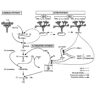
FIGURE 1 illustrates a new understanding of the lectin and alternative
pathways;
FIGURE 2 is a schematic diagram adapted from Schwaeble et al., Immunobiol
205:455-466 (2002), as modified by Yongqing et at., BBA 1824:253 (2012),
illustrating
the MASP-2 and MAp19 protein domains and the exons encoding the same;
FIGURE 3 is a schematic diagram adapted from Schwaeble et al., Imniunobiol
205:455-466 (2002), as modified by Yongqing et at., BRA 1824:253 (2012),
illustrating
the MASP-1, MASP-3 and MAp44 protein domains and the exons encoding the same;
FIGURE 4 shows an alignment of the amino acid sequences of the MASP-1,
MASP-2 and MASP-3 proteins and indicates consensus regions therebetween;
FIGURE 5 shows an alignment of the amino acid sequences of the MASP-1,
MASP-2 and MASP-3 Alpha chains;
FIGURE 6 shows an alignment of the amino acid sequences of the MASP-1,
MASP-2 and MASP-3 Beta Chains;
FIGURE 7A shows a pairwise alignment of the amino acid sequences of the
MASP-1 and MASP-2 Protease Domains (Beta-chains);
FIGURE 7B shows a pairwise alignment of the amino acid sequences of the
MASP-I and MASP-3 Protease Domains (Beta-chains);
FIGURE 7C shows a pairwise alignment of the amino acid sequences of the
MASP-2 and MASP-3 Protease Domains (Beta-chains);
-12-
CA 2875567 2019-10-10
CA 02875567 2014-12-02
WO 2013/192240 PCT/US2013/046432
FIGURE 8 is a Kaplan-Meyer plot graphically illustrating the percent survival
of
MASP-2 KO and WT mice after administration of an infective dose of 2.6 x 10⁷
cfu of N.
meningitidis serogroup A Z2491, demonstrating that MASP-2 deficient mice are
protected from N. meningitidis induced mortality, as described in Example 1;
FIGURE 9 is a Kaplan-Meyer plot graphically illustrating the percent survival
of
MASP-2 KO and WT mice after administration of an infective dose of 6 x 10⁶ cfu
of N.
meningitidis serogroup B strain MC58, demonstrating that MASP-2 deficient mice
are
protected from N. meningitidis induced mortality, as described in Example 1;
FIGURE 10 graphically illustrates the log cfu/mL of N. meningitidis serogroup
B
strain MC58 per mL of blood recovered from MASP-2 KO and WT mice at different
time points after i.p. infection with 6x10⁶ cfu of N. meningitidis serogroup B
strain MC58
(n=3 at different time points for both groups of mice), demonstrating that
although the
MASP-2 KO mice were infected with the same dose of N. meningitidis serogroup B
strain MC58 as the WT mice, the MASP-2 KO mice have enhanced clearance of
bacteremia as compared to WT, as described in Example 1;
FIGURE 11 graphically illustrates the average illness score of MASP-2 KO and
WT mice at 3, 6, 12 and 24 hours after infection with 6x10⁶ cfu of N.
meningitidis
serogroup B strain MC58, demonstrating that the MASP-2-deficient mice showed
much
lower illness scores at 6 hours, 12 hours, and 24 hours after infection, as
compared to WT
mice, as described in Example 1;
FIGURE 12 is a Kaplan-Meyer plot graphically illustrating the percent survival
of
mice after administration of an infective dose of 4x10⁶ cfu of N. meningitidis
serogroup B
strain MC58, followed by administration 3 hours post-infection of either
inhibitory
MASP-2 antibody (1 mg/kg) or control isotype antibody, demonstrating that MASP-
2
antibody is effective to treat and improve survival in subjects infected with
N.
meningitidis, as described in Example 2;
FIGURE 13 graphically illustrates the log cfu/mL of viable counts of N.
meningitidis serogroup B strain MC58 recovered at different time points in the
human
sera samples shown in TABLE 5 taken at various time points after incubation
with N.
meningitidis serogroup B strain MC58, as described in Example 3;
FIGURE 14 graphically illustrates the log cfu/mL of viable counts of N.
meningitidis serogroup B-MC58 recovered at different time points in the human
sera
samples shown in TABLE 7, showing that complement-dependent killing of N.
-13-
CA 02875567 2014-12-02
WO 2013/192240 PCT/US2013/046432
meningitidis in human 20% (v/v) serum is MASP-3 and MBL-dependent, as
described in
Example 3;
FIGURE 15 graphically illustrates the log cfu/mL of viable counts of N.
meningitidis serogroup B-MC58 recovered at different time points in the mouse
sera
samples shown in TABLE 9, showing that the MASP-2 -/- knockout mouse (referred
to
as "MASP-2 -/-") serum has a higher level of bactericidal activity for N.
meningitidis
than WT mouse serum, whereas in contrast, the MASP-1/3 -/- mouse serum does
not
have any bactericidal activity, as described in Example 3;
FIGURE 16 graphically illustrates the kinetics of C3 activation under lectin
pathway-specific conditions (1% plasma) in WT, C4-/-, MASP-1/3-/-, Factor B-/-
and
MASP-2-/- mouse sera, as described in Example 4;
FIGURE 17 graphically illustrates the level of alternative pathway-driven (AP-
driven) C3b deposition on zymosan-coated microtiter plates under "traditional"
alternative pathway-specific (AP-specific) conditions (i.e. BBS/EGTA/Mg++
without
Ca) as a function of serum concentration in serum samples obtained from MASP-3-
deficient, C4-deficient and MBL-deficient human subjects, as described in
Example 4;
FIGURE 18 graphically illustrates the level of AP-driven C3b deposition on
zymosan-coated microtiter plates under "traditional" AP-specific conditions
(i.e.,
BBS/EGTA/Mg-' without Ca--) as a function of time in 10% human serum samples
obtained from MASP-3-deficient, C4-deficient and MBL-deficient human subjects,
as
described in Example 4;
FIGURE 19A graphically illustrates the level of C3b deposition on mannan-
coated microtiter plates as a function of serum concentration in serum samples
obtained
from WT, MA S P-2-de fi ci ent, and MA SP-1/3 -defi el ent mice under
"traditional" AP-
specific conditions (i.e. BBS/EGTA/Mg++ without Ca) or under physiological
conditions allowing both the lectin pathway and the alternative pathway (AP)
to function
(BBS/Mg++/Ca++), as described in Example 4;
FIGURE 19B graphically illustrates the level of C3b deposition on zymosan-
coated microtiter plates as a function of serum concentration in serum samples
obtained
from WT, MASP-2-deficient, and MASP-1/3-deficient mice under traditional AP-
specific
conditions (i.e. BBS/EGTA/Mg H without Ca) or under physiological conditions
allowing both the lectin pathway and the alternative pathway to function
(BBS/Mg ''/Ca '), as described in Example 4;
-14-
CA 02875567 2014-12-02
WO 2013/192240 PCT/US2013/046432
FIGURE 19C graphically illustrates the level of C3b deposition on S.
pneumoniae
D39-coated microtiter plates as a function of serum concentration in serum
samples
obtained from WT, MASP-2-deficient, and MASP-1/3-deficient mice under
traditional
AP-specific conditions (i.e. BBS/EGTA/Mg++ without Ca) or under physiological
conditions allowing both the lectin pathway and the alternative pathway to
function
(BBS/Mg '/Ca ), as described in Example 4;
FIGURE 20A graphically illustrates the results of a C3b deposition assay in
highly diluted sera carried out on mannan-coated microtiter plates under
traditional AP-
specific conditions (i.e. BBS/EGTA/Mg without Ca' ) or under physiological
conditions allowing both the lectin pathway and the alternative pathway to
function
(BBS/Mg /Ca '), using serum concentrations ranging from 0 % up to 1.25%, as
described in Example 4;
FIGURE 20B graphically illustrates the results of a C3b deposition assay
carried
out on zymosan-coated microtiter plates under traditional AP-specific
conditions (i.e.
BBS/EGTA/Mg-+ without Ca) or under physiological conditions allowing both the
lectin pathway and the alternative pathway to function (BBS/Mg++/Ca++), using
serum
concentrations ranging from 0 % up to 1.25%, as described in Example 4;
FIGURE 20C graphically illustrates the results of a C3b deposition assay
carried
out on S. pneumoniae D39-coated microtiter plates under traditional AP-
specific
conditions (i.e. BBS/EGTA/Mg without Ca) or under physiological conditions
allowing both the lectin pathway and the alternative pathway to function
(BBS/Mg ), using scrum concentrations ranging from 0 % up to 1.25%, as
described in Example 4;
FIGURE 21 graphically illustrates the level of hemolysis (as measured by
hemoglobin release of lysed mouse erythrocytes (Crry/C3-/-) into the
supernatant
measured by photometry) of mannan-coated murine erythrocytes by human serum
under
physiological conditions (i.e., in the presence of Ca-+) over a range of serum
dilutions in
serum from MASP-3-/-, heat inactivated normal human serum (HI NHS), MBL-/-,
NHS
+ MASP-2 monoclonal antibody and NHS control, as described in Example 5;
FIGURE 22 graphically illustrates the level of hemolysis (as measured by
hemoglobin release of lysed mouse erythrocytes (Crry/C3-/-) into the
supernatant
measured by photometry) of mannan-coated murine erythrocytes by human serum
under
physiological conditions (i.e., in the presence of Ca) over a range of serum
-15-
CA 02875567 2014-12-02
WO 2013/192240 PCT/US2013/046432
concentration in serum from MASP-3-/-, heat inactivated (HI) NHS, MBL-/-, NHS
+
MASP-2 monoclonal antibody and NHS control, as described in Example 5;
FIGURE 23 graphically illustrates the level of hemolysis (as measured by
hemoglobin release of lysed WT mouse erythrocytes into the supernatant
measured by
photometry) of non-coated murine erythrocytes by human serum under
physiological
conditions (i.e., in the presence of Ca') over a range of serum concentrations
in serum
from 3MC (MASP-3-/-), heat inactivated (HI) NHS, MBL-/-, NHS + MASP-2
monoclonal antibody and NHS control, as described in Example 5;
FIGURE 24 graphically illustrates hemolysis (as measured by hemoglobin release
of lysed mouse erythrocytes (CD55/59-/-) into the supernatant measured by
photometry)
of non-coated murine erythrocytes by human serum under physiological
conditions (i.e.,
in the presence of Ca) over a range of serum concentrations in serum from heat
inactivated (HI) NHS, MBL-/-, NHS + MASP-2 monoclonal antibody and NHS
control,
as described in Example 5;
FIGURE 25 graphically illustrates hemolysis (as measured by hemoglobin release
of lysed rabbit erythrocytes into the supernatant measured by photometry) of
mannan-
coated rabbit erythrocytes by MASP-1/3-/- mouse serum and WT control mouse
serum
under physiological conditions (i.e., in the presence of Ca) over a range of
serum
concentrations, as described in Example 6;
FIGURE 26 graphically illustrates the level of C3b deposition (OD 405 nm) on a
zymosan-coated microtiter plate as a function of serum concentration in serum
samples
from factor D-/-, MASP-2-/- and WT mouse sera in a C3 deposition assay carried
out
under AP-specific conditions, as described in Example 7;
FIGURE 27 graphically illustrates the level of C3b deposition (OD 405 nm) on a
zymosan-coated microtiter plate as a function of serum concentration in serum
samples
from factor D-/-; MASP-2-/- and WT mouse sera in a C3 deposition assay carried
out
under physiological conditions (in the presence of Ca), as described in
Example 7;
FIGURE 28 graphically illustrates the level of C3b deposition (OD 405 nm) on a
zymosan-coated microtiter plate as a function of serum incubation time
(minutes) in
mouse serum samples obtained from factor D-/-; factor B-/-; plus and minus
MASP-2
monoclonal antibody in a C3b deposition assay carried out under physiological
conditions (in the presence of Ca), as described in Example 7;
-16-
CA 02875567 2014-12-02
WO 2013/192240 PCT/US2013/046432
FIGURE 29A graphically illustrates lectin pathway specific C4b deposition on a
zymosan-coated microtiter plate, measured ex vivo in undiluted serum samples
taken
from mice (n=3 mice/group) at various time points after subcutaneous dosing of
either
0.3 mg/kg or 1.0 mg/kg of the mouse MASP-2 MoAb, as described in Example 13;
FIGURE 29B graphically illustrates the time course of lectin pathway recovery
for three weeks following a single intraperitoneal administration of mouse
MASP-2
MoAb at 0.6 mg/kg in mice, as described in Example 13;
FIGURE 30A is a FACS histogram of MASP-3 antigen/antibody binding for
clone M3J5, as described in Example 15;
FIGURE 30B is a FACS histogram of MASP-3 antigen/antibody binding for
clone M3M1, as described in Example 15;
FIGURE 31 graphically illustrates a saturation binding curve of clone M3J5
(Clone 5) for the MASP-3 antigen, as described in Example 15;
FIGURE 32A is an amino acid sequence alignment of the VH regions of M3J5,
M3M1, D14 and 1E10 to the chicken DT40 VH sequence, wherein dots represent
amino
acid identity with the DT40 sequence and dashes indicate spaces introduced to
maximize
the alignment, as described in Example 15;
FIGURE 32B is an amino acid sequence alignment of the VL regions of M3J5,
M3M1, D14 and 1E10 to the chicken DT40 VL sequence, wherein dots represent
amino
acid identity with the DT40 sequence and dashes indicate spaces introduced to
maximize
the alignment, as described in Example 15;
FIGURE 33 is a bar graph showing the inhibitory activity of the mAblE10 in the
Wieslab Complement System Screen, MBL Pathway in comparison to the positive
serum
provided with the assay kit, as well as an isotype control antibody,
demonstrating that
mAb 1E10 partial inhibits LEA-2-dependent activation, (via inhibition of MASP-
1-
dependent activation of MASP-2), whereas the isotype control antibody does
not, as
described in Example 15;
FIGURE 34 graphically illustrates the level of C3b deposition for 1% normal
human serum plus isotype control, SGMI-1Fc or SGMI-2Fc over a concentration
range of
0.15 to 1000 nM, demonstrating that both SGMI-1Fc and SGMI-2Fc inhibited C3b
deposition from normal serum in mannan-coated ELISA wells, with IC50 values of
approximately 27nM and 300nM, respectively, as described in Example 16;
-17-
CA 02875567 2014-12-02
WO 2013/192240 PCT/US2013/046432
FIGURE 35A provides the results of flow cytometry analysis for C3b deposition
on heat-killed Staphylococcus aureus, demonstrating that in normal human serum
in the
presence of EDTA, which is known to inactivate the lectin and alternative
pathways, no
C3b deposition was observed (panel 1), in normal human serum treated with
Mg tiEGTA, alternative pathway-driven C3b deposition is observed (panel 2),
and as
shown in panel 3, 4 and 5, in factor B-depleted, factor D-depleted and
properdin (factor
P)-depleted serum, respectively, no alternative pathway driven C3b deposition
is
observed, as described in Example 17;
FIGURE 35B provides the results of flow cytometry analysis for C3b deposition
on heat-killed S. aureus, demonstrating that, as in EDTA-treated normal scrum
(panel 1),
AP-driven C3b deposition is absent in 3MC serum in the presence of Mg' '/EGTA
(panel
3), whereas panels 4 and 5 show that active full length rMASP-3 (panel 4) and
active
rMASP-3 (CCP1-CCP2-SP) (panel 5) both restore AP-driven C3b deposition in 3MC
serum to levels observed in normal serum treated with Mg++/EGTA (panel 2),
neither
inactive rMASP-3 (S679A) (panel 6) nor wild type rMASP-1 (panel 7) can restore
AP-
driven C3b deposition in 3MC serum, as described in Example 17;
FIGURE 36 shows the results of a Western blot analysis to determine factor B
cleavage in response to S. aureus in 3MC serum in the presence or absence of
rMASP-3,
demonstrating that the normal human serum in the presence of EDTA (negative
control,
lane 1) demonstrates very little Factor B cleavage relative to normal human
serum in the
presence of Mg /EGTA, shown in lane 2 (positive control), as further shown in
lane 3,
3MC scrum demonstrates very little Factor B cleavage in the presence of Mg
VEGTA.
However, as shown in lane 4, Factor B cleavage is restored by the addition and
pre-
incubation of full-length, recombinant MASP-3 protein to the 3MC serum, as
described
in Example 17;
FIGURE 37 shows Comassie staining of a protein gel in which Factor B cleavage
is analyzed, demonstrating that Factor B cleavage is most optimal in the
presence of C3,
MASP-3 and pro-factor D (lane 1), and as shown in lanes 4 and 5, either MASP-3
or pro-
factor D alone are able to mediate Factor B cleavage, as long as C3 is
present, as
described in Example 17;
FIGURE 38 graphically illustrates the mean fluorescent intensities (MFI) of
C3b
staining of S. aureus obtained from mAbD14 (which binds MASP-3), mAb1A5
(negative
control antibody) and an isotype control antibody plotted as a function of mAb
-18-
CA 02875567 2014-12-02
WO 2013/192240 PCT/US2013/046432
concentration in 3MC serum in the presence of rMASP-3, demonstrating that
mAbD14
inhibits MASP-3-dependent C3b deposition in a concentration-dependent manner,
as
described in Example 17;
FIGURE 39 shows Western blot analysis of pro-factor D substrate cleavage,
wherein compared to pro-factor D alone (lane 1) or the inactive full length
recombinant
MASP-3 (S679A; lane 3) or MASP-1 (S646A; lane 4), full length wild type
recombinant
MASP-3 (lane2) and MASP-1 (lane 5) either completely or partially cleave pro-
factor D
to generate mature factor D, as described in Example 18;
FIGURE 40 is a Western blot showing the inhibitory activity of the MASP-3
binding mAbs D14 (lane 2) and M3M1 (lane 3) on MASP-3-dependent pro-factor D
cleavage in comparison to a control reaction containing only MASP-3 and pro-
factor D
(no mAb, lane 1), as well as a control reaction containing a mAb obtained from
the
DTLac0 library that binds MASP-1, but not MASP-3 (lane 4), as described in
Example
18;
FIGURE 41 graphically illustrates the level of AP-driven C3b deposition on
zymosan-coated microtiter plates as a function of serum concentration in serum
samples
obtained from MASP-3-deficient (3MC), C4-deficient and MBL-deficient subjects,
demonstrating that MASP-3-deficient sera from Patient 2 and Patient 3 have
residual AP
activity at high serum concentrations (25%, 12.5%, 6.25% serum
concentrations), but a
significantly higher AP50 (i.e., 8.2% and 12.3% of serum needed to achieve 50%
of
maximum C3 deposition), as described in Example 19;
FIGURE 42A graphically illustrates the level of AP-driven C3b deposition on
zymosan-coated microtiter plates under "traditional" AP-specific conditions
(i.e.,
BBS/EGTA/Mg without Ca ) as a function of time in 10% human serum samples
obtained from MASP-3 deficient, C4-deficient and MBL-deficient human subjects,
as
described in Example 19;
FIGURE 42B shows a western blot with plasma obtained from 3MC patient #2
(MASP-3 (-/-), MASP-1 (+/+)), 3MC patient #3 (MASP-3 (-/-), MASP-1 (-/-)), and
sera
from normal donors (W), wherein human pro-factor D (25,040 Da) and/or mature
factor
D (24,405 Da) was detected with a human factor D-specific antibody, as
described in
Example 19;
-19-
CA 02875567 2014-12-02
WO 2013/192240 PCT/US2013/046432
FIGURE 42C graphically illustrates the results of the Weislab classical,
lectin and
alternative pathway assays with plasma obtained from 3MC patient #2, 3MC
patient #3,
and normal human serum, as described in Example 19;
FIGURE 43 graphically illustrates the percent hemolysis (as measured by
hemoglobin release of lysed rabbit erythrocytes into the supernatant measured
by
photometry) of mannan-coated rabbit erythrocytes over a range of serum
concentrations
in serum from two normal human subjects (NHS) and from two 3MC patients
(Patient 2
and Patient 3), measured in the absence of Ca- demonstrating that MASP-3
deficiency
reduces the percentage of complement-mediated lysis of mannan-coated
erythrocytes as
compared to normal human scrum, as described in Example 19;
FIGURE 44 graphically illustrates the level of AP-driven C3b deposition on
zymosan-coated microtiter plates as a function of the concentration of
recombinant full
length MASP-3 protein added to serum samples obtained from human 3MC Patient 2
(MASP-3-/), demonstrating that, compared to the negative control inactive
recombinant
MASP-3 (MASP-3A; S679A), active recombinant MASP-3 protein reconstitutes AP-
driven C3b deposition on zymosan-coated plates in a concentration-dependent
manner, as
described in Example 19;
FIGURE 45 graphically illustrates the percent hemolysis (as measured by
hemoglobin release of lysed rabbit erythrocytes into the supernatant measured
by
photometry) of mannan-coated rabbit erythrocytes over a range of serum
concentrations
in (1) normal human serum (NHS); (2) 3MC patient serum; (3) 3MC patient serum
plus
active full length recombinant MASP-3 (20 ,tg/m1); and (4) heat-inactivated
human
serum (HIS), measured in the absence of Ca ' , demonstrating that the percent
lysis of
rabbit erythrocytes is significantly increased in 3MC serum containing rMASP-3
as
compared to the percent lysis in 3MC serum without recombinant MASP-3
(p=0.0006),
as described in Example 19;
FIGURE 46 graphically illustrates the percentage of rabbit erythrocyte lysis
in 7%
human serum from 3MC Patient 2 and from 3MC Patient 3 containing active
recombinant
MASP-3 at a concentration range of 0 to 110 ug/m1 (in BBS/ Mg++/EGTA,
demonstrating
that the percentage of rabbit erythrocyte lysis increases with the amount of
recombinant
MASP-3 in a concentration-dependent manner, as described in Example 19; and
FIGURE 47 graphically illustrates the level of LEA-2-driven C3b deposition on
Mannan-coated ELISA plates as a function of the concentration of human serum
diluted
-20-
CA 02875567 2014-12-02
WO 2013/192240 PCT/US2013/046432
in BBS buffer, for serum from a normal human subject (NHS), from two 3MC
patients
(Patient 2 and Patient 3), from the parents of Patient 3 and from a MBL-
deficient subject.
FIGURE 48A presents results showing the baseline VEGF protein levels in
RPE-choroid complex isolated from wild type (WT) (+/+) and MASP-2 (-/-) mice,
as
described in Example 20;
FIGURE 48B presents results showing the VEGF protein levels in RPE-choroid
complex at day 3 in (WT) (+/+) and MASP-2 (-/-) mice following laser-induced
injury in
a macular degeneration model, as described in Example 20;
FIGURE 49 presents results showing the mean choroidal neovascularization
(CN V) volume at day 7 following laser induced-injury in (WT) (+/+) and MASP-2
(-/-)
mice, as described in Example 20;
FIGURE 50 graphically illustrates the mean choroidal neovascularization (CNV)
area at day 7 following laser-induced injury in WT (+/+) mice pre-treated with
a single ip
injection of 0.3 mg/kg or 1.0 mg,/kg mouse MASP-2 monoclonal antibody; as
described
in Example 21;
FIGURE 51A presents results demonstrating the infarct size for (WT) (+/+) and
reduced infarct size in MASP-2 (-/-) mice after injury in a coronary artery
occlusion and
reperfusion model, as described in Example 22;
FIGURE 51B presents results showing the distribution of the individual animals
tested in the coronary artery occlusion and reperfusion model, as described in
Example 22;
FIGURE 52A graphically illustrates the mean area-at-risk (AAR) and infarct
volumes (INF) as a percentage of total myocardial volumes in WT (+/+) and MASP-
2
(-/-) mice after undergoing left anterior descending coronary artery occlusion
and
reperfusion, as described in Example 23;
FIGURE 52B graphically illustrates the relationship between infarct volume
(INF) plotted against the mean area-at-risk (AAR) as a percentage of left
ventricle
myocardial volume in WT (+7+) and MASP-2 (-/-) mice after undergoing artery
occlusion
and reperfusion, as described in Example 23;
FIGURE 52C graphically illustrates the infarct volume (INF) in the buffer-
perfused hearts of WT (+/+) and MASP-2 (-/-) mice prepared in accordance with
the
Langendorff isolated-perfused mouse heart model, in which global ischemia and
reperfusion was carried out in the absence of serum, as described in Example
23;
-21-
CA 02875567 2014-12-02
WO 2013/192240 PCT/US2013/046432
FIGURE 52D graphically illustrates the relationship between infarct volume
(INF) and risk zone (RZ) in the buffer-perfused hearts of WT (+1+) and MASP-2
(-/-)
mice prepared in accordance with the Langendorff isolated-perfused mouse heart
model,
as described in Example 23;
FIGURE 53A graphically illustrates the results of a C3b deposition assay on
immune complex-coated plates, wherein the symbol "*" symbol indicates serum
from
WT (MASP-2 (+1+)); the symbol "=" indicates serum from WT (C 1 q depleted);
the
symbol "o" indicates serum from MASP-2 (-/-); and the symbol "A" indicates
serum from
MASP-2 (-/-) (Clq depleted), demonstrating that MASP-2 (-/-) mice retain a
functional
classical pathway, as described in Example 24;
FIGURE 53B graphically illustrates the results of a C3b deposition assay on
zymosan-coated plates, wherein the symbol "*" symbol indicates serum from WT
(MASP-2 (+/+)), and the symbol "o" indicates serum from MASP-2 (-/-);
demonstrating
that MASP-2 (-/-) mice retain a functional alternative pathway, as described
in Example
24;
FIGURE 54A graphically illustrates myocardial ischemia/reperfusion injury
(MIRI)-induced tissue loss following ligation of the left anterior descending
branch of the
coronary artery (LAD) and reperfusion in C4 (-/-) mice (n=6) and matching WT
littermate controls (n=7), showing area at risk (AAR) and infarct size (INF)
as described
in Example 24;
FIGURE 54B graphically illustrates infarct size (INF) as a function of area at
risk
(AAR) in C4 (-/-) and WT mice treated as describe in FIGURE 42A, demonstrating
that
C4 (-/-) mice are as susceptible to MIRI as WT controls (dashed line), as
described in
Example 24;
FIGURE 55A graphically illustrates the results of a C3b deposition assay using
serum from WT mice, C4 (-/-) mice and serum from C4 (-I-) mice pre-incubated
with
mannan, as described in Example 24;
FIGURE 55B graphically illustrates the results of a C3b deposition assay using
serum from WT, C4 (-/-), and MASP-2 (-I-) mice mixed with various
concentrations of a
murine MASP-2 mAb (mAbM11), as described in Example 24;
FIGURE 55C graphically illustrates the results of a C3b deposition assay using
human serum from WT (C4-sufficient) and C4-deficient subjects, and serum from
C4
deficient subjects pre-incubated with mannan, as described in Example 24;
-22-
CA 02875567 2014-12-02
WO 2013/192240 PCT/US2013/046432
FIGURE 55D graphically illustrates the results of a C3b deposition assay using
human serum from WT (C4-sufficient) and C4-deficient subjects mixed with a
human
MASP-2 mAb (mAbH3), as described in Example 24;
FIGURE 56A graphically illustrates a comparative analysis of C3 convertase
activity in plasma from various complement-deficient mouse strains tested
either under
lectin activation pathway-specific assay conditions, or under classical
activation pathway-
specific assay conditions, as described in Example 24;
FIGURE 56B graphically illustrates the time-resolved kinetics of C3 convertase
activity in plasma from various complement-deficient mouse strains tested
under lectin
activation pathway-specific conditions, as described in Example 24;
FIGURE 57A graphically illustrates the degree of tissue damage in WT and
MASP-2 (-I-) mice after induction of transient ischemia/reperfusion injury in
the
gastrointestinal tract (GIRI), demonstrating that MASP-2 (-/-) mice have a
significant
degree of protection as compared to WT controls, as described in Example 25;
FIGURE 57B graphically illustrates the results of a C4b deposition assay
carried
out using serum obtained from mice (n=3) over time after an intraperitoneal
single dose
bolus injection of a recombinant murine MASP-2 antibody (mAbM11),
demonstrating in
vivo ablation of lectin pathway functional activity, as described in Example
25;
FIGURE 57C graphically illustrates the effect of a MASP-2 mAb treatment on the
severity of GIRT pathology, demonstrating that mice dosed with the a murine
MASP-2
mAb (mAbM11) 24 hours before being subjected to transient ischemia/reperfusion
injury
in the gastrointestinal tract (GIRT) had significantly reduced tissue damage
as compared
to mice dosed with saline (*p<0.05 when comparing animals treated with either
the
MASP-2 inhibitory antibody mAbM .1 1 or an irrelevant isotype control
antibody), as
described in Example 25;
FIGURE 57D shows histological presentation of GIRI-mediated pathology of the
small intestine in mice pre-treated with a single dose intraperitoneal
injection of saline, an
isotope control antibody, or a recombinant murine MASP-2 antibody (mAbM11) 12
hours prior to induction of GIRI, as described in Example 25;
FIGURE 58 graphically illustrates the cerebral infarct volume in WT (MASP-2
(+1+)) and MASP-2 (-/-) mice following 30 minutes ischemia and 24 hours
reperfusion,
as described in Example 26;
-23-
CA 02875567 2014-12-02
WO 2013/192240 PCT/US2013/046432
FIGURE 59A shows a series of photographs of stained brain sections from a WT
(MASP-2 +/+) mouse after 30 minutes ischemia and 24 hours reperfusion. Panels
1-8 of
FIGURE 52A show the different section areas of the brain corresponding to
Bregma 1-8,
respectively, in relation to the exit of the acoustic nerve (Bregma 0), as
described in
Example 26;
FIGURE 59B shows a series of photographs of stained brain sections from a
MASP-2 (-/-) mouse after 30 minutes ischemia and 24 hours reperfusion. Panels
1-8 of
FIGURE 52B show the different sections areas of the brain corresponding to
Bregma 1-8,
respectively, in relation to the exit of the acoustic nerve (Bregma 0), as
described in
Example 26;
FIGURE 60 presents results showing the mean clinical arthritis score of (WT)
(+/+) and MASP-2 (-I-) mice over time following Col2 mAb- induced rheumatoid
arthritis, as described in Example 27;
FIGURE 61 graphically illustrates the results of the C3 deposition assay on
serum
samples obtained from WT mice in the presence of house dust mite or zymosan,
as
described in Example 28;
FIGURES 62A and 62B present dose response curves for the inhibition of C4b
deposition (FIG. 62A) and the inhibition of thrombin activation following the
administration of a MASP-2 Fab2 antibody (H1) in normal rat serum, as
described in
Example 29;
FIGURES 63A and 63B present measured platelet aggregation (expressed as
aggregate area) in MASP-2 (-/-) mice (FIG. 63B) as compared to platelet
aggregation in
untreated wild-type mice and wild-type mice in which the complement pathway is
inhibited by depletory agent cobra venom factor (CVF) and a terminal pathway
inhibitor
(C5aR antagonist) (FIGURE 63A) in a localized Schwartzman reaction model of
disseminated intravascular coagulation, as described in Example 30;
FIGURE 64 illustrates the results of a Western blot analysis showing
activation of
human C3, shown by the presence of the a' chain, by thrombin substrates FXIa
and FXa,
as described in Example 31;
FIGURE 65 graphically illustrates the results of a C3b deposition assay on
serum
samples obtained from WT, MASP-2 (-/-), F11(-/-), F 11(-/-)/C4 (-/-) and C4 (-
/-) mice,
demonstrating that there is a functional lectin pathway even in the complete
absence of
-24-
CA 02875567 2014-12-02
WO 2013/192240 PCT/US2013/046432
C4, or F11, while mice with combined F11-(-/-)/C4 (-/-)-deficiency lack a
functional
lectin pathway, as described in Example 31;
FIGURE 66 graphically illustrates the time to onset of microvaseular occlusion
following LPS injection in MASP-2 -/- and WT mice, showing the percentage of
mice
with thrombus formation measured over 60 minutes, demonstrating that thrombus
formation is detected after 15 minutes in WT mice, with up to 80% of the WT
mice
demonstrating thrombus formation at 60 minutes; in contrast, none of the MASP-
2 -/-
mice showed any thrombus formation during the 60-minute period (log rank:
p=0.0005),
as described in Example 32;
FIGURE 67 graphically illustrates the percent survival of saline-treated
control
mice (n.-.5) and M ASP-2 antibody-treated mice (n=5) in the STX11.2S-indueed
model of
HUS over time (hours), demonstrating that all of the control mice died by 42
hours,
whereas, in contrast, 100 % of the MASP-2 antibody-treated mice survived
throughout
the time course of the experiment, as described in Example 33;
FIGURE 68 graphically illustrates, as a function of time after injury
induction, the
percentage of mice with microvascular occlusion in the FITC/Dextran UV model
after
treatment with isotype control, or human MASP-2 antibody mAbH6 (10mg/kg) dosed
at
16 hours and 1 hour prior to injection of FITC/Dextran, as described in
Example 34;
FIGURE 69 graphically illustrates the occlusion time in minutes for mice
treated
with the human MASP-2 antibody (mAbH6) and the isotype control antibody,
wherein
the data are reported as scatter-dots with mean values (horizontal bars) and
standard error
bars (vertical bars). The statistical test used for analysis was the unpaired
t test; wherein
the symbol "*" indicates p=0.0129, as described in Example 34; and
FIGURE 70 graphically illustrates the time until occlusion in minutes for wild-
type mice, MASP-2 KO mice, and wild-type mice pre-treated with human MASP-2
antibody (mAbH6) administered i.p. at 10mg/kg 16 hours before, and again 1
hour prior
to the induction of thrombosis in the FITC-dextran/light induced endothelial
cell injury
model of thrombosis with low light intensity (800-1500), as described in
Example 34.
DESCRIPTION OF SEQUENCE LISTING
-25-
CA 02875567 2014-12-02
WO 2013/192240 PCT/US2013/046432
SEQ ID NO:1 human MAp19 cDNA
SEQ ID NO:2 human MAp19 protein (with leader)
SEQ ID NO:3 human MAp19 protein (mature)
SEQ ID NO:4 human MASP-2 cDNA
SEQ ID NO:5 human MASP-2 protein (with leader)
SEQ ID NO:6 human MASP-2 protein (mature)
SEQ ID NO:7 human MASP-3 cDNA
SEQ ID NO:8 human MASP-3 protein (w/leader)
SEQ ID NO:9 human MASP-1 cDNA
SEQ ID NO:10 human MASP-1 protein (w/leader)
SEQ ID NO:11 human MAp44 protein (w/leader)
SEQ ID NO:12 rat MA SP-2 cDNA
SEQ ID NO:13 rat MASP-2 protein (with leader)
SEQ ID NO:14 DNA encoding 17D20_dc35VH21N11VL (0M5646) heavy chain
variable region (VH) (without signal peptide)
SEQ ID NO:15 17D20_dc35VH21N11VL (0MS646) heavy chain variable region (VH)
polypeptide
SEQ ID NO:16 17N16mc heavy chain variable region (VH) polypeptide
SEQ ID NO:17 17D20_dc21N11VL (0M5644) light chain variable region (VL)
polypeptide
SEQ ID NO:18 DNA encoding 17N16_dc17N9 (0M5641) light chain variable region
(VL) (without signal peptide)
SEQ ID NO:19 17N16_dc17N9 (0MS641) light chain variable region (VL)
polypeptide
SEQ ID NO:20: scFv daughter clone 17N16m_d17N9 full length polypeptide
SEQ ID NO:21: scFv daughter clone 17D20m_d3521N11 full length polypeptide
SEQ ID NO:22: scFv daughter clone 17N16m_d17N9 DNA encoding full length
polypeptide (without signal peptide)
SEQ ID NO:23: scFv daughter clone 17D20m_d3521N11 DNA encoding full length
polypeptide (without signal peptide)
SEQ ID NO:24: parent DTLac0 heavy chain variable region (VH) polypeptideSEQ ID
NO:25: MASP-3 specific clone M3J5 heavy chain variable region (VH) polypeptide
SEQ ID NO:26: MASP-3 specific clone M3M1 heavy chain variable region (VH)
polypeptide
-26-
CA 02875567 2014-12-02
WO 2013/192240 PCT/US2013/046432
SEQ ID NO:27: parent DTLac0 light chain variable region (VL) polypeptide
SEQ ID NO:28: MASP-3 specific clone M3J5 light chain variable region (VL)
polypeptide
SEQ ID NO:29: MASP-3 specific clone M3M1 light chain variable region (VL)
polypeptide
SEQ ID NO:30: MASP-3 clone D14 heavy chain variable region (VH) polypeptide
SEQ ID NO:31: MASP-3 clone D14 light chain variable region (VL) polypeptide
SEQ ID NO:32: MASP-1 clone 1E10 heavy chain variable region (VH) polypeptide
SEQ ID NO:33: MASP-1 clone 1E10 light chain variable region (VL) polypeptide
SEQ ID NO:34 SGM1-1 peptide
SEQ ID NO:35 SGM1-2 peptide
SEQ ID NO:36 human IgGl-Fc polypeptide;
SEQ ID NO:37 peptide linker #1 (12aa);
SEQ ID NO:38: peptide linker #2 (10aa);
SEQ ID NO:39: nucleic acid encoding polypeptide fusion comprising the human IL-
2-
signal sequence, SGMI-1, linker#1, and human IgGl-Fc;
SEQ ID NO:40: mature polypeptide fusion comprising SGMI-1, linker#1 and human
IgGl-Fc (SGMI-1Fc);
SEQ ID NO:41: nucleic acid encoding polypeptide fusion comprising the human IL-
2-
signal sequence, SGMI-2, linker#1 and human IgG 1 -Fc;
SEQ ID NO:42: mature polypeptide fusion comprising SGMI-2, linker#1 and human
IgGI-Fc (SGMI-2Fc).
DETAILED DESCRIPTION
I. DEFINITIONS
Unless specifically defined herein, all terms used herein have the same
meaning
as would be understood by those of ordinary skill in the art of the present
invention. The
following definitions are provided in order to provide clarity with respect to
the terms as
they are used in the specification and claims to describe the present
invention.
As used herein, the lectin pathway effector arm 1 ("LEA-1") refers to lectin-
dependent activation of factor B and factor D by MASP-3.
-27-
CA 02875567 2014-12-02
WO 2013/192240 PCT/US2013/046432
As used herein, the lectin pathway effector arm 2 ("LEA-2") refers to MASP-2-
dependent complement activation.
As used herein, the term "MASP-3-dependent complement activation" comprises
two components: (i) lectin MASP-3-dependent activation of factor B and factor
D,
encompassed in LEA-1-mediated complement activation, occurs in the presence of
Ca,
commonly leading to the conversion of C3bB to C3bBb and of pro-factor D to
factor D;
and (ii) lectin-independent conversion of factor B and factor D, which can
occur in the
absence of Ca, commonly leading to the conversion of C3bB to C3bBb and of pro-
factor D to factor D. LEA-1-mediated complement activation and lectin-
independent
conversion of factor B and factor D have been determined to cause opsonization
and/or
lysis. While not wishing to be bound by any particular theory, it is believed
that only
when multiple C3b molecules associate and bind in close proximity, the C3bBb
C3
convertase changes its substrate specificity and cleaves C5 as the alternative
pathway C5
convertase termed C3bBb(C3b)n.
As used herein, the term "MASP-2-dependent complement activation", also
referred to herein as LEA-2-mediated complement activation, comprises MASP-2
lectin-
dependent activation, which occurs in the presence of Ca, leading to the
formation of
the lectin pathway C3 convertase C4b2a and upon accumulation of the C3
cleavage
product C3b subsequently to the C5 convertase C4b2a(C3b)n, which has been
determined
to cause opsonization and/or lysis.
As used herein, the term "traditional understanding of the alternative
pathway"
also referred to as the "traditional alternative pathway" refers to the
alternative pathway
prior to the instant discovery described herein, i.e., complement activation
that is
triggered, for example, by zymosan from fungal and yeast cell walls,
lipopolysaccharide
(LPS) from Gram negative outer membranes, and rabbit erythrocytes, as well as
from
many pure polysaccharides, viruses, bacteria, animal tumor cells, parasites
and damaged
cells, and which has traditionally been thought to arise from spontaneous
proteolytic
generation of C3b from complement factor C3. As used herein, activation of the
"traditional alternative pathway", also referred to herein as the "alternative
pathway", is
measured in Mg-+/EGTA buffer (i.e., in the absence of Ca
As used herein, the term "lectin pathway" refers to complement activation that
occurs via the specific binding of serum and non-serum carbohydrate-binding
proteins
including mannan-binding lectin (MBL), CL-11 and the ficolins (H-ficolin, M-
ficolin, or
-28-
CA 02875567 2014-12-02
WO 2013/192240 PCT/US2013/046432
L-ficolin). As described herein, the inventors have discovered that the lectin
pathway is
driven by the two effector arms, lectin pathway effector arm 1 (LEA-1), which
is now
known to be MASP-3-dependent, and lectin pathway effector arm 2 (LEA-2), which
is
MASP-2-dependent. As used herein, activation of the lectin pathways are
assessed using
Ca + containing buffers.
As used herein, the term "classical pathway" refers to complement activation
that
is triggered by antibody bound to a foreign particle and requires binding of
the
recognition molecule Cl q.
As used herein, the term "HTRA-1" refers to the serine peptidase High-
temperature requirement serine protease Ai.
As used herein, the term "MASP-3 inhibitory agent" refers to any agent that
directly or indirectly inhibits MASP-3-dependent complement activation,
including
agents that bind to or directly interact with MASP-3, including MASP-3
antibodies and
MASP-3 binding fragments thereof, natural and synthetic peptides, competitive
substrates, small-molecules, expression inhibitors and isolated natural
inhibitors, and also
encompasses peptides that compete with MASP-3 for binding to another
recognition
molecule (e.g., MBL, CL-11, H-ficolin, M-ficolin, or L-ficolin) in the lectin
pathway. In
one embodiment, the MASP-3 inhibitory agent is specific to MASP-3, and does
not bind
to MASP-1 or MASP-2. An inhibitory agent that directly inhibits MASP-3 can be
referred to as a direct MASP-3 inhibitory agent (e.g., a MASP-3 antibody),
while an
inhibitory agent that indirectly inhibits MASP-3 can be referred to as an
indirect MASP-3
inhibitory agent (e.g., a MASP-1 antibody that inhibits MASP-3 activation). An
example
of a direct MASP-3 inhibitory agent is a MASP-3 specific inhibitory agent,
such as a
MASP-3 inhibitory agent that specifically binds to a portion of MASP-3 (SEQ ID
NO:8)
with a binding affinity of at least 10 times greater than to other components
in the
complement system. In one embodiment, a MASP-3 inhibitory agent indirectly
inhibits
MASP-3 activity, such as, for example, an inhibitor of MASP-3 activation,
including an
inhibitor of MASP-1-mediated MASP-3 activation (e.g., a MASP-1 antibody or
MASP-1
binding fragments thereof, natural and synthetic peptides, small-molecules,
expression
inhibitors and isolated natural inhibitors, and also encompasses peptides that
compete
with MASP-1 for binding to MASP-3). In another embodiment, a MASP-3 inhibitory
agent inhibits MASP-3-mediated maturation of factor D. In another embodiment,
a
MASP-3 inhibitory agent inhibits MASP-3-mediated activation of factor B. MASP-
3
-29-
CA 02875567 2014-12-02
WO 2013/192240 PCT/US2013/046432
inhibitory agents useful in the method of the invention may reduce MASP-3-
dependent
complement activation by greater than 10%, such as greater than 20%, greater
than 50%,
or greater than 90%. In one embodiment, the MASP-3 inhibitory agent reduces
MASP-3-
dependent complement activation by greater than 90% (i.e., resulting in MASP-3
complement activation of only 10% or less). It is expected that MASP-3
inhibition will
block, in full or in part, both LEA-1-related lysis and opsonization and
lectin-independent
conversion of factor B and factor D-related lysis and opsonization.
As used herein, the term "MASP-1 inhibitory agent" refers to any agent that
binds
to or directly interacts with MASP-1 and inhibits at least one of (i) MASP-3-
dependent
complement activation and/or (ii) MASP-2-dependent complement activation
and/or (iii)
lectin-independent or lectin-dependent MASP-1-mediated maturation of factor D,
wherein the lectin-dependent MASP-1 maturation of factor D involves direct
activation
of factor D, including MASP-1 antibodies and MASP-1 binding fragments thereof,
natural and synthetic peptides, small-molecules, expression inhibitors and
isolated natural
inhibitors, and also encompasses peptides that compete with MASP-1 for binding
to
another recognition molecule (e.g., MBL, CL-11, H-ficolin, M-ficolin, or L-
ficolin) in the
lectin pathway. In one embodiment, MASP-1 inhibitory agents useful in the
method of
the invention reduce MASP-3-dependent complement activation by greater than
10%,
such as greater than 20%, greater than 50%, or greater than 90%. In one
embodiment, the
MASP-1 inhibitory agent reduces MASP-3-dependent complement activation by
greater
than 90% (i.e., resulting in MASP-3 complement activation of only 10% or
less). In
another embodiment, MASP-1 inhibitory agents useful in the method of the
invention
reduce MASP-2-dependent complement activation by greater than 10%, such as
greater
than 20%, greater than 50%, or greater than 90%. In one embodiment, the MASP-1
inhibitory agent reduces MASP-2-dependent complement activation by greater
than 90%
(i.e., resulting in MASP-2 complement activation of only 10% or less).
In another embodiment, MASP-1 inhibitory agents useful in the method of the
invention reduce both MASP-3-dependent complement activation (LEA-1), lectin-
independent conversion of factor B and factor D, and MASP-2-dependent
complement
activation (LEA-2) by greater than 10%, such as greater than 20%, greater than
50%, or
greater than 90%. In one embodiment, the MASP-1 inhibitory agent reduces MASP-
3-
dependent complement activation (LEA-1), lectin-independent conversion of
factor B and
factor D, and MASP-2-dependent complement activation (LEA-2) by greater than
90%
-30-
CA 02875567 2014-12-02
WO 2013/192240 PCT/US2013/046432
(i.e., resulting in MASP-3 complement activation of only 10% or less and MASP-
2
complement activation of only 10% or less).
An example of a direct MASP-1 inhibitory agent is a MASP-1-specific inhibitory
agent, such as a MASP-1 inhibitory agent that specifically binds to a portion
of MASP-1
(SEQ ID NO:10) with a binding affinity of at least 10 times greater than to
other
components in the complement system. In many instances, given that MASP-1 can
activate MASP-3, and given the MASP-1 can activate MASP-2, inhibition of MASP-
1
would be expected to be effective in inhibiting MASP-3 and/or MASP-2. In some
instances, however, inhibition of either MASP-1 or MASP-3 or MASP-2 may be a
preferred embodiment relative to inhibition of the other MASI' targets. For
example, in
the setting of Staphylococcus aureus (S. aureus) infection, MASP-3 has been
shown to be
activated and is responsible for S. aureus opsonization in the absence of MASP-
1 (see
Iwaki D. et al., J Immunol 187(7):3751-8 (2011)). Therefore, in the treatment
of
paroxysmal nocturnal hemoglobinuria (PNH), for example, it might be
advantageous to
directly inhibit MASP-1 rather than MASP-3, thereby reducing the potential
susceptibility to S. aureus during LEA-1-inhibitory treatment of PNH.
As used herein, the term "MASP-2 inhibitory agent" refers to any agent that
binds
to or directly interacts with MASP-2 and inhibits at least one of (i) MASP-2-
dependent
complement activation and/or (ii) MASP-1-dependent complement activation,
including
MASP-2 antibodies and MASP-2 binding fragments thereof, natural and synthetic
peptides, small-molecules, expression inhibitors and isolated natural
inhibitors, and also
encompasses peptides that compete with MASP-2 for binding to another
recognition
molecule (e.g., MBL, CL-11, H-ficolin, M-ficolin, or L-ficolin) in the lectin
pathway.
MASP-2 inhibitory agents useful in the method of the invention may reduce MASP-
2-
dependent complement activation by greater than 10%, such as greater than 20%,
greater
than 50%, or greater than 90%. In one embodiment, the MASP-2 inhibitory agent
reduces MASP-2-dependent complement activation by greater than 90% (i.e.,
resulting in
MASP-2 complement activation of only 10% or less). An example of a direct MASP-
2
inhibitory agent is a MASP-2-specific inhibitory agent, such as a MASP-2
inhibitory
agent that specifically binds to a portion of MASP-2 (SEQ ID NO:5) with a
binding
affinity of at least 10 times greater than to other components in the
complement system.
As used herein, the term "antibody" encompasses antibodies and antibody
fragments thereof, derived from any antibody-producing mammal (e.g., mouse,
rat,
-31-
CA 02875567 2014-12-02
WO 2013/192240 PCT/US2013/046432
rabbit, and primate including human), or from a hybridoma, phage selection,
recombinant
expression or transgenic animals (or other methods of producing antibodies or
antibody
fragments"), that specifically bind to a target polypeptide, such as, for
example, MASP-1,
MASP-2 or MASP-3 polypeptides or portions thereof. It is not intended that the
term
"antibody" limited as regards to the source of the antibody or the manner in
which it is
made (e.g., by hybridoma, phage selection, recombinant expression, transgenic
animal,
peptide synthesis, etc). Exemplary antibodies include polyclonal, monoclonal
and
recombinant antibodies; pan-specific, multispecific antibodies (e.g.,
bispecific antibodies,
trispecific antibodies); humanized antibodies; murine antibodies; chimeric,
mouse-human, mouse-primate, primate-human monoclonal antibodies; and anti-
idiotypc
antibodies, and may be any intact antibody or fragment thereof. As used
herein, the term
"antibody" encompasses not only intact polyclonal or monoclonal antibodies,
but also
fragments thereof (such as dAb, Fab, Fab', F(ab')2, Fv), single chain (ScFv),
synthetic
variants thereof, naturally occurring variants, fusion proteins comprising an
antibody
portion with an antigen-binding fragment of the required specificity,
humanized
antibodies, chimeric antibodies, and any other modified configuration of the
immunoglobulin molecule that comprises an antigen-binding site or fragment
(epitope
recognition site) of the required specificity.
A "monoclonal antibody" refers to a homogeneous antibody population wherein
the monoclonal antibody is comprised of amino acids (naturally occurring and
non-
naturally occurring) that are involved in the selective binding of an epitope.
Monoclonal
antibodies arc highly specific for the target antigen. The term "monoclonal
antibody"
encompasses not only intact monoclonal antibodies and full-length monoclonal
antibodies, but also fragments thereof (such as Fab, Fab', F(ab')2, Fv),
single chain
(ScFv), variants thereof, fusion proteins comprising an antigen-binding
portion,
humanized monoclonal antibodies, chimeric monoclonal antibodies, and any other
modified configuration of the immunoglobulin molecule that comprises an
antigen-
binding fragment (epitope recognition site) of the required specificity and
the ability to
bind to an epitope. It is not intended to be limited as regards the source of
the antibody or
the manner in which it is made (e.g., by hybridoma, phage selection,
recombinant
expression, transgenic animals, etc.). The term includes whole immunoglobulins
as well
as the fragments etc. described above under the definition of "antibody".
-32-
CA 02875567 2014-12-02
WO 2013/192240 PCT/US2013/046432
As used herein, the term "antibody fragment" refers to a portion derived from
or
related to a full-length antibody, such as, for example, a MASP-1, MASP-2 or
MASP-3
antibody, generally including the antigen binding or variable region thereof.
Illustrative
examples of antibody fragments include Fab, Fab', F(ab)2, F(ab')2 and Fv
fragments,
scFv fragments, diabodies, linear antibodies, single-chain antibody molecules
and
multispecific antibodies formed from antibody fragments.
As used herein, a "single-chain Fv" or "scFv" antibody fragment comprises the
VH and VL domains of an antibody, wherein these domains are present in a
single
polypeptide chain. Generally, the Fv polypeptide further comprises a
polypeptide linker
between the VH and VL domains, which enables the scFv to form the desired
structure
for antigen binding.
As used herein, a "chimeric antibody" is a recombinant protein that contains
the
variable domains and complementarity-determining regions derived from a non-
human
species (e.g., rodent) antibody, while the remainder of the antibody molecule
is derived
from a human antibody.
As used herein, a "humanized antibody" is a chimeric antibody that comprises a
minimal sequence that conforms to specific complementarity-determining regions
derived
from non-human immunoglobulin that is transplanted into a human antibody
framework.
Humanized antibodies are typically recombinant proteins in which only the
antibody
complementarity-determining regions are of non-human origin (including
antibodies
generated from phage display or yeast).
As used herein, the term "mannan-binding lectin" ("MBL") is equivalent to
mannan-binding protein ("MBP").
As used herein, the "membrane attack complex" ("MAC") refers to a complex of
the terminal five complement components (C5b combined with C6, C7, C8 and C9)
that
inserts into and disrupts membranes (also referred to as C5b-9).
As used herein, "a subject" includes all mammals, including without limitation
humans, non-human primates, dogs, cats, horses, sheep, goats, cows, rabbits,
pigs and
rodents.
As used herein, the amino acid residues are abbreviated as follows: alanine
(Ala;A), asparagine (Asn;N), aspartic acid (Asp;D), arginine (Arg;R), cysteine
(Cys;C),
glutamic acid (Glu;E), glutamine (Gln;Q), glycine (Gly;G), histidine (His;H),
isoleucine
(Ile;I), leucine (Leu;L), lysine (Lys;K), methionine (Met;M), phenylalanine
(Phe;F),
-33-
CA 02875567 2014-12-02
WO 2013/192240 PCT/US2013/046432
proline (Pro;P), serine (Ser;S), threonine (Thr;T), tryptophan (Trp;W),
tyrosine (Tyr;Y),
and valine (Val;V).
In the broadest sense, the naturally occurring amino acids can be divided into
groups based upon the chemical characteristic of the side chain of the
respective amino
acids. By "hydrophobic" amino acid is meant either Ile, Leu, Met, Phe, Trp,
Tyr, Val,
Ala, Cys or Pro. By "hydrophilic" amino acid is meant either Gly, Asn, Gln,
Ser, Thr,
Asp, Glu, Lys, Arg or His. This grouping of amino acids can be further
subclassed as
follows. By "uncharged hydrophilic" amino acid is meant either Ser, Thr, Asn
or Gin.
By "acidic" amino acid is meant either Glu or Asp. By "basic" amino acid is
meant either
Lys, Arg or His.
As used herein the term "conservative amino acid substitution" is illustrated
by a
substitution among amino acids within each of the following groups: (1)
glycine, alanine,
valine, leucine, and isoleucine, (2) phenylalanine, tyrosine, and tryptophan,
(3) serine and
threonine, (4) aspartate and glutamate, (5) glutamine and asparagine, and (6)
lysine,
arginine and histidine.
The term "oligonucleotide" as used herein refers to an oligomer or polymer of
ribonucleic acid (RNA) or deoxyribonucleic acid (DNA) or mimetics thereof.
This term
also covers those oligonucleobases composed of naturally-occurring
nucleotides, sugars
and covalent internucleoside (backbone) linkages as well as oligonucleotides
having
non-naturally-occurring modifications.
As used herein, an "epitope" refers to the site on a protein (e.g., a human
MASP-3
protein) that is bound by an antibody. "Overlapping epitopes" include at least
one (e.g.,
two, three, four, five, or six) common amino acid residue(s), including linear
and non-
linear epitopes.
As used herein, the terms "polypeptide," "peptide," and "protein" are used
interchangeably and mean any peptide-linked chain of amino acids, regardless
of length
or post-translational modification. The MASP proteins (MASP-1, MASP-2 or MASP-
3)
described herein can contain or be wild-type proteins or can be variants that
have not
more than 50 (e.g., not more than one, two, three, four, five, six, seven,
eight, nine, ten,
12, 15, 20, 25, 30, 35, 40, or 50) conservative amino acid substitutions.
Conservative
substitutions typically include substitutions within the following groups:
glycine and
alanine; valine, isoleucine, and leucine; aspartic acid and glutamic acid;
asparagine,
-34-
CA 02875567 2014-12-02
WO 2013/192240 PCT/US2013/046432
glutamine, serine and threonine; lysine, histidine and arginine; and
phenylalanine and
tyrosine.
The human MASP-1 protein (set forth as SEQ ID NO:10), human MASP-2
protein (set forth as SEQ ID NO:5) and human MASP-3 protein (set forth as SEQ
ID
NO :8) described herein also include "peptide fragments" of the proteins,
which are
shorter than full-length and/or immature (pre-pro) MASP proteins, including
peptide
fragments of a MASP protein include terminal as well internal deletion
variants of the
protein. Deletion variants can lack one, two, three, four, five, six, seven,
eight, nine, ten,
11, 12, 13, 14, 15, 16, 17, 18, 19, or 20 amino acid segments (of two or more
amino
acids) or noncontiguous single amino acids. In some embodiments, the human
MASP-1
protein can have an amino acid sequence that is, or is greater than, 70 (e.g.,
71, 72, 73,
74, 75, 76, 77, 78, 79, 80, 81, 82, 83, 84, 85, 86, 87, 88, 89, 90, 91, 92,
93, 94, 95, 96, 97,
98, 99, or 100) % identical to the human MASP-1 protein having the amino acid
sequence set forth in SEQ ID NO: 10.
In some embodiments, the human MASP-3 protein can have an amino acid
sequence that is, or is greater than, 70 (e.g., 71, 72, 73, 74, 75, 76, 77,
78, 79, 80, 81, 82,
83, 84, 85, 86, 87, 88, 89, 90, 91, 92, 93, 94, 95, 96, 97, 98, 99, or 100) %
identical to the
human MASP-3 protein having the amino acid sequence set forth in SEQ ID NO: 8.
In some embodiments, the human MASP-2 protein can have an amino acid
sequence that is, or is greater than, 70 (e.g., 71, 72, 73, 74, 75, 76, 77,
78, 79, 80, 81, 82,
83, 84, 85, 86, 87, 88, 89, 90, 91, 92, 93, 94, 95, 96, 97, 98, 99, or 100) %
identical to the
human MASP-2 protein having the amino acid sequence set forth in SEQ ID NO: 5.
In some embodiments, peptide fragments can be at least 6 (e.g., at least 7, 8,
9, 10,
11, 12, 13, 14, 15, 16, 17, 18, 19, 20, 21, 22, 23, 24, 25, 26, 27, 28, 29,
30, 31, 32, 33, 34,
35, 36, 37, 38, 39, 40, 41, 42, 43, 44, 45, 46, 47, 48, 49, 50, 55, 60, 65,
70, 75, 80, 85, 90,
95, 100, 110, 120, 130, 140, 150, 160, 170, 180, 190, 200, 250, 300, 350, 400,
450, 500,
or 600 or more) amino acid residues in length (e.g., at least 6 contiguous
amino acid
residues in any one of SEQ ID NOS: 5, 8 or 10). In some embodiments, an
antigenic
peptide fragment of a human MASP protein is fewer than 500 (e.g., fewer than
450, 400,
350, 325, 300, 275, 250, 225, 200, 190, 180, 170, 160, 150, 140, 130, 120,
110, 100, 95,
90, 85, 80, 75, 70, 65, 60, 55, 50, 49, 48, 47, 46, 45, 44, 43, 42, 41, 40,
39, 38, 37, 36, 35,
34, 33, 32, 31, 30, 29, 28, 27, 26, 25, 24, 23, 22, 21, 20, 19, 18, 17, 16,
15, 14, 13, 12, 11,
-35-
CA 02875567 2014-12-02
WO 2013/192240 PCT/US2013/046432
10, 9, 8, 7, or 6) amino acid residues in length (e.g., fewer than 500
contiguous amino
acid residues in any one of SEQ ID NOS: 5, 8 or 10).
In some embodiments, in the context of generating an antibody that binds MASP-
1, MASP-2 and/or MASP-3, the peptide fragments are antigenic and retain at
least 10%
(e.g., at least 10%, at least 15%, at least 20%, at least 25%, at least 30%,
at least 35%, at
least 40%, at least 50%, at least 55%, at least 60%, at least 70%, at least
80%, at least
90%, at least 95%, at least 98%, at least 99%, at least 99.5%, or 100% or
more) of the
ability of the full-length protein to induce an antigenic response in a mammal
(see below
under "Methods for Producing an Antibody").
Percent (%) amino acid sequence identity is defined as the percentage of amino
acids in a candidate sequence that are identical to the amino acids in a
reference
sequence, after aligning the sequences and introducing gaps, if necessary, to
achieve the
maximum percent sequence identity. Alignment for purposes of determining
percent
sequence identity can be achieved in various ways that are within the skill in
the art, for
instance, using publicly available computer software such as BLAST, BLAST-2,
ALIGN,
ALIGN-2 or Megalign (DNASTAR) software. Appropriate parameters for measuring
alignment, including any algorithms needed to achieve maximal alignment over
the full-
length of the sequences being compared can be determined by known methods.
In representative embodiments, the human MASP-1 protein (SEQ ID NO:10) is
encoded by the cDNA sequence set forth as SEQ ID NO:9; the human MASP-2
protein
(SEQ ID NO:5) is encoded by the cDNA sequence set forth as SEQ ID NO:4; and
the
human MASP-3 protein (SEQ ID NO:8) is encoded by the cDNA sequence set forth
as
SEQ ID NO:7. Those skilled in the art will recognize that the cDNA sequences
disclosed
in SEQ ID NO:9, SEQ ID NO:4 and SEQ ID NO:7 represent a single allele of human
MASP-1, MASP-2 and MASP-3, respectively, and that allelic variation and
alternative
splicing are expected to occur. Allelic variants of the nucleotide sequences
shown in
SEQ ID NO:9, SEQ ID NO:4 and SEQ ID NO:7, including those containing silent
mutations and those in which mutations result in amino acid sequence changes,
are within
the scope of the present invention. Allelic variants of the MASP-1, MASP-2 or
MASP-3
sequence can be cloned by probing cDNA or genomic libraries from different
individuals
according to standard procedures, or may be identified by homology comparison
search
(e.g., BLAST searching) of databases containing such information.
-36-
CA 02875567 2014-12-02
WO 2013/192240 PCT/US2013/046432
THE LECTIN PATHWAY: A NEW UNDERSTANDING
i. Overview: the Lectin pathway has been redefined
As described herein, the inventors have made the surprising discovery that the
lectin pathway of complement has two effector arms to activate complement,
both driven
by lectin pathway activation complexes formed of carbohydrate recognition
components
(MBL, CL-11 and ficolins): i) the effector arm formed by the lectin pathway-
associated
serine proteases MASP-1 and MASP-3, referred to herein as "lectin pathway
effector arm
1" or "LEA-1"; and (ii) the MASP-2 driven activation effector arm, referred to
herein as
"lectin pathway effector arm 2", or "LEA-2". Both LEA-1 and LEA-2 can effect
lysis
and/or opsonization.
It has also been determined that lectin-independent conversion of factor B by
MASP-3 and lectin-independent conversion of factor D by HTRA-1, MASP-1 and
MASP-3, which both can occur in the absence of Ca, commonly lead to the
conversion
of C3bB to C3bBb and of pro-factor D to factor D. Therefore, inhibiting MASP-3
can
inhibit both LEA-1 and the lectin-independent activation of factor B and/or
factor D,
which can result in the inhibition of lysis and/or opsonization.
FIGURE 1 illustrates this new understanding of the pathways of complement
activation. As shown in FIGURE 1, LEA-1 is driven by lectin-bound MASP-3,
which
can activate the zymogen of factor D to its active form and/or cleave the C3b-
or
C3b(H20)-bound factor B, leading to conversion of the C3bB zymogen complex
into its
enzymatically active form C3bBb. Activated factor D, generated by MASP-3, can
also
convert the C3bB or C3b(H20) zymogen complexes into their enzymatically active
form.
MASP-1 is capable of rapid self-activation, whereas MASP-3 is not. In many
cases,
MASP-1 is the activator of MASP-3.
While in many examples lectins (i.e., MBL, CL-11 or ficolins) can direct
activity
to cellular surfaces, FIGURE 1 also outlines the lectin-independent functions
of MASP-
3, MASP-1, and HTRA-1 in factor B activation and/or factor D maturation. As
with the
lectin-associated form of MASP-3 in LEA-1, the lectin-independent form of MASP-
3 is
capable of mediating conversion of C3bB or C3b(H20) to C3bBb (see also FIGURES
36
and 37) and pro-factor D to factor D (see FIGURE 39). MASP-1 (see also FIGURE
39)
and the non-MASP-related protein HTRA-1 can also activate factor D (Stanton et
al.,
Evidence That the HTRA1 Interactoine Influences Susceptibility to Age-Related
Macular
-37-
CA 02875567 2014-12-02
WO 2013/192240 PCT/US2013/046432
Degeneration, presented at The Association for Research in Vision and
Ophthalmology
2011 conference on May 4, 2011) in a manner in which no lectin component is
required.
Thus, MASP-1 (via LEA-1 and lectin-independent forms), MASP-3 (via LEA-1
and lectin-independent forms), and HTRA-1 (lectin-independent only) are
capable of
either direct or indirect activation at one or more points along a MASP-3 -
factor D -
factor B axis. In doing so, they generate C3bBb, the C3 convertase of the
alternative
pathway, and they stimulate the production and deposition of C3b on microbial
surfaces.
C3b deposition plays a critical role in opsonization, labeling the surfaces of
microbes for
destruction by host phagocytic cells, such as macrophages. As an example
herein
(FIGURE 35), MASF'-3 is critical for opsonization of S. aureus. C3b deposition
occurs
rapidly on S. aureus exposed to human serum in a MASP-3-dependent fashion
(FIGURE
35).
The contributions of LEA-1 and the lectin-independent functions of MASP-3,
MASP-1, or HTRA-1 are not limited to opsonization, however. As diagrammed in
FIGURE 1, these three components can also cause cell lysis by indirect or
direct
activation of factor B, and the production of C3b. These components form
complexes
that generate the alternative pathway C5 convertase, C3bBb(C3b)õ. As described
further
herein, the requirement for MASP-3 and MBL, but not MASP-2 (and, therefore,
not
LEA-2 in this example), in the lysis of N. ineningitidis (see FIGURES 13, 14
and 15)
demonstrates a role for LEA-1 in lysis. In summary, the opsonization results
obtained
from the S. aureus studies and the lysis results observed in the N.
meningitidis studies
support the role of LEA-1 in both processes (as diagrammed in FIGURE 1).
Furthermore, these studies demonstrate that both opsonization and lysis can
result from
the conversion of C3bB or C3b(H20) and/or of pro-factor D to factor D;
therefore, both
processes can be outcomes of the lectin-independent roles of MASP-3, MASP-1,
or
HTRA-1. Thus, the model developed by the inventors in FIGURE 1 supports the
use of
inhibitors of principally MASP-3, but also MASP-1 and/or HTRA-1, to block
opsonization and/or lysis and to treat pathologies caused by dysregulation of
these
processes.
1. Lectin Pathway Effector Arm (LEA-1)
The first effector arm of the lectin pathway, LEA-1, is formed by the lectin
pathway-
associated serine proteases MASP-1 and MASP-3. As described herein, the
inventors
have now shown that, in the absence of MASP-3 and in the presence of MASP-1,
the
-38-
CA 02875567 2014-12-02
WO 2013/192240 PCT/US2013/046432
alternative pathway is not effectively activated on surface structures. These
results
demonstrate that MASP-3 plays a previously undisclosed role in initiating the
alternative
pathway, and this is confirmed using the MASP-3-deficient 3MC serum obtained
from
patients with the rare 3MC autosomal recessive disorder (Rooryck C, et al.,
Nat Genet.
43(3):197-203 (2011)) with mutations that render the serine protease domain of
MASP-3
dysfunctional. Based on these novel findings, it is expected that complement
activation
involving the alternative pathway, as conventionally defined, is MASP-3-
dependent. In
fact, MASP-3, and its activation of LEA-1, may represent the hitherto elusive
initiator of
the alternative pathway.
As further described in Examples 1-4 herein, in MASP-2-deficient sera, the
inventors
observed a higher activity of lectin-dependent alternative pathway activation
resulting in
a higher bactericidal activity (i.e., lytic activity) against N. meningitidis.
While not
wishing to be bound by any particular theory, it is believed that in absence
of MASP-2,
MASP-1-bearing carbohydrate recognition complexes are more likely to bind
close to
MASP-3-bearing carbohydrate recognition complexes to activate MASP-3. It is
known
that, in many instances, activation of MASP-3 is dependent on MASP-1 activity,
as
MASP-3 is not an auto-activating enzyme and very often requires the activity
of MASP-1
to be converted from its zymogen form into its enzymatically active form. MASP-
1 (like
MASP-2) is an auto-activating enzyme, while MASP-3 does not auto-activate and,
in
many instances, needs the enzymatic activity of MASP-1 to be converted into
its
enzymatically active form. See, Zundel S, et al., J
172(7):4342-50 (2004). In
absence of MASP-2, all lectin pathway recognition complexes arc either loaded
with
MASP-1 or MASP-3. Therefore, the absence of MASP-2 facilitates the MASP-1-
mediated conversion of MASP-3 into its enzymatically active form. Once MASP-3
is
activated, activated MASP-3 initiates alternative pathway activation, now
referred to as
"LEA-1" activation, through a MASP-3-mediated conversion of C3bB to C3bBb
and/or
conversion of pro-factor D to factor D. C3bBb, also referred to as the
alternative
pathway C3 convertase, cleaves additional C3 molecules yielding deposition of
opsonic
C3b molecules. If several C3b fragments bind in close proximity to the C3bBb
convertase complex, this results in the formation of the alternative pathway
C5
convertase C3bBb(C3b)n, which promotes formation of MAC. Additionally, C3b
molecules deposited on the surface form new sites for factor B binding, which
can now
be cleaved by factor D and/or MASP-3 to form additional sites where
alternative pathway
-39-
CA 02875567 2014-12-02
WO 2013/192240 PCT/US2013/046432
C3 and C5 convertase complexes can be formed. This latter process is needed
for
effective lysis and does not require lectins once the initial C3b deposition
has occurred.
A recent publication (Iwaki D. et al., J Immunol 187(7):3751-8 (2011)) as well
as data
generated from the inventors (FIGURE 37) demonstrate that the alternative
pathway C3
convertase zymogen complex C3bB is converted into its enzymatically active
form by
activated MASP-3. The inventors now have discovered that the MASP-3-mediated
cleavage of factor B represents a subcomponent of the newly described LEA-1,
which
promotes lectin-dependent formation of the alternative pathway C3 convertase
C3bBb.
2. Lectin Pathway Effector Arm (LEA-2)
The second effector arm of the lectin pathway, LEA-2, is formed by the lectin
pathway-associated serine protease MASP-2. MASP-2 is activated upon binding of
the
recognition components to their respective pattern, and may also be activated
by MASP-
1, and subsequently cleaves the complement component C4 into C4a and C4b.
After the
binding of the cleavage product C4b to plasma C2, C4b-bound C2 becomes
substrate of a
second MASP-2-mediated cleavage step which converts C4b-bound C2 into the
enzymatically active complex C4bC2a and a small C2b cleavage fragment. C4b2a
is the
C3-converting C3 convertase of the lectin pathway, converting the abundant
plasma
component C3 into C3a and C3b. C3b binds to any surface in close proximity via
a
thioester bond. If several C3b fragments bind in close proximity to the C3
convertase
complex C4b2a, this convertase alters its specificity to convert C5 into C5b
and C5a,
forming the C5 convertase complex C4b2a(C3b)n. While this C5 convertase can
initiate
formation of MAC, this process is thought to be insufficiently effective to
promote lysis
on its own. Rather, the initial C3b opsonins produced by LEA-2 form the
nucleus for the
formation of new alternative pathway C3 convertase and C5 convertase sites,
which
ultimately lead to abundant MAC formation and lysis. This latter event is
mediated by
factor D activation of factor B associated with LEA-2-formed C3b, and hence is
dependent on LEA-1 by virtue of the essential role for MASP-1 in the
maturation of
factor D. There is also a MASP-2-dependent C4-bypass activation route to
activate C3 in
the absence of C4, which plays an important role in the pathophysiology of
ischemia-
reperfusion injury, since C4-deficient mice are not protected from ischemia-
reperfusion
injury while MASP-2-deficient mice are (Schwaeble et al., PNAS, 2011 supra).
LEA-2 is
also tied to the coagulation pathway, involving the cleavage of prothrombin to
thrombin
(common pathway) and also the cleavage of factor XII (Hageman factor) to
convert into
-40-
CA 02875567 2014-12-02
WO 2013/192240 PCT/US2013/046432
its enzymatically active form XIIa. Factor XIIa in turn cleaves factor XI to
XIa (intrinsic
pathway). The intrinsic pathway activation of the clotting cascade leads to
fibrin
formation, which is of critical importance for thrombus formation.
FIGURE 1 illustrates the new understanding of the lectin pathway and
alternative
pathway based on the results provided herein. FIGURE 1 delineates the role of
LEA-2
in both opsonization and lysis. While MASP-2 is the initiator of "downstream"
C3b
deposition (and resultant opsonization) in multiple lectin-dependent settings
physiologically (FIGURES 20A, 20B, 20C), it also plays a role in lysis of
serum-
sensitive bacteria. As illustrated in FIGURE 1, the proposed molecular
mechanism
responsible for the increased bactericidal activity of MASP-2-deficient or
MASP-2-
depleted serum/plasma for serum-sensitive pathogens such as N. meningitidis is
that, for
the lysis of bacteria, lectin pathway recognition complexes associated with
MASP-1 and
MASP-3 have to bind in close proximity to each other on the bacterial surface,
thereby
allowing MASP-1 to cleave MASP-3. In contrast to MASP-1 and MASP-2, MASP-3 is
not an auto-activating enzyme, but, in many instances, requires
activation/cleavage by
MASP-1 to be converted into its enzymatically active form.
As further shown in FIGURE 1, activated MASP-3 can then cleave C3b-bound
factor B on the pathogen surface to initiate the alternative activation
cascade by formation
of the enzymatically active alternative pathway C3 and C5 convertases C3bBb
and
C3bBb(C3b)n, respectively. MASP-2-bearing lectin-pathway activation complexes
have
no part in the activation of MASP-3 and, in the absence of or after depletion
of MASP-2,
all-lectin pathway activation complexes will either be loaded with MASP-1 or
MASP-3.
Therefore, in the absence of MASP-2, the likelihood is markedly increased that
on the
microbial surface MASP-1- and MA SP-3-bearing lectin-pathway activation
complexes
will come to sit in close proximity to each other, leading to more MASP-3
being activated
and thereby leading to a higher rate of MASP-3-mediated cleavage of C3b-bound
factor
B to form the alternative pathway C3 and C5 convertases C3bBb and C3bBb(C3b)n
on
the microbial surface. This leads to the activation of the terminal activation
cascades
C5b-C9 that forms the Membrane Attack Complex, composed of surface-bound C5b
associated with C6, C5bC6 associated with C7, C5bC6C7 associated with C8, and
C5bC6C7C8, leading to the polymerization of C9 that inserts into the bacterial
surface
structure and forms a pore in the bacterial wall, which will lead to osmolytic
killing of the
complement-targeted bacterium.
-41-
CA 02875567 2014-12-02
WO 2013/192240 PCT/US2013/046432
The core of this novel concept is that the data provided herein clearly show
that
the lectin pathway activation complexes drive the following two distinct
activation routes,
as illustrated in FIGURE 1:
i) LEA-1: A MASP-3-dependent activation route that initiates and drives
activation of complement by generating the alternative pathway convertase
C3bBb
through initial cleavage and activation of factor B on activator surfaces,
which will then
catalyze C3b deposition and formation of the alternative pathway convertase
C3bBb.
The MASP-3-driven activation route plays an essential role in the opsonization
and lysis
of microbes and drives the alternative pathway on the surface of bacteria,
leading to
optimal rates of activation to generate membrane attack complexes; and
ii) LEA-2: A MASP-2-dependent activation route leading to the formation of the
lectin pathway C3 convertase C4b2a and, upon accumulation of the C3 cleavage
product
C3b, subsequently to the C5 convertase C4b2a(C3b)n. In the absence of
complement C4,
MASP-2 can form an alternative C3 convertase complex which involves C2 and
clotting
factor XI.
In addition to its role in lysis, the MASP-2-driven activation route plays an
important role in bacterial opsonization leading to microbes being coated with
covalently
bound C3b and cleavage products thereof (i.e., iC3b and C3dg), which will be
targeted
for the uptake and killing by C3 receptor-bearing phagocytes, such as
granulocytes,
macrophages, monocytes, microglia cells and the reticuloendothelial system.
This is the
most effective route of clearance of bacteria and microorganisms that are
resistant to
complement lysis. These include most of the gram-positive bacteria.
In addition to LEA-1 and LEA-2, there is the potential for lectin-independent
activation of factor D by MASP-3, MASP-1 and/or HTRA-1, and there is also the
potential for lectin-independent activation of factor B by MASP-3.
While not wishing to be bound by any particular theory, it is believed that
each of
(i) LEA-1, (ii) LEA-2 and (iii) lectin-independent activation of factor B
and/or factor D
lead to opsonization and/or the formation of MAC with resultant lysis.
Background of MASP-1, MASP-2 and MASP-3
Three mannan-binding lectin-associated serine proteases (MASP-1, MASP-2 and
MASP-3) are presently known to be associated in human serum with the mannan-
binding
lectin (MBL). Mannan-binding lectin is also called `mannose-binding protein'
or
`mannose-binding lectin' in the recent literature. The MBL¨MASP complex plays
an
-42-
CA 02875567 2014-12-02
WO 2013/192240 PCT/US2013/046432
important role in innate immunity by virtue of the binding of MBL to
carbohydrate
structures present on a wide variety of microorganisms. The interaction of MBL
with
specific arrays of carbohydrate structures brings about the activation of the
MASP
proenzymes which, in turn, activate complement by cleaving the complement
components
C4 and C2 to form the C3 convertase C4b2b (Kawasaki et at., J. Biochem 106:483-
489
(1989); Matsushita & Fujita, J. Exp Med. 176:1497-1502 (1992); Ji et at., J.
Immunol
150:571-578 (1993)).
The MBL-MASP proenzyme complex was, until recently, considered to contain
only one type of protease (MASP-1), but it is now clear that there are two
other distinct
proteascs (i.e., MASP-2 and MASP-3) associated with MBL (Thiel et at., Nature
386:506-510 (1997); Dahl et at., Immunity 15:127-135 (2001)), as well as an
additional
serum protein of 19 kDa, referred to as "MAp19" or "sMAP" (Stover et al., J.
Immunol
162:3481-3490 (1999); Stover et al., J. Immunol 163:6848-6859 (1999);
Takahashi et at.,
Int. Immunol 11:859-63 (1999)).
MAp19 is an alternatively spliced gene product of the structural gene for MASP-
2 and
lacks the four C-terminal domains of MASP-2, including the serine
endopeptidase
domain. The abundantly expressed truncated mRNA transcript encoding MAp19 is
generated by an alternative splicing/polyadenylation event of the MASP-2 gene.
By a
similar mechanism, the MASP-1/3 gene gives rise to three major gene products,
the two
serine proteases MASP-1 and MASP-3 and a truncated gene product of 44 kDa
referred
to as "MAp44" (Degn et al., J. Immunol 183(11):7371-8 (2009); Skjoedt et al.,
J Biol
Chem 285:8234-43 (2010)).
MASP-1 was first described as the P-100 protease component of the serum Ra-
reactive factor, which is now recognized as being a complex composed of MBL
plus
MASP (Matsushita et al., Collectins and Innate Immunity, (1996); Ji et at., J
Immunol
150:571-578 (1993). The ability of an MBL-associated endopeptidase within the
MBL-
MASPs complex to act on the complement components C4 and C2 in a manner
apparently identical to that of the Cls enzyme within the Cl q¨(C102¨(C1s)2
complex of
the classical pathway of complement suggests that there is a MBL¨MASPs complex
which is functionally analogous to the C1q¨(C102¨(C1s)2 complex. The
C1q¨(C102¨
(C15)2 complex is activated by the interaction of Clq with the Fe regions of
antibody IgG
or IgM present in immune complexes. This brings about the autoactivation of
the C lr
-43-
CA 02875567 2014-12-02
WO 2013/192240 PCT/US2013/046432
pro enzyme which, in turn, activates the C 1 s proenzyme which then acts on
complement
components C4 and C2.
The stoichiometry of the MBL-MASPs complex differs from the one found for the
Cl q¨(C102¨(C1s)2 complex in that different MBL oligomers appear to associate
with
different proportions of MASP-1/MAp19 or MASP-2/MASP-3 (Dahl et al., Immunity
15:127-135 (2001). The majority of MASPs and MAp19 found in serum are not
complexed with MBL (Thiel et al., J Immunol 165:878-887 (2000)) and may
associate in
part with ficolins, a recently described group of lectins having a fibrinogen-
like domain
able to bind to N-acetylglucosamine residues on microbial surfaces (Le et al.,
FEBS Lett
425:367 (1998); Sugimoto et al., J. Biol Chem 273:20721 (1998)). Among these,
human
L-ficolin, H-ficolin and M-ficolin associate with MASPs as well as with MAp19
and may
activate the lectin pathway upon binding to the specific carbohydrate
structures
recognized by ficolins (Matsushita et al., J Immunol 164:2281-2284 (2000);
Matsushita et
al., J Immunol 168:3502-3506 (2002)). In addition to the ficolins and MBL, an
MBL-like
lectin collectin, called CL-11, has been identified as a lectin pathway
recognition
molecule (Hansen et al. J Immunol 185:6096-6104 (2010); Schwaeble et al. PNAS
108:7523-7528 (2011)). There is overwhelming evidence underlining the
physiological
importance of these alternative carbohydrate recognition molecules and it is
therefore
important to understand that MBL is not the only recognition component of the
lectin
activation pathway and that MBL deficiency is not to be mistaken for lectin-
pathway
deficiency. The existence of possibly an array of alternative carbohydrate-
recognition
complexes structurally related to MBL may broaden the spectrum of microbial
structures
that initiate a direct response of the innate immune system via activation of
complement.
All lectin pathway recognition molecules are characterized by a specific MASPs-
binding motif within their collagen-homologous stalk region (Wallis et al.
Biol Chem
279:14065-14073 (2004)). The MASP-binding site in 1VIRLs, CL-11 and ficolins
is
characterized by a distinct motif within this domain: Hyp-Gly-Lys-Xaa-Gly-Pro,
where
Elyp is hydroxyproline and Xaa is generally an aliphatic residue. Point
mutations in this
sequence disrupt MASP binding.
1. Respective structures, sequences, chromosomal localization and splice
variants
FIGURE 2 is a schematic diagram illustrating the domain structure of the MASP-
2 polypeptide (SEQ ID NO:5) and MAp19 polypeptide (SEQ ID NO:2) and the exons
-44-
CA 02875567 2019-12-02
WO 2013/192240 PCT/US2013/046432
encoding the same. FIGURE 3 is a schematic diagram illustrating the domain
structure
of the MASP-1 polypeptide (SEQ ID NO:10), MASP-3 polypeptide (SEQ ID NO:8) and
MAp44 polypeptide (SEQ ID NO:11) and the exons encoding the same. As shown in
FIGURES 2 and 3, the serine proteases MASP-1, MASP-2 and MASP-3 consist of six
distinct domains arranged as found in Clr and Cis; i.e., (I) an N-terminal
Clr/C1s/sea
urchin VEGF/bone morphogenic protein (or CUB!) domain; (II) an epidermal
growth
factor (EGF)-like domain; (III) a second CUB domain (CUBII); (IV and V) two
complement control protein (CCP1 and CCP2) domains; and (VI) a serinc protease
(SP)
domain.
The cDNA-derived amino acid sequences of human and mouse MASP-1 (Sato et
al., Int hnnzunol 6:665-669 (1994); Takada et al., Biochem Biophys Res Carlin=
196:1003-1009 (1993); Takayama et al., J. Immunol 152:2308-2316 (1994)),
human,
mouse, and rat MASP-2 (Thiel et al., Nature 386:506-510 (1997); Endo et al., J
Immunol
161:4924-30 (1998); Stover et al., J. Immunol 162:3481-3490 (1999); Stover et
al., J.
Inunurzol 163:6848-6859 (1999)), as well as human MASP-3 (Dahl et al.,
Immunity
15:127-135 (2001)) indicate that these proteases are serine peptidases having
the
characteristic triad of His, Asp and Ser residues within their putative
catalytic domains
(Genbank Accession numbers: human MASP-1: BAA04477.1; mouse MASP-1:
BAA03944; rat MASP-1: AJ457084; Human MASP-3:AAK84071; mouse MASP-3:
AB049755, as accessed on Genbank on 2/15/2012).
As further shown in FIGURES 2 and 3, upon conversion of the zymogen to the
active form, the heavy chain (alpha, or A chain) and light chain (beta, or B
chain) are split
to yield a disulphide-linked A-chain and a smaller B-chain representing the
serine
protease domain. The single-chain proenzyme MASP-1 is activated (like
proenzyme Clr
and Cis) by cleavage of an Arg-Ile bond located between the second CCF' domain
(domain V) and the serine protease domain (domain VI). Proenzymes MASP-2 and
MASP-3 are considered to be activated in a similar fashion to that of MASP-1.
Each
MASP protein forms homodimers and is individually associated with MBL and the
ficolins in a Ca-dependent manner.
2. MASP-I/3
The human MASP-1 polypeptide (SEQ ID NO:10) and MASP-3 polypeptide
(SEQ ID NO:8) arise from one structural gene (Dahl et al., Immunity 15:127-135
(2001),
-45-
CA 2 8 7556 7 2 0 1 9-1 0-1 0
CA 02875567 2014-12-02
WO 2013/192240 PCT/US2013/046432
which has been mapped to the 3q27-28 region of the long arm of chromosome 3
(Takada
et al., Genomics 25:757-759 (1995)). The MASP-3 and MASP-1 mRNA transcripts
are
generated from the primary transcript by an alternative
splicing/polyadenylation process.
The MASP-3 translation product is composed of an alpha chain, which is common
to
both MASP-1 and MASP-3, and a beta chain (the serine protease domain), which
is
unique to MASP-3. As shown in FIGURE 3, the human MASP-1 gene encompasses 18
exons. The human MASP-1 cDNA (set forth as SEQ ID NO:9) is encoded by exons 2,
3,
4, 5, 6, 7, 8, 10, 11, 13, 14, 15, 16, 17 and 18. As further shown in FIGURE
3, the
human MASP 3 gene encompasses twelve exons. The human MASP-3 cDNA (set forth
as SEQ ID NO:7) is encoded by exons 2, 3, 4, 5, 6, 7, 8, 10, 11 and 12. An
alternative
splice results in a protein termed MBL-associated protein 44 ("MAp44)," (set
forth as
SEQ ID NO:11), arising from exons 2, 3, 4, 5, 6, 7, 8 and 9.
The human MASP-1 polypeptide (SEQ ID NO: 10 from Genbank BAA04477.1)
has 699 amino acid residues, which includes a leader peptide of 19 residues.
When the
leader peptide is omitted, the calculated molecular mass of MASP-1 is 76,976
Da. As
shown in FIGURE 3, the MASP-1 amino acid sequence contains four N-linked
glycosylation sites. The domains of the human MASP-1 protein (with reference
to SEQ
ID NO:10) are shown in FIGURE 3 and include an N-terminal Clr/C1s/sea urchin
VEFG/bone morphogenic protein (CUBI) domain (aa 25-137 of SEQ ID NO:10), an
epidermal growth factor-like domain (aa 139-181 of SEQ ID NO:10), a second CUB
domain (CUBIT) (aa 185-296 of SEQ ID NO:10), as well as a tandem of complement
control protein (CCP1 aa 301-363 and CCP2 aa 367-432 of SEQ ID NO:10) domains
and
a serine protease domain (aa 449-694 of SEQ ID NO:10).
The human MASP-3 polypeptide (SEQ ID NO:8, from Genbank AAK84071) has
728 amino acid residues, which includes a leader peptide of 19 residues. When
the leader
peptides are omitted, the calculated molecular mass of MASP-3 is 81,873 Da. As
shown
in FIGURE 3, there are seven N glycosylation sites in MASP-3. The domains
of
the human MASP-3 protein (with reference to SEQ ID NO:8) are shown in FIGURE 3
and include an N-terminal Clr/C1s/sea urchin VEGF/bone morphogenic protein
(CUBI)
domain (aa 25-137 of SEQ ID NO:8), an epidermal growth factor-like domain (aa
139-
181 of SEQ ID NO:8), a second CUB domain (CUBIT) (aa 185-296 of SEQ ID NO:8),
as
well as a tandem of complement control protein (CCP1 aa 301-363 and CCP2 aa
367-432
of SEQ ID NO:8) domains and a serine protease domain (aa 450-711 of SEQ ID
NO:8).
-46-
CA 02875567 2014-12-02
WO 2013/192240 PCT/US2013/046432
The MASP-3 translation product is composed of an alpha chain (heavy chain),
containing the CUB-1-EGF-CUB-2-CCP-1-CCP-2 domains (alpha chain: aa 1-448 of
SEQ ID NO:8) which is common to both MASP-1 and MASP-3, and a light chain
(beta
chain: aa 449-728 of SEQ ID NO:8), containing the serine protease domain,
which is
unique to MASP-3 and MASP-1.
3. MASP-2
The human MASP-2 gene is located on chromosome 1p36.3-2 (Stover et al.,
Cytogenet and Cell Genet 84:148-149 (1999) and encompasses twelve exons, as
shown in
FIGURE 2. MASP-2 (SEQ ID NO:5) and MAp19 (SEQ ID NO:2) are encoded by
transcripts of a single structural gene generated by alternative
splicing/polyadenylation
(Stover et al., Genes and Immunity 2:119-127 (2001)). The human MASP-2 cDNA
(SEQ
ID NO:4) is encoded by exons 2, 3, 4, 6, 7, 8, 9, 10, 11 and 12. The 20 kDa
protein
termed MBL-associated protein 19 ("MAp19", also referred to as "sMAP") (SEQ ID
NO:2), encoded by (SEQ ID NO:1) arises from exons 2, 3, 4 and 5. MAp19 is a
nonenzymatic protein containing the N-terminal CUB1-EGF region of MASP-2 with
four
additional residues (EQSL) derived from exon 5 as shown in FIGURE 2.
The MASP-2 polypeptide (SEQ ID NO:5) has 686 amino acid residues, which
includes a leader peptide of 15 residues that is cleaved off after secretion,
resulting in the
mature form of human MASP-2 (SEQ ID NO:6). As shown in FIGURE 2, the MASP-2
amino acid sequence does not contain any N-linked glycosylation sites. The
MASP-2
polypeptide exhibits a molecular structure similar to MASP-1, MASP-3, and Clr
and
Cl s, the proteases of the Cl complement system. The domains of the human MASP-
2
protein (numbered with reference to SEQ ID NO:5) are shown in FIGURE 2 and
include
an N-terminal C1 r/C is/sea urchin VEGF,Tbone morphogenic protein (CUBI)
domain
(aa 24-136 of SEQ ID NO:5), an epidermal growth factor-like domain (aa 138-180
of
SEQ ID NO:5), a second CUB domain (CUBIT) (aa 184-295 of SEQ ID NO:5), as well
as
a tandem of complement control protein (CCP1 aa 300-359 and CCP2 aa 364-431 of
SEQ
ID NO:5) domains and a serine protease domain (aa 445-682 of SEQ ID NO:5).
As shown in FIGURE 2, the MASP-2 polypeptide has an alpha chain (heavy
chain) containing the CUB-1-EGF-CUB-2-CCP-1-CCP-2 domains (alpha chain: aa 1-
443
of SEQ ID NO:5) and a beta chain (light chain) containing the serine protease
domain
(beta chain: aa 444-686). The CUB-1, EGF and CUB-2 domains are required for
dimerization and the CUB-1, EGF, CUB-2 and CCP-1 domains contain the binding
site
-47-
CA 02875567 2014-12-02
WO 2013/192240 PCT/US2013/046432
for MBP. As described in Wallis et al., I Biol Chem 279:14065-14073 (2004),
each
MASP-2 dimer binds to two MBL subunits.
4. Comparison of MASP-1, MASP-2 and MASP-3 amino acid sequences
FIGURE 4 is an amino acid alignment of the protein sequences of MASP-1 (SEQ
ID NO:10), MASP-2 (SEQ ID NO:6) and MASP-3 (SEQ ID NO:8), showing the CUBI,
EGF, CUBIT, CCP1, CCP2 domains and conserved catalytic triad residues (H, D,
S) in
the serine protease (SP) domains. The symbol "." indicates an identical amino
acid
sequence.
FIGURE 5 is an amino acid alignment of the alpha chain sequences, including
the CUBI-EGF-CUBII-CCP1-CCP2, of MASP-1 (alpha chain: aa 1-447 of SEQ ID
NO:10) MASP-2 (alpha chain: aa 1-443 of SEQ ID NO:5) and MASP-3 (alpha chain:
aa
1-448 of SEQ ID NO:8). There are numerous patches of identity in the CUBI,
EGF, and
CUBII domains, as indicated by dotted boxes in FIGURE 5. The CCP1 and CCP2
domains are indicated by the dark shaded boxes. The overall percent identity
between the
alpha chains of human MASP1/3 and human MASP-2 is provided below in TABLE 1.
FIGURE 6 is an amino acid alignment of the beta chain sequences (including the
serine protease domains) of MASP-1 (beta chain: aa 448-699 of SEQ ID NO:10),
MASP-
2 (beta chain: aa 444-686 of SEQ ID NO:5) and MASP-3 (beta chain: aa 449-728
of SEQ
ID NO:8). FIGURE 7A shows a pairwise amino acid alignment between the beta
chain
sequences of MASP-1 (beta chain: aa 448-699 of SEQ ID NO:10) and MASP-2 (beta
chain: aa 444-686 of SEQ ID NO:5). FIGURE 7B shows a pairwise amino acid
alignment between the beta chain sequences of MASP-I (beta chain: aa 448-699
of SEQ
ID NO:10) and MASP-3 (beta chain: aa 449-728 of SEQ ID NO:8). FIGURE 7C shows
a pairwise amino acid alignment between the beta chain sequences of MASP-2
(beta
chain: aa 444-686 of SEQ ID NO:5) and MASP-3 (beta chain: aa 449-728 of SEQ ID
NO:8). The regions of identity in FIGURES 5-7 are shown as dotted boxes
surrounding
the identical amino acids (shown as "." Symbol).
The percent identity between the alpha and beta chains of the human MASP-1,
MASP-2 and MASP-3 proteins is provided in TABLE 1 below.
TABLE 1: Percent Identity between human MASP proteins
A Identity Between A Chains % Identity Between B Chains
MASP-1 MASP-2 MASP-3 MASP-1 MASP-2 MASF'-3
-48-
CA 02875567 2014-12-02
WO 2013/192240 PCT/US2013/046432
MA SP-1 100% 45.6% 98% 100% 27% 27%
MASP-2 45.6% 100% 45.4% 27% 100% 28.6%
MASP-3 98% 45.4% 100% 27% 28.6% 100%
With regard to the alpha chains (heavy chains), as indicated above in TABLE 1,
the MASP-1 and MASP-3 alpha chains are identical (except for the 15 amino acid
sequence at 3' end). The overall % identity between the MASP-2 and MASP-3
alpha
chain is 45.4%, with numerous patches of identity in the CUBI-EGF-CUBII
domains, as
shown in FIGURE 5.
With regard to the beta chains (light chains), the overall percent identity
between
the three beta chains is low, in the range of 27% to 28%. However, although
overall
identity between the three B-chains is low, there are numerous patches of
identity, as
shown in FIGURE 6. As further shown in FIGURES 7A-C, identical patches of
sequence are more broadly distributed between MASP-2 and 3 than between 1 and
2 or 1
and 3.
All the cysteinc residues present in MASP-2, MASP-3, Clr and Cls align with
equivalent residues in MASP-1; however, MASP-1 has two cysteine residues (at
positions 465 and 481 in the L chain) that are not found in the MASP-2, MASP-
3, Clr
and Cis. These two cysteine residues in MASP-1 are in the expected positions
used to
form the `histidine-loop' disulfide bridge as found in trypsin and
chymotrypsin. This
suggests that MASP-2, MASP-3, Clr, and Cis may have evolved, by gene
duplication
and divergence, from MASP-1 (Nonaka & Miyazawa, Genome Biology 3 Reviews
1001.1-1001.5 (2001)).
5. Respective biological functions/activities, including relevant human
genetic data
The role of the MBL/Ficolin¨MASPs complexes in innate immunity is mediated
via the calcium-dependent binding of the C-type lectin domains (present in the
MBL
molecule) or via the binding of the fibrinogen-like domains (present in the
ficolin
molecule) to carbohydrate structures found on yeast, bacteria, viruses, and
fungi. This
recognition phase brings about the activation of the proenzyme MASP-2, which
then
mimics the action of the activated Cis within the Cl q¨(C102¨(C1s)2 complex by
-49-
CA 02875567 2014-12-02
WO 2013/192240 PCT/US2013/046432
cleaving C4 and C2 to form the C3 convertase C4b2b. This allows deposition of
C4b and
C3b on target pathogens and thus promotes killing and clearance through
phagocytosis.
Evidence in the recent literature suggests that the lectin pathway activation
complex only requires the activity of MASP-2 to cleave C4 and C2: i) the
reconstitution
of a minimal lectin-pathway activation complex using recombinant MBL and
recombinantly expressed MASP-2 appears to be sufficient to effectively cleave
both C4
and C2 in vitro (Vorup-Jensen et al., J. Immunol 165:2093-2100 (2000); Rossi
et al., J
Biol Chem 276:40880-40887 (2001); Ambrus et al., J Immunol 170:1374-1382
(2003);
Gal et al, J Biol Chem 280:33435-33444 (2005)); while ii) the serum of mice
with a gene-
targeted deficiency of MASP-2 is devoid of any lectin pathway functional
activity
(Schwaeble et al., PNAS 108:7523-7528 (2011)). Recently, a genetically
determined
deficiency of MASP-2 was described (Stengaard-Pedersen et al., New Eng. J.
Med.
349:554-560, (2003)). The mutation of a single nucleotide leads to an Asp-Gly
exchange
in the CUB1 domain and renders MASP-2 incapable of binding to MBL.
In addition, the functional characterization of sera of mice deficient of both
MASP-1 and MASP-3 shows that lectin pathway activity is slower, but not absent
when
comparing sera of wild-type and MASP-1/MASP-3 knockout (MASP-1/3-/-) mice
under
physiological conditions (Takahashi et al., J. Immunol 180:6132-6138 (2008);
Schwaeble
et al., PNAS (2011)). These studies suggest that in contrast to the classical
pathway
effector endopeptidase Cl s, activation of MASP-2 does not essentially involve
or require
the activity of any of the other MBL-associated serine endopeptidases (i.e.,
MASP-1 or
MASP-3) and that the proteolytic activity of MASP-2 suffices to translate
binding of the
carbohydrate recognition molecules of the lectin pathway (i.e., MBL, ficolins
or CL-11)
into complement activation. However, more recent studies have demonstrated
that while
MASP-2 does have the capacity to autoactivate, the catalytic rate of MASP-1
activation
of the MASP-2 zymogen exceeds that of MASP-2 cleavage of its own zymogen form
by
about 85,000 fold (Heja et al., PNAS 106:10498-503 (2011); Megyeri et al., J.
Biol.
Chem. 288(13):8922-34 (2013)). Therefore, it is likely that the primary
activator of
MASP-2 in physiological settings is MASP-1. As judged by the size of the
fragments of
C4 generated, and the functional C3 convertase activity generated, it seems
likely that the
activated MASP-2 cleaves C4 and C2 in an identical manner to that carried out
by
activated Cl s, i.e,. at a single arginyl bond (Arg76 Ala77) within the alpha-
chain of C4
and at a single arginyl bond (Arg223 Lys224) within the proenzyme chain of C2.
It has
-50-
CA 02875567 2014-12-02
WO 2013/192240 PCT/US2013/046432
also been reported that the mouse MASP (in the form of the mouse MBL¨MASP
complex designated Ra-reactive factor) can, unlike Cis, cleave the alpha-chain
of
complement component C3 to yield the biologically active fragments C3a and C3b
(Ogata et al, J. Immunol 154:2351-2357 (1995)). If this were to take place in
the human
system, it would require the cleavage of a single arginyl bond (Arg77 Ser78)
within the
alpha-chain of C3. Activated MASP-2, like activated Cis, is unable to cleave
complement component C5. The proteolytic activities of MASP-1 and MASP-2 are
inhibited by Cl-Inhibitor (Matsushita et al., J Immunol 165:2637-2642 (2000)
whereas
CI-Inhibitor does not react with MASP-3 (Dahl et al., Immunity 15:127-135
(2001);
Zundel et al., J Inununol 172:4342-4350 (2004)).
The biological functions of MASP-1 and MASP-3 have been slow to emerge. The
substrate specificity and the physiological role of MASP-1 have been a subject
of debate
since its discovery. Numerous potential substrates have been identified during
the recent
years. It was suggested that MASP-1 can cleave native C3 slowly and this
direct
cleavage of C3 may initiate the complement cascade perhaps with the
contribution of the
alternative pathway (Matsushita et al., J Inzmunol 165:2637-2642 (2000)).
Later it was
shown that recombinant MASP-1 cleaves the inactive (thioester hydrolyzed) form
of C3
which is unproductive in terms of initiating the complement cascade (Ambrus et
al., J
Immunol 170:1374-1382 (2003)). The lack of lectin pathway activity in the
serum
dilutions of MASP-2-deficient mice unequivocally proved that the MASP-1-driven
C3-
bypass mechanism does not exist (Schwaeble et al., PNAS 108:7523-7528 (2011)).
The
complement components that arc cleaved by MASP-1 with considerable efficiency
are C2
(Rossi et al.õ1 Biol Chenz 276:40880-40887 (2001); Ambrus etal., Jhnnzunol
170:1374-
1382 (2003)) and the zymogen form of factor D (Takahashi et al., J Exp Med
207:29-37
(2010)). As for the ability of MASP-1 to cleave C2, it is plausible therefore
that MASP-1
can augment the C3-convertase (C4b2a)-forming ability of MASP-2 via C2
cleavage.
This suggestion is supported by the observation that the activity of the
lectin pathway is
diminished in MASP-1-depleted human serum and in the serum of MASP-1/3-
deficient
mice (Takahashi et al., J Immunol 180:6132-6138 (2008)), which observation
also
suggests that MASP-1 has a role in activating MASP-2. Moreover, while every
C4b
deposited by MBL-MASPs complex can form C4b2a convertase, only one out of four
C4b deposited by the classical pathway Cl complex can do the same (Rawal et
al., J Biol
Chem 283 (12):7853-63 (2008)).
-51-
CA 02875567 2014-12-02
WO 2013/192240 PCT/US2013/046432
MASP-1 also cleaves MASP-2 and MASP-3 (Megyeri M., et at, J Biol Chem.
2013 Mar 29;288(13):8922-34). Recent experiments suggest that although MASP-2
can
autoactivate, MASP-1 is the primary activator of zymogen MASP-2. The
activation of
MASP-2 was delayed in the serum of MASP-1 knockout mice (Takahashi et al., J
Immunol 180: 6132-6138 (2008)) and a similar result was obtained when the
activity of
MASP-1 was blocked by a specific inhibitor in normal human serum (Kocsis et
al., J
Immunol 185(7):4169-78 (2010)). Moreover, Degn et at. (J. Immunol. 189(8):
3957-69
(2012)) found MASP-1 to be critical for MASP-2 activation and subsequent C4
cleavage
in human serum. The catalytic rate for the conversion of zymogen MASP-2 to
active
MASP-2 is more than 85,000-fold greater than the rate by which MASP-2 can
autoactivate (Megyeri et al., 1. Biol. Chem. 288:8922-8934 (2013); Heja et
al., J. Biol.
Chem. 287(24):20290-300 (2012); Heja et al., PATAS 109:10498-503 (2012)).
Recent discoveries have also linked MASP-1 to the alternative pathway. MASP-1
can convert zymogen factor D into its enzymatically active form (FIGURE 39;
Takahashi et al., J Exp Med 207:29-37 (2010)). Furthermore, MASP-1 activates
the
zymogen form of MASP-3 (Megyeri et al., J. Biol. Chem. 288:8922-8934 (2013);
Degn
et al. J. Immunol. 189(8): 3957-69 (2012)), which itself can activate zymogen
factor D
(FIGURE 39) as well as cleave factor B, another essential component of the
alternative
pathway, to its active form (Iwaki et al., J. Immunol. 187:3751-58 (2011)).
The
conversion of pro-factor D and pro-factor B, however, is likely to be
independent of the
activation state of LEA-2 and may occur through non-complex-bound MASP-1.
Several lines of evidence indicate that MASP-1 is a thrombin-like enzyme and
is
important in activation of the coagulation pathway. MASP-1 can cleave several
substrates of thrombin including fibrinogen (Hajela K. et al., Immunobiology
205(4-
5):467-75 (2002)), factor XIII (Krarup et al., Biochim Biophys Acta
1784(9):1294-1300
(2008)) and protease-activated receptor 4 (PAR4) (Megyeri et al., J Immunol
183(5):3409-16 (2009)). Moreover, antithrombin in the presence of heparin is a
more
efficient inhibitor of MASP-1 than Cl-inhibitor (Dobo et al., J Immunol
183:1207-1214
(2009)). The connection between the complement and the coagulation pathway is
also
underlined by the observation that MASP-2 is able to activate prothrombin
(Krarup A. et
at., PLoS One 2(7):e623 (2007)). Limited coagulation represents an ancient
type of
innate immunity when the spreading of invading pathogens is prevented by the
fibrin clot.
The releasing fibrinopeptide B has proinflammatory activity. The MASP-1-
mediated
-52-
CA 02875567 2014-12-02
WO 2013/192240 PCT/US2013/046432
cleavage of PAR4 activates the endothelial cells-initiating inflammatory
reaction
(Megyeri et al., J Imtnunol 183(5):3409-16 (2009)).
MASP-3 has no proteolytic activity towards C4, C2 or C3 substrates.
Conversely,
MASP-3 was reported to act as an inhibitor of the lectin pathway (Dahl et al.,
Immunity
15:127-135 (2001)). This conclusion may have come about because in contrast to
MASP-1 and MASP-2, MASP-3 is not an autoactivating enzyme (Zundel S. et al., J
Immunol 172:4342-4350 (2004); Megyeri et al., J. Biol. Chem. 288:8922-8934
(2013).
Recently, evidence for possible physiological functions of MASP-1 and MASP-3
emerged from transgenic mouse studies using a mouse strain with a combined
MASP-1
and MASP-3 deficiency. While MASP-1/3-knockout mice have a functional lectin
pathway (Schwaeble et al., PNAS 108:7523-7528 (2011)), they appear to lack
alternative
pathway activity (Takahashi et al., JEW 207(1):29-37 (2010)). Lack of
alternative
pathway activity appears to be due to a processing defect of complement factor
D, which
is necessary for alternative pathway activity. In MASP-1/3 knockout mice, all
factor D is
circulating as a proteolytically inactive pro-form, whereas in the serum of
normal mice,
substantially all of factor D is in the active form. Biochemical analysis
suggested that
MASP-1 may be able to convert complement factor D from its zymogen form into
its
enzymatically active form (FIGURE 39; Takahashi et al., JEM 207(1):29-37
(2010)).
MASP-3 also cleaves pro-factor D zymogen and produce active factor D in vitro
(FIGURE 39; Takahashi et al., JEM 207(1):29-37 (2010)). Factor D is present as
an
active enzyme in circulation in normal individuals, and MASP-1 and MASP-3, as
well as
HTRA-1, may be responsible for this activation. Furthermore, mice with
combined MBL
and ficolin deficiencies still produce normal levels of factor D and have a
fully functional
alternative pathway. Thus, these physiological functions of MASP-1 and MASP-3
do not
necessarily involve lectins, and are thus unrelated to the lectin pathway.
Recombinant
mouse and human MASP-3 also appear to cleave factor B and support C3
deposition on
S. aureus in vitro (FIGURE 36; Iwaki D. et al., J Immunol 187(7):3751-8
(2011)).
An unexpected physiological role for MASP-3 has emerged from recent studies of
patients with 3MC syndrome (previously designated the Carnevale, Mingarelli,
Malpuech, and Michels syndrome; OMIM # 257920). These patients display severe
developmental abnormalities, including cleft palate, cleft lip, cranial
malformations and
mental retardation. Genetic analysis identified 3MC patients that were
homozygous for a
dysfunctional MASP-3 gene (Rooryck et at., Nat Genet. 43(3):197-203 (2011)).
Another
-53-
CA 02875567 2014-12-02
WO 2013/192240 PCT/US2013/046432
group of 3MC patients was found to be homozygous for a mutation in the MASP-1
gene
that leads to the absence of functional MASP-1 and MASP-3 proteins. Yet
another group
of 3MC patients lacked a functional CL-11 gene. (Rooryck et al., Nat Genet.
43(3):197-
203 (2011)). Thus, the CL-11 MASP-3 axis appears to play a role during
embryonic
development. The molecular mechanisms of this developmental pathway are
unclear. It
is unlikely, however, to be mediated by a conventional complement-driven
process since
individuals with deficiencies of common complement components C3 do not
develop this
syndrome. Thus, prior to the discovery of the instant inventors, as described
herein, a
functional role for MASP-3 in lectin-dependent complement activation was
previously
not established.
The structures of the catalytic fragment of MASP-1 and MASP-2 have been
determined by X-ray crystallography. Structural comparison of MASP-1 protease
domain with those of other complement proteases revealed the basis of its
relaxed
substrate specificity (Dob6 et al., J. Innnunol 183:1207-1214 (2009)). While
the
accessibility of the substrate binding groove of MASP-2 is restricted by
surface loops
(Harmat et al., J MO1 Biol 342:1533-1546 (2004)), MASP-1 has an open substrate
binding
pocket which resembles that of trypsin rather than other complement proteases.
A
thrombin-like property of the MASP-1 structure is the unusually large 60 amino
acid loop
(loop B) which may interact with substrates. Another interesting feature of
the MASP-1
structure is the internal salt bridge between the Si Asp189 and Arg224. A
similar salt
bridge can be found in the substrate binding pocket of factor D, which can
regulate its
protease activity. C Is and MASP-2 have almost identical substrate
specificities.
Surprisingly, some of the eight surface loops of MASP-2, which determine the
substrate
specificities, have quite different conformations compared to those of Cis.
This means
that the two functionally related enzymes interact with the same substrates in
a different
manner. The structure of zymogen MASP-2 shows an inactive protease domain with
disrupted oxyanion hole and substrate binding pocket (Gal et al., J Biol Chen?
280:33435-
33444 (2005)). Surprisingly, zymogen MASP-2 shows considerable activity on a
large
protein substrate, C4. It is likely that the structure of zymogen MASP-2 is
quite flexible,
enabling the transition between the inactive and the active forms. This
flexibility, which
is reflected in the structure, may play a role in the autoactivation process.
Northern blot analysis indicates that liver is the major source of MASP-1 and
MASP-2 mRNA. Using a 5' specific cDNA probe for MASP-1, major MASP-1
transcript
-54-
CA 02875567 2014-12-02
WO 2013/192240 PCT/US2013/046432
was seen at 4.8 kb and a minor one at approximately 3.4 kb, both present in
human and
mouse liver (Stover et al., Genes Immunity 4:374-84 (2003)). MASP-2 mRNA (2.6
kb)
and MAp19 mRNA (1.0 kb) are abundantly expressed in liver tissue. MASP-3 is
expressed in the liver, and also in many other tissues, including neuronal
tissue (Lynch N.
J. et al., J Inununol 174:4998-5006 (2005)).
A patient with a history of infections and chronic inflammatory disease was
found
to have a mutated form of MASP-2 that fails to form an active MBL¨MASP complex
(Stengaard-Pedersen et al., N Engl J Med 349:554-560 (2003)). Some
investigators have
determined that deficiency of MBL leads to a tendency to frequent infections
in
childhood (Super et at., Lancet 2:1236-1239 (1989); Garred et at., Lancet
346:941-943
(1995) and a decreased resistance to HIV infection (Nielsen et at., Clin Exp
Immunol
100:219-222 (1995); Garred et at., Mol Inununol 33 (suppl 1):8 (1996)).
However, other
studies have not demonstrated a significant correlation of low MBL levels with
increased
infections (Egli et al., PLoS One. 8(1):e51983(2013); Ruskamp et al., J Infect
Dis.
198(11):1707-13 (2008); Israels et al., Arch Dis Child Fetal Neonatal Ed.
95(6):F452-61
(2010)). While the literature is mixed, deficiency, or non-utilization, of
MASP may have
an adverse effect on an individual's ability to mount immediate, non-antibody-
dependent
defense against certain pathogens.
Supporting data for the new understanding, underscoring traditional assay
conditions that are devoid of Ca H- and results obtained using a more
physiological set of conditions that include Ca
Several independent lines of strong experimental evidence are provided herein
pointing to the conclusion that the lectin pathway activation route of
complement
activates complement via two independent effector mechanisms: i) LEA-2: a MASP-
2-
driven path that mediates complement-driven opsonisation, chemotaxis
(Schwaeble et al.,
PNAS 108:7523-7528 (2011)), and cell lysis, and ii) LEA-1: a novel MASP-3-
dependent
activation route that initiates complement activation by generating the
alternative
pathway convertase C3bBb through cleavage and activation of factor B on
activator
surfaces, which will then catalyze C3b deposition and formation of the
alternative
pathway convertase C3bBb, which can result in cell lysis as well as microbial
opsonization. In addition, as described herein, separate lectin-independent
activation of
factor B and/or factor D by MASP-1, MASP-3, or HTRA-1, or a combination of any
the
-55-
CA 02875567 2014-12-02
WO 2013/192240 PCT/US2013/046432
three, can also lead to complement activation via the alternative pathway.
A lectin pathway-dependent MASP-3-driven activation of the alternative pathway
appears to contribute to the well-established factor D-mediated cleavage of
C3b-bound
factor B to achieve optimal activation rates for complement-dependent lysis
through the
terminal activation cascade to lyse bacterial cells through the formation of
C5b-9
membrane attack complexes (MAC) on the cellular surface (FIGURES 14-15). This
rate-limited event appears to require optimal coordination as it is defective
in the absence
of MASP-3 functional activity as well as in the absence of factor D functional
activity.
As described in Examples 1-4 herein, the inventors discovered this MASP-3-
dependent
lectin pathway function when studying the phenotype of MASP-2 deficiency and
MASP-
2 inhibition in experimental mouse models of N. nzenigitidis infection. Gene-
targeted,
MASP-2-deficient mice and wild-type mice treated with antibody-based MASP-2
inhibitors were highly resistant to experimental N. meningiddis infection (see
FIGURES
8-12). When the infectious dose was adjusted to give approximately 60%
mortality in the
wild-type littermates, all of the MASP-2-deficient or MASP-2-depleted mice
cleared the
infection and survived (see FIGURE 8 and FIGURE 12). This extremely high
degree of
resistance was reflected in a significant increase of serum bactericidal
activity in MASP-
2-deficient or MASP-2-depleted mouse serum. Further experiments showed that
this
bactericidal activity was dependent on alternative pathway-driven bacterial
lysis. Mouse
sera deficient of factor B, or factor D, or C3 showed no bactericidal activity
towards N.
meningitidis, indicating that the alternative pathway is essential for driving
the terminal
activation cascade. A surprising result was that mouse sera deficient of MBL-A
and
MBL-C (both being the lectin-pathway recognition molecules that recognize N.
meningitidis) as well as mouse sera deficient of the lectin pathway-associated
serine
proteases MASP-1 and MASP-3 had lost all bacteriolytic activity towards N.
ineningitidis
(FIGURE 15). A recent paper (Takahashi M. et al., JEM 207: 29-37 (2010)) and
work
presented herein (FIGURE 39) demonstrate that MASP-1 can convert the zymogen
form
of factor D into its enzymatically active form and may in part explain the
loss of lytic
activity through the absence of enzymatically active factor D in these sera.
This does not
explain the lack of bactericidal activity in MBL-deficient mice since these
mice have
normal enzymatically active factor D (Banda et al., M61 Imunol 49(1-2):281-9
(2011)).
Remarkably, when testing human sera from patients with the rare 3MC autosomal
recessive disorder (Rooryck C, et al., Nat Genet. 43(3):197-203) with
mutations that
-56-
CA 02875567 2014-12-02
WO 2013/192240 PCT/US2013/046432
render the serine protease domain of MASP-3 dysfunctional, no bactericidal
activity
against N. tneningitidis was detectable (n.b.: These sera have MASP-1 and
factor D, but
no MASP-3).
The hypothesis that human serum requires lectin pathway-mediated MASP-3-
dependent activity to develop bactericidal activity is further supported by
the observation
that MBL-deficient human sera also fail to lyse N. meningitidis (FIGURES 13-
14).
MBL is the only human lectin-pathway recognition molecule that binds to this
pathogen.
Since MASP-3 does not auto-activate, the inventors hypothesize that the higher
bacteriolytic activity in MASP-2-deficient sera could be explained by a
favored activation
of MASP-3 through MASP-1 since, in the absence of MASP-2, all lectin-pathway
activation complexes that bind to the bacterial surface will be loaded with
either MASP-1
or MASP-3. Since activated MASP-3 cleaves both factor D (FIGURE 39) and factor
B
to generate their respective enzymatically active forms in vitro (FIGURE 37
and lwaki
D., et al., J. Immuno1.187(7):3751-3758 (2011)), the most likely function of
MASP-3 is to
facilitate the formation of the alternative pathway C3 convertase (i.e.,
C3bBb).
While the data for the lectin-dependent role are compelling, multiple
experiments
suggest that MASP-3 and MASP-1 are not necessarily obligated to function in a
complex
with lectin molecules. Experiments such as that shown in FIGURE 35B
demonstrate the
ability of MASP-3 to activate the alternative pathway (as demonstrated by C3b
deposition
on S. aureus) under conditions (i.e., the presence of EGTA) in which complexes
with
lectin would not be present. FIGURE 35A demonstrates that deposition under
these
conditions is dependent upon factor B, factor D, and factor P, all critical
components of
the alternative pathway. Addtionally, factor D activation by MASP-3 and MASP-1
(FIGURE 39), and factor B activation by MASP-3 (FIGURE 37) can occur in vitro
in
the absence of lectin. Finally, hemolysis studies of mouse erythrocytes in the
presence of
human serum demonstrate a clear role for both MBL and MASP-3 for cell lysis.
However, the deficiency of MBL does not completely reproduce the severity of
the
deficiency of MASP-3, in contrast to what would be expected if all functional
MASP-3
were complexed with MBL. Thus, the inventors do not wish to be constrained by
the
notion that all of the roles for MASP-3 (and MASP-1) demonstrated herein can
be
attributed solely to function associated with lectin.
The identification of the two effector arms of the lectin pathway, as well as
the
possible lectin-independent functions of MASP-1, MASP-3, and HTRA-1, represent
-57-
CA 02875567 2014-12-02
WO 2013/192240 PCT/US2013/046432
novel opportunities for therapeutic interventions to effectively treat defined
human
pathologies caused by excessive complement activation in the presence of
microbial
pathogens or altered host cells or metabolic deposits. As described herein,
the inventors
have now discovered that in the absence of MASP-3 and in the presence of MASP-
1, the
alternative pathway is not activated on surface structures (see FIGURES 17-18,
35B, 41-
42, 45-46). Since the alternative pathway is important in driving the rate-
limiting events
leading to bacterial lysis as well as cell lysis (Mathieson PW, et al., J Exp
Med
177(6):1827-3 (1993)), our results demonstrate that activated MASP-3 plays an
important
role in the lytic activity of complement. As shown in FIGURES 14-15, 21-23, 43-
44,
and 46-47, in scrum of 3MC patients lacking MASP-3 but not MASP-1, the lytic
terminal
activation cascade of complement is defective. The data shown in FIGURES 14
and 15
demonstrate a loss of bacteriolytic activity in absence of MASP-3 and/or MASP-
1/MASP-3 functional activity. Likewise, the loss of hemolytic activity in MASP-
3-
deficient human serum (FIGURES 21-23, 43-44 and 46-47), coupled with the
ability to
reconstitute hemolysis by adding recombinant MASP-3 (FIGURES 46-47), strongly
supports the conclusion that activation of the alternative pathway on target
surfaces
(which is essential to drive complement-mediated lysis) depends on the
presence of
activated MASP-3. Based on the new understanding of the lectin pathway
detailed
above, alternative pathway activation of target surfaces is thus dependent
upon LEA-1,
and/or lectin-independent activation of factor B and/or factor D, which is
also mediated
by MASP-3, and therefore, agents that block MASP-3-dependent complement
activation
will prevent alternative pathway activation on target surfaces.
The disclosure of the essential role of MASP-3-dependent initiation of
alternative
pathway activation implies that the alternative pathway is not an independent,
stand-alone
pathway of complement activation as described in essentially all current
medical
textbooks and recent review articles on complement. The current and widely
held
scientific view is that the alternative pathway is activated on the surface of
certain
particulate targets (microbes, zymosan, and rabbit erythrocytes) through the
amplification
of spontaneous "tick-over" C3 activation. However, the absence of any
alternative
pathway activation in sera of MASP-1 and MASP-3 double-deficient mice and
human
3MC patient serum on both zymosan-coated plates and two different bacteria (N.
meningitidis and S. aureus), and the reduction of hemolysis of erythrocytes in
MASP-3-
deficient sera from human and mouse indicate that initiation of alternative
pathway
-58-
CA 02875567 2014-12-02
WO 2013/192240 PCT/US2013/046432
activation on these surfaces requires functional MASP-3. The required role for
MASP-3
may be either lectin-dependent or ¨independent, and leads to formation of the
alternative
pathway C3 convertase and C5 convertase complexes, i.e. C3bBb and C3bBb(C3b)n,
respectively. Thus, the inventors here disclose the existence of a previously
elusive
initiation routes for the alternative pathway. This initiation route is
dependent upon (i)
LEA-1, a newly discovered activation arm of the lectin pathway, and/or (ii)
lectin-
independent roles of the proteins MASP-3, MASP-1, and HTRA-1.
III. THE ROLE OF MASP-2 AND MASP-3 IN PAROXYSMAL NOCTURNAL
HEMOGLOBINURIA AND THERAPEUTIC METHODS USING MASP-2
AND MASP-3 INHIBITORY AGENTS
i. Overview of PNH
Paroxysmal nocturnal hemoglobinuria (PNH), sometimes also referred to as
Marchiafava-Micheli syndrome, is an acquired, potentially life-threatening
disease of the
blood. PNH may develop on its own, referred to as "primary PNH" or in the
context of
other bone marrow disorders such as aplastic anemia, referred to as "secondary
PNH."
The majority of cases are primary PNH. PNH is characterized by complement-
induced
destruction of red blood cells (hemolysis), low red blood cell counts
(anemia), thrombosis
and bone marrow failure. Laboratory findings in PNH show changes consistent
with
intravascular hemolytic anemia: low hemoglobin, raised lactate dehydrogenase,
raised
reticulocyte counts (immature red cells released by the bone marrow to replace
the
destroyed cells), raised bilirubin (a breakdown product of hemoglobin), in the
absence of
autoreactive RBC-binding antibodies as a possible cause.
The hallmark of PNH is the chronic complement-mediated hemolysis caused by
the unregulated activation of terminal complement components, including the
membrane
attack complex, on the surface of circulating RBCs. PNH RBCs are subject to
uncontrolled complement activation and hemolysis due to the absence of the
complement
regulators CD55 and CD59 on their surface (Lindorfer, M.A., et al., Blood
115(11):2283-
91 (2010), Risitano, et al., Mini-Reviews in Medicinal Chemistry, 11:528-535
(2011)).
CD55 and CD59 are abundantly expressed on normal RBCs and control complement
activation. CD55 acts as a negative regulator of the alternative pathway,
inhibiting the
assembly of the alternative pathway C3 convertase (C3bBb) complex and
accelerating the
decay of preformed convertase, thus blocking the formation of the membrane
attack
-59-
CA 02875567 2014-12-02
WO 2013/192240 PCT/US2013/046432
complex (MAC). CD59 inhibits the complement membrane attack complex directly
by
binding the C5b678 complex and preventing C9 from binding and polymerizing.
While hemolysis and anemia are the dominant clinical features of PNH, the
disease is a complex hematologic disorder that further includes thrombosis and
bone
marrow failure as part of the clinical findings (Risitano et al, Mini Reviews
in Med
Chem11:528-535 (2011)). At the molecular level, PNH is caused by the abnormal
clonal
expansion of hematopoietic stem cells lacking a functional PIG A gene. PIG A
is an X-
linked gene encoding a glycosyl-phosphatidyl inositol transferase required for
the stable
surface expression of GPI-anchored class A glycoproteins, including CD55 and
CD59.
For reasons that are presently under investigation, hematopoietic stem cells
with a
dysfunctional PIG A gene that arise as the result of spontaneous somatic
mutations can
undergo clonal expansion to the point where their progeny constitute a
significant portion
of the peripheral hematopoietic cell pool. While both erythrocyte and
lymphocyte
progeny of the mutant stem cell clone lack CD55 and CD59, only the RBCs
undergo
overt lysis after they enter the circulation.
Current treatment for PNH includes blood transfusion for anemia,
anticoagulation
for thrombosis and the use of the monoclonal antibody eculizumab (Soliris0),
which
protects blood cells against immune destruction by inhibiting the complement
system
(Hillmen P. et al., N. Engl. J. Med. 350(6):552-559 (2004)). Eculizumab
(Soliris0) is a
humanized monoclonal antibody that targets the complement component C5,
blocking its
cleavage by C5 convertases, thereby preventing the production of C5a and the
assembly
of MAC. Treatment of PNH patients with eculizumab has resulted in a reduction
of
intravascular hemolysis, as measured by lactate dehydrogenase (LDH), leading
to
hemoglobin stabilization and transfusion independence in about half of the
patients
(Risitano et al, Mini-Reviews in Medicinal Chemistry, 11(6) (2011)). While
nearly all
patients undergoing therapy with eculizumab achieve normal or almost normal
LDH
levels (due to control of intravascular hemolysis), only about one third of
the patients
reach a hemoglobin value about llgr/dL, and the remaining patients on
eculizumab
continue to exhibit moderate to severe (i.e., transfusion-dependent) anemia,
in about
equal proportions (Risitano A.M. et al., Blood 113:4094-100 (2009)). As
described in
Risitano et al., Mini-Reviews in Medicinal Chemistry 11:528-535 (2011), it was
demonstrated that PNH patients on eculizumab contained large amounts of C3
fragments
bound to their PNH erythrocytes (while untreated patients did not). This
finding lead to
-60-
CA 02875567 2014-12-02
WO 2013/192240 PCT/US2013/046432
the recognition that in Soliris treated PNH patients, PNH RBCs that are no
longer
hemolyzed due to C5 blockade now can accumulate copious amounts of membrane-
bound C3 fragments, which operate as opsonins, resulting in their entrapment
in the
reticuloendothelial cells through specific C3 receptors and subsequent
extravascular
hemolysis. Thus, while preventing intravascular hemolysis and the resulting
sequelae,
eculizumab therapy simply diverts the disposition of these RBCs from
intravascular to
extravascular hemolysis, resulting in substantial residual untreated anemia in
many
patients (Risitano A.M. et al., Blood 113:4094-100 (2009)). Therefore,
therapeutic
strategies in addition to the use of eculizumab are needed for those patients
developing
C3-fragment-mediated extravascular hemolysis, because they continue to require
red cell
transfusions. Such C3 fragment targeting approaches have demonstrated utility
in
experimental systems (Lindorfer et al., Blood 115:2283-91, 2010).
Complement-initiating mechanisms in PNH
The causal link between defective surface expression of the negative
complement
regulators CD55 and CD59 in PNH, combined with the effectiveness of eculizumab
in
preventing intravascular hemolysis, clearly define PNH as a condition mediated
by the
complement system. While this paradigm is widely accepted, the nature of the
events
initiating complement activation, and the complement activation pathway(s)
involved
remain unresolved. Because CD55 and CD59 negatively regulate the terminal
amplification steps in the complement cascade common to all complement
initiation
pathways, deficiency of these molecules will lead to exaggerated formation and
membrane integration of membrane attack complexes, regardless of whether
complement
activation is initiated by the lectin pathway, by the classical pathway or by
spontaneous
turnover of the alternative pathway. Thus, in PNH patients, any complement
activation
events that lead to C3b deposition on the RBC surface can trigger subsequent
amplification and pathological hemolysis (intravascular and/or extravascular)
and
precipitate a hemolytic crisis. A clear mechanistic understanding of the
molecular events
triggering hemolytic crisis in PNH patients has remained elusive. Because no
complement initiating event is overtly evident in PNH patients undergoing a
hemolytic
crisis, the prevailing view is that complement activation in PNH may occur
spontaneously owing to low level "tick-over" activation of the alternative
pathway, which
is subsequently magnified by inappropriate control of terminal complement
activation
due to lack of CD55 and CD59.
-61-
CA 02875567 2014-12-02
WO 2013/192240 PCT/US2013/046432
However, it is important to note that in its natural history, PNH usually
develops
or is exacerbated after certain events, such as an infection or an injury
(Risitano,
Biologies 2:205-222 (2008)), which have been shown to trigger complement
activation.
This complement activation response is not dependent on prior immunity of the
host
towards the inciting pathogen, and hence likely does not involve the classical
pathway.
Rather, it appears that this complement activation response is initiated by
lectin binding
to foreign or "altered self" carbohydrate patterns expressed on the surface of
microbial
agents or damaged host tissue. Thus, the events precipitating hemolytic crisis
in PNH are
tightly linked to complement activation initiated via lectins. This makes it
very likely
that lectin activation pathways provide the initiating trigger that ultimately
leads to
hemolysis in PNH patients.
Using well-defined pathogens that activate complement via lectins as
experimental models to dissect the activation cascades at the molecular level,
we
demonstrate that, depending on the inciting microbe, complement activation can
be
initiated by either LEA-2 or LEA-1, leading to opsonization and/or lysis. This
same
principle of dual responses (i.e., opsonization and/or lysis) to lectin
initiation events will
likely also apply to other types of infectious agents, or to complement
activation by
lectins following tissue injury to the host, or other lectin-driven complement
activation
events that may precipitate PNH. On the basis of this duality in the lectin
pathway, we
infer that LEA-2- and/or LEA-1-initiated complement activation in PNH patients
promotes opsonization and/or lysis of RBCs with C3b and subsequent
extravascular and
intravascular hemolysis. Therefore, in the setting of PNH, inhibition of both
LEA-1 and
LEA-2 would be expected to address both intravascular and extravascular
hemolysis,
providing a significant advantage over the C5 inhibitor eculizumab.
It has been determined that exposure to S. pneumoniae preferentially triggers
lectin-dependent activation of LEA-2, which leads to opsonization of this
microbe with
C3b. Since S. pneumonia is resistant to MAC-mediated lysis, its clearance from
circulation occurs through opsonisation with C3b. This opsonization and
subsequent
removal from circulation is LEA-2-dependent, as indicated by compromised
bacterial
control in MASP-2-deficient mice and in mice treated with MASP-2 monoclonal
antibodies (PLOS Pathog., 8: e1002793. (2012)).
In exploring the role of LEA-2 in the innate host responses to microbial
agents,
we tested additional pathogens. A dramatically different outcome was observed
when
-62-
CA 02875567 2014-12-02
WO 2013/192240 PCT/US2013/046432
Neis.s'eria meningitidis was studied as a model organism. N. meningitidis also
activates
complement via lectins, and complement activation is required for containment
of N.
meningitidis infections in the naïve host. However, LEA-2 plays no host
protective
functional role in this response: As shown in FIGURES 8 and 9, blockade of LEA-
2
through genetic ablation of MASP-2 does not reduce survival following
infection with N.
meningitidis. To the contrary, LEA-2 blockade by MASP-2 ablation significantly
improved survival (FIGURES 8 and 9) as well as illness scores (FIGURE 11) in
these
studies. LEA-2 blockade by administration of MASP-2 antibody yielded the same
result
(FIGURE 12), eliminating secondary or compensatory effects in the knockout-
mouse
strain as a possible cause. These favorable outcomes in LEA-2-ablated animals
were
associated with a more rapid elimination of N. meningitidis from the blood
(FIGURE
10). Also, as described herein, incubation of N. meningitidis with normal
human serum
killed N. meningitidis (FIGURE 13). Addition of a functional monoclonal
antibody
specific for human MASP-2 that blocks LEA-2, but not administration of an
isotype
control monoclonal antibody, may enhance this killing response. Yet, this
process
depends on lectins and at least a partially functional complement system, as
MBL-
deficient human serum or heat-inactivated human serum was unable to kill N.
meningitidis (FIGURE 13). Collectively, these novel findings suggest that N.
meningitidis infections in the presence of a functional complement system are
controlled
by a lectin-dependent but LEA-2-independent pathway of complement activation.
The hypothesis that LEA-1 may be the complement pathway responsible for
lectin-dependent killing of N. meningitidis was tested using a serum specimen
from a
3MC patient. This patient was homozygous for a nonsense mutation in exon 12 of
the
MASP-1/3 gene. As a result, this patient lacked a functional MASP-3 protein,
but was
otherwise complement sufficient (exon 12 is specific for the MASP-3
transcript; the
mutation has no effect on MASP-1 function or expression levels) (see Nat Genet
43(3):197-203 (2011)). Normal human serum efficiently kills N. meningitidis,
but heat-
inactivated serum deficient in MBL (one of the recognition molecules for the
Lectin
pathway) and MASP-3-deficient serum were unable to kill N. meningitidis
(FIGURE
14). Thus, LEA-1 appears to mediate N. meningitidis killing. This finding was
confirmed using serum samples from knockout mouse strains. While complement
containing normal mouse serum readily killed N. meningitidis, MBL-deficient or
MASP-
1/3-deficient mouse serum was as ineffective as heat-inactivated serum that
lacks
-63-
CA 02875567 2014-12-02
WO 2013/192240 PCT/US2013/046432
functional complement (FIGURE 15). Conversely, MASP-2-deficient serum
exhibited
efficient killing of N. meningitidis.
These findings provide evidence for a hitherto unknown duality in the lectin
pathway by revealing the existence of separate LEA-2 and LEA-1 pathways of
lectin-
dependent complement activation. In the examples detailed above, LEA-2 and LEA-
1
are non-redundant and mediate distinct, functional outcomes. The data suggest
that
certain types of lectin pathway activators (including, but not limited to S.
pneumonia)
preferentially initiate complement activation via LEA-2 leading to
opsonization, while
others (exemplified by N. meningitidis) preferentially initiate complement
activation via
LEA-1 and promote cytolytic processes. The data do not, however, necessarily
limit
LEA-2 to opsonization and LEA-1 to cytolytic processes, as both pathways in
other
settings can mediate opsonization andlor lysis.
In the context of lectin-dependent complement activation by N. meningitidis,
LEA-2 and LEA-1 arms appear to compete with each other, as blockade of LEA-2
enhanced LEA-1-dependent lytic destruction of the organism in vitro (FIGURE
15). As
detailed above, this finding can be explained by the increased likelihood of
lectin MASP-
1 complexes residing in close proximity to lectin MASP-3 complexes in the
absence of
MASP-2, which will enhance LEA-1 activation and thus promote more effective
lysis of
N. meningitides. Because lysis of N. meningitidis is the main protective
mechanism in
the naïve host, blockade of LEA-2 in vivo increases N. meningitidis clearance
and leads to
enhanced killing.
While the examples discussed above illustrate opposing effects of LEA-2 and
LEA-1 with respect to outcomes following infection with N. meningitidis, there
may be
other settings where both LEA-2 and LEA-1 may synergize to produce a certain
outcome.
As detailed below, in other situations of pathological complement activation
via lectins
such as those present in PNH, LEA-2- and LEA-1-driven complement activation
may
cooperate in a synergistic manner to contribute to the overall pathology of
PNH. In
addition, as described herein, MASP-3 also contributes to the lectin-
independent
conversion of factor B and factor D, which can occur in the absence of Cat
commonly
leading to the conversion of C3bB to C3bBb and of pro-factor D to factor D,
which may
further contribute to the pathology of PNH.
Biology and expected functional activity in PNH
-64-
CA 02875567 2014-12-02
WO 2013/192240 PCT/US2013/046432
This section describes the inhibitory effects of LEA-2 and LEA-1 blockade on
hemolysis in an in vitro model of PNH. The findings support the utility of LEA-
2-
blocking agents (including, but not limited to, antibodies that bind to and
block the
function of MASP-2) and LEA-1-blocking agents (including, but not limited to,
antibodies that bind to and block the function of MASP-1-mediated activation
of MASP-
3, MASP-3, or both) to treat subjects suffering from one or more aspects of
PNH, and
also the use of inhibitors of LEA-2 and/or LEA-1, and/or MASP-3-dependent,
lectin-
independent complement activation (including MASP-2 inhibitors, MASP-3
inhibitors,
and dual- or bispecific MASP-2/MASP-3 or MASP-1/MASP-2 inhibitors, and pan-
specific MASP-1/MASP-2/MASP-3 inhibitors) to ameliorate the effects of C3-
fragment-
mediated extravascular hemolysis in PNH patients undergoing therapy with a C5-
inhibitor such as eculizumab.
iv. MASP-2 inhibitors to block opsonization and extravascular
hemolysis of
PNH RBCs through the reticuloendothelial system
As detailed above, PNH patients become anemic owing to two distinct
mechanisms of RBC clearance from circulation: intravascular hemolysis via
activation of
the membrane attack complex (MAC), and extravascular hemolysis following
opsonization with C3b and subsequent clearance following complement receptor
binding
and uptake by the reticuloendothelial system. The intravascular hemolysis is
largely
prevented when a patient is treated with eculizumab. Because eculizumab blocks
the
terminal lytic effector mechanism that occurs downstream of both the
complement-
initiating activation event as well as the ensuing opsonization, eculizumab
does not block
extravascular hemolysis (Risitano A.M. et al., Blood 113:4094-100 (2009)).
Instead,
RBCs that would have undergone hemolysis in untreated PNH patients now can
accumulate activated C3b proteins on their surface, which augments uptake by
the
reticuloendothelial system and enhances their extravascular hemolysis. Thus,
eculizumab
treatment effectively diverts RBC disposition from intravascular hemolysis to
potential
extravascular hemolysis. As a result, some eculizumab-treated PNH patients
remain
anemic. It follows that agents that block complement activation upstream and
prevent the
opsonization of PNH RBCs may be particularly suitable to block the
extravascular
hemolysis occasionally seen with eculizumab.
The microbial data presented here suggest that LEA-2 is often the dominant
route
for lectin-dependent opsonization. Furthermore, when lectin-dependent
opsonization
-65-
CA 02875567 2014-12-02
WO 2013/192240 PCT/US2013/046432
(measured as C3b deposition) was assessed on three prototypic lectin
activation surfaces
(mannan, FIGURE 19A; zymosan, FIGURE 19B, and S. pneumonia; FIGURE 19C),
LEA-2 appears to be the dominant route for lectin-dependent opsonization under
physiologic conditions (i.e., in the presence of Ca -+ wherein all complement
pathways are
operational). Under these experimental conditions, MASP-2-deficient serum
(which
lacks LEA-2) is substantially less effective in opsonizing the test surfaces
than WT
serum. MASP-1/3-deficient serum (which lacks LEA-1) is also compromised,
though
this effect is much less pronounced as compared to serum lacking LEA-2. The
relative
magnitude of the contributions of LEA-2 and LEA-1 to lectin-driven
opsonization is
further illustrated in FIGURES 20A ¨ 20C. While the alternative pathway of
complement has been reported to support opsonization of lectin activating
surfaces in the
absence of lectin pathway or classical pathway (Selander et al., J Clin Invest
116(5):1425-1434 (2006)), the alternative pathway in isolation (measured under
Ca-free
assay conditions) appears substantially less effective than the LEA-2- and LEA-
1-
initiated processes described herein. By extrapolation, these data suggest
that
opsonization of PNH RBCs may also be preferentially initiated by LEA-2 and, to
a lesser
extent, by LEA-1 (possibly amplified by the alternative pathway amplification
loop),
rather than the result of lectin-independent alternative pathway activation.
Therefore,
LEA-2 inhibitors may be expected to be most effective at limiting opsonization
and
preventing extravascular hemolysis in PNH. However, recognition of the fact
that lectins
other than MBL, such as ficolins, bind to non-carbohydrate structures such as
acetylated
proteins, and that MASP-3 preferentially associates with H-ficolin (Skjocdt et
al.,
Immunobiol. 215:921-931, 2010), leaves open the possibility of a significant
role for
LEA-1 in PNH-associated RBC opsonization as well. Therefore, LEA-1 inhibitors
are
expected to have additional anti-opsonization effects, and the combination of
LEA-1 and
LEA-2 inhibitors is expected to be optimal and mediate the most robust
treatment benefit
in limiting opsonization and extravascular hemolysis in PNH patients. This
concept is
further supported by opsonization data shown in FIGURE 28: factor D-deficient
mouse
serum (which lacks the ability to activate the alternative pathway in fluid
phase but has a
functional classical pathway as well as functional LEA-1 and LEA-2 pathways)
shows no
deficit in opsonization compared to WT serum. Factor B-deficient serum, which
lacks
LEA-1, shows reduced opsonization while factor D-deficient serum treated with
MASP-2
monoclonal antibody to block LEA-2-mediated complement activation yields a
more
-66-
CA 02875567 2014-12-02
WO 2013/192240 PCT/US2013/046432
robust suppression of opsonization (FIGURE 28). Importantly, addition of MASP-
2
monoclonal antibody to factor B-deficient serum suppressed opsonization more
effectively than either MASP-2 blockade or factor D blockade alone. Thus, LEA-
2 and
LEA-1 act additively or synergistically to promote opsonization, and a
crossreactive or
bispecific LEA-1/LEA-2 inhibitor is expected to be most effective at blocking
opsonization and extravascular hemolysis in PNH.
v. Role of MASP-3 inhibitors in PNH
Using an in vitro model of PNH, we demonstrated that complement activation and
the resulting hemolysis in PNH are indeed initiated by LEA-2 and/or LEA-1
activation,
and that it is not an independent function of the alternative pathway. These
studies used
mannan-sensitized RBCs of various mouse stains, including RBCs from Crry-
deficient
mice (an important negative regulator of the terminal complement pathway in
mice) as
well as RBCs from CD55/CD59-deficient mice, which lack the same complement
regulators that are absent in PNH patients). When mannan-sensitized Crry-
deficient
RBCs were exposed to complement-sufficient human serum, the RBCs effectively
hemolysed at a serum concentration of 3% (FIGURE 21 and 22) while complement-
deficient serum (HI: heat-inactivated) was not hemolytic. Remarkably,
complement-
sufficient serum where LEA-2 was blocked by addition of MASP-2 antibody had
reduced
hemolytic activity, and 6% serum was needed for effective hemolysis. Similar
observations were made when CD55/CD59-deficient RBCs were tested (FIGURE 24).
Complement-sufficient human serum supplemented with MASP-2 monoclonal antibody
(i.e., scrum where LEA-2 is suppressed) was about two-fold less effective than
untreated
serum in supporting hemolysis. Furthermore, higher concentrations of LEA-2-
blocked
serum (i.e., treated with antiMASP-2 monoclonal antibody) were needed to
promote
effective hemolysis of untreated WT RBCs compared to untreated serum (FIGURE
23).
Even more surprisingly, serum from a 3MC patient homozygous for a
dysfunctional MASP-3 protein (and hence lacking LEA-1) was completely unable
to
hemolyze mannan-sensitized Crry-deficient RBCs (FIGURE 22 and FIGURE 23). A
similar outcome was observed when unsensitized normal RBCs were used: As shown
in
FIGURE 23, LEA-1-defective serum isolated from a 3MC patient was completely
ineffective at mediating hemolysis. Collectively, these data indicate that
whereas LEA-2
contributes significantly to the intravascular hemolysis response, LEA-1 is
the
predominant complement-initiating pathway leading to hemolysis. Thus, while
LEA-2
-67-
CA 02875567 2014-12-02
WO 2013/192240 PCT/US2013/046432
blocking agents are expected to significantly reduce intravascular hemolysis
of RBCs in
PNH patients, LEA-1 blocking agents are expected to have a more profound
effect and
largely eliminate complement-driven hemolysis.
It should be noted that the serum of the LEA-1-deficient 3MC patient used in
this
study possessed a diminished but functional alternative pathway when tested
under
conventional alternative pathway assay conditions (FIGURE 17). This finding
suggests
that LEA-1 makes a greater contribution to hemolysis than alternative pathway
activity as
conventionally defined in this experimental setting of PNH. By inference, it
is implied
that LEA-1-blocking agents will be at least as effective as agents blocking
other aspects
of the alternative pathway in preventing or treating intravascular hemolysis
in PNH
patients.
vi. Role of MASP-2 inhibitors in PNH
The data presented herein suggest the following pathogenic mechanisms for
anemia in PNH: intravascular hemolysis due to unregulated activation of
terminal
complement components and lysis of RBC by formation of MAC, which is initiated
predominantly, though not exclusively, by LEA-1, and extravascular hemolysis
caused by
opsonization of RBCs by C3b, which appears to be initiated predominately by
LEA-2.
While a discernible role for LEA-2 in initiating complement activation and
promoting
MAC formation and hemolysis is apparent, this process appears substantially
less
effective than LEA-1-initiated complement activation leading to hemolysis.
Thus, LEA-
2-blocking agents are expected to significantly reduce intravascular hemolysis
in PNH
patients, though this therapeutic activity is expected to be only partial. By
comparison, a
more substantial reduction in intravascular hemolysis in PNH patients is
expected for
LEA-1 -blocking agents.
Extravascular hemolysis, a less dramatic, yet equally important mechanism of
RBC destruction that leads to anemia in PNH, is primarily the result of
opsonization by
C3b, which appears to be predominantly mediated by LEA-2. Thus, LEA-2-blocking
agents may be expected to preferentially block RBC opsonization and the
ensuing
extravascular hemolysis in PNH. This unique therapeutic activity of LEA-2-
blocking
agents is expected to provide a significant treatment benefit to all PNH
patients as no
treatment currently exists for those PNH patients who experience this
pathogenic process.
vii. LEA-2 inhibitors as adjunct treatment to LEA-1 inhibitors or terminal
complement blocking agents
-68-
CA 02875567 2014-12-02
WO 2013/192240 PCT/US2013/046432
The data presented herein detail two pathogenic mechanisms for RBC clearance
and anemia in PNH which can be targeted, separately or in combination, by
distinct
classes of therapeutic agents: the intravascular hemolysis initiated
predominantly, though
not exclusively, by LEA-1 and thus expected to be effectively prevented by a
LEA-1-
blocking agent, and extravascular hemolysis due to C3b opsonization driven
predominantly by LEA-2, and thus effectively prevented by a LEA-2-blocking
agent.
It is well documented that both intravascular and extravascular mechanisms of
hemolysis lead to anemia in PNH patients (Risitano et al., Blood 113:4094-4100
(2009)).
Therefore, it is expected that a LEA-1-blocking agent that prevents
intravascular
hemolysis in combination with a LEA-2 blocking agent that primarily prevents
extravascular hemolysis will be more effective than either agent alone in
preventing the
anemia that develops in PNH patients. In fact, the combination of LEA-1- and
LEA-2-
blocking agents is expected to prevent all relevant mechanisms of complement
initiation
in PNH and thus block all symptoms of anemia in PNH.
It is also known that C5-blocking agents (such as eculizumab) effectively
block
intravascular hemolysis but do not interfere with opsonization. This leaves
some anti-05-
treated PNH patients with substantial residual anemia due to extravascular
hemolysis
mediated by LEA-2 that remains untreated. Therefore, it is expected that a C5-
blocking
agent (such as eculizumab) that prevents intravascular hemolysis in
combination with a
LEA-2 blocking agent that reduces extravascular hemolysis will be more
effective than
either agent alone in preventing the anemia that develops in PNH patients.
Other agents that block the terminal amplification loop of the complement
system
leading to C5 activation and MAC deposition (including, but not limited to
agents that
block properdin, factor B or factor D or enhance the inhibitory activity of
factor I, factor
H or other complement inhibitory factors) are also expected to inhibit
intravascular
hemolysis. However, these agents are not expected to interfere with LEA-2-
mediated
opsonization in PNH patients. This leaves some PNH patients treated with such
agents
with substantial residual anemia due to extravascular hemolysis mediated by
LEA-2 that
remains untreated. Therefore, it is expected that treatment with such agents
that prevent
intravascular hemolysis in combination with a LEA-2-blocking agent that
minimizes
extravascular hemolysis will be more effective than either agent alone in
preventing the
anemia that develops in PNH patients. In fact, the combination of such agents
and a
-69-
CA 02875567 2014-12-02
WO 2013/192240 PCT/US2013/046432
LEA-2 blocking agent is expected to prevent all relevant mechanisms of RBC
destruction
in PNH and thus block all symptoms of anemia in PNH.
viii. Use of LEA-1 and LEA-2 multiple, bispecific or pan-specific antibodies
to
treat PNH
As detailed above, the use of a combination of pharmacologic agents that
individually block LEA-1 and LEA-2, and thus in combination block all
complement
activation events that mediate the intravascular as well as the extravascular
hemolysis, is
expected to provide the best clinical outcome for PNH patients. This outcome
can be
achieved for example, by co-administration of an antibody that has LEA-1-
blocking
activity together with an antibody that has LEA-2-blocking activity. In some
embodiments, LEA-1- and LEA-2-blocking activities are combined into a single
molecular entity, and that such entity with combined LEA-1- and LEA-2-blocking
activity will effectively block intravascular as well as the extravascular
hemolysis and
prevent anemia in PNH. Such an entity may comprise or consist of a bispecific
antibody
where one antigen-combining site specifically recognizes MASP-1 and blocks LEA-
1 and
diminishes LEA-2 and the second antigen-combining site specifically recognizes
MASP-
2 and further blocks LEA-2. Alternatively, such an entity may consist of a
bispecific
monoclonal antibody where one antigen-combining site specifically recognizes
MASP-3
and thus blocks LEA-1 and the second antigen-combining site specifically
recognizes
MASP-2 and blocks LEA-2. Such an entity may optimally consist of a bispecific
monoclonal antibody where one antigen-combining site specifically recognizes
both
MASP-1 and MASP-3 and thus blocks LEA-1 and diminishes LEA-2 while the second
antigen-combining site specifically recognized MASP-2 and further blocks LEA-
2.
Based on the similarities in the overall protein sequence and architecture, it
can also be
envisioned that a conventional antibody with two identical binding sites can
be developed
that specifically binds to MASP-1 and to MASP-2 and to MASP-3 in a functional
manner, thus achieving functional blockade of LEA-1 and LEA-2. Such an
antibody with
pan-MASP inhibitory activity is expected to block both the intravascular as
well as the
extravascular hemolysis and thus effectively treat the anemia in PNH patients.
-70-
CA 02875567 2014-12-02
WO 2013/192240
PCT/US2013/046432
IV. THE ROLE OF
MASP-2 AND MASP-3 IN AGE-RELATED MACULAR
DEGENERATION AND THERAPEUTIC METHODS USING MASP-2
AND MASP-3 INHIBITORY AGENTS
Age-related macular degeneration (AMD) is the leading cause of visual
impairment and blindness in the elderly and accounts for up to 50% of cases of
blindness
in developed countries. The prevalence of AMD is around 3% in adults and
increases
with age such that almost two-thirds of the population over 80 years of age
will have
some signs. It is estimated that over 1.75 million individuals in the United
States have
advanced AMD and the prevalence is increasing as the population ages and is
expected to
reach almost 3 million by 2020 (Friedman, D.S., et al., Arch. Ophthahnol.
122:564-572,
2004). AMD is an abnormality of the retinal pigment epithelium (RPE) that
results in
degeneration of the photoreceptors of the overlying central retina, or macula,
and loss of
central vision. Early and intermediate forms of AMD are characterized by
progressive
deposits of drusen, a yellowish material containing lipid, protein,
lipoprotein, and cellular
debris, in the subretinal space adjacent to the RPE, along with pigmentary
irregularities in
the retina. Advanced AMD consists of two clinical subtypes: non-neovascular
geographic atrophic (dry') AMD and neovascular exudative ('wet') AMD. Although
dry
AMD accounts for 80-90% of advanced AMD, the majority of sudden and severe
vision
loss occurs in patients with wet AMD. It is not known whether the two types of
AMD
represent differing phenotypes arising from similar pathologies or two
distinct conditions.
Currently no therapy has been approved by the United States Food and Drug
Administration (FDA) to treat dry AMD. FDA-approved treatment options for wet
AMD
include intravitreal injections of anti-angiogenic drugs (ranibizumab,
pegaptanib sodium,
aflibercept), laser therapy, photodynamic laser therapy, and implantable
telescope.
The etiology and pathophysiology of AMD are complex and incompletely
understood. Several lines of evidence support the role of dysregulation of the
complement system in the pathogenesis of AMD. Gene association studies have
identified multiple genetic loci associated with AMD, including genes coding
for a range
of complement proteins, factors, and regulators. The strongest association is
with
polymorphisms in the complement factor H (CFH) gene, with the Y402H variant
homozygotes having approximately 6-fold and heterozygotes approximately 2.5-
fold
increased risk for developing AMD compared to the non-risk genotype
(Khandhadia, S.,
-71-
CA 02875567 2014-12-02
WO 2013/192240 PCT/US2013/046432
et al., Immunobiol. 217:127-146, 2012). Mutations in other complement pathway
encoding genes have also been associated with increased or decreased risk of
AMD,
including complement factor B (CFB), C2, C3, factor I, and CFH-related
proteins 1 and 3
(Khandhadia et al.). Immunohistochemical and proteomic studies in donor eyes
from
AMD patients showed that proteins of the complement cascade to be increased
and
localized in drusen (Issa, P.C., et al., Graefes. Arch. Clin. Exp. Ophthalmol.
249:163-174,
2011). Furthermore, AMD patients have increased systemic complement activation
as
measured in peripheral blood (Issa et al., 2011, supra).
The alternative pathway of complement appears to be more relevant than the
classical pathway in the pathogenesis of AMD. Cl q, the essential recognition
component
for activation of the classical pathway, was not detected in drusen by
immunohistochemical analyses (Mullins et al., FASEMI. /4:835-846, 2000;
Johnson
et al., Exp. Eye Res. 70:441-449, 2000). Genetic association studies have
implicated CFH
and CFB genes. These proteins are involved in the alternative pathway
amplification
loop, with CFH being a fluid phase inhibitor and CFB being an activating
protease
component of the alternative pathway. The Y402H variant of CFH affects
interaction
with ligand binding, including binding with C-reactive protein, heparin, M
protein, and
glycosaminoglycans. This altered binding to ligands may reduce binding to cell
surfaces,
which in turn may lead to reduced factor I mediated degradation of C3b
activation
fragment and impaired regulation of the alternative C3 convertase, resulting
in over
activation of the alternative pathway (Khandhadia et al., 2012, supra).
Variations in the
CFB gene are associated with a protective effect for the development of AMD. A
functional variant fl332Q had 4 times less binding affinity to C3b than the
risk variant
fB32R, resulting in a reduction in C3 convertase formation (Montes, T. et al.,
Proc. Natl.
Acad. Sci. U.S.A. 106:4366-4371, 2009).
Complement-initiating mechanisms in AMD
The human genetic linkage studies discussed above suggest an important role
for
the complement system in AMD pathogenesis. Furthermore, complement activation
products are abundantly present in drusen (Issa, P.C., et al., Graefes. Arch.
Clin. Exp.
Ophthalmol. 249:163-174, 2011), a hallmark pathologic lesion in both wet and
dry AMD.
However, the nature of the events initiating complement activation, and the
complement
activation pathway(s) involved remain incompletely understood.
-72-
CA 02875567 2014-12-02
WO 2013/192240 PCT/US2013/046432
It is important to note that drusen deposits are composed of cellular debris
and
oxidative waste products originating from the retina that accumulate beneath
the RPE as
the eye ages. In addition, oxidative stress appears to play an important role
(Cai et al;
Front Biosci., 17:1976-95, 2012), and has been shown to cause complement
activation in
RPE (J Biol Chem., 284(25):16939-47, 2009). It is widely appreciated that both
oxidative stress and cellular or tissue injury activate the complement system
lectins. For
example, Collard et al. have demonstrated that endothelial cells exposed to
oxidative
stress trigger abundant complement deposition mediated by lectins (Collard CD
et al.,
Mol Immunol., 36(13-14):941-8, 1999; Collard C.D. et al., Am J Pathol.,
156(5):1549-56,
2000), and that blockade of lectin binding and lectin-dependent complement
activation
improves outcomes in experimental models of oxidative stress injury (Collard
C.D. et al.,
Am J Patho/.,156(5):1549-56, 2000). Thus, it appears likely that oxidative
waste
products present in drusen also activate complement via the lectins. By
inference, lectin-
dependent complement activation may play a pivotal role in AMD pathogenesis.
The role of the complement system has been evaluated in mouse models of AMD.
In the light-damage mouse model, an experimental model for oxidative stress-
mediated
photoreceptor degeneration, knockout mice with an elimination of the classical
pathway
(Clqa-'- on a C57BL/6 background) had the same sensitivity to light damage
compared to
wild-type littermates, whereas elimination of complement factor D of the
alternative
pathway (CFD-/-) resulted in protection from light damage (Rohrer, B. et al.,
Invest.
Ophthahnol. Vis. Sci. 48:5282-5289, 2007). In a
mouse model of choroidal
ncovascularization (CNV) induced by laser photocoagulation of the Bruch
membrane,
knockout mice without complement Factor B (CF13-/-) were protected against CNV
compared with wild-type mice (Rohrer, B. et al., Invest. Ophthahnol. Vis. Sci.
50:3056-
3064, 2009). In the same model, intravenous administration of a recombinant
form of
complement Factor H targeted to sites of complement activation (CR2-fH)
reduced the
extent of CNV. This protective effect was observed whether CR2-fH was
administered at
the time of laser injury or therapeutically (after laser injury). A human
therapeutic
version of CR2-fH (TT30) was also efficacious in the murine CNV model (Rohrer,
B. et
al. J. Ocul. Pharmacol. Ther.,28:402-409, 2012). Because fl3 is activated by
LEA-1, and
because MASP-1 and MASP-3 contribute to the maturation of factor D, these
findings
imply that LEA-1 inhibitors may have therapeutic benefit in AMD patients.
-73-
CA 02875567 2014-12-02
WO 2013/192240 PCT/US2013/046432
Initial experimental studies in a rodent model of AMD using MBL-deficient mice
did not support a critical role for the lectin pathway in pathogenic
complement activation
(Rohrer et al., Mol Immunol. 48:e1-8, 2011). However, MBL is only one of
several
lectins, and lectins other than MBL may trigger complement activation in AMD.
Indeed,
our previous work has shown that MASP-2, the rate-limiting serine protease
that is
critically required for lectin pathway function, plays a critical role in AMD.
As described
in US Patent No. 7,919,094 (assigned to Omeros Corporation),
and in Examples 20 and 21 herein, MASP-2-deficient mice and mice treated
with MASP-2 antibody were protected in a mouse model of laser-induced CNV, a
validated preclinical model of wet AMD (Ryan et al., Tr Am Opth Soc LXXVII:707-
745,
1979). Thus, inhibitors of LEA-2 are expected to effectively prevent CNV and
improve
outcomes in AMD patients.
Thus, in view of the above, LEA-1 and LEA-2 inhibitors are expected to have
independent therapeutic benefit in AMD. In addition, LEA-1 and LEA-2
inhibitors used
together may achieve additional treatment benefit compared to either agent
alone, or may
provide effective treatment for a wider spectrum of patient subsets. Combined
LEA-1
and LEA-2 inhibition may be accomplished by co-administration of a LEA-1-
blocking
agent and a LEA-2-blocking agent. Optimally, LEA-1 and LEA-2 inhibitory
function
may be encompassed in a single molecular entity, such as a bispecific antibody
composed
of MASP-1/3 and a MASP-2-specific binding site, or a dual specificity antibody
where
each binding site can bind to and block MASP-1/3 or MASP-2.
In accordance with the foregoing, an aspect of the invention thus provides a
method for inhibiting LEA-I-dependent complement activation to treat age-
related
macular degeneration (wet and dry forms) by administering a composition
comprising a
therapeutically effective amount of a MASP-1 inhibitory agent, a MASP-3
inhibitory
agent, or a combination of a MASP-1/3 inhibitory agent, in a pharmaceutical
carrier to a
subject suffering from such a condition. The MASP-1, MASP-3, or MASP-1/3
inhibitory
composition may be administered locally to the eye, such as by irrigation,
intravitreal
administration, or application of the composition in the form of a gel, salve
or drops.
Alternately, the MASP-1, MASP-3, or MASP-1/3 inhibitory agent may be
administered
to the subject systemically, such as by intra-arterial, intravenous,
intramuscular,
inhalational, nasal, subcutaneous or other parenteral administration, or
potentially by oral
-74-
CA 2875567 2019-10-10
CA 02875567 2014-12-02
WO 2013/192240 PCT/US2013/046432
administration for non-peptidergic agents. Administration may be repeated as
determined
by a physician until the condition has been resolved or is controlled.
In one embodiment, the method according to this aspect of the invention
further
comprises inhibiting LEA-2-dependent complement activation in a subject
suffering from
age-related macular degeneration, comprising administering a therapeutically
effective
amount of a MASP-2 inhibitory agent and a MASP-1, MASP-3 or MASP1/3 inhibitory
agent to the subject in need thereof. As detailed above, the use of a
combination of
pharmacologic agents that individually block LEA-1 and LEA-2 is expected to
provide an
improved therapeutic outcome in AMD patients as compared to the inhibition of
LEA-1
alone. This outcome can be achieved for example, by co-administration of an
antibody
that has LEA-1-blocking activity together with an antibody that has LEA-2-
blocking
activity. In some embodiments, LEA-I - and LEA-2-blocking activities are
combined
into a single molecular entity, and that such entity with combined LEA-1- and
LEA-2-
blocking activity. Such an entity may comprise or consist of a bispecific
antibody where
one antigen-combining site specifically recognizes MASP-1 and blocks LEA-1 and
the
second antigen-combining site specifically recognizes MASP-2 and blocks LEA-2.
Alternatively, such an entity may consist of a bispecific monoclonal antibody
where one
antigen-combining site specifically recognizes MASP-3 and thus blocks LEA-1
and the
second antigen-combining site specifically recognizes MASP-2 and blocks LEA-2.
Such
an entity may optimally consist of a bispecific monoclonal antibody where one
antigen-
combining site specifically recognizes both MASP-1 and MASP-3 and thus blocks
LEA-
1 while the second antigen-combining site specifically recognized MASP-2 and
blocks
LEA-2.
The MASP-2 inhibitory composition may be administered locally to the eye, such
as by irrigation, intravitreal injection or topical application of the
composition in the form
of a gel, salve or drops. Alternately, the MASP-2 inhibitory agent may be
administered
to the subject systemically, such as by intra-arterial, intravenous,
intramuscular,
inhalational, nasal, subcutaneous or other parenteral administration, or
potentially by oral
administration for non-peptidergic agents. Administration may be repeated as
determined
by a physician until the condition has been resolved or is controlled.
Application of the MASP-3 inhibitory compositions and optional MASP-2
inhibitory compositions of the present invention may be carried out by a
single
administration of the composition (e.g., a single composition comprising MASP-
2 and
-75-
CA 02875567 2014-12-02
WO 2013/192240 PCT/US2013/046432
MASP-3 inhibitory agents, or bispecific or dual inhibitory agents, or co-
administration of
separate compositions), or a limited sequence of administrations, for
treatment of AMD.
Alternatively, the composition may be administered at periodic intervals such
as daily,
biweekly, weekly, every other week, monthly or bimonthly over an extended
period of
time for treatment of AMD.
V. THE ROLE OF MASP-2 AND MASP-3 IN ISCHEMIA REPERFUSION
INJURY AND THERAPEUTIC METHODS USING MASP-2 AND MASP-3
INHIBITORY AGENTS
Tissue ischemia is the basis for a wide spectrum of clinical disorders.
Although
timely restoration of blood flow is essential to preservation of ischemic
tissue, it has long
been recognized that reperfusion, which can occur either spontaneously or
through
therapeutic intervention, may lead to additional tissue injury, a phenomenon
that has been
termed ischemia reperfusion (I/R) injury (Eltzschig, H.K. and Tobias, E., Nat.
Med.
17:1391-1401, 2011). I/R injury may affect single organs, such as the heart
(acute
coronary syndrome), kidney (acute kidney injury), intestine (intestinal FR),
and brain
(stroke). I/R injury may also affect multiple organs, such as following major
trauma and
resuscitation (multiple organ failure), circulatory arrest (hypoxic brain
injury, acute
kidney injury), peripheral vascular disease, and sickle cell disease (acute
chest syndrome,
acute kidney injury). Major surgery may be associated with PR injury,
including cardiac
surgery (acute heart failure after cardiopulmonary bypass), thoracic surgery
(acute lung
injury), peripheral vascular surgery (compartment syndrome), vascular surgery
(acute
kidney injury), and solid organ transplantation (acute graft failure).
Currently there are
no specific therapies that target FR injury and there is a need for effective
treatments in
order to maximize the salvage of tissue in the ischemic zone and improve
functional
outcome in these common settings.
The pathophysiology of FR injury is complex and characterized by a robust
inflammatory response following reperfusion. Activation of the complement
system has
been implicated as an important component of I/R injury and inhibition of
complement
activity has been efficacious in a variety of animal models (Diepenhorst,
G.M.P., et al.,
Ann. Surg. 249:889-899, 2009). The relative importance of the classical,
lectin, and
alternative pathways in I/R injury is largely unsettled and may differ
depending on the
-76-
CA 02875567 2014-12-02
WO 2013/192240 PCT/US2013/046432
organs affected. Recently the availability of knockout mice deficient in
specific
complement proteins and pathway-specific inhibitors has generated data that
implicate
the lectin and alternative pathways in FR injury.
The role of the alternative pathway in gastrointestinal I/R injury was
investigated
using factor D-deficient (-/-) and heterozygotus (+1-) mice (Stahl, G.L., et
al. Am. J.
Pathol. 162:449-455, 2003). Following transient gastrointestinal ischemia,
intestinal and
pulmonary injury were reduced but not prevented in factor D-deficient mice
compared
with heterozygotus mice, and addition of human factor D to -/- mice restored
I/R injury.
The same model was evaluated in Cl q-deficient and MBL-A/C-deficient mice and
the
results showed that gastrointestinal I/R injury was independent of Cl q and
classical
pathway activation, but that MBL and lectin pathway activation was required
for
intestinal injury (Hart, M.L., et al. I Immunol. 174:6373-6380, 2005).
Conversely, the
Clq recognition molecule of the classical pathway was responsible for
pulmonary injury
after intestinal FR (Hart, M.L., et al. J. Immunol. 174:6373-6380, 2005). One
hypothesis
is that activation of complement during I/R injury occurs through natural IgM
binding to
self-antigens present on the surface of ischemic (but not normal) tissue, for
example non-
muscle myosin heavy chains type II. In a mouse gastrointestinal I/R injury
model,
immunocomplexes from gut tissue were evaluated for the presence of initiating
factors in
the classical (Clq), lectin (MBL), or alternative (Factor B) pathways (Lee,
H., et al.,
Immunol. 47:972-981, 2010). The results showed that Clq and MBL were detected
whereas Factor B was not detected in these immunocomplexes, indicating
involvement of
the classical and lectin pathways but not the alternative pathway. In the same
model,
Factor B-deficient mice were not protected from local tissue injury, providing
additional
support for the lack of involvement of the alternative pathway. The role of
the lectin
pathway in gastrointestinal I/R injury was directly evaluated in MASP-2-
deficient mice
and the results showed that gastrointestinal injury was reduced in these mice
compared
with wide-type controls; treatment with MASP-2 monoclonal antibody was
similarly
protective (Schwaeble, W.J., et al., Proc. Natl. Acad. Sci. 108:7523-7528,
2011), see also
Example 23 herein. Taken together, these results provide support for the
involvement of
the lectin pathway in gastrointestinal I/R injury, with conflicting data
regarding
involvement of the alternative pathway.
In a mouse myocardial I/R injury model, a pathogenic role was demonstrated for
the lectin pathway as MBL-deficient mice were protected from myocardial injury
-77-
CA 02875567 2014-12-02
WO 2013/192240 PCT/US2013/046432
whereas Clq-deficient and C2/fB-deficient mice were not (Walsh, M.C. et at.,
J.
Immunol. 175:541-546, 2005). Protection from myocardial FR injury was also
observed
in MASP-2-deficient mice (Schwaeble, W.J., et at., Proc. Natl. Acad. Sci.
108:7523-
7528, 2011); see also Examples 22 and 23 herein. Treatment of rats in a
myocardial I/R
model with monoclonal antibodies against rat MBL resulted in reduced
postischemic
reperfusion injury (Jordan, J.E., et al., Circulation 104:1413-18, 2001). In a
study of
myocardial infarction patients treated with angioplasty, MBL deficiency was
associated
with reduced 90-day mortality compared to MBL-sufficient counterparts (M
Trendelenburg et al., Eur Heart J. 31:1181, 2010). Furthermore, myocardial
infarction
patients that develop cardiac dysfunction after angioplasty have approximately
¨ three
fold higher MBL levels compared to patients with functional recovery (Haahr-
Pedersen
S., et al., J Inv Cardiology, 21:13, 2009). MBL antibodies also reduced
complement
deposition on endothelial cells in vitro after oxidative stress indicating a
role for the lectin
pathway in myocardial I/R injury (Collard, C.D., et al., Am. J. Pathol.
156:1549-56,
2000). In a mouse heterotopic isograft heart transplant model of I/R injury,
the role of the
alternative pathway was investigated using the pathway-specific fusion protein
CR2-fH
(Atkinson, C., et al., J. Immunol. 185:7007-7013, 2010). Systemic
administration of
CR2-fH immediately posttransplantation resulted in a reduction in myocardial
I/R injury
to an extent comparable to treatment with CR2-Crry, which inhibits all
complement
pathways, indicating that the alternative pathway is of key importance in this
model.
In a mouse model of renal I/R injury, the alternative pathway was implicated
as
factor B-deficient mice were protected from a decline in renal function and
tubular injury,
compared with wild-type mice (Thurman, J.M., et al., J. Immunol. 170:1517-
1523, 2003).
Treatment with an inhibitory monoclonal antibody to factor B prevented
complement
activation and reduced murine renal I/R injury (Thurman, J.M., et al., J. Am.
Soc.
Nephrol. 17:707-715, 2006). In a bilateral renal I/R injury model, MBL-A/C-
deficient
mice were protected from kidney damage compared with wild-type mice and
recombinant human MBL reversed the protective effect in MBL-A/C-deficient
mice,
implicating a role for MBL in this model (Moller-Kristensen, M., et al.,
Scand. J.
Immunol. 61:426-434, 2005). In a rat unilateral renal I/R injury model,
inhibition of
MBL with a monoclonal antibody to MBL-A preserved renal function after I/R
(van der
Pol, P., et al., Am. J. Transplant. 12:877-887, 2010). Interestingly, the role
of MBL in
this model did not appear to involve activation of the terminal complement
components,
-78-
CA 02875567 2014-12-02
WO 2013/192240 PCT/US2013/046432
as treatment with a C5 antibody was ineffective in preventing renal injury.
Rather, MBL
appeared to have a direct toxic effect on tubular cells, as human proximal
tubular cells
incubated with MBL in vitro internalized MBL with subsequent cellular
apoptosis. In a
swine model of renal I/R, Castellano G. et al., (Am J Pathol, 176(4):1648-59,
2010),
tested a Cl inhibitor, which irreversibly inactivates C lr and Cis proteases
in the classical
pathway and also MASP-1 and MASP-2 proteases in MBL complexes of the lectin
pathway, and found that Cl inhibitor reduced complement deposition in
peritubular
capillaries and glomerulus and reduced tubular damage.
The alternative pathway appears to be involved in experimental traumatic brain
injury as factor B-deficient mice had reduced systemic complement activation
as
measured by serum C5a levels and reduced posttraumatic neuronal cell death
compared
with wide-type mice (Leinhase, I., et al., BM(' Neurosci. 7:55-67, 2006). In
human
stroke, complement components Clq, C3c, and C4d were detected by
immunohistochemical staining in ischemic lesions, suggesting activation via
the classical
pathway (Pedersen, E.D., et al., Scand. J. Immunol. 69:555-562, 2009).
Targeting of the
classical pathway in animal models of cerebral ischemia has yielded mixed
results, with
some studies demonstrating protection while others showing no benefit
(Arumugam,
T.V., et al., Neuroscience 158:1074-1089, 2009). Experimental and clinical
studies have
provided strong evidence for lectin pathway involvement. In experimental
stroke models,
deficiency of either MBL or MASP-2 results in reduced infarct sizes compared
to wild-
type mice (Cervera A, et al.; PLoS One 3;5(2):e8433, 2010; Osthoff M. et al.,
PLoS One,
6(6):c21338, 2011, and Example 26 herein). Furthermore, stroke patients with
low levels
of MBL have a better prognosis compared to their MBL-sufficient counterpart
(Osthoff
M. et al., PLoS One, 6(6):e21338, 2011).
In a baboon model of cardiopulmonary bypass, treatment with a factor D
monoclonal antibody inhibited systemic inflammation as measured by plasma
levels of
C3a, sC5b-9, and IL-6, and reduced myocardial tissue injury, indicating
involvement of
the alternative pathway in this model (Undar, A., et al., Ann. Thorac. Surg.
74:355-362,
2002).
Thus, depending on the organ affected by I/R, all three pathways of complement
can contribute to pathogenesis and adverse outcomes. Based on the experimental
and
clinical findings detailed above, LEA-2 inhibitors are expected to be
protective in most
settings of I/R. Lectin-dependent activation of LEA-1 may cause complement
activation
-79-
CA 02875567 2014-12-02
WO 2013/192240 PCT/US2013/046432
via the alternative pathway at least in some settings. In addition, LEA-2-
initiated
complement activation may be further amplified by the alternative pathway
amplification
loop and thus exacerbate I/R-related tissue injury. Thus, LEA-1 inhibitors are
expected
to provide additional or complementary treatment benefits in patients
suffering from an
ischemia-related condition.
In view of the above, LEA-1 and LEA-2 inhibitors are expected to have
independent therapeutic benefits in treating, preventing or reducing the
severity of
ischemia-reperfusion related conditions. In addition, LEA-1 and LEA-2
inhibitors used
together may achieve additional treatment benefits compared to either agent
alone. An
optimally effective treatment for an I/R-related condition therefore comprises
active
pharmaceutical ingredients that, alone or in combination, block both LEA-1 and
LEA-2.
Combined LEA-1 and LEA-2 inhibition may be accomplished by co-administration
of a
LEA-1 blocking agent and a LEA-2 blocking agent. Preferentially, LEA-1 and LEA-
2
inhibitory function may be encompassed in a single molecular entity, such as a
bispecific
antibody composed of MASP-1/3 and a MASP-2-specific binding site, or a dual
specificity antibody where each binding site can bind to and block MASP-1/3 or
MASP-
2.
In accordance with the foregoing, an aspect of the invention thus provides a
method for inhibiting LEA-1-dependent complement activation for treating,
preventing or
reducing the severity of ischemia reperfusion injuries by administering a
composition
comprising a therapeutically effective amount of a LEA-1 inhibitory agent
comprising a
MASP-1 inhibitory agent, a MASP-3 inhibitory agent, or a combination of a MASP-
1/3
inhibitory agent, in a pharmaceutical carrier to a subject experiencing
ischemic
reperfusion. The MASP-1, MASP-3, or MASP-1/3 inhibitory composition may be
administered to the subject by intra-arterial, intravenous, intracranial,
intramuscular,
subcutaneous, or other parenteral administration, and potentially orally for
non-peptidergic inhibitors, and most suitably by intra-arterial or intravenous
administration. Administration of the LEA-1 inhibitory compositions of the
present
invention suitably commences immediately after or as soon as possible after an
ischemia
reperfusion event. In instances where reperfusion occurs in a controlled
environment
(e.g., following an aortic aneurism repair, organ transplant or reattachment
of severed or
traumatized limbs or digits), the LEA-1 inhibitory agent may be administered
prior to
-80-
CA 02875567 2014-12-02
WO 2013/192240 PCT/US2013/046432
and/or during and/or after reperfusion. Administration may be repeated
periodically as
determined by a physician for optimal therapeutic effect.
In some embodiments, the methods are used to treat or prevent an ischemia-
reperfusion injury associated with at least one of aortic aneurysm repair,
cardiopulmonary
bypass, vascular reanastomosis in connection with organ transplants and/or
extremity/digit replantation, stroke, myocardial infarction, and hemodynamic
resuscitation following shock and/or surgical procedures.
In some embodiments, the methods are used to treat or prevent an ischemia-
reperfusion injury in a subject that is about to undergo, is undergoing, or
has undergone
an organ transplant. In some embodiments the methods arc used to treat or
prevent an
ischemica-reperfusion injury in a subject that is about to undergo, is
undergoing, or has
undergone an organ transplant, provided that the organ transplant is not a
kidney
transplant.
In one embodiment, the method according to this aspect of the invention
further
comprises inhibiting LEA-2-dependent complement activation in a subject
experiencing
ischemic reperfusion, comprising administering a therapeutically effective
amount of a
MASP-2 inhibitory agent and a MASP-1, MASP-3, or MASP-1/3 inhibitory agent to
the
subject. As detailed above, the use of a combination of pharmacologic agents
that
individually block LEA-1 and LEA-2, is expected to provide an improved
therapeutic
outcome in treating, preventing, or reducing the severity of ischemia
reperfusion injuries
as compared to the inhibition of LEA-1 alone. This outcome can be achieved for
example, by co-administration of an antibody that has LEA-1-blocking activity
together
with an antibody that has LEA-2-blocking activity. In some embodiments, LEA-1-
and
LEA-2-blocking activities are combined into a single molecular entity, and
that such
entity with combined LEA-1- and LEA-2-blocking activity. Such an entity may
comprise
or consist of a bispecific antibody where one antigen-combining site
specifically
recognizes MASP-1 and blocks LEA-1 and the second antigen-combining site
specifically recognizes MASP-2 and blocks LEA-2. Alternatively, such an entity
may
consist of a bispecific monoclonal antibody where one antigen-combining site
specifically recognizes MASP-3 and thus blocks LEA-1 and the second antigen-
combining site specifically recognizes MASP-2 and blocks LEA-2. Such an entity
may
optimally consist of a bispecific monoclonal antibody where one antigen-
combining site
-81-
CA 02875567 2014-12-02
WO 2013/192240
PCT/US2013/046432
specifically recognizes both MASP-1 and MASP-3 and thus blocks LEA-1 while the
second antigen-combining site specifically recognized MASP-2 and blocks LEA-2.
The MASP-2 inhibitory composition may be administered to a subject in need
thereof by intra-arterial, intravenous, intracranial, intramuscular,
subcutaneous, or other
parenteral administration, and potentially orally for non-peptidergic
inhibitors, and most
suitably by intra-arterial or intravenous administration. Administration of
the MASP-2
inhibitory compositions of the present invention suitably commences
immediately after or
as soon as possible after an ischemia reperfusion event. In instances where
reperfusion
occurs in a controlled environment (e.g., following an aortic aneurism repair,
organ
transplant or reattachment of severed or traumatized limbs or digits), the
MASP-2
inhibitory agent may be administered prior to and/or during and/or after
reperfusion.
Administration may be repeated periodically as determined by a physician for
optimal
therapeutic effect.
Application of the MASP-3 inhibitory compositions and optional MASP-2
inhibitory compositions of the present invention may be carried out by a
single
administration of the composition (e.g., a single composition comprising MASP-
2 and
MASP-3 inhibitory agents, or bispecific or dual inhibitory agents, or co-
administration of
separate compositions), or a limited sequence of administrations, for
treatment or
prevention of ischemia reperfusion injuries. Alternatively, the composition
may be
administered at periodic intervals such as daily, biweekly, weekly, every
other week,
monthly or bimonthly over an extended period of time for treatment of a
subject
experiencing ischemic reperfusion.
VI. THE ROLE OF
MASF'-2 AND MASF'-3 IN INLAMMATORY AND NON-
INFLAMMATORY ARTHRITIDES AND THERAPEUTIC METHODS
USING MASP-2 AND MASP-3 INHIBITORY AGENTS
Rheumatoid arthritis (RA) is a chronic inflammatory disease of synovial joints
that may also have systemic manifestations. RA affects approximately 1% of the
world
population, with women being two to three times more likely to be afflicted.
Joint
inflammation manifests in swelling, pain, and stiffness. As the disease
progresses there
may be joint erosion and destruction, resulting in impaired range of motion
and
deformities. Treatment goals in RA include prevention or control of joint
damage,
-82-
CA 02875567 2014-12-02
WO 2013/192240 PCT/US2013/046432
prevention of loss of joint function and disease progression, relief of
symptoms and
improvement in quality of life, and achievement of drug-free remission.
Pharmacological
treatment of RA includes disease-modifying anti-rheumatic drugs (DMARDs),
analgesics, and anti-inflammatory agents (glucocorticoids and non-steroidal
anti-
inflammatory drugs). DMARDs are the most important treatment because they can
induce durable remissions and delay or halt the progression of joint
destruction, which is
irreversible. Traditional DMARDs include small molecules such as methotrexate,
sulfasalazine, hydroxychloroquine, gold salts, leflunomide, D-penicillamine,
cyclosporine, and azathioprine. If traditional DMARDs are inadequate to
control the
disease then several biologic agents targeting inflammatory cells or mediators
arc
available treatment options, such as tumor necrosis factor inhibitors
(etanercept,
infliximab, adalimumab, certolizumab pegol, and golimumab), cytokine
antagonists
(anakinra and tocilizumab), rituximab, and abatacept.
Although adaptive immunity is clearly central to RA pathogenesis as evidenced
by genetic association with T-cell activation genes and the presence of
autoantibodies,
innate immune mechanisms have also been implicated (McInnes, I.B. and Schett,
G. New
Engl. J. Med. 365:2205-2219, 2011). In human RA, synovial fluid levels of the
alternative pathway cleavage fragment Bb were several fold higher than samples
from
patients with crystal-induced arthritis or degenerative joint disease,
implicating
preferential activation of the alternative pathway in RA patients (Brodeur,
J.P., et al.,
Arthritis Rheum. 34:1531-1537, 1991). In the experimental anti-type II
collagen
antibody-passive transfer model of arthritis, factor B-deficient mice had
decreased
inflammation and joint damage compared with wild-type mice, whereas C4-
deficient
mice had similar disease activity as wild-type mice, indicating the
requirement for the
alternative pathway and not the classical pathway in this model (Banda, N.K.
et al., J.
Immunol. 177:1904-1912, 2006). In the same experimental model of collagen
antibody-
induced arthritis (CAIA), mice with only classical pathway active or only
lectin pathway
active were not capable of developing arthritis (Banda, N.K. et al., Clin.
Exp. Immunol.
159:100-108, 2010). Data from this study suggested that either the classical
or lectin
pathways were capable of activating low levels of C3 in vitro. However, in the
absence
of the alternative pathway amplification loop, the level of joint deposition
of C3 was
inadequate to produce clinical disease. A key step in the activation of the
alternative
pathway is conversion of the zymogen of factor D (pro-factor D) to mature
factor D,
-83-
CA 02875567 2014-12-02
WO 2013/192240 PCT/US2013/046432
which is mediated by MASP-1 and/or MASP-3 (Takahashi, M., et al., J. Exp. Med.
207:29-37, 2010) and/or HTRA1 (Stanton et al., Evidence That the HTRA1
Mteractome
Influences Susceptibility to Age-Related Macular Degeneration, presented at
The
Association for Research in Vision and Ophthalmology 2011 conference on May 4,
2011). The role of MASP-1/3 was evaluated in murine CAIA and the results
showed that
MASP-1/3 deficient mice were protected from arthritis compared with wild-type
mice
(Banda, N.K., et al., J. Immunol. 185:5598-5606, 2010). In MASP-1/3-deficient
mice,
pro-factor D but not mature factor D was detected in serum during the
evolution of
CAIA, and the addition of human factor D in vitro reconstituted C3 activation
and C5a
generation using sera from these mice. In contrast, in a murine model of the
effector
phase of arthritis, C3-deficient mice developed very mild arthritis compared
to WT mice
while factor B-deficient mice still developed arthritis, indicating
independent contribution
of both the classical/lectin and alternative pathways (Hietala, M.A. et al.,
Eur.
Immunol. 34:1208-1216, 2004). In the K/BxN T cell receptor transgenic mouse
model of
inflammatory arthritis, mice lacking C4 or Clq developed arthritis similar to
wild-type
mice whereas mice lacking factor B either did not develop arthritis or had
mild arthritis,
demonstrating the requirement for the alternative pathway and not the
classical pathway
in this model (Ji H. et al., Immunity 16:157-168, 2002). In the K/BxN model,
mice
lacking MBL-A were not protected from serum-induced arthritis, but as the role
of MBL-
C was not investigated, a potential role for the lectin pathway could not be
eliminated (Ji
et al., 2002, supra).
Two research groups have independently proposed that lectin-dependent
complement activation promotes inflammation in RA patients via interaction of
MBL
with specific IgG glycoforms (Malhotra et al., Nat. Med. 1:237-243, 1995;
Cuchacovich et al., J. Rhetunatol. 23:44-51, 1996). It is noted that
rheumatoid conditions
are associated with a marked increase in IgG glycoforms that lack galactose
(referred to
as IgG0 glycoforms) in the Fc region of the molecule (Rudd et al., Trends
Biotechnology
22:524-30, 2004). The percentage of IgG0 glycoforms increases with disease
progression
of rheumatoid conditions, and returns to normal when patients go into
remission. In vivo,
IgG0 is deposited on synovial tissue and MBL is present at increased levels in
synovial
fluid in individuals with RA. Aggregated agalactosyl IgG (IgG0) associated
with RA can
bind MBL and therefore can initiate lectin-dependent complement activation via
LEA-1
and/or LEA-2. Furthermore, results from a clinical study looking at allelic
variants of
-84-
CA 02875567 2014-12-02
WO 2013/192240 PCT/US2013/046432
MBL in RA patients suggest that MBL may have an inflammatory-enhancing role in
the
disease (Garred et al., J. Rheumatol. 27:26-34, 2000). Therefore, the lectin-
dependent
complement activation via LEA-1 and/or LEA-2 may play an important role in the
pathogenesis of RA.
Complement activation also plays in important role in juvenile rheumatoid
arthritis (Mollnes, T.E., et al., Arthritis Rheum. 29:1359-64, 1986). Similar
to adult RA,
in juvenile rheumatoid arthritis, elevated serum and synovial fluid levels of
alternative
pathway complement activation product Bb compared to C4d (a marker for
classical or
LEA-2 activation), indicate that complement activation is mediated
predominantly by
LEA-1 (El-Ghobarey, A.F. et al., J. Rheumatology 7:453-460, 1980; Agarwal, A.,
et al.,
Rheurnatology 39:189-192, 2000).
Similarly, complement activation plays an important role in psoriatic
arthritis.
Patients with this condition have increased complement activation products in
their
circulation, and their red blood cells appear to have lower levels of the
complement
regulator CD59 (Triolo,. Clin Exp Rheumatol., 21(2):225-8, 2003). Complement
levels
are associated with disease activity, and have a high predictive value to
determine
treatment outcomes (Chimenti at al., Clin Exp Rheumatol., 30(1):23-30, 2012).
In fact,
recent studies suggest that the effect of anti-TNF therapy for this condition
is attributable
to complement modulation (Ballanti et al., Autoinzmun Rev., 10(10):617-23,
2011).
While the precise role of complement in psoriatic arthritis has not been
determined, the
presence of C4d and Bb complement activation products in the circulation of
these
patients suggests an important role in pathogenesis. On the basis of the
products
observed, it is believed that LEA-1, and possibly also LEA-2 are responsible
for
pathologic complement activation in these patients.
Osteoarthritis (OA) is the most common faun of arthritis, affecting over 25
million people in the United States. OA is characterized by breakdown and
eventual loss
of joint cartilage, accompanied by new bone formation and synovial
proliferation, leading
to pain, stiffness, loss of joint function, and disability. Joints that are
frequently affected
by OA are hands, neck, lower back, knees and hips. The disease is progressive
and
current treatments are for symptomatic pain relief and do not alter the
natural history of
disease. The pathogenesis of OA is unclear, but a role for complement has been
implicated. In a proteomic and transcriptomic analyses of synovial fluid from
patients
with OA, several components of complement were aberrantly expressed compared
to
-85-
CA 02875567 2014-12-02
WO 2013/192240 PCT/US2013/046432
samples from healthy individuals, including classical (C is and C4A) and
alternative
(factor B) pathways, and also C3, C5, C7, and C9 (Wang, Q., et al., Nat. Med.
17:1674-
1679, 2011). Moreover, in a mouse model of OA induced by medial meniscectomy,
C5-
deficient mice had less cartilage loss, osteophyte formation and synovitis
than C5-
positive mice, and treatment of wild-type mice with CR2-fH, a fusion protein
that inhibits
the alternative pathway, attenuated the development of OA (Wang et al., 2011
supra).
Ross River virus (RRV) and chikungunya virus (CHIKV) belong to a group of
mosquito-borne viruses that can cause acute and persistent arthritis and
myositis in
humans. In addition to causing endemic disease, these viruses can cause
epidemics that
involve millions of infected individuals. The arthritis is believed to be
initiated by viral
replication and induction of host inflammatory response in the joint and the
complement
system has been invoked as a key component in this process. Synovial fluid
from
humans with RRV-induced polyarthritis contains higher levels of C3a than
synovial fluid
from humans with OA (Morrison, T.E., et al., J. Virol. 81:5132-5143, 2007). In
a mouse
model of RRV infection, C3-deficient mice developed less severe arthritis
compared with
wild-type mice, implicating the role of complement (Morrison et al., 2007,
supra). The
specific complement pathway involved was investigated and mice with
inactivated lectin
pathway (MBL-A-I- and MBL-C-/-) had attenuated arthritis compared with wide-
type
mice. In contrast, mice with inactivated classical pathway (Cie-) or
alternative pathway
(factor B-/-) developed severe arthritis, indicating that the lectin pathway
initiated by
MBL had an essential role in this model (Gunn, B.M., et al., PLoS Pathog.
8:e1002586,
2012). Because arthritides involve damage to the joints, the initial joint
damage caused
by various etiologies may trigger a secondary wave of complement activation
via LEA-2.
In support of this concept, our previous work has demonstrated that MASP-2 KO
mice
have reduced joint injury compared to WT mice in the collagen-induced model of
RA, as
described in Example 27 herein.
In view of the body of evidence detailed above, LEA-1 and LEA-2 inhibitors,
alone or in combination, are expected to be therapeutically useful for the
treatment of
arthritides. An optimally effective treatment for arthritides may therefore
comprise active
pharmaceutical ingredients that, alone or in combination, can block both LEA-1
and
LEA-2. Combined LEA-1 and LEA-2 inhibition may be accomplished by co-
administration of an LEA-1 blocking agent and a LEA2 blocking agent.
Preferentially,
LEA-1 and LEA-2 inhibitory function may be encompassed in a single molecular
entity,
-86-
CA 02875567 2014-12-02
WO 2013/192240 PCT/US2013/046432
such as a bispecific antibody composed of MASP-1/3 and a MASP-2-specific
binding
site, or a dual specificity antibody where each binding site can bind to and
block MASP-
1/3 or MASP-2.In accordance with the foregoing, an aspect of the invention
thus provides
a method for inhibiting LEA-1-dependent complement activation for treating,
preventing,
or reducing the severity of inflammatory or non-inflammatory arthritides,
including
osteoarthritis, rheumatoid arthritis, juvenile rheumatoid arthritis and
psoriatic arthritis, by
administering a composition comprising a therapeutically effective amount of a
LEA-1
inhibitory agent comprising a MASP-1 inhibitory agent, a MASP-3 inhibitory
agent, or a
combination of a MASP-1/3 inhibitory agent, in a pharmaceutical carrier to a
subject
suffering from, or at risk for developing, inflammatory or non-inflammatory
arthritides.
The MASP-1, MASP-3, or MASP-1/3 inhibitory composition may be administered to
the
subject systemically, such as by intra-arterial, intravenous, intramuscular,
subcutaneous,
or other parenteral administration, or by oral administration.
Alternatively,
administration may be by local delivery, such as by intra-articular injection.
The LEA-1
inhibitory agent may be administered periodically over an extended period of
time for
treatment or control of a chronic condition, or may be by single or repeated
administration in the period before, during and/or following acute trauma or
injury,
including surgical procedures performed on the joint.
In one embodiment, the method according to this aspect of the invention
further
comprises inhibiting LEA-2-dependent complement activation in a subject
suffering
from, or at risk for developing, inflammatory or non-inflammatory arthritides
(including
ostcoarthritis, rheumatoid arthritis, juvenile rheumatoid arthritis and
psoriatic arthritis),
by administering a therapeutically effective amount of a MASP-2 inhibitory
agent and a
MASP-1, MASP-3, or MASP1 /3 inhibitory agent to the subject. As detailed
above, the
use of a combination of pharmacologic agents that individually block LEA-1 and
LEA-2,
is expected to provide an improved therapeutic outcome in treating or
preventing
arthritides as compared to the inhibition of LEA-1 alone. This outcome can be
achieved
for example, by co-administration of an antibody that has LEA-1-blocking
activity
together with an antibody that has LEA-2-blocking activity. In some
embodiments, LEA-
1- and LEA-2-blocking activities are combined into a single molecular entity,
and that
such entity with combined LEA-1- and LEA-2-blocking activity. Such an entity
may
comprise or consist of a bispecific antibody where one antigen-combining site
specifically recognizes MASP-1 and blocks LEA-1 and the second antigen-
combining
-87-
CA 02875567 2014-12-02
WO 2013/192240 PCT/US2013/046432
site specifically recognizes MASP-2 and blocks LEA-2. Alternatively, such an
entity
may consist of a bispecific monoclonal antibody where one antigen-combining
site
specifically recognizes MASP-3 and thus blocks LEA-1 and the second antigen-
combining site specifically recognizes MASP-2 and blocks LEA-2. Such an entity
may
optimally consist of a bispecific monoclonal antibody where one antigen-
combining site
specifically recognizes both MASP-1 and MASP-3 and thus blocks LEA-1 while the
second antigen-combining site specifically recognized MASP-2 and blocks LEA-2.
The MASP-2 inhibitory composition may be administered to the subject in need
thereof systemically, such as by intra-arterial, intravenous, intramuscular,
subcutaneous,
or other parenteral administration, or potentially by oral administration for
non-peptidergic inhibitors. Alternatively, administration may be by local
delivery, such
as by intra-articular injection. The MASP-2 inhibitory agent may be
administered
periodically over an extended period of time for treatment or control of a
chronic
condition, or may be by single or repeated administration in the period
before, during
and/or following acute trauma or injury, including surgical procedures
performed on the
joint.
Application of the MASP-3 inhibitory compositions and optional MASP-2
inhibitory compositions of the present invention may be carried out by a
single
administration of the composition (e.g., a single composition comprising MASP-
2 and
MASP-3 inhibitory agents, or bispecific or dual-inhibitory agents, or co-
administration of
separate compositions), or a limited sequence of administrations, for
treating, preventing
or reducing the severity of inflammatory or non-inflammatory arthritides.
Alternatively,
the composition may be administered at periodic intervals such as daily,
biweekly,
weekly, every other week, monthly or bimonthly over an extended period of time
for
treatment of a subject suffering from inflammatory or non-inflammatory
arthritides.
VII. THE ROLE OF MASP-2 AND MASP-3 IN DISSEMINATED
INTRAVASCULAR COAGULATION (DIC) AND THERAPEUTIC
METHODS USING MASP-2 AND MASP-3 INHIBITORY AGENTS
Disseminated intravascular coagulation (DIC) is a syndrome of pathologic
overstimulation of the coagulation system that can manifest clinically as
hemorrhage
and/or thrombosis. DIC does not occur as a primary condition but rather in
association
-88-
CA 02875567 2014-12-02
WO 2013/192240 PCT/US2013/046432
with a variety of disease processes, including tissue damage (trauma, burns,
heat stroke,
transfusion reaction, acute transplant rejection), neoplasia, infections,
obstetric conditions
(placenta previa, amniotic fluid embolism, toxemia of pregnancy), and
miscellaneous
conditions such as cardiogenic shock, near drowning, fat embolism, aortic
aneurysm.
Thrombocytopenia is a frequent abnormality in patients in the intensive care
unit, with an
incidence of 35% to 44%, and DIC is the etiology in about 25% of these cases,
i.e., DIC
occurs in approximately 10% of critically ill patients (Levi, M. and Opal,
S.M. Crit. Care
10:222-231, 2006). The pathophysiology of DIC is that the underlying disease
process
initiates a physiological coagulation response. However, the prothrombotic
substances
overwhelm the normal counterbalancing mechanisms such that there is the
inappropriate
deposition of fibrin and platelets in the microcirculation, leading to organ
ischemia,
hypofibrinogenemia, and thrombocytopenia. The diagnosis of DIC is based on the
clinical presentation in the appropriate underlying illness or process, along
with
abnormalities in laboratory parameters (prothrombin time, partial
thromboplastin time,
fibrin degradation products, D-dimer, or platelet count). The primary
treatment of DIC is
to address the underlying condition that is the responsible trigger. Blood
product support
in the form of red blood cells, platelets, fresh frozen plasma, and
cryoprecipitate may be
necessary to treat or prevent clinical complications.
The role of the complement pathways in DIC has been investigated in several
studies. Complement activation was evaluated in pediatric patients with
meningococcal
infection comparing the clinical course in relation to MBL genotype (Sprong,
T. et al.,
Clin. Infect. Dis. 49:1380-1386, 2009). At admission to the hospital, patients
with MBL
deficiency had lower circulating levels of C3bc, terminal complement complex,
C4bc,
and C3bBbP than MBL-sufficient patients, indicating lower extent of common
complement, teiminal complement, and alternative pathway activation.
Furtheimore,
extent of systemic complement activation correlated with disease severity and
parameters
of DIC and the MBL-deficient patients had a milder clinical course than MBL-
sufficient
patients. Therefore, although MBL deficiency is a risk factor for
susceptibility to
infections, MBL deficiency during septic shock may be associated with lower
disease
severity.
As demonstrated in Examples 1-4 herein, experimental studies have highlighted
the important contribution of MBL and MASP-1/3 in innate immune response to
Neisseria inenigitidis, the etiological agent of meningococcal infection. MBL-
deficient
-89-
CA 02875567 2014-12-02
WO 2013/192240 PCT/US2013/046432
sera from mice or humans, MASP-3 deficient human sera, or the MASP-1/3
knockout
mouse are less effective at activating complement and lysing meningococci in
vitro
compared to wild-type sera. Similarly, naïve MASP-1/3 knockout mice are more
susceptible to neisserial infection than their wild-type counterparts. Thus,
in the absence
of adaptive immunity, the LEA-1 pathway contributes to innate-host resistance
to
neisserial infection. Conversely, LEA-1 augments pathologic complement
activation
triggering a harmful host response, including DIC.
In a murine model of arterial thrombosis, MBL-null and MASP-1/-3 knockout
mice had decreased FeC13-induced thrombogenesis compared with wild-type or
C2/factor
B-null mice, and the defect was reconstituted with recombinant human MBL (La
Bonte,
L.R., et al., .1. Immunol. 188:885-891, 2012). In vitro, MBL-null or MASP-1/-3
knockout
mouse sera had decreased thrombin substrate cleavage compared with wild-type
or
C2/factor B-null mouse sera; addition of recombinant human MASP-1 restored
thrombin
substrate cleavage in MASP-1/-3 knockout mouse sera (La Bonte et al., 2012,
supra).
These results indicate that MBL/MASP complexes, in particular MASP-1, play a
key role
in thrombus formation. Thus, LEA-1 may play an important role in pathologic
thrombosis, including DIC.
Experimental studies have established an equally important role for LEA-2 in
pathologic thrombosis. As described in Example 30 herein, in a mouse model of
localized DIC, we have demonstrated that MASP-2 knockout mice are much less
susceptible than wild-type mice to LPS-induced microvascular coagulation. In
vitro
studies further demonstrate that LEA-2 provides a molecular link between the
complement system and the coagulation system. As described in Examples 29 and
31
herein, MASP-2 has factor Xa-like activity and activates prothrombin through
cleavage to
form thrombin, which can subsequently clear fibrinogen and promote fibrin clot
formation (see also Krarup et al., PLoS One, 18:2(7):e623, 2007).
Separate studies have shown that lectin-MASP complexes can promote clot
formation, fibrin deposition and fibrinopeptide release in a MASP-2 dependent
process
(Gulla et al., Immunology, 129(4):482-95, 2010). Thus, LEA-2 promotes
simultaneous
lectin-dependent activation of complement and the coagulation system.
In vitro studies have further shown that MASP-1 has thrombin-like activity
(Presanis J.S., et al., Mol Immunol, 40(13):921-9, 2004), and cleaves
fibrinogen and
-90-
CA 02875567 2014-12-02
WO 2013/192240 PCT/US2013/046432
factor XIII (Gulla K. C. et la., Immunology, 129(4):482-95, 2010), suggesting
that LEA-1
may activate coagulation pathways independently or in concert with LEA-2.
The data detailed above suggest that LEA-1 and LEA-2 provide independent links
between lectin-dependent complement activation and coagulation. Thus, in view
of the
above, LEA-1 and LEA-2 inhibitors are expected to have independent therapeutic
benefits in treating a subject suffering from disseminated intravascular
coagulation. In
some embodiments, the subject is suffering from disseminated intravascular
coagulation
secondary to sepsis, trauma, infection (bacterial, viral, fungal, parasitic),
malignancy,
transplant rejection, transfusion reaction, obstetric complication, vascular
aneurysm,
hepatic failure, heat stroke, burn, radiation exposure, shock, or severe toxic
reaction (e.g.,
snake bite, insect bite, transfusion reaction). In some embodiments, the
trauma is a
neurological trauma. In some embodiments, the infection is a bacterial
infection, such as
a Neisseria meningitidis infection.
In addition, LEA-1 and LEA-2 inhibitors used together may achieve additional
treatment benefits compared to either agent alone. As both LEA-1 and LEA-2 are
known
to be activated by conditions that lead to DIC (for example infection or
trauma), LEA-1-
and LEA-2-blocking agents, either separately or in combination, are expected
to have
therapeutic utility in the treatment of DIC. LEA-1 and LEA-2 blocking agents
may
prevent different cross-talk mechanisms between complement and coagulation.
LEA-1-
and LEA-2-blocking agents may thus have complementary, additive or synergistic
effects
in preventing DIC and other thrombotic disorders.
In addition, LEA-1 and LEA-2 inhibitors used together may achieve additional
treatment benefit compared to either agent alone, or may provide effective
treatment for a
wider spectrum of patient subsets. Combined LEA-1 and LEA-2 inhibition may be
accomplished by co-administration of a LEA-1-blocking agent and a LEA-2-
blocking
agent. Optimally, LEA-1 and LEA-2 inhibitory function may be encompassed in a
single
molecular entity, such as a bispecific antibody composed of MASP-1/3 and a
MASP-2-
specific binding site, or a dual specificity antibody where each binding site
and bind to
and block MASP-1/3 or MASP-2.
In accordance with the foregoing, an aspect of the invention thus provides a
method for inhibiting LEA-1-dependent complement activation for treating,
preventing,
or reducing the severity of disseminated intravascular coagulation in a
subject in need
thereof comprising administering a composition comprising a therapeutically
effective
-91-
CA 02875567 2014-12-02
WO 2013/192240 PCT/US2013/046432
amount of a LEA-1 inhibitory agent comprising a MASP-1 inhibitory agent, a
MASP-3
inhibitory agent, or a combination of a MASP-1/3 inhibitory agent, in a
pharmaceutical
carrier to a subject experiencing, or at risk for developing, disseminated
intravascular
coagulation. The MASP-1, MASP-3, or MASP-1/3 inhibitory composition may be
administered to the subject systemically, such as by intra-arterial,
intravenous,
intramuscular, inhalational, nasal, subcutaneous or other parenteral
administration, or
potentially by oral administration for non-peptidergic agents. Administration
may be
repeated as determined by a physician until the condition has been resolved or
is
controlled. For treatment or prevention of DIC secondary to trauma or other
acute event,
the LEA-1 inhibitory composition may be administered immediately following the
traumatic injury or prophylactically prior to, during, immediately following,
or within one
to seven days or longer, such as within 24 hours to 72 hours, after trauma-
inducing injury
or situations such as surgery in patients deemed at risk of DIC. In some
embodiments,
the LEA-1 inhibitory composition may suitably be administered in a fast-acting
dosage
form, such as by intravenous or intra-arterial delivery of a bolus of a
solution containing
the LEA-1 inhibitory agent composition.
In one embodiment, the method according to this aspect of the invention
further
comprises inhibiting LEA-2-dependent complement activation for treating,
preventing, or
reducing the severity of disseminated intravascular coagulation in a subject
in need
thereof, comprising administering a therapeutically effective amount of a MASP-
2
inhibitory agent and a MASP-1, MASP-3, or MASP-1/3 inhibitory agent to the
subject.
As detailed above, the use of a combination of pharmacologic agents that
individually
block LEA-1 and LEA-2 is expected to provide an improved therapeutic outcome
in
treating or preventing disseminated intravascular coagulation as compared to
the
inhibition of LEA-1 alone. This outcome can be achieved for example, by co-
administration of an antibody that has LEA-1-blocking activity together with
an antibody
that has LEA-2-blocking activity. In some embodiments, LEA-1- and LEA-2-
blocking
activities are combined into a single molecular entity, and that such entity
with combined
LEA-1- and LEA-2-blocking activity. Such an entity may comprise or consist of
a
bispecific antibody where one antigen-combining site specifically recognizes
MASP-1
and blocks LEA-1 and the second antigen-combining site specifically recognizes
MASP-
2 and blocks LEA-2. Alternatively, such an entity may consist of a bispecific
monoclonal
antibody where one antigen-combining site specifically recognizes MASP-3 and
thus
-92-
CA 02875567 2014-12-02
WO 2013/192240 PCT/US2013/046432
blocks LEA-1 and the second antigen-combining site specifically recognizes
MASP-2
and blocks LEA-2. Such an entity may optimally consist of a bispecific
monoclonal
antibody where one antigen-combining site specifically recognizes both MASP-1
and
MASP-3 and thus blocks LEA-1 while the second antigen-combining site
specifically
recognized MASP-2 and blocks LEA-2.
The MASP-2 inhibitory agent may be administered to the subject in need thereof
systemically, such as by intra-arterial, intravenous, intramuscular,
inhalational, nasal,
subcutaneous or other parenteral administration, or potentially by oral
administration for
non-peptidergic agents. Administration may be repeated as determined by a
physician
until the condition has been resolved or is controlled. For D1C secondary to
trauma or
other acute event, the MASP-2 inhibitory composition may be administered
immediately
following the traumatic injury or prophylactically prior to, during,
immediately
following, or within one to seven days or longer, such as within 24 hours to
72 hours,
after trauma-inducing injury or situations such as surgery in patients deemed
at risk of
DIC. In some embodiments, the MASP-2 inhibitory composition may suitably be
administered in a fast-acting dosage form, such as by intravenous or intra-
arterial delivery
of a bolus of a solution containing the MASP-2 inhibitory agent composition.
Application of the MASP-3 inhibitory compositions and optional MASP-2
inhibitory compositions of the present invention may be carried out by a
single
administration of the composition (e.g., a single composition comprising MASP-
2 and
MASP-3 inhibitory agents, or bispecific or dual-inhibitory agents, or co-
administration of
separate compositions), or a limited sequence of administrations, for
treating, preventing,
or reducing the severity of disseminated intravascular coagulation in subject
in need
thereof. Alternatively, the composition may be administered at periodic
intervals such as
daily, biweekly, weekly, every other week, monthly or bimonthly over an
extended
period of time for treatment of a subject experiencing, or at risk for
developing
disseminated intravascular coagulation.
VIII. THE ROLE OF MASP-2 AND MASP-3 IN THROMBOTIC
MICROANGIOPATHY (TMA), INCLUDING HEMOLYTIC UREMIC
SYNDROME (HUS), ATYPICAL HEMOLYTIC UREMIC SYNDROME
(AHUS) AND THROMBOTIC THROMBOCYTOPENIC PURPURA (TTP)
-93-
CA 02875567 2014-12-02
WO 2013/192240 PCT/US2013/046432
AND THERAPEUTIC METHODS USING MASP-2 AND MASP-3
INHIBITORY AGENTS
Thrombotic microangiopathy (TMA) refers to a group of disorders characterized
clinically by thrombocytopenia, microangiopathic hemolytic anemia, and
variable organ
ischemia. The characteristic pathological features of TMA are platelet
activation and the
formation of microthrombi in the small arterioles and venules. The classic
TMAs are
hemolytic uremic syndrome (HUS) and thrombotic thrombocytopenic purpura (TTP).
HUS is distinguished from TTP by the presence of acute renal failure. HUS
occurs in
two forms: diarrhea-associated (D+) or typical HUS, and diarrhea negative (D-)
or
atypical HUS (aHUS).
HUS
D+HUS is associated with a prodromal diarrheal illness usually caused by
Escherichia coil 0157 or another Shiga-toxin-producing strain of bacteria,
accounts for
over 90% of the HUS cases in children, and is the most common cause of acute
renal
failure in children. Although human infection with Escherichia colt 0157 is
relatively
frequent, the percentages of bloody diarrhea that progresses to D+HUS ranged
from 3%
to 7% in sporadic cases and 20% to 30% in some outbreaks (Zheng, X.L. and
Sadler, J.E.,
Annu. Rev. Pathol. 3:249-277, 2008). HUS usually occurs 4 to 6 days after the
onset of
diarrhea and approximately two-third of children require dialysis in the acute
phase of the
disease. Treatment of D+HUS is supportive as no specific treatments have been
shown to
be effective. The prognosis of D+HUS is favorable, with the majority of
patients
regaining renal function.
The pathogenesis of D+HUS involves bacteria-produced Shiga toxins that bind to
membranes on microvascular endothelial cells, monocytes, and platelets. The
microvasculature of the kidney is most often affected. Following binding, the
toxin is
internalized, leading to release of proinflammatory mediators and eventual
cell death. It
is thought that endothelial cell damage triggers renal microvascular
thrombosis by
promoting the activation of the coagulation cascade. There is evidence for
activation of
the complement system in D+HUS. In children with D+HUS, plasma levels of Bb
and
SC5b-9 were increased at the time of hospitalization compared to normal
controls and, at
day 28 after hospital discharge, the plasma levels had normalized (Thurman,
J.M. et al.,
Clin. J. Am. Soc. NephroL 4:1920-1924, 2009). Shiga toxin 2 (Stx2) was found
to
-94-
CA 02875567 2014-12-02
WO 2013/192240 PCT/US2013/046432
activate human complement in the fluid phase in vitro, predominantly via the
alternative
pathway as activation proceeded in the presence of ethylene glycol tetraacetic
acid which
blocks the classical pathway (Orth, D. et al., J. Immunol. 182:6394-6400,
2009).
Furthermore, Stx2 bound factor H and not factor I, and delayed the cofactor
activity of
factor H on cell surfaces (Orth et al, 2009, supra). These results suggest
that Shiga toxin
may cause renal damage through multiple potential mechanisms, including a
direct toxic
effect, and indirectly through activation of complement or inhibition of
complement
regulators. Toxic effects on the vascular endothelium are expected to activate
complement via LEA-2, as evidenced by the effectiveness of MASP-2 blockade in
preventing complement-mediated reperfusion injury in various vascular beds as
demonstrated in Examples 21-23 herein, see also Schwaeble, W.J., et al., Proc.
Natl.
Acad. Sci. 108:7523-7528, 2011.
In a murine model of HUS induced by co-injection of Shiga toxin and
lipopolysaccharide, factor B-deficient mice had less thrombocytopenia and were
protected from renal impairment compared with wild-type mice, implicating LEA-
1-
dependent activation of the alternative pathway in microvascular thrombosis
(Morigi, M.
et al., J. Immunol. 187:172-180, 2011). As described in Example 33 herein, in
the same
model, administration of MASP-2 antibody was also effective and increased
survival
following STX challenge, implicating LEA-2-dependent complement pathway in
microvascular thrombosis.
Based on the foregoing, LEA-1 and LEA-2 inhibitors are expected to have
independent therapeutic benefit in the treatment or prevention of HUS. In
addition, LEA-
1 and LEA-2 inhibitors used together may achieve additional treatment benefit
compared
to either agent alone, or may provide effective treatment for a wider spectrum
of patient
subsets. Combined LEA-1 and LEA-2 inhibition may be accomplished by co-
administration of a LEA-1-blocking agent and a LEA-2-blocking agent.
Optimally,
LEA-1 and LEA-2 inhibitory function may be encompassed in a single molecular
entity,
such as a bispecific antibody composed of MASP-1/3 and a MASP-2-specific
binding
site, or a dual-specificity antibody where each binding site can bind to and
block MASP-
1/3 or MASP-2.
aHUS
Atypical HUS is a rare disease, with an estimated incidence of 2 per million
in the
United States (Loirat, C. and Fremeaux-Bacchi, V. Orphanet J. Rare Dis. 6:60-
90, 2011).
-95-
CA 02875567 2014-12-02
WO 2013/192240 PCT/US2013/046432
Atypical HUS can develop at any age, although the majority of patients have an
onset
during childhood. Atypical HUS is heterogeneous: some cases are familial, some
are
recurring, and some are triggered by an infectious illness, typically upper
respiratory tract
or gastroenteritis. The onset of aHUS is usually sudden and most patients
require dialysis
at admission. Extra renal manifestations are present in about 20% of patients
and may
involve the central nervous system, myocardial infarction, distal ischemic
gangrene, or
multiorgan failure. Treatment of aHUS includes supportive care for organ
dysfunction,
plasma infusion or plasma exchange, and eculizumab, a humanized monoclonal
antibody
that targets C5 that was recently approved for use in the United States and
European
Union. The prognosis in aHUS is not as good as in D+HUS, with approximately
25%
mortality during the acute stage and most survivors develop end-stage renal
disease.
Atypical HUS has been characterized as a disease of complement dysregulation
in
that approximately 50% of patients have mutations in genes encoding complement
regulatory proteins (Zheng and Sadler, 2008 supra). Most mutations are seen in
factor H
(FH); other mutations include membrane cofactor protein (MCP), factor I (Fl),
factor B,
and C3. Functional studies showed that the mutations in FH, MCP, and Fl lead
to loss of
function and therefore more complement activation, whereas mutations in factor
B are
gain of function. The effects of these mutations predominantly affect the
alternative
pathway. These genetic abnormalities are risk factors rather than the only
cause of
disease as approximately 50% of family members who carry the mutation do not
present
with the disease by age 45 (Loirat and Fremeaux-Bacchi, 2011 supra).
Factor H is a complement control protein that protects host tissue from
alternative
pathway complement attack. FH regulates the alternative pathway amplification
loop in
three ways: it is a cofactor for Fl, which cleaves C3b, it inhibits the
formation of the
alternative pathway C3 convertase, C3bBb, and it binds to polyanions on cell
surfaces
and tissue matrices and blocks deposition of C3b (Atkinson, J.P. and Goodship,
T.H.J.,
Exp. Med. 6:1245-1248, 2007). The majority of FH mutations in aHUS patients
occur in
the C-terminal short consensus repeat domains of the protein, which result in
defective
binding of FH to heparin, C3b, and endothelium, but do not alter plasma C3
regulation
which resides among N-terminal domains (Pickering, M.C. et al., J. Exp. Med.
204:1249-
1256, 2007). FH-deficient mice have uncontrolled plasma C3 activation and
spontaneously develop membranoproliferative glomerulonephritis type II, but
not aHUS.
However, FH-deficient mice that transgenically expressed a mouse FH protein
-96-
CA 02875567 2014-12-02
WO 2013/192240 PCT/US2013/046432
functionally equivalent to aHUS-associated human FH mutants spontaneously
develop a
HUS but not membranoproliferative glomerulonephritis type II, providing in
vivo
evidence that defective control of alternative pathway activation in renal
endothelium is a
key event in the pathogenesis of FH-associated aHUS (Pickering et al., 2007
supra).
Another form of FH-associated aHUS occurs in patients who have anti-FH
autoantibodies
resulting in a loss of FH functional activity; most of these patients have
deletions in genes
encoding five FH-related proteins (Loirat and Fremeaux-Bacchi, 2011, supra).
Similar to FH, MCP inhibits complement activation by regulating C3b deposition
on target cells. MCP mutations result in proteins with low C3b-binding and
cofactor
activity, thus allowing for dysregulated alternative pathway activation. Fl is
a serine
protease that cleaves C3b and C4b in the presence of cofactors, such as FH and
MCP, and
thereby prevents the formation of C3 and C5 convertases and inhibits both the
alternative
and the classical complement pathways. Most of the Fl-associated aHUS
mutations
result in reduced FT activity for the degradation of C3b and C4b (Zheng and
Stadler,
2008, supra). FB is a zymogen that carries the catalytic sites of the
alternative pathway
convertase C3bBb. Functional analysis showed that the aHUS associated FB
mutations
result in increased alternative pathway activation (Loirat and Fremeaux-
Bacchi, 2011,
supra). Heterozygous mutations in C3 are associated with aHUS. Most C3
mutations
induce a defect of C3 to bind MCP, leading to an increased capacity of FB to
bind C3b
and increased formation of C3 convertase (Loirat and Fremeaux-Bacchi, 2011,
supra).
Thus, aHUS is a disease closely associated with mutations in the complement
genes that
lead to inadequate control of the alternative pathway amplification loop.
Since the
alternative pathway amplification loop is dependent on factor B proteolytic
activity, and
since LEA-1 is required for factor B activation (either by MASP-3 dependent
cleavage or
by factor D-mediated cleavage wherein the MASP-1 contributes to the maturation
of
factor D), LEA-1-blocking agents are expected to prevent uncontrolled
complement
activation in susceptible individuals. As a result, it is expected that LEA-1
blocking
agents will effectively treat aHUS.
While the central role of a deregulated alternative pathway amplification loop
in
aHUS is widely accepted, the triggers initiating complement activation and the
molecular
pathways involved are unresolved. Not all individuals carrying the above-
described
mutations develop aHUS. In fact, familial studies have suggested that the
penetrance of
aHUS is only ¨50% (Sullivan M. et al., Ann Hum Genet 74:17-26 2010). The
natural
-97-
CA 02875567 2014-12-02
WO 2013/192240 PCT/US2013/046432
history of the disease suggests that aHUS most often develops after an
initiating event
such as an infectious episode or an injury. Infectious agents are well known
to activate
the complement system. In the absence of pre-existing adaptive immunity,
complement
activation by infectious agents may be primarily initiated via LEA-I or LEA-2.
Thus,
lectin-dependent complement activation triggered by an infection may represent
the
initiating trigger for subsequent pathological amplification of complement
activation in
aHUS-predisposed individuals, which may ultimately lead to disease
progression.
Accordingly, another aspect of the present invention comprises treating a
patient
suffering with aHUS secondary to an infection by administering an effective
amount of a
LEA-1- or a LEA-2-inhibitory agent.
Other forms of injury to host tissue will activate complement via LEA-2, in
particular injury to the vascular endothelium. Human vascular endothelial
cells subject to
oxidative stress, for example, respond by expressing surface moieties that
bind lectins and
activate the LEA-2 pathway of complement (Collard et al., Am J. Pathol
156(5):1549-56,
2000). Vascular injury following ischemia/reperfusion also activates
complement via
LEA-2 in vivo (Moller-Kristensen et al., Scand J Immunol 61(5):426-34, 2005).
Lectin
pathway activation in this setting has pathological consequences for the host,
and as
shown in Examples 22 and 23, inhibition of LEA-2 by blocking MASP-2 prevents
further
host tissue injury and adverse outcomes (see also Schwaeble RYAS, 2011,
supra).
Thus, other processes that precipitate aHUS are also known to activate LEA-1
or
LEA-2. It is therefore likely that the LEA-1 and/or LEA-2 pathway may
represent the
initial complement activating mechanism that is inappropriately amplified in a
deregulated fashion in individuals genetically predisposed to aHUS, thus
initiating aHUS
pathogenesis. By inference, agents that block activation of complement via LEA-
1
and/or LEA-2 are expected to prevent disease progression or reduce
exacerbations in
aHUS susceptible individuals.
In further support of this concept, recent studies have identified
Streptococcus-
pneumoniae as an important etiological agent in pediatric cases of aHUS. (Lee,
C.S. et al,
Nephrology, 17(1):48-52 (2012); Banerjee R. et al., Pediatr Infect Dis J.,
30(9):736-9
(2011)). This particular etiology appears to have an unfavorable prognosis,
with
significant mortality and long-term morbidity. Notably, these cases involved
non-enteric
infections leading to manifestations of microangiopathy, uremia and hemolysis
without
evidence of concurrent mutations in complement genes known to predispose to
aHUS. It
-98-
CA 02875567 2014-12-02
WO 2013/192240 PCT/US2013/046432
is important to note that S. pneumoniae is particularly effective at
activating complement,
and does so predominantly through LEA-2. Thus, in cases of non-enteric HUS
associated
with pneumococcal infection, manifestations of microangiopathy, uremia and
hemolysis
are expected to be driven predominantly by activation of LEA-2, and agents
that block
LEA-2, including MASP-2 antibodies, are expected to prevent progression of
aHUS or
reduce disease severity in these patients. Accordingly, another aspect of the
present
invention comprises treating a patient suffering with non-enteric aHUS that is
associated
with S. pneumoniae infection by administering an effective amount of a MASP-2
inhibitory agent.
TTP
Thrombotic thrombocytopenic purpura (TTP) is a life-threatening disorder of
the
blood-coagulation system caused by autoimmune or hereditary dysfunctions that
activate
the coagulation cascade or the complement system (George, IN, N Engl J Med;
354:1927-35, 2006). This results in numerous microscopic clots, or thomboses,
in small
blood vessels throughout the body, which is a characteristic feature of TMAs.
Red blood
cells are subjected to shear stress, which damages their membranes, leading to
intravascular hemolysis. The resulting reduced blood flow and endothelial
injury results
in organ damage, including brain, heart, and kidneys. TTP is clinically
characterized by
thrombocytopenia, microangiopathic hemolytic anemia, neurological changes,
renal
failure and fever. In the era before plasma exchange, the fatality rate was
90% during
acute episodes. Even with plasma exchange, survival at six months is about
80%.
TTP may arise from genetic or acquired inhibition of the enzyme ADAMTS-13, a
metalloprotease responsible for cleaving large multimers of von VVillebrand
factor (vWF)
into smaller units. ADAMTS-13 inhibition or deficiency ultimately results in
increased
coagulation (Tsai, H. JAin Soc Nephrol 14: 1072-1081, 2003). ADAMTS-13
regulates
the activity of vWF; in the absence of ADAMTS-13, vWF forms large multimers
that are
more likely to bind platelets and predisposes patients to platelet aggregation
and
thrombosis in the microvasculature.
Numerous mutations in ADAMTS13 have been identified in individuals with
TTP. The disease can also develop due to autoantibodies against ADAMTS-13. In
addition, TTP can develop during breast, gastrointestinal tract, or prostate
cancer (George
IN., Oncology (Williston Park). 25:908-14, 2011), pregnancy (second trimester
or
postpartum), (George IN., Craw Opin Hernatol 10:339-344, 2003), or is
associated with
-99-
CA 02875567 2014-12-02
WO 2013/192240 PCT/US2013/046432
diseases, such as HIV or autoimmune diseases like systemic lupus erythematosis
(Hamasaki K, et at., Clin Rheumato1.22:355-8, 2003). TTP can also be caused by
certain
drug therapies, including heparin, quinine, immune mediated ingredient, cancer
chemotherapeutic agents (bleomycin, cisplatin, cytosine arabinoside,
daunomycin
gemcitabine, mitomycin C, and tamoxifen), cyclosporine A, oral contraceptives,
penicillin, rifampin and anti-platelet drugs including ticlopidine and
clopidogrel (Azarm,
T. et al., J Res Med Sci., 16: 353-357, 2011). Other factors or conditions
associated with
TTP are toxins such as bee venoms, sepsis, splenic sequestration,
transplantation,
vasculitis, vascular surgery, and infections like Streptococcus pneumoniae and
cytomegalovirus (Moake JL., N Engl J Med., 347:589-600, 2002). TTP due to
transient
functional ADAMTS-13 deficiency can occur as a consequence of endothelial cell
injury
associated with S. pneunwniae infection (Pediatr Nephrol, 26:631-5, 2011).
Plasma exchange is the standard treatment for TTP (Rock GA, et al., N Engl J
Med 325:393-397, 1991). Plasma exchange replaces ADAMTS-13 activity in
patients
with genetic defects and removes ADAMTS-13 autoantibodies in those patients
with
acquired autoimmune TTP (Tsai, H-M, Hematol Oncol Clin North Am., 21(4):
609¨v,
2007). Additional agents such as immunosuppressive drugs are routinely added
to
therapy (George, .11\1, N Engl J Med, 354:1927-35, 2006). However, plasma
exchange is
not successful for about 20% of patients, relapse occurs in more than a third
of patients,
and plasmapheresis is costly and technically demanding. Furthermore, many
patients are
unable to tolerate plasma exchange. Consequently there remains a critical need
for
additional and better treatments for TTP.
Because TTP is a disorder of the blood coagulation cascade, treatment with
antagonists of the complement system may aid in stabilizing and correcting the
disease.
While pathological activation of the alternative complement pathway is linked
to aHUS,
the role of complement activation in TTP is less clear. The functional
deficiency of
ADAMTS13 is important for the susceptibility to TTP, however it is not
sufficient to
cause acute episodes. Environmental factors and/or other genetic variations
may
contribute to the manifestation of TTP. For example, genes encoding proteins
involved
in the regulation of the coagulation cascade, vWF, platelet function,
components of the
endothelial vessel surface, or the complement system may be implicated in the
development of acute thrombotic microangiopathy (Galbusera, M. et al.,
Haematologica,
94: 166-170, 2009). In particular, complement activation has been shown to
play a
-100-
CA 02875567 2014-12-02
WO 2013/192240 PCT/US2013/046432
critical role; serum from thrombotic microangiopathy associated with ADAMTS-13
deficiency has been shown to cause C3 and MAC deposition and subsequent
neutrophil
activation which could be abrogated by complement inactivation (Ruiz-Torres
MP, et al.,
Thromb Haemost, 93:443-52, 2005). In addition, it has recently been shown that
during
acute episodes of TTP there are increased levels of C4d, C3bBbP, and C3a (M.
Rai et al.,
J Thromb Haemost. 10(5):791-798, 2012), consistent with activation of the
classical,
lectin and alternative pathways. This increased amount of complement
activation in
acute episodes may initiate the terminal pathway activation and be responsible
for further
exacerbation of TTP.
The role of ADAMTS-13 and vWF in TTP clearly is responsible for activation
and aggregation of platelets and their subsequent role in shear stress and
deposition in
microangiopathies. Activated platelets interact with and trigger both the
classical and
alternative pathways of complement. Platelet-mediated complement activation
increases
the inflammatory mediators C3a and C5a (Peerschke E. et al., Mol Immunol,
47:2170-5
(2010)). Platelets may thus serve as targets of classical complement
activation in
inherited or autoimmune TTP.
As described above, the lectin-dependent activation of complement, by virtue
of
the thrombin-like activity of MASP-1 and the LEA-2-mediated prothombin
activation, is
the dominant molecular pathway linking endothelial injury to the coagulation
and
microvascular thrombosis that occurs in HUS. Similarly, activation of LEA-1
and LEA-2
may directly drive the coagulation system in TTP. LEA-1 and LEA-2 pathway
activation
may be initiated in response to the initial endothelium injury caused by
ADAMTS-13
deficiency in TTP. It is therefore expected that LEA-1 and LEA-2 inhibitors,
including
but not limited to antibodies that block MASP-2 function, MASP-1 function,
MASP-3
function, or MASP-1 and MASP-3 function will mitigate the microangiopathies
associated with microvascular coagulation, thrombosis, and hemolysis in
patients
suffering from TTP.
Patients suffering from TTP typically present in the emergency room with one
or
more of the following: purpura, renal failure, low platelets, anemia and/or
thrombosis,
including stroke. The current standard of care for TTP involves intra-catheter
delivery
(e.g., intravenous or other form of catheter) of replacement plasmapheresis
for a period of
two weeks or longer, typically three times a week, but up to daily. If the
subject tests
positive for the presence of an inhibitor of ADAMTS13 (i.e., an endogenous
antibody
-101-
CA 02875567 2014-12-02
WO 2013/192240 PCT/US2013/046432
against ADAMTS13), then the plasmapheresis may be carried out in combination
with
immunosuppressive therapy (e.g., corticosteroids, rituxan, or cyclosporine).
Subjects
with refractory TTP (approximately 20% of TTP patients) do not respond to at
least two
weeks of plasmapheresis therapy.
In accordance with the foregoing, in one embodiment, in the setting of an
initial
diagnosis of TTP, or in a subject exhibiting one or more symptoms consistent
with a
diagnosis of TTP (e.g., central nervous system involvement, severe
thrombocytopenia (a
platelet count of less than or equal to 5000/4 if off aspirin, less than or
equal to
20,000/1aL if on aspirin), severe cardiac involvement, severe pulmonary
involvement,
gastro-intestinal infarction or gangrene), a method is provided for treating
the subject
with an effective amount of a LEA-2 inhibitory agent (e.g., a MASP-2 antibody)
or a
LEA-1 inhibitory agent (e.g., a MASP-1 or MASP-3 antibody) as a first line
therapy in
the absence of plasmapheresis, or in combination with plasmapheresis. As a
first-line
therapy, the LEA-1 and/or LEA-2 inhibitory agent may be administered to the
subject
systemically, such as by intra-arterial, intravenous, intramuscular,
inhalational, nasal,
subcutaneous or other parenteral administration. In some embodiments, the LEA-
1
and/or LEA-2 inhibitory agent is administered to a subject as a first-line
therapy in the
absence of plasmapheresis to avoid the potential complications of
plasmapheresis, such as
hemorrhage, infection, and exposure to disorders and/or allergies inherent in
the plasma
donor, or in a subject otherwise averse to plasmapheresis, or in a setting
where
plasmapheresis is unavailable. In some embodiments, the LEA-1 and/or LEA-2
inhibitory agent is administered to the subject suffering from TTP in
combination
(including co-administration) with an immunosuppressive agent (e.g.,
corticosteroids,
rituxan or cyclosporine) and/or in combination with concentrated ADAMTS-13.
In some embodiments, the method comprises administering a LEA-1 and/or LEA-
2 inhibitory agent to a subject suffering from TTP via a catheter (e.g.,
intravenously) for a
first time period (e.g., an acute phase lasting at least one day to a week or
two weeks)
followed by administering a LEA-1 and/or LEA-2 inhibitory agent to the subject
subcutaneously for a second time period (e.g., a chronic phase of at least two
weeks or
longer). In some embodiments, the administration in the first and/or second
time period
occurs in the absence of plasmapheresis. In some embodiments, the method is
used to
maintain the subject to prevent the subject from suffering one or more
symptoms
associated with TTP.
-102-
CA 02875567 2014-12-02
WO 2013/192240 PCT/US2013/046432
In another embodiment, a method is provided for treating a subject suffering
from
refractory TTP (i.e., a subject that has not responded to at least two weeks
of
plasmaphoresis therapy), by administering an amount of a LEA-1 and/or LEA-2
inhibitor
effective to reduce one or more symptoms of TTP. In one embodiment, the LEA-1
and/or LEA-2 inhibitor is administered to a subject with refractory TTP on a
chronic
basis, over a time period of at least two weeks or longer via subcutaneous or
other
parenteral administration. Administration may be repeated as determined by a
physician
until the condition has been resolved or is controlled.
In some embodiments, the method further comprises determining the level of at
least one complement factor (e.g., C3, C5) in the subject prior to treatment,
and
optionally during treatment, wherein the determination of a reduced level of
the at least
one complement factor in comparison to a standard value or healthy control
subject is
indicative of the need for continued treatment with the LEA-1 and/or LEA-2
inhibitory
agent.
In some embodiments, the method comprises administering, either subcutaneously
or intravenously, a LEA-1 and/or LEA-2 inhibitory agent to a subject suffering
from, or
at risk for developing, TTP. Treatment is preferably daily, but can be as
infrequent as
monthly. Treatment is continued until the subject's platelet count is greater
than
150,000/m1 for at least two consecutive days.
In summary, LEA-1 and LEA-2 inhibitors are expected to have independent
therapeutic benefit in the treatment of TMAs, including HUS, aHUS and TTP. In
addition, LEA-1 and LEA-2 inhibitors used together arc expected to achieve
additional
treatment benefit compared to either agent alone, or may provide effective
treatment for a
wider spectrum of patient subsets suffering from variant forms of TMA.
Combined LEA-
1 and LEA-2 inhibition may be accomplished by co-administration of a LEA-1
blocking
agent and a LEA2 blocking agent. Optimally, LEA-1 and LEA-2 inhibitory
function may
be encompassed in a single molecular entity, such as a bispecific antibody
composed of
MASP-1 /3 and a MASP-2-specific binding site, or a dual specificity antibody
where each
binding site can bind to and block MASP-1/3 or MASP-2.
In accordance with the foregoing, an aspect of the invention thus provides a
method for inhibiting LEA-1-dependent complement activation for treating,
preventing,
or reducing the severity of a thrombotic microangiopathy, such as hemolytic
uremic
syndrome (HUS), atypical hemolytic uremic syndrome (aHUS) or thrombotic
-103-
CA 02875567 2014-12-02
WO 2013/192240 PCT/US2013/046432
thrombocytopenic purpura (TTP) comprising administering a composition
comprising a
therapeutically effective amount of a LEA-1 inhibitory agent comprising a MASP-
1
inhibitory agent, a MASP-3 inhibitory agent, or a combination of a MASP-1/3
inhibitory
agent, in a pharmaceutical carrier to a subject suffering from, or at risk for
developing a
thrombotic microangiopathy. The MASP-1, MASP-3, or MASP-1/3 inhibitory
composition may be administered to the subject systemically, such as by intra-
arterial,
intravenous, intramuscular, inhalational, nasal, subcutaneous or other
parenteral
administration, or potentially by oral administration for non-peptidergic
agents.
Administration may be repeated as determined by a physician until the
condition has been
resolved or is controlled.
In one embodiment, the method according to this aspect of the invention
further
comprises inhibiting LEA-2-dependent complement activation for treating,
preventing, or
reducing the severity of a thrombotic microangiopathy, such as hemolytic
uremic
syndrome (HUS), atypical hemolytic uremic syndrome (aHUS) or thrombotic
thrombocytopenic purpura (TTP) comprising administering a therapeutically
effective
amount of a MASP-2 inhibitory agent and a MASP-1, MASP-3, or MASP-1/3
inhibitory
agent to a subject suffering from, or at risk for developing a thrombotic
microangiopathy.
As detailed above, the use of a combination of pharmacologic agents that
individually
block LEA-1 and LEA-2, is expected to provide an improved therapeutic outcome
in
treating or preventing or reducing the severity of a thrombotic
microangiopathy as
compared to the inhibition of LEA-1 alone. This outcome can be achieved for
example,
by co-administration of an antibody that has LEA-1-blocking activity together
with an
antibody that has LEA-2-blocking activity. In some embodiments, LEA-1- and LEA-
2-
blocking activities are combined into a single molecular entity, and that such
entity with
combined LEA-1- and LEA-2-blocking activity. Such an entity may comprise or
consist
of a bispecific antibody where one antigen-combining site specifically
recognizes MASP-
1 and blocks LEA-1 and the second antigen-combining site specifically
recognizes
MASP-2 and blocks LEA-2. Alternatively, such an entity may consist of a
bispecific
monoclonal antibody where one antigen-combining site specifically recognizes
MASP-3
and thus blocks LEA-1 and the second antigen-combining site specifically
recognizes
MASP-2 and blocks LEA-2. Such an entity may optimally consist of a bispecific
monoclonal antibody where one antigen-combining site specifically recognizes
both
-104-
CA 02475567 2014-12-02
WO 2013/192240
PCT/US2013/046432
MASP-1 and MASP-3 and thus blocks LEA-1 while the second antigen-combining
site
specifically recognized MASP-2 and blocks LEA-2.
The MASP-2 inhibitory agent may be administered to the subject systemically,
such as by intra-arterial, intravenous, intramuscular, inhalational, nasal,
subcutaneous or
other parenteral administration, or potentially by oral administration for non-
peptidergic
agents. Administration may be repeated as determined by a physician until the
condition
has been resolved or is controlled.
IX. APPLICATION
OF THE MASP-3 INHIBITORY COMPOSITIONS AND
OPTIONAL MASP-2 INHIBITORY COMPOSITIONS OF THE PRESENT
INVENTION MAY BE CARRIED OUT BY A SINGLE
ADMINISTRATION OF THE COMPOSITION (E.G., A SINGLE
COMPOSITION COMPRISING MASP-2 AND MASP-3 INHIBITORY
AGENTS, OR BISPECIFIC OR DUAL INHIBITORY AGENTS, OR CO-
ADMINISTRATION OF SEPARATE COMPOSITIONS), OR A LIMITED
SEQUENCE OF ADMINISTRATIONS, FOR TREATING, PREVENTING
OR REDUCING THE SEVERITY OF A THROMBOTIC
MICROANGIOPATHY IN A SUBJECT SUFFERING FROM, OR AT RISK
FOR DEVELOPING, A THROMBOTIC MICROANGIOPATHY.
ALTERNATIVELY, THE COMPOSITION MAY BE ADMINISTERED AT
PERIODIC INTERVALS SUCH AS DAILY, BIWEEKLY, WEEKLY,
EVERY OTHER WEEK, MONTHLY OR BIMONTHLY OVER AN
EXTENDED PERIOD OF TIME FOR TREATMENT OF A SUBJECT IN
NEED THEREOF.THE ROLE OF MASP-2 AND MASP-3 IN ASTHMA
AND THERAPEUTIC METHODS USING MASP-2 AND MASP-3
INHIBITORY AGENTS
Asthma is a common chronic inflammatory disease of the airways.
Approximately 25 million people in the United States have asthma, including
seven
million children under the age of 18, with more than half experiencing at
least one asthma
attack each year, leading to more than 1.7 million emergency department visits
and
450,000 hospitalizations annually.
-105-
CA 2875567 2019-10-10
CA 02875567 2014-12-02
WO 2013/192240 PCT/US2013/046432
The disease is heterogeneous with multiple clinical phenotypes. The most
common
phenotype is allergic asthma. Other phenotypes include nonallergic asthma,
aspirin-
exacerbated respiratory disease, post-infectious asthma, occupational asthma,
airborne
irritant-induced asthma, and exercise-induced asthma. The cardinal features of
allergic
asthma include airway hyperresponsiveness (AHR) to a variety of specific and
nonspecific stimuli, excessive airway mucus production, pulmonary
eosinophilia, and
elevated concentration of serum IgE. The symptoms of asthma include coughing,
wheezing, chest tightness, and shortness of breath. The goal of asthma
treatment is to
control the disease and minimize exacerbations, daily symptoms, and allow
patients to be
physically active. Current treatment guidelines recommend stepwise treatments
until
asthma control is attained. The first treatment step is as needed rapid-acting
inhaled 132-
agonist, followed by addition of controller medications such as inhaled
corticosteroids,
long-acting inhaled p2-agonists, leukotriene modifier drugs, theophylline,
oral
glucocorticosteroids, and anti-IgE monoclonal antibody.
Although asthma is multifactorial in origin, it is generally accepted that it
arises as
a result of inappropriate immunological responses to common environmental
antigens in
genetically susceptible individuals. Asthma is associated with complement
activation and
the anaphylatoxins (AT) C3a and C5a have proinflammatory and immunoregulatory
properties that are relevant to the development and modulation of the allergic
response
(Zhang, X. and Kohl, J. Expert. Rev. Clin. Itninunol., 6:269-277, 2010).
However, the
relative involvement of the classical, alternative, and lectin pathways of
complement in
asthma is not well understood. The alternative pathway may be activated on the
surface
of allergens and the lectin pathway may be activated through recognition of
allergen
polysaccharide structures, both processes leading to the generation of AT.
Complement
may be activated by different pathways depending on the causative allergen
involved.
Highly allergic grass pollen of the Parietaria family for example is very
effective at
promoting MBL-dependent activation of C4, implicating LEA-2. Conversely, house
dust
mite allergen does not require MBL for complement activation (Varga et al. Mol
Itninunol., 39(14):839-46, 2003).
Environmental triggers of asthma may activate complement by the alternative
pathway. For example, in vitro exposure of human serum to cigarette smoke or
diesel
exhaust particles resulted in activation of complement and the effect was
unaffected by
-106-
CA 02875567 2014-12-02
WO 2013/192240 PCT/US2013/046432
the presence of EDTA, suggesting activation was via the alternative rather
than classical
pathway (Robbins, R.A. et at, Am. J. Physiol. 260:L254-L259, 1991; Kancmitsu,
H., et
al., Biol. Phann. Bull. 21:129-132, 1998). The role of complement pathways in
allergic
airway inflammation was evaluated in a mouse ovalbumin sensitization and
challenge
model. Wild-type mice developed AHR and airway inflammation in response to
aeroallergen challenge. A Crry-Ig fusion protein which inhibits all pathways
of
complement activation, was effective in preventing AHR and lung inflammation
when
administered systemically or locally by inhalation in the mouse ovalbumine
model of
allergic lung inflammation (Taube et al., Am J Respir Crit Care Med., 168 (11)
: 1333-41,
2003).
In comparison to wild-type mice, factor B-deficient mice demonstrated less AHR
and airway inflammation whereas C4-deficient mice had similar effects as wild-
type mice
(Taube, C., et al., Proc. Natl. Acad. Sci. USA 103:8084-8089, 2006). These
results
support a role for alternative pathway and not classical pathway involvement
in the
murine aeroallergen challenge model. Further evidence for the importance of
the
alternative pathway was provided in a study of factor H (FH) using the same
mouse
model (Takeda, K., et al., J. Immunol. 188:661-667, 2012). FH is a negative
regulator of
the alternative pathway and acts to prevent autologous injury of self tissues.
Endogenous
FH was found to be present in airways during allergen challenge and inhibition
of FH
with a recombinant competitive antagonist increased the extent of AHR and
airway
inflammation (Takeda et al., 2012, supra). Therapeutic delivery of CR2-fH, a
chimeric
protein that links the iC3b/C3d binding region of CR2 to the complement-
regulatory
region of FH which targets the complement regulatory activity of fH to sites
of existing
complement activation, protected the development of AHR and eosinophil
infiltration
into the airways after allergen challenge (Takeda et al., 2012, supra). The
protective
effect was demonstrated with ovalbumin as well as ragweed allergen, which is a
relevant
allergen in humans.
-107-
CA 02875567 2014-12-02
WO 2013/192240 PCT/US2013/046432
The role of lectin-dependent complement activation in asthma was evaluated in
a
mouse model of fungal asthma (Hogaboam et al., J. Leukocyte Biol. 75:805-814,
2004).
These studies used mice genetically deficient in mannan-binding lectin-A (MBL-
A), a
carbohydrate binding protein that functions as the recognition component for
activation
of the lectin complement pathways. MBL-A(+/+) and MBL-A(-/-) Aspergillus.
fumigatus-sensitized mice were examined at days 4 and 28 after an 1. t.
challenge with A.
furnigatus conidia. AHR in sensitized MBL-A(-/-) mice was significantly
attenuated at
both times after conidia challenge compared with the sensitized MBL-A (+/+)
group.
Lung TH2 cytokine levels (1L-4, 1L-5 and IL-13) were significantly lower in
A. fumigatus-sensitized MBL-A(-/-) mice compared to the wild-type group at day
4 after
conidia. These results indicate that MBL-A and the lectin pathway have a major
role in
the development and maintenance of AHR during chronic fungal asthma.
The findings detailed above suggest the involvement of lectin-dependent
complement activation in the pathogenesis of asthma. Experimental data suggest
that
factor B activation plays a pivotal role. In light of the fundamental role for
LEA-1 in the
lectin-dependent activation of factor B and subsequent activation of the
alternative
pathway, it is expected that LEA-1 blocking agents will be beneficial for the
treatment of
certain forms of asthma mediated by the alternative pathway. Such a treatment
may thus
be particularly useful in house dust mite-induced asthma, or asthma caused by
environmental triggers such as cigarette smoke or diesel exhaust. Asthmatic
responses
triggered by grass pollen on the other hand are likely to invoke LEA-2-
dependent
complement activation. Therefore, LEA-2-blocking agents are expected to be
particularly useful in treating the asthmatic conditions in this subset of
patients.
In view of the data detailed above, the inventors believe that LEA-1 and LEA-2
mediate pathologic complement activation in asthma. Depending on the inciting
allergic
agent, LEA-1 or LEA-2 may be preferentially involved. Thus, a LEA-1-blocking
agent
combined with a LEA-2-blocking agent may have utility in the treatment of
multiple
forms of asthma regardless of the underlying etiology. LEA-1 and LEA-2-
blocking
-108-
CA 02875567 2014-12-02
WO 2013/192240 PCT/US2013/046432
agents may have complementary, additive or synergistic effects in preventing,
treating or
reversing pulmonary inflammation and symptoms of asthma.
Combined LEA-I and LEA-2 inhibition may be accomplished by co-
administration of a LEA-1-blocking agent and a LEA2-blocking agent. Optimally,
LEA-
I and LEA-2 inhibitory function may be encompassed in a single molecular
entity, such
as a bispecific antibody composed of MASP-1/3 and a MASP-2-specific binding
site, or a
dual specificity antibody where each binding site can bind to and block MASP-
1/3 or
MA SP-2.
In accordance with the foregoing, an aspect of the invention thus provides a
method for inhibiting LEA-I-dependent complement activation for treating,
preventing,
or reducing the severity of asthma, comprising administering a composition
comprising a
therapeutically effective amount of a LEA-1 inhibitory agent comprising a MASP-
1
inhibitory agent, a MASP-3 inhibitory agent, or a combination of a MASP-1/3
inhibitory
agent, in a pharmaceutical carrier to a subject suffering from, or at risk for
developing
asthma. The MASP-1, MASP-3, or MASP-1/3 inhibitory composition may be
administered to the subject systemically, such as by infra-arterial,
intravenous,
intramuscular, inhalational, nasal, subcutaneous or other parenteral
administration, or
potentially by oral administration for non-peptidergic agents. Administration
may be
repeated as determined by a physician until the condition has been resolved or
is
controlled.
In one embodiment, the method according to this aspect of the invention
further
comprises inhibiting LEA-2-dependent complement activation for treating,
preventing, or
reducing the severity of asthma, comprising administering a therapeutically
effective
amount of a MASP-2 inhibitory agent and a MASP-1, MASP-3, or MASP-1/3
inhibitory
agent to a subject suffering from, or at risk for developing asthma. As
detailed above, the
use of a combination of pharmacologic agents that individually block LEA-I and
LEA-2,
is expected to provide an improved therapeutic outcome in treating or
preventing or
reducing the severity of asthma as compared to the inhibition of LEA-1 alone.
This
-109-
CA 02875567 2014-12-02
WO 2013/192240 PCT/US2013/046432
outcome can be achieved for example, by co-administration of an antibody that
has LEA-
1-blocking activity together with an antibody that has LEA-2-blocking
activity. In some
embodiments, LEA-1- and LEA-2-blocking activities are combined into a single
molecular entity, and that such entity with combined LEA-1- and LEA-2-blocking
activity. Such an entity may comprise or consist of a bispecific antibody
where one
antigen-combining site specifically recognizes MASP-1 and blocks LEA-1 and the
second antigen-combining site specifically recognizes MASP-2 and blocks LEA-2.
Alternatively, such an entity may consist of a bispecific monoclonal antibody
where one
antigen-combining site specifically recognizes MASP-3 and thus blocks LEA-1
and the
second antigen-combining site specifically recognizes MASP-2 and blocks LEA-2.
Such
an entity may optimally consist of a bispecific monoclonal antibody where one
antigen-
combining site specifically recognizes both MASP-1 and MASP-3 and thus blocks
LEA-
1 while the second antigen-combining site specifically recognized MASP-2 and
blocks
LEA-2.
The MASP-2 inhibitory agent may be administered to the subject systemically,
such as by intra-arterial, intravenous, intramuscular, inhalational, nasal,
subcutaneous or
other parenteral administration, or potentially by oral administration for non-
peptidergic
agents. Administration may be repeated as determined by a physician until the
condition
has been resolved or is controlled.
Application of the MASP-3 inhibitory compositions and optional MASP-2
inhibitory compositions of the present invention may be carried out by a
single
administration of the composition (e.g., a single composition comprising MASP-
2 and
MASP-3 inhibitory agents, or bispecific or dual inhibitory agents, or co-
administration of
separate compositions), or a limited sequence of administrations, for
treating, preventing
or reducing the severity of a asthma in a subject suffering from, or at risk
for developing
asthma. Alternatively, the composition may be administered at periodic
intervals such as
daily, biweekly, weekly, every other week, monthly or bimonthly over an
extended
period of time for treatment of a subject in need thereof.
-110-
CA 02875567 2014-12-02
WO 2013/192240 PCT/US2013/046432
X. THE ROLE OF MASP-2 AND MASP-3 IN DENSE DEPOSIT DISEASE,
AND THERAPEUTIC METHODS USING MASP-2 AND MASP-3
INHIBITORY AGENTS
Membranoproliferative glomerulonephritis (MPGN) is a kidney disorder
characterized morphologically by mesangial cell proliferation and thickening
of the
glomerular capillary wall due to subendothelial extension of the mesangium.
MPGN is
classified as primary (also referred to as idiopathic) or secondary, with
underlying
diseases such as infectious diseases, systemic immune complex diseases,
neoplasms,
chronic liver disease, and others. Idiopathic MPGN includes three morphologic
types.
Type 1, or classical MPGN, is characterized by subendothelial deposits of
immune
complexes and activation of the classical complement pathway. Type II, or
dense deposit
disease (DDD), is characterized by additional intra-membraneous dense
deposits. Type
III is characterized by additional subepithelial deposits. Idiopathic MPGN is
rare,
accounting for approximately 4 to 7% of primary renal causes of nephrotic
syndrome
(Alchi, B. and Jayne, D. Pediatr. Nephrol. 25:1409-1418, 2010). MPGN primarily
affects children and young adults and may present as nephrotic syndrome, acute
nephritic
syndrome, asymptomatic proteinuria and hematuria, or recurrent gross
hematuria. Renal
dysfunction occurs in the majority of patients and the disease has a slowly
progressive
course, with approximately 40% of patients developing end-stage renal disease
within 10
years of diagnosis (Alchi and Jayne, 2010, supra). Current treatment options
include
corticosteroids, immunosuppressives, antiplatelet regimens, and plasma
exchange.
DDD is diagnosed by the absence of immunoglobulin and presence of C3 by
immunofluorescence staining of renal biopsies, and electron microscopy shows
characteristic dense osmiophilic deposits along the glomerular basement
membranes.
DDD is caused by dysregulation of the alternative pathway of complement (Sethi
et al,
Clin J Atn Sac Nephrol. 6(5):1009-17, 2011), which can arise from a number of
different
mechanisms. The most common complement system abnormality in DDD is the
presence of C3 nephritic factors which are autoantibodies to the alternative
pathway C3
convertase (C3bBb) that increases its half-life and therefore activation of
the pathway
(Smith, R.J.H. et al., Mol. Immunol. 48:1604-1610, 2011). Other alternative
pathway
abnormalities include factor H autoantibody that blocks the function of factor
H, gain of
function C3 mutations, and genetic deficiency of factor H (Smith et al., 2011,
supra).
-111-
CA 02875567 2014-12-02
WO 2013/192240 PCT/US2013/046432
Recent case reports show that eclizumab (anti-05 monoclonal antibody)
treatment was
associated with improvements in renal function in two patients with DDD
(Daina, E. et
al., New Engl. J. Med. 366:1161-1163, 2012; Vivarelli, M. et al., New Engl. J.
Med.
366:1163-1165, 2012), suggesting a causative role for complement activation in
renal
outcomes.
Given the above genetic, functional and immunohistochemical and anecdotal
clinical data, the central role for complement in the pathogenesis of DDD is
well
established. Thus, interventions that block the disease-causing mechanisms
of
complement activation, or the subsequent complement activation products, are
expected
to be therapeutically useful to treat this condition.
While the human genetic data suggest that inappropriate control or excessive
activation of the alternative pathways amplification loop plays a key role,
complement-
initiating events have not been identified. Immunohistochemical studies in
renal biopsies
show evidence of MBL deposition in diseased tissue, suggesting involvement of
the
lectin pathways in the initiation of pathological complement activation in DDD
(Lhotta et
al, Nephrol Dial Transplant., 14(4):881-6, 1999). The importance of the
alternative
pathway has been further corroborated in experimental models. Factor H-
deficient mice
develop progressive proteinuria and the renal pathological lesions
characteristic of the
human condition (Pickering et al., Nat Genet., 31(4):424, 2002). Pickering et
al. further
demonstrated that ablation of factor B, which mediates LEA-1-dependent
activation of
the alternative pathway, fully protects factor H-deficient mice from DDD
(Pickering et
al., Nat Genet., 31(4):424, 2002).
Thus it is expected that agents that block LEA-1 will effectively block lectin-
dependent activation of the alternative pathway, and will thus provide an
effective
treatment for DDD. Given that the alternative pathway amplification loop is
dysregulated
in DDD patients, it can further be expected that agents that block the
amplification loop
will be effective. Since LEA-1-targeting agents that block MASP-1 or MASP-1
and
MASP-3 inhibit the maturation of factor D, such agents are predicted to
effectively block
the alternative pathway amplification loop.
As detailed above, pronounced MBL deposition has been found in diseased renal
specimens, highlighting the probable involvement of lectin-driven activation
events in
DDD pathogenesis. Once an initial tissue injury to the glomerular capillaries
is
established, it is likely that additional MBL binding to injured glomerular
endothelium
-112-
CA 02875567 2014-12-02
WO 2013/192240 PCT/US2013/046432
and underlying mesangial structures occurs. Such tissue injuries are well
known to lead
to activation of LEA-2, which can thus cause further complement activation.
Therefore,
LEA-2-blocking agents are also expected to have utility in preventing further
complement activation on injured glomerular structures, and thus forestall
further disease
progression towards end stage renal failure.
The data detailed above suggest that LEA-1 and LEA-2 promote separate
pathologic complement activation processes in DDD. Thus, a LEA-1-blocking
agent and
a LEA-2 blocking agent, either alone or in combination are expected to be
useful for
treating DDD.
When used in combination, LEA-1- and LEA-2-blocking agents arc expected to
be more efficacious than either agent alone, or useful for treating different
stages of the
disease. LEA-1- and LEA-2-blocking agents may thus have complementary,
additive or
synergistic effects in preventing, treating or reversing DDD-associated renal
dysfunction.
Combined LEA-1 and LEA-2 inhibition may be accomplished by co-
administration of a LEA-1 blocking agent and a LEA2 blocking agent. Optimally,
LEA-
1 and LEA-2 blocking agents with inhibitory function may be encompassed in a
single
molecular entity, such as a bispecific antibody composed of MASP-1/3 and a
MASP-2-
specific binding site, or a dual-specificity antibody where each binding site
can bind to
and block MASP-1/3 or MASP-2.
In accordance with the foregoing, an aspect of the invention thus provides a
method for inhibiting LEA-1-dependent complement activation for treating,
preventing,
or reducing the severity of dense deposit disease, comprising administering a
composition
comprising a therapeutically effective amount of a LEA-1 inhibitory agent
comprising a
MASP-1 inhibitory agent, a MASP-3 inhibitory agent, or a combination of a MASP-
1/3
inhibitory agent, in a pharmaceutical carrier to a subject suffering from, or
at risk for
developing dense deposit disease. The MASP-1, MASP-3, or MASP-1/3 inhibitory
composition may be administered to the subject systemically, such as by intra-
arterial,
intravenous, intramuscular, inhalational, nasal, subcutaneous or other
parenteral
administration, or potentially by oral administration for non-peptidergic
agents.
Administration may be repeated as determined by a physician until the
condition has been
resolved or is controlled.
In another aspect, a method is provided for inhibiting LEA-2-dependent
complement activation for treating, preventing, or reducing the severity of
dense deposit
-113-
CA 02875567 2014-12-02
WO 2013/192240 PCT/US2013/046432
disease, comprising administering a therapeutically effective amount of a MASP-
2
inhibitory agent to a subject suffering from, or at risk for developing dense
deposit
disease. In another aspect, a method is provided comprising inhibiting both
LEA-1 and
LEA-2-dependent complement activation for treating, preventing, or reducing
the
severity of dense deposit disease, comprising administering a therapeutically
effective
amount of a MASP-2 inhibitory agent and a MASP-1, MASP-3, or MASP-1/3-
inhibitory
agent to a subject suffering from, or at risk for developing dense deposit
disease.
In some embodiments, the method comprises inhibiting both LEA-1-dependent
complement activation and LEA-2-dependent complement activation. As detailed
above,
the use of a combination of pharmacologic agents that individually block LEA-1
and
LEA-2, is expected to provide an improved therapeutic outcome in treating,
preventing or
reducing the severity of dense deposit disease as compared to the inhibition
of LEA-1
alone. This outcome can be achieved for example, by co-administration of an
antibody
that has LEA-1-blocking activity together with an antibody that has LEA-2-
blocking
activity. In some embodiments, LEA-1- and LEA-2-blocking activities are
combined
into a single molecular entity, and that such entity with combined LEA-1- and
LEA-2-
blocking activity. Such an entity may comprise or consist of a bispecific
antibody where
one antigen-combining site specifically recognizes MASP-1 and blocks LEA-1 and
the
second antigen-combining site specifically recognizes MASP-2 and blocks LEA-2.
Alternatively, such an entity may consist of a bispecific monoclonal antibody
where one
antigen-combining site specifically recognizes MASP-3 and thus blocks LEA-1
and the
second antigen-combining site specifically recognizes MASP-2 and blocks LEA-2.
Such
an entity may optimally consist of a bispecific monoclonal antibody where one
antigen-
combining site specifically recognizes both MASP-1 and MASP-3 and thus blocks
LEA-
1 while the second antigen-combining site specifically recognized MASP-2 and
blocks
LEA-2.
The LEA-1 and/or LEA-2 inhibitory agents may be administered to the subject
systemically, such as by intra-arterial, intravenous, intramuscular,
inhalational, nasal,
subcutaneous or other parenteral administration, or potentially by oral
administration for
non-peptidergic agents. Administration may be repeated as determined by a
physician
until the condition has been resolved or is controlled.
Application of the MASP-3 inhibitory compositions and/or the MASP-2
inhibitory compositions of the present invention may be carried out by a
single
-114-
CA 02875567 2014-12-02
WO 2013/192240 PCT/US2013/046432
administration of the composition (e.g., a single composition comprising MASP-
2 and/or
MASP-3 inhibitory agents, or bispecific or dual inhibitory agents, or co-
administration of
separate compositions), or a limited sequence of administrations, for
treating, preventing
or reducing the severity of dense deposit disease in a subject in need
thereof.
Alternatively, the composition may be administered at periodic intervals such
as daily,
biweekly, weekly, every other week, monthly or bimonthly over an extended
period of
time for treatment of a subject in need thereof.
XI. THE ROLE OF MASP-2 AND MASP-3 IN PAUCI-IMMUNE
NECROTIZING CRESCENTIC GLOMERULONEPHRITIS, AND
THERAPEUTIC METHODS USING MASP-2 AND MASP-3 INHIBITORY
AGENTS
Pauci-immune necrotizing crescentic glomerulonephritis (NCGN) is a form of
rapidly progressive glomerulonephritis in which glomerular capillary walls
show signs of
inflammation yet have a paucity of detectable immunocomplex deposition or
antibodies
against the glomerular basement membrane. The condition is associated with a
rapid
decline in renal function. Most patients with NCGN are found to have
antineutrophil
cytoplasmic autoantibodies (ANCA) and thus belong to a group of diseases
termed
ANCA-associated vasculitis. Vasculitis is a disorder of blood vessels
characterized by
inflammation and fibrinoid necrosis of the vessel wall. Systemic vasculitides
are
classified based on vessel size: large, medium, and small. Several forms of
small-vessel
vasculitis are associated with the presence of ANCA, namely Wegener
granulomatosis,
microscopic polyangiitis, Churg-Strauss syndrome, and renal-limited vasculitis
(NCGN).
They can also be a manifestation of underlying conditions such as systemic
lupus
erythematosus. The target antigens for ANCA include proteinase-3 (PR3) and
myeloperoxidase (MPO). Pauci-immune NCGN is rare, with a reported incidence of
approximately 4 per million in Wessex, United Kingdom (Hedger, N. et al.,
Nephrol.
Dial. Transplant. 15:1593-1599, 2000). In the Wessex series of 128 patients
with pauci-
immune NCGN, 73% were ANCA-positive and initial dialysis was required by 59%
of
patients and 36% needed long-term dialysis. Treatments for pauci-immune NCGN
include corticosteroids and immunsuppressive agents such as cyclophosphamide
and
-115-
CA 02875567 2014-12-02
WO 2013/192240 PCT/US2013/046432
azathioprine. Additional treatment options for ANCA-associated vasculitides
include
rituximab and plasma exchange (Chen, M. and Kallenberg, C.G.M. Nat. Rev.
Rheurnatol.
6:653-664, 2010).
Although NCGN is characterized by a paucity of complement deposition, the
alternative pathway of complement has been implicated in its pathogenesis. A
renal
biopsy evaluation of 7 patients with MPO-ANCA-associated pauci-immune NCGN
detected the presence of membrane attack complex, C3d, factor B, and factor P
(which
were not detected in biopsies from normal controls or patients with minimal
change
disease), whereas C4d and mannose binding lectin were not detected, suggesting
selective
activation of the alternative pathway (Xing, G.Q. et al. J. Clin. Immunol.
29:282-291,
2009). Experimental NCGN can be induced by transfer of anti-MPO IgG into wild-
type
mice or anti-MPO splenocytes into immune-deficient mice (Xiao, H. et al. J.
Clin. Invest.
110:955-963, 2002). In this mouse model of NCGN, the role of specific
complement
activation pathways was investigated using knockout mice. After injection of
anti-MPO
IgG, C4-I- mice developed renal disease comparable to wild-type mice whereas
C5-I- and
factor B-1- mice did not develop renal disease, indicating that the
alternative pathway was
involved in this model and the classical and lectin pathways were not (Xiao,
H. et al. Am.
J. Pathol. 170:52-64, 2007). Moreover, incubation of MPO-ANCA or PR3-ANCA IgG
from patients with TNF-a-primed human neutrophils caused release of factors
that
resulted in complement activation in normal human serum as detected by
generation of
C3a; this effect was not observed with IgG from healthy subjects, suggesting
the potential
pathogenic role of ANCA in neutrophil and complement activation (Xiao et al.,
2007,
supra).
Based on the role outlined above for the alternative pathway in this
condition, it is
expected that blocking the activation of the alternative pathway will have
utility in the
treatment of ANCA positive NCGN. Given the requirement for fl3 activation for
pathogenesis, it is expected that inhibitors of LEA-1 will be particularly
useful in treating
this condition, and in preventing the further decline in renal function in
these patients.
Yet another subset of patients develops progressive renal vasulitis with
crescent
formation accompanied by a rapid decline in renal function in the absence of
ANCA.
This form of the condition is termed ANCA-negative NCGN and constitutes about
one
third of all patients with pauci immune NCGN (Chen et al, JASN 18(2): 599-605,
2007).
These patients tend to be younger, and renal outcomes tend to be particularly
severe.
-116-
CA 02875567 2014-12-02
WO 2013/192240 PCT/US2013/046432
(Chen et at., Nat Rev Nephrol., 5(6):313-8, 2009). A discriminating
pathological feature
of these patients is the deposition of MBL and C4d in renal lesions (Xing et
al., J Clin
Immunol. 30(1):144-56, 2010). MBL and C4d staining intensity in renal biopsies
correlated negatively with renal function (Xing et al., 2010, supra). These
findings
suggest an important role for lectin-dependent complement activation in
pathogenesis.
The fact that C4d, but not factor B is commonly found in diseased tissue
specimens
indicates LEA-2 involvement.
Based on the role of lectin-dependent complement activation in ANCA negative
NCGN described above, it is expected that blocking the activation of the LEA-2
pathway
will have utility in the treatment of ANCA negative NCGN.
The data detailed above suggest that LEA-1 and LEA-2 mediate pathologic
complement activation in ANCA-positive and ANCA-negative NCGN, respectively.
Thus, a LEA-1-blocking agent combined with a LEA-2-blocking agent is expected
to
have utility in the treatment of all forms of pauci-immune NCGN, regardless of
the
underlying etiology. LEA-1- and LEA-2-blocking agents may thus have
complementary,
additive or synergistic effects in preventing, treating or reversing NCGN-
associated renal
dysfunction.
LEA-1 and LEA-2 inhibitors used together may achieve additional treatment
benefit compared to either agent alone, or may provide effective treatment for
a wider
spectrum of patient subsets. Combined LEA-1 and LEA-2 inhibition may be
accomplished by co-administration of a LEA-1 blocking agent and a LEA2
blocking
agent. Optimally, LEA-1 and LEA-2 inhibitory function may be encompassed in a
single
molecular entity, such as a bispecific antibody composed of MASP-1/3 and a
MASP-2-
specific binding site, or a dual-specificity antibody where each binding site
can bind to
and block MASP-1/3 or MASP-2.
In accordance with the foregoing, an aspect of the invention thus provides a
method for inhibiting LEA-1-dependent complement activation for treating,
preventing,
or reducing the severity of pauci-immune necrotizing crescentic
glomerulonephritis,
comprising administering a composition comprising a therapeutically effective
amount of
a LEA-1 inhibitory agent comprising a MASP-1 inhibitory agent, a MASP-3
inhibitory
agent, or a combination of a MASP-1/3 inhibitory agent, in a pharmaceutical
carrier to a
subject suffering from, or at risk for developing pauci-immune necrotizing
crescentic
glomerulonephritis. The MASP-1, MASP-3, or MASP-1/3 inhibitory composition may
-117-
CA 02875567 2014-12-02
WO 2013/192240 PCT/US2013/046432
be administered to the subject systemically, such as by intra-arterial,
intravenous,
intramuscular, inhalational, nasal, subcutaneous or other parenteral
administration, or
potentially by oral administration for non-peptidergic agents. Administration
may be
repeated as determined by a physician until the condition has been resolved or
is
controlled.
In another aspect, a method is provided for inhibiting LEA-2-dependent
complement activation for treating, preventing, or reducing the severity of
pauci-immune
necrotizing crescentic glomerulonephritis, comprising administering a
therapeutically
effective amount of a MASP-2 inhibitory agent to a subject suffering from, or
at risk for
developing pauci-immune necrotizing crescentic glomerulonephritis. In another
aspect, a
method is provided comprising inhibiting both LEA-1 and LEA-2-dependent
complement
activation for treating, preventing, or reducing the severity of pauci-immune
necrotizing
crescentic glomerulonephritis, comprising administering a therapeutically
effective
amount of a MASP-2 inhibitory agent and a MASP-1, MASP-3, or MASP-1/3
inhibitory
agent to a subject in need thereof.
In some embodiments, the method comprises inhibiting both LEA-1-dependent
complement activation and LEA-2-dependent complement activation. As detailed
above,
the use of a combination of pharmacologic agents that individually block LEA-1
and
LEA-2, is expected to provide an improved therapeutic outcome in treating or
preventing
or reducing the severity of pauci-immune necrotizing crescentic
glomerulonephritis as
compared to the inhibition of LEA-I alone. This outcome can be achieved for
example,
by co-administration of an antibody that has LEA-1-blocking activity together
with an
antibody that has LEA-2-blocking activity. In some embodiments, LEA-1- and LEA-
2-
blocking activities are combined into a single molecular entity, and that such
entity with
combined LEA-1- and LEA-2-blocking activity. Such an entity may comprise or
consist
of a bispecific antibody where one antigen-combining site specifically
recognizes MASP-
1 and blocks LEA-1 and the second antigen-combining site specifically
recognizes
MASP-2 and blocks LEA-2. Alternatively, such an entity may consist of a
bispecific
monoclonal antibody where one antigen-combining site specifically recognizes
MASP-3
and thus blocks LEA-1 and the second antigen-combining site specifically
recognizes
MASP-2 and blocks LEA-2. Such an entity may optimally consist of a bispecific
monoclonal antibody where one antigen-combining site specifically recognizes
both
-118-
CA 02875567 2014-12-02
WO 2013/192240 PCT/US2013/046432
MASP-1 and MASP-3 and thus blocks LEA-1 while the second antigen-combining
site
specifically recognized MASP-2 and blocks LEA-2.
The MASP-2 inhibitory agent may be administered to the subject systemically,
such as by intra-arterial, intravenous, intramuscular, inhalational, nasal,
subcutaneous or
other parenteral administration, or potentially by oral administration for non-
peptidergic
agents. Administration may be repeated as determined by a physician until the
condition
has been resolved or is controlled.
Application of the MASP-3 inhibitory compositions and/or the MASP-2
inhibitory compositions of the present invention may be carried out by a
single
administration of the composition (e.g., a single composition comprising MASP-
2 and/or
MASP-3 inhibitory agents, or bispecific or dual inhibitory agents, or co-
administration of
separate compositions), or a limited sequence of administrations, for
treating, preventing
or reducing the severity of pauci-immune necrotizing crescentic
glomerulonephritis.
Alternatively, the composition may be administered at periodic intervals such
as daily,
biweekly, weekly, every other week, monthly or bimonthly over an extended
period of
time for treatment of a subject in need thereof.
XII. THE ROLE OF MASP-2 AND MASP-3 IN TRAUMATIC BRAIN INJURY,
AND THERAPEUTIC METHODS USING MASP-2 AND MASP-3
INHIBITORY AGENTS
Traumatic brain injury (TBI) is a major global health problem that leads to at
least
million deaths or hospitalizations annually (Langlois, J.A. et al., J. Head
Trauma
Rehabil. 21:375-378, 2006). In 2003 there were an estimated 1.6 million TBIs
in the
United States, including 1.2 million emergency department visits, 290,000
hospitalizations, and 51,000 deaths (Rutland-Brown, W. et al., I Head Trauma
Rehabil.
21:544-548, 2006). The majority of TB1s in the United States are caused by
falls and
motor vehicle traffic. TBI can result in long-term or lifelong physical,
cognitive,
behavioral, and emotional consequences. Over 5 million Americans are living
with long-
term or lifelong disability associated with a TBI (Langlois et al., 2006,
supra).
TBI may involve penetration of the brain substance ("penetrating" injuries) or
injuries that do not penetrate the brain ("closed" injuries). The injury
profiles and
associated neurobehavioral sequelae can be quite different between penetrating
and
-119-
CA 02875567 2014-12-02
WO 2013/192240 PCT/US2013/046432
closed TBI. Although each injury is unique, certain brain regions are
particularly
vulnerable to trauma-induced damage, including the frontal cortex and
subfrontal white
matter, the basal ganglia and diencephalon, the rostral brain stem, and the
temporal lobes
including the hippocampi (McAllister, T.W. Dialogues Clin. Neurosci. 13:287-
300,
2011). TBI can lead to changes in several neurotransmitter systems, including
release of
glutamate and other excitatory amino acids during the acute phase and chronic
alterations
in the catecholaminergic and cholinergic systems, which may be associated with
neurobehavioral disability (McAllister, 2011, supra). Survivors of significant
TBI often
suffer from cognitive defects, personality changes, and increased psychiatric
disorders,
particularly depression, anxiety, and post-traumatic stress disorder. Despite
intense
research, no clinically effective treatment for TBI that can reduce mortality
and morbidity
and improve functional outcome has yet to be found.
Complement factors and TBI
Numerous studies have identified a relationship of complement proteins and
neurological disorders, including Alzheimer's disease, multiple sclerosis,
myasthenia
gravis, Guillain-Barre syndrome, cerebral lupus, and stroke (reviewed in
Wagner, E., et
al., Nature Rev Drug Disc. 9: 43-56, 2010). Recently a role for Clq and C3 in
synapse
elimination has been demonstrated, thus complement factors are likely involved
in both
normal CNS function and neurodegenerative disease (Stevens, B. et al., Cell
131: 1164-
1178, 2007). The gene for MASP-1 and MASP-3 is extensively expressed in the
brain
and also in a glioma cell line, T98G (Kuraya, M. et al., Int Immunol., 15:109-
17, 2003),
consistent with a role of the lectin pathway in the CNS.
MASP-1 and MASP-3 are key to immediate defense against pathogens and
altered self-cells, but the lectin pathway also is responsible for severe
tissue damage after
stroke, heart attack, and other ischemia reperfusion injuries. Similarly, MASP-
1 and
MASP-3 are likely mediators in the tissue damage caused by TBI. Inhibition of
Factor B
in the alternative pathway has been shown to attenuate TBI in two mouse
models. Factor
B knockout mice are protected from complement-mediated neuroinflammation and
neuropathology after TBI (Leinhase I, et al., BilIC Neurosci. 7:55, 2006). In
addition,
anti-factor B antibody attenuated cerebral tissue damage and neuronal cell
death in TBI
induced mice (Leinhase I, et al., J Neuroinflammation 4:13, 2007). MASP-3
directly
activates Factor B (Iwaki, D. et at., J Immunol. 187:3751-8, 2011) and
therefore is also a
likely mediator in TBI. Similar to inhibition of Factor B, LEA-1 inhibitors,
such as
-120-
CA 02875567 2014-12-02
WO 2013/192240 PCT/US2013/046432
antibodies against MASP-3 are expected to provide a promising strategy for
treating
tissue damage and subsequent sequelae in TBI.
Thus, LEA-1 and LEA-2 inhibitors may have independent therapeutic benefit in
TBI. In addition, LEA-1 and LEA-2 inhibitors used together may achieve
additional
treatment benefit compared to either agent alone, or may provide effective
treatment for a
wider spectrum of patient subsets. Combined LEA-1 and LEA-2 inhibition may be
accomplished by co-administration of a LEA-1-blocking agent and a LEA2-
blocking
agent. Optimally, LEA-1 and LEA-2 inhibitory function may be encompassed in a
single
molecular entity, such as a bispecific antibody composed of MASP-1/3 and a
MASP-2-
specific binding site, or a dual-specificity antibody where each binding site
can bind to
and block MASP-1/3 or MASP-2.
In accordance with the foregoing, an aspect of the invention thus provides a
method for inhibiting LEA-1-dependent complement activation for treating, or
reducing
the severity of traumatic brain injury, comprising administering a composition
comprising a therapeutically effective amount of a LEA-1 inhibitory agent
comprising a
MASP-1 inhibitory agent, a MASP-3 inhibitory agent, or a combination of a MASP-
1/3
inhibitory agent, in a pharmaceutical carrier to a subject suffering from a
traumatic brain
injury. The MASP-1, MASP-3, or MASP-1/3 inhibitory composition may be
administered to the subject systemically, such as by intra-arterial,
intravenous,
intramuscular, inhalational, nasal, intracranial, subcutaneous or other
parenteral
administration, or potentially by oral administration for non-peptidergic
agents.
Administration may be repeated as determined by a physician until the
condition has been
resolved or is controlled.
In another aspect, a method is provided for inhibiting LEA-2-dependent
complement activation for treating, or reducing the severity of traumatic
brain injury,
comprising administering a therapeutically effective amount of a MASP-2
inhibitory
agent to a subject suffering from a traumatic brain injury. In another aspect,
a method is
provided comprising inhibiting both LEA-1 and LEA-2-dependent complement
activation
for treating, or reducing the severity of traumatic brain injury, comprising
administering a
therapeutically effective amount of a MASP-2 inhibitory agent and a MASP-1,
MASP-3,
or MASP-1/3 inhibitory agent to a subject suffering from a traumatic brain
injury.
In some embodiments, the method comprises inhibiting both LEA-1-dependent
complement activation and LEA-2-dependent complement activation. As detailed
above,
-121-
CA 02875567 2014-12-02
WO 2013/192240 PCT/US2013/046432
the use of a combination of pharmacologic agents that individually block LEA-1
and
LEA-2 is expected to provide an improved therapeutic outcome in treating or
reducing
the severity of traumatic brain injury as compared to the inhibition of LEA-1
alone. This
outcome can be achieved for example, by co-administration of an antibody that
has LEA-
1-blocking activity together with an antibody that has LEA-2-blocking
activity. In some
embodiments, LEA-1- and LEA-2-blocking activities are combined into a single
molecular entity, and that such entity with combined LEA-1- and LEA-2-blocking
activity. Such an entity may comprise or consist of a bispecific antibody
where one
antigen-combining site specifically recognizes MASP-1 and blocks LEA-1 and the
second antigen-combining site specifically recognizes MASP-2 and blocks LEA-2.
Alternatively, such an entity may consist of a bispecific monoclonal antibody
where one
antigen-combining site specifically recognizes MASP-3 and thus blocks LEA-1
and the
second antigen-combining site specifically recognizes MASP-2 and blocks LEA-2.
Such
an entity may optimally consist of a bispecific monoclonal antibody where one
antigen-
combining site specifically recognizes both MASP-1 and MASP-3 and thus blocks
LEA-
1 while the second antigen-combining site specifically recognized MASP-2 and
blocks
LEA-2.
The MASP-2 inhibitory agent may be administered to the subject systemically,
such as by intra-arterial, intravenous, intramuscular, inhalational, nasal,
subcutaneous,
intracranial, or other parenteral administration, or potentially by oral
administration for
non-peptidergic agents. Administration may be repeated as determined by a
physician
until the condition has been resolved or is controlled.
Application of the MASP-3 inhibitory compositions and/or the MASP-2
inhibitory compositions of the present invention may be carried out by a
single
administration of the composition (e.g., a single composition comprising MASP-
2 and/or
MASP-3 inhibitory agents, or bispecific or dual inhibitory agents, or co-
administration of
separate compositions), or a limited sequence of administrations, for treating
or reducing
the severity of traumatic brain injury. Alternatively, the composition may be
administered at periodic intervals such as daily, biweekly, weekly, every
other week,
monthly or bimonthly over an extended period of time for treatment of a
subject in need
thereof.
-122-
CA 02875567 2014-12-02
WO 2013/192240 PCT/US2013/046432
XIII. THE ROLE OF MASP-2 AND MASP-3 IN ASPIRATION PNEUMONIA,
AND THERAPEUTIC METHODS USING MASP-2 AND MASP-3
INHIBITORY AGENTS
Aspiration is defined as the inhalation of either oropharyngeal or gastric
contents
into the lower airways. Aspiration may result in complications of aspiration
(chemical)
pneumonitis, primary bacterial aspiration pneumonia, or secondary bacterial
infection of
chemical pneumonitis. Risk factors for aspiration include decreased levels
of
consciousness (e.g., head trauma, alcohol or drug-induced alterations in
sensorium,
stroke), various gastrointestinal and esophageal abnormalities, and
neuromuscular
diseases. It is estimated that 5-15% of the 4.5 million cases of community-
acquired
pneumonia are due to aspiration pneumonia (Marik, P.E. New Engl. J. Med.
344:665-671,
2001). Treatment of chemical pneumonitis is mainly supportive and the use of
empiric
antibiotics is controversial. Treatment of bacterial aspiration pneumonia is
with
appropriate antibiotics, which is based on whether the aspiration occurred in
the
community or in the hospital as the likely causative organisms differ between
these
settings. Measures should be taken to prevent aspiration in high-risk
patients, for
example elderly patients in nursing homes who have impaired gag reflexes.
Measures
that have been shown to be effective prophylaxis include elevation of the head
of the bed
while feeding, dental prophylaxis, and good oral hygiene. Prophylactic
antibiotics have
not been shown to be effective and are discouraged as they may lead to the
emergence of
resistant organisms.
Modulation of complement components has been proposed for numerous clinical
indications, including infectious disease---sepsis, viral, bacterial, and
fungal infections---
and pulmonary conditions---respiratory distress syndrome, chronic obstructive
pulmonary
disease, and cystic fibrosis (reviewed in Wagner, E., et al., Nature Rev Drug
Disc. 9: 43-
56, 2010). Support for this proposal is provided by numerous clinical and
genetic studies.
For example, there is a significantly decreased frequency of patients with low
MBL levels
with clinical tuberculosis (Soborg et al., Journal of Infectious Diseases
188:777-82,
2003), suggesting that low levels of MBL are associated with protection from
disease.
In a murine model of acid aspiration injury, Weiser MR et al., J. Appl.
Physiol.
83(4): 1090-1095, 1997, demonstrated that C3-knockout mice were protected from
-123-
CA 02875567 2014-12-02
WO 2013/192240 PCT/US2013/046432
serious injury; whereas C4-knockout mice were not protected, indicating that
complement activation is mediated by the alternative pathway. Consequently,
blocking
the alternative pathway with LEA-1 inhibitors is expected to provide a
therapeutic benefit
in aspiration pneumonia.
Thus, LEA-1 and LEA-2 inhibitors may have independent therapeutic benefit in
aspiration pneumonia. In addition, LEA-1 and LEA-2 inhibitors used together
may
achieve additional treatment benefit compared to either agent alone, or may
provide
effective treatment for a wider spectrum of patient subsets. Combined LEA-1
and LEA-2
inhibition may be accomplished by co-administration of a LEA-1-blocking agent
and a
LEA-2-blocking agent. Optimally, LEA-1 and LEA-2 inhibitory function may be
encompassed in a single molecular entity, such as a bi-specific antibody
composed of
MASP-1 /3 and a MASP-2-specific binding site, or a dual-specificity antibody
where each
binding site binds to and blocks MASP-1/3 or MASP-2
An aspect of the invention thus provides a method for inhibiting LEA-
1-dependent complement activation to treat aspiration pneumonia by
administering a
composition comprising a therapeutically effective amount of a MASP-1
inhibitory agent,
a MASP-3 inhibitory agent, or a combination of a MASP-1/3 inhibitory agent, in
a
pharmaceutical carrier to a subject suffering from such a condition or other
complement-mediated pneumonia. The MASP-1, MASP-3, or MASP-1/3 inhibitory
composition may be administered locally to the lung, as by an inhaler.
Alternately, the
MASP-1, MASP-3, or MASP-1/3 inhibitory agent may be administered to the
subject
systemically, such as by intra-arterial, intravenous, intramuscular,
inhalational, nasal,
subcutaneous or other parenteral administration, or potentially by oral
administration.
Administration may be repeated as determined by a physician until the
condition has been
resolved or is controlled.
In accordance with the foregoing, an aspect of the invention thus provides a
method for inhibiting LEA-1-dependent complement activation for treating,
preventing or
reducing the severity of aspiration pneumonia, comprising administering a
composition
comprising a therapeutically effective amount of a LEA-1 inhibitory agent
comprising a
MASP-1 inhibitory agent, a MASP-3 inhibitory agent, or a combination of a MASP-
1/3
inhibitory agent, in a pharmaceutical carrier to a subject suffering from, or
at risk for
developing aspiration pneumonia. The MASP-1, MASP-3, or MASP-1/3 inhibitory
composition may be administered to the subject systemically, such as by intra-
arterial,
-124-
CA 02875567 2014-12-02
WO 2013/192240 PCT/US2013/046432
intravenous, intramuscular, inhalational, nasal, subcutaneous or other
parenteral
administration, or potentially by oral administration for non-peptidergic
agents.
Administration may be repeated as determined by a physician until the
condition has been
resolved or is controlled.
In another aspect, a method is provided for inhibiting LEA-2-dependent
complement activation for treating, preventing or reducing the severity of
aspiration
pneumonia, comprising administering a therapeutically effective amount of a
MASP-2
inhibitory agent to a subject suffering from, or at risk for developing
aspiration
pneumonia. In another aspect, a method is provided comprising inhibiting both
LEA-1
and LEA-2-dependent complement activation for treating, or reducing the
severity of
aspiration pneumonia, comprising administering a therapeutically effective
amount of a
MASP-2 inhibitory agent and a MASP-1, MASP-3, or MASP-1 /3 inhibitory agent to
a
subject suffering from aspiration pneumonia.In some embodiments, the method
comprises inhibiting both LEA-1-dependent complement activation and LEA-2-
dependent complement activation. As detailed above, the use of a combination
of
pharmacologic agents that individually block LEA-1 and LEA-2, is expected to
provide
an improved therapeutic outcome in treating or reducing the severity of
aspiration
pneumonia as compared to the inhibition of LEA-1 alone. This outcome can be
achieved
for example, by co-administration of an antibody that has LEA-1-blocking
activity
together with an antibody that has LEA-2-blocking activity. In some
embodiments, LEA-
1- and LEA-2-blocking activities are combined into a single molecular entity,
and that
such entity with combined LEA-1- and LEA-2-blocking activity. Such an entity
may
comprise or consist of a bispecific antibody where one antigen-combining site
specifically recognizes MASP-1 and blocks LEA-1 and the second antigen-
combining
site specifically recognizes MASP-2 and blocks LEA-2. Alternatively, such an
entity
may consist of a bispecific monoclonal antibody where one antigen-combining
site
specifically recognizes MASP-3 and thus blocks LEA-1 and the second antigen-
combining site specifically recognizes MASP-2 and blocks LEA-2. Such an entity
may
optimally consist of a bispecific monoclonal antibody where one antigen-
combining site
specifically recognizes both MASP-1 and MASP-3 and thus blocks LEA-1 while the
second antigen-combining site specifically recognized MASP-2 and blocks LEA-2.
The MASP-2 inhibitory agent may be administered to the subject systemically,
such as by intra-arterial, intravenous, intramuscular, inhalational, nasal,
subcutaneous, or
-125-
CA 02875567 2014-12-02
WO 2013/192240 PCT/US2013/046432
other parenteral administration, or potentially by oral administration for non-
peptidergic
agents. Administration may be repeated as determined by a physician until the
condition
has been resolved or is controlled.
Application of the MASP-3 inhibitory compositions and/or the MASP-2
inhibitory compositions of the present invention may be carried out by a
single
administration of the composition (e.g., a single composition comprising MASP-
2 and/or
MASP-3 inhibitory agents, or bispecific or dual-inhibitory agents, or co-
administration of
separate compositions), or a limited sequence of administrations, for
treating, preventing
or reducing the severity of aspiration pneumonia in a subject in need thereof.
Alternatively, the composition may be administered at periodic intervals such
as daily,
biweekly, weekly, every other week, monthly or bimonthly over an extended
period of
time for treatment of a subject in need thereof.
XIV. THE ROLE OF MASP-2 AND MASP-3 IN ENDOPHTHALMITIS, AND
THERAPEUTIC METHODS USING MASP-2 AND MASP-3 INHIBITORY
AGENTS
Endophthalmitis is an inflammatory condition of the intraocular cavities and
is
usually caused by infection. Endophthalmitis may be endogeneous, resulting
from
hematogenous spread of organisms from a distant source of infection (e.g.,
endocarditis),
or exogeneous, from direct inoculation of an organism from the outside as a
complication
of ocular surgery, foreign bodies, and/or blunt or penetrating ocular trauma.
Exogeneous
endophthalmitis is much more common than endogenous and most cases of
exogeneous
endophthalmitis occur following ocular surgery. In the United States, cataract
surgery is
the leading cause of endophthalmitis and occurs in 0.1-0.3% of this procedure,
with an
apparent increase in the incidence over the last decade (Taban, M. et al.,
Arch.
Ophthahitol. 123:613-620, 2005). Post-surgical endophthalmitis may present
either
acutely, within 2 weeks of surgery, or delayed, months after surgery. Acute
endophthalmitis typically presents with pain, redness, lid swelling, and
decreased visual
acuity. Delayed-onset endophthalmitis is less common than the acute form and
patients
may report only mild pain and photosensitivity. Treatment of endophthalmitis
depends
on the underlying cause and may include systemic and/or intravitreal
antibiotics.
Endophthalmitis may result in decreased or loss of vision.
-126-
CA 02875567 2014-12-02
WO 2013/192240 PCT/US2013/046432
As previously described for AMD, multiple complement pathway genes have
been associated with ophthalmologic disorders, and these specifically include
genes of
the lectin pathway. For example, MBL2 has been identified with subtypes of AMD
(Dinu V, et al., Genet Epidemiol 31: 224-37, 2007). The LEA-1 and LEA-2
pathways
are likely to be involved in ocular inflammatory conditions such as
endophthalmitis
(Chow SP et al., Clin Experiment Ophthalniol. 39:871-7, 2011). Chow et at.
examined
MBL levels of patients with endophthalmitis and demonstrated that both MBL
levels and
functional lectin pathway activity are significantly elevated in inflamed
human eyes but
virtually undetectable in non-inflamed control eyes. This suggests a role for
MBL and
the lectin pathway in sight-threatening ocular inflammatory conditions,
particularly
endophthalmitis. Furthermore, in a murine model of corneal fungal keratitis,
the MBL-A
gene was one of five upregulated inflammatory pathway genes (Wang Y., et al.,
Mol Vis
13: 1226-33, 2007).
Thus, LEA-1 and LEA-2 inhibitors are expected to have independent therapeutic
benefit in treating endophthalmitis. In addition, LEA-1 and LEA-2 inhibitors
used
together may achieve additional treatment benefit compared to either agent
alone, or may
provide effective treatment for a wider spectrum of patient subsets. Combined
LEA-1
and LEA-2 inhibition may be accomplished by co-administration of a LEA-1-
blocking
agent and a LEA-2-blocking agent. Optimally, LEA-1 and LEA-2 inhibitory
function
may be encompassed in a single molecular entity, such as a bi-specific
antibody
composed of MASP-1/3 and a MASP-2-specific binding site, or a dual-specificity
antibody where each binding site binds to and blocks MASP-1/3 or MASP-2
In accordance with the foregoing, an aspect of the invention thus provides a
method for inhibiting LEA-1-dependent complement activation for treating,
preventing,
or reducing the severity of endophthalmitis, comprising administering a
composition
comprising a therapeutically effective amount of a LEA-1 inhibitory agent
comprising a
MASP-1 inhibitory agent, a MASP-3 inhibitory agent, or a combination of a MASP-
1/3
inhibitory agent, in a pharmaceutical carrier to a subject suffering from, or
at risk for
developing endophthalmitis. The MASP-1, MASP-3, or MASP-1/3 inhibitory
composition may be administered locally to the eye, such as by irrigation or
application
of the composition in the form of a topical gel, salve or drops, or by
intravitreal
administration. Alternately, the MASP-1, MASP-3, or MASP-1/3 inhibitory agent
may
be administered to the subject systemically, such as by intra-arterial,
intravenous,
-127-
CA 02875567 2014-12-02
WO 2013/192240 PCT/US2013/046432
intramuscular, inhalational, nasal, subcutaneous or other parenteral
administration, or
potentially by oral administration for non-peptidergic agents. Administration
may be
repeated as determined by a physician until the condition has been resolved or
is
controlled.
In another aspect, a method is provided for inhibiting LEA-2-dependent
complement activation for treating, preventing, or reducing the severity of
endophthalmitis, comprising administering a therapeutically effective amount
of a
MASP-2 inhibitory agent to a subject suffering from, or at risk for developing
endophthalmitis. In another aspect, a method is provided comprising inhibiting
both
LEA-1 and LEA-2-dependent complement activation for treating, or reducing the
severity
of endophthalmitis, comprising administering a therapeutically effective
amount of a
MASP-2 inhibitory agent and a MASP-1 , MASP-3, or MASP-1 /3 inhibitory agent
to a
subject suffering from endophthalmitis.
In some embodiments, the method comprises inhibiting both LEA-1-dependent
complement activation and LEA-2-dependent complement activation. As detailed
above,
the use of a combination of pharmacologic agents that individually block LEA-1
and
LEA-2 is expected to provide an improved therapeutic outcome in treating or
preventing
or reducing the severity of endophthalmitis, as compared to the inhibition of
LEA-1
alone. This outcome can be achieved for example, by co-administration of an
antibody
that has LEA-1-blocking activity together with an antibody that has LEA-2-
blocking
activity. In some embodiments, LEA-1- and LEA-2-blocking activities are
combined
into a single molecular entity, and that such entity with combined LEA-1- and
LEA-2-
blocking activity. Such an entity may comprise or consist of a bispecific
antibody where
one antigen-combining site specifically recognizes MASP-1 and blocks LEA-1 and
the
second antigen-combining site specifically recognizes MASP-2 and blocks LEA-2.
Alternatively, such an entity may consist of a bispecific monoclonal antibody
where one
antigen-combining site specifically recognizes MASP-3 and thus blocks LEA-1
and the
second antigen-combining site specifically recognizes MASP-2 and blocks LEA-2.
Such
an entity may optimally consist of a bispecific monoclonal antibody where one
antigen-
combining site specifically recognizes both MASP-1 and MASP-3 and thus blocks
LEA-
1 while the second antigen-combining site specifically recognized MASP-2 and
blocks
LEA-2.
-128-
CA 02875567 2014-12-02
WO 2013/192240 PCT/US2013/046432
The MASP-2 inhibitory agent may be administered locally to the eye, such as by
irrigation or application of the composition in the form of a topical gel,
salve or drops, or
by intravitreal injection. Alternately, the MASP-2 inhibitory agent may be
administered
to the subject systemically, such as by intra-arterial, intravenous,
intramuscular,
inhalational, nasal, subcutaneous or other parenteral administration, or
potentially by oral
administration for non-peptidergic agents. Administration may be repeated as
determined
by a physician until the condition has been resolved or is controlled.
Application of the MASP-3 inhibitory compositions and/or the MASP-2 inhibitory
compositions of the present invention may be carried out by a single
administration of the
composition (e.g., a single composition comprising MASP-2 and/or MASP-3
inhibitory
agents, or bispecific or dual inhibitory agents, or co-administration of
separate
compositions), or a limited sequence of administrations, for treating,
preventing or
reducing the severity of endophthalmitis in a subject in need thereof.
Alternatively, the
composition may be administered at periodic intervals such as daily, biweekly,
weekly,
every other week, monthly or bimonthly over an extended period of time for
treatment of
a subject in need thereof.
XV. THE ROLE OF MASP-2 AND MASP-3 IN NEUROMYELITIS OPTICA, AND
THERAPEUTIC METHODS USING MASP-2 AND MASP-3 INHIBITORY AGENTS
Neuromyelitis optica (NMO) is an autoimmune disease that targets the optic
nerves and spinal cord. This results in inflammation of the optic nerve, known
as optic
neuritis, and the spinal cord, known as myelitis. Spinal cord lesions in NMO
may lead to
weakness or paralysis in the legs or arms, blindness, bladder and bowel
dysfunction, and
sensory dysfunction.
NMO shares several similarities to multiple sclerosis (MS), since both are due
to
immune attack of CNS targets and both result in demyelination (Papadopoulos
and
Verkman, Lancet Neurol., 11(6):535-44, 2013). However, the molecular targets,
treatments, and lesions for NMO are distinct from those of MS. While MS is
largely
mediated by T cells, NMO patients typically have antibodies that target the
water channel
protein aquaporin 4 (AQP4), a protein found in astrocytes that surround the
blood¨brain
barrier. Interferon beta is the most commonly used therapy for MS, but it is
generally
-129-
CA 02875567 2014-12-02
WO 2013/192240 PCT/US2013/046432
acknowledged to be harmful in NMO. The inflammatory lesions of NMO are found
in
the spinal cord and optic nerve and may progress to the brain, including white
and gray
matter. The demyelination that occurs in NMO lesions is mediated by complement
(Papadopoulos and Verkman, Lancet Neurol., 11(6):535-44, 2013).
Complement-dependent cytotoxicity appears to be the major mechanism causing
development of NMO. Over 90% of NMO patients have IgG antibodies against AQP4
(Janus and Wildemann, Janus S, Wildemann B., Nat Rev Neurol. 2010 Jul;6(7):383-
92).
These antibodies initiate formation of a lesion at the blood brain barrier.
The initial
antigen-antibody complex---AQP4/AQP4-IgG---on the surface of astrocytes
activates the
classical pathway of complement. This results in formation of the membrane
attack
complex on the astrocyte surface, leading to granulocyte infiltration,
demyelination, and
ultimately necrosis of astrocytes, oligodendrocytes and neurons (Misu et al.,
Acta
Neuropathol 125(6):815-27, 2013). These cellular events are reflected in
tissue
destruction and formation of cystic, necrotic lesions.
The classical pathway of complement clearly is critical for NMO pathogenesis.
NMO lesions show a vasculocentric deposition of immunoglobulin and activated
complement components (Janus et al., Nat Clin Pract Neurol. 4(4):202-14,
2008). In
addition, complement proteins such as C5a have been isolated from
cerebrospinal fluid of
NMO patients (Kuroda et al., J Neuroimmunol.,254(1-2):178-82, 2013).
Furthermore,
serum IgG obtained from NMO patients can cause complement-dependent
cytotoxicity in
a mouse NMO model (Saadoun et al., Brain, 133(Pt 2):349-61, 2010). A
monoclonal
antibody against Cl q prevents the complement mediated destruction of
astrocytes and
lesions in a mouse model of NMO (Phuan et al., Acta Neuropathol, 125(6):829-
40, 2013).
The alternative pathway of complement serves to amplify overall complement
activity. Harboe and colleagues (2004) demonstrated that selective blockade of
the
alternative pathway inhibited more than 80% of membrane attack complex
formation
induced by the classical pathway (Harboe et al., Clin Exp Irnmunol 138(3):439-
46, 2004).
Tiizi.in and colleagues (2013) examined both classical and alternative pathway
products in
-130-
CA 02875567 2014-12-02
WO 2013/192240 PCT/US2013/046432
NMO patients (Tiiziin E, et al., 1 Neuroimmunol. 233(1-2): 211-5, 2011). C4d,
the
breakdown product of C4, was measured to evaluate classical pathway activity
and was
increased in NMO patient sera compared to controls (an elevation of 2.14
fold). In
addition, an increase of Factor Bb, the breakdown product of the alternative
pathway
Factor B, was observed in NMO patients compared to MS patients or normal
control
individuals (an elevation of 1.33 fold). This suggests that alternative
pathway function is
also increased in NMO. This activation would be expected to increase overall
complement activation, and in fact sC5b-9, the final product of the complement
cascade,
was significantly increased (a 4.14 fold elevation).
Specific inhibitors of MASP-3 are expected to provide benefit in treating
patients
suffering from NMO. As demonstrated in Examples 17 and 18, serum lacking MASP-
3
is unable to activate Factor B, an essential component of C5 convertase, or
Factor D, the
central activator of the alternative pathway. Therefore, blocking MASP-3
activity with
an inhibitory agent such as an antibody or small molecule would also be
expected to
inhibit activation of Factor B and Factor D. Inhibition of these two factors
will arrest the
amplification of the alternative pathway, resulting in diminished overall
complement
activity. MASP-3 inhibition should thus significantly improve therapeutic
outcomes in
NMO.
Thus, LEA-1 and/or LEA-2 inhibitors are expected to have independent
therapeutic benefit in treating NMO. In addition, LEA-1 and LEA-2 inhibitors
used
together may achieve additional treatment benefit compared to either agent
alone, or may
provide effective treatment for a wider spectrum of patient subsets. Combined
LEA-1
and LEA-2 inhibition may be accomplished by co-administration of a LEA-1-
blocking
agent and a LEA-2-blocking agent. Optimally, LEA-1 and LEA-2 inhibitory
function
may be encompassed in a single molecular entity, such as a bi-specific
antibody
composed of MASP-1/3 and a MASP-2-specific binding site, or a dual-specificity
antibody where each binding site binds to and blocks MASP-1/3 or MASP-2
In accordance with the foregoing, an aspect of the invention thus provides a
method for inhibiting LEA-1-dependent complement activation for treating,
preventing,
or reducing the severity of NMO, comprising administering a composition
comprising a
-131-
CA 02875567 2014-12-02
WO 2013/192240 PCT/US2013/046432
therapeutically effective amount of a LEA-1 inhibitory agent comprising a MASP-
1
inhibitory agent, a MASP-3 inhibitory agent, or a combination of a MASP-1/3
inhibitory
agent, in a pharmaceutical carrier to a subject suffering from, or at risk for
developing
NMO. The MASP-1, MASP-3, or MASP-1/3 inhibitory composition may be
administered locally to the eye, such as by irrigation or application of the
composition in
the form of a topical gel, salve or drops, or by intravitreal administration.
Alternately, the
MASP-1, MASP-3, or MASP-1/3 inhibitory agent may be administered to the
subject
systemically, such as by intra-arterial, intravenous, intramuscular,
inhalational, nasal,
subcutaneous or other parenteral administration, or potentially by oral
administration for
non-peptidergic agents. Administration may be repeated as determined by a
physician
until the condition has been resolved or is controlled.
In another aspect, a method is provided for inhibiting LEA-2-dependent
complement activation for treating, preventing, or reducing the severity of
NMO,
comprising administering a therapeutically effective amount of a MASP-2
inhibitory
agent to a subject suffering from, or at risk for developing NMO. In another
aspect, a
method is provided comprising inhibiting both LEA-1 and LEA-2-dependent
complement
activation for treating, or reducing the severity of NMO, comprising
administering a
therapeutically effective amount of a MASP-2 inhibitory agent and a MASP-1,
MASP-3,
or MASP-1/3 inhibitory agent to a subject suffering from NMO.
In some embodiments, the method comprises inhibiting both LEA-1-dependent
complement activation and LEA-2-dependent complement activation. As detailed
above,
the use of a combination of pharmacologic agents that individually block LEA-1
and
LEA-2 is expected to provide an improved therapeutic outcome in treating or
preventing
or reducing the severity of NMO, as compared to the inhibition of LEA-1 alone.
This
outcome can be achieved for example, by co-administration of an antibody that
has LEA-
1-blocking activity together with an antibody that has LEA-2-blocking
activity. In some
embodiments, LEA-1- and LEA-2-blocking activities are combined into a single
molecular entity, and that such entity with combined LEA-1- and LEA-2-blocking
activity. Such an entity may comprise or consist of a bispecific antibody
where one
antigen-combining site specifically recognizes MASP-1 and blocks LEA-1 and the
second antigen-combining site specifically recognizes MASP-2 and blocks LEA-2.
Alternatively, such an entity may consist of a bispecific monoclonal antibody
where one
antigen-combining site specifically recognizes MASP-3 and thus blocks LEA-1
and the
-132-
CA 02875567 2014-12-02
WO 2013/192240 PCT/US2013/046432
second antigen-combining site specifically recognizes MASP-2 and blocks LEA-2.
Such
an entity may optimally consist of a bispecific monoclonal antibody where one
antigen-
combining site specifically recognizes both MASP-1 and MASP-3 and thus blocks
LEA-
1 while the second antigen-combining site specifically recognized MASP-2 and
blocks
LEA-2.
The MASP-2 inhibitory agent may be administered locally to the eye, such as by
irrigation or application of the composition in the form of a topical gel,
salve or drops, or
by intravitreal injection. Alternately, the MASP-2 inhibitory agent may be
administered
to the subject systemically, such as by intra-arterial, intravenous,
intramuscular,
inhalational, nasal, subcutaneous or other parenteral administration, or
potentially by oral
administration for non-peptidergic agents. Administration may be repeated as
determined
by a physician until the condition has been resolved or is controlled.
Application of the MASP-3 inhibitory compositions and/or the MASP-2
inhibitory compositions of the present invention may be carried out by a
single
administration of the composition (e.g., a single composition comprising MASP-
2 and/or
MASP-3 inhibitory agents, or bispecific or dual inhibitory agents, or co-
administration of
separate compositions), or a limited sequence of administrations, for
treating, preventing
or reducing the severity of NMO in a subject in need thereof. Alternatively,
the
composition may be administered at periodic intervals such as daily, biweekly,
weekly,
every other week, monthly or bimonthly over an extended period of time for
treatment of
a subject in need thereof.
XVI. THE ROLE OF MASP-2 AND MASF'-3 IN BEHCET'S DISEASE, AND
THERAPEUTIC METHODS USING MASF1-2 AND MASF'-3 INHIBITORY
AGENTS
Behcet's disease, or Behcet's syndrome, is a rare, immune-mediated small-
vessel
systemic vasculitis that often presents with mucous membrane ulceration and
ocular
problems. Behcet's disease (BD) was named in 1937 after the Turkish
dermatologist
Hulusi Behcet, who first described the triple-symptom complex of recurrent
oral ulcers,
genital ulcers, and uveitis. BD is a systemic, relapsing inflammatory disorder
of
unknown cause. The inflammatory perivasculitis of BD may involve the
gastrointestinal
-133-
CA 02875567 2014-12-02
WO 2013/192240 PCT/US2013/046432
tract, pulmonary, musculoskeletal, cardiovascular, and neurological systems.
BD can be
fatal due to ruptured vascular aneurysms or severe neurological complications.
Optic
neuropathy and atrophy may result from vasculitis and occlusion of the vessels
supplying
the optic nerve. See Al-Araji A, et al., Lancet Neural., 8(2):192-204, 2009.
The highest incidence of BD is in the Middle East and Far East regions, but it
is
rare in Europe and North America. BD is often initially controlled with
corticosteroids
and immunosuppressants, but many cases are refractory with serious morbidity
and
mortality. Biologic agents, including interferon-alpha, IVIG, anti-TNF, anti-
1L-6, and
anti-CD20, have shown benefit in some cases, but there is no consensus on best
treatment.
While BD is clearly an inflammatory disorder, its pathobiology is not clear.
There are genetic associations with HLA antigens, and genome wide association
studies
have implicated numerous cytokine genes (Kirino et al., Nat Genet, 45(2):202-
7, 2013).
The hyperactivity of the immune system appears to be regulated by the
complement
system. Increased levels of C3 have been observed in BD patient sera (Bardak
and
Aridogan, Ocul Inununol Inflanun 12(1):53-8, 2004), and elevated C3 and C4 in
the
cerebrospinal fluid correlates with disease (Jongen et al., Arch Neural,
49(141075-8,
1992).
Tiiziin and colleagues (2013) examined both classical and alternative pathway
products in sera of BD patients (Tiiziin E, et al., J Neuroimmunol, 233(1-
2):211-5, 2011).
4d, the breakdown product of C4, is generated upstream of the alternative
pathway and
was measured to evaluate initial classical pathway activity. C4d was increased
in BD
patient sera compared to controls (an elevation of 2.18 fold). Factor Bb is
the breakdown
product of Factor B, and was measured to determine activity of the alternative
pathway.
BD patients had an increase of factor Bb compared to normal control
individuals (an
elevation of 2.19 fold) consistent with an increase in BD alternative pathway
function.
Because the alternative pathway of complement serves to amplify overall
complement
activity, this activation would be expected to increase overall complement
activation.
-134-
CA 02875567 2014-12-02
WO 2013/192240 PCT/US2013/046432
Harboe and colleagues (2004) demonstrated that selective blockade of the
alternative
pathway inhibited more than 80% of membrane attack complex formation induced
by the
classical pathway (Harboe M, et al., Clin Exp Immunol, 138(3):439-46, 2004).
In fact
sC5b-9, the final product of the complement cascade, was significantly
increased in BD
patients (a 5.46 fold elevation). Specific inhibitors of MASP-3 should provide
benefit in
BD. Blocking MASP-3 should inhibit activation of Factor B and Factor D. This
will stop
the amplification of the alternative pathway, resulting in a diminished
response of overall
complement activity. MASP-3 inhibition should thus significantly improve
therapeutic
outcomes in BD.Thus, LEA-1 and/or LEA-2 inhibitors are expected to have
independent
therapeutic benefit in treating BD. In addition, LEA-1 and LEA-2 inhibitors
used
together may achieve additional treatment benefit compared to either agent
alone, or may
provide effective treatment for a wider spectrum of patient subsets. Combined
LEA-1
and LEA-2 inhibition may be accomplished by co-administration of a LEA-1-
blocking
agent and a LEA-2-blocking agent. Optimally, LEA-1 and LEA-2 inhibitory
function
may be encompassed in a single molecular entity, such as a bi-specific
antibody
composed of MASP-1/3 and a MASP-2-specific binding site, or a dual-specificity
antibody where each binding site binds to and blocks MASP-1/3 or MASP-2.In
accordance with the foregoing, an aspect of the invention thus provides a
method for
inhibiting LEA-1-dependent complement activation for treating, preventing, or
reducing
the severity of BD, comprising administering a composition comprising a
therapeutically
effective amount of a LEA-1 inhibitory agent comprising a MASP-1 inhibitory
agent, a
MASP-3 inhibitory agent, or a combination of a MASP-1/3 inhibitory agent, in a
pharmaceutical carrier to a subject suffering from, or at risk for developing
BD. The
MASP-1, MASP-3, or MASP-1/3 inhibitory composition may be administered locally
to
the eye, such as by irrigation or application of the composition in the form
of a topical
gel, salve or drops, or by intravitreal administration. Alternately, the MASP-
1, MASP-3,
or MASP-1/3 inhibitory agent may be administered to the subject systemically,
such as
by intra-arterial, intravenous, intramuscular, inhalational, nasal,
subcutaneous or other
-135-
CA 02875567 2014-12-02
WO 2013/192240 PCT/US2013/046432
parenteral administration, or potentially by oral administration for non-
peptidergic agents.
Administration may be repeated as determined by a physician until the
condition has been
resolved or is controlled.In another aspect, a method is provided for
inhibiting LEA-2-
dependent complement activation for treating, preventing, or reducing the
severity of BD,
comprising administering a therapeutically effective amount of a MASP-2
inhibitory
agent to a subject suffering from, or at risk for developing BD. In another
aspect, a
method is provided comprising inhibiting both LEA-1 and LEA-2-dependent
complement
activation for treating, or reducing the severity of BD, comprising
administering a
therapeutically effective amount of a MASP-2 inhibitory agent and a MASP-1,
MASP-3,
or MASP-1/3 inhibitory agent to a subject suffering from BD.
In some embodiments, the method comprises inhibiting both LEA-1-dependent
complement activation and LEA-2-dependent complement activation. As detailed
above,
the use of a combination of pharmacologic agents that individually block LEA-1
and
LEA-2 is expected to provide an improved therapeutic outcome in treating or
preventing
or reducing the severity of BD, as compared to the inhibition of LEA-1 alone.
This
outcome can be achieved for example, by co-administration of an antibody that
has LEA-
1-blocking activity together with an antibody that has LEA-2-blocking
activity. In some
embodiments, LEA-1- and LEA-2-blocking activities are combined into a single
molecular entity, and that such entity with combined LEA-1- and LEA-2-blocking
activity. Such an entity may comprise or consist of a bispecific antibody
where one
antigen-combining site specifically recognizes MASP-1 and blocks LEA-1 and the
second antigen-combining site specifically recognizes MASP-2 and blocks LEA-2.
Alternatively, such an entity may consist of a bispecific monoclonal antibody
where one
antigen-combining site specifically recognizes MASP-3 and thus blocks LEA-1
and the
second antigen-combining site specifically recognizes MASP-2 and blocks LEA-2.
Such
an entity may optimally consist of a bispecific monoclonal antibody where one
antigen-
combining site specifically recognizes both MASP-1 and MASP-3 and thus blocks
LEA-
1 while the second antigen-combining site specifically recognized MASP-2 and
blocks
LEA-2.
The MASP-2 inhibitory agent may be administered locally to the eye, such as by
irrigation or application of the composition in the form of a topical gel,
salve or drops, or
-136-
CA 02875567 2014-12-02
WO 2013/192240 PCT/US2013/046432
by intravitreal injection. Alternately, the MASP-2 inhibitory agent may be
administered
to the subject systemically, such as by intra-arterial, intravenous,
intramuscular,
inhalational, nasal, subcutaneous or other parenteral administration, or
potentially by oral
administration for non-peptidergic agents. Administration may be repeated as
determined
by a physician until the condition has been resolved or is controlled.
Application of the MASP-3 inhibitory compositions and/or the MASP-2
inhibitory compositions of the present invention may be carried out by a
single
administration of the composition (e.g., a single composition comprising MASP-
2 and/or
MASP-3 inhibitory agents, or bispecific or dual inhibitory agents, or co-
administration of
separate compositions), or a limited sequence of administrations, for
treating, preventing
or reducing the severity of BD in a subject in need thereof. Alternatively,
the
composition may be administered at periodic intervals such as daily, biweekly,
weekly,
every other week, monthly or bimonthly over an extended period of time for
treatment of
a subject in need thereof.
XVII. MASP INHIBITORY AGENTS
With the recognition that the lectin pathway of complement is composed of two
major complement activation arms, LEA-1 and LEA-2, and that there also is a
lectin-
independent complement activation arm, comes the realization that it would be
highly
desirable to specifically inhibit one or more of these effector anus that
cause a pathology
associated with at least one of paroxysmal nocturnal hemoglobinuria (PNH), ),
age-
related macular degeneration (AMD), ischemia-reperfusion injury, arthritis,
disseminated
intravascular coagulation, thrombotic microangiopathy (including hemolytic
uremic
syndrome (HUS), atypical hemolytic uremic syndrome (aHUS) and thrombotic
thrombocytopenic purpura (TTP)), asthma, dense deposit disease, pauci-immune
necrotizing crescentic glomerulonephtiris, traumatic brain injury, aspiration
pneumonia,
endophthalmitis, neuromyelitis optica or Behcet's disease without completely
shutting
down the immune defense capabilities of complement (i.e., leaving the
classical pathway
intact). This would leave the Cl q-dependent complement activation system
intact to
handle immune complex processing and to aid in host defense against infection.
i. Compositions for inhibiting LEA-1-mediated complement activation
-137-
CA 02875567 2014-12-02
WO 2013/192240 PCT/US2013/046432
As described herein, the inventors have unexpectedly discovered that
activation of
LEA-1, leading to lysis, is MASP-3-dependent. As further described herein,
under
physiological conditions, MASP-3-dependent LEA-1 activation also contributes
to
opsonization, thereby providing an additive effect with LEA-2-mediated
complement
activation. As demonstrated in Example 7, in the presence of Ca factor D is
not
required, as MASP-3 can drive activation of LEA-1 in factor DJ- sera. MASP-3,
MASP-
1, and HTRA-1 are able to convert pro-factor D to active factor D. Likewise,
MASP-3
activation appears, in many instances, to be dependent on MASP-1, since MASP-3
(in
contrast to MASP-1 and MASP-2) is not an auto-activating enzyme and is
incapable of
converting into its active form without the help of MASP-1 (Zundel, S. et al.,
J.Inimunol.
172: 4342-4350 (2004); Megyeri et al.õ1. Biol. Chem. 288:8922-8934 (2013). As
MASP-3 does not autoactivate and, in many instances, requires the activity of
MASP-1 to
be converted into its enzymatically active form, the MASP-3-mediated
activation of the
alternative pathway C3 convertase C3Bb can either be inhibited by targeting
the MASP-3
zymogen or already-activated MASP-3, or by targeting MASP-1-mediated
activation of
MASP-3, or both, since, in many instances, in the absence of MASP-1 functional
activity,
MASP-3 remains in its zymogen form and is not capable of driving LEA-1 through
direct
formation of the alternative pathway C3 convertase (C3bBb).
Therefore, in one aspect of the invention, the preferred protein component to
target in the development of therapeutic agents to specifically inhibit LEA-1
is an
inhibitor of MASP-3 (including inhibitors of MASP-1-mediated MASP-3 activation
(e.g.,
a MASP-1 inhibitor that inhibits MASP-3 activation)).
In accordance with the foregoing, in one aspect, the invention provides
methods
of inhibiting the adverse effects of LEA-1 (i.e., hemolysis and opsonization)
in a subject
suffering from, or at risk for developing , a disease or disorder selected
from the group
consisting of paroxysmal nocturnal hemoglobinuria (PNH), age-related macular
degeneration (AMD), ischemia-reperfusion injury, arthritis, disseminated
intravascular
coagulation, thrombotic microangiopathy (including hemolytic uremic syndrome
(HUS),
atypical hemolytic uremic syndrome (aHUS) and thrombotic thrombocytopenic
purpura
(TTP), asthma, dense deposit disease, pauci-immune necrotizing crescentic
glomerulonephritis, traumatic brain injury, aspiration pneumonia,
endophthalmitis,
neuromyelitis optica and Behcet's disease, comprising administering to the
subject a
pharmaceutical composition comprising an amount of a MASP-3 inhibitory agent
-138-
CA 02875567 2014-12-02
WO 2013/192240 PCT/US2013/046432
effective to inhibit MASP-3-dependent complement activation and a
pharmaceutically
acceptable carrier.
MASP-3 inhibitory agents are administered in an amount effective to inhibit
MASP-3-dependent complement activation in a living subject suffering from, or
at risk
for developing, paroxysmal nocturnal hemoglobinuria (PNH), age-related macular
degeneration (AMD), ischemia-reperfusion injury, arthritis, disseminated
intravascular
coagulation, thrombotic microangiopathy (including hemolytic uremic syndrome
(HUS),
atypical hemolytic uremic syndrome (aHUS) or thrombotic thrombocytopenic
purpura
(TTP)), asthma, dense deposit disease, pauci-immune necrotizing crescentic
glomerulonephritis, traumatic brain injury, aspiration pneumonia,
endophthalmitis,
neuromyelitis optica or Behcet's disease. In the practice of this aspect of
the invention,
representative MASP-3 inhibitory agents include: molecules that inhibit the
biological
activity of MASP-3, including molecules that inhibit at least one or more of
the
following: lectin MASP-3-dependent activation of factor B, lectin MASP-3-
dependent
activation of pro-factor D, MASP-3-dependent, lectin-independent activation of
factor B,
and MASP-3-dependent, lectin-independent activation of pro-factor D (such as
small-
molecule inhibitors, MASP-3 antibodies and fragments thereof, or blocking
peptides
which interact with MASP-3 or interfere with a protein-protein interaction),
and
molecules that decrease the expression of MASP-3 (such as MASP-3 antisense
nucleic
acid molecules, MASP-3 specific RNAi molecules and MASP-3 ribozymes). A MASP-3
inhibitory agent may effectively block MASP-3 protein-to-protein interactions,
interfere
with MASP-3 dimerization or assembly, block Ca binding, interfere with the
MASP-3
serine protease active site, or reduce MASP-3 protein expression, thereby
preventing
MA SP -3 from activating LEA-1-m edi ated, or lectin-in depen dent, complement
activation.
The MASP-3 inhibitory agents can be used alone as a primary therapy or in
combination
with other therapeutics as an adjuvant therapy to enhance the therapeutic
benefits of other
medical treatments, as further described herein.
In one embodiment, the MASP-3 inhibitory agent specifically binds to a portion
of MASP-3 (SEQ ID NO:8) with a binding affinity of at least 10 times greater
than to
other components in the complement system. In another embodiment, a MASP-3
inhibitory agent specifically binds to a portion of MASP-3 (SEQ ID NO:8) with
a binding
affinity of at least 100 times greater than to other components in the
complement system.
In one embodiment, the MASP-3 inhibitory agent specifically binds to the
serine protease
-139-
CA 02875567 2014-12-02
WO 2013/192240 PCT/US2013/046432
domain of MASP-3 (aa 450-711 of SEQ ID NO:8) and inhibits MASP-3-dependent
complement activation, with the proviso that the inhibitory agent does not
bind to the
serine protease domain of MASP-1 (SEQ ID NO:10), and it does not bind to the
serine
protease domain of MASP-2 (SEQ ID NO:5). In one embodiment, the MASP-3
inhibitory agent is a MASP-3 monoclonal antibody, or fragment thereof, that
specifically
binds to MASP-3.
In another embodiment, the MASP-3 inhibitory agent specifically binds to a
portion of MASP-1 (SEQ ID NO:10) with a binding affinity of at least 10 times
greater
than to other components in the complement system, and inhibits MASP-1-
mediated
activation of MASP-3. In another embodiment, the MASP-3 inhibitory agent
specifically
binds to a portion of MASP-1 (SEQ ID NO:10) with a binding affinity of at
least 100
times greater than to other components (i.e., polypeptides, or fragments
thereof) in the
complement system, and inhibits MASP-1-mediated activation of MASP-3. In some
embodiments, the MASP-3 inhibitory agent specifically binds to the serine
protease
domain of MASP-1 (aa 449-694 of SEQ ID NO:10) and inhibits MASP-1-mediated
activation of MASP-3, with the proviso that the inhibitory agent does not bind
to the
serine protease domain of MASP-2 (SEQ ID NO:5), and it does not bind to the
serine
protease domain of MASP-3 (SEQ ID NO:8). In one embodiment, the MASP-3
inhibitory agent is a MASP-1 monoclonal antibody, or fragment thereof, that
specifically
binds to MASP-1 and inhibits MASP-1-mediated activation of MASP-3. In some
embodiments, the MASP-3 inhibitory agent that binds to MASP-1 inhibits MASP-1-
mediated activation of MASP-3 and further inhibits MASP-1-mediated maturation
of
factor D.
In another embodiment, the MASP-3 inhibitory agent binds to a portion of
MASP-3 (SEQ ID ON:8) and also binds to a portion of MASP-1 (SEQ ID NO:10),
with
the proviso that the inhibitory agent does not bind to MASP-2 (SEQ ID NO:5),
or MAp19
(SEQ ID NO:3). In one embodiment, the MASP-3 inhibitory agent binds to a
portion of
MASP-3 (SEQ ID ON:8) and also binds to a portion of MASP-1 (SEQ ID NO:10),
with
the proviso that the inhibitory agent does not bind to MASP-2 (SEQ ID NO:5) or
MAp19
(SEQ ID NO:3). In one embodiment, the MASP-3 inhibitory agent binds to a
portion of
MASP-3 (SEQ ID ON:8) and also binds to a portion of MASP-1 (SEQ ID NO:10),
with
the proviso that the inhibitory agent does not bind to MASP-2 (SEQ ID NO:5),
MAp19
(SEQ ID NO:3), or MAp44 (SEQ ID NO:11), thereby providing allowing for a lower
-140-
CA 02875567 2014-12-02
WO 2013/192240 PCT/US2013/046432
effective dose for inhibiting MASP-3-dependent complement activation due to
the lack of
binding to MAp44, which is present at a high concentration in human serum.
In one embodiment, the MASP-3 inhibitory agent is a MASP-1/MASP-3 dual
inhibitory agent that binds to an epitope within the amino acid region that is
conserved
between MASP-1 and MASP-3, such as the CUBI-CCP2 domain (aa 25-432 of SEQ ID
NO:10), as illustrated in FIGURES 3-5. In one embodiment, the MASP-3
inhibitory
agent is a MASP-1/MASP-3 dual inhibitory agent that binds to an epitope within
the
amino acid region that is conserved between MASP-1 and MASP-3, with the
proviso that
the inhibitory agent does not bind to MAp44, such as the CCP domain (aa 367-
432 of
SEQ ID NO:10). In another embodiment, the MASP-3 inhibitory agent is a
bispecific
inhibitory agent, such as a bispecific monoclonal antibody, that specifically
binds to an
epitope on the MASP-3 protein (SEQ ID NO:8) and an epitope on the MASP-1
protein
(SEQ ID NO:10). In some embodiments, the MASP-3 inhibitory agent is a
bispecific
monoclonal antibody that binds to the serine protease domain of MASP-1 (aa 449-
694 of
SEQ ID NO:10) and also binds to a domain in the serine protease of MASP-3 (aa
450-
711 of SEQ ID NO:8).
The binding affinity of the MASP-3 inhibitory agents can be determined using a
suitable binding assay.
The inhibition of MASP-3-dependent complement activation is characterized by
at least one of the following changes in a component of the complement system
that
occurs as a result of administration of a MASP-3 inhibitory agent in
accordance with the
methods of the invention: the inhibition of LEA-1-mediated complement
activation
(inhibition of hemolysis and/or opsonization); inhibition of lectin-
independent conversion
of factor B; inhibition of lectin-independent conversion of factor D,
inhibition of MASP-
3 serine protease substrate-specific cleavage, the reduction of hemolysis
(measured, for
example as described in Example 5) or the reduction of C3 cleavage and C3b
deposition
(measured, for example, as described in Example 4 and Example 11).
In some embodiments, the MASP-3 inhibitory agents selectively inhibit MASP-3-
dependent complement activation (i.e., LEA-1-mediated complement activation
and/or
lectin-independent conversion of factor B and/or lectin-independent conversion
of factor
D), leaving the Clq-dependent complement activation system functionally
intact.
In some embodiments, the MASP-3 inhibitory agents are antibodies, or fragments
thereof, including MASP-3 antibodies and MASP-3 binding fragments thereof,
MASP-1
-141-
CA 02875567 2014-12-02
WO 2013/192240 PCT/US2013/046432
antibodies and fragments thereof, natural and synthetic peptides, or small-
molecules. In
some embodiments, the MASP-3 inhibitory agents are small-molecule protease
inhibitors
that are selective for MASP-1, or selective for MASP-3, or selective for MASP-
1 and
MASP-3.
Compositions for inhibiting activation of LEA-2
As described herein, LEA-2-mediated complement activation is MASP-2-
dependent, leading to opsonization and/or lysis. Therefore, the preferred
protein
component to target in the development of therapeutic agents to specifically
inhibit the
LEA-2 lectin-dependent complement system is MASP-2. Several proteins have been
shown to bind to, or interact with MASP-2 through protein-to-protein
interactions. For
example, MASP-2 is known to bind to, and form calcium-dependent complexes
with, the
lectin proteins MBL, H-ficolin and L-fi cofin and collectin-11. Ma Y., et al.,
J Innate
Epub Dec 4 (2012). Each MASP-2/lectin complex has been shown to activate
complement through the MASP-2-dependent cleavage of proteins C4 and C2 (Ikeda,
K.,
et al., J. Biol. Chem. 262:7451-7454, (1987); Matsushita, M., et al., J. Exp.
Med.
/76:1497-2284, (2000); Matsushita, M., et al., I Immunol. /68:3502-3506,
(2002)).
Studies have shown that the CUB1-EGF domains of MASP-2 are essential for the
association of MASP-2 with MBL (Thielens, N.M., et al., J. Immunol. /66:5068,
(2001)).
It has also been shown that the CUB lEGFCUBII domains mediate dimerization of
MASP-2, which is required for formation of an active MBL complex (Wallis, R.,
et al., J.
Biol. Chem. 27 5:30962-30969, 2000). Therefore, MASP-2 inhibitory agents can
be
identified that bind to or interfere with MASP-2 target regions known to be
important for
MASP-2-dependent complement activation.
In accordance with the foregoing, in one aspect, the invention provides
methods
of inhibiting the adverse effects of LEA-2-mediated complement activation in a
subject
suffering from, or at risk for developing a disease or disorder selected from
the group
consisting of paroxysmal nocturnal hemoglobinuria (PNH), age-related macular
degeneration (AMD), ischemia-reperfusion injury, arthritis, disseminated
intravascular
coagulation, thrombotic microangiopathy (including hemolytic uremic syndrome
(HUS),
atypical hemolytic uremic syndrome (aHUS) or thrombotic thrombocytopenic
purpura
(TTP)), asthma, dense deposit disease, pauci-immune necrotizing crescentic
glomerulonephritis, traumatic brain injury, aspiration pneumonia,
endophthalmitis,
neuromyelitis optica and Behcet's disease, comprising administering to the
subject a
-142-
CA 02875567 2014-12-02
WO 2013/192240 PCT/US2013/046432
pharmaceutical composition comprising an amount of a MASP-2 inhibitory agent
effective to inhibit MASP-2-dependent complement activation and a
pharmaceutically
acceptable carrier.
In some embodiments, the invention provides methods of inhibiting the adverse
effects of LEA-2-mediated complement activation in a subject suffering from,
or at risk
for developing a disease or disorder selected from the group consisting of
dense deposit
disease, pauci-immune necrotizing crescentic glomerulonephritis, traumatic
brain injury,
aspiration pneumonia, endophthalmitis, neuromyelitis optica and Behcet's
disease,
comprising administering to the subject a pharmaceutical composition
comprising an
amount of a MASP-2 inhibitory agent effective to inhibit MASP-2 dependent
complement activation and a pharmaceutically acceptable carrier. MASP-2
inhibitory
agents are administered in an amount effective to inhibit MASP-2-dependent LEA-
2 in a
living subject suffering from, or at risk for developing PNH, age-related
macular
degeneration (AMD), ischemia-reperfusion injury, arthritis, disseminated
intravascular
coagulation, thrombotic microangiopathy (including hemolytic uremic syndrome
(HUS),
atypical hemolytic uremic syndrome (aHUS) or thrombotic thrombocytopenic
purpura
(TTP)), asthma, dense deposit disease, pauci-immune necrotizing crescentic
glomerulonephritis, traumatic brain injury, aspiration pneumonia,
endophthalmitis,
neuromyelitis optica or Behcet's disease. In the practice of this aspect of
the invention,
representative MASP-2 inhibitory agents include: molecules that inhibit the
biological
activity of MASP-2 (such as small-molecule inhibitors, MASP-2 antibodies or
blocking
peptides that interact with MASP-2 or interfere with a protein-protein
interaction), and
molecules that decrease the expression of MASP-2 (such as MASP-2 antisense
nucleic
acid molecules, MASP-2 specific RNAi molecules and MASP-2 ribozymes), thereby
preventing MASP-2 from activating LEA-2.
A MASP-2 inhibitory agent may effectively block MASP-2 protein-to-protein
interactions, interfere with MASP-2 dimerization or assembly, block Ca ++
binding,
interfere with the MASP-2 senile protease active site, or may reduce MASP-2
protein
expression, thereby preventing MASP-2 from activating LEA-2. The MASP-2
inhibitory
agents can be used alone as a primary therapy or in combination with other
therapeutics
as an adjuvant therapy to enhance the therapeutic benefits of other medical
treatments, as
further described herein.
-143-
CA 02875567 2014-12-02
WO 2013/192240 PCT/US2013/046432
In one embodiment, the MASP-2 inhibitory agent specifically binds to a portion
of MASP-2 (SEQ ID NO:5) with a binding affinity of at least 10 times greater
than to
other antigens in the complement system. In another embodiment, the MASP-2
inhibitory agent specifically binds to a portion of MASP-2 (SEQ ID NO:5) with
a binding
affinity of at least 100 times greater than to other antigens in the
complement system. In
one embodiment, the MASP-2 inhibitory agent specifically binds to at least one
of (i) the
CCP1-CCP2 domain (aa 300-431 of SEQ ID NO:5) or the serine protease domain of
MASP-2 (aa 445-682 of SEQ ID NO:5) and inhibits MASP-2-dependent complement
activation, with the proviso that the inhibitory agent does not bind to the
serine protease
domain of MASP-1 (SEQ ID NO:10), and it does not bind to the scrine protease
domain
of MASP-3 (SEQ ID NO:8). In one embodiment, the MASP-2 inhibitory agent is a
MASP-2 monoclonal antibody, or fragment thereof that specifically binds to
MASP-2.
The binding affinity of the MASP-2 inhibitory agent can be determined using a
suitable binding assay.
The inhibition of MASP-2-dependent complement activation is characterized by
at least one of the following changes in a component of the complement system
that
occurs as a result of administration of a MASP-2 inhibitory agent in
accordance with the
methods of the invention: the inhibition of the generation or production of
MASP-2-dependent complement-activation-system products C4b, C3a, C5a and/or
C5b-9
(MAC) (measured, for example, as described in Example 2 of US Patent No.
7,919,094),
the reduction of C4 cleavage and C4b deposition (measured, for example as
described in
Example 8 or Example 9), or the reduction of C3 cleavage and C3b deposition
(measured,
for example, as described in Example 11).
In some embodiments, the MASP-2 inhibitory agents selectively inhibit MASP-2
complement activation (i.e., LEA-2), leaving the Clq-dependent complement
activation
system functionally intact.
In some embodiments, the MASP-2 inhibitory agents are antibodies, or fragments
thereof, including MASP-2 antibodies and MASP-2 binding fragments thereof,
natural
and synthetic peptides, or small-molecules. In some embodiments, the MASP-2
inhibitory agents are small-molecule protease inhibitors that are selective
for MASP-2.
Compositions for inhibiting LEA-1-mediated complement activation and
LEA-2-mediated complement activation
-144-
CA 02875567 2014-12-02
WO 2013/192240 PCT/US2013/046432
In another aspect, the invention provides methods for inhibiting the adverse
effects of LEA-1 and inhibiting the adverse effects of LEA-2 in a subject
suffering from,
or at risk for developing a disease or disorder selected from the group
consisting of
paroxysmal nocturnal hemoglobinuria (PNH), age-related macular degeneration
(AMD),
ischemia-reperfusion injury, arthritis, disseminated intravascular
coagulation, thrombotic
microangiopathy (including hemolytic uremic syndrome (HUS), atypical hemolytic
uremic syndrome (aHUS) or thrombotic thrombocytopenic purpura (TTP)), asthma,
dense deposit disease, pauci-immune necrotizing crescentic glomerulonephritis,
traumatic
brain injury, aspiration pneumonia, endophthalmitis, neuromyelitis optica and
Behcet's
disease, comprising administering to the subject a composition comprising an
amount of
at least one of a MASP-1 inhibitory agent and/or a MASP-3 inhibitory agent
effective to
inhibit MA SP-3 -d ep end ent complement activation.
In one embodiment, the composition comprises a MASP-1 inhibitory agent. In
one embodiment, the MASP-1 inhibitory agent inhibits MASP-3-mediated
complement
activation and also inhibits MASP-2-mediated complement activation.
In one embodiment, the composition comprises a MASP-3 inhibitory agent. In
one embodiment, the MASP-3 inhibitory agent inhibits at least one of: lectin
MASP-3-
dependent activation of factor B; lectin MASP-3-dependent activation of factor
D;
MASP-3-dependent, lectin-independent activation of factor B; and/or MASP-3-
dependent, lectin-independent, activation of factor D.
In one embodiment, the composition comprises a MASP-1 inhibitory agent and a
MASP-3 inhibitory agent.
In some embodiments, the method further comprises administering to the subject
a composition comprising a MASP-2 inhibitory agent.
In another embodiment, this aspect of the invention comprises administering to
a
subject suffering from PNH a pharmaceutical composition comprising an amount
of a
MASP-2 inhibitory agent effective to inhibit MASP-2-dependent complement
activation
and an amount of a MASP-3 inhibitory agent effective to inhibit MASP-3-
dependent
complement activation and a pharmaceutically acceptable carrier.
In some embodiments, the composition comprises a single agent that inhibits
both
LEA-1 and LEA-2 (i.e., a dual MASP-2/MASP-3 inhibitory agent, a dual MASP-
1/MASP-2 inhibitory agent, a bispecific MASP-2/MASP-3 inhibitory agent, a
bispecific
MASP-1/MASP-2 inhibitory agent, or a pan-MASP-1/2/3 inhibitory agent or a
trispecific
-145-
CA 02875567 2014-12-02
WO 2013/192240 PCT/US2013/046432
MASP-1/2/3 inhibitory agent). In some embodiments, the composition comprises a
combination of LEA-1 and LEA-2 inhibitory agents, for example, a combination
of dual
inhibitory agents plus a single inhibitory agent, a combination of bispecific
inhibitory
agents plus a single inhibitory agent, or a combination of any of the MASP-1,
MASP-2
and/or MASP-3 inhibitory agents as described herein that in combination
inhibit both
LEA-1 and LEA-2, as further described herein.
In one embodiment, the invention provides a pharmaceutical composition for
inhibiting both LEA-1 and LEA-2, comprising at least one MASP-3 inhibitory
agent and
at least one MASP-2 inhibitory agent and a pharmaceutically acceptable
carrier. In one
embodiment, the pharmaceutical composition comprises a combination of a first
molecule that is a MASP-3 inhibitory agent and a second molecule that is a
MASP-2
inhibitory agent. In another embodiment, the pharmaceutical composition
comprises a
single molecular entity that includes activity as a MASP-3 inhibitory agent
and activity as
a MASP-2 inhibitory agent (i.e., an inhibitory agent that inhibits both MASP-2-
mediated
LEA-2 activation and MASP-3-mediated LEA-1 activation). In one embodiment, the
inhibitory agent is a MASP-2/MASP-3 dual inhibitory agent that binds to an
epitope
within an amino acid region that is conserved between MASP-2 (SEQ ID NO:5) and
MASP-3 (SEQ ID NO:8), such as the serine protease domain, for example the N-
terminal
region of the beta chain (e.g., the first 150 aa of the N-terminal region of
the beta chain of
SEQ ID N05 and SEQ ID NO:8:), as shown in FIGURES 4, 6 and 7C. In one
embodiment, the inhibitory agent is a bispecific inhibitory agent, such as a
bispecific
monoclonal antibody, that specifically binds to an epitope on the MASP-2
protein (SEQ
ID NO:5) and an epitope on the MASP-3 protein (SEQ ID NO:8). In some
embodiments,
the inhibitory agent is a bispecific monoclonal antibody that binds to at
least one of the
CCP1-CCP2 domain of MASP-2 (aa 300-431 of SEQ ID NO:5) or the serine protease
domain of MASP-2 (aa 445-682 of SEQ ID NO:5) and also binds to an epitope in
the
serine protease of MASP-3 (aa 450-711 of SEQ ID NO:8).
In another embodiment, the invention provides a composition for inhibiting
both
LEA-1 and LEA-2, comprising an inhibitory agent that inhibits both MASP-2-
mediated
LEA-2 activation and MASP-1-mediated activation of MASP-3, thereby inhibiting
MASP-3-mediated LEA-1 activation (and optionally also inhibiting the MASP-1-
mediated maturation of factor D). In one embodiment, the inhibitory agent is a
MASP-
1/MASP-2 dual inhibitory agent that binds to an epitope within an amino acid
region that
-146-
CA 02875567 2014-12-02
WO 2013/192240 PCT/US2013/046432
is conserved between MASP-1 (SEQ ID NO:10) and MASP-2 (SEQ ID NO:5), such as
the serine protease domain, as shown in FIGURES 4, 6 and 7A. In one
embodiment, the
inhibitory agent is a bispecific inhibitory agent, such as a bispecific
monoclonal antibody,
that specifically binds to an epitope on the MASP-1 protein (SEQ ID NO:10) and
an
epitope on the MASP-2 protein (SEQ ID NO:5). In some embodiments, the
inhibitory
agent is a bispecific monoclonal antibody that binds to the serine protease
domain of
MASP-1 (aa 449-694 of SEQ ID NO:10) and also binds to at least one of the CCP1-
CCP2 domain of MASP-2 (aa 300-431 of SEQ ID NO:5) or the serine protease
domain of
MASP-2 (aa 445-682 of SEQ ID NO:5).
In another embodiment, the invention provides a composition for inhibiting
both
LEA-1 and LEA-2, comprising an inhibitory agent that inhibits MASP-2-mediated
LEA-
2 activation, MASP-3-mediated LEA-I activation by directly binding to MASP-3
and
also inhibits MASP-1-mediated activation of MASP-3, thereby inhibiting MASP-3-
mediated LEA-1 activation (and optionally also inhibiting the MASP-1-mediated
maturation of factor D). In one embodiment, the inhibitory agent is a pan-MASP
inhibitor that binds to an amino acid region that is conserved between MASP-1
(SEQ ID
NO:10), MASP-2 (SEQ ID NO:5) and MASP-3 (SEQ ID NO:8), for example a
conserved region in the CUBI-EGF-CUB2 domain, as shown in FIGURES 4 and 5. As
illustrated in FIGURES 4 and 5, there are numerous patches of identity shared
between
MASP-1, MASP-2 and MASP-3 in the CUBI-EGF-CUBII domains, thereby allowing for
the generation of pan-specific MASP antibodies. In some embodiments, the pan-
specific
MASP antibody can bind to an epitope within the CUB2 domain of MASP-1 (aa 185-
296
of SEQ ID NO:10), MASP-2 (aa 184-295 of SEQ ID NO:5) and MASP-3 (aa 185-296 of
SEQ ID NO:8). It is noted that a pan-specific MASP inhibitor that binds to
CUBI-EGF
of MASP-1, MASP-2 and MASP-3 would also bind to MAp19 and MAp44, therefore the
effective therapeutic dosage of such an inhibitor would be adjusted to a
higher level to
compensate for this binding. It is further noted that a pan-specific MASP
inhibitor that
binds to the CUBIT domain of MASP-1, MASP-2 and MASP-3 would also bind to
MAp44, therefore the effective therapeutic dosage of such an inhibitor would
be adjusted
to a higher level to compensate for this binding.
In one embodiment, the inhibitory agent is a trispecific MASP-1/2/3 inhibitor
that
binds to an epitope on the MASP-1 protein (SEQ ID NO:10), an epitope on the
MASP-2
protein (SEQ ID NO:5) and an epitope on the MASP-3 protein (SEQ ID NO:8). In
some
-147-
CA 02875567 2014-12-02
WO 2013/192240 PCT/US2013/046432
embodiments, the inhibitory agent is a trispecific monoclonal antibody that
binds to the
serine protease domain of MASP-1 (aa 449-694 of SEQ ID NO:10), binds to at
least one
of the CCP1-CCP2 domain of MASP-2 (aa 300-431 of SEQ ID NO:5) or the serine
protease domain of MASP-2 (aa 445-682 of SEQ ID NO :5) and also binds to an
epitope
in the serine protease of MASP-3 (aa 450-711 of SEQ ID NO:8).
Exemplary inhibitory agents for inhibiting LEA-1, LEA-2 or LEA-1 and LEA-2
are described below in TABLE 2.
-148-
CA 02875567 2014-12-02
WO 2013/192240 PCT/US2013/046432
TABLE 2: MASP Inhibitory Agents
Type of MASP inhibitor Inhibitor Cross- Assay for
Therapeutic
Binding reactivity* inhibitory Utility
domain(s) activity
MASP-3 specific MASP-3 Binds to MASP- Inhibition of Inhibit LEA-1-
serine 3; not to MASP- MASP-3 serine mediated
protease 1; MASP-2; protease complement
domain MAp44 or substrate-specific activation
(aa 450-711 MAp19 cleavage; LEA-1 (inhibit lysis
and
of SEQ ID inhibition, assay opsonization)
NO:8) for inhibition of
factor D
activation;
inhibition of
hcmolysis of non-
human RBCs by
human serum
MASP-2 specific MASP-2 Binds to MASP- Inhibition of Inhibit LEA-2-
CCP1-CCP2 2; not to MASP- MASP-2-specific mediated
domain (aa 1; MASP-3; protease complement
300-431 of MAP19 or substrate-specific activation
SEQ ID MAp44 cleavage, LEA-2 (inhibit
NO:5); or inhibition opsonization
MASP-2 and/or lysis)
serine
protease
domain (aa
445-682 of
SEQ ID
NO:5)
MASP-1 specific MASP-1 Binds to MASP- Inhibition of Inhibit LEA-1
and
serine 1; not to MASP- MASP-1-specific LEA-2mediated
protease 2, MASP-3, protease complement
domain MAp44 or substrate-specific activation
(aa449-694 MAp19 cleavage; LEA-1 (inhibit lysis
of SEQ ID and LEA2 and/or
NO:10) inhibition, opsonization)
Assay for
inhibition of
factor D
activation; assay
for restoration of
AP-1 activity in
factor D depleted
serum
supplemented
with pro-factor D
MASP-2/MASP-3 dual Region of Binds MASP-2 Assay for MASP- Inhibit LEA-1 and
inhibitor serine and MASP-3; 2- and MASP-3 LEA-2-mediated
(one antibody binds to protease not MASP-1, protease
complement
conserved region) domain MAp44, or substrate-specific activation
conserved MAp19. cleavage, (inhibit lysis
between inhibition of and/or
-149-
CA 02875567 2014-12-02
WO 2013/192240 PCT/US2013/046432
MASP-2 LEA-1 and LEA- opsonization)
and MASP- 2
3, especially
the N-
terminal
region of
beta chain
(first 150aa)
MASP-1/3 dual MASP-1/3 Binds MASP-1 Assay for MASP- Inhibit LEA-1-
inhibitor that excludes CCP2 and MASP-3; 3 and MASP-1 and
LEA-2-
MAp44 domain (aa not MAp44, protease mediated
367-432 of MASP-2, or substrate-specific complement
SEQ ID MAp19 cleavage and activation
NO:10) inhibition of (inhibit lysis
factor D and/or
activation, LEA-1 opsonization)
and LEA-2
inhibition
MASP-1/3 dual MASP-1/3 Binds MASP-1, Assay for MASP- Inhibit LEA-1-
inhibitor that includes CUBI-CCP1 MASP-3, and 3 and
MASP-1 and LEA-2-
MAp44 domain MAp44; not protease mediated
(aa25-363 of MASP-2 or substrate-specific complement
SEQ ID MAp19 cleavage and activation
(inhibit
NO:10) inhibition of lysis and/or
factor D opsonization)
activation, LEA-1
and LEA-2
inhibition
MASP-1/2 dual Region of Binds MASP-1 Assay for Inhibit LEA-1-and
inhibitor senile and MASP-2; inhibition of LEA-2-mediated
protease not MASP-3, MASP-1 and complement
domain MAp19 or MASP-2 serine activation
conserved MAp44 protease (inhibit lysis
between substrate-specific and/or
MASP-1 cleavage; LEA-1 opsonization)
and MASP- and LEA-2
2 inhibition
MASP-1/2/3 pan Conserved In addition to Assay for MASP- Inhibit LEA-
1-
inhibitor region of MASP-1/2/3 1-, MASP-2- and and LEA-2-
CUB1-EGF- would bind to MASP-3-specific mediated
CUB2, MAp44, and protease complement
especially possibly Map19 substrate-specific activation
CUB2 cleavage and (inhibit lysis
domain inhibition of and/or
(common factor D opsonization)
interaction activation;
site) inhibition of
LEA-1 and LEA-
2
-150-
CA 02875567 2014-12-02
WO 2013/192240 PCT/US2013/046432
MASP-2/MASP-3 MASP-2- Binds MASP-2 Assay for MASP- Inhibit LEA-1-
bispecific inhibitor specific and MASP-3; 2- and MASP-3-
and LEA-2-
binding to not specific protease mediated
CCP1-CCP2 MASP-1, substrate-specific complement
(aa 300-431 MAp44 or cleavage, activation
of SEQ ID MAp19 inhibition of (inhibit lysis
NO:5); or LEA-1 and LEA- and/or
MASP-2 2 opsonization)
serine
protease
domain (aa
445-682 of
SEQ ID
NO:5)
and MASP-
3-specific
binding to
serine
protease
domain (aa
450-711 of
SEQ ID
NO:8)
MASP-1/MASP-2 MASP-1 Binds to MASP- Assay for Inhibit LEA- I -
bispecific inhibitor scrinc 1 and to MASP- inhibition of and LEA-2-
protease 2; not MASP-3, MASP-1- and mediated
domain MAp19 or MASP-2-specific complement
(aa449-694 MAp44 serine protease activation
of SEQ ID substrate-specific (inhibit lysis
NO:10), and cleavage; LEA-1 and/or
MASP-2- and LEA-2 opsonization)
specific inhibition
binding to
CCP1-CCP2
(aa 300-431
of SEQ ID
NO:5); or
MASP-2
serine
protease
domain (aa
445-682 of
SEQ ID
NO:5)
MASP-1/MASP-3 MASP-1 Binds to MASP- Assay for MASP- Inhibit LEA-1-
bispecific serine 1 and MASP-3; 1 and MASP-3- and LEA-2-
protease not to MASP-2, protease mediated
domain MAp44 or substrate- complement
(aa449-694 MAp19 specificcleavage activation
of SEQ ID and inhibition of (inhibit
lysis
NO:10) and factor D and/or
MASP-3- activation, LEA-1 opsonization)
specific and LEA-2
-151-
CA 02875567 2014-12-02
WO 2013/192240 PCT/US2013/046432
binding to inhibition
serine
protease
domain (aa
450-711 of
SEQ ID
NO:8)
MASP-1/MASP- MASP -1 Binds to MASP- Assay for MASP- Inhibit LEA-1-
2/11ASP-3 serine 1 and MASP-2 1-, MASP-2- and and LEA-2-
trispecific protease and MASP-3; MASP-3- mediated
domain, not MAp19 or protease complement
MASP-2 MAp44 substrate-specific activation
serine cleavage and (inhibit lysis
protease inhibition of and/or
domain or factor D op s onization)
CCP-CCP2 activation,
domain and inhibition of
MASP-3 LEA-1 and LEA-
serine 2
protease
domain
*With regard to cross-reactivity column as set forth in TABLE 2, the
designated
MASP inhibitor binds to the inhibitor binding domain with a binding affinity
of at least
times greater (e.g., at least 20 times, at least 50 times or at least 100
times greater) than
to the other complement components (i.e., polypeptides or fragments thereof)
listed as
"not" binding.
In some embodiments, the composition comprises a combination of LEA-1 and
LEA-2 inhibitory agents, for example, a combination of single inhibitory
agents as
described above and shown in TABLE 2. For example, in one embodiment, the
composition comprises a combination of a MASP-1 antibody and a MASP-2
antibody. In
one embodiment, the composition comprises a combination of a MASP-1 antibody
and a
MASP-3 antibody. In one embodiment, the composition comprises a combination of
a
MASP-2 antibody and a MASP-3 antibody. In one embodiment, the composition
comprises a combination of a MASP-1, and MASP-2 and a MASP-3 antibody. In some
embodiments, the methods of the invention comprise administration of a single
composition comprising a combination of inhibitory agents. In other
embodiments, the
methods of the invention comprise co-administering separate compositions.
In some embodiments, the compositions comprise a combination of a dual
inhibitory agent plus a single inhibitory agent (i.e., a MASP-2/3 dual
inhibitor plus a
MASP-1 inhibitor; a MASP-1/3 dual inhibitor plus a MASP-2 inhibitor; or a MASP-
1/2
-152-
CA 02875567 2014-12-02
WO 2013/192240 PCT/US2013/046432
dual inhibitor plus a MASP-3 inhibitor). In other embodiments, the methods of
the
invention comprise co-administering separate compositions comprising a dual
inhibitor
and a single inhibitor.
In some embodiments, the compositions comprise a combination of a bispecific
inhibitory agent plus a single inhibitory agent (i.e., a MASP-2/3 bispecific
inhibitor plus a
MASP-1 inhibitor; a MASP-1/3 bispecific inhibitor plus a MASP-2 inhibitor; or
a
MASP-1/2 bispecific inhibitor plus a MASP-3 inhibitor). In other embodiments,
the
methods of the invention comprise co-administering separate compositions
comprising a
bispecific inhibitor and a single inhibitor.
In accordance with various embodiments of the invention, it is noted that MASP-
3
inhibitory agents and/or MASP-2 inhibitory agents and/or MASP-1 inhibitory
agents
would be used to clear the target protein from the plasma as compared to a C5
antibody
which must localize to the site of action.
MASP ANTIBODIES
In some embodiments of this aspect of the invention, the MASP inhibitory agent
comprises a MASP antibody (e.g., a MASP-1, MASP-2 or MASP-3 antibody) that
inhibits at least one of the LEA-1 and/or LEA-2 complement activation
pathways. The
MASP antibodies useful in this aspect of the invention include polyclonal,
monoclonal or
recombinant antibodies derived from any antibody producing mammal and may be
multispecific (i.e., bispecific or trispecific), chimeric, humanized, fully
human,
anti-idiotype, and antibody fragments. Antibody fragments include Fab, Fab',
F(ab)2,
F(ab')2, Fv fragments, scFv fragments and single-chain antibodies as further
described
herein.
MASP antibodies can be screened for the ability to inhibit the LEA-1 or LEA-
2-dependent complement activation system using the assays described herein.
Several
MASP-1, MASP-2 and MASP-3 antibodies have been described in the literature and
some have been newly generated, some of which are listed below in TABLE 3.
These
exemplary MASP antibodies can be screened for the ability to inhibit the LEA-1-
and/or
LEA-2-dependent complement activation system using the assays described
herein. For
example, as described in Examples 11-13 herein, anti-rat MASP-2 Fab2
antibodies have
been identified that block MASP-2-dependent complement activation. As further
described in Example 14, fully human MASP-2 scFv antibodies have been
identified that
-153-
CA 02875567 2014-12-02
WO 2013/192240 PCT/US2013/046432
block MASP-2-dependent complement activation. As further described in Example
15,
MASP-3 antibodies have been generated. Once a MASP antibody is identified that
functions as an inhibitor of LEA-1 or LEA-2, it can be used in a
pharmaceutical
composition as descrbed herein, and it can also be used to generate bispecific
and
trispecific inhibitory agents, as set forth in TABLE 2 and further described
herein (see
e.g., Example 8).
TABLE 3: MASP-1, MASP-2 and MASP-3 SPECIFIC ANTIBODIES
TARGET ANTIGEN ANTIBODY TYPE REFERENCE
MASP-2 Recombinant Rat Polyclonal Peterson, S.V., et al., Mo/.
MASP-2 Inithunol. 37:803-811, 2000
MASP-2
Recombinant Rat MoAb Moller-Kristensen, M., et al.,
human (subclass IgG1) J. of hninunol. Methods
CCP1 /2-SP 282:159-167, 2003
fragment (MoAb
8B5)
MASP-2
Recombinant Rat MoAb Moller-Kristensen, M., et al.,
human MAp19 (subclass IgG1) J. of Immunol. Methods
(MoAb 6G12) 282:159-167, 2003
(cross-reacts with
MASP-2)
MASP-2
hMASP-2 Mouse MoAb (SIP) Peterson, S.V., et al., Mo/.
Mouse MoAb Immunol. 35:409, April 1998
(N-term)
MASP-2
hMASP-2 rat MoAb: WO 2004/106384
(CCP1-CCP2-SP Nimoab101,
domain produced by
hybridoma cell line
03050904
(ECACC)
-154-
CA 02875567 2014-12-02
WO 2013/192240 PCT/US2013/046432
TARGET ANTIGEN ANTIBODY TYPE REFERENCE
MA SP-2 hMASP-2 (full- murine MoAbs: WO 2004/106384
length his-
NimoAb104,
tagged)
produced by
hybridoma cell line
M0545YM035
(DSMZ)
NimoAb108,
produced by
hybridoma cell line
M0545YM029
(DSMZ)
NimoAb109
produced by
hybridoma cell line
M0545YM046
(DSMZ)
NimoAb110
produced by
hybridoma cell line
M0545YM048
(DSMZ)
MASP-2 Rat MASP-2 MASP-2 Fab2 Examples 11-12
(full-length) antibody fragments
MASP-2 hMASP-2 (full- Fully human scFy Example 14
length) clones
MASP-1 hMASP-1 (full- Mouse MoAbs: Terai I. et al., Clin Exp
length) MoaAbs1E2 and Immunol 110:317-323
2B11 produced by (1997);
hybridoma line 1E2 MoAblE2: Commercially
and 2B11 (do not available from Hycult
cross-react with Biotech Cat#HM2092
MASP-2). Both abs MoAb2B11: commercially
recognize the heavy available from Hycult
chain common to Biotech: Cat#HM2093
both MASP-1 and
MASP-3
-155-
CA 02875567 2014-12-02
WO 2013/192240 PCT/US2013/046432
TARGET ANTIGEN ANTIBODY TYPE REFERENCE
MA SP-1 hMASP-1 (full- Mouse MoAb 4C2 Endo M. et al., Nephrol Dial
length) Transplant 13:1984-1990
(1998)
MASP-1 hMASP-1 (full- MASP-1 chicken Example 15
length) abs
MASP-3 hMASP-3 (full- Mouse MoAbs: Skjoedt et al.,
length) Immunobiology
7B7;MoAb-8B3;
MoAb-7D8;MoAb-
215(11):921-31 (2010)
and MoAb-5H3
MoAb-7D8 and
mAb-5H3 are
MASP-3-specific,
others cross-react
with MASP-1
MASP-3 hMASP-3 (full- Rat MoAb 38:12-3, Moller-Kristensen et al.,
Int
length) Does not recognize Inununol 19:141 (2007);
MASP-1 Commercially available
from Hycult Biotech: Cat
#HM2216
MA SP-3 hMASP-3 (full- MASP-3 chicken Example 15
length) abs
-156-
CA 02875567 2014-12-02
WO 2013/192240 PCT/US2013/046432
i. MASP antibodies with reduced effector function
In some embodiments of this aspect of the invention, the MASP antibodies
described herein have reduced effector function in order to reduce
inflammation that may
arise from the activation of the classical complement pathway. The ability of
IgG
molecules to trigger the classical complement pathway has been shown to reside
within
the Fc portion of the molecule (Duncan, AR., et al., Nature 332:738-740
(1988)). IgG
molecules in which the Fc portion of the molecule has been removed by
enzymatic
cleavage are devoid of this effector function (see Harlow, Antibodies: A
Laboratory
Manual, Cold Spring Harbor Laboratory, New York, 1988). Accordingly,
antibodies
with reduced effector function can be generated as the result of lacking the
Fc portion of
the molecule by having a genetically engineered Fc sequence that minimizes
effector
function, or being of either the human IgG or IgG4 isotype.
Antibodies with reduced effector function can be produced by standard
molecular
biological manipulation of the Fc portion of the IgG heavy chains as described
in Jolliffe
et al., Intl Rev. Immunol. /0:241-250, (1993), and Rodrigues et at., J.
Immunol. 151:6954-6961, (1998). Antibodies with reduced effector function also
include
human IgG2 and IgG4 isotypes that have a reduced ability to activate
complement and/or
interact with Fc receptors (Ravetch, J.V., et al., Annu. Rev. Immunol. 9:457-
492, (1991);
Isaacs, J.D., et al., J. Immunol. /48:3062-3071, 1992; van de Winkel, J.G., et
al.,
Immunol. Today 14:215-221, (1993)). Humanized or fully human antibodies
specific to
human MASP-1, MASP-2 or MASP-3 (including dual, pan, bispecific or trispecific
antibodies) comprised of IgG2 or IgG4 isotypes can be produced by one of
several
methods known to one of ordinary skilled in the art, as described in Vaughan,
T.J., et al.,
Nature Biotechnical /6:535-539, (1998).
Production of MASP antibodies
MASP-1, MASP-2 or MASP-3 antibodies can be produced using MASP-1,
MASP-2 or MASP-3 polypeptides (e.g., full-length MASP-1, MASP-1 or MASP-3) or
using antigenic MASP-1, 2 or 3 epitope-bearing peptides (e.g., a portion of
the MASP-2
polypeptide). Immunogenic peptides may be as small as five amino acid
residues. For
example, the MASP-2 polypeptide including the entire amino acid sequence of
SEQ ID
NO:5 may be used to induce MASP-2 antibodies useful in the method of the
invention.
Particular MASP domains known to be involved in protein-protein interactions,
such as
the CUBI, and CUBI-EGF domains, as well as the region encompassing the
-157-
CA 02875567 2014-12-02
WO 2013/192240 PCT/US2013/046432
serine-protease active site, for example, as set forth in TABLE 2, may be
expressed as
recombinant polypeptides using methods well known in the art and used as
antigens. In
addition, peptides comprising a portion of at least 6 amino acids of the MASP-
1
polypeptide (SEQ ID NO:10), or of the MASP-2 polypeptide (SEQ ID NO:5) or of
the
MASP-3 polypeptide (SEQ ID NO:8) are also useful to induce MASP-1, MASP-2 or
MASP-3 antibodies, respectively. The MASP peptides and polypeptides used to
raise
antibodies may be isolated as natural polypeptides, or recombinant or
synthetic peptides
and catalytically inactive recombinant polypeptides. Antigens useful for
producing
MASP antibodies also include fusion polypeptides, such as fusions of a MASP
polypeptide or a portion thereof with an immunoglobulin polypeptide or with
maltose-binding protein. The polypeptide immunogen may be a full-length
molecule or a
portion thereof. If the polypeptide portion is hapten-like, such portion may
be
advantageously joined or linked to a macromolecular carrier (such as keyhole
limpet
hemocyanin (KLH), bovine serum albumin (BSA) or tetanus toxoid) for
immunization.
Polyclonal antibodies
Polyclonal antibodies against MASP-1, MASP-2 or MASP-3 can be prepared by
immunizing an animal with MASP-1, MASP-2 or MASP-3 polypeptide or an
immunogenic portion thereof using methods well known to those of ordinary
skill in the
art. See, for example, Green et al., "Production of Polyclonal Antisera,"
in
Inununochemical Protocols (Manson, ed.). The immunogenicity of a MASP
polypeptide
can be increased through the use of an adjuvant, including mineral gels, such
as
aluminum hydroxide or Freund's adjuvant (complete or incomplete), surface
active
substances such as lysolecithin, pluronic polyols, polyanions, oil emulsions,
KLH and
dinitrophenol. Polyclonal antibodies are typically raised in animals such as
horses, cows,
dogs, chicken, rats, mice, rabbits, guinea pigs, goats, or sheep.
Alternatively, a MASP
antibody useful in the present invention may also be derived from a subhuman
primate.
General techniques for raising diagnostically and therapeutically useful
antibodies in
baboons may be found, for example, in Goldenberg et al., International Patent
Publication
No. WO 91/11465, and in Losman, M.J., et al., Int. J. Cancer 46:310, (1990).
Sera
containing immunologically active antibodies are then produced from the blood
of such
immunized animals using standard procedures well known in the art.
iv. Monoclonal antibodies
-158-
CA 02875567 2014-12-02
WO 2013/192240 PCT/US2013/046432
In some embodiments, the LEA-2 inhibitory agent is a MASP-2 monoclonal
antibody and/or the LEA-1 inhibitory agent is a MASP-3 monoclonal antibody or
a
MASP-1 monoclonal antibody. As described above, in some embodiments, MASP-1,
MASP-2 and MASP-3 monoclonal antibodies are highly specific, being directed
against a
single MASP-1, MASP-2 or MASP-3 epitope. As used herein, the modifier
"monoclonal" indicates the character of the antibody as being obtained from a
substantially homogenous population of antibodies, and is not to be construed
as
requiring production of the antibody by any particular method. Monoclonal
antibodies
can be obtained using any technique that provides for the production of
antibody
molecules by continuous cell lines in culture, such as the hybridoma method
described by
Kohler, G., et al., Nature 256:495, (1975), or they may be made by recombinant
DNA
methods (see, e.g., U.S. Patent No. 4,816,567 to Cabilly). Monoclonal
antibodies may
also be isolated from phage antibody libraries using the techniques described
in Clackson,
T., et al., Nature 352:624-628, (1991), and Marks, J.D., et al., I Mol. Biol.
222:581-597,
(1991). Such antibodies can be of any immunoglobulin class including IgG, IgM,
IgE,
IgA, IgD and any subclass thereof.
For example, monoclonal antibodies can be obtained by injecting a suitable
mammal (e.g., a BALB/c mouse) with a composition comprising a MASP-1
polypeptide,
a MASP-2 polypeptide or a MASP-3 polypeptide, or portion thereof. After a
predetermined period of time, splenocytes are removed from the mouse and
suspended in
a cell culture medium. The splenocytes are then fused with an immortal cell
line to form
a hybridoma. The formed hybridomas are grown in cell culture and screened for
their
ability to produce a monoclonal antibody against MASP-1, MASP-2 or MASP-3.
(See
also Current Protocols in Immunology, Vol. 1., John Wiley & Sons, pages 2.5.1-
2.6.7,
1991.)
Human monoclonal antibodies may be obtained through the use of transgenic
mice that have been engineered to produce specific human antibodies in
response to
antigenic challenge. In this technique, elements of the human immunoglobulin
heavy and
light chain locus are introduced into strains of mice derived from embryonic
stem cell
lines that contain targeted disruptions of the endogenous immunoglobulin heavy
chain
and light chain loci. The transgenic mice can synthesize human antibodies
specific for
human antigens, such as the MASP-2 antigens described herein, and the mice can
be used
to produce human MASP-2 antibody-secreting hybridomas by fusing B-cells from
such
-159-
CA 02875567 2014-12-02
WO 2013/192240 PCT/US2013/046432
animals to suitable myeloma cell lines using conventional Kohler-Milstein
technology.
Methods for obtaining human antibodies from transgenic mice are described, for
example, by Green, L.L., et al., Nature Genet. 7:13, 1994; Lonberg, N., et
al., Nature
368:856, 1994; and Taylor, L.D., et al., Int. Inunun. 6:579, 1994.
Monoclonal antibodies can be isolated and purified from hybridoma cultures by
a
variety of well-established techniques. Such isolation techniques include
affinity
chromatography with Protein-A Sepharose, size-exclusion chromatography, and
ion-exchange chromatography (see, for example, Coligan at pages 2.7.1-2.7.12
and
pages 2.9.1-2.9.3; Baines et al., "Purification of Immunoglobulin G (IgG)," in
Methods in
Molecular Biology, The Humana Press, Inc., Vol. 10, pages 79-104, 1992).
Once produced, polyclonal, monoclonal or phage-derived antibodies are first
tested for specific MASP-1, MASP-2 or MASP-3 binding or, where desired, dual
MASP-
1/3, MASP-2/3 or MASP-1/2 binding. Methods for determining whether an antibody
binds to a protein antigen and/or the affinity for an antibody to a protein
antigen are
known in the art. For example, the binding of an antibody to a protein antigen
can be
detected and/or quantified using a variety of techniques such as, but not
limited to,
Western blot, dot blot, plasmon surface resonance method (e.g., BIAcore
system;
Pharmacia Biosensor AB, Uppsala, Sweden and Piscataway, NJ), or enzyme-linked
immunosorbent assays (ELISA). See, e.g., Harlow and Lane (1988) "Antibodies: A
Laboratory Manual" Cold Spring Harbor Laboratory Press, Cold Spring Harbor, N.
Y.;
Benny K. C. Lo (2004) "Antibody Engineering: Methods and Protocols," Humana
Press
(ISBN: 1588290921); Borrebaek (1992) "Antibody Engineering, A Practical
Guide,"
W.H. Freeman and Co., NY; Borrebaek (1995) "Antibody Engineering," 2nd
Edition,
Oxford University Press, NY, Oxford; Johne et al. (1993), Immunol. Meth.
160:191-198;
Jonsson et al. (1993) Ann. Biol. Clin. 51: 19-26; and Jonsson et al. (1991)
Biotechniques
11:620-627. See also, U.S. Patent No. 6,355,245.
The affinity of MASP monoclonal antibodies can be readily determined by one of
ordinary skill in the art (see, e.g., Scatchard, A., NY Acad. Sci. 51:660-672,
1949). In one
embodiment, the MASP-1, MASP-2 or MASP-3 monoclonal antibodies useful for the
methods of the invention bind to MASP-1, MASP-2, or MASP-3 with a binding
affinity
of <100 nM, preferably <10 nM and most preferably <2 nM.
Once antibodies are identified that specifically bind to MASP-1, MASP-2 or
MASP-3, the MASP-1, MASP-2 or MASP-3 antibodies are tested for the ability to
-160-
CA 02875567 2014-12-02
WO 2013/192240 PCT/US2013/046432
function as a LEA-1 inhibitory agent or a LEA-2 inhibitory agent in one of
several
functional assays, for example as described in TABLE 2. For example,
antibodies
identified that specifically bind to MASP-2 are tested for the ability to
function as a LEA-
2 inhibitory agent in one of several assays, such as, for example, as
described in TABLE
2 (e.g., a lectin-specific C4 cleavage assay (such as the assay described in
Example 8 or
Example 9), or a C3b deposition assay (such as the assay described in Example
4 or
Example 11)). As a further example, antibodies identified that specifically
bind to
MASP-1 or MASP-3 are tested for the ability to function as a LEA-1 inhibitory
agent in
one of several assays, such as, for example, as described in TABLE 2 (e.g.,
the reduction
of hemolysis, measured, for example as described in Example 5, or the
reduction of C3
cleavage and C3b deposition, measured, for example, as described in Example 4
and
Example 11).
v. Chimeric/humanized antibodies
Monoclonal antibodies useful in the method of the invention include chimeric
antibodies in which a portion of the heavy and/or light chain is identical
with or
homologous to corresponding sequences in antibodies derived from a particular
species
or belonging to a particular antibody class or subclass, while the remainder
of the chain(s)
is identical with or homologous to corresponding sequences in antibodies
derived from
another species or belonging to another antibody class or subclass, as well as
fragments
of such antibodies (U.S. Patent No. 4,816,567, to Cabilly; and Morrison, S.L.,
et al.,
Proc. Nat'l Acad. Sci. USA 8/:6851-6855, (1984)).
One form of a chimeric antibody useful in the invention is a humanized
monoclonal MASP-1, MASP-2 or MASP-3 antibody. Humanized forms of non-human
(e.g., murine) antibodies are chimeric antibodies, which contain minimal
sequence
derived from non-human immunoglobulin. Humanized monoclonal antibodies are
produced by transferring the non-human (e.g., mouse) complementarity
determining
regions (CDR), from the heavy and light variable chains of the mouse
immunoglobulin
into a human variable domain. Typically, residues of human antibodies are then
substituted in the framework regions of the non-human counterparts.
Furthermore,
humanized antibodies may comprise residues that are not found in the recipient
antibody
or in the donor antibody. These modifications are made to further refine
antibody
performance. In general, the humanized antibody will comprise substantially
all of at
least one, and typically two, variable domains in which all or substantially
all of the
-161-
CA 02875567 2014-12-02
WO 2013/192240 PCT/US2013/046432
hypervariable loops correspond to those of a non-human immunoglobulin and all
or
substantially all of the Fv framework regions are those of a human
immunoglobulin
sequence. The humanized antibody optionally also will comprise at least a
portion of an
immunoglobulin constant region (Fe), typically that of a human immunoglobulin.
For
further details, see Jones, P.T., et al., Nature 321:522-525, (1986);
Reichmann, L., et al.,
Nature 332:323-329, (1988); and Presta, Cum Op. Struct. Biol. 2:593-596,
(1992).
The humanized antibodies useful in the invention include human monoclonal
antibodies including at least a MASP-1, MASP-2, or MASP-3 binding CDR3 region.
In
addition, the Fe portions may be replaced so as to produce IgA or IgM as well
as human
lgG antibodies. Such humanized antibodies will have particular clinical
utility because
they will specifically recognize human MASP-1, MASP-2 or MASP-3 but will not
evoke
an immune response in humans against the antibody itself. Consequently, they
are better
suited for in vivo administration in humans, especially when repeated or long-
term
administration is necessary.
Techniques for producing humanized monoclonal antibodies are also described,
for example, by Jones, P.T., et al., Nature 321:522, (1986); Carter, P., et
al., Proc. Nat'l.
Acad. Sci. USA 89:4285, (1992); Sandhu, J.S., Crit. Rev. Biotech. /2:437,
(1992); Singer,
LI., et al., J. Immun. /50:2844, (1993); Sudhir (ed.), Antibody Engineering
Protocols,
Humana Press, Inc., (1995); Kelley, "Engineering Therapeutic Antibodies," in
Protein
Engineering: Principles and Practice, Cleland et al. (eds.), John Wiley &
Sons, Inc.,
pages 399-434, (1996); and by U.S. Patent No. 5,693,762, to Queen, 1997. In
addition,
there are commercial entities that will synthesize humanized antibodies from
specific
murine antibody regions, such as Protein Design Labs (Mountain View, CA).
vi. Recombinant antibodies
MASP-1, MASP-2 or MASP-3 antibodies can also be made using recombinant
methods. For example, human antibodies can be made using human immunoglobulin
expression libraries (available for example, from Stratagene, Corp., La Jolla,
CA) to
produce fragments of human antibodies (VH, VL, Fv, Factor D, Fab or F(ab')2).
These
fragments are then used to construct whole human antibodies using techniques
similar to
those for producing chimeric antibodies.
vii. Anti-idiotype antibodies
Once MASP-1, MASP-2 or MASP-3 antibodies are identified with the desired
inhibitory activity, these_antibodies can be used to generate anti-idiotype
antibodies that
-162-
CA 02875567 2014-12-02
WO 2013/192240 PCT/US2013/046432
resemble a portion of MASP-1, MASP-2 or MASP-3 using techniques that are well
known in the art. See, e.g., Greenspan, N.S., et al., FASEB J. 7:437, (1993).
For
example, antibodies that bind to MASP-2 and competitively inhibit a MASP-2
protein
interaction required for complement activation can be used to generate anti-
idiotypes that
resemble the MBL binding site on MASP-2 protein and therefore bind and
neutralize a
binding ligand of MASP-2 such as, for example, MBL.
viii. Immunoglobulin fragments
The MASP-2 and MASP-3 inhibitory agents useful in the method of the invention
encompass not only intact immunoglobulin molecules but also the well known
fragments
including Fab, Fab', F(ab)2, F(ab')2 and Fv fragments, scFv fragments,
diabodies, linear
antibodies, single-chain antibody molecules and multispecific (e.g.,
bispecific and
trispecific) antibodies formed from antibody fragments.
It is well known in the art that only a small portion of an antibody molecule,
the
paratope, is involved in the binding of the antibody to its epitope (see,
e.g., Clark, W.R.,
The Experimental Foundations of Modern Immunology, Wiley & Sons, Inc., NY,
1986).
The pFc' and Fe regions of the antibody are effectors of the classical
complement
pathway but are not involved in antigen binding. An antibody from which the
pFc' region
has been enzymatically cleaved, or which has been produced without the pFc'
region, is
designated an F(ab')2 fragment and retains both of the antigen binding sites
of an intact
antibody. An isolated F(ab')2 fragment is referred to as a bivalent monoclonal
fragment
because of its two antigen binding sites. Similarly, an antibody from which
the Fe region
has been enzymatically cleaved, or which has been produced without the Fe
region, is
designated a Fab fragment, and retains one of the antigen binding sites of an
intact
antibody molecule.
Antibody fragments can be obtained by proteolytic hydrolysis, such as by
pepsin
or papain digestion of whole antibodies by conventional methods. For example,
antibody
fragments can be produced by enzymatic cleavage of antibodies with pepsin to
provide a
5S fragment denoted F(ab')2. This fragment can be further cleaved using a
thiol reducing
agent to produce 3.5S Fab' monovalent fragments. Optionally, the cleavage
reaction can
be performed using a blocking group for the sulfhydryl groups that result from
cleavage
of disulfide linkages. As an alternative, an enzymatic cleavage using pepsin
produces
two monovalent Fab fragments and an Fe fragment directly. These methods are
described, for example, U.S. Patent No. 4,331,647 to Goldenberg; Nisonoff, A.,
et al.,
-163-
CA 02875567 2014-12-02
WO 2013/192240 PCT/US2013/046432
Arch. Biochern. Biophys. 89:230, (1960); Porter, R.R., Biochern. J. 73:119,
(1959);
Edelman, et al., in Methods in Enzymology /:422, Academic Press, (1967); and
by
Coligan at pages 2.8.1-2.8.10 and 2.10.-2.10.4.
In some embodiments, the use of antibody fragments lacking the Fe region are
preferred to avoid activation of the classical complement pathway which is
initiated upon
binding Fe to the Fey receptor. There are several methods by which one can
produce a
monoclonal antibody that avoids Fey receptor interactions. For example, the Fe
region of
a monoclonal antibody can be removed chemically using partial digestion by
proteolytic
enzymes (such as ficin digestion), thereby generating, for example, antigen-
binding
antibody fragments such as Fab or F(ab)2 fragments (Mariani, M., et al., Mol.
Immunol. 28:69-71, (1991)). Alternatively, the human y4 IgG isotype, which
does not
bind Fey receptors, can be used during construction of a humanized antibody as
described
herein. Antibodies, single chain antibodies and antigen-binding domains that
lack the Fe
domain can also be engineered using recombinant techniques described herein.
-164-
CA 02875567 2014-12-02
WO 2013/192210 PCT/US2013/016132
ix. Single-chain antibody fragments
Alternatively, one can create single peptide chain binding molecules specific
for
MASP-1, MASP-2 or MASP-3 in which the heavy and light chain Fv regions are
connected. The Fv fragments may be connected by a peptide linker to form a
single-chain antigen binding protein (scFv). These single-chain antigen
binding proteins
are prepared by constructing a structural gene comprising DNA sequences
encoding the
VH and VL domains which are connected by an oligonucleotide. The structural
gene is
inserted into an expression vector, which is subsequently introduced into a
host cell, such
as E. coli. The recombinant host cells synthesize a single polypeptide chain
with a linker
peptide bridging the two V domains. Methods for producing scEvs are described
for
example, by Whitlow, et al., "Methods: A Companion to Methods in Enzymology"
2:97,
(1991); Bird, et al., Science 242:423, (1988); U.S. Patent No. 4,946,778, to
Ladner; Pack,
P., et al., Bio/Technology 11:1271, (1993).
As an illustrative example, a MASP-3-specific scEv can be obtained by exposing
lymphocytes to MASP-3 polypeptide in vitro and selecting antibody display
libraries in
phage or similar vectors (for example, through the use of immobilized or
labeled
MASP-3 protein or peptide). Genes encoding polypeptides having potential MASP-
3
polypeptide binding domains can be obtained by screening random peptide
libraries
displayed on phage or on bacteria such as E. coli. These random peptide
display libraries
can be used to screen for peptides which interact with MASP-3. Techniques for
creating
and screening such random peptide display libraries are well known in the art
(U.S.
Patent No. 5,223,409, to Lardner; U.S. Patent No. 4,946,778, to Ladner; U.S.
Patent
No. 5,403,484, to Lardner; U.S. Patent No. 5,571,698, to Lardner; and Kay et
al., Phage
Display of Peptides and Proteins Academic Press, Inc., 1996) and random
peptide
display libraries and kits for screening such libraries are available
commercially, for
instance from CLONTECH Laboratories, Inc. (Palo Alto, Calif.), Invitrogen
Inc.
(San Diego, Calif.), New England Biolabs, Inc. (Beverly, Mass.), and Pharmacia
LKB Biotechnology Inc. (Piscataway, N.J.).
Another form of a MASP-3 antibody fragment useful in this aspect of the
invention is a peptide coding for a single complementarity-determining region
(CDR) that
binds to an epitope on a MASP-3 antigen and inhibits MASP-3-dependent
complement
activation (i.e., LEA-1). Another form of a MASP-1 antibody fragment useful in
this
aspect of the invention is a peptide coding for a single complementarity-
determining
-165-
CA 2875567 2019-10-10
CA 02875567 2014-12-02
WO 2013/192240 PCT/US2013/046432
region (CDR) that binds to an epitope on a MASP-1 antigen and inhibits
MASP-3-dependent complement activation (i.e., LEA-1). Another form of a MASP-2
antibody fragment useful in this aspect of the invention is a peptide coding
for a single
complementarity-determining region (CDR) that binds to an epitope on a MASP-2
antigen and inhibits MASP-2-dependent complement activation (i.e., LEA-2).
CDR peptides ("minimal recognition units") can be obtained by constructing
genes encoding the CDR of an antibody of interest. Such genes are prepared,
for
example, by using the polymerase chain reaction to synthesize the variable
region from
RNA of antibody-producing cells (see, for example, Larrick et al., Methods: A
Companion to Methods in Enzymology 2:106, (1991); Courtenay-Luck, "Genetic
Manipulation of Monoclonal Antibodies," in Monoclonal Antibodies: Production,
Engineering and Clinical Application, Ritter et al. (eds.), page 166,
Cambridge
University Press, (1995); and Ward et al., "Genetic Manipulation and
Expression of
Antibodies," in Monoclonal Antibodies: Principles and Applications, Birch et
al. (eds.),
page 137, Wiley-Liss, Inc., 1995).
The MASP antibodies described herein are administered to a subject in need
thereof to inhibit LEA-1, LEA-2 or a combination of LEA-1 and LEA-2 complement
activation. In some embodiments, the MASP inhibitory agent is a high-affinity
human or
humanized monoclonal MASP-1, MASP-2 or MASP-3 antibody with reduced effector
function.
x. Bispecific antibodies
The MASP-2 and MASP-3 inhibitory agents useful in the method of the invention
encompass multispecific (i.e., bispecific and trispecific) antibodies.
Bispecific antibodies
are monoclonal, preferably human or humanized, antibodies that have binding
specificities for at least two different antigens. As described above and
shown in
TABLE 2, in one embodiment, the method comprises the use of a bispecific
antibody
comprising a binding specificity for MASP-2 (e.g., binding to at least one of
CCP1-CCP2
or serine protease domain of MASP-2) and a binding specificity for MASP-3
(e.g.,
binding to the serine protease domain of MASP-3). In another embodiment, the
method
comprises the use of a bispecific antibody comprising a binding specificity
for MASP-1
(e.g., binding to the serine protease domain of MASP-1) and a binding
specificity for
MASP-2 (e.g., binding to at least one of CCP1-CCP2 or serine protease domain
of
MASP-2). In another embodiment, the method comprises the use of a bispecific
antibody
-166-
CA 02875567 2014-12-02
WO 2013/192240 PCT/US2013/046432
comprising a binding specificity for MASP-1 (e.g., binding to the serine
protease domain
of MASP-1) and a binding specificity for MASP-3 (e.g., binding to the serine
protease
domain of MASP-3). In another embodiment, the method comprises the use of a
trispecific antibody comprising a binding specificity for MASP-1 (e.g.,
binding to the
serine protease domain of MASP-1), a binding specificity for MASP-2 (e.g.,
binding to at
least one of CCP1-CCP2 or serine protease domain of MASP-2) and a binding
specificity
for MASP-3 (e.g., binding to the serine protease domain of MASP-3).
Methods for making bispecific antibodies are within the purview of those
skilled
in the art. Traditionally, the recombinant production of bispecific antibodies
is based on
the co-expression of two immunoglobulin heavy-chain/light-chain pairs, where
the two
heavy chains have different specificities (Milstein and Cuello, Nature 305:537-
539
(1983)). Antibody variable domains with the desired binding specificities
(antibody-
antigen combining sites) can be fused to immunoglobulin constant domain
sequences.
The fusion preferably is with an immunoglobulin heavy-chain constant domain,
including
at least part of the hinge, C112, and C113 regions. DNAs encoding the
immunoglobulin
heavy-chain fusions and, if desired, the immunoglobulin light chain, are
inserted into
separate expression vectors, and are co-transfected into a suitable host
organism. For
further details of illustrative currently known methods for generating
bispecific antibodies
see, e.g., Suresh et al., Methods in Enzynzology 121:210 (1986); W096/27011;
Brennan et
al., Science 229:81 (1985); Shalaby et al., J. Exp. Med. 175:217-225 (1992);
Kostelny et
al., J. Imntunol. 148(5):1547-1553 (1992); Hollinger et al. Proc. Natl. Acad.
Sci USA
90:6444-6448 (1993); Gruber et al., J. Immunol. 152:5368 (1994); and Tutt et
al., J.
Immunol. 147:60 (1991). Bispecific antibodies also include cross-linked or
heteroconjugate antibodies. Heteroconjugate antibodies may be made using any
convenient cross -linking methods. Suitable crosslinking agents are well known
in the
art, and are disclosed in U.S. Pat. No. 4,676,980, along with a number of
cross-linking
techniques.
Various techniques for making and isolating bispecific antibody fragments
directly from recombinant cell culture have also been described. For example,
bispecific
antibodies have been produced using leucine zippers. (See, e.g., Kostelny et
al.
J.Immunol. 148(5):1547-1553 (1992)). The "diabody" technology described by
Hollinger
et al. Proc. Natl. Acad. Sci USA 90:6444-6448 (1993), has provided an
alternative
mechanism for making bispecific antibody fragments. The fragments comprise a
heavy-
-167-
CA 02875567 2014-12-02
WO 2013/192240 PCT/US2013/046432
chain variable domain (VH) connected to a light-chain variable domain (VL) by
a linker
which is too short to allow pairing between the two domains on the same chain.
Accordingly, the VH and VL domains of one fragment are forced to pair with the
complementary VL and VH domains of another fragment, thereby forming two
antigen-
binding sites. Bispecific diabodies, as opposed to bispecific whole
antibodies, may also
be particularly useful because they can be readily constructed and expressed
in E. coli.
Diabodies (and many other polypeptides such as antibody fragments) of
appropriate
binding specificities can be readily selected using phage display (W094/13804)
from
libraries. If one arm of the diabody is to be kept constant, for instance,
with a specificity
directed against antigen X, then a library can be made where the other arm is
varied and
an antibody of appropriate specificity selected.
Another strategy for making bispecific antibody fragments by the use of single-
chain Fv (scFv) dimers has also been reported. (Sec, e.g., Gruber et al. J.
linmunol.,
152:5368 (1994)). Alternatively, the antibodies can be "linear antibodies" as
described
in, e.g., Zapata et al., Protein Eng. 8(10):1057-1062 (1995). Briefly
described, these
antibodies comprise a pair of tandem Factor D segments (VH-CHI-VH-CH1) which
form a
pair of antigen binding regions. Linear antibodies can be bispecific or
monospecific. The
methods of the invention also embrace the use of variant forms of bispecific
antibodies
such as the tetravalent dual variable domain immunoglobulin (DVD-Ig) molecules
described in Wu et al., Nat Biotechnol 25:1290-1297 (2007). The DVD-Ig
molecules are
designed such that two different light chain variable domains (VL) from two
different
parent antibodies are linked in tandem directly or via a short linker by
recombinant DNA
techniques, followed by the light chain constant domain. Methods for
generating DVD-
Ig molecules from two parent antibodies are further described in, e.g.,
W008/024188 and
W007/024715.
NON-PEPTIDE INHIBITORS
In some embodiments, the MASP-3 or MASP-2 inhibitory agent is a MASP-3 or
a MASP-2 or a MASP-1 inhibitory peptide or a non-peptide inhibitor of MASP-3,
or of
MASP-2 or of MASP-1. Non-peptide MASP inhibitory agents may be administered to
the subject systemically, such as by intra-arterial, intravenous,
intramuscular,
subcutaneous or other parenteral administration, or by oral administration.
The MASP
inhibitory agent may be administered periodically over an extended period of
time for
-168-
CA 2875567 2019-10-10
CA 02875567 2014-12-02
WO 2013/192240 PCT/US2013/046432
treatment or control of a chronic condition, or may be by single or repeated
administration in the period before, during or following acute trauma or
injury.
XVIII. PHARMACEUTICAL COMPOSITIONS AND DELIVERY
METHODS
DOSING
In another aspect, the invention provides compositions for inhibiting the
adverse
effects of MASP-3-dependent complement activation in a subject suffering from
a
hemolytic disease, such as PNH, or a disease or disorder selected from the
group
consisting of age-related macular degeneration (AMD), ischemia-reperfusion
injury,
arthritis, disseminated intravascular coagulation, thrombotic microangiopathy
(including
hemolytic uremic syndrome (HUS), atypical hemolytic uremic syndrome (aHUS) or
thrombotic thrombocytopenic purpura (TTP)), asthma, dense deposit disease,
pauci-
immune necrotizing crescentic glomerulonephritis, traumatic brain injury,
aspiration
pneumonia, endophthalmitis, neuromyelitis optica and Behcet's disease,
comprising
administering to the subject a composition comprising an amount of a MASP-3
inhibitory
agent effective to inhibit MASP-3-dependent complement activation and a
pharmaceutically acceptable carrier. In some embodiments, the method further
comprises
administering a composition comprising a MASP-2 inhibitory agent. The MASP-3
and
MASP-2 inhibitory agents can be administered to a subject in need thereof, at
therapeutically effective doses to treat or ameliorate conditions associated
with
MASP-3-dependent complement activation (LEA-1), and optionally also MASP-2-
dependent complement activation (LEA-2). A therapeutically effective dose
refers to the
amount of the MASP-3 inhibitory agent, or a combination of a MASP-3 inhibitory
agent
and a MASP-2 inhibitory agent sufficient to result in amelioration of symptoms
of the
condition.
Toxicity and therapeutic efficacy of MASP-3 and MASP-2 inhibitory agents can
be determined by standard pharmaceutical procedures employing experimental
animal
models. Using such animal models, the NOAEL (no observed adverse effect level)
and
the MED (the minimally effective dose) can be determined using standard
methods. The
dose ratio between NOAEL and MED effects is the therapeutic ratio, which is
expressed
as the ratio NOAEL/MED. MASP-3 inhibitory agents and MASP-2 inhibitory agents
that exhibit large therapeutic ratios or indices are most preferred. The data
obtained from
-169-
CA 02875567 2014-12-02
WO 2013/192240 PCT/US2013/046432
the cell culture assays and animal studies can be used in formulating a range
of dosages
for use in humans. The dosage of the MASP-3 inhibitory agent and MASP-2
inhibitory
agent preferably lies within a range of circulating concentrations that
include the MED
with little or no toxicity. The dosage may vary within this range depending
upon the
dosage form employed and the route of administration utilized.
For any compound formulation, the therapeutically effective dose can be
estimated using animal models. For example, a dose may be formulated in an
animal
model to achieve a circulating plasma concentration range that includes the
MED.
Quantitative levels of the MASP-3 inhibitory agent or MASP-2 inhibitory agent
in
plasma may also be measured, for example, by high performance liquid
chromatography.
In addition to toxicity studies, effective dosage may also be estimated based
on
the amount of target MASP protein present in a living subject and the binding
affinity of
the MASP-3 or MASP-2 inhibitory agent.
It has been reported that MASP-1 levels in normal human subjects is present in
serum in levels in the range of from 1.48 to 12.83 iug/mL (Terai I. et al, OM
Exp
Immunol 110:317-323 (1997); Theil et al., OM. Exp. Immunol. 169:38 (2012)).
The
mean serum MASP-3 concentrations in normal human subjects has been reported to
be in
the range of about 2.0 to 12.9 g/mL (Skjoedt M et al., Immunobiology
215(11):921-31
(2010); Degn et al., J. Immunol Methods, 361-37 (2010); Csuka et al., 11/101.
Immunol.
54:271 (2013). It has been shown that MASP-2 levels in normal human subjects
is
present in serum in low levels in the range of 500 ng/mL, and MASP-2 levels in
a
particular subject can be determined using a quantitative assay for MASP-2
described in
Moller-Kristensen M., et al., I. Inznzunol. Methods 282:159-167 (2003) and
Csuka et al.,
Moir. Immunol. 54:271 (2013).
Generally, the dosage of administered compositions comprising MASP-3
inhibitory agents or MASP-2 inhibitory agents varies depending on such factors
as the
subject's age, weight, height, sex, general medical condition, and previous
medical
history. As an illustration, MASP-3 inhibitory agents or MASP-2 inhibitory
agents (such
as MASP-3 antibodies, MASP-1 antibodies or MASP-2 antibodies), can be
administered
in dosage ranges from about 0.010 to 100.0 mg/kg, preferably 0.010 to 10
mg/kg,
preferably 0.010 to 1.0 mg/kg, more preferably 0.010 to 0.1 mg/kg of the
subject body
weight. In some embodiments, MASP-2 inhibitory agents (such as MASP-2
antibodies)
are administered in dosage ranges from about preferably 0.010 to 10 mg/kg,
-170-
CA 02875567 2014-12-02
WO 2013/192240 PCT/US2013/046432
preferably 0.010 to 1.0 mg/kg, more preferably 0.010 to 0.1 mg/kg of the
subject body
weight. In some embodiments, MASP-1 inhibitory agents (such as MASP-1
antibodies)
or MASP-3 inhibitory agents (such as MASP-3 antibodies) are administered in
dosage
ranges from about 0.010 to 100.0 mg/kg, preferably 0.010 to 10 mg/kg,
preferably 0.010
to 1.0 mg/kg, more preferably 0.010 to 0.1 mg/kg of the subject body weight.
Therapeutic efficacy of MASP-3 inhibitory compositions, optionally in
combination with MASP-2 inhibitory compositions, or of MASP-1 inhibitory
compositions, optionally in combination with MASP-2 inhibitory compositions,
and
methods of the present invention in a given subject, and appropriate dosages,
can be
determined in accordance with complement assays well known to those of skill
in the art.
Complement generates numerous specific products. During the last decade,
sensitive and
specific assays have been developed and are available commercially for most of
these
activation products, including the small activation fragments C3a, C4a, and
C5a and the
large activation fragments iC3b, C4d, Bb, and sC5b-9. Most of these assays
utilize
monoclonal antibodies that react with new antigens (neoantigens) exposed on
the
fragment, but not on the native proteins from which they are formed, making
these assays
very simple and specific. Most rely on ELISA technology, although
radioimmunoassay
is still sometimes used for C3a and C5a. These latter assays measure both the
unprocessed fragments and their 'desArg' fragments, which are the major forms
found in
the circulation. Unprocessed fragments and C5adesArg are rapidly cleared by
binding to
cell surface receptors and are hence present in very low concentrations,
whereas C3adesATg
does not bind to cells and accumulates in plasma. Measurement of C3a provides
a
sensitive, pathway-independent indicator of complement activation. Alternative
pathway
activation can be assessed by measuring the Bb fragment and/or measurement of
factor D
activation. Detection of the fluid-phase product of membrane attack pathway
activation, sC5b-9, provides evidence that complement is being activated to
completion.
Because both the lectin and classical pathways generate the same activation
products, C4a and C4d, measurement of these two fragments does not provide any
information about which of these two pathways has generated the activation
products.
The inhibition of MASP-3-dependent complement activation is characterized by
at least one of the following changes in a component of the complement system
that
occurs as a result of administration of a MASP-3 inhibitory agent in
accordance with the
methods of the invention: the inhibition of LEA-1-mediated complement
activation
-171-
CA 02875567 2014-12-02
WO 2013/192240 PCT/US2013/046432
(inhibition of hemolysis and opsonization); inhibition of MASP-3 serine
protease
substrate-specific cleavage, the reduction of hemolysis (measured, for example
as
described in Example 5) or the reduction of C3 cleavage and C3b deposition
(measured,
for example, as described in Example 4 or Example 11).
The inhibition of MASP-2-dependent complement activation is characterized by
at least one of the following changes in a component of the complement system
that
occurs as a result of administration of a MASP-2 inhibitory agent in
accordance with the
methods of the invention: the inhibition of the generation or production of
MASP-2-
dependent complement activation system products C4b, C3a, C5a and/or C5b-9
(MAC)
(measured, for example, as described in measured, for example, as described in
Example
2 of US Patent No. 7,919,094), the reduction of C4 cleavage and C4b deposition
(measured, for example as described in Example 8 or Example 9), or the
reduction of C3
cleavage and C3b deposition (measured, for example, as described in Example
11).
i. Pharmaceutical carriers and delivery vehicles
In general, the MASP-3 inhibitory agent compositions and the MASP-2 inhibitory
agent compositions of the present invention, or compositions comprising a
combination
of MASP-2 and MASP-3 inhibitory agents, may be combined with any other
selected
therapeutic agents, are suitably contained in a pharmaceutically acceptable
carrier. The
carrier is non-toxic, biocompatible and is selected so as not to detrimentally
affect the
biological activity of the MASP-3 inhibitory agent or the MASP-2 inhibitory
agent (and
any other therapeutic agents combined therewith). Exemplary pharmaceutically
acceptable carriers for peptides are described in U.S. Patent No. 5,211,657 to
Yamada.
The MASP antibodies useful in the invention, as described herein, may be
formulated
into preparations in solid, semi-solid, gel, liquid or gaseous forms such as
tablets,
capsules, powders, granules, ointments, solutions, depositories, inhalants and
injections
allowing for oral, parenteral or surgical administration. The invention also
contemplates
local administration of the compositions by coating medical devices and the
like.
Suitable carriers for parenteral delivery via injectable, infusion or
irrigation and
topical delivery include distilled water, physiological phosphate-buffered
saline, normal
or lactated Ringer's solutions, dextrose solution, Hank's solution, or
propanediol. In
addition, sterile, fixed oils may be employed as a solvent or suspending
medium. For this
purpose any biocompatible oil may be employed including synthetic mono- or
diglycerides. In addition, fatty acids such as oleic acid find use in the
preparation of
-172-
CA 02875567 2014-12-02
WO 2013/192240 PCT/US2013/046432
injectables. The carrier and agent may be compounded as a liquid, suspension,
polymerizable or non-polymerizable gel, paste or salve.
The carrier may also comprise a delivery vehicle to sustain (i.e., extend,
delay or
regulate) the delivery of the agent(s) or to enhance the delivery, uptake,
stability or
pharmacokinetics of the therapeutic agent(s). Such a delivery vehicle may
include, by
way of non-limiting example, microparticles, microspheres, nanospheres or
nanoparticles
composed of proteins, liposomes, carbohydrates, synthetic organic compounds,
inorganic
compounds, polymeric or copolymeric hydrogels and polymeric micelles. Suitable
hydrogel and micelle delivery systems include the PEO:PHB:PEO copolymers and
copolymer/cyclodextrin complexes disclosed in WO 2004/009664 A2 and the PEO
and
PEO/cyclodextrin complexes disclosed in U.S. Patent Application Publication
No. 2002/0019369 Al. Such hydrogels may be injected locally at the site of
intended
action, or subcutaneously or intramuscularly to form a sustained release
depot.
Compositions of the present invention may be formulated for delivery
subcutaneously, intra-muscularly, intravenously, intra-arterially or as an
inhalant.
For intra-articular delivery, the MASP-3 inhibitory agent or the MASP-2
inhibitory agent may be carried in above-described liquid or gel carriers that
are
injectable, above-described sustained-release delivery vehicles that are
injectable, or a
hyaluronic acid or hyaluronic acid derivative.
For oral administration of non-peptidergic agents, the MASP-3 inhibitory agent
or
MASP-2 inhibitory agent may be carried in an inert filler or diluent such as
sucrose,
cornstarch, or cellulose.
For topical administration, the MASP-3 inhibitory agent or MASP-2 inhibitory
agent may be carried in ointment, lotion, cream, gel, drop, suppository,
spray, liquid or
powder, or in gel or microcapsular delivery systems via a transdermal patch.
Various nasal and pulmonary delivery systems, including aerosols, metered-dose
inhalers, dry powder inhalers, and nebulizers, are being developed and may
suitably be
adapted for delivery of the present invention in an aerosol, inhalant, or
nebulized delivery
vehicle, respectively.
For intrathecal (IT) or intracerebroventricular (ICV) delivery, appropriately
sterile
delivery systems (e.g., liquids; gels, suspensions, etc.) can be used to
administer the
present invention.
-173-
CA 02875567 2014-12-02
WO 2013/192240 PCT/US2013/046432
The compositions of the present invention may also include biocompatible
excipients, such as dispersing or wetting agents, suspending agents, diluents,
buffers,
penetration enhancers, emulsifiers, binders, thickeners, flavoring agents (for
oral
administration).
Pharmaceutical carriers for antibodies and peptides
More specifically with respect to MASP antibodies, as described herein,
exemplary formulations can be parenterally administered as injectable dosages
of a
solution or suspension of the compound in a physiologically acceptable diluent
with a
pharmaceutical carrier that can be a sterile liquid such as water, oils,
saline, glycerol or
ethanol. Additionally, auxiliary substances such as wetting or emulsifying
agents,
surfactants, pH buffering substances and the like can be present in
compositions
comprising MA SP antibodies. Additional components of pharmaceutical
compositions
include petroleum (such as of animal, vegetable or synthetic origin), for
example,
soybean oil and mineral oil. In general, glycols such as propylene glycol or
polyethylene
glycol are preferred liquid carriers for injectable solutions.
The MASP antibodies can also be administered in the form of a depot injection
or
implant preparation that can be formulated in such a manner as to permit a
sustained or
pulsatile release of the active agents.
XVIX. MODES OF ADMINISTRATION
The pharmaceutical compositions comprising the MASP-3 inhibitory agents or
MASP-2 inhibitory agents may be administered in a number of ways depending on
whether a local or systemic mode of administration is most appropriate for the
condition
being treated. Further, the compositions of the present invention can be
delivered by
coating or incorporating the compositions on or into an implantable medical
device.
i. Systemic delivery
As used herein, the terms "systemic delivery" and "systemic administration"
are
intended to include but are not limited to oral and parenteral routes
including
intramuscular (IM), subcutaneous, intravenous (IV), intraarterial,
inhalational, sublingual,
buccal, topical, transdermal, nasal, rectal, vaginal and other routes of
administration that
effectively result in dispersement of the delivered agent to a single or
multiple sites of
intended therapeutic action. Preferred routes of systemic delivery for the
present
compositions include intravenous, intramuscular, subcutaneous, intraarterial
and
inhalational. It will be appreciated that the exact systemic administration
route for
-174-
CA 02875567 2014-12-02
WO 2013/192240 PCT/US2013/046432
selected agents utilized in particular compositions of the present invention
will be
determined in part to account for the agent's susceptibility to metabolic
transformation
pathways associated with a given route of administration. For example,
peptidergic
agents may be most suitably administered by routes other than oral.
The MASP inhibitory antibodies, as described herein, can be delivered into a
subject in need thereof by any suitable means. Methods of delivery of MASP
antibodies
and polypeptides include administration by oral, pulmonary, parenteral (e.g.,
intramuscular, intraperitoneal, intravenous (IV) or subcutaneous injection),
inhalation
(such as via a fine powder formulation), transdermal, nasal, vaginal, rectal,
or sublingual
routes of administration, and can be formulated in dosage forms appropriate
for each
route of administration.
By way of representative example, MASP inhibitory antibodies and peptides can
be introduced into a living body by application to a bodily membrane capable
of
absorbing the polypeptides, for example the nasal, gastrointestinal and rectal
membranes.
The polypeptides are typically applied to the absorptive membrane in
conjunction with a
permeation enhancer. (See, e.g., Lee, V.H.L., Grit. Rev. Ther. Drug Carrier
Sys. 5:69,
(1988); Lee, V.H.L., J. Controlled Release /3:213, (1990); Lee, V.H.L., Ed.,
Peptide and
Protein Drug Delivery, Marcel Dekker, New York (1991); DeBoer, A.G., et al.,
J. Controlled Release 13:241, (1990). For example, STDHF is a synthetic
derivative of
fusidic acid, a steroidal surfactant that is similar in structure to the bile
salts, and has been
used as a permeation enhancer for nasal delivery. (Lee, W.A., Biopharrn. 22,
Nov./Dec.
1990.)
The MASP inhibitory antibodies as described herein may be introduced in
association with another molecule, such as a lipid, to protect the
polypeptides from
enzymatic degradation. For example, the covalent attachment of polymers,
especially
polyethylene glycol (PEG), has been used to protect certain proteins from
enzymatic
hydrolysis in the body and thus prolong half-life (Fuertges, F., et al., I
Controlled
Release 11:139, (1990)). Many polymer systems have been reported for protein
delivery
(Bae, Y.H., et al., I Controlled Release 9:271, (1989); Hori, R., et al.,
Pharin. Res. 6:813,
(1989); Yamakawa, I., et al., J. Pharm. Sci. 79:505, (1990); Yoshihiro, I., et
al.,
J. Controlled Release 10:195, (1989); Asano, M., et al., J. Controlled Release
9:111,
(1989); Rosenblatt, J., et al., J. Controlled Release 9:195, (1989); Makino,
K.,
-175-
CA 02875567 2014-12-02
WO 2013/192240 PCT/US2013/046432
Controlled Release /2:235, (1990); Takakura, Y., et al., J. Pharm. Sci.
78:117, (1989);
Takakura, Y., et al., I Pharm. Sci. 78:219, (1989)).
Recently, liposomes have been developed with improved serum stability and
circulation half-times (see, e.g., U.S. Patent No. 5,741,516, to Webb).
Furthermore,
various methods of liposome and liposome-like preparations as potential drug
carriers
have been reviewed (see, e.g., U.S. Patent No. 5,567,434, to Szoka; U.S.
Patent
No. 5,552,157, to Yagi; U.S. Patent No. 5,565,213, to Nakamori; U.S. Patent
No. 5,738,868, to Shinkarenko; and U.S. Patent No. 5,795,587, to Gao).
For transdermal applications, the MASP inhibitory antibodies, as described
herein, may be combined with other suitable ingredients, such as carriers
and/or
adjuvants. There are no limitations on the nature of such other ingredients,
except that
they must be pharmaceutically acceptable for their intended administration,
and cannot
degrade the activity of the active ingredients of the composition. Examples of
suitable
vehicles include ointments, creams, gels, or suspensions, with or without
purified
collagen. The MASP inhibitory antibodies may also be impregnated into
transdermal
patches, plasters, and bandages, preferably in liquid or semi-liquid form.
The compositions of the present invention may be systemically administered on
a
periodic basis at intervals determined to maintain a desired level of
therapeutic effect.
For example, compositions may be administered, such as by subcutaneous
injection,
every two to four weeks or at less frequent intervals. The dosage regimen will
be
determined by the physician considering various factors that may influence the
action of
the combination of agents. These factors will include the extent of progress
of the
condition being treated, the patient's age, sex and weight, and other clinical
factors. The
dosage for each individual agent will vary as a function of the MASP-3
inhibitory agent
or the MASP-2 inhibitory agent that is included in the composition, as well as
the
presence and nature of any drug delivery vehicle (e.g., a sustained release
delivery
vehicle). In addition, the dosage quantity may be adjusted to account for
variation in the
frequency of administration and the pharmacokinetic behavior of the delivered
agent(s).
Local delivery
As used herein, the term "local" encompasses application of a drug in or
around a
site of intended localized action, and may include for example topical
delivery to the skin
or other affected tissues, ophthalmic delivery, intrathecal (IT),
intracerebroventricular
(ICV), intra-articular, intracavity, intracranial or intravesicular
administration, placement
-176-
CA 02875567 2014-12-02
WO 2013/192240 PCT/US2013/046432
or irrigation. Local administration may be preferred to enable administration
of a lower
dose, to avoid systemic side effects, and for more accurate control of the
timing of
delivery and concentration of the active agents at the site of local delivery.
Local
administration provides a known concentration at the target site, regardless
of interpatient
variability in metabolism, blood flow, etc. Improved dosage control is also
provided by
the direct mode of delivery.
Local delivery of a MASP-3 inhibitory agent or a MASP-2 inhibitory agent may
be achieved in the context of surgical methods for treating a disease or
condition, such as
for example during procedures such as arterial bypass surgery, atherectomy,
laser
procedures, ultrasonic procedures, balloon angioplasty and stent placement.
For example,
a MASP-3 inhibitory agent or a MASP-2 inhibitory agent can be administered to
a
subject in conjunction with a balloon angioplasty procedure. A balloon
angioplasty
procedure involves inserting a catheter having a deflated balloon into an
artery. The
deflated balloon is positioned in proximity to the atherosclerotic plaque and
is inflated
such that the plaque is compressed against the vascular wall. As a result, the
balloon
surface is in contact with the layer of vascular endothelial cells on the
surface of the
blood vessel. The MASP-3 inhibitory agent or MASP-2 inhibitory agent may be
attached
to the balloon angioplasty catheter in a manner that permits release of the
agent at the site
of the atherosclerotic plaque. The agent may be attached to the balloon
catheter in
accordance with standard procedures known in the art. For example, the agent
may be
stored in a compartment of the balloon catheter until the balloon is inflated,
at which
point it is released into the local environment. Alternatively, the agent may
be
impregnated on the balloon surface, such that it contacts the cells of the
arterial wall as
the balloon is inflated. The agent may also be delivered in a perforated
balloon catheter
such as those disclosed in Flugelman, M.Y., et al., Circulation 85:1110-1117,
(1992).
See also published PCT Application WO 95/23161 for an exemplary procedure for
attaching a therapeutic protein to a balloon angioplasty catheter. Likewise,
the MASP-3
inhibitory agent or MASP-2 inhibitory agent may be included in a gel or
polymeric
coating applied to a stent, or may be incorporated into the material of the
stent, such that
the stent elutes the MASP-3 inhibitory agent or MASP-2 inhibitory agent after
vascular
placement.
MASP-3 inhibitory agents or MASP-2 inhibitory agents used in the treatment of
arthritides and other musculoskeletal disorders may be locally delivered by
intra-articular
-177-
CA 02875567 2014-12-02
WO 2013/192240 PCT/US2013/046432
injection. Such compositions may suitably include a sustained release delivery
vehicle.
As a further example of instances in which local delivery may be desired, MASP-
2
inhibitory compositions used in the treatment of urogenital conditions may be
suitably
instilled intravesically or within another urogenital structure.
XX. TREATMENT REGIMENS
In prophylactic applications, the pharmaceutical compositions are administered
to
a subject susceptible to, or otherwise at risk of, a disease or disorder
selected from the
group consisting of paroxysmal nocturnal hemoglobinuria (PNH), age-related
macular
degeneration (AMD), ischemia-reperfusion injury, arthritis, disseminated
intravascular
coagulation, thrombotic microangiopathy (including hemolytic uremic syndrome
(HUS),
atypical hemolytic uremic syndrome (aHUS) and thrombotic thrombocytopenic
purpura
(TTP)), asthma, dense deposit disease, pauci-immune necrotizing crescentic
glomerulonephritis, traumatic brain injury, aspiration pneumonia,
endophthalmitis,
neuromyelitis optica and Behcet's disease, in an amount sufficient to
eliminate or reduce
the risk of developing symptoms of the condition. In therapeutic applications,
the
pharmaceutical compositions are administered to a subject suspected of, or
already
suffering from, a disease or disorder selected from the group consisting of
paroxysmal
nocturnal hemoglobinuria (PNH), age-related macular degeneration (AMD),
ischemia-
reperfusion injury, arthritis, disseminated intravascular coagulation,
thrombotic
microangiopathy (including hemolytic uremic syndrome (HUS), atypical hemolytic
uremic syndrome (aHUS) or thrombotic thrombocytopenic purpura (TTP)), asthma,
dense deposit disease, pauci-immune necrotizing crescentic glomerulonephritis,
traumatic
brain injury, aspiration pneumonia, endophthalmitis, neuromyelitis optica and
Behcet's
disease, in a therapeutically effective amount sufficient to relieve, or at
least partially
reduce, the symptoms of the condition.
In one embodiment, the pharmaceutical composition is administered to a subject
suffering from, or at risk for developing PNH. In accordance with this the
subject's red
blood cells are opsonized by fragments of C3 in the absence of the
composition, and
administration of the composition to the subject increases the survival of red
blood cells
in the subject. In one embodiment, the subject exhibits one or more symptoms
in the
absence of the composition selected from the group consisting of (i) below
normal levels
of hemoglobin, (ii) below normal levels of platelets; (iii) above normal
levels of
-178-
CA 02875567 2014-12-02
WO 2013/192240 PCT/US2013/046432
reticulocytes, and (iv) above normal levels of bilirubin, and administration
of the
composition to the subject improves at least one or more of the symptoms,
resulting in (i)
increased, normal, or nearly normal levels of hemoglobin (ii) increased,
normal or nearly
normal levels of platelets, (iii) decreased, normal or nearly normal levels of
reticulocytes,
and/or (iv) decreased, normal or nearly normal levels of bilirubin.
In both prophylactic and therapeutic regimens for the treatment, prevention or
reduction in severity of a disease or condition selected from the group
consisting of
paroxysmal nocturnal hemoglobinuria (PNH), age-related macular degeneration
(AMD),
ischemia-reperfusion injury, arthritis, disseminated intravascular
coagulation, thrombotic
microangiopathy (including hemolytic uremic syndrome (HUS), atypical hemolytic
uremic syndrome (aHUS) or thrombotic thrombocytopenic purpura (TTP)), asthma,
dense deposit disease, pauci-immune necrotizing crescentic glomerulonephritis,
traumatic
brain injury, aspiration pneumonia, endophthalmitis, neuromyelitis optica and
Behcet's
disease, compositions comprising MASP-3 inhibitory agents and optionally MASP-
2
inhibitory agents may be administered in several dosages until a sufficient
therapeutic
outcome has been achieved in the subject. In one embodiment of the invention,
the
MASP-3 and/or MASP-2 inhibitory agent comprises a MASP-1 antibody, a MASP-2
antibody or a MASP-3 antibody, which suitably may be administered to an adult
patient
(e.g., an average adult weight of 70 kg) in a dosage of from 0.1 mg to 10,000
mg, more
suitably from 1.0 mg to 5,000 mg, more suitably 10.0 mg to 2,000 mg, more
suitably 10.0
mg to 1,000 mg and still more suitably from 50.0 mg to 500 mg, or 10 to 200
mg. For
pediatric patients, dosage can be adjusted in proportion to the patient's
weight.
Application of the MASP-3 inhibitory compositions and optional MASP-2
inhibitory compositions of the present invention may be carried out by a
single
administration of the composition (e.g., a single composition comprising MASP-
2 and
MASP-3 inhibitory agents, or bispecific or dual inhibitory agents, or co-
administration of
separate compositions), or a limited sequence of administrations, for
treatment of a
disease or disorder selected form the group consisting of paroxysmal nocturnal
hemoglobinuria (PNH), age-related macular degeneration (AMD), ischemia-
reperfusion
injury, arthritis, disseminated intravascular coagulation, thrombotic
microangiopathy
(including hemolytic uremic syndrome (HUS), atypical hemolytic uremic syndrome
(aHUS) or thrombotic thrombocytopenic purpura (TTP)), asthma, dense deposit
disease,
pauci-immune necrotizing crescentic glomerulonephritis, traumatic brain
injury,
-179-
CA 02875567 2014-12-02
WO 2013/192240 PCT/US2013/046432
aspiration pneumonia, endophthalmitis, neuromyelitis optica and Behcet's
disease.
Alternatively, the composition may be administered at periodic intervals such
as daily,
biweekly, weekly, every other week, monthly or bimonthly over an extended
period of
time for as determined by a physician for optimal therapeutic effect.
In some embodiments, a first composition comprising at least one MASP-3
inhibitory agent and a second composition comprising at least one MASP-2
inhibitory
agent are administered to a subject suffering from, or at risk for developing
a disease or
condition selected from the group consisting of paroxysmal nocturnal
hemoglobinuria
(PNH), age-related macular degeneration (AMD), ischemia-reperfusion injury,
arthritis,
disseminated intravascular coagulation, thrombotic microangiopathy (including
hemolytic uremic syndrome (HUS), atypical hemolytic uremic syndrome (aHUS) or
thrombotic thrombocytopenic purpura (TTP)), asthma, dense deposit disease,
pauci-
immune necrotizing crescentic glomerulonephritis, traumatic brain injury,
aspiration
pneumonia, endophthalmitis, neuromyelitis optica and Behcet's disease. In one
embodiment, the first composition comprising at least one MASP-3 inhibitory
agent and
a second composition comprising at least one MASP-2 inhibitory agent are
administered
simultaneously (i.e., within a time separation of no more than about 15
minutes or less,
such as no more than any of 10, 5 or 1 minute). In one embodiment, the first
composition
comprising at least one MASP-3 inhibitory agent and a second composition
comprising at
least one MASP-2 inhibitory agent are administered sequentially (i.e., the
first
composition is administered either prior to or after the administration of the
second
composition, wherein the time separation of administration is more than 15
minutes). In
some embodiments, the first composition comprising at least one MASP-3
inhibitory
agent and a second composition comprising at least one MASP-2 inhibitory agent
are
administered concurrently (i.e., the administration period of the first
composition
overlaps with the administration of the second composition). For example, in
some
embodiments, the first composition and/or the second composition are
administered for a
period of at least one, two, three or four weeks or longer. In one embodiment,
at least one
MASP-3 inhibitory agent and at least one MASP-2 inhibitory agent are combined
in a
unit dosage form. In one embodiment, a first composition comprising at least
one
MASP-3 inhibitory agent and a second composition comprising at least one MASP-
2
inhibitory agent are packaged together in a kit for use in treatment of
paroxysmal
nocturnal hemoglobinuria (PNH), age-related macular degeneration (AMD),
ischemia-
-180-
CA 02475567 2014-12-02
WO 2013/192240 PCT/US2013/046432
reperfusion injury, arthritis, disseminated intravascular coagulation,
thrombotic
microangiopathy (including hemolytic uremic syndrome (HUS), atypical hemolytic
urcmic syndrome (aHUS) or thrombotic thrombocytopenic purpura (TTP)), asthma,
dense deposit disease, pauci-immune necrotizing crescentic glomerulonephritis,
traumatic
brain injury, aspiration pneumonia, endophthalmitis, neuromyclitis optica or
Behcct's
disease.
In some embodiments, the subject suffering from PNH, age-related macular
degeneration (AMD), ischernia-reperfusion injury, arthritis, disseminated
intravascular
coagulation, thrombotic microangiopathy (including hemolytic uremic syndrome
(HUS),
atypical hemolytic urcmic syndrome (aHUS) or thrombotic thrombocytopcnic
purpura
(TTP)), asthma, dense deposit disease, pauci-immune necrotizing crescentic
glomcrulonephritis, traumatic brain injury, aspiration pneumonia,
cndophthalmitis,
neuromyelitis optica or Behcet's disease, has previously undergone, or is
currently
undergoing treatment with a terminal complement inhibitor that inhibits
cleavage of
complement protein C5. In some embodiments, the method comprises administering
to
the subject a composition of the invention comprising a MASP-3 and optionally
a MASP-
2 inhibitor and further administering to the subject a terminal complement
inhibitor that
inhibits cleavage of complement protein C5. In some embodiments, the terminal
complement inhibitor is a humanized anti-05 antibody or antigen-binding
fragment
thereof. In some embodiments, the terminal complement inhibitor is cculizumab.
XXI . EXAMPLES
The following examples merely illustrate the best mode now contemplated for
practicing the invention, but should not be construed to limit the invention.
EXAMPLE 1
This Example demonstrates that MASP-2 deficient mice are protected from
Neisseria meningitidis induced mortality after infection with either N
meningitidis
serogroup A or N. meningitidis serogroup B.
Methods:
MASP-2 knockout mice (MASP-2 KO mice) were generated as described in
Example 1 of US 7,919,094. 10-week-
old
-181-
CA 2875567 2019-10-10
CA 02875567 2014-12-02
WO 2013/192240 PCT/US2013/046432
MASP-2 KO mice (n=10) and wild-type (WT) C57/BL6 mice (n=10) were inoculated
by
intraperitoneal (i.p.) injection with a dosage of 2.6 x 10⁷ CFU of N.
meningitidis
serogroup A Z2491 in a volume of 100 i1. The infective dose was administered
to mice
in conjunction with iron dextran at a final concentration of 400 mg/kg.
Survival of the
mice after infection was monitored over a 72-hour time period.
In a separate experiment, 10-week-old MASP-2 KO mice (n=10) and WT
C57/BL6 mice (n=10) were inoculated by i.p. injection with a dosage of 6 x 10⁶
CFU of
N. meningitidis serogroup B strain MC58 in a volume of 100 ttl. The infective
dose was
administered to mice in conjunction with iron dextran at a final dose of 400
mg/kg.
Survival of the mice after infection was monitored over a 72-hour time period.
An illness
score was also determined for the WT and MASP-2 KO mice during the 72-hour
time
period after infection, based on the illness scoring parameters described
below in TABLE
4, which is based on the scheme of Fransen et al. (2010) with slight
modifications.
-182-
CA 02875567 2014-12-02
WO 2013/192240 PCT/US2013/046432
TABLE 4: Illness Scoring associated with clinical signs in infected mice
Signs Score
Normal 0
Slightly ruffled fur 1
Ruffled fur, slow and sticky eyes 2
Ruffled fur, lethargic and eyes shut 3
Very sick and no movement after 4
stimulation
Dead 5
Blood samples were taken from the mice at hourly intervals after infection and
analyzed to determine the serum level (log cfulmL) of N. meningitidis in order
to verify
infection and determine the rate of clearance of the bacteria from the serum.
Results:
FIGURE 8 is a Kaplan-Meyer plot graphically illustrating the percent survival
of
MASP-2 KO and WT mice after administration of an infective dose of 2.6 x 10⁷
cfu of N.
meningitidis serogroup A Z2491. As shown in FIGURE 8, 100% of the MASP-2 KO
mice survived throughout the 72-hour period after infection. In contrast, only
80% of the
WT mice (p=0.012) were still alive 24 hours after infection, and only 50% of
the WT
mice were still alive at 72 hours after infection. These results demonstrate
that MASP-2-
deficient mice are protected from N. meningitidis serogroup A Z2491-induced
mortality.
FIGURE 9 is a Kaplan-Meyer plot graphically illustrating the percent survival
of
MASP-2 KO and WT mice after administration of an infective dose of 6 x 10⁶ cfu
of N.
meningitidis serogroup B strain MC58. As shown in FIGURE 9, 90% of the MASP-2
KO mice survived throughout the 72-hour period after infection. In contrast,
only 20% of
the WT mice (p=0.0022) were still alive 24 hours after infection. These
results
demonstrate that MASP-2-deficient mice are protected from N. meningitidis
serogroup B
strain MC58-induced mortality.
FIGURE 10 graphically illustrates the log cfu/mL of N. meningitidis serogroup
B
strain MC58 recovered at different time points in blood samples taken from the
MASP-2
KO and WT mice after i.p. infection with 6x10⁶ cfu of N. meningitidis
serogroup B strain
MC58 (n=3 at different time points for both groups of mice). The results are
expressed as
Means SEM. As shown in FIGURE 10, in WT mice the level of N. meningitidis in
the
-183-
CA 02875567 2014-12-02
WO 2013/192240 PCT/US2013/046432
blood reached a peak of about 6.0 log cfulmL at 24 hours after infection and
dropped to
about 4.0 log cfu/mL by 36 hours after infection. In contrast, in the MASP-2
KO mice,
the level of N. meningitidis reached a peak of about 4.0 log cfu/mL at 12
hours after
infection and dropped to about LO log cfu/mL by 36 hours after infection (the
symbol "*"
indicates p<0.05; the symbol "**" indicates p=0.0043). These results
demonstrate that
although the MASP-2 KO mice were infected with the same dose of N.
meningitidis
serogroup B strain MC58 as the WT mice, the MASP-2 KO mice have enhanced
clearance of bactcracmia as compared to WT.
FIGURE 11 graphically illustrates the average illness score of MASP-2 KO and
WT mice at 3, 6, 12 and 24 hours after infection with 6x10⁶ cfu of N.
meningitidis
serogroup B strain MC58. As shown in FIGURE 11, the MASP-2-deficicnt mice
showed high resistance to the infection, with much lower illness scores at 6
hours
(symbol "*" indicates p=0.0411), 12 hours (symbol "*" indicates p=0.0049) and
24
hours (symbol "***" indicates p=0.0049) after infection, as compared to WT
mice. The
results in FIGURE 11 arc expressed as means SEM.
In summary, the results in this Example demonstrate that MASP-2-deficient mice
arc protected from N. meningitides-induced mortality after infection with
either N.
meningitidis serogroup A or N. meningitidis serogroup B.
EXAMPLE 2
This Example demonstrates that the administration of MASP-2 antibody after
infection with N. meningitidis increases the survival of mice infected with N.
tneningitidis.
Background/Rationale:
As described in Example 24 of US Patent 7,919,094,
rat MASP-2 protein was utilized to pan a Fab phage display library, from
which Fab2 #11 was identified as a functionally active antibody. Full-length
antibodies
of the rat IgG2c and mouse IgG2a isotypes were generated from Fab2 #11. The
full-
length MASP-2 antibody of the mouse IgG2a isotypc was characterized for
pharmacodynamic parameters (as described in Example 38 of US Patent
7,919,094).
In this Example, the mouse MASP-2 full-length antibody derived from Fab2 #11
was analyzed in the mouse model of N. meningitidis infection.
-184-
CA 2 8 755 6 7 2 0 1 9 -1 0 -1 0
CA 02875567 2014-12-02
WO 2013/192240 PCT/US2013/046432
Methods:
The mouse IgG2a full-length MASP-2 antibody isotype derived from Fab2 #11,
generated as described above, was tested in the mouse model of N. meningitidis
infection
as follows.
1. Administration of mouse-MASP-2 monoclonal antibodies (MoAb) after
infection
9-week-old C57/BL6 Charles River mice were treated with inhibitory mouse
MASP-2 antibody (1.0 mg/kg) (n=12) or control isotype antibody (n=10) at 3
hours after
i.p. injection with a high dose (4x10⁶ cfu) of N. meningitidis serogroup B
strain MC58.
Results:
FIGURE 12 is a Kaplan-Meyer plot graphically illustrating the percent survival
of mice after administration of an infective dose of 4x10⁶ cfu of N.
meningitidis
serogroup B strain MC58, followed by administration 3 hours post-infection of
either
inhibitory MASP-2 antibody (1.0 mg/kg) or control isotype antibody. As shown
in
FIGURE 12, 90% of the mice treated with MASP-2 antibody survived throughout
the
72-hour period after infection. In contrast, only 50% of the mice treated with
isotype
control antibody survived throughout the 72-hour period after infection. The
symbol "*"
indicates p=0.0301, as determined by comparison of the two survival curves.
These results demonstrate that administration of a MASP-2 antibody is
effective
to treat and improve survival in subjects infected with N. meningitidis.
As demonstrated herein, the use of MASP-2 antibody in the treatment of a
subject
infected with N. meningitidis is effective when administered within 3 hours
post-
infection, and is expected to be effective within 24 hours to 48 hours after
infection.
Meningococcal disease (either meningococcemia or meningitis) is a medical
emergency,
and therapy will typically be initiated immediately if meningococcal disease
is suspected
(i.e., before N. meningitidis is positively identified as the etiological
agent).
In view of the results in the MASP-2 KO mouse demonstrated in EXAMPLE 1, it
is believed that administration of MASP-2 antibody prior to infection with N.
meningitidis would also be effective to prevent or ameliorate the severity of
infection.
EXAMPLE 3
This Example demonstrates the complement-dependent killing of N. meningitidis
in human sera is MASP-3-dependent.
Rationale:
-185-
CA 02875567 2014-12-02
WO 2013/192240 PCT/US2013/046432
Patients with decreased serum levels of functional MBL display increased
susceptibility to recurrent bacterial and fungal infections (Kilpatrick et
al., Biochim
Biophys Ada 1572:401-413 (2002)). It is known that N. rneningitidis is
recognized by
MBL, and it has been shown that MBL-deficient sera do not lyse N.
meningitidis.
In view of the results described in Examples 1 and 2, a series of experiments
were
carried out to determine the efficacy of administration of MASP-2 antibody to
treat N.
meningitidis infection in complement-deficient and control human sera.
Experiments
were carried out in a high concentration of serum (20%) in order to preserve
the
complement pathway.
Methods:
1. Serum bactericidal activity in various complement-deficient human sera and
in human sera treated with human MASP-2 antibody
The following complement-deficient human sera and control human sera were
used in this experiment:
TABLE 5: Human serum samples tested (as shown in FIGURE 13)
Sample Serum type
A Normal human sera (NHS) + human MASP-2 Ab
NHS + isotype control Ab
MBL -/- human serum
NHS
Heat-Inactivated (HI) NHS
A recombinant antibody against human MASP-2 was isolated from a
combinatorial Antibody Library (Knappik, A., et al., J. Mol. Biol. 296:57-86
(2000)),
using recombinant human MASP-2A as an antigen (Chen, C.B. and Wallis, J. Biol.
Chem. 276:25894-25902 (2001)). An anti-human scFv fragment that potently
inhibited
lectin pathway-mediated activation of C4 and C3 in human plasma (IC50-20 nM)
was
identified and converted to a full-length human IgG4 antibody.
meningitidis serogroup B-MC58 was incubated with the different sera show in
TABLE 5, each at a serum concentration of 20%, with or without the addition of
inhibitory human MASP-2 antibody (3 lig in 100 j.il total volume) at 37 C with
shaking.
Samples were taken at the following time points: 0-, 30-, 60- and 90-minute
intervals,
-186-
CA 02875567 2014-12-02
WO 2013/192240 PCT/US2013/046432
plated out and then viable counts were determined. Heat-inactivated human
serum was
used as a negative control.
Results:
FIGURE 13 graphically illustrates the log cfulmL of viable counts of N.
meningitidis serogroup B-MC58 recovered at different time points in the human
sera
samples shown in TABLE 5. TABLE 6 provides the Student's t-test results for
FIGURE 13.
TABLE 6: Student's t-test Results for FIGURE 13 (time point 60 minutes)
Mean Diff. (Log) Significant? P value summary
P<0.05?
A vs B -0.3678 Yes * * * (0. 0002)
A vs C -1.1053 Yes ***(p<0.0001)
A vs D -0.2111 Yes "(0.0012)
C vs D 1.9 Yes ***(p<0.0001)
As shown in FIGURE 13 and TABLE 6, complement-dependent killing of N.
meningitidis in human 20% serum was significantly enhanced by the addition of
the
human MASP-2 inhibitory antibody.
-187-
CA 02875567 2014-12-02
WO 2013/192240 PCT/US2013/046432
2. Serum bactericidal activity in various complement-deficient human sera
The following complement-deficient human sera and control human sera were
used in this experiment:
TABLE 7: Human serum samples tested (as shown in FIGURE 14)
Sample Serum Type
A Normal human scrum (NHS)
Heat-inactivated NHS
MBL -/-
MASP-3 -/- (MASP-1 +)
Note: The MASP-3 -/- (MASP-1 +) serum in sample D was taken from a subject
with
3MC syndrome, which is a unifying term for the overlapping Carnevale,
Mingarelli,
Malpuech and Michels syndromes. As further described in Example 4, the
mutations in
exon 12 of the MASP-1/3 gene render the serine protease domain of MASP-3, but
not
MASP-1 dysfunctional. As described in Example 19, pro-factor D is
preferentially
present in 3MC serum, whereas activated factor D is preferentially present in
normal
human serum.
N. meningitidis serogroup B-MC58 was incubated with different complement-
deficient human sera, each at a serum concentration of 20%, at 37 C with
shaking.
Samples were taken at the following time points: 0-, 15-, 30-, 45-, 60-, 90-
and 120-
minute intervals, plated out and then viable counts were determined. Heat-
inactivated
human serum was used as a negative control.
Results:
FIGURE 14 graphically illustrates the log cfu/mL of viable counts of N.
meningitidis serogroup B-MC58 recovered at different time points in the human
sera
samples shown in TABLE 7. As shown in FIGURE 14, the WT (NHS) serum has the
highest level of bactericidal activity for N. meningitidis. In contrast, the
MBL -I- and
MASP-3 -/- (which is MASP-1-sufficient) human sera do not have any
bactericidal
activity. These results indicate that complement-dependent killing of N.
meningitidis in
human 20% (v/v) serum is MASP-3- and MBL-dependent. TABLE 8 provides the
Student's t-test results for FIGURE 14.
-188-
CA 02875567 2014-12-02
WO 2013/192240 PCT/US2013/046432
TABLE 8: Student's t-test Results for FIGURE 14
Comparison Time Mean Diff. Significant? P value
Point (Log) P<0.05? Summary
(min)
A vs B 60 -0.8325 Yes ***(p<0.0001)
A vs B 90 -1.600 Yes ***(p<0.0001)
A vs C 60 -1.1489 Yes ***(p<0.0001)
A vs C 90 -1.822 Yes ***(p<0.0001)
A vs D 60 -1.323 Yes ***(0.0005)
A vs D 90 -2.185 Yes ***(p<0.0001)
In summary, the results shown in FIGURE 14 and TABLE 8 demonstrate that
complement-dependent killing of N. meningitidis in 20% human serum is MASP-3-
and
MBL-dependent.
3. Complement-dependent killing of N. meningiddis in 20% (v/v) mouse sera
deficient of MASP-2, MASP-1/3 or MBL A/C.
The following complement-deficient mouse sera and control mouse sera were
used in this experiment:
TABLE 9: Mouse serum samples tested (as shown in FIGURE 15)
Sample Serum Type
A WT
MASP-2 -/-
C MASP-1/3 -/-
D MBL A/C -I-
WT heat-inactivated (HIS)
meningitidis serogroup B-MC58 was incubated with different complement-
deficient mouse sera, each at a serum concentration of 20%, at 37 C with
shaking.
Samples were taken at the following time points: 0-, 15-, 30-, 60-, 90- and
120-minute
intervals, plated out and then viable counts were determined. Heat-inactivated
human
serum was used as a negative control.
-189-
CA 02875567 2014-12-02
WO 2013/192240 PCT/US2013/046432
Results:
FIGURE 15 graphically illustrates the log cfulnaL of viable counts of N.
meningitidis serogroup B-MC58 recovered at different time points in the mouse
serum
samples shown in TABLE 9. As shown in FIGURE 15, the MASP-2 -/- mouse sera
have a higher level of bactericidal activity for N. ineningitidis than WT
mouse sera. In
contrast, the MASP-1/3 -/- mouse sera do not have any bactericidal activity.
The symbol
"**" indicates p=0.0058, the symbol "***" indicates p=0.001. TABLE 10 provides
the
Student's t-test results for FIGURE 15.
TABLE 10: Student's t-test Results for FIGURE 15
Comparison Time point Mean Diff. Significant? P value summary
(LOG) (p<0.05)?
A vs. B 60 min. 0.39 yes ** (0.0058)
A vs. B 90 min. 0.6741 yes ***(0.001)
In summary, the results in this Example demonstrate that MASP-2 scrum has a
higher level of bactericidal activity for N. meningitidis than WT serum and
that
complement-dependent killing of N. meningitidis in 20% serum is MASP-3- and
MBL-
depen dent.
EXAMPLE 4
This Example describes a series of experiments that were carried out to
determine
the mechanism of the MASP-3-dependent resistance to N. meningitidis infection
observed in MASP-2 KO mice, as described in Examples 1-3.
Rationale:
In order to determine the mechanism of MASP-3-dependent resistance to N.
meningitidis infection observed in MASP-2 KO mice (described in Examples 1-3
above),
a series of experiments were carried out as follows.
1. MASP-1/3-deficient mice are not deficient of lectin pathway functional
activity (also referred to as "LEA-2")
-190-
CA 02875567 2019-12-02
WO 2013/192240 PCT/US2013/046.132
Methods:
In order to determine whether MASP-1/3-deficient mice are deficient of lectin
pathway functional activity (also referred to as LEA-2), an assay was carried
out to
measure the kinetics of C3 convertase activity in plasma from various
complement-
deficient mouse strains tested under lectin activation pathway-specific assay
conditions
(1% plasma), as described in Schwaeble W. et al., PNAS vol 108(18):7523-7528
(2011).
Plasma was tested from WT, C4-/-, MASP-1/3-/-; Factor B-/-, and MASP-2-/-
mice as follows.
To measure C3 activation, microtiter plates were coated with mannan (1
lug/well),
zyrnosan (1 lag/well) in coating buffer (15 mM Na2Co3, 35 mM NaHCO3), or
immune
complexes, generated in situ by coating with 1% human serum albumin (FISA) in
coating
buffer then adding sheep anti-HAS scrum (2 ug/mL) in TBS (10mM Tris, 140 mM
NaCI,
pH 7.4) with 0.05% Tween 20 and 5 mM Ca: Plates were blocked with 0.1% HSA in
TBS and washed three times with TBS/Tween20/ Ca. Plasma samples were diluted
in 4
mM barbital, 145 mM NaCl, 2 mM CaC12, 1 rriM MgC12, pH 7.4, added to the
plates and
incubated for 1.5 h at 37 C. After washing, bound C3b was detected using
rabbit anti-
human C3c (Dako), followed by alkaline phosphatase-conjugated goat anti-rabbit
IgG
and p-nitrophenyl phosphate.
Results:
The kinetics of C3 activation (as measured by C3b deposition on mannan-coated
plates with 1% serum) under lectin pathway-specific conditions is shown in
FIGURE 16.
No C3 cleavage was seen in MASP-2-/- plasma. Factor B-/- (Factor B -/-) plasma
cleaved C3 at half the rate of WT plasma, likely due to the loss of the
amplification loop.
A significant delay in the lectin pathway-dependent conversion of C3 to C3b
was seen in
C4-/- (T1/2-33min) as well as in MASP-1/3-/- deficient plasma (T1/2=49 mM).
This delay
of C3 activation in MASP-1/3 -/- plasma has been shown to be MASP-1- rather
than
MASP-3-dependent. (See Takahashi M. et al., J Immunol 180:6132-6138 (2008)).
These
results demonstrate that MASP-1/3-deficient mice are not deficient of lectin
pathway
functional activity (also referred to as "LEA-2").
2. Effect of hereditary MASP-3 deficiency on alternative pathway activation.
Rationale:
-191-
CA 2875567 2019-10-10
CA 02875567 2014-12-02
WO 2013/192240 PCT/US2013/046432
The effect of hereditary MASP-3 deficiency on alternative pathway activation
was
determined by testing serum of a MASP-3-deficient patient with 3MC syndrome
caused
by a frame-shift mutation in the exon encoding the serine protease of MASP-3.
The 3MC
syndrome is a unifying term for the overlapping Carneavale, Mingarelli,
Malpuech and
Michels syndromes. These rare autosomal recessive disorders exhibit a spectrum
of
developmental features, including characteristic facial dysmorphism, cleft lip
and/or
palate, craniosynostosis, learning disability and genital, limb and
vesicorenal
abnormalities. Rooryck et al., Nature Genetics 43:197-203 (2011) studied 11
families
with 3MC syndrome and identified two mutated genes, COLEC11 and MASP-1. The
mutations in the MASP-1 gene render the exon encoding the scrine protease
domain of
MASP-3, but not the exons encoding the serine protease of MASP-1,
dysfunctional.
Therefore, 3MC patients with mutations in the exon encoding the serine
protease of
MASP-3 are deficient of MASP-3 but sufficient in MASP-1.
Methods:
MASP-3-deficient serum was obtained from a 3MC patient, the mother and father
of the 3MC patient (both heterozygous for the allele bearing a mutation that
renders the
exon encoding the MASP-3 serine protease domain dysfunctional), as well as
from a C4-
deficient patient (deficient in both human C4 genes) and an MBL-deficient
subject. An
alternative pathway assay was carried out under traditional AP-specific
conditions (BBS/
Mg 'VEGTA, without Ca wherein BBS = barbital buffered saline containing
sucrose),
as described in Bitter-Suermann et al., Eur. J. Inununol 11:291-295 (1981)),
on zymosan-
coated microtiter plates at scrum concentrations ranging from 0.5 to 25% and
C3b
deposition was measured over time.
Results:
FIGURE 17 graphically illustrates the level of alternative pathway-driven C3b
deposition on zymosan-coated microtiter plates as a function of serum
concentration in
serum samples obtained from MASP-3-deficient, C4-deficient and MBL-deficient
subjects. As shown in FIGURE 17, MASP-3-deficient patient serum has residual
alternative pathway (AP) activity at high serum concentrations (25%, 12.5%,
6.25%
serum concentrations), but a significantly higher AP50 (i.e.. 9.8% of serum
needed to
achieve 50% of maximum C3 deposition).
FIGURE 18 graphically illustrates the level of alternative pathway-driven C3b
deposition on zymosan-coated microtiter plates under "traditional" alternative
pathway-
-192-
CA 02875567 2014-12-02
WO 2013/192240 PCT/US2013/046432
specific (AP-specific) conditions (i.e., BBS/EGTA/Mg++ without Ca-+) as a
function of
time in 10% human serum samples obtained from MASP-3-deficient, C4-deficient
and
MBL-deficient human subjects.
TABLE 11 below summarizes the AP50 results shown in FIGURE 17 and the
half-times for C3b deposition shown in FIGURE 18.
TABLE 11: Summary of Results shown in FIGURES 17 and 18
Serum type AP50 (%) T112 (min)
MA SP-3 -defi ci ent 9.8 37.4
(3MC patient)
Mother of 3MC patient 4.3 17.2
(heterozygous)
Father of 3MC patient 4.3 20.9
(heterozygous)
C4-deficient 4.0 11.6
MBL-deficient 4.8 11.0
Note: In BBS/ Mg-+/EGTA buffer, the lectin pathway-mediated effects are
deficient due
to absence of Ca ++ in this buffer.
In summary, under the conditions of these assays, the alternative pathway is
significantly compromised in the 3MC patient.
3. Measurement of C3b deposition on mannan, zymosan and S. pneumonia
D39 in mouse sera deficient of MASP-2 or MASP-1/3.
Methods:
C3b deposition was measured on mannan, zymosan and S. pneumonia D39-coated
microtiter plates using mouse serum concentrations ranging from 0% to 20%
obtained
from MASP-2-/-, MASP-1/3-/- and WT mice. The C3b deposition assays were
carried
out under either "traditional" alternative pathway-specific conditions (i.e.
BBS/EGTA/Mg-+ without Ca), or under physiological conditions allowing both the
lectin pathway and the alternative pathway to function (i.e., BBS/Mg++/Ca+-).
Results:
-193-
CA 02875567 2014-12-02
WO 2013/192240 PCT/US2013/046432
FIGURE 19A graphically illustrates the level of C3b deposition on mannan-
coated microtiter plates as a function of serum concentration in serum samples
obtained
from WT, MASP-2-deficient, and MASP-1/3-deficient mice under traditional
alternative
pathway-specific conditions (i.e., BBS/EGTA/Mg++ without Ca), or under
physiological
conditions allowing both the lectin pathway and the alternative pathway to
function
(BBS/Mg ''/Ca ). FIGURE 19B graphically illustrates the level of C3b
deposition on
zymosan-coated microtiter plates as a function of serum concentration in serum
samples
from WT, MASP-2-deficient, and MASP-1/3-deficient mice under traditional AP-
specific
conditions (i.e., BBS/EGTA/Mg without CO, or under physiological conditions
allowing both the lectin pathway and the alternative pathway to function
(BBS/Mg '/Ca ). FIGURE 19C graphically illustrates the level of C3b deposition
on S.
pneumoniae D39-coated microtiter plates as a function of serum concentration
in serum
samples from WT, MASP-2-deficient, and MASP-1/3-deficient mice under
traditional
AP-specific conditions (i.e., BBS/EGTA/Mg++ without Ca), or under
physiological
conditions allowing both the lectin pathway and the alternative pathway to
function
(BBS/Mg++/Ca++).
FIGURE 20A graphically illustrates the results of a C3b deposition assay in
highly diluted sera carried out on mannan-coated microtiter plates under
traditional AP-
specific conditions (i.e. BBS/EGTA/Mg without Ca' ) or under physiological
conditions allowing both the lectin pathway and the alternative pathway to
function
(BBS/Mg /Ca ), using serum concentrations ranging from 0 % up to 1.25%. FIGURE
20B graphically illustrates the results of a C3b deposition assay carried out
on zymosan-
coated microtiter plates under traditional AP-specific conditions (i.e.
BBS/EGTA/Mg'
without Ca ) or under physiological conditions allowing both the lectin
pathway and the
alternative pathway to function (BBS/EGTAIMg++/Ca++), using serum
concentrations
ranging from 0 % up to 1.25%. FIGURE 20C graphically illustrates the results
of a C3b
deposition assay carried out on S. pneumoniae D39-coated microtiter plates
under
traditional AP-specific conditions (i.e. BBS/EGTAIMg++ without Ca) or under
physiological conditions allowing both the lectin pathway and the alternative
pathway to
function (BBS/EGTA/Mg+-/Ca++), using serum concentrations ranging from 0 % up
to
1.25%.
As shown in FIGURES 20A-C, C3b deposition assays were also carried out
under traditional alternative pathway-specific conditions (i.e. BBS/EGTA/Mg
without
-194-
CA 02875567 2014-12-02
WO 2013/192240 PCT/US2013/046432
Ca) or under physiological conditions allowing both the lectin pathway and the
alternative pathway to function (BBS/Mg++/Ca++), using higher dilutions
ranging from 0
% up to 1.25% serum on mannan-coated plates (FIGURE 20A); zymosan-coated
plates
(FIGURE 20B) and S. pneumoniae D39-coated plates (FIGURE 20C). The alternative
pathway tails off under higher serum dilutions, so the activity observed in
the MASP-1/3-
deficient serum in the presence of Ca-- is MASP-2-mediated LP activity, and
the activity
in MASP-2-deficient serum in the presence of Ca is MASP-1/3-mediated residual
activation of the AP.
Discussion:
The results described in this Example demonstrate that a MASP-2 inhibitor (or
MASP-2 KO) provides significant protection from N. meningitidis infection by
promoting MASP-3-driven alternative pathway activation. The results of the
mouse
serum bacteriolysis assays and the human serum bacteriolysis assays further
show, by
monitoring the serum bactericidal activity against N. ineningitidis, that
bactericidal
activity against N. meningitidi.s' is absent in MBL-deficient (mouse MBL A and
MBL C
double-deficient and human MBL-deficient sera).
FIGURE 1 illustrates the new understanding of the lectin pathway and
alternative
pathway based on the results provided herein. FIGURE 1 delineates the role of
LEA-2
in both opsonization and lysis. While MASP-2 is the initiator of "downstream"
C3b
deposition (and resultant opsonization) in multiple lectin-dependent settings
physiologically (FIGURE 20A, 20B, 20C), it also plays a role in lysis of serum-
sensitive
bacteria. As illustrated in FIGURE 1, the proposed molecular mechanism
responsible
for the increased bactericidal activity of MASP-2-deficient or MASP-2-depleted
serum/plasma for serum-sensitive pathogens such as IV. meningitidis is that,
for the lysis
of bacteria, lectin pathway recognition complexes associated with MASP-1 and
MASP-3
have to bind in close proximity to each other on the bacterial surface,
thereby allowing
MASP-1 to cleave MASP-3. In contrast to MASP-1 and MASP-2, MASP-3 is not an
auto-activating enzyme, but, in many instances, requires activation/cleavage
by MASP-1
to be converted into its enzymatically active form.
As further shown in FIGURE 1, activated MASP-3 can then cleave C3b-bound
factor B on the pathogen surface to initiate the alternative pathway
activation cascade by
formation of the enzymatically active alternative pathway C3 and C5 convertase
C3bBb
and C3bBb(C3b)n, respectively. MASP-2-bearing lectin-pathway activation
complexes
-195-
CA 02875567 2014-12-02
WO 2013/192240 PCT/US2013/046432
have no part in the activation of MASP-3 and, in the absence or after
depletion of MASP-
2, all-lectin pathway activation complexes will either be loaded with MASP-1
or MASP-
3. Therefore, in the absence of MASP-2, the likelihood is markedly increased
that on the
microbial surface MASP-1 and MASP-3-bearing lectin-pathway activation
complexes
will come to sit in close proximity to each other, leading to more MASP-3
being activated
and thereby leading to a higher rate of MASP-3-mediated cleavage of C3b-bound
factor
B to form the alternative pathway C3 and C5 convertases C3bBb and C3bBb(C3b)n
on
the microbial surface. This leads to the activation of the terminal activation
cascades
C5b-C9 that forms the Membrane Attack Complex, composed of surface-bound C5b
associated with C6, C5bC6 associated with C7, C5bC6C7 associated with C8, and
C5bC6C7C8, leading to the polymerization of C9 that inserts into the bacterial
surface
structure and forms a pore in the bacterial wall, which will lead to osmolytic
killing of the
complement-targeted bacterium.
The core of this novel concept is that the data provided herein clearly show
that
the lectin-pathway activation complexes drive the following two distinct
activation
routes, as illustrated in FIGURE I:
-196-
CA 02875567 2014-12-02
WO 2013/192240 PCT/US2013/046432
EXAMPLE 5
This Example demonstrates the inhibitory effect of MASP-2 deficiency and/or
MASP-3 deficiency on lysis of red blood cells from blood samples obtained from
a
mouse model of paroxysmal nocturnal hemoglobinuria (PNH).
Background/Rationale:
Paroxysmal nocturnal hemoglobinuria (PNH), also referred to as Marchiafava-
Micheli syndrome, is an acquired, potentially life-threatening disease of the
blood,
characterized by complement-induced intravascular hemolytic anemia. The
hallmark of
PNH is the chronic complement-mediated intravascular hemolysis that is a
consequence
of unregulated activation of the alternative pathway of complement due to the
absence of
the complement regulators CD55 and CD59 on PNH erythrocytes, with subsequent
hemoglobinuria and anemia. Lindorfer, M.A., et al., Blood 115(11) (2010),
Risitano,
A.M, Mini-Reviews in Medicinal Chemistry, 11:528-535 (2011). Anemia in PNH is
due
to destruction of red blood cells in the bloodstream. Symptoms of PNH include
red urine,
due to appearance of hemoglobin in the urine, back pain, fatigue, shortness of
breath and
thrombosis. PNH may develop on its own, referred to as "primary PNH" or in the
context
of other bone marrow disorders such as aplastic anemia, referred to as
"secondary PNH".
Treatment for PNH includes blood transfusion for anemia, anticoagulation for
thrombosis
and the use of the monoclonal antibody eculizumab (Soliris0), which protects
blood cells
against immune destruction by inhibiting the complement system (Hillmen P. et
al., N.
EngL J. 114-ed. 350(6):552-9 (2004)). Eculizumab (Soliris0) is a humanized
monoclonal
antibody that targets the complement component C5, blocking its cleavage by C5
convertases, thereby preventing the production of C5a and the assembly of MAC.
Treatment of PNH patients with eculizumab has resulted in a reduction of
intravascular
hemolysis, as measured by lactate dehydrogenase (LDH), leading to hemoglobin
stabilization and transfusion independence in about half of the patients
(Hillmen P, et al.,
Mini-Reviews in Medicinal Chemistry, vol 11(6) (2011)). While nearly all
patients
undergoing therapy with eculizumab achieve normal or almost normal LDH levels
(due
to control of intravascular hemolysis), only about one third of the patients
reach a
hemoglobin value about llgr/dL, and the remaining patients on eculizumab
continue to
exhibit moderate to severe (i.e., transfusion-dependent) anemia, in about
equal
proportions (Risitano A.M. et al., Blood 113:4094-100 (2009)). As described in
Risitano
et al., Mini-Reviews in Medicinal Chemistry 11:528-535 (2011), it was
demonstrated that
-197-
CA 02875567 2014-12-02
WO 2013/192240 PCT/US2013/046432
PNH patients on eculizumab contained C3 fragments bound to a substantial
portion of
their PNH erythrocytes (while untreated patients did not), leading to the
conclusion that
membrane-bound C3 fragments work as opsonins on PNH erythrocytes, resulting in
their
entrapment in the reticuloendothelial cells through specific C3 receptors and
subsequent
extravascular hemolysis. Therefore, therapeutic strategies in addition to the
use of
eculizumab are needed for those patients developing C3 fragment-mediated
extravascular
hemolysis because they continue to require red cell transfusions.
This Example describes methods to assess the effect of MASP-2- and MASP-3-
deficient serum on lysis of red blood cells from blood samples obtained from a
mouse
model of PNH and demonstrates the efficacy of MASP-2 inhibition and/or MASP-3
inhibition to treat subjects suffering from PNH, and also supports the use of
inhibitors of
MASP-2 and/or inhibitors of MASP-3 (including dual or bispecific MASP-2/MASP-3
inhibitors) to ameliorate the effects of C3 fragment-mediated extravascular
hemolysis in
PNH subjects undergoing therapy with a C5 inhibitor such as eculizumab.
Methods:
PNH animal model:
Blood samples were obtained from gene-targeted mice with deficiencies of Crry
and C3 (Crry/C3-/-) and CD55/CD59-deficient mice. These mice are missing the
respective surface complement regulators on their erythrocytes and these
erythrocytes
are, therefore, susceptible to spontaneous complement autolysis as are PNH
human blood
cells.
In order to sensitize these erythrocytes even more, these cells were used with
and
without coating by mannan and then tested for hemolysis in WT C56/BL6 plasma,
MBL
null plasma, MASP-2 -/- plasma, MASP-1/3 -/- plasma, human NHS, human MBL -/-
plasma, and NHS treated with human MASP-2 antibody.
-198-
CA 02875567 2014-12-02
WO 2013/192240 PCT/US2013/046432
1. Hemolysis assay of Crry/C3 and CD55/CD59 double-deficient marine
erythrocytes in MASP-2-deficient/depleted sera and controls
Day 1. Preparation of murine RBC ( mannan coating).
Materials included: fresh mouse blood, BBS/Mg++I Ca ++ (4.4 mM barbituric
acid,
1.8 mM sodium barbitone, 145 mM NaC1, pH 7.4, 5m1VT Mg 5mM CO, chromium
chloride, CrC13=6H20 (0.5mg/mL in BBS/Mg V Ca and
mannan, 100 itig/mL in
BB S/Mg' VC a
Whole blood (2 mL) was spun down for 1-2 min at 2000xg in a refrigerated
centrifuge at 4 C. The plasma and buffy coat were aspirated off. The sample
was then
washed 3x by re-suspending RBC pellet in 2 mL ice-cold BBS/gelatin/Mg /Ca and
repeating centrifugation step. After the third wash, the pellet was re-
suspended in 4 mL
BBS/Mg++/Ca++. A 2 mL aliquot of the RBC was set aside as an uncoated control.
To the
remaining 2 mL, 2 mL CrC13 and 2 mL mannan were added and the sample was
incubated with gentle mixing at RT for 5min. The reaction was terminated by
adding 7.5
mL BBS/gelatin/Mg-+/Ca++. The sample was spun down as above, re-suspended in 2
mL
BBS/gelatin/Mg++/Ca++ and washed a further two times as above, then stored at
4 C.
Day 2. Hemolysis assay
Materials included BBS/gelatin/Mg /Ca (as above), test sera, 96-well round-
bottomed and flat-bottomed plates and a spectrophotometer that reads 96-well
plates at
410-414 nm.
The concentration of the RBC was first determined and the cells were adjusted
to
109/mL, and stored at this concentration. Before use, the cells were diluted
in assay
buffer to 108/mL, and then 100 ul per well was used. Hemolysis was measured at
410-
414 nm (allowing for greater sensitivity than 541m). Dilutions of test sera
were
prepared in ice-cold BBS/gelatin/Mg++/Ca++. 1000 of each serum dilution was
pipetted
into round-bottomed plate. 100 jil of appropriately diluted RBC preparation
was added
(i.e., 108/mL), incubated at 37 C for about 1 hour, and observed for lysis.
(The plates
may be photographed at this point.) The plate was then spun down at maximum
speed for
minutes. 100 !al of the fluid phase was aspirated, transferred to flat-bottom
plates, and
the OD was recorded at 410-414 nm. The RBC pellets were retained (these can be
subsequently lysed with water to obtain an inverse result).
Experiment #1
-199-
CA 02875567 2014-12-02
WO 2013/192240 PCT/US2013/046432
Fresh blood was obtained from CD55/CD59 double-deficient mice and blood of
Crry/C3 double-deficient mice and erythrocytes were prepared as described in
detail in
the above protocol. The cells were split and half of the cells were coated
with mannan
and the other half were left untreated, adjusting the final concentration to
108/mL, of
which 100 jt1 was used in the hemolysis assay, which was carried out as
described above.
Results of Experiment #1: The lectin pathway is involved in erythrocyte lvsis
in the PNH animal model
In an initial experiment, it was determined that non-coated WT mouse
erythrocytes were not lysed in any mouse serum. It was further determined that
mannan-
coated Crry-/- mouse erythrocytes were slowly lysed (more than 3 hours at 37
degrees) in
WT mouse serum, but they were not lysed in MBL null serum. (Data not shown).
It was determined that mannan-coated Crry-/- mouse erythrocytes were rapidly
lysed in human serum but not in heat-inactivated NHS. Importantly, mannan-
coated
Crry-/- mouse erythrocytes were lysed in NHS diluted down to 1/640 (i.e.,
1/40, 1/80,
1/160, 1/320 and 1/640 dilutions all lysed). (Data not shown). In this
dilution, the
alternative pathway does not work (AP functional activity is significantly
reduced below
8% serum concentration).
Conclusions from Experiment #1
Mannan-coated Crry-/- mouse erythrocytes are very well lysed in highly diluted
human serum with MBL but not in that without MBL. The efficient lysis in every
serum
concentration tested implies that the alternative pathway is not involved or
needed for this
lysis. The inability of MBL-deficient mouse scrum and human scrum to lyse the
mannan-coated Crry-/- mouse erythrocytes indicates that the classical pathway
also has
nothing to do with the lysis observed. As lectin pathway recognition molecules
are
required (i.e., MBL), this lysis is mediated by the lectin pathway.
Experiment #2
Fresh blood was obtained from the Crry/C3 and CD55/CD59 double-deficient
mice and mannan-coated Crry-/- mouse erythrocytes were analyzed in the
haemolysis
assay as described above in the presence of the following human serum: MASP-3 -
/-;
MBL null; WT; NHS pretreated with human MASP-2 antibody; and heat-inactivated
NHS as a control.
-200-
CA 02875567 2014-12-02
WO 2013/192240 PCT/US2013/046432
Results of Experiment #2: MASP-2 inhibitors and MASP-3 deficiency prevents
erythrocyte lysis in PNH animal model
With the mannan-coated Crry-/- mouse erythrocytes, NHS was incubated in the
dilutions diluted down to 1/640 (i.e., 1/40, 1/80, 1/160, 1/320 and 1/640),
human MBL-/-
serum, human MASP-3-deficient serum (from 3MC patient), and NHS pretreated
with
MASP-2 mAb, and heat-inactivated NHS as a control.
The ELISA microtiter plate was spun down and the non-lysed erythrocytes were
collected on the bottom of the round-well plate. The supernatant of each well
was
collected and the amount of hemoglobin released from the lysed erythrocytes
was
measured by reading the 0D415 nm in an EL1SA reader.
It was observed that MASP-3-/- serum did not lyse mannan-coated mouse
erythrocytes at all. In the control heat-inactivated NHS (negative control),
as expected,
no lysis was observed. MBL-/- human serum lysed mannan-coated mouse
erythrocytes at
1/8 and 1/16 dilutions. MASP-2-antibody-pretreated NHS lysed mannan-coated
mouse
erythrocytes at 1/8 and 1/16 dilutions while WT human serum lysed mannan-
coated
mouse erythrocytes down to dilutions of 1/32.
FIGURE 21 graphically illustrates hemolysis (as measured by hemoglobin
release of lysed mouse erythrocytes (Crry,/C3-/-) into the supernatant
measured by
photometry) of mannan-coated murine erythrocytes by human serum over a range
of
serum dilutions in serum from MASP-3-/-, heat-inactivated (HI) NHS, MBL-/-,
NHS
pretreated with MASP-2 antibody, and NHS control.
FIGURE 22 graphically illustrates hemolysis (as measured by hemoglobin
release of lysed mouse erythrocytes (Crry/C3-/-) into the supernatant measured
by
photometry) of mannan-coated murine erythrocytes by human serum over a range
of
serum concentration in serum from MASP-3-/-, heat-inactivated (HI) NHS, MBL-/-
, NHS
pretreated with MASP-2 antibody, and NHS control.
From the results shown in FIGURE 21 and 22, it is demonstrated that inhibiting
MASP-3 will prevent any complement-mediated lysis of sensitized erythrocytes
with
deficient protection from autologous complement activation. MASP-2 inhibition
with
MASP-2 antibody significantly shifted the CH50 and was protective to some
extent, but
MASP-3 inhibition was more effective.
Experiment #3
-201-
CA 02875567 2014-12-02
WO 2013/192240 PCT/US2013/046432
Non-coated Crry-/- mouse erythrocytes obtained from fresh blood from the
Crry/C3 and CD55/CD59 double-deficient mice were analyzed in the hemolysis
assay as
described above in the presence of the following sera: MASP-3-/-; MBL-/-; WT;
NHS
pretreated with human MASP-2 antibody, and heat-inactivated NHS as a control.
Results:
FIGURE 23 graphically illustrates hemolysis (as measured by hemoglobin
release of lysed WT mouse erythrocytes into the supernatant measured by
photometry) of
non-coated murine erythrocytes over a range of serum concentrations in human
sera from
a 3MC (MASP-3-/-) patient, heat inactivated (HI) NHS, MBL-/-, NHS pretreated
with
MASP-2 antibody, and NHS control. As shown in FIGURE 23 and summarized in
TABLE 12, it is demonstrated that inhibiting MASP-3 inhibits complement-
mediated
lysis of non-sensitized WT mouse erythrocytes.
FIGURE 24 graphically illustrates hemolysis (as measured by hemoglobin
release of lysed mouse erythrocytes (CD55/59 -/-) into the supernatant
measured by
photometry) of non-coated murine erythrocytes by human serum over a range of
serum
concentrations in human sera from heat-inactivated (HI) NHS, MBL-/-, NHS
pretreated
with MASP-2 antibody, and NHS control. As shown in FIGURE 24 and summarized in
TABLE 12, it is demonstrated that inhibiting MASP-2 was protective to a
limited extent.
-202-
CA 02875567 2014-12-02
WO 2013/192240 PCT/US2013/046432
TABLE 12: CH50 values expressed as serum concentrations
Serum WT CD55/59 -/-
3MC patient No lysis No lysis
Heat-inactivated NHS No lysis No lysis
MBL AO/XX donor 7.2% 2.1%
(MBL deficient)
NHS + MASP-2 antibody 5.4% 1.5%
NHS 3.1% 0.73%
Note: "CH50" is the point at which complement-mediated hemolysis reachs 50%.
In summary, the results in this Example demonstrate that inhibiting MASP-3
prevents any complement lysis of sensitized and non-sensitized erythrocytes
with
deficient protection from autologous complement activation. MASP-2 inhibition
also is
protective to some extent. Therefore, MASP-2 and MASP-3 inhibitors alone or in
combination (i.e., co-administered, administered sequentially) or MASP-2/MASP-
3
bispecific or dual inhibitors may be used to treat subjects suffering from
PNH, and may
also be used to ameliorate (i.e., inhibit, prevent or reduce the severity of)
extravascular
hemolysis in PNH patients undergoing treatment with a C5 inhibitor such as
eculizumab
(Soliris0).
EXAMPLE 6
This Example describes a hemolysis assay testing mannan-coated rabbit
erythrocytes for lysis in the presence of WT or MASP-1/3-/- mouse sera.
Methods:
1. Hemolysis assay of rabbit RBC (mannan coated) in mouse MASP-1/3-
deficient sera and WT control sera
Day 1. Preparation of rabbit RBC.
Materials included: fresh rabbit blood, BBS/ Mg/Ca-- (4.4 mM barbituric acid,
1.8 mM sodium barbitone, 145 mM NaCl, pH 7.4, 5 mM Mg, 5 mM Ca), BBS/
Mg/Ca++ with 0.1% gelatin, chromium chloride contained in buffer; i.e.,
CrC13.6 H20
(0.5 mg !mL in BBS/ Mg/Ca) and mannan, 100 lug/mL in BBS/ Mg/Ca.
1. Rabbit whole blood (2 mL) was split into two 1.5 mL eppendorf tubes and
centrifuged for 3 minutes at 8000 rpm (approximately 5.9 rcf) in a
refrigerated eppendorf
-203-
CA 02875567 2014-12-02
WO 2013/192240 PCT/US2013/046432
centrifuge at 4 C. The RBC pellet was washed three times after re-suspending
in ice-cold
BBS/Mg++/Ca++. After the third wash, the pellet was re-suspended in 4 mL
BBS/Mg++/Ca++. Two mL of this aliquot were added to a 15-mL falcon tube to be
used
as the uncoated control. The remaining 2 mL of the RBCs aliquot were diluted
in 2 mL
of CrC13 buffer, 2 mL of the mannan solution were added and the suspension was
incubated at room temperature for 5 minutes with gentle mixing. The reaction
was
terminated by adding 7.5 mL of BBS/0.1 A gelatin/Mg ''/Ca'' to the mixture.
The
erythrocytes were pelleted and the RBCs were washed twice with BBS/0.1%
gelatin/Mg/Ca as described above. The RBCs suspension was stored in BBS/0.1%
gelatin/ Mg /Ca at 4 C.
2. 100 p.l of suspended RBCs were diluted with 1.4 mL water and spun down
at 8000 rpm (approximately 5.9 rcf) for 3 minutes and the OD of the
supernatant was
adjusted to 0.7 at 541m (an OD of 0.7 at 541m corresponds to approximately 109
erythrocytes/mL).
3. The re-suspended RBCs were diluted with BBS/0.1% gelatin/Mg+-/Ca++ to
a concentration of 108 /mL.
4. Dilutions of the test sera were prepared in ice-cold BBS/gelatin/
Mg /Ca and 100 1il of each serum dilution were pipetted into the corresponding
well of
round-bottom plate. 100 j.il of appropriately diluted RBC (108/mL) were added
to each
well. As a control for complete lysis, purified water (100juL) was mixed with
the diluted
RBC (100 1,LL) to cause 100 A lysis, while BBS/0.1 A gelatin/ Mg '-/Ca without
serum
(100 pi) was used as a negative control. The plate was then incubated for 1
hour at 37 C.
5. The round-bottom plate was centrifuged at 3250 rpm for 5 minutes. The
supernatant from each well (100 jiL) was transferred into the corresponding
wells of a
flat-bottom plate and OD was read in an ELISA reader at 415-490nm. Results are
reported as the ratio of the OD at 415 nm to that at 49 Onm.
Results:
FIGURE 25 graphically illustrates hemolysis (as measured by hemoglobin
release of lysed rabbit erythrocytes into the supernatant measured by
photometry) of
mannan-coated rabbit erythrocytes by mouse serum over a range of serum
concentrations
in serum from MASP-1/3-/- and WT control. As shown in FIGURE 25, it is
demonstrated that inhibiting MASP-3 prevents complement-mediated lysis of
mannan-
-204-
CA 02875567 2014-12-02
WO 2013/192240 PCT/US2013/046432
coated WT rabbit erythrocytes. These results further support the use of MASP-3
inhibitors for the treatment of one or more aspects of PNH as described in
Example 5.
EXAMPLE 7
This Example demonstrates that the alternative pathway is activated in factor
D-
deficient serum in the presence of Ca.
Experiment #1: C3b deposition assay under alternative pathway specific
conditions
Methods:
A C3b deposition assay on a zymosan-coated microtiter plate was carried out
under alternative pathway-specific conditions (BBS/EGTA/Mg no Ca)
using
increasing dilutions of the following mouse sera: factor D-/-; MASP- -/-; and
WT.
Results:
FIGURE 26 graphically illustrates the level of C3b deposition (OD 405 nm) as a
function of serum concentration in serum samples from factor D-/-, MASP-2 -/-;
and WT
mice sera in a C3 deposition assay carried out under alternative pathway
specific
conditions. As shown in FIGURE 26, under these conditions, factor D -/- mouse
serum
does not activate C3 at all and the alternative pathway is not working. MASP-2-
/- serum
shows alternative pathway activation at a similar rate as WT serum. These
results
confirm that, in the absence of Ca, factor D is required for C3b deposition.
This is
consistent with the evidence that MASP-3 cannot be converted into its
enzymatically
active form under these conditions because the interactions of MASP-1, the
MASP-3
activating enzyme, and MASP-3 with their respective carbohydrate recognition
components is Ca ' -dependent.
Experiment #2: C3b deposition assay under physiological conditions
Methods:
A C31) deposition assay was carried under physiological conditions
(BBS/Ca++/Mg++) (allowing both the LP and AP to function) using increasing
dilutions of
the following mouse sera: factor D -/-; MASP-2 -/-; and WT.
-205-
CA 02875567 2014-12-02
WO 2013/192240 PCT/US2013/046432
Results:
FIGURE 27 graphically illustrates the level of C3b deposition (OD 405 nm) as a
function of serum concentration using samples of sera from factor D-/-; MASP-2-
/-; and
WT mice in a C3b deposition assay carried out under physiological conditions
(in the
presence of CO. As shown in FIGURE 27, factor D-/- mouse serum activates C3
via
both the lectin and the alternative pathway with no difference as compared to
WT serum
through the serum dilutions indicated. MASP- -/- serum shows the turnover of
C3 in
lower serum dilutions by the alternative pathway only (i.e., MASP-3-driven
alternative
pathway activation). These results indicate that in the presence of Ca factor
D is not
required given that MASP-3 can drive alternative pathway activity.
Experiment #3: C3b Deposition Assay Using Mouse Sera deficient in factor B
or factor D in the presence or absence of MASP-2 mAb
Methods:
A C3b deposition assay was carried out under physiological conditions
(BBS/Ca/Mg) on mannan coated microtiter plates as follows:
1. Micro-titer ELISA plates were coated overnight at 4 C with mannan (1
lig/mL) in coating buffer (15 mM Na2CO3, 35 mM NaHCo3, 0.02% sodium
azide, pH 9.6).
2. The next day, residual protein binding sites were blocked for 2 hours at
room
temperature with 250 )11/well with 0.1% HSA in BBS (4 mM barbital, 145
mM NaC1, 2 mM CaCl2, 1 mM MgCl2, pH 7.4).
3. Plates were washed three times with wash buffer (TBS with 0.05% Tween 20
and 5 mM CaC12).
4. 1:10 diluted serum samples in BBS were added to the wells at the specified
time points. Wells receiving only buffer were used as negative controls. The
plate was incubated at 37 C for up to 40 minutes.
5. The plates were then washed 3 times with the wash buffer.
6. Then 100 !al of rabbit anti-human C3c (Dako) diluted 1:5000 in washing
buffer was added to the wells and plates were incubated for 90 minutes at
37 C.
7. After being washed for three times with washing buffer, 100 pi of alkaline
phosphatase-conjugated anti-rabbit diluted 1:5000 in washing buffer was
added to the wells followed by incubation for 90 minutes at room temperature.
-206-
CA 02875567 2014-12-02
WO 2013/192240 PCT/US2013/046432
8. After washing, alkaline phosphatase was detected by adding 100 jil of
substrate solution.
9. After incubation for 15 minutes, the optical density was measured at OD 405
nm.
Results:
FIGURE 28 graphically illustrates the level of C3b deposition (OD 405 nm) as a
function of serum incubation time (minutes) in mouse serum samples obtained
from
factor D-/- or factor B-/- mice in the presence or absence of MASP-2 mAb in a
C3b
deposition assay carried out under physiological conditions (in the presence
of Ca). As
shown in FIGURE 28, there is no difference in the amount of C3b deposition in
WT and
factor D-/- serum, providing strong support for the conclusion that MASP-3 can
initiate
alternative pathway activation, even in the absence of factor D. The observed
signal is
thought to be due to both lectin pathway and alternative pathway activation.
As further
shown in FIGURE 28, factor D-/- plus MASP-2 mAb shows MASP-3-mediated
alternative pathway activation only. The factor B-/- plus MASP-2 mAb was
background
only (data not shown). Heat-inactivated serum was used as the background
control value,
which was identical to factor D-/- and factor B-/- with MASP-2 (data not
shown).
In summary, the results in this Example demonstrate that factor D is only
essential
under non-physiological conditions (i.e. when testing for alternative pathway
activation in
BBS/EGTA/ Mg in the absence of CO. In contrast, when testing for alternative
pathway activation under physiological conditions (in the presence of CO,
which
allows the alternative pathway to be activated via MASP-3, factor D-deficient
scrum is
not at all deficient in alternative pathway activity as compared to the WT
control.
Therefore, under physiological conditions, factor D is redundant, in that the
initiation of
alternative pathway activation is driven by MASP-3. These results support the
conclusion that the lectin pathway directs AP activation through a MASP-3-
dependent
activation event.
-207-
CA 02875567 2014-12-02
WO 2013/192240 PCT/US2013/046432
EXAMPLE 8
This example describes exemplary methods for producing murine monoclonal
antibodies against human MASP-1, MASP-2 or MASP-3 polypeptides, and for
generating dual, bispecific or pan-specific MASP antibodies.
1. Methods for generating MASP antibodies
Male A/J mice (Harlan, Houston, Tex.), 8 to 12 weeks of age, are injected
subcutaneously with 100 [ig human full-length polypeptides: rMASP-1 (SEQ ID
NO:10),
rMASP-2 (SEQ ID NO:5) or rMASP-3 (SEQ ID NO:8), or antigen fragments thereof,
for
example as set forth in TABLE 2, in complete Freund's adjuvant (Difco
Laboratories,
Detroit, Mich.) in 200111 of phosphate buffered saline (PBS) pH 7.4. Two weeks
later,
themice are injected subcutaneously with 50 [ig of the same human polypeptide
in
incomplete Freund's adjuvant. At the sixth week, the mice are injected with 50
lug of the
same human polypeptide in PBS and are fused 4 days later.
For each fusion, single-cell suspensions are prepared from the spleen of an
immunized mouse and used for fusion with Sp2/0 myeloma cells. 5x10⁸ of the
Sp2/0 and
5x10⁸ spleen cells are fused in a medium containing 50% polyethylene glycol
(M.W. 1450) (Kodak, Rochester, N.Y.) and 5% dimethylsulfoxide (Sigma Chemical
Co.,
St. Louis, Mo.). The cells are then adjusted to a concentration of 1.5x10⁵
spleen cells per
200 1 of the suspension in Iscove medium (Gibco, Grand Island, N.Y.),
supplemented
with 10% fetal bovine serum, 100 units/mL of penicillin, 100 [tg/mL of
streptomycin,
0.1 mM hypoxanthine, 0.4 [tM aminopterin and 16 [EM thymidine. Two hundred
microliters of the cell suspension are added to each well contained in roughly
twenty
96-well microculture plates. After about ten days, culture supernatants are
withdrawn for
screening for reactivity with the target purified antigen (MASP-1, MASP-2 or
MASP-3,
or the antigen fragment from TABLE 2) in an ELISA assay.
ELISA Assay (described with reference to MASP-2): Wells of Immulon 2
(Dynatech Laboratories, Chantilly, Va.) microtest plates are coated by adding
50 il of
purified hMASP-2 at 50 ng/mL overnight at room temperature. The low
concentration of
MASP-2 used for coating enables the selection of high-affinity antibodies.
After the
coating solution is removed by flicking the plate, 200 pl of BLOTTO (non-fat
dry milk)
in PBS is added to each well for one hour to block the non-specific sites. An
hour later,
the wells are then washed with a buffer PBST (PBS containing 0.05% Tween 20).
The
culture supernatants from each fusion well (50 uL) are mixed with 50 ml of
BLOTTO and
-208-
CA 02875567 2014-12-02
WO 2013/192210 PCT/US2013/016432
then added to individual MASP-2-coated wells of the microtest plates. After
one hour of
incubation, the wells are washed with PBST and antibody binding to MASP-2 is
detected
by adding horseradish peroxidase (HRP)-conjugated goat anti-mouse IgG (Fc
specific)
(Jackson ImmunoResearch Laboratories, West Grove, Pa.). The HRP-conjugated
anti-
mouse IgG is diluted appropriately in BLOTTO to provide an appropriate signal
to noise
ratio, and added to each sample-containing well. After washing, the bound HRP-
conjugated antibody is detected with the peroxidase substrate solution.
Peroxidase
substrate solution containing 0.1% 3,3,5,5 tetramcthyl benzidine (Sigma, St.
Louis, Mo.)
and 0.0003% hydrogen peroxide (Sigma) is added to the wells for color
development for
30 minutes. The reaction is terminated by addition of 50 vl of 2M H2SO4 per
well and
the optical density at 450 urn of the reaction mixture is measured with a
BioTekT"
ELISA Reader (BioTek Instruments, Winooski, Vt.).
Binding Assay (described with reference to MASP-2):
Culture supernatants that test positive in the MASP-2 ELISA assay described
above can be tested in a binding assay to determine the binding affinity that
the MASP-2
inhibitory agents have for MASP-2. A similar assay can also be used to
determine if the
inhibitory agents bind to other antigens in the complement system.
Polystyrene microtiter plate wells (96-well medium binding plates, Corning
Costar, Cambridge, MA) are coated with MASP-2 (20 ng/100 Advanced
Research Technology, San Diego, CA) in phosphate-buffered saline (PBS) pH 7.4
overnight at 4 C. After aspirating the MASP-2 solution, wells are blocked with
PBS
containing 1% bovine serum albumin (BSA; Sigma Chemical) for 2 hours at room
temperature. Wells without MASP-2 coating serve as the background controls.
Aliquots
of hybridoma supernatants or purified MASP-2 MoAbs, at varying concentrations
in BSA
PBS blocking solution, are added to the wells. Following a two-hour incubation
at room
temperature, the wells are extensively rinsed with PBS. MASP-2-bound MASP-2
MoAb
is detected by the addition of peroxidase-conjugated goat anti-mouse IgG
(Sigma
Chemical) in blocking solution, which is allowed to incubate for lhour at room
temperature. The plate
is rinsed again thoroughly with PBS, and 100111 of
3,3',5,5'-tetramethyl benzidine (TMB) substrate (Kirkegaard and Perry
Laboratories,
Gaithersburg, MD) is added. The reaction of TMB is quenched by the addition of
100 vl
of 1M phosphoric acid, and the plate is read at 450 nm in a microplate reader
(SPECTRA
MAX 250, Molecular Devices, Sunnyvale, CA).
-209-
CA 2 8 7556 7 2 0 1 9-1 0-1 0
CA 02875567 2014-12-02
WO 2013/192240 PCT/US2013/046432
The culture supernatants from the positive wells are then tested for the
ability to
inhibit complement activation in a functional assay such as the C4 cleavage
assay as
described herein (Example 9). The cells in positive wells are then cloned by
limiting
dilution. The MoAbs are tested again for reactivity with hMASP-2 in an ELISA
assay as
described above. The selected hybridomas are grown in spinner flasks and the
spent
culture supernatant collected for antibody purification by protein A affinity
chromatography.
MASP-2 antibodies may be assayed for LEA-2 inhibitory activity in a C4
cleavage assay, for example as described in Example 9.
While the ELISA and Binding Assay above are described with reference to
MASP-2, it will be understood by those of skill in the art that the same ELISA
and
binding assays may be carried out using MASP-1 or MASP-3 polypeptides and
antigen
fragments thereof (e.g., as described in TABLE 2). MASP-3 antibodies may be
assayed
for inhibition of MASP-3 serine protease cleavage of a MASP-3 substrate and
for LEA-1
inhibitory activity in a C3b deposition assay, for example as described in
Example 4, in a
hemolysis assay as described in Example 5. MASP-1 antibodies may be assayed
for
inhibition of MASP-1 serine protease cleavage of a MASP-1 substrate, for
inhibition of
MASP-3 activation, and for LEA-1 inhibitory activity in a C3b deposition
assay, for
example as described in Example 4, and in a hemolysis assay as described in
Example 5.
2. Methods for Generating Dual-MASP antibodies
MASP-2/3 dual inhibitory antibodies: As shown in FIGURES 4, 6 and 7C, there
are regions conserved between MASP-2 and MASP-3 in the scrine protease domain,
encoded by the beta chain of SEQ ID NO:5 and SEQ ID NO:8. Therefore, a dual
MASP-
2/3 antibody can be generated using an antigen comprising or consisting of the
serine
protease domain of MASP-2 (or MASP-3), such as the beta chain of SEQ ID NO:5
(or
SEQ ID NO:8) to generate a monoclonal antibody as described above, or
alternatively,
these antigen(s) may be used to screen a phage library for clones that
specifically bind to
these antigen(s), followed by screening for dual binding to MASP-3 (or MASP-
1). The
dual MASP-2/3 antibodies are then screened for inhibitory activity in a
functional assay,
for example as described in TABLE 2.
MASP-1/3 dual inhibitory antibodies: As shown in FIGURES 3-5, MASP-1 and
MASP-3 share an identical conserved region in the CUBI-CCP2 domain (aa 25-432
of
SEQ ID NO:10), which is also shared by MAp44. As shown in FIGURE 3, MAp44 does
-210-
CA 02875567 2014-12-02
WO 2013/192240 PCT/US2013/046432
not contain a CCP2 domain. Therefore, a dual MASP-1/3 antibody inclusive of
MAp44
is generated using an antigen comprising or consisting of the CUBI-CCP-2
domain of
MASP-1 (or MASP-3) to generate a monoclonal antibody as described above, or
alternatively, this antigen is used to screen a phage library for clones that
specifically
bind to this antigen, followed by screening for dual binding to MASP-3 (or
MASP-1). A
dual MASP-1/3 antibody exclusive of MAp44 is generated in a similar manner,
using an
antigen comprising or consisting of the CCP2 domain of MASP-1 (or MASP-3). The
dual MASP-1/3 antibodies are then screened for inhibitory activity in a
functional assay,
for example as described in TABLE 2.
MASP-1/2 dual inhibitory antibodies: As shown in FIGURES 4, 6 and 7A, the
serine protease domain of MASP-1 and MASP-2 contains regions that are
conserved.
Therefore, a dual MASP-1/2 antibody is generated using an antigen comprising
or
consisting of the serine protease domain of MASP-1 (or MASP-2) to generate a
monoclonal antibody as described above, or alternatively, this antigen is used
to screen a
phage library for clones that specifically bind to this antigen, followed by
screening for
dual binding to MASP-2 (or MASP-1). The dual MASP-1/2 antibodies are then
screened
for inhibitory activity in a functional assay, for example as described in
TABLE 2.
3. Methods for Generating Pan-specific MASP antibodies:
Alpha Chain: Numerous patches of identity between MASP-2 and MASP-1/3
suggest that it may be possible to generate monoclonal antibodies that bind
MASP-1/3
and MASP-2. In particular, most of the identity lies within the CUB1-EGF-CUB2
domains, as shown in FIGURE 5. The various domains illustrated in FIGURE 5
were
identified according to Yongqing, et al., Biochemica et Biophysica Acta
1824:253-262
(2012); Teillet et al., J. Biol. Chem. 283:25715-25724 (2008); and Marchler-
Bauer et al.,
Nucleic Acids Res. 39:D225-229 (2011).
Beta Chain: Numerous patches of identity between MASP-2 and MASP-1/3, as
shown in FIGURE 6, would allow for the generation of a pan-specific MASP-1/2/3
inhibitor.
Methods:
Pan-specific MASP inhibitory antibodies (i.e., antibodies that inhibit MASP-1,
2
and 3 activity) are generated as follows:
1. Screen a library against MASP-1/3 and MASP-2 Alpha-chain CUB1-EGF-
CUB2 domains and select clones that cross-react to both MASP-1/3 and MASP-2.
-211-
CA 02875567 2014-12-02
WO 2013/192240 PCT/US2013/046432
2. Screen the clones for the ability to inhibit functional activity, for
example as
described in TABLE 2.
3. Use the DTLac0 affinity/functionality maturation technology (Yabuki et al.,
PLoS ONE, 7(4):e36032 (2012)) to optimize both binding to all three proteins
and
inhibitory function.
4. As described in TABLE 2, pan-MASP inhibitors can be used to inhibit LEA-
1- and LEA-2-mediated complement activation.
4. Methods for generating bispecific MASP-2/3 antibodies
Bispecific MASP-2/3 inhibitory antibodies are generated as follows:
1. Exemplary MASP-2 specific inhibitory antibodies that bind to the CCP1
domain and inhibit MASP-2-dependent complement activation have been
identified, as
described in Examples 11-14.
2. A MASP-3 specific inhibitory antibody is generated by screening a library
against the MASP-3 polypeptide and identifying MASP-3 antibodies, as described
in
Example 15, followed by assaying the antibodies for LEA-1 inhibitory activity
in a
functional assay, for example as described in TABLE 2. Exemplary MASP-3
antibodies
are described in Example 15.
3. The antigen binding region specific for MASP-2 and MASP-3 are cloned into
a framework to generate a bi-specific antibody. Numerous bispecific antibody
formats
have been described, including immunoglobulin G-like formats as well as
various fusion
protein and single chain variable fragment configurations (Holmes, Nature
Reviews, Drug
Discovery 10:798-800 (2011), Muller and Kontermann, Biodrugs 24:89-98 (2010)).
In
one example, bispecific antibodies can be generated by fusing two hybridomas
expressing
antibodies against two distinct antigens, resulting in various heavy and light
chain
pairings, a percentage of which comprise a heavy and light chain specific for
one antigen
paired with a heavy and light chain specific for the other antigen (Milstein
and Cuello,
Nature 305:537-539 (1983)). A similar bispecific antibody may be generated
recombinantly, by co-expressing two immunoglobulin heavy-chain/light-chain
pairs,
where the two heavy chains have different specificities Antibody variable
domains with
the desired binding specificities (antibody-antigen combining sites) (e.g.
MASP-2
antibodies as described in Examples 11-14, MASP-3 antibodies as described in
Example
15) can be fused to immunoglobulin constant domain sequences. The fusion
preferably is
with an immunoglobulin heavy-chain constant domain, including at least part of
the
-212-
CA 02875567 2014-12-02
WO 2013/192240 PCT/US2013/046432
hinge, CH2, and CH3 regions. DNAs encoding the immunoglobulin heavy-chain
fusions
and, if desired, the immunoglobulin light chain, are inserted into separate
expression
vectors, and are co-transfected into a suitable host organism.
In addition to paired immunoglobulin heavy and light chains, linkage of single
chain variable fragments specific for two different targets is exemplified in
Kipriyanov et
al., J. Mol. Biol. 293:41 (1999)). In this example, a single polynucleotide
expression
construct is designed to encode two pairs of heavy and light chain variable
regions
separated by linker peptides, with each pair imparting specificity for a
distinct protein
target. Upon expression, the polypeptide assembles in a configuration in which
the heavy
and light chain pair specific for one target forms one antigen-binding surface
of the
protein, and the other pair forms a separate antigen-binding surface, creating
a molecule
termed a single chain diabody. Depending on the length of the linker between
the central
heavy and light chain variable region pair, the polypeptide can also be forced
to dimerize,
resulting in the formation of a tandem diabody.
For example, DNA encoding the following immunoglobulin polypeptides may be
inserted into one or more vectors and expressed in a suitable host organism to
generate
the following illustrative, non-limiting examples of a bi-specific antibodies.
MASP-2/3 bispecific antibodies
In one embodiment, a bispecific antibody is provided that binds human MASP-2
and human MASP-3 and comprises:
(I) a MASP-2 specific binding region comprising at least one or more of the
following:a) a heavy chain variable region comprising: i) a heavy chain CDR1
comprising the amino acid sequence from 31-35 of SEQ ID NO:21; and ii) a heavy
chain
CDR2 comprising the amino acid sequence from 50-65 of SEQ ID NO:21; and iii) a
heavy chain CDR3 comprising the amino acid sequence from 95-102 of SEQ ID
NO:21;
and/or at least one or more of the following:b) a light chain variable region
comprising: i)
a light chain CDR1 comprising the amino acid sequence from 24-34 of either SEQ
ID
NO:25 or SEQ ID NO:27; and ii) a light chain CDR2 comprising the amino acid
sequence from 50-56 of either SEQ ID NO:25 or SEQ ID NO:27; and iii) a light
chain
CDR3 comprising the amino acid sequence from 89-97 of either SEQ ID NO:25 or
SEQ
ID NO:27; and
(II) a MASP-3 specific binding region, optionally a MASP-3 specific binding
region comprising at least one of a) a heavy chain variable region comprising:
i) a heavy
-213-
CA 02875567 2014-12-02
WO 2013/192240 PCT/US2013/046432
chain CDR1 comprising the amino acid sequence from 31-35 of SEQ ID NO:25 or
SEQ
ID NO:26; and ii) a heavy chain CDR2 comprising the amino acid sequence from
50-65
of SEQ ID NO:25 or SEQ ID NO:26; and iii) a heavy chain CDR3 comprising the
amino
acid sequence from 95-102 of SEQ ID NO:25 or SEQ ID NO:26; and
b) a light chain variable region comprising: i) a light chain CDR1 comprising
the
amino acid sequence from 24-34 of either SEQ ID NO:28 or SEQ ID NO:29; and ii)
a
light chain CDR2 comprising the amino acid sequence from 50-56 of either SEQ
ID
NO:28 or SEQ ID NO:29; and iii) a light chain CDR3 comprising the amino acid
sequence from 89-97 of either SEQ ID NO:28 or SEQ ID NO:29.
MASP-1/2 bispecific antibodies
In one embodiment, a bispecific antibody is provided that binds human MASP-1
and human MASP-2 and comprises:
(I) a MASP-2 specific binding region comprising at least one or more of the
following:a) a heavy-chain variable region comprising: i) a heavy-chain CDR1
comprising the amino acid sequence from 31-35 of SEQ ID NO:21; and ii) a heavy-
chain
CDR2 comprising the amino acid sequence from 50-65 of SEQ ID NO:21; and iii) a
heavy-chain CDR3 comprising the amino acid sequence from 95-102 of SEQ ID
NO:21;
and/or at least one or more of the following:b) a light-chain variable region
comprising: i)
a light-chain CDR1 comprising the amino acid sequence from 24-34 of either SEQ
ID
NO:25 or SEQ ID NO:27; and ii) a light chain CDR2 comprising the amino acid
sequence from 50-56 of either SEQ ID NO:25 or SEQ ID NO:27; and iii) a light-
chain
CDR3 comprising the amino acid sequence from 89-97 of either SEQ ID NO:25 or
SEQ
ID NO:27; and
(II) a MASP-1 specific binding region.
4. Testing for functional inhibitory activity against MASP-2 and/or MASP-3 is
carried out, for example as described in TABLE 2, and as further described
herein.
EXAMPLE 9
This example describes an in vitro C4 cleavage assay used as a functional
screen
to identify MASP-2 inhibitory agents capable of blocking MASP-2-dependent
complement activation via L-ficolin/P35, H-ficolin, M-ficolin or mannan.
C4 Cleavage Assay: A C4 cleavage assay has been described by Petersen,
S.V., et al., J. Immunol. Methods 257:107, 2001, which measures lectin pathway
activation resulting from lipoteichoic acid (LTA) on S. aureus, which binds to
L-ficolin.
-214-
CA 02875567 2014-12-02
WO 2013/192240 PCT/US2013/046432
Reagents: Formalin-fixed S. aureus (DSM20233) is prepared as follows:
bacteria is grown overnight at 37 C in tryptic soy blood medium, washed three
times with
PBS, then fixed for 1 hour at room temperature in PBS/0.5% formalin, and
washed a
further three times with PBS, before being resuspended in coating buffer (15
mM
Na2CO3, 35 mM NaHCO3, pH 9.6).
Assay: The wells of a Nunc MaxiSorb microtiter plate (Nalgene Nunc
International, Rochester, NY) are coated with: 100 tl of formalin-fixed S.
aureus
DSM20233 (0D550 = 0.5) in coating buffer with 1 jig of L-ficolin in coating
buffer.
After overnight incubation, wells are blocked with 0.1% human serum albumin
(HSA) in
TBS (10 mM Tris-HC1, 140 mM NaCl, pH 7.4), then are washed with TBS containing
0.05% Tween 20 and 5 niM CaCl2 (wash buffer). Human serum samples are diluted
in
20 InM Tris-HC1, 1 M NaC1, 10 mM CaCl2, 0.05% Triton X-100, 0.1% HSA, pH 7.4,
which prevents activation of endogenous C4 and dissociates the Cl complex
(composed
of Clq, Clr and Cis). MASP-2 inhibitory agents, including MASP-2 MoAbs, are
added
to the serum samples in varying concentrations. The diluted samples are added
to the
plate and incubated overnight at 4 C. After 24 hours, the plates are washed
thoroughly
with wash buffer, then 0.1 ug of purified human C4 (obtained as described in
Dodds,
A.W., Methods Enzymol. 223:46, 1993) in 100 ul of 4 mM barbital, 145 mM NaCl,
2 mM
CaCl2, 1 mM MgCl2, pH 7.4 is added to each well. After 1.5 hours at 37 C, the
plates
are washed again and C4b deposition is detected using alkaline phosphatase-
conjugated
chicken anti-human C4c (obtained from Immunsystem, Uppsala, Sweden) and
measured
using the colorimetric substrate p-nitrophenyl phosphate.
C4 Assay on mannan: The assay described above is adapted to measure lectin
pathway activation via MBL by coating the plate with LSP and mannan prior to
adding
serum mixed with various MASP-2 inhibitory agents.
C4 assay on H-ficolin (Hakata A2): The assay described above is adapted to
measure lectin pathway activation via H-ficolin by coating the plate with LPS
and
H-ficolin prior to adding serum mixed with various MASP-2 inhibitory agents.
EXAMPLE 10
The following assay is used to test whether a MASP inhibitory agent blocks the
classical pathway by analyzing the effect of a MASP inhibitory agent under
conditions in
which the classical pathway is initiated by immune complexes.
-215-
CA 02875567 2014-12-02
WO 2013/192240 PCT/US2013/046432
Methods: To test the effect of a MASP inhibitory agent on conditions of
complement activation where the classical pathway is initiated by immune
complexes,
triplicate 50 IA samples containing 90% NHS are incubated at 37 C in the
presence of
[tg/mL immune complex or PBS, and parallel triplicate samples (+/- immune
complexes) are also included containing 200 nM anti-properdin monoclonal
antibody
during the 37 C incubation. After a two-hour incubation at 37 C, 13 mM EDTA is
added
to all samples to stop further complement activation and the samples are
immediately
cooled to 5 C. The samples are then stored at -70 C prior to being assayed for
complement activation products (C3a and sC5b-9) using ELISA kits (Quidel,
Catalog
Nos. A015 and A009) following the manufacturer's instructions.
-216-
CA 02875567 2014-12-02
WO 2013/192240 PCT/US2013/046432
EXAMPLE 11
This example describes the identification of high-affinity MASP-2 Fab2
antibody
fragments that block MASP-2 activity.
Background and rationale: MASP-2 is a complex protein with many separate
functional domains, including: binding site(s) for MBL and ficolins, a senile
protease
catalytic site, a binding site for proteolytic substrate C2, a binding site
for proteolytic
substrate C4, a MASP-2 cleavage site for autoactivation of MASP-2 zymogen, and
two
Ca binding sites. Fab2 antibody fragments were identified that bind with high
affinity
to MASP-2, and the identified Fab2 fragments were tested in a functional assay
to
determine if they were able to block MASP-2 functional activity.
To block MASP-2 functional activity, an antibody or Fab2 antibody fragment
must bind and interfere with a structural epitope on MASP-2 that is required
for MASP-2
functional activity. Therefore, many or all of the high-affinity binding MASP-
2 Fab2s
may not inhibit MASP-2 functional activity unless they bind to structural
epitopes on
MASP-2 that are directly involved in MASP-2 functional activity.
A functional assay that measures inhibition of lectin pathway C3 convertase
formation was used to evaluate the "blocking activity" of MASP-2 Fab2s. It is
known
that the primary physiological role of MASP-2 in the lectin pathway is to
generate the
next functional component of the lectin-mediated complement pathway, namely
the lectin
pathway C3 convertase. The lectin pathway C3 convertase is a critical
enzymatic
complex (C4bC2a) that proteolytically cleaves C3 into C3a and C3b. MASP-2 is
not a
structural component of the lectin pathway C3 convertase (C4bC2a); however,
MASP-2
functional activity is required in order to generate the two protein
components (C4b, C2a)
that comprise the lectin pathway C3 convertase. Furthermore, all of the
separate
functional activities of MASP-2 listed above appear to be required in order
for MASP-2
to generate the lectin pathway C3 convertase. For these reasons, a preferred
assay to use
in evaluating the "blocking activity" of MASP-2 Fab2s is believed to be a
functional
assay that measures inhibition of lectin pathway C3 convertase formation.
Generation of High Affinity Fab2s: A phage display library of human variable
light and heavy chain antibody sequences and automated antibody selection
technology
for identifying Fab2s that react with selected ligands of interest was used to
create high-
affinity Fab2s to rat MASP-2 protein (SEQ ID NO:13). A known amount of rat
MASP-2
(-1 mg, >85% pure) protein was utilized for antibody screening. Three rounds
of
-217-
CA 02875567 2014-12-02
WO 2013/192240 PCT/US2013/046432
amplification were utilized for selection of the antibodies with the best
affinity.
Approximately 250 different hits expressing antibody fragments were picked for
ELISA
screening. High affinity hits were subsequently sequenced to determine
uniqueness of
the different antibodies.
Fifty unique MASP-2 antibodies were purified and 250 lug of each purified Fab2
antibody was used for characterization of MASP-2 binding affinity and
complement
pathway functional testing, as described in more detail below.
Assays used to Evaluate the Inhibitory (blocking) Activity of MASP-2 Fab2s
1. Assay to Measure Inhibition of Formation of Lectin Pathway C3
Convertase:
Background: The lectin pathway C3 convertase is the enzymatic complex
(C4bC2a) that proteolytically cleaves C3 into the two potent proinflammatory
fragments,
anaphylatoxin C3a and opsonic C3b. Formation of C3 convertase appears to be a
key
step in the lectin pathway in terms of mediating inflammation. MASP-2 is not a
structural component of the lectin pathway C3 convertase (C4bC2a); therefore
MASP-2
antibodies (or Fab2) will not directly inhibit activity of preexisting C3
convertase.
However, MASP-2 serine protease activity is required in order to generate the
two protein
components (C4b, C2a) that comprise the lectin pathway C3 convertase.
Therefore,
MASP-2 Fab2, which inhibits MASP-2 functional activity (i.e., blocking MASP-2
Fab2)
will inhibit de novo formation of lectin pathway C3 convertase. C3 contains an
unusual
and highly reactive thioester group as part of its structure. Upon cleavage of
C3 by C3
convertase in this assay, the thioester group on C3b can form a covalent bond
with
hydroxyl or amino groups on macromolecules immobilized on the bottom of the
plastic
wells via ester or amide linkages, thus facilitating detection of C3b in the
ELISA assay.
Yeast mannan is a known activator of the lectin pathway. In the following
method to measure formation of C3 convertase, plastic wells coated with mannan
were
incubated for 30 minutes at 37 C with diluted rat serum to activate the lectin
pathway.
The wells were then washed and assayed for C3b immobilized onto the wells
using
standard ELISA methods. The amount of C3b generated in this assay is a direct
reflection of the de novo formation of lectin pathway C3 convertase. MASP-2
Fab2s at
selected concentrations were tested in this assay for their ability to inhibit
C3 convertase
formation and consequent C3b generation.
-218-
CA 02875567 2014-12-02
WO 2013/192210 PCT/US2013/046432
Methods:
96-well Costar Medium Binding plates were incubated overnight at 5 C with
mannan diluted in 50 mM carbonate buffer, pH 9.5 at 1 Ag/50 pl/well. After
overnight
incubation, each well was washed three times with 200 ul PBS. The wells were
then
blocked with 100 pi/well of 1% bovine serum albumin in PBS and incubated for
one hour
at room temperature with gentle mixing. Each well was then washed three times
with
200 IA of PBS. The MASP-2 Fab2 samples were diluted to selected concentrations
in
Ca¨ and Mg+-' containing GVB buffer (4.0 mM barbital, 141 mM NaC1, 1.0 mM
MgCl2,
2.0 mM CaCl2, 0.1% gelatin, pH 7.4) at 5 C. A 0.5% rat serum was added to the
above
samples at 5 C and 100 ul was transferred to each well. Plates were covered
and
incubated for 30 minutes in a 37 C waterbath to allow complement activation.
The
reaction was stopped by transferring the plates from the 37 C waterbath to a
container
containing an ice-water mix. Each well was washed five times with 200 Al with
PBS-Tween 20 (0.05% Tween 20 in PBS), then washed two times with 200 Al PBS. A
100 Al/well of 1:10,000 dilution of the primary antibody (rabbit anti-human
C3c, DAKO
A0062) was added in PBS containing 2.0 mg/mL bovine serum albumin and
incubated 1
hour at room temperature with gentle mixing. Each well was washed 5 times with
200 Al
PBS. 100 Al/well of 1:10,000 dilution of the secondary antibody (peroxidase-
conjugated
goat anti-rabbit I gG , American QualexTM A102PU) was added in PBS
containing 2.0 mg/mL bovine serum albumin and incubated for one hour at room
temperature on a shaker with gentle mixing. Each well was washed five times
with 200
pi with PBS. 100 Ill/well of the peroxidase substrate TMB (Kirkegaard & Perry
Laboratories) was added and incubated at room temperature for 10 minutes. The
peroxidase reaction was stopped by adding 100 TUwell of 1.0 M H3PO4 and the
01)451
was measured.
2. Assay to Measure Inhibition of MASP-2-dependent C4 Cleavage
Background: The serine protease activity of MASP-2 is highly specific and only
two protein substrates for MASP-2 have been identified; C2 and C4. Cleavage of
C4
generates C4a and C4b. MASP-2 Fab2 may bind to structural epitopes on MASP-2
that
are directly involved in C4 cleavage (e.g., MASP-2 binding site for C4; MASP-2
serine
protease catalytic site) and thereby inhibit the C4 cleavage functional
activity of
MASP-2.
Yeast mannan is a known activator of the lectin pathway. In the following
method
to measure the C4 cleavage activity of MASP-2, plastic wells coated with
-219-
CA 2875567 2 0 1 9-1 0-1 0
CA 02879567 2014-12-02
WO 2013/192240 PCT/US2013/046432
mannan were incubated for 30 minutes at 37 C with diluted rat serum to
activate the
lectin pathway. Since the primary antibody used in this ELISA assay only
recognizes
human C4, the diluted rat serum was also supplemented with human C4 (1.0
,ug/mL).
The wells were then washed and assayed for human C4b immobilized onto the
wells
using standard ELISA methods. The amount of C4b generated in this assay is a
measure
of MASP-2-dependent C4 cleavage activity. MASP-2 Fab2 at selected
concentrations
was tested in this assay for ability to inhibit C4 cleavage.
Methods: 96-well Costar Medium Binding plates were incubated overnight at
C with mannan diluted in 50 mM carbonate buffer, pH 9.5 at 1.0 Tg/50 l/well.
Each
well was washed 3 times with 200 1d PBS. The wells were then blocked with 100
l/well
of 1% bovine serum albumin in PBS and incubated for one hour at room
temperature
with gentle mixing. Each well was washed 3 times with 200 I of PBS. MASP-2
Fab2
samples were diluted to selected concentrations in Ca and Mg containing GVB
buffer
(4.0 mM barbital, 141 mM NaCl, 1.0 mM MgCl2, 2.0 mM CaCl2, 0.1% gelatin, pH
7.4) at
5 C. 1.0 jig/mL human C4 (Quidel )vas also included in these samples. 0.5% rat
serum
was added to the above samples at 5 C and 100 jd was transferred to each well.
The
plates were covered and incubated for 30 minutes in a 37 C waterbath to allow
complement activation. The reaction was stopped by transferring the plates
from the
37 C waterbath to a container containing an ice-water mix. Each well was
washed
5 times with 200 I with PBS-Tween 20 (0.05% Tween 20 in PBS), then each well
was
washed with 2 times with 200 1 PBS. 100 l/well of 1:700 dilution of biotin-
conjugated
chicken anti-human C4c (Immunsystem AB, Uppsala, Sweden) was added in PBS
containing 2.0 mg/mL bovine serum albumin (BSA) and incubated one hour at room
temperature with gentle mixing. Each well was washed 5 times with 200 p1 PBS.
100
pi/well of 0.1 ing/mL of peroxidase-conjugated streptavidin (Pierce Chemical
#21126)
was added in PBS containing 2.0 mg/mL BSA and incubated for one hour at room
temperature on a shaker with gentle mixing. Each well was washed 5 x 200 1
with PBS.
100 l/well of the peroxidase substrate TMB (Kirkegaard & Perry Laboratories)
was
added and incubated at room temperature for 16 min. The peroxidase reaction
was
stopped by adding 100 l/well of 1.0 M H3PO4 and the 01345, was measured.
3. Binding Assay of anti-rat MASP-2 Fab2 to 'Native' rat MASP-2
Background: MASP-2 is usually present in plasma as a MASP-2 dimer complex
that also includes specific lectin molecules (mannose-binding protein (MBL)
and
-220-
CA 2875567 2019-10-10
CA 028755E7 2014-12-D2
WO 2013/192240 PCT/US2013/046432
ficolins). Therefore, if one is interested in studying the binding of MASP-2
Fab2 to the
physiologically relevant form of MASP-2, it is important to develop a binding
assay in
which the interaction between the Fab2 and 'native' MASP-2 in plasma, rather
than
purified recombinant MASP-2, is used. In this binding assay, the 'native' MASP-
2-MBL
complex from 10% rat serum was first immobilized onto mannan-coated wells. The
binding affinity of various MASP-2 Fab2s to the immobilized 'native' MASP-2
was then
studied using a standard ELISA methodology.
Methods: 96-well Costar High Binding plates were incubated overnight at 5 C
with mannan diluted in 50 mM carbonate buffer, pH 9.5 at 1 g/50 p.1/well.
Each well
was washed 3 times with 200 1 PBS. The wells were blocked with 100 l/well of
0.5%
nonfat dry milk in PBST (PBS with 0.05% Tween 20) and incubated for one hour
at room
temperature with gentle mixing. Each well was washed 3 times with 200 iLt1 of
TBS/Tween/Ca Wash Buffer (Tris-buffered saline, 0.05% Tween 20, containing
5.0 mM CaC12, pH 7.4. 10% rat serum in High Salt Binding Buffer (20 mM Iris,
1.0 M
NaC1, 10 mM CaCl2, 0.05% Triton -X100, 0.1% (w/v) bovine serum albumin, pH
7.4) was prepared on ice. 100 l/well was added and incubated overnight at 5
C. Wells
were washed 3 times with 200 1.11 of TBS/Tween/Ca' + Wash Buffer. Wells were
then
washed 2 times with 200 1 PBS. 100 Id/well of selected concentration of MASP-
2 Fab2
diluted in Ca'-' and Mg' containing GVB Buffer (4.0 mM barbital, 141 mM NaCl,
1.0
mM MgC12, 2.0 mM CaCl2, 0.1% gelatin, pH 7.4) was added and incubated for one
hour at room temperature with gentle mixing. Each well was washed 5 times with
200
I PBS. 100 p1/well of HRP-conjugated goat anti-Fab2 (Biogenesis Cat No 0500-
0099)
diluted 1:5000 in 2.0 mg/mL bovine serum albumin in PBS was added and
incubated
for one hour at room temperature with gentle mixing. Each well was washed 5
times
with 200 I PBS. 100 l/well of the peroxidase substrate TMB (Kirkegaard &
Perry
Laboratories) was added and incubated at room temperature for 70 minutes. The
peroxidase reaction was stopped by adding 100 l/well of 1.0 M H3PO4 and 0D456
was
measured.
RESULTS:
Approximately 250 different Fab2s that reacted with high affinity to the rat
MASP-2 protein were picked for ELISA screening. These high-affinity Fab2s were
sequenced to determine the uniqueness of the different antibodies, and 50
unique
MASP-2 antibodies were purified for further analysis. 250 ug of each purified
Fab2
-221-
CA 2875567 2019-10-10
CA 02875567 2014-12-02
WO 2013/192240 PCT/US2013/046432
antibody was used for characterization of MASP-2 binding affinity and
complement
pathway functional testing. The results of this analysis are shown below in
TABLE 13.
TABLE 13: MASP-2 FAB2 THAT BLOCK LECTIN PATHWAY COMPLEMENT
ACTIVATION
Fab2 antibody # C3 Convertase Kd C4 Cleavage
(IC50 (nM) (nM) IC50 (nM)
88 0.32 4.1 ND
41 0.35 0.30 0.81
11 0.46 0.86 <2 nM
86 0.53 1.4 ND
81 0.54 2.0 ND
66 0.92 4.5 ND
57 0.95 3.6 <2 nM
40 1.1 7.2 0.68
58 1.3 2.6 ND
60 1.6 3.1 ND
52 1.6 5.8 <2 nM
63 2.0 6.6 ND
49 2.8 8.5 <2 nM
89 3.0 2.5 ND
71 3.0 10.5 ND
87 6.0 2.5 ND
67 10.0 7.7 ND
As shown above in TABLE 13, of the 50 MASP-2 Fab2s tested, 17 were
identified as MASP-2-blocking Fab2s that potently inhibit C3 convertase
formation with
IC50 equal to or less than 10 nM Fab2s (a 34% positive hit rate). Eight of the
17 Fab2s
identified have IC50s in the subnanomolar range. Furthermore, all seventeen of
the
MASP-2 blocking Fab2s shown in TABLE 13 gave essentially complete inhibition
of C3
convertase formation in the lectin pathway C3 convertase assay. This is an
important
consideration, since it is theoretically possible that a "blocking" Fab2 may
only
fractionally inhibit MASP-2 function even when each MASP-2 molecule is bound
by the
Fab2.
-222-
CA 02875567 2014-12-02
WO 2013/192240 PCT/US2013/046432
Although mannan is a known activator of the lectin pathway, it is
theoretically
possible that the presence of anti-mannan antibodies in the rat serum might
also activate
the classical pathway and generate C3b via the classical pathway C3
convertase.
However, each of the seventeen blocking MASP-2 Fab2s listed in this example
potently
inhibits C3b generation (>95 %), thus demonstrating the specificity of this
assay for
lectin pathway C3 convertase.
Binding assays were also performed with all seventeen of the blocking Fab2s in
order to calculate an apparent Kd for each. The results of the binding assays
of anti-rat
MASP-2 Fab2s to native rat MASP-2 for six of the blocking Fab2s are also shown
in
TABLE 13. Similar binding assays were also carried out for the other Fab2s,
the results
of which are shown in TABLE 13. In general, the apparent Kds obtained for
binding of
each of the six Fab2s to 'native' MASP-2 corresponds reasonably well with the
IC50 for
the Fab2 in the C3 convertase functional assay. There is evidence that MASP-2
undergoes a conformational change from an 'inactive' to an 'active' form upon
activation
of its protease activity (Feinberg et al., EMBO J 22:2348-59 (2003); Gal et
al., J. Biol.
Chem. 280:33435-44 (2005)). In the normal rat plasma used in the C3 convertase
formation assay, MASP-2 is present primarily in the 'inactive' zymogen
conformation. In
contrast, in the binding assay, MASP-2 is present as part of a complex with
MBL bound
to immobilized mannan; therefore, the MASP-2 would be in the 'active'
conformation
(Petersen et al., J. Immunol Methods 257:107-16, 2001). Consequently, one
would not
necessarily expect an exact correspondence between the IC50 and Kd for each of
the
seventeen blocking Fab2 tested in these two functional assays because, in each
assay, the
Fab2 would be binding a different conformational form of MASP-2. Nevertheless,
with
the exception of Fab2 #88, there appears to be a reasonably close
correspondence
between the IC50 and apparent IQ for each of the other sixteen Fab2 tested in
the two
assays (see TABLE 13).
Several of the blocking Fab2s were evaluated for inhibition of MASP-2-mediated
cleavage of C4. As shown in TABLE 13, all of the Fab2s tested were found to
inhibit C4
cleavage with IC5os similar to those obtained in the C3 convertase assay.
Although mannan is a known activator of the lectin pathway, it is
theoretically
possible that the presence of anti-mannan antibodies in the rat serum might
also activate
the classical pathway and thereby generate C4b by Cis-mediated cleavage of C4.
However, several MASP-2 Fab2s have been identified that potently inhibit C4b
-223-
CA 02875567 2014-12-02
WO 2013/192240 PCT/US2013/046432
generation (>95 %), thus demonstrating the specificity of this assay for MASP-
2-
mediated C4 cleavage. C4, like C3, contains an unusual and highly reactive
thioester
group as part of its structure. Upon cleavage of C4 by MASP-2 in this assay,
the
thioester group on C4b can form a covalent bond with hydroxyl or amino groups
on
macromolecules immobilized on the bottom of the plastic wells via ester or
amide
linkages, thus facilitating detection of C4b in the ELISA assay.
These studies clearly demonstrate the creation of high-affinity FAB2s to rat
MASP-2 protein that functionally block both C4 and C3 convertase activity,
thereby
preventing lectin pathway activation.
EXAMPLE 12
This Example describes the epitope mapping for several of the blocking anti-
rat
MASP-2 Fab2 antibodies that were generated as described in Example 11.
Methods:
The following proteins, all with N-terminal 6X His tags were expressed in CHO
cells using the pED4 vector:
rat MASP-2A, a full-length MASP-2 protein, inactivated by altering the senile
at
the active center to alanine (S613A);
rat MASP-2K, a full-length MASP-2 protein altered to reduce autoactivation
(R424K);
CUBI-II, an N-terminal fragment of rat MASP-2 that contains the CUBI,
EGF-like and CUBII domains only; and
CUBI/EGF-like, an N-terminal fragment of rat MASP-2 that contains the CUBI
and EGF-like domains only.
These proteins were purified from culture supernatants by nickel-affinity
chromatography, as previously described (Chen et al., J. Biol. Chem. 276:25894-
02
(2001)).
A C-terminal polypeptide (CCPII-SP), containing CCPII and the serine protease
domain of rat MASP-2, was expressed in E. coli as a thioredoxin fusion protein
using
pTrxFus (Invitrogen). Protein was purified from cell lysates using Thiobond
affinity
resin. The thioredoxin fusion partner was expressed from empty pTrxFus as a
negative
control.
All recombinant proteins were dialyzed into TBS buffer and their
concentrations
determined by measuring the OD at 280 nm.
-224-
CA 02875567 2014-12-02
WO 2013/192240 PCT/US2013/046432
Dot Blot Analysis:
Serial dilutions of the five recombinant MASP-2 polypeptides described above
(and the thioredoxin polypeptide as a negative control for CCPII-serine
protease
polypeptide) were spotted onto a nitrocellulose membrane. The amount of
protein
spotted ranged from 100 ng to 6.4 pg, in five-fold steps. In later
experiments, the amount
of protein spotted ranged from 50 ng down to 16 pg, again in five-fold steps.
Membranes
were blocked with 5% skimmed milk powder in TBS (blocking buffer) then
incubated
with 1.0 1..tg/mL MASP-2 Fab2s in blocking buffer (containing 5.0 mM CO. Bound
Fab2s were detected using HRP-conjugated anti-human Fab (AbD/Serotec; diluted
1/10,000) and an ECL detection kit (Amersham). One membrane was incubated with
polyclonal rabbit-anti human MASP-2 Ab (described in Stover et al., Immunol
/63:6848-59 (1999)) as a positive control. In this case, bound Ab was detected
using
HRP-conjugated goat anti-rabbit IgG (Dako; diluted 1/2,000).
MASP-2 Binding Assay:
ELISA plates were coated with 1.0 [tg/well of recombinant MASP-2A or CUBI-II
polypeptide in carbonate buffer (pH 9.0) overnight at 4 C. Wells were blocked
with 1%
BSA in TBS, then serial dilutions of the MASP-2 Fab2s were added in TBS
containing
5.0 mM Ca The plates were incubated for one hour at RT. After washing three
times
with TBS/tween/Ca'', HRP-conjugated anti-human Fab (AbD/Serotec) diluted
1/10,000
in TBS/Ca was added and the plates incubated for a further one hour at room
temperature. Bound antibody was detected using a TMB peroxidase substrate kit
(Biorad).
Results:
Results of the dot blot analysis demonstrating the reactivity of the Fab2s
with
various MASP-2 polypeptides are provided below in TABLE 14. The numerical
values
provided in TABLE 14 indicate the amount of spotted protein required to give
approximately half-maximal signal strength. As shown, all of the polypeptides
(with the
exception of the thioredoxin fusion partner alone) were recognized by the
positive control
Ab (polyclonal anti-human MASP-2 sera, raised in rabbits).
-225-
CA 02875567 2014-12-02
WO 2013/192240 PCT/US2013/046432
TABLE 14: REACTIVITY WITH VARIOUS RECOMBINANT RAT MASP-2
POLYPEPTIDES ON DOT BLOTS
Fab2 MASP-2A CUBI-II CUBI/EGF-like CCPII-SP Thioredoxin
Antibody #
40 0.16 ng NR NR 0.8 ng NR
41 0.16 ng NR NR 0.8 ng NR
11 0.16 ng NR NR 0.8 ng NR
49 0.16 ng NR NR >20 ng NR
52 0.16 ng NR NR 0.8 ng NR
57 0.032 ng NR NR NR NR
58 0.4 ng NR NR 2.0 ng NR
60 0.4 ng 0.4 ng NR NR NR
63 0.4 ng NR NR 2.0 ng NR
66 0.4 ng NR NR 2.0 ng NR
67 0.4 ng NR NR 2.0 ng NR
71 0.4 ng NR NR 2.0 ng NR
81 0.4 ng NR NR 2.0 ng NR
86 0.4 ng NR NR 10 ng NR
87 0.4 ng NR NR 2.0 ng NR
Positive <0.032 ng 0.16 ng 0.16 ng <0.032 ng NR
Control
NR = No reaction. The positive control antibody is polyclonal anti-human MASP-
2 sera,
raised in rabbits.
All of the Fab2s reacted with MASP-2A as well as MASP-2K (data not shown).
The majority of the Fab2s recognized the CCPII-SP polypeptide but not the N-
terminal
fragments. The two exceptions are Fab2 #60 and Fab2 #57. Fab2 #60 recognizes
MASP-2A and the CUBI-II fragment, but not the CUBI/EGF-like polypeptide or the
CCPII-SP polypeptide, suggesting it binds to an epitope in CUBIT, or spanning
the CUBIT
and the EGF-like domain. Fab2 # 57 recognizes MASP-2A but not any of the MASP-
2
fragments tested, perhaps indicating that this Fab2 recognizes an epitope in
CCP1. Fab2
#40 and #49 bound only to complete MASP-2A. In the ELISA binding assay, Fab2
#60
also bound to the CUBI-II polypeptide, albeit with a slightly lower apparent
affinity (data
not shown).
-226-
CA 02875567 2014-12-02
WO 2013/192240 PCT/US2013/046432
These finding demonstrate the identification of unique blocking Fab2s to
multiple
regions of the MASP-2 protein.
EXAMPLE 13
This Example describes the pharmacodynamic analysis of representative high-
affinity MASP-2 Fab2 antibodies that were identified as described in Example
11.
Background/Rationale:
As described in Example 11, in order to identify high-affinity antibodies that
block the rat lectin pathway, rat MASP-2 protein was utilized to pan a phage
display
library. This library was designed to provide for high immunological diversity
and was
constructed using entirely human immunoglobin gene sequences. As shown in
Example 11, approximately 250 individual phage clones were identified that
bound with
high affinity to the rat MASP-2 protein by ELISA screening. Sequencing of
these clones
identified 50 unique MASP-2 antibody-encoding phage. Fab2 protein was
expressed
from these clones, purified and analyzed for MASP-2 binding affinity and
lectin
complement pathway functional inhibition.
As shown in TABLE 13 of Example 11, 17 MASP-2 Fab2s with functional
blocking activity were identified as a result of this analysis (a 34% hit rate
for blocking
antibodies). Functional inhibition of the lectin complement pathway by Fab2s
was
apparent at the level of C4 deposition, which is a direct measure of C4
cleavage by
MASP-2. Importantly, inhibition was equally evident when C3 convertase
activity was
assessed, demonstrating functional blockade of the lectin complement pathway.
The 17
MASP-2 blocking Fab2s identified as described in Example 11 potently inhibit
C3
convertase formation with IC50 values equal to or less than 10 nM. Eight of
the 17 Fab2s
identified have IC 50 values in the sub-nanomolar range. Furthermore, all 17
of the
MASP-2 blocking Fab2s gave essentially complete inhibition of the C3
convertase
formation in the lectin pathway C3 convertase assay, as summarized in TABLE 13
of
Example 11. Moreover, each of the 17 blocking MASP-2 Fab2s shown in TABLE 13
potently inhibit C3b generation (>95%), thus demonstrating the specificity of
this assay
for lectin pathway C3 convertase.
Rat IgG2c and mouse IgG2a full-length antibody isotype variants were derived
from Fab2 #11. This Example describes the in vivo characterization of these
isotypes for
pharmacodynamic parameters.
-227-
CA 02875567 2014-12-02
WO 2013/192240 PCT/US2013/046432
Methods:
As described in Example 11, rat MASP-2 protein was utilized to pan a Fab phage
display library, from which Fab2 #11 was identified. Rat IgG2c and mouse IgG2a
full-
length antibody isotype variants were derived from Fab2 #11. Both rat IgG2c
and mouse
IgG2a full-length antibody isotypes were characterized in vivo for
pharmacodynamic
parameters as follows.
In vivo study in mice:
A pharmacodynamic study was carried out in mice to investigate the effect of
MASP-2 antibody dosing on the plasma lectin pathway activity in vivo. In this
study, C4
deposition was measured ex vivo in a lectin pathway assay at various time
points
following subcutaneous (sc) and intraperitoneal (ip) administration of 0.3
mg/kg or 1.0
mg/kg of the mouse MASP-2 MoAb (mouse IgG2a full-length antibody isotype
derived
from Fab2#11).
FIGURE 29A graphically illustrates lectin pathway specific C4b deposition on a
zymosan-coated microtiter plate, measured ex vivo in undiluted serum samples
taken
from mice (n=3 mice/group) at various time points after subcutaneous dosing of
either
0.3 mg/kg or 1.0 mg/kg of the mouse MASP-2 MoAb. Serum samples from mice
collected prior to antibody dosing served as negative controls (100%
activity), while
serum supplemented in vitro with 100 nM of the same blocking MASP-2 antibody
was
used as a positive control (0% activity).
The results shown in FIGURE 29A demonstrate a rapid and complete inhibition
of C4b deposition following subcutaneous administration of 1.0 mg/kg dose of
mouse
MASP-2 MoAb. A partial inhibition of C4b deposition was seen following
subcutaneous
administration of a dose of 0.3 mg/kg of mouse MASP-2 MoAb.
The time course of lectin pathway recovery was followed for three weeks
following a single ip administration of mouse MASP-2 MoAb at 0.6 mg/kg in
mice. As
shown in FIGURE 29B, a precipitous drop in lectin pathway activity occurred
after
antibody dosing followed by complete lectin pathway inhibition that lasted for
about 7
days after i.p. administration. Slow restoration of lectin pathway activity
was observed
over the second and third weeks, with complete lectin pathway restoration in
the mice by
17 days following MASP-2 MoAb administration.
-228-
CA 02875567 2014-12-02
WO 2013/192240 PCT/US2013/046432
These results demonstrate that the mouse MASP-2 Moab derived from Fab2 #11
inhibits the lectin pathway of mice in a dose-responsive manner when delivered
systemically.
EXAMPLE 14
This example describes the identification, using phage display, of fully human
scEv antibodies that bind to MASP-2 and inhibit lectin-mediated complement
activation
(LEA-2) while leaving the classical (Clq-dependent) pathway component of the
immune
system intact.
Overview:
Fully human, high-affinity MASP-2 antibodies were identified by screening a
phage display library. The variable light and heavy chain fragments of the
antibodies
were isolated in both a scEv format and in a full-length IgG format. The human
MASP-2
antibodies are useful for inhibiting cellular injury associated with lectin
pathway-
mediated alternative complement pathway activation while leaving the classical
(Clq-dependent) pathway component of the immune system intact. In some
embodiments, the subject MASP-2 inhibitory antibodies have the following
characteristics: (a) high affinity for human MASP-2 (e.g., a KD of 10 nM or
less), and
(b) inhibit MASP-2-dependent complement activity in 90% human serum with an
IC50 of
30 nM or less.
Methods:
Expression offull-length catalytically inactive MASP-2:
The full-length cDNA sequence of human MASP-2 (SEQ ID NO: 4), encoding
the human MASP-2 polypeptide with leader sequence (SEQ ID NO:5) was subcloned
into the mammalian expression vector pCI-Nco (Promega), which drives
culcaryotic
expression under the control of the CMV enhancer/promoter region (described in
Kaufman R.J. et al., Nucleic Acids Research /9:4485-90, 1991; Kaufman, Methods
in
Enzymology, /85:537-66 (1991)).
In order to generate catalytically inactive human MASP-2A protein, site-
directed
mutagenesis was carried out as described in US2007/0172483.
The PCR products were purified after agarose gel electrophoresis
and band preparation and single adenosine overlaps were generated using a
standard
tailing procedure. The adenosine-tailed MASP-2A was then cloned into the pGEM-
T
-229-
CA 2875567 2019-10-10
CA 02875567 2014-12-02
WO 2013/192240 PCT/US2013/046432
easy vector and transformed into E. co/i. The human MASP-2A was further
subcloned
into either of the mammalian expression vectors pED or pCI-Neo.
The MASP-2A expression construct described above was transfected into DXB1
cells using the standard calcium phosphate transfection procedure (Maniatis et
al., 1989).
MASP-2A was produced in serum-free medium to ensure that preparations were not
contaminated with other serum proteins. Media was harvested from confluent
cells every
second day (four times in total). The level of recombinant MASP-2A averaged
approximately 1.5 mg/liter of culture medium. The MASP-2A (Ser-Ala mutant
described
above) was purified by affinity chromatography on MBP-A-agarose columns
MASP-2A ELISA on ScFv Candidate Clones identified by panning/scFv
conversion and filter screening
A phage display library of human immunoglobulin light- and heavy-chain
variable region sequences was subjected to antigen panning followed by
automated
antibody screening and selection to identify high-affinity scFv antibodies to
human
MASP-2 protein. Three rounds of panning the scFv phage library against HIS-
tagged or
biotin-tagged MASP-2A were carried out. The third round of panning was eluted
first
with MBL and then with TEA (alkaline). To monitor the specific enrichment of
phages
displaying scFv fragments against the target MASP-2A, a polyclonal phage ELISA
against immobilized MASP-2A was carried out. The scFv genes from panning round
3
were cloned into a pHOG expression vector and run in a small-scale filter
screening to
look for specific clones against MASP-2A.
Bacterial colonies containing plasmids encoding scFv fragments from the third
round of panning were picked, gridded onto nitrocellulose membranes and grown
overnight on non-inducing medium to produce master plates. A total of 18,000
colonies
were picked and analyzed from the third panning round, half from the
competitive elution
and half from the subsequent TEA elution. Panning of the scFv phagemid library
against
MASP-2A followed by scFv conversion and a filter screen yielded 137 positive
clones.
108/137 clones were positive in an ELISA assay for MASP-2 binding (data not
shown),
of which 45 clones were further analyzed for the ability to block MASP-2
activity in
normal human serum.
Assay to Measure Inhibition of Formation of Lectin Pathway C3 Con vertase
A functional assay that measures inhibition of lectin pathway C3 convertase
formation was used to evaluate the "blocking activity" of the MASP-2 scFv
candidate
-230-
CA 02875567 2014-12-02
WO 2013/192240 PCT/US2013/046432
clones. MASP-2 serine protease activity is required in order to generate the
two protein
components (C4b, C2a) that comprise the lectin pathway C3 convertase.
Therefore, a
MASP-2 scFv that inhibits MASP-2 functional activity (i.e., a blocking MASP-2
scFv),
will inhibit de novo formation of lectin pathway C3 convertase. C3 contains an
unusual
and highly reactive thioester group as part of its structure. Upon cleavage of
C3 by C3
convertase in this assay, the thioester group on C3b can form a covalent bond
with
hydroxyl or amino groups on macromolecules immobilized on the bottom of the
plastic
wells via ester or amide linkages, thus facilitating detection of C3b in the
ELISA assay.
Yeast mannan is a known activator of the lectin pathway. In the following
method to measure formation of C3 convertase, plastic wells coated with mannan
were
incubated with diluted human serum to activate the lectin pathway. The wells
were then
washed and assayed for C3b immobilized onto the wells using standard ELISA
methods.
The amount of C3b generated in this assay is a direct reflection of the de
novo formation
of lectin pathway C3 convertase. MASP-2 scFv clones at selected concentrations
were
tested in this assay for their ability to inhibit C3 convertase formation and
consequent
C3b generation.
-231-
CA 02875567 2014-12-02
WO 2013/192240 PCT/US2013/046432
Methods:
The 45 candidate clones identified as described above were expressed, purified
and diluted to the same stock concentration, which was again diluted in Ca ++
and Mg'+
containing GVB buffer (4.0 mM barbital, 141 mM NaC1, 1.0 mM MgCl2, 2.0 mM
CaCl2,
0.1% gelatin, pH 7.4) to assure that all clones had the same amount of buffer.
The scFv
clones were each tested in triplicate at the concentration of 2 mg/mL. The
positive control
was OMS100 Fab2 and was tested at 0.4 pg/mL. C3c formation was monitored in
the
presence and absence of the scFv/IgG clones.
Mannan was diluted to a concentration of 20 pg/mL (1 .tg/Avell) in 50m1V1
carbonate buffer (15mM Na2CO3 + 35mM NaHCO3 + 1.5 mM NaN3), pH 9.5 and coated
on an EL1SA plate overnight at 4 C. The next day, the mannan-coated plates
were
washed 3 times with 200 pi PBS. 100 [11 of 1% HSA blocking solution was then
added to
the wells and incubated for 1 hour at room temperature. The plates were washed
3 times
with 200 ill PBS, and stored on ice with 200 ILl PBS until addition of the
samples.
Normal human serum was diluted to 0.5% in CaMgGVB buffer, and scFv clones
or the OMS100 Fab2 positive control were added in triplicates at 0.01 [ig/mL;
1 [ig/mL
(only OMS100 control) and 10 pg/mL to this buffer and preincubated 45 minutes
on ice
before addition to the blocked ELISA plate. The reaction was initiated by
incubation for
one hour at 37 C and was stopped by transferring the plates to an ice bath.
C3b
deposition was detected with a Rabbit a-Mouse C3c antibody followed by Goat a-
Rabbit
HRP. The negative control was buffer without antibody (no antibody = maximum
C3b
deposition), and the positive control was buffer with EDTA (no C3b
deposition). The
background was determined by carrying out the same assay except that the wells
were
mannan-free. The background signal against plates without mannan was
subtracted from
the signals in the mannan-containing wells. A cut-off criterion was set at
half of the
activity of an irrelevant scFv clone (VZV) and buffer alone.
Results: Based on the cut-off criterion, a total of 13 clones were found to
block
the activity of MASP-2. All 13 clones producing > 50% pathway suppression were
selected and sequenced, yielding 10 unique clones. All ten clones were found
to have the
same light chain subclass, k3, but three different heavy chain subclasses:
VH2, VH3 and
VH6. In the functional assay, five out of the ten candidate scFv clones gave
IC50 nM
values less than the 25 nM target criteria using 0.5% human serum.
-232-
CA 02875567 2014-12-02
WO 2013/192240 PCT/US2013/046432
To identify antibodies with improved potency, the three mother scFv clones,
identified as described above, were subjected to light-chain shuffling. This
process
involved the generation of a combinatorial library consisting of the VH of
each of the
mother clones paired up with a library of naïve, human lambda light chains
(VL) derived
from six healthy donors. This library was then screened for scFv clones with
improved
binding affinity and/or functionality.
TABLE 15: Comparison of functional potency in IC50 (nM) of the lead daughter
clones and their respective mother clones (all in scFv format)
1% human serum 90% human serum 90% human serum
C3 assay C3 assay C4 assay
scFv clone (IC50 nM) (IC50 nM) (IC50 nM)
17D20mc 38 nd nd
17D20m_d3521N11 26 >1000 140
17N16mc 68 nd nd
17N16m_d17N9 48 15 230
Presented below are the heavy-chain variable region (VH) sequences for the
mother clones and daughter clones shown above in TABLE 15, and listed below in
TABLES 16A-F.
The Kabat CDRs (31-35 (H1), 50-65 (H2) and 95-102 (H3)) are bolded; and the
Chothia CDRs (26-32 (H1), 52-56 (H2) and 95-101 (H3)) are underlined.
17D20 35VH-21N11VL heavy chain variable region (VH) (SEQ ID NO:15,
encoded by SEQ ID NO:14)
QVTLKESGPVLVKPTETLTLTCTVSGFSLSRGKMGVSWIRQPPGKALEW
LAHIFSSDEKSYRTSLKSRLTISKDTSKNQVVLTMTNMDPVDTATYYCARIRRG
GIDYWGQGTLVTVSS
dl7N9 heavy chain variable region (VH) (SEQ ID NO:16)
QVQLQQSGPGLVKPSQTLSLTCAISGDSVSSTSAAWNWIRQSPSRGLEWLGRTY
YRSKWYNDYAVSVKSRITINPDTSKNQFSLQLNSVTPEDTAVYYCARDPFGVPF
DIWGQGTMVTVSS
-233-
CA 02875567 2014-12-02
WO 2013/192240 PCT/US2013/046432
Heavy Chain Variable Region
TABLE 16A: Heavy chain (aa 1-20)
Heavy
chain
aa 1 2 3 4 5 6 7 8 9 10 11 12 13 14 15 16 17 18 19 20
d3521N1IQVTLKESGPVLVKPTETLTL
(SEQ:15)
d17N9 QVQLQQSGPGLVKPSQTLSL
(SEQ:16)
TABLE 16B: Heavy chain (aa 21-40)
Heavy CDR-H1
chain
aa 21 22 23 24 25 26 27 28 29 30 31 32 33 34 35 36 37 38 39 40
d3521N11TCTVSGFSLSRGKMGVSWIR
(SEQ:15)
d17N9 TCAISGDSVSSTSAAWNWIR
(SEQ:16)
TABLE 16C: Heavy chain (aa 41-60)
Heavy CDR-H2
chain
aa 41 42 43 44 45 46 47 48 49 50 51 52 53 54 55 56 57 58 59 60
d3521N11QPPGKALEWLAHIFSSDEKS
(SEQ:15)
d17N9 QSPSRGLEWLGRTYYRSKWY
(SEQ:16)
TABLE 16D: Heavy chain (aa 61-80)
Heavy CDR-H2(cont'd)
chain
aa 61 62 63 64 65 66 67 68 69 70 71 72 73 74 75 76 77 78 79 80
d3521N11YRTSLKSRLTISKDTSKNQV
(SEQ:15)
d17N9 NDYAVSVKSRI TINPDTSKN
-234-
CA 02875567 2014-12-02
WO 2013/192240 PCT/US2013/046432
(SEQ:16)
TABLE 16E: Heavy chain (aa 81-100)
Heavy CDR-H3
chain
aa 81 82 83
84 85 86 87 88 89 90 91 92 93 94 95 96 97 98 99 100
d3521N11VL TMTNMDPVDT AT YYCARI
(SEQ:15)
d17N9 QF SLQLNSVTPEDTAVYYCA
(SEQ:16)
TABLE 16F: heavy chain (aa 101-118)
Heavy CDR-H3 (coned)
chain
aa 101 102
103 104 105 106 107 108 109 110 111 112 113 114 115 116 117 118 119 120
d3521N11RRGGI DYWGQGT L V T VS S
(SEQ:15)
d17N9 RDP F GVP F DI WGQGT MN' T VS
(SEQ:16)
Presented below are the light-chain variable region (VL) sequences for the
mother
clones and daughter clones listed below in TABLES 17A-F.
The Kabat CDRs (24-34 (L1); 50-56 (L2); and 89-97 (L3) are bolded; and the
Chothia CDRs (24-34 (L1); 50-56 (L2) and 89-97 (L3) are underlined. These
regions are
the same whether numbered by the Kabat or Chothia system.
17D20m d3521N11 light chain variable region (VL) (SEQ ID NO:17)
QPVLTQPPSLSVSPGQTASITCSGEKLGDKYAYWYQQKPGQSPVLVMYQ
DKQRPSGIPERFSGSNSGNTATLTISGTQAMDEADYYCQAWDSSTAVFGGGTKL
TVL
17N16m d17N9 light chain variable region (VL) (SEQ ID NO:19, encoded by
SEQ ID NO:18)
-235-
CA 02875567 2014-12-02
WO 2013/192240 PCT/US2013/046432
SYELIQPPSVSVAPGQTATITCAGDNLGKKRVHWYQQRPGQAPVLVIYD
DSDRPSGIPDRF SASNSGNTATLTITRGEAGDEADYYCQVWDIATDHVVFGGGT
KLTVLAAAGSEQKLISE
TABLE 17A: Light chain (aa 1-20)
Light
chain
aa 1 2 3 4 5 6 7 8 9 10 11 12 13 14 15 16 17 18 19 20
d3521N11 QP VLTQPPSL SVSP GQ T A S 1
(SEQ:17)
d17N9 S YELIQPPSV S VAPGQT ATI
(SEQ:19)
TABLE 17B: Light chain (aa 21-40)
Light CDR-L1
chain
aa 21 22 23 24 25 26 27 28 29 30 31 32 33 34 35 36 37 38 39 40
d3521N11 T CS GEKL GDK Y A YWYQQKP
(SEQ:17)
d17N9 T C AGDNL GKKR VHWYQQRP G
(SEQ:19)
-236-
CA 02875567 2014-12-02
WO 2013/192240 PCT/US2013/046432
TABLE 17C: Light chain (aa 41-60)
Light CDR-L2
chain
aa 41 42 43 44 45 46 47 48 49 50 51 52 53 54 55 56 57 58 59 60
d35211\111QSPVLVMYQDKQRPSGI PER
(SEQ:17)
d17N9 QAPVLVIYDDSDRPSGIPDR
(SEQ:19)
TABLE 17D: Light chain (aa 61-80)
Light CDR-L2 (cont'd)
chain
aa 61 62 63 64 65 66 67 68 69 70 71 72 73 74 75 76 77 78 79 80
d3521N1IFSGSNSGNIATLTISGTQAM
(SEQ:17)
d17N9 FSASNSGNTATLTITRGEAG
(SEQ:19)
TABLE 17E: Light chain (aa 81-100)
Light CDR-L3
chain
aa 81 82 83 84 85 86 87 88 89 90 91 92 93 94 95 96 97 98 99 100
d3521N11 DX ADYYC QAWDS S T AVF GGG
(SEQ:17)
dl7N9 DE ADYYCQVWDI AT DHVVF G
(SEQ:19)
-237-
CA 02875567 2014-12-02
WO 2013/192240 PCT/US2013/046432
TABLE 17F: Light chain (aa 101-120)
Light CDR-L3(cont'd)
chain
aa 10 10 10 10 10 10 10 10 10 11 11 11 11 11 11 11 11 11 11 12
1 2 3 4 5 6 7 8 9 0 1 2 3 4 5 6 7 8 9 0
d3521N1 TKL T VL AAAGS EQKLI SEED
1
(SEQ:17)
d17N9 GGTKLTVLAAAGSEQKLI SE
(SEQ:19)
The MASP-2 antibodies OMS100 and MoAb_d3521N11VL, which have both
been demonstrated to bind to human MASP-2 with high affinity and have the
ability to
block functional complement activity, were analyzed with regard to epitope
binding by
dot blot analysis. The results show that d3521N11 and OMS100 antibodies are
highly
specific for MASP-2 and do not bind to MASP-1/3. Neither antibody bound to
MAp19
nor to MASP-2 fragments that did not contain the CCP1 domain of MASP-2,
leading to
the conclusion that the binding sites encompass CCP1.
Accordingly, in one embodiment, a MASP-2 inhibitory agent for use in the
compositions and methods of the claimed invention comprises a human antibody
that
binds a polypeptide consisting of human MASP-2 (SEQ ID NO:3), wherein the
antibody
comprises:
I) a) a heavy chain variable region comprising: i) a heavy chain CDR1
comprising the
amino acid sequence from 31-35 of SEQ ID NO:21; and ii) a heavy chain CDR2
comprising the amino acid sequence from 50-65 of SEQ ID NO:21; and iii) a
heavy chain
CDR3 comprising the amino acid sequence from 95-102 of SEQ ID NO:21; and
b) a light chain variable region comprising: i) a light chain CDR1 comprising
the
amino acid sequence from 24-34 of either SEQ ID NO:25 or SEQ ID NO:27; and ii)
a
light chain CDR2 comprising the amino acid sequence from 50-56 of either SEQ
ID
NO:25 or SEQ ID NO:27; and iii) a light chain CDR3 comprising the amino acid
sequence from 89-97 of either SEQ ID NO:25 or SEQ ID NO:27; or II) a variant
thereof
that is otherwise identical to said variable domains, except for up to a
combined total of 6
amino acid substitutions within said CDR regions of said heavy-chain variable
region and
up to a combined total of 6 amino acid substitutions within said CDR regions
of said
-238-
CA 02875567 2014-12-02
WO 2013/192240 PCT/US2013/046432
light-chain variable region, wherein the antibody or variant thereof inhibits
MASP-2-
dependent complement activation.
EXAMPLE 15
This Example describes the generation of MASP-1 and MASP-3 monoclonal
antibodies using an in vitro system comprising a modified DT40 cell line,
DTLac0.
Background/Rationale:
Antibodies against human MASP-1 and MASP-3 were generated using an in vitro
system comprising a modified DT40 cell line, DTLac0, that permits reversible
induction
of diversification of a particular polypeptide, as further described in
W02009029315 and
US2010093033. DT40 is a chicken B cell line that is known to constitutively
mutate its
heavy and light chain immunoglobulin (Ig) genes in culture. Like other B
cells, this
constitutive mutagenesis targets mutations to the V region of Ig genes, and
thus, the
CDRs of the expressed antibody molecules. Constitutive mutagenesis in DT40
cells
takes place by gene conversion using as donor sequences an array of non-
functional V
gene segments (pseudo-V genes; TV) situated upstream of each functional V
region.
Deletion of the TV region was previously shown to cause a switch in the
mechanism of
diversification from gene conversion to somatic hypermutation, the mechanism
commonly observed in human B cells. The DT40 chicken B cell lymphoma line has
been
shown to be a promising starting point for antibody evolution ex vivo
(Cumbers, S.J. et al.
Nat Biotechnol 20, 1129-1134 (2002); Seo, H. et al. Nat Biotechnol 23, 731-735
(2005)).
DT40 cells proliferate robustly in culture, with an 8-10 hour doubling time
(compared to
20-24 hr for human B cell lines), and they support very efficient homologous
gene
targeting (Buerstedde, J.M. et al. Embo J 9, 921-927 (1990)). DT40 cells
command
enormous potential V region sequence diversity given that they can access two
distinct
physiological pathways for diversification, gene conversion and somatic
hypermutation,
which create templated and nontemplated mutations, respectively (Maizels, N.
Annu Rev
Genet 39, 23-46 (2005)). Diversified heavy and light chain immunoglobulins
(Igs) are
expressed in the form of a cell-surface displayed IgM. Surface IgM has a
bivalent form,
structurally similar to an IgG molecule. Cells that display IgM with
specificity for a
particular antigen can be isolated by binding either immobilized soluble or
membrane
displayed versions of the antigen. However, utility of DT40 cells for antibody
evolution
has been limited in practice because ______________________________ as in
other transformed B cell lines
diversification occurs at less than 1% the physiological rate.
-239-
CA 02875567 2014-12-02
WO 2013/192240 PCT/US2013/046432
In the system used in this example, as described in W02009029315 and
US2010093033, the DT40 cells were engineered to accelerate the rate of Ig gene
diversification without sacrificing the capacity for further genetic
modification or the
potential for both gene conversion and somatic hypermutation to contribute to
mutagenesis. Two key modifications to DT40 were made to increase the rate of
diversification and, consequently, the complexity of binding specificities in
our library of
cells. First, Ig gene diversification was put under the control of the potent
E. colt lactose
operator/repressor regulatory network. Multimers consisting of approximately
100
polymerized repeats of the potent E. colt lactose operator (PolyLac0) were
inserted
upstream of the rearranged and expressed 1gX and IgH genes by homologous gene
targeting. Regulatory factors fused to lactose repressor protein (Lad) can
then be
tethered to the Lac regulatory elements to regulate diversification, taking
advantage of
the high affinity (kp=10-14 M) of lactose repressor for operator DNA. DT40
PolyLac0-
XR cells, in which PolyLac0 was integrated only at IgX, exhibited a 5-fold
increase in Ig
gene diversification rate relative to the parental DT40 cells prior to any
engineering
(Cummings, W.J. et at. PLoS Biol 5, e246 (2007)). Diversification was further
elevated
in cells engineered to carry PolyLac0 targeted to both the IgX and the IgH
genes
("DTLac0"). DTLac0 cells were demonstrated to have diversification rates 2.5-
to 9.2-
fold elevated relative to the 2.8% characteristic of the parental DT40
PolyLac0-XR LacI-
HP1 line. Thus, targeting PolyLac0 elements to both the heavy and light chain
genes
accelerated diversification 21.7-fold relative to the DT40 parental cell line.
Tethering
regulatory factors to the Ig loci not only alters the frequency of
mutagenesis, but also can
change the pathway of mutagenesis creating a larger collection of unique
sequence
changes (Cummings et al. 2007; Cummings et al. 2008). Second, a diverse
collection of
sequence starting points for the tethered factor-accelerated Ig gene
diversification was
generated. These diverse sequence starting points were added to DTLac0 by
targeting
rearranged Ig heavy-chain variable regions, isolated from a two month old
chick, to the
heavy chain locus. The addition of these heavy chain variable regions created
a
repertoire of 107 new starting points for antibody diversification. Building
these new
starting points into the DTLac0 cell line permits the identification of clones
that bind a
particular target, and then rapid affinity maturation by the tethered factors.
Following
affinity maturation, a full-length, recombinant chimeric IgG is made by
cloning the
matured, rearranged heavy- and light-chain variable sequences (VH and W.;
consisting of
-240-
CA 02875567 2014-12-02
WO 2013/192240 PCT/US2013/046432
chicken framework regions and the complementarity determining regions or CDRs)
into
expression vectors containing human IgG1 and lambda constant regions. These
recombinant mAbs are suitable for in vitro and in vivo applications, and they
serve as the
starting point for humanization.
Methods:
Selection for MA SP-1 and MASP-3 antigen binding.
Initial selections were performed by binding DTLac0 populations diversified by
gene targeting to beads complexed with human MASP-1 (SEQ ID NO:10) and MASP-3
antigen (SEQ ID NO:8); and subsequent selections by FACS, using fluorescence-
labeled
soluble antigen (Cumbers, S.J. et al. Nat Biotechnol 20, 1129-1134 (2002);
Sco, H. et al.
Nat Biotechnol 23, 731-735 (2005). Because of the conserved amino acid
sequence in the
alpha chain that is shared between MASP-1 and MASP-3 (shown in FIGURE 5), and
the
distinct beta chain sequences (shown in FIGURE 6), separate, parallel screens
for
binders to MASP-1 and MASP-3 were carried out to identify MASP-1 specific
mAbs,
MASP-3 specific mAbs and also mAbs capable of binding to both MASP-1 and MASP-
3
(dual-specific). Two forms of antigen were used to select and screen for
binders. First,
recombinant MASP-1 or MASP-3, either full-length or a fragment, fused to an Fc
domain
were bound to Dynal magnetic Protein G beads or used in FACS-based selections
using
a PECy5-labeled anti-human IgG(Fc) secondary antibody. Alternatively,
recombinant
versions of MASP-1 or MASP-3 proteins were directly labeled with Dylight
flours and
used for selections and screening.
Binding and affinity.
Recombinant antibodies were generated by cloning PCR-amplified V regions into
a vector that supported expression of human IgG1 in 293F cells (Yabuki et al.,
PLUS
ONE, 7(4):e36032 (2012)). Saturation binding kinetics were determined by
staining
DTLac0 cells expressing antibody binding MASP-1 or MASP-3 with various
concentrations of fluorescent-labeled soluble antigen. Functional assays for
MASP-3
specific activityincluding MASP-3-dependent C3b deposition and MASP-3-
dependent
factor D cleavage were carried out as described in Examples 17 and 18,
respectively. A
functional assay for MASP-1-specific activity, namely the inhibition of MASP-1-
dependent C3b deposition was carried out as described below.
Results:
-241-
CA 02875567 2014-12-02
WO 2013/192240 PCT/US2013/046432
Numerous MASP-1 and MASP-3 binding antibodies were generated using the
methods described above. Binding, as demonstrated by FACS analysis, is
described for
the representative clones M3J5 and M3M1, which were isolated in screens for
MASP-3
binders.
FIGURE 30A is a FACS histogram of MASP-3 antigen/antibody binding for
DTLac0 clone M3J5. FIGURE 30B is a FACS histogram of MASP-3 antigen/antibody
binding for DTLac0 clone M3M1. In FIGURES 30A and 30B the gray filled curves
are
IgGl-stained negative control, and thick black curves are MASP-3-staining.
FIGURE 31 graphically illustrates a saturation binding curve of clone M3J5
(Clone 5) for the MASP-3 antigen. As shown in FIGURE 31, the apparent binding
affinity of the M3J5 antibody for MASP-3 is about 31 nM.
Sequence analysis of identified clones was performed using standard methods.
All clones were compared to the common (DT40) VH and VL sequences and to each
other. Sequences for the two aforementioned clones, M3J5 and M3M1 are provided
in
an alignment with two additional representative clones, D14 and 1E10, which
were
identified in screens for CCP1-CCP2-SP fragments of MASP-1 and MASP-3,
respectively. D14 and 1E10 bind regions common to both MASP-1 and MASP-3.
FIGURE 32A is an amino acid sequence alignment of the VH regions of M3J5,
M3M1, D14 and 1E10 to the chicken DT40 VH sequence.
FIGURE 32B is an amino acid sequence alignment of the VL regions of M3J5,
M3M1, D14 and 1E10 to the chicken DT40 VL sequence.
The VH and VL amino acid sequence of each clone is provided below.
Heavy Chain Variable Region (V11) sequences
FIGURE 32A shows an amino acid alignment of the heavy-Chain Variable
Region (VH) sequences for the parent DTLac0 (SEQ ID NO: 24), the MASP-3-
binding
clones M3J5 (SEQ ID NO: 25), and M3M1 (SEQ ID NO: 26), and the MASP-1/MASP-3
dual binding clones D14 (SEQ ID NO:30), and 1E10.
The Kabat CDRs in the VH sequences below are located at the following amino
acid positions::H1:aa 31-35; H2:aa 50-62; and H3:aa 95-102.
The Chothia CDRs in the VH sequences below are located at the following amino
acid positions: Hi :aa 26-32; H2: aa 52-56; and H3: aa 95-101.
Parent DTLac0 VH (SEQ ID NO:24)
-242-
CA 02875567 2014-12-02
WO 2013/192240 PCT/US2013/046432
AVTLDESGGGLQTPGGAL SLVCKASGFTFS SNAMGWVRQAPGKGLEWVAGIDD
DGSGTRYAPAVKGRATISRDNGQSTLRLQLNNLRAEDTGTYYCTKCAYS S GC DY
EGGYIDAWGHGTEVIVS S
Clone M3J5 VH: (SEQ ID NO:25)
AVTLDESGGGLQTPGGGL SLVCKASGFTFSSYAMGWMRQAPGKGLEYVAGIRS
DGSFTLYATAVKGRATISRDNGQSTVRLQLNNLRAEDTATYFCTRSGNVGDIDA
WGHGTEVIVSS
Clone M3M1 VH: (SEQ ID NO:26)
AVTLDESGGGLQTPGGGLSLVCKASGFDFSSYQMNWIRQAF'GKGLEFVAAINRF
GNSTGHGAAVKGRVTISRDDGQSTVRLQLSNLRAEDTATYYCAKGVYGYCGSY
SCCGVDTIDAWGHGTEVIVS S
Clone D14 VH: (SEQ ID NO:30)
AVTLDESGGGLQTPGGAL SLVCKASGFTFS SYAMHWVRQAPGKGLEWVAGIYK
S GAGTNYAPAVKGRATI SRDNGQ STVRL QLNNLRAEDTGTYYCAKTTG S GC S SG
YRAEYIDAWGHGTEVIVSS
Clone 1E10 VH: (SEQ ID NO:32)
AVTLDESGGGLQTPGGAL SLVCKASGFTFS SYDMVWVRQAP GKGLEFVAGI S RN
DGRYTEYGSAVKGRATISRDNGQSTVRLQLNNLRAEDTATYYCARDAGGSAYW
FDAGQIDAVVGHGTEVIVSS
Light Chain Variable Region (VL) sequences
FIGURE 32B shows an amino acid alignment of the light-Chain Variable Region
(VL) sequences for the parent DTLac0 (SEQ ID NO:27) and the MASP-3-binding
clones
M3J5 (SEQ ID NO:28), and M3M1 (SEQ ID NO:29), and the MASP-1/MASP-3 dual
binding clones D14 (SEQ ID NO:31) and 1E10 (SEQ ID NO: 33).
Parent DTLac0 VL (SEQ ID NO:27):
ALTQPASVSANLGGTVKITC SGGGSYAGSYYYGWYQQKSPGSAPVTVIYDNDKR
P S DIP SRF S G SL S G STNTLTITGVRADDEAVYF C G SADN S GAAFGAGTTLTVL
-243-
CA 02875567 2014-12-02
WO 2013/192240 PCT/US2013/046432
Clone M3J5 VL (SEQ ID NO:28):
ALTQPASVSANPGETVKITCSGGYSGYAGSYYYGWYQQKAPGSAPVTLIYYNNK
RPSDIPSRFSGSLSGSTNTLTITGVRADDEAVYFCGSADNSGAAFGAGTTLTVL
Clone M3M1 VL (SEQ ID NO:29):
ALTQPASVSANPGETVKITCSGGGSYAGSYYYGWYQQKAPGSAPVTLIYYNNKR
P S DIP SRF S G SL S G STNTLTITGVRADDEAVYF C G SADN SGAAFGAGTTLTVL
Clone D14 VL: (SEQ ID NO:31)
ALTQPASVSANPGETVKITCSGGGSYAGSYYYGWYQQKAPGSAPVTLIYYNNKR
PSDIPSRFSG SLSGSTNTLTITGVRADDEAVYFCG SADNSGAAFGAGTTLTVL
Clone 1E10 VL: (SEQ ID NO:33)
ALTQPASVSANPGETVKITCSGGGSYAGSYYYGWYQQKAPGSAPVTLIYYNNKR
P S DIP SRF S G SL S G STNTLTITGVRADDEAVYF C G SADN SGAAFGAGTTLTVL
LEA-2 (MASP-2-dependent) Functional Assay
MASP-1 contributes to LEA-2 via its ability to activate MASP-2 (see FIGURE
1). The Wieslab0 Complement System Screen MBL assay (Euro Diagnostica, Malmo,
Sweden) measures C5b-C9 deposition under conditions that isolate LEA-2-
dependent
activation (i.e., traditional lectin pathway activity). The assay was carried
out according
to the manufacturer's instructions with representative clone 1E10 tested as a
final
concentration of 400 nM.
FIGURE 33 is a bar graph showing the inhibitory activity of the mAb 1E10 in
comparison to the positive serum provided with the assay kit, as well as an
isotype
control antibody. As shown in FIGURE 33, mAb 1E10 demonstrates partial
inhibition
of LEA-2-dependent activation (via inhibition of MASP-1-dependent activation
of
MASP-2), whereas the isotype control antibody does not. Stronger inhibition
should be
achieved by continued affinity maturation of this antibody for MASP-1 binding
using the
tethered factors in the DTLac0 system.
LEA-1 (MASP-3-dependent) Function Assays for representative mAbs are
described below in Examples 17 and 18.
-244-
CA 02875567 2014-12-02
WO 2013/192240 PCT/US2013/046432
Summary of Results:
The above results showed that the DTLac0 platform permitted rapid ex vivo
discovery of MASP-1 and MASP-3 monoclonal antibodies with inhibitory
properties on
LEA-1 (as shown below in Examples 17 and 18) and on LEA-2 (as shown in this
Example).
EXAMPLE 16
This Example describes the generation of polypeptide inhibitors of MASP-1 and
MASP-2.
Rationale:
The generation of specific inhibitors of MASP-1 and MASP-2, termed SGMI-1
and SGMI-2, respectively, is described in Hcja et al., J Biol Chem 287:20290
(2012) and
Heja et al., PIVAS 109:10498 (2012).
SGMI-1 and SGM1-2 are each 36 amino acid peptides which were selected
from a phage library of variants of the Schistocerca gregaria protease
inhibitor 2 in
which six of the eight positions of the protease binding loop were fully
randomized.
Subsequent in vitro evolution yielded mono-specific inhibitors with single
digit nM
values (Heja et al., J. Biol. Chem. 287:20290, 2012). Structural studies
revealed that the
optimized protease binding loop forms the primary binding site that defines
the
specificity of the two inhibitors. The amino acid sequences of the extended
secondary
and internal binding regions are common to the two inhibitors and contribute
to the
contact interface (Heja ct al., 2012. J. Biol. Chem. 287:20290).
Mechanistically, both
SGMI-1 and SGMI-2 block the lectin pathway of complement activation without
affecting the classical or alternative pathways (Heja et at., 2012. Proc.
Natl. Acad. Sci.
109:10498).
The amino acid sequences of the SGMI-1 and SGMI-2 inhibitors are set forth
below:
SGMI-1-full-length: LEVTCEPGTTEKDKCNTCRCGSDGKSAFCTRKLCY9 (SEQ ID NO:34)
SGMI-2-full-length: LEVTCEPGTTFKDKCNTCRCGSDGKSAVCTKLWCNO (SEQ ID
NO:35)
SGMI-1 and SGMI-2 are highly specific inhibitors of MASP-1 and
MASP-2, respectively. However, as peptides they have limited potential for use
in
biological studies. To address these limitations, we engrafted these bioactive
peptide
amino acid sequences onto the amino terminus of human IgG1 Fc region to create
an Fe-fusion protein,
-245-
CA 2875567 2019-10-10
CA 02875567 2014-12-02
WO 2013/192240 PCT/US2013/046432
Methods:
To express the SGMI-IgG1 Fe fusion proteins, polynucleotides encoding the
SGMI-1 (SEQ ID NO:34) and SGMI-2 (SEQ ID NO:35) peptides were synthesized
(DNA 2.0) and inserted into the expression vector pFUSE-hIgGl-Fc2 (InvivoGen)
between nucleotide sequences encoding the IL-2 signal sequence and the human
IgG1 Fe
region (SEQ ID NO:36). A flexible polypeptide linker (e.g., SEQ ID NO:37 or
SEQ ID
NO:38) was included between the SGMI peptide and the IgG1 Fc region.
Flexible Polypeptide Linker Sequences:
GTGGGSGSSSRS (SEQ ID NO:37)
GTGGGSGSSS (SEQ ID NO:38)
The resulting constructs are described as follows:
A polynucleotide encoding the polypeptide fusion comprising the human IL-2
signal sequence, SGMI-1, linker and human IgGl-Fc (pFUSE-SGMI-1Fc), is set
forth as
SEQ ID NO:39, which encodes the mature polypeptide fusion comprising SGMI-1
(underlined), linker region (italicized) and human IgGl-Fc (together referred
to as
"SGMI-1Fc"), which is set forth as SEQ ID NO:40.
-246-
CA 02875567 2014-12-02
WO 2013/192240 PCT/US2013/046432
SEQ ID NO:40
LEVTCEPGTTFKDKCNTCRCGSDGKSAFCTRKLCYQ GTGGGSGSSSRSDK
THTCPPCPAPELLGGPSVFLFPPKPKDTLMISRTPEVTCVVVDVSHEDPEVKFNW
YVDGVEVHNAKTKPREEQYNSTYRVVSVLTVLHQDWLNGKEYKCKVSNKALP
APIEKTI SKAKGQPREPQVYTLPP S REEMTKNQVS LTC LVKGFYP S DIAVEWE SN
GQPENNYKTTPPVLDSDGSFFLYSKLTVDKSRWQQGNVFSC SVMHEALHNHYT
QKSLSLSPGK
A polynucleotide encoding the polypeptide fusion comprising the human IL-2
signal sequence, SGMI-2, linker and human IgGl-Fc (pFUSE-SGMI-2Fc), is set
forth as
SEQ ID NO:41, which encodes the mature polypeptide fusion comprising SGMI-2
(underlined), linker region (italicized) and human IgG 1 -Fc (together
referred to as
"SGMI-2Fc"), which is set forth as SEQ ID NO:42:
SEQ ID NO:42
LEVTCEPGTTFKDKCNTCRCGSDGKSAVCTKLWCNQ GTGGGSGSSSRSDK
THTCPPCPAPELLGGPSVFLFPPKPKDTLMISRTPEVTCVVVDVSHEDPEVKFNW
YVDGVEVHNAKTKPREEQYNSTYRVVSVLTVLHQDWLNGKEYKCKVSNKALP
APIEKTI SKAKGQPREPQVYTLPP S REEMTKNQVS LTC LVKGFYP S DIAVEWE SN
GQPENNYKTTPPVLDSDGSFFLYSKLTVDKSRWQQGNVFSC SVMHEALHNHYT
QKSLSLSPGK
Production of Recombinant proteins:
Freestyle 293-F or Expi293F cells (lnvitrogen) were transiently transfected
according to the supplier's protocol with one of the two expression plasmids
(pFUSE-
SGMI-1Fc (SEQ ID NO:39) and pFUSE-SGMI-2Fc (SEQ ID NO:41). After four days of
incubation at 37 C, the culture media were harvested. The Fc-fusion proteins
were
purified by Protein A affinity chromatography.
Assays measuring activation of the lectin pathway.
The SGMI-1Fc and SGMI-2Fc fusion proteins were tested for the ability to
inhibit
deposition of C3b from 1% serum on a mannan-coated 96-well plate, which is a
measure
of lectin pathway activity. SGMI-1Fc and SGMI-2Fc were pre-incubated with 1%
normal human serum for one hour on ice before addition to wells coated with
mannan (2
-247-
CA 02875567 2014-12-02
WO 2013/192240 PCT/US2013/046432
lug/well). C3b deposition was measured by ELISA as described in Schwaeble et
al.
PA/AS 108:7523, 2011.
FIGURE 34 graphically illustrates the level of C3b deposition for 1% normal
human serum plus isotype control, SGMI-1Fc or SGMI-2Fc over a concentration
range of
0.15 to 1000 nM. As shown in FIGURE 34, both SGMI-1Fc and SGMI-2Fc inhibited
C3b deposition from normal serum in mannan-coated ELISA wells, with IC50
values of
approximately 27nM and 300nM, respectively.
These results demonstrate that the MASP-1 and MASP-2 inhibitory functions of
the SGMI peptides are retained in the SGM1-1Fc and SGMI-2Fc fusion proteins.
EXAMPLE 17
Analysis of the complement pathway in 3MC serum with S. aureus
Background/Rationale:
It was determined that MASP-3 is not activated through exposure to non-
immobilized fluid-phase mannan, zymosan A or N-acetyl cysteine either in the
presence
or absence of normal human serum. However it was determined that recombinant
and
native MASP-3 are activated on the surface of heat-inactivated S. aureus in
the presence
and absence of normal human serum (NHS) or heat-inactivated human serum (HIS)
(data
not shown). It was also determined that C3b deposition occurs on the surface
of S.
aureus in the presence of normal human serum, and that the deposition can be
monitored
using a flow cytometer. Therefore, the alternative pathway (AP) response to S.
aureus
was measured as described in this Example as a means of assessing the
contribution of
MASP-3 to LEA-1.
Methods:
Recombinant MASP-3: polynucleotide sequences encoding full length
recombinant human MASP-3, a truncated serine protease (SP) active version of
MASP-3
(CCP1-CCP2-SP), and a SP-inactivated form of MASP-3 (S679A) were cloned into
the
pTriEx7 mammalian expression vector (Invivogen). The resulting expression
constructs
encode the full length MASP-3 or the CCP1-CCP2-SP fragment with an amino-
terminal
Streptag and a carboxy-terminal His6 tag. The expression constructs were
transfected
into Freestyle 293-F or Expi293F cells (Invitrogen) according to the protocols
provided
by the manufacturer. After three to four days of culture in 5% CO2 at 37 C,
recombinant
proteins were purified utilizing Streptactin affinity chromatography.
-248-
DEMANDES OU BREVETS VOLUMINEUX
LA PRESENTE PARTIE DE CETTE DEMANDE OU CE BREVETS
COMPREND PLUS D'UN TOME.
CECI EST LE TOME 1 _______________________ DE 2
NOTE: Pour les tomes additionels, veillez contacter le Bureau Canadien des
Brevets.
JUMBO APPLICATIONS / PATENTS
THIS SECTION OF THE APPLICATION / PATENT CONTAINS MORE
THAN ONE VOLUME.
THIS IS VOLUME 1 OF 2
NOTE: For additional volumes please contact the Canadian Patent Office.