Note: Descriptions are shown in the official language in which they were submitted.
CA 02876107 2014-12-08
WO 2013/184642 PCT/US2013/044038
RADIOPHARMACUETICAL DELIVERY DEVICE
Cross-Reference to Related Applications:
[00011 This application claims priority to U.S. Provisional No. 61/656,716
filed July 6, 2012,
entitled "Radiopharmacuetical Delivery Device", and U.S. Non-Provisional No.
13/828,987 filed
March 14, 2013 entitled "Radiopharmaceutical Delivery Device", the contents of
which are hereby
incorporated by reference in their entirety.
Background:
100021 Contrast agents are provided by manufacturers in numerous
concentrations in
sterilized containers (such as glass bottles or plastic packages) ranging
incrementally in size from 20
ml to 200 ml. These containers are generally designed for a single use in
which once a container is
opened for a patient, then it is used for that patient only. The contrast is,
generally, aspirated from
such containers via a syringe pump used to inject the contrast agent, and any
contrast agent
remaining in the container is discarded to prevent infection with potentially
contaminated contrast.
The medical staff is faced with the task of choosing an appropriately sized
contrast container to
assure an adequate injection while minimizing discarded contrast. Time
consuming procedures are
required to reload the syringe if more contrast is required than originally
calculated, and expensive
waste results if only a portion of a filled syringe is injected. The inventory
of contrast containers
required under the current system increases costs and regulatory burdens
throughout the contrast
media supplier-consumer chain.
Summary of the Invention:
100031 Various embodiments are directed to a fluid delivery device including a
confluence
valve, a four-way valve, a first tubing section in fluid communication with a
first input and the
confluence valve, a second tubing section in fluid communication with a second
input and the
confluence valve, a third tubing section in fluid communication with the
confluence valve and the
four-way valve, an output tubing section in fluid communication with the four-
way valve and at least
one output fitting, a waste tubing section in fluid communication with the
four-way valve and at
least one waste receptacle, an auxiliary tubing section in fluid communication
with the four-way
valve and the confluence valve, and one or more pumps operably connected to
the fluid path. In
particular embodiments, the first input and the confluence valve, the second
tubing section in fluid
communication with the second input and the confluence valve, the third tubing
section in fluid
CA 02876107 2014-12-08
WO 2013/184642 PCT/US2013/044038
communication with the confluence valve and the four-way valve, the output
tubing section in fluid
communication with the four-way valve and at least one output fitting, the
waste tubing section in
fluid communication with the four-way valve and at least one waste receptacle,
the auxiliary tubing
section in fluid communication with the four-way valve and the confluence
valve may be provided
in a fluid path set. In some embodiments, these tubing sections and valves may
be pre-connected
and configured to be placed within the device by a user. In particular
embodiments, the device or
the fluid path may include a holder configured to hold a separate the
components of the fluid path
operably coupled to the fluid path.
[0004] In certain embodiments, the fluid path may include a coil assembly
disposed between
the confluence valve and the four way valve in fluid communication with at
least the third tubing
section. In some embodiments, the device may include well configured to accept
the coil assembly,
and one or more radiation detectors may be associated with the well. These
radiation detectors can
be any type of radiation detector including, for example, ionization chambers,
CZT crystal
detectors, Geiger-Muller counters, scintillating counters, parabolic
detectors, and combinations
thereof.
[0005] In some embodiments, at least one of the one or more pumps may be
operably
connected to the first tubing section, the second tubing section, or a
combination thereof. In some
embodiments, a medical fluid storage container may be coupled to the first
input, and in certain
embodiments, the medical fluid storage container may be a cylindrical device
having a plunger
slidabiy inserted into the fluid storage container creating a seal and a motor
operably associated with
the plunger. In particular embodiments, a fluid reservoir may be in fluid
communication with the
medical fluid storage container.
100061 In some embodiments, a vial spike may be coupled to the second input,
and in
particular embodiments, a pharmaceutical vial coupled to the second input or
reversibly coupled to
the second input_ The pharmaceutical vial of some embodiments may include a
radiopharmaceutical.
[00071 In some embodiments, a pharmaceutical delivery port may be in fluid
communication
with the output tubing section, and in particular embodiments, a
pharmaceutical delivery device may
be operably connected with the pharmaceutical delivery port.
100081 In certain embodiments, the a control system operably connected to the
one or more
pumps, and the control system may be at least capable of individually
operating each of the one or
#1774074I v 2
CA 02876107 2014-12-08
WO 2013/184642 PCT/US2013/044038
more pumps. In some embodiments, the device may include a graphical user
interface operably
connected to the control system.
100091 In particular embodiments, the device may include a body, and in some
embodiments,
the body may include troughs and wells configured to accommodate the fluid
path. In certain
embodiments, a holder configured to hold a separate the components of the
fluid path in position to
be inserted into the troughs and wells of the body may be operably coupled to
the fluid path. In
some embodiments, at least a portion of the body may include radioactive
shielding. In some
embodiments, a lid attached to the body, and the lid may be pivotably attached
to the body. In
certain embodiments, the first input, the second input, the output fitting or
combinations thereof may
include a swabable valve.
[00101 Certain embodiments are directed to a fluid path set including a
confluence valve, a
four-way valve; a first tubing section in fluid communication with a first
input and the confluence
valve, a second tubing section in fluid communication with a second input and
the confluence valve,
a third tubing section in fluid communication with the confluence valve and
the four-way valve, an
output tubing section in fluid communication with the four-way valve and at
least one output fitting;,
a waste tubing section in fluid communication with the four-way valve and at
least one waste
receptacle, and an auxiliary tubing section in fluid communication with the
four-way valve and the
confluence valve. In particular embodiments, each of the first tubing section,
second tubing section
a third tubing section, output tubing section, a waste tubing section, and the
auxiliary tubing section
may be permanently attached to the confluence valve and four-way valve. In
some embodiments,
the fluid path set may further include a coil assembly disposed between the
confluence valve and the
four way valve in fluid communication with at least the third tubing section.
In certain
embodiments, the fluid path set may include a medical fluid storage container
coupled to the first
input, and the medical fluid storage container may include a cylindrical
device having a plunger
slidably inserted into the fluid storage container creating a seal. In some
embodiments, the fluid path
set may include a connector configured to connect to a fluid reservoir in
fluid communication with
the fluid storage container.
[00111 In some embodiments, the second input may include a vial spike. In
other
embodiments, the fluid path set may include a pharmaceutical delivery port in
fluid communication
with the output tubing section. In certain embodiments, the at least one waste
receptacle may
include an IV bag. The fluid path set of such embodiments may include various
joints, linear joints,
#17740741 v3 3
CA 02876107 2014-12-08
WO 2013/184642 PCT/US2013/044038
T-joints, 4-way joints, valves, check valves, by-pass valves, stop cocks,
linkers, luer linkers, screw-
type linkers, pressure fittings, and the like and combinations thereof. In
some embodiments, the first
input, the second input, the output fitting, or combinations thereof may
include a swabable valve. In
certain embodiments, the fluid path set may further include a holder operably
coupled to the fluid
path configured to hold a separate the components of the fluid path, and in
some embodiments, the
holder may be composed of a rigid material. In particular embodiments, the
holder may include one
or more grooves designed an configured to accept one or more of the first
tubing section, second
tubing section a third tubing section, output tubing section, a waste tubing
section, and the auxiliary
tubing section. In some embodiments, the holder may include one or more
openings. In some
embodiments, the holder may include a vial spike permanently attached to a
portion of the holder.
Description of Drawings:
100121 In the following detailed description, reference is made to the
accompanying
drawings, which form a part hereof. In the drawings, similar symbols typically
identify similar
components unless context dictates otherwise. The illustrative embodiments
described in the detailed
description, drawings, and claims are not meant to be limiting. Other
embodiments may be utilized
and other changes may be made, without departing from the spirit or scope of
the subject matter
presented herein. It will be readily understood that the aspects of the
present disclosure, as generally
described herein and illustrated in the Figures, can be arranged, substituted,
combined, separated,
and designed in a wide variety of different configurations, all of which are
explicitly contemplated
herein.
100131 FIG. 1 is a thawing showing external features of the
radiophartnaceutical delivery
system of some exemplary embodiments.
100141 FIG. 2A is a drawing showing features of the troughs and wells
configured to
accommodate the fluid path set of the radiophatmaceutical delivery system of
some exemplary
embodiments.
[0015] FIG. 2B is a schematic drawing showing the fluid path set and devices
contacting the
fluid path set of the radiopharmaceutical delivery system of some exemplary
embodiments.
100161 FIG. 2C is a drawing of a four-way valve.
[00171 FIG. 21) is a drawing showing an exemplary multiple patient disposable
set (MPDS)
in a holder.
100181 FIG. 3 is a drawing of a dual zone syringe.
#17740741 v3 4
CA 02876107 2014-12-08
WO 2013/18-1642 PCT/US2013/044038
10019) FIG. 4A is a schematic drawing showing external features of the tube
coil of the
radiopharrnaceutical delivery system of some exemplary embodiments.
[0020) FIG. 4B is a schematic drawing showing a cross-section of the tube coil
of the
radiopharmaceutical delivery system of some exemplary embodiments.
100211 FIG. 5 is a schematic representing the control system of the
radiopharmaceutical
delivery system of some exemplary embodiments.
[00221 FIG. 6 is flow chart representing exemplary methods for using the
radiopharmaceutical delivery system of some exemplary embodiments.
[00231 FIG. 7 is a schematic drawing showing an exemplary fluid path set
during delivery of
a radiopharmaceutical.
100241 FIG. 8 is a schematic drawing showing an exemplary fluid path set
during delivery of
a radiopharmaceutical.
100251 FIG. 9 is a schematic drawing showing an exemplary fluid path set
during delivery of
a radiopharmaceutical.
100261 FIG. 10 is a schematic drawing showing an exemplary fluid path set
during delivery
of a radiopharmaceutical.
[00271 FIG. 11 is a schematic drawing showing an exemplary fluid path set
during delivery
of a radiopharmaceutical.
[00281 FIG. 12 is a series of bar graphs representing the delivery of a total
desired amount of
radiophannaceutical.
[0029] FIG. 13 is screen shot representing an exemplary main operator
interface.
[00301 FIG. 14 is screen shot representing an exemplary pop-up window
configured for
entry of patient information.
[00311 FIG. 15 is screen shot representing an exemplary pop-up window showing
a patient
schedule.
[00321 FIG. 16 is screen shot representing an exemplary dosing protocol
selection screen.
[00331 FIG. 17 is screen shot representing an exemplary dosing protocol dose
delivery input
screen.
[00341 FIG. 18 is screen shot representing an exemplary pop-up window
providing a key
pad for entry of dosing information into fields in the dose delivery input
screen.
*17740741 µ,3 5
CA 02876107 2014-12-08
WO 2013/184642 PCT/US2013/044038
100351 FIG. 19 is screen shot representing an exemplary dosing delivery input
screen
including fields for entry of patient information for delivery by patient
weight dosing.
100361 FIG. 20 is screen shot representing an exemplary pop-up window for
entry of patient
data used in connection with dosing delivery.
[0037] FIG. 21 is screen shot representing an exemplary dosing delivery input
screen during
priming.
[00381 FIG. 22 is screen shot representing an exemplary dosing delivery input
screen before
test injection.
[0039] FIG. 23 is screen shot representing an exemplary dosing delivery input
screen during
saline test injection.
[0040] FIG. 24 is screen shot representing art exemplary dosing delivery input
screen after
completion of saline test injection.
[0041] FIG. 25 is screen shot representing an exemplary dosing delivery input
screen
showing the progress of radiopharmaceutical dose measurement.
[0042] FIG. 26 is screen shot representing an exemplary dosing delivery input
screen prior
to dose injection of the stress agent.
100431 FIG. 27 is screen shot representing an exemplary dosing delivery input
screen
indicating progress of the dosing protocol.
[0044] FIG. 28 is screen shot representing an exemplary dosing delivery input
screen when
the dosing protocol is paused.
100451 FIG. 29 is screen shot representing an exemplary dosing delivery input
screen upon
completion of dose injection and transitioning to the radiopharmaceutical
injection.
100461 FIG. 30 is screen shot representing an exemplary dosing delivery input
screen prior
to injection of the radiopharmaceutical.
[00471 FIG. 31 is screen shot representing an exemplary dosing delivery input
screen
indicating the progress of radiopharmaceutical injection.
[0048] FIG. 32 is screen shot representing an exemplary dosing delivery input
screen when
the radiopharmaceutical injection is almost complete.
[0049] FIG. 33 is screen shot representing an exemplary window upon completion
of the
dosing protocol showing the summary of the dosing protocol.
NI7740741 v3 6
CA 02876107 2014-12-08
WO 2013/184642 PCT/US2013/044038
[0050] FIG. 34 is screen shot representing an exemplary window upon completion
of the
dosing protocol showing graphs produced during the dosing protocol.
[0051] FIG. 35 is screen shot representing an exemplary window upon completion
of the
dosing protocol showing the relative amount of radiopharmaceutical that is
absorbed by various
organs.
[0052] FIG. 36 is screen shot representing an exemplary window showing a
patient
schedule.
100531 FIG. 37 is screen shot representing an exemplary window showing the
tracking of the
amount of saline and radiopharmaceutical delivered in real time.
[0054] FIG. 38 is screen shot representing an exemplary window showing the
amount of
materials remaining in the system and the amount of waste in the waste
receptacle.
[0055] FIG. 39 is screen shot representing an exemplary pop-up window for
providing
parameters for a selected component.
1006 lrlIGtio. n4:0 is screen shot representing an exemplary window showing
the progress of
the priming protocol.
[0057] FIG. 41 is screen shot representing an exemplary window upon completion
of the
priming protocol.
Detailed Description:
[0058] The above summary of the present invention is not intended to describe
each
illustrated embodiment or every possible implementation of the present
invention. The detailed
description, which follows, particularly exemplifies these embodiments.
[0059] Before the present compositions and methods are described, it is to be
understood that
they are not limited to the particular compositions, methodologies or
protocols described, as these
may vary. It is also to be understood that the terminology used in the
description is for the purpose of
describing the particular versions or embodiments only, and is not intended to
limit their scope
which will be limited only by the appended claims.
[0060] It must also be noted that as used herein and in the appended claims,
the singular
farms "a," "an," and "the" include plural reference unless the context clearly
dictates otherwise.
Unless defined otherwise, all technical and scientific terms used herein have
the same meanings as
commonly understood by one of ordinary skill in the art. Although any methods
and materials
#17740741 vJ 7
CA 02876107 2014-12-08
WO 2013/184642 PCT/US2013/044038
similar or equivalent to those described herein can be used in the practice or
testing of embodiments
disclosed, the preferred methods, devices, and materials are now described.
100611 "Optional" or "optionally" means that the subsequently described event
or
circumstance may or may not occur, and that the description includes instances
where the event
occurs and instances where it does not.
[0062] "Substantially no" means that the subsequently described event may
occur at most
about less than 10% of the time or the subsequently described component may be
at most about less
than 10% of the total composition, in some embodiments, and in others, at most
about less than 5%,
and in still others at most about less than 1%,
(0063) For purposes of the description hereinafter, the terms "upper,"
"lower," "right,"
"left," "vertical," "horizontal," "top," "bottom," "lateral," "longitudinal,"
and derivatives thereof
shall relate to the orientation of embodiments disclosed in the drawing
figures. However, it is to be
understood that embodiments may assume alternative variations and step
sequences, except where
expressly specified to the contrary. It is also to be understood that the
specific devices and processes
illustrated in the attached drawings, and described in the following
specification, are simply
exemplary embodiments. Hence, specific dimensions and other physical
characteristics related to the
embodiments disclosed herein are not to be considered as limiting.
100641 It is to be understood that the disclosed embodiments may assume
various alternative
variations and step sequences, except where expressly specified to the
contrary. It is also to be
understood that the specific devices and processes illustrated in the attached
drawings, and described
in the following specification, are simply exemplary embodiments.
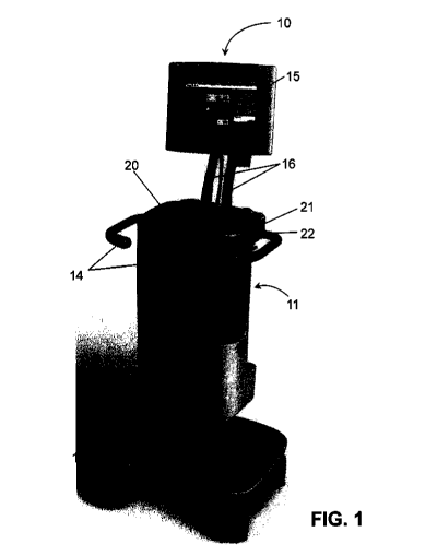
100651 FIG. 1 shows an exemplary embodiment of a radiopharmaceutical fluid
delivery
system 10. The fluid delivery system 10 may include a body 11 configured to
house various
components of the system in a confined area and wheels and/or casters 12 fixed
to the body and
being positioned to allow the system to be moved in one or more directions. In
various
embodiments, one or more of the wheels 12 may be lockable to prevent the
system 10 from moving
once it is in position. In some embodiments, the system 10 may include one or
more handles 14
fixed to the body 11 and positioned to allow an operator to grasp the handle
and move or position the
system 10. In other embodiments, the fluid delivery system 10 may be a stand-
alone or fixed-
position apparatus, and in such embodiments, the fluid delivery system may not
include wheels or
casters or the wheels or casters may be movably concealed in the body 11. Such
stand-alone or fixed
8
CA 02876107 2014-12-08
WO 2013/184642 PCT/US2013/044038
position apparatuses may also not include handles or handles may be movably
concealed in the body
11.
[0066] The fluid delivery system 10 generally includes a display or graphical
user interface
("GUI") 15 attached to the body 11, and positioned to allow a user to view the
display 15. In some
embodiments, the display 15 may be immovably fixed to the body, and in other
embodiments, the
display 15 may be positioned away from the system 10 and attached to the
system 10 by a hard
wired or wireless network. In other embodiments, the display 15 may be
pivotally connected to the
body 11, by means of one or more movable arm 16 that is pivotally connected to
a joint on the
display 15 and/or a joint on the body 11. Such display 15 may be configured to
be tilted or swiveled
with respect to the arm 16 to allow the display 15 to be positioned by an
operator.
[0067] The display 15 may be a color display, a black and white display, or a
green-screen
display, and in various embodiments, the display 15 may display real-time data
with regard to the
operation of the system 10. In some embodiments, the display 15 may be
configured to allow a user
to program or otherwise operate the system 10. For example, in certain
embodiments, the display 15
may have touch-screen capabilities or be otherwise configured to allow a user
to interact with the
system 10, and in particular, the computer portion of the system 10, by
manipulating or touching the
display 15. In other embodiments, the system 10 may include a keyboard, mouse,
microphone, hand
switch, footswitch, or other device configured to allow the user to program or
otherwise operate the
system 10. In still other embodiments, the display 15 may be included as part
of a laptop or tablet
computer that is electronically associated to the system 10 by a hard wired or
wireless network.
[0068] The body 11 may include a retractable lid or cover 20 having a primary
handle 21
including a latch release 22, and in some embodiments, the lid or cover 20 may
include a secondary
handle (not shown). In some embodiments, the lid 20 may include a locking
mechanism, such as a
combination or a key lock (not shown) that is capable of interacting with the
body 11 to lock the lid
20 in a closed position to prevent access of the system 10. In other
embodiments, the locking
mechanism may be a software-implemented lock, such as a password-protected
access point, that is
accessible through the display 15 and is adapted to lock the cover 20 in a
closed position and/or to
prevent access or operation of the system 10.
[0069] As illustrated in FIG. 2A, the lid 20 may be movably attached to the
body 11 at an
upper portion of the body 11 and may cover an upper surface 203 that defines a
number of recessed
portions including, for example, wells 24 into which a vial or container of a
pharmaceutical or a
9
CA 02876107 2014-12-08
WO 2013/184642 PCT/US2013/044038
radiopharmaceutical and troughs 25 into which various components of a fluid
path set (not shown)
can be positioned during an injection procedure. The lid 20 may be reversibly
moved with respect to
the body 11 to allow access to the recessed portions of the body, and the lid
20 may be configured to
allow for insertion and removal of vials or containers that may be positioned
within the wells 24 and
a fluid path set. In this manner, the radiopharmaceutical vial and the
components of the fluid path
set can lie below the plane of upper surface 203 of the body and can be
completely covered by the
lid 20. In other embodiments, a drawer type mechanism may be used that
slidably displaces a surface
having recessed portions into and out of the body 11.
[0070] In some embodiments, the system 10 may include an interrupt button 26
in FIG. 2A
that is configured to allow an operator to pause or abort an injection
procedure in the event of, for
example, patient discomfort or an emergency, while by-passing the display 15,
which also can be
configured to allow the user to pause or abort an injection procedure. The
interrupt button 26 may be
connected to LEDs and/or a printed circuit board to provide visual and/or
auditory alarms when the
interrupt button 26 has been activated.
[0071] In some embodiments, the lid 20, upper surface 203, and various other
portions of the
body 11 may include suitable radioactive shielding (such as lead) for
minimizing potential radiation
exposure from the radiopharrnaceutical to the operator. The upper surface 203
or one or more
portions thereof can be covered by the lid 20 during use to limit radiation
exposure to the operator,
other medical personnel, patient, and other observers. In particular
embodiments, the lid 20 may be
configured to be operated using one hand. For example, in certain exemplary
embodiments, the lid
20 may be attached to the body 11 by a pivot that allows the door to easily
pivot away from the work
surface of the body 11 during set-up and pivot back over the work surface
during operation. The
pivot hinge of such embodiments may be position away from the work surface
sufficiently such that
when the lid 20 is pivoted away from the work surface, the entire work surface
is exposed.
Therefore, the user can have access to any part of the system 10 during set-
up. The lid 20 may then
pivot back to cover the entire work surface thereby shielding the user during
operation of the system
10. Single hand operation may be achieved by positioning a handle and locking
mechanism at a
position in which the operator can unlock and pivot the door with one hand. In
certain
embodiments, a motor may be used to assist the user in pivoting the door.
[0072] The recesses or troughs 25 of the upper surface 203 of the body may be
configured to
removably accept the components of the fluid path set, and place the fluid
path set in position to
CA 02876107 2014-12-08
WO 2013/184642 PCT/US2013/044038
connect the wells 24, pumps, and so forth required for the fluid delivery
system 10. The wells 24 and
troughs 25, and the fluid path described thereby, may be configured in any way
to accommodate the
necessary fluid path set.
[0073] The wells 24 and recesses or troughs 25 formed in the upper surface 203
can be sized,
configured, or arranged to accommodate any length, design, or configuration of
the fluid path set and
the various components of the fluid path set 32 including pumps, medical fluid
containers or bags,
syringes and other medical delivery devices, radiopharmaceutical vials, vial
shields,
ionization/calibration chamber tubing, waste receptacle, and the like.
Additionally, the arrangement
of components provided in FIG. 1-3 are examples, and the arrangement of the
components may vary
among embodiments. Therefore, the wells 24 and troughs 25 may be configured to
accommodate
these various arrangements and the lengths of tubing necessary to connect the
components in such
arrangements. Thus, the size, i.e., the width, depth, and length, of the wells
24 and troughs 25 may
vary among embodiments. The various recesses and troughs 25 of various
embodiments may further
include tubing holders for holding tube sections and preventing kinking and
tangling.
[0074] As used herein, the term "fluid path set" refers to a one or more
sections of tubing
designed and configured to fluidly connect elements of the fluid delivery
system 10 including a
medical fluid source, a radiopharmaceutical source, a pharmaceutical source,
and the like to a fluid
delivery tube configured to deliver medical fluid and the radiopharmaceutical
and/or the
pharmaceutical to a patient. In various embodiments, the one or more sections
of tubing making up
the fluid path set 32 may be joined to one another in a manner that allows
fluids traveling within the
tubing to be carried to various portions of the system 10, mixed with one
another, delivered to a
patient or a waste receptacle. Thus, the fluid path set 32 may include one or
more joints including,
but not limited to, linear joints, T-joints, 4-way joints, and the like. In
still other embodiments, the
one or more of the one or more joints may include valves such as, for example,
check valves, by-
pass valves, stop cocks, and the like, and combinations thereof. The fluid
path set 32 of various
embodiments may further include one or more linkers that link the fluid path
set 32 or portions
thereof to the medical fluid, radiopharmaceutical, pharmaceutical, and
patient. Such linkers may
include luer linkers, screw-type linkers, pressure fittings, and the like.
[0075] In some embodiments, tube set may include a delivery tube section 317,
that is used
on a per-patient basis and discarded after use with a single patient to
prevent, for example, cross-
contamination between patients that can be collectively be referred to as
"single patient delivery
11
CA 02876107 2014-12-08
WO 2013/184642 PCT/US2013/044038
systems" ("SPDS") or "patient administration set" ("PAS"). The remaining
portions of the fluid
path set 32 in which the radiopharmaceutical is calibrated and prepared for
delivery can be used for
multiple patients and can be referred to as a "multiple patient delivery
system" ("MPDS") or "source
administration set" ("SAS").
[0076] FIG. 2B shows schematic of an exemplary fluid path set 32. In such
embodiments, a
first section of tubing 301 in the fluid path set 32 may be configured to
deliver fluid from the
medical fluid storage container 302 to a three-way confluence valve 303. In
some embodiments, the
first tubing section 301 may be configured to be placed within a pump (not
shown), and in other
embodiments, the medical fluid storage container 302 may be configured to be
placed under
pressure. For example, in particular embodiments as illustrated in FIG. 2B,
the medical fluid storage
container 302 may be a rigid, cylindrical device having a stopper or plunger
302a slidably inserted
into the fluid storage container thereby creating a seal within the medical
fluid storage device, A
motor 302b may be operably associated with the stopper or plunger 302a to
drive the stopper or
plunger 302a into the medical fluid storage device 302 increasing the pressure
within the medical
fluid storage device 302 and forcing the medical fluid held within the medical
fluid storage device
302 in the first tubing section 301. In other embodiments, a peristaltic or
inline pump may be
associated with the medical fluid storage device to drive fluid from the
medical fluid storage device
302 into the first tubing section 301 of the fluid path set 32, and the
medical fluid storage device 302
may be prepared from either a rigid or pliable material. The flow of medical
fluid and
radiopharmaceutical can be regulated throughout the device based on the
movement of medical fluid
from the medical fluid storage device 302 into the first tubing section 301
either by the stopper 302a
and motor 302b or the peristaltic or in line pumping device.
[0077] In embodiments including a rigid, cylindrical medical fluid storage
device 302,
stopper 302a, and motor 302b, the device may further include a means for
refilling the medical fluid
storage device either manually or automatically. For example, in certain
embodiments, a fluid
reservoir such as a saline bag 302c, as provided in FIG. 2A, may be removably
attached to the
medical fluid storage device 302 when the medical fluid storage device 302 has
been emptied and
can be used to introduce saline, or other medical fluid, into the medical
fluid storage device 302. In
some embodiments, the first tubing section 301 may be removed during refilling
and the fluid
reservoir 302c may be attached to the medical fluid storage device 302 through
the connector 302d
associated with the first tubing section 301. In other embodiments, an
auxiliary port 302e may be
12
CA 02876107 2014-12-08
WO 2013/184642
PCT/1182013/044038
provided in the medical fluid storage device 302, the connector 302d to the
first tubing section 301,
or a portion of the first tubing section 301 (shown), and the fluid reservoir
302c may be operably
connected to the fluid path through the auxiliary port 302e. In particular
embodiments, the fluid
reservoir 302c may be detached after the medical fluid storage device 302 has
been filled. In other
embodiments, the fluid reservoir 302c may remain associated with the medical
fluid storage device
302 throughout use of the device and may be used to refill the medical fluid
storage device 302 more
than one time before being detached from the medical fluid storage device 302.
100781 In certain embodiments, the medical fluid storage device 302 may be a
syringe like
apparatus, such as the dual zone syringe 3000 of FIG. 1 As illustrated, such a
dual zone syringe
may have at least two zones, a working zone 3001 which provides a reservoir
for the medical fluid
and a non-working zone 3002 through which the plunger 3003 passes. The plunger
3003 may
include any number of other features and may have any shape. For example, in
some embodiments
the plunger 3003 may have a cylindrical shape with a conical shaped upper
portion, which may
facilitate evacuation of the working zone 3001 of the syringe 3000 by
substantially matching the
shape of the upper portion of the syringe 3000 (as shown). The lower portion
of the plunger may be
flat or may be shaped to contact a piston or other motor associated with the
system 10 that is
positioned to advance the plunger 3003 through the syringe 3000 during use.
The plunger 3003 may
generally include at least two seals, an upper seal 3004 that is in
communication with the inner walls
of the working 3001 portion of the syringe 3000 and seals fluid within the
working zone 3001 and a
lower seal 3005 that is in communication with the inner walls of the non-
working zone 3002.
(00791 The upper and lower seals 3004, 3005 can be effectuated by any means.
For
example, in certain embodiments, an 0-ring may be set within a groove on the
portions of the
plunger 3003 associated with the upper and lower seals 3004, 3005. In further
embodiments,
working zone 3001 and the non-working zone 3002 may have different diameters.
For example, in
some embodiments, the portion of the syringe 3000 making up the working zone
3001 may have a
smaller diameter than the portion of the syringe 3000 making up the non-
working zone 3002. This
arrangement may avoid contamination between the working and non-working zones
3001, 3002.
Without wishing to be bound by theory, the inclusion of a sealed non-working
zone 3002 may
prevent direct contact of ambient air with the inside walls of the working
zone 3001 that will contact
the medical fluid thereby preserving the sterility of the medical fluid
touching the inner walls of tht
working zone 3001 during repeated use.
13
CA 02876107 2014-12-08
WO 2013/184642 PCT/IJS2013/044038
[00801 The syringe body 3000 may have any external features. For example, as
illustrated in
FIG. 3, the syringe 3000 may include connector flanges 3006 that circle the
diameter of the syringe
body 3000 and connect with similar flanges in the body 10 of the system. The
syringe body 3000
may further include a stop 3007 that halts advancement of the syringe 3000
into the body 10 during
insertion. In certain embodiments, the syringe body 3000 may further include
markings that can be
read by the system to ensure the proper syringe is being used in the system
10. For example, the
system 10 may detect a syringe that is the wrong size or does not include the
dual zone system
described above, and provide a warning or shut the system down. Such markings
may be a visible,
radio, or a light tag, and in particular embodiments, the markings may be a
series of etched grooves
in the syringe body 3000 that can be identified by a light reader in the body
of the device. In some
embodiments, the markings may be markings as described in U.S. Patent No.
7,018,363, which is
hereby incorporated by reference in its entirety.
[0081] A second tubing section 304 may be configured to transport fluid from a
first well
305 to the three-way confluence valve 303. In some embodiments, the first well
305 may be
configured to accommodate a one or more vial or container of a
radiopharmaceutical 305a, and as
such, the first well 305 may be individually shielded to reduce radiation
exposure to the operator and
patient. In other embodiments, the first well 305 may be configured to
accommodate a vial or
container 305a disposed in a vial shield or pig (as shown). In particular
embodiments, the first well
305/205 may further include an individual lid or cap 215 that also may be
shielded to reduce
radiation exposure (FIG. 2A). The second tubing section 304 may allow
transport of the
radiopharmaceutical from the first well 305 to the three-way confluence valve
303, and in certain
embodiments, the second tubing section 304 may be configured to be placed
within a pump 306
which may be a peristaltic or in line pump. In still other embodiments, the
second tubing section 304
may include a spike 304a or other device for connecting with the
radiopharmaceutical vial 305a and
drawing radiopharmaceutical from the vial 305a. In further embodiments, the
second tubing section
304 and/or the first tubing section 301 may include additional devices such
as, for example, air
detection devices, pressure sensors, and the like. Another embodiment (not
shown) allows for a
second radiopharmaceutical to be connected to the system, creating a system in
which two
radiopharmaceuticals, e.g., Technesium and Thallium, can be connected to the
system 10
simultaneously. In this configuration, the second radiopharmaceutical would be
provided in a
second vial in a separate pig. The tubing section associated with
radiopharmaceutical would connect
14
CA 02876107 2014-12-08
WO 2013/18-1642 PCT/US2013/044038
include a three way valve connecting tubing section connecting the first
radiopharmaceutical to the
system 10 and a second tubing section 304 connecting the second
radiopharmaceutical to the system
before the 306 pump.
[0082] The three-way confluence valve 303 may be configured to allow fluid
from the first
tubing section 301 and/or the second tubing section 304 to individually pass
into the third tubing
section 307. For example, the three-way confluence valve 303 may be configured
to allow fluid to
flow from position "c" to position "b" allowing medical fluid from the medical
fluid storage device
302 to flow from the first tubing section 301 directly into the third tubing
section 307. The three-way
confluence valve 303 may be reconfigured based on commands from the control
system, described
below, to allow fluid to flow from position "a" to position "b" allowing
radiopharmaceutical to flow
from the second tubing section 304 into the third tubing section 307. In some
embodiments, the
three-way confluence valve 303 may be configured to allow mixing of medical
fluid and
radiopharmaceutical by allowing fluid flow through both position "c" and
position "a" through
position "b" before entering the third tubing section 307.
[0083] The third tubing section 307 may lead to a second well 309 that is
configured as a
ionization/calibration chamber 309. Thus, the second well 309 may include the
components
necessary to determine the radiation level of the fluid entering the second
well 309. For example, in
various embodiments, the second well 309 may be associated with the components
of detectors such
as, but not limited to, a CZT crystal detector, a Geiger-Muller counter, a
scintillating counter, or a
parabolic detector, such as the parabolic sensor disclosed in U.S. Application
No. 12/664,653, which
is hereby incorporated by reference. The fluid path set 32 may be configured
in any way to allow
emissions from the radiopharmaceutical to be quantified. For example, in some
embodiments, the
fluid path set 32 may include a linear loop of tubing contained within the
second well 309, and fluid
flow may be stopped for a period of time sufficient to allow quantification of
the radioactive
emissions from the radiopharmaceutical. In other embodiments, as illustrated
in FIG. 2B, the second
well 309 may be configured to accommodate a coil assembly 310 portion of the
fluid path set 32.
The coil portion may provide sufficient residence time within the second well
309 to allow for
emission from the radiopharmaceutical to be quantified without stopping or
slowing fluid flow
through the device.
[0084] A fourth tubing section 311 may extend from the second well 309 to a
three way
valve or four way valve 315. As shown in FIG. 2B, a four-way valve 315 may
regulate fluid flow
CA 02876107 2014-12-08
WO 2013/184642 PCT/US2013/044038
from the fourth tubing section 311 through port "d" of the four-way valve into
a waste tubing section
314 out port "f' or a output tubing section 314 out port "e." The four-way
valve may further
regulate fluid flow from an auxiliary tubing section 316 through port "g" that
is separately associated
with the medical fluid source 302 into the waste tubing section 314 out port
"f" or output tubing
section 314 out port "e." In such embodiments, the output tubing section 314
may extend away from
the four-way valve toward a delivery tubing section 317 through which the
radiopharmaceutical is
delivered to the patient. The waste tubing section 314 may extend away from
the four-way valve to
carry fluid to a waste receptacle 313 and function to divert from, for
example, a priming procedure
to prepare the system 10 for injection away from delivery tubing section 317
and ultimately the
patient. The auxiliary tubing section 316 may be associated with the a T-joint
or three-way valve
301a and may extend from the first tubing section 301 to the four-way valve
315. The auxiliary
tubing section 316 may be configured to transport fluid from the medical fluid
source 302 directly to
the four-way valve 315 providing a by-pass for the majority of the fluid path
while allowing flow of
medical fluid to the fluid delivery section 317 and patient.
[0085] The four-way valve of some embodiments may be designed as illustrated
in FIG. 2C
I. FIG. 2C shows a four-way valve 2100 having a rotating internal stem 2101
and an external four-
way tubing connectors 2102. In some embodiments, the four-way tubing connector
2102 may be
prepared from a flexible material that has sufficient tensile strength to
allow the valve to maintain a
seal between. the internal stem 2101 and the tubing connector 2102, and in
other embodiments and
rigid external tubing connector 2012 may be coupled to an semi-rigid or
rotating internal stem. In
other embodiments, the internal stem 2102 may include distal separations 2103
that allow the stem
2102 to be compressed slightly allowing for the tubing connector 2102 to be
placed over internal
stem 2101 during manufacture. In the cross-sectional view provided in FIG. 2C
II, the passageway
2104 though the internal stem 2101 can be seen. The passageway 2106 may be
configured to allow
passage of fluid through the passageways, 2102a and 2102b in this drawing, of
neighboring tubing
extensions only while sealing off the remaining passageways 2102c and 2102d.
In various
embodiments, the stem may be designed to press-fit into to the body 11 of the
device when the
MPDS is properly positioned on the device.
[0086] The auxiliary or by-pass tubing section 316 may allow for the delivery
of different
fluids to a patient without mixing. For example, in some embodiments, medical
fluid may be passed
directly from the medical fluid storage container 302 through the auxiliary or
by-pass tubing section
16
CA 02876107 2014-12-08
WO 2013/184642 PCT/US2013/044038
316 to the four-way valve 315 which directs the medical fluid to the output
tubing section 314 and
the delivery tubing section without mixing with radiopharmaceutical or other
fluids contained within
the remainder of the fluid path set 32. Thus, the patient may continually
receive medical fluid even
when radiopharmaceutical is not being delivered. This arrangement further
allows for the delivery
of pharmaceutical from the pharmaceutical delivery port 318 (described below)
without the
administering radiopharmaceutical.
[0087] In still further embodiments, the system may include additional
auxiliary tubing
sections (not shown). Such additional auxiliary tubing sections may carry any
medical fluid to the
patient and additional auxiliary tubing sections may integrate into the tubing
set through 5-, 6-, 7-, or
8-way valves positioned in place of the four-way valve 315 described above, or
additional 3-, 4-, 5-,
or 6-way valves may be incorporated into the tube set at one or more
locations, such as, for example,
in the output tubing section 314. In various embodiments, additional auxiliary
tubing sections may
be associated with saline or other medical fluids, pharmaceutical, or other
fluid that may be required
for particular patients. The multiport valves of various embodiments including
the four-way valve
315 described above may be commercially available multi-port valves or may be
specially designed
to limit mixing between input tubes.
[0088] In yet further embodiments including a three way valve (not shown), the
auxiliary
tubing section 316 may be absent. The fourth tubing section 311 may deliver
fluid to the three way
valve where the fluid can be diverted from port "d" through port "?' into the
waste tubing section
314 and waste receptacle or bag 313 or from port "d" through port "e" to the
output tubing section
314 and toward the delivery tubing section 317.
[0089] In some embodiments, the sixth tubing section may terminate in an
output fitting
314a, which may be a connector or adaptor, or other fitting configured to
operably connect the
output tubing section 314 to the delivery tubing section 317. In particular
embodiments, the
connector at the terminus of the output tubing section 314 may be a swabable
valve that can be
disinfected or washed when the delivery tubing section 317 is replaced between
patients.
[0090] The MPDS 31 portion of this fluid path set 32 may include any tubing
section from
the medical fluid storage device 302 to the connector 314a of the output
tubing section 314 and is
indicated by the components within the dashed line box. In various
embodiments, the MPDS 31
may include a connector 302d such as a spike or luer lock for connecting the
MPDS 31 to the
medical fluid storage device 302; a spike or vented cannula 304a for
connecting to
17
CA 02876107 2014-12-08
WO 2013/184642 PCT/US2013/044038
radiopharmaceutical vial; a coil assembly 310; a connector for a waste
receptacle (not shown); a
connector for the output tubing section 314; and various connectors and tube
sections connecting
these elements. In some embodiments, the MPDS may farther include the various
valves as
described above, 303, 301a, 315. In certain embodiments, the connector 302d
for connecting the
MPDS 31 to the medical fluid storage device 302 may include a means for
sensing the MPDS 31 and
the medical fluid storage device 302. Embodiments are not limited to a
particular sensing device.
For example, a tag including a bar code or radiofrequency identification
(RFID) may be associated
with MPDS 31 may be read by the connector 302d or at the connection site to
ensure that the proper
MPDS 31 is connected to the system. In other embodiments, an optical system
such as that
described in U.S. Patent No. 7,018,363 may be used to encode the connectors.
[0091] Each component of the MPDS 31 may be pre-connected and can be stored in
a sterile
packet or container for use in a fluid delivery system, and in certain
embodiments, the upper portion
of the body may be configured to accept a tray or holder 220 designed to hold
and separate the
components of the MPDS 31 in a position to be inserted into the troughs 25 and
wells 24 of the
device without realignment by the operator. FIG. 2D shows the contents of a
sterile packet 2000
which includes the holder 220, which can have one or more grips 2001
configured to be grasped by
the user during insertion and removal of the MPDS 31 from the system 10, and
various troughs 2005
for routing the tubing sections 2002. The packet 2000 may further include a
medical fluid storage
reservoir or a container, such as the syringe 2006 provided in FIG. 2D, for
storing and introducing
medical fluid such as saline into the fluid path set 32, a waste receptacle
2007, and a coil assemble
2010 that can be incorporated into an ionization chamber. A spike or vented
cannula 2004 for
connecting the tube set to a vial of radiopharmaceutical and a fitting such as
a luer connector 2012
for connecting the MPDS to an SPDS. The packet also includes the various
tubing sections
necessary to connect these elements as well as valves or tubing configured to
be incorporated into a
valve 2013, and sections of tubing configured to be introduced into pumps
2014. Using the holder
220 and the packet 2000 , the operator can introduce the MPDS 31 into the
system without
individually inserting each tube section, valve or connector into the system.
Rather, the user can
introduce the tube set into the system by merely placing the holder 220 into
the corresponding
groove in the device 10, inserting the medical fluid storage reservoir or a
container 2006, waste
receptacle 2007, and coil assemble 2010 into the appropriate wells, and
connecting the tube set to the
pumps where appropriate.
18
CA 02876107 2014-12-08
WO 2013/184642 PCT/US2013/044038
[0092] The container or vial of radiopharmaceutical may be any suitable
container known in
the art and the well 24 for holding the radiopharmaceutical may be configured
to accept any such
container or vial and securely hold the container during use. In some
embodiments, an adaptor may
be used that encases all or a portion of the vial or container before it is
placed in the well 24 to
ensure that the vial or container is secured within the well 24. In still
further embodiments, the
adaptor may be prepared from or include a material that blocks emission of the
radioactive particles
from the radiopharmaceutical.
[0093] In particular embodiments, the vial or container may be a multi-dose
container
configuration to hold and store a sufficient amount of radiopharmaceutical for
delivery to a plurality
of patients in a single container. In other embodiments, the well 24 may be
configured to hold more
than one container or vial for holding and storing radiopharmaceuticals. In
some embodiments, each
container in the multi-container configuration may include individual doses of
radiopharmaceutical
sufficient for administration to a single patient. In other embodiments, each
container or vial may
hold and store multiple doses of the radiopharmaceutical and the system may be
configured such that
doses of the radiopharmaceutical can be pulled from a new vial when the
proceeding vial is used to
completion. In still other embodiments, a different radiopharmaceutical
composition may be held
and stored in each of two or more different multi-dose containers, and in such
embodiments, the
system may be configured to deliver different radiopharmaceutical compositions
either
simultaneously to a single patient or consecutively to different patients
during different procedures.
In still further embodiments, a micro-fluidic device or other
radiopharmaceutical generation
technology capable of real-time generation of a radiopharmaceutical can be
included as part of the
multi-dose container configuration.
[0094] The system may further include a pharmaceutical delivery port that
includes a 3-way
connector 318a, a check valve 318b that only allows fluid to flow one-way from
the stress agent to
the main line only, and a connector 318c for connecting a syringe or vial
including a pharmaceutical
agent such as a stimulant to the system thereby providing a pharmaceutical
delivery port 318. In
some embodiments, the pharmaceutical delivery port 318 makes up a portion of
the delivery tubing
section 317 (as illustrated in FIG. 3). In other embodiments, the
pharmaceutical delivery port 318
may be incorporated into the output tubing section 314 between the three or
four-way valve 315 and
the connector 314a. The pharmaceutical delivery port may be any type of port
known in the art such
as, but not limited to, a luer, a needle vial adaptor, needleless vial
adaptor, or other fitting capable of
19
CA 02876107 2014-12-08
WO 2013/184642 PCT/US2013/044038
accepting a delivery device 319a such as a syringe or vial. The delivery port
may further include a T-
joint or a three way valve or stopcock. The pharmaceutical delivery port 318
may be configured to
allow for the introduction of a pharmaceutical agent into the delivery tubing
section 317 during a
procedure. For example, in some embodiments, a syringe 319a holding a
pharmaceutical agent may
be fitted to the pharmaceutical delivery port 318, and the pharmaceutical
agent may be introduced
into the delivery tubing section 317 during the procedure either manually by
depressing a plunger in
the syringe 319a or automatically using a motor associated with the plunger or
a pump 319b
delivering an appropriate dose of the pharmaceutical to the patient. In other
embodiments, fluid may
be diverted into the syringe or vial by the pharmaceutical delivery port 318
where the
pharmaceutical is mixed with the fluid before being introduced back into the
pharmaceutical
delivery port 318 and out to the delivery tube section 317. In additional
embodiments, the system
may include one or more pumps, motors, or the like associated with the
pharmaceutical delivery port
318, delivery device 319, output tubing section 314, or delivery tube section
317. In some
embodiments, the SPDS connector 317a can be encoded through RFID, light
sensors, mechanical
sensors, etc. to ensure that the correct SPDS is connected. This ensures that
the correct protocol is
executed with the correct SPDS.
[00951 The sixth tube section 314 and the delivery tube section 317 may be an
integral part
of the fluid path set 32, or in other embodiments, the delivery tube section
317 and/or the sixth tube
section 314, pharmaceutical delivery port 318, and other components associated
with this portion of
the fluid path set 32 may be one or more separate fluid path sets configured
to attach to the fluid path
set 32 by, for example, a luer fitting or swabable valve. For example, in some
embodiments as
illustrated in FIG. 3, the delivery tube section 317 may include a first end
317a that can be reversibly
attached to the connector 314a associated with the output tubing section 314
and a patient end 317b
having a connector such as a luer connector, that is capable of being attached
to, for example, a
catheter, IV needle, intravenous port, or the like that can be used to deliver
the radiopharmaceutical
to a patient. In other embodiments, the delivery tube section 317 may have a
first end that can be
reversibly attached to the pharmaceutical delivery port 318 or a T-joint or
three-way valve associated
with the fluid delivery port 318c. In still other embodiments, the delivery
tubing section 317, the
output tubing section 314, and any intervening device or tube section such as
the pharmaceutical
delivery port may be separately, reversibly connected to the fluid path set
32. In some embodiments,
the pharmaceutical delivery port may be absent, blocked, or otherwise
eliminated such that the
CA 02876107 2014-12-08
WO 2013/184642 PCT/US2013/044038
radiopharmaceutical can be delivered in the absence of the addition of an
additional pharmaceutical
or stimulating agent.
[0096] Embodiments are not limited to a particular pharmaceutical agent, and
any agent that
is known or may be usefully administered may be contained within the syringe
318a and
administered to the patient during a procedure. For example, in some
embodiments, the
pharmaceutical agent may be a stress agent such as, but not limited to, IV
Dobutamine, IV
Dipyridiamole (Persantine), IV Adenosine (Adenoscan), IV Lexiscan
(Regadenoson), and the like.
In other embodiments, the pharmaceutical agent may reduce vasodilation such
as, for example, IV
Aminophylline. In still other embodiments, the system may include a first
pharmaceutical delivery
port 318 and a second pharmaceutical delivery port (not shown). In such
embodiments, a first
syringe associated with the first pharmaceutical delivery port that holds a
stress agent and a second
syringe associated with the second pharmaceutical deliver port may include a
pharmaceutical that
acts to reduce vasodilation and act as an antidote to stress agent, allowing
the user to reduce the
stress under which the patient is placed as part of the procedure or as a
precaution in the event of an
adverse event. The pharmaceutical agent can be introduced into the fluid flow
through the
pharmaceutical delivery port continuously or in one or more controlled doses,
[0097] The delivery tube section 317 may be configured to connect to typical
patient
delivery apparatuses such as, IV needles, ports, catheters, or other means for
delivering intravenous
pharmaceuticals. In other embodiments, the delivery tube section 317 may
incorporate such delivery
devices, In still other embodiments, the delivery tube section 317 may be
configured to connect to
other sections of tubing, which may incorporate the delivery apparatuses.
100981 In some embodiments, the system 10 may include one or more additional
components
including, but are not limited to, pinch valves, air detectors, and mounts or
retainers for holding the
connector ends of the delivery tube section, and the like and combinations
thereof. In particular
embodiments, pinch valves may be powered and controlled by the fluid delivery
system 10, and/or
manually operated. In other embodiments, the pinch valves can be replaced with
a manual or
automated 3-way stop cock. The fluid delivery system 10 may include one or
more pumping
mechanisms configured to facilitate the movement of liquids from wells in the
body to the delivery
tube section 317 of the fluid path set 32 at any position in the system 10.
Any suitable type of
pumping mechanism can be used including, but not limited to, piston-driven
syringe pumps, gear
pumps, rotary pumps, in-line pumps, and peristaltic pumps. In some
embodiments, the pumping
- - 21
CA 02876107 2014-12-08
WO 2013/184642 PCT/US2013/044038
mechanism may be peristaltic pump. In various embodiments, the pumping
mechanism may be
opened to receive a length of tubing associated with the fluid path set 32.
[0099] The output tubing section 314 may terminate in a connector 314a
configured to
connect the MPDS 31 with an SPDS 32. In some embodiments, the connector end
314a of the
MPDS 31 may be a swabable luer valve that is biased to close or seal off the
connector end 314a of
the MPDS 31 when the SPDS 32 is not connected. The swabable luer valve
prevents the MPDS 31
from being contaminated and allows an operator to swab or clean the connector
end 314a using, for
example, an alcohol wipe, prior to connecting an SPDS 32 to the connector. In
other embodiments,
the connector end 314a may be a standard luer connector or another connector
as known in the art.
[00100] The tubing of each of the sections of the MPDS 31 and SPDS 32 may be
prepared
from the same or different materials. For example, in various embodiments, the
tubing may be
silicone, C-Flex, standard PVC, silicone-like PVC material, or pump tubing. In
particular
embodiments, the microbore tubing of second tubing section 304 may be formed
from, for example,
silicone, C-Flex, or silicone-like PVC material, and the other tubing sections
301, 307, 311, 312,
314, 317, and tube coil 310 may be formed from any suitable polymeric
material, including standard
PVC.
[00101] The dimensions of the components of the MPDS 31 as shown in FIG. 3,
including
the various tubing sections, may vary among embodiments and may depend, for
example, on the
procedure for which the system is being used and the type and amount of
radiopharmaceutical being
delivered. In certain exemplary embodiments, the first tubing section 301 may
be about 3 inches to
about 4 inches in length or 3.4 inches in length, may have an outer diameter
(OD) of about 0.05
inches to about 0.25 inches or about 0.17 inches and an inner diameter (ID) of
about 0.05 inches to
about 0.15 inches or about 0.08 inches, and may have an about 90 to about 95
Shore A durometer.
The second tubing section 304 may be about 7 inches to about 10 inches in
length or about 8.9
inches in length and can be formed of microbore tubing having an OD of about
0.05 inches to about
0.10 inches or about 0.09 inches, an ID of about 0.01 inches to about 0.07
inches or about 0.03
inches and an about 35 to about 55 or about 45 Shore A durometer. The use of
rnicrobore tubing in
second tubing section 304 improves volume accuracy and thereby improves
measured activity
accuracy (i.e., of pharmaceutical delivered to the patient) and reduces
radiopharmaceutical waste.
The third tubing section 307 may be about 9.0 to about 14 inches in length or
about 11.75 inches in
length, may have an OD of about 0.05 inches to about 0.25 inches or about 0.17
inches and an ID of
22
CA 02876107 2014-12-08
WO 2013/184642 PCT/US2013/044038
about 0.05 inches to about 0.15 inches or about 0.08 inches, and may have an
about 90 to about 95
Shore A durometer. The fourth tubing section 311 may be about 8.0 inches to
about 12 inches in
length or approximately 10.5 inches in length, may have an OD of about 0.05
inches to about 0.25
inches or about 0.17 inches and an ID of about 0.05 inches to about 0.15
inches or about 0.08 inches,
and may have an about 90 to about 95 Shore A durometer. The waste tubing
section 314 and the
output tubing section 314 may each be about 1.0 inches to about 5.0 inches in
length or
approximately 3.0 inches in length, may have an OD of about 0.05 inches to
about 0.25 inches or
about 0.17 inches and an ID of about 0.05 inches to about 0.15 inches or about
0.08 inches, and may
have an about 90 to about 95 Shore A durometer. The tubing in tube coil 310
may be from about 20
inches to about 55 inches in length or approximately 41.75 inches in length,
has an OD of about 0.10
inches to about 0.30 inches or about 0.22 inches and an ID of about 0.05
inches to about 0.20 inches
about 0.16 inches, and may have an about 90 to about 95 Shore A durometer. All
of these
dimensions are provided for exemplary purposes only and are not to be
construed as limiting the
present disclosure.
[00102] The MFDS 31 may include a coil assembly 310. The coil assembly 310
may,
generally, include a section of that is simply gathered tubing in a coiled or
an uncoiled, amorphous
fashion and placed inside ionization/calibration chamber 309. In some
embodiments, the coil
assembly may be an individually constructed unit, and in other embodiments,
the coil assembly 310
may include all or portions of third tubing section 307 and fourth tubing
section 311. The coil
assembly 310 of various embodiments positions the radiopharmaceutical such
that the radioactivity
level of the radiopharmaceutical in the tube coil 310 can be measured by the
components
surrounding the ionization/calibration chamber 309. More specifically, the
coil assembly 310 orients
and locates the radiopharmaceutical within a "linear region" of the
ionization/calibration chamber
309 to more accurately measure its activity level and prepare an optimal dose
for injection into a
patient.
[00103] In some embodiments, the tubing may be coiled on itself or stacked in
a coil by
bonding the tubing layers, and in other embodiments, as illustrated in FIG.
4A, the coil assembly 410
may include a core element or structure 420 that is configured to allow the
tube coil 410 to be
wrapped around the core element 420. As such, the tube coil 410 can be formed
on the core element
420. The core element 420 may be configured to facilitate optimal positioning
of the tube coil 410,
and may be sized to fit within the ionization/calibration chamber 309 of the
body 11. In some
23
CA 02876107 2014-12-08
WO 2013/184642 PCT/US2013/044038
embodiments, the core element 420 may include a tube channel 421 between an
upper shoulder 422
and a lower shoulder 423. The tube coil 410 may be retained within the tube
channel 421 and
between the upper and lower shoulders 422, 423 to hold the tube coil 410 in
position and prevent
kinking. In further embodiments, an upper surface 424 of core element 420 may
include one or more
inlet channels or grooves 425 and an outlet channel or groove 426 to
accommodate third tubing
section 407 and fourth tubing section 411, respectively.
[001041 In various embodiments, the coil assembly 410 may be positioned
concentrically in
the ionization/calibration chamber 309. In some embodiments, the core element
420 may be self-
centering when inserted into the ionization chamber 309 of the fluid delivery
system 10 to facilitate
optimal positioning and performance. This may be achieved either through
structural features of the
coil assembly 40, the structure of core element 420, or a combination thereof.
For example, in some
embodiments, the upper shoulder 422, the lower shoulder 423, or both can be
configured to associate
with an outer wall of the ionization/calibration chamber 309. For example, the
core element 420 may
include additional features such as, for example, extensions, indentations, or
notches may be
provided on the core element 420 either on the upper or lower shoulders 422,
423 or another portion
of the coil assembly 40, that engage corresponding elements in the
ionization/calibration chamber
309 to aid in the proper positioning of the tube coil 410. In other
embodiments, the lower shoulder
423 may be sized to provide an appropriate distance between the lower surface
of the
ionization/calibration chamber when the lower shoulder contacts the lower
surface of the
ionization/calibration chamber or a diameter that corresponds with a the
appropriate diameter of the
ionization/calibration chamber 309.
[00105] With reference to FIG. 4B, in particular embodiments, the core element
420 and the
tube coil 410 may be sized and dimensioned so that the coil assembly 410 can
be optimally
positioned within the "linear region" of the ionization/calibration chamber
309. The "linear region"
of an ionization chamber refers to the region of the chamber in which activity
level measurements
are repeatable and predictable. For an exemplary ionization/calibration
chamber (Model IK-102
Short Ionization Chamber provided by Veenstra Instruments), the "linear
region" is located within a
window of about 5 mm to about 65 mm measured from the base or bottom wall of
the
ionization/calibration chamber 309. The tube coil 410 of various embodiments
may have a volume
capacity of about 1 ml to about 10 ml or about 1.5 ml to about 7 ml and may be
configured in any
way to achieve the desired volume. Moreover, the tube coil 410 may have any
number of turns. For
24
CA 02876107 2014-12-08
WO 2013/184642 PCT/US2013/044038
example, in some embodiments, the tube coil 410 may have about 4 to about 10
turns, and in other
embodiments, the tube coil 410 may have about 5 to about 7 turns. In various
embodiment, the tube
coil may have one or more 1/2 or 'A turns that allow appropriate placement of
the third tube section
407 and fourth tube section 411. A tube coil having this number of turns may
be formed from an
length of tubing sufficient to make the desired number of turns based on the
diameter of the core
element 420. For example, a core element having a diameter (w) of about 0.5 in
to about 4 in or
about 1 in to about 3 may require tubing having a length of about 5 in to
about 24 in, about 8 in to
about 15 in, or about 10 in to about 12 in. The height (h) of the tube coil
410 may similarly vary
depending on the number of turns, the diameter of the tubing, and the diameter
to the core element.
For example, a tube coil 410 having from about 5 to about 7 turns may have a
height (h) of from
about 0.5 in to about 8 in or about 1 in to about 5 in. The tube coil 410 may
be prepared from any
type of tubing; however, in certain embodiment, the tubing may have an OD of
from about 0.01 in to
about 0.5 in and an ID of about 0.025 to about 0.5 in.
[001061 In various embodiments, the fluid delivery system 10 may include a
control system
50 (schematically represented in FIG. 5) in communication with the various
components of the
injector system 1050 that for the purposes of the schematic of FIG. 5 can,
include, for example,
pumps, motors, ionization/calibration chamber, interrupt button, air detectors
valves, stopcocks, and
the like. The control system 50 may, generally, control the operation of the
injector system 1050,
while also providing an interface with input and output devices such as the
display 15, printer 1032,
and network devices 1040 used to program and direct the action of the injector
system 1050.
[001071 The control system 50 may include, but is not limited to, at least one
computer 1000
having certain components for appropriate operation, execution of code, and
creation and
communication of data. The computer 1000 includes one or more processing units
1004 (typically
referred to as a central processing unit or CPU) that serves to execute
computer-based instructions
received in the appropriate data form and format. Further, this processing
unit 1004 may be in the
form of multiple processors executing code in series, in parallel, or in any
other manner for
appropriate implementation of the computer-based instructions. As used herein,
the computer 1000
may be operably configured to execute appropriate software to perform and
implement the
processing steps of the methods and systems disclosed herein. The system may
include one Or more
computers 1000 or similar computing devices having a computer-readable storage
medium capable
of storing computer-readable program code or instructions that cause the
processing unit 1004 to
CA 02876107 2014-12-08
WO 2013/184642 PCT/US2013/044038
execute, configure, or otherwise implement the methods, processes, and
transformational data
manipulations discussed herein. Still further, the computer 1000 may be in the
form of a personal
computer coupled to the fluid delivery system 10, a processor formed
integrally with the fluid
delivery system 10, a computer provided remotely from the fluid delivery
system 10, or any other
type of computing device having the necessary processing hardware to
appropriately process data to
effectively implement the method and system described herein.
[00108] The control system 50 may further include a system bus 1006 to
facilitate
appropriate data communication and processing information between the various
components of the
computer 1000. The system bus 1006 may be any of several types of bus
structures, including a
memory bus or memory controller, a peripheral bus, or a local bus using any of
a variety of bus
architectures. In particular embodiments, the system bus 1006 may facilitate
data and information
communication between the various components (whether internal or external to
the computer 1000)
through interfaces.
[00109] In some embodiments, the computer 1000 may include one or more
discrete
computer-readable media components. For example, computer-readable media may
include any
media that can be accessed by the computer 1000, such as volatile media, non-
volatile media,
removable media, non-removable media, and the like. In certain embodiments,
the computer-
readable media may include computer storage media, such as media implemented
in any method or
technology for storage of information such as computer-readable instructions,
data structures,
program modules, or other data, including, but not limited to, random access
memory (RAM), read
only memory (ROM), electrically erasable programmable read only memory
(EEPROM), flash
memory, or other memory technology, CD-ROM, digital versatile disks (DVDs), or
other optical
disk storage, magnetic cassettes, magnetic tape, magnetic disk storage, or
other magnetic storage
devices, or any other medium which can be used to store the desired
information and which can be
accessed by the computer 1000. In some embodiments, the computer-readable
media may include
communications media, such as computer-readable instructions, data structures,
program modules,
or other data in a modulated data signal such as a carrier wave or other
transport mechanism. In
other embodiments, the computer-readable media may include any information
delivery media,
wired media (such as a wired network and a direct-wired connection), and
wireless media (such as
acoustic signals, radio frequency signals, optical signals, infrared signals,
biometric signals, bar code
26
CA 02876107 2014-12-08
WO 2013/184642 PCT/US2013/044038
signals, etc.). Combinations of any of the above are also included within the
scope of computer-
readable media.
[00110] In still other embodiments, the computer 1000 may further include
system memory
1008 with computer storage media such as volatile and non- volatile memory,
ROM, and/or RAM. A
basic input/output system (BIOS) with appropriate computer-based routines
assists in transferring
information between components within the computer 1000 and can be stored in
ROM. The RAM
portion of the system memory 1008 typically contains data and program modules
that are
immediately accessible to or presently being operated on by processing unit
1004, e.g., an operating
system, application programming interfaces, application programs, program
modules, program data,
and other instruction-based computer-readable code.
[001111 The computer 1000 may also include other removable or non-removable,
volatile or
non-volatile computer storage media products. For example, the computer 1000
may include a non-
removable memory interface 1010 that communicates with and controls a hard
disk drive 1012, i.e.,
a non-removable, non- volatile magnetic medium, a removable, non-volatile
memory interface 1014
that communicates with and controls a magnetic disk drive unit 1016 (which
reads from and writes
to a removable, non- volatile magnetic disk 1018), an optical disk drive unit
1020 (which reads from
and writes to a removable, non-volatile optical disk, such as a CD ROM 1022),
a Universal Serial
Bus (USB) port for use in connection with, for example, a removable memory
card 1023. Other
removable or non-removable, volatile or non-volatile computer storage media
can be used in the
exemplary computing system environment 1002, including, but not limited to,
magnetic tape
cassettes, DVDs, digital video tape, solid state RAM, solid state ROM, and the
like. These
removable or non-removable, volatile or non-volatile magnetic media are in
communication with the
processing unit 1004 and other components of the computer 1000 via the system
bus 1006. The
drives and their associated computer storage media discussed above and
illustrated in FIG. 4A
provide storage of operating systems, computer-readable instructions,
application programs, data
structures, program modules, program data, and other instruction-based
computer-readable code for
the computer 1000 (whether duplicative or not of the information and data in
the system memory
1008).
j001121 In particular embodiments, the fluid delivery system 10 may be
configured to allow
a user to enter commands, information, and data into the computer 1000 using
the touch-screen of
the GUI display 15 via an operator input interface 1028. However, it has been
envisioned that an
27
CA 02876107 2014-12-08
WO 2013/184642 PCT/US2013/044038
operator may enter conunands, information, and data into the computer 1000
using other attachable
or operable input devices, such as a keyboard 1024, a mouse 1026, a remote
control device, a
microphone, a trackball, a joystick, a touchpad, a scanner, a tablet computer,
and the like, via the
operator input interface 1028. Any arrangement that facilitates the input of
data and information to
the computer 1000 from an outside source may be used including, for example,
hard wiring or
accessing using a wireless network device, such as blue tooth, a wireless
intemet connection, or a
cellular connection. As discussed, these and other input devices are often
connected to the
processing unit 1004 through the operator input interface 1028 coupled to the
system bus 1006, but
may be connected by other interface and bus structures, such as a parallel
port, game port, or a USB.
[001131 In still further embodiments, data and information can be presented or
provided to
an operator in an intelligible form or format through certain output devices,
such as the GUI display
15 (to visually display this information and data in electronic form), a
printer 1032 (to physically
display this information and data in print form), a speaker 1034 (to audibly
present this information
and data in audible form), etc. All of these devices are in communication with
the computer 1000
through an output interface 1036 coupled to the system bus 1006.
[001141 The computer 1000 may operate in a network environment 1038 through
the use of
a communications device 1040, which is integral to the computer or remote.
This communications
device 1040 is operable by and in communication with the other components of
the computer 1000
through a communications interface 1042. Using such an arrangement, the
computer 1000 may
connect with or otherwise communicate with one or more remote computers, such
as a remote
computer 1044 of a hospital information system, which typically includes many
or all of the
components described above in connection with the computer 1000. Using
appropriate
communications devices 1040 such as, for example, a modem, a network
interface, adapter,
telephone line, cellular telephone connection, wifi network, and the like, the
computer 1000 may
operate within and communicate through a local area network (LAN) and a wide
area network
(WAN), but may also include other networks such as a virtual private network
(VPN), an office
network, an enterprise network, an intranet, the Internet, and the like and
combinations thereof. It
will be appreciated that the network connections shown are exemplary and other
means of
establishing a communications link between the computers 1000, 1044 may he
used.
[00115] Generally, the fluid delivery system of embodiments described above
may be
configured to deliver a radiophamiaceutical drawn from a bulk
radiopharmaceutical vial 305a in
28
CA 02876107 2014-12-08
WO 2013/184642 PCT/US2013/044038
FIG. 3. A dose of the radiopharmaceutical may be withdrawn from the bulk
radiopharmaceutical vial
305a contained in the first well 305, and passed though the coil assembly 310
of the fluid path set 32
in the ionization/calibration chamber 309. The radioactive activity, i.e., the
average emission of the
dose of radiopharmaceutical, can be determined based on measurements made in
the
ionization/calibration chamber 309. The system may be configured to determine
the volume of a
dose having a desired activity level based on the measured amount of
radioactive activity and the
volume of a test sample. The system may then draw the proper amount of
radiopharmaceutical to
deliver the appropriate dose of radiopharmaceutical as identified by the user
to the patient.
[00116] The fluid delivery system 10 may further be configured for priming (L
e., purging air
from the IVFPDS 31), delivering a radiopharmaceutical to a patient, providing
a saline flush to
remove residual radiopharmaceutical, while minimizing or eliminating exposing
the individuals
operating system to the radiopharmaceutical and minimizing or eliminating
contaminated waste.
Moreover, MPDS 31 and other elements disclosed herein also facilitate safe
delivery of the
pharmaceutical to multiple destinations (for example, dose delivery to a
series of patients).
[00117] The system 10 may be further configured to provide feedback
information to the
operator. For example, in some embodiments, the system may provide the
operator with information
regarding the administration such as, but not limited to, the dosage of
radiopharmaceutical delivered
to the patient by milligram (mg), volume (ml), and/or radioactive activity
(mCi), the amount of other
pharmaceutical composition delivered to the patient (mg/nil), the flow rate of
the
radiopharmaceutical or other pharmaceutical (ml/s), the amount of saline
administered (m1), dosing
time (i.e., the time required for delivery), the delivery time (i.e., the time
of day), date, and the fluid
pressure in the delivery system during delivery. In particular embodiments,
the system may further
provide the operator with absorption data with regard to the particular
radiopharmaceutical
administered including the expected amount of the radiopharmaceutical absorbed
by particular
organs such as brain, lung, liver, kidney, bladder, bone, thyroid, heart,
breast, stomach, colon, and
skin. In some embodiments, the system may reference patient data to determine
the amount of
radiopharmaceutical administered to the particular patient over time and
provide a warning to the
operator if absorbed levels become too high. In various embodiments, the
information may be
provided to the operator in real time or provide an estimate of the
absorption, based on the planned
dose, prior to an injection.
29
CA 02876107 2014-12-08
WO 2013/184642 PCT/US2013/044038
[00118] Following administration or the completion of an administration
protocol, the
system may provide a summary of the procedure including any relevant data. For
example, in
various embodiments, the system may provide the dosage of radiopharmaceutical
delivered to the
patient by milligram (mg), volume (m1), and/or radioactive activity (mCi), the
amount of other
pharmaceutical composition delivered to the patient (mg,/m1), the flow rate of
the
radiopharmaceutical or other pharmaceutical (His), the amount of saline
administered (m1), dosing
time (i.e., the time required for delivery), the delivery time (i.e., the time
of day), date, and the fluid
pressure in the delivery system during delivery and the like and combinations
thereof. The system
may further provide absorption data such as that described above.
[00119] The data provided either in real time during performance of the
protocol or in
summary of the procedure may be provided numerically or graphically, and in
certain embodiments,
the screens providing the data may provide both numeric and graphic data
simultaneously.
[00120] The system may further provide the patients name and any critical data
such as,
height, weight, allergies, disease being treated or tested for, the procedure
to be performed, the
location of the injection/infusion site, and the like and various combinations
thereof. Such data may
be inputted at the time of the procedure or may be inputted prior to the
procedure. In certain
embodiments, the operator may input the patients name and the system may
retrieve appropriate
patient data from electronically archived patient records using a computer
network or Internet
connection. In still further embodiments, the system may store patient
information for more than one
procedure. For example, in some embodiments, a patient schedule including a
series of patient
scheduled to undergo procedures in the course of a number of hours, a day, a
week, and so on or any
time period therebetvveen, may be inputted into the system and the system may
store patient
information for the time period necessary to complete the procedures
scheduled. As above, patient
data for the schedule may be provided in advance of completion of the patient
schedule, or the
system may retrieve patient information from electronic patient archives.
[00121] The system may further be configured to run a self-check to determine,
for
example, the level of various fluids in the system, including the amount of
radiopharmaceutical
remaining, the amount of medical fluid remaining, the amount of the other
pharmaceutical
remaining, the amount of waste, and the like and combinations thereof. In some
embodiments, the
system may be configured to provide a warning when insufficient
radiopharmaceutical, other
pharmaceutical, or medical fluid remains to complete a procedure, or the waste
receptacle reaches a
CA 02876107 2014-12-08
WO 2013/184642 PCT/US2013/044038
particular level of fullness. The system may further provide information
regarding the internal
pressure, temperature of the system or portions thereof, computer system,
power supply, battery life,
pump status, motor status, the number of protocols carried out with an MPDS
31, and the like and
combinations thereof. In some embodiments, the system may be configured to
provide an audible or
visual warning when the system pressure drops below a minimum or rises above a
maximum level.
The system may also provide warnings if pump fails or the temperature in the
calibration chamber or
vial holding well reaches a critical level, power is lost, or other
interruption in the procedure is
identified. In certain embodiments, the system may automatically stop without
input from the
operator when critical parameters have been reached to avoid injury to the
patient.
[00122] The systems of various embodiments may be configured to deliver any
radiopharmaceutical known in the art, and the radiopharmaceutical may be
delivered alone or in
combination with another pharmaceutical composition. For example, in some
embodiments, the
system may be designed and configured to deliver 47Ca-Ca2+, 11C-L-methyl-
methionine,
glycocholic acid, '4C- para-amino benzoic acid (PABA), 14C-urea, 14C-d-xylose,
51Cr-red blood cells,
51Cr-Cr3+, 51Cr- ethylenediaminetetnacetic acid (EDTA), 57Co-cyanocobalarnin
(vitamin B12), 58Co-
cyanocobalamin (vitamin Bi2), 169Er-colloid, 18F-fluorodeoxyglucose (FDG), 18F-
f1uoride, 18F-
fluorocholine, 68Ga-dotatoc or dotatate, 3H-water, 1"In-
diethylenetriaminepenta-acetic acid (DTPA),
'111n-leukocytes, "In-platelets, 1111n-pentetreotide, 111In-octreotide, 123I-
iodide, 123I-o-iodohippurate, 123I-
m-iodobenzylguanidine (MIBG), 123I-FP-CIT, 125I-fibrinogen, 131I-iodide, 131I-
iodide, 131I-m-
iodobenzylguanidine (M1I3G), 59Fe-Fe2+ or Fe, 8li9Kr-aqueous, 13N-ammonia, 150-
water, 32P-phosphate,
82Rb-chloride, 153Sm-ethylenediaminotetramethylenephosphoric acid (ED'TMP),
75Se-selenorcholesterol,
75 Se-23-Seleno-25-homo-tauro-cholate (SeHCAT), 22Na-Na, 24Na-Na, 89Sr-
chloride, 99mTc-
pertechnetate, 99mTc-human albumin, 99"Tc-human albumin macroaggregates or
microspheres, 99mT0-
phosphonates and phosphate, 99mTc-diethylenetriaminepenta-acetic acid (DTPA),
99mTc-
dimereaptosuccinic acid (V) (DMSA), 99mTc-dirnercaptosuccinic acid (III)
(DMSA),
99'Tc-hepatic iminodiacetic acid (HIDA), 99'Tc-denatured red bond cells, 99'Tc-
red blood cells, 99111Te -
me rc aptoac etyl tr iglycine (MAG3), 99311Tc-exametazime, 99"Tc-sestamibi
(MIBI-methoxy isobutyl
isonitrile), 99mTc-sulesomab (IMMIJ-MN3 murine Fab'-SH antiganulocyte
monoclonal antibody
fragments), 99mTc-human immunoglobulin, 99mTc-tetrofo sm in, 9931/Tc -ethyl
cysteinate dimer (ECD), 2011'1-
Tr, 133Xe in isotonic sodium chloride solution, 90Y-silicate, and the like and
combinations thereof. In
certain embodiments, the system may be configured for delivery of
radiopharmaceuticals for
imaging myocardial or other cardiovascular conditions during, for example, a
stress tests. In such
31
CA 02876107 2014-12-08
WO 2013/184642 PCT/US2013/044038
embodiments, the system may be configured to deliver 18F-fluorodeoxyglucose
(FDG), 13N-ammonia,
150-Water, g2Rb-Chloride, 99mTc-pertechnetate, Te-
human albumin, 99'Tc-human albumin
macroaggregates or microspheres, 99mTc-diethylenetriaminepenta-acetie acid
(DTPA), 99'T c -den atu re d
red blood cells, 99mTc-red blood cells, 99mTe-exametazime, 99tarre-sestamibi
(MD3I- methoxy isobutyl
isonitrile), 99mTc-tetrofosmin, and the like and combinations thereof.
[00123] In some embodiments, the system may be configured to administer a
single
radiopharmaceutical composition, and in other embodiments the system may be
configured to
deliver two or more different radiopharmaceuticals. In embodiments in which
the system is
configured to deliver multiple radiopharmaceuticals, the system may allow the
operator to switch
configurations depending on the intended procedure. The amount of
radiopharmaceutical delivered
by the system may vary among embodiments and based on the protocol being used.
Generally, a
physician or other qualified medical personnel can determine an appropriate
amount of the
radiopharmaceutical to be delivered to a particular patient using metrics
regarding the patient known
in the art. Because of the flexibility of the system, any amount of
radiopharmaceutical can be
delivered.
[00124] The system may likewise be configured to deliver any other
pharmaceutical
composition alone or in addition to the radiopharmaceutical. For example, in
various embodiments,
the system may be configured to administer stress agent such as, but not
limited to, IV dobutamine,
IV clipyridiamole (Persantine), IV adenosine (Adenoscan), W lexiscan
(Regadenoson), and the like
and combinations thereof. In other embodiments, the pharmaceutical agent may
reduce vasodilation
such as, for example, IV aminophylline. The amount of other pharmaceutical or
stimulant delivered
by the system may vary among embodiments and based on the protocol being used,
and a physician
or other qualified medical personnel can determine an appropriate amount of
the pharmaceutical to
be delivered based on patient metrics known in the art. Because of the
flexibility of the system, any
amount of other pharmaceutical can be delivered.
[00125] The system may be configured to deliver the radiopharmaceutical and
other
pharmaceutical or stimulant separately or simultaneously depending on the
protocol used. For
example, in some embodiments, the radiopharmaceutical may be administered to
the patient
followed by the administration of the other pharmaceutical or stimulant, and
in other embodiments,
the radiopharmaceutical and other pharmaceutical or stimulant may be
administered simultaneously
be the system. In still other embodiments, the other pharmaceutical may be
administered and the
32
CA 02876107 2014-12-08
WO 2013/184642 PCT/US2013/044038
radiopharmaceutical may be delivered at an appropriate time following
administration of the other
pharmaceutical. For example, in certain embodiments, a stimulant may be
administered to a patient,
and a radiopharmaceutical may be administered based on real time patient data
such as a target heart
rate, pulse, and the like. Similarly, the system may be configured to
administer additional
pharmaceuticals based on real time patient data. For example, if real time
patient data indicates that a
particular patient metric such as heart rate is too high a depressant may be
administered.
[00126] Other capabilities and functions not expressly discussed hereinabove
or shown in
the drawings are of course conceivable in accordance with the embodiments. For
example, if the
extraction of a dose of the radiopharmaceutical from a vial is interrupted,
the system could alert the
operator to discard the dose and present a button for that purpose on the GUI.
[001271 Various embodiments are directed to methods for using the system and
devices
encompassed by the system. FIGS. 6-11 show schematics for some exemplary
methods of the
invention. The exemplary procedure provided below describes the use of a first
volume (i.e., a first
bolus or slug) 800 and a second volume(i.e., a second bolus or slug) 802 of a
radiopharmaceutical
that is delivered to a patient. This is not to be construed as limiting the
injection procedure disclosed.
Any suitable number of slugs may be delivered to the patient including, for
example, 1, 2, 3, 4, 5, 6,
and so on volumes of radiopharrnaceutical. In some embodiments, the number of
slugs can be
increased until there is a continuous flow of radiopharmaceutical into the
system. Thus, the activity
of the radiopharmaceutical entering the ion chamber can be continuously
measured to prepare the
dose to provide a real time activity measurement as the dose is being
prepared. This can be
displayed to the user as it is being prepared on the GUI.
[001281 Generally, the injection procedure can be divided into five phases as
represented in
the flow chart of FIG. 6: 1) an initialization phase 910, 2) a calibration
phase 920, 3) a delivery
phase 910, 4) a procedure review phase 940 in which it is determined whether
another injection shall
be performed and the injection procedure is reinitiated or the injection
procedure is complete, and 5)
a shutdown phase 950.
[001291 In some embodiments, before starting the injection procedure, the
operator may
determine, i) the desired amount of radiopharmaceutical to be delivered to the
patient based on the
activity of the radiopharmaceutical, Ar, and ii) the estimated concentration
of activity in the vial Cv
(i.e., the activity per unit of volume, MBq/m1). These data may be provided to
the system controller.
In other embodiments, data provided to the controller may further include, the
type of
33
CA 02876107 2014-12-08
WO 2013/184642 PCT/US2013/044038
radiophannaceutical provided in the system, patient information including, for
example, patient
name and vital statistics for the patient, the treating physician, the time of
day arid/or date, the type
or procedure to be performed, the type of procedure and patient information
for procedures to be
performed before or after the procedure, the name and/or identification number
of the operator, a
password or other security measure, and the like and combinations thereof. The
methods of various
embodiments, may include the step of inputting such information before
beginning the procedure. In
certain embodiments, methods may further include generating a list of
procedures to be performed
over a time period. While the information provided in such a list may vary, in
some embodiments,
the list may include patient names, type of procedure, amount of
radiophannaceutical to be delivered
to the identified patient, the time necessary of the procedure and/or a
projected start time for the
procedure, the treating physician, and the like. In particular embodiments,
the information required
for such a list may be inputted into the system before initiation, and in
other embodiments,
information for the list may be provided before the initiation of the
procedure for each individual
patient. In still other embodiments, information for the list may be inputted
remotely, and patient
information may be provided to the system via an Internet or other network
connection that is
hardwired or wireless.
[00130] Initialization 910 may include any number of steps necessary to
prepare the system
for delivery of a radiophannaceutical. In some embodiments, initialization may
include the step of
filling the system including all tubing and connectors with saline or another
medical fluid to remove
air from the fluid path set 32, i.e., flushing the system 911. As illustrated
in FIG. 7, in some
embodiments, the step of flushing the system may be carried out by bypassing
the
radiophannaceutical 905 and associated portion of the flow path 904 by
configuring the three-way
valve 903 in position such that ports "c" and "b" are connected. At the same
time, the four-way
valve 915 can be positioned such that ports "d" and "e" are connected. Saline
or another medical
fluid may than be introduced into the system by injecting fluid from the
reservoir 902 to point B.
The three-way valve 903 may then be configured such that ports "a" and "b" are
connected, while
the four-way valve 915 remains configured to allow fluid to flow from port "d"
through port "e."
Pump 906 may then pump fluid from the radiopharmaceutical container into the
radiopharmaceutical
tubing section 904 until the tubing is filled from point A to point B. The
three-way valve 903 may
then be repositioned to connect port "c" and "d" and saline may be pumped from
the medical fluid
34
CA 02876107 2014-12-08
WO 2013/184642 PCT/1JS2013/044038
storage device 902 to a portion of the flow path beyond the four-way valve 915
through the sixth
tubing section 914.
100131] In some embodiments, the tubing section associated with the
radiopharmaceutical
904 may be first flushed with saline before the radiopharmaceutical is
introduced into the system. In
such embodiments, saline may be pumped from a source at point A through the
three-way valve 903
and four-way valve 915 before valve 903 is repositioned to allow fluid to be
pumped from the
medical fluid storage device 902 through the system to valve 915.
[00132] After flushing, such methods may include the step of introducing a
radiopharmaceutical into the system 912 in the flow chart of FIG. 6. Returning
to FIG. 7, introducing
the first volume 800 can be carried out by configuring valve 903 to allow
fluid flow through ports
"a" and "b," while valve 915 is configured to allow flow through ports "d" and
"I" thereby diverting
fluid in the system to the waste receptacle. Radiopharmaceutical 905 may be
introduced into the
system by pumping radiopharmaceutical from vial or container at inlet point A
and past point B at
valve 903 to point C in the third tubing section 907 using pump 906. The
volume of
radiopharmaceutical between points B and C is the first volume of
radiopharmaceutical 800. The
actual amount of radiopharmaceutical in the first volume does not need to be
known exactly so long
as section of tubing from A to B is completely filled with radiopharmaceutical
and the activity in the
volume between B and C is not larger than the activity Ar to be administered.
[00133] After the first volume of radiopharmaceutical 800 has been introduced
into the
system, it may be introduced into the ionization/calibration chamber 912 in
FIG. 6 by flushing the
system with additional medical fluid from the medical fluid storage device
902. This step in the
method of various embodiments can be carried out by configuring valve 903 to
allow flow from port
"c" through port "b" and introducing a volume of saline that is slightly
larger than the third tubing
section 907, i.e., slightly larger than the volume between points B and D from
the into the system
and pushing the first volume of radiopharmaceutical 800 from the third tubing
section 907 into the
tube coil 910. Movement of the first volume 800 into the tube coil 910 is
illustrated in FIG. 8.
[00134] After the first volume of radiopharmaceutical 800 has been introduced
into the
ionization/calibration chamber 910 step 912 in FIG. 6, the initial activity of
the radiopharmaceutical
can be determined 913. This step can be carried out by measuring the activity
of the first volume of
radiopharmaceutical, Al, using the radioactive emissions detectors associated
with the
CA 02876107 2014-12-08
WO 2013/184642 PCT/US2013/044038
ionization/calibration chamber. With these data, the system controller 5 may
calculate the missing
activity, Am, based on total desired activity, Ar, as shown in Equation 1:
Am = Ar ¨ Al Eq. 1
Determination of the missing activity Am is graphically illustrated in FIG. 12
column labeled Ml,
indicating the first measurement.
[001351 The concentration of activity in the vial, Cv, can be inputted into
the control system
by the user during initialization of the system, and this value can then be
used to estimate the
remaining volume of the radiopharmaceutical, Vm, that should be delivered to
achieve the total
desired activity, Ar, as shown in Equation 2:
Vm = Arn/Cv Eq. 2
[001361 After the estimated remaining volume, Vm, has been determined, the
calibration
phase 920 may begin. This step is accomplished by introducing a second volume
of
radiopharmaceutical into the system 921. As illustrated in FIG. 9, the second
volume of
radiopharmaceutical 802 can be introduced into the system by switching valve
905 to connect ports
"a" and "b," and pumping a second volume of radiopharmaceutical 802 into the
third tubing section
907 using pump 906. The second volume 802 may fill the portion of the third
tubing section to point
C. Volume of the second volume of radiopharmaceutical 802 is half of the
estimated missing
volume, Vin, determined using Eq. 1. This volume is designated Vc' in Equation
3:
Vc' = Vm/2 Eq. 3
[00137] The second volume of radiopharmaceutical may then be introduced into
the
ionization chamber, step 922 in FIG. 6. This portion of the process may
include the steps of
repositioning valve 903 to connect ports "c" and "b," and pumping a volume of
medical fluid into
the system to move the second volume of radiopharmaceutical into the tube coil
910 as illustrated in
FIG. 10.
[001381 The step of calibrating the ionization/calibration chamber, step 923
in FIG. 6. This
portion of the process may include the steps of measuring the radioactive
emissions of the second
volume of radiopharmaceutical in the tube coil 901, measurement M2, to
determine the activity of
the second volume of radiopharmaceutical, Ac', as illustrated in FIG. 12. As
illustrated in FIG. 11,
the second measurement, M2, corresponds to the radioactive emission of both
the first volume of
radiopharmaceutical 800 and the second volume of radiopharmaceutical 802
because both volumes
are present in the tube coil 910 during the measurement. Therefore, the
activity of the second
36
CA 02876107 2014-12-08
WO 2013/184642 PCT/US2013/044038
volume, Ac', can be determined by subtracting the activity of the first volume
Al measured in MI
from the activity, A2, derived from the second measurement M2. The
concentration of
radiopharmaceutical in the vial based on the emission, Cs, can be calculated
based on the amount of
radiopharmaceutical, Vc', introduced into the system in the second volume of
radiopharmaceutical
802, and the activity of these volume of radiopharmaceutical, AG', as set-
forth in Eq. 4:
Cs = Ac'/Vc' = (A2-A1)Nc' Eq. 4
The system is now calibrated and can deliver an accurate dose, in mCi, of
radiopharmaceutical based
on the volume of radiopharmaceutical introduced into the system.
[001391 The additional amount of radiopharmaceutical required for the desired
total dose Ar
can be determined by determining the amount of activity Ac" required to reach
a total activity of Ar
as set forth in Eq. 5:
Ac" = Ar - A2 Eq. 5.
The volume Vc" required to provide this dose of radiopharmaceutical can then
calculated as set forth
in Eq. 6:
Ye" = Ac"/Cs = (Ar-A2)/Cs (Ar-A2)/(A2-A1)Vc' Eq. 6
[00140] Having determined the correct amount of radiopharmaceutical to provide
the total
desired dose Ar, the delivery phase, 930, can be initiated. Delivery can
include the steps of
introducing a third volume of radiopharmaceutical, 804, into the system, and
this portion of the
process can be carried out by switching the valve 903 to connect ports "a" and
"b" and pumping the
volume, Ye", through valve 903 and into the third tubing section 907 to, for
example, point C" as
illustrated in FIG. 11. The third volume of radiopharmaceutical 804 is then
introduced into the
ionization/calibration chamber, step 932, by switching valve 903 to connect
ports "c" and "b" and
pumping a volume of medical fluid into the system sufficient to allow the
third volume of
radiopharmaceutical to enter the tube coil 910. In some embodiments, the total
activity in the tube
coil 910 can be measured (measurement M3) to confirm that the appropriate
total dose of
radiopharmaceutical, corresponding to the total desired activity Ar, has been
introduced into the
system and is prepared for delivery, and if a significant discrepancy is
detected, the system can be
stopped before the radiopharmaceutical is delivered. In such embodiments, the
volume of the tube
coil 910 must be large enough to hold all three volumes of radiopharmaceutical
800, 802, and 804.
In particular embodiments, this condition can be satisfied by providing a tube
coil 910 that is at least
five times the volume of the third tubing section 907. In other embodiments,
the third volume of
37
CA 02876107 2014-12-08
WO 2013/184642 PCT/US2013/044038
radiopharmaceutical 804 may be pushed past the tubing coil for delivery
without further measuring
the activity of the radiopharmaceutical.
[001411 Having introduced the total desired dose of radiopharmaceutical, Ar,
into the
system, the radiopharmaceutical can be delivered to the delivery tube set,
step 933. Delivery can be
effectuated by positioning valve 915 to connect ports "d" and "e" and pumping
at least a volume of
medical fluid from the medical fluid storage device equal to the volume of the
tube coil 910 and the
delivery tube set 914 into the system. Thus, all liquid in the tube coil 910
can be flushed to the
patient, and exactly the required dose of radioactivity is delivered to the
patient.
[00142] In various embodiments, the method presented above may further include
the step
of delivering a dose of a pharmaceutical agent to the patient, step 960, This
step can be carried out at
any point in the process and may include the steps of introducing a volume of
pharmaceutical agent
sufficient to illicit the desired effect, and delivering the pharmaceutical
agent to the patient. In some
embodiments, the amount of pharmaceutical agent to be delivered can be
determined by a physician
or other medical professional. This amount can be provided in a single use
syringe 9I8a provided
with an appropriate volume of pharmaceutical agent for delivery to a single
patient. In such
embodiments, the system may be configured to depress the plunger completely
when the step of
delivering the pharmaceutical agent is initiated. In other embodiments, the
amount of pharmaceutical
agent may be provided in a multiuse syringe including a sufficient amount of
radiopharmaceutical to
be delivered to more than one patient. In such embodiments, a motor, for
example, may be used to
discharge an appropriate amount of pharmaceutical agent for each individual
patient. The user can
control the amount of radiopharmaccutical administered by controlling the
motor, or providing
instructions to the control system to discharge an appropriate amount of
pharmaceutical agent.
[00143] The pharmaceutical agent can be introduced into the system at any
point within the
flow path. For example, in some embodiments, the pharmaceutical agent may be
introduced into the
SPDS at either the proximal or distal end of the delivery tube, and in other
embodiments, the
pharmaceutical agent can be introduced into MPDS either before or after the
tube coil. As illustrated
in FIG. 6, the step of introducing the pharmaceutical agent into the system
may be carried out after
flushing the system 901 and before the radiopharmaceutical delivery procedure
has been initiated or
the step of introducing the pharmaceutical agent into the system can be
carried out after the
radiopharmaceutical has been delivered to the patient and before the system is
shut down. In still
38
CA 02876107 2014-12-08
WO 2013/184642 PCT/US2013/044038
other embodiments, the pharmaceutical agent can be introduced into the system
simultaneously with
the radiopharmaceutical and both compositions can be delivered to the patient
at the same time.
[001441 In some embodiments, another injection of the radiopharmaceutical
and/or another
injection of pharmaceutical agent may be delivered to the same or a different
patient. In such
embodiments, procedure may continue by repeating the delivery phases 930
alone, or the calibration
phase 920 and delivery phase 930 when additional radiopharmaceutical is
required, and/or the
pharmaceutical agent delivery phase 960, when additional pharmaceutical agent
is required. In
various embodiments, the initialization phase 910 may not be repeated, since
the tube coil 910 has
been flushed with saline, and the radiopharmaceutical extends to point B.
Moreover, because no
activity is present in the coil section 444, Al, in the above calculations,
can be set to zero, and Am
and Ar are equal. In the event that no further injections are necessary, the
procedure maybe
terminated using a shutdown protocol, which may include one or more steps of
flushing system with
a medical fluid.
[00145] The systems, methods, and devices described above may include a number
of
inherent safety features. For example, redundancy in the operation of the
device may reduce the
possibility that more than the desired dose of radiopharmaceutical will be
delivered to the patient,
even in the event of failure of one component, such as a pump or a valve. In
particular, only the dose
of radiopharmaceutical in the tube coil 910 will be delivered to the patient
because there is no direct
connection from the vial 902 and the fluid delivery set. Additionally,
sequential measurement of
activity within the tube coil 910 allows the radioactive dose of
radiopharmaceutical to be determined
before the complete dose is introduced into the system. Thus, measurement M3
confirms that the
correct amount of radiopharmaceutical is present in the tube coil 910 before
the radiopharmaceutical
is delivered. If significant discrepancies are detected between the expected
result and the actual
measurement, procedure can be terminated, and/or the user will be notified of
the discrepancy using,
for example, an audible or visible alarm.
[00146] In some embodiments, no radiopharmaceutical will enter the waste
reservoir 313
thereby minimizing the generation of radioactive waste.
[00147] The methods described above may include any number of additional steps
including, for example, replacing the MPDS 31, placing the waste receptacle
into the waste
receptacle well, placing tube coil into ionization/calibration chamber,
placing tubing into operative
connection with pump, placing the tubing into operative connection tubing
holder, placing a spike or
39
CA 02876107 2014-12-08
WO 2013/184642 PCT/US2013/044038
camiula into fluid connection with radiopharmaceutical source or vial, placing
tubing into operative
connection with pinch valve, and placing tubing into operative connection with
air detectors,
mounts, and other devices, hanging a medical fluid source on a hook, mounting
on fluid delivery
system, and combinations thereof. The method may further include priming the
system by flushing
with medical fluid, connecting the SPDS with the MPDS, priming the SPDS to
provide a wet
connection at the patient end.
[00148] Additional embodiments are directed to a method for estimating the
flow rate of the
device using, for example, flow rate sensors, pressure sensors, or the change
of activity (slope) of the
radiopharmaceutical. In embodiments in which the activity of the
radiopharmaceutical is used to
determine flow rate, a known volume of the radiopharmaceutical can be pumped
into the and out of
the ionization/calibration chamber by pumping additional fluid into the third
and fourth tubing
sections, The activity of the radiopharmaceutical in the
ionization/calibration chamber can be
measured repeatedly during this process and a slope of the radioactive
emissions can calculated from
the measured activity values over time. Based on the slope of the emitted
radiation and the volume
of the ionization/calibration chamber, the average rate at which the
radiopharmaceutical is replaced
by saline can be calculated which corresponds to the flow rate of the fluid is
in the device. Because
the radiopharmaceutical and chamber materials may be chosen such that
radioactive emissions from
the radiopharmaceutical penetrate the walls of the ionization/calibration
chamber before being
measured, it is possible to measure the flow rate of the fluid without placing
mechanical measuring
devices in the fluid stream. Similarly, the flow rate of a radiopharmaceutical
to a patient and the
location of the radiopharmaceutical within the MPDS 40 can be determined. In
particular, the
activity in the chamber (AO in the ionization chamber and activity in the
tubing (At) at the beginning
of the procedure can be measured directly. Based on these data, the activity
per unit concentration
(e.g., IVIN/m1) can be determined for the vial as a whole. In some
embodiments, the decay rate for
the radioactive tag can be used to determine the activity of the
radiopharmaceutical remaining in the
vial. In still other embodiments, the total time for the infusion attempt, and
the volume of tubing
between the ionization chamber 310 and the end of the patient line can be used
in conjunction with
the data described above to determine precisely the amount of
radiopharmaceutical administered to
the patient.
[00149] Once the average flow rate of the radiopharmaceutical through the MPDS
40 is
determined, this information can be used to determine the location or
distribution of the first volume
CA 02876107 2014-12-08
WO 2013/184642 PCT/US2013/044038
of radiopharmaceutical 800, the second volume of radiopharmaceutical 802,
and/or the third volume
of radiopharmaceutical within the system. Additionally the average flow rate
along with fluid
mechanical properties of the tubing such as diameter and surface treatment,
can be used to determine
the location of the leading edge and the trailing edge of the
radiopharmaceutical volume. By
knowing the location of the radiopharmaceutical within the fluid path set 32,
system parameters can
be adjusted to ensure that the injection is fully completed and the
radiopharrnaceutical dose and
pharmaceutical agent are completely administered.
[00150] Shown schematically in FIG. 13-23 are examples of a touch screen
displays for the
GUI 15, that can be employed with the fluid delivery system 10. As a non-
restrictive example, such
a touch screen arrangement could be utilized in conjunction with the system
controller 5 and/or
computer 1000, 1044 of any of a variety of fluid delivery systems as broadly
contemplated herein.
To clearly and unambiguously communicate to an operator the current status of
the fluid delivery
system 10, a GUI 15 with easily legible symbols and icons, including operator-
friendly data entry
mechanisms can be used. While a touch screen is described in these
embodiments, other types of
data entry devices can be used to achieve an equivalent purpose, for example,
soft or hard key entry,
trackball, mouse, a cursor control touch pad, and the like a separate computer
system that provides
data or instructions via a network or intemet connection.
[00151] FIG. 13, a main operator interface provided on a touch screen is
illustrated before an
injection procedure has been started. The interface may provide any amount or
type of data and may
prompt the user to begin the procedure in any way. For example, in some
embodiments, the user
may be prompted to select a button for providing a schedule of procedures,
which will bring up a list
of patients scheduled to be administered radiopharmaceutical during the course
of a defined period
of time such as, for example, a 8 hour, 10 hour, 12 hour, or several day long
period, as illustrated in
FIG. 15. In some embodiments, the user may select a patient and begin entering
relevant patient data
as prompted by the system, and in other embodiments, as illustrated in FIG.
13, the user may be
prompted to begin the procedure by entering patient information. In such
embodiments, after
activating an enter patient information button, a window listing necessary
patient information may
appear, as illustrated in FIG. 14. Such patient information may include, but
is not limited to, patient
name or ID number, case ID number, treating physician name or ID number, the
type or procedure to
be performed, gender, weight, height, location of injection for delivery, and
the like. The user can
enter relevant information using a touch screen keypad or a keypad provided
with the system. After
41
CA 02876107 2014-12-08
WO 2013/184642 PCT/US2013/044038
patient information is entered, a second window may appear, as illustrated in
FIG. 15, that provides a
schedule of patients that includes the patient whose information was entered.
In certain
embodiments, the patient data may be accessed using, for example, an RFID or
bar code associated
with the patient. For example, in some embodiments, the system may further
include a bar code
reader that is positioned to scan a bar code on a patient wristband. The user
may scan the bar code
on the patient wristband and the system will automatically access the proper
protocol for that patient
even if the patient is entered out of turn.
[00152] The system may return to the main operator interface that includes a
window
including patient information for the next scheduled patient as illustrated in
FIG. 16. The user may
be prompted to choose a protocol to be carried out by selecting an icon. In
other embodiments, the
protocol can be entered when the patient information is entered into the
schedule. In that case, the
protocol does not need to be selected here, and the user will be prompted to
move directly to the
screen provided in FIG. 17. The pre-entered protocol can be overridden at any
time by pressing the
"Protocol" button. This will take the user back to the protocol selection
screen and a new protocol
can be selected by choosing the appropriate icon. Icons associated with the
main operator interface
may include a list of protocols that can be carried out either graphically,
using images,
alphabetically, using words, numerically, or combinations thereof. For
example, FIG. 16 shows
graphical icons for protocols including a cardiac rest procedure (a), a
cardiac stress test with exercise
(h), pharmaceutically induced cardiac stress test (e), bone scan (d), breast
scan (c), and thyroid scan
(1).
[00153] Following selection of a protocol, a window prompting a user to
connect and prime
the patient line, as illustrated in FIG. 17, may appear. In some embodiments,
patient information
may be displayed in this window that includes infusion site. Additional
information may also be
displayed including, for example, the flow rate and time of delivery for a
stressor, Lexiscan in FIG.
17, and a flow rate and amount of activity of radiopharmaceutical to be
delivered during the
protocol. In some embodiments, the information displayed may be general
procedural conditions,
and in other embodiments, the information displayed may have been inputted
with the patient data in
the screen described above. In still other embodiments, the window may be
configured to be
manipulated by the user such that, for example, a flow rate and time for
delivery of stressor can be
inputted directly into the screen. FIG. 18 provides an illustrative embodiment
of a pop-up window
that appears when the user changes a manipuIateable variable.
42
CA 02876107 2014-12-08
WO 2013/184642
PCT/US2013/044038
[00154] In some embodiments, as illustrated in FIG. 19, the amount of
radiopharmaceutical
delivered to the patient may be based on, for example, the weight of the
patient (mCi/lbs). In other
embodiments, another physiological factor such as body mass index (mCi/BMI),
blood glucose
(mCi/mg/dL), and the like can be used to determine the dosage. In embodiments
in which weight is
used, the user may be prompted to enter the patient's weight into an
appropriate box on the screen,
or the user may select an icon, such as the scale provided in FIG. 19, which
will cause a pop-up
screen including patient information to be brought up where the user can
provide the patient's
weight (FIG, 20). When the patient's weight has been inputted, the amount of
activity and/or flow
rate may automatically adjust to provide the correct amount of
radiopharmaceutical to be delivered.
Once all patient data has been inputted, the system may be configured to
prompt the user to carry out
the steps of preprogramed protocols such as those identified above, namely,
protocols including a
cardiac rest procedure (a), a cardiac stress test with exercise (b),
pharmaceutically induced cardiac
stress test (c), bone scan (d), breast scan (e), and thyroid scan (f).
[00155] In some embodiments, the MPDS can be installed and primed at the
beginning of
the day before any protocols are selected. In such embodiments, the setup
(Supply) buttons may
allow the user to navigate to the screen where the MPDS is installed, and
radiopharmaceutical assay
information and saline volume is entered. This screen may further include an
MPDS "Prime" button
that once depressed instructs the system to carry out the MPDS priming
procedure.
[00156] While the protocol window is displayed (FIG. 19), the user may connect
the SPDS
to the system and prepare the system to be primed. Here, priming may include
the steps of, for
example, checking medical fluid, radiopharmaceutical, and pharmaceutical agent
levels or inserting
any of these materials into the system. If any of the fluid levels are low,
the system may prompt the
user to refill. When the levels are sufficient, the system may allow the user
to then contact the
"Prime" button to begin the procedure. The screen may indicate that the system
is priming, and in
some embodiments, a progress bar may be provided on the screen to allow the
user to monitor the
time necessary for the priming procedure, as illustrated in FIG. 21.
[00157] After the priming protocol has been completed, the user may be
prompted to
= connect the patient to the system as indicated in FIG. 22, and the user
may be prompted to prepare or
inject a dose or perform a test injection to ensure that the system is
functioning properly. In some
embodiments, a pressure graph may appear as the test injection is carried out
providing a means for
the user to visually verify that the test injection results in acceptable
delivery pressure. Buttons
43
CA 02876107 2014-12-08
WO 2013/184642 PCT/US2013/044038
provided on these screens may allow the user to choose the proper procedure.
As illustrated in FIG.
23, when test inject is selected, the screen may be modified to indicate that
the test injection protocol
is underway and a progress bar may be provided, pressure graph, or other means
for tracking the
procedure or combination of tracking means may be provided. The system may
indicate when the
test injection protocol is completed either by indicating that the test
injection is complete or
prompting the user that the system is ready to measure a dose of
radiopharmaceutical. A button for
preparing a dose may further be highlighted, as illustrated in FIG. 24. In
certain embodiments, the
test injection step may be omitted, and the user may select the "Prepare Dose"
button without first
performing a test injection.
[001581 In some embodiments, the protocol window may remain in place and a
progress bar
may be introduced indicating that a dose is being measured and the time period
remaining before the
dose has been measured when the prepare dose button is activated. In other
embodiments, a screen
illustrating the procedure and a progress bar, as illustrated in FIG. 25 may
be brought up, which
shows the progress of the dose measurement. When dose measurement is complete
another window
may appear that prompts the user to inject or discard the dose. As illustrated
in FIG. 26, in some
embodiments, the window may indicate that the dose is ready to inject and may
provide a means for
tracking the delivery of the dose such as, for example, a progress bar or a
graph configured to show
the change in a variable that results from the injection of the
pharmaceutical. agent. For example, as
illustrated in FIG. 26, the change in pressure (psi) in the system may change
over time when the
pharmaceutical agent is introduced into the system. FIG. 27 shows a screen
shot of the screen during
introduction of the pharmaceutical agent and the progress of the protocol as
indicated by the change
in pressure of the system. The amount of pharmaceutical agent delivered in
real time may also be
presented on this screen as well as the time remaining in the procedure. The
screen may further
include a pause button. FIG. 28 shows a screen shot taken further into the
procedure after delivery of
the pharmaceutical agent is almost complete.
[00159] FIG. 29 shows a screen shot after completion of delivery of the
pharmaceutical
agent. In some embodiments, the system may provide an indication that
administration of the
pharmaceutical agent is complete. For example, a checkmark is provided next to
Lexiscan in the
exemplary screen shot provided in FIG. 29. A screen indicating that the system
is preparing to
deliver the radiopharmaceutical may be brought up or otherwise moved into
position indicating that
the system is prepared for radiopharmaceutical delivery.
44
CA 02876107 2014-12-08
WO 2013/184642 PCT/US2013/044038
1001601 In some embodiments, the system may automatically begin injection of
the
radiopharmaceutical after delivery of the pharmaceutical agent is complete. In
other embodiments, a
screen such as that presented in FIG. 30 may appear which prompts the user to
inject the dose of
radiopharmaceutical by, for example, indicating that the system is ready to
inject. In some
embodiments, the system may include a stopwatch function that shows how long
it has been since
the stimulant was injected. The user can initiate the radiopharmaceutical
injection after the correct
amount of time has passed via the GUI, a hand switch (cable or wireless), or
foot switch (cable or
wireless). In other embodiments, the radiopharmaceutical may begin injection
automatically after a
programmed time has elapsed from the injection of the stimulant. In some
embodiments, the time
period may be a pre-programmed time period and in other embodiments, the user
can program
injection of the radiopharmaceutical after a specific time period, for
example, 20 seconds after the
stimulant injection is completed.
[00161] This screen (FIG. 30) may further provide a means for tracking the
injection and
one or more button to inject the dose or discard the dose. Further information
provided by the screen
may include the injection rate, the amount of radiopharmaceutical to be
injected based on the
radioactive emissions of the radiopharmaceutical and the time remaining in the
injection procedure.
During the procedure, the delivery may be tracked based on the change in
pressure of the system,
and in some embodiments, delivery may be tracked based on the amount of
radiopharmaceutical
delivered based on, for example, the percent delivered or the percent
remaining to be delivered as
illustrated in FIG. 31. Figure 32 show a screen shot showing the screen after
injection is nearly
complete. As illustrated in FIG. 31 and 32, in some embodiments, pressure
(psi) and the amount of
radiopharmaceutical delivered based on, for example, activity of the
radiopharmaceutical remaining
in the coil, can be tracked simultaneously providing the user with immediate
verification that the
radiopharmaceutical is being delivered. The user may be alerted either
audibly, visually, or both
audibly and visually when the injection is completed. In some embodiments, a
window may appear
showing the results of the procedure. For example, as illustrated in FIG. 33,
a window including a
summary of the injection protocol may be provided that includes the volume and
flow rate of the
pharmaceutical agent and radiopharmaceutical delivered, and in some
embodiments, the prescribed
amount of radiopharmaceutical to be delivered, the actual amount of
radiopharmaceutical delivered
and the time of delivery. The total fluid delivered including both
pharmaceuticals and saline or
medical fluid may also be provided in the summary. In other embodiments
windows providing the
CA 02876107 2014-12-08
WO 2013/184642 PCT/US2013/044038
graphs produced during the procedure may be accessible after the procedure is
complete as
illustrated in FIG. 34, and yet another window may provide the relative amount
of
radiophannaceutical that is absorbed by various organs as illustrated in FIG.
35. These data may
generally represent the current rate of absorption for various organs. The
screens provided in the
FIG. 33-35 may further include a button for printing the individual reports or
a compilation of the
reports presented in each of the screens provided in FIG. 33-35. These screens
may further include a
button for transmitting the data and other information compiled during the
procedure to a database
such as hospital database systems such as a Picture Archiving Communication
System (PACS),
Hospital Information System (HIS), or Radiology Information System (RIS).
Transmission of the
data to these databases may be carried out via a wired or wireless Internet
connection or wirelessly
using, for example, bluetooth. The screens may further include buttons for
preparing the system for
another injection either with the same patient or a new patient.
[001621 While the systems described above include a graphical display in the
form of an x-y
plot with the X-axis indicating the percent of the infusion that has been
completed from 0 to 100 and
the Y-axis indicating the percent of the dose that is remaining in the tube
coil 444 from 0 to 100, this
is not to be construed as limiting the present disclosure. For instance,
various other values can be
monitored against the percent of the dose remaining in the tube coil 444 to
provide the operator with
an indication of the status of the injection procedure such as, but not
limited to, time, the percent of
saline that has been injected, flow rate of saline or FDG, volume of saline or
FDG injected, etc. In
addition, the graphical display is not limited to an x-y plot and various
other 1 -dimensional and 2-
dimensional graphical indications of the status of the injection procedure may
be provided. For
instance, the graphical representation may be a numeric display, a bar graph,
or a scatter plot. In
addition, the graphical representation may be a graphical display of vial 902
which is shown
emptying as the injection procedure occurs.
[001631 As illustrated in FIG. 36, the schedule screen may further include
buttons to add an
appointment or individual buttons to remove a patient from the list, presented
as an "X" in the list
provided in FIG. 36. Further buttons may allow for the schedule to be imported
or exported.
Importing and exporting can be carried out using a hard wired or wireless
network or Internet, or
secondary storage devices may be used to transfer patient data onto or off of
the computer associated
with the system. In other embodiments, patient information may be uploaded
after a barcode
associated with the patient has been scanned. In still further embodiments,
the screen may include a
46
CA 02876107 2014-12-08
WO 2013/184642 PCT/US2013/044038
button for saving data and/or clearing the entire patient list. In certain
embodiments, the patient list
may be interactive. For example, the list may allow for individual patient
entries to be clicked on and
the screen may provide information regarding the sufficiency of the patient
data entered. In the event
that one or more required fields have not been completed, the screen may
prompt the user to add the
data by indicating that insufficient data is provided or indicate which fields
need to be completed
before the procedure can be initiated,
[001641 In some embodiments, such as those illustrated in FIG. 37, the system
may track the
amount of saline and radiopharmaceutical in the system. As illustrated in FIG.
37, one or more
windows may be provided that show the amount of saline and radiopharmaceutical
remaining in real
time. In some embodiments, the system may warn the user when insufficient
radiopharmaceutical
remains in the system to complete the schedule. In such cases, the list may be
modified to indicate
which procedures can be completed and those that cannot. For example, in FIG.
37, insufficient
radiopharmaceutical is provided in the system to complete the scheduled
procedure for Casey Joslin,
but the previous procedures can be completed.
[001651 In still other embodiments, a graphical display of the system may be
provided in one
or more windows that shows the amount of materials, such as saline,
radiopharmaceutical
(Sestimibi), and therapeutic agent remaining in the system and the amount of
waste in the waste
receptacle as illustrated in FIG. 38. In some embodiments, this graphical
display may be interactive,
such that the user can select any component of the system and input, for
example, the lot number of
the fluid, the date added, the time, the activity, and the volume as
illustrated in the pop-up window
shown in FIG. 39. Once the components have been identified, the system may
prompt the user to
prime the system by providing a button for priming the system, and the screen
may show the
progress of the priming protocol using, for example, a progress bar as shown
in FIG. 40 or the tube
set graphically illustrated in the system may highlight the portions of the
system that have been
primed during the priming protocol. The system may indicate that the priming
protocol is complete
audibly or graphically. For example, as provided in FIG. 41, the graphical
system display of the
system may be completely highlighted indicating that every portion of the
system is properly primed
and an indication that the system is primed may be provided elsewhere on the
screen.
[001661 The system of various embodiments may include any number of cords for
powering
the system using standard AC outlets, and in some embodiments, the system may
include a battery
configured to power to the system controller and to the ionization/calibration
chamber 310 in the
47
CA 02876107 2014-12-08
WO 2013/184642 PCT/US2013/044038
event that the system 10 is disconnected from an AC power source. In some
embodiments, the
system battery may be charged while the system 10 is connected to an AC power
source.
[00167] Although various embodiments have been described in detail for the
purpose of
illustration, it is to be understood that such detail is solely for that
purpose and that the disclosure is
not limited to the disclosed embodiments, but, on the contrary, is intended to
cover modifications
and equivalent arrangements. For example, it is to be understood that this
disclosure contemplates
that, to the extent possible, one or more features of any embodiment can be
combined with one or
more features of any other embodiment.
48