Note: Descriptions are shown in the official language in which they were submitted.
CA 02876410 2014-12-24
=
DEVICES, SYSTEMS, AND METHODS FOR NAVIGATING A BIOPSY TOOL TO A
TARGET LOCATION AND OBTAINING A TISSUE SAMPLE USING THE SAME
CROSS-REFERENCE TO RELATED APPLICATIONS
[0001] This application claims the benefit of, and priority to, U.S.
Provisional Patent
Appin. No. 61/955,407, filed on March 19, 2014, the entire contents of which
are incorporated
herein by reference.
BACKGROUND
7 echnical Field
[0002] The present disclosure relates to biopsy sampling and, more
particularly, to
devices, systems, and methods for navigating a biopsy tool to a target
location and obtaining a
tissue sample using the biopsy tool.
Description of Related Art
[0003] A bronchoscope is inserted into a patient's airways through the
patient's nose or
mouth. A typical bronchoscope includes an elongated flexible tube having an
illumination
assembly for illuminating the region distal to the bronchoscope's tip, an
imaging assembly for
providing a video image from the bronchoscope's tip, and a working channel
through which
instruments, e.g., diagnostic instruments such as biopsy tools and/or
therapeutic instruments such
as ablation probes, can be inserted.
[0004] Bronchoscopes are limited in how far they may be advanced through
the airways
due to their size. Where the bronchoscope is too large to reach a target
location deep in the
lungs, a locatable guide ("LC") enveloped by a sheath is often utilized to
navigate from the end
of the bronchoscope to the target location. That is, the 1,G, together with a
navigation system,
enables the. position and orientation of the LG to be tracked as the LG is
advanced through the
airways.
1
CA 02876410 2014-12-24
=
1_0005] In use, the LG/sheath combination is inserted through the working
channel of the
bronchoscope and into the patient's airways. Once the LG has been navigated to
the target
=
location, aided by the position and orientation tracking provided by the
navigation system, the
LO is retracted through the sheath, leaving the sheath in position. With the
LG retracted, the
sheath is often referred to as an extended working channel (-MC") because it
effectively
functions as an extension of the working channel of' the bronchoscope.
[00061 Once the LG has been retracted from the EWC, the EWC may be used as
an
avenue for guiding working tools, e.g., biopsy tools, ablation probes, etc.,
to the target location.
However, once the LG is removed from the EWC, tracking is no longer provided
and, thus, the
operator is operating blind, relying on the EWC to remain fixed at the target
location.
Repositioning of the working tool at the target location is likewise required
to be performed
without guidance.
SUMMARY
[0007] A system for performing a surgical procedure provided in accordance
with the
present disclosure includes a bronchoscope, monitoring equipment coupled to
the bronchoscope,
a tracking system, a positioning assembly, and a biopsy tool. The biopsy tool
includes an
elongated flexible body extending from a proximal end to a distal end and a
biopsy member
formed on a distal end of the elongated flexible body. The biopsy member
includes a tissue-
receiving portion defining an opening including sharpened edges. The sharpened
edges are
disposed on the interior perimeter of the opening and are capable of cutting
tissue.
[0008] In aspects, the biopsy member includes a sensor assembly including
at least one
location sensor. The location sensor is configured to enable detection of a
location of the sensor
assembly within a patient's airways.
2
CA 02876410 2014-12-24
[0009] In some aspects, the system includes a computer configured to
execute software
to facilitate navieation of a EWC to a target.
[0010] In certain aspects, the opening includes first and second
longitudinally extending
faces. The first and second longitudinally extending faces are disposed on
either side of the
opening and are angled inwardly and towards one another to define an acute
interior angle
therebetween. Each face includes a sharpened cutting edge disposed on either
side of the
opening and are positioned such that the sharpened cutting edges increasingly
approximate one
another in a distal to proximal direction culminating at an apex joint.
[0011] In aspects, the biopsy member defines a body separate from the
elongated flexible
body of the biopsy tool. The biopsy member is secured to the distal end of the
elongated flexible
body.
[0012] In some aspects, the biopsy member defines a generally hollow
interior. The
hollow interior is in fluid communication with the opening of the tissue
receiving portion of the
biopsy member.
[0013] In certain aspects, the biopsy tool is configured to connect to a
vacuum source
capable of applying suction at the biopsy member.
[00141 In aspects, the opening of the tissue receiving portion of the
biopsy member is
configured to capture tissue of a patient when the vacuum source is applied to
the biopsy tool.
[0015] In some aspects, the tracking system includes a tracking module, a
plurality of
reference sensors, and a transmitter mat.
[0016] In certain aspects, the positioning assembly includes a locatable
guide, an
extended working channel, and a handle. The locatable guide includes a
steerable distal tip and a
3
CA 02876410 2014-12-24
sensor disposed within the distal tip. The locatable guide and the extended
working channel are
dimensioned for insertion through a working channel defined through the
bronchoscope.
1.00171 According to another aspect of the present disclosure, a biopsy
tool includes an
elongated flexible body defining a distal end. The distal end includes a
biopsy member including
a tissue-receiving portion. The. tissue-receiving portion defines an opening
including sharpened
edges disposed on the interior perimeter of the opening capable of cutting
tissue.
[00.18] in aspects, the biopsy member includes a sensor assembly including
at least one
location sensor. The location sensor is configured to enable detection of a
location of the sensor
assembly within a patient's airways.
[0019] In some aspects, the opening includes first and second
longitudinally extending
faces disposed on either side of the opening. The first and second
longitudinally-extending faces
are angled inwardly and towards one another to define an acute interior angle
therebetween.
Each face. includes a sharpened cutting edge disposed on either side of the
opening. The first and
second faces are positioned such that the sharpened cutting edges increasingly
approximate one
another in a distal to proximal direction culminating at an apex joint.
[002(1] In certain aspects, the biopsy tool includes a proximal handle
portion coupled to a
proximal end of the elongated flexible body. The proximal handle portion is
configured for
manual manipulation to drive rotation of the screw member.
[0021] In aspects, the biopsy member defines a generally hollow interior.
The hollow
interior is in fluid communication with the opening of the tissue receiving
portion of the biopsy
member.
100221 In some aspects, the biopsy tool is configured to connect to a
vacuum source
capable of applying suction at the biopsy member.
4
CA 02876410 2014-12-24
[0023] In certain aspects, the opening of the tissue receiving portion of
the biopsy
member is configured to capture tissue of a patient when the vacuum source is
applied to the
biopsy tool.
[0024] ln aspects, the tissue receiving portion is defined by one or more
plates.
[0025] in some aspects, the distal end of the biopsy member defines a
generally blunt
configuration.
[0026] In certain aspects, the biopsy member defines a body separate from
the elongated
flexible body of the biopsy tool which is fixedly secured thereto.
BRIEF DESCRIPTION OF THE DRAWINGS
[0027] Various aspects and features of the present disclosure arc described
hereinbelow
with references to the drawings, wherein:
[0028] FIG. 1 is a perspective view of a system provided in accordance with
the present
disclosure configured for navigating a biopsy tool to a target location and
obtaining a tissue
sample using the biopsy tool;
[0029] FIG. 2 is a side view of a biopsy tool, provided in accordance with
the present
disclosure and configured. for use with the system of FIG. 1, showing a
bronchial aspiration
attachment including an extended working channel and biopsy catheter;
[0030] FIG. 2A is a side view of the biopsy tool of FIG. 2;
[0031] Fla 3 is a perspective view of the distal end of a biopsy tool
provided in
accordance with the present disclosure and configured for use with the system
of MG. 1;
(0032] FIG. 3A is a side view of another biopsy tool provided in accordance
with the
present disclosure and configured for use with the system of FIG. 1;
[0033] FIG. 313 is an enlarged view of the area of detail of FIG. 3A;
CA 02876410 2014-12-24
[0034] FEC. 3C is a top view of the biopsy tool of FIG. 3A;
[0035] FIG. 3D is an enlarged view of the area of detail of FIG. 3C;
[0036] FIG. 4 is a perspective view of the distal end of another biopsy
tool provided in
accordance with the present disclosure and configured for use with the system
of FIG. 1;
[0037] FIG. 5 is a perspective view of the distal end of yet another biopsy
tool provided
in accordance with the present disclosure and configured for use with the
system of FIG. 1;
[0038] FIG. 6 is a perspective view of the distal end of still another
biopsy tool provided
in accordance with the present disclosure and configured for use with the
system of FIG. 1;
[0039] FIG. 7 is a perspective view of the distal end of still yet another
biopsy tool
provided in accordance with the present disclosure and configured for USC with
the system of
FIG. 1;
[0040] FIG. 8 is a perspective view of the distal end of another biopsy
tool provided in
accordance with the present disclosure and configured for use with the system
of FIG. 1;
[0041] FIG. 9 is a perspective view of a sensor configured for use with any
of the biopsy
tools of the present disclosure;
[0042] FIG. 10 is a perspective view of another a sensor configured for use
with any of
the biopsy tools of the present disclosure;
[0043] FIG. 11 is a perspective view of yet another sensor configured for
use with any of
the biopsy tools of the present disclosure; and
[0044] FIG. 2 is an exploded, perspective view of a transmitter mat
configured for use
with the system of FIG. 1 for tracking a biopsy tool through a patient's
airways.
6
CA 02876410 2014-12-24
DETA ILED DESCRIPTION
[0045] Devices, systems, and methods for navigating a biopsy tool to a
target location
and obtaining a tissue sample using the biopsy tool are provided in accordance
with the present
disclosure and described in detailed below. The various biopsy tools of the
present disclosure,
for example, each generally include a flexible body, a biopsy member disposed
at the distal end
of the flexible body, and a sensor assembly integrated into the biopsy tool
and positioned
adjacent the biopsy member. The biopsy member is configured to facilitate
obtaining a tissue
sample. The sensor assembly enables determination of the current location of
the biopsy
member, thus facilitating navigation of the biopsy member to target tissue
and/or manipulation of
the biopsy member relative to target tissue. However, it is also envisioned
that the biopsy
member be provided without the sensor assembly, depending on a particular
purpose. Detailed
embodiments of such devices, systems incorporating such devices, and methods
using the same
as described below. However, these detailed embodiments are merely examples of
the 'present
disclosure, which may be embodied in various forms.
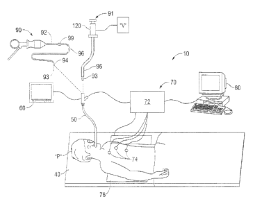
[0046] With reference to FIGS. 1 and 2, a system provided in accordance
with the
present disclosure and configured for planning a pathway to target tissue
(planning phase),
navigating a positioning assembly to the target tissue (navigation phase), and
navigating a biopsy
tool to the target tissue to obtain a tissue sample from the target tissue
using the biopsy tool
(biopsy phase) is shown generally identified by reference numeral 10. System
10 generally
includes an operating table 40 configured to support a patient "P;" a
bronchoscope 50 configured
for insertion through the patient's mouth into the patient's airways;
monitoring equipment 60
coupled to bronchoscope 50 for displaying video images received from
bronchoscope 50; a
tracking system 70 including a tracking [nodule 72, a plurality of reference
sensors 74, and a
7
CA 02876410 2014-12-24
transmitter mat 76; a computer 80 including software and/or hardware used to
facilitate pathway
planning, identification of target tissue, and navigation to target tissue; a
positioning assembly 90
or 91 including a locatable guide ( LG) 92 an extended working channel (EWC)
96; and a
biopsy tool 100 insertable through the positioning assembly 90, 91 and
operable to obtain a
tissue sample, e.g., for subsequent diagnostic testing. The planning and
navigation phases will
initially be detailed below, followed by a detailed description of biopsy
tools provided in
accordance with the present disclosure and use of such biopsy tools in
conjunction with system
in performing the biopsy phase.
1.00471 With respect to the planning phase, computer SO utilizes computed
tomographic
(CT) image data for generating and viewing a three-dimensional model of the
patient's airways,
enables the identification of target tissue on the three-dimensional model
(automatically, semi-
automatically or manually), and allows for the selection of a pathway through
the patient's
airways to the target tissue. More specifically, the CT scans are processed
and assembled into a
three-dimensional CT volume, which is then utilized to generate a three-
dimensional model of
the patient's airways. The three-dimensional model may be displayed on a
display monitor
associated with computer SO, or in any other suitable fashion. Using computer
80, various views
of the three-dimensional model may be provided and/or the three-dimensional
model may be
manipulated to facilitate identification of target tissue on the three-
dimensional model and
selection of a suitable pathway through the patient's airways to access the
target (issue. Once
selected, the pathway is saved for use during the navigation phase(s).
[0048] Continuing with reference to FIG. 1, patient "P" is shown lying on
operating table
40 with bronchoscope 50 inserted through the patient's mouth and into the
patient's airways.
Bronchoscope 50 includes a source of illumination and a video imaging system
(not explicitly
8
CA 02876410 2014-12-24
shown) and is coupled to monitoring equipment 60, e.g., a video display, for
displaying the video
images received from the video imaging system of bronchoscope 50.
[0049] With
respect to the navigation phase, a six degrees-of-freedom electromagnetic
tracking system 70, e.g.. similar to those disclosed in U.S. Patent No.
6,188,355 and published
PCT Application Nos. WO 00/10456 and WO 01/67035, the entire contents of each
of which is
incorporated herein by reference, or other suitable positioning measuring
system, is utilized for
performing registration and navigation, although other configurations are also
contemplated.
Tracking system 70 includes a tracking module 72, a plurality of reference
sensors 74. and a
transmitter mat 76. Tracking system 70 is configured for use with either
positioning assembly 90
or positioning assembly 91, and biopsy tool 100, as detailed below.
Positioning assemblies 90
and 91 include a LG 92 having a distal tip 93, which may be steerable.
Positioning assemblies
90 and 91 further include an EWC 96 and a handle 98. LG 92 and EWC 96 are
configured for
insertion through a working channel of bronchoscope 50 into the patient's
airways (although LG
92 and WC 96 may alternatively be used without bronchoscope 50) and are
selectively lockable
relative to one another via a locking mechanism 99. Distal tip 93 of 1..0 92
may be configured
for steering in any suitable fashion, e.g., using a plurality of steering
wires (not shown) coupled
between handle 98 and distal tip 93. to facilitate maneuvering distal tip 93
of LG 92 and EWC 96
through the patient's airways. Alternatively, rotation and translation of
handle 120 may facilitate
maneuvering of the distal tip 93 of LG 92, and in particular embodiments the
EWC 96 may be
angled or curved to assist in maneuvering the distal tip 93 through the
airways. Sensor 94 is
integrated with distal tip 93 of LG 92 and allows monitoring of the position
and orientation of
distal tip 93, in six degrees of freedom, relative to the reference coordinate
system. Sensor 94 of
[Al 92 may be configured similar to any of the sensors detailed below (see
FIGS. 6-8).
9
CA 02876410 2014-12-24
[0050] AS shown in FIG. 1, transmitter mat 76 is positioned beneath
patient "P." With
additional reference to FIG. 12, an embodiment of the internal configuration
of transmitter mat
76 of tracking system 70 (174(3. 1) is shown, although other suitable
configurations are also
contemplated. Transmitter mat 76 is a transmitter of electromagnetic radiation
and includes a
stack of three substantially planar rectangular loop antennas 77a, 77b, 77c
configured to be
connected to drive circuitry (not shown). For a detailed discussion of the
construction of
exemplary transmitter mats, which may also be referred to as location boards,
reference may be
made to U.S. Patent Application Publication No. 2009/0284255, filed April 2,
2009, the entire
contents of which arc incorporated herein by reference.
[0051] Transmitter mat 76 and the plurality of reference sensors 74 are
interconnected
with tracking module 72, which derives the location of each sensor 74 in six
degrees of freedom.
One or more of reference sensors 74 are attached to the chest of the patient
"P." The six degrees
of freedom coordinates of reference sensors 74 are sent to computer 80 (which
includes the
appropriate software) where they are used to calculate a patient coordinate
frame of reference.
Registration, as detailed below, is generally performed by identifying
locations in both the three-
dimensional model and the patient's airways and measuring the coordinates in
both systems.
Further details of such a registration technique can be found in U.S. Patent
Application Pub. No.
2011/0085720, the entire contents of which are incorporated herein by
reference, although other
suitable registration techniques are also contemplated.
[0052] in use, with respect to the navigation phase, LG 92 is inserted
into positioning
assembly 90, 91 and FAVC 96 such that sensor 94 projects from the distal end
of EWC 96. LG
92 and EWC 96 are then locked together via locking mechanism 99 (for example).
LG 92,
together with F,WC 96, are then inserted through bronchoscope 50 and into the
airways of the
CA 02876410 2014-12-24
patient "P," with LO 92 and EWC 96 moving in concert with one another through
bronchoscope
50 and into the airways of the patient "P." Automatic registration is
performed by moving 1-G
92 through the airways of the patient "P." More specifically, data pertaining
to locations of
sensor 94 while LG 92 is moving through the airways is recorded using
transmitter mat 76,
reference sensors 74, and tracking module 72. A shape resulting from this
location data is
compared to an interior geometry of passages of the three-dimensional model
generated in the
planning phase. and a location correlation between the shape and the three-
dimensional model
based on the comparison is determined, e.g.. utilizing the software on
computer 80. hi addition,
the software identifies non-tissue space (e.g., air filled cavities) in the
three-dimensional model.
The software aligns, or registers, an image representing a location of sensor
94 of I ,G 92 with an
image of the three-dimensional model based on the recorded location data and
an assumption
that LG 92 remains located in non-tissue space in the patient's airways. This
completes the
registration portion of the navigation phase.
[0053]
Referring still to FIG. I, once the planning phase has been completed, e.g.,
the
target tissue has been identified and the pathway thereto selected, and
registration has been
completed, system 10 may be utilized to navigate LG 92 through the patient's
airway to the
target tissue. To facilitate such navigation, computer 80, monitoring
equipment 60, and/or any
other suitable display may be configured to display the three-dimensional
model including the
selected pathway from the current location of sensor 94 of LG 92 to the target
tissue. Navigation
of LG 92 to the target tissue using tracking system 70 is similar to that
detailed below with
respect to the navigation of biopsy tool 100 to the target tissue and, thus,
is not detailed here tor
purposes of brevity.
11
CA 02876410 2014-12-24
[0054] Once LG 92 has been successfully navigated to the target tissue,
completing the
navigation phase, LO 92 may be unlocked from F,WC 96 and removed, leaving 1-WC
96 in place
as a guide channel for guiding biopsy tool 100 to the target tissue. Details
of various
embodiments of biopsy tools, along with the use of the same in the biopsy
phase, are described
below.
[0055] Referring now to FIG. 2, in conjunction with FIG. 1, one embodiment
of a biopsy
tool provided in accordance with the present disclosure for obtaining a tissue
sample from the
target tissue is shown generally identified by reference numeral 100. As
detailed below, biopsy
tool 100 is depicted inserted into navigation assembly 91 and further
configured for use. in
conjunction with tracking system 70 to facilitate navigation of biopsy tool
100 to the target tissue
and/or tracking of biopsy tool 100 as it is manipulated relative to the target
tissue to obtain the
tissue sample. Although registration and navigation are detailed above with
respect to LG 92 of
positioning assembly 90, 91, it is also envisioned that LG 92 be eliminated
and biopsy tool 100
itself is utilized for registration and navigation, similarly as detailed
above with respect to LU 92.
[0056] Biopsy tool 100. as best shown in FIG. 2A, in conjunction with
FIGS. 1 and 2,
generally includes an elongated flexible body 110 and a connector (122)
securing the biopsy tool
to the handle 120 of the navigation assembly 91. Connector 122 may include a
vacuum source
connector such as luer lock which fluidly connects the vacuum source to the
biopsy tool 100.
Flexible body 110 is configured to enable insertion of biopsy tool 100 into a
patient's airways,
e.g., through bronchoscope 50 and EWC 96 to the target tissue.
10057] With reference to FIG. 3, rigid distal biopsy member 130 includes a
base portion
140, a tissue-receiving portion 150, and a distal end cap 160. Base portion
140 defines a
generally cylindrical configuration and houses a sensor 170. Sensor 170. in
conjunction with
12
CA 02876410 2014-12-24
trackina system 70 (FIG. 1), may he employed to enable tracking of biopsy
member 130 of
biopsy tool 100 as biopsy member 130 is advanced through the patient's
airways, as detailed
below. Thus, with additional reference to FIG. 1, computer 80, monitoring
equipment 60, and/or
any other suitable display may be configured to display the three-dimensional
model and
selected pathway, both of which were generated during the planning phase,
along with the
current location of sensor 170 of biopsy member 130 to facilitate navigation
of biopsy member
130 to the target tissue and/or manipulation of biopsy member 130 relative to
the target tissue.
Various sensors suitable for use with biopsy member 130 for this purpose are
detailed below (see
FIGS. 9-11). Alternatively, biopsy tool 100 may not include a sensor and,
rather, only LG 92
may be utilized for navigation and positioning. Distal end cap 160 of biopsy
member 130
defines a generally blunt configuration. Alternatively, distal end cap 160 may
be configured to
cut or dissect tissue.
[0058] Tissue-
receiving portion 150 defines a planar surface 153 and an opening 152
configured to receive a tissue sample therethrotigh and into the generally
hollow interior of
biopsy member 130. Opening 152 is defined by first and second longitudinally-
extending faces
154, 156. Faces 154, 156 are angled into the interior of tissue-receiving
portion 150 and are
oriented to define an acute interior angle therebetween, e.g., a generally "V"-
shaped
configuration. Faces 154, 156 each includes a sharpened cutting edge 155, 157,
respectively,
disposed on one side of opening 152. Faces 154, 156 are further oriented
relative to one another
such that edges 155, 157 increasingly approximate one another in the distal to
proximal
direction, ultimately culminating at an apex point 158 adjacent to proximal
shoulder 159. This
feature facilitates dynamic tissue cutting, as detailed below. Although
generally shown as being
formed from a single plate 161, in one embodiment, tissue receiving portion
150 may be defined
13
CA 02876410 2014-12-24
by two or more plates 16 I disposed on base portion 140. It is contemplated
that the two or more
plates 161 may be arranged in a planar configuration (i.e., side by side), or
stacked one over the
other as detailed hereinbelow.
[0059] With
reference to FIGS. 3A-3D, an alternate embodiment of biopsy tool 100 is
shown, generally referred to as 100'. In this embodiment, biopsy tool 100'
includes a
monolithically formed biopsy member 130' that is separate from flexible body
110'. Biopsy
member 130' includes a shoulder portion 180 on a proximal end thereof, The
shoulder portion
180 defines a cavity therein such that biopsy member 130' may be disposed over
the distal end
of flexible body 110'. Biopsy member 130' may be fixedly secured to the distal
end of flexible
body 110' by any suitable means, such as welding, swage fit, adhesives, etc. A
base portion 140'
defines a cutout such that an opening 152' is formed therein. Opening 152' is
configured to
receive a tissue sample therethrough and into the generally hollow interior of
biopsy member
130'. A pair of plates 161', 161" are disposed on an upper surface of biopsy
member 130' in a
stacked configuration (see FIG. 3D). Plates 161', 161" may be fixedly secured
to biopsy
member 130' by any suitable means, such as welding, adhesives, etc. Each of
plates 161', 161"
defines first and second sharpened cutting edges 155', 157'. Edges 155', 157'
are angled into
the interior of tissue-receiving portion 152' and are oriented to define an
acute interior angle
therebetween, e.g., a generally "V"-shaped configuration. Edges 154', 156' are
further oriented
relative to one another such that edges 155. 157 increasingly approximate one
another in the
distal to proximal direction, ultimately culminating at an apex point 158'
adjacent to proximal
shoulder 159'. This feature facilitates dynamic tissue cutting, as detailed
below with respect to
biopsy member 130.
14
CA 02876410 2014-12-24
100601 Referring to FIGS. 1-3, in ti.. once the planning and navigation
phases have been
completed, and 1,(1 92 removed from FWC 96, biopsy tool 100 may be inserted
through
navigation assemblies 90, 91 and bronchoscope 50 to the target tissue. Sensor
170 of biopsy
member 130. in conjunction with tracking system 70, as mentioned above,
enables tracking of
sensor [70 as it is advanced through the patient's airways. Thus, even after
biopsy member 130
is extended distally from EWC 96, the position of biopsy member 130 can be
tracked, thus
permitting navigation of biopsy member 130 to and/or manipulation of biopsy
member 130
relative to the target tissue to ensure proper positioning of biopsy member
130 relative to the
target tissue and allowing certain tissue structures adjacent the target
tissue to be avoided.
Details of tracking and navigating using suitable sensors and tracking system
70 will be
described in greater detail below, following the description of the various
embodiments thereof.
10061] Once biopsy member 130 of, biopsy tool 100 is positioned as
desired, vacuum
source "V" may be activated (e.g., via a syringe, mechanical pump, etc.) to
apply suction at
opening 152 of tissue-receiving portion 150 of biopsy member 130 to suction
tissue into the
interior of tissue-receiving portion 150. As a sample of tissue is suctioned
through opening 152,
the sample begins to be cut away from laterally surrounding tissue via the
urging of tissue into
contact with edges 155, 157, e.g., as a result of the suction force applied to
tissue. Once the
tissue sample has been at least partially received within the interior of
tissue-receiving portion
150, biopsy member 130 may be translated distally relative to tissue, e.g.,
via grasping and
translating proximal handle portion 120 distally, such that the tissue sample
is completely
severed from surrounding, tissue. This severing of the tissue sample is aided
by the relative
movement of approximating edges 155, 157 and apex point 158 relative to and
through tissue.
Upon receiving and fully separating the tissue sample from surrounding tissue,
biopsy tool 100
CA 02876410 2014-12-24
may be withdrawn. from the patient's airways and the tissue sample retrieved
from biopsy tool
100 for testing. Et is also contemplated that multiple samples be taken with
biopsy tool 100, e.g.,
at the same location or various different locations, prior to withdrawal.
[0062] Referring now to FIG. 4, another embodiment of a biopsy tool
provided in
accordance with the present disclosure for obtaining a tissue sample from the
target tissue is
shown generally identified by reference numeral 630. Similarly as detailed
above with respect to
the previous embodiment, biopsy tool 630 is configured for use in conjunction
with tracking
system 70 (FIG. 1) to facilitate navigation of biopsy tool 630 to the target
tissue and/or tracking
of biopsy tool 630 as it is manipulated relative to the target tissue to
obtain the tissue sample.
[0063] Biopsy member 630 includes a base portion 640, a tissue-receiving
portion 650,
and a distal end cap 660. Base portion 640 defines a generally cylindrical
configuration and may
house a sensor 670. Sensor 670 may be configured similarly to sensor 170 (FIG.
3) and, thus,
will not be detailed herein for purposes of brevity. Distal end cap 660 of
biopsy member 630
defines a generally blunt configuration. Alternatively, distal end cap 660 may
be configured to
cut or dissect tissue.
[0064] Tissue-receiving portion 650 defines a planar surface 653 and an
opening 652
configured to receive tissue therethrough and into the generally hollow
interior of biopsy
member 630. Opening 652 is defined by a one or more semi-circular faces 654.
In one non-
limiting cmbodhnent, opening 652 is defined by a series of four
interconnecting and overlapping
semi-circular faces 654, 656a, 656b, 662a, 662b, and 663. Faces 654, 656a,
656b, 662a, 662b,
and 663 arc angled into the interior of tissue-receiving portion. Faces 654,
656a, 656b, 662a,
662b, and 663 each includes a sharpened cutting edge 655, 657a, 657b, 664a,
664b, and 665
respectively, disposed on one side of opening 652. Faces 654, 656a, 656b,
662a, 662b, and 663
16
CA 02876410 2014-12-24
are further oriented relative to one another such that a plurality of
projections 667, extending
towards the center of opening 652, are formed at the junction between adjacent
faces 654. This
feature, in conjunction with sharpened cutting edges 655. 657a, 657b, 664a,
6641, and 665,
facilitates dynamic tissue cutting, similarly as detailed above with respect
to biopsy member 130
(FIG. 3). In one non-limiting embodiment, tissue receiving portion 650 may be
defined by one
or more plates 661 disposed between distal end cap 660 and proximal shoulder
659.
[0065] Biopsy member 630 may be utilized in a similar respect to biopsy
member 130
(FIG. 3) as detailed above, with the exception of the ability to sever the
tissue by translating
biopsy member 630 proximally or distally relative to the tissue.
[0066] Referring to FIG. 5, another embodiment of a biopsy tool provided in
accordance
with the present disclosure for obtaining a tissue sample from the target
tissue is shown generally
identified by reference numeral 730. Biopsy member 730 includes a base portion
740, a tissue-
receiving portion 750, and a distal end cap 760. Base portion 740 defines a
generally cylindrical
configuration and may houses a sensor 770. Sensor 770 is similar to sensor 170
(FIG. 3) and,
thus will not be detailed here for purposes of brevity. Distal end cap 760 of
biopsy member 730
defines a generally blunt configuration. Alternatively, distal end cap 760 may
be configured to
cut or dissect tissue.
[0067] Tissue-receiving portion 750 defines a planar surface 753 and an
opening 752
configured to receive tissue therethrough and into the generally hollow
interior of biopsy
member 730. Opening 752 is defined by first and second longitudinally-
extending faces 754,
756, and curvate face 762. Faces 754 and 756 are angled into the interior of
tissue-receiving
portion 750 and are oriented to define an acute interior angle therebetween,
e.g., a generally "V"-
shaped configuration. Faces 754, 756, and 762 each includes a sharpened
cutting edge 755, 757,
17
CA 02876410 2014-12-24
and 763 respectively, disposed on one side of opening 752, thereby forming a
continuous cutting
edge capable of cutting tissue. Faces 754 and 756 are further oriented
relative to one another
such that edges 755 and 757 increasingly approximate one another in the distal
to proximal
direction, ultimately culminating at radiused cutting edge 763 adjacent to
proximal shoulder 759.
This feature facilitates dynamic tissue cutting, similarly as detailed above
with respect to biopsy
member 130 (FIG. 3). In one non-limiting embodiment, tissue receiving portion
750 may be
defined by one or more plates 761 disposed between distal end cap 760 and
proximal shoulder
759.
room Biopsy member 730 may be utilized in a similar respect to biopsy
member 130
(HG. 3) as detailed above to cut tissue.
100691 Referring now to FIG. 6, another embodiment of a biopsy tool
provided in
accordance with the present disclosure for obtaining a tissue sample from the
target tissue is
shown generally identified by reference numeral 830. Similarly as detailed
above with respect to
the previous embodiment, biopsy tool 830 is configured for use in conjunction
with tracking
system 70 (FIG. 1) to facilitate navigation of biopsy tool 830 to the target
tissue and/or tracking
of biopsy tool 830 as it is manipulated relative to the target tissue to
obtain the tissue sample.
[0070] Biopsy member 830 includes a base portion 840, a tissue-receiving
portion 850,
and a distal end cap 860. Base poriion 840 defines a generally cylindrical
configuration and may
house a sensor 870. Sensor 870 may be configured similarly to sensor 170 (FIG.
I) and, thus,
will not be detailed herein for purposes of brevity. Distal end cap 860 of
biopsy member 830
defines a generally blunt configuration. Alternatively, distal end cap 860 may
be configured to
cut or dissect tissue.
18
CA 02876410 2014-12-24
[00711 Tissue-receiving portion 850 defines a planar surface 853 and an
opening 852
configured to receive tissue therethrough and into the generally hollow
interior of biopsy
member 830. Opening 852 is defined by a one or more semi-circular faces 854.
In one non-
limiting embodiment, opening 852 is defined by a series of interconnecting and
overlapping
semi-circular faces 854, 856, and 862 arranged in a clover shaped
configuration. Faces 854, 856,
and 862 are angled into the interior of tissue-receiving portion 850. Faces
854, 856, and 862
each includes a sharpened cutting edge 855, 857. and 863 respectively,
disposed on one side of
opening 852. Faces 854, 856. and 862 are further oriented relative to one
another such that a
plurality of projections 867 with cutting edge 868, extending towards the
center of opening 852,
are formed at the junction between adjacent faces 854, 856, and 862. This
feature, in
conjunction with sharpened cutting edges 855, 857, and 863, facilitates
dynamic tissue cutting,
similarly as detailed above with respect to biopsy member 130 (FIG. 3).
Although generally
shown as being formed from a single plate 861, in other embodiments, tissue
receiving portion
850 may be defined by two or more plates 861 disposed on base portion 840.
[0072] Biopsy member 830 may be utilized in a similar respect to biopsy
member 130
(FIG. 3) as detailed above, with the exception of the ability to sever the
tissue by translating
biopsy member 830 in any direction (e.g. proximally, distally, laterally,
diagonally, etc.) relative
to tissue.
[0073] Turning to FIG. 7, yet another embodiment of a biopsy tool provided
in
accordance with the present disclosure, for obtaining a tissue sample from the
target tissue is
shown generally identified by reference numeral 930. Biopsy member 930
includes a base
portion 940, a tissue-receiving portion 950, and a distal end cap 960. Base
portion 940 defines a
generally cylindrical configuration and may house a sensor 970. Sensor 970 is
similar to sensor
19
CA 02876410 2014-12-24
170 (FIG. 3) and, thus will not be. detailed here for purposes of brevity.
Distal end cap 960 of
biopsy member 930 defines a generally blunt configuration. Alternatively,
distal end cap 960
may he configured to cut or dissect tissue.
1.00741 Tissue-receiving portion 950 defines a planar surface 953 and an
opening 952
configured to receive tissue therethrough and into the generally hollow
interior of biopsy
member 930. Opening 952 is defined by a distal region having a large opening
952a, including
smooth walls 956, tapering proximally to a long narrow opening 952b having a
width less than
that of large opening 952a and further including a plurality of tines 954
extending towards the
center of opening 952. In one non-limiting embodiment, tines 954 may be
oriented such that
they extend towards the center of opening 952 at an angle such they terminate
at a proximal
position relative to their base. Large opening 952a may be of any shape,
including, but not
limited to, triangular, circular, rectangular, or the like. One non-limiting
embodiment of large
opening 952a is of a triangular configuration. Long narrow opening 952b may
include parallel
walls or walls forming an acute angle terminating with an apex 955 adjacent to
proximal
shoulder 959. This feature facilitates dynamic tissue tearing, as detailed
below. Although
generally shown as being formed. from a single plate 961, in other
embodiments. tissue receiving
portion 950 may be defined by two or more plates 961 disposed on base portion
940.
1.00751 Biopsy member 930 may be utilized in a similar respect to biopsy
member 130
(FIG. 3) as detailed above, with the exception of once biopsy member 930 of
biopsy tool 100 is
positioned as desired, vacuum source "V" may be activated to apply suction at
opening 952 of
tissue-receiving portion 950 of biopsy member 930 to suction tissue into the
interior of tissue-
receiving portion 950. As a sample of tissue is suctioned through opening 952,
the sample is
trapped within long narrow opening 952b e.g., as a result of the suction force
applied to tissue.
CA 02876410 2014-12-24
Once the tissue sample has been at least partially received within the
interior of tissue-receiving
portion 950, biopsy member 930 may he translated proximally or distally
relative to tissue, e.g.,
via grasping and translating proximal handle portion 120 proximally or
distally, such that the
tissue sample is completely torn or severed from the surrounding tissue. This
tearing of the
tissue sample is aided by the plurality of tines 954 which provide a secure
grasp on the tissue
sample.
[0076J Referring now to FIG. 8, yet another embodiment of a biopsy tool
provided in
accordance with the present disclosure for obtaining a tissue sample from the
target tissue is
shown generally identified by reference numeral 1030. Biopsy member 1030
includes a base
portion 1040, a tissue-receiving portion 1050, and a distal end cap 1060. Base
portion 1040
defines a generally cylindrical configuration and may house a sensor 1070.
Sensor 1070 is
similar to sensor 170 (FIG. 3) and, thus will not be detailed here for
purposes of brevity. Distal
end cap 1060 of biopsy member 1030 defines a generally blunt configuration.
Alternatively,
distal end cap 1060 may be configured to cut or dissect tissue.
[0077] Tissue-receiving portion 1050 defines a planar surface 1053 and an
opening1052
configured to receive tissue therethrough and into the generally hollow
interior of biopsy
member 1030. Opening 1052 is defined by a distal region having a large opening
1052a,
including smooth walls 1056, tapering proximally to a long narrow opening
1052b having a
width less than that of large opening 1052a. Large opening 1052a may be of any
shape,
including, but not limited to, triangular, circular, rectangular, heart or the
like. One non-limiting
embodiment of large opening 1052a is of a heart shaped configuration. Long
narrow opening
1052b includes walls forming an acute angle terminating with an apex 1055
adjacent to proxitnal
shoulder 1059. This feature facilitates dynamic tissue tearing, similarly as
detailed above with
21
CA 02876410 2014-12-24
respect to biopsy member 930 (FIG. 7). Although generally shown as being
formed from a
single plate 1061, in one non-limiting embodiment, tissue receiving portion
1050 may be defined
by two or more plates 1061 disposed on base portion 1040.
[00781 Biopsy member 1030 may be utilized in a similar respect to biopsy
member 930
(FIG. 7) as detailed above, with the exception of once biopsy member 1030 of
biopsy tool 100 is
positioned as desired, vacuum source "V" may be activated to apply suction at
opening 1052 of
tissue-receiving portion 1050 of biopsy member 1030 to suction tissue into the
interior of tissue-
receiving portion 1050. As a sample of tissue is suctioned through opening
1052, the sample is
trapped within long narrow opening 1052b e.g., as a result of the suction
force applied to tissue.
Once the tissue sample has been at least partially received within the
interior of tissue-receiving
portion 1050, biopsy member 1030 may be translated proximally relative to
tissue, e.g., via
grasping and translating proximal handle portion 120 distally, such that the
tissue sample is
completely torn or severed from surrounding tissue. This tearing of the tissue
sample is aided by
the long narrow opening 1052b which provides a secure grasp on the tissue
sample.
[0079] Turning now to FIGS. 9-11, in conjunction with FIG. 1, various
different sensors
248, 348, 448 (FIGS. 9-11, respectively) configured for use as the sensor of
any of the biopsy
tools detailed herein and/or sensor 94 of 1,CI 92 are described. Although each
of the sensors 248,
348, 448 are generally described as employing a plurality of sensor elements,
it is contemplated
that the sensor of any of the biopsy tools detailed herein and/or sensor 94 of
LC; 92 may employ
any number of sensor elements (e.g., one, two, three, etc.). Therefore, the
descriptions to follow
should not be construed as limiting, but merely as exemplifications or
particular embodiments.
Referring to FIG. 9, sensor 248 is shown. Sensor 248 includes a plurality of
field component
sensor elements 251a, 251b, 1252a, 252b, 253. Each sensor element 25.1a, 251b,
252a, 252b,
22
CA 02876410 2014-12-24
253 is formed as a coil and arranged for sensing a different component of an
electromagnetic
field generated by transmitter mat 76 (FIG. 12). More specifically, first and
second pairs of
sensor elements 251a, 251b and 252a, 252b are arranged within sensor housing
246 such that the
respective elements 251a, 251b and 252a, 252b of each pair are equidistant
from a common
reference point 254, while sensor element 253 is centered about reference
point 254. Although
shown in FIG. 9 as collinearly disposed, other configurations of sensor
elements 251a, 251h,
1252a, 252b, 253 are also contemplated. Further, as opposed to providing five
sensor elements
251a, 25 lb, 1252a, 252b, 253 wherein sensor element 253 is centered about the
reference point
254, six sensors may be provide, e.g., wherein sensor element 253 is provided
as a pair of
elements disposed equidistant from reference point 254. The above-described
configuration of
sensor 248 enables transmitter mat 76 and the plurality of reference sensors
74 (FIG. 1), together
with tracking module 72 and computer 80 (FIG. 1), to derive the location of
sensor 248 in six
degrees of freedom, as detailed below, and as further detailed in U.S. Patent
No. 6,188,355 and
published PCT Application Nos. WO 00/10456 and WO 01/67035, previously
incorporated
herein by reference.
[0080] With
reference to FTG. 10, sensor 348 is shown including two sensor components
351, 353 arranged within sensor housing 346, each component 351, 353 including
three sensor
elements 352a, 352h, 352c and 354a, 354b, 354c, respectively. Each sensor
element 352a, 352b,
352c and 354a, 354b, 354c is configured as a flat rectangular coil, e.g.,
including a plurality of
turns of conducting wire, bent to define an arcuate shape. As such, the
elements 352a, 352b,
352c and 354a, 354b, 354c combine to define first and second generally
cylindrical components
351, 353. Components 351, 353 are centered about reference axis 356 and
positioned such that
each of elements 352a, 352b, 352e and 354a, 354b, 354e are equidistant from
reference axis 356
23
CA 02876410 2014-12-24
and such that each of elements 352a, 352b, 352c of component 351 are oriented
180 degrees
offset as compared to corresponding elements 354a, 354b, 354c, respectively,
of component 353.
Thus, similarly as with sensor 248 (FIG. 9), sensor 348 enables transmitter
mat 76 and the
plurality of reference sensors 74 (FIG. I), together with tracking module 72
and computer 80
(FIG. 1), to derive the location of sensor 348 in six degrees of freedom.
[0081] Turning to FIG. 11, sensor 448 includes three coils 451, 452. 453.
Coils 451 and
452, 453 are angled relative to housing 446, while coil 453 is
circumferentially disposed within
housing 446. Coils 451, 452, 453 are oriented to lie in perpendicular planes
relative to one
another and share a Common center reference point 454. By sharing a common
center reference
point. 454, each portion of each coil 451, 452, 453 is equidistant from center
reference point 454.
Further, this configuration, e.g., wherein coils share a common center
reference point 454 rather
than being longitudinally displaced relative to one another, allows for the
longitudinal dimension
of sensor 448 to be minimized. Such a configuration still, however, enables
transmitter mat 76
and the plurality of reference sensors 74 (FIG. 1), together with tracking
module 72 and
computer 80 (FIG. 1), to derive the location of sensor 448 in six degrees of
freedom.
[0082] Referring additionally to FIG. 1, the electromagnetic waves
generated by
transmitter mat 76 are received by the various sensor elements of the sensor
assembly e.g., the
sensor elements of sensors 248, 348, 448 (FIGS. 9-11, respectively) configured
for use any of the
biopsy tools provided herein or sensor 94 of LG 92, and are converted into
electrical signals that
are sensed via reference sensors 74. Tracking system 70 further includes
reception circuitry (not
shown) that has appropriate amplifiers and A/D converters that are utilized to
receive the
electrical signals from reference sensors 74 and process these signals to
determine and record
location data of the sensor assembly. Computer 80 may be configured to receive
the location
24
CA 02876410 2014-12-24
data from tracking system 70 and display the current location of the sensor
assembly on the
three-dimensional model and relative to the selected pathway generated during
the planning
phase, e.g., on computer 80, monitoring equipment 60, or other suitable
display. Thus,
navigation of the biopsy tool and/or LCi 92 to the target tissue and/or
manipulation of the biopsy
tool relative to the target tissue, as detailed above, can be readily
achieved.
[0083] As used herein, the term "distal" refers to the portion that is
being described
which is further from a user, while the term "proximal" refers to the portion
that is being
described which is closer to a user. Further, to the extent consistent, any of
the aspects and
features detailed herein may be used in conjunction with any or all of the
other aspects and
features detailed herein.
[0084] While several embodiments of the disclosure have been shown in the
drawings, it
is not intended that the disclosure be limited thereto, as it is intended that
the disclosure be as
broad in scope as the art will allow and that the specification be read
likewise. Therefore, the
above description should not be construed as limiting, but merely as
eXernplifications of
particular embodiments.