Note: Descriptions are shown in the official language in which they were submitted.
CA 02876734 2015-01-08
WO 2014/012001
PCT/US2013/050293
TITLE OF THE INVENTION
USE OF CART19 TO DEPLETE NORMAL B CELLS TO INDUCE TOLERANCE
CROSS-REFERENCE TO RELATED APPLICATION
This application claims priority to U.S. Provisional Application No.
61/671,508, filed July 13,2012, the content of which is hereby incorporated
herein by
reference in its entirety.
BACKGROUND OF THE INVENTION
Using gene transfer technologies, T cells can be genetically modified
to stably express antibody binding domains on their surface that confer novel
antigen
specificities that are major histocompatibility complex (MHC)¨independent.
Chimeric
antigen receptors (CARs) are an application of this approach that combines an
antigen
recognition domain of a specific antibody with an intracellular domain of the
CD3-z
chain or FcgRI protein into a single chimeric protein (Gross et al., 1989
Proc. Natl.
Acad. Sci. U.S.A. 86: 10024-10028; Irving et al., 1991 Cell 64: 891-901).
Trials
testing CARs are presently under way at a number of academic medical centers
(Kohn
et al. 2011 Mol. Ther. 19: 432-438; Jena et al., 2010 Blood 116: 1035-1044).
In most
cancers, tumor-specific antigens are not yet well defined, but in B cell
malignancies,
CD19 is an attractive tumor target. Expression of CD19 is restricted to normal
and
malignant B cells (Uckun et al., 1988 Blood 71: 13-29), and CD19 is a widely
accepted target to safely test CARs. Although CARs can trigger T cell
activation in a
manner similar to an endogenous T cell receptor, a major impediment to the
clinical
application of this technology to date has been the limited in vivo expansion
of CAR+
T cells, rapid disappearance of the cells after infusion, and disappointing
clinical
activity (Jena et al., 2010 Blood 116: 1035-1044; Sadelain et al., 2009 Curr.
Opin.
Immunol. 21: 215-223).
CAR-mediated T cell responses may be further enhanced with addition
of costimulatory domains. In a preclinical model, inclusion of the CD137 (4-
1BB)
signaling domain was found to significantly increased antitumor activity and
in vivo
persistence of CARs compared to inclusion of the CD3-z chain alone (Milone et
al.,
2009 Mol. Ther. 17,1453-1464; Carpenito et al., 2009 Proc. Natl. Acad. Sci.
U.S.A.
106: 3360-3365). To evaluate the safety and feasibility for adoptive transfer
of T cells
gene-modified to express such CARs, a pilot clinical trial using autologous T
cells
CA 02876734 2015-01-08
WO 2014/012001
PCT/US2013/050293
expressing an anti-CD19 CAR including both CD3-z and the 4-1BB costimulatory
domain (CART19 cells) to target CD19+ malignancies was conducted. Three
patients
have been treated under this protocol. Some of the findings from one of these
patients
are described in (Porter et al., 2011 N. Engl. J. Med. 365: 8), which reports
that this
treatment results in tumor regression, CART19 cell persistence, and the
unexpected
occurrence of delayed tumor lysis syndrome. It was also observed that the
CART19
cells mediated potent clinical antitumor effects in all three patients
treated. On
average, each infused CAR T cell and/or their progeny eliminated more than
1000
leukemia cells in vivo in patients with advanced chemotherapy-resistant
chronic
lymphocytic leukemia (CLL). CART19 cells underwent robust in vivo T cell
expansion, persisted at high levels for at least 6 months in blood and bone
marrow
(BM), continued to express functional receptors on cells with a memory
phenotype,
and maintained anti-CD19 effector function in vivo. However, it still remains
unclear
how the CART19 cells evade the rejection by the human host given that the
CAR19
construct contains both murine sequences (the antibody determinants) and
unique
junctional fragments between the different components of the CAR19 construct.
Thus, there still remains a need in the art as to the mechanism of long
term persistence of the CART19 cells and why these cells are not rejected by
the
human host. The present invention addresses this need.
SUMMARY OF THE INVENTION
The invention provides a method of depleting B cells in a subject. In
one embodiment, the method comprises administering to a subject an effective
amount of a cell genetically modified to express a CAR wherein the CAR
comprises
an antigen binding domain, a costimulatory signaling region, and a CD3 zeta
signaling domain, wherein the antigen binding domain targets a B cell surface
marker,
thereby depleting B cells in the subject.
The invention provides a method of promoting tolerance in a subject.
In one embodiment, the method comprises administering to a subject an
effective
amount of a cell genetically modified to express a CAR wherein the CAR
comprises
an antigen binding domain, a costimulatory signaling region, and a CD3 zeta
signaling domain, wherein the antigen binding domain targets a B cell surface
marker,
thereby promoting tolerance in the subject.
CA 02876734 2015-01-08
WO 2014/012001
PCT/US2013/050293
In one embodiment, the tolerance is transplant tolerance to a
transplanted tissue.
In one embodiment, the genetically modified cell depletes B cells.
In one embodiment, the genetically modified cell is administered at the
same time as the transplanted tissue.
In one embodiment, the genetically modified cell is administered
before the administration of the transplanted tissue.
In one embodiment, the genetically modified cell is administered after
the administration of transplanted tissue.
The invention provides a method for treating graft versus host disease
(GVHD). In one embodiment, the method comprises administering a cell
genetically
modified to express a CAR to a subject in need thereof, wherein the CAR
comprises
an antigen binding domain, a costimulatory signaling region, and a CD3 zeta
signaling domain, wherein the antigen binding domain targets a B cell surface
marker,
thereby treating GVHD in the subject.
In one embodiment, the genetically modified cell depletes B cells.
In one embodiment, the genetically modified cell is administered at the
same time as a transplanted tissue.
In one embodiment, the genetically modified cell is administered
before the administration of the transplanted tissue.
In one embodiment, the genetically modified cell is administered after
the administration of the transplanted tissue.
BRIEF DESCRIPTION OF THE DRAWINGS
The following detailed description of preferred embodiments of the
invention will be better understood when read in conjunction with the appended
drawings. For the purpose of illustrating the invention, there are shown in
the
drawings embodiments which are presently preferred. It should be understood,
however, that the invention is not limited to the precise arrangements and
instrumentalities of the embodiments shown in the drawings.
Figure 1, comprising Figures lA through 1F, is a series of images
demonstrating sustained in vivo expansion and persistence in blood and marrow
of
CART19 cells. DNA isolated from whole blood as depicted in Figure IA through
1C
or marrow as depicted in Figure 1D through IF, samples obtained from UPN 01 as
CA 02876734 2015-01-08
WO 2014/012001
PCT/US2013/050293
depicted in Figure IA and 1D, UPN 02 as depicted in Figure 1B and 1E and UPN
03
as depicted in Figure 1C and 1F was subjected in bulk to Q-PCR analysis using
a
qualified assay to detect and quantify CART19 sequences. Each data point
represents
the average of triplicate measurements on 100-200 ng genomic DNA, with maximal
% CV less than 1.56%. Pass/fail parameters for the assay included pre-
established
ranges for slope and efficiency of amplification, and amplification of a
reference
sample. The lower limit of quantification for the assay established by the
standard
curve range was 2 copies transgene/microgram genomic DNA; sample values below
that number are considered estimates and presented if at least 2/3 replicates
generated
a Ct value with % CV for the values 15%. CART19 cells were infused at day 0,
1, and
2 for UPN 01 and UPN 03, and days 0, 1, 2 and 11 for UPN 02.
Figure 2, comprising Figures 2A through 2D, is a series of images
depicting prolonged surface CART19 expression and establishment of functional
memory CARs in vivo. Figure 2A depicts detection of CAR-expressing CD3+
lymphocytes and absence of B cells in periphery and marrow. Freshly processed
peripheral blood or marrow mononuclear cells obtained from UPN 03 at day 169
post-CART19 cell infusion were evaluated by flow-cytometry for surface
expression
of CAR19 (top) or presence of B cells (bottom); as a control, PBMC obtained
from a
healthy donor ND365 were stained. To evaluate CAR19 expression in CD3+
lymphocytes, samples were co-stained with antibodies to CD14-PE-Cy7 and CD16-
PE-Cy7 (dump channel) and CD3-FITC, positively gated on CD3+, and evaluated
for
CAR19 expression in the CD8+ and CD8-Iymphocyte compartments by co-staining
with CD8a-PE and the anti-CAR19 idiotype antibody conjugated to Alexa-647.
Data
in plots are gated on the dump channel-negative/CD3-positive cell population.
To
evaluate the presence of B cells, samples were co-stained with antibodies to
CD14-
APC and CD3-FITC (dump channels) and evaluated for the presence of B cells in
the
dump channel-negative fraction by co-staining with antibodies to CD2O-PE and
CD19-PE-Cy-7. In all cases, negative gate quadrants were established on no-
stain
controls as depicted in Figures 2B and 2C. T cell immunophenotyping of CD4+
(Figure 2B) and CD8+ (Figure 2C) T cell subsets is shown. Frozen peripheral
blood
samples from UPN 03 obtained by apheresis at day 56 and 169 post T cell
infusion
were rested overnight in culture medium with no added factors, washed, and
subjected
to multi-parametric immunophenotyping for expression of markers of T cell
memory,
activation, and exhaustion. The gating strategy, as depicted in Figure 6,
involved an
CA 02876734 2015-01-08
WO 2014/012001
PCT/US2013/050293
initial gating on dump channel (CD14, CD16, Live/Dead Aqua)-negative and CD3-
positive cells, followed by positive gates on CD4+ and CD8+ cells. Gates and
quadrants were established using FM0 controls (CAR, CD45RA, PD-1, CD25,
CD127, CCR7) or by gating on positive cell populations (CD3, CD4, CD8) and
clearly delineated subsets (CD27, CD28, CD57); data were displayed after bi-
exponential transformation for objective visualization of events. Figure 2D
depicts
functional competence of persisting CAR cells. Frozen peripheral blood samples
from
UPN 03 obtained by apheresis at day 56 and 169 post T cell infusion were
rested
overnight in culture medium with no added factors, washed, and evaluated
directly ex-
vivo for the ability to recognize CD19-expressing target cells using CD107
degranulation assays. Following a two-hour incubation in the presence of anti-
CD28,
anti-CD49d, and CD107-FITC, cell mixtures were harvested, washed, and
subjected
to multi-parametric flow cytometric analysis to evaluate the ability of CART19
cells
to de-granulate in response to CD! 9-expressing targets. The gating strategy
involved
an initial gate on dump channels (CD14-PE-Cy7, CD16-PE-Cy7, Live/Dead Aqua)-
negative and CD3-PE-positive cells, followed by gating on CD8-PE-Texas Red-
positive cells; presented data is for the CD8+ gated population. In all cases,
negative
gate quadrants were established on no-stain controls.
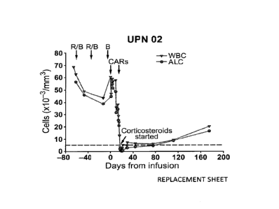
Figure 3, comprising Figures 3A through 3C, is series of images
depicting the results of experiments evaluating clinical responses after
infusion of
CART19 cells. Figure 3A depicts that UPN 02 was treated with two cycles of
rituximab and bendamustine with minimal response (R/B, arrow). CART19 T cells
were infused beginning 4 days after bendamustine only (B, arrow). The
rituximab and
bendamustine-resistant leukemia was rapidly cleared from blood, as indicated
by a
decrease in the absolute lymphocyte count (ALC) from 60,600/Alto 200/ 1 within
18
days of the infusion. Corticosteroid treatment was started on day 18 post
infusion due
to malaise and non-infectious febrile syndrome. The reference line (dotted)
indicates
upper limit of normal for ALC. Figure 3B depicts the results of example
experiments
staining sequential bone marrow biopsy or clot specimens from patient UPN 01
and
03 for CD20. Pretreatment infiltration with leukemia present in both patients
was
absent on post treatment specimens accompanied by normalization of cellularity
and
tri-lineage hematopoiesis. UPN 01 has not had any CLL cells detected as
assessed by
flow cytometry, cytogenetics and fluorescence in-situ hybridization or normal
B cells
detected by flow cytometry in bone marrow or blood. UPN 03 had 5% residual
CA 02876734 2015-01-08
WO 2014/012001
PCT/US2013/050293
normal CD5-negative B cells confirmed by flow cytometry on day +23, which also
showed them to be polyclonal; no normal B cells were detected at day +176.
Figure
3C depicts the results of experiments using sequential CT imaging to assess
the rapid
resolution of chemotherapy-resistant generalized lymphadenopathy. Bilateral
axillary
masses resolved by 83 (UPN 01) and 31 (UPN 03) days post infusion, as
indicated by
arrows and circle.
Figure 4, comprising Figures 4A through 4C, is a series of images
depicting absolute lymphocyte counts and total CART19+ cells in circulation
for
UPN 01, 02, 03. The total number of lymphocytes (Total normal and CLL cells)
vs.
Total CART19+ cells in circulation is plotted for all 3 subjects using the
absolute
lymphocyte count from CBC values, and assuming a 5.0 L volume of blood. The
total
number of CART19 cells in circulation was calculated by using the tandem CBC
values with absolute lymphocyte counts and the Q-PCR marking values as
depicted in
Figure 1, converting copies/1.1g DNA to average % marking as described
elsewhere
herein. The Q-PCR % marking was found to correlate closely (<2 fold variation)
with
the flow cytometric characterization of the infusion products and with data
from
samples where concomitant flow cytometry data was available to directly
enumerate
CART19 cells by staining.
Figure 5, comprising Figures 5A through 5D is a series of images
depicting experiments involving the direct ex vivo detection of CART19-
positive cells
in UPN-01 PBMC 71 days post-T cell infusion. UPN-01 PBMC collected either
fresh
post-apheresis on day71 day post infusion, or frozen at the time of apheresis
for
manufacture of the T cell product(baseline) and viably thawed prior to the
staining,
were subjected to flow-cytometric analysis to detect the presence ofCART19
cells
that express the CAR19 moiety on the surface. To evaluate the expression of
CAR19
in lymphocytes, samples were co-stained with CD3-PE and the anti-CAR19
idiotype
antibody conjugated to Alexa-647, or co-stained with CD3-PE alone (FMO for
CAR19). Figure 5A depicts that an initial lymphocyte gate was established
based on
forward and side scatter (FSC vs. SSC), followed by gating on CD3+ cells.
Figure 5B
depicts CD3+ lymphocyte gate; Figure 5C depicts CAR idiotype stain; Figure 5D
depicts CAR idiotype FMO. The CAR19-positive gate was established on the CAR19
FMO samples.
Figure 6, comprising Figures 6A through 6C, is a series of images
depicting the gating strategy to identify CART19 expression by using
polychromatic
CA 02876734 2015-01-08
WO 2014/012001
PCT/US2013/050293
flow cytometry in UPN 03 blood specimens. The gating strategy for Figure 6C is
shown for the UPN 03 Day 56 sample and is representative of the strategy used
on the
UPN 03 Day 169 sample. Figure 6A depicts primary gate: Dump (CD14, CD16,
LIVE/dead Aqua) negative, CD3-positive. Figure 6B depicts secondary gates: CD4-
positive, CD8-positive. Figure 6C depicts tertiary gates: CAR19-positive and
CAR19-
negative, established on CAR FM0 samples (right-most panels).
Figure 7 is an image summarizing the patient demographics and
response.
Figure 8 is an image depicting long term expression of CART19.
Figure 9, comprising Figures 9A and 9B, is a series of images
depicting deep B cell aplasia.
Figure 10 is an image demonstrating a reduction in plasma cells in all 3
patients.
DETAILED DESCRIPTION
The present invention is based in part on the surprising discovery that
T cells expressing an anti-CD19 CAR including both CD3z and the 4-1BB
costimulatory domain (CART19 cells) persisted in a mammalian host for a long
period time. For example, at this time, cells expressing surface CAR19 have
been
observed to be present in a mammalian host for over 21 months after CAR19 T
cell
infusion. Accordingly, the present invention provides a method for depleting
normal
B cells in a mammal by administering to the mammal in need thereof a CAR that
targets B cells in order to induce tolerance in the mammal.
The invention relates to compositions and methods for depleting B
cells, and therefore inducing tolerance. The present invention relates to a
method of
adoptive cell transfer of T cells transduced to express a chimeric antigen
receptor
(CAR). CARs are molecules that combine antibody-based specificity for a target
antigen (e.g., B cell antigen) with a T cell receptor-activating intracellular
domain to
generate a chimeric protein that exhibits a specific anti-B cell cellular
immune
activity.
In one embodiment, the CAR of the invention comprises an
extracellular domain having an antigen recognition domain that targets a B
cell
antigen, a transmembrane domain, and a cytoplasmic domain.
CA 02876734 2015-01-08
WO 2014/012001
PCT/US2013/050293
In one embodiment, the CAR T cells of the invention can be generated
by introducing a lentiviral vector comprising a desired CAR. The CAR T cells
of the
invention are able to replicate in vivo resulting in long-term persistence
that can lead
to sustained B cell depletion and tolerance.
In one embodiment the invention relates to administering a genetically
modified T cell expressing a CAR to effectively reduce the incidence,
severity, or
duration of graft versus host disease (GVHD), a rejection episode, or post-
transplant
lymphoproliferative disorder.
Definitions
Unless defined otherwise, all technical and scientific terms used herein
have the same meaning as commonly understood by one of ordinary skill in the
art to
which the invention pertains. Although any methods and materials similar or
equivalent to those described herein can be used in the practice for testing
of the
present invention, the preferred materials and methods are described herein.
In
describing and claiming the present invention, the following terminology will
be used.
It is also to be understood that the terminology used herein is for the
purpose of describing particular embodiments only, and is not intended to be
limiting.
The articles "a" and "an" are used herein to refer to one or to more
than one (i.e., to at least one) of the grammatical object of the article. By
way of
example, "an element" means one element or more than one element.
"About" as used herein when referring to a measurable value such as
an amount, a temporal duration, and the like, is meant to encompass variations
of
20% or 10%, in some instances 5%, in some instances 1%, and in some
instances +0.1% from the specified value, as such variations are appropriate
to
perform the disclosed methods.
"Activation," as used herein, refers to the state of a T cell that has been
sufficiently stimulated to induce detectable cellular proliferation.
Activation can also
be associated with induced cytokine production, and detectable effector
functions.
The term "activated T cells" refers to, among other things, T cells that are
undergoing
cell division.
The term "antibody," as used herein, refers to an immunoglobulin
molecule which specifically binds with an antigen. Antibodies can be intact
immunoglobulins derived from natural sources or from recombinant sources and
can
CA 02876734 2015-01-08
WO 2014/012001
PCT/US2013/050293
be immunoreactive portions of intact immunoglobulins. Antibodies are often
tetramers of immunoglobulin molecules. The antibodies in the present invention
may
exist in a variety of forms including, for example, polyclonal antibodies,
monoclonal
antibodies, Fv, Fab and F(ab)2, as well as single chain antibodies and
humanized
antibodies (Harlow et al., 1999, In: Using Antibodies: A Laboratory Manual,
Cold
Spring Harbor Laboratory Press, NY; Harlow et al., 1989, In: Antibodies: A
Laboratory Manual, Cold Spring Harbor, New York; Houston et al., 1988, Proc.
Natl.
Acad. Sci. USA 85:5879-5883; Bird et al., 1988, Science 242:423-426).
The term "antibody fragment" refers to a portion of an intact antibody
and refers to the antigenic determining variable regions of an intact
antibody.
Examples of antibody fragments include, but are not limited to, Fab, Fab',
F(ab')2,
and Fv fragments, linear antibodies, scFv antibodies, and multispecific
antibodies
formed from antibody fragments.
The term "antigen" or "Ag" as used herein is defined as a molecule
that provokes an immune response. This immune response may involve either
antibody production, or the activation of specific immunologically-competent
cells, or
both. The skilled artisan will understand that any macromolecule, including
virtually
all proteins or peptides, can serve as an antigen. Furthermore, antigens can
be derived
from recombinant or genomic DNA. A skilled artisan will understand that any
DNA,
which comprises a nucleotide sequences or a partial nucleotide sequence
encoding a
protein that elicits an immune response therefore encodes an "antigen" as that
term is
used herein. Furthermore, one skilled in the art will understand that an
antigen need
not be encoded solely by a full length nucleotide sequence of a gene. It is
readily
apparent that the present invention includes, but is not limited to, the use
of partial
nucleotide sequences of more than one gene and that these nucleotide sequences
are
arranged in various combinations to elicit the desired immune response.
Moreover, a
skilled artisan will understand that an antigen need not be encoded by a
"gene" at all.
It is readily apparent that an antigen can be generated synthesized or can be
derived
from a biological sample. Such a biological sample can include, but is not
limited to a
tissue sample, a tumor sample, a cell or a biological fluid.
The term "auto-antigen" means, in accordance with the present
invention, any self-antigen which is recognized by the immune system as if it
were
foreign. Auto-antigens comprise, but are not limited to, cellular proteins,
CA 02876734 2015-01-08
WO 2014/012001
PCT/US2013/050293
phosphoproteins, cellular surface proteins, cellular lipids, nucleic acids,
glycoproteins,
including cell surface receptors.
The term "autoimmune disease" as used herein is defined as a disorder
that results from an autoimmune response. An autoimmune disease is the result
of an
inappropriate and excessive response to a self-antigen. Examples of autoimmune
diseases include but are not limited to, Addision's disease, alopecia greata,
ankylosing
spondylitis, autoimmune hepatitis, autoimmune parotitis, Crohn's disease,
diabetes
(Type I), dystrophic epidermolysis bullosa, epididymitis, glomerulonephritis,
Graves'
disease, Guillain-Barr syndrome, Hashimoto's disease, hemolytic anemia,
systemic
lupus erythematosus, multiple sclerosis, myasthenia gravis, pemphigus
vulgaris,
psoriasis, rheumatic fever, rheumatoid arthritis, sarcoidosis, scleroderma,
Sjogren's
syndrome, spondyloarthropathies, thyroiditis, vasculitis, vitiligo, myxedema,
pernicious anemia, ulcerative colitis, among others.
As used herein, the term "autologous" is meant to refer to any material
derived from the same individual to which it is later to be re-introduced into
the
individual.
"Allogeneic" refers to a graft derived from a different animal of the
same species.
"Xenogeneic" refers to a graft derived from an animal of a different
species.
A "B cell surface marker" as used herein is an antigen expressed on the
surface of a B cell which can be targeted with an agent which binds thereto.
Exemplary B cell surface markers include the CD10, CD19, CD20, CD21, CD22,
CD23, CD24, CD25, CD37, CD53, CD72, CD73, CD74, CD75, CD77, CD79a,
CD79b, CD80, CD81, CD82, CD83, CD84, CD85 and CD86 leukocyte surface
markers. The B cell surface marker of particular interest is preferentially
expressed on
B cells compared to other non-B cell tissues of a mammal and may be expressed
on
both precursor B cells and mature B cells. In one embodiment, the preferred
marker is
CD19, which is found on B cells throughout differentiation of the lineage from
the
pro/pre-B cell stage through the terminally differentiated plasma cell stage.
As used herein, "B cell depletion" refers to a reduction in B cell levels
in an animal or human after drug, cellular or antibody treatment, as compared
to the
level before treatment. B cell levels are measurable using well known assays
such as
by getting a complete blood count, by FACS analysis staining for known B cell
CA 02876734 2015-01-08
WO 2014/012001
PCT/US2013/050293
markers, and by methods described elsewhere herein. B cell depletion can be
partial
or complete. In one embodiment, the depletion of B cells is 25% or more.
The terms "deplete" and "depletion" are used herein in reference to B
cells, and for purposes of the specification and claims, to mean one or more
of:
blocking of B cell function; functional inactivation of B cells; cytolysis of
B cells;
inhibiting the proliferation of B cells; inhibiting the differentiation of B
cells to
plasma cells; causing a B cell dysfunction which results in a therapeutic
benefit;
inhibiting production of anti-shed antigen antibody; reduction in the number
of B
cells; inactivation of B cells which have been primed or activated by shed
antigen;
blocking of one or more functions of B cells which have been primed or
activated by
shed antigen; cytolysis of B cells which have been primed or activated by shed
antigen; and reduction in the number of B cells which have been primed or
activated
by shed antigen. B cell depletion may be a result of one or more mechanisms
including, but not limited to, clonal inactivation, apoptosis, antibody-
dependent
cellular cytotoxicity, complement-mediated cytotoxicity, and a signal pathway
mediated inactivation, dysfunction, or cell death.
The term "cancer" as used herein is defined as disease characterized by
the rapid and uncontrolled growth of aberrant cells. Cancer cells can spread
locally or
through the bloodstream and lymphatic system to other parts of the body.
Examples of
various cancers include but are not limited to, breast cancer, prostate
cancer, ovarian
cancer, cervical cancer, skin cancer, pancreatic cancer, colorectal cancer,
renal cancer,
liver cancer, brain cancer, lymphoma, leukemia, lung cancer and the like.
The "CD19" antigen refers to an antigen of about 90 kDa which can be
identified, for example, by the HD237 or B4 antibody (Kiesel et at., 1987
Leukemia
Research II, 12:1119). CD19 is found on cells throughout differentiation of B-
lineage
cells from the stem cell stage through terminal differentiation into plasma
cells,
including but not limited to, pre-B cells, B cells (including naive B cells,
antigen-
stimulated B cells, memory B cells, plasma cells, and B lymphocytes) and
follicular
dendritic cells. CD19 is also found on B cells in human fetal tissue. In
preferred
embodiments, the CD19 antigen targeted by the antibodies of the invention is
the
human CD19 antigen.
"Co-stimulatory ligand," as the term is used herein, includes a
molecule on an antigen presenting cell (e.g., an aAPC, dendritic cell, B cell,
and the
like) that specifically binds a cognate co-stimulatory molecule on a T cell,
thereby
CA 02876734 2015-01-08
WO 2014/012001
PCT/US2013/050293
providing a signal which, in addition to the primary signal provided by, for
instance,
binding of a TCR/CD3 complex with an MHC molecule loaded with peptide,
mediates a T cell response, including, but not limited to, proliferation,
activation,
differentiation, and the like. A co-stimulatory ligand can include, but is not
limited to,
CD7, B7-1 (CD80), B7-2 (CD86), PD-L I, PD-L2, 4-1BBL, OX4OL, inducible
costimulatory ligand (ICOS-L), intercellular adhesion molecule (ICAM), CD3OL,
CD40, CD70, CD83, HLA-G, MICA, MICB, HVEM, lymphotoxin beta receptor,
3/TR6, ILT3, ILT4, HVEM, an agonist or antibody that binds Toll ligand
receptor and
a ligand that specifically binds with B7-H3. A co-stimulatory ligand also
encompasses, inter alia, an antibody that specifically binds with a co-
stimulatory
molecule present on a T cell, such as, but not limited to, CD27, CD28, 4-1BB,
0X40,
CD30, CD40, PD-1, ICOS, lymphocyte function-associated antigen-1 (LFA-1), CD2,
CD7, LIGHT, NKG2C, B7-H3, and a ligand that specifically binds with CD83.
A "co-stimulatory molecule" refers to the cognate binding partner on a
T cell that specifically binds with a co-stimulatory ligand, thereby mediating
a co-
stimulatory response by the T cell, such as, but not limited to,
proliferation. Co-
stimulatory molecules include, but are not limited to an MHC class I molecule,
BTLA
and a Toll ligand receptor.
A "co-stimulatory signal," as used herein, refers to a signal, which in
combination with a primary signal, such as TCR/CD3 ligation, leads to T cell
proliferation and/or upregulation or downregulation of key molecules.
A "disease" is a state of health of an animal wherein the animal cannot
maintain homeostasis, and wherein if the disease is not ameliorated then the
animal's
health continues to deteriorate. In contrast, a "disorder" in an animal is a
state of
health in which the animal is able to maintain homeostasis, but in which the
animal's
state of health is less favorable than it would be in the absence of the
disorder. Left
untreated, a disorder does not necessarily cause a further decrease in the
animal's state
of health.
An "effective amount" as used herein, means an amount which
provides a therapeutic or prophylactic benefit.
As used herein "endogenous" refers to any material from or produced
inside an organism, cell, tissue or system.
CA 02876734 2015-01-08
WO 2014/012001
PCT/US2013/050293
As used herein, the term "exogenous" refers to any material introduced
to an organism, cell, tissue or system that was produced outside the organism,
cell,
tissue or system.
The term "expression" as used herein is defined as the transcription
and/or translation of a particular nucleotide sequence driven by its promoter.
"Expression vector" refers to a vector comprising a recombinant
polynucleotide comprising expression control sequences operatively linked to a
nucleotide sequence to be expressed. An expression vector comprises sufficient
cis-
acting elements for expression; other elements for expression can be supplied
by the
host cell or in an in vitro expression system. Expression vectors include all
those
known in the art, such as cosmids, plasmids (e.g., naked or contained in
liposomes)
and viruses (e.g., lentiviruses, retroviruses, adenoviruses, and adeno-
associated
viruses) that incorporate the recombinant polynucleotide.
"Homologous" refers to the sequence similarity or sequence identity
between two polypeptides or between two nucleic acid molecules. When a
position in
both of the two compared sequences is occupied by the same base or amino acid
monomer subunit, e.g., if a position in each of two DNA molecules is occupied
by
adenine, then the molecules are homologous at that position. The percent of
homology
between two sequences is a function of the number of matching or homologous
positions shared by the two sequences divided by the number of positions
compared
X 100. For example, if 6 of 10 of the positions in two sequences are matched
or
homologous then the two sequences are 60% homologous. By way of example, the
DNA sequences ATTGCC and TATGGC share 50% homology. Generally, a
comparison is made when two sequences are aligned to give maximum homology.
The term "immunoglobulin" or "1g," as used herein, is defined as a
class of proteins, which function as antibodies. Antibodies expressed by B
cells are
sometimes referred to as the BCR (B cell receptor) or antigen receptor. The
five
members included in this class of proteins are IgA, IgG, IgM, IgD, and IgE.
IgA is the
primary antibody that is present in body secretions, such as saliva, tears,
breast milk,
gastrointestinal secretions and mucus secretions of the respiratory and
genitourinary
tracts. IgG is the most common circulating antibody. IgM is the main
immunoglobulin
produced in the primary immune response in most subjects. It is the most
efficient
immunoglobulin in agglutination, complement fixation, and other antibody
responses,
and is important in defense against bacteria and viruses. IgD is the
immunoglobulin
CA 02876734 2015-01-08
WO 2014/012001
PCT/US2013/050293
that has no known antibody function, but may serve as an antigen receptor. IgE
is the
immunoglobulin that mediates immediate hypersensitivity by causing release of
mediators from mast cells and basophils upon exposure to allergen.
As used herein, the term "immune response" includes T cell mediated
and/or B cell mediated immune responses. Exemplary immune responses include T
cell responses, e.g., cytokine production and cellular cytotoxicity. In
addition, the
term immune response includes immune responses that are indirectly effected by
T
cell activation, e.g., antibody production (humoral responses) and activation
of
cytokine responsive cells, e.g., macrophages. Immune cells involved in the
immune
response include lymphocytes, such as B cells and T cells (CD4+, CD8+, Th 1
and
Th2 cells); antigen presenting cells (e.g., professional antigen presenting
cells such as
dendritic cells, macrophages, B lymphocytes, Langerhans cells, and non-
professional
antigen presenting cells such as keratinocytes, endothelial cells, astrocytes,
fibroblasts, oligodendrocytes); natural killer cells; myeloid cells, such as
macrophages, eosinophils, mast cells, basophils, and granulocytes.
As used herein, the term "immunological tolerance" refers to methods
performed on a proportion of treated subjects in comparison with untreated
subjects
where: a) a decreased level of a specific immunological response (thought to
be
mediated at least in part by antigen-specific effector T lymphocytes, B
lymphocytes,
antibody, or their equivalents); b) a delay in the onset or progression of a
specific
immunological response; or c) a reduced risk of the onset or progression of a
specific
immunological response. "Specific" immunological tolerance occurs when
immunological tolerance is preferentially invoked against certain antigens in
comparison with others.
As used herein, an "instructional material" includes a publication, a
recording, a diagram, or any other medium of expression which can be used to
communicate the usefulness of the compositions and methods of the invention.
The
instructional material of the kit of the invention may, for example, be
affixed to a
container which contains the nucleic acid, peptide, and/or composition of the
invention or be shipped together with a container which contains the nucleic
acid,
peptide, and/or composition. Alternatively, the instructional material may be
shipped
separately from the container with the intention that the instructional
material and the
compound be used cooperatively by the recipient.
"Isolated" means altered or removed from the natural state. For
CA 02876734 2015-01-08
WO 2014/012001
PCT/US2013/050293
example, a nucleic acid or a peptide naturally present in a living animal is
not
"isolated," but the same nucleic acid or peptide partially or completely
separated from
the coexisting materials of its natural state is "isolated." An isolated
nucleic acid or
protein can exist in substantially purified form, or can exist in a non-native
environment such as, for example, a host cell.
A "lentivirus" as used herein refers to a genus of the Retroviridae
family. Lentiviruses are unique among the retroviruses in being able to infect
non-
dividing cells; they can deliver a significant amount of genetic information
into the
DNA of the host cell, so they are one of the most efficient methods of a gene
delivery
vector. HIV, SIV, and FIV are all examples of lentiviruses. Vectors derived
from
lentiviruses offer the means to achieve significant levels of gene transfer in
vivo.
By the term "modulating," as used herein, is meant mediating a
detectable increase or decrease in the level of a response in a subject
compared with
the level of a response in the subject in the absence of a treatment or
compound,
and/or compared with the level of a response in an otherwise identical but
untreated
subject. The term encompasses perturbing and/or affecting a native signal or
response
thereby mediating a beneficial therapeutic response in a subject, preferably,
a human.
"Parenteral" administration of an immunogenic composition includes,
e.g., subcutaneous (s.c.), intravenous (i.v.), intramuscular (i.m.), or
intrasternal
injection, or infusion techniques.
The terms "patient," "subject," "individual," and the like are used
interchangeably herein, and refer to any animal, or cells thereof whether in
vitro or in
situ, amenable to the methods described herein. In certain non-limiting
embodiments,
the patient, subject or individual is a human.
The term "rejection" refers to a state in which a transplanted organ or
tissue is not accepted by the body of the recipient. Rejection results from
the
recipient's immune system attacking the transplanted organ or tissue.
Rejection can
occur days to weeks after transplantation (acute) or months to years after
transplantation (chronic).
By the term "specifically binds," as used herein with respect to an
antibody, is meant an antibody which recognizes a specific antigen, but does
not
substantially recognize or bind other molecules in a sample. For example, an
antibody
that specifically binds to an antigen from one species may also bind to that
antigen
from one or more species. But, such cross-species reactivity does not itself
alter the
CA 02876734 2015-01-08
WO 2014/012001
PCT/US2013/050293
classification of an antibody as specific. In another example, an antibody
that
specifically binds to an antigen may also bind to different allelic forms of
the antigen.
However, such cross reactivity does not itself alter the classification of an
antibody as
specific. In some instances, the terms "specific binding" or "specifically
binding," can
be used in reference to the interaction of an antibody, a protein, or a
peptide with a
second chemical species, to mean that the interaction is dependent upon the
presence
of a particular structure (e.g., an antigenic determinant or epitope) on the
chemical
species; for example, an antibody recognizes and binds to a specific protein
structure
rather than to proteins generally. If an antibody is specific for epitope "A,"
the
presence of a molecule containing epitope A (or free, unlabeled A), in a
reaction
containing labeled "A" and the antibody, will reduce the amount of labeled A
bound
to the antibody.
By the term "stimulation," is meant a primary response induced by
binding of a stimulatory molecule (e.g., a TCR/CD3 complex) with its cognate
ligand
thereby mediating a signal transduction event, such as, but not limited to,
signal
transduction via the TCR/CD3 complex. Stimulation can mediate altered
expression
of certain molecules, such as downregulation of TGF-13, and/or reorganization
of
cytoskeletal structures, and the like.
A "stimulatory molecule," as the term is used herein, means a
molecule on a T cell that specifically binds with a cognate stimulatory ligand
present
on an antigen presenting cell.
A "stimulatory ligand," as used herein, means a ligand that when
present on an antigen presenting cell (e.g., an aAPC, a dendritic cell, a B-
cell, and the
like) can specifically bind with a cognate binding partner (referred to herein
as a
"stimulatory molecule") on a T cell, thereby mediating a primary response by
the T
cell, including, but not limited to, activation, initiation of an immune
response,
proliferation, and the like. Stimulatory ligands are well-known in the art and
encompass, inter alia, an MHC Class I molecule loaded with a peptide, an anti-
CD3
antibody, a superagonist anti-CD28 antibody, and a superagonist anti-CD2
antibody.
The term "subject" is intended to include living organisms in which an
immune response can be elicited (e.g., mammals). Examples of subjects include
humans, dogs, cats, mice, rats, and transgenic species thereof.
As used herein, a "substantially purified" cell is a cell that is
essentially free of other cell types. A substantially purified cell also
refers to a cell
CA 02876734 2015-01-08
WO 2014/012001
PCT/US2013/050293
which has been separated from other cell types with which it is normally
associated in
its naturally occurring state. In some instances, a population of
substantially purified
cells refers to a homogenous population of cells. In other instances, this
term refers
simply to cell that have been separated from the cells with which they are
naturally
associated in their natural state. In some embodiments, the cells are cultured
in vitro.
In other embodiments, the cells are not cultured in vitro.
The term "therapeutic" as used herein means a treatment and/or
prophylaxis. A therapeutic effect is obtained by suppression, remission, or
eradication
of a disease state.
The term "therapeutically effective amount" refers to the amount of the
subject compound that will elicit the biological or medical response of a
tissue,
system, or subject that is being sought by the researcher, veterinarian,
medical doctor
or other clinician. The term "therapeutically effective amount" includes that
amount
of a compound that, when administered, is sufficient to prevent development
of, or
alleviate to some extent, one or more of the signs or symptoms of the disorder
or
disease being treated. The therapeutically effective amount will vary
depending on the
compound, the disease and its severity and the age, weight, etc., of the
subject to be
treated.
A "transplant," as used herein, refers to cells, tissue, or an organ that is
introduced into an individual. The source of the transplanted material can be
cultured
cells, cells from another individual, or cells from the same individual (e.g.,
after the
cells are cultured in vitro). Exemplary organ transplants are kidney, liver,
heart, lung,
and pancreas.
To "treat" a disease as the term is used herein, means to reduce the
frequency or severity of at least one sign or symptom of a disease or disorder
experienced by a subject.
The term "transfected" or "transformed" or "transduced" as used
herein refers to a process by which exogenous nucleic acid is transferred or
introduced into the host cell. A "transfected" or "transformed" or
"transduced" cell is
one which has been transfected, transformed or transduced with exogenous
nucleic
acid. The cell includes the primary subject cell and its progeny.
The term "tolerant" refers to an individual with a reduced or absent
immune response to a specific antigen or group of antigens. In the context of
the
invention, an individual is considered tolerant if he or she does not reject
(i.e., mount
CA 02876734 2015-01-08
WO 2014/012001
PCT/US2013/050293
a significant immune response against) transplanted cells. In some cases, the
tolerant
individual does not reject transplanted cells, even in the absence of
immunosuppressive therapy. In the context of the invention, an individual is
considered "non-tolerant" if the individual rejects transplanted cells. Non-
tolerant
individuals include those where rejection is controlled using
immunosuppressive
therapy (e.g., standard immunosuppression), as well as those that are
experiencing an
active immune response against transplanted cells.
As used herein, "in vivo tolerance" refers to the substantial lack of
immune response specific for the foreign tissue. The immune response may stem
from
the recipient subject mounting an immune response to a foreign tissue, or
conversely,
the immune response may stem from the foreign tissue mounting an immune
response
to the recipient subject (e.g. GVHD). Methods of measuring in vivo tolerance
are
commonly known in the art.
Ranges: throughout this disclosure, various aspects of the invention
can be presented in a range format. It should be understood that the
description in
range format is merely for convenience and brevity and should not be construed
as an
inflexible limitation on the scope of the invention. Accordingly, the
description of a
range should be considered to have specifically disclosed all the possible
subranges as
well as individual numerical values within that range. For example,
description of a
range such as from 1 to 6 should be considered to have specifically disclosed
subranges such as from 1 to 3, from 1 to 4, from 1 to 5, from 2 to 4, from 2
to 6, from
3 to 6 etc., as well as individual numbers within that range, for example, 1,
2, 2.7, 3,
4, 5, 5.3, and 6. This applies regardless of the breadth of the range.
Description
The present invention provides compositions and methods for
depleting normal B cells in a mammal. In one embodiment, depletion of B cells
using
the CAR of the invention induces tolerance in the mammal.
In one embodiment, the present invention provides a method of
inducing in vivo tolerance to transplanted foreign tissue. In some
embodiments, the
method may be used, in part, to prevent and/or treat the rejection of a
transplanted
tissue. Generally speaking, the method comprises administering a CART cell of
the
invention to a subject exposed to transplanted foreign tissue. The term
"foreign
tissue," as used herein, may encompass a bone marrow transplant, an organ
transplant,
CA 02876734 2015-01-08
WO 2014/012001
PCT/US2013/050293
a blood transfusion, or any other foreign tissue or cell that is purposefully
introduced
into a subject.
In another embodiment, the method may be used, in part, to prevent
and/or treat graft-versus-host disease (GVHD). Generally speaking, the method
comprises administering a CAR T cell of the invention to a subject exposed to
transplanted foreign tissue. The term "foreign tissue," as used herein, may
encompass
a bone marrow transplant, an organ transplant, a blood transfusion, or any
other
foreign tissue or cell that is purposefully introduced into a subject.
In one embodiment, the CAR of the invention can be engineered to
comprise an extracellular domain having an antigen binding domain that targets
a B
cell antigen fused to an intracellular signaling domain of the T cell antigen
receptor
complex zeta chain (e.g., CD3 zeta). An exemplary B cell antigen is CD19
because
this antigen is expressed on malignant B cells. However, the invention is not
limited
to targeting CD19. Rather, the invention includes any B cell antigen binding
moiety
that when bound to its cognate antigen. The antigen binding moiety is
preferably
fused with an intracellular domain from one or more of a costimulatory
molecule and
a zeta chain. Preferably, the antigen binding moiety is fused with one or more
intracellular domains selected from the group of a CD137 (4-1BB) signaling
domain,
a CD28 signaling domain, a CD3zeta signal domain, and any combination thereof.
In one embodiment, the CAR of the invention comprises a CD137 (4-
1BB) signaling domain. This is because the present invention is partly based
on the
discovery that CAR-mediated T-cell responses can be further enhanced with the
addition of costimulatory domains. For example, inclusion of the CD137 (4-1BB)
signaling domain significantly increased CAR mediated activity and in vivo
persistence of CAR T cells compared to an otherwise identical CAR T cell not
engineered to express CD137 (4-1BB). However, the invention is not limited to
a
specific CAR. Rather, any CAR that targets a B cell can be used in the present
invention. Compositions and methods of making CARS have been described in
PCT/US11/64191, which is incorporated by reference herein.
Methods
The invention relates to methods of using the CAR and CAR T cells of
the invention to deplete B cells and to promote tolerance. In one embodiment,
the
method includes promoting transplantation tolerance (e.g., of organ or tissue
CA 02876734 2015-01-08
WO 2014/012001
PCT/US2013/050293
transplants) in a patient. In another embodiment, the method includes the
prevention
and/or treatment of GVHD. In a specific embodiment, the CAR of the invention
targets CD19 on B cells.
In one embodiment, the ability to induce sustained donor humoral
tolerance is a key to achieving robust transplantation tolerance and/or
preventing or
treating GVHD. The invention encompasses the use of the CAR T cells of the
invention to deplete B cells and to induce tolerance by administering the CAR
T cells
to an animal, preferably a mammal, and most preferably a human, patient for
treating
one or more diseases, disorders, symptoms, or conditions associated with organ
or
tissue transplant (e.g., transplant rejection, GVHD and/or conditions
associated
therewith).
Organ rejection occurs by host immune cell destruction of the
transplanted tissue through an immune response. Similarly, an immune response
is
also involved in GVHD, but, in this case, the foreign transplanted immune
cells
destroy the host tissues. For example, organ rejection and/or GVHD may occur
after
heart, heart valve, lung, kidney, liver, pancreas, intestine, skin blood
vessel, bone
marrow, stem cell, bone, or islet cell transplantation. However, the invention
is not
limited to a specific type of transplantation. By way of a non-limiting
example, an
islet cell transplantation can be performed to prevent the onset of diabetes
or as a
treatment of diabetes. The administration of the CAR T cells of the invention
that
inhibit an immune response, particularly the proliferation, differentiation,
or survival
of B-cells, is an effective therapy in preventing organ and/or tissue
rejection or
GVHD. The administration of CART cells of the invention also can be used to
promote transplantation tolerance following organ and/or tissue
transplantation.
The CART cells of the invention can also be used to promote
transplantation tolerance; to treat, decrease, inhibit and/or prevent the
rejection of
organ and/or tissue transplants; and/or to decrease antibody titer in a
patient who has
received an organ or tissue transplant. In one embodiment, the CAR T cells of
the
invention can be used to promote transplantation tolerance in a patient by
administering to the patient an effective amount of the CART cells of the
invention,
thereby preventing or delaying transplant rejection. In another embodiment,
the CAR
T cells of the invention can be used to treat organ or transplant rejection in
a patient
by administering to the patient an effective amount of the CAR T cells of the
invention, thereby inhibiting transplant organ or tissue rejection. In yet
another
CA 02876734 2015-01-08
WO 2014/012001
PCT/US2013/050293
embodiment, the CART cells of the invention can be used to decrease antibody
titer
in a patient who has received, or will receive, an organ or tissue transplant
by
administering to the patient an effective amount of the CAR T cells of the
invention,
thereby decreasing antibody titer.
In one embodiment, the invention provides a method of promoting
transplantation tolerance in a patient comprising administering to the patient
an
effective amount of the CAR T cells of the invention thereby delaying
transplant
rejection in the patient.
In another embodiment, the invention provides a method of treating
transplant organ or tissue rejection in a patient comprising administering to
the patient
an effective amount of the CAR T cells of the invention, thereby inhibiting
transplant
organ or tissue rejection in the patient.
In another embodiment, the invention provides a method of decreasing
antibody titer in a patient who has received, or will received, an organ or
tissue
transplant comprising administering to the patient an effective amount of the
CAR T
cells of the invention, thereby decreasing antibody titer in the patient.
In one embodiment, the invention provides a method of inhibiting or
reducing immunoglobulin production in a patient comprising administering to
the
patient an effective amount of the CART cells of the invention.
In one embodiment, the CAR T cells of the invention decrease or
inhibit B cell function. In another embodiment, the CAR T cells of the
invention
deplete or eliminate B cells from the subject. For example, the CART cells of
the
invention can be engineered to target a B cell surface antigen in order to
allow the T
cell to exhibit effector functions against the B cell.
Therapy to Inhibit Adverse Immune Responses Following Transplantation
The present invention includes a method of using CART cells of the
invention as a therapy to inhibit GVHD or graft rejection following
transplantation.
Accordingly, the present invention encompasses a method of contacting a donor
transplant, for example a biocompatible lattice or a donor tissue, organ or
cell, with
CART cells of the invention prior to, concurrently with, or after
transplantation of the
transplant into a recipient. The CAR T cells of the invention serve to
ameliorate,
inhibit or reduce an adverse response by the donor transplant against the
recipient,
thereby preventing or treating GVHD.
CA 02876734 2015-01-08
WO 2014/012001
PCT/US2013/050293
As discussed elsewhere herein, T cells can be obtained from any
source, for example, from the tissue donor, the transplant recipient or an
otherwise
unrelated source (a different individual or species altogether) for generation
of CART
cells of the invention for the use of eliminating or reducing an unwanted
immune
response by a transplant against a recipient of the transplant. Accordingly,
CAR T
cells of the invention can be autologous, allogeneic or xenogeneic to the
tissue donor,
the transplant recipient or an otherwise unrelated source.
In an embodiment of the present invention, the transplant is exposed to
the CAR T cells of the invention prior, at the same time, or after
transplantation of the
transplant into the recipient. In this situation, an immune response against
the
transplant caused by any alloreactive recipient cells would be suppressed by
the CAR
T cells of the invention present in the transplant because the CAR T cells can
deplete
B cells and induce tolerance.
In another embodiment of the present invention, the donor transplant
can be "preconditioned" or "pretreated" by treating the transplant prior to
transplantation into the recipient in order to reduce the immunogenicity of
the
transplant against the recipient, thereby reducing and/or preventing GVHD or
graft
rejection. The transplant can be contacted with cells or a tissue from the
recipient
prior to transplantation in order to activate T cells that may be associated
with the
transplant. Following the treatment of the transplant with cells or a tissue
from the
recipient, the cells or tissue may be removed from the transplant. The treated
transplant is then further contacted with CAR T cells of the invention in
order to
reduce, inhibit or eliminate the activity of the T and/or B cells that were
activated by
the treatment of the cells or tissue from the recipient. Following this
treatment of the
transplant with CAR T cells of the invention, the CAR T cells may be removed
from
the transplant prior to transplantation into the recipient. However, some CAR
T cells
may adhere to the transplant, and therefore, may be introduced to the
recipient with
the transplant. In this situation, the CAR T cells introduced into the
recipient can
suppress an immune response against the recipient caused by any cell
associated with
the transplant. Without wishing to be bound to any particular theory, the
treatment of
the transplant with CAR T cells prior to transplantation of the transplant
into the
recipient serves to reduce, inhibit or eliminate the activity of the activated
T and/or B
cells, thereby preventing restimulation, or inducing hyporesponsiveness of the
T
and/or cells to subsequent antigenic stimulation from a tissue and/or cells
from the
CA 02876734 2015-01-08
WO 2014/012001
PCT/US2013/050293
recipient. One skilled in the art would understand based upon the present
disclosure,
that preconditioning or pretreatment of the transplant prior to
transplantation may
reduce or eliminate the graft versus host response.
Therapeutic Application
In one embodiment, the present invention includes a type of cellular
therapy where T cells are genetically modified to express a CAR and the CAR T
cell
is infused to a recipient in need thereof The infused cell is able to kill a
targeted cell.
In one embodiment, the targeted cell is a B cell. Unlike antibody therapies,
CAR T
cells are able to replicate in vivo resulting in long-term persistence that
can lead to
sustained B cell depletion and tolerance.
In one embodiment, the CAR T cells of the invention can undergo
robust in vivo T cell expansion and can persist for an extended amount of
time. In
another embodiment, the CART cells of the invention evolve into specific
memory T
cells that can be reactivated to inhibit B cell proliferation. For example, a
CART19
cells elicits an immune response specific against cells expressing CD19.
The CAR-modified T cells of the invention may also serve as a type of
vaccine for ex vivo immunization and/or in vivo therapy in a mammal.
Preferably, the
mammal is a human.
With respect to ex vivo immunization, at least one of the following
occurs in vitro prior to administering the cell into a mammal: i) expansion of
the cells,
ii) introducing a nucleic acid encoding a CAR to the cells, and/or iii)
cryopreservation
of the cells.
Ex vivo procedures are well known in the art and are discussed more
frilly below. Briefly, cells are isolated from a mammal (preferably a human)
and
genetically modified (i.e., transduced or transfected in vitro) with a vector
expressing
a CAR disclosed herein. The CAR-modified cell can be administered to a
mammalian
recipient to provide a therapeutic benefit. The mammalian recipient may be a
human
and the CAR-modified cell can be autologous with respect to the recipient.
Alternatively, the cells can be allogeneic, syngeneic or xenogeneic with
respect to the
recipient.
In addition to using a cell-based vaccine in terms of ex vivo
immunization, the present invention also provides compositions and methods for
in
CA 02876734 2015-01-08
WO 2014/012001
PCT/US2013/050293
vivo immunization to elicit an immune response directed against a B cell
antigen in a
patient.
Generally, the cells activated and expanded as described herein may be
utilized in the depletion of B cells and induction of tolerance. In
particular, the CAR-
S modified T cells of the invention are used in the treatment of one or
more diseases,
disorders, symptoms, or conditions associated with organ or tissue transplant
(e.g.,
GVHD and/or conditions associated therewith). Thus, the present invention
provides
methods for the treatment or prevention of organ rejection and GVHD comprising
administering to a subject in need thereof, a therapeutically effective amount
of the
CAR-modified T cells of the invention.
In one embodiment, the CAR T cells of the invention are administered
in conjunction with an immunosuppressant agent. Any immunosuppressant agent
known in the art may be used. For example, the immunosuppressant agent may be
Cyclosporine, Azathioprine, Rapamycin, Mycophenolate mofetil, Mycophenolic
acid,
Prednisone, Sirolimus, Basiliximab, or Daclizumab, or any combination thereof.
Additional specific immunosuppressants that may be used include, but are not
limited
to, ORTHOCLONE OKTTm 3 (muromonab-CD3), SANDIMMUNETm, NEORALTM,
SANGDYATM (cyclosporine), PROGRAFTM (FK506, tacrolimus), CELLCEPTTm
(mycophenolate motefil, of which the active metabolite is mycophenolic acid),
IMURANTm (azathioprine), glucorticosteroids, adrenocortical steroids such as
DELTASONETm (prednisone) and HYDELTRASOLTm (prednisolone), FOLEXTM
and MEXATETm (methotrxate), OXSORALEN-ULTRATm (methoxsalen),
RITUXANTm (rituximab), and RAPAMUNETm (sirolimus).
The CAR T cells of the invention can be administered to the patient
before, after, or concomitant with the immunosuppressant agent. For example,
the
CAR T cells of the invention can be administered after the immunosuppressant
agent
is administered to the patient or the CAR T cells of the invention can be
administered
before the immunosuppressant agent is administered to the patient.
Alternatively, or
in addition, the CAR T cells of the invention are administered at the same
time the
immunosuppressant agent is administered to the patient.
The CART cells of the invention and/or the immunosuppressant agent
can be administered to the patient after transplantation. Alternatively, or in
addition,
the CAR T cells of the invention and/or the immunosuppressant agent can be
administered to the patient before transplantation. The CAR T cells of the
invention
CA 02876734 2015-01-08
WO 2014/012001
PCT/US2013/050293
and/or the immunosuppressant agent also can be administered to the patient
during
transplantation surgery.
In some embodiments, the method of the invention of administering
CAR T cells to the patient is carried out once immunosuppressive therapy has
been
initiated. In some embodiments, the method is carried out more than once,
e.g., to
monitor the transplant recipient over time, and, if applicable, in different
immunosuppressive therapy regimes. In some embodiments, immunosuppressive
therapy is reduced if the transplant recipient is predicted to be tolerant of
the
transplant. In some embodiments, no immunosuppressive therapy is prescribed,
e.g.,
immunosuppressive therapy is ceased, if the transplant recipient is predicted
to be
tolerant of the transplant. If the transplant recipient demonstrates a non-
tolerant
biomarker signature, immunosuppressive therapy can be restored to or continued
at a
standard level.
The organ or tissue transplant may be a heart, heart valve, lung,
kidney, liver, pancreas, intestine, skin, blood vessels, bone marrow, stem
cells, bone,
or, islet cells.
The CAR T cells of the invention can be administered following a
diagnosis of transplant organ or tissue rejection followed by doses of both
the CART
cells of the invention and an immunosuppressant agent until symptoms of organ
or
tissue rejection subside.
In some embodiments, the CART cells of the invention is
administered following a diagnosis of increased antibody titer followed by
doses of
both the CAR T cells of the invention and the immunosuppressant agent until
antibody titer decreases.
Preferably, treatment using the CAR T cells of the invention is
accomplished by administering an effective amount of CAR T cells of the
invention
to the patient.
The CAR T cells of the present invention may be administered either
alone, or as a pharmaceutical composition in combination with diluents and/or
with
other components such as IL-2 or other cytokines or cell populations. Briefly,
pharmaceutical compositions of the present invention may comprise a target
cell
population as described herein, in combination with one or more
pharmaceutically or
physiologically acceptable carriers, diluents or excipients. Such compositions
may
comprise buffers such as neutral buffered saline, phosphate buffered saline
and the
CA 02876734 2015-01-08
WO 2014/012001
PCT/US2013/050293
like; carbohydrates such as glucose, mannose, sucrose or dextrans, mannitol;
proteins;
polypeptides or amino acids such as glycine; antioxidants; chelating agents
such as
EDTA or glutathione; adjuvants (e.g., aluminum hydroxide); and preservatives.
Compositions of the present invention are preferably formulated for
intravenous
administration.
Pharmaceutical compositions of the present invention may be
administered in a manner appropriate to the disease to be treated (or
prevented). The
quantity and frequency of administration will be determined by such factors as
the
condition of the patient, and the type and severity of the patient's disease,
although
appropriate dosages may be determined by clinical trials.
When the "effective amount" is indicated, the precise amount of the
compositions of the present invention to be administered can be determined by
a
physician with consideration of individual differences in age, weight,
antibody titer,
and condition of the patient (subject). It can generally be stated that a
pharmaceutical
composition comprising the T cells described herein may be administered at a
dosage
of 104 to 109cells/kg body weight, preferably i05 to106cells/kg body weight,
including all integer values within those ranges. T cell compositions may also
be
administered multiple times at these dosages. The cells can be administered by
using
infusion techniques that are commonly known in immunotherapy (see, e.g.,
Rosenberg et al., New Eng. J. of Med. 319:1676, 1988). The optimal dosage and
treatment regime for a particular patient can readily be determined by one
skilled in
the art of medicine by monitoring the patient for signs of disease and
adjusting the
treatment accordingly.
In certain embodiments, it may be desired to administer activated T
cells to a subject and then subsequently redraw blood (or have an apheresis
performed), activate T cells therefrom according to the present invention, and
reinfiise
the patient with these activated and expanded T cells. This process can be
carried out
multiple times every few weeks. In certain embodiments, T cells can be
activated
from blood draws of from lOcc to 400cc. In certain embodiments, T cells are
activated from blood draws of 20cc, 30cc, 40cc, 50cc, 60cc, 70cc, 80cc, 90cc,
or
100cc. Not to be bound by theory, using this multiple blood draw/multiple
reinfusion
protocol may serve to select out certain populations of T cells.
The administration of the subject compositions may be carried out in
any convenient manner, including by aerosol inhalation, injection, ingestion,
CA 02876734 2015-01-08
WO 2014/012001
PCT/US2013/050293
transfusion, implantation or transplantation. The compositions described
herein may
be administered to a patient subcutaneously, intradermally, intratumorally,
intranodally, intramedullary, intramuscularly, by intravenous (1. v.)
injection, or
intraperitoneally. In one embodiment, the T cell compositions of the present
invention
are administered to a patient by intradermal or subcutaneous injection. In
another
embodiment, the T cell compositions of the present invention are preferably
administered by i. v. injection. The compositions of T cells may be injected
directly
into a tumor, lymph node, or site of infection.
In certain embodiments of the present invention, cells activated and
expanded using the methods described herein, or other methods known in the art
where T cells are expanded to therapeutic levels, are administered to a
patient in
conjunction with (e.g., before, simultaneously or following) any number of
relevant
treatment modalities, including but not limited to treatment with agents such
as
antiviral therapy, cidofovir and interleukin-2, Cytarabine (also known as ARA-
C) or
natalizumab treatment for MS patients or efalizumab treatment for psoriasis
patients
or other treatments for PML patients. In further embodiments, the T cells of
the
invention may be used in combination with chemotherapy, radiation,
immunosuppressive agents, such as cyclosporin, azathioprine, methotrexate,
mycophenolate, and FK506, antibodies, or other immunoablative agents such as
CAM
PATH, anti-CD3 antibodies or other antibody therapies, cytoxin, fludaribine,
cyclosporin, FK506, rapamycin, mycophenolic acid, steroids, FR901228,
cytokines,
and irradiation. These drugs inhibit either the calcium dependent phosphatase
calcineurin (cyclosporine and FK506) or inhibit the p70S6 kinase that is
important for
growth factor induced signaling (rapamycin) (Liu et al., Cell 66:807-815,
1991;
Henderson et al., Immun. 73:316-321, 1991; Bierer et al., Curr. Opin. Immun.
5:763-
773, 1993). In a further embodiment, the cell compositions of the present
invention
are administered to a patient in conjunction with (e.g., before,
simultaneously or
following) bone marrow transplantation, T cell ablative therapy using either
chemotherapy agents such as, fludarabine, external-beam radiation therapy
(XRT),
cyclophosphamide, or antibodies such as OKT3 or CAMPATH. In another
embodiment, the cell compositions of the present invention are administered
following B-cell ablative therapy such as agents that react with CD20, e.g.,
Rituxan.
For example, in one embodiment, subjects may undergo standard treatment with
high
dose chemotherapy followed by peripheral blood stem cell transplantation. In
certain
CA 02876734 2015-01-08
WO 2014/012001
PCT/US2013/050293
embodiments, following the transplant, subjects receive an infusion of the
expanded
immune cells of the present invention. In an additional embodiment, expanded
cells
are administered before or following surgery.
The dosage of the above treatments to be administered to a patient will
vary with the precise nature of the condition being treated and the recipient
of the
treatment. The scaling of dosages for human administration can be performed
according to art-accepted practices. The dose for CAMPATH, for example, will
generally be in the range 1 to about 100 mg for an adult patient, usually
administered
daily for a period between 1 and 30 days. The preferred daily dose is 1 to 10
mg per
day although in some instances larger doses of up to 40 mg per day may be used
(described in U.S. Patent No. 6,120,766).
EXPERIMENTAL EXAMPLES
The invention is further described in detail by reference to the
following experimental examples. These examples are provided for purposes of
illustration only, and are not intended to be limiting unless otherwise
specified. Thus,
the invention should in no way be construed as being limited to the following
examples, but rather, should be construed to encompass any and all variations
which
become evident as a result of the teaching provided herein.
Without further description, it is believed that one of ordinary skill in
the art can, using the preceding description and the following illustrative
examples,
make and utilize the compounds of the present invention and practice the
claimed
methods. The following working examples therefore, specifically point out the
preferred embodiments of the present invention, and are not to be construed as
limiting in any way the remainder of the disclosure.
Example 1: T cells expressing chimeric receptors deplete normal B cells and
induce
tolerance
The results presented herein demonstrate that that CART 19 cells
persist and provide a therapeutic benefit in the patient for at least 18
months. The
engineered T cells expanded more than a thousand-fold in vivo, trafficked to
bone
marrow and continued to express functional CARs at high levels for at least 6
months.
On average, each infused CAR+ T cell eradicated at least 1000 CLL cells. A
CD19
specific immune response was demonstrated in the blood and bone marrow,
CA 02876734 2015-01-08
WO 2014/012001
PCT/US2013/050293
accompanied by complete remission in two of three patients. A portion of the
cells
persist as memory CAR+ T cells, indicating the potential of this non-MHC
restricted
approach for the effective treatment of B cell malignancies.
The materials and methods employed in these experiments are now
described.
Materials and Methods
Protocol Design
The clinical trial (NCT01029366) was conducted as described in
PCT/US11/64191, which is incorporated by reference herein in its entirety.
Vector Production
The CD19-BB-z transgene (GeMCRIS 0607-793) was designed and
constructed as described (Milone et al., 2009, Mol Ther. 17:1453-1464).
Lentiviral
vector was produced according to current good manufacturing practices using a
three-
plasmid production approach at Lentigen Corporation as described (Zufferey et
al.,
1997, Nature biotechnol 15:871-875).
Preparation of CART19 cell product
Methods of T cell preparation using paramagnetic polystyrene beads
coated with anti-CD3 and anti-CD28 monoclonal antibodies have been described
(Laport et al., 2003, Blood 102: 2004-2013). Lentiviral transduction was
performed as
described (Levine et al., 2006, Proc Natl Acad Sci U S A 103:17372-17377).
The results of the experiments are now described.
In vivo expansion and persistence of CART19 and trafficking to bone marrow
CAR+ T cells expanded using CD3/CD28 beads and expressing a 4-
1BB signaling domain is believed to be in improvement to CARs lacking 4-1BB. A
Q-PCR assay was developed to enable quantitative tracking of CART19 cells in
blood
and bone marrow. All patients had expansion and persistence of the CART19-
cells in
blood for at least 6 months as depicted in Figures lA and 1C. Notably,
patients UPN
CA 02876734 2015-01-08
WO 2014/012001
PCT/US2013/050293
01 and UPN 03 had a 1,000 to 10,000 fold expansion of CAR+ T cells in blood
during
the first month post infusion. The peak expansion levels coincided with onset
of the
post-infusion clinical symptoms in patient UPN 01 (day 15) and patient UPN 03
(day
23). Furthermore, following an initial decay that can be modeled with first
order
kinetics, the CART19 T cell levels stabilized in all 3 patients from day 90 to
180 post
infusion. Significantly, the CART19 T cells also trafficked to bone marrow in
all
patients, albeit at 5-to 10-fold lower levels than observed in blood as
depicted in
Figures 1D through 1F. Patients UPN 01 and 03 had a log linear decay in the
marrow,
with a disappearance T'/2 of ¨35 days.
Prolonged expression and establishment of a population of memory CART19 cells
in blood
A central question in CAR-mediated cancer immunotherapy is
whether optimized cell manufacturing and costimulation domains enhance the
persistence of genetically modified T cells and permit the establishment of
CAR+
memory T cells in patients. Previous studies have not demonstrated robust
expansion, prolonged persistence and/or expression of CARs on T cells after
infusion (Kershaw et al., 2006, Clin Cancer Res 12:6106-6115; Lamers et al.,
2006,
J Clin Oncol 24:e20-e22; Till et al., 2008, Blood, 112, 2261-2271; Savoldo et
al.,
2011, J Clin Invest doi:10.1172/JCI46110). Flow-cytometric analysis of samples
from both blood and marrow at 169 days post infusion revealed the presence of
CAR19 expressing cells in UPN 03 (Figures 2A and 2B), and an absence of B
cells
as depicted in Figure 2A. Notably, by Q-PCR assay, all three patients have
persisting CAR+ cells at 4 months and beyond as depicted in Figures 1 and
Figures
4. The in vivo frequency of CAR+ cells by flow cytometry closely matched the
values obtained from the PCR assay for the CART19 transgene. Importantly, in
patient UPN 03, only CD3+ cells expressed the CAR19, as CAR19-- cells were not
detectable in CD16-or CD14-positive subsets as depicted in Figure 2A. CAR
expression was also detected on the surface of 4.2% of T cells in the blood of
patient UPN 01 on day 71 post infusion as depicted in Figure 5.
Next, polychromatic flow cytometry was used to perform detailed
studies to further characterize the expression, phenotype, and function of
CART19
cells in UPN 03 using an anti-CAR idiotype antibody (MDA-647) and a gating
CA 02876734 2015-01-08
WO 2014/012001
PCT/US2013/050293
strategy shown in Figure 6. Notable differences in the expression of memory
and
activation markers in both CD8+ and CD4+ cells based on CAR19 expression was
observed. At day 56, CART19 CD8+ cells displayed primarily an effector memory
phenotype (CCR7-CD27-CD28-) consistent with prolonged and robust exposure to
antigen as depicted in Figure 2C. In contrast, CAR-negative CD8+ cells
consisted of
mixtures of effector and central memory cells, with CCR7 expression in a
subset of
cells, and substantial numbers in the CD27+/CD28-and CD27+/CD28+ fractions.
While both CART19 and CAR-negative cell populations substantially expressed
CD57, this molecule was uniformly co-expressed with PD-1 in the CART19 cells,
a
possible reflection of the extensive replicative history of these cells. In
contrast to
the CAR-negative cell population, the entirety of the CART19 CD8+ population
lacked expression of both CD25 and CD127. By day 169, while the phenotype of
the CAR-negative cell population remained similar to the day 56 sample, the
CART19 population had evolved to contain a minority population with features
of
central memory cells, notably expression of CCR7, higher levels of CD27 and
CD28, as well as CAR+ cells that were PD-1-negative, CD57-negative and CD127-
positive.
In the CD4+ compartment, at day 56 CART19 cells were
characterized by uniform lack of CCR7 and a predominance of CD27+/CD28+/PD-
1+ cells distributed within both CD57+ and -compartments, and an essential
absence of CD25 and CD127 expression as depicted in Figure 2B. In contrast,
CAR-negative cells at this time-point were heterogeneous in CCR7, CD27 and PD-
1 expression, expressed CD127 and also contained a substantial CD25+/CD127-
(potential regulatory T cell) population. By day 169, while CD28 expression
remained uniformly positive in all CAR+CD4+ cells, a fraction of the CART19
CD4+ cells had evolved toward a central memory phenotype with expression of
CCR7, a higher percentage of CD27-cells, the appearance of a PD-1-negative
subset, and acquisition of CD127 expression. CAR-negative cells remained
reasonably consistent with their day 56 counterparts, with the exception of a
reduction in CD27 expression a decrease in the percentage of CD25+/CD127-
cells.
CART19 cells can retain effector function after 6 months in blood
In addition to short persistence and inadequate in vivo proliferation, a
limitation of previous trials with CAR+ T cells has been the rapid loss of
functional
CA 02876734 2015-01-08
WO 2014/012001
PCT/US2013/050293
activity of the infused T cells in vivo. The high level CART19 cell
persistence and
surface expression of the CAR19 molecule in patient UPN 01 and 03 provided the
opportunity to directly test anti-CD19-specific effector functions in cells
recovered
from cryopreserved peripheral blood samples. PBMC from patient UPN 03 were
cultured with target cells that were either positive or negative for CD19
expression
(Figure 2D). Robust CD19-specific effector function of CART19 T cells was
demonstrated by specific degranulation against CD19-positive but not CD19-
negative target cells, as assessed by surface CD107a expression. Notably,
exposure
of the CART19 population to CD19-positive targets induced a rapid
internalization
of surface CAR-19 as depicted in Figure 6 for surface expression of CAR19 in
the
same effector cells in standard flow-cytometric staining. The presence of
costimulatory molecules on target cells was not required for triggering CART19
cell
degranulation because the NALM-6 line does not express CD80 or CD86 (Brentjens
et al., 2007, Clin Cancer Res 13:5426-5435). Effector function was evident at
day
56 post infusion and was retained at the day 169 time-point. Robust effector
function of CAR+ and CAR-T cells could also be demonstrated by pharmacologic
stimulation.
Clinical activity of CART19 cells
There were no significant toxicities observed during the four days
following the infusion in any patient, other than transient febrile reactions.
However, all patients subsequently developed significant clinical and
laboratory
toxicities between day 7 and 21 following the first infusion. These toxicities
were
short-term and reversible. Of the three patients treated to date, there are 2
CRs and 1
PR at >6 months post CART19 infusion according to standard criteria (Hallek et
al.,
2008, Blood 111:5446). Details of past medical history and response to therapy
for
each patient are depicted in Figure 7.
In brief, patient UPN 01 developed a febrile syndrome, with rigors
and transient hypotension beginning 10 days after infusion. The fevers
persisted for
approximately 2 weeks and resolved; the patient has had no further
constitutional
symptoms. The patient achieved a rapid and complete response as depicted in
Figure 3. Between 1 and 6 months after infusion, no circulating CLL cells have
been detected in the blood by flow cytometry. Bone marrow at 1, 3, and 6
months
after CART19 cell infusions shows sustained absence of the lymphocytic
infiltrate
CA 02876734 2015-01-08
WO 2014/012001
PCT/US2013/050293
by morphology and flow cytometric analysis as depicted in Figure 3B. CT scans
at 1
and 3 months after infusion show resolution of adenopathy as depicted in
Figure 3C.
Complete remission was sustained for 10+ months at the time of this report.
Patient UPN 02 was treated with 2 cycles of bendamustine with
rituximab resulting in stable disease as depicted in Figure 3A. The patient
received a
third dose of bendamustine as lymphodepleting chemotherapy prior to CART19 T
cell infusion. The patient developed fevers to 40 C, rigors and dyspnea
requiring a
24 hour hospitalization on day 11 after the first infusion and on the day of
the
second CART19 cell boost. Fevers and constitutional symptoms persisted and on
-- day 15, the patient had transient cardiac dysfunction; all symptoms
resolved after
corticosteroid therapy was initiated on day 18. Following CART19 infusion, and
coincident with the onset of high fevers, the patient had rapid clearance of
the p53-
deficient CLL cells from peripheral blood as depicted in Figure 3A and a
partial
reduction of adenopathy, bone marrow showed persistent extensive infiltration
of
-- CLL one month after therapy despite dramatic peripheral blood
cytoreduction. The
patient remains asymptomatic.
Patient UPN 03 received pentostatin and cyclophosphamide as
lymphodepleting chemotherapy prior to CART19 cell infusion. Three days after
chemotherapy but prior to cell infusion, bone marrow was hypercellular (60%)
with
-- approximately 50% involvement by CLL. The patient received a low dose of
CART19 cells (1.5x10⁵CAR+ T cells/kg divided over 3 days). Again, there were
no
acute infusional toxicities. However, 14 days after the first infusion, the
patient
began having rigors, fevers, nausea and diarrhea. By day 22 after infusion,
tumor
lysis syndrome was diagnosed requiring hospitalization. The patient had
resolution
-- of constitutional symptoms, and within 1 month of CART19 infusions, the
patient
had clearance of circulating CLL from the blood and bone marrow by morphology,
flow cytometry, cytogenetic, and FISH analysis. CT scans showed resolution of
abnormal adenopathy as depicted in Figures 3B and 3C. Complete remission was
sustained beyond 8 months from the initial CART19 cell infusion.
Considerations of in vivo CART19 effector to CLL target cell ratio
Pre-clinical studies showed that large tumors could be ablated, and
that the infusion of 2.2x10⁷CARs could eradicate tumors comprised of
lx109cells,
for an in vivo E:T ratio of 1:42 in humanized mice (Carpenito et al., 2009,
Proc Natl
CA 02876734 2015-01-08
WO 2014/012001
PCT/US2013/050293
Acad Sci U S A 106:3360-3365), although these calculations did not take into
account the expansion of T cells after injection. Estimation of CLL tumor
burden
over time permitted the calculation of tumor reduction and the estimated
CART19
E:T ratios achieved in vivo in the three subjects based on number of CAR+ T
cells
infused. Tumor burdens were calculated by measuring CLL load in bone marrow,
blood and secondary lymphoid tissues. The baseline tumor burdens as shown in
Figure 7 indicate that each patient had on the order of 1012 CLLcells (i.e., 1
kilogram tumor load) before CART19 infusion. Patient UPN 03 had an estimated
baseline tumor burden of 8.8x10¹¹CLL cells in the bone marrow on day -1 (i.e.
post
chemotherapy and pre-CART19 infusion), and a measured tumor mass in secondary
lymphoid tissues of 3.3 -5.5x10¹¹CLL cells, depending on the method of
volumetric
CT scan analysis. Given that UPN 03 was infused with only 1.4x10⁷CART19 cells,
using the estimate of initial total tumor burden (1.3x10'2 CLL cells), and
that no
CLL cells are detectable post treatment, a striking 1:93,000 E:T ratio was
achieved.
By similar calculations, an effective E:T ratio in vivo of 1:2200 and 1:1000
was
calculated for UPN 01 and UPN 02. In the end, a contribution of serial killing
by
CART19 T cells, combined with in vivo CART19 expansion of >1,000-fold likely
contributed to the powerful anti-leukemic effects mediated by CART19 cells.
T cells expressing chimeric receptors establish memory and potent antitumor
effects
in patients with advanced leukemia
Limited in vivo expression and effector function of CARs has been a
central limitation in the trials testing first generation CARs (Kershaw et
al., 2006, Clin
Cancer Res 12:6106-6115; Lamers et al., 2006, J Clin Oncol 24:e20-e22; Till et
al.,
2008, Blood, 112, 2261-2271; Park et al., 2007, Mol Ther 15:825833; Pule et
al.,
2008, Nat Med 14:1264-1270). Based on pre-clinical modeling demonstrating
enhanced persistence of CARs containing a 4-1BB signaling module (Milone et
al.,
2009, Mol Ther. 17:1453-1464; Carpenito et al., 2009, Proc Natl Acad Sci U S A
106:3360-3365), experiments were designed to develop a second generation of
CARs
engineered with lentiviral vector technology. This second generation of CARs
was
found to be safe in the setting of chronic HIV infection (Levine et al., 2006,
Proc Natl
Acad Sci USA 103:17372-17377). The present results show that when this second
generation CAR was expressed in T cells and cultured under conditions designed
to
promote engraftment of central memory T cells (Rapoport et al., 2005, Nat Med
CA 02876734 2015-01-08
WO 2014/012001
PCT/US2013/050293
11:1230-1237; Bondanza etal., 2006, Blood 107:1828-1836), improved expansion
of
CART cells after infusion was observed compared to previous reports. CART19
cells
established CD19-specific cellular memory, and killed tumor cells at E:T
ratios in
vivo not previously achieved.
CART19 is the first CAR trial to incorporate a 4-1BB signaling
domain and the first to use lentiviral vector technology. The present results
demonstrate efficient tracking of CARs to sites of tumor, with the de facto
establishment of "tumor infiltrating lymphocytes" that exhibited CD19
specificity.
The pronounced in vivo expansion permitted the first demonstration that CARS
directly recovered from patients can retain effector function in vivo for
months. A
previous study had suggested that introduction of a first generation CAR into
virus
specific T cells is preferable to primary T cells (Pule etal., 2008, Nat Med
14:1264-
1270), however the results with second generation CARS introduced into
optimally
costimulated primary T cells calls this notion into question. Without wishing
to be
bound by any particular theory, a cautionary note is raised that the clinical
effects
were profound and unprecedented with the lysis of kilogram sized tumor burdens
in
all three patients accompanied with the delayed release of potentially
dangerously
high levels of cytokines in two of the patients. Classical cytokine storm
effects were
not observed. However, the present study was designed to mitigate this
possibility by
deliberate infusion of CART19 over a period of three days.
It was found that very low doses of CARS can elicit potent clinical
responses. This was a pilot study that demonstrated safety of the CART19
vector
design. The observation that doses of CART19 cells several orders of magnitude
below those tested in previous trials can have clinical benefit may have
important
implications for future implementation of CAR therapy on a wider scale, and
for the
design of trials testing CARS directed against targets other than CD19.
The present studies further indicate that CART19 is expressed in
both central memory and effector T cells, and this likely contributes to their
long
term survival compared to previous reports. Without wishing to be bound by any
particular theory, CAR T cells may differentiate in vivo into a central memory-
like
state upon encounter and subsequent elimination of target cells (e.g. CLL
tumor
cells or normal B cells) expressing the surrogate antigen. Indeed signaling of
4-1BB
has been reported to promote the development of memory in the context of TCR
signaling (Sabbagh et al., 2007, Trends Immunol 28:333-339).
CA 02876734 2015-01-08
WO 2014/012001
PCT/US2013/050293
= The extended proliferation and survival of CART19 has revealed
aspects of the pharmacokinetics of CAR T cells that have not previously been
reported. It was observed that the kinetics of cytokine release in serum and
marrow
correlated with peak CART19 levels, so that it is possible that the decay is
initiated
when cellular targets expressing CD19 become limiting. The mechanism of the
extended survival of CART19 may relate to the aforementioned incorporation of
the
4-1BB domain or to signaling through the natural TCR and/or CAR. An intriguing
possibility is that the extended survival is related to the population of
CART19 that
has been identified in marrow specimens, raising the hypothesis that CD19 CARs
could be maintained by encounter with B cell progenitors in the bone marrow.
Related to this question is what drives the initial expansion of CART 19 cells
in
vivo? With rare exceptions (Savoldo et al., 2011, J Clin Invest
doi:10.1172/JCI46110; Pule et al., 2008, Nat Med 14:1264-1270), the present
study
is the only trial to have omitted IL-2 infusions, so that the CART19 cells
likely
either expanded in response to homeostatic cytokines or more likely, to CD19
expressed on leukemic targets and/or normal B cells. In the latter case, this
could be
an attractive feature for CARs directed against targets on normal APCs such as
CD19 and CD20, as it is possible that self-renewal of CART19 occurs on the
normal cells, providing a mechanism for CAR memory by means of "self
vaccination/boosting" and therefore, long term tumor immunosurveillance. The
mechanisms of CART19 homeostasis may require further study to elucidate cell
intrinsic and extrinsic mechanisms of persistence. Previous to these results,
most
investigators have viewed CAR therapy as a transient form of immunotherapy,
however CARs with optimized signaling domains may have a role in both
remission
induction and consolidation as well as for long term immunosurveillance.
Potent anti-leukemic effects have been observed in all three patients,
including two patients with p53 deficient leukemia. Previous studies with CARs
have had difficulty separating antitumor effects from lymphodepleting
chemotherapy. However, the delayed cytokine release combined with the kinetics
of
tumor lysis in fludarabine-refractory patients that was coincident, and
possibly
dependent on in vivo CAR expansion in the present study, indicate that CART19
mediates potent antitumor effects. The present results do not exclude a role
for
chemotherapy in potentiating the effects of CARs.
A thorough comparison of the vector, transgene and cell
CA 02876734 2015-01-08
WO 2014/012001
PCT/US2013/050293
manufacturing procedures with results from ongoing studies at other centers
may be
required to gain a full understanding of the key features required to obtain
sustained
function of CAR T cells in vivo. Unlike antibody therapies, CAR-modified T
cells
have the potential to replicate in vivo, and long-term persistence could lead
to
sustained tumor control. The availability of an off the shelf therapy
comprised of
non-cross resistant killer T cells has the potential to improve the outcome of
patients
with B cell malignancies. A limitation of antibody therapy, as for example,
with
agents such as rituximab and bevicizumab, is that the therapy requires
repeated
antibody infusions, that is inconvenient and costly. The delivery of prolonged
antibody therapy (in this case for at least 6 months in 3 of 3 patients
treated to date)
with anti-CD19 scFv expressed on T cells following a single infusion of CART19
cells has a number of practical advantages, including conveniences and cost
savings.
Sustained detection of CART 19 eighteen months post-infusion
The results presented herein show long term expression of CART19
and deep B cell aplasia (Figures 8 and 9), and a reduction in plasma cells in
all 3
patients (Figure 10). A major surprise from the CART19 trial was that the
CART19
cells having a murine scFv that exhibited highly immunogenic phenotypes were
in
fact not rejected by the immune system of the host patient. This suggests that
the
CART 19 cells depleted normal B cells in the host patient and as a result
induced
tolerance.
Without wishing to be bound by any particular theory, CART 19 cells
can be used for the following applications: 1) solid organ transplant patients
who are
"cross match" positive; elimination of pre-existing memory B cells might
permit
organ transplants that are not currently possible in these immunized patients;
2)
induction of tolerance to immunogenic proteins that are given to patients
(hemophilia,
as an example); 3) rituximab has therapeutic efficacy in arthritis and other
autoimmune disorders; CART19 may work as well or better.
In some instances, the CART19 cells can be used to eliminate all B cell
subsets (e.g,. naïve, memory, plasma cell precursors, and "suppressive B
regs"). Bregs
may contribute to the immunosuppression of some cancers and therefore CART19
might improve immune responses by removing the Bregs.
CA 02876734 2015-01-08
WO 2014/012001
PCT/US2013/050293
The disclosures of each and every patent, patent application, and
publication cited herein are hereby incorporated herein by reference in their
entirety.
While this invention has been disclosed with reference to specific
embodiments, it is
apparent that other embodiments and variations of this invention may be
devised by
others skilled in the art without departing from the true spirit and scope of
the
invention. The appended claims are intended to be construed to include all
such
embodiments and equivalent variations.