Note: Descriptions are shown in the official language in which they were submitted.
CA 02876796 2014-12-15
WO 2013/186630
PCT/1B2013/001834
1
HUMANIZED ANTIBODIES TO CA215
Technical Field
[0001] The invention relates to the field of humanization of RP215 monoclonal
antibody which is of murine origin. RP215 is known to recognize specifically a
carbohydrate-associated epitope of cancer cell-expressed glycoproteins, known
as CA215.
The primary structure of the humanized forms of RP215 can then be utilized for
the
therapeutic treatment of human cancers as antibody-based anticancer drugs.
Background Art
[0002] RP215 monoclonal antibody described in U.S. patent 5,650,291 and PCT
publication W02008/138,139 is one of three thousand monoclonal antibodies
which were
generated in mice immunized against the extract of 0C-3-VGH ovarian cancer
cell.
Through years of effort, it was documented that RP215 reacts specifically with
cancer
cell-expressed pan cancer biomarker or glycoproteins, designated as CA215. The
amino
acid sequence of the variable regions of RP215 is disclosed in the PCT
publication. The
contents of these documents as related to uses for antibodies that bind to
CA215 are
incorporated herein by reference.
[0003] Following comprehensive analysis of more than 100 CA215-derived tryptic
peptides by MALDI-TOF MS, it was further demonstrated that CA215 is a mixture
of
glycoproteins expressed by cancer cells, each of which contains an RP215-
specific
carbohydrate-associated epitope. Among these glycoproteins are mainly
immunoglobulin
superfamily (IgSF) proteins including immunoglobulin heavy chains, T cell
receptors and
cell adhesion molecules as well as mucins and others.
[0004] Both in vitro and in vivo biochemical and immunological assays were
performed to document that RP215 reacts with the surface of almost all of
cancer cells or
cancerous tissues in humans. Besides immunohistochemical studies, apoptosis as
well as
complement-dependent cytotoxicity can be induced to cancer cells in the
presence of
RP215 at concentrations on the order of p g/ml. Growth inhibition of implanted
tumor
cells in model systems by RP215 was also demonstrated in nude mouse
experiments. In
addition, rat anti-idiotypic (Aid) monoclonal antibodies against RP215 were
generated.
The Ab3 response upon immunizations of these Aid monoclonal antibodies in mice
was
CA 02876796 2014-12-15
WO 2013/186630
PCT/1B2013/001834
2
successfully induced. The anti-aid (Ab3) anti-sera were shown to be
functionally
equivalent to RP215. Aid monoclonal antibodies may also be used for the
development of
anti-cancer vaccines for human applications.
[0005] To develop RP215-based anti-cancer drugs for human application, it is
essential to modify the original murine RP215 monoclonal antibody into
humanized form.
Disclosure of the Invention
[0006] The invention is directed to the humanized forms of RP215 monoclonal
antibody. The humanized versions of RP215 of the invention were shown to have
affinity
and specificity to CA215 comparable to, or equivalent to, those of original
murine RP215.
Thus, in one aspect, the invention is directed to humanized antibodies or
fragments that
bind CA215 with specificities and affinities substantially equivalent to that
of RP215. In
particular, the antibodies or fragments with variable regions shown in Figure
8 are part of
the invention.
[0007] For complete antibodies of the invention, it is preferred that the
constant region
of the heavy chain be IgG and the constant region of the light chain be kappa.
However,
other Ig forms, including IgM, for example, are included as well as those
embodiments
that have lambda constant regions in their light chains.
[0008] In still other aspects, the invention is directed to methods to use the
antibodies
of the invention in the treatment of cancer in human subjects.
Brief Description of the Drawings
[0009] Figure 1 shows antigen binding curves of various humanized and chimeric
forms of RP215 monoclonal antibodies.
[0010] Figure 2 shows the results of an ELISA to determine the low cross-
reactivity of
various humanized RP215 monoclonal antibodies to human IgG.
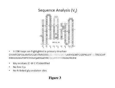
[0011] Figure 3 shows sequence analysis of VL: 3CDR of murine RP215 loops
which
are highlighted in primary amino acid sequence with key residues (C-W-C-F)
identified
and with no free Cys and N-linked glycosylation sites.
[0012] Figure 4 shows human framework donors considered.
[0013] Figure 5 is a template antibody structure model of murine RP215 (VL
shown
on left-hand side, VH on right-hand side, and CDR loops shown as spheres with
van der Waals radii).
CA 02876796 2014-12-15
WO 2013/186630
PCT/1B2013/001834
3
[0014] Figure 6 is a template antibody structure model of murine RP215.
Residues in
close proximity to CDR loops are shown as sticks. These residue positions are
candidates
for back-mutation.
[0015] Figure 7 shows humanized sequences that exhibit high homology to human
antibody sequences. hRP215_VH1 has -97% sequence identity to its closest human
FR
donor (germline) outside of CDR regions hRP215 -99% identity to germline FR
Alignment of Humanized VH with Human VH Framework Donors regions, while
hRP215_VH2 has donor outside of CDRs. Different residues are mostly located
within or
adjacent to CDRs, with the exception of Loop 3 in VH.
[0016] Figure 8 shows comparison of amino acid sequences of heavy chain and
light
chain of humanized forms of RP215 with those of murine RP215.
[0017] Figure 9 shows the nucleotide sequences that encode the amino acid
sequences
of Figure 8.
Modes of Carrying Out the Invention
[0018] The humanized antibodies of the present invention may be in a variety
of forms
¨ including whole antibodies, fragments that are immunoreactive with CA215,
including
Fab, Fab', and F(ab')2 fragments as well as recombinantly produced single-
chain
antibodies. The resulting humanized forms as noted below are of equivalent
affinity and
specificity to the murine RP215 and contain substantially similar or identical
CDR
regions.
[0019] The CDR regions of the variable region of both heavy and light chains
can be
determined by a number of art-known methods, including the numbering system of
Kabat
which defines, in the light chain CDR1 as residues 24-34, CDR2 as residues 50-
56, CDR3
as residues 89-97 and in the heavy chain CDR1 as residues 31-35, CDR2 as
residues 50-65 and CDR3 as residues 95-102 (Wu, T. T., and Kabat, E. A., Exp.
Med.
(1970) 132:211-250). CDRs can also be determined according to the system of
Clothia
which gives slightly different results (Clothia, C., et al., Nature (1989)
342:877-883;
Al-Laziken, et al., J. MoL Biol. (1997) 273:927-948). Various subsequent
authors have
suggested some minor modifications. The CDRs as assigned by both Kabat and
Clothia
systems are included within the scope of the present invention.
CA 02876796 2014-12-15
WO 2013/186630
PCT/1B2013/001834
4
[0020] Sandwich and/or binding immunoassays were used to demonstrate the
substantial equivalence between the humanized forms and murine RP215. Their
respective affinity and specificity to the cognate antigen, the cancer cell-
expressed CA215,
are two important parameters to establish their substantial equivalence.
General Approach for Humanization
[0021] Based on the published sequence of the RP215 variable chain, it was
determined that five of the CDRs (excluding H3) fall into one of the canonical
structure
classes indicated as follows:
CDR Li L2 L3 H1 H2
Canonical Structure class 3/17A 1/7A 1/9A 1/10A 2/10A
[0022] Human framework donor selection was made through a search of germline
followed by rearrangement of human IgG data base using VL and VH sequences
with or
without CDRs. To obtain human IgG results, normally, we go through each hit to
eliminate inappropriate donors (such as mouse sequence or humanized sequence,
etc.).
For VL and VH sequences, the best hits in each group were aligned. Finally,
one germline
FR donor and one rearranged (mature) FR donor based on sequence similarity and
other
factors are selected. These factors included CDR length (except for CDR-H3),
CDR
canonical structure, proline residues at key positions or factors which may
affect proper
folding of humanized antibody.
[0023] Homology modeling was used to obtain template antibody structure by
searching the PDB data base for the template antibody VL and VH sequences with
or
without CDR. The following conditions are taken into consideration:
(1) Sequence homology
(2) CDR length
(3) CDR canonical structure, and
(4) Model with correct disulfide linkage
[0024] The antigen binding region of the antibody structure model can then be
optimized through the CDR loop data base and canonical structure class as well
as
comparison to the template structure.
CA 02876796 2014-12-15
WO 2013/186630
PCT/1B2013/001834
[0025] Structural modeling was used to identify residues outside of the CDR
loops
that might affect CDR configurations. The following binding or interaction
factors should
be taken into consideration: hydrogen bonding, steric hindrance and other
interactions to
main chain and side chains of CDR residues.
[0026] Back mutation was performed to those residues that are predicted to
significantly affect CDR loop structure. Other critical residues were also
verified
including those in (1) the heterodimer interface in FR donors for proper VL
and VH
interactions, (2) the intra-chain domain interface and (3) direct interactions
to
antigen/epitopes in the known structure.
[0027] Therefore, based on the above considerations, combinations of different
back
mutations were designed to balance the minimal need of such process. As a
result, low
immunogenicity to humans can be obtained and the maximal preservations of
antigen-
binding affinity can be preserved.
[0028] The following examples are offered to illustrate but not to limit the
invention.
Example 1
Characterization of Humanized RP215 Monoclonal Antibodies
[0029] Humanized RP215 monoclonal antibodies with heavy and light chains
designated 0021-0023 (VH1-VH3 and VL1-VL2) and the parent murine chains 0024
were
generated, expressed and affinity-purified. Various heavy chain/light chain
combinations
were used to construct antibodies FY1-FY6 as shown in Table 1. ChRP215 is a
murine
chimera with human IgG.
Table 1
VL1 VL2 L0024
VH1 FY1 FY4
VH2 FY2 FY5
VH3 FY3 FY6
H0024 ChRP215
CA 02876796 2014-12-15
WO 2013/186630
PCT/1B2013/001834
6
[0030] The titers and amounts of FY1-FY6 from CHO cells are shown in Table 2.
Table 2
Name of antibody Apparent titer (ng/ml) Production yield (N=1)
Neg. control NA 305
RP215 chimera (with human IgG) 4.2 219
FY1 5.7 287
FY2 13.1 166
FY3 8.4 152
FY4 4.4 351
FY5 9.6 158
FY6 7.1 350
[0031] Binding immunoassays were performed using affinity-purified CA215
coated
on microwells, to determine the binding affinities of these humanized RP215 to
CA215
and to compare with that of the original murine RP215. The results of such
comparative
binding assays are presented in Figure 1. As shown, all of FY1-FY6 have
comparable
affinities to RP215.
[0032] Previously, RP215 was established to have no cross-reactivity to normal
human
IgG. It reacts only with CA215 expressed by cancer cells that contain RP215-
specific
carbohydrate-associated epitope. Therefore, the humanized RP215 monoclonal
antibodies
should also show no cross-reactivity to normal human IgG. This lack-of-cross
reactivity
result was demonstrated, as humanized RP215 FY1, FY4 and FY5 (similar to the
murine
chimera) revealed no binding to human IgG as shown in Figure 2.
[0033] Figure 8 shows the complete amino acid sequences of both heavy and
light
chains of the humanized antibodies. The complete nucleotide sequences for
these chains
is shown in Figure 9.
[0034] As a matter of interest, Figures 3-7 show various additional structural
features
of these antibodies.