Note: Descriptions are shown in the official language in which they were submitted.
CA 02877746 2014-12-22
WO 2014/008218
PCMJS2013/048999
OPTIMIZATION OF ANTIBODIES THAT BIND LYMPHOCYTE
ACTIVATION GENE-3 (LAG-3), AND USES THEREOF
Background of the Invention
Therapeutic antibodies are one of the fastest growing segments of the
pharmaceutical industry. To maintain potency (i.e., activity) and minimize
immunogenicity, antibodies and other protein drugs must be protected from
physical and
chemical degradation during manufacturing and storage. Indeed, one of the
primary
difficulties in developing antibody therapeutics is the potential immunogenic
response
when administered to a subject, which can lead to rapid clearance or even
induce life-
threatening side effects including anaphylactic shock. Various factors
influence the
immunogenicity of an antibody such as its physiochemical properties (e.g.,
purity,
stability, or solubility), clinical factors (e.g., dose, route of
administration, heterogeneity
of the disease, or patient features), and concomitant treatment with other
agents (Swann
et al. (2008) Curt- Opinion Immuol 20:493-499).
lmmunogenicity of antibodies and/or loss of antibody activity is often due to
deamidation. Deamidation is a chemical degradative process that spontaneously
occurs
in proteins (e.g., antibodies). Deamidation removes an amide functional group
from an
amino acid residue, such as asparagine and glutamine, thus damaging its amide-
containing side chains. This, in turn, causes structural and biological
alterations
throughout the protein, thus creating heterogeneous forms of the antibody.
Deamidation
is one of the most common post-translational modifications that occurs in
recombinantly
produced therapeutic antibodies.
For example, heterogeneity in the heavy chain of monoclonal antibody h1B4 (a
humanized anti-CD18 antibody) due to deamidation during cell culture was
reported by
Tsai et al. (Pharm Res 10(11):1580 (1993)). In addition, reduction/loss of
biological
activity due to deamidation has been a recognized problem. For example, Kroon
et al.
characterized several deamidation sites in therapeutic antibody OKT3, and
reported that
samples of OKT3 production lots (aged 14 months to 3 years) had fallen below
75%
activity (Pharm Res 9(11):1386 (1992), page 1389, second column). In addition,
samples of OKT3 showing large amounts of the oxidized peptides in their maps
had
significantly reduced activity in the antigen binding potency assay (page
1390, first
column). The authors concluded that specific sites of chemical modification
that occur
upon storage of OKT3 were identified by peptide mapping and correlated with
observed
1
CA 02877746 2014-12-22
WO 2014/008218
PCT/US2013/048999
changes in chemical analyses and biological assays of the antibody (page 1392,
first
column). Loss of biological activity also has been reported for a variety of
other
deamidated therapeutic proteins, including recombinant human DNasc (Cacia et
al.
(1993) J. Chromatogr. 634:229-239) and recombinant soluble CD4 (Teshima et al.
(1991) Biochemistry 30:3916-3922).
Overall, deamidation poses a significant and unpredictable problem to the
pharmaceutical industry. Efforts associated with monitoring the variability
caused by
deamidation within antibody therapeutics, in particular, as well as FDA
concerns
associated with this variability, increase costs and delay clinical trials.
Moreover,
modifications to address this issue, including shifting conditions (e.g.,
temperature, pH,
and cell type) associated with recombinant production and/or alteration of
amino acids
which are susceptible to deamidation (e.g., site-directed mutagenesis) can
negatively
impact stability and activity, especially when changes are made within the
complementarity determining regions (CDRs) of the antibody. Accordingly, the
need
exists for more stable versions of therapeutic antibodies.
Summary
The present invention provides isolated monoclonal antibodies (e.g., human
monoclonal antibodies) that bind LAG-3 (e.g., human LAG-3) and have optimized
physical stability compared to previously described anti-LAG-3 antibodies. In
particular, the invention relates to a modified form of antibody 25F7 (US
2011/0150892
Al) which exhibits significantly improved thermal and chemical stability
compared to
the unmodified antibody. Specifically, by altering the critical binding region
of the
heavy chain CDR2 domain of antibody 25F7, it was shown that the modified
antibody
exhibited significantly higher physical and thermal stability, reduced
deamidation,
higher thermal reversibility, and lower aggregation. At the same time, it was
unexpectedly observed that the modified antibody retained the same high
binding
affinity to human LAG-3 and functional activity of the unmodified antibody,
including
the ability to inhibit binding of LAG-3 to major histocompatibility (MHC)
Class II
molecules and stimulate antigen-specific T cell responses. The combined
substantial
increase in stability and retention of binding / biological activity of the
modified
antibody was surprising, particularly in view of the criticality of CDRs
regions to
antibody function.
2
88145363
The antibodies of the invention can be used for a variety of applications,
including
detection of LAG-3 protein and stimulation of antigen-specific T cell
responses in tumor-
bearing or virus-bearing subjects.
Accordingly, in one aspect, the invention pertains to an isolated monoclonal
antibody (e.g., a human antibody), or an antigen-binding portion thereof,
having a heavy
chain variable region comprising the amino acid sequence of SEQ ID NO: 12. In
another
embodiment, the antibody further includes a light chain variable region
comprising the
amino acid sequence of SEQ ID NO: 14. In another embodiment, the antibody, or
antigen-
binding portion thereof, includes the CDR1, CDR2, and CDR3 regions of a heavy
chain
variable region comprising the amino acid sequence of SEQ ID NO: 12 (e.g., SEQ
ID NOs:
15, 16, and 17, respectively). In another embodiment, the antibody further
includes the CDR1,
CDR2, and CDR3 regions of a light chain variable region comprising the amino
acid sequence
of SEQ ID NO: 12 (e.g., SEQ ID NOs: 18, 19, and 20, respectively).
In other embodiments, the invention further pertains to:
- an isolated monoclonal antibody, or an antigen-binding portion thereof, that
binds
human lymphocyte activation gene-3 (LAG-3), comprising heavy and light chain
variable
regions, (i) wherein the heavy chain CDR1, CDR2, and CDR3 comprise the amino
acid
sequences of SEQ ID NOs: 15, 16, and 17, respectively, and the light chain
CDR1, CDR2,
and CDR3 comprise the amino acid sequences of SEQ ID NOs: 18, 19, and 20,
respectively;
(ii) wherein the heavy chain CDR1, CDR2, and CDR3 comprise the amino acid
sequences of
SEQ ID NOs: 15, 26, and 17, respectively, and the light chain CDR1, CDR2, and
CDR3
comprise the amino acid sequences of SEQ ID NOs: 18, 19, and 20, respectively;
(iii) wherein
the heavy chain CDR1, CDR2, and CDR3 comprise the amino acid sequences of SEQ
ID
NOs: 15, 25, and 17, respectively, and the light chain CDR1, CDR2, and CDR3
comprise the
amino acid sequences of SEQ ID NOs: 18, 19, and 20, respectively; or (iv)
wherein the heavy
chain CDR1, CDR2, and CDR3 comprise the amino acid sequences of SEQ ID NOs:
15, 24,
and 17, respectively, and the light chain CDR1, CDR2, and CDR3 comprise the
amino acid
sequences of SEQ ID NOs: 18, 19, and 20, respectively;
3
Date recue / Date received 2021-11-22
88145363
- an isolated monoclonal antibody, or an antigen-binding portion thereof,
that binds
human LAG-3, comprising heavy and light chain variable regions, comprising the
amino acid
sequences of SEQ ID NOs: 12 and 14, respectively;
- an isolated full-length IgG4 human monoclonal antibody that binds human
LAG-3,
comprising heavy and light chain variable regions, wherein the heavy chain
variable region
comprises the amino acid sequence of SEQ ID NO: 12 and the light chain
variable region
comprises the amino acid sequence of SEQ ID NO: 14;
- an isolated full-length IgG4 human monoclonal antibody that binds human
LAG-3,
comprising heavy and light chain variable regions, wherein the amino acid
sequence of the
heavy chain variable region is SEQ ID NO: 12 and the amino acid sequence of
the light chain
variable region is SEQ ID NO: 14;
- an isolated full-length IgG4 human monoclonal antibody that binds human
LAG-3,
comprising heavy and light chains, wherein the heavy chain comprises the amino
acid
sequence of SEQ ID NO: 35 and the light chain comprises the amino acid
sequence of SEQ
ID NO: 37; and
- an isolated full-length IgG4 human monoclonal antibody that binds human
LAG-3,
comprising heavy and light chains, wherein the amino acid sequence of the
heavy chain is
SEQ ID NO: 35 and the amino acid sequence of the light chain is SEQ ID NO: 37.
In a preferred embodiment the antibody exhibits increased physical properties
(i.e.,
thermal and chemical stability) compared to antibody 25F7, while still
retaining at least the
same binding affinity for human LAG-3 as 25F7. For example, the antibody
exhibits
decreased sequence variability in the heavy chain CDR2 region due to
deamidation, compared
to antibody 25F7, e.g., approximately 2.5% or less modification of the amino
acid sequence
after 12 weeks at 4C (i.e., under "real-time" stability studies as described
herein) and/or
approximately 12.0% or less modification of the amino acid sequence after 12
weeks at 40C
(i.e., under accelerated stress conditions, as described herein), while still
retaining a binding
affinity for human LAG-3 of about at least KD of 1 x 1 oY7 M or less (more
preferably, a KD of
1 x 10⁻⁸M or less, a KD of 5 x 10⁻⁹M or less, or a KD of 1 x i09 M or less).
In another
embodiment, the antibody exhibits thermal reversibility of at least about 40%
in PBS at
p}18Ø
3a
Date recue / Date received 2021-11-22
88145363
In another embodiment, the antibody possesses a higher melting temperature
(indicating greater overall stability in vivo), compared to the unmodified
antibody
(Krishnamurthy R and Manning MC (2002) Curr Pharm Biotechnol 3:361-71). In one
embodiment, the antibody exhibits a Tmi (the temperature of initial unfolding)
of greater
than 60 C, e.g., greater than 65 C, or greater than 70 C. The melting point of
an antibody
can be measured using differential scanning calorimetry (Chen et al (2003)
3b
Date recue / Date received 2021-11-22
CA 02877746 2014-12-22
WO 2014/008218
PCT/US2013/048999
Pharm Res 20:1952-60; Ghirlando et al (1999) Immunol Lett 68:47-52) or
circular
dichroism (Murray et al. (2002) Chromatogr Sei 40:343-9).
In another embodiment, the antibody is characterized by its resistance to
rapid
degradation. Degradation of an antibody can be measured using capillary
electrophoresis (CE) and MALDI-MS (Alexander AJ and Hughes DE (1995) Anal Chem
67:3626-32).
In another embodiment, the antibody exhibits minimal aggregation effects,
e.g.,
aggregation of 25% or less, such as 20% or less, 15% or less, 10% or less, 5%
or less, or
4% or less. Aggregation can lead to the triggering of an unwanted immune
response
and/or altered or unfavorable pharmacokinetic properties, Aggregation can be
measured
by several techniques, including size-exclusion column (SEC), high performance
liquid
chromatography (HPLC), and light scattering.
In another embodiment, the antibody further exhibits at least one of the
following
properties:
(a) binding to monkey LAG-3;
(b) lack of binding to mouse LAG-3;
(c) inhibition of binding of LAG-3 to major histocompatibility (MHC) class II
molecules; and
(d) stimulation of immune responses, particularly antigen-specific T cell
responses.
Preferably, the antibody exhibits at least two of properties (a), (b), (c) and
(d). More
preferably, the antibody exhibits at least three of properties (a), (b), (c)
and (d). Even
more preferably, the antibody exhibits all four of properties (a), (b), (c)
and (d).
In another embodiment, the antibody stimulates an antigen-specific T cell
response, such as interleukin-2 (IL-2) production in an antigen-specific T
cell response.
In other embodiments, the antibody stimulates an immune response, such as an
anti-
tumor response (e.g., inhibition of tumor growth in an in vivo tumor graft
model) or an
autoimmune response (e.g., development of diabetes in NOD mice).
In another embodiment, the antibody binds an epitope of human LAG-3
comprising the amino acid sequence PGHPLAPG (SEQ ID NO: 21). In another
embodiment, the antibody binds an epitope of human LAG-3 comprising the amino
acid
sequence HPAAPSSW (SEQ ID NO: 22) or PAAPSSWG (SEQ ID NO: 23).
In other embodiments, the antibody stains pituitary tissue by
immunohistochemistry, or does not stain pituitary tissue by
immunohistochemistry.
4
CA 02877746 2014-12-22
WO 2014/008218
PCT/US2013/048999
Antibodies of the invention can be full-length antibodies, for example, of an
IgGI, IgG2 or IgG4 isotype, optionally with a serine to proline mutation in
the heavy
chain constant region hinge region (at a position corresponding to position
241 as
described in Angal et al. (1993) AM. Immunol. 30:105-108), such that inter-
heavy chain
disulfide bridge heterogeneity is reduced or abolished. In one aspect, the
constant region
isotype is IgG4 with a mutation at amino acid residues 228, e.g., S228P.
Alternatively,
the antibodies can be antibody fragments, such as Fab, Fab' or Fab'2
fragments, or
single chain antibodies.
In another aspect of the invention, the antibody (or antigen-binding portion
thereof) is part of an immunoconjugate which includes a therapeutic agent,
e.g., a
cytotoxin or a radioactive isotope, linked to the antibody. In another aspect,
the
antibody is part of a bispecific molecule which includes a second functional
moiety
(e.g., a second antibody) having a different binding specificity than said
antibody, or
antigen binding portion thereof.
Compositions comprising antibodies, or antigen-binding portions thereof,
immunoconjugates or bispecific molecules of the invention, optionally
formulated in a
pharmaceutically acceptable carrier, also are provided.
Nucleic acid molecules encoding the antibodies, or antigen-binding portions
(e.g., variable regions and/or CDRs) thereof, of the invention also are
provided, as well
as expression vectors comprising such nucleic acids and host cells comprising
such
expression vectors. Methods for preparing anti-LAG-3 antibodies using the host
cells
comprising such expression vectors also are provided, and can include the
steps of (i)
expressing the antibody in the host cell and (ii) isolating the antibody from
the host cell.
In another aspect, the invention provides methods of stimulating immune
responses using anti-LAG-3 antibodies of the invention. In one embodiment, the
method involves stimulating an antigen-specific T cell response by contacting
T cells
with an antibody of the invention, such that an antigen-specific T cell
response is
stimulated. In a preferred embodiment, interleukin-2 production by the antigen-
specific
T cell is stimulated. In another embodiment, the subject is a tumor-bearing
subject and
an immune response against the tumor is stimulated. In another embodiment, the
subject
is a virus-bearing subject and an immune response against the virus is
stimulated.
In yet another embodiment, the invention provides a method for inhibiting
growth of tumor cells in a subject comprising administering to the subject an
antibody,
5
or antigen-binding portion thereof, of the invention, such that growth of the
tumor is
inhibited in the subject. In still another embodiment, the invention provides
a method
for treating viral infection in a subject comprising administering to the
subject an
antibody, or antigen-binding portion thereof, of the invention such that the
viral
infection is treated in the subject In another embodiment, these methods
comprise
administering a composition, bispecific, or immunoconjugate of the invention.
In yet another embodiment, the invention provides a method for stimulating an
immune response in a subject comprising administering to the subject an
antibody, or
antigen-binding portion thereof, of the invention and at least one additional
immunostimulatory antibody, such as an anti-PD-1 antibody, an anti-PD-Li
antibody
and/or an anti-CTLA-4 antibody, such that an immune response is stimulated in
the
subject, for example to inhibit tumor growth or to stimulate an anti-viral
response. In
one embodiment, the additional immunostimulatory antibody is an anti-PD-1
antibody.
In another embodiment, the additional immunostimulatory agent is an anti-PD-L1
antibody. In yet another embodiment, the additional immunostimulatory agent is
an
anti-CTLA-4 antibody. In yet another embodiment, an antibody, or antigen-
binding
portion thereof, of the invention is administered with a cytokine (e.g., IL-2
and/or IL-
21), or a costimulatory antibody (e.g., an anti-CD137 and/or anti-GITR
antibody). The
antibodies can be, for example, human, chimeric or humanized antibodies.
In another aspect, the invention provides anti-LAG-3 antibodies and
compositions of the invention for use in the foregoing methods, or for the
manufacture
of a medicament for use in the foregoing methods (e.g., for treatment).
Other features and advantages of the instant disclosure will be apparent from
the
following detailed description and examples, which should not be construed as
limiting.
Brief Description of the Drawinms
Figure IA shows the nucleotide sequence (SEQ ID NO: 1) and amino acid
sequence (SEQ ID NO: 2) of the heavy chain variable region of the 25F7 human
monoclonal antibody. The CDR1 (SEQ ID NO: 5), CDR2 (SEQ ID NO: 6) and CDR3
(SEQ ID NO: 7) regions are delineated and the V, D and J germline derivations
are
indicated. The CDR regions are delineated using the Kabat system (Kabat et al.
(1991)
6
CA 2877746 2019-10-18
CA 02877746 2015-04-15
Sequences of Proteins of Immunological Interest, Fifth Edition, U.S.
Department of
Health and Human Services, NIH Publication No. 91-3242).
Figure 1B shows the nucleotide sequence (SEQ ID NO: 3) and amino acid
sequence (SEQ ID NO: 4) of the kappa light chain variable region of the 25F7
human
monoclonal antibody. The CDR1 (SEQ ID NO: 8), CDR2 (SEQ ID NO: 9) and CDR3
(SEQ ID NO: 10) regions are delineated and the V and J germline derivations
are
indicated. The full-length heavy and light chain amino acid sequences antibody
25F7
are shown in SEQ ID NOs: 32 and 34, respectively.
Figure 2A shows the amino acid sequence (SEQ ID NO: 12) of the heavy chain
variable region of the LAG3.5 monoclonal antibody. The CDR1 (SEQ ID NO: 15),
CDR2 (SEQ ID NO: 16) and CDR3 (SEQ ID NO: 17) regions are delineated. The full-
length heavy and light chain amino acid sequences antibody LAG3.5 are shown in
SEQ
ID NOs: 35 and 37, respectively.
Figure 2B shows the nucleotide sequence (SEQ ID NO: 13) and amino acid
sequence (SEQ ID NO: 14) of the kappa light chain variable region of the
LAG3.5
monoclonal antibody. The CDR1 (SEQ ID NO: 18), CDR2 (SEQ ID NO: 19) and
CDR3 (SEQ ID NO: 20) regions arc delineated.
Figure 3 shows the amino acid sequences of the CDR2 heavy chain variable
region sequences of the LAG-3 variants LAG3.5 (SEQ ID NO: 42), LAG3.6 (SEQ ID
.. NO: 43), LAG3.7 (SEQ ID NO:44), and LAG3.8 (SEQ ID NO:45), compared to the
amino acid sequence of the CDR2 heavy chain variable region sequence of
antibody
25F7 (LAG3.1) (SEQ ID NO: 41) and corresponding human germline sequence (SEQ
ID
NO: 27). The CDR2 heavy chain variable region of 1.AG3.5 differs from the CDR2
heavy chain variable region of 251'7 by argininc (R) at position 54 (versus
asparagine
(N)) and serine (S) at position 56 (versus asparagines (N)). The remaining
CDRs of
LAG3.5 anf 25F7 are identical. Figure 3 also discloses SEQ ID NO: 40.
Figures 4A and 4B arc graphs showing the binding activity (EC50 and affinity,
respectively) of antibodies LAG3.1 (25F7), LAG3.2, LAG3.5, LAG3.6, LAG3.7, and
LAG3.8 to activated human CD4+ T cells. Figure 4B discloses SEQ ID NOs: 41,
42,
.. 45, 44 and 43, respectively, in order of appearance.
Figures 5A, B, C, D, and E are graphs showing thermal melting curves (i.e.,
thermal stability) of antibodies LAG3.1 (25E7), LAG3.5, LAG3.6, LAG3.7, and
LAG3.8, respectively.
7
CA 02877746 2015-04-15
Figures 6A, B, C, D, and E are graphs showing thermal reversibility curves
(i.e.,
thermal stability) of antibodies LAG3.1 (25F7), LAG3.5, LAG3.6, LAG3.7, and
LAG3.8, respectively.
Figure 7 is a graph, showing the binding activity of antibodies LAG3. I (25F7)
and LAG3.5 to activated human CD4+ T cells and antigen binding (Biacorc).
Figure 8 shows the results of peptide mapping using mass-sepctrometry
(chemical modifications / molecular stability) for antibodies LAG3.1 (25F7)
and
LAG3.5 reflecting deamidation and isomerization after incubating for 5 days
under
accelerated stress conditions as described herein. Figure 8 discloses SEQ 1D
NOs
46-52, respectively, in order of appearance.
Figure 9 is a graph comparing the hydrophilicity profiles of antibodies LAG3.1
(25F7) and LAG3.5.
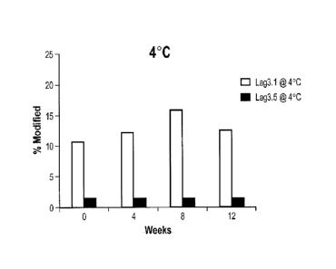
Figures 10 A, B, C, and D are graphs comparing the affinity and physical
stability (i.e., thermal and chemical stability) of antibodies LAG3.1 and
LAG3.5 at 4C
and 40C , i.e., both accelerated stress conditions and "real-time" stability
studies, as
described herein.
Figures 11 A and B are graphs comparing the percent modification of the amino
acid sequences of antibodies LAG3.1 and LAG3.5 at 4C and 40C .
8
CA 02877746 2014-12-22
WO 2014/008218
PCT/US2013/048999
Detailed Description of the Invention
In order that the present disclosure may be more readily understood, certain
terms are first defined. Additional definitions are set forth throughout the
detailed
description.
The terms "25F7," "antibody 25F7," "antibody LAG3.1," and "LAG3.1" refer to
the anti-human LAG-3 antibody described in US2011/0150892 Al. The nucleotide
sequence (SEQ ID NO: 1) encoding the heavy chain variable region of 25F7
(LAG3.1)
and the corresponding amino acid sequence (SEQ ID NO: 2) is shown in Figure 1A
(with CDR sequences designated as SEQ ID NOs: 4, 5, and 7, respectively). The
nucleotide sequence (SEQ ID NO: 3) encoding the light chain variable region of
25F7
(LAG3.1) and the corresponding amino acid sequence (SEQ ID NO: 4) is shown in
Figure 1B (with CDR sequences designated as SEQ ID NOs: 8, 9, and 10,
respectively).
The term "LAG-3" refers to Lymphocyte Activation Gene-3. The term "LAG-3"
includes variants, isoforms, homologs, orthologs and paralogs. For example,
antibodies
specific for a human LAG-3 protein may, in certain cases, cross-react with a
LAG-3
protein from a species other than human. In other embodiments, the antibodies
specific
for a human LAG-3 protein may be completely specific for the human LAG-3
protein
and may not exhibit species or other types of cross-reactivity, or may cross-
react with
LAG-3 from certain other species but not all other species (e.g., cross-react
with monkey
LAG-3 but not mouse LAG-3). The term "human LAG-3" refers to human sequence
LAG-3, such as the complete amino acid sequence of human LAG-3 having Genbank
Accession No. NP 002277 (SEQ ID NO: 29). The term "mouse LAG-3" refers to
mouse sequence LAG-3, such as the complete amino acid sequence of mouse LAG-3
having Genbank Accession No. NP_032505. LAG-3 is also known in the art as, for
example, CD223. The human LAG-3 sequence may differ from human LAG-3 of
Genbank Accession No. NP 002277 by having, e.g., conserved mutations or
mutations
in non-conserved regions and the LAG-3 has substantially the same biological
function
as the human LAG-3 of Genbank Accession No. NP 002277. For example, a
biological
function of human LAG-3 is having an cpitopc in the extracellular domain of
LAG-3
that is specifically bound by an antibody of the instant disclosure or a
biological function
of human LAG-3 is binding to MHC Class II molecules.
The term "monkey LAG-3" is intended to encompass LAG-3 proteins expressed
by Old World and New World monkeys, including but not limited to cynomolgus
9
CA 02877746 2014-12-22
WO 2014/008218
PCT/US2013/048999
monkey LAG-3 and rhesus monkey LAG-3. A representative amino acid sequence for
monkey LAG-3 is the rhesus monkey LAG-3 amino acid sequence which is also
deposited as Genbank Accession No. XM_001108923. Another representative amino
acid sequence for monkey LAG-3 is the alternative rhesus monkey sequence of
clone
pa23-5 as described in US 2011/0150892 Al. This alternative rhesus sequence
exhibits
a single amino acid difference, at position 419, as compared to the Genbank-
deposited
sequence.
A particular human LAG-3 sequence will generally be at least 90% identical in
amino acid sequence to human LAG-3 of Genbank Accession No. NP_002277 and
contains amino acid residues that identify the amino acid sequence as being
human when
compared to LAG-3 amino acid sequences of other species (e.g., murine). In
certain
cases, a human LAG-3 can be at least 95%, or even at least 96%, 97%, 98%, or
99%
identical in amino acid sequence to LAG-3 of Genbank Accession No. NP_002277.
In
certain embodiments, a human LAG-3 sequence will display no more than 10 amino
acid differences from the LAG-3 sequence of Genbank Accession No. NP_002277.
In
certain embodiments, the human LAG-3 can display no more than 5, or even no
more
than 4, 3, 2, or 1 amino acid difference from the LAG-3 sequence of Genbank
Accession
No. NP_002277. Percent identity can be determined as described herein.
The term "immune response" refers to the action of, for example, lymphocytes,
antigen presenting cells, phagocytic cells, granulocytes, and soluble
macromolecules
produced by the above cells or the liver (including antibodies, cytokines, and
complement) that results in selective damage to, destruction of, or
elimination from the
human body of invading pathogens, cells or tissues infected with pathogens,
cancerous
cells, or, in cases of autoimmunity or pathological inflammation, normal human
cells or
tissues.
An "antigen-specific T cell response" refers to responses by a T cell that
result
from stimulation of the T cell with the antigen for which the T cell is
specific. Non-
limiting examples of responses by a T cell upon antigen-specific stimulation
include
proliferation and cytokinc production (e.g., IL-2 production).
The term "antibody" as referred to herein includes whole antibodies and any
antigen binding fragment (i.e., "antigen-binding portion") or single chains
thereof.
Whole antibodies are glycoproteins comprising at least two heavy (H) chains
and two
light (L) chains inter-connected by disulfide bonds. Each heavy chain is
comprised of a
CA 02877746 2014-12-22
WO 2014/008218
PCT/US2013/048999
heavy chain variable region (abbreviated herein as VH) and a heavy chain
constant
region. The heavy chain constant region is comprised of three domains, CHI,
CH2 and
CH3. Each light chain is comprised of a light chain variable region
(abbreviated herein
as VL) and a light chain constant region. The light chain constant region is
comprised of
one domain, CL. The VH and VL regions can be further subdivided into regions
of
hypervariability, termed complementarity determining regions (CDR),
interspersed with
regions that arc more conserved, termed framework regions (FR). Each VH and VL
is
composed of three CDRs and four FRs, arranged from amino-terminus to carboxy-
terminus in the following order: FRI, CDR1, FR2, CDR2, FR3, CDR3, FR4. The
variable regions of the heavy and light chains contain a binding domain that
interacts
with an antigen. The constant regions of the antibodies can mediate the
binding of the
immunoglobulin to host tissues or factors, including various cells of the
immune system
(e.g., effector cells) and the first component (Clq) of the classical
complement system.
The term "antigen-binding portion" of an antibody (or simply "antibody
portion"), as used herein, refers to one or more fragments of an antibody that
retain the
ability to specifically bind to an antigen (e.g., a LAG-3 protein). It has
been shown that
the antigen-binding function of an antibody can be performed by fragments of a
full-
length antibody. Examples of binding fragments encompassed within the term
"antigen-
binding portion" of an antibody include (i) a Fab fragment, a monovalent
fragment
consisting of the VL, VH, CL and CHI domains; (ii) a F(abT)2 fragment, a
bivalent
fragment comprising two Fab fragments linked by a disulfide bridge at the
hinge region;
(iii) a Fd fragment consisting of the VH and Cm domains; (iv) a Fv fragment
consisting
of the VH and CH 1 domains; (v) a FIT fragment consisting of the VL and VH
domains of a
single arm of an antibody, (vi) a dAb fragment (Ward et al., (1989) Nature
341:544-
546), which consists of a VH domain; (vii) an isolated complementarity
determining
region (CDR); and (viii) a nanobody, a heavy chain variable region containing
a single
variable domain and two constant domains. Furthermore, although the two
domains of
the Fv fragment, VI and VH, are coded for by separate genes, they can be
joined, using
recombinant methods, by a synthetic linker that enables them to be made as a
single
protein chain in which the VL and VH regions pair to form monovalent molecules
(known as single chain Fv (scFv); see e.g., Bird et al. (1988) Science 242:423-
426; and
Huston et al. (1988) Proc. NatL Acad. Sci. USA 85:5879-5883). Such single
chain
antibodies are also intended to be encompassed within the term "antigen-
binding
11
CA 02877746 2014-12-22
WO 2014/008218
PCT/US2013/048999
portion" of an antibody. These antibody fragments are obtained using
conventional
techniques known to those with skill in the art, and the fragments are
screened for utility
in the same manner as are intact antibodies.
An "isolated antibody", as used herein, is intended to refer to an antibody
that is
substantially free of other antibodies having different antigenic
specificities (e.g., an
isolated antibody that specifically binds a LAG-3 protein is substantially
free of
antibodies that specifically bind antigens other than LAG-3 proteins). An
isolated
antibody that specifically binds a human LAG-3 protein may, however, have
cross-
reactivity to other antigens, such as LAG-3 proteins from other species.
Moreover, an
isolated antibody can be substantially free of other cellular material and/or
chemicals.
The terms "monoclonal antibody" or "monoclonal antibody composition" as used
herein refer to a preparation of antibody molecules of single molecular
composition. A
monoclonal antibody composition displays a single binding specificity and
affinity for a
particular epitope.
The term "human antibody", as used herein, is intended to include antibodies
having variable regions in which both the framework and CDR regions are
derived from
human germline immunoglobulin sequences. Furthermore, if the antibody contains
a
constant region, the constant region also is derived from human germline
immunoglobulin sequences. The human antibodies of the invention can include
amino
acid residues not encoded by human germline immunoglobulin sequences (e.g.,
mutations introduced by random or site-specific mutagenesis in vitro or by
somatic
mutation in vivo). However, the term "human antibody", as used herein, is not
intended
to include antibodies in which CDR sequences derived from the germline of
another
mammalian species, such as a mouse, have been grafted onto human framework
sequences.
The term "human monoclonal antibody" refers to antibodies displaying a single
binding specificity, which have variable regions in which both the framework
and CDR
regions are derived from human germline immunoglobulin sequences. In one
embodiment, the human monoclonal antibodies are produced by a hybridoma which
includes a B cell obtained from a transgenic nonhuman animal, e.g., a
transgenic mouse,
having a genome comprising a human heavy chain transgene and a light chain
transgene
fused to an immortalized cell.
12
CA 02877746 2014-12-22
WO 2014/008218
PCT/US2013/048999
The term "recombinant human antibody", as used herein, includes all human
antibodies that are prepared, expressed, created or isolated by recombinant
means, such
as (a) antibodies isolated from an animal (e.g., a mouse) that is transgenic
or
transchromosomal for human immunoglobulin genes or a hybridoma prepared
therefrom
(described further below), (b) antibodies isolated from a host cell
transformed to express
the human antibody, e.g., from a transfectoma, (c) antibodies isolated from a
recombinant, combinatorial human antibody library, and (d) antibodies
prepared,
expressed, created or isolated by any other means that involve splicing of
human
immunoglobulin gene sequences to other DNA sequences. Such recombinant human
antibodies have variable regions in which the framework and CDR regions are
derived
from human germline immunoglobulin sequences. In certain embodiments, however,
such recombinant human antibodies can be subjected to in vitro mutagenesis
(or, when
an animal transgenic for human Ig sequences is used, in vivo somatic
mutagenesis) and
thus the amino acid sequences of the VH and VI_ regions of the recombinant
antibodies
are sequences that, while derived from and related to human germline Vu and V.
sequences, may not naturally exist within the human antibody germline
repertoire in
vivo.
The term "isotype" refers to the antibody class (e.g., IgM or IgG1) that is
encoded
by the heavy chain constant region genes.
The phrases "an antibody recognizing an antigen" and "an antibody specific for
an antigen" are used interchangeably herein with the term "an antibody which
binds
specifically to an antigen."
The term "human antibody derivatives" refers to any modified form of the human
antibody, e.g., a conjugate of the antibody and another agent or antibody.
The term "humanized antibody" is intended to refer to antibodies in which CDR
sequences derived from the germline of another mammalian species, such as a
mouse,
have been grafted onto human framework sequences. Additional framework region
modifications can be made within the human framework sequences.
The term "chimeric antibody" is intended to refer to antibodies in which the
variable region sequences are derived from one species and the constant region
sequences are derived from another species, such as an antibody in which the
variable
region sequences are derived from a mouse antibody and the constant region
sequences
are derived from a human antibody.
13
CA 02877746 2014-12-22
WO 2014/008218
PCT/US2013/048999
As used herein, an antibody that "specifically binds human LAG-3" is intended
to refer to an antibody that binds to human LAG-3 protein (and possibly a LAG-
3
protein from one or more non-human species) but does not substantially bind to
non-
LAG-3 proteins. Preferably, the antibody binds to a human LAG-3 protein with
"high
affinity", namely with a KD of 1 x 10⁻⁷ M or less, more preferably 1 x 10⁻⁸M
or less,
more preferably 5 x 10¹⁹ M or less, more preferably 1 x 10⁻⁹ M or less.
The term "does not substantially bind" to a protein or cells, as used herein,
means
does not bind or does not bind with a high affinity to the protein or cells,
i.e. binds to the
protein or cells with a KD of 1 x 10⁶ M or more, more preferably 1 x 10 5 M or
more,
more preferably 1 x 10⁻⁴ M or more, more preferably 1 x 10⁻³ M or more, even
more
preferably 1 x 10⁻²M or more.
The term "K.,.," or "K.", as used herein, is intended to refer to the
association
rate of a particular antibody-antigen interaction, whereas the term "Kdis" or
"Kd," as used
herein, is intended to refer to the dissociation rate of a particular antibody-
antigen
interaction. The term "KD," as used herein, is intended to refer to the
dissociation
constant, which is obtained from the ratio of Kd to Ka (i.e., Ka/Ka) and is
expressed as a
molar concentration (M). KD values for antibodies can be determined using
methods
well established in the art. A preferred method for determining the KD of an
antibody is
by using surface plasmon resonance, preferably using a biosensor system such
as a
Biacore system.
The term "high affinity" for an IgG antibody refers to an antibody having a KD
of
1 x 10⁻² M or less, more preferably 5 x 10⁻⁸M or less, even more preferably
1x10⁻⁸ M or
less, even more preferably 5 x 10⁻⁹M or less and even more preferably 1 x 10⁻⁹
M or less
for a target antigen. However, "high affinity" binding can vary for other
antibody
isotypes. For example, "high affinity" binding for an IgM isotype refers to an
antibody
having a KD of 10-6M or less, more preferably 10-7 M or less, even more
preferably 10-8
M or less.
The term "deamidation" refers to a chemical degredative process that
spontaneously occurs in proteins (e.g., antibodies). Deamidation removes an
amide
functional group from an amino acid residue, such as asparagine and glutamine,
thus
damaging its amide-containing side chains. Specifically, the side chain of an
asparagine
attacks the adjacent peptide group, forming a symmetric succinimide
intermediate. The
symmetry of the intermediate results in two hydrolysis products, either
aspartate or
14
CA 02877746 2014-12-22
WO 2014/008218
PCT/US2013/048999
isoaspartate. A similar reaction can also occur in aspartate side chains,
yielding a partial
conversion to isoaspartate. In the case of glutamine, the rate of deamidation
is generally
ten fold less than asparagine, however, the mechanism is essentially the same,
requiring
only water molecules to proceed.
The term "subject" includes any human or nonhuman animal. The term
"nonhuman animal" includes all vertebrates, e.g., mammals and non-mammals,
such as
non-human primates, sheep, dogs, cats, cows, horses, chickens, amphibians, and
reptiles,
although mammals are preferred, such as non-human primates, sheep, dogs, cats,
cows
and horses.
Various aspects of the invention are described in further detail in the
following
subsections.
Anti-LAG-3 Antibodies Having Increased Stability and Advantageous Functional
Properties
Antibodies of the invention specifically bind to human LAG-3 and have
optimized stability compared to previously described anti-LAG-3 antibodies,
particularly compared to antibody 25F7 (LAG3.1). This optimization includes
reduced
deamidation (e.g., increased chemical stability) and increased thermal
refolding (e.g.,
increased physical stability), while still retaining high affinity binding to
human LAG-3.
Methods for identifying deamidation sites are known in the art (see, e.g., ion
exchange, reversed phase, and hydrophobic interaction chromatography, and
peptide
mapping of proteolytic digests (LC-MS)). Suitable assays for measuring
physical
stability include, e.g., analysis of melting points and/or refolding of
antibody structure
following denaturation (e.g., percent reversibility as described, e.g., in
Example 3,
Section 3).
Binding to human LAG-3 can be assessed using one or more techniques also
well established in the art. For example, an antibody can be tested by a flow
cytometry
assay in which the antibody is reacted with a cell line that expresses human
LAG-3, such
as CHO cells that have been transfected to express LAG-3 (e.g., human LAG-3,
or
monkey LAG-3 (e.g., rhesus or cynomolgus monkey) or mouse LAG-3) on their cell
surface. Other suitable cells for use in flow cytometry assays include anti-
CD3-
stimulated CD4+ activated T cells, which express native LAG-3. Additionally or
alternatively, binding of the antibody, including the binding kinetics (e.g.,
KD value), can
CA 02877746 2014-12-22
WO 2014/008218
PCT/US2013/048999
be tested in BIAcore assays. Still other suitable binding assays include ELISA
assays,
for example, using a recombinant LAG-3 protein.
Antibodies of the invention preferably bind to human LAG-3 protein with a KD
of 1 x 10⁻⁷ M or less, and more preferably 1 x 10⁻⁸ M or less, 5 x 10⁻⁹ M or
less, or 1 x
10-9M or less.
Typically, the antibody binds to LAG-3 in lymphoid tissues, such as tonsil,
spleen or thymus, which can be detected by immunohistochemistry. In one
embodiment, the antibody stains pituitary tissue (e.g., are retained in the
pituitary) as
measured by immunohistochemistry. In another embodiment, the antibody does not
stain pituitary tissue (i.e., is not retained in the pituitary) as measured by
immunohistochemistry.
Additional functional properties include cross-reactivity with LAG-3 from
other
species. For example, the antibody can bind to monkey LAG-3 (e.g., cynomolgus
monkey, rhesus monkey), but not substantially bind to LAG-3 from mouse LAG-3.
Preferably, an antibody of the invention binds to human LAG-3 with high
affinity.
Other functional properties include the ability of the antibody to stimulate
an
immune response, such as an antigen-specific T cell response. This can be
tested, for
example, by assessing the ability of the antibody to stimulate interleukin-2
(IL-2)
production in an antigen-specific T cell response. In certain embodiments, the
antibody
binds to human LAG-3 and stimulates an antigen-specific T cell response. In
other
embodiments, the antibody binds to human LAG-3 but does not stimulate an
antigen-
specific T cell response. Other means for evaluating the capacity of the
antibody to
stimulate an immune response include testing its ability to inhibit tumor
growth, such as
in an in vivo tumor graft model (see, e.g., Example 6) or the ability to
stimulate an
autoimmune response, such as the ability to promote the development of an
autoimmune
disease in an autoimmune model, e.g., the ability to promote the development
of
diabetes in the NOD mouse model.
Preferred antibodies of the invention are human monoclonal antibodies.
Additionally or alternatively, the antibodies can be, for example, chimeric or
humanized
monoclonal antibodies.
Monoclonal Antibody LAG3.5
A preferred antibody of the invention is the human monoclonal antibody,
LAG3.5, structurally and chemically characterized as described below and in
the
16
CA 02877746 2014-12-22
WO 2014/008218
PCT/US2013/048999
following Examples. The VH amino acid sequence of LAG3.5 is shown in SEQ ID
NO:
12 (Figure 2A). The VL amino acid sequence of LAG3.5 is shown in SEQ TD NO: 14
(Figure 2B).
The VH and VL sequences (or CDR sequences) of other anti-LAG-3 antibodies
which bind human LAG-3 can be "mixed and matched" with the Vll and VL
sequences
(or CDR sequences) of antibody LAG3.5. Preferably, when VH and VL chains (or
the
CDRs within such chains) arc mixed and matched, a VH sequence from a
particular
VH/VL pairing is replaced with a structurally similar VH sequence. Likewise,
preferably
a VL sequence from a particular VH/VL pairing is replaced with a structurally
similar VL
sequence.
Accordingly, in one embodiment, antibodies of the invention, or antigen
binding
portions thereof, comprise:
(a) a heavy chain variable region comprising amino acid sequence SEQ ID NO: 12
(i.e.. the VH of LAG3.5); and
(13) a light chain variable region comprising amino acid sequence SEQ ID NO:
14 (i.e.,
the VL of LAG3.5) or the VL of another anti-LAG3 antibody (i.e., which differs
from
LAG3.5);
wherein the antibody specifically binds human LAG-3.
In another embodiment, antibodies of the invention, or antigen binding
portions
thereof, comprise:
(a) the CDR1, CDR2, and CDR3 regions of the heavy chain variable region
comprising amino acid sequence SEQ ID NO: 12 (i.e., the CDR sequences of
LAG3.5,
SEQ ID NOs:15, 16, and 17, respectively); and
(b) the CDR1, CDR2, and CDR3 regions of the light chain variable region
comprising
amino acid sequence SEQ ID NO: 14 (i.e., the CDR sequences of LAG3.5, SEQ ID
NOs:18, 19, and 20, respectively) or the CDRs of another anti-LAG3 antibody
(i.e.,
which differs from LAG3.5);
wherein the antibody specifically binds human LAG-3.
In yet another embodiment, the antibody, or antigen binding portion thereof,
includes the heavy chain variable CDR2 region of LAG3.5 combined with CDRs of
other antibodies which bind human LAG-3, e.g., a CDR1 and/or CDR3 from the
heavy
chain variable region, and/or a CDR1, CDR2, and/or CDR3 from the light chain
variable
region of a different anti-LAG-3antibody.
17
In addition, it is well known in the art that the CDR3 domain, independently
from the CDR] and/or CDR2 domain(s), alone can determine the binding
specificity of
an antibody for a cognate antigen and that multiple antibodies can predictably
be
generated having the same binding specificity based on a common CDR3 sequence.
See, e.g., Klimka etal., British J. of Cancer 83(2):252-260 (2000); Beiboer et
al., J.
MoL Biol. 296:833-849 (2000); Rader et al., Proc. Natl. Acad. Sc!. U.S.A.
95:8910-8915
(1998); Barbas et al., .1 Am. Chem. Soc. 116:2161-2162 (1994); Barbas et al.,
Proc.
Natl. Acad. Sc!. U.S.A. 922529-2533 (1995); Ditzel etal., J. Immunol. 157:739-
749
(1996); Berezov etal., BIAjournal 8:Scientific Review 8(2001); Igarashi et
al., J.
Biochem (Tokyo) 117:452-7 (1995); Bourgeois etal., .1 Viral 72:807-10 (1998);
Levi et
al., Proc. Natl. Acad. Sc!. U.S.A. 90:4374-8 (1993); Polymenis and Stoller, J.
Immuno1
152:5218-5329 (1994) and Xu and Davis, Immunity 13:37-45 (2000). See also, US
Patents Nos. 6,951,646; 6,914,128; 6,090,382; 6,818,216; 6,156,313; 6,827,925;
5,833,943; 5,762,905 and 5,760,185.
Accordingly, in another embodiment, antibodies of the invention include the
CDR2 of the heavy chain variable region of LAG3.5 and at least the CDR3 of the
heavy
and/or light chain variable region of LAG3.5 (SEQ ID NOs: 17 and/or 20), or
the CDR3
of the heavy and/or light chain variable region of another LAG-3 antibody,
wherein the
antibody is capable of specifically binding to human LAG-3. These antibodies
preferably (a) compete for binding with; (b) retain the functional
characteristics; (c) bind
to the same epitope; and/or (d) have a similar binding affinity as LAG3.5. In
yet another
embodiment, the antibodies further may include the CDR2 of the light chain
variable
region of LAG3.5 (SEQ ID NOs: 17 and/or 20), or the CDR2 of the light chain
variable
region of another LAG-3 antibody, wherein the antibody is capable of
specifically
binding to human LAG-3. In another embodiment, the antibodies of the invention
further may include the CDR1 of the heavy and/or light chain variable region
of LAG3.5
(SEQ ID NOs: 17 and/or 20), or the CDR1 of the heavy and/or light chain
variable
region of another LAG-3 antibody, wherein the antibody is capable of
specifically
binding to human LAG-3.
Conservative Modifications
In another embodiment, antibodies of the invention comprise a heavy and/or
light chain variable region sequences of CDR1, CDR2 and CDR3 sequences which
18
CA 2877746 2019-10-18
CA 02877746 2014-12-22
WO 2014/008218
PCT/US2013/048999
differ from those of LAG3.5 by one or more conservative modifications. In a
preferred
embodiment, however, residues 54 and 56 of the VH CDR2 remain as arginine and
serine, respectively (i.e., arc not mutated). It is understood in the art that
certain
conservative sequence modification can be made which do not remove antigen
binding.
See, e.g., Brummell etal. (1993) Biochem 32:1180-8; de Wildt etal. (1997)
Prot. Eng.
10:835-41; Komissarov et al. (1997)J. Biol. Chem. 272:26864-26870; Hall et al.
(1992)
J. Immunol. 149:1605-12; Kelley and O'Connell (1993) Biochem. 32:6862-35; Adib-
Conquy etal. (1998) Mt. Immunol. 10:341-6 and Beers etal. (2000) Clin. Can.
Res.
6:2835-43. Accordingly, in one embodiment, the antibody comprises a heavy
chain
variable region comprising CDR1, CDR2, and CDR3 sequences and/or a light chain
variable region comprising CDR1, CDR2, and CDR3 sequences, wherein:
(a) the heavy chain variable region CDR1 sequence comprises SEQ ID NO: 15,
and/or
conservative modifications thereof, except at positions 54 and 56; and/or
(b) the heavy chain variable region CDR3 sequence comprises SEQ ID NO: 17, and
conservative modifications thereof; and/or
(c) the light chain variable region CDR1, and/or CDR2, and/or CDR3 sequences
comprise SEQ ID NO: 18, and/or, SEQ ID NO: 19, and/or SEQ ID NO: 20, and/or
conservative modifications thereof; and
(d) the antibody specifically binds human LAG-3.
Additionally or alternatively, the antibody can possess one or more of the
following functional properties described above, such as high affinity binding
to human
LAG-3, binding to monkey LAG-3, lack of binding to mouse LAG-3, the ability to
inhibit binding of LAG-3 to MHC Class II molecules and/or the ability to
stimulate
antigen-specific T cell responses.
In various embodiments, the antibody can be, for example, a human, humanized
or chimeric antibody
As used herein, the term "conservative sequence modifications" is intended to
refer to amino acid modifications that do not significantly affect or alter
the binding
characteristics of the antibody containing the amino acid sequence. Such
conservative
modifications include amino acid substitutions, additions and deletions.
Modifications
can be introduced into an antibody of the invention by standard techniques
known in the
art, such as site-directed mutagenesis and PCR-mediated mutagenesis.
Conservative
amino acid substitutions are ones in which the amino acid residue is replaced
with an
19
CA 02877746 2014-12-22
WO 2014/008218
PCT/US2013/048999
amino acid residue having a similar side chain. Families of amino acid
residues having
similar side chains have been defined in the art. These families include amino
acids
with basic side chains (e.g., lysine, argininc, histidine), acidic side chains
(e.g., aspartic
acid, glutamic acid), uncharged polar side chains (e.g., glycine, asparagine,
glutamine,
serine, threonine, tyrosine, cysteine, tryptophan), nonpolar side chains
(e.g., alanine,
valine, leucine, isoleucine, proline, phenylalanine, methionine), beta-
branched side
chains (e.g., threonine, valine, isolcucinc) and aromatic side chains (e.g.,
tyrosine,
phenylalanine, tryptophan, histidine). Thus, one or more amino acid residues
within the
CDR regions of an antibody of the invention can be replaced with other amino
acid
residues from the same side chain family and the altered antibody can be
tested for
retained function (i.e., the functions set forth above) using the functional
assays
described herein.
Engineered and Modified Antibodies
Antibodies of the invention can be prepared using an antibody having one or
more of the VII and/or VI, sequences of LAG3.5 as starting material to
engineer a
modified antibody. An antibody can be engineered by modifying one or more
residues
within one or both variable regions (i.e., VH and/or Vi), for example within
one or more
CDR regions and/or within one or more framework regions. Additionally or
alternatively, an antibody can be engineered by modifying residues within the
constant
region(s), for example to alter the effector function(s) of the antibody.
In certain embodiments, CDR grafting can be used to engineer variable regions
of antibodies. Antibodies interact with target antigens predominantly through
amino
acid residues that are located in the six heavy and light chain
complementarity
determining regions (CDRs). For this reason, the amino acid sequences within
CDRs
are more diverse between individual antibodies than sequences outside of CDRs.
Because CDR sequences are responsible for most antibody-antigen interactions,
it is
possible to express recombinant antibodies that mimic the properties of
specific
naturally occurring antibodies by constructing expression vectors that include
CDR
sequences from the specific naturally occurring antibody grafted onto
framework
sequences from a different antibody with different properties (see, e.g.,
Riechmann et al.
(1998) Nature 332:323-327; Jones et al. (1986) Nature 321:522-525; Queen et
al.
(1989) Proc. Natl. Acad. See. U.S.A. 86:10029-10033; U.S. Pat. Nos. 5,225,539;
5,530,101; 5,585,089; 5,693,762 and 6,180,370).
Accordingly, another embodiment of the invention pertains to an isolated
monoclonal antibody, or antigen binding portion thereof, comprising a heavy
chain
variable region comprising CDR1, CDR2, and CDR3 sequences comprising SEQ ID
NOs: 15, 16, 17, respectively, and/or a light chain variable region comprising
CDR1,
CDR2, and CDR3 sequences comprising SEQ ID NOs: 18, 19, 20, respectively
(i.e., the
CDRs of LAG3.5). While these antibodies contain the VH and VI. CDR sequences
of
monoclonal antibody LAG3.5, they can contain differing framework sequences.
Such framework sequences can be obtained from public DNA databases or
published references that include germline antibody gene sequences. For
example,
germline DNA sequences for human heavy and light chain variable region genes
can be
found in the "VBase" human germline sequence database (available on the
Internet at
www.mrc-cpe.cam.ac.uk/vbase), as well as in Kabat et al. (1991), cited supra;
Tomlinson et al. (1992) "The Repertoire of Human Germline VH Sequences Reveals
about Fifty Groups of VH Segments with Different Hypervariable Loops" J. Mol.
Biol.
227:776-798; and Cox et al. (1994) "A Directory of Human Germ-line VII
Segments
Reveals a Strong Bias in their Usage" Eur. J. Immunol. 2:827-836.
As another example, the
germline DNA sequences for human heavy and light chain variable region genes
can be
found in the Genbank database. For example, the following heavy chain germline
sequences found in the HCo7 HuMAb mouse are available in the accompanying
Genbank Accession Nos.: 1-69 (NG_0010109, NT_024637 & BC070333), 3-33
(NG_0010109 & NT_024637) and 3-7 (NG_0010109 & NT_024637). As another
example, the following heavy chain germline sequences found in the HCo12 HuMAb
mouse are available in the accompanying Genbank Accession Nos.: 1-69
(NG_0010109,
NT_024637 & BC070333), 5-51 (NG_0010109 & NT_024637), 4-34 (NG_0010109 &
NT_024637), 3-30.3 (CAJ556644) & 3-23 (AJ406678).
Antibody protein sequences are compared against a compiled protein sequence
database using one of the sequence similarity searching methods called the
Gapped
BLAST (Altschul et al. (1997), supra), which is well known to those skilled in
the art.
Preferred framework sequences for use in the antibodies of the invention are
those that are structurally similar to the framework sequences used by
selected
antibodies of the invention, e.g., similar to the VII 4-34 framework sequences
and/or the
VK L6 framework sequences used by preferred monoclonal antibodies of the
invention.
21
CA 2877746 2019-10-18
CA 02877746 2014-12-22
WO 2014/008218
PCT/US2013/048999
The Vll CDR1, CDR2, and CDR3 sequences, and the VK CDR1, CDR2, and CDR3
sequences, can be grafted onto framework regions that have the identical
sequence as
that found in the germline immunoglobulin gene from which the framework
sequence
derive, or the CDR sequences can be grafted onto framework regions that
contain one or
more mutations as compared to the germline sequences. For example, it has been
found
that in certain instances it is beneficial to mutate residues within the
framework regions
to maintain or enhance the antigen binding ability of the antibody (see e.g.,
U.S. Patent
Nos. 5,530,101; 5,585,089; 5,693,762 and 6,180,370).
Another type of variable region modification is to mutate amino acid residues
within the VII and/or VL CDR1, CDR2 and/or CDR3 regions to thereby improve one
or
more binding properties (e.g., affinity) of the antibody of interest. Site-
directed
mutagenesis or PCR-mediated mutagenesis can be performed to introduce the
mutation(s) and the effect on antibody binding, or other functional property
of interest,
can be evaluated in in vitro or in vivo assays as described herein and
provided in the
Examples. Preferably conservative modifications (as discussed above) are
introduced.
The mutations can be amino acid substitutions, additions or deletions, but are
preferably
substitutions. Moreover, typically no more than one, two, three, four or five
residues
within a CDR region are altered.
Accordingly, in another embodiment, the invention provides isolated anti-LAG-3
monoclonal antibodies, or antigen binding portions thereof, comprising a heavy
chain
variable region comprising: (a) a VH CDR1 region comprising SEQ ID NO: 15, or
an
amino acid sequence having one, two, three, four or five amino acid
substitutions,
deletions or additions as compared to SEQ ID NO: 15; (b) a VH CDR2 region
comprising SEQ ID NO: 16, or an amino acid sequence having one, two, three,
four or
five amino acid substitutions, deletions or additions as compared to SEQ ID
NO: 16
(preferably wherein positions 54 and 56 are the same as in SEQ ID NO:16); (c)
a VH
CDR3 region comprising SEQ ID NO: 17, or an amino acid sequence having one,
two,
three, four or five amino acid substitutions, deletions or additions as
compared to SEQ
ID NO: 17; (d) a VL CDRI region comprising SEQ ID NO: 18, or an amino acid
sequence having one, two, three, four or five amino acid substitutions,
deletions or
additions as compared to SEQ ID NO: 18; (e) a VL CDR2 region comprising SEQ ID
NO: 19, or an amino acid sequence having one, two, three, four or five amino
acid
substitutions, deletions or additions as compared to SEQ ID NO: 19; and (f) a
VL CDR3
22
CA 02877746 2014-12-22
WO 2014/008218
PCT/US2013/048999
region comprising SEQ ID NO: 20, or an amino acid sequence having one, two,
three,
four or five amino acid substitutions, deletions or additions as compared to
SEQ ID NO:
20.
Engineered antibodies of the invention include those in which modifications
have been made to framework residues within V11 and/or VL, e.g. to improve the
properties of the antibody. Typically such framework modifications are made to
decrease the immunogcnicity of the antibody. For example, one approach is to
"backmutate" one or more framework residues to the corresponding germline
sequence.
More specifically, an antibody that has undergone somatic mutation can contain
framework residues that differ from the germline sequence from which the
antibody is
derived. Such residues can be identified by comparing the antibody framework
sequences to the germline sequences from which the antibody is derived.
Another type of framework modification involves mutating one or more residues
within the framework region, or even within one or more CDR regions, to remove
T cell
epitopes to thereby reduce the potential immunogenicity of the antibody. This
approach
is also referred to as "deimmunization" and is described in further detail in
U.S. Patent
Publication No. 20030153043.
In addition or alternative to modifications made within the framework or CDR
regions, antibodies of the invention can be engineered to include
modifications within
the Fe region, typically to alter one or more functional properties of the
antibody, such
as serum half-life, complement fixation, Fe receptor binding, and/or antigen-
dependent
cellular cytotoxicity. Furthermore, an antibody of the invention can be
chemically
modified (e.g., one or more chemical moieties can be attached to the antibody)
or be
modified to alter its glycosylation, again to alter one or more functional
properties of the
antibody. Each of these embodiments is described in further detail below. The
numbering of residues in the Fe region is that of the EU index of Kabat.
In a preferred embodiment, the antibody is an IgG4 isotype antibody comprising
a Serine to Proline mutation at a position corresponding to position 228
(5228P; EU
index) in the hinge region of the heavy chain constant region. This mutation
has been
reported to abolish the heterogeneity of inter-heavy chain disulfide bridges
in the hinge
region (Angal et al. supra; position 241 is based on the Kabat numbering
system).
In one embodiment, the hinge region of CHI is modified such that the number of
cysteine residues in the hinge region is altered, e.g., increased or
decreased. This
23
CA 02877746 2014-12-22
WO 2014/008218
PCT/US2013/048999
approach is described further in U.S. Patent No. 5,677,425. The number of
cysteine
residues in the hinge region of CHI is altered to, for example, facilitate
assembly of the
light and heavy chains or to increase or decrease the stability of the
antibody.
In another embodiment, the Fc hinge region of an antibody is mutated to
decrease the biological half life of the antibody. More specifically, one or
more amino
acid mutations are introduced into the CH2-CH3 domain interface region of the
Fe-
hinge fragment such that the antibody has impaired Staphylococcyl protein A
(SpA)
binding relative to native Fc-hinge domain SpA binding. This approach is
described in
further detail in U.S. Patent No. 6,165,745.
In another embodiment, the antibody is modified to increase its biological
half
life. Various approaches are possible. For example, one or more of the
following
mutations can be introduced: T252L, T2545, T256F, as described in U.S. Patent
No.
6,277,375. Alternatively, to increase the biological half life, the antibody
can be altered
within the CH1 or CL region to contain a salvage receptor binding epitope
taken from
two loops of a CH2 domain of an Fc region of an IgG, as described in U.S.
Patent Nos.
5,869,046 and 6,121,022.
In yet other embodiments, the Fe region is altered by replacing at least one
amino
acid residue with a different amino acid residue to alter the effector
function(s) of the
antibody. For example, one or more amino acids selected from amino acid
residues 234,
235, 236, 237, 297, 318, 320 and 322 can be replaced with a different amino
acid residue
such that the antibody has an altered affinity for an effector ligand but
retains the
antigen-binding ability of the parent antibody. The effector ligand to which
affinity is
altered can be, for example, an Fc receptor or the Cl component of complement.
This
approach is described in further detail in U.S. Patent Nos. 5,624,821 and
5,648,260.
In another example, one or more amino acids selected from amino acid residues
329, 331 and 322 can be replaced with a different amino acid residue such that
the
antibody has altered Clq binding and/or reduced or abolished complement
dependent
cytotoxicity (CDC). This approach is described in further detail in U.S.
Patent No.
6,194,551.
In another example, one or more amino acid residues within amino acid
positions
231 and 239 are altered to thereby alter the ability of the antibody to fix
complement.
This approach is described further in PCT Publication WO 94;29351.
24
CA 02877746 2014-12-22
WO 2014/008218
PCT/US2013/048999
In yet another example, the Fc region is modified to increase the ability of
the
antibody to mediate antibody dependent cellular cytotoxicity (ADCC) and/or to
increase
the affinity of the antibody for an Fc7 receptor by modifying one or more
amino acids at
the following positions: 238, 239, 248, 249, 252, 254, 255, 256, 258, 265,
267, 268,
269, 270, 272, 276, 278, 280, 283, 285, 286, 289, 290, 292, 293, 294, 295,
296, 298,
301, 303, 305, 307, 309, 312, 315, 320, 322, 324, 326, 327, 329, 330, 331,
333, 334,
335, 337, 338, 340, 360, 373, 376, 378, 382, 388, 389, 398, 414, 416, 419,
430, 434,
435, 437, 438 or 439. This approach is described further in PCT Publication WO
00/42072. Moreover, the binding sites on human IgG1 for FcyR I, FcyRII,
FcyRIII and
FcRn have been mapped and variants with improved binding have been described
(see
Shields et al. (2001)J. Biol. Chem. 276:6591-6604). Specific mutations at
positions
256, 290, 298, 333, 334 and 339 were shown to improve binding to FcyR1II.
Additionally, the following combination mutants were shown to improve FcyR1II
binding: T256A/S298A, S298A/E333A, S298A/K224A and S298A/E333A/K334A.
In still another embodiment, the glycosylation of an antibody is modified. For
example, an aglycoslated antibody can be made (i.e., the antibody lacks
glycosylation).
Glycosylation can be altered to, for example, increase the affinity of the
antibody for
antigen. Such carbohydrate modifications can be accomplished by, for example,
altering
one or more sites of glycosylation within the antibody sequence. For example,
one or
more amino acid substitutions can be made that result in elimination of one or
more
variable region framework glycosylation sites to thereby eliminate
glycosylation at that
site. Such aglycosylation may increase the affinity of the antibody for
antigen. See, e.g.,
U.S. Patent Nos. 5,714,350 and 6,350,861.
Additionally or alternatively, an antibody can be made that has an altered
type of
glycosylation, such as a hypofucosylated antibody having reduced amounts of
fucosyl
residues or an antibody having increased bisecting GlcNac structures. Such
altered
glycosylation patterns have been demonstrated to increase the ADCC ability of
antibodies. Such carbohydrate modifications can be accomplished by, for
example,
expressing the antibody in a host cell with altered glycosylation machinery.
Cells with
altered glycosylation machinery have been described in the art and can be used
as host
cells in which to express recombinant antibodies of the invention to thereby
produce an
antibody with altered glycosylation. For example, the cell lines Ms704, Ms705,
and
Ms709 lack the fucosyltransferase gene, FUT8 (a (1,6)-fucosyltransferase),
such that
CA 02877746 2014-12-22
WO 2014/008218
PCT/US2013/048999
antibodies expressed in the Ms704, Ms705, and Ms709 cell lines lack fucose on
their
carbohydrates. The Ms704, Ms705, and Ms709 FUT8 cell cell lines were created
by the
targeted disruption of the FUT8 gene in CHO/DG44 cells using two replacement
vectors
(see U.S. Patent Publication No. 20040110704 and Yamane-Ohnuki et al. (2004)
Biotechnol Bioeng 87:614-22). As another example, EP 1,176,195 describes a
cell line
with a functionally disrupted FUT8 gene, which encodes a fiicosyl transferase,
such that
antibodies expressed in such a cell line exhibit hypofucosylation by reducing
or
eliminating the a-1,6 bond-related enzyme. EP 1,176,195 also describes cell
lines which
have a low enzyme activity for adding fucose to the N-acetylglucosamine that
binds to
the Fc region of the antibody or does not have the enzyme activity, for
example the rat
myeloma cell line YB2/0 (ATCC CRL 1662). PCT Publication WO 03/035835
describes a variant CHO cell line, Lec13 cells, with reduced ability to attach
fucose to
Asn(297)-linked carbohydrates, also resulting in hypofucosylation of
antibodies
expressed in that host cell (see also Shields et al. (2002) 1 Biol. Chem.
277:26733-
26740). Antibodies with a modified glycosylation profile can also be produced
in
chicken eggs, as described in PCT Publication WO 06/089231. Alternatively,
antibodies
with a modified glycosylation profile can be produced in plant cells, such as
Lemna.
Methods for production of antibodies in a plant system are disclosed in the
U.S. Patent
application corresponding to Alston & Bird LLP attorney docket No.
040989/314911,
filed on August 11, 2006. PCT Publication WO 99/54342 describes cell lines
engineered to express glycoprotein-modifying glycosyl transferases (e.g.,
13(1,4)-N-
acetylglucosaminyltransferase III (GnTIII)) such that antibodies expressed in
the
engineered cell lines exhibit increased bisecting GlcNac structures which
results in
increased ADCC activity of the antibodies (see also Umana et al. (1999) Nat.
Biotech.
17:176-180). Alternatively, the fucose residues of the antibody can be cleaved
off using
a fucosidase enzyme; e.g., the fucosidase a-L-fucosidase removes fucosyl
residues from
antibodies (Tarentino et al. (1975) Biochem. 14:5516-23).
Another modification of the antibodies herein that is contemplated by this
disclosure is pegylation. An antibody can be pegylated to, for example,
increase the
biological (e.g., serum) half life of the antibody. To pegylate an antibody,
the antibody,
or fragment thereof, typically is reacted with polyethylene glycol (PEG), such
as a
reactive ester or aldehyde derivative of PEG, under conditions in which one or
more
PEG groups become attached to the antibody or antibody fragment. Preferably,
the
26
CA 02877746 2014-12-22
WO 2014/008218
PCT/US2013/048999
pegylation is carried out via an acylation reaction or an alkylation reaction
with a
reactive PEG molecule (or an analogous reactive water-soluble polymer). As
used
herein, the term "polyethylene glycol" is intended to encompass any of the
forms of PEG
that have been used to derivatize other proteins, such as mono (CI-CIO) alkoxy-
or
aryloxy-polyethylene glycol or polyethylene glycol-maleimide. In certain
embodiments,
the antibody to be pegylated is an aglycosylated antibody. Methods for
pegylating
proteins arc known in the art and can be applied to the antibodies of the
invention. See,
e.g., EP 0 154 316 and EP 0 401 384.
Antibody Physical Properties
Antibodies of the invention can be characterized by their various physical
properties, to detect and/or differentiate different classes thereof.
For example, antibodies can contain one or more glycosylation sites in either
the
light or heavy chain variable region. Such glycosylation sites may result in
increased
immunogenicity of the antibody or an alteration of the pK of the antibody due
to altered
antigen binding (Marshall et al (1972) Anna Rev Biochem 41.673-702, Gala and
Morrison (2004) jlmmunol 172:5489-94; Wallick et al (1988) JExp Med 168:1099-
109; Spiro (2002) Glycobiology 12:43R-56R; Parekh eta! (1985) Nature 316:452-
7;
Mimura et al. (2000) Mol Immunol 37:697-706). Glycosylation has been known to
occur at motifs containing an N-X-S/T sequence. In some instances, it is
preferred to
have an anti-LAG-3 antibody that does not contain variable region
glycosylation. This
can be achieved either by selecting antibodies that do not contain the
glycosylation motif
in the variable region or by mutating residues within the glycosylation
region.
In a preferred embodiment, the antibodies do not contain asparagine isomerism
sites. The deamidation of asparagine may occur on N-G or D-G sequences and
result in
the creation of an isoaspartic acid residue that introduces a kink into the
polypeptide
chain and decreases its stability (isoaspartic acid effect).
Each antibody will have a unique isoelectric point (pI), which generally falls
in
the pH range between 6 and 9.5. The pI for an IgG1 antibody typically falls
within the
pH range of 7-9.5 and the pI for an IgG4 antibody typically falls within the
pH range of
6-8. There is speculation that antibodies with a pI outside the normal range
may have
some unfolding and instability under in vivo conditions. Thus. it is preferred
to have an
anti-LAG-3 antibody that contains a pI value that falls in the normal range.
This can be
27
CA 02877746 2014-12-22
WO 2014/008218
PCT/US2013/048999
achieved either by selecting antibodies with a pI in the normal range or by
mutating
charged surface residues.
Nucleic Acid Molecules Encoding Antibodies of the Invention
In another aspect, the invention provides nucleic acid molecules that encode
heavy and/or light chain variable regions, or CDRs, of the antibodies of the
invention.
The nucleic acids can be present in whole cells, in a cell lysate, or in a
partially purified
or substantially pure form. A nucleic acid is "isolated" or "rendered
substantially pure"
when purified away from other cellular components or other contaminants, e.g.,
other
cellular nucleic acids or proteins, by standard techniques, including
alkaline/SDS
treatment, CsC1 banding, column chromatography, agarose gel electrophoresis
and
others well known in the art. See, Ausubel, et al., ed. (1987) Current
Protocols in
Molecular Biology, Greene Publishing and Wiley Interscicnce, New York. A
nucleic
acid of the invention can be, e.g., DNA or RNA and may or may not contain
intronic
sequences. In a preferred embodiment, the nucleic acid is a cDNA molecule.
Nucleic acids of the invention can be obtained using standard molecular
biology
techniques. For antibodies expressed by hybridomas (e.g., hybridomas prepared
from
transgenic mice carrying human immunoglobulin genes as described further
below),
cDNAs encoding the light and heavy chains of the antibody made by the
hybridoma can
be obtained by standard PCR amplification or cDNA cloning techniques. For
antibodies
obtained from an immunoglobulin gene library (e.g., using phage display
techniques), a
nucleic acid encoding such antibodies can be recovered from the gene library.
Preferred nucleic acids molecules of the invention include those encoding the
VH
and VL sequences of LAG3.5 monoclonal antibody (SEQ ID NOs: 12 and 14,
respectively) or the CDRs. Once DNA fragments encoding VH and VL segments are
obtained, these DNA fragments can be further manipulated by standard
recombinant
DNA techniques, for example to convert the variable region genes to full-
length
antibody chain genes, to Fab fragment genes or to a scFv gene. In these
manipulations, a
VL- or VH-encoding DNA fragment is operatively linked to another DNA fragment
encoding another protein, such as an antibody constant region or a flexible
linker. The
term "operatively linked", as used in this context, is intended to mean that
the two DNA
fragments are joined such that the amino acid sequences encoded by the two DNA
fragments remain in-frame.
The isolated DNA encoding the VH region can be converted to a full-length
heavy chain gene by operatively linking the VH-encoding DNA to another DNA
molecule encoding heavy chain constant regions (CHI, CH2 and CH3). The
sequences
of human heavy chain constant region genes are known in the art (see e.g.,
Kabat etal.
(1991), supra) and DNA fragments encompassing these regions can be obtained by
standard PCR amplification. The heavy chain constant region can be an IgGI,
IgG2.
IgG3, IgG4, IgA, IgE, IgM or 1gD constant region, but most preferably is an
IgG1 or
IgG4 constant region. For a Fab fragment heavy chain gene, the VH-encoding DNA
can
be operatively linked to another DNA molecule encoding only the heavy chain
CHI
constant region.
The isolated DNA encoding the VL region can be converted to a full-length
light
chain gene (as well as a Fab light chain gene) by operatively linking the VI,-
encoding
DNA to another DNA molecule encoding the light chain constant region, CL. The
sequences of human light chain constant region genes are known in the art (see
e.g.,
Kabat etal., supra) and DNA fragments encompassing these regions can be
obtained by
standard PCR amplification. In preferred embodiments, the light chain constant
region
can be a kappa or lambda constant region.
To create a scFv gene, the VH- and VL-encoding DNA fragments arc operatively
linked to another fragment encoding a flexible linker, e.g., encoding the
amino acid
sequence (Gly4 -Ser)1 (SEQ ID NO: 28), such that the VH and VI sequences can
be expressed as a
contiguous single-chain protein, with the Vi. and VH regions joined by the
flexible linker
(see e.g., Bird etal. (1988) Science 242;423-426; Huston etal. (1988) Proc.
Natl. Acad.
Sc!. USA L:5879-5883; McCafferty et al, (1990) 1Vature 32:552-554).
Production of Monoclonal Antibodies of the Invention
Monoclonal antibodies (mAbs) of the present invention can be produced using
the well-known somatic cell hybridization (hybridoma) technique of Kohler and
Milstein (1975) Nature 256: 495. Other embodiments for producing monoclonal
antibodies include viral or oncogenic transformation of B lymphocytes and
phage
display techniques. Chimeric or humanized antibodies are also well known in
the art.
See e.g., U.S. Patent Nos. 4,816,567; 5,225,539; 5,530,101; 5,585,089;
5,693,762 and
6,180,370.
29
CA 2877746 2019-10-18
In a preferred embodiment, the antibodies of the invention are human
monoclonal antibodies. Such human monoclonal antibodies directed against human
LAG-3 can be generated using transgenic or transchromosomic mice carrying
parts of
the human immune system rather than the mouse system. These transgenic and
transchromosomic mice include mice referred to herein as the HuMAb Mouse and
KM
Mouse , respectively, and are collectively referred to herein as "human Ig
mice."
The HuMAb Mouse (Medarex , Inc.) contains human immunoglobulin gene
miniloci that encode unrearranged human heavy (11 and y) and ic light chain
immunoglobulin sequences, together with targeted mutations that inactivate the
endogenous j.t and ic chain loci (see e.g., Lonberg et al. (1994) Nature
M8(6474): 856-
859). Accordingly, the mice exhibit reduced expression of mouse IgM or K, and
in
response to immunization, the introduced human heavy and light chain
transgenes
undergo class switching and somatic mutation to generate high affinity human
Igthc
monoclonal antibodies (Lonberg et al (1994), supra; reviewed in Lonberg (1994)
Handbook of Experimental Pharmacology 113:49-101; Lonberg, N. and Huszar, D.
(1995) Intern. Rev. Immunol. 65-93, and Harding and Lonberg (1995) Ann.
N.Y.
Acad. Sci. 764:536-546). Preparation and use of the HuMAb Mouse , and the
genomic
modifications carried by such mice, is further described in Taylor et al.
(1992) Nucleic
Acids Research 20:6287-6295; Chen et al. (1993) International Immunology 5:
647-656;
Tuaillon et al. (1993) Proc. Natl. Acad. Sci. USA 90:3720-3724; Choi et al.
(1993)
Nature Genetics 4:117-123; Chen et al. (1993) EMBO J. 12: 821-830; Tuaillon et
al.
(1994)J. Immunol. 1522912-2920; Taylor et al. (1994) International Immunology
6:
579-591; and Fishwild et al. (1996) Nature Biotechnology 14: 845-851.
See
further, U.S. Patent Nos. 5,545,806; 5,569,825; 5,625,126; 5,633,425;
5,789,650;
5,877,397; 5,661,016; 5,814,318; 5,874,299; 5,770,429; and 5,545,807; PCT
Publication
Nos. WO 92/03918; WO 93/12227; WO 94/25585; WO 97/13852; WO 98/24884; WO
99/45962 and WO 01/14424.
In another embodiment, human antibodies of the invention can be raised using a
mouse that carries human immunoglobulin sequences on transgenes and
transchomosomes, such as a mouse that carries a human heavy chain transgene
and a
human light chain transchromosome. This mouse is referred to herein as a "KM
CA 2877746 2019-10-18
mouse," and is described in detail in PCT Publication WO 02/43478. A modified
form
of this mouse, which further comprises a homozygous disruption of the
endogenous
FcyRIIB receptor gene, is also described in PCT Publication WO 02/4347g and
referred
to herein as a "KM/FCGR2D mouse ." In addition, mice with either the HCo7 or
HCol2 heavy chain transgenes or both can be used.
Additional transgenic animal embodiments include the Xenomouse (Abgenix,
Inc., U.S. Patent Nos. 5,939,598; 6,075,181; 6,114,598; 6,150,584 and
6,162,963).
Further embodiments include "TC mice" (Tomizuka et al. (2000) Proc. Natl.
Acad. ScL
USA 97:722-727) and cows carrying human heavy and light chain transchromosomes
(Kuroiwa et al. (2002) Nature Biotechnology 20:889-894; PCT Publication WO
02/092812).
In one embodiment, human monoclonal antibodies of the invention are prepared
using phage display methods for screening libraries of human immunoglobulin
genes.
See, e.g. U.S. Patent Nos. 5,223,409; 5,403,484; 5,571,698; 5,427,908;
5,580,717;
5,969,108;6,172,197; 5,885,793; 6,521,404; 6,544,731; 6,555,313; 6,582,915;
and
6,593,081.
Human monoclonal antibodies of the invention can also be prepared using SCID
mice into which human immune cells have been reconstituted such that a human
antibody response can be generated upon immunization. See, e.g., U.S. Patent
Nos.
5,476,996 and 5,698,767.
In another embodiment, human anti-LAG-3 antibodies are prepared using phage
display where the phages comprise nucleic acids encoding antibodies generated
in
transgenic animals previously immunized with LAG-3. In a preferred embodiment,
the
transgenic animal is a HuMab, KM, or Kirin mouse. See, e.g. U.S. Patent No.
6,794,132.
Immunization of Human IR Mice
In one embodiment of the invention, human Ig mice are immunized with a
purified or enriched preparation of a LAG-3 antigen, recombinant LAG-3
protein, or
cells expressing a LAG-3 protein. See, e.g., Lonberg et al. (1994), supra;
Fishwild etal.
(1996), supra; PCT Publications WO 98/24884 or WO 01/14424.
In a preferred embodiment, 6-16
31
CA 2877746 2019-10-18
CA 02877746 2014-12-22
WO 2014/008218
PCT/US2013/048999
week old mice are immunized with 5-50 lug of LAG-3 protein. Alternatively, a
portion
of LAG-3 fused to a non-LAG-3 polypeptide is used.
In one embodiment, the transgenic mice are immunized intraperitoneally (IP) or
intravenously (IV) with LAG-3 antigen in complete Freund's adjuvant, followed
by
subsequent IP or IV immunizations with antigen in incomplete Freund's
adjuvant. In
other embodiments, adjuvants other than Freund's or whole cells in the absence
of
adjuvant are used. The plasma can be screened by ELISA and cells from mice
with
sufficient titers of anti-LAG-3 human immunoglobulin can be used for fusions.
Generation of Hybridomas Producing Human Monoclonal Antibodies of the
Invention
To generate hybridomas producing human monoclonal antibodies of the
invention, splenocytes and/or lymph node cells from immunized mice can be
isolated
and fused to an appropriate immortalized cell line, such as a mouse myeloma
cell line.
The resulting hybridomas can be screened for the production of antigen-
specific
antibodies. Generation of hybridomas is well-known in the art. See, e.g.,
Harlow and
Lane (1988) Am/bodies, A Laborutoty Manual, Cold Spring Harbor Publications,
New
York.
Generation of Transfectomas Producing Monoclonal Antibodies of the Invention
Antibodies of the invention also can be produced in a host cell transfectoma
using, for example, a combination of recombinant DNA techniques and gene
transfection methods as is well known in the art (e.g., Morrison, S. (1985)
Science
229:1202). In one embodiment, DNA encoding partial or full-length light and
heavy
chains obtained by standard molecular biology techniques is inserted into one
or more
expression vectors such that the genes are operatively linked to
transcriptional and
translational regulatory sequences. In this context, the term "operatively
linked" is
intended to mean that an antibody gene is ligated into a vector such that
transcriptional
and translational control sequences within the vector serve their intended
function of
regulating the transcription and translation of the antibody gene.
The term "regulatory sequence" is intended to include promoters, enhancers and
other expression control elements (e.g., polyadenylation signals) that control
the
transcription or translation of the antibody chain genes. Such regulatory
sequences are
described, e.g., in Goeddel (Gene Expression Technology. Methods in Enzymology
185,
Academic Press, San Diego, CA (1990)). Preferred regulatory sequences for
32
CA 02877746 2014-12-22
WO 2014/008218
PCT/US2013/048999
mammalian host cell expression include viral elements that direct high levels
of protein
expression in mammalian cells, such as promoters and/or enhancers derived from
cytomegalovirus (CMV), Simian Virus 40 (SV40), adenovirus, (e.g., the
adenovirus
major late promoter (AdMLP) and polyoma. Alternatively, nonviral regulatory
sequences can be used, such as the ubiquitin promoter or P-globin promoter.
Still
further, regulatory elements composed of sequences from different sources,
such as the
SRa promoter system, which contains sequences from the SV40 early promoter and
the
long terminal repeat of human T cell leukemia virus type 1 (Takebe et al.
(1988) Mol.
Cell. Biol. 8:466-472). The expression vector and expression control sequences
are
chosen to be compatible with the expression host cell used.
The antibody light chain gene and the antibody heavy chain gene can be
inserted
into the same or separate expression vectors In preferred embodiments, the
variable
regions are used to create full-length antibody genes of any antibody isotype
by inserting
them into expression vectors already encoding heavy chain constant and light
chain
constant regions of the desired isotype such that the VII segment is
operatively linked to
the CH segment(s) within the vector and the VL segment is operatively linked
to the CL
segment within the vector. Additionally or alternatively, the recombinant
expression
vector can encode a signal peptide that facilitates secretion of the antibody
chain from a
host cell. The antibody chain gene can be cloned into the vector such that the
signal
peptide is linked in-frame to the amino terminus of the antibody chain gene.
The signal
peptide can be an immunoglobulin signal peptide or a heterologous signal
peptide (i.e., a
signal peptide from a non-immunoglobulin protein).
In addition to the antibody chain genes and regulatory sequences, the
recombinant expression vectors of the invention can carry additional
sequences, such as
sequences that regulate replication of the vector in host cells (e.g., origins
of replication)
and selectable marker genes. The selectable marker gene facilitates selection
of host
cells into which the vector has been introduced (see, e.g., U.S. Pat. Nos.
4,399,216;
4,634,665 and 5,179,017). For example, typically the selectable marker gene
confers
resistance to drugs, such as G418, hygromycin or methotrexate, on a host cell
into which
the vector has been introduced. Preferred selectable marker genes include the
dihydrofolate reductase (DHFR) gene (for use in dhfr-host cells with
methotrexate
selection/amplification) and the neo gene (for G418 selection).
33
CA 02877746 2014-12-22
WO 2014/008218
PCT/US2013/048999
For expression of the light and heavy chains, the expression vector(s)
encoding
the heavy and light chains is transfected into a host cell by standard
techniques. The
various forms of the term "transfcction" arc intended to encompass a wide
variety of
techniques commonly used for the introduction of exogenous DNA into a
prokaryotic or
eukaryotic host cell, e.g., electroporation, calcium-phosphate precipitation,
DEAE-
dextran transfection and the like. Although it is theoretically possible to
express the
antibodies of the invention in either prokaryotic or eukaryotic host cells,
expression of
antibodies in eukaryotic cells, and most preferably mammalian host cells, is
the most
preferred because such eukaryotic cells, and in particular mammalian cells,
are more
likely than prokaryotic cells to assemble and secrete a properly folded and
immunologically active antibody.
Preferred mammalian host cells for expressing the recombinant antibodies of
the
invention include Chinese Hamster Ovary (CHO cells) (including dhfr- CHO
cells,
described in Urlaub and Chasin, (1980) Proc. Natl. Acad. Sei. USA 77:4216-
4220, used
with a DHFR selectable marker, e.g., as described in R. J. Kaufman and P. A.
Sharp
(1982) J. Mol. Biol. 159:601-621), NSO myeloma cells, COS cells and SP2 cells.
In
particular, for use with NSO myeloma cells, another preferred expression
system is the
GS gene expression system disclosed in WO 87/04462, WO 89/01036 and EP
338,841.
When recombinant expression vectors encoding antibody genes are introduced
into
mammalian host cells, the antibodies are produced by culturing the host cells
for a
period of time sufficient to allow for expression of the antibody in the host
cells or, more
preferably, secretion of the antibody into the culture medium in which the
host cells are
grown. Antibodies can be recovered from the culture medium using standard
protein
purification methods.
Immunoconjugates
Antibodies of the invention can be conjugated to a therapeutic agent to form
an
immunoconjugate such as an antibody-drug conjugate (ADC). Suitable therapeutic
agents include antimetabolites, alkylating agents, DNA minor groove binders,
DNA
intercalators, DNA crosslinkers, histone deacetylase inhibitors, nuclear
export inhibitors,
proteasome inhibitors, topoisomerase I or II inhibitors, heat shock protein
inhibitors,
tyrosine kinase inhibitors, antibiotics, and anti-mitotic agents. In the ADC,
the antibody
and therapeutic agent preferably are conjugated via a linker cleavable such as
a peptidyl,
disulfide, or hydrazone linker. More preferably, the linker is a peptidyl
linker such as
34
Val-Cit, Ala-Val, Val-Ala-Val, Lys-Lys, Pro-Val-Gly-Val-Val (SEQ ID NO: 39),
Ala-
Asn-Val, Val-Leu-Lys, Ala-Ala-Asn, Cit-Cit, Val-Lys, Lys, Cit, Ser, or Glu.
The ADCs
can be prepared as described in U.S. Patent Nos. 7,087,600; 6,989,452; and
7,129,261;
PCT Publications WO 02/096910; WO 07/038658; WO 07/051081; WO 07/059404;
WO 08/083312; and W0081103693; U.S. Patent Publications 20060024317;
20060004081; and 20060247295.
Bisoecific Molecules
In another aspect, the present disclosure features bispecific molecules
comprising
one or more antibodies of thc invention linked to at least one other
functional molecule,
e.g., another peptide or protein (e.g., another antibody or ligand for a
receptor) to
generate a bispecific molecule that binds to at least two different binding
sites or target
molecules. Thus, as used herein, "bispecific molecule" includes molecules that
have
three or more specificities. In a preferred embodiment, the bispecific
molecule
comprises a first binding specificity for LAG-3 and a second binding
specificity for a
triggering molecule that recruits cytotoxie effector cells that can kill a LAG-
3 expressing
target cell. Examples of suitable triggering molecules are CD64, CD89, CD16,
and
CD3. See, e.g., Kufer etal., TRENDS in Biotechnology, 22 (5), 238-244(2004).
in an embodiment, a bispecific molecule has, in addition to an anti-Fe binding
specificity and an anti-LAG-3 binding specificity, a third specificity. The
third
specificity can be for an anti-enhancement factor (EF), e.g., a molecule that
binds to a
surface protein involved in cytotoxic activity and thereby increases the
immune response
against the target cell. For example, the anti-enhancement factor can bind a
cytotoxic T-
cell (e.g. via CD2, CD3, CDS, CD28, CD4, CD40, or ICAM-1) or other immune
cell,
resulting in an increased immune response against the target cell.
Bispeeific molecules can come in many different formats and sizes. At one end
of the size spectrum, a bispecific molecule retains the traditional antibody
format, except
that, instead of having two binding arms of identical specificity, it has two
binding arms
each having a different specificity. At the other extreme arc bispccific
molecules
consisting of two single-chain antibody fragments (scFv's) linked by a peptide
chain, a
so-called Bs(seFv)2 construct. Intermediate-sized bispecific molecules include
two
different F(ab) fragments linked by a peptidyl linker. Bispecifie molecules of
these and
other formats can be prepared by genetic engineering, somatic hybridization,
or
CA 2877746 2019-10-18
chemical methods. See, e.g., Kufer et al, cited supra; Cao and Suresh,
Bioconjugate
Chemistry, 9 (6), 635-644 (1998); and van Spriel et al., Immunology Today, 21
(8), 391-
397 (2000), and the references cited therein.
Pharmaceutical Compositions
In another aspect, the present disclosure provides a pharmaceutical
composition
comprising one or more antibodies of the present invention formulated together
with a
pharmaceutically acceptable carrier. The composition may optionally contain
one or
more additional pharmaceutically active ingredients, such as another antibody
or a drug.
The pharmaceutical compositions of the invention also can be administered in a
combination therapy with, for example, another immunostimulatory agent, anti-
cancer
agent, an anti-viral agent, or a vaccine, such that the anti-LAG-3 antibody
enhances the
immune response against the vaccine.
The pharmaceutical composition can comprise any number of excipients.
Excipients that can be used include carriers, surface active agents,
thickening or
emulsifying agents, solid binders, dispersion or suspension aids,
solubilizers, colorants,
flavoring agents, coatings, disintegrating agents, lubricants, sweeteners,
preservatives,
isotonic agents, and combinations thereof. The selection and use of suitable
excipients
is taught in Gennaro, ed., Remington: The Science and Practice of Pharmacy,
20th Ed.
(Lippincott Williams & Wilkins 2003).
Preferably, the pharmaceutical composition is suitable for intravenous,
intramuscular, subcutaneous, parenteral, spinal or epidermal administration
(e.g., by
injection or infusion). Depending on the route of administration, the active
compound
can be coated in a material to protect it from the action of acids and other
natural
conditions that may inactivate it. The phrase "parenteral administration" as
used herein
means modes of administration other than enteral and topical administration,
usually by
injection, and includes, without limitation, intravenous, intramuscular,
intraarterial,
intrathecal, intracapsular, intraorbital, intracardiac, intradermal,
intraperitoneal,
transtracheal, subcutaneous, subcuticular, intraarticular, subcapsular,
subarachnoid,
intraspinal, epidural and intrastemal injection and infusion. Alternatively,
an antibody
of the invention can be administered via a non-parenteral route, such as a
topical,
epidermal or mucosal route of administration, e.g., intranas ally, orally,
vaginally,
rectally, sublingually or topically.
36
CA 2877746 2019-10-18
CA 02877746 2014-12-22
WO 2014/008218
PCT/US2013/048999
The pharmaceutical compositions of the invention can include pharmaceutically
acceptable salts. A "pharmaceutically acceptable salt" refers to a salt that
retains the
desired biological activity of the parent compound and does not impart any
undesired
toxicological effects. Examples of such salts include acid addition salts and
base
addition salts. Acid addition salts include those derived from nontoxic
inorganic acids,
such as hydrochloric, nitric, phosphoric, sulfuric, hydrobromie, hydroiodic,
phosphorous
and the like, as well as from nontoxic organic acids such as aliphatic mono-
and
dicarboxylic acids, phenyl-substituted alkanoic acids, hydroxy alkanoic acids,
aromatic
acids, aliphatic and aromatic sulfonic acids and the like. Base addition salts
include
those derived from alkaline earth metals, such as sodium, potassium,
magnesium,
calcium and the like, as well as from nontoxic organic amines, such as
dibenzylethylenediamine, N-methylglucamine, chloroprocaine, choline,
diethanolamine,
ethylenediamine, procaine and the like.
Pharmaceutical compositions can be in the form of sterile aqueous solutions or
dispersions. They can also be formulated in a microemulsion, liposome, or
other ordered
structure suitable to high drug concentration.
The amount of active ingredient which can be combined with a carrier material
to produce a single dosage form will vary depending upon the subject being
treated and
the particular mode of administration and will generally be that amount of the
composition which produces a therapeutic effect. Generally, out of one hundred
percent,
this amount will range from about 0.01% to about ninety-nine percent of active
ingredient, preferably from about 0.1% to about 70%, most preferably from
about 1% to
about 30% of active ingredient in combination with a pharmaceutically
acceptable
carrier.
Dosage regimens are adjusted to provide the optimum desired response (e.g., a
therapeutic response). For example, a single bolus can be administered,
several divided
doses can be administered over time or the dose can be proportionally reduced
or
increased as indicated by the exigencies of the therapeutic situation. It is
especially
advantageous to formulate parenteral compositions in dosage unit form for ease
of
administration and uniformity of dosage. Dosage unit form as used herein
refers to
physically discrete units suited as unitary dosages for the subjects to be
treated; each unit
contains a predetermined quantity of active compound calculated to produce the
desired
therapeutic effect in association with the required pharmaceutical carrier.
Alternatively,
37
CA 02877746 2014-12-22
WO 2014/008218
PCT/US2013/048999
antibody can be administered as a sustained release formulation, in which case
less
frequent administration is required.
For administration of the antibody, the dosage ranges from about 0.0001 to 100
mg/kg, and more usually 0.01 to 5 mg/kg, of the host body weight. For example
dosages can be 0.3 mg/kg body weight, 1 mg/kg body weight, 3 mg/kg body
weight, 5
mg/kg body weight or 10 mg/kg body weight Or within the range of 1-10 mg/kg.
An
exemplary treatment regime entails administration once per week, once every
two
weeks, once every three weeks, once every four weeks, once a month, once every
3
months or once every three to 6 months. Preferred dosage regimens for an anti-
LAG-3
antibody of the invention include 1 mg/kg body weight or 3 mg/kg body weight
via
intravenous administration, with the antibody being given using one of the
following
dosing schedules: (i) every four weeks for six dosages, then every three
months; (ii)
every three weeks; (iii) 3 mg/kg body weight once followed by 1 mg/kg body
weight
every three weeks. In some methods, dosage is adjusted to achieve a plasma
antibody
concentration of about 1-1000 ug /m1 and in some methods about 25-300 ug /ml.
A "therapeutically effective dosage" of an anti-LAG-3 antibody of the
invention
preferably results in a decrease in severity of disease symptoms, an increase
in
frequency and duration of disease symptom-free periods, or a prevention of
impairment
or disability due to the disease affliction. For example, for the treatment of
tumor-
bearing subjects, a "therapeutically effective dosage" preferably inhibits
tumor growth
by at least about 20%, more preferably by at least about 40%, even more
preferably by
at least about 60%, and still more preferably by at least about 80% relative
to untreated
subjects. A therapeutically effective amount of a therapeutic compound can
decrease
tumor size, or otherwise ameliorate symptoms in a subject, which is typically
a human
or can be another mammal.
The pharmaceutical composition can be a controlled release formulation,
including implants, trans dermal patches, and microencapsulated delivery
systems.
Biodegradable, biocompatible polymers can be used, such as ethylene vinyl
acetate,
polyanhydridcs, polyglycolic acid, collagen, polyorthoesters, and polylactic
acid. See,
e.g., Sustained and Controlled Release Drug Deliver); Systems, J.R. Robinson,
ed.,
Marcel Dekker, Inc., New York, 1978.
Therapeutic compositions can be administered via medical devices such as (1)
needleless hypodermic injection devices (e.g., US 5,399,163; 5,383,851;
5,312,335;
5,064,413; 4,941,880; 4,790,824; and 4,596,556); (2) micro-infusion pumps (US
4,487,603); (3) transdermal devices (US 4,486,194); (4) infusion apparati (US
4,447,233
and 4,447,224); and (5) osmotic devices (US 4,439,196 and 4,475,196).
In certain embodiments, the human monoclonal antibodies of the invention can
be formulated to ensure proper distribution in vivo. For example, to ensure
that the
therapeutic compounds of the invention cross the blood-brain barrier, they can
be
formulated in liposomes, which may additionally comprise targeting moieties to
enhance
selective transport to specific cells or organs. See, e.g. US 4,522,811;
5,374,548;
5,416,016; and 5,399,331; V.V. Ranade (1989) J. Clin. Pharmacol. 29:685;
Umezawa et
al., (1988) Biochem. Biophys. Res. Commun. 153:1038; Bloeman et al. (1995)
FEBS
Lett. 357:140; M. Owais et al. (1995) Antimicrob. Agents Chemother. 39:180;
Briscoe et
al. (1995) Am. J. Physiol. 1233:134; Schreier etal. (1994) J. Biol. Chem.
269:9090;
Keinanen and Laukkanen (1994) FEBS Lett. 346:123; and Killion and Fidler
(1994)
Immunomethods 4:273.
Uses and Methods of the Invention
Antibodies (compositions, bispecifics, and immunoconjugates) of the present
invention have numerous in vitro and in vivo utilities involving, for example,
detection
of LAG-3 or enhancement of immune responses by blockade of LAG-3. In a
preferred
embodiment, the antibodies are human antibodies. Such antibodies can be
administered
to cells in culture, in vitro or ex vivo, or to human subjects, e.g., in vivo,
to enhance
immunity in a variety of situations. Accordingly, in one aspect, the invention
provides a
method of modifying an immune response in a subject comprising administering
to the
subject the antibody, or antigen-binding portion thereof, of the invention
such that the
immune response in the subject is modified. Preferably, the response is
enhanced,
stimulated or up-regulated.
Preferred subjects include human patients in need of enhancement of an immune
response. The methods are particularly suitable for treating human patients
having a
disorder that can be treated by augmenting an immune response (e.g., the 1-
cell
mediated immune response). In a particular embodiment, the methods are
particularly
suitable for treatment of cancer in vivo. To achieve antigen-specific
enhancement of
immunity, the anti-LAG-3 antibodies can be administered together with an
antigen of
interest or the antigen may already be present in the subject to be treated
(e.g., a tumor-
39
CA 2877746 2019-10-18
CA 02877746 2014-12-22
WO 2014/008218
PCT/US2013/048999
bearing or virus-bearing subject). When antibodies to LAG-3 are administered
together
with another agent, the two can be administered in either order or
simultaneously.
The invention further provides methods for detecting the presence of human
LAG-3 antigen in a sample, or measuring the amount of human LAG-3 antigen,
comprising contacting the sample, and a control sample, with a human
monoclonal
antibody, or an antigen binding portion thereof, which specifically binds to
human LAG-
3, under conditions that allow for formation of a complex between the antibody
or
portion thereof and human LAG-3. The formation of a complex is then detected,
wherein a difference complex formation between the sample compared to the
control
sample is indicative the presence of human LAG-3 antigen in the sample.
Moreover, the
anti-LAG-3 antibodies of the invention can be used to purify human LAG-3 via
immunoaffinity purification.
Given the ability of anti-LAG-3 antibodies of the invention to inhibit the
binding
of LAG-3 to MHC Class II molecules and to stimulate antigen-specific T cell
responses,
the invention also provides in vitro and in vivo methods of using the
antibodies to
stimulate, enhance or upregulatc antigen-specific T cell responses. For
example, the
invention provides a method of stimulating an antigen-specific T cell response
comprising contacting said T cell with an antibody of the invention, such that
an
antigen-specific T cell response is stimulated. Any suitable indicator of an
antigen-
specific T cell response can be used to measure the antigen-specific T cell
response.
Non-limiting examples of such suitable indicators include increased T cell
proliferation
in the presence of the antibody and/or increase cytokine production in the
presence of
the antibody. In a preferred embodiment, interleukin-2 production by the
antigen-
specific T cell is stimulated.
The invention also provides method for stimulating an immune response (e.g.,
an
antigen-specific T cell response) in a subject comprising administering an
antibody of
the invention to the subject such that an immune response (e.g., an antigen-
specific T
cell response) in the subject is stimulated. In a preferred embodiment, the
subject is a
tumor-bearing subject and an immune response against the tumor is stimulated.
In
another preferred embodiment, the subject is a virus-bearing subject and an
immune
response against the virus is stimulated.
In another embodiment, the invention provides methods for inhibiting growth of
tumor cells in a subject comprising administering to the subject an antibody
of the
CA 02877746 2014-12-22
WO 2014/008218
PCT/US2013/048999
invention such that growth of the tumor is inhibited in the subject. In yet
another
embodiment, the invention provides methods for treating a viral infection in a
subject
comprising administering to the subject an antibody of the invention such that
the viral
infection is treated in the subject.
These and other methods of the invention are discussed in further detail
below.
Cancer
Blockade of LAG-3 by antibodies can enhance the immune response to
cancerous cells in the patient. In one aspect, the present invention relates
to treatment of
a subject in vivo using an anti-LAG-3 antibody such that growth of cancerous
tumors is
inhibited. An anti-LAG-3 antibody can be used alone to inhibit the growth of
cancerous
tumors. Alternatively, an anti-LAG-3 antibody can be used in conjunction with
other
immunogenic agents, standard cancer treatments, or other antibodies, as
described
below.
Accordingly, in one embodiment, the invention provides a method of inhibiting
growth of tumor cells in a subject, comprising administering to the subject a
therapeutically effective amount of an anti-LAG-3 antibody, or antigen-binding
portion
thereof. Preferably, the antibody is a human anti-LAG-3 antibody (such as any
of the
human anti-human LAG-3 antibodies described herein). Additionally or
alternatively,
the antibody can be a chimeric or humanized anti-LAG-3 antibody.
Preferred cancers whose growth may be inhibited using the antibodies of the
invention include cancers typically responsive to immunotherapy. Non-limiting
examples of preferred cancers for treatment include melanoma (e.g., metastatic
malignant melanoma), renal cancer (e.g. clear cell carcinoma), prostate cancer
(e.g.
hormone refractory prostate adenocarcinoma), breast cancer, colon cancer and
lung
cancer (e.g. non-small cell lung cancer). Additionally, the invention includes
refractory
or recurrent malignancies whose growth may be inhibited using the antibodies
of the
invention.
Examples of other cancers that can be treated using the methods of the
invention
include bone cancer, pancreatic cancer, skin cancer, cancer of the head or
neck,
cutaneous or intraocular malignant melanoma, uterine cancer, ovarian cancer,
rectal
cancer, cancer of the anal region, stomach cancer, testicular cancer,
carcinoma of the
fallopian tubes, carcinoma of the endometrium, carcinoma of the cervix,
carcinoma of
the vagina, carcinoma of the vulva, Hodgkin's Disease, non-Hodgkin's lymphoma,
41
CA 02877746 2014-12-22
WO 2014/008218
PCT/US2013/048999
cancer of the esophagus, cancer of the small intestine, cancer of the
endocrine system,
cancer of the thyroid gland, cancer of the parathyroid gland, cancer of the
adrenal gland,
sarcoma of soft tissue, cancer of the urethra, cancer of the penis, chronic or
acute
leukemias including acute myeloid leukemia, chronic myeloid leukemia, acute
lymphoblastic leukemia, chronic lymphocytic leukemia, solid tumors of
childhood,
lymphocytic lymphoma, cancer of the bladder, cancer of the kidney or ureter,
carcinoma
of the renal pelvis, neoplasm of the central nervous system (CNS), primary CNS
lymphoma, tumor angiogenesis, spinal axis tumor, brain stem glioma, pituitary
adenoma, Kaposi's sarcoma, epidermoid cancer, squamous cell cancer, T-cell
lymphoma, environmentally induced cancers including those induced by asbestos,
and
combinations of said cancers. The present invention is also useful for
treatment of
metastatic cancers, especially metastatic cancers that express PD-Li (Iwai et
al. (2005)
Int. Immunol. 17:133-144).
Optionally, antibodies to LAG-3 can be combined with an immunogenic agent,
such as cancerous cells, purified tumor antigens (including recombinant
proteins,
peptides, and carbohydrate molecules), cells, and cells transfected with genes
encoding
immune stimulating cytokines (He et al (2004) J. Immunol. 173:4919-28). Non-
limiting
examples of tumor vaccines that can be used include peptides of melanoma
antigens,
such as peptides of gp100, MACE antigens, Trp-2, MART1 and/or tyrosinasc, or
tumor
cells transfected to express the cytokine GM-CSF (discussed further below).
In humans, some tumors have been shown to be immunogenic such as
melanomas. By raising the threshold of T cell activation by LAG-3 blockade,
the tumor
responses in the host can be activated.
LAG-3 blockade is likely to be more effective when combined with a
vaccination protocol. Many experimental strategies for vaccination against
tumors have
been devised (see Rosenberg, S., 2000, Development of Cancer Vaccines, ASCO
Educational Book Spring: 60-62; Logothetis, C., 2000, ASCO Educational Book
Spring:
300-302; Khayat, D. 2000, ASCO Educational Book Spring: 414-428; Foon, K.
2000,
ASCO Educational Book Spring: 730-738; see also Restifo, N. and Sznol, M.,
Cancer
Vaccines, Ch. 61, pp. 3023-3043 in DeVita et al. (eds.), 1997, Cancer:
Principles and
Practice of Oncology, Fifth Edition). In one of these strategies, a vaccine is
prepared
using autologous or allogeneic tumor cells. These cellular vaccines have been
shown to
be most effective when the tumor cells are transduccd to express GM-CSF. GM-
CSF has
42
CA 02877746 2014-12-22
WO 2014/008218
PCT/US2013/048999
been shown to be a potent activator of antigen presentation for tumor
vaccination
(Dranoff et a/. (1993) Proc. Natl. Acad. Sei U.S.A. 90: 3539-43).
The study of gene expression and large scale gene expression patterns in
various
tumors has led to the definition of so called tumor specific antigens
(Rosenberg, SA
(1999) Immunity 10: 281-7). In many cases, these tumor specific antigens are
differentiation antigens expressed in the tumors and in the cell from which
the tumor
arose, for example melanocyte antigens gp100, MAGE antigens, and Trp-2. More
importantly, many of these antigens can be shown to be the targets of tumor
specific T
cells found in the host. LAG-3 blockade can be used in conjunction with a
collection of
recombinant proteins and/or peptides expressed in a tumor in order to generate
an
immune response to these proteins. These proteins are normally viewed by the
immune
system as self antigens and are therefore tolerant to them. The tumor antigen
can
include the protein telomerase, which is required for the synthesis of
telomeres of
chromosomes and which is expressed in more than 85% of human cancers and in
only a
limited number of somatic tissues (Kim et al. (1994) Science 266: 2011-2013).
(These
somatic tissues may be protected from immune attack by various means). Tumor
antigen can also be "neo-antigens" expressed in cancer cells because of
somatic
mutations that alter protein sequence or create fusion proteins between two
unrelated
sequences (i.e., bcr-abl in the Philadelphia chromosome), or idiotypc from B
cell tumors.
Other tumor vaccines can include the proteins from viruses implicated in human
cancers such a Human Papilloma Viruses (HPV), Hepatitis Viruses (HBV and HCV)
and Kaposi's Herpes Sarcoma Virus (KHSV). Another form of tumor specific
antigen
which can be used in conjunction with LAG-3 blockade is purified heat shock
proteins
(HSP) isolated from the tumor tissue itself. These heat shock proteins contain
fragments
of proteins from the tumor cells and these HSPs are highly efficient at
delivery to
antigen presenting cells for eliciting tumor immunity (Suot & Srivastava
(1995) Science
269:1585-1588; Tamura etal. (1997) Science 278:117-120).
Dendritic cells (DC) are potent antigen presenting cells that can be used to
prime
antigen-specific responses. DC's can be produced ex vivo and loaded with
various
protein and peptide antigens as well as tumor cell extracts (Nestle etal.
(1998) Nature
Medicine 4: 328-332). DCs can also be transduced by genetic means to express
these
tumor antigens as well. DCs have also been fused directly to tumor cells for
the purposes
of immunization (Kugler etal. (2000) Nature Medicine 6:332-336). As a method
of
43
CA 02877746 2014-12-22
WO 2014/008218
PCT/US2013/048999
vaccination, DC immunization can be effectively combined with LAG-3 blockade
to
activate more potent anti-tumor responses.
LAG-3 blockade can also be combined with standard cancer treatments. LAG-3
blockade can be effectively combined with chemotherapeutic regimes. In these
instances, it may be possible to reduce the dose of chemotherapeutic reagent
administered (Mokyr et al. (1998) Cancer Research 58: 5301-5304). An example
of
such a combination is an anti-LAG-3 antibody in combination with decarbazinc
for the
treatment of melanoma. Another example of such a combination is an anti-LAG-3
antibody in combination with interleukin-2 (IL-2) for the treatment of
melanoma. The
scientific rationale behind the combined use of LAG-3 blockade and
chemotherapy is
that cell death, that is a consequence of the cytotoxic action of most
chemotherapeutic
compounds, should result in increased levels of tumor antigen in the antigen
presentation
pathway. Other combination therapies that may result in synergy with LAG-3
blockade
through cell death are radiation, surgery, and hormone deprivation. Each of
these
protocols creates a source of tumor antigen in the host. Angiogenesis
inhibitors can also
be combined with LAG-3 blockade. Inhibition of angiogencsis leads to tumor
cell death
which may feed tumor antigen into host antigen presentation pathways.
LAG-3 blocking antibodies can also be used in combination with bispecific
antibodies that target Feu, or Fey leeeptut-explessing effeetuts cells to [mum
cells (see,
e.g., U.S. Pat. Nos. 5,922,845 and 5,837,243). Bispecific antibodies can be
used to
target two separate antigens. For example anti-Fe receptor/anti tumor antigen
(e.g., Her-
2/neu) bispecific antibodies have been used to target macrophages to sites of
tumor.
This targeting may more effectively activate tumor specific responses. The T
cell arm of
these responses would be augmented by the use of LAG-3 blockade.
Alternatively,
antigen may be delivered directly to DCs by the use of bispecific antibodies
which bind
to tumor antigen and a dendritic cell specific cell surface marker.
Tumors evade host immune surveillance by a large variety of mechanisms.
Many of these mechanisms may be overcome by the inactivation of proteins which
are
expressed by the tumors and which are immunosuppressive. These include among
others TGF-I3 (Kehrl et al. (1986)J. Exp. Med. 163: 1037-1050), IL-10 (Howard
&
O'Gana (1992) Immunology Today 13: 198-200), and Fas ligand (Hahne et al.
(1996)
Science 274: 1363-1365). Antibodies to each of these entities can be used in
44
CA 02877746 2014-12-22
WO 2014/008218
PCT/US2013/048999
combination with anti-LAG-3 to counteract the effects of the immunosuppressive
agent
and favor tumor immune responses by the host.
Other antibodies which activate host immune responsiveness can be used in
combination with anti-LAG-3. These include molecules on the surface of
dendritic cells
which activate DC function and antigen presentation. Anti-CD40 antibodies are
able to
substitute effectively for T cell helper activity (Ridge etal. (1998) Nature
393: 474-478)
and can be used in conjunction with LAG-3 antibodies (Ito et al. (2000)
Immunobiologv
201 (5) 527-40). Activating antibodies to T cell costimulatory molecules such
as
CTLA-4 (e.g., US Patent No. 5,811,097), OX-40 (Weinberg etal. (2000) Immunol
164:
2160-2169), 4-1BB (Melero et al. (1997) Nature Medicine 3: 682-685 (1997), and
ICOS
(Hulloff et al. (1999) Nature 397: 262-266) may also provide for increased
levels of T
cell activation.
Bone marrow transplantation is currently being used to treat a variety of
tumors
of hematopoietic origin. While graft versus host disease is a consequence of
this
treatment, therapeutic benefit may be obtained from graft vs. tumor responses.
LAG-3
blockade can be used to increase the effectiveness of the donor engrafted
tumor specific
T cells.
There are also several experimental treatment protocols that involve ex vivo
activation and expansion of antigen specific T cells and adoptive transfer of
these cells
into recipients in order to stimulate antigen-specific T cells against tumor
(Greenberg &
Riddell (1999) Science 285: 546-51). These methods can also be used to
activate T cell
responses to infectious agents such as CMV. Ex vivo activation in the presence
of anti-
LAG-3 antibodies can increase the frequency and activity of the adoptively
transferred T
cells.
Infectious Diseases
Other methods of the invention are used to treat patients that have been
exposed
to particular toxins or pathogens. Accordingly, another aspect of the
invention provides
a method of treating an infectious disease in a subject comprising
administering to the
subject an anti-LAG-3 antibody, or antigen-binding portion thereof, such that
the subject
is treated for the infectious disease. Preferably, the antibody is a human
anti-human
LAG-3 antibody (such as any of the human anti-LAG-3 antibodies described
herein).
Additionally or alternatively, the antibody can be a chimeric or humanized
antibody.
CA 02877746 2014-12-22
WO 2014/008218
PCT/US2013/048999
Similar to its application to tumors as discussed above, antibody mediated LAG-
3 blockade can be used alone, or as an adjuvant, in combination with vaccines,
to
stimulate the immune response to pathogens, toxins, and self-antigens.
Examples of
pathogens for which this therapeutic approach can be particularly useful,
include
pathogens for which there is currently no effective vaccine, or pathogens for
which
conventional vaccines are less than completely effective. These include, but
are not
limited to HIV, Hepatitis (A, B, & C), Influenza, Herpes, Giardia, Malaria,
Lcishmania,
Staphylococcus aureus, Pseudomonas aeruginosa. LAG-3 blockade is particularly
useful
against established infections by agents such as HIV that present altered
antigens over
the course of the infections. These novel epitopes are recognized as foreign
at the time
of anti-human LAG-3 administration, thus provoking a strong T cell response
that is not
dampened by negative signals through LAG-3.
Some examples of pathogenic viruses causing infections treatable by methods of
the invention include HIV, hepatitis (A, B, or C), herpes virus (e.g., VZV,
HSV-1,
HAV-6, HSV-II, and CMV, Epstein Barr virus), adenovirus, influenza virus,
flaviviruscs, echovirus, rhinovirus, coxsackic virus, coronavirus, respiratory
syncytial
virus, mumps virus, rotavirus, measles virus, rubella virus, panTovirus,
vaccinia virus,
HTLV virus, dengue virus, papillomavirus, molluscum virus, poliovirus, rabies
virus, JC
virus and arboviral encephalitis virus.
Some examples of pathogenic bacteria causing infections treatable by methods
of
the invention include chlamydia, rickettsial bacteria, mycobacteria,
staphylococci,
streptococci, pneumonococci, meningococci and gonococci, klebsiella, proteus,
serratia,
pseudomonas, legionella, diphtheria, salmonella, bacilli, cholera, tetanus,
botulism,
anthrax, plague, leptospirosis, and Lymes disease bacteria.
Some examples of pathogenic fungi causing infections treatable by methods of
the invention include Candida (albicans, krusei, glabrata, tropicalis, etc.),
Cryptococcus
neoformans, Aspergillus (fumigatus, niger, etc.), Genus Mucorales (mucor,
absidia,
rhizopus), Sporothrix schenkii, Blastomyces dermatitidis, Paracoccidioides
brasiliensis,
Coceidioides immitis and Histoplasma capsulatum.
Some examples of pathogenic parasites causing infections treatable by methods
of the invention include Entamoeba histolytica, Balantidium coli,
Naegleriafowleri,
Acanthamoeba sp., Giardia lambia, Cryptosporidium sp., Pneumocystis carinii,
46
CA 02877746 2014-12-22
WO 2014/008218
PCT/US2013/048999
Plasmodium vivax, Babesia microti, Trypanosoma brucei, Trypanosoma cruzi,
Leishmania donovani, Toxoplasma gondii, Nippostrongylus brasiliensis.
In all of the above methods, LAG-3 blockade can be combined with other forms
of immunotherapy such as cytokine treatment (e.g., interferons, GM-CSF, G-CSF,
IL-2),
or bispecific antibody therapy, which provides for enhanced presentation of
tumor
antigens (see, e.g., Holliger (1993) Proc. Natl. Acad. Sci. USA 90:6444-6448;
Poljak
(1994) Structure 2:1121-1123).
Autoimmune reactions
Anti-LAG-3 antibodies may provoke and amplify autoimmune responses.
Indeed, induction of anti-tumor responses using tumor cell and peptide
vaccines reveals
that many anti-tumor responses involve anti-self reactivities (van Elsas et
al. (2001)J.
Exp. Med. 194:481-489; Overwijk, et al. (1999)Proc. Natl. Acad. Sci. U.S.A.
96: 2982-
2987; Hurwitz, (2000) supra; Rosenberg & White (1996) J. Immunother Emphasis
Tumor Immunol 19 (1): 81-4). Therefore, it is possible to consider using anti-
LAG-3
blockade in conjunction with various self proteins in order to devise
vaccination
protocols to efficiently generate immune responses against these self proteins
for disease
treatment. For example, Alzheimer's disease involves inappropriate
accumulation of A13
peptide in amyloid deposits in the brain; antibody responses against amyloid
are able to
clear these amyloid deposits (Schenk et al., (1999) Nature 400: 173-177).
Other self proteins can also be used as targets such as IgE for the treatment
of
allergy and asthma, and TNFa for rheumatoid arthritis. Finally, antibody
responses to
various hormones may be induced by the use of anti-LAG-3 antibody.
Neutralizing
antibody responses to reproductive hormones can be used for contraception.
Neutralizing antibody response to hormones and other soluble factors that are
required
for the growth of particular tumors can also be considered as possible
vaccination
targets.
Analogous methods as described above for the use of anti-LAG-3 antibody can
be used for induction of therapeutic autoimmune responses to treat patients
having an
inappropriate accumulation of other self-antigens, such as amyloid deposits,
including
AP in Alzheimer's disease, cytokines such as TNFa, and IgE.
Vaccines
Anti-LAG-3 antibodies can be used to stimulate antigen-specific immune
responses by coadministration of an anti-LAG-3 antibody with an antigen of
interest
47
CA 02877746 2014-12-22
WO 2014/008218
PCT/US2013/048999
(e.g., a vaccine). Accordingly, in another aspect the invention provides a
method of
enhancing an immune response to an antigen in a subject, comprising
administering to
the subject: (i) the antigen; and (ii) an anti-LAG-3 antibody, or antigen-
binding portion
thereof, such that an immune response to the antigen in the subject is
enhanced.
Preferably, the antibody is a human anti-human LAG-3 antibody (such as any of
the
human anti-LAG-3 antibodies described herein). Additionally or alternatively,
the
antibody can be a chimeric or humanized antibody. The antigen can be, for
example, a
tumor antigen, a viral antigen, a bacterial antigen or an antigen from a
pathogen. Non-
limiting examples of such antigens include those discussed in the sections
above, such as
the tumor antigens (or tumor vaccines) discussed above, or antigens from the
viruses,
bacteria or other pathogens described above.
Suitable routes of administering the antibody compositions (e.g., human
monoclonal antibodies, multispecific and bispecific molecules and
immunoconjugates )
of the invention in vivo and in vitro are well known in the art and can be
selected by
those of ordinary skill. For example, the antibody compositions can be
administered by
injection (e.g., intravenous or subcutaneous). Suitable dosages of the
molecules used
will depend on the age and weight of the subject and the concentration and/or
formulation of the antibody composition.
As previously described, human anti-LAC-3 antibodies of the invention can be
co-administered with one or other more therapeutic agents, e.g., a cytotoxic
agent, a
radiotoxic agent or an immunosuppressive agent. The antibody can be linked to
the
agent (as an immuno-complex) or can be administered separate from the agent.
In the
latter case (separate administration), the antibody can be administered
before, after or
concurrently with the agent or can be co-administered with other known
therapies, e.g.,
an anti-cancer therapy, e.g., radiation. Such therapeutic agents include,
among others,
anti-neoplastic agents such as doxorubicin (adriamycin), cisplatin bleomycin
sulfate,
carmus tine, chlorambucil, dacarbazine and cyclophosphamide hydroxyurea which,
by
themselves, are only effective at levels which are toxic or subtoxic to a
patient.
Cisplatin is intravenously administered as a 100 mg/ml dose once every four
weeks and
adriamycin is intravenously administered as a 60-75 mg/ml dose once every 21
days.
Co-administration of the human anti-LAG-3 antibodies, or antigen binding
fragments
thereof, of the present invention with chemotherapeutic agents provides two
anti-cancer
agents which operate via different mechanisms which yield a cytotoxic effect
to human
CA 02877746 2014-12-22
WO 2014/008218
PCT/US2013/048999
tumor cells. Such co-administration can solve problems due to development of
resistance to drugs or a change in the antigenicity of the tumor cells which
would render
them unreactive with the antibody.
Also within the scope of the present invention are kits comprising the
antibody
compositions of the invention (e.g., human antibodies, bispecific or
multispecific
molecules, or immunoconjugates) and instructions for use. The kit can further
contain at
least one additional reagent, or one or more additional human antibodies of
the invention
(e.g., a human antibody having a complementary activity which binds to an
epitope in
LAG-3 antigen distinct from the first human antibody). Kits typically include
a label
indicating the intended use of the contents of the kit. The term label
includes any
writing, or recorded material supplied on or with the kit, or which otherwise
accompanies the kit.
Combination therapy
In another aspect, the invention provides methods of combination therapy in
which an anti-LAG-3 antibody (or antigen-binding portion thereof) of the
present
invention is coadministered with one or more additional antibodies that are
effective in
stimulating immune responses to thereby further enhance, stimulate or
upregulate
immune responses in a subject. In one embodiment, the invention provides a
method for
stimulating an immune response in a subject comprising administering to the
subject an
anti-LAG-3 antibody and one or more additional immunostimulatory antibodies,
such as
an anti-PD-1 antibody, an anti-PD-L1 antibody and/or an anti-CTLA-4 antibody,
such
that an immune response is stimulated in the subject, for example to inhibit
tumor
growth or to stimulate an anti-viral response. hi another embodiment, the
subject is
administered an anti-LAG-3 antibody and an anti-PD-1 antibody. In still
another
embodiment, the subject is administered an anti-LAG-3 antibody and an anti-PD-
L1
antibody. In yet another embodiment, the subject is administered an anti-LAG-3
antibody and an anti-CTLA-4 antibody. In one embodiment, the anti-LAG-3
antibody is
a human antibody, such as an antibody of the disclosure. Alternatively, the
anti-LAG-3
antibody can be, for example, a chimeric or humanized antibody (e.g., prepared
from a
mouse anti-LAG-3 mAb). In another embodiment, the at least one additional
immunostimulatory antibody (e.g., anti-PD-1, anti-PD-L1 and/or anti-CTLA-4
antibody)
is a human antibody. Alternatively, the at least one additional
immunostimulatory
49
CA 02877746 2014-12-22
WO 2014/008218
PCT/US2013/048999
antibody can be, for example, a chimeric or humanized antibody (e.g., prepared
from a
mouse anti-PD-1, anti-PD-Ll and/or anti-CTLA-4 antibody).
In another embodiment, the invention provides a method for treating a
hyperproliferative disease (e.g., cancer), comprising administering a LAG-3
antibody
and a CTLA-4 antibody to a subject. In further embodiments, the anti-LAG-3
antibody
is administered at a subtherapeutic dose, the anti-CTLA-4 antibody is
administered at a
subtherapcutic dose, or both are administered at a subtherapeutic dose. In
another
embodiment, the present invention provides a method for altering an adverse
event
associated with treatment of a hyperproliferative disease with an
immunostimulatory
agent, comprising administering an anti-LAG-3 antibody and a subtherapeutic
dose of
anti-CTLA-4 antibody to a subject. In certain embodiments, the subject is
human. In
other embodiments, the anti-CTLA-4 antibody is human sequence monoclonal
antibody
10D1 (described in PCT Publication WO 01/14424) and the anti-LAG-3 antibody is
human sequence monoclonal antibody, such as LAG3.5 described herein. Other
anti-
CTLA-4 antibodies encompassed by the methods of the present invention include,
for
example, those disclosed in: WO 98/42752; WO 00/37504; U.S. Patent No.
6,207,156;
Hurwitz et al. (1998) Proc. Natl. Acad. Sci. USA 95(17):10067-10071; Camacho
et al.
(2004) J. Cl/n. Oncology 22(145): Abstract No. 2505 (antibody CP-675206); and
Mokyr
et al. (1998) Cancer Res. 58:5301-5304. In certain embodiments, the anti-CTLA-
4
antibody binds to human CTLA-4 with a KD of 5 x 10⁻⁸ M or less, binds to human
CTLA-4 with a KD of 1 x 10⁻⁸ M or less, binds to human CTLA-4 with a KD of 5 x
10-9
M or less, or binds to human CTLA-4 with a KD of between 1 x 10-s M and 1 x 10-
10 M
or less.
In another embodiment, the present invention provides a method for treating a
hyperproliferative disease (e.g., cancer), comprising administering a LAG-3
antibody
and a PD-1 antibody to a subject. In further embodiments, the anti-LAG-3
antibody is
administered at a subtherapeutic dose, the anti-PD-1 antibody is administered
at a
subtherapeutic dose, or both are administered at a subtherapeutic dose. In
another
embodiment, the present invention provides a method for altering an adverse
event
associated with treatment of a hyperproliferative disease with an
immunostimulatory
agent, comprising administering an anti-LAG-3 antibody and a subtherapeutic
dose of
anti-PD-1 antibody to a subject. In certain embodiments, the subject is human.
In
certain embodiments, the anti-PD-1 antibody is a human sequence monoclonal
antibody
88145363
and the anti-LAG-3 antibody is human sequence monoclonal antibody, such as
LAG3.5
described herein. Examples of human sequence anti-PD-1 antibodies include
17D8,
2D3, 4H1, 5C4 and 4A11, which are described in PCT Publication WO 06/121168.
WO 06/121168 discloses the sequences of the variable regions (SEQ ID NOs: 53
and 54)
and CDRs (SEQ ID NOs: 56 to 60) of 5C4, for example, in Figures 4A and 4B. A
further
example of human anti-PD-1 antibodies is MDX-1106 disclosed in J Clin Oncol.
2010 Jul 1;28(19):3167-75. Other anti-PD-1 antibodies include, e.g.,
lambrolizumab
(W02008/156712), and AMP514 (W02010/027423, W02010/027827, W02010/027828,
W02010/098788). In certain embodiments, the anti-PD- I antibody binds to human
PD-1 with a KD of 5 x 10⁻⁸ M or less, binds to human PD-1 with a KD of 1 x 10-
8 M
or less, binds to human PD-1 with a KD of 5 x 10⁻⁹ M or less, or binds to
human PD-1
with a KD of between 1 x 10⁻⁸M and 1 x 10⁻¹ M or less.
In another embodiment, the present invention provides a method for treating a
hyperproliferative disease (e.g., cancer), comprising administering a LAG-3
antibody
and a PD-L1 antibody to a subject. In further embodiments, the anti-LAG-3
antibody is
administered at a subtherapeutic dose, the anti-PD-Li antibody is administered
at a
subtherapeutic dose, or both are administered at a subtherapeutic dose. In
another
embodiment, the present invention provides a method for altering an adverse
event
associated with treatment of a hypeiproliferative disease with an
immunostimulatory
agent, comprising administering an anti-LAG-3 antibody and a subtherapeutic
dose of
anti-PD-L1 antibody to a subject. In certain embodiments, the subject is
human. In
other embodiments, the anti-PD-Ll antibody is a human sequence monoclonal
antibody
and the anti-LAG-3 antibody is human sequence monoclonal antibody, such as
LAG3.5
described herein. Examples of human sequence anti-PD-Li antibodies include
3G10,
12A4, 10A5, 5F8, 10H10, 1B12, 7H1, 11E6, 12B7 and 13G4, which are described in
PCT Publication WO 07/005874. Other anti-PD-Li antibodies include, e.g.,
MPDL3280A (RG7446) (W02010/077634), MED14736 (W02011/066389), and
MDX1105 (W02007/005874). In certain embodiments, the anti-PD-Li antibody binds
to human PD-Li with a KD of 5 x 10⁻⁸ M or less, binds to human PD-Li with a KD
of 1
x 10-8 M or less, binds to human PD-Li with a KD of 5 x 10⁻⁹ M or less, or
binds to
human PD-L1 with a Ku of between 1 x 10⁻⁸M and 1 x 10⁴ M or less.
Blockade of LAG-3 and one or more second target antigens such as CTLA-4
and/or PD-1 and/or PD-Li by antibodies can enhance the immune response to
cancerous
cells in the patient. Cancers whose growth may be inhibited using the
antibodies of the
instant disclosure include cancers typically responsive to immunotherapy.
Representative examples of cancers for treatment with the combination therapy
of the
51
Date Recue/Date Received 2021-05-07
CA 02877746 2014-12-22
WO 2014/008218
PCT/US2013/048999
instant disclosure include those cancers specifically listed above in the
discussion of
monotherapy with anti-LAG-3 antibodies.
In certain embodiments, the combination of therapeutic antibodies discussed
herein can be administered concurrently as a single composition in a
pharmaceutically
acceptable carrier, or concurrently as separate compositions with each
antibody in a
pharmaceutically acceptable carrier. In another embodiment, the combination of
therapeutic antibodies can be administered sequentially. For example, an anti-
CTLA-4
antibody and an anti-LAG-3 antibody can be administered sequentially, such as
anti-
CTLA-4 antibody being administered first and anti-LAG-3 antibody second, or
anti-
LAG-3 antibody being administered first and anti-CTLA-4 antibody second.
Additionally or alternatively, an anti-PD-1 antibody and an anti-LAG-3
antibody can be
administered sequentially, such as anti-PD-1 antibody being administered first
and anti-
LAG-3 antibody second, or anti-LAG-3 antibody being administered first and
anti-PD-1
antibody second. Additionally or alternatively, an anti-PD-Li antibody and an
anti-
LAG-3 antibody can be administered sequentially, such as anti-PD-Li antibody
being
administered first and anti-LAG-3 antibody second, or anti-LAG-3 antibody
being
administered first and anti-PD-Li antibody second.
Furthermore, if more than one dose of the combination therapy is administered
sequentially, the order of the sequential administration can be reversed or
kept in the
same order at each time point of administration, sequential administrations
can be
combined with concurrent administrations, or any combination thereof. For
example,
the first administration of a combination anti-CTLA-4 antibody and anti-LAG-3
antibody can be concurrent, the second administration can be sequential with
anti-
CTLA-4 first and anti-LAG-3 second, and the third administration can be
sequential
with anti-LAG-3 first and anti-CTLA-4 second, etc. Additionally or
alternatively, the
first administration of a combination anti-PD-1 antibody and anti-LAG-3
antibody can
be concurrent, the second administration can be sequential with anti-PD-1
first and anti-
LAG-3 second, and the third administration can be sequential with anti-LAG-3
first and
anti-PD-1 second, etc. Additionally or alternatively, the first administration
of a
combination anti-PD-Li antibody and anti-LAG-3 antibody can be concurrent, the
second administration can be sequential with anti-PD-Li first and anti-LAG-3
second,
and the third administration can be sequential with anti-LAG-3 first and anti-
PD-Ll
second, etc. Another representative dosing scheme can involve a first
administration
52
CA 02877746 2014-12-22
WO 2014/008218
PCT/US2013/048999
that is sequential with anti-LAG-3 first and anti-CTLA-4 (and/or anti-PD-1
and/or anti-
PD-L1) second, and subsequent administrations may be concurrent.
Optionally, the combination of anti-LAG-3 and one or more additional
antibodies (e.g., anti-CTLA-4 and/or anti-PD-1 and/or anti-PD-Li antibodies)
can be
further combined with an immunogenic agent, such as cancerous cells, purified
tumor
antigens (including recombinant proteins, peptides, and carbohydrate
molecules), cells,
and cells transfected with genes encoding immune stimulating cytokines (He et
al.
(2004)J. Immunol. 173:4919-28). Non-limiting examples of tumor vaccines that
can be
used include peptides of melanoma antigens, such as peptides of gp100, MAGE
antigens, Trp-2, MARTI and/or tyrosinase, or tumor cells transfected to
express the
cytokine GM-CSF (discussed further below). A combined LAG-3 and CTLA-4 and/or
PD-1 and/or PD-L1 blockade can be further combined with a vaccination
protocol, such
as any of the vaccination protocols discussed in detail above with respect to
monotherapy with anti-LAG-3 antibodies.
A combined LAG-3 and CTLA-4 and/or PD-1 and/or PD-Li blockade can also
be further combined with standard cancer treatments. For example, a combined
LAG-3
and CTLA-4 and/or PD-1 and/or PD-Li blockade can be effectively combined with
chemotherapeutic regimes. In these instances, it is possible to reduce the
dose of other
chemotherapeutic reagent administered with the combination of the instant
disclosure
(Mokyr et al. (1998) Cancer Research 58: 5301-5304). An example of such a
combination is a combination of anti-LAG-3 and anti-CTLA-4 antibodies and/or
anti-
PD-1 antibodies and/or anti-PD-Li antibodies further in combination with
decarbazine
for the treatment of melanoma. Another example is a combination of anti-LAG-3
and
anti-CTLA-4 antibodies and/or anti-PD-1 antibodies and/or anti-PD-L1
antibodies
further in combination with interleukin-2 (IL-2) for the treatment of
melanoma. The
scientific rationale behind the combined use of LAG-3 and CTLA-4 and/or PD-1
and/or
PD-Li blockade with chemotherapy is that cell death, which is a consequence of
the
cytotoxic action of most chemotherapeutic compounds, should result in
increased levels
of tumor antigen in the antigen presentation pathway. Other combination
therapies that
may result in synergy with a combined LAG-3 and CTLA-4 and/or PD-1 and/or PD-
Li
blockade through cell death include radiation, surgery, or hormone
deprivation. Each of
these protocols creates a source of tumor antigen in the host. Angiogenesis
inhibitors
can also be combined with a combined LAG-3 and CTLA-4 and/or PD-1 and/or PD-Li
53
CA 02877746 2014-12-22
WO 2014/008218
PCT/US2013/048999
blockade. Inhibition of angiogenesis leads to tumor cell death, which can be a
source of
tumor antigen fed into host antigen presentation pathways.
A combination of LAG-3 and CTLA-4 and/or PD-1 and/or PD-Li blocking
antibodies can also be used in combination with bispecific antibodies that
target Fca or
Fey receptor-expressing effector cells to tumor cells (see, e.g., U.S. Pat.
Nos. 5,922,845
and 5,837,243). Bispecific antibodies can be used to target two separate
antigens. The
T cell arm of these responses would be augmented by the use of a combined LAG-
3 and
CTLA-4 and/or PD-1 and/or PD-Li blockade.
In another example, a combination of anti-LAG-3 and anti-CTLA-4 and/or anti-
PD-1 antibodies and/or anti-PD-L1 antibodies can be used in conjunction with
anti-
neoplastic antibodies, such as Rituxan (rituximab), Herceptin (trastuzumab),
Bexxar
(tositumomab), Zevalin (ibritumomab), Campath (alemtuzumab), Lymphocide
(eprtuzumab), Avastin (bevacizumab), and Tarceva (erlotinib), and the like.
By way
of example and not wishing to be bound by theory, treatment with an
anti¨cancer
antibody or an anti¨cancer antibody conjugated to a toxin can lead to cancer
cell death
(e.g., tumor cells) which would potentiate an immune response mediated by CTLA-
4,
PD-1, PD-L1 or LAG-3. In an exemplary embodiment, a treatment of a
hyperproliferative disease (e.g., a cancer tumor) can include an anti-cancer
antibody in
combination with anti-LAG-3 and anti-CTLA-4 and/or anti-PD-1 and/or anti-PD-Ll
antibodies, concurrently or sequentially or any combination thereof, which can
potentiate an anti-tumor immune responses by the host.
Tumors evade host immune surveillance by a large variety of mechanisms.
Many of these mechanisms may be overcome by the inactivation of proteins,
which are
expressed by the tumors and which are immunosuppressive. These include, among
others, TGF-I3 (Kehrl et al. (1986) J. Exp. Med. 163: 1037-1050), IL-10
(Howard &
O'Garra (1992) Immunology Today 13: 198-200), and Fas ligand (Hahne et al.
(1996)
Science 274: 1363-1365). In another example, antibodies to each of these
entities can be
further combined with an anti-LAG-3 and anti-CTLA-4 and/or anti-PD-1 and/or
anti-
PD-Li antibody combination to counteract the effects of immunosuppressive
agents and
favor anti-tumor immune responses by the host.
Other antibodies that can be used to activate host immune responsiveness can
be
further used in combination with an anti-LAG-3 and anti-CTLA-4 and/or anti-PD-
1
and/or anti-PD-Li antibody combination. These include molecules on the surface
of
54
CA 02877746 2014-12-22
WO 2014/008218
PCT/US2013/048999
dendritic cells that activate DC function and antigen presentation. Anti-CD40
antibodies
(Ridge et al., supra) can be used in conjunction with an anti-LAG-3 and anti-
CTLA-4
and/or anti-PD-1 and/or anti-PD-Li combination (Ito et al., supra). Other
activating
antibodies to T cell costimulatory molecules Weinberg et al., supra, Melero et
al. supra,
Hutloff et al., supra) may also provide for increased levels of T cell
activation.
As discussed above, bone marrow transplantation is currently being used to
treat
a variety of tumors of hematopoietic origin. A combined LAG-3 and CTLA-4
and/or
PD-1 and/or PD-Li blockade can be used to increase the effectiveness of the
donor
engrafted tumor specific T cells.
Several experimental treatment protocols involve ex vivo activation and
expansion of antigen specific T cells and adoptive transfer of these cells
into recipients
in order to antigen-specific T cells against tumor (Greenberg & Riddell,
supra). These
methods can also be used to activate T cell responses to infectious agents
such as CMV.
Ex vivo activation in the presence of anti-LAG-3 and anti-CTLA-4 and/or anti-
PD-1
and/or anti-PD-Li antibodies can be expected to increase the frequency and
activity of
the adoptively transferred T cells.
In certain embodiments, the present invention provides a method for altering
an
adverse event associated with treatment of a hyperproliferative disease (e.g.,
cancer)
with an immunostimulatory agent, comprising administering an anti-LAG-3
antibody
and a subtherapeutic dose of anti-CTLA-4 and/or anti-PD-land/or anti-PD-Li
antibody
to a subject. For example, the methods of the present invention provide for a
method of
reducing the incidence of immunostimulatory therapeutic antibody-induced
colitis or
diarrhea by administering a non-absorbable steroid to the patient. Because any
patient
who will receive an immunostimulatory therapeutic antibody is at risk for
developing
colitis or diarrhea induced by such an antibody, this entire patient
population is suitable
for therapy according to the methods of the present invention. Although
steroids have
been administered to treat inflammatory bowel disease (IBD) and prevent
exacerbations
of IBD, they have not been used to prevent (decrease the incidence of) IBD in
patients
who have not been diagnosed with IBD. The significant side effects associated
with
steroids, even non-absorbable steroids, have discouraged prophylactic use.
In further embodiments, a combination LAG-3 and CTLA-4 and/or PD-1 and/or
PD-Ll blockade (i.e., immunostimulatory therapeutic antibodies anti-LAG-3 and
anti-
CTLA-4 and/or anti-PD-1 antibodies and/or anti-PD-L1 antibodies) can be
further
CA 02877746 2014-12-22
WO 2014/008218
PCT/US2013/048999
combined with the use of any non-absorbable steroid. As used herein, a "non-
absorbable steroid" is a glucocorticoid that exhibits extensive first pass
metabolism such
that, following metabolism in the liver, the bioavailability of the steroid is
low, i.e., less
than about 20%. In one embodiment of the invention, the non-absorbable steroid
is
budesonide. Budesonide is a locally-acting glucocorticosteroid, which is
extensively
metabolized, primarily by the liver, following oral administration. ENTOCORT
EC
(Astra-Zeneca) is a pH- and time-dependent oral formulation of budesonide
developed
to optimize drug delivery to the ileum and throughout the colon. ENTOCORT EC
is
approved in the U.S. for the treatment of mild to moderate Crohn's disease
involving the
ileum and/or ascending colon. The usual oral dosage of ENTOCORT BC for the
treatment of Crohn's disease is 6 to 9 mg/day. ENTOCORT EC is released in the
intestines before being absorbed and retained in the gut mucosa. Once it
passes through
the gut mucosa target tissue, ENTOCORT EC is extensively metabolized by the
cytochrome P450 system in the liver to metabolites with negligible
glucocorticoid
activity. Therefore, the bioavailability is low (about 10%). The low
bioavailability of
budesonide results in an improved therapeutic ratio compared to other
glucocorticoids
with less extensive first-pass metabolism. Budesonide results in fewer adverse
effects,
including less hypothalamic-pituitary suppression, than systemically-acting
corticosteroids. However, chronic administration of ENTOCORT BC can result in
systemic glucocorticoid effects such as hypercorticism and adrenal
suppression. See
PDR 58th ed. 2004; 608-610.
In still further embodiments, a combination LAG-3 and CTLA-4 and/or PD-1
and/or PD-Li blockade (i.e., immunostimulatory therapeutic antibodies anti-LAG-
3 and
anti-CTLA-4 and/or anti-PD-1 and/or anti-PD-L1 antibodies) in conjunction with
a non-
absorbable steroid can be further combined with a salicylate. Salicylates
include 5-ASA
agents such as, for example: sulfasalazine (AZULFIDINE , Pharmacia & Upjohn);
olsalazine (DIPENTUM , Pharmacia & Upjohn); balsalazide (COLAZAL , Salix
Pharmaceuticals, Inc.); and mesalamine (ASACOL , Procter & Gamble
Pharmaceuticals; PENTASA , Shire US; CANAS,e, Axean Seandipharm, Inc.;
ROWASA , Solvay).
In accordance with the methods of the present invention, a salicylate
administered in combination with anti-LAG-3 and anti-CTLA-4 and/or anti-PD-1
and/or
anti-PD-Li antibodies and a non-absorbable steroid can includes any
overlapping or
56
sequential administration of the salicylate and the non-absorbable steroid for
the purpose
of decreasing the incidence of colitis induced by the immunostimulatory
antibodies.
Thus, for example, methods for reducing the incidence of colitis induced by
the
immunostimulatory antibodies according to the present invention encompass
administering a salicylate and a non-absorbable concurrently or sequentially
(e.g., a
salicylate is administered 6 hours after a non-absorbable steroid), or any
combination
thereof. Further, according to the present invention, a salicylate and a non-
absorbable
steroid can be administered by the same route (e.g., both are administered
orally) or by
different routes (e.g., a salicylate is administered orally and a non-
absorbable steroid is
administered rectally), which may differ from the route(s) used to administer
the anti-
LAG-3 and anti-CTLA-4 and/or anti-PD-1 and/or anti-PD-Li antibodies.
The present disclosure is further illustrated by the following examples, which
should not be construed as further limiting.
In particular,
the disclosures of PCT publications WO 09/045957, WO 09/073533, WO 09/073546,
and WO 09/054863.
Examples
Example 1: Design of Variants of LAG3.1 (Antibody 25F7)
Antibody variants of the previously described anti-LAG-3 antibody, 25F7,
referred to herein as LAG3.1, were created by first analyzing the amino acid
sequence of
the antibody for potential sites of degradation. Expression of site-directed
mutagenesis
of LAG3.1 VH region was performed using QuikChange 11 XL Site-Directed
Mutagenesis Kit (Agilent Technologies). The altered VH regions were then
subcloned
into UCOEO (EMD Millipore) vectors that contain the human IgG4-S228P constant
region. The various heavy chain vectors were each co-transfected with a vector
expressing the LAG3.1 kappa chain into CHO-S cells, and stable pools were
selected for
expression
Five potential deamidation motifs were identified within the variable region
heavy chain CDR2. These sites were located at positions 52, 54, 56, 58, and 60
of the
heavy chain variable region of LAG3.1 (SEQ ID NO: 2) (see Figure 1A). In
particular,
deamidation of the "NG" sequence within the VH CDR2 (SEQ ID NO: 6) was
observed
57
CA 2877746 2019-10-18
CA 02877746 2014-12-22
WO 2014/008218
PCT/US2013/048999
under all conditions, as well as further isomerization of the sequence.
Deamidation of
the starting material was about 10%. Further, it was found that this "NC"
sequence did
not correspond to a germline sequence (see Figure 3). However, the consensus
germline
sequence was a potential glycosylation site and, therefore, was not included
among the
antibody variants.
Four variants (referred to herein as LAG3.5, LAG3.6, LAG3.7, and LAG3.8)
were designed which addressed two of the potential deamidation motifs
(positions 54
and 56), as shown in Figure 3. These variants were subjected to a matrix of
conditions
as summarized in Table 1 below and the following characteristics were
analyzed: (a)
chemical and thermal stabilities (physical stability); (b) size exclusion
chromatography
(aggregation); (c) Isoelectric Focusing gel (IEF) (charge heterogeneity); (d)
activity by
Biacore analysis (binding and functional activity); and (e) peptide mapping by
mass-
spectrometry (chemical modifications / molecular stability).
Table 1
Buffer Acetate (100nM NaCl, 3% w/v Citrate (100nM NaCl, 3% w/v
mannitol, 0.03% Twcen-20) mannitol, 0.03% Tween-20)
PH 5.5, 6.0, 6.5, 7.0 5.5, 6.0, 6.5, 7.0
Temperature 4 C and 37 C 4 C and 37 C
Time 0, 4, 8, 12 weeks 0, 4, 8, 12 weeks
Example 2: Characterization of LAG-3 Variants
1. Activated human CD4' T Cell Binding
To test the ability of the antibody variants to bind to native human LAG-3 on
the
surface of activated human T cells, normal healthy donor peripheral blood
mononuclear
cells were stimulated in 15 cm tissue culture plates at a density of 2x10e6
cells/mL, with
a combination of anti-CD3 (eBioscience, Cat #16-0037-85) and anti-CD28 (BD
Bioseience, Cat # 555725) antibodies present in solution at 5 g/mL and 3
g/mL,
respectively. Following three days of stimulation cells were harvested, washed
IX with
lx PFAE buffer (lx PBS + 2% FBS, 0.02% sodium azide, 2mM Na EDTA), and
resuspended in lx PFAE buffer for staining.
CA 02877746 2014-12-22
WO 2014/008218
PCT/US2013/048999
For the binding reaction, the LAG3.1 variants were serially diluted with cold
lx
PFAE buffer, then 50 tl of diluted antibody solution was mixed with 50 1 of
Fite-
labeled anti-human CD4 (BD Bioscience, Cat #555346) diluted 1:16 in lx PFAE
buffer.
For the binding reaction, 100 IA of this diluted antibody mixture was added to
2 x 10⁵
cells and the mixture was incubated on at 4 C for 30 minutes. The cells were
then
washed two times with lx PFAE buffer. A 1:200 dilution of PE-labeled goat anti-
human Fcy-specific antibody (Jackson ImmunoResearch, Cat. # 109-116-170) was
added and the mixture was incubated for 30 minutes at 4 C, followed by washing
twice
with cold lx PFAE buffer. After the final wash, 150 I of cold lx PFAE was
added to
each solution and analysis of antibody binding was carried out by flow
cytometry using
a FACSCanto flow cytometer (BD Bioscience).
The results of the flow cytometry analysis are summarized in Figure 4A which
is
a graph showing the EC50 for antibody binding to activated human CD4+ T cells.
Figure
4B is a graph showing antibody binding to soluble human LAG-3/Fc antigen by
BIACORE. As shown, the binding affinities of LAG3.5 and LAG3.8 are slightly
lower,
compared to LAG3.1, while their off-rate constants are slightly higher
compared to
LAG3.1.
2. Physical Stability
Thermal stability and thermal denaturation of the variants was tested using
Microcal VP-DSC. Specifically, each variant was diluted into PBS (Mediatech
cat #21-
040-CV lot #21040139). The final concentration of sample was 250 ttg/mL after
dilution into PBS. The sample was scanned to 74 C, cooled to 25 C, and
reheated to
74 C. PBS buffer was used as a blank control. Data was fit to a Non-2-state
model and
curve fitting performed by Origin software.
As summarized in Table 2 and shown in Figure 5, LAG3.5 had a higher melting
temperature TM2 than LAG3.1, indicating greater overall stability.
Table 2
MAb
Tml ( C) Tm2 ( C)
Corresponds to CH2 and/or Corresponds to CH3 and/or
Fab domains Fab domains
LAG3.1 70.7 75.7
59
CA 02877746 2014-12-22
WO 2014/008218
PCT/US2013/048999
LAG3.5 70.5 76.3
LAG3.6 67.8 70.8
LAG3.7 69.4 73.5
LAG3.8 70.3 75.4
Antibody refolding following denaturation is an inverse measure of long-term
aggregation potential. Accordingly, the LAG-3 variants also were tested and
compared
in terms of thermal reversability. Specifically, the antibodies were heated to
74 C and
cooled to room temperature before heated back to 74 C. The ratio of area
under the
curve of the second to first thermograms provides the estimate of thermal
reversibility,
which is a direct measure of conformational reversibility.
As summarized in Table 3 and shown in Figure 6, LAG3.5 had substantially
higher thermal reversibility than all other variants. Notably, the percent
reversibility for
LAG3.5 (47%) was more than double that of LAG3.1 (20%). The thermal
reversibility
is strongly correlated to the long-term aggregation potential. Lower
reversibility
corresponds to higher potential aggregation. Based on this observation, LAG3.1
would
potentially exhibit substantially higher aggregation over time, compared to
LAG3.5.
Similarly, all other variants could potentially exhibit substantially higher
aggregation
over time compared to LAG3.5.
Table 3
Thermal
MAb
reversibility (%)
LAG3.1 20
LAG3.5 47
LAG3.6 0
LAG3.7 11
LAG3.8 26
3. Aggregation
The variants also were tested for stability as a measure of protein
aggregation
using standard Size Exclusion HPLC (SEC-HPLC) according the following
protocol:
CA 02877746 2014-12-22
WO 2014/008218
PCT/US2013/048999
antibody test samples were diluted to 1.0 mg/m1 with phosphate buffered saline
(PBS)
and 10 uL was applied to an HPLC (Waters, model 2795). Separation was
accomplished on a gel filtration column (TOSOH Bioscience, TSKgel G3000 SWxl,
7.8mm x 300mm, product #08541) using a mobile phase of 0.1M sodium phosphate,
0.15M sodium chloride, 0.1M sodium sulfate, pH 7.2. The analyte was detected
by
monitoring UV absorbance at 280nm, and the antibody peak area percent
composition
was determined using Empower software. As shown in Table 4, LAG3.5 exhibited
substantially reduced aggregation compared to LAG3.1.
Table 4
Sample IgG Monomer IgG Aggregate
(% peak area) (% peak area)
LAG3.1 90 10
LAG3.5 96 4
LAG3.6 96 4
LAU3.7 95 5
LAG3.8 95 5
Example 3: Variant Selection
Based on the studies described above, antibody variant LAG3.5 was selected for
further analysis, in view of its significantly improved physical and chemical
stability
computed to its unmodified form (LAG3.1), particularly its high capacity for
conformational refolding (thermal reversibility) This analysis included a two-
step
approach of (a) accelerated stress, (b) followed by 12-week real-time
stability
evaluation. Specifically, LAG3.5 was incubated at 1.0 mg/ml in pH 8.0, 50 mM
Ammonium Bicarbonate, for 5 days at 40C . The degree of modifications after 5
days
was analyzed, as well as the effects on activity and stability. The LAG3.5
variant was
then subjected to real-time stability in PBS for a duration of 12 weeks and
subsequently
analyzed. The results of these studies are described below.
1. Antigen Binding
As shown in Figure 7 (and Table 5), no change in antigen binding was observed
after 5 days. As also shown in Figures 10 A and B, LAG3.5 exhibited no change
in
61
CA 02877746 2014-12-22
WO 2014/008218
PCT/US2013/048999
antigen binding or physical stability after 12 weeks. In particular, LAG3.5
maintains
higher affinity than LAG3.8 over the entire 12 week period at both 4 C and 40
C.
Table 5
KEI X 10-9
kon X 104
Koff X 10-4
Clone ID Antigen
(M) (1/Ms) (1/0
PBS 0.21 166 3.44
Lag3.1
pH8 0.20 184 3.61
PBS 0.25 130 3.22
Lag3.5
pH8 0.20 148 2.98
PBS 0.25 147 3.68
Lag3.8
pH8 0.25 162 4.02
2. Chemical Modifications / Molecular Stability
Peptide mapping by mass spectrometry was used to analyze the chemical /
molecular stability of LAG3.5 compared to LAG3.1. Specifically, purified
antibody was
reduced, alkylated, dialyzed, and digested with trypsin (Promega Cat. V5111)
and GluC
(Roche Cat. 11047817001). Digests were analyzed by nano-LC MSMS mass
spectrometry (Thermo Fisher LTQ Orbitrap).
As shown in Figure 8, LAG3.1 showed increased heterogeneity in VH compared
to LAG3.5 when subjected to accelerated stability at higher pH, which
deamidates
asparagine residues (step 1). Change in mass due to isomerization could not be
detected
under the current experimental conditions. The percentage change is expressed
as a
ratio of all changes combined to the parental peak.
In addition, as shown in Figure 11, LAG3.1 showed increased heterogeneity in
VH compared to LAG3.5 when subjected to prolonged real-time stability of 12
weeks, at
both 4 C and 40' C (step 2).
3. Physical Stability
Thermal reversibility was measured in PBS and at pH 8Ø Under both
conditions, LAG3.5 again exhibited approximately double the level of refolding
compared to LAG3.1. Specifically, as shown in Tables 6-8, LAG3.5 exhibited 43%
62
CA 02877746 2014-12-22
WO 2014/008218
PCT/US2013/048999
refolding compared to 18% for LAG3.1 in PBS. LAG3.5 also exhibited 48%
refolding
compared to 29% refolding for LAG3.1 at pH 8Ø
Table 6 ¨ DSC:melting
MAb Condition Tml Tm2
Lag3.1 PBS 70.7 75.7
Lag3.1 pH8 70.4 75.6
Lag3.5 PBS 70.8 76.4
Lag3.5 pH8 70.5 76.3
Table 7 ¨ Fluorolog-2:unfolding
Mab/mutants Midpoint (M) Aggregation (M)
Lag3.1 PBS 1.99
Lag3.1 pH8 2.08
Lag3.5 PBS 1.86
Lag3.5 pH8 2.00
Table 8: DSC:refolding
MAb %reversibility PBS %reversibility p118
Lag3.1 18 29
Lag3.5 43 48
4. Charge Heterogeneity
To assess charge heterogeneity, the variants were analyzed using
isoelectrofocusing (IEF) with standard markers of pI 5.5 and p110.0 compared
to
LAG3.1. Briefly, antibody solutions were applied onto a 1 mm thick IEF p13-7
pre-
made gel (Invitrogen, Cat# EC6648B0X) along with a pI 3-10 markers (SERVA,
Cat#
39212). Electrophoresis was carried out using IEF 3-7 Cathode buffer
(Invitrogen, Cat#
LC5370) and TEF Anode buffer (Invitrogen, Cat# LC5300) and applying electrical
current in the order of 100 V constant for 1 hr, 200 V constant for 1 hr, and
SOO V
constant for 30 min. The IEF gels were stained with Coomassie blue to detect
the
63
CA 02877746 2014-12-22
WO 2014/008218 PCT/US2013/048999
protein bands and destained with methanol-acetic acid solution. IEF gels were
then
analyzed by ImageQuant TL software. Based on this analysis (data not shown),
LAG3.5
exhibited significantly less heterogeneity compared to LAG3.1.
5. HIC-HPLC
To assess solubility, the variants were analyzed using standard Hydrophobic
Interaction Chromatography (HIC-HPLC) according to the following protocol: 50
uL of
2M ammonium sulfate was added to 50 uL of antibody test sample at 1 mg/ml. 80
uL of
the test sample was then applied to an HPLC (Waters, model 2795) connected in-
line to
an HIC column (TOSOH Bioscience, Ether-5PW TSK-gel, 7.5mm x 75mm, product
#07573). The sample was eluted at a flow rate of 1.0 ml/min with a gradient of
100%
buffer A (2M ammonium sulfate, 0.1M sodium phosphate, pH 7.0) to 100% buffer B
(0.1M sodium phosphate, pH 7.0) over 50 minutes. The antibody was detected by
monitoring UV absorbance at 280nm and data was analyzed using Empower
software.
As shown in Figure 9, the hydrophilicity of LAG3.5 exhibited solubility at
high
concentrations of ammonium sulfate.
Example 4: Reversal of T-Cell Mediated Immune Response Inhibition
The activity of LAG3.5 was determined by means of a functional assay that
utilized an antigen-specific mouse T cell hybridoma (3A9). Hybridoma 3A9
expresses
a T cell receptor specific for a peptide from hen egg lysozyme (HEL48-62) and
secretes
IL-2 when co-cultured with peptide-pulsed, MHC-matched, antigen presenting
cells
(LK35.2). Since huLAG-3-Fc is capable of binding to MHC Class II-positive
mouse B
cell lines, expression of huLAG-3 in the 3A9 line could exert an inhibitory
effect
through engagement with Class II on the murine presenting line. A comparison
of the
peptide response profile of the 3A9 parent with that of the human LAG-3-
transduced
3A9 cells co-cultured with MHC-matched antigen presenting cells demonstrated
that the
expression of human LAG-3 inhibited peptide responsiveness compared to control
3A9
cells. This inhibition was reversed by LAG-3 blockade using LAG3.5. Therefore,
blockade of LAG-3-mediated inhibition was demonstrated for LAG3.5.
Example 5: T-Cell Activation by LAG3.5
The functional activity of LAG3.5 on primary T cells was assessed using human
64
CA 02877746 2014-12-22
WO 2014/008218
PCT/US2013/048999
PBMC cultures stimulated by the superantigen SEB. Total PBMC were isolated
from
the blood of eighteen human donors and stimulated for 72 hours in either of
two assay
formats: (i) a fixed amount of antibody (20 g/mL) and serial dilutions of
SEB, or ( ii) a
fixed amount of SEB (85 ngimL) and serial dilutions of antibody. Secreted IL-
2, as a
measure of T cell activity, was monitored by ELISA. Antibody anti-PD-1
antibody and
Ipilimumab were used as positive controls and the activity of LAG3.5 in
combination
with anti-PD-1 or anti-CTLA-4 was also evaluated for a subset of donors.
Enhanced IL-2 secretion was observed over a range of SEB concentrations from
fifteen of the eighteen donors treated with LAG3.5 alone, compared to isotype
control
antibody treatment. In most instances the stimulation was less than that
observed for
treatment with anti-PD-1 or Ipilimumab. With respect to LAG3.5, the
results of the two assay formats (described above) were in agreement with one
another.
Moreover, in 5 of 6 donors tested, combining LAG3.5 with anti-PD-1 or
Ipilimumab
resulted in higher levels of stimulation than observed for isotype control
antibody
combined with anti-PD-1 or Ipilimumab. These data revealed that LAG3.5 can
function
in normal human T cell assays and can further activate responses mediated by
inhibition
of PD-1 and CTLA-4 function.
CA 02877746 2014-12-22
WO 2014/008218 PCT/US2013/048999
SUMMARY OF SEQUENCE LISTING
SEQ ID NO: DESCRIPTION SEQUENCE
VH n.a. 25F7 (LAG3.1)
>1408_LAG-3_403_25F7.1_VH 1 _NT
CAGGTGCAGCTACAGCAGTGGGGCGCAGGACTGTTGAAGCCTTCGGAGACC
CTGTCCCTCACCTGCGCTGTCTATGGTGGGICCTICAGTGATTACTACTGGAA
CTGGATCCGCCAGCCCCCAGGGAAGGGGCTGGAGTGGATTGGGGAAATCAA
TCATAATGGAAACACCAACTCCAACCCGTCCCTCAAGAGTCGAGTCACCCTA
TCACTAGACACGTCCAAGAACCAGTICTCCCTGAAGCTGAGGTCTGTGACCG
CCGCGGACACGGCTGTGTATTACTGTGCGTTTGGATATAGTGACTACGAGTA
CAACTGGTTCGACCCCTGGGGCCAGGGAACCCTGGTCACCGTCTCCTCA
2 VH a.a. 25F7
>1408 LAG-3 403 25F7.1 VH1 AA
QVQLQQWGAGLLKP SETLSLTCAVYGGSFSDYYWNWIRQPPGKGLEWIGEINH
NONTNSNPSLKSRVTLSLDTSKNQFSLKLRSVTAADTAVYYCAFOYSDYEYNW
FDPWGQGTLVTVSS
3 VK n.a. 25F7
>1408_LAG-3_403_25F7.1_VKl_NT
GAAATTGTGTTGACACAGTCTCCAGCCACCCTGTCTTTGTCTCCAGGGGAAA
GAGCCACCCTCTCCTGCAGGGCCAGTCAGAGTATTAGCAGCTACTTAGCCTG
GTACCAACAGAAACCTGGCCAGGCTCCCAGGCTCCTCATCTATGATGCATCC
AACAGGGCCACTGGCATCCCAGCCAGGTICAGTGGCAGIGGGTCTGGGACA
GACTTCACTCTCACCATCAGCAGCCTAGAGCCTGAAGATTTTGCAGTTTATT
ACTGTCAGCAGCGTAGCAACTGGCCTCTCACTTTTGGCCAGGGGACCAACCT
GGAGATCAAA
4 VK a.a. 25F7
>1408 LAG-3 403 25F7.1 VK1 AA
EIVLT Q SPATLSLS PGERATLSCRAS Q SIS SYLAWYQ QKP GQAPRLLIYDA SNRAT
GIPARFSGSGSGTDFTLTISSLEPEDFAVYYCQQRSNWPLTFGQGTNLEIK
5 V11 CDR1 a.a. 25F7 DYYWN
6 VH CDR2 a.a. 25F7 EINHNGNTN SNP SLKS
7 VH CDR3 a.a. 25F7 GYSDYEYNWFDP
8 VK CDR1 a.a. 25F7 RASQS1SSYLA
9 VK CDR2 a.a. 25F7 DASNRAT
10 VK CDR3 a.a. 25F7 QQRSNWPLT
11 VH n.a. LAG3.5
66
CA 02877746 2015-04-15
VR n.a. LAG3.5
caggtgcagctacageagtggggcgcaggactgttgaagectteggagaccetgtecctcacctgegetgtctatggtg
ggte
cttcagtgattactactggaactggatccgccagcceccagggaaggggctggagtggattggggaaatcaatcatcgt
ggaa
gcaccaactccaaccegtcceteaagagtcgagtcaccetatcactagacacgtccaagaaceagttctccetgaaget
gaggt
ctgtgaccgccgcggacacggctglgtattactmcgtttggatatagtgactacgagtacaactggttcgacccctggg
gcc
agggaaccctggtcaccgtctcctca
12 VII a.a. LAG3.5
a.a. LAG3.5
QVQLQQWGAGLLKPSETLSLTCAVYGGSFSDYYWNWIRQPPGKGLEWIGE
INHRGSTNSNPSLKSRVTLSLDTSKNQFSLKLRSVTAADTAVYYCAFGYS
DYEYNWFDPWGQGTLVTVSS
13 VK n.a. LAG3.5
VK n.a. LAG3.5
gaaattgtgttgacacagtctccagccaccctgtetttgtetecaggggaaagagccaccetetcctgcagggccagtc
agagt
attagcagetacttageetggtaccaaeagaaacctggceaggeteCcaggetectcatetatgatgcatecaacaggg
ccact
ggcalcccagccaggtteagtggcagtgggtctgggacagacttcactctcaccatcagcagcctagagcctgaagatt
ttgca
gttlattactgtcagcagcgtagcaactggcctctcacttttggccaggggaccaacctggagatcaaa
14 VK a.a. LAG3.5
Vic a.a. LAG3.5
EIVLTQSPATLSLSPGERATLSCRASQSISSYLAWYQQKPGQAPRLLIYD
ASNRATGIPARFSGSCiSGTDFTLTISSLEPEDFAVYYCQQRSNWPLTEGQ
GTNLEIK
15 VII CDR1 a.a. LAG3.5 DVYWN
16 VII CDR2 a.a. LAG3.5 EINHRGSTNSNPSLKS
17 CDR3 a.a. LAG3.5 GYSDYEYNWFDP
18 VK CDRI a.a. LAG3.5 RASQSISSYLA
19 VK CDR2 a.a. LAG3.5 DASNRAT
20 VK CDR3 a.a. LAG3.5 QQRSNWPLT
21 LAG-3 epitope PGHPLAPG
22 LAG-3 epitope HPAAPSSW
23 LAG-3 epitope PAAPSSWG
24 V1, CDR2 a.a. LAG3.6 EIIHSGSTNSNPSLKS
VH CDR2 a.a. LAG3.7 EINHGGGTNSNPSLKS
26 V11 CDR2 a.a. LAG3.8 EINHIGNTNSNPSLICS
27 VH CDR2 a.a.HUMAN GEINHSGSTNY
GERMLINE
28 (01y4 -Ser);
67
CA 02877746 2014-12-22
WO 2014/008218
PCT/US2013/048999
29 Human LAG-3 a.a.
human LAG-3 a.a. sequence
MWEAQFL GLLFLQPLWVAPVKPLQP GAEVPVVWAQEGAPAQLP C SP TIPLQD L
SLLRRAGVTWQHQPD SGP PAAAP GHPLAP GPHPAAPSSWGPRPRRYTVLSVGP
GGLRSGRLPLQPRVQLDERGRQRGDFSLWLRPARRADAGEYRAAVHLRDRALS
CRLRLRLGQASMTA SPPGSLRASDWVILNC SF SRPDRPASVHWFRNRGQGRVPV
RE SPHHHLAE SFLFLP QV SPMD S GPWG CILTYRD GFNV SIMYNLTVLGLEPP TP L
TVYAGAGSRVGLPCRLPAGVGTRSFLTAKWIPPGGGPDLLVTGDNGDFTLRLE
DVSQAQAGTYTCHIHLQEQQLNATVTLAIITVTPKSEGSPGSLGKLLCEVTPVSG
QERFVWSSLDTPSQRSFSGPWLEAQEAQLLSQPWQCQLYQGERLLGAAVYFTE
LS SP GAQRSGRAP GALPAGHLLLF LTLGVL SLLLLVT GAFGF HLWRRQ WRP RRF
SALEQGIHPPQAQSKIEELEQEPEPEPEPEPEPEPEPEPEQL*
30 Vj CDR2 a.a. LAG3.2
VIWYDGSN1CYYADSVKG
31 VH LAG3 1 n. a.
LAG3.1HC
CAGGTGCAGCTACAGCAGTGGGGCGCAGGACTGTTGAAGCCTTCGGAGACC
CTGTCCCTCACCTGCGCTGTCTATGGTGGGICCTICAGTGATTACTACTGGAA
CTGGATCCGCCAGCCCCCAGGGAAGGGGCTGGAGTGGATTGGGGAAATCAA
TCATAATGGAAACACCAACTCCAACCCGTCCCTCAAGAGTCGAGTCACCCTA
TCACTAGACACGTCCAAGAACCAGTICTCCCTGAAGCTGAGGTCTGTGACCG
CCGCGGACACGGCTGTGTATTACTGTGCGTTTGGATATAGTGACTACGAGTA
CAACTGGITCGACCCCTGGGGCCAGGGAACCCTGGTCACCGICTCCICAGCT
AGCACCAAGGGCCCATCCGTCTICCCCCTGGCGCCCTGCTCCAGGAGCACCT
CCGAGAGCACAGCCGCCCTGGGCTGCCTGGTCAAGGACTACTTCCCCGAACC
GGTGACGGTGTCGTGGA A CTCAGGCGCC CT GA CCA GCGGCGT GCA C A CCTTC
CCGGCTGTCCTACAGTCCTCAGGACTCTACTCCCTCAGCAGCGTGGTGACCG
TGCCCICCAGCAGCTTGGGCACGAAGACCTACACCTGCAACGTAGATCACA
AGCCCAGCAACAC CAAGGT GGACAAGAGAGTT GAGTC CAAATAT GGTCC CC
CATGCCCACCATGCCCAGCACCTGAGTICCTGGGGGGACCATCAGTCTTCCT
GTTCCCCCCAAAACCCAAGGACACTCTCATGATCTCCCGGACCCCTGAGGTC
ACGTGCGTGGTGGTGGACGTGAGCCAGGAAGACCCCGAGGTCCAGTTCAAC
TGGTACGTGGATGGCGTGGAGGTGCATAATGCCAAGACAAAGCCGCGGGAG
GAGCAGTTCAACAGCACGTACCGTGTGGTCAGCGICCTCACCGTCCIGCACC
AGGACTGGCTGAACGGCAAGGAGTACAAGTGCAAGGICTCCAACAAAGGCC
TCCCGTCCTCCATCGAGAAAACCATCTCCAAAGCCAAAGGGCAGCCCCGAG
AGCCACAGGTCTACACCCTGCCCCCATCCCAGGAGGAGATGACCAAGAACC
AGGTCAGCCTGACCT GCCTGGT CAAAGGCTTCTACCCCAGCGACAT CGCCGT
GGAGTGGGAGAGCAATGGGCAGCCGGAGAACAACTACAAGACCACGCCTCC
CGTGCTGGACTCCGACGGCTCCTTCTTCCICTACAGCAGGCTAACCGIGGAC
AAGAGCAGGTGGCAGGAGGGGAATGTCTTCTCATGCTCCGTGATGCATGAG
C CT CT G CA CAAC GAC TACACA GAG AAG AC CCT CT CCCT T CT G G TAAAT
GA
32 VH LAG3 . 1 a.a.
TRANSLATION1OF\LAG3.1HC
QVQLQQWGAGLLKP SETLSLICAVYG G SF SDYYWNWIRQPPGKGLEWIGEINH
NGNTNSNPSLK SRVTLSLDTSKNQF SLKLRSVTAADTAVYYCAFGYSDYEYNW
68
CA 02877746 2014-12-22
WO 2014/008218 PCT/US2013/048999
FDPWGQGTLVTVS SASTKGPSVFPLAPCSRSTSESTAALGCLVKDYFPEPVTVS
WNS GALT SGVHTFPAVLQ SSGLY SLSSVVTVP SSSLGTKTYTCNVDHKP SNTKV
DKRVESKYGPPCPPCPAPEFLGGPSVELEPPKPKDILMISRTPEVICV V VDVSQE
DPEVQFNWYVDGVEVHNAKTKPREEQFNSTYRVVS VLTVLHQDVVLNGKEYKC
KVSNKGLPSSIEKTISKAKGQPREPQVYTLPPSQEEMTKNQVSLTCLVKGFYPSD
IAVEWESNGQPENNYKTIPPVLDSDGSFELYSRLTVDKSRWQEGNVESCSVMH
EALHNHYTQKSLSLSLGK*
33 YLLAG3.1 n.a.
LAG3.1LC
GAAATTGIGTTGACACAGTCTCCAGCCACCCTGTCTTTGTCTCCAGGGGAAA
GAGCCACCCTCTCCTGCAGGGCCAGTCAGAGTATTAGCAGCTACTTAGCCTG
GTACCAACAGAAACCTGGCCAGGCTCCCAGGCTCCTCATCTATGATGCATCC
AACAGGGCCACTGGCATCCCAGCCAGGTICAGTGGCAGIGGGTCTGGGACA
GACTTCACTCTCACCATCAGCAGCCTAGAGCCTGAAGATTTTGCAGTTTATT
ACTGTCAGCAGCGTAGCA ACTGGCCTCTCACTTTTGGCCAGGGGACCA ACCT
GGAGATCAAACGTACGGTGGCTGCACCATCTGTCTTCATCTTCCCGCCATCT
GATGAGCAGTTGAAATCTGGAACTGCCICTGTTGTGTGCCTGCTGAATAACT
TCTATCCCAGAGAGGCCAAAGTACAGTGGAAGGTGGATAACGCCCTCCAAT
CGGGTAACTCCCAGGAGAGTGTCACAGAGCAGGACAGCAAGGACAGCACCT
ACAGCCTCAGCAGCACCCTGACGCTGAGCAAAGCAGACTACGAGAAACACA
AAGTCTACGCCTGCGAAGTCACCCATCAGGGCCTGAGCTCGCCCGTCACAAA
GAGCTTCAACAGGGGAGAGTGTTAG
34 VL LAG3.1 a.a.
TRANSLAT1ON\OF\LAG3.1LC
EIVLTQSPATLSLSPGERATLSCRASQSISSYLAWYQQKPGQAPRLLIYDASNRAT
75 GTP AR FSGSGSGTDFTI TISST FPFDFAVYYCQQRSNWPT TFGQGTNT FTKR TVA
APSVFIFPP SDEQLKSGTASVVCLLNNFYPREAKVQWKVDNALQSGNSQESVTE
QDSKDSTYSL SSTLTLSKADYEKHKVYACEVTHQGLSSPVTKSFNRGEC*
35 VII LAG3.5 a.a.
LAG3.5 heavy chain sequence - complete
QVQLQQWGAGLLKPSETLSLTCAVYG G SF SDYYWNWIRQP PGKGLEWIG E
INHRGSTNSNP SLKSRVTLSLDTSKNQFSLKLRSVTAADTAVYYCAFGYS
DYEYNWFDPWGQGTLVTVSSASTKGPSVFPLAP CSRSTSESTAALGCLVK
DYFPEPVTVSWNSGALTSGVHTFPAVLQSSGLYSLSSVVTVPS SSLGTKT
YTCNVDHKP SNTKVDKRVESKYGPPCPPCPAPEFLGGPSVFLEPPKPKDT
LMISRTPEVTCVVVDVSQEDPEVQFNWYVDGVEVHNAKTKPREEQFNSTY
RVVSVLTVLHQDWLNGKEYKCKVSNKGLPSSIEKTISKAKGQPREPQVYT
LPPSQEEMTKNQVSLTCLVKGFYP SDIAVEWESNGQPENNYKTTPPVLDS
DGSFFLYSRLTVDKSRWQEGNVF SCSVMHEALHNHYTQKSLSLSL GK*
36 VH LAG3 .5 n.a.
LAG3.5 heavy chain sequence - complete
caggtgcagetacagcagtggggcgcaggactgttgaagcctteggagaccagtccetcacctgcgctgtetatggtgg
gtc
ettcagtgattactactggaactggatccgccagcceccagggaaggggctggagtggattggggaaatcaatcatcgt
ggaa
gcaccaactccaacccgtecctcaagagtegagtcaccetatcactagacacgtecaagaaccagttctecctgaaget
gaggt
ctgtgaccgccgeggacacggctgtgtattactgtgcgffiggatatagtgactacgagtacaactggttegacccctg
gggcc
agggaaccctggtcaccgtctcctcagctagcaccaagggcccatecgtcttccccctggcgccetgctccaggagcac
ctec
69
CA 02877746 2014-12-22
WO 2014/008218
PCT/US2013/048999
gagagcac agcc gccctgggctgcctggtcaaggactacttccccgaacc ggtgac ggtgtc
gtggaactcaggc gccctg
accageggcgtgcacacettcccggctgtcctacagtcctcaggactctactccctcageagcgtggtgaccgtgccct
ccag
cagcttgggcacgaagacctacacctgcaacgtagatcacaagcccagcaacaccaaggtggacaagagagttgagtcc
aa
atatggtcceccatgcccaccatgcccagcacctgagnectggggggaccatcagtettectgttccecccaaaaccca
agga
cactctcatgatctcccggacccctgaggtcacgtgcgtggtggtggacgtgagccaggaagaccccgaggtccagttc
aact
ggtac gtggatggcgtggaggtgc ataatgcc aagac aaagccgcgggaggagc agttcaac agcac
gtacc gtgtggtc a
gcgtectcaccgtectgcaccaggactggctgaacggcaaggagtacaagtgcaaggtctccaacaaaggcctcccgte
ctc
catcgagaaaaccatetccaaagccaaagggcagccccgagagccacaggtgtacaccctgcccecatcccaggaggag
a
tgaccaagaaccaggtcagectgacctgcctggtcaaaggettetaccccagcgacatcgccgtggagtgggagagcaa
tgg
gcagccggagaacaactacaagaccacgccteccgtgctggactecgacggctecttatectetacagcaggctaaccg
tgg
acaagagcaggtggcaggaggggaatgtcttctcatgctccgtgatgcatgaggctctgcacaaccactacacacagaa
gag
cctctccctgtctctgggtaaatga
37 VLLAG3.5 a.a.
LAG3.5 kappa chain sequence - Complete
EIVLTQSPATLSLSPGERATLSCRASQSISSYLAWYQQKPGQAPRLLTYD
ASNRATGIPARFSGSGSGTDFTLTISSLEPEDFAVYYCQQRSNWPLTFGQ
GTNLEIKRTVAAPSVFIFPPSDEQLKSGTASVVCLLNNFYPREAKVQWKV
DNALQSGNSQESVTEQDSKDSTYSLSSTLTL SKADYEKHKVYACEVTHQG
LSSPVIKSFNRGEC*
38 VT LAG3.5 n.a.
LAG3.5 - kappa chain sequence - Complete
gaaattgtgttgac ac agtctccagccaccctgtangtctcc aggggaaagagcc accctctcctgcagggcc
agtc agagt
attagcagctacttagccIggtaccaacagaaacctggcc aggctccc aggctcctc atctatgatgc
atccaac agggcc act
ggcateccagccaggttcagtggcagtgggictgggacagacttcactacaccatcagcagcctagagcctgaagattt
tgca
23
gtttattactgtcagcagcgtagcaactggccictcactiftggccaggggaccaacctggagatcaaacgtacggtgg
ctgca
cc atctgtcttc
atettcccgccatctgatgagcagttgaaatctggaactgcctetgngtgtgcctgctgaataacttctatccc a
gagaggccaaagtacagtggaaggtggataacgccctccaatcgggtaactcccaggagagtgtcacagagcaggacag
c
aaggacagc acctac agcctc agc agc accctgac gctgagc aaagc agactac gagaaac
acaaagtctacgcctgc gaa
gtcacccatcagggcctgagctcgcccgtcacaaagagatcaacaggggagagtgnag
70