Language selection

Search

Patent 2877865 Summary

Third-party information liability

Some of the information on this Web page has been provided by external sources. The Government of Canada is not responsible for the accuracy, reliability or currency of the information supplied by external sources. Users wishing to rely upon this information should consult directly with the source of the information. Content provided by external sources is not subject to official languages, privacy and accessibility requirements.

Claims and Abstract availability

Any discrepancies in the text and image of the Claims and Abstract are due to differing posting times. Text of the Claims and Abstract are posted:

  • At the time the application is open to public inspection;
  • At the time of issue of the patent (grant).
(12) Patent: (11) CA 2877865
(54) English Title: APPARATUS AND METHOD FOR DELIVERING SURGICAL TISSUE CONNECTORS INTO AN ABDOMINAL CAVITY AND REMOVING THE SURGICAL TISSUE CONNECTORS FROM THE ABDOMINAL CAVITY
(54) French Title: APPAREIL ET PROCEDE POUR POSER DES RACCORDS DE TISSU CHIRURGICAUX DANS UNE CAVITE ABDOMINALE ET RETIRER LES RACCORDS DE TISSU CHIRURGICAUX DE LA CAVITE ABDOMINALE
Status: Granted
Bibliographic Data
(51) International Patent Classification (IPC):
  • A61B 17/04 (2006.01)
(72) Inventors :
  • SMITH, JEFFREY (United States of America)
  • SHERMAN, DARREN R. (United States of America)
(73) Owners :
  • FREEHOLD SURGICAL, LLC (United States of America)
(71) Applicants :
  • SMITH, JEFFREY (United States of America)
  • SHERMAN, DARREN R. (United States of America)
(74) Agent: OSLER, HOSKIN & HARCOURT LLP
(74) Associate agent:
(45) Issued: 2020-09-01
(86) PCT Filing Date: 2013-06-26
(87) Open to Public Inspection: 2014-01-03
Examination requested: 2018-06-26
Availability of licence: N/A
(25) Language of filing: English

Patent Cooperation Treaty (PCT): Yes
(86) PCT Filing Number: PCT/US2013/047862
(87) International Publication Number: WO2014/004654
(85) National Entry: 2014-12-23

(30) Application Priority Data:
Application No. Country/Territory Date
61/666,380 United States of America 2012-06-29

Abstracts

English Abstract

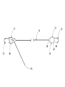
[0001] The present invention is directed to an apparatus and its method of use in delivering surgical tissue connectors into an area of the body and removing the surgical tissue connectors from the body area. Svlore specifically, the present invention is directed to a surgical tissue connector apparatus having at least two tissue connectors connected by a length of cord and a delivery and removai tube. At least one of the tissue connectors has a base with a tapered, beveled or chamfered surface projecting from one end of the base. A hook or other type of tissue connector projects from the opposite side of the base. The hook is positioned on the base where a peripheral side surface of the base shields the hook from unintentionally snagging objects. The base peripheral surface is also dimensioned to slide easily through an interior bore of the tube. This enables the base and the projecting hook to be easily delivered through the tube into an area of the body. The chamfered or tapered surface on the base is positioned to engage with the distal end opening of the tube and direct the base into the center of the tube as the surgical tissue connector is retracted info the tube from the body area in removing the apparatus from the abdominal cavity.


French Abstract

La présente invention concerne un appareil et son procédé d'utilisation pour poser des raccords de tissu chirurgicaux dans une zone du corps et retirer les raccords de tissu chirurgicaux de la zone de corps. De manière plus spécifique, la présente invention concerne un appareil de raccord de tissu chirurgical ayant au moins deux raccords de tissu reliés par une longueur de cordon et un tube de pose et de retrait. Au moins l'un des raccords de tissu a une base ayant une surface conique, biseautée ou chanfreinée faisant saillie à partir d'une extrémité de la base. Un crochet ou un autre type de raccord de tissu fait saillie à paritr du côté opposé de la base. Le crochet est positionné sur la base, sur laquelle une surface latérale périphérique de la base protège le crochet d'objets s'accrochant involontairement. La surface périphérique de la base est également dimensionnée pour coulisser facilement à travers un alésage intérieur du tube. Cela permet à la base et au crochet faisant saillie d'être facilement posés à travers le tube dans une zone du corps. La surface chanfreinée ou conique sur la base est positionnée pour venir en prise avec l'ouverture d'extrémité distale du tube et orienter la base dans le centre du tube lorsque le raccord de tissu chirurgical est rétracté dans le tube à partir de la zone de corps pour retirer l'appareil de la cavité abdominale.

Claims

Note: Claims are shown in the official language in which they were submitted.


The embodiments of the present invention for which an exclusive property or
privilege is claimed are defined as follows:
1. A tissue connector apparatus comprising:
a tissue connector, having a base with a side surface, the side surface
having a center axis that defines mutually perpendicular axial and radial
directions relative to the base, the base having a radius dimension between
the
center axis and the side surface, the base having a proximal end surface and a

distal end surface at axially opposite ends of the side surface;
a hook secured to the base; and
a cord characterized by a first end and a second end, and said cord
second end characterized by a first portion, a second portion and an
intermediate portion, said cord connected to and extending from the base
proximal end surface; and
a releasable one-way cord lock through which the cord second end
passes, said releasable one-way cord lock being operable to lock the cord
second end to the base and thereby adjust the length of the cord extending
from the tissue connector;
wherein the tissue connector has an interior bore, and a pin extending
across the interior bore, and an intermediate portion of the cord is tied in a
knot
around the pin, thereby attaching the cord second end to the tissue connector;

and
the releasable one-way cord lock comprises a lock pin axially slidably
disposed in a first axially extending slot disposed in the base, said lock pin

extending across the interior bore of the base, and a sleeve surrounding the
base side surface and axially slidable between first and second positions
relative to the first base, said sleeve secured to the lock pin, whereby
movement
of the sleeve causes the lock pin to engage against the cord intermediate
portion to prevent the cord intermediate portion from rotating in a direction
corresponding to movement of the cord second end away from the proximal
end surface of the tissue connector.
2. The apparatus of claim 1, wherein the lock pin extends from the sleeve,
- 20 -


through the first axially extending slot to a second axially extending slot,
and projects
through the first and second axially extending slots and is secured to the
sleeve.
3. The apparatus of claim 2, further comprising:
a biasing device disposed within the bore, said biasing device operable
to urge the lock pin toward the intermediate portion.
4. The apparatus of claim 2, further comprising:
a side hole in the base, wherein a portion of the cord extends through
the side hole to the exterior of the tissue connector.
5. The apparatus of claim 1, further comprising:
a biasing device disposed within the bore, said biasing device operable
to urge the lock pin toward the intermediate portion.
6. The apparatus of claim 5, further comprising:
a side hole in the base, wherein a portion of the cord extends through
the side hole to the exterior of the tissue connector.
7. The apparatus of claim 1, further comprising:
a side hole in the base, wherein a portion of the cord extends through
the side hole to the exterior of the tissue connector.
8. The apparatus of claim 1, further comprising:
an additional tissue connector disposed at the first end of the cord.
9. The apparatus of claim 8, wherein the additional tissue connector is
also
characterized by:
an additional base with a side surface, the side surface having a center
axis that defines mutually perpendicular axial and radial directions relative
to
the base, the additional base having a radius dimension between the center
axis and the side surface, the base having a proximal end surface and a distal

end surface at axially opposite ends of the base side surface;

- 21 -


the additional base proximal end surface having a tapered configuration
that tapers as the proximal end surface extends axially away from the
additional
base side surface;
an additional hook secured to the additional base;
wherein the first end is secured to the additional base proximal end
surface.
10. The apparatus of claim 9, further comprising:
a third tissue connector connected to the additional tissue connector
through a second cord.
11. The apparatus of claim 1 wherein the hook comprises:
a shank portion that extends axially from the base distal end surface, the
shank portion being parallel with and radially spaced from the center axis,
the
hook having a bent portion that extends from the shank portion across the
center axis to an end tip of the hook, the hook end tip and the hook shank
portion each being radially spaced on opposite sides of the center axis by
radial
distance dimensions that are smaller than the base radius dimension.
12. The apparatus of claim 1, further comprising:
a tissue connector delivery and removal tube, the tube having an interior
bore with an interior diameter dimension that is larger than a diameter
dimension of the base side surface.
13. The apparatus of claim 1, further comprising:
the base proximal end surface having a conical configuration.
14. The apparatus of claim 1, further comprising:
the length of cord extending from the base proximal end surface being
adjustable in length.
15. The apparatus of claim 1, further comprising:
the base distal end surface having a circular peripheral edge that is

- 22 -


coaxial with the center axis; and the hook bent portion and the hook end tip
do
not extend radially beyond the distal end surface peripheral edge.
16. A tissue connector apparatus comprising:
a tissue connector, having a base with a side surface, the side surface
having a center axis that defines mutually perpendicular axial and radial
directions relative to the base, the base having a radius dimension between
the
center axis and the side surface, the base having a proximal end surface and a

distal end surface at axially opposite ends of the side surface;
a hook secured to the base; and
a cord characterized by a first end and a second end, and said cord
second end characterized by a first portion, a second portion and an
intermediate portion, said cord connected to and extending from the base
proximal end surface; and
a releasable one-way cord lock through which the cord second end
passes, said releasable one-way cord lock being operable to lock the cord
second end to the base and thereby adjust the length of the cord extending
from the tissue connector;
wherein the tissue connector has an interior bore, and a pin extending
across the interior bore, and an intermediate portion of the cord is tied in a
knot
around the pin, thereby attaching the cord second end to the tissue connector;

and
the releasable one-way cord lock comprises a lock pin axially slidably
disposed in a first axially extending slot disposed in the base, said lock pin

extending across the interior bore of the base, and a sleeve surrounding the
base side surface and axially slidable between first and second positions
relative to the first base, said sleeve secured to the lock pin, whereby
movement
of the sleeve causes the lock pin to engage against the cord intermediate
portion to prevent movement of the cord first end away from the proximal end
surface of the tissue connector.
17. The apparatus of claim 16, wherein the lock pin extends from the
sleeve,
through the first axially extending slot to a second axially extending slot,
and projects

- 23 -


through the first and second axially extending slots and is secured to the
sleeve.
18. The apparatus of claim 16, further comprising a biasing device disposed

within the bore, said biasing device operable to urge the lock pin toward the
intermediate portion.
19. The apparatus of claim 17, further comprising a biasing device disposed

within the bore, said biasing device operable to urge the lock pin toward the
intermediate portion.
20. The apparatus of claim 16, further comprising:
a side hole in the base, wherein a portion of the cord extends through
the side hole to the exterior of the tissue connector.
21. The apparatus of claim 17, further comprising:
a side hole in the base, wherein a portion of the cord extends through
the side hole to the exterior of the tissue connector.
22. The apparatus of claim 18, further comprising:
a side hole in the base, wherein a portion of the cord extends through
the side hole to the exterior of the tissue connector.
23. The apparatus of claim 16, further comprising:
an additional tissue connector disposed at the first end of the cord.
24. The apparatus of claim 23, wherein the additional tissue connector is
also characterized by:
an additional base with a side surface, the side surface having a center
axis that defines mutually perpendicular axial and radial directions relative
to
the base, the additional base having a radius dimension between the center
axis and the side surface, the base having a proximal end surface and a distal

end surface at axially opposite ends of the base side surface;
the additional base proximal end surface having a tapered configuration

- 24 -


that tapers as the proximal end surface extends axially away from the
additional
base side surface;
an additional hook secured to the additional base;
wherein the first end is secured to the additional base proximal end
surface.
25. The apparatus of claim 24, further comprising:
a third tissue connector connected to the additional tissue connector
through a second cord.
26. The apparatus of claim 16 wherein the hook comprises:
a shank portion that extends axially from the base distal end surface, the
shank portion being parallel with and radially spaced from the center axis,
the
hook having a bent portion that extends from the shank portion across the
center axis to an end tip of the hook, the hook end tip and the hook shank
portion each being radially spaced on opposite sides of the center axis by
radial
distance dimensions that are smaller than the base radius dimension.
27. The apparatus of claim 16, further comprising:
a tissue connector delivery and removal tube, the tube having an interior
bore with an interior diameter dimension that is larger than a diameter
dimension of the base side surface.
28. The apparatus of claim 16, further comprising:
the base proximal end surface having a conical configuration.
29. The apparatus of claim 16, further comprising:
the length of cord extending from the base proximal end surface being
adjustable in length.
30. The apparatus of claim 16, further comprising:
the base distal end surface having a circular peripheral edge that is
coaxial with the center axis; and

- 25 -


the hook bent portion and the hook end tip do not extend radially beyond
the distal end surface peripheral edge.

- 26 -

Description

Note: Descriptions are shown in the official language in which they were submitted.


CA 02877865 2014-12-23
WO 2014/004654
PCT/US2013/047862
APPARATUS AND METHOD FOR DELIVERING SURGICAL TISSUE
CONNECTORS INTO AN ABDOMINAL CAVITY AND REMOVING THE
SURGICAL TISSUE CONNECTORS FROM THE ABDOMINAL CAVITY
BACKGROUND OF THE INVENTION
FIELD OF THE INVENTION
[0001]The present invention is directed to an apparatus and its method of use
in
delivenng surgical tissue connectors into an area of the body and removing the

surgical tissue connectors from the body area. More specifically, the present
invention is directed to a surgical tissue connector apparatus having at least
two
tissue connectors connected by a length of cord and a delivery and removal
tube. At
least one of the tissue connectors has a base with a tapered, beveled or
chamfered
surface projecting from one end of the base. A hook or other type of tissue
connector projects from the opposite side of the base. The hook is positioned
on the
base where a peripheral side surface of the base shields the hook from
unintentionally snagging objects. The base peripheral surface is also
dimensioned
to slide easily through an interior bore of the tube. This enables the base
and the
projecting hook to be easily delivered through the tube into an area of the
body. The
chamfered or tapered surface on the base is positioned to engage with the
distal end
opening of the tube and direct the base into the center of the tube as the
surgical
tissue connector is retracted into the tube from the body area in removing the

apparatus from the abdominal cavity.
-1 -

CA 02877865 2014-12-23
WO 2014/004654
PCT/US2013/047862
DESCRIPTION OF THE RELATED ART
(0002]In laparoscopic surgery procedures, it is often necessary to pass
surgical
tissue connectors though a cannula, trocar, endosope or other similar device
that is
positioned through an incision in the body to deliver the tissue connectors
into an
area of the body where the connectors are used in a laparoscopic surgery
procedure. A disadvantage associated with the current constructions of
surgical
tissue connectors and the tubular delivery device used to deliver the
connectors into
the body and then remove the connectors from the body is that often the
connectors
will snag or catch a portion of the tubular delivery device being used to
deliver or
retrieve the connectors.
SUMMARY OF THE INVENTION
(0003] The present invention overcomes the above described disadvantages
associated with tissue connectors used in laparoscopic and other surgery
procedures by providing an apparatus that facilitates the delivery of a
surgical tissue
connector through a tubular delivery device and into the body, and the
subsequent
removal of the surgical tissue connector through the tubular device from the
body.
[0004] The basic construction of the apparatus of the invention includes at
least two
surgical tissue connectors. Each tissue connector has a base. The base has a
side
surface that extends around the base, and a distal end surface and proximal
end
surface at the opposite ends of the side surface. In one embodiment of the
tissue
connector, the base side surface is a cylindrical surface having a center axis
that
defines mutually perpendicular axial and radial directions relative to the
base.
- 2 -

CA 02877865 2014-12-23
WO 2014/004654
PCT/US2013/047862
[0009A hook is secured stationary to and projects axially from the base distal
end
surface, The hook has a shank portion that extends axially from the base
distal end
surface. The shank portion is substantially parallel with the base center axis
but
spaced radially from the center axis. The hook also has a bent portion that
extends
from the shank portion across the base distal end surface and through the
center
axis to a distal end tip of the hook. The end tip and the shank are radially
spaced on
opposite sides of the base center axis and the end tip does not project
radially
beyond the base side surface. Thereby, the base side surface shields the end
tip of
the hook from snagging or catching with other objects_
[0006]The base proximal end surface of at least one of the connectors has a
chamfered or tapered configuration that tapers as the proximal end surface
extends
axially away from the base side surface. In one embodiment of the apparatus,
the
base proximal end surface has a conical configuration. The conical
configuration of
the base proximal end surface extends axially from the base side surface to an
apex
that is centered on the base distal end surface
[0007]The apparatus also includes a length of cord that extends between the
proximal end surfaces of the tissue connectors. In one embodiment of the
apparatus, the length of the cord extending between the tissue connectors is
flexible
and can be adjusted.
[0008)The apparatus also includes a tissue connector delivery and removal
tube. In
one embodiment of the apparatus, the tube has a straight length with opposite
proximal and distal ends, and a cylindrical interior bore that extends through
the
length of the tube. The interior bore has an interior diameter dimension that
is
- 3 -

CA 02877865 2014-12-23
WO 2014/004654
PCT/US2013/047862
slightly larger than an exterior diameter dimension of the base cylindrical
side
surface. This enables the tissue connectors to easily slide through the
interior bore
of the tube when delivering the tissue connectors through the tube, or
removing the
tissue connector through the tube. Additionally, the tapered configuration of
the
base proximal end surface of at least one of the tissue connectors enables
that
tissue connector to be easily pulled into the tube at the tube distal end.
Removing
the apparatus by pulling the other tissue connector through the tube distal
end and
then pulling the cord and the tapered tissue connector into the tube causes
the
tapered configuration of the base proximal end surface to first enter the end
of the
tube, whereby the tapered configuration centers the base cylindrical side
surface
relative to the tube interior bore as the tapered proximal end surface is
pulled into the
tube interior bore. With the base side surface centered in the tube interior
bore, the
hook projecting from the base distal end surface is prevented from snagging or

catching the distal end of the tube as the tissue connector is pulled into and
through
the tube.
(0009] The apparatus also includes a rod having a straight length that is
dimensioned
to be inserted into the tube proximal end to push the tissue connectors and
their
connecting cord through the tube interior bore to deliver the tissue
connectors and
the connecting cord from the tube distal end.
[0010]When use of the apparatus in the surgical procedure is complete, the
apparatus can be easily removed from the body by inserting the tube through a
cannula or an incision to position the tube distal end adjacent the connectors
and the
cord, and then inserting a surgical grasper through the tube and grasping the
tissue
- 4 -

CA 02877865 2014-12-23
WO 2014/004654
PCT/US2013/047862
connector without the tapered end surface. The tissue connector is grasped at
its
hook. Retracting the grasper back through the tube will move the grasped
connector
into the tube and then pull the cord into the tube interior bore. As the cord
is pulled
through the tube bore it pulls the tapered proximal end surfaces of the other
tissue
connectors into the bore. The tapered proximal end surface of the connector
will
center the connector and the cylindrical side surface of the connector
relative to the
interior bore as the connector enters the tube distal end and passes through
the
interior bore. The cylindrical side surface of the connector prevents the hook

extending from the distal end surface of the connector from snagging or
catching
against the tube distal end as the connector enters into and passes through
the tube
interior bore.
[0011]As described above, the apparatus of the invention and its method of use

facilitate the delivery of surgical tissue connectors to a surgery site and
facilitate the
removal of the surgical tissue connectors from the surgery site.
DESCRIPTION OF THE DRAWINGS
(0012] Further features of the apparatus of the invention and its method of
use are
set forth in the following detailed description of the apparatus and method
and are
shown in the drawing figures,
[0013] Figures is and lb show an embodiment of the apparatus comprising a pair
of
tissue connectors connected by a cord where one of the tissue connectors
employs
a releasable one-way cord lock that enables adjusting the length of the
portion of the
cord extending between the pair of tissue connectors.
- 5 -

CA 02877865 2019-12-23
WO 2014/004654
PCT/US2013/047862
[0014] Figures 2a and 2b show further embodiments of the apparatus similar to
that
of Figures 1 a and lb.
tool si Figure 3 is a perspective view of one of the tissue connectors.
[0016] Figure 4 is a plan view of the tissue connector of Figure 3.
[0017] Figures 58 and 5b illustrate the assembly of the tissue connector of
Figure 3.
[0018] Figure 6 is a perspective view of a further embodiment of a tissue
connector.
[0019] Figure 7 is a plan view of the tissue connector of Figure 6.
[0020] Figures 8a and 8b illustrate the assembly of the tissue connector of
Figure 6.
[0021] Figure 9 is a perspective view of a further embodiment of a tissue
connector.
[0022] Figure 10 is a side view of the tissue connector of Figure 9.
[0023] Figure 11 is a cross-section view of the tissue connector of Figure 9.
[0024] Figure 12 is a perspective view of the disassembled component parts of
the
tissue connector of Figure 9.
[0025] Figure 13 is a cross-section view of the delivery and removal tube of
the
apparatus.
[0026] Figure 14 is an end view of the proximal end of the delivery and
removal tube.
[0027] Figure 15 is a side view of the delivery rod of the apparatus.
- 6 -

CA 02877865 2014-12-23
WO 2014/004654
PCT/US2013/047862
[0028]Figure 16 is a partial sectioned view of the apparatus illustrating the
method of
removing a tissue connector of the apparatus from an abdominal cavity using
the
delivery and removal tube of the apparatus.
[0029] Figure 17 is a partial sectioned view of the apparatus further
illustrating the
method of removing a tissue connector of the apparatus from an abdominal
cavity
using the delivery and removal tube of the apparatus,
DETAILED DESCRIPTION OF THE PREFERRED EMBODIMENTS
[0030] Figs. lA and 1B show an embodiment of the tissue connector apparatus of

the invention that is constructed to facilitate the delivery of the tissue
connectors
through a cannula or other type of tubular delivery device to a surgical site
of a body
for use in a laparoscopic surgery or other type of surgical procedure, and
facilitate
the removal of the tissue connectors of the apparatus following completion of
the
procedure. As is conventional with laparoscopic apparatus, the component parts
of
the apparatus are dimensioned to be inserted through an incision or through a
cannula, trocar, endoscope or other type of tubular delivery device extending
into the
body to position the apparatus at a surgery site. All of the component parts
of the
apparatus to be described are constructed of biocompatibie materials.
[0031]The first embodiment of the apparatus is shown in Figs. la and lb. The
embodiment shown is basically comprised of a first tissue connector 12, a
second
tissue connector 14 and a length of cord 16 extending between and connecting
the
two tissue connectors. The cord could be a suture, IV tubing or other
equivalent
types of cord and could be one piece or several pieces connected together.
- 7 -

CA 02877865 2014-12-23
WO 2014/004654
PCT/US2013/047862
[0032] Details of the construction of the first tissue connector 12 are shown
in Figs.
3, 4, 5a and 5b The first tissue connector 12 is comprised of a base 18 and a
hook
22. The base 18 for the most part is a solid single piece of material having a
side
exterior surface that extends completely around the base. In the embodiment
shown
in the drawing figures, the side surface 24 is cylindrical. The cylindrical
surface 24
has a center axis 26 that defines mutually perpendicular axial and radial
directions
relative to the base. The base also has a proximal end surface 28 at one axial
end
of the base and a distal end surface 30 at an opposite axial end of the base.
[0033] The proximal end surface 28 is chamfered or tapers as it extends
axially away
from the base side surface 24. In the embodiment shown, the proximal end
surface
28 has a conical configuration with a circular peripheral edge 32 defined by
the
intersection of the proximal end surface with the cylindrical base side
surface 24.
The proximal end surface 28 tapers as it extends axially from the base side
surface
24 to an apex end 34 of the proximal end surface.
[0034]The base distal end surface 30 is substantially flat. The distal end
surface 30
has a circular peripheral edge 36 defined by the intersection of the distal
end surface
with the cylindrical base side surface 24.
[0035] A hook hole 38 is formed in the base distal end surface 30. The hook
hole 38
is spaced radially inwardly from the distal end surface peripheral edge 36 and

radially from the center axis 26. The hook hole extends axially into the base
a short
distance from the distal end surface 30 and does not extend entirely through
the
base. A center hole 42 is also formed in the base at the center of the base
distal end
surface 30. The center hole 42 extends axially through the base and emerges at
the
- 8

CA 02877865 2014-12-23
WO 2014/004654
PCT/US2013/047862
apex 34 of the base proximal end surface 28. The center hole 42 has a slightly

larger interior diameter dimension adjacent the base distal end surface 30
than the
interior diameter dimension of the hole 42 adjacent the apex 34 of the base
proximal
end surface 28.
[0036] The hook 22 has a straight shank portion 44 extending from a proximal
end
46 of the hook. The hook shank portion adjacent the hook proximal end 46 is
dimensioned with a cylindrical exterior surface that fits tightly into the
hook hole 38 in
the base distal end surface 30. The hook shank portion 34 is secured
stationary to
the base by laser welding or other equivalent means. The hook 22 is secured to
the
base distal end surface 32 with the hook shank portion 34 extending axially
from the
distal end surface 32 substantially parallel with the center axis 26, but
radially
spaced from the center axis. The hook shank portion 44 extending from the
distal
end surface 30 has a square or other similar cross-section configuration to
facilitate
grasping the hook with a surgical grasper and preventing the hook shank from
rotating in the grasper. The shank portion 44 extends axially from the base
distal
end surface 30 to a bent portion 48 of the hook that extends radially across
the distal
end surface 30, through the base center axis 26 and to a distal end tip 52 of
the
hook. The distal end tip 52 and shank portion 44 of the hook are each radially

spaced on opposite sides of the base center axis. The shank portion 44 and end
tip
52 are both spaced a radial distance dimension on opposite sides of the center
axis
26 that is smaller than the base radius dimension, whereby the hook bent
portion
and tip do not project radially beyond the cylindrical side surface of the
base.
Although the hook 22 is shown as the part of the tissue connector 12 that is
- 9 -

CA 02877865 2014-12-23
WO 2014/004654
PCT/US2013/047862
removably attachable to body tissue, other equivalent devices that do not
project
radially beyond the side surface of the base could be used.
[0037]in the embodiment of the apparatus shown in Figs. la and lb, the cord 16

has a flexible length with opposite first 54 and second 56 ends, The cord
first end 54
passes through the center hole 42 of the first tissue connector 12 from the
proximal
end surface 28 to the distal end surface 30. A knot 58 is tied in the cord
first end 54.
A medical grade glue. for example cyanoacrylate is applied to the knot. The
cord is
pulled from the base proximal end surface 28 to pull the knot 58 into the
center hole
42 in the base distal end surface 30. The knot 56 is too large to pass though
the
smaller portion of the center hole 42 that emerges from the apex 34 of the
proximal
end surface 28. Thereby, the cord first end 54 is secured to the first tissue
connector
12 with the cord extending from the base proximal end surface 28 of the tissue

connector.
[0038] Figs. 6, 7, 8a and 8b show an alternate embodiment of the first tissue
connector where a post 62 of an eyelet ring 64 is inserted through the base
center
hole 42 from the base proximal end surface 28. The post 62 is secured to the
base
by laser welding the end of the post 62 in the center hole at the base distal
end
surface 32. The eyelet ring 64 could be secured to the base by other
equivalent
means. The first end 54 of the cord 16 is then secured to the eyelet 64 by a
knot
being tied in the cord around the eyelet ring 64 and the knot being secured by
a
medical grade glue.
[0039] Details of the construction of the second tissue connector 14 are shown
in
Figs. 9, 10,11 and 12.
- 10 -

CA 02877865 2014-12-23
WO 2014/004654
PCT/US2013/047862
[0040] The second tissue connector 14 is also basically comprised of a base 68
and
a hook 72 projecting from the base. The base 68 has a side surface 74 that
extends
completely around the base. The in embodiment shown in the drawing figures the

side surface 74 is cylindrical and has a center axis 76 that defines mutually
perpendicular axial and radial directions relative to the base 68. The base 68
also
has a proximal end surface 78 at one axial end of the base and a distal end
surface
80 at the opposite axial end of the base.
[0041] The base proximal end surface 78 is chamfered or tapers as it extends
axially
away from the cylindrical side surface 74. In the embodiment of the second
tissue
connector 68 shown, the base proximal end surface 78 has a conical
configuration
with a circular peripheral edge 82 defined by the intersection of the proximal
end
surface 78 with the cylindrical base side surface 74. The proximal end surface
78
tapers as it extends from the cylindrical side surface 74 to an apex 84 on the

proximal end surface. A hole 86 passes through the center of the apex 84 of
the
proximal end surface 78 to an interior bore of the base that is described
later.
[0042] The base distal end surface 80 is substantially flat. The distal end
surface
80 has a circular peripheral edge 88 defined by the intersection of the distal
end
surface 80 and the cylindrical base side surface 74.
[0043] A hook hole 92 is formed in the base distal end surface 80. The hook
hole 92
is spaced radially inwardly from the distal end surface peripheral edge 88 and

radially from the center axis 76. The hook hole 92 extends axially into the
base a
short distance from the distal end surface 80 and does not extend entirely
through
the base.
-11 -

CA 02877865 2014-12-23
WO 2014/004654
PCT/US2013/047862
[0044] The hook 72 of the second tissue connector 14 has substantially the
same
construction as the hook 22 of the first tissue connector 12. The hook 72 has
a
straight shank portion 94 extending from a proximal end 96 of the hook.
Adjacent
the proximal end 96 the hook shank portion has a cylindrical configuration
dimensioned to fit tightly into the hook hole 88 on the distal end surface 80.
The
hook shank portion 94 is secured to the base distal end surface 80 by laser
welding
or other equivalent means. The hook shank portion 94 extends axially from the
base
distal end surface 80 substantially parallel with the base center axis 76 but
spaced
radially from the center axis. The shank portion 94 extending from the distal
end
surface 80 has a square or other similar cross-section configuration that
facilitates
grasping the shank portion with surgical graspers and preventing rotation of
the
shank portion in the graspers. The hook shank portion 94 extends axially from
the
base to a bent portion 98 of the hook that extends radially over the base
distal end
surface 80 and through the base center axis 76. The hook bent portion 98
extends
from the shank portion 94 to a distal end tip 100 of the hook. The hook distal
end tip
100 and shank portion 94 are both spaced a radial distance dimension on
opposite
sides of the center axis 76 that is smaller than the base radius dimension,
whereby
the hook bent portion and the distal end tip 100 do not project radially
beyond the
cylindrical side surface 74 of the base. Although the hook 72 is shown as the
part of
the tissue connector that is removably attachable to body tissue, other
equivalent
devices that do not project radially beyond the side surface of the base could
be
used. Additionally. although both tissue connectors 12, 14 are described as
having
chamfered or tapered surfaces 28, 78, it is not necessary that both have
tapered
surfaces for the intended functioning of the apparatus.
- 12 -

CA 02877865 2014-12-23
WO 2014/004654
PCT/US2013/047862
[0045]Although the first 12 and second 14 tissue connectors have similar
constructions as discussed above, the second tissue connector 14 is
constructed
with a releasable one-way cord lock that enables the length of the portion of
the cord
16 extending between the two tissue connectors 12, 14 to be adjusted.
[0046]The second tissue connector base 68 has a tubular configuration with a
cylindrical housing wall having the base cylindrical side surface 74 on its
exterior,
and a hollow interior bore 102 extending through the length of the housing
wall
interior.
[0047]A disk shaped cap 104 is secured to a distal end of the base housing
wall by
laser welding or other equivalent means. The disk-shaped cap 104 defines the
distal
end surface 80 of the base.
[0048] A rod or pin 106 is secured to the base 68 in the base interior bore
102. The
rod 106 extends radially across the base interior bore 102 adjacent the
proximal end
surface 78 of the base. Opposite ends of the rod 106 are secured to the
interior
surface of the base cylindrical housing.
[0049]The cord 16 is secured to the second tissue connector 14 inside the base

interior bore 102. The second end 56 of the cord 16 extends through the center
hole
86 in the apex of the base proximal end surface 78 and into the hollow
interior bore
102 of the base. An intermediate portion of the cord 112 is wrapped around the
rod
106 in the base interior, thereby attaching the cord 16 to the second tissue
connector
14. The intermediate portion of the cord 112 is wrapped around the rod 106 by
being tied in a knot around the rod. The preferred Knot 112 is a Munter hitch
knot. A
- 13 -

CA 02877865 2014-12-23
WO 2014/004654
PCT/US2013/047862
first portion 114 of the cord length extends from the intermediate portion 112
though
the base center hole 86. The cord first portion 114 extends from the proximal
end
surface 78 of the base to the cord first end 54 attached to the first tissue
connector
12. A second portion of the cord length 116 extends from the cord intermediate

portion 112 through a side hole 118 in the base cylindrical side surface 74
and the
proximal end surface 78. The side hole 118 is provided to separate the cord
first
portion 114 and the cord second portion 116 to prevent tangling. The side hole
118
also allows for a greater angle that the cord second portion 116 can be pulled
from
the connector. The second portion of the cord 116 extends from second tissue
connector 14 to a second, free end 122 of a cord.
[0050]The releasable one-way cord lock is comprised of a pair slots 124 that
are
formed in radially opposite sides of the cylindrical side wall of the base 68.
As seen
in the drawing figures, the slots 124 extend axially along a portion of the
cylindrical
wall of the base 68 and are positioned radially to one side of the base center
axis 76.
A lock pin 126 is positioned in the slots 124 and extends from one slot 124
across
the interior bore of the base 68 to the other slot. The lock pin 126 slides
axially along
the slots 124.
[0051]An actuator 128 is mounted on the base cylindrical side surface 74 and
is
connected to the lock pin 126 inside the base. The actuator 128 is formed as
cylindrical sleeve that surrounds the base cylindrical side surface 74. The
actuator
128 is mounted on the cylindrical side surface 74 of the base 68 for axially
reciprocating movements between first and second positions of the actuator 128

relative to the base 68. The opposite ends of the lock pin 126 project beyond
the
- 14 -

CA 02877865 2014-12-23
WO 2014/004654
PCT/US2013/047862
slots 124 in the base cylindrical side surface 74 and are secured in opposite
sides of
the actuator 128. Moving the actuator 128 to its first position shown in Fig.
11 causes
the lock pin 126 to engage against the cord intermediate portion 112 that is
wrapped
around the rod 106. In Fig. 11, the pin 126 prevents the knot 112 from
rotating
counterclockwise around the rod 106 when the knot 112 is in a 3 o'clock
position, or
to the right of the rod as shown in Fig. 11 and the cord first portion 114 is
pulled.
The engagement of the lock pin 126 against the cord intermediate portion or
knot
112 locks the cord and prevents the first portion of the cord length 114 from
being
pulled from the proximal end surface 78 of the second tissue connector 14.
However, in the position of the knot 112 shown in Fig. 11 the cord second
portion
116 can be pulled from the second tissue connector 14 to shorten the length of
the
cord first portion 114, but the cord first portion 114 cannot be pulled from
the
connector. Moving the actuator 128 to its second position causes the lock pin
126 to
disengage from the cord intermediate portion or knot 112. With the pin 126
moved
upward, the cord first portion 114 can be pulled to move the knot 112
counterclockwise from the 3 o'clock position to a 6 o'clock position as viewed
in Fig.
11. This allows the length of the cord first portion 114 extending between the
second
tissue connector 14 and the first tissue connector 12 to be pulled from the
second
tissue connector 14 to adjust or increase the length of the cord first portion
114.
With the pin 126 moved upward the cord second portion 116 can also be pulled
from
the connector.
- 15 -

[0052]A biasing device 132 urges the lock pin 126 to its first position shown
in Fig.
11. In the drawing figures the biasing device 132 is a coiled spring. Other
equivalent types of biasing devices could be employed.
[00531The slot 124 and the pin 126 are off center relative to the base
cylindrical side
surface 74 to facilitate the knot 112 pushing the pin 126 upward against the
bias of
the spring 132 when the cord second portion 116 is pulled and the knot 112
moves
clockwise from the 6 o'clock position to the 3 o'clock position relative to
the rod 106_
[00543 Tissue connectors having one way cord locks such as that described
above
are disclosed in the currently pending international patent application No.
PC-M.352011/01494, which is assigned to the assignee of this invention.
[00553 Fig. 2a shows a further embodiment of the apparatus of the invention
comprising the first tissue connector 12 and the second tissue connector 14
connected by the length of cord 16. However, the first end of the length of
cord 16 is
connected to the first tissue connector 12 having the eyelet ring 64 that was
described earlier. In addition, a second length of cord 134 connects a third
tissue
connector 136 to the first tissue connector 12. The construction of the third
tissue
connector 136 is substantially the same as that of the second tissue connector
14
described earlier. In the embodiment of Fig. 2A, at least two of the three
tissue
connectors would require tapered proximal end surfaces in order for the
apparatus to
function as intending in removing the apparatus from a surgical site as
described
below.
- 16 -
CA 2877865 2019-10-07

CA 02877865 2014-12-23
WO 2014/004654
PCT/US2013/047862
[0056] Fig. 28 shows a still further embodiment of the apparatus where the
tissue
connector hook 72 or other equivalent device is not directly connected to the
base 68
of the second tissue connector 14, but is connected by a further length of
suture 138.
[0057] The apparatus of the invention also comprises a tissue connector
deliver and
removal tube 142 shown in Figs. 13 and 14, and a delivery rod 144 shown in
Fig. 15.
[0068] In one embodiment of the apparatus, the tube 142 has a straight length
with
opposite proximal 146 and distal 148 ends. An interior bore having a smooth
cylindrical interior surface 152 extends through the length of the tube. The
cylindrical
interior surface 152 has an interior diameter dimension that is just slightly
larger than
the exterior diameter dimension of the first tissue connector base cylindrical
side
surface 24 and the second tissue connector base cylindrical side surface 74.
This
enables the tissue connectors 12, 14 to easily slide through the interior bore
of the
tube when delivering the tissue connectors through the tube, or removing the
tissue
connectors through the tube.
[0059] The delivery rod 144 has a straight length that is dimensioned to be
inserted
into the tube proximal end 146 to push the tissue connectors 12, 14 and the
connecting cord 16 through the tube interior bore to deiiver the tissue
connectors
and the connecting cord from the tube distal end 142 and to a surgical site in
a body.
[0060] Additionally; the tapered configuration of the base proximal end
surface 28 of
the first tissue connector 12 and the tapered configuration of the base
proximal end
surface 78 of the second tissue connector 14 enables the tissue connectors to
be
easily pulled into the interior bore of the tube 42 at the tube distal end
148.
- 17 -

CA 02877865 2014-12-23
WO 2014/004654
PCT/US2013/047862
However, it is only necessary that one tissue connector 12 have a tapered
proximal
end surface in a two connector embodiment of the apparatus such as that shown
in
Figs. 1A and 1B. When it is desired to remove the tissue connectors 12, 14
from a
surgical site in an area of the body, the tube distal end 142 is positioned in
the area
by inserting the distal end through a cannula or through an incision to
position the
tube distal end 148 at the surgical site. An instrument, for example a
surgical
grasper is then inserted through the tube 142 until it emerges from the tube
distal
end 148. The surgical grasper then grasps the second tissue connector 14 at
the
hook 72. The surgical grasper is then removed through the tube 142 moving the
second connector 14 hook first into the tube distal end and pulling the cord
into the
tube distal end 142. The cord 16 is then continued to be pulled through the
tube until
the tapered configuration of the base proximal end surface 28 of the first
tissue
connector 12 is pulled into the tube distal end 148. The tapered configuration
of the
proximal end surface 28 centers the base cylindrical side surface 24 of the
first
tissue connector 12 relative the tube interior bore as the tapered proximal
end
surface is pulled into the tube interior bore. With the base cylindrical side
surface 24
centered in the interior bore of the tube 142, the hook 22 projecting from the
base
distal end surface 30 is prevented from snagging or catching the distal end
148 of
the tube 142 as the first tissue connector is pulled into an through the tube.
[0061]Thus, as described above, the apparatus of the invention and its method
of
use facilitate the delivery of surgical tissue connectors to a laparoscopic
surgical site
and facilitate the removal of the surgical connectors from the surgical site.
- 18 -

CA 02877865 2014-12-23
WO 2014/004654
PCT/US2013/047862
[0062]AS various modifications could be made in the apparatus herein described

and illustrated and its method of use without departing from the scope of the
invention, it is intended that all the matter contained in the foregoing
description or
shown in the accompanying drawings shall be interpreted as illustrative rather
than
limiting. Thus, the breath and scope of the present invention should not be
limited by
any of the above described exemplary embodiments, but should be defined only
in
accordance with the following claims appended hereto and their equivalents.
- 19 -

Representative Drawing
A single figure which represents the drawing illustrating the invention.
Administrative Status

For a clearer understanding of the status of the application/patent presented on this page, the site Disclaimer , as well as the definitions for Patent , Administrative Status , Maintenance Fee  and Payment History  should be consulted.

Administrative Status

Title Date
Forecasted Issue Date 2020-09-01
(86) PCT Filing Date 2013-06-26
(87) PCT Publication Date 2014-01-03
(85) National Entry 2014-12-23
Examination Requested 2018-06-26
(45) Issued 2020-09-01

Abandonment History

There is no abandonment history.

Maintenance Fee

Last Payment of $263.14 was received on 2023-06-16


 Upcoming maintenance fee amounts

Description Date Amount
Next Payment if small entity fee 2024-06-26 $125.00
Next Payment if standard fee 2024-06-26 $347.00

Note : If the full payment has not been received on or before the date indicated, a further fee may be required which may be one of the following

  • the reinstatement fee;
  • the late payment fee; or
  • additional fee to reverse deemed expiry.

Patent fees are adjusted on the 1st of January every year. The amounts above are the current amounts if received by December 31 of the current year.
Please refer to the CIPO Patent Fees web page to see all current fee amounts.

Payment History

Fee Type Anniversary Year Due Date Amount Paid Paid Date
Application Fee $200.00 2014-12-23
Maintenance Fee - Application - New Act 2 2015-06-26 $100.00 2015-06-02
Maintenance Fee - Application - New Act 3 2016-06-27 $100.00 2016-06-03
Maintenance Fee - Application - New Act 4 2017-06-27 $50.00 2017-06-07
Maintenance Fee - Application - New Act 5 2018-06-26 $100.00 2018-06-15
Request for Examination $400.00 2018-06-26
Maintenance Fee - Application - New Act 6 2019-06-26 $100.00 2019-06-10
Registration of a document - section 124 2020-03-04 $100.00 2020-03-04
Maintenance Fee - Application - New Act 7 2020-06-26 $100.00 2020-06-19
Final Fee 2020-06-29 $150.00 2020-06-24
Maintenance Fee - Patent - New Act 8 2021-06-28 $204.00 2021-06-18
Maintenance Fee - Patent - New Act 9 2022-06-27 $203.59 2022-06-17
Maintenance Fee - Patent - New Act 10 2023-06-27 $263.14 2023-06-16
Owners on Record

Note: Records showing the ownership history in alphabetical order.

Current Owners on Record
FREEHOLD SURGICAL, LLC
Past Owners on Record
FREEHOLD SURGICAL, INC.
SHERMAN, DARREN R.
SMITH, JEFFREY
Past Owners that do not appear in the "Owners on Record" listing will appear in other documentation within the application.
Documents

To view selected files, please enter reCAPTCHA code :


To view images, click a link in the Document Description column. To download the documents, select one or more checkboxes in the first column and then click the "Download Selected in PDF format (Zip Archive)" or the "Download Selected as Single PDF" button.

List of published and non-published patent-specific documents on the CPD .

If you have any difficulty accessing content, you can call the Client Service Centre at 1-866-997-1936 or send them an e-mail at CIPO Client Service Centre.


Document
Description 
Date
(yyyy-mm-dd) 
Number of pages   Size of Image (KB) 
Final Fee 2020-06-24 4 107
Representative Drawing 2020-08-06 1 2
Cover Page 2020-08-06 1 46
Recordal Fee/Documents Missing 2021-03-17 2 241
Patent Correction Requested 2021-03-19 16 477
Correction Request Denied 2021-05-07 2 241
Patent Correction Requested 2021-12-14 5 115
Correction Certificate 2022-04-29 2 280
Abstract 2014-12-23 2 73
Claims 2014-12-23 4 125
Drawings 2014-12-23 21 143
Description 2014-12-23 19 1,370
Representative Drawing 2014-12-23 1 4
Cover Page 2015-02-20 2 50
Request for Examination 2018-06-26 2 64
Examiner Requisition 2019-04-05 4 225
Amendment 2019-10-07 12 420
Description 2019-10-07 19 1,284
Claims 2019-10-07 7 231
PCT 2014-12-23 24 1,033
Assignment 2014-12-23 4 119