Note: Descriptions are shown in the official language in which they were submitted.
CA 02877902 2014-12-24
WO 2014/000085
PCT/CA2013/000592
PATHOLOGY SLIDE SCANNERS FOR FLUORESCENCE AND BRIGHTFIELD
IMAGING AND METHOD OF OPERATION
BACKGROUND OF THE INVENTION
FIELD OF THE INVENTION
[0001] This invention relates to the fields of microscopic imaging of large
specimens
with particular emphasis on brightfield and fluorescence imaging, including
photoluminescence and spectrally-resolved fluorescence. Applications include
imaging
tissue specimens, genetic microarrays, protein arrays, tissue arrays, cells
and cell
populations, biochips, arrays of biomolecules, detection of nanoparticles,
photoluminescence imaging of semiconductor materials and devices, and many
others.
DESCRIPTION OF THE PRIOR ART
[0002] The macroscope originally described in US Patent No. 5,381,224 is a
scanning-
laser system that uses a telecentric laser-scan lens to provide a wide field
of view.
Several embodiments are presently in use. These include instruments for
fluorescence
and photoluminescence (including spectrally-resolved) imaging (several other
contrast
mechanisms are also possible), instruments in which a raster scan is provided
by the
combination of a scanning mirror and a scanning specimen stage, instruments in
which
the specimen stage is stationary and the raster scan is provided by two
scanning mirrors
rotating about perpendicular axes, confocal and non-confocal versions, and
other
embodiments. A macroscope with fine focus adjustment was described in US
Patent
No. 7,218,446, and versions for reflected-light, fluorescence,
photoluminescence, multi-
photon fluorescence, transmitted-light, and brightfield imaging were
described. The
combination of a scanning laser macroscope with a scanning laser microscope to
provide an imaging system with a wide field of view and the high resolution
capability of
a microscope was described in US Patent No. 5,532,873.
[0003] When the macroscope is used for fluorescence imaging, it has several
advantages. Exposure for each fiuorophore can be adjusted separately without
changing scan speed by changing either laser intensity and/or detector gain
(in the case
of a detector comprised of a photomultiplier tube (pmt) followed by a
preamplifier, both
the pmt voltage (which changes pmt gain) and preamplifier gain can be
changed). The
ability to adjust the detection gain for each fluorophore separately allows
the instrument
CA 02877902 2014-12-24
WO 2014/000085
PCT/CA2013/000592
to simultaneously collect multiple fluorophore images that are all correctly
exposed. In
addition, the appropriate laser wavelength can be provided to excite a chosen
fluorophore, and excitation wavelengths can be chosen so they do not overlap
detection
wavelength ranges.
[0004] Several other technologies are used for imaging large specimens at high
resolution. With tiling microscopes, the image of a small area of the specimen
is
recorded with a digital camera (usually a CCD camera), the specimen is moved
with a
computer-controlled microscope stage to image an adjacent area, an image of
the
adjacent area is recorded, the stage is moved again to the next area, and so
on until a
number of image tiles have been recorded that together cover the whole area of
the
specimen. Images of each area (image tiles) are recorded when the stage is
stationary,
after waiting long enough for vibrations from the moving stage to dissipate,
and using an
exposure time that is sufficient to record the fluorescence images. These
image tiles
can be butted together, or overlapped and stitched using computer stitching
algorithms,
to form one image of the entire specimen. Such images may contain tiling
artifacts,
caused by focus changes between adjacent tiles, differences in illumination
intensity
across the field of view of the microscope, barrel or pincushion distortion
near the edge
of the tiles, and microscope objectives that do not have a flat focal plane.
For large
specimens, thousands of tiles may be required to image the entire specimen,
increasing
the chance of tiling artifacts. Tiling microscopes are very slow for
fluorescence imaging.
[0005] When tiling microscopes are used for fluorescence imaging, the areas
surrounding each tile and the overlapping edges of adjacent tiles are exposed
twice (and
the corners four times) which can bleach some fluorophores. Exposure is
adjusted by
changing the exposure time for each tile. If multiple fluorophores are imaged,
a different
exposure time is required for each, so each fluorophore requires a separate
image at
each tile position. Multiple exposure of the specimen for imaging multiple
fluorophores
can also increase bleaching. After all tiles have been collected, considerable
effort (both
human and computer) is required to stitch the tiles together and correct each
tile for
illumination intensity and collection sensitivity changes across the field of
view of the
microscope (correction for variations in illumination intensity and collection
sensitivity is
sometimes called "field flattening"). Stitching tiles together is also
complicated by
distortion and curvature of field of the microscope objective, which occur
near the edges
of the field of view (just where stitching of tiles occurs).
[0006] Strip scanning instruments are also used for imaging large specimens.
In these
instruments infinity-corrected microscope optics are used, with a high
Numerical
Aperture (high NA) microscope objective and a tube lens of the appropriate
focal length
2
,
CA 02877902 2014-12-24
WO 2014/000085
PCT/CA2013/000592
to focus an image of the specimen directly on a CCD or CMOS linear array
sensor or
TDI sensor with the correct magnification to match the resolution of the
microscope
objective with the detector pixel size for maximum magnification in the
digitized image
{as described in "Choosing Objective Lenses: The Importance of Numerical
Aperture
and Magnification in Digital Optical Microscopy", David W. Piston, Biol. Bull.
195, 1-4
(1998)). A linear CCD detector array with 1000 or 2000 pixels is often used,
and three
separate linear detectors with appropriate filters to pass red, green and blue
light are
used for RGB brightfield imaging. The sample is moved at constant speed in the
direction perpendicular to the long dimension of the linear detector array to
scan a
narrow strip across a microscope slide. The entire slide can be imaged by
imaging
repeated strips and butting them together to create the final image. Another
version of
this technology uses linear TDI (Time Delay Integration) array sensors which
increase
both sensitivity and imaging speed. In both of these instruments, exposure is
varied by
changing illumination intensity and/or scan speed.
[0007] Such a microscope is shown in Fig. 1 (Prior Art). A tissue specimen 100
(or
other specimen to be imaged) mounted on microscope slide 101 is illuminated
from
below by illumination source 110. Light passing through the specimen is
collected by
infinity-corrected microscope objective 115, which is focused on the specimen
by piezo
positioner 120. The microscope objective 115 and tube lens 125 form a real
image of
the specimen on linear detector array 130. An image of the specimen is
collected by
moving the microscope slide at constant speed using motorized stage 105 in a
direction
perpendicular to the long dimension of the detector array 130, combining a
sequence of
equally-spaced line images from the array to construct an image of one strip
across the
specimen. Strips are then assembled to form a complete image of the specimen.
[0008] For brightfield imaging, most strip-scanning instruments illuminate the
specimen
from below, and detect the image in transmission using a sensor placed above
the
specimen. In brightfield, signal strength is high, and red, green and blue
channels are
often detected simultaneously with separate linear detector arrays to produce
a colour
image.
[0009] Compared to brightfield imaging, fluorescence signals can be thousands
of times
weaker, and some fluorophores have much weaker emission than others.
Fluorescence
microscopy is usually performed using illumination from the same side as
detection
(epifluorescence) so that the bright illumination light passing through the
specimen does
not enter the detector. In strip-scanning instruments, exposure is varied by
changing
scan speed, so present strip-scanning instruments scan each fluorophore
separately,
reducing the scan speed when greater exposure is required for a weak
fluorophore.
3
CA 02877902 2014-12-24
WO 2014/000085 PCT/CA2013/000592
Varying exposure by changing scan speed makes it difficult to design a strip-
scanner for
simultaneous imaging of multiple fluorophores, where each channel would have
the
same exposure time, and present strip-scanners scan one fluorophore at-a-time.
In
addition, in fluorescence microscopy, relative intensity measurements are
sometimes
important for quantitative measurement, and 12 or 16 bit dynamic range may be
required. For present strip scanners, this would require larger dynamic range
detectors
and slower scan speeds.
[0010] Before scanning a large specimen in fluorescence, it is important to
set the
exposure time (in a tiling or strip-scanning microscope) or the combination of
laser
intensity, detector gain and scan speed (in a scanning laser macroscope or
microscope)
so that the final image will be properly exposed ¨ in general it should not
contain
saturated pixels, but the gain should be high enough that the full dynamic
range will be
used for each fluorophore in the final image. Two problems must be solved to
achieve
this result ¨ the exposure must be estimated in advance for each fluorophore
and for
simultaneous detection of multiple fluorophores, the exposure time must be
adjusted
separately for each detection channel before scanning. For strip-scanning
instruments,
estimating the exposure in advance is difficult without scanning the whole
specimen first
to check exposure, and this must be done for each fluorophore. Instead of
scanning first
to set exposure, many operators simply set the scan speed to underexpose
slightly, with
resulting noisy images, or possibly images with some overexposed (saturated)
areas if
the estimated exposure was not correct. For macroscope-based instruments, a
high-
speed preview scan can be used to set detection gain in each channel before
final
simultaneous imaging of multiple fluorophores (see W02009/137935, "Imaging
System
with Dynamic Range Maximization").
[0011] A prior art scanning microscope for fluorescence imaging is shown in
Fig. 2. A
tissue specimen 100 (or other specimen to be imaged) mounted on microscope
slide
101 is illuminated from above by illumination source 200. In fluorescence
imaging the
illumination source is usually mounted above the specimen (epifluorescence) so
that the
intense illumination light that passes through the specimen is not mixed with
the weaker
fluorescence emission from the specimen, as it would be if the illumination
source were
below the specimen. Several different optical combinations can be used for
epifluorescence illumination ¨ including illumination light that is injected
into the
microscope tube between the microscope objective and the tube lens, using a
dichroic
beamsplitter to reflect it down through the microscope objective and onto the
specimen.
In addition, a narrow wavelength band for the illumination light is chosen to
match the
absorption peak of the fluorophore in use. Fluorescence emitted by the
specimen is
4
CA 02877902 2014-12-24
WO 2014/000085 PCT/CA2013/000592
collected by the infinity-corrected microscope objective 115 which is focused
on the
specimen by the piezo positioner 120. Emission filter 205 is chosen to reject
light at the
illumination wavelength and to pass the emission band of the fluorophore in
use. The
microscope objective 115 and the tube lens 125 form a real image of the
specimen on
TDI detector array 210. An image of the specimen is collected by moving the
microscope slide at constant speed using the motorized stage 105 in a
direction
perpendicular to the long dimension of the detector array 210, combining a
sequence of
equally-spaced, time-integrated line images from the array to construct an
image of one
strip across the specimen. Strips are then assembled to form a complete image
of the
specimen. When a CCD-based TDI array is used, each line image stored in memory
is
the result of integrating the charge generated in all of the previous lines of
the array
while the scan proceeds, and thus has both increased signal/noise and
amplitude (due
to increased exposure time) when compared to the result from a linear array
detector.
Exposure is also increased by reducing scan speed, so the scan time (and thus
image
acquisition time) is increased when using weak fluorophores. In addition, it
is difficult to
predict the best exposure time before scanning. When multiple fluorophores are
used
on the same specimen, the usual imaging method is to choose illumination
wavelengths
to match one fluorophore, select the appropriate emission filter and scan time
(speed)
for the chosen fluorophore, and scan one strip in the image. Then, the
illumination
wavelength band is adjusted to match the absorption band of the second
fluorophore, a
matching emission filter and scan speed are chosen, and that strip is scanned
again.
Additional fluorophores require the same steps to be repeated. Finally, this
is repeated
for all strips in the final image. Some instruments use multiple TDI detector
arrays to
expose and scan multiple fluorophores simultaneously, but this usually results
in a final
image where one fluorophore is exposed correctly and the others are either
under- or
over-exposed. Exposure can be adjusted by changing the relative intensity of
the
excitation illumination for each fluorophore, which should be easy to do if
LED
illumination is used. When multiple illumination bands are used at the same
time, the
resulting image for each fluorophore may differ from that produced when only
one
illumination band is used at a time because of overlap of the fluorophore
emission
bands, and because autofluorescence from the tissue itself may be excited by
one of the
illumination bands. Autofluorescence emission usually covers a wide spectrum
and may
cause a bright background in all of the images when multiple fluorophores are
illuminated and imaged simultaneously.
5
CA 02877902 2014-12-24
WO 2014/000085 PCT/CA2013/000592
[0012] A good description of strip scanning instruments, using either linear
arrays or TDI
arrays, is given in US Patent Application Publication No. US2009/0141126
("Fully
Automatic Rapid Microscope Slide Scanner", by Dirk Soenksen).
[0013] Linear arrays work well for brightfield imaging, but the user is often
required to
perform a focus measurement at several places on the specimen before scanning,
or a
separate detector is used for automatic focus. Linear arrays are not often
used for
fluorescence imaging because exposure time is inversely proportional to scan
speed,
which makes the scan time very long for weak fluorophores. In addition,
exposure (scan
speed) must be adjusted for each fluorophore, making simultaneous measurement
of
multiple fluorophores difficult when they have widely different fluorescence
intensity
(which is common).
[0014] TDI arrays and associated electronics are expensive, but the on-chip
integration
of several exposures of the same line on the specimen provides the increased
exposure
time required for fluorescence imaging while maintaining a reasonable scan
speed.
Simultaneous imaging of multiple fluorophores using multiple TDI detector
arrays is still
very difficult however, since each of the detectors has the same integration
time (set by
the scan speed), so it is common to use only one TDI array, adjusting exposure
for each
fluorophore by changing the scan speed and collecting a separate image for
each
fluorophore. Focus is set before scanning at several positions on the
specimen, or
automatic focus is achieved using a separate detector or focus measuring
device.
[0015] Single-chip colour cameras (including those often used on ordinary
optical
microscopes to record an image of the specimen area seen in the field of view
of the
microscope) often use a mosaic Colour Filter Array (CFA) on the photosensors
of an
area detector that enables a single-chip camera to record a colour image. The
most
common CFA is the Bayer filter, named after the inventor, which arranges red,
green
and blue filters in a square grid (usually RGG13) of photosensors on the array
(see US
Patent No. 3,971,065). Bayer chose to use two green pixels in each set as
luminance
elements to match the sensitivity of the human eye. Data from each pixel only
records
one colour, so in order to obtain a full-colour image, the red, green and blue
values for
each pixel are calculated by interpolation using information from the
surrounding pixels.
This is called demosaicing, and several different demosaicing algorithms have
been
developed. Demosaicing may happen inside the camera (producing jpeg or tiff
files), or
outside using raw data from the sensor. Because of the computing power
required for
demosiacing, and the possibility of false colours and moire, most tiling
microscopes use
separate array detectors to detect red, green and blue. Cameras for digital
photography
often use an optical low-pass filter in front of the detector array to reduce
moire and false
6
CA 02877902 2014-12-24
WO 2014/000085 PCT/CA2013/000592
colour caused by use of a Bayer filter, resulting in some loss of resolution.
Other mosaic
Colour Filter Arrays have been proposed, including one using a white
(transparent) filter
in place of one of the green filters in the Bayer array. White (transparent)
pixels accept
all wavelengths of light in the visible spectrum (they are panchromatic), and
provide one
bright pixel in each array of four pixels, increasing the sensitivity of the
array. Other
combinations of filters in the square mosaic grid include CYGM (cyan, yellow,
green,
magenta) and RGBE (red, green, blue, emerald). All require demosaicing.
DEFINITIONS AND OBJECTS OF THE INVENTION
[0016] For the purposes of this patent document, a "macroscopic specimen" (or
large
microscope specimen") is defined as one that is larger than the field of view
of a
compound optical microscope containing a microscope objective that has the
same
Numerical Aperture (NA) as that of the scanner described in this document.
[0017] For the purposes of this patent document, TD1 or Time Delay and
Integration is
defined as the method and detectors used for scanning moving objects, usually
consisting of a CCD-based detector array in which charge is transferred from
one row of
pixels in the detector array to the next in synchronism with the motion of the
real image
of the moving object. As the object moves, charge builds up and the result is
charge
integration just as if a longer exposure was used in a stationary imaging
situation. When
the image (and integrated charge) reaches the last row of the array, that line
of pixels is
read out. In operation the last line of the moving image is read out
continuously, one
row of pixels at a time. One example of such a camera is the DALSA Piranha TDI
camera.
[0018] For the purposes of this patent document the term "image acquisition"
includes
all of the steps necessary to acquire and produce the final image of the
specimen,
including some of but not limited to the following: the steps of preview
scanning,
instrument focus, predicting and setting gain for imaging each fluorophore,
image
adjustments including scan linearity adjustment, field flattening
(compensating for
fluorescence intensity variation caused by excitation intensity and detection
sensitivity
changes across the field of view), correction of fluorescence signal in one
channel
caused by overlap of fluorescence from adjacent (in wavelength) channels when
two or
more fluorophores are excited simultaneously, dynamic range adjustment,
butting or
stitching together adjacent image strips (when necessary), storing,
transmitting and
viewing the final image.
[0019] For the purposes of this patent document, the term "image processing"
means all
of the steps required to process the data to prepare the final image file,
including some
7
CA 02877902 2014-12-24
WO 2014/000085 PCT/CA2013/000592
of but not limited to the following: the steps of scan linearity adjustment,
field flattening,
correction for crosstalk when simultaneously scanning multiple fluorophores,
correcting
fluorescence image data by subtracting fluorescence originating from the glass
of the
microscope slide, subtracting the dark-current noise floor from the
detector, and
contracting the dynamic range of the image data to match the (smaller) dynamic
range
of the final image.
[0020] "Proper exposure" is defined as a gain setting such that in the output
image file
no (or only a small number of) pixels are saturated, and the dynamic range of
the image
data matches the dynamic range of the output image file (8 bits for an 8 bit
file, 12 bits
for a 12 bit file, etc.) and includes substantially the entire range of pixel
amplitudes from
the noise floor to the brightest pixel. The output image file may have a
smaller dynamic
range than that of the detection system, and that of an intermediate image
file that is
collected during scanning. W02009/137935 describes two methods of maximizing
the
dynamic range of data stored in the output image file ¨ (1) accurately
estimating the gain
required to maximize the dynamic range of each detection channel when the
dynamic
range of the detection channel and the dynamic range of the output image data
file are
the same, and (2) using a dynamic range in the detection channel that is
larger than that
required in the final image data file and contracting the acquired data to
utilize
substantially the entire dynamic range of the final image data file.
[0021] For the purposes of this patent document, the term "sparse image" means
an
image in which only pixels in a sparse grid exist in the image ¨ e.g. one
pixel at the
centre of a square area of the image that would normally contain 100 or more
pixels.
The pixel values (intensities) are the same as they would be in the complete
image, and
do not reflect in any way the values of the pixels that were discarded (or not
measured)
to produce the sparse image.
[0022] For the purposes of this patent document, a "frame grabber" is any
electronic
device that captures individual, digital still frames from an analog video
signal or a digital
video stream or digital camera. It is often employed as a component of a
computer
vision system, in which video frames are captured in digital form and then
displayed,
stored or transmitted in raw or compressed digital form. This definition
includes direct
camera connections via USB, Ethernet, IEEE 1394 ("FireWire") and other
interfaces that
are now practical.
OBJECTS OF THE INVENTION:
8
CA 02877902 2014-12-24
WO 2014/000085 PCT/CA2013/000592
[0023] It is an object of this invention to provide a method of using a CCD or
CMOS or
other technology two-dimensional sensor array for imaging moving objects
instead of
using linear array or TDI (time delay and integration) line scan technology.
[0024] It is an object of this invention to provide an instrument and method
of scanning
large microscope specimens on a moving microscope stage using one or more CCD
or
CMOS or other technology two-dimensional sensor arrays in place of linear
arrays or
TDI arrays.
[0025] It is an object of this invention to provide an imaging system for
large microscope
specimens using one or more CCD or CMOS or other technology two-dimensional
sensor arrays whereby noise in the image is reduced by adding together a
sequence of
overlapping images on a line-by-line basis, whereby each line of the final
image is the
result of adding several exposures of the same line, thus increasing the
exposure time
for that line in the image.
[0026] (Each line in the final image is the result of adding several exposures
of the same
line and then dividing by the number of exposures, or adding the data from
each
exposure to a data set with a larger dynamic range, e.g. one could add 256
images from
an 8-bit detector into a 16-bit image store). (Then, dynamic-range contraction
can be
used on each fluorophore image to fill the dynamic range required in the
output file for
each fluorophore, as described in W02009/137935).
[0027] It is an object of this invention to provide a method of scanning large
microscope
specimens on a moving microscope stage using one or more CCD or CMOS or other
technology two-dimensional sensor arrays in place of linear arrays or TDI
arrays that
allows simultaneous imaging of multiple fluorophores, even where there is a
large
difference in the signal strength of the different fluorophores. {For example,
consider an
8-bit sensor array (or an array in which the 8 most-significant bits are
commonly read
out) and a 16-bit image store for each fluorescence detection channel. Up to
256 8-bit
measurements can be added to each pixel in the 16-bit image store, and, if
desired, the
resulting 16-bit image can be contracted back to 8 bits, using the contraction
methods
described in W02009/137935. Contraction can be different for each fluorescence
channel so that the resulting 8-bit image from each channel fills the 8 bit
dynamic range
commonly available for viewing each colour.)
[0028] It is an object of this invention to provide a fluorescence imaging
system for large
microscope specimens using CCD or CMOS or other technology two-dimensional
sensor arrays in place of linear arrays or TDI arrays whereby the dynamic
range of the
instrument is larger than the dynamic range of the detector. (e.g. using an 8-
bit detector,
9
CA 02877902 2014-12-24
WO 2014/000085 PCT/CA2013/000592
adding together 256 8-bit images results in a final image with a dynamic range
of 16
bits)
[0029] It is an object of this invention to provide a fluorescence imaging
system for
detecting multiple fluorophores in large microscope specimens using CCD or
CMOS or
other technology two-dimensional sensor arrays in place of linear arrays or
TDI arrays
whereby the dynamic range of the acquired data in each of the separate
fluorescence
images (one from each fluorophore) can be contracted to fill (or substantially
fill) the
entire dynamic range of the output image data file for each fluorophore. (See
W02009/137935 for examples of image data dynamic range contraction.)
[0030] It is an object of this invention using CCD or CMOS or other technology
two-
dimensional sensor arrays in place of linear arrays or TDI arrays to provide a
method of
acquiring fluorescence images in which the image data from each fluorophore
substantially fills the dynamic range available in the final image file, by
estimating the
gain required to maximize the dynamic range for each fluorophore in a
fluorescence
image before scanning, using detection channels that have larger dynamic range
than
that required in the final image, and contracting the dynamic range of the
acquired data
to fill substantially the entire dynamic range of the output image data file
for each
fluorophore.
[0031] It is an object of this invention using CCD or CMOS or other technology
two-
dimensional sensor arrays in place of linear arrays or TDI arrays to provide a
fluorescence imaging system for macroscopic specimens in which the correct
gain
setting for fluorescence imaging can be estimated from a preview scan of the
entire
specimen (or part of the specimen) before the final scan is started. (For
example, using
a sparse pixel image from a high-speed preview scan)
[0032] It is an object of this invention using CCD or CMOS or other technology
two-
dimensional sensor arrays in place of linear arrays or TDI arrays to provide a
fluorescence imaging system for macroscopic specimens in which the correct
gain
setting for each fluorophore detection channel when simultaneously imaging
multiple
fluorophores can be estimated from a preview scan of the entire specimen (or
part of the
specimen) before the final scan is started. (e.g. Sparse pixel images from
each
detection channel)
[0033] It is an object of this invention to provide an imaging system for
imaging
specimens containing fluorescent nanoparticles using CCD or CMOS or other
technology two-dimensional sensor arrays in place of linear arrays or TDI
arrays in
which the correct gain setting for fluorescence imaging can be estimated from
a preview
scan of the entire specimen (or part of the specimen) before the final scan is
started.
CA 02877902 2014-12-24
WO 2014/000085 PCT/CA2013/000592
[0034] It is an object of this invention using CCD or CMOS or other technology
two-
dimensional sensor arrays in place of linear arrays or TDI arrays to provide a
method of
using the data stored in the image histogram during scanning to contract the
dynamic
range of the image data file after scanning is complete, and to provide a
method of
performing such contraction either manually or automatically on the stored
images of
scan strips before the final image is assembled. This operation can be
performed in the
background while scanning of the next strip is underway (but all strips must
be
contracted equally).
[0035] It is an object of this invention using CCD or CMOS or other technology
two-
dimensional sensor arrays in place of linear arrays or TDI arrays to provide a
method of
using the preview image histogram to provide a method of performing dynamic
range
contraction and other image processing operations on the data stream during
scan, such
that the image being stored during scan has already been contracted to the
dynamic
range required in the output image file, and required image processing
operations have
been completed during scan.
[0036] It is an object of this invention using CCD or CMOS or other technology
two-
dimensional sensor arrays in place of linear arrays or TDI arrays to provide a
means and
method for fluorescence imaging of genetic, protein or tissue microarrays.
[0037] It is an object of this invention using CCD or CMOS or other technology
two-
dimensional sensor arrays in place of linear arrays or TDI arrays to provide a
means and
method for fluorescence imaging of microarrays, in which the correct gain
setting and
dark current offset can be estimated from a high-speed preview scan of the
entire
specimen or part of the specimen.
[0038] It is an object of this invention using CCD or CMOS or other technology
two-
dimensional sensor arrays in place of linear arrays or TDI arrays and the
scanning
microscope described in Figure 3 or Figure 7 to provide a means and method for
acquiring an image of the entire specimen which can be used as an index image,
followed by acquisition of single field-of-view images at one or several
positions on the
specimen, where such single field-of-view images are acquired while the stage
is
stationary. These single field-of-view images can be either brightfield or
fluorescence,
and allow the operator to view changes in the specimen as a function of time.
Video-
rate acquisition enables these changes to be viewed in real time. Increased
exposure of
single field-of-view fluorescence images can be accomplished by increasing the
time the
shutter is open, or by adding a series of images of the same field-of-view
(which also
allows the dynamic range in the image to be larger than that of the detector
array).
Increased optical resolution can be achieved in the single field-of-view
images using
11
CA 02877902 2014-12-24
WO 2014/000085
PCT/CA2013/000592
structured illumination (see "Widefield fluorescence microscopy with extended
resolution" by A Stemmer, M Beck & R Fiolka, Histochem Cell Biol 130 (807-817)
2008).
Optical sectioning can be accomplished in the single field-of-view images by
injecting a
laser beam into the tube of the microscope through a diffuser plate, which is
imaged
onto the back aperture of the objective (or by simply illuminating the
specimen directly).
Two images can be acquired, one illuminated by speckle when the diffuser plate
is
stationary, and a second uniform-illumination image when the diffuser plate is
in rapid
motion. These two images can be processed and combined to give a final
optically-
sectioned high resolution image, as described in "Wide-field fluorescence
sectioning with
hybrid speckle and uniform-illumination microscopy" by D. Lim, K. Chu & J.
Mertz, Optics
Letters 33 (1819-1821) 2008.
[0039] It is an object of this invention using CCD or CMOS or other technology
two-
dimensional sensor arrays to provide a slide-scanner instrument and method for
brightfield imaging of large specimens mounted on microscope slides using a
single two-
dimensional sensor array in which the array is divided into thirds, with one
third covered
with a red transmission filter, one with a green transmission filter, and one
with a blue
transmission filter, in which each third of the detector acquires a strip
image and the
three images can be combined digitally to produce an RGB brightfield image.
[0040] It is an object of this invention using CCD or CMOS or other technology
two-
dimensional sensor arrays to provide a slide scanner instrument and method for
fluorescence imaging of large specimens containing multiple fluorescent dyes
or other
sources of fluorescence mounted on microscope slides using a single two-
dimensional
sensor array in which the array is divided into fractions, one for each
fluorescent source,
with each section covered with a transmission filter that transmits the
emission peak of
one of the fluorescent dyes or sources, in which each fraction of the detector
acquires a
strip image and the multiple strip images can be combined digitally to produce
a single
fluorescence image (which may be presented as a false colour image) or each
image
can be viewed separately.
[0041] It is an object of this invention using CCD or CMOS or other technology
two-
dimensional sensor arrays and a tunable filter to provide a multi-spectral
fluorescence
slide scanner and method for imaging large specimens mounted on microscope
slides.
[0042] It is an object of this invention to provide an instrument and method
for brightfield
scanning using a two-dimensional sensor array that uses a Bayer filter (or
other filter
using a mosaic square grid array) that does not require demosaicing.
[0043] It is an object of this invention to provide an instrument and method
for scanning
a specimen on a microscope slide containing multiple fluorophores in a single
scan,
12
CA 02877902 2014-12-24
WO 2014/000085
PCT/CA2013/000592
using a two-dimensional sensor array and Moving Specimen Image Averaging in
which
changes in the excitation and emission wavelengths are synchronized together
and with
the motion of the specimen stage.
[0044] It is an object of this invention to provide new designs for Colour
Filter Arrays that
are optimized for use with Moving Specimen Image Averaging, do not require
demosaicing, and can be used for brightfield and/or fluorescence imaging.
[0045] It is an object of this invention to provide a colour camera and method
for
brightfield MSIA imaging.
[0046] It is an object of this invention to provide a camera and method for
brightfield
and/or fluorescence imaging using MSIA.
BRIEF DESCRIPTION OF THE DIAGRAMS
[0047] Figure 1 is a schematic view of a prior-art brightfield microscope
slide scanner
using a linear or TDI detector array.
[0048] Figure 2 is a schematic view of a prior-art fluorescence microscope
slide scanner
using a linear or TDI detector array.
[0049] Figure 3 is a schematic view of a microscope slide scanner for
fluorescence and
brightfield imaging using an area detector array.
[0050] Figure 4 shows the relative motion of the field-of-view of an area
detector array
with the motion of a large specimen on a microscope slide which is moving at
constant
speed on a motor-driven stage.
[0051] Figure 5 shows a 256 X 4000 pixel detector array (top) and the motion
of the
field-of-view of the array as the stage moves the specimen during scan.
[0052] Figure 6 shows two different output arrangements for a detector array:
Top ¨ the entire image is read out one pixel at-a-time which is common in area
arrays.
Bottom ¨ all lines in the array are transferred out in parallel directly to
lines in the
image store.
[0053] Figure 7 shows a microscope slide scanner using area detector arrays
and
multiple detection arms for simultaneous detection of three fluorophores.
[0054] Figure 8 shows a microscope slide scanner using a tunable filter to
select the
wavelength band for recording either brightfield or fluorescence images, where
each
colour image is recorded in sequence.
[0055] Figure 9 shows a detector array for simultaneous imaging of three
colours by
covering fractions of the detector with filters that pass red, green or blue
light to the three
areas of the detector array.
13
CA 02877902 2014-12-24
WO 2014/000085
PCT/CA2013/000592
[0056] Figure 10 shows a microscope slide scanner that uses a detector array
with
multiple strips of coloured transmission filters to record simultaneous images
of
brightfield or multiple fluorescence sources.
[0057] Figure 11 shows a microscope slide scanner for fluorescence imaging in
which
changes in excitation and emission wavelengths are synchronized together, and
with the
motion of a scanning specimen stage, allowing multiple fluorophores to be
detected in a
single scan.
[0058] Figure 12 shows a pair of rotating excitation and emission filters for
fluorescence
imaging.
[0059] Figure 13 shows a microscope slide scanner using an area detector array
that
includes a Bayer Colour Filter Array.
[0059A] Figure 14a shows the data flow in a microscope scanner using MSIA with
a
detector using a Bayer filter array, for the first and second exposures in a
sequence.
[0059B] Figure 14b shows the data flow in a microscope scanner using MSIA with
a
detector using a Bayer filter array, for the third and fourth exposures in a
sequence.
[0060] Figure 14c shows the data flow in a microscope scanner using MSIA with
a
detector using a Bayer filter array, for the fifth exposure in a sequence.
[0061] Figure 15 shows an RGB Colour Scan Filter Array designed for scanning
moving
specimens, and the data flow for two sequential exposures.
[0062] Figure 16 shows an RGBW Colour Scan Filter Array designed for scanning
moving specimens which includes transparent (White) filter elements.
[0063] Figure 17 shows an RWGWBW Colour Scan Filter Array designed for
scanning
moving specimens which includes a row of transparent (white) filter elements
between
rows of coloured filter elements.
DESCRIPTION OF THE INVENTION
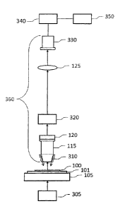
[0064] Figure 3 shows a microscope for fluorescence and brightfield imaging
that is a
first embodiment of this invention. A tissue specimen 100 (or other specimen
to be
imaged) mounted on microscope slide 101 on a scanning stage 105. When used for
fluorescence imaging, the tissue specimen is illuminated from above by
illumination
source 310, mounted above the specimen (epifluorescence) so that the intense
illumination light that passes through the specimen is not mixed with the
weaker
fluorescence emission from the specimen, as it would be if the fluorescence
illumination
source were below the specimen. Several different optical combinations can be
used for
epifluorescence illumination ¨ light from a source mounted on the microscope
objective,
as shown; illumination light that is injected into the microscope tube between
the
14
CA 02877902 2014-12-24
WO 2014/000085 PCT/CA2013/000592
microscope objective and the tube lens, imaged onto the back aperture of the
objective,
using a dichroic beamsplitter to reflect it down through the microscope
objective and
onto the specimen; and several others. A narrow wavelength band for the
illumination
light is chosen to match the absorption peak of the fluorophore in use. This
narrow-band
illumination may come from a filtered white-light source, an LED or laser-
based source
(including a laser sent through a diffuser plate in rapid motion to eliminate
speckle), or
other source. Fluorescence emitted by the specimen is collected by infinity-
corrected
microscope objective 115 (or other high-numerical-aperture objective lens)
which is
focused on the specimen by piezo positioner 120 (or other focusing mechanism).
Emission filter 320 is chosen to reject light at the illumination wavelength
and to pass the
emission band of the fluorophore in use. The microscope objective 115 and tube
lens
125 form a real image of the specimen on two-dimensional detector array 330.
An
image of the specimen is collected by moving the microscope slide at constant
speed
using motorized stage 105 in a direction perpendicular to the long dimension
of detector
array 330, combining a sequence of equally-spaced overlapping two-dimensional
images from the array (usually spaced one line apart) to construct a time-
integrated
image of one strip of the specimen. Data from detector array 330 is read out
by frame
grabber 340 and passed to computer 350 where strips are then assembled to form
a
complete image of the specimen.
[0065] When used for brightfield imaging, transmitted-light illumination
source 305 is
used instead of illumination source 310 (which illuminates the specimen from
above) and
emission filter 320 is removed from the optical train.
[0066] Figure 4 shows a specimen 100 mounted on a microscope slide 101. Note
that
in this diagram the microscope slide is square, but it can have any convenient
size and
shape, including standard 1x3 inch microscope slides up to very large slides
(we have
imaged specimens on slides 6x8 inches in size) and for the purposes of this
document,
the term "microscope slide" includes slides made from glass or other medium
(usually
but not always transparent) and any other specimen carrier including but not
limited to
microwell plates and tissue blocks. Specimens may be covered with a cover
slip.
[0067] In this diagram, the specimen is larger than the field-of-view 412 of
the
microscope detector array. In this example, three image strips are required to
image the
entire specimen 100, but for a larger specimen many more strips may be
required. In
order to scan specimen strip 400, microscope stage 105 of Fig. 3 moves the
microscope
slide 101 at constant speed in the direction shown in Fig. 4 (or the
microscope optical
train 350 is moved at constant speed in the opposite direction). An electronic
or
mechanical shutter opens for a short time to expose the sensors that make up
the two-
CA 02877902 2014-12-24
WO 2014/000085 PCT/CA2013/000592
dimensional detector array 330, which is also shown in detail as detector
array 500 in
Figure 5. The exposure time is short enough so that during exposure the
constant
relative motion of the detector and microscope slide moves field-of-view 410
only part of
the way to the adjacent field-of-view 411, which is one pixel away from 410.
During the
time the shutter is closed, data in the entire two-dimensional detector array
is transferred
to frame-buffer RAM in a frame grabber 340 or to other electronic frame
capture device,
and is then transferred to a computer 350. When the field-of-view moves to
position
411, the shutter is opened again and a new image frame is collected, the
shutter is then
closed and this new image is then transferred via the frame grabber to the
computer,
where this data is added to the image data already stored, but shifted one
pixel in the
direction of motion of the field-of-view. This process is repeated until a
complete image
of that specimen strip is stored, starting with a first image frame (first
exposure) just
above the top edge of the specimen of Fig. 4 to a final image frame just below
the
bottom edge of the specimen (in order to ensure that every part of the
specimen is
exposed 256 times, once for each line of pixels in the detector array). For
example, the
detector array 330 is comprised of 4000 pixels by 256 lines, as shown in
Figure 5 {which
shows a CCD or CMOS (or other technology) two-dimensional sensor array 330
with
256 lines of 4000 pixels each (a 256 x 4000 pixel array)). Although this
particular array
has been chosen as an example, arrays with different numbers of pixels and
different
aspect ratios can also be used. In particular, this means inexpensive
arrays
manufactured for consumer products can be used if necessary to reduce cost.
Using
the array shown in Figure 5, each pixel in the final strip image stored in
computer 350 is
the sum of 256 exposures of the same pixel position in the specimen. In this
particular
example, if the frame grabber produces 8-bit images, the resulting stored
image has a
dynamic range of 16 bits (each pixel is made up of a sum of 256 exposures
where each
exposure has a maximum value of 255). The fluorescence image of the strip is
stored
and adjacent strip images are assembled to produce a final image of the
specimen.
Adjacent strips may be assembled by butting them together, or by collecting
overlapping
strip images and using feature-matching software for registration.
[0068] As an example, using the 256 x 4000 pixel 8-bit pixel array described
above, if a
specimen lcm long is scanned at 0.25 micron resolution (approx. 40X), a total
of 40,255
frames must be acquired in order to expose every pixel 256 times (1cm x 40,000
lines/cm + 255). The strip image will contain 40,000 x 4,000 pixels. If the 16-
bit memory
locations for each pixel are set to zero before the scan starts, then the
value for each
pixel at the end of the scan is given by:
16
CA 02877902 2014-12-24
WO 2014/000085 PCT/CA2013/000592
i=m+255
Pm,n = E P{i-(m-1)},n,i
i=m
where P,n is the final value for each pixel in the strip image, m is the line
number in the
strip image (in this example of a 1cm strip on the specimen, m varies from Ito
40,000),
and n is the column number in the strip image (in this example varies from 1
to 4,000).
On the right-hand side of the equation, p{iqm.1)},n,/ represents the pixel
value for pixels in
each detector image frame, where {i-(m-1)} represents the row number of the
pixel and n
represents the column number of the pixel in frame number L Each pixel P in
the final
image is the sum of 256 detector image pixels from 256 sequential frames,
where the
column number varies from 1 to 4,000 (the same number as in the detector image
frames) and the row number varies from 1 to 40,000. The running index in the
sum is i,
and i also equals the frame number (in this example varies from 1 to 40,255).
[0069] If the resulting image from the example above is to be viewed in a
display with
the same dynamic range as the image from each detector frame (8 bits in the
example
above), the value stored in each pixel position above can be multiplied by
1/N, where N
is the number of frames exposed and this value stored in each pixel position
in the final
image (N=256 in the example above). To ensure the best possible dynamic range
in the
final image, data contraction as described in W02009/137935 can be used when
converting from an image stored in 16-bit memory locations in order to use the
entire
dynamic range in the final 8-bit image.
[0070] If the scanning stage is set to move at a constant speed of 100
microns/second
(1/10 mm/second), and assuming the same 0.25 micron object pixel resolution
and 4000
x 256 pixel detector array as used in the example above, lines of data are
collected at
400 lines/second (this is similar to a scan rate of 400 lines/second in a
scanning laser
microscope or macroscope). If an exposure time of 1/1000 second is used, the
moving
specimen stage will move less than half the distance between adjacent pixels
during the
time the shutter is open, and since 256 lines of data from the detector array
are summed
into each line of data in the final image, the total exposure time for each
pixel in the final
image is 256/1000 seconds, or approximately 250 milliseconds. By comparison,
if a
linear detector array is used at the same scan speed, the exposure time is
only 1
millisecond, which is too short for weak fluorophores. Note that the operation
of the
shutter should be closely synchronized with stage motion, just as it must be
if TDI
detectors were used instead of the two-dimensional detector arrays described
in this
document. (Note: the specimen image may have enough features to allow
sequential
17
CA 02877902 2014-12-24
WO 2014/000085 PCT/CA2013/000592
image frames to be registered using feature-matching software, which reduces
the
requirement for synchronization between sequential image frames and therefore
would
allow a less-expensive moving stage to be used.)
[0071] In the example above, the exposure time for each image was 1 msec.,
leaving
approximately 1 msec. to read out the data in the array before the scanning
stage has
moved a distance equal to the distance between pixels on the specimen. If this
read-out
time is too short to read out the array, the next exposure can be synchronized
to start
when the stage has moved a distance equal to an integral number of pixels
instead of
the distance between adjacent pixels, thus increasing the read-out time while
keeping
the scan speed unchanged. The number of images added together to form the
final
image will be reduced by a factor equal to 1/s, where s is the number of
pixels the stage
moves between exposures. (s = 1 when the next exposure is at the next pixel
position, s
= 2 if the next exposure is two pixels distance away, etc.) This technique can
also be
used to increase the scan speed, while keeping the exposure time constant. If
s = 16,
for example, then only 16 images are added together (or averaged), but the
scan speed
can be increased dramatically. If the exposure time is kept constant, then the
measured
pixels will be elongated in the direction of scan, but this may be acceptable
if the image
collected is a high-speed preview scan, and the dynamic range of data in this
preview
image can be used to calculate proper exposure for a final, slower scan before
that scan
starts.
[0072] Figure 6 (top) shows a rectangular detector array 600 of 40 pixels (10
pixels by 4
lines). In an ordinary array, the entire frame is read out through the same
output port,
which can be time consuming (especially for a large array like the 1,024,000
pixel array
described in the example above (4000 pixels by 256 lines). Figure 6 (bottom)
shows a
second rectangular detector array 610 that also has 40 pixels (10 pixels by 4
lines), but
in this array each line is read out through its own output port. Read-out time
can be
reduced substantially if the lines of data are read out simultaneously into a
frame buffer,
where they can be stored for further processing. In theory, this could reduce
the read-
out time of the 1,024,000 pixel array used as example previously by a factor
of 1/256.
Additionally, since in this application lines of data from the detector array
are added in a
moving sequence to lines of data stored in a strip image, lines can be shifted
and added
to the appropriate memory locations in the strip image as parallel processes,
thus
reducing the computational load as well.
[0073] Using this same example of a 4000 pixel by 256 line array, consider a
scanner
where the required magnification is similar to that from an optical microscope
with a 40X
objective. The digital image produced by this scanner will have pixels
approximately
18
CA 02877902 2014-12-24
WO 2014/000085 PCT/CA2013/000592
0.25 microns in size, and 4000 pixels represent the width of a 1mm wide strip
on the
specimen. The microscope objective needs a resolving power of 0.5 microns or
smaller
(numerical aperture of 0.6 or larger), and the Nyquist theorem requires at
least two
pixels per resolving power for digital imaging {see "Choosing Objective
Lenses: The
Importance of Numerical Aperture and Magnification in Digital Optical
Microscopy",
David W. Piston, Biol. Bull. 195, 1-4 (1998) for a good explanation of the
requirements
for diffraction-limited digital microscopy). To image 0.25 micron pixels on
the specimen
onto a detector array with sensors spaced 10 microns apart, the system
magnification
(objective lens plus tube lens) must be 40X. A microscope objective with a
numerical
aperture of 0.75 and a focal length of 10 mm is available (this is labeled a
20X objective,
but that label assumes a particular tube lens with focal length 200mm). Since
Magnification= = f
tube lens / f microscope objective,
f tube lens = 40 x 10 = 400mm.
The tube lens must be able to form an image at least 4cm wide for the detector
array
described above. The combination of an infinity-corrected microscope objective
(or
other infinity-corrected objective lens) and a tube lens is used because it is
possible to
insert filters, filter cubes, and beamsplitters into the optical path between
the objective
and the tube lens without affecting instrument focus and optical performance.
[0074] Figure 7 shows a microscope for fluorescence or brightfield imaging
that is a
second embodiment of this invention. When used for fluorescence imaging, a
tissue
specimen 700 (or other specimen to be imaged) which has been stained with
three
different fluorescent dyes is mounted on microscope slide 101 on a scanning
stage 105.
The tissue specimen is illuminated from above by illumination source 705,
mounted
above the specimen (epifluorescence) so that the intense illumination light
that passes
through the specimen is not mixed with the weaker fluorescence emission from
the
specimen, as it would be if the illumination source were below the specimen.
Several
different optical combinations can be used for epifluorescence illumination ¨
light from a
source mounted on the microscope objective, as shown; converging illumination
light
that is injected into the microscope tube between the microscope objective and
the tube
lens that focuses on the back aperture of the objective, using a dichroic
beamsplitter to
reflect it down through the microscope objective and onto the specimen; and
several
others. Narrow wavelength bands are chosen for the illumination light to match
the
absorption peaks of the fluorophores in use. This narrow-band illumination may
come
from a filtered white-light source, an LED or laser-based source (including an
amplitude
or frequency-modulated laser or LED source), or other source. Fluorescence
emitted by
the specimen is collected by infinity-corrected microscope objective 115 (or
other high-
19
CA 02877902 2014-12-24
WO 2014/000085 PCT/CA2013/000592
numerical-aperture objective lens) which is focused on the specimen by piezo
positioner
120 (or other focusing mechanism). Dichroic mirror 730 is chosen to reflect
light in the
emission band of the first fluorophore towards tube lens 710 placed in front
of two-
dimensional detector array 720.
Microscope objective 115 and tube lens 710 form a
real image of the specimen on two-dimensional detector array 720. Data from
the two-
dimensional detector array is collected by frame grabber 770 or other
electronic frame
capture device and passed to computer 350.
[0075] Light from the specimen 700 that was not reflected by dichroic mirror
730
continues up the microscope to reach dichroic mirror 740, which is chosen to
reflect light
in the emission band of the second fluorophore towards tube lens 750 placed in
front of
two-dimensional detector array 760. The microscope objective 115 and tube lens
750
form a real image of the specimen on two-dimensional detector array 760. Data
from
this two-dimensional detector array is read out by frame grabber 780 or other
electronic
frame capture device and passed to computer 350.
[0076] Light from the specimen 700 that was not reflected by dichroic mirrors
730 and
740 contains light in the emission band wavelengths for fluorophore three, and
continues
up the microscope to reach tube lens 125, in front of two-dimensional detector
array 330.
The microscope objective 115 and tube lens 125 form a real image of the
specimen on
two-dimensional detector array 330. Data from this two-dimensional detector
array is
read out by frame grabber 340 or other electronic frame capture device and
passed to
computer 350.
[0077] An image of the specimen is collected by moving the microscope slide at
constant speed using motorized stage 105 in a direction perpendicular to the
long
dimension of the three detector arrays 720, 760 and 330 (which are all
oriented with the
long dimension of the arrays perpendicular to the motion of the real images
projected on
them by the microscope objective 115 and tube lenses 710, 750 and 125
respectively).
A sequence of equally-spaced overlapping two-dimensional images from the each
of the
three arrays is passed to computer 350 by frame grabbers 770, 780 and 340
where
three time-integrated images of one strip of the specimen are constructed, one
for each
fluorophore. These three images can be viewed separately (fluorescence images
are
essentially greyscale images) or combined using false colours into a colour
image for
viewing. In many cases the false colours are chosen to make the final image
look like
the image that would be seen through a fluorescence microscope.
[0078] Figure 7 shows a scanner with three detection arms, one for each of
three
fluorophores (a scanner can also be envisioned for other numbers of
fluorophores). In
particular, if quantum dots (nanocrystals) are used as a contrast agent in
fluorescence,
CA 02877902 2014-12-24
WO 2014/000085 PCT/CA2013/000592
several detection arms can be used. This is possible because quantum dots can
be
manufactured with very narrow emission bands, and they are inherently brighter
and
more stable than fluorophores. In addition, all quantum dots in a specimen can
be
excited with the same excitation wavelength, so a single wavelength source can
be used
which is not in the emission bands of any of the dots in the specimen, making
it easier to
separate the emission signals.
[0079] When used for brightfield imaging, white light source 110 is used to
illuminate the
specimen from below (instead of using light source 310), and the dichroic
mirrors 730
and 740 are chosen to separate the colours detected by area detectors 770, 780
and
340 into red, green and blue. Images from each of the three detection arms are
combined to produce a colour brightfield image. If area detector 340 is
replaced by an
RGB detector, dichroic mirrors 730 and 740 can be removed from the optical
train and
the single colour detector will produce a colour brightfield image.
[0080] Instead of using three detection arms, as shown in Figure 7, it is also
possible to
use a trichroic prism to separate light emitted from three fluorophores to be
focused on
three ccd detectors. Such an assembly can also be used for RGB brightfield
imaging.
[0081] Figure 8 shows a third embodiment of this invention, a scanner in which
a
tunable filter 810 is used to provide a multi-spectral fluorescence slide
scanner and
method for imaging large specimens mounted on microscope slides. The tunable
filter
can be set to transmit a band of emission wavelengths from one fluorophore (or
other
fluorescent source) and a strip image recorded for that source, followed by
setting a
second wavelength band for a second fluorophore to record a strip image for
that
source, and so on until a strip image has been recorded for each fluorescence
source in
the specimen. The strip images can either be viewed separately or combined
into a
single image (usually false coloured) and the strips can then be assembled
into a single
image of the entire specimen. This instrument can also be used for brightfield
imaging
by replacing epifluorescence source 705 with white light transmission source
110, and
using the tunable filter 810 to pass red, green and blue wavelengths to record
red, green
and blue strip images in sequence which can be combined into a single RGB
brightfield
image.
[0082] Figure 9 shows a single two-dimensional CCD (or other technology) array
900 in
which the top third 910 of the array is covered with a red transmission
filter, the middle
third 920 is covered with a green transmission filter, and the bottom third
930 is covered
with a blue transmission filter. Such an array can be used to simultaneously
image three
colours, for example red, green and blue for brightfield imaging, or three
different
21
CA 02877902 2014-12-24
WO 2014/000085
PCT/CA2013/000592
fluorophores in multi-spectral fluorescence (where the transmission filters
are chosen
with bandwidths that match the fluorescence emission peaks).
[0083] Figure 10 shows a fourth embodiment of this invention, a scanner in
which a
detector array 900 (covered with red, green and blue transmission filters as
discussed
above) simultaneously records three strip images (red, green and blue) when
white light
transmission source 110 is used to illuminate the specimen from below. Image
data
from the top third 910 of array 900 is used to record the red image, data from
the middle
third 920 of array 900 is used to record the green image, and that from the
bottom third
930 of array 900 is used to record the blue image. Each of these images is
recorded in
separate strip images that can be combined into an RGB image after the scan
for that
strip is completed. Note that Figure 9 shows a 4000 pixel x 256 line array ¨
this is for
example only ¨ arrays with different pixel number width and number of lines
can also be
used.
[0084] For fluorescence imaging, the epifluorescence light source 310 (or
other
epifluorescence source) is used instead of white light source 110, and
transmission
filters are chosen to cover fractions of the array 900, one matching the peak
of the
emission band of each fluorophore in the specimen. In particular, if
fluorescent
nanoparticles are used as the fluorescence source, a filter is chosen with
transmission
bandwidth to match the emission peak of each of the nanoparticles, and
fluorescence
from several nanoparticles can be imaged simultaneously.
[0085] Figure 11 is a schematic representation of a scanning fluorescence
microscope
that is a fifth embodiment of this invention. Light from white light source
1110 passes
through tunable filter 1120, is partially reflected by beamsplitter 1130,
passes through
microscope objective 115, and illuminates an area of the surface of specimen
100, which
is mounted on microscope slide 101 on moving microscope stage 105. Motion of
the
microscope stage is in a direction perpendicular to rows in the detector array
{data is
read out from rows in the detector array, usually the long dimension of the
array (for
example see Hamamatsu's ORCA-flash 4.0 camera, or PCO's pco.edge camera, both
of
which use Scientific CMOS (sCMOS) detector arrays)]. Fluorescence emitted from
the
specimen is collected by microscope objective 115, is partially transmitted by
beamsplitter 1130, and then passes through tunable filter 810 and is focused
by tube
lens 125 onto detector array 330, which is not covered by a colour filter
array. Data from
the detector array 330 is read out by frame grabber 340 and passed to computer
350
where an image strip is assembled for each fluorophore.
[0086] As an example, suppose there are two fluorophores in the specimen.
Tunable
filter 1120 is adjusted to transmit the appropriate excitation wavelength
range for the first
22
CA 02877902 2014-12-24
WO 2014/000085 PCT/CA2013/000592
fluorophore, and tunable filter 810 is adjusted to transmit the emission
wavelength band
of the first fluorophore. Detector array 330 is read by frame grabber 340
which passes
the data for the first image frame to computer 350, which stores this image
frame at the
beginning of a first strip image. The microscope stage is moved a distance
equal to the
distance required to move the image projected on the detector array by tube
lens 125 a
distance equal to the distance between rows in the array. Tunable filter 1120
is then
adjusted to transmit the appropriate excitation wavelength range for the
second
fluorophore, and tunable filter 810 is adjusted to transmit the emission
wavelength band
of the second fluorophore. The first image frame for fluorophore 2 is stored
in a second
strip image. Next, the microscope stage is moved a distance equal to the
distance
required to move the image the distance between pixels in the detector array,
and a
second image of the first fluorophore is collected, moved in the scan
direction a distance
equal to the distance between pixels, and added to the first image already
stored in the
strip image for the first fluorophore. Imaging continues from one fluorophore
to the other
until the entire strip of specimen has been imaged and a strip image has been
collected
for each fluorophore. in operation, the scanning stage usually moves at a
constant
speed, and the tunable filters are synchronized with the motion to produce one
exposure
every time the stage has moved a distance that is equivalent to the distance
between
image pixels. In this case (when two fluorophores are present), each image
frame from
the detector is moved by one pixel in the scan direction from the image before
it, so
when each image is added to one of the fluorophore strip images, it must be
displaced
by 2 pixels. When three fluorophores are present, three images are collected
during the
time the stage moves a distance that will cause the moving image to move a
distance
equal to that between three rows of pixels in the detector array, and the
tunable filters
are synchronized to provide the appropriate excitation and emission
wavelengths during
the specimen motion. Alternatively, when two fluorophores are used for
example, the
specimen stage can be moved a distance equivalent to half the distance between
image
pixels between exposures and the tunable filters can be synchronized to change
excitation and emission wavelengths each time the stage has moved a distance
equivalent to half the distance between image pixels. This has the advantage
that the
number of exposures of the same area of the specimen is doubled, but scan
speed is
reduced and the two image strips are now out of registration by 1/2 pixel in
the scan
direction.
[0087] Several other optical arrangements in addition to that shown in Figure
11 are
possible. For example, tunable filters 810 and 1120 can be replaced by
rotating optical
filters or filter wheels that are synchronized to each other and to stage
motion to pass
23
CA 02877902 2014-12-24
WO 2014/000085 PCT/CA2013/000592
the appropriate excitation and emission wavelengths. Figure 12 shows one
example of
a pair of rotating optical filters designed for use for simultaneous imaging
of three
fluorophores, with first rotating optical filter 1210 (which replaces tunable
filter 1120 in
Figure 11) designed to pass three excitation wavelength bands X1 , X2 and X3,
chosen
to excite the three fluorophores known to be present in specimen 100. Second
rotating
optical filter 1220 replaces tunable filter 810, and is designed to pass the
three emission
bands El, E2 and E3 of the three fluorophores present in the specimen. The
rotation of
these two optical filters is synchronized so that excitation filter X1 and
emission filter El
are both positioned in the optical path at the same time (as are X2 and E2,
and X3 and
E3), and their rotation is further synchronized with the motion of the
scanning stage so
that the stage moves a distance such that the image of the specimen projected
on the
detector array moves a distance equal to the distance between rows in the
array during
the time the filters rotate 120 degrees.
[0088] As a second example, a tunable light source can be used in place of the
combination of white Light Source 1110 and tunable filter 1120 to provide an
excitation
wavelength band that is appropriate for the fluorophore in use.
[0089] As a third example, a tunable light source placed in the
epifluorescence position
shown by light source 310 in Figure 3 and synchronized with stage scan and the
tunable
emission filter 810 can replace white light source 1110 and tunable filter
1120.
[0090] As a fourth example, if a white light illumination source is placed
below the
specimen (like light source 305 in Fig. 3), then tunable filter 810 can be
adjusted
sequentially to pass red, green and blue, and R, G and B strip images can be
acquired
simultaneously and assembled after scanning into a single RGB image. (Note:
instead
of collecting three strip images during scanning, and assembling three strip
images to
produce a single RGB image after scanning, data can be added to or averaged
with data
already present in the R, G and B components of a single RGB strip image if
that is
more convenient for data flow.) The addition of a white illumination source
below the
specimen has resulted in a scanner that performs both brightfield and
fluorescence
imaging. If beamsplitter 1130, tunable filter 1120 and white light source 1110
are
removed, this becomes a brightfield-only scanner. The further addition of a
tunable light
source in the same position as light source 310 in Figure 3 (an
epifluorescence position)
results in a brightfield and fluorescence scanner.
[0091] When a white light source is placed below the specimen for transmission
brightfield imaging, a rotating filter like RGB filter 1220 can be used either
to filter the
incoming light from the source (where the filter is placed between the white
light source
and the specimen) or to filter light collected by the objective lens (where
the filter is
24
CA 02877902 2014-12-24
WO 2014/000085 PCT/CA2013/000592
placed between the objective lens and the tube lens). When filter rotation is
synchronized with stage scan, either of these arrangements can be used to
produce a
colour brightfield scanner. Other colour combinations are possible, including
but not
limited to RGBW, CYGM and RGBE. When used in this manner, no colour filter
array is
required on the detector array, and the images do not require demosaicing.
[0092] Figure 13 is a schematic representation of a scanning brightfield
microscope that
is a sixth embodiment of this invention. In this embodiment, area detector
array 1300
contains a mosaic Colour Filter Array (a Bayer filter, which is commonly used
in single-
chip colour cameras). Images acquired using a Bayer filter (or other common
mosaic
filters) are normally demosaiced using a computer algorithm before viewing,
but this will
not be necessary when Moving Specimen Image Averaging is used. A small 4x4
pixel
area array 1410 using a Bayer filter is shown on the left side of Figure 14.
This small
array example is used to explain how an area array using a Bayer filter can be
used for
Moving Specimen Image Averaging, however in practice much larger arrays are
used
(for example, 2560 x 256). The first exposure in a scan using the small array
1410 is
shown at the top of Figure 14a. Three strip images are set up in RAM in
computer 350,
one strip each for red, green and blue, and data values in each pixel position
are initially
set to zero. Data from the first exposure is transferred to each of the
strips, resulting in
red strip image 1421, green strip image 1431, and blue strip image 1441, as
shown at
the top of Figure 14a. When the stage has moves a distance such that the image
on the
detector has moved a distance equal to the distance between rows of detector
pixels, a
second exposure is acquired, and data from this second exposure is transferred
to the
colour strip images, resulting in strip images 1422, 1432 and 1442. The
results after
adding data from exposure #3 to the data already stored in the three image
strips is
shown in Figure 14b (top), and results after exposures 4 and 5 are shown in
Figure 14b
(bottom) and Figure 14c. At the end of the first five exposures, data in rows
4 and 5 of
each strip image are complete (for a detector with only four rows, no
additional data will
be added to rows 4 and 5 of the strip images as the scan continues). Note that
after four
exposures, row 4 in the final image strips contains two exposures of pixel R1
and R3,
two exposures of all four Green pixels (G1, G2, G3 and G4) and two exposures
of pixels
B2 and B4. After the fifth exposure (row 5 from the top of the three strip
images), it is
obvious that the same pixel positions have been exposed in the fifth row, with
the same
exposure. After the scan has been completed (assume several hundred rows have
been exposed), the first 3 rows of data should be discarded (because they are
not fully
exposed), and the last 3 should also be discarded. Also note that the final
red and blue
images have full resolution in the vertical direction, but resolution in the
horizontal
CA 02877902 2014-12-24
WO 2014/000085
PCT/CA2013/000592
direction is only half as good. The green image has full resolution in both
directions.
The green image requires no interpolation before assembling the three strips
to produce
a final RGB image, but both the red and blue images will require some
interpolation to fill
in the blanks. The simplest interpolation (which may be good enough in many
cases) is
simply to average the red or green pixels on either side of an empty pixel
position. More
complex interpolation could include an algorithm that includes changes in
local
brightness using the green pixels, or nearest neighbor interactions between
adjacent
pixels. Since a single chip colour camera was used to take all of the separate
images,
the colour image resulting from each exposure can be demosaiced and then added
to
the demosaiced images resulting from subsequent exposures using MSIA, but this
is a
calculation-intensive process that will not be necessary in most applications,
and slows
down image acquisition.
[0093] A more realistic situation is one where the detector array is much
larger than the
4 x 4 pixel array shown in Figure 14. Assume that the same RGGB Bayer filter
is used.
If the array has 256 rows and 2560 columns of pixels, then each image strip is
2560
pixels wide. The first row to be fully exposed (after exposure #256) will be
row 256 in
each strip image (the first 255 rows of image data should be discarded), each
green
pixel will have been exposed 128 times, and the same rows of red and blue
pixels as
shown in Figure 14c will be exposed 128 times, or not at all. The final 255
rows in the
image strip should be discarded. Since a 1cm scan contains 40,000 rows of data
(assuming 1/4 micron pixels), discarding the first 255 and last 255 rows will
not increase
the scan time very much.
[0094] When a colour camera with a Bayer filter (or other common Colour Filter
Array) is
used, after scanning is complete it is possible to return to areas of interest
that have
been identified in the scanned image to view those areas in either a single-
field-of-view
or tiling-microscope mode, and to collect 4D colour image data from those
areas (X, Y,
Z, time). This instrument and method (the sixth embodiment) uses a mosaic
Colour
Filter Array, and is used for brightfield scanning of large specimens using
MSIA (where
no demosaicing of the image is required), as well as real-time and 3D imaging
of small
areas of interest (in which the images are mosaiced in the normal way for
instruments
using mosaic Colour Filter Arrays).
[0095] The Bayer filter (and other common Colour Filter Arrays) were designed
for
cameras in which an entire colour image is captured in a single exposure. As
discussed
above, such filters can also be used for microscope slide scanners using
Moving
Specimen Image Averaging, but when the Bayer filter is used in that
application the
resolution of red and blue pixels in the horizontal direction is only half
that in the vertical
26
CA 02877902 2014-12-24
WO 2014/000085
PCT/CA2013/000592
direction. The green pixels have full resolution in both directions.
Interpolation is
necessary to fill in the missing red and blue pixels in the horizontal
direction, but this
interpolation is much simpler than the demosaicing usually used with Bayer
filter
cameras. Many cameras are commercially available with Bayer filters, which is
an
advantage.
[0096] A two-dimensional sensor array in which the top third of the array was
covered
with a red transmission filter, the middle third was covered with a green
transmission
filter, and the bottom third was covered with a blue transmission filter, was
shown in
Figure 9. This colour filter array is not useful for capturing an entire
colour image in a
single exposure, but is useful for scanning using Moving Specimen Image
Averaging,
and results in a final strip image in which all three colour components of the
image have
maximum resolution in both directions. We define this and other Colour Filter
Arrays
that are used only for scanning as "Scanning Colour Filter Arrays" (SCFA).
Several
additional embodiments of Scanning Colour Filter Arrays are described below.
Figure 9
shows a first embodiment of a Scanning Colour Filter Array.
[0097] Figure 15 shows a second embodiment of a Scanning Colour Filter Array.
This
SCFA is shown on a 5 x 6 pixel sensor array (top left), with the data flow
from the first
exposure to the image store for red, green and blue monochrome images (or to
the R,G
and B components of a single RGB image) on the right. The second exposure is
shown
at the bottom of Figure 15, after the moving stage has moved a distance such
that the
image on the detector has moved a distance equal to the distance between rows
of
detector pixels. Data from the rows of Red, Green and Blue pixels is
transferred to the
image store and fills in the next rows of R, G and B pixel image data. After
the third
exposure, the rows of R, G and B pixels in the R, G and B images will be
completely
filled in (not shown). Note that all three images have full resolution in both
the horizontal
and vertical directions, and no interpolation is required. As the scan
proceeds, data is
transferred from the detector array to the image store after each exposure.
For a
Scanning Colour Filter Array in which there are two RGB sequences in the
vertical
direction (as shown in this example), each pixel in the final R, G and B
images will be
exposed twice and averaged together using Moving Specimen Image Averaging. In
a
more practical example, for example a 2560 x 258 pixel array, 86 rows are
covered with
a Red filter, 86 are covered with a Blue filter, and 86 are covered with a
Green filter, in
the same RGB sequence as shown in Figure 15. Red, Blue and Green pixels are
averaged 86 times. When low light levels are used, random noise can seriously
degrade
the resolution of a microscopy image, and this is especially true in
fluorescence, where
light levels are often very low. Image averaging improves noisy images
dramatically,
27
CA 02877902 2014-12-24
WO 2014/000085
PCT/CA2013/000592
with a signal/noise ratio improvement proportional to the square root of the
number of
exposures. In this case, where each image pixel is exposed 86 times and
averaged
using MSIA, signal/noise ratio of a noisy image is increased by a factor of
9.3, which
improves the image considerably.
[0098] Figure 16 shows a four-line sequence RGBW Scanning Colour Filter Array
that is
a third embodiment of a Scanning Colour Filter Array. The addition of a row of
white
(panchromatic) pixels with three rows of colour pixels adds several new
possibilities for
scanned images using MSIA. For example, the clear (white) pixels are the
brightest
pixels in the array, and result in a panchromatic image strip that is the
brightest of the
four images and can also be used to detect infrared. This image will have the
best
contrast of the four images, and will be useful for image processing later
since it is
perfectly registered with the three single-colour images. When the four-line
sequence
RGBW scanning colour filter array shown in Figure 16 is used on a 2560 x 256
sensor
array, each of the four images will be composed of pixels that have been
exposed 64
times, and after MSIA the signal/noise ratio in each image will have been
increased by a
factor of 8.
[0099] Because of its brightness and high contrast, the white image will be
useful for
autofocusing, and can also be used for fluorescence imaging (the R, G and B
images
can be ignored or discarded when not required) with the addition of a
fluorescence light
source and emission filter. For single-fluorophore imaging, the light source
and emission
filter do not have to be synchronized with the scanning stage, and can be
implemented
using a white light epifluorescence source and filter cube. A standard
fluorescence
microscope, with the addition of a scanning stage and a
brightfield/fluorescence MSIA
camera (a single-chip camera using an RGBW or RWGWBW Scanning Colour Filter
Array), can be used for both RGB and fluorescence scanning. A second
fluorophore can
be imaged simply by changing the filter cube in the microscope and scanning
again.
[0100] A third example of a Scanning Colour Filter Array is shown in Figure
17, an
RWGWBW Scanning Filter Colour Array. In this array a row of clear (white)
pixels is
placed after each row of colour pixels. This filter array results in four
images, as the
RGBW array did, however in this case the exposure of the panchromatic image
will be
three times that of each of the colour images. This is particularly important
for
fluorescence imaging, where signal strength is low and there is considerable
background
noise. In this case, using a 2560 x 256 pixel array, each of the single-colour
images will
be exposed 42 times, and each panchromatic image will be exposed 128 times.
This
results in increased signal/noise ratio in the fluorescence image (where it is
required)
compared to each of the R, G and B images.
28