Note: Descriptions are shown in the official language in which they were submitted.
CA 02878025 2015-01-15
1
METHODS OF DETECTING SIGNATURES
OF DISEASE OR CONDITIONS IN BODILY FLUIDS
[01]
FIELD
[02] The present invention relates to methods of identifying markers of
conditions such as
gender of a fetus or disease such as tumor genomic, proteomic, metabolomic,
glycomic. glycoproteomic, lipidomic and/or lipoproteomic signatures in cells
obtained
from bodily fluids of a patient.
BACKGROUND
[03] Tumors originate from normal cells upon the accumulation of genetic and
epigenetic
alterations. This multi-step process involves multiple genetic alterations
that lead to
the progressive transformation of normal cells to a malignant phenotype. These
alterations are comprised of irreversible changes in DNA sequence (e.g.,
mutations,
deletions, translocations) and lead to the activation of oncogenes,
inactivation of
tumor suppressor genes, and fusion of genes. The stochastic nature of these
events
confers genetic heterogeneity that gives the transformed cells molecular
fingerprints
(e.g., one or more cellular components such as DNA, RNA, protein, lipid,
carbohydrate, and the like) indicative of cancer that give them unique
phenotypes.
Consequently, unique gene set hallmarks/signatures are known to be expressed
by
various tumors (Perou et al. (2000) Nature 406:747; Lobenhoferet al. (2001)
Health
Perspect. 109:881; van't Veer et al. (2002) Nature 415:530 (2002); Liotta and
Kohn
(2003) Nat. Genet. 33:10; Ginos et al.(2004) Cancer Res. 64:55; Liu (2005)
Proc.
Natl. Acad. Sci. USA 102:3531; Grigoriadis et al. (2006) Breast Cancer
Research
8:R56).
CA 02878025 2015-01-15
2
[04] Both primary and metastatic tumors can lie silent and undetected for
years. However,
these dormant and occult tumors, as well as previously diagnosed primary and
metastatic solid tumors, shed daily into the circulation approximately one-to-
six
million cells per gram of tumor. A large proportion of these circulating tumor
cells,
known as CTCs, undergo apoptosis and die, whereas distinct cell populations
may
develop into metastatic disease. Tumor cell apoptotic bodies, DNA,
nucleosomes,
RNA, and proteins are also found in the blood of cancer patients. Holmgren et
at.,
Blood 93, 3956 (1999). Efforts have been made to investigate whether
signatures of
tumors can be identified and whether they can be used to detect or monitor
cancer.
See, Ransohoff, Nature Reviews Cancer 5, 142 (2005) and McLerran et al.,
('fin.
Chem. 54, 44 (2008).
[05] DNA can be easily transfeeted into various eukaryotic cells, i.e.,
once it is internalized
into the cytoplasm of cells, it is able to integrate its genes into the genome
of the host
cell. For example, neutrophils and macrophages can be rapidly and very
efficiently
(50%-90%) transfected. Passage of DNA from prokaryotic to eukaryotic cells has
also been demonstrated and is believed to occur from eukaryotic to eukaryotic
cells.
DNA released from tumor cells has a high transforming activity. Adding
supernatant
medium from cultured tumor cells to normal cells results in the appearance of
as
many transformed foci as those occurring after a transfection with a cloned
ras gene
administered as a calcium precipitate. Furthermore, when healthy rats were
injected
with plasma from tumor-bearing rats (therefore containing tumor DNA) the tumor
marker gene was found in the DNA of their lung cells, i.e., tumor genes have
been
transcribed in lung cells.
[06j Leukocytes begin as pluripotcnt hematopoietic stem cells in the bone
marrow and
develop along either the myeloid lineage (monocytes, macrophages, neutrophils,
eosinophils, and basophils) or the lymphoid lineage (T and B lymphocytes and
natural
kilter cells). The major function of the myeloid lineage cells (e.g.,
neutrophils and
macrophages) is the phagocytosis of infectious organisms, live unwanted
damaged
cells, senescent and dead cells (apoptotic and necrotic), as well as the
clearing of
cellular debris. Phagocytes from healthy animals do not replicate and are
diploid, i.e.,
CA 02878025 2015-01-15
3
have a DNA index of one. On average, each cell contains <10 rig DNA, <20 ng
RNA,
and <300 rig of protein.
[07] Distinct gene expression patterns of variation, e.g., those associated
with cell type,
gender, age, interindividual differences and the like, have been recognized in
WBCs
of healthy donors. For example, a "lymphocyte-associated" cluster has 55
unique
genes. In neutrophils, significant variability in the expression of 52 unique
gene
clusters has also been reported. The genes in this cluster can be grouped into
three
increasingly specific families: (i) those ubiquitously expressed in many types
of
circulating immune cells; (ii) those expressed by cells of the myeloid
lineage; and (iii)
those specific to granulocytes.
[08] The lifetime of various WBC subpopulations varies from a few days (e.g.,
neutrophils) to several months (e.g., macrophages). Like other cell types,
leukocytes
age and eventually die. During their aging process, human blood- and tissue-
derived
phagocytes (e.g., neutrophils) exhibit all the classic markers of programmed
cell death
(i.e., apoptosis), including caspase activation, pyknotic nuclei, and
chromatin
fragmentation. These cells also display a number of "eat-me" flags (e.g.,
phosphatidylserine, sugars) on the extracellular surfaces of their plasma
membranes.
Consequently, dying and dead cells and subcellular fragments thereof are
cleared
from tissues and blood by other phagocytic cells.
[09] The apoptosis of phagocytes is accelerated following their activation.
For example,
following the engulfment of S. aureus by neutrophils, phosphatidylserine is
externalized on their plasma membranes, thereby leading to their rapid
phagocytosis
by macrophages. Activated monocytes have also been shown to bind various tumor-
cell lines with elevated levels of phosphatidylserine.
[10] Circulating phagocytic cells are known to engulf live and dead CTCs and
fragments
thereof, a process that leads to an increase in the DNA (and other cellular
constituent)
contents of the phagocytosing cell. For example, apoptotic tumor cells have
been
shown to be phagocytosed by macrophages and dendritic cells. Consequent to
such
phagocytic activity, blood macrophages obtained from prostate cancer patients
have
been shown to contain intracellularly much higher levels of prostate-specific
antigen
CA 02878025 2015-01-15
4
(PSA) than macrophages obtained from patients with benign prostate conditions.
See,
Herwig et al., Clinical Prostate Cancer 3, 184 (2004) and Herwig et al.,
Prostate 62
290 (2005). This is believed to be a consequence of phagocytosing tumor cells.
Fetal
stem cells, nucleated erythrocytes, fetal lymphocytes, as well as significant
amounts
of cell-free fetal nucleic acids are known to circulate in maternal blood. See
Chetmg
et al., Nat. Genet. 14, 264 (1996).
111] It has also been shown that when apoptotic bodies (membrane-encapsulated
cell
fragments) derived from human Burkitt's lymphoma cells are cultured with human
monocytes (phagocytic) or vascular smooth muscle cells (non-phagocytic), the
monocytes show a high percentage of Epstein-Barr virus (EBV)-specific, tumor-
gene-
positive cells, whereas smooth muscle cells exhibit approximately 0.01%
frequency of
uptake and expression.
[12] Methods are needed that enable the early diagnosis of the presence
of disease (e.g.,
tumors) in individuals, e.g., individuals who are not known to have the
disease or who
have recurrent disease. One object of the present invention is to facilitate
the
detection of disease-specific (e.g., tumor-specific) markers, e.g., proteins,
RNA,
DNA, carbohydrates and/or lipids and the like within subpopulations of white
blood
cells (WBCs) in an animal, including a human.
SUMMARY
[13] Embodiments of the present invention are based on the use of phagocytes
to
determine the presence or absence of markers associated with certain diseases
or
conditions. According to certain embodiments of the present invention,
phagocytes
incorporate cells and/or fragments and/or components thereof circulating in
blood that
are characteristic of a particular disease or condition. The contents of the
phagocytes
provide a marker profile for the disease or condition, for example through DNA
and/or proteins content in the cell or through DNA or protein expression by
the cell.
Comparison of DNA expression profiles of phagocytic and non-phagocytic WBC
lead
to the detection of tumor specific, disease specific or condition specific DNA
signatures within phagocytic cells that were either not expressed or under-
expressed
in the non-phagocytic cell. Likewise, protein expression profiles of
phagocytic and
CA 02878025 2015-01-15
non-phagocytic WBC lead to the detection of tumor specific, disease specific
or
condition specific protein signatures within phagocytic cells that were either
not
expressed or under-expressed in the non-phagocytic cell. Accordingly, in
certain
embodiments, the methods of the present invention identify the presence of
solid
tumors (e.g., primary and metastatic lesions) in an individual suspected of
having
cancer and/or identify the presence of cancer prior to the manifestation of
pathologic
signs and symptoms and detect disease recurrence. According to other
embodiments,
the methods of the present invention diagnose certain diseases or other
conditions by
identifying specific signatures from blood or other bodily fluid.
[14] The present invention is based in part on the discovery that blood
cell components,
such as phagocytic cells and non-phagocytic cells, of an individual are
ideally suited
for the facile identification and differentiation of tumor specific and
normal, non-
specific signatures and therefore the elimination of the inequality of
baseline
consequent to intrinsic interindividual (e.g., age, gender, ethnic background,
health
status) and temporal variations in gene expressions.
[15] In certain exemplary embodiments, methods for the identification of
tumor- and/or
other disease-specific signatures within the WBCs (obtained from the blood or
other
bodily fluids, e.g., urine, stool, saliva, lymph, cerebrospinal fluid and the
like) of an
individual suspected of having cancer and/or one or more other diseases or
disorders
or conditions are provided. Embodiments of the present invention provide
patient
specific results and are not dependent on population-derived average signature
profiles and values obtained from "healthy" controls, i.e., the
baseline/background
signature(s) is/are specific to the genomic, proteomic, metabolomic, glycomic,
glycoproteomic, lipidomic, and/or lipoprotcomic profile(s) of the individual
being
evaluated. Embodiments of the present invention provide a personalized
predisposition to, screening, diagnosis, and monitoring of disease.
[16] In certain embodiments and with reference to Figure 1, the present
invention is based
on the ability of phagocytic cells to engulf and ingest viable, dying and dead
cells
(e.g., apoptotic cells, necrotic cells), microorganisms (e.g., bacteria (e.g.,
Rickettsia),
viruses, fungi, yeast, protozoa and the like) subcellular particles and/or
fragments
thereof (cajal bodies, cell membrane, centrioles, centrosomes, gems, golgi
apparatus,
CA 02878025 2015-01-15
6
lysosomes, mitochondria, nuclear membrane, nuclei, nucleolus, paraspeckles,
promyelocytic leukemia bodies (PML bodies), ribosomcs, rough endoplasmic
reticulum, smooth endoplasmic reticulum, vacuoles, vesicles, microvesicles,
and the
like), and cellular debris, e.g. chromosomes, DNA (nuclear and mitochondrial),
exons, genes, introns, proteins, prions, carbohydrate-binding proteins,
glycoproteins,
lipoproteins, RNA, microRNA, lipids, apoptotic bodies, nuclei, microvesicles,
exosomes, nucleosomes, polymorphic interphase karyosomal associations (PIKA),
splicing spreckles, and the like), and the absence of these characteristics in
non-
phagocytic cells. Accordingly, the analysis of DNA (nuclear, mitochondrial),
RNA,
microRNA, protein, prions, carbohydrate binding proteins, glycoproteins,
lipids,
lipoproteins, apoptotic bodies, nuclei, microvesicles, exosomes and/or
nucleosomes
and/or expression profiles of phagocytic WBCs and their comparison with those
from
non-phagocytic cells obtained from the blood or other bodily fluids of the
same donor
provides an identification of tumor- and/or disease-specific signatures within
the
phagocytic cells (patient-specific signal) that are either not expressed or
significantly
differentially expressed in the non-phagocytic cells (patient-specific noise).
Since
both phagocytic and non-phagocytic cells arise from the same pluripotent stem
cell
within the bone marrow, subtraction of the non-tumor-associated/induced
signature
profile (identified in the non-phagocytic cells) from the signatures found in
the
phagocytic cells allows the identification of tumor- and/or disease-specific
signatures
in the sample of the particular patient as shown in Figure 2. According to
certain
other embodiments, cellular debris in bodily fluids is internalized by entosis
(cell
absorption), endocytosis and pinocytosis.
MI According to one
embodiment of the present invention and with reference to Figure 3,
a blood sample is obtained from an individual with the blood sample including
both
phagocytic and non-phagocytic cells (e.g., WBCs). Phagocytic cells(s) (e.g.,
neutrophils, monocytes, macrophages dendritic cells, foam cells) are then
separated
from non-phagocytic (e.g., T cells, B cells, null cells, basophils) cell(s) by
various
methods known to those of skill in the art. According to the present
invention, the
phenotype of WBCs is altered by the phagocytosis of live/dying/dead CTCs (and
subcellular fragments thereof) and/or tumor- and/or disease-specific DNA, RNA,
protein, carbohydrate and/or lipid present in blood. Phagocytosis leads to the
CA 02878025 2015-01-15
7
internalization of these tumor and/or disease signatures into the
phagocytosing cell
and possibly the integration of tumor-cell DNA with its tumor-specific somatic
mutations (or other disease-related mutations) into the normal phagocytic cell
DNA
(i.e., its transfection of the chromosomes of the target cell). The subsequent
transcription of the "transfected" DNA of phagocytic cells into RNA and the
translation of the latter into proteins produces a phenotype different from
non-
phagocytic Wl3Cs.
[18] Therefore, comparison using genomic, proteomic, metabolomic, glycomic,
glycoproteomic, lipidomic and/or lipoproteomic methods known to those of skill
in
the art of the DNA, RNA, protein, and/or lipid expression profiles of
phagocytic and
non-phagocytic WBCs (as shown in Figure 3) obtained from an individual with
cancer (and/or one or more other diseases) is used to identify tumor-specific
(and/or
disease-specific and/or condition specific) signature(s) and/or profile(s)
selectively in
the phagocytic cells which confirm the presence of occult tumor(s) (or other
diseases
or conditions) in the individual. According to the present invention, the
subtraction of
the DNA, RNA, protein, carbohydrate and/or lipid profiles of phagocytic cells
from
non-phagocytic cells provides a method for the identification (e.g., after
genomic,
proteomic, metabolomic, glycomic, glycoproteomic, lipidomic and/or
lipoproteomic
analyses) of tumor-specific (and/or disease-specific) signatures in a blood
sample
(and/or other biological sample) of a particular patient and signify the
presence of
occult tumor(s) and/or other disease as shown in Figure 2.
1191 In certain exemplary embodiments, phagocytic and non-phagocytic cells
(e.g.,
obtained from the blood or one or more other biological samples (e.g., urine,
stool,
saliva, lymph, cerebrospinal fluid and the like), are separated. Since the
phagocytosis
of CTCs (and subcellular fragments thereof) by phagocytic WBC leads to the
internalization of the tumor cells into the cytoplasm of phagocytic cells, the
quantity
of DNA. RNA, protein, carbohydrate and/or lipid within phagocytic cells will
be
higher than that of non-phagocytic cells. Therefore, comparison of the
quantity and
profile of these components between the phagocytic and non-phagocytic cells is
used
as an indication of the presence of cancer.
CA 02878025 2015-01-15
8
1201 In certain exemplary embodiments, a method for diagnosing the presence of
a cancer
cell in an individual is provided. The method includes the steps of obtaining
a first
expression profile from a blood phagocytic cell from an individual, obtaining
a
second expression profile from a blood non-phagocytic cell from the
individual,
comparing the first and second expression profiles, identifying differential
expression
of one or more markers specific to the first expression profile, and relating
the
differential expression of the one or more markers specific to the first
expression
profile to the presence of a cancer cell in the individual.
[21] In certain exemplary embodiments, a method for identifying a tumor-
specific
signature in an individual having cancer is provided. The method includes the
steps
of obtaining a first expression profile from a blood phagocytic cell from an
individual
having cancer, obtaining a second expression profile from a blood non-
phagocytic cell
from the individual having cancer, comparing the first and second expression
profiles,
identifying differential expression of two or more markers specific to the
first
expression profile, and relating the differential expression of the two or
more markers
specific to a tumor-specific signature in the individual having cancer.
[22] In certain exemplary embodiments, a method for diagnosing the presence of
a cancer
cell in an individual including the steps of obtaining a first expression
profile from a
blood phagocytic cell from an individual and obtaining a second expression
profile
from a blood non-phagocytic cell from the individual is provided. The method
includes the steps of comparing the first and second expression profiles,
identifying
the presence of a circulating tumor cell or subcellular fragment thereof
specific to the
first expression profile, and relating the presence of a circulating tumor
cell or
subcellular fragment thereof to the presence of a cancer cell in the
individual. In
certain aspects, an increase in the quantity of a marker in the first
expression profile
relative to the second expression profile indicates the presence of a
circulating tumor
cell or subcellular fragment thereof.
[23] In certain exemplary embodiments and with reference to Figures 4-6, a
method for
diagnosing the presence of a cancer cell in an individual including the steps
of
isolating a population of phagocytic cells from an individual and separating
2n
phagocytic cells from >2n phagocytic cells is provided. The method includes
the
CA 02878025 2015-01-15
9
steps of obtaining a first expression profile from the 2n phagocytic cells,
obtaining a
second expression profile from the >2n phagocytic cells, comparing the first
and
second expression profiles, and identifying differential expression of one or
more
markers specific to the first expression profile. The method also includes the
step of
relating the differential expression of the one or more markers specific to
the first
expression profile to the presence of a cancer cell in the individual.
[24] In certain aspects of the methods described herein, markers include DNA,
RNA,
microRNA (e.g., DNA or RNA corresponding to cancer gene, oncogene, a tumor
suppressor gene or any combination of these), protein (e.g., a protein or
polypeptide
encoded by a cancer gene, oncogene, a tumor suppressor gene or any combination
of
these), lipid, carbohydrate and/or any combination of these. In certain
aspects, a
blood phagocytic cell is a neutrophit, a macrophage, a monocyte, a dendritic
cell, an
eosinophil, a foam cell or any combination of these. In certain aspects, a
blood non-
phagocytic cell is a T cell, a B cell, a null cell, a basophil or any
combination thereof.
In other aspects, a blood phagocytic cell and a blood non-phagocytic cell are
isolated
from whole blood using methods known to those skilled in the art, such as
antibodies.
In still other aspects, a blood phagocytic cell and a blood non-phagocytic
cell are
isolated from a population of white blood cells using methods know to those of
skill
in the art such as fluorescence activated cell sorting (FACS). In other
aspects, the
blood phagocytic cell and the blood non-phagocytic cell are separated using a
ligand
that binds to a molecular receptor expressed on the plasma membranes of WBC
populations. In yet other aspects, the blood phagocytic cell and the blood non-
phagocytic cell are separated by one or methods including filtration, gradient-
based
centrifugation, elution, microfluidics and the like. In certain aspects, an
individual
has one or more of occult (e.g., dormant, undiagnosed, hidden or concealed)
cancer,
previously diagnosed primary cancer and metastatic cancer. In certain aspects,
a
method includes the step of relating the presence of one or more markers to
efficacy
of a cancer therapy.
[25] In certain exemplary embodiments, the above described methods are applied
to detect,
identify or diagnose the presence of an infectious agent or disease other than
cancer
by comparing expression profiles of phagocytic and nonphagocytic cells to
determine
CA 02878025 2015-01-15
differential expression of markers characteristics of the infectious agent or
disease
other than cancer. In yet another aspect, one or more of the methods described
herein
are used to detect the DNA, RNA, protein, carbohydrate and/or lipid profiles
of
pathogens (e.g., viruses, bacteria, rickettsia, protozoans, helminthes, fungi,
yeasts and
the like) and other diseases or pathologies (e.g., Alzheimer's, dementia,
heart failure,
atherosclerosis, arthritis, genetic disorders, bone diseases, gastrointestinal
diseases,
prion diseases, and infectious diseases).
[26] In certain aspects of the methods described herein, markers include
pathogen DNA,
pathogen RNA, pathogen protein, pathogen polypeptide, pathogen lipid and any
combination of these. In certain aspects, an infectious agent is a virus, a
bacterium, a
fungus, a parasite, an infectious protein and any combination of these. In
certain
aspects, a method includes the step of relating the presence of one or more
markers to
the efficacy of a pathogen therapy.
[27] The methods and compositions described herein, therefore, enable
the facile
identification of tumor specific signatures in the blood sample of a patient,
without
depending on population-derived average signature profiles and values obtained
from
"healthy" controls. Specifically, the methods and compositions described
herein can
easily and economically: (i) identify tumor (primary and metastatic lesions)
presence
in an individual prior to the manifestation of pathologic signs and symptoms;
(ii)
identify tumor (primary and metastatic lesions) presence in an individual
suspected of
having cancer; and/or (iii) detect tumor (primary and metastatic lesions)
recurrence in
an individual undergoing/following various treatments.
[28] Accordingly, the methods and compositions described herein (i) enable the
noninvasive screening of cancer; (ii) allow the diagnosis of tumors,
especially at the
earliest time points; (iii) move meaningful intervention(s) to a much earlier
point in
the path of tumor progression, thereby forestalling the development of
metastatic
disease; (iv) monitor the early response to routine or experimental
treatment(s); (v)
predict response to routine or experimental treatment(s); (vi) facilitate the
selection of
effective treatment by allowing rapid identification of ineffective treatments
whose
side effects might not be balanced by expected benefits; (vii) minimize
patient
inconvenience and incapacitation; (viii) allow tumor detection, diagnosis, and
CA 02878025 2015-01-15
11
treatment to be closely coupled (e.g., personalization of anticancer therapy);
(ix)
provide for prediction and early detection of tumor type and staging; (x)
provide for
therapy selection; (xi) determine whether a tumor is metastatic or not; (xii)
provide
methods for the monitoring of diseases; and (xiii) methods for the prognosis
of
diseases.
BRIEF DESCRIPTION OF THE DRAWINGS
[29] The patent or
application file contains at least one drawing executed in color. Copies
of this patent or patent application publication with color drawing(s) will be
provided
by the Office upon request and payment of the necessary fee. The foregoing and
other features and advantages of the present invention will be more fully
understood
from the following detailed description of illustrative embodiments taken in
conjunction with the accompanying drawings in which:
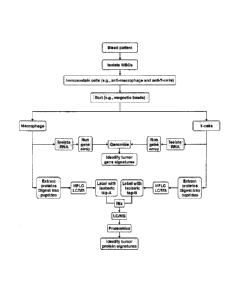
[301 Figure 1 schematically depicts a proposed pathway leading to acquisition
of tumor-
specific DNA. RNA, protein and/or lipid signatures by phagocytes following
engulfment of live CTCs, apoptotic CTCs, fragmented CTCs, tumor DNA, RNA,
proteins, and lipids released by viable and/or apoptotic tumor cells. Note
that only
phagocytic cells (and not non-phagocytic cells) acquire tumor signatures.
[31] Figure 2 schematically depicts an analytical method used in the
identification of
cancer signatures expressed in/by phagocytic cells of patients with ovarian
cancer
(0C).
[32] Figure 3 schematically depicts a general flowchart of one embodiment of a
method of
the invention.
[33] Figure 4 schematically depicts a proposed pathway leading to acquisition
of tumor-
specific DNA, RNA, protein and lipid signatures by blood phagocytes following
engulfment of live CTCs, apoptotic CTCs, fragmented CTCs, tumor DNA, RNA,
proteins and lipids released by viable and/or apoptotic tumor cells. Note that
DNA
contents of phagocytes following phagocytosis is >2n.
CA 02878025 2015-01-15
12
[34] Figure 5 schematically depicts analytical approaches used in the
identification of
breast cancer (BC) signatures in BC-bearing animals.
[35] Figure 6 schematically depicts a general flowchart of another embodiment
of a
method of the invention.
[36] Figure 7 depicts gel electrophoresis analysis of total RNA isolated from
LNCaP and
LLC I cells.
[37] Figure 8 lists the yield and quality of RNA obtained from mouse white
blood cells
(WBCs).
[38] Figures 9A-9D depict arrays showing seven up-regulated (>2 fold), cancer
related
genes detected in neutrophils from LNCaP (human prostate cancer) tumor-bearing
nude mice. (A) LNCaP tumor. (B) Neutrophils obtained from nude mice bearing
LNCaP tumors (NT). (C) T cells obtained from nude mice bearing LNCaP tumors
(TT). (D) Neutrophils obtained from non-tumor-bearing nude mice (NN). Circled
signatures expressed in tumor cells (A) and in neutrophils from tumor-bearing
mice
(B), and minimally expressed in neutrophils from non-tumor-bearing mice (D),
and in
non-phagocytic T cells (C). Expression in NT was >2-fold than that in NN and
TT.
[39] Figures 10A-10D depict arrays showing three up-regulated, cancer related
genes
detected in macrophages from LNCaP (human prostate cancer) tumor-bearing nude
mice. (A) LNCaP tumor. (B) macrophages obtained from nude mice bearing LNCaP
tumors (Mr). (C) T cells obtained from nude mice bearing LNCaP tumors (TT).
(D)
macrophages obtained from non-tumor-bearing nude mice (MN). Circled signatures
expressed in tumor cells (A) and in macrophages from tumor-bearing mice (B),
and
minimally expressed in macrophages from non-tumor-bearing mice (D), and in non-
phagocytic T cells (C). Expression in MT was >2-fold than that in MN and TT.
[40] Figures 11A-11D depict arrays showing four up-regulated (>2 fold), cancer
related
genes detected in neutrophils from LS174T (human colon cancer) tumor-bearing
nude
mice. (A) LS174T tumor. (B) Neutrophils obtained from nude mice bearing LS174T
tumors (NT). (C) T cells obtained from nude mice bearing LS174T tumors (TT).
(D)
Neutrophils obtained from non-tumor-bearing nude mice (NN). Circled signatures
CA 02878025 2015-01-15
13
expressed in tumor cells (A) and in neutrophils from tumor-bearing mice (B),
and
minimally expressed in neutrophils from non-tumor-bearing mice (D), and in non-
phagocytic T cells (C). Expression was NT is >2-fold than that in NN and Ti.
[41] Figures 12A-12D depict arrays showing three up-regulated (>2 fold),
cancer related
genes detected in macrophages from LS 174T (human colon cancer) tumor-bearing
nude mice. (A) LS174T tumor. (B) Macrophages obtained from nude mice bearing
LS174I tumors (MT). (C) I cells obtained from nude mice bearing LS174T tumors
(TT). (D) Macrophages obtained from non-tumor-bearing nude mice (MN). Circled
signatures expressed in tumor cells (A) and in macrophages from tumor-bearing
mice
(B), and minimally expressed in macrophages from non-tumor-bearing mice (D),
and
in non-phagocytic T cells (C). Expression in MT is >2-fold than that in MN and
TT.
[42] Figures 13A-13D depict arrays showing five up-regulated (>2 fold), cancer
related
genes detected in neutrophils from LLC1 (mouse metastatic lung cancer) tumor-
bearing C57/B1 mice. (A) LLC1 tumor. (B) Neutrophils obtained from C57/BI mice
bearing LLC1 tumors (Ni). (C) T cells obtained from C57/BI mice bearing LLC I
tumors (TT). (D) Neutrophils obtained from non-tumor-bearing C57/B1 mice (NN).
Circled signatures expressed in tumor cells (A) and in neutrophils from tumor-
bearing
mice (B), and minimally expressed in neutrophils from non-tumor-bearing mice
(D),
and in non-phagocytic T cells (C). Expression in NT was >2-fold than that in
NN
and TT.
[431 Figures 14A-14D depict arrays showing two up-regulated (>2 fold), cancer
related
genes detected in macrophages from LLC1 (mouse metastatic lung cancer) tumor-
bearing C57/131 mice. (A) LLC1 tumor. (B) Macrophages obtained from C57/B1
mice bearing LLC1 tumors (MT). (C) T cells obtained from C57/B1 mice bearing
LLC I tumors (TT). (D) Macrophages obtained from non-tumor-bearing C57/B1 mice
(MN). Circled signatures expressed in tumor cells (A) and in neutrophils from
tumor-
bearing mice (B), and minimally expressed in neutrophils from non-tumor-
bearing
mice (D), and in non-phagocytic T cells (C). Expression in MT was >2-fold than
that
in MN and TT.
CA 02878025 2015-01-15
14
[44] Figure 15A-15D depict arrays showing two up-regulated (22 fold), cancer
related
genes detected in neutrophils from Bl6F10 (mouse metastatic melanoma) tumor
bearing C57/B1 mice. (A) B16F10 tumor. (B) Neutrophils obtained from C57/B1
mice bearing B16F10 tumors (NT). (C) T cells obtained from C57/B1 mice-bearing
B 16F10 tumors (TT). (D) Neutrophils obtained from non-tumor-bearing C57/B1
mice
(Ny). Circled signatures expressed in tumor cells (A) and in neutrophils from
tumor-
bearing mice (B), and minimally expressed in neutrophils from non-tumor-
bearing
mice (D), and in non-phagocytic T cells (C). Expression in NT was 22-fold than
that
in NN and TT.
[45] Figure 16A-16D depict arrays showing one up-regulated (22 fold), cancer
related
genes detected in macrophages from B16F10 (mouse metastatic melanoma) tumor-
bearing C57/BI mice. (A) B16F10 tumor. (B) Macrophages obtained from C57/B1
mice bearing B16F10 tumors (MT). (C) T cells obtained from C57/B1 mice bearing
B I 6F10 tumors (TT). (D: Macrophages obtained from non-tumor-bearing C57/B1
mice (MN). Circled signatures expressed in tumor cells (A) and in macrophages
from
tumor-bearing mice (B), and minimally expressed in macrophages from non-tumor-
bearing mice (D), and in non-phagocytic T cells (C). Expression in MT was 22-
fold
than that in MN and TT.
[46] Figure 17A-17D depict arrays showing five up-regulated (22 fold), cancer
related
genes detected in neutrophils from patient with head and neck cancer (squamous
cell
carcinoma). (A) Normal tissue (skin) biopsy. (B) Tumor tissue biopsy. (C)
Neutrophils obtained from patient blood (NT). (D) T cells obtained from
patient
blood (TT). Circled signatures expressed in tumor cells (B) and in neutrophils
from
patient blood (C), and minimally expressed or not expressed in normal skin (A)
or
non-phagocytic T cells (D). Expression in NT was 22-fold than that in TT and
skin.
[47] Figure 18A-18D depict arrays showing 23 up-regulated (22 fold), cancer
related
genes detected in macrophages from patient with ovarian cancer
(adenocarcinoma).
(A) Macrophages obtained from patient blood (MT). (B) T cells obtained from
patient
blood (TT). Circled signatures expressed in macrophages from patient (A) and
minimally expressed in non-phagocytic T cells (B). Expression in MI was 22-
fold
than that in TT,
15
[48] Figure 19 depicts a method used to identify tumor signatures in
phagoeytie cells. In
this example, expression intensities of cancer associated genes in macrophages
from
tumor-bearing animals (114.1) were quantified compared to those from T cells
from the
same animals (Ti) and those overexpressed by >2-fold identi.tied. Next, the
intensities of all expressed genes in MT were quantified and compared to those
in
macrophages obtained from non-tumor bearing animals (MO and the genes
overexpressed >2-fold were identified. The genes common to both lists were
selected
and compared to those expressed by the same tumor (shaded area).
149] Figures 20A-20B depict gene expression intensity comparisons in (A)
macrophages
obtained from nude mice bearing LNCaP human prostate tumors (ML,No,p) and T
cells
from the same animals (T cellsLN(>p), (B) Neap and
macrophages obtained from
non-tumor-bearing mice (Ninon-lumor), (C) neutrophils obtained from nude mice
bearing
LNCaP human prostate tumors (NL.Nct,p) and T cells from the same animals (T
cellsiNcap), and (D) NT,Nco and macrophages obtained from non-tumor-bearing
mice
Genes in red were overexpressed >2 fold; those in green were under-
expressed >2 fold.
[50]
[51]
[52] .Figure 21 depicts SDS gel (10%) electrophoresis of protein sample (5.9
i.ig) obtained
from mouse WBC.
[53] 'Figure 22 depicts Western blot analysis of TAG-72 and PSA expression
in T cells
and monoeytesimaeropha.ges (M/M) obtained from tumor-bearing mice,
illustrating
the presence of signatures in phagocytic cells only.
DETAILED DESCRIPTION OF CERTAIN EMBODIMENTS
[54] Embodiments of the present invention are directed to a method of
providing a patient-
specific expression profile of markers associated with diseases, infectious
agents and
CA 2878025 2018-03-08
CA 02878025 2015-01-15
16
bodily conditions based on the cellular content and/or expression profiles of
phagocytic cells. According to one aspect of the present invention, the
cellular
contents and/or expression profiles of phagocytic cells is compared to known
markers
for a particular disease state or condition to detect and/or diagnose the
particular
disease state or condition. According to an additional aspect of the present
invention,
the cellular content and/or expression profile of phagocytic cells is compared
to the
cellular content and/or expression profile of non-phagocytic cells from the
blood of a
single patient. Subtracting the cellular content and/or expression profile
from non-
phagocytic cells from that of phagocytic cells creates a cellular content
and/or
expression profile representative of only the disease state of the individual.
[55] According to an additional embodiment of the present invention, a
phagocytic cell
population from an individual is obtained and the cellular content and/or
expression
profile of phagocytic cells from the population where the DNA content is
greater than
2n is compared with the cellular content and/or expression profile of
phagocytic cells
from the same population where the DNA content is 2n. According to a still
additional embodiment of the present invention, a phagocytic cell population
from an
individual is obtained and the expression profile of phagocytic cells from the
population where the RNA, protein, carbohydrate and/or lipid content is larger
than
normal and have a DNA index greater than 1 is compared with the expression
profile
of phagocytic cells from the same population where the RNA, protein,
carbohydrate
and/or lipid content is normal and/or have a DNA index of 1.
[56] Such a patient specific expression profile eliminates the dependence
on a population-
derived average signature profile for a particular disease or infectious
agent, which
may introduce error into the detection or diagnosis of a particular disease in
the
individual. Such a patient specific expression profile for a disease state of
the present
invention allows detection, diagnosis and treatment to be personalized to the
individual.
[57] With reference to Figures 1-3 and according to certain embodiments of the
present
invention, the gene expression profiles of phagocytic and non-phagocytic WBCs
obtained from mice bearing approximately three week old human subcutaneous
(s.c.)
tumors (prostate LNCaP adenocarcinoma or LS174T colon adenocarcinoma) or
17
mouse tumors (B16F10 metastatic melanoma, administered intravenously, or LLCI
lung cancer, injected s.c.), were compared. The results demonstrated that
neutrophils
and macrophages obtained from these tumor-bearing mice express various
oncogenes
and other cancer-related gene signatures that are also expressed in each of
the
respective tumors. See Figures 9-16 and 19-20, and Table 5. These cancer-
related genes and
oncogenes (e.g., ERBB2, Jan, Fos, etc.) are not expressed or are minimally
expressed
by (i) non-phagocytic I cells isolated from tumor-bearing mice, and (ii)
neutrophils
and macrophages obtained from non-tumor-bearing mice. Furthermore, only the
phagocytic cells from tumor-bearing mice were found to express tumor-specific
proteins. See Figures 21 and 22. CTCs and/or tumor-specific DNA and/or
proteins in
the blood of the mice were phagocyto.sed and some of the tumor-cell DNA, with
its
tumor-specific mutations and genes, was integrated, likely by transfeetion
(without
intending to be bound by theory), into normal phagocyte DNA, transcribed into
RNA,
and translated into protein.
=
1581 With reference to Figures 1-3 and according to certain exemplary
embodiments of the
present invention, the gene expression profiles of phagocytic and non-
phagocytic
WBCs obtained from patients with head and neck tumors or with ovarian cancer
were
also compared. The results demonstrated that neutrophils and macrophages
obtained
from these patients express various oncogenes and other cancer-related gene
signatures that arc also expressed in each of the respective tumors. See
Figures 17-18
and Table 6. These cancer-related genes and oncogenes were not expressed or
were
minimally expressed by non-phagocytic T cells isolated from the same
individual
patient. CTCs and/or tumor-specific DNA and RNA in the blood of the patient
were
phagocytoscd and some of the tumor-cell DNA and/or RNA, with its tumor-
specific
mutations and genes, was integrated, likely through transfection (without
intending to
be bound by theory), into normal phagocyte DNA, transcribed into RNA, and
translated into protein.
[59] With reference to Figures 4-6 and according to certain exemplary
embodiments, the
quantitative analysis of DNA (nuclear and/or mitochondrial), RNA, microRNA,
protein, and/or lipid expression profiles of phagocytic cells (e.g.,
macrophages)
obtained from the blood or one or more other biological samples (e.g., urine,
stool,
CA 2878025 2018-03-08
CA 02878025 2015-01-15
18
saliva, lymph, cerebrospinal fluid and the like) whose (1) DNA content is >2n
(P11>2),
or (2) RNA, protein, carbohydrate and/or lipid content is larger than normal,
i.e., cells
that have phagocytosed CTCs and/or their subeellular fragments or
DNA/RNA/lipids
(i.e., tumor-specific signatures or other disease-specific signatures) and/or
have a
DNA index greater than one, and their comparison with the same phagocytic cell
population (e.g., macrophages) whose (1) DNA content is 2n (Pn-2), or (2) RNA,
protein, carbohydrate and/or lipid content is normal, i.e.. cells that have
not
phagocytosed CTCs and/or their subcellular fragments and have a DNA index of
one,
provides a method to detect tumor-specific (or other disease-specific)
signatures
within the Pn>2 cells (patient-specific signal) that are either not expressed
or minimally
expressed in the Pi,-2 cells (patient-specific noise). With reference to
Figure 6, the
subtraction of the DNA, RNA, protein, and/or lipid profiles of Pn=2 from those
of P11,2
as shown in Figure 5 provides a method to identify (e.g., after one or more
genomic,
proteomic, metabotomic, glycomic, glycoproteomic, tipidomic and/or
lipoproteomic
analyses) tumor-specific (and/or disease-specific and/or condition specific)
signatures
in a blood sample (or one or more other biological samples such as, e.g.,
other bodily
fluids) of an animal and/or a human with cancer (and/or disease and or bodily
conditions) and signify the presence of occult tumor(s) and/or other disease
and/or
other conditions. Unlike the methods described above in which the gcnomic,
proteomic, metabolomic, glycomic, glycoproteomic, lipidomic and/or
lipoproteomic
profiles of phagocytic cells are compared with those of non-phagocytic cells,
the
major advantages of this analytic detection method according to the present
invention
are: (i) it utilizes a single phagocytic cell subpopulation as a source of the
"tumor-
specific" (e.g., Pn>2 macrophage) and "normal-non-specific" (e.g., 1),-.2
macrophage)
signatures, i.e., both share the same baseline genotype; and (ii) the
signature-acquiring
cells (e.g., Pn>2 neutrophiI) are not diluted with those that have not
phagocytosed, and
therefore have not acquired, dead CTCs and/or fragments thereof (e.g., Pn-2
neutrophils).
[60] With reference to Figures 4-6 and according to certain exemplary
embodiments, the
quantitative analysis of phagocytic cells (e.g., macrophages) obtained from
the blood
or one or more other biological samples or bodily fluid (e.g., urine, stool,
saliva,
lymph, cerebrospinal fluid and the like) whose intracellular content
consequent to
CA 02878025 2015-01-15
19
phagocytosis or internalization of other live, dying, or dead (e.g., apoptotic
or
necrotic) cells, apoptotic bodies, nuclei, microvesicles, exosomes,
nucleosomes,
mitochondria, endoplasmic reticulum. and the like, is greater than that of the
same
phagocytic cell population (e.g., macrophages) with normal intracellular
contents
(PNIC), i.e., cells that have not phagocytosed any of the above mentioned
cells
and/cellular debris (patient-specific noise), provides a method to detect
tumor-specific
(or other disease-specific or other condition specific) signatures within the
phagocytes
with increased intracellular content (Plic) that are either not expressed or
minimally
expressed in the phagocytes with a normal intracellular contents (patient-
specific
noise). With reference to Figure 6, the subtraction of the DNA, RNA, protein,
and/or
lipid profiles of PNIC from those of Puc as shown in Figure 5 provides a
method to
identify (e.g., after one or more gcnomic, proteomic, metabolomic, glycomic,
glycoproteomic, lipidomic, and/or lipoproteomic analyses) tumor-specific
(and/or
disease-specific) signatures in a blood sample (or one or more other
biological
samples such as, e.g., other bodily fluids) of an animal with cancer or other
disease
and signify the presence of occult tumor(s) and/or other disease. Unlike the
methods
described above in which the genomic, proteomic, metabolomic, glycomic,
glycoproteomic, lipidomic, and/or lipoproteomic profiles of phagocytic cells
are
compared with those of non-phagocytic cells, the major advantages of this
analytic
detection method according to the present invention are: (i) it utilizes a
single
phagocytic cell subpopulation as a source of the "disease-specific" (e.g., Pm
-
macrophage) and "normal-non-specific" (e.g., PNIC macrophage) signatures,
i.e., both
share the same baseline genotype; and (ii) the signature-acquiring cells
(e.g., Pm'
neutrophil) are not diluted with those that have not phagocytosed, and
therefore have
not acquired, dead CTCs and/or fragments thereof (e.g., Pic neutrophils).
[61] The methods described herein (i) have high specificity, sensitivity, and
accuracy and
should enable the detection of tumor-specific (and/or other disease-specific)
and
normal-nonspecific signatures present within a blood sample (or other
biological
sample such as, e.g., a bodily fluid); and (ii) eliminate the "inequality of
baseline" that
is known to occur among individuals due to intrinsic (e.g., age, gender,
ethnic
background, health status and the like) and temporal variations in gene
expression.
Accordingly, in certain aspects, the invention provides non-invasive assays
for the
20
early detection of occult primary and metastatic tumors (and/or one or more
other
diseases or conditions) in patients, i.e., before the disease can be diagnosed
by
conventional imaging techniques (e.g., PET, MRI, CT and the like), and,
therefore,
provide a foundation for improved decision-making relative to the needs and
strategies for intervention, prevention, and treatment of individuals with
cancer.
1621 As used herein, the term "tumor specific marker" is intended to
include, but is not
limited to, one or more cellular components such as one or more DNA sequences,
one
or more RNA sequences, one or more proteins, one or more polypeptides, one or
more lipids and the like In certain aspects, a tumor specific marker is
present in one
or more WBCs such as, for example, a neutrophil, a macrophage and/or a
dendritic
cell.
1631 As used herein, the term 'cancer related genes" refers to genes such as,
for example,
cancer genes, oneogenes and/or tumor suppressor genes, that have altered
expression
(e.g., increased expression or decreased expression when compared to a non-
cancerous cell) in a cancerous cell (e.g., a WBC such as, for example, a
macrophage.,
a neutrophil, a T cell or the like), Many cancer related genes are known in
the art.
Cancer related genes include for example, but are not limited to. ERBB2, JUN,
RBI,
SUP!'!, MDM2, MA P2KI, AIMP2. PDGFB, PLAUR, FGR, MYCL I, BLYM, NRASI,
PEI, SKI, 7'RKõ4BL2, MYCN, RAB I , REL, RALB, LCO, ERBB4, RAF!, ECT2, KIT,
FGF5, GROI, GRO2, GRO3, FMS, P1/VT, KRASIP, FYN, MYB, ROSI, MASI. RALA,
MYCLKI,GLI3, 4RAF2, MET, BRA!', MOS, LYN, MYBLI, MYC, OVC, VAV2,11M11,
RET, BRAS, SPII, RELA, SEA, EMS], ETSI , KRAS2, ERBB3, GLI, FL?', BRCA2,
RBI, FOS, AKTI, ELK2, FES, MAP, TP.53, CRK, ERBA I , NP], EVI2, ERBBB2,
1NT4, HRC'Al, YES], JUN!), JUNB, MEL, LPSA, VA VI, AKT2, FOSB, RRAS, HAW 1,
HKR2, ERBAL2, SRC, MYBL2, ETS2, ERG, ARAFI, YUASA, ITS2, INT3, SNO,
RMYC, RMYC, PIRASP, TC2 TIM, PT!- I , JAK, one or members of the CEA family
(see, e.g.. Zhou et al. (2001) Gene 264105), P5.4, I 6 and the like.
[641 As used herein, the term "cancer" refers to various types of malignant
neoplasms,
most of which can invade surrounding tissues, and may metastasize to different
sites
(see, for example, PDR Medical Dictionary, 1st edition (1995).
The terms "neoplasm" and "tumor'' refer
CA 2878025 2018-03-08
CA 02878025 2015-01-15
21
to an abnormal tissue that grows by cellular proliferation more rapidly than
normal
and continues to grow after the stimuli that initiated proliferation is
removed. Id.
Such abnormal tissue shows partial or complete lack of structural organization
and
functional coordination with the normal tissue which may be either benign
(i.e.,
benign tumor) or malignant (i.e., malignant tumor),
[65] Examples of general categories of cancer include, but are not
limited to, carcinomas
(i.e., malignant tumors derived from epithelial cells such as, for example,
common
forms of breast, prostate, lung and colon cancer), sarcomas (i.e., malignant
tumors
derived from connective tissue or mesenchymal cells), lymphomas (i.e.,
malignancies
derived from hematopoietic cells), leukemias (i.e., malignancies derived from
hematopoietic cells), germ cell tumors (i.e., tumors derived from totipotent
cells. In
adults most often found in the testicle or ovary; in fetuses, babies and young
children,
most often found on the body midline, particularly at the tip of the
tailbone), blastic
tumors (i.e., a typically malignant tumor which resembles an immature or
embryonic
tissue) and the like.
[66] Examples of the types of neoplasms intended to be encompassed by the
present
invention include but are not limited to those neoplasms associated with
cancers of
neural tissue, blood forming tissue, breast, skin, bone, prostate, ovaries,
uterus, cervix,
liver, lung, brain, larynx, gallbladder, pancreas, rectum, parathyroid,
thyroid, adrenal
gland, immune system, head and neck, colon, stomach, bronchi, and/or kidneys.
[67] In certain exemplary embodiments, one or more methods and/or compositions
described herein are applied to detect, identify and/or diagnose disorders
associated
with the presence of fetal chromosomal abnormalities (e.g., Down's syndrome,
autism
and related autism spectrum disorders (including, but not limited to,
Asperger's
syndrome and pervasive developmental disorder-not otherwise specified), sickle
cell
anemia, thalassemia and the like) consequent to the presence of fetal cells
and DNA
within maternal blood. Screening and diagnosing of one or more of these
disorders
can be performed using the methods and/or compositions described herein to
detect
one or more chromosomal markers, e.g., DNA and RNA, and the like, within
maternal blood phagocytic cells.
CA 02878025 2015-01-15
22
1681 In certain exemplary embodiments, one or more methods and/or compositions
described herein can be applied to test the gender of a fetus within a
pregnant woman
by detecting the presence of fetus-derived proteomic, lipidomic, and/or
genomic
signatures within blood of the pregnant woman, as fetal stem cells, nucleated
erythrocytes, fetal lymphocytes, as well as significant amounts of cell-free
fetal
nucleic acids are known to circulate in maternal blood. According to the
methods
described herein, the cellular content and/or expression profile of phagocytic
cells is
compared to the cellular content and/or expression profile of non-phagocytic
cells
from the blood of a pregnant woman. Subtracting the cellular content and/or
expression profile from non-phagocytic cells from that of phagocytic cells
creates a
cellular content and/or expression profile representative of the gender of the
fetus
being carried by the pregnant woman.
[69] In certain exemplary embodiments, one or more methods and/or compositions
described herein can be used to detect, identify and/or diagnose disorders
associated
with the presence of proteomic and/or genomic myocyte signatures within blood
of
subjects having or at risk of developing cardiac disease (e.g., myocardial
infarction,
chronic heart failure, ischemic heart disease, cardiovascular death and the
like) by
detecting the presence of dying/dead myocytes and/or fragments thereof (e.g.,
DNA,
proteins and the like). Screening and diagnosing of one or more of these
disorders is
performed using methods and/or compositions described herein to detect one or
more
markers, e.g., DNA and RNA, protein and the like, within blood phagocytic
cells.
[70] In certain exemplary embodiments, one or more methods and/or compositions
described herein can be used to detect, identify and/or diagnose disorders
associated
with the presence of protcomic, lipidomic, and/or gcnonnic signatures within
blood of
subjects having or at risk of developing atherosclerosis consequent to
coronary artery
narrowing, abdominal aortic aneurism, and the like. Screening and diagnosing
of
these disorders can performed using the methods and/or compositions described
herein to detect one or more markers, e.g., DNA, RNA, protein and the like,
within
blood phagocytic cells.
[71] Biopsy-confirmed rejection, one method for diagnosis of allograft
rejection, is
invasive and subject to sampling errors. Therefore, the development of
noninvasive
CA 02878025 2015-01-15
23
assays that detect molecular biomarkers for diagnosing and managing
transplanted
organ rejection is useful in management of transplant recipients by (a)
detecting a pre-
rejection profile that will allow therapeutic interventions before rejection
causes graft
dysfunction, (b) improving the sensitivity and specificity of rejection
diagnosis, (c)
developing new classification systems for rejection that will improve
prognosis, and
(d) providing information for designing individualized immunosuppressive
regimens
that could prevent rejection while minimizing drug toxicity.
[72] Accordingly, in certain exemplary embodiments, one or more methods and/or
compositions described herein can be used to detect, identify or diagnose
disorders
associated with the presence of proteomic, lipidomic, and genomic signatures
within
blood of subjects having undergone organ transplants by detecting one or more
markers, e.g., DNA, RNA, protein or the like, within blood phagocytic cells.
[73] Mitochondrial diseases result from failures of the mitochondria.
Cell injury and even
cell death follow. Diseases of the mitochondria appear to cause the most
damage to
cells of the brain, heart, liver, skeletal muscles, kidney and the endocrine
and
respiratory systems as well as diabetes, respiratory complications, seizures,
Alzheimer's disease, visual/hearing problems, lactic acidosis, developmental
delays,
susceptibility to infection, and cancer.
[74] Accordingly, in certain exemplary embodiments, one or more methods and/or
compositions described herein can be used to screen, diagnose and/or detect
mitochondria' disease, by detecting one or more genomic, mitochondria-
associated
DNA markers within blood phagocytic cells.
[75] In certain exemplary embodiments, one or more methods and/or compositions
described herein can be used to screen, diagnose and/or detect Alzheimer's
disease
and/or dementia by detecting one or more markers, e.g., DNA, RNA, protein and
the
like, within blood phagocytic cells.
[76] Systemic lupus erythematosus (SLE) is a complex autoimmune disorder that
affects
various organs and systems. Accordingly, in certain exemplary embodiments, one
or
more methods and/or compositions described herein can be used to screen,
diagnose
CA 02878025 2015-01-15
24
and/or detect SLE by detecting one or more markers, e.g., DNA, RNA, lipids,
protein
and the like, within blood phagocytic cells.
[77] In certain exemplary embodiments, one or more methods and/or compositions
described herein can be used to screen and/or detect gcnomic and/or protcomic
signatures useful in the development of therapeutic and/or imaging molecules
by
detecting one or more markers, e.g., DNA, RNA, protein and the like, within
blood
phagocytic cells.
[78] In certain exemplary embodiments, one or more methods and/or compositions
described herein can be used to screen, diagnose and/or detect alteration in
genomic,
proteornic and/or Iipidomic signatures useful in detection of diseases and
pathologies
consequent to one or more external or internal insults (e.g., dirty bomb
exposure,
radiation exposure, chemical exposure, radiotherapy, radiopharmaceutical
administration, therapeutic molecule exposure, radon exposure, asbestos
exposure,
pollution exposure and the like) by detecting one or more markers, e.g., DNA,
RNA,
protein, lipid and the like, within blood phagocytic cells.
[79] As used herein, the term "organism" includes, but is not limited to, a
human
individual, a non-human primate, a cow, a horse, a sheep, a goat, a pig, a
dog, a cat, a
rabbit, a mouse, a rat, a gerbil, a frog, a toad and a transgenic species
thereof. The
term "organism" further includes pathogenic organisms, including, but not
limited to,
a pathogen such as a parasite, a yeast cell, a yeast tetrad, a yeast colony, a
bacterium,
a bacterial colony, a virion, a virosome, a virus-like particle and/or
cultures of any of
these, and the like.
[80] In certain exemplary embodiments, the assays described herein can be used
for the
detection of an infectious agent and/or the diagnosis of a disorder associated
with an
infection of a cell, tissue, organ or the like by an infectious agent. In
certain aspects,
detection of an infectious agent and/or the diagnosis of a disorder associated
with an
infection is performed using the methods and/or compositions described herein
to
detect one or more infectious agent markers, e.g., DNA, RNA, proteins, lipids
and the
like, from one or more infectious agents.
CA 02878025 2015-01-15
[81] As used herein, the term "infectious agent" includes, but is not
limited to, pathogenic
organisms such as viruses, bacteria, fungi, parasites, infectious proteins and
the like.
[82] Viruses include, but are not limited to, DNA or RNA animal viruses. As
used herein,
RNA viruses include, but are not limited to, virus families such as
Picornaviridae
(e.g., polioviruses), Reoviridae (e.g., rotaviruses), Togaviridae (e.g.,
encephalitis
viruses, yellow fever virus, rubella virus), Orthomyxoviridae (e.g., influenza
viruses),
Paramyxoviridae (e.g., respiratory syncytial virus, measles virus, mumps
virus,
parainfluenza virus), Rhabdoviridae (e.g., rabies virus), Coronaviridae,
Bunyaviridae,
Flaviviridae, Filoviridae, Arenaviridae, Bunyaviridae and Retroviridae (e.g.,
human
T cell lymphotropic viruses (HTLV), human immunodeficiency viruses (HIV)). As
used herein, DNA viruses include, but are not limited to, virus families such
as
Papovaviridae (e.g., papilloma viruses), Adenoviriclae (e.g., adcnovirus),
Herpesviridae (e.g., herpes simplex viruses), and Poxviridae (e.g., variola
viruses).
[83] Bacteria include, but are not limited to, gram positive bacteria, gram
negative
bacteria, acid-fast bacteria and the like.
[84] As used herein, gram positive bacteria include, but are not limited to,
Actinonzedurae,
Actinomyces israelii, Bacillus anthracis, Bacillus cereus, Clostridium
botulinum,
Clostridium difficile, Clostridium pedi-ingens, Clostridium tetani,
Corynebacterium,
Enterococcus faecalis, Listeria monocytogenes, Nocardia, Propionibacterium
acnes,
Staphylococcus aureus, Staphylococcus epiderm, Streptococcus ntutans,
Streptococcus pneuntoniae and the like.
[85] As used herein, gram negative bacteria include, but are not limited to,
Alipia felts,
Bacteriodes, Bartonella bacilliformis, Bortadella pertussis, Borrelia
burgdorferi,
Borrelia recurrentis, Brucella, Calymmatobacterium granutomatis,
Campylobacter,
Escherichia coli, Francisella tularensis, Gardnerella vaginalis, Haemophilius
aegyptius, Haemophilitts ducreyi, Haemophilius influenziae, Heliobacter
pylori,
Legionella pneuntophila, Leptospira interrogans, Neisseria meningitidia,
Porphyromonas gin givalis, Providencia sturti, Pseudomonas aeruginosa,
Salmonella
enteridis, Salmonella typhi, Serratia marce,scens, Shigella boydii,
Streptobacillus
CA 02878025 2015-01-15
26
tnonilifortnis, Streptococcus pyo genes, Treponenta pallidutti, Vibrio
cholerae,
Yersinia enterocolitic:a, Yersinia pestis and the like.
[86] As used herein, acid-fast bacteria include, but are not limited to,
Myobacteriunt
aviutn, Myobacteritnn leprae, Myo bacterium tuberculosis and the like.
[87] As used herein, other bacteria not falling into the other three
categories include, but
are not limited to, BartoneIla henseiae, Chlatnydia psittaci, Chlatnydia
trachornatis,
Coxiella burnetii, Mycoplasina pneuntoniae, Rickettsia akari, Rickettsia
provvazekii,
Rickettsia rickettsii, Rickettsia tsutsuganzushi, Rickettsia typhi,
Ureaplastna
urealyticutn, Diplococcus pneutnoniae, Ehrlichia chalensis, Entemcoccus
laeciutn,
Meningococci and the like.
[88] As used herein, fungi include, but are not limited to, Aspergilli,
Candidae, Candida
albicans, Cocciclioides inunitis, Ctyptococci, and combinations thereof.
[89] As used herein, parasitic microbes include, but are not limited to,
Balantidium coli,
Cryptosporidiutn parwan, Cyclospora cayatanensis, Encephalitozoa, Entattioeba
histolytica, Enterocytozoon bieneusi, Giardia lanthlia, Leishmaniae,
Plasmodii,
Toxoplastna gondii, Ttypanosoniae, trapezoidal amoeba and the like.
[90] As used herein, parasites include worms (e.g., helminthes),
particularly parasitic
worms including, but not limited to, Ncmatoda (roundworms, e.g., whipworms,
hookworms, pinworms, ascarids, filarids and the like), Cestoda (e.g.,
tapeworms)
[91] As used herein, infectious proteins include prions. Disorders caused by
prions
include, but are not limited to, human disorders such as Creutzfeldt-Jakob
disease
(CJD) (including, e.g., iatrogenic Creutzfeldt-Jakob disease (iCJD), variant
Creutzfeldt-Jakob disease (vCJD), familial Creutzfeldt-Jakob disease (fCJD),
and
sporadic Creutzfeldt-Jakob disease (sCJD)), Gerstmann-Straussler-Scheinker
syndrome (GSS), fatal familial insomnia (fFI), sporadic fatal insomnia (sFI),
kuru,
and the like, as well as disorders in animals such as scrapic (sheep and
goats), bovine
spongiform encephalopathy (BSE) (cattle), transmissible mink encephalopathy
(TME)
(mink), chronic wasting disease (CWD) (elk, mule deer), feline spongiform
CA 02878025 2015-01-15
27
encephalopathy (cats), exotic ungulate encephalopathy (EUE) (nyala, oryx,
greater
kudu), spongiform encephalopathy of the ostrich and the like.
[92] In certain exemplary embodiments, methods of detecting markers such as
nucleic acid
sequences (e.g., DNA, RNA and the like), proteins, polypeptides, lipids
polysaccharides and the like in a biological sample are provided. As used
herein, the
term "nucleic acid" is intended to include DNA molecules (e.g., cDNA or
genomic
DNA) and RNA molecules (e.g., mRNA) and analogs of the DNA or RNA generated
using nucleotide analogs. The nucleic acid molecule can be single-stranded or
double-stranded.
[93] As used herein, the term "amino acid" includes organic compounds
containing both a
basic amino group and an acidic carboxyl group. Included within this term are
natural
amino acids (e.g., L-amino acids), modified and unusual amino acids (e.g., D-
amino
acids and 13-amino acids), as well as amino acids which are known to occur
biologically in free or combined form but usually do not occur in proteins.
Natural
protein occurring amino acids include alanine, arginine, asparagine, aspartic
acid,
cysteine, glutamic acid, glutamine, glycine, histidinc, isoleucine, leucine,
lysine,
methionine, phenylalanine, serine, threonine, tyrosine, tryptophan, proline,
and valine.
Natural non-protein amino acids include arginosuccinic acid, citrulline,
cysteine
sulfinic acid, 3,4-dihydroxyphenylalanine, homocysteine, homoserine, omithine,
3 -monoiodotyrosine, 3,5-diiodotryosine, 3 ,5 ,5 ,-triiodothyronine, and
3,31,5,5 I-
tetraiodothyronine. Modified or unusual amino acids include D-amino acids,
hydroxylysine, 4-hydroxyproline, N-Cbz-protected amino acids, 2,4-
diaminobutyric
acid, homoarginine, norleucine, N-methylaminobutyric acid, naphthylalanine,
phenylglycine, .alpha.-phenylproline, tert-leucine, 4-aminocyclohexylalanine,
N-methyl-norleucine, 3 ,4-dehydroproline , N,N-
dimethylaminoglycine,
N-methylaminoglycine, 4-aminopiperidine-4-carboxylic acid, 6-aminocaproic
acid,
trans-4-(aminomethyI)-cyclohexanecarboxylic acid, 2-, 3-, and 4-(aminomethyl.)-
benzoic acid, 1-aminocyclopentanecarboxylic acid, 1-
aminocyclopropanecarboxylic
acid, and 2-benzy1-5-aminopentanoic acid.
1941 As used herein, the term "peptide" includes compounds that consist of two
or more
amino acids that are linked by means of a peptide bond. Peptides may have a
CA 02878025 2015-01-15
28
molecular weight of less than 10,000 Daltons, less than 5,000 Daltons, or less
than
2,500 Daltons. The term "peptide" also includes compounds containing both
peptide
and non-pcptide components, such as pscudopeptide or peptidomimetic residues
or
other non-amino acid components. Such compounds containing both peptide and
non-peptide components may also be referred to as a "peptide analog."
[95] As used herein, the term "protein" includes compounds that consist of
amino acids
arranged in a linear chain and joined together by peptide bonds between the
carboxyl
and amino groups of adjacent amino acid residues.
[96] As used herein, the term "lipid" includes synthetic or naturally-
occurring compounds
which are generally amphipathic and biocompatible. Lipids typically comprise a
hydrophilic component and a hydrophobic component. Exemplary lipids include,
but
are not limited to fatty acids, neutral fats, phosphatides, glycotipids and
the like, As
used herein, the term "lipid composition" refers to a composition which
comprises a
lipid compound, typically in an aqueous medium. Exemplary lipid compositions
include, but are not limited to, suspensions, emulsions, vesicle compositions
and the
like.
[97] An exemplary method for detecting the presence or absence of a
polypeptide or
nucleic acid corresponding to a marker of the invention in a biological sample
involves obtaining a biological sample (e.g., a bodily fluid sample (e.g.,
blood) and/or
tumor sample) from a test subject and contacting the biological sample with a
compound or an agent capable of detecting one or more markers (e.g., DNA, RNA,
protein, polypeptide, carbohydrate, lipid or the like).
[98] Detection methods described herein can be used to detect one or more
markers (e.g.,
DNA, RNA, protein, polypeptide, carbohydrate, lipid or the like) in a
biological
sample in vitro as well as in vivo. For example, in vitro techniques for
detection of
mRNA include Northern hybridizations and in situ hybridizations. In vitro
techniques
for detection of a polypeptide corresponding to a marker of the invention
include
enzyme linked immunosorbent assays (ELISAs), Western blots,
immunoprecipitations and immunofluorescence. In vitro techniques for detection
of
genomic DNA include Southern hybridizations. Furthermore, in vivo techniques
for
CA 02878025 2015-01-15
29
detection of a polypeptide corresponding to a marker of the invention include
introducing into a subject a labeled antibody directed against the
polypeptide. For
example, the antibody can be labeled with a radioactive marker whose presence
and
location in a subject can be detected by standard imaging techniques.
[99] Methods for analyzing lipid content in a biological sample are
known in the art (See,
e.g., Kang et al. (1992) Biochint. Biophys. Acta. 1128:267; Weylandt et al.
(1996)
Lipids 31:977; J. Schiller et al. (1999) Anal. Biochetn. 267:46; Kang et al.
(2001)
Proc. Natl. Acad. Sci. USA 98:4050; Schiller et al. (2004) Prog. Lipid Res.
43:499).
An exemplary method of lipid analysis is to extract lipids from a biological
sample
(e.g. using chloroform:methanol (2:1, vol:vol) containing 0.005% butylated
hydroxytoluene (BHT, as an antioxidant)), prepare fatty acid methyl esters
were (e.g.,
14% BF3-methanol reagent), and quantifying the fatty acid methyl esters are
quantified (e.g., by HPLC, TLC, by gas chromatography-mass spectroscopy using
commercially available gas chromatographs, mass spectrometers, and/or
combination
gas chromatograph/mass spectrometers). Fatty acid mass is determined by
comparing
areas of various analyzed fatty acids to that of a fixed concentration of
internal
standard.
[100] A general principle of diagnostic and prognostic assays involves
preparing a sample
or reaction mixture that may contain a marker (e.g., one or more of DNA, RNA,
protein, polypeptide, carbohydrate, lipid and the like) and a probe under
appropriate
conditions and for a time sufficient to allow the marker and probe to interact
and bind,
thus forming a complex that can be removed and/or detected in the reaction
mixture.
These assays can be conducted in a variety of ways.
[101] For example, one method to conduct such an assay would involve anchoring
the
marker or probe onto a solid phase support, also referred to as a substrate,
and
detecting target marker/probe complexes anchored on the solid phase at the end
of the
reaction. In one embodiment of such a method, a sample from a subject, which
is to
be assayed for presence and/or concentration of marker, can be anchored onto a
carrier or solid phase support, In another embodiment, the reverse situation
is
possible, in which the probe can be anchored to a solid phase and a sample
from a
subject can be allowed to react as an unanchored component of the assay.
CA 02878025 2015-01-15
11021 There are many established methods for anchoring assay components to a
solid phase.
These include, without limitation, marker or probe molecules which are
immobilized
through conjugation of biotin and streptavidin. Such biotinylated assay
components
can be prepared from biotin-NHS(N-hydroxy-succinimide) using techniques known
in
the art (e.g., biotinylation kit, Pierce Chemicals, Rockford, IL), and
immobilized in
the wells of streptavidin-coated 96 well plates (Pierce Chemical). In certain
embodiments, the surfaces with immobilized assay components can be prepared in
advance and stored.
[103] Other suitable carriers or solid phase supports for such assays include
any material
capable of binding the class of molecule to which the marker or probe belongs.
Well
known supports or carriers include, but are not limited to, glass,
polystyrene, nylon,
polypropylene, nylon, polyethylene, dcxtran, amylases, natural and modified
celluloses, polyacrylamides, gabbros, and magnetite.
[104] In order to conduct assays with the above mentioned approaches, the non-
immobilized component is added to the solid phase upon which the second
component is anchored. After the reaction is complete, uncomplexed components
may be removed (e.g., by washing) under conditions such that any complexes
formed
will remain immobilized upon the solid phase. The detection of marker/probe
complexes anchored to the solid phase can be accomplished in a number of
methods
outlined herein.
[105] In certain exemplary embodiments, the probe, when it is the unanchored
assay
component, can be labeled for the purpose of detection and readout of the
assay,
either directly or indirectly, with detectable labels discussed herein and
which are
well-known to one skilled in the art.
[106] It is also possible to directly detect marker/probe complex formation
without further
manipulation or labeling of either component (marker or probe), for example by
utilizing the technique of fluorescence energy transfer (see, for example,
U.S. Patent
Nos. 5,631,169 and 4,868,103). A fluorophore label on the first, 'donor'
molecule is
selected such that, upon excitation with incident light of appropriate
wavelength, its
emitted fluorescent energy will be absorbed by a fluorescent label on a second
CA 02878025 2015-01-15
31
'acceptor' molecule, which in turn is able to fluoresce due to the absorbed
energy.
Alternately, the 'donor' protein molecule may simply utilize the natural
fluorescent
energy of tryptophan residues. Labels are chosen that emit different
wavelengths of
light, such that the 'acceptor' molecule label may be differentiated from that
of the
'donor'. Since the efficiency of energy transfer between the labels is related
to the
distance separating the molecules, spatial relationships between the molecules
can be
assessed. In a situation in which binding occurs between the molecules, the
fluorescent emission of the 'acceptor' molecule label in the assay should be
maximal.
An FET binding event can be conveniently measured through standard
fluorometric
detection means well known in the art (e.g., using a fluorimeter).
[107] In another embodiment, determination of the ability of a probe to
recognize a marker
can be accomplished without labeling either assay component (probe or marker)
by
utilizing a technology such as real-time Biomolecular Interaction Analysis
(BIA) (see,
e.g., Sjolander, S. and Urbaniczky, C., 1991, Anal. Chem. 63:2338 2345 and
Szabo et
al., 1995, Curr. Opin. Struct. Biol. 5:699 705). As used herein, "BIA" or
"surface
piasmon resonance" is a technology for studying biospecific interactions in
real time,
without labeling any of the interactants (e.g., BIAcore). Changes in the mass
at the
binding surface (indicative of a binding event) result in alterations of the
refractive
index of light near the surface (the optical phenomenon of surface plasmon
resonance
(SPR)), resulting in a detectable signal which can be used as an indication of
real-time
reactions between biological molecules.
11081 Alternatively, in another embodiment, analogous diagnostic and
prognostic assays can
be conducted with marker and probe as solutes in a liquid phase. In such an
assay, the
complexed marker and probe are separated from uncomplexed components by any of
a number of standard techniques, including but not limited to: differential
centrifugation, chromatography, electrophoresis and immunoprecipitation. In
differential centrifugation, marker/probe complexes may be separated from
uncomplexed assay components through a series of centrifugal steps, due to the
different sedimentation equilibria of complexes based on their different sizes
and
densities (see, for example, Rivas and Minton (1993) Trends Biochem. Sci.
18:284).
Standard chromatographic techniques may also be utilized to separate complexed
CA 02878025 2015-01-15
32
molecules from uncomplexed ones. For example, gel filtration chromatography
separates molecules based on size, and through the utilization of an
appropriate gel
filtration resin in a column format, for example, the relatively larger
complex may be
separated from the relatively smaller uncomplexed components. Similarly, the
relatively different charge properties of the marker/probe complex as compared
to the
uncomplexed components may be exploited to differentiate the complex from
uncomplexed components, for example through the utilization of ion-exchange
chromatography resins. Such resins and chromatographic techniques are well
known
to one skilled in the art (see, e.g., Heegaard (1998) .1. Mo1 Recognit.
11:141; Hage
and Tweed (1997)J. Chroniatogr. B. Bionted. Sci. Appl. 12:499). Gel
electrophoresis
may also be employed to separate complexed assay components from unbound
components (see, e.g., Ausubel et al., ed., Current Protocols in Molecular
Biology,
John Wiley & Sons, New York, 1987 1999). In this technique, protein or nucleic
acid
complexes are separated based on size or charge, for example. In order to
maintain
the binding interaction during the electrophoretic process, non-denaturing gel
matrix
materials and conditions in the absence of reducing agent are typically
preferred.
Appropriate conditions to the particular assay and components thereof will be
well
known to one skilled in the art.
[109] In certain exemplary embodiments, the level of mRNA corresponding to the
marker
can be determined either by in situ and/or by in vitro formats in a biological
sample
using methods known in the art. Many expression detection methods use isolated
RNA. For in vitro methods, any RNA isolation technique that does not select
against
the isolation of mRNA can be utilized for the purification of RNA from blood
cells
(see, e.g., Ausubel et al, ed., Current Protocols in Molecular Biology, John
Wiley &
Sons, New York 1987 1999). Additionally, large numbers of cells and/or samples
can
readily be processed using techniques well known to those of skill in the art,
such as,
for example, the single-step RNA isolation process of Chomczynski (1989, U.S.
Patent No. 4,843,155).
[1101 Isolated mRNA can be used in hybridization or amplification assays that
include, but
arc not limited to, Southern or Northern analyses, polymerase chain reaction
analyses
and probe arrays. In certain exemplary embodiments, a diagnostic method for
the
CA 02878025 2015-01-15
33
detection of mRNA levels involves contacting the isolated mRNA with a nucleic
acid
molecule (probe) that can hybridize to the mRNA encoded by the gene being
detected. The nucleic acid probe can be, for example, a full-length cDNA, or a
portion thereof, such as an oligonucleotide of at least 7, 15, 30, 50, 100,
250 or 500
nucleotides in length and sufficient to specifically hybridize under stringent
conditions to an mRNA or genomic DNA encoding a marker of the present
invention.
Other suitable probes for use in the diagnostic assays of the invention are
described
herein. Hybridization of an mRNA with the probe indicates that the marker in
question is being expressed.
[111] In one format, the mRNA is immobilized on a solid surface and contacted
with a
probe, for example by running the isolated mRNA on an agarose gel and
transferring
the mRNA from the gel to a membrane, such as nitrocellulose. In an alternative
format, the probe(s) are immobilized on a solid surface and the mRNA is
contacted
with the probe(s), for example, in a gene chip array. A skilled artisan can
readily
adapt known mRNA detection methods for use in detecting the level of mRNA
encoded by the markers of the present invention.
11121 An alternative method for determining the level of mRNA corresponding to
a marker
of the present invention in a sample involves the process of nucleic acid
amplification,
e.g., by rtPCR (the experimental embodiment set forth in U.S. Patent Nos.
4,683,195
and 4,683,202), COLD-PCR (Li et al. (2008) Nat. Med. 14:579), ligase chain
reaction
(Barany, 1991, Proc. Natl. Acad. Sci. USA, 88:189), self sustained sequence
replication (Guatelli et al., 1990, Proc. Natl. Acad. Sc!. USA 87:1874),
transcriptional
amplification system (Kwoh et al. (1989) Proc. Natl. Acad. Sci. USA 86:1173),
Q-
Beta Replicase (Lizardi et al. (1988) Bio/Technologv, 6:1197), rolling circle
replication (U.S. Patent No. 5,854,033) or any other nucleic acid
amplification
method, followed by the detection of the amplified molecules using techniques
well
known to those of skill in the art. These detection schemes are especially
useful for
the detection of nucleic acid molecules if such molecules are present in very
low
numbers. As used herein, amplification primers are defined as being a pair of
nucleic
acid molecules that can anneal to 5' or 3' regions of a gene (plus and minus
strands,
respectively, or vice-versa) and contain a short region in between. In
general,
CA 02878025 2015-01-15
34
amplification primers are from about 10 to 30 nucleotides in length and flank
a region
from about 50 to 200 nucleotides in length. Under appropriate conditions and
with
appropriate reagents, such primers permit the amplification of a nucleic acid
molecule
comprising the nucleotide sequence flanked by the primers.
[113] For in situ methods, mRNA does not need to be isolated from the sample
(e.g., a
bodily fluid (e.g., blood cells)) prior to detection. In such methods, a cell
or tissue
sample is prepared/processed using known histological methods. The sample is
then
immobilized on a support, typically a glass slide, and then contacted with a
probe that
can hybridize to mRNA that encodes the marker.
[114] As an alternative to making determinations based on the absolute
expression level of
the marker, determinations may be based on the normalized expression level of
the
marker. Expression levels are normalized by correcting the absolute expression
level
of a marker by comparing its expression to the expression of a gene that is
not a
marker, e.g., a housekeeping gene that is constitutively expressed. Suitable
genes for
normalization include housekeeping genes such as the actin gene, or epithelial
cell-
specific genes. This normalization allows the comparison of the expression
level in a
patient sample from one source to a patient sample from another source, e.g.,
to
compare a phagocytic blood cell from an individual to a non-phagocytic blood
cell
from the individual.
[115] In another exemplary embodiment, a protein or polypeptide corresponding
to a
marker is detected. In certain exemplary embodiments, an agent for detecting a
polypeptide of the invention is an antibody capable of binding to a
polypeptide
corresponding to a marker of the invention, such as an antibody with a
detectable
label. Antibodies can be polyclonal, or more preferably, monoclonal. An intact
antibody, or a fragment thereof (e.g., Fab or F(a02) can be used. The term
"labeled,"
with respect to the probe or antibody, is intended to encompass direct
labeling of the
probe or antibody by coupling (i.e., physically linking) a detectable
substance to the
probe or antibody, as well as indirect labeling of the probe or antibody by
reactivity
with another reagent that is directly labeled. Examples of indirect labeling
include
detection of a primary antibody using a fluorescently labeled secondary
antibody and
CA 02878025 2015-01-15
end-labeling of a DNA probe with biotin such that it can be detected with
fluorescently labeled streptavidin.
[116] A variety of formats can be employed to determine whether a sample
contains a
protein that binds to a given antibody. Examples of such formats include, but
arc not
limited to, enzyme immunoassay (EIA), radioimmunoassay (RIA), Western blot
analysis, enzyme linked immunoabsorbant assay (ELISA) and the like. A skilled
artisan can readily adapt known protein/antibody detection methods for use in
determining whether cells (e.g., bodily fluid cells such as blood cells)
express a
marker of the present invention.
[117] In one format, antibodies, or antibody fragments, can be used in methods
such as
Western blots or immunofluorescence techniques to detect the expressed
proteins. In
such uses, it is generally preferable to immobilize either the antibody or
proteins on a
solid support. Suitable solid phase supports or carriers include any support
capable of
binding an antigen or an antibody. Well known supports or carriers include
glass,
polystyrene, polypropylene, polyethylene, dextran, nylon, amylases, natural
and
modified celluloses, polyacrylamides, gabbros, magnetite and the like.
[118] One skilled in the art will know many other suitable carriers for
binding antibody or
antigen, and will be able to adapt such support for use with the present
invention. For
example, protein isolated from cells (e.g., bodily fluid cells such as blood
cells) can be
run on a polyacrylamide gel electrophoresis and immobilized onto a solid phase
support such as nitrocellulose. The support can then be washed with suitable
buffers
followed by treatment with the detectably labeled antibody. The solid phase
support
can then be washed with the buffer a second time to remove unbound antibody.
The
amount of bound label on the solid support can then be detected by
conventional
means.
[119] In certain exemplary embodiments, diagnostic assays are provided. An
exemplary
method for detecting the presence or absence of a bodily condition, a disease
and/or
disorder associated with cancer, an infectious agent, and/or another disease
in a
biological sample involves obtaining a biological sample from a test subject
and
contacting the biological sample with a compound or an agent capable of
detecting
CA 02878025 2015-01-15
36
one or more of the markers of a disease and/or disorder associated with
cancer, an
infectious agent, and/or another disease or condition, e.g., marker nucleic
acid (e.g.,
mRNA, gcnomic DNA), marker peptide (e.g., polypeptide or protein) or marker
lipid
encoded by the marker nucleic acid such that the presence of a marker nucleic
acid or
marker peptide encoded by the nucleic acid is detected in the biological
sample. In
one embodiment, an agent for detecting marker mRNA or genomic DNA is a labeled
nucleic acid probe capable of hybridizing to marker mRNA or genomic DNA. The
nucleic acid probe can be, for example, a full-length marker nucleic acid or a
portion
thereof. Other suitable probes for use in the diagnostic assays of the
invention are
described herein.
[120] An agent for detecting marker peptide can be an antibody capable of
binding to a
marker peptide, such as an antibody with a detectable label. Antibodies can be
polyclonal or monoclonal. An intact antibody, or a fragment thereof (e.g., Fab
or
F(ab')?) can be used. The term "labeled," with regard to the probe or
antibody, is
intended to encompass direct labeling of the probe or antibody by coupling
(i.e.,
physically linking) a detectable substance to the probe or antibody, as well
as indirect
labeling of the probe or antibody by reactivity with another reagent that is
directly
labeled. Examples of indirect labeling include detection of a primary antibody
using a
fluorescently labeled secondary antibody and end-labeling of a DNA probe with
biotin such that it can be detected with fluorescently labeled streptavidin.
11211 As used herein, the term "biological sample" is intended to include
tissues, cells (e.g.,
phagocytic cells, non-phagocytic cells, 2n cells, >2n cells and the like) and
biological
fluids (e.g., whole blood, WBCs and the like) isolated from a subject, as well
as
tissues, cells (e.g., phagocytic cells, non-phagocytic cells, 2n cells, >2n
cells and the
like) and bodily fluids (e.g., urine, whole blood, WBCs and the like) present
within a
subject. That is, the detection method of the invention can be used to detect
marker
polypeptide, protein, carbohydrate, lipid, oligosaccharide, mRNA, microRNA,
genomic DNA and the like in a biological sample in vitro as well as in vivo.
In one
embodiment, the biological sample contains proteins, polypeptides, lipids
and/or
oligosaccharides from the test subject. Alternatively, the biological sample
can
contain mRNA molecules from the test subject and/or genomic DNA molecules from
CA 02878025 2015-01-15
37
the test subject. In one embodiment biological sample is a serum sample,
saliva
sample or a biopsy sample isolated by conventional means from a subject.
[1221 In another embodiment, the methods further involve obtaining a control
biological
sample from a control subject (e.g., non-phagocytic cell or 2n cell),
contacting the
control sample (e.g., non-phagocytic cell or 2n cell) with a compound or agent
capable of detecting marker polypeptide, protein lipid, oligosaccharide, mRNA,
microRNA, genomic DNA and the like is detected in the biological sample, and
comparing the presence of marker polypeptide, protein lipid, oligosaccharide,
mRNA,
genomic DNA and the like in control sample with the presence of marker
polypeptide,
protein lipid, oligosaccharide, mRNA, genomic DNA and the like in the test
sample
(e.g., phagocytic cell or >2n cell). Alternatively, the presence of marker
polypeptide,
protein lipid, oligosaccharide, mRNA, genomic DNA and the like in the test
sample
(e.g., phagocytic cell or >2n cell) can be compared with information in a
database or
on a chart to result in detection or diagnosis.
[123] The invention also encompasses kits for detecting the presence of one or
more
markers associated with cancer and/or an infectious agent in a biological
sample. For
example, the kit can comprise a labeled compound or agent capable of detecting
marker polypeptide, protein lipid, oligosaccharide, mRNA, microRNA, genomic
DNA and the like in a biological sample; means for determining the amount of
marker
in the sample; and means for comparing the amount of marker in the sample with
a
standard (e.g., a non-phagocytic cell or a 2n cell). The compound or agent can
be
packaged in a suitable container. The kit can further comprise instructions
for using
the kit to detect marker peptide or nucleic acid.
[124] In certain exemplary embodiments, prognostic assays are provided. The
diagnostic
methods described herein can furthermore be utilized to identify subjects
having a
condition or at risk of developing a disease and/or disorder associated with
cancer
andlor an infectious agent, or another disorder described herein associated
with
upregulated (or downregulated) expression of one or more of the markers
described
herein. For example, the assays described herein, such as the preceding
diagnostic
assays or the following assays, can be utilized to identify a subject having
or at risk of
CA 02878025 2015-01-15
38
developing a disease and/or disorder associated with cancer and/or an
infectious agent
and/or one or more other disorders described herein.
[1251 The prognostic assays described herein can be used to determine whether
a subject
can be administered an agent (e.g., an agonist, antagonist, peptidomimetic,
protein,
peptide, nucleic acid, small molecule, or other drug candidate) to treat a
disease
and/or disorder associated with cancer and/or an infectious agent, and/or one
or more
other disorders described herein associated with one or more of the markers
described
herein. For example, such methods can be used to determine whether a subject
can be
effectively treated with an agent for treating, ameliorating or reducing one
or more
symptoms associated with cancer. Thus, the present invention provides methods
for
determining whether a subject can be effectively treated with an agent for a
disease
and/or disorder associated with cancer and/or an infectious agent, and/or one
or more
other disorders described herein.
[1261 The methods of the invention can also be used to detect genetic
alterations in a marker
gene, thereby determining if a subject with the altered gene is at risk for
developing a
disease and/or disorder associated with cancer and/or an infectious agent,
and/or one
or more other disorders described herein characterized by misregulation in a
marker
protein activity or nucleic acid expression, such as cancer. In certain
embodiments,
the methods include detecting, in a sample of cells (e.g., bodily fluid cells
such as
blood cells) from the subject, the presence or absence of a genetic alteration
characterized by an alteration affecting the integrity of a gene encoding a
marker
peptide and/or a marker gene. For example, such genetic alterations can be
detected
by ascertaining the existence of at least one of: 1) a deletion of one or more
nucleotides from one or more marker genes; 2) an addition of one or more
nucleotides
to one or more marker genes; 3) a substitution of one or more nucleotides of
one or
more marker genes, 4) a chromosomal rearrangement of one or more marker genes;
5)
an alteration in the level of a messenger RNA transcript of one or more marker
genes;
6) aberrant modification of one or more marker genes, such as of the
methylation
pattern of the genomic DNA; 7) the presence of a non-wild type splicing
pattern of a
messenger RNA transcript of one or more marker genes; 8) a non-wild type level
of a
one or more marker proteins; 9) allelic loss of one or more marker genes; and
10)
CA 02878025 2015-01-15
39
inappropriate post-translational modification of one or more marker proteins.
As
described herein, there are a large number of assays known in the art which
can be
used for detecting alterations in one or more marker genes.
[127] In certain embodiments, detection of the alteration involves the use of
a probe/primer
in a polymerase chain reaction (PCR) (see, e.g., U.S. Patent Nos. 4,683,195,
4,683,202 and 5,854,033), such as real-time PCR, COLD-PCR, anchor PCR,
recursive PCR or RACE PCR, or, alternatively, in a ligation chain reaction
(LCR)
(see, e.g., Landegran et al. (1988) Science 241:1077; Prodromou and Pearl
(1992)
Protein Eng. 5:827; and Nakazawa et al. (1994) Proc. Natl. Acad. Sci. USA
91:360),
the latter of which can be particularly useful for detecting point mutations
in a marker
gene (see Abravaya et al. (1995) Nucleic Acids Rec. 23:675). This method can
include the steps of collecting a sample of cells from a subject, isolating
nucleic acid
(e.g., genomic, mRNA or both) from the cells of the sample, contacting the
nucleic
acid sample with one or more primers which specifically hybridize to a marker
gene
under conditions such that hybridization and amplification of the marker gene
(if
present) occurs, and detecting the presence or absence of an amplification
product, or
detecting the size of the amplification product and comparing the length to a
control
sample. It is anticipated that PCR and/or LCR may be desirable to use as a
preliminary amplification step in conjunction with any of the techniques used
for
detecting mutations described herein.
[128] Alternative amplification methods include: self sustained sequence
replication
(Guatelli et al., (1990) Proc. Natl. Acad. Sci. USA 87:1874), transcriptional
amplification system (Kwoh et al., (1989) Proc. Natl. Acad. Sci. USA 86:1173),
Q-Beta Replicase (Lizardi et al. (1988) Bio-Technolog,v 6:1197), or any other
nucleic
acid amplification method, followed by the detection of the amplified
molecules using
techniques well known to those of skill in the art. These detection schemes
are
especially useful for the detection of nucleic acid molecules if such
molecules are
present in very low numbers.
[129] In an alternative embodiment, mutations in one or more marker genes from
a sample
cell can be identified by alterations in restriction enzyme cleavage patterns.
For
example, sample and control DNA is isolated, optionally amplified, digested
with one
CA 02878025 2015-01-15
or more restriction endonucleases, and fragment length sizes are determined by
gel
electrophoresis and compared. Differences in fragment length sizes between
sample
and control DNA indicates mutations in the sample DNA. Moreover, the use of
sequence specific ribozymes (see, for example, U.S. Pat. No. 5,498,531) can be
used
to score for the presence of specific mutations by development or loss of a
ribozyme
cleavage site.
[130] In other embodiments, genetic mutations in one or more of the markers
described
herein can be identified by hybridizing a sample and control nucleic acids,
e.g., DNA
or RNA, to high density arrays containing hundreds or thousands of
oligonucleotides
probes (Cronin et al. (1996) Human Mutation 7: 244; Kozal et at. (1996) Nature
Medicine 2:753). For example, genetic mutations in a marker nucleic acid can
be
identified in two dimensional arrays containing light-generated DNA probes as
described in Cronin, M. T. et al. supra. Briefly, a first hybridization array
of probes
can be used to scan through long stretches of DNA in a sample and control to
identify
base changes between the sequences by making linear arrays of sequential
overlapping probes. This step allows the identification of point mutations.
This step
is followed by a second hybridization array that allows the characterization
of specific
mutations by using smaller, specialized probe arrays complementary to all
variants or
mutations detected. Each mutation array is composed of parallel probe sets,
one
complementary to the wild-type gene and the other complementary to the mutant
gene.
11311 In yet another embodiment, any of a variety of sequencing reactions
known in the art
can be used to directly sequence a marker gene and detect mutations by
comparing the
sequence of the sample marker gene with the corresponding wild-type (control)
sequence. Examples of sequencing reactions include those based on techniques
developed by Maxam and Gilbert ((1977) Proc. Natl. Acad. Sci. USA 74:560) or
Sanger ((1977) Proc. Natl. Acad. Sci. USA 74:5463). It is also contemplated
that any
of a variety of automated sequencing procedures can be utilized when
performing the
diagnostic assays ((1995) Biotechniques 19:448), including sequencing by mass
spectrometry (see, e.g., PCT International Publication No. WO 94/16101; Cohen
et al.
CA 02878025 2015-01-15
41
(1996) Adv. Chrotnatogr. 36:127-162; and Griffin et al. (1993) App!. Biochetn.
Biotechnol. 38:147).
[132] Other methods for detecting mutations in a marker gene include methods
in which
protection from cleavage agents is used to detect mismatched bases in RNA/RNA
or
RNA/DNA heteroduplexes (Myers et al. (1985) Science 230:1242). In general, the
art
technique of "mismatch cleavage" starts by providing heteroduplexes formed by
hybridizing (labeled) RNA or DNA containing the wild-type marker sequence with
potentially mutant RNA or DNA obtained from a tissue sample. The double-
stranded
duplexes are treated with an agent which cleaves single-stranded regions of
the duplex
such as which will exist due to base pair mismatches between the control and
sample
strands. For instance, RNA/DNA duplexes can be treated with RNase and
DNA/DNA hybrids treated with Si nuclease to enzymatically digesting the
mismatched regions. In other embodiments, either DNA/DNA or RNA/DNA
duplexes can be treated with hydroxylamine or osmium tetroxide and with
piperidine
in order to digest mismatched regions. After digestion of the mismatched
regions, the
resulting material is then separated by size on denaturing polyacrylamide gels
to
determine the site of mutation. See, for example, Cotton et al. (1988) Proc.
Natl.
Acad. Sci. USA 85:4397; Saleeba et al. (1992) Methods Enzymol. 217:286. In one
embodiment, the control DNA or RNA can be labeled for detection.
[133] In still another embodiment, the mismatch cleavage reaction employs one
or more
proteins that recognize mismatched base pairs in double-stranded DNA (so
called
"DNA mismatch repair" enzymes) in defined systems for detecting and mapping
point mutations in marker cDNAs obtained from samples of cells. For example,
the
mutY enzyme of E. coli cleaves A at G/A mismatches and the thymidinc DNA
glycosylase from HeLa cells cleaves T at G/T mismatches (Hsu et al. (1994)
Carcinogenesis 15:1657). According to an exemplary embodiment, a probe based
on
a marker sequence, e.g., a wild-type marker sequence, is hybridized to a cDNA
or
other DNA product from a test cell(s). The duplex is treated with a DNA
mismatch
repair enzyme, and the cleavage products, if any, can be detected from
electrophoresis
protocols or the like. Sec, for example, U.S. Patent No. 5,459,039.
CA 02878025 2015-01-15
42
[134] In other embodiments, alterations in electrophoretic mobility will be
used to identify
mutations in marker genes. For example, single strand conformation
polymorphism
(SSCP) may be used to detect differences in electrophoretic mobility between
mutant
and wild type nucleic acids (Orita et al. (1989) Proc. Natl. Acad. Sci. USA
86:2766,
see also Cotton (1993) Mutat. Res. 285:125; and Hayashi (1992) Genet. Anal.
Tech,
Appl. 9:73). Single-stranded DNA fragments of sample and control marker
nucleic
acids will be denatured and allowed to renature. The secondary structure of
single-
stranded nucleic acids varies according to sequence, the resulting alteration
in
electrophoretic mobility enables the detection of even a single base change.
The
DNA fragments may be labeled or detected with labeled probes. The sensitivity
of
the assay may be enhanced by using RNA (rather than DNA), in which the
secondary
structure is more sensitive to a change in sequence. In one embodiment, the
subject
method utilizes heteroduplex analysis to separate double stranded heteroduplex
molecules on the basis of changes in electrophoretic mobility (Keen et al.
(1991)
Trends Genet. 7:5).
[135] In yet another embodiment the movement of mutant or wild-type fragments
in
polyaerylamide gels containing a gradient of denaturant is assayed using
denaturing
gradient gel electrophoresis (DGGE) (Myers et al. (1985) Nature 313:495). When
DGGE is used as the method of analysis, DNA will be modified to insure that it
does
not completely denature, for example by adding a GC clamp of approximately 40
bp
of high-melting GC-rich DNA by PCR. In a further embodiment, a temperature
gradient is used in place of a denaturing gradient to identify differences in
the
mobility of control and sample DNA (Rosenbaum and Reissner (1987) Biophys.
Chem. 265:12753).
[1361 Examples of other techniques for detecting point mutations include, but
are not
limited to, selective oligonucleotide hybridization, selective amplification
or selective
primer extension. For example, oligonucleotide primers may be prepared in
which
the known mutation is placed centrally and then hybridized to target DNA under
conditions which permit hybridization only if a perfect match is found (Saiki
et al.
(1986)Nature 324:163; Saiki et al. (1989) Proc. Natl. Acad. Sci. USA 86:6230).
Such
allele specific oligonucleotides are hybridized to PCR amplified target DNA or
a
CA 02878025 2015-01-15
43
number of different mutations when the oligonucleotides are attached to the
hybridizing membrane and hybridized with labeled target DNA.
[137] Alternatively, allele specific amplification technology which depends on
selective
PCR amplification may be used in conjunction with the instant invention.
Oligonucleotides used as primers for specific amplification may carry the
mutation of
interest in the center of the molecule (so that amplification depends on
differential
hybridization) (Gibbs et al. (1989) Nucl. Acids Res. 17:2437) or at the
extreme 3' end
of one primer where, under appropriate conditions, mismatch can prevent, or
reduce
polymerase extension (Prossner (1993) Tibtech 11:238). In addition it may be
desirable to introduce a novel restriction site in the region of the mutation
to create
cleavage-based detection (Gasparini et al. (1992) Mol, Cell Probes 6:1). It is
anticipated that in certain embodiments amplification may also be performed
using
Tay ligase for amplification (Barany (1991) Proc. Natl. Acad. Sci. USA
88:189). In
such cases, ligation will occur only if there is a perfect match at the 3' end
of the 5'
sequence making it possible to detect the presence of a known mutation at a
specific
site by looking for the presence or absence of amplification.
[138]
[139] The following examples are set forth as being representative of the
present invention.
These examples are not to be construed as limiting the scope of the invention
as these
and other equivalent embodiments will be apparent in view of the present
disclosure,
figures, and accompanying claims.
CA 02878025 2015-01-15
44
EXAMPLE 1
Representative Method 1 for the Separation of Phagocytic Cells from Non-
Phagocytic Cells and the Analysis of Expression Profiles
[140] 1. With reference to Figures 2 and 3, coat plates with avidin,
[141] 2. Add biotinylated antibody to non-phagocytic blood cell (e.g., T
cells) to the wells,
incubate for 30 min at RT, wash wells.
[142] 3. Add magnetic beads.
[143] 4. Add WBC blood sample.
[144] 5. Incubate at 37 C (30 minutes ¨ 1 hour).
[145] 6. Following phagocytosis of beads by phagocytic cells and binding of
avidin-biotin-
antibody to non-phagocytic cells, place plate on top of magnet and wash (the
phagocytic cells that internalized the magnetic beads and the non-phagocytic
cells
bound to the antibody will stay; all other cells will be washed away).
[146] 7. Remove magnet and collect phagocytic cells.
[147] 8. Isolate RNA from phagocytic cells (e.g., cells bound to a magnetic
bead) and of
non-phagocytic cells (e.g., those cells attached to the bottom of the welts
via the anti-
non-phagocytic cell biotinylated antibody-avidin bound), prepare cDNA, cRNA
and
use to differentiate genetic profiles (e.g., whole gene arrays and/or cancer
gene arrays)
of phagocytic and non-phagocytic cells.
[148] 9. Isolate DNA from each cell sample and identify tumor-DNA signatures
selectively
present in phagocytes (i.e., absent in non-phagocytes); compare the profiles
(e.g.,
whole gene arrays, DNA mutations and/or SNPs obtained in phagocytic and non-
phagocytic cells).
[1491 10. Isolate protein from each cell sample, run Western blots using
antibodies to
known proteins overexpressed by human tumors (e.g., PSA and PSMA in prostate
cancer; CEA in colon cancer; and CA125 in ovarian cancer), and compare the
profiles
CA 02878025 2015-01-15
obtained in phagocytic and non-phagocytic cells. Alternatively, use mass
spectroscopy to identify the proteins.
[150] 11. Isolate lipids from each cell sample and compare quantity and
quality, for
example using HPLC.
EXAMPLE 2
Representative Method II for the Separation of Phagocytic Cells from Non-
Phagocytic Cells and the Analysis of Expression Profiles
[151] 1. With reference to Figures 2 and 3, lyse RBCs in blood sample.
[152] 2. Cytospin WBC on glass slides.
[153] 3. Fix cells in acetone/methanol (-20 C for 5 minutes).
[154] 4. Stain with hematoxylin and eosin stain and anti-T cell antibody.
[155] 5. Isolate T cells (non-phagocytic) and macrophages (phagocytic) using
laser capture
microscopy (LCM).
[156] 6. Isolate RNA from phagocytic cells and of non-phagocytic cells,
prepare cDNA,
cRNA and use to differentiate genetic profiles (e.g., whole gene arrays and/or
cancer
gene arrays) of phagocytic and non-phagocytic cells.
[157] 7. Isolate DNA from each cell sample, run DNA arrays, and compare the
profiles
(e.g., whole gene arrays, DNA mutations and/or SNPs) obtained in phagocytic
and
non-phagocytic cells.
[158] K Isolate protein from each cell sample, run Western blots using
antibodies to known
proteins overexpressed by human tumors (e.g., PSA and PSMA in prostate cancer;
CEA in colon cancer; and CA125 in ovarian cancer), and compare the profiles
obtained in phagocytic and non-phagocytic cells. Alternatively,
use mass
spectroscopy to identify the proteins.
[159] 9. Isolate lipids from each cell sample and compare quantity and
quality, for example
using HPLC.
CA 02878025 2015-01-15
46
EXAMPLE 3
Representative Method III for the Separation of Phagocytic Cells from Non-
Phagocytic Cells and the Analysis of Expression Profiles
[160] 1. With reference to Figures 2 and 3, lyse RBC from a blood sample.
[161] 2. Use magnetic antibody-conjugated beads to isolate non-phagocytic
(e.g., T cells)
and phagocytic cells (e.g., neutrophils and/or macrophages and/or monocytes)
from
whole blood.
[162] 3. Isolate RNA from each cell sample, prepare cDNA, cRNA and use to
differentiate
genetic profiles (e.g., cancer gene array) of phagocytic and non-phagocytic
cells.
[163] 4. Isolate DNA from each cell sample, run DNA arrays, and compare the
profiles
obtained in phagocytic and non-phagocytic cells.
[164] 5. Isolate protein from each cell sample, run Western blots using
antibodies to known
proteins overexpressed by human tumors (e.g., PSA and PSMA in prostate cancer;
CEA in colon cancer; and CA125 in ovarian cancer), and compare the profiles
obtained in phagocytic and non-phagocytic cells. Alternatively, use mass
spectroscopy to identify the proteins.
[165] 6. Isolate lipids from each cell sample and compare quantity and
quality, for example
using HPLC.
EXAMPLE 4
Representative Method IV for the Separation of Phagocytic Cells from Non-
Phagocytic Cells and the Analysis of Expression Profiles
[166] 1. With reference to Figures 2 and 3, stain WBC with fluorescent
antibodies specific
against a particular cell subpopulation (e.g., neutrophils, macrophages,
monocytes, T
cells and the like).
[167] 2. Sort the cells (e.g., by FACS).
[168] 3. Isolate RNA from each cell sample, prepare cDNA, cRNA and use to
differentiate
genetic profiles (e.g., cancer gene array) of phagocytic and non-phagocytic
cells,
CA 02878025 2015-01-15
47
[169] 4. Isolate DNA from each cell sample, run DNA arrays, and compare the
profiles
obtained in phagocytic and non-phagocytic cells.
[170] 5. Isolate protein from each cell sample, run Western blots using
antibodies to known
proteins oyerexpressed by human tumors (e.g., PSA and PSMA in prostate cancer;
CEA in colon cancer; and CA125 in ovarian cancer), and compare the profiles
obtained in phagocytic and non-phagocytic cells. Alternatively, use mass
spectroscopy to identify the proteins.
[171] 6. Isolate lipids from each cell sample and compare quantity and
quality, for example
using HPLC.
EXAMPLE 5
Representative Method V for the Separation of Phagocytic Cells from Non-
Phagocytic Cells and the Analysis of Expression Profiles
[172] 1. With reference to Figures 5 and 6, stain WBC with fluorescent
antibodies to each
cell subpopulation (e.g., neutrophils, macrophages, monocytes, and T cells),
and then
stain with DNA dye (e.g., propidium iodide).
[173] 2. Sort the cells
(FACS) into T cells, neutrophils (2n), neutrophils (>2n),
macrophages (2n), macrophages (>2n), monocytes (2n), and monocytes (>2n).
[174] 3. Isolate RNA from T cells, neutrophils (>2n), macrophages (>2n), and
monocytes
(>2n). Then prepare cDNA, cRNA and use to differentiate genetic profiles
(e.g.,
cancer gene array) of phagocytic and non-phagocytic cells.
[175] 4. Isolate DNA from T cells, neutrophils (>2n), macrophages (>2n), and
monocytes
(>2n). Run DNA arrays and compare the profiles obtained in phagocytic and non-
phagocytic cells.
[176] 5. Isolate protein from T cells, neutrophils (>2n), macrophages (>2n),
and monocytes
(>2n). Run Western blots using antibodies to known proteins overcxpressed by
human tumors (e.g., PSA and PSMA in prostate cancer; CEA in colon cancer; and
CA125 in ovarian cancer), and compare the profiles obtained in phagocytic and
non-
phagocytic cells. Alternatively, use mass spectroscopy to identify the
proteins.
CA 02878025 2015-01-15
48
[177] 6. Isolate lipids from T cells, neutrophils (>2n), macrophages (>2n),
and monocytes
(>2n). Compare quantity and quality of lipids, for example using HPLC
EXAMPLE 6
Representative Method VI for the Separation of Phagocytic Cells and the
Analysis of Expression Profiles
[178] 1. With reference to figures 5 and 6, stain WBC with fluorescent
antibodies specific
against one or more phagocytie cells (e.g., neutrophils, macrophages, or
monocytes)
and then stain with DNA-binding dye (e.g., propidium iodide).
[179] 2. Sort the cells (FACS) into 2n and >2n phagocytes.
[180] 3. Isolate RNA from each of the 2n and >2n phagocytes. Prepare cDNA,
cRNA and
use to differentiate genetic profiles (e.g., cancer gene array) of 2n-
phagocytic and
>2n-phagocytic cells.
[181] 4. Isolate DNA from each of the 2n and >2n phagocytes. Run DNA arrays
and
compare the profiles obtained from 2n-phagocytic and >2n-phagocytic cells.
[182] 5. Isolate protein from each of the 2n and >2n phagocytes. Run Western
blots using
antibodies to known proteins overexpressed by human tumors (e.g., PSA and PSMA
in prostate cancer; CEA in colon cancer; and CA125 in ovarian cancer), and
compare
the profiles obtained from 2n-phagocytic and >2n-phagocytic cells.
[183] 6. Isolate lipids from each of the 2n and >2n phagocytes. Compare
quantity and
quality of lipids, for example using HPLC.
EXAMPLE 7
Detection of Tumor-Specific Gene Signatures in Phagocytes Obtained from
Tumor-Bearing Mice
[184] According to embodiments of the present invention, methods are provided
to
differentiate between "normal non-specific noise" and "tumor-specific" and/or
"disease-specific" signatures in blood or other bodily fluids. The gene-
expression
profiles of blood monocytes/macrophages and neutrophils from tumor-bearing
mice
CA 02878025 2015-01-15
49
were compared with that of non-phagocytic T cells from the same donor mice to
identify tumor-specific signatures within the phagocytic cells that were
either not
expressed or significantly differentially expressed in non-phagocytic cells
from the
same tumor-bearing mice and from non-tumor-bearing animals.
Human Prostate LNCaP Cancer Cells
[185] Athymic nude mice (n = 5) were injected subcutaneously (s.c.) with human
prostate
LNCaP cancer cells. Twenty-seven days later (tumor size ¨ ¨0.4 cm), the mice
were
bled by cardiac puncture (-1 mLimouse) into EDTA-containing tubes that were
then
centrifuged. The buffy coat was isolated and washed, and neutrophils,
macrophages,
and T cells were separated using, respectively, anti-mouse neutrophil-,
macrophage-,
and T cell-immunomagnetic DynaBcads. RNA was isolated from each cell sample
(Triazolg' ). The RNA quality was determined as shown in Figure 3. The RNA
yield
is shown in Figure 20. cDNA and biotinylated cRNA (cRNA-B) were prepared.
Finally, the cRNA-B samples were incubated with cancer-gene human microarrays
(Oligo GEArray6' Human Cancer PathwayFinder Microarray ¨ OHS-033 ¨
SuperArray Bioscicnce). Following hybridization, the membranes were washed and
stained with avidin¨alkaline phosphatase, and the genes were detected using
chemiluminescence (X-ray film).
Human LS174T Colon Adenocarcinoma Tumors,
LLC1 Carcinoma Cells, B16F10 Mouse Melanoma Cells
[186] Similar experiments were carried out with cells isolated from athymic
nude mice (n =
5) injected s.c. with human LS174T colon adenocarcinoma tumors (tumor size =
¨0.3
cm), C57B1 mice (n = 5) injected s.c. with Lewis lung mouse LLC1 carcinoma
cells
(tumor size = ¨0.6 cm), and C57B1 mice (n = 5) injected intravenously 22 days
earlier
with 106 B16F10 mouse melanoma cells (when the tumor cells were of mouse
origin,
the cRNA-B samples were hybridized with the Oligo GEArray Mouse Cancer
PathwayFinder Microarray ¨ OMIVI-033 ¨ SuperArray Bioscicnce). RNA was also
isolated from exponentially growing LS174T, LLC1, B16F10, and LNCaP cells in
culture and from neutrophils, macrophages and T cells isolated from non-tumor-
bearing C57B1 and nude mice, and their cancer-related gene profiles were
determined.
50
[187] According to the data obtained from these experiments and shown in
Figures 9-17,
neutrophi.ls and macrophages obtained from mice injected with human prostate
or
colon tumor cells and from mice bearing mouse lung cancer or melanoma -- have
various cancer-related gene signatures that are also found in their respective
tumor
cells. These cancer-related genes were not expressed or were minimally
expressed by
(i) non-phagocytic T cells isolated from tumor-bearing mice; and (ii)
phagocytic
neutrophils and macrophages obtained from non-tumor-bearing mice,
11881 For example, neutroph.ils isolated from the blood of nude mice bearing
.1._,NCaP human
prostate cancer cells expressed several human tumor gene signatures (Human
Cancer
PathwayFinder Microarray) that were also expressed in LNCaP cells (compare
profiles of arrays in Figures 9A and 9B). These genes were either not
expressed or
minimally expressed in T cells obtained from tumor-bearing mice or
.neutrophils
isolated from normal mice (see profiles in Figures 9C and 91)). Similarly,
neutrophils
isolated from the blood of mice bearing LLC1 mouse lung cancer cells expressed
several mouse tumor gene signatures (Mouse Cancer PathwayFinder .Microarray)
that
were expressed in LLC1 cells (compare profiles of arrays in Figures 13A and
13B).
11891 These genes were either not expressed or minimally expressed in T cells
obtained
from tumor-bearing mice or .neutrophils isolated from normal mice (see
profiles
shown in Figures 13C and 13D). Finally, the arrays were scanned, the intensity
of
each gene quantified using the software provided by the company, and those
genes
overexpressed selectively by phagocytic cells identified as shown in Figures
19 and
20. Tables 5 and 6 list the gene signatures acquired and differentially
exhibited by
the phagocytic W.BCs of tumor-bearing mice. As shown in Table 5, many
oncogenes (genes depicted in red, e.g., ERFIB2 and An) were detected and often
they
were expressed simultaneously in macrophages and neutrophils.
11901 C571131 mice (11 = 5) were injected subcutaneously with 1E6 Lewis lung
mouse
carcinoma cells (LLCI). Twenty days later, the mice were anesthetized and bled
by
cardiac puncture (approximately 1 mUrnouse) into an EDTA-containing tube.
Following centrifugation at 2,000 rpm for 5 minutes at room temperature, the
buffy
coat was transferred to a tube and washed with PBS.
CA 2878025 2018-03-08
50a
Table 5. Expression of cancer-related genes within phagocytic
neutrophils (N) and macrophages (M).
LNCaP LS174T B16F10 LLC1
Over- (human (human (mouse (mouse
expressed prostate Ca.) colon Ca.) melanoma) lung Ca.)
Genes (nude mice) (nude mice)
(black mice) (black mice)
M N M N M N M N
,
BAK1 ¨ ¨ i, , ,i
¨ _ ¨ + , , I , ¨ ¨
EGFR ¨ ¨ + + ¨ ¨ ¨ ¨
ERB32 ¨
+ t - _ _ _
FOS ¨ ¨ ¨ + ¨ ¨ ¨
¨
JUN Z.5i70,.- 4-74
.1-0-6, ,.-., + ' , ¨ + ¨ ¨ ¨ +
'
MAP2K1 ¨ + ¨ ¨ ¨ ¨ ¨ ¨
. _
Mdm2 +
MMP2 ¨ + ¨ ¨ ¨ ¨ ¨ ¨
PDGFB ¨ + ¨ ¨ ¨ ¨ ¨ +
Plaur +
,
RB1 ¨ + ¨ ¨ ¨ ¨ +
¨
SNCG ,,,, ;._ , ,.. ¨ ¨ ¨ ¨ ¨
SERP1NB2 + ¨ + ¨ ¨ ¨ ¨ ¨
SPP1 ¨ 4. 4. ¨ ¨ ¨ ¨ -4.,.
,
¨
Total 12 3 7 , 3 4 1 2 2 5
Genes overexpressed
8 5 2 6
in phagocytes (M+N):
Red = oncogenes; blue = tumor-specific genes; and black = tumor-related genes.
¨1 = gene acquired/expressed simultaneously by M and N.
CA 2878025 2018-03-08
50b
Table 6. Upregulated (>2-fold) cancer-related
genes in macrophages of ovarian cancer patient
Gene Symbol MacrophagesIT cells
AKT1* 4.62
APAF1 4.95
ATM 5.26
CDC25A 2.01
CDKN1A 4.57
ETS2 3.47
FOS 6.49
IL8 3.62
ITGA4 6.58
ITGA6 3.53
1TGAV 2.50
JUN 3.01
MAP2K1 3.09
NFKBIA 2.77
PLAU 2.13
PLAUR 38.79
RAF1 8.13
SERPINB2 5.37
SYK 20.81
TIMP1 2.04
TNF 3.75
TNFRSF1013 3.67
TNFRSF1A 6.96
* = oncogcno
CA 2878025 2018-03-08
CA 02878025 2015-01-15
51
11911 Anti-mouse macrophage/monocyte rat IgG antibodies (monocyte/macrophage
marker
- F4/80 - IgG2b from AbD Serotec, Raleigh, NC) were incubated (room
temperature
for 30 minutes) with anti-rat IgG antibody magnetic beads (DYNABEAD1' sheep
anti-rat IgG from INVITROGENTm, Carlsbad, CA). The anti-macrophage/monocyte
beads were then washed in PBS and stored on ice.
[192] Anti-mouse neutrophil rat IgG (Neutrophil Marker NIMP-R14 - IgG2a -
Santa Cruz
Biotechnology, Santa Cruz, CA) was incubated (room temperature for 30 minutes)
with anti-rat IgG antibody magnetic beads (DYNABEAD1' sheep anti-rat IgG -
INVITROGENTm), washed in PBS, and stored on ice.
[193] DYNABEAW' mouse Pan T (Thy1.2) beads (INVITROGENTm) were also washed in
PBS and stored on ice.
[194] Mouse blood macrophages and monocytes were isolated from the WBC
suspension
prepared above using the anti-macrophage/monocyte beads. In essence, the beads
were added to the WBC sample and following their incubation (4 C for 30
minutes),
the macrophage-bound beads were isolated using a magnet and washed with PBS
three times and stored on ice.
[195] Mouse T cells were then isolated from the remaining WBC. Briefly, the
anti-mouse T
cell beads were added to the WBC suspension, the samples incubated (4 'V for
30
minutes), the T cell-bound beads were isolated using a magnet, washed with
PBS, and
stored on ice.
[196] Finally, mouse neutrophils were isolated from the remaining WBC sample.
The anti-
mouse neutrophil magnetic beads were added to the cells and the samples were
incubated (4 C for 30 minutes). The neutrophil-bound beads were isolated
using a
magnet, washed with PBS, and stored on ice.
[197] RNA was then isolated from each sample (using TRIZOO, INVITROGENTm,
Carlsbad, CA). The RNA quality was determined as shown in Figure 7. The RNA
yield is shown in Figure 8. Next, cDNA (biotinylated) were prepared and
incubated
(60 C overnight) with cancer-gene human microarrays (OLIGO GEARRAY Human
Cancer PathwayFinder Microarray OMM-033, SuperArray Bioscience, Frederick,
52
MD). Following hybridization, the membranes were washed and stained with
avidin-
alkaline phosphatase and the genes detected using chemi luminescence (X-ray
film).
Human 1,S175T Colon Adenocarcinorna Tumors, 1,1,C1 Carcinoma
and B16F10 Mouse Melanoma Cells
11981 Similar experiments were carried out with cells isolated from athymic
nude mice =
5) injected s.c. with human LS174T colon adenocarcinoma tumors (tumor size - -
0.3
cm), C57/B1 mice (n --- 5) injected s.c. with Lewis lung mouse LLC I carcinoma
cells
(tumor size = -0.6 cm), and C57B1 mice (n = 5) injected intravenously 22 days
earlier
with 106 B 16F10 mouse melanoma cells (when the tumor cells were of mouse
origin,
the cRNA-B samples were hybridized with the Oligo GEARRAY'' Mouse Cancer
PathwayFinder Microarray - OMM-033 - SuperArray Biosciencc (when the tumors
were of human origin, the Oligo GEArray Human Cancer Pathway Finder
Microarray - OHS-033 - was used). RNA was also isolated from exponentially
growing LS174T, LLC I , B16F10, and LNCaP cells in culture and from
ncutrophils,
macrophages and T cells isolated from non-tumor-bearing C57131 and nude mice,
and
their cancer-related gene profiles were determined.
11991 According to the data obtained from these experiments and shown in
Figures 9A-9D,
10A- I OD, I 1A-11 D, 12A-1 2D, 13A-13D, 14A-14D, 15A-15D, 16A-16D,
neutrophils
and macrophages (obtained from mice injected with human prostate or colon
tumor
cells and from mice bearing mouse lung cancer or melanoma) had various cancer-
related gene signatures that were also found in their respective tumor cells
(Table
5). These cancer-related genes were not expressed or were minimally expressed
by
(i) non-phagocytic T cells isolated from tumor-bearing mice; and (ii)
phagoeytie
neutrophils and macrophages obtained from non-tumor-bearing mice,
12001 For example, neutrophils isolated from the blood of nude mice bearing
LNCaP human
prostate cancer cells expressed seven human tumor gene signatures (Human
Cancer
PathwayFinder Microarray) that were also expressed in LNCaP cells (compare
profiles of arrays in Figures 9A and 9B). These genes were either not
expressed or
minimally expressed in T cells obtained from tumor-bearing mice or neutrophiLs
isolated from normal mice (see profiles in Figures 9C and 91)). Finally, the
arrays
CA 2878025 2018-03-08
= 53
were scanned, the intensity of each gene quantified using the software
provided by the
company, and those genes overexpressed selectively by phagocytic cells
identified as
shown in Figures 19 and 20. Tables 5 and 6 list the gene signatures acquired
and
differentially exhibited by the phagocyte VVBCs of tumor-bearing mice. As
shown in
Table 5, many oneogenes (e.g., ERBB2 and Jun) were detected and often they
were
expressed simultaneously in macrophages and neutrophils (shown by the genes
highlighted in green).
EXAMPLE 8
Detection of Tumor-Specific Gene Signatures in Phagocytes Obtained from
Cancer Patients
(2011 According to certain embodiments of the present invention, the gene-
expression
profiles of blood monocytes/macrophages and neutrophils from cancer patients
were
compared with that of non-phagocytic T cells from the same donor individuals
to
identify tumor-specific signatures within the phagocytic cells that were
either not
expressed or significantly differentially expressed in non-phagocytic cells.
Patients with Head and Neck Tumors
12021 Ten milliliters of venous blood was obtained (into an EDTA-containing
tube) from
patients known to have squamous cell carcinoma of the neck and scheduled for
surgery. Following centrifugation at 2,000 rpm for 5 minutes at room
temperature,
the buffy coat was transferred to a tube and washed with PBS.
(2031 The cells were separated employing T cell-, neutrophil-, and
macrophage/monocyte-
rat anti-human immunomagnetic DynaBeads'' from INVITROGEN-rm, Carlsbad, CA.
in essence, the beads were added consecutively to the WBC sample and following
individual 4 "C, 30 minute incubations, the T cells-, neutrophils-, and
rnaerophages/monocytes-bound beads were isolated using a magnet and washed
with
PBS three times.
12041 RNA was then isolated from each sample (using TRIZOL*, TNVITROGENTm,
Carlsbad, CA). The RNA quantity and quality was determined and (DNA and
biotinylated eRNA (eRNA-B) were prepared. Finally, the eRN A-B samples were
incubated (60 C. overnight) with cancer-gene human mieroarrays (Oligo
CiEArray'''
CA 2878025 2018-03-08
CA 02878025 2015-01-15
54
Human Cancer PathwayFinder Mieroarray ¨ OHS-033 ¨ SuperArray Bioscienee,
Frederick, MD). Following hybridization, the membranes were washed and stained
with avidin¨alkaline phosphatase, and the genes were detected using
chemiluminescence (X-ray film).
[205] According to the data obtained from these experiments, neutrophils and
macrophages
(obtained from head and neck cancer patients) had various cancer-related gene
signatures that were also found in their respective tumor cells. These cancer-
related
genes were not expressed or were minimally expressed by non-phagocytic T
cells.
[206] For example, neutrophils isolated from the blood of one such patient
expressed four
human tumor gene signatures (Human Cancer PathwayFinder Mieroarray) that were
also expressed in the tumor biopsy obtained from the same patient (compare
profiles
of arrays in Figures 17B and 17C). These genes were either not expressed or
minimally expressed in normal skin biopsy and in T cells isolated from the
same
blood sample (see profiles in Figures 17A and 17D, respectively). Finally, the
arrays
were scanned, the intensity of each gene quantified using the software
provided by the
company, and the following genes that were overexpressed (>2-fold) selectively
by
phagocytic cells were identified: E26 viral oneogene homolog (ETS2), HIV-1 Tat
interactive protein (HTAT1P2), IL8 (neutrophil activation and chemotaxis), Jun
oncogcne (JUN), and matrix metalloproteinase 9 (MMP9).
Ovarian Cancer Patients
[207] Similar experiments were carried out with cells isolated from a patient
with ovarian
cancer. According to the data obtained from these experiments, neutrophils and
macrophages (obtained from the diseased woman) expressed many cancer-related
genes that were not expressed or were minimally expressed by non-phagocytic T
cells.
[208] For example, macrophages isolated from the blood of the ovarian cancer
patient
expressed 23 human tumor gene signatures (Human Cancer PathwayFinder
Microarray) that were either not expressed or minimally expressed in T cells
isolated
from the same blood sample (compare profiles in Figures 18A and 18B). Finally,
the
arrays were scanned, the intensity of each gene quantified using the software
provided
55
=
by the company, and the intensities of each cancer-related gene in each cell
type
determined. The list of 23 cancer-related genes differentially
upregulated/overexpressed in
macrophages as well as the macrophage-to-T cell intensity ratios are both
shown in
Table 6. Note that a total of live oncogenes were detected (shown in red in
Table 5).
EXAMPLE 9
Detection of Tumor-Specific Protein Signatures in Phagocytes Obtained from
Mice Bearing Human Prostate LNCaP Tumors and
Human Colon LS174T Tumors
12091 A protein purification kit (Norgen., Incorporated, Product # 23500) was
used to isolate
and purify proteins from mouse WBCs, T cells, and -macrophages. The assay was
very simple and fast (approximately 30 minutes) and the isolated proteins,
which were
of high quality and excellent yield (117.6 10.60 pig per 4 mL blood, ii = 5),
could be
used in a number of downstream applications, such as SOS-PAGE analysis as
shown
in Figure 21 and Western blots.
12101 Protein samples were isolated from phagocytic (monocytes/macrophages)
and non-
phagocytic (T-lymphocytes) cells obtained from mice bearing 1....NCaP and
LS1741
tumors were selected for these studies since the former cell line expresses
PSA
(Denmeadc ct al. (2001) Prostate 48:1; Lin et al. (2001) J. (Iral. 166:1943)
and the
latter exhibits a tumor-specific glycoprotein (TAG-72), a high molecular
weight
mucin (Colcher et al. (1981) Proc. Nail, Acad. Sci. USA 78:3199); Colcher et
al.
(1984) Cancer Res. 44:5744; Kassis et al. (1996) / Nucl. Med. 37:343. Western
blot
analysis was carried out with 16 ug of the purified protein samples. In
essence, each
sample was mixed with two volumes of SOS loading buffer and run on 10% SDS-
PAGE along with unstained precision plus protein standards (Biorad) in Tris-
glycine-
SOS buffer (pH 8.4) at 200 volts. The proteins were transferred to a
nitrocellulose
membrane (overnight at 4 C) using a Mini Trans-Blot (Biora.d) apparatus and a
transfer buffer containing 25 m.M Tris, pH 8.4, 192 mM glycine, and 20%
methanol.
The membrane was blocked with 5% nonfat dry milk (60 min at room temperature
(RT)) and incubated (1 hour, RT) with either B72.3, a mouse monoclonal
antibody
against human TA.G-72, or Ell,PR8, a mouse monoclonal antibody against human
CA 2878025 2018-03-08
56
PSA. The blots were washed and then incubated with Immun-Star Goat Anti Mouse-
HRP conjugate (Biorad), a secondary antibody specific to mouse IgG, and
developed
by incubation (5 min, RT) with a 1:1 mixture of luminol solution and peroxide
buffer
(Biorad), followed by autoradiography.
12111 The data clearly indicated that phagocytic cells from LNCaP tumor-
bearing mice
were positive for PSA, whereas this protein could not be detected in non-
phagocytic T
cells from the same animals as shown in Figure 22. Similarly, TAG-72 was
expressed
by monocytesimacrophages obtained from L5174T tumor-bearing mice and was
completely absent in I cells from the same animals. These findings demonstrate
the
"acquisition" and expression of tumor-specific protein signatures by
phagocytic cells.
12121 While these data are specific to animals with cancer and phagocytic and
non-
phagocytic cells obtained from the blood of mice, the described methods are
also
useful in humans and in the diagnosis and/or detection of one or more other
disorders
and/or diseases and with phagocytic and non-phagocytic cells obtained from
other
bodily fluids.
EXAMPLE 10
Profiling Experiments
Isolation of Blood Phagocvtie Cells
12131 A sample of blood is obtained from a patient. The blood (-5 mL) will be
transferred
to a 50-mL tube containing 50 L 0.5 M EDTA (final EDTA concentration = -4.8
mM). The tube will be vortexed gently and 25 mL PBC Lysis Buffer (Norgen,
Incorporated) will be added. The tube will be vortexed gently again, incubated
at
room temperature until the color of the solution changes to bright red (3-5
min), and
centrifuged at 2,000 rpm for 3 min. Following careful aspiration of the
supernatant,
the WBCs will be washed with 40 mL Ca/Mg-free 0.1 M PBS (containing 2% FBS, 2
rnivl EDTA, and 20 mM glucose), and the cells ( I 06/mL) will then be
incubated (30
min, 4 'C, in the dark) with a cell-staining solution containing (i) the DNA,
viable
cell-permeable stain Hoechst 33342 (4 pg,/mL, Em - 483 nm), (ii) the anti-
human
monocytestmacrophages monoclonal antibody (Alcxa Fluor(') 647-conjugate; Em =
CA 2878025 2018-03-08
CA 02878025 2015-01-15
57
668 nm), which recognizes the human F4/80 antigen expressed by circulating
monocytes/macrophages, and (iii) the anti-human neutrophil monoclonal antibody
(RPE-conjugate; Em = 578 nm), which recognizes human circulating neutrophils.
The cells will then be washed and sorted (BD FACSAria) into neutrophils
(N11=2),
neutrophils (Nn>2), monocytes/macrophages (M/Mõ2), and monocytes/macrophages
(M/M>2).
Gene Profiling
[214] Human whole-genome gene profiling will be performed. For RNA samples
obtained
from human tumor cells or neutrophils (N11=2, Nn>2) and monocytes/macrophages
(M/M=2, M/M.,2), the GeneChip' Human Genome U133 Plus 2.0 Array by
Affymetrix, Incorporated will be used. This array analyzes the expression
level of
over 47,000 transcripts and variants, including 38,500 well-characterized
human
genes. In general, the extracted RNA will be used to determine the expression
profiles of human genes using the above-mentioned array. To ensure array
reproducibility, each sample will be profiled in triplicate and the experiment
repeated
once. The microarray data will be filtered for cancer-induction-related genes
as
described below and validated using quantitative real-time, reverse
transcriptase,
polymerase chain reaction (RT-PCR).
Upregulation/Downregulation of Cancer-Induction-Related Genes
[215] RNA will be isolated using Triazol (Invitrogen, Incorporated) and
purified using the
cartridges provided in the kit. The RNA quality and quantity will be assessed
with the
Bioanalyzer 2100 (Agilent Technologies, Incorporated, Palo Alto, CA) and
Degradometer software version 1.41 (Worldwide Web: dnaarrays.org). These
experimental results will help in distinguishing the molecular pathways
perturbed
consequent to the presence of tumors.
Analysis of Microarray Experiments
[216] The analysis of the large scale/high throughput molecular expression
data generated
will rely heavily on the ability to (i) identify genes differentially
expressed in
phagocytic cells with a DNA content >2, (ii) annotate the identified genes,
and (iii)
CA 02878025 2015-01-15
58
assign the annotated genes to those specifically expressed by a specific
tumors.
Statistical analysis of the microarray data can be done, for example, using
the dChip
package which easily accommodates this type of gene list construction in its
"Analysis/Compare Samples" menu. When using Affymetrix GeneChips, one or
more Gene Chips and associated methods will be applied to ascertain the
quality of
the raw microarray data (Gautier et al. (2004) Bioinformatics 20:307).
Furthermore,
various background correction and normalization procedures will be utilized to
arrive
at an optimal protocol for normalization and summarization of the probe sets
(to
produce expression values) (Huber et al. (2002) BioinfOrmatics 18(Suppl.
1.):S96; Wu
et al. (2004) Journal of the American Statistical Association 99:909; Sco and
Hoffman (2006) BioMed Central Bioinformatics 7:395). In a two-step filtration
approach, we will compare the gene profiles of Pn=2 to those of P11>2 and
construct a
list of expressed genes and then compare these genes to the tumor-specific
genes
identified for each tumor cell line ¨ post filtration of Pn-2 gene profile as
shown in
Figure 5. For example, (i) blood will he obtained from breast cancer patients;
(ii)
neutrophils (n>2 and n=2) will be isolated and their gene profiles determined
in
triplicate; (iii) the mean (from the 3 samples) of each identified gene and
its respective
standard error (SE) will be calculated for each group (N11>2 and Is411-2);
(iv) the gene
expression profiles of the two groups will then be compared and a list (L-1)
of
expressed genes identified on the basis of an absolute >2-fold log change
(N.,2/IN.-2),
according to the Welch modified two-sample t-test; (v) the gene expression
profiles of
N11---2 and that of breast cancer (obtained from tumor and normal breast
tissue biopsies)
will be compared and a list (L-2) of expressed genes identified; and (vi)
breast-
cancer-specific gene signatures that have been acquired/expressed by N11>2
will be
identified by comparing the genes in L-1 and L-2 ("Analysis/Compare
Samples/Combine Comparisons," dChip) and filtering common genes.
Protein Profiling
[217] Fifty to one hundred micrograms of the total protein from each type of
cells will be
denatured and reduced with tris-(2-carboxyethypphosphinetrypsin (1 mM) and
0.02%
sodium dodecyl sulfate at 60 C for 1 hour. Cysteines are subsequently blocked
and
total protein is digested with trypsin at 37 C for 12-16 hours. The resulting
peptides
CA 02878025 2015-01-15
59
will be iTRAQ-labeled (with tags 113-119 and 121) for 1 hour (4-plex or 8-plex
depending on the number of cell types to be compared). Following labeling, the
separately tagged samples are combined and injected into an Agilent 1200
Series
HF'LC system equipped with a strong cation exchange column (Applied Biosystems
4.6 x 100 Porous). The 96 collected fractions are then pooled into 14
fractions, and
each fraction is injected into the LC Packings Ultimate HPLC System for a
second
round of fractionation under reverse-phase conditions (LC Packings 15 cm x 75
um
analytical column). The reverse-phase fractions are spotted directly onto the
target
plate using an LC Packings Probot and are analyzed with mass spectrometry
(Applied
Biosystems 4800 Plus Proteomics Analyzer). Following data acquisition, the
spectra
arc processed using the ProteinPilot software package (Applied Biosystems MDS
Sciex), and the individual proteins in each of the cell types with their
relative
expression levels are identified using the ProteinPilotTm software (the
analysis and
identification of cancer-associated proteomic signatures will be similar to
that
outlined in Figure 5 for the genomic signatures).