Note: Descriptions are shown in the official language in which they were submitted.
CA 02878292 2014-12-31
WO 2014/008465
PCT/US2013/049437
POTENTIATION OF ANTIBIOTIC TREATMENT WITH A PROTEIN-LIPID
COMPLEX
CROSS-REFERENCE TO RELATED APPLICATIONS
[0001] This application claims priority to U.S. Provisional
Application No.
61/668,390, filed on July 5, 2012, now pending, the disclosure of which is
incorporated
herein by reference.
FIELD OF THE DISCLOSURE
[0002] This disclosure relates generally to the field of treatment of
infectious
diseases, and more particularly to compositions and methods to potentiate the
activity of
antibiotics.
BACKGROUND OF THE DISCLOSURE
[0003] Emergence of antibiotic resistance is a major health care
concern. Since the
discovery of penicillin, at least 17 different classes of antibiotics have
been produced.
Antibiotic use has become widespread and a cornerstone of medical treatment ¨
being used to
treat infections ranging from the seriously life-threatening to the more
trivial and frequently
non-bacterial illnesses. This constant antibiotic pressure, combined with the
ability of
bacteria to incorporate DNA from other strains and closely related species,
has led to the
evolution and acquisition of resistance traits. Multiple-antibiotic-resistant
strains are now
widespread and bacteria have developed at least one mechanism of resistance
(and frequently
many more) to every single antibiotic class. For example, Methicillin-
resistant
Staphylococcus aureus (MRSA) is one of the principal multi-drug resistant
bacterial
pathogens causing serious community and hospital-acquired infections, such as
skin and soft
tissue infections, bone, joint and implant infections, ventilator-associated
pneumonia, and
sepsis. It is estimated that multi-drug resistant Staphylococcus aureus
infections leads to
19,000 deaths per year in the United States, with an associated 3-4 billion US
dollars in
additional annual health care costs. Despite this high mortality rate, there
are relatively few
new antibacterial agents in the pharmaceutical pipeline. Instead, the majority
of antibiotics
developed in the last decade are molecules re-engineered from existing
antibiotic classes for
which underlying resistance mechanisms are already present. Therefore
effective new
therapeutic options for treatment of infections caused, particularly those
caused by multi-drug
resistant bacteria are urgently needed.
- 1 -
CA 02878292 2014-12-31
WO 2014/008465
PCT/US2013/049437
BRIEF SUMMARY OF THE DISCLOSURE
[0004] This disclosure is based on the unexpected observation that a
non-covalent
complex of alpha-lactalbumin and fatty acid (a-lactalbumin fatty acid complex,
hereinafter,
"ALAFAC") potentiates the activity of antibiotics. In one aspect, the present
disclosure
provides compositions for use in inhibiting the growth of bacteria. In one
embodiment, the
composition comprises a-lactalbumin fatty acid complex and one or more
antibiotics. In one
embodiment, the composition comprises ALAFAC without the antibiotic in an
amount that is
sufficient to act as an adjuvant to the activity of an antibiotic but is not
sufficient to have a
detectable bactericidal activity by itself.
[0005] In one aspect, this disclosure provides a method of reducing the
growth of
bacteria comprising the step of contacting the bacteria with ALAFAC and an
antibiotic. The
bacteria may be contacted with the ALAFAC and an antibiotic, together or
separately. The
bacteria may be residing in a mammalian body (such as a human body), on a
mammalian
body, or may be at a site outside the body.
[0006] In one aspect, this disclosure provides kits for treatment of
bacterial infections.
The kit comprises compositions comprising ALAFAC with or without one or more
antibiotics, and instructions for use of the compositions.
DESCRIPTION OF THE DRAWINGS
[0007] For a fuller understanding of the nature and objects of the
disclosure, reference
should be made to the following detailed description taken in conjunction with
the
accompanying drawings. In the drawings or elsewhere in the disclosure, ALAFAC
may be
labeled as "HAMLET" or "HL".
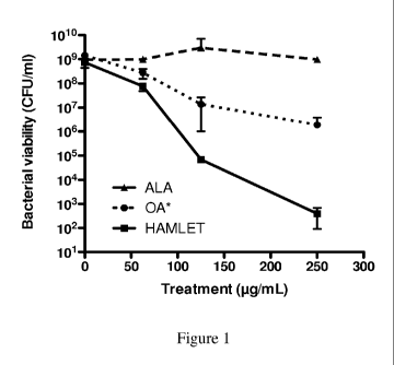
[0008] Figure 1 is a chart showing S. pneumoniae D39 treated with
native alpha-
lactalbumin (ALA), ALAFAC, or oleic acid (OA) at the concentration equivalent
to the
concentration present in ALAFAC (6% w/w) and incubated for 1 hour at 37 C.
Viable
organisms were assessed after plating dilutions of each sample onto blood agar
plates and
enumerating colony forming units after overnight growth.
[0009] Figures 2A and 2B are charts showing ALAFAC (labeled as HL)
lowering the
MICs of penicillin G. The penicillin-sensitive S. pneumoniae strain D39 (A)
and the
penicillin-resistant strain 5P670 (B) were grown in broth for 16 hours in the
presence of
penicillin G with and without the addition of ALAFAC. The figures show
representative
- 2 -
CA 02878292 2014-12-31
WO 2014/008465
PCT/US2013/049437
growth curves for the lowest concentration of antibiotic and ALAFAC that
inhibited bacterial
growth by combination treatment without either agent alone affecting growth.
[0010] Figures 3A and 3B are charts showing potentiation of short-time
pneumococcal killing by penicillin in the presence of ALAFAC (labeled as HL).
Short-time
killing of the penicillin-sensitive strain D39 (A) by penicillin (20 p.g/mL),
ALAFAC (50
p.g/mL) and penicillin combined with ALAFAC over 4 hours. (B) Killing of the
penicillin-
resistant strain SP670 by penicillin (20 or 30 p.g/mL), ALAFAC (50 p.g/mL), or
penicillin
combined with ALAFAC over 4 hours. The dashed horizontal line represents the
total
bacterial inoculum of an untreated sample. The results are based on three
individual
experiments with duplicate samples and are expressed as means S.D.
Statistics was
performed using the unpaired Student t-test. Significance was indicated as
follows: * = P <
0.05, ** = P < 0.01, *** = P < 0.001, ns = not significant.
[0011] Figures 4A and 4B are charts showing the effect of ALAFAC
(labeled as HL)
/antibiotic combination treatment on in vitro biofilm viability. The activity
of penicillin G
(100 p.g/mL), ALAFAC (250 p.g/mL), or the combination of both agents were
tested on in
vitro biofilms of the penicillin-sensitive strain D39 (A) or the penicillin-
resistant strain 5P670
(B) formed over a prefixed epithelium of NCI-H292 cells and were tested by
determining the
bacterial death (in logio) after culturing bacterial dilutions overnight on
blood agar. The
dashed horizontal line represents the mean total bacterial biomass of biofilms
that were
treated with buffer alone. The results are based on three individual
experiments with
duplicate samples. Statistics was performed using the paired Student t-test.
Significance was
indicated as follows: * = P < 0.05, ** = P < 0.01, *** = P < 0.001.
[0012] Figures 5A, 5B, and 5C are charts illustrating that antibiotic
combination
treatment eradicates pneumococci and MRSA during nasopharyngeal colonization.
(A) Mice
were colonized with the penicillin-sensitive S. pneumoniae EF3030 strain for
48 hours,
treated intranasally with various doses of gentamicin in the presence
(circles) or absence
(squares) of ALAFAC (labeled as HL) (50 p g) for 6 hours, and the bacterial
burden
associated with the nasopharyngeal tissue was determined. (B) Mice were
colonized with the
penicillin-resistant strain S. pneumoniae 5P670 (MIC = 4 p.g/mL) for 48 hours,
treated
intranasally with various doses of penicillin in the presence (squares) or
absence (circles) of
ALAFAC (50 p g) for 12 hours and the bacterial burden associated with the
nasopharyngeal
tissue was determined. Penicillin alone had no effect on the bacterial burden.
(C) Mice were
colonized with the methicillin-resistant strain S. aureus NRS70 for 48 hours,
treated
- 3 -
CA 02878292 2014-12-31
WO 2014/008465
PCT/US2013/049437
intranasally with various doses of methicillin in the presence (circles) or
absence (squares) of
ALAFAC (50 p g) for 12 hours and the bacterial burden associated with the
nasopharyngeal
tissue was determined. Methicillin alone had no effect on the bacterial
burden. The graph
shows colonization data for individual mice, with the mean recovered bacteria
and the
standard deviation depicted. The results are based on experiments using groups
of 6-10 mice.
Statistics was performed using the unpaired Student t-test. Significance was
indicated as
follows: * = P < 0.05, ** = P < 0.01, ns = non-significant.
[0013] Figure 6 shows an example of how ALAFAC (labeled as HL) lowers
the
MICs of gentamicin, erythromycin and penicillin. S. pneumoniae D39 were grown
in broth
for 16 hours in the presence of penicillin G (A) or gentamicin (B) with and
without the
addition of ALAFAC. The figure shows representative growth curves for the
lowest
concentration of antibiotic and ALAFAC that inhibited bacterial growth by
combination
treatment without either agent alone affecting growth.
[0014] Figure 7 shows a representative potentiation of short-time
pneumococcal
killing by gentamicin, penicillin and erythromycin in the presence of ALAFAC
(labeled as
HL). Short-time killing of the penicillin-sensitive strain D39 (A) by
penicillin G (20 p.g/mL),
ALAFAC (50 p.g/mL) and penicillin combined with ALAFAC over 4 hours. (B)
Killing of
the penicillin-resistant strain 5P670 by penicillin G (20 or 30 p.g/mL),
ALAFAC (50 p.g/mL),
or penicillin G combined with ALAFAC over 4 hours. (C) Killing of the
erythromycin-
sensitive strain D39 by erythromycin (200 p.g/mL), ALAFAC (50 p.g/mL), or
erythromycin
combined with ALAFAC over 4 hours. (D) Killing of the erythromycin-resistant
D39-
derivative JY53 by erythromycin (200 p.g/mL), ALAFAC (50 p.g/mL), or
erythromycin
combined with ALAFAC over 4 hours. (E) Killing of D39 by gentamicin (50
p.g/mL),
ALAFAC (50 p.g/mL), or gentamicin combined with ALAFAC over 1 hour. The
results are
based on three individual experiments with duplicate samples and are expressed
as means
S.D. Statistics was performed using the unpaired Student t-test. Significance
was indicated as
follows: * = P < 0.05, ** = P < 0.01, *** = P < 0.001, ns = not significant.
[0015] Figure 8 shows a representative effect of ALAFAC (labeled as
HL) /antibiotic
combination treatment on in vitro biofilm viability. The activity of
penicillin G (100 pg/mL),
ALAFAC (250 p.g/mL), or the combination of both agents were tested on in vitro
biofilms of
the penicillin-sensitive strain D39 (A) or the penicillin-resistant strain
5P670 (B) formed over
a prefixed epithelium of NCI-H292 cells and were tested by determining the
bacterial death
(in logio) after culturing bacterial dilutions overnight on blood agar.
Similarly, the activity of
- 4 -
CA 02878292 2014-12-31
WO 2014/008465
PCT/US2013/049437
erythromycin (250 p.g/mL), ALAFAC (250 p.g/mL), or the combination of both
agents on in
vitro biofilms of the erythromycin-sensitive strain D39 (C) or the
erythromycin-resistant
strain D39-P2A1 (D) were tested in a similar fashion as was the activity of
200 p g/ml
gentamicin (E) or 500 p g/ml gentamicin (F) alone or in combination with 100 p
g/ml
ALAFAC (100 p.g/mL) over 3 hours on pre-established biofilms formed by the
strain
EF3030. The results are based on three individual experiments with duplicate
samples.
Statistics was performed using the paired Student t-test. Significance was
indicated as
follows: * = P < 0.05, ** = P < 0.01. To visualize the morphology of the
treated biofilms,
SEM studies were performed. Images show (G) the structure of an untreated 48
hour EF3030
biofilm, (H) an EF3030 biofilm after 3 hour treatment with 500 pg/mL
gentamicin alone, (I)
an EF3030 biofilm after 3 hour treatment with ALAFAC (100 p.g/mL) alone and
(J) an
EF3030 biofilm after 3 hour treatment with the combination of 100 pg/mL ALAFAC
and 500
p.g/mL Gentamicin. An epithelial substratum prior to biofilm formation has
been included as
a control (insert in panel J). The increased bactericidal activity of the
combination treatment
was associated with a reduction in the density of adherent bacteria and
biofilm matrix.
[0016] Figure 9 shows a representative ALAFAC (labeled as HL) -
antibiotic
combination treatment eradicates pneumococci during nasopharyngeal
colonization. (A and
B) Mice were colonized with S. pneumoniae EF3030 for 48 hours, treated
intranasally with
various doses of gentamicin in the presence (red circles) or absence (black
squares) of
ALAFAC (50 p g) for 6 hours, and the bacterial burden associated with the
nasal lavage (A)
and the nasopharyngeal tissue (B) was determined. Bacteria in nasal lavage and
associated
with the nasopharyngeal tissue were significantly more sensitive to
gentamicin/ALAFAC
combination therapy than gentamicin alone. (C and D) Mice were colonized with
the
penicillin-resistant strain S. pneumoniae 5P670 (MIC = 4 p.g/mL) for 48 hours,
treated
intranasally with various doses of penicillin G in the presence (red circles)
or absence (black
squares) of ALAFAC (50 p g) for 12 hours and the bacterial burden associated
with the nasal
lavage (C) and the nasopharyngeal tissue (D) was determined. Penicillin G
alone had no
effect on the bacterial burden in either the nasal lavage or in the tissue.
However,
combination therapy with ALAFAC and penicillin caused a dose-dependent
decrease in
bacterial burden leading to eradication of colonization. The graph shows
colonization data for
individual mice, with the mean recovered bacteria and the standard deviation
depicted. The
results are based on experiments using groups of 6-10 mice. Statistics was
performed using
- 5 -
CA 02878292 2014-12-31
WO 2014/008465
PCT/US2013/049437
the unpaired Student t-test. Significance was indicated as follows: * = P <
0.05, ** = P <
0.01, ns = non-significant.
[0017] Figure 10 shows an example of the impact of ALAFAC (labeled as
HAMLET)
on uptake and binding of gentamicin and Bocillin FL. (A) S. pneumoniae D39
were
incubated with Alexa Fluor 488-gentamicin in the presence or absence of
ALAFAC.
ALAFAC significantly increased the cell-associated level of gentamicin. (B)
The penicillin-
resistant strain SP670 and the penicillin-sensitive strain D39 were incubated
with the
fluorescent beta-lactam Bocillin FL in the presence or absence of ALAFAC.
ALAFAC did
not increase the cell-associated level of Bocillin FL in either strain. The
results are based on
three individual experiments with duplicate samples. Statistics was performed
using the
unpaired Student t-test. Significance was indicated as follows: ** = P<0.01,
*** = P<0.001,
ns = not significant.
[0018] Figure 11 shows an example of the effect of calcium and kinase
inhibitors on
ALAFAC (labeled as HAMLET or HL)-induced sensitization of pneumococci to
gentamicin.
(A) S. pneumoniae D39 were treated with a lethal concentration of ALAFAC (12X
MIC) in
the absence of inhibitor (HL) or presence of 20 uM staurosporine (HL + Sts),
or 30 p.M
ruthenium red (HL + RuR) for 1 hour at 37 C. The treated bacteria were diluted
and plated on
blood agar plates and viable CFU/ml were determined after overnight growth. (B
and C) S.
pneumoniae D39 were treated with 50 pg/mL ALAFAC (HL), 50 p.g/mL gentamicin
(Gent),
20 p g/mL penicillin G (PcG), or a combination of gentamicin and ALAFAC or
penicillin G
and ALAFAC in the absence (HL + Gent, HL + PcG) or presence of 20 uM
staurosporine
(Sts), or 30 p.M ruthenium red (RuR) for 1 hour at 37 C. The treated bacteria
were diluted
and plated on blood agar plates and viable CFU/ml were determined after
overnight growth.
The graph depicts the logio death induced by each treatment and showed that
staurosporine
and ruthenium red significantly reduced ALAFAC-induced death (A) and also
significantly
blocked ALAFAC's ability to sensitize pneumococci to gentamicin (B) and
penicillin G (C).
The results are based on three individual experiments with duplicate samples.
Statistics was
performed using the unpaired Student t-test. Significance was indicated as
follows: *** =
P<0.001.
[0019] Figure 12 shows a representative autolysis during ALAFAC (labeled as
HAMLET or HL)-gentamicin combination therapy. (A) Optical density at 600 nm of
S.
pneumoniae D39 (black line) after exposure to a lethal concentration of ALAFAC
(250
p g/m1; red line), a sublethal concentration of ALAFAC (50 p g/m1; blue line),
a sublethal
- 6 -
CA 02878292 2014-12-31
WO 2014/008465
PCT/US2013/049437
concentration of gentamicin (50 p.g/mL; green line) or the combination of
sublethal
concentrations of ALAFAC and gentamicin (purple line) that resulted in
complete death of
the inoculum. The data shows a representative experiment. (B) Representative
scanning
electron micrographs of untreated S. pneumoniae D39, as well as bacteria
treated 4 minutes
with a lethal concentration of ALAFAC (250 p.g/mL), one hour with a similarly
lethal
concentration of gentamicin (500 p.g/mL) or one hour with a sublethal
concentration of
ALAFAC (50 p.g/mL) or a sublethal concentration of gentamicin (50 pg/mL) in
combination
(HL + Gent). Note the numerous defects of the pneumococcal cell wall after
exposure to
ALAFAC or gentamicin alone compared with the structurally intact cells after
exposure to
the combination of the two agents.
[0020] Figure 13 shows an example of how ALAFAC (labeled as HL) lowers
the
methicillin MIC. S. aureus strains 11090306 (MSSA) (left) and NRS 384 (MRSA)
(right)
were grown in broth for 16 hours in the presence of 2 p g/ml methicillin (5 p
M) with and
without the addition of 100 p g/mL (6 p M) ALAFAC. The figure shows
representative
growth curves for the lowest concentration of antibiotic and ALAFAC that
inhibited bacterial
growth by combination treatment without either agent alone affecting growth.
[0021] Figure 14 shows a representative effect of ALAFAC (labeled as
HL)
/antibiotic combination treatment on in vitro biofilm viability. (A) The
activity of methicillin
(250 p.g/mL or 660 pM), ALAFAC (200 pg/mL or 12 p M), or the combination of
both
agents were tested on in vitro biofilms of the methicillin-resistant strain
NRS 70 (MRSA) or
the methicillin-sensitive strain 11090306 (MSSA) by determining the bacterial
death (in
logio) after culturing dilutions overnight on blood agar. (B) The activity of
vancomycin (32
p.g/mL or 21 p M), ALAFAC (500 p.g/mL or 30 p M), or the combination of both
agents on in
vitro biofilms of the vancomycin-resistant strain NRS 1 (VISA) and the
vancomycin-
sensitive strain NR5384 (VSSA) were tested in a similar fashion as was the
activity of (C) 50
p g/ml (105 p M) gentamicin alone or in combination with 500 p g/ml (30 p M)
ALAFAC for
the gentamicin-resistant strain. The results are based on three independent
experiments with
duplicate samples. Statistics was performed using the paired Student t-test.
Significance was
indicated as follows: ns = not significant, * = P < 0.05, ** = P < 0.01.
[0022] Figure 15 shows an example of how ALAFAC (labeled as
HAMLET)/Methicillin combination treatment reduces Staphylococcal
nasopharyngeal
colonization. Mice were colonized with S. aureus NRS 70 for 24 hours, treated
intranasally
with various doses of gentamicin in the presence (blue) or absence (black) of
ALAFAC (100
- 7 -
CA 02878292 2014-12-31
WO 2014/008465
PCT/US2013/049437
p g) for 12 hours, and the bacterial burden associated with the nasal wash (A)
and the
nasopharyngeal tissue (B) was determined. The graph shows colonization data
for individual
mice, with the mean recovered bacteria depicted with a line. The results are
based on
experiments using groups of 6 mice. Statistics was performed using the
unpaired Student t-
test. Significance was indicated as follows: * = P < 0.05.
[0023] Figure 16 shows a representative effect of ALAFAC (labeled as
HAMLET or
HL) on membrane potential. (A) Representative growth curves for S. aureus
strain NRS 123
(MRSA) grown in broth for 16 hours (960 min) in the presence of methicillin
with and
without the addition of ALAFAC and the inhibitors Ruthenium Red (RuR) or
Amiloride
(Amil). (B) Mid-log phase grown NRS 123 Staphylococci re-suspended in PBS
alone or PBS
plus Amiloride or Ruthenium Red, were incubated with the fluorescent indicator
dye
DiBAC4(3) and membrane depolarization was detected by measuring fluorescence
over time.
ALAFAC was added at twenty minutes (arrow). The detergent Triton X-100 (0.1%)
was
included as a positive control. The results presented are from one
representative experiment.
(C) Mid-log phase grown NRS 384 Staphylococci were incubated with the
radioisotope
45Ca2+ (2.5 p Ci/mL) in PBS or PBS + Ruthenium Red (30 p M). After recording
baseline
readings, PBS (untreated), or ALAFAC was added (Time = 0 min) to the bacteria
and
radioactivity was measured over time. Results from a representative experiment
are shown.
(D) NRS 384 Staphylococci were loaded with the pH sensitive dye BCECF-AM, and
were
washed and resuspended in PBS + 25 mM glucose. After recording baseline
readings, at the
first arrow, PBS (untreated), the protonophore CCCP (100 p M), ALAFAC (100 p
g/mL or 6
p M), ALAFAC + RuR (30 p M), or ALAFAC + Amiloride (1 mM) were added to the
bacteria and fluorescence was measured over time. At the second arrow 20 p M
each of
nigericin and valinomycin was added to completely dissipate the transmembrane
proton
gradient.
[0024] Figure 17 shows a representative impact of ALAFAC (labeled as
HAMLET)
on uptake and binding of Bocillin FL and vancomycin FL. (A) Staphylococci were
incubated
with Bocillin FL or (B) with vancomycin FL in the presence or absence of 100 p
g/mL (6 p M)
ALAFAC. The results are based on three individual experiments with duplicate
samples.
Statistics was performed using the unpaired Student's t-test. Significance was
indicated as
follows: ** = P < 0.01, *** = P < 0.001, ns = not significant.
[0025] Figure 18 shows an example of ALAFAC (labeled as HL) and
resistance
development. Methicillin adaptation of the MRSA strain NRS 384 after exposure
to stepwise
increasing concentrations of methicillin alone (1 ¨ 512 p g/mL or 2.5 ¨ 1,350
p M) or
- 8 -
CA 02878292 2014-12-31
WO 2014/008465
PCT/US2013/049437
methicillin in combination with 100 p g/mL (6 p M) ALAFAC. The filled blue
circles show
the methicillin MICs after each cycle when no ALAFAC was used. The addition of
100
p g/mL of ALAFAC reduced methicillin-induced resistance (blue unfilled
circles). The
unfilled green circle represents the MIC of methicillin of the bacteria grown
in presence of
ALAFAC, when ALAFAC was also present during the MIC assay to potentiate the
effect of
the antibiotic. Reintroduction of ALAFAC to these isolates again returned the
methicillin
MIC back to the levels denoted by the green unfilled circle.
DETAILED DESCRIPTION OF THE DISCLOSURE
[0026] A novel complex of a-lactalbumin and fatty acid in human milk
was
previously identified (Hakansson et al., 1995, Proc Natl Acad Sci U S A
92:8064-8068;
Svensson, et al., 2000, Proc Natl Acad Sci U S A 97:4221-4226). A non-covalent
complex of
alpha-lactalbumin and fatty acid is hereinafter referred to as alpha-
lactalbumin fatty acid
complex or ALAFAC. The complex purified from the casein fraction of human
milk, was
found to be made up of alpha-lactalbumin in a partially unfolded conformation
that could be
stabilized under physiological conditions by a human derived fatty acid
fraction containing
oleic acid and linoleic acid. This protein-lipid complex was found to have
limited bactericidal
effect against only a few respiratory tract pathogens (Hakansson et al., 2000,
Mol Microbiol
35:589-600). For example, while ALAFAC has bactericidal effect against the
respiratory
tract pathogens Streptococcus pneumoniae, Haemophilus influenzae, and some
strains of
Moraxella catarrhalis, it showed no detectable activity even at concentrations
up to 5 mg/ml
against gram-positive organisms such as Staphylococci and Bacillus subtilis
and against the
gram-negative organisms Escherichia coli and Pseudomonas aeruginosa.
[0027] The present disclosure is based on the unexpected finding that
ALAFAC can
potentiate anti-bacterial activity of antibiotics. This potentiating or
synergistic effect is seen
against bacteria, which are sensitive to ALAFAC as well as in bacteria which
are not
sensitive to ALAFAC. Further, the potentiation of the anti-bacterial effect of
antibiotics is
seen against bacteria that are sensitive to the antibiotics and also with
bacteria that are
resistant to antibiotics.
[0028] ALAFAC can be isolated from biological materials or can be
prepared by
complexing fatty acids and alpha-lactalbumin. For example, ALAFAC can be
isolated from
the milk of primates, including humans. It can also be prepared by complexing
ALA with
fatty acids. See, e.g., U.S. patent numbers 6,808,930, 7,053,185, and
7,524,932, which are
incorporated herein by reference. The alpha-lactalbumin can be obtained from
any
- 9 -
CA 02878292 2014-12-31
WO 2014/008465
PCT/US2013/049437
mammalian source (such as milk), including but not limited to, primates,
cattle, rodents, and
the like. For example, it can be obtained from humans, cows, dogs, goats,
sheep, horses, and
the like. Alpha-lactalbumin is also available commercially (such as from Sigma
Aldrich). In
one embodiment, alpha-lactalbumin can also be produced by recombinant methods.
See, e.g.,
Svensson et al., 2000., Proc Natl Acad Sci USA 97:4221-4226. Genbank accession
number
for human ALA is NP_002280.1; GI:4504947). Genbank accession numbers for ALA
from
other species are: Cow: NP_776803.1 GI:27805979; Horse: P08334.2 GI:125991,
Donkey:
AAB24573.1 GI:262063, Sheep: NP_001009797.1 GI:57526478, Goat: CAA28797.1
GI:980, Pig: NP_999525.1 GI:47523778, and Dog: NP_001003129.1 GI:50978848.
[0029] Fatty acids useful for making the ALAFAC complex include unsaturated
cis
C14 to C20 fatty acids. In one embodiment, the fatty acids are C16 and C18
fatty acids. In
one embodiment, the fatty acids are oleic acid and/or linoleic acid. In one
embodiment, the
milk fraction containing oleic acid and/or linoleic acid is obtained from the
milk of primates,
such as humans. This milk is known to be high in oleic and linoleic acid. Milk
from other
mammals is known to be rich in smaller fatty acids (such as C14 or lower) and
is not known
to contain significant amounts of C18 or C16 fatty acids. In one embodiment,
commercially
available C18 or C16 can also be used. Additionally, several vegetable oils
(such as olive oil)
are known to be a rich source of oleic and linoleic acids.
[0030] In one embodiment, ALAFAC can be isolated as described in U.S.
Patent
Nos. 6,808,930; 7,053,185, and 7,524,932, the disclosures of which with
respect to isolation
of ALAFAC are incorporated herein by reference. For example, it can be
purified from milk
by removal of fat (such as by centrifugation), and separation into casein and
whey (such as by
acid precipitation). The separated casein is harvested (such as by
centrifugation) and washed.
The casein fraction can be fractionated (such as by using ion exchange
chromatography) and
elutes with salt (such as 1M NaC1). The eluent can then be desalted against
distilled water. In
one embodiment, ALAFAC can be isolated from human milk or may be produced by
exposing milk-derived or recombinant apo-ALA (EDTA-treated) to oleic acid
bound to a
DEAE matrix using fast protein liquid chromatography or by loading lipid to
the protein
under alkaline conditions.
[0031] In one embodiment, the ALAFAC complex has from 1 to 40 (and all
integers
therebetween) fatty acid molecules complexed to a molecule of ALA. In various
embodiments, the ALAFAC complex has 35 or less, 30 or less, 25 or less, 20 or
less, 15 or
less, or 10 or less fatty acid molecules complexed to a molecule of ALA. In
one
embodiment, the ALAFAC has from 1 to 15 fatty acid molecules complexed to a
molecule of
- 10 -
CA 02878292 2014-12-31
WO 2014/008465
PCT/US2013/049437
ALA. In one embodiment, there are from 5 to 10 fatty acid molecules complexed
to a
molecule of ALA. In one embodiment, the toxicity of ALAFAC when there were 10
or less
fatty acid molecules complexed to a molecule of ALA, was found to be
acceptable. In
various embodiments, the ALAFAC complex comprises 1, 2, 3, 4, 5, 6, 7, 8, 9,
10, 11, 12, 13,
14, or 15oleic acid and/or linoleic acid molecules complexed to a molecule of
ALA.
[0032] The ALAFAC can be stored in the refrigerator or can be frozen.
For example,
ALAFAC can be stored in physiological buffer (such as 0.9% saline or phosphate
buffered
saline) at 4 C for at least 3 months and stability is maintained in solution
for over 1 year. If
can also be stored at room temperature for at least three weeks.
[0033] In one embodiment, the components for forming ALAFAC may be provided
separately. For example, alpha-lactalbumin and fatty acids may be provided
separately and
may be combined in a suitable buffer (such as physiological buffer) to effect
the formation of
the complex. Generally, ALA in the unfolded form will non-covalently bind to
fatty acids. In
one embodiment, the fatty acids and ALA are provided such that from 1 to 40, 1
to 30, 1 to
20 or 1 to 10 fatty acids may bind to each ALA molecule.
[0034] By the term "no bactericidal effect" or "anti-bacterial
effect", it is meant that
no detectable effect on the growth or survival of bacteria is observed. For
bacteria considered
resistant to an antibiotic, no bactericidal effect is observed at up to 5
mg/ml when tested in
vitro. Alternatively, bacteria may be deemed resistant to an antibiotic
because no
improvement is seen clinically in a patient's condition upon administration of
a full regimen
of that antibiotic. Conversely, bacteria are considered to be sensitive to an
antibiotic when
bactericidal activity can be detected at therapeutically effective ranges or
when an
improvement is seen in a patient's condition upon administration of a full
regimen of that
antibiotic.
[0035] In one embodiment, the present disclosure provides compositions and
methods
for potentiating the effects of antibiotics against bacteria, which are known
to be antibiotic
resistant. In this embodiment, the potentiation is in the form of conferring
sensitivity toward
an antibiotic against which the bacteria was previously resistant. Those
skilled in the art will
recognize that the concentrations at which antibiotics are effective against
various bacteria
depends upon the type of bacteria. Determination of such ranges is within the
purview of one
skilled in the art. For example, penicillin sensitivity can be seen at 0.1 p
g/ml. In one
embodiment, the amount of antibiotic effective against sensitive strains is in
the range of 0.2
to 250 p g/ml. In various embodiments, the concentration of antibiotics when
used with
ALAFAC is from 0.1 to 1.0 mg/ml and all concentrations to the tenth decimal
place
- 11 -
CA 02878292 2014-12-31
WO 2014/008465
PCT/US2013/049437
therebetween. In some embodiments, the concentration is from 1 to 500 tg/m1
and all values
to the tenth decimal place therebetween.
[0036] In another embodiment, the present disclosure provides
compositions and
methods for potentiating the effects of antibiotics against bacteria, which
are sensitive to
antibiotics. In this embodiment, potentiation is in the form of reducing the
amount of an
antibiotic that is needed to treat the infection compared to the amount needed
without
ALAFAC. In various embodiments, the concentration of antibiotics when used
with
ALAFAC is from 0.1 to 1.0 mg/ml and all concentrations to the tenth decimal
place
therebetween. In some embodiments, the concentration is from 1 to 500 tg/m1
and all values
to the tenth decimal place therebetween. In some embodiments, the antibiotic
needed to treat
the infection is 2 to 300 (and all integers therebetween) fold less than the
amount required
without ALAFAC.
[0037] The potentiation of the effect of antibiotics may be seen as a
decrease in the
MIC or short-kill assay times for in vitro studies. The potentiation may also
be seen in the
form of improvement of an individual's condition, for example, as determined
by a clinician.
The combination of ALAFAC complex and an antibiotic may decrease the MIC,
reduce the
short-kill time for bacteria which are sensitive or resistant to ALAFAC and/or
the antibiotic.
Determination of MICs is well within the purview of those skilled in the art.
Further MIC
values for different antibiotics and different bacteria can be obtained from
the Antimicrobial
Index at http://antibiotics.toku-e.com. In various embodiments, the MIC for
antibiotics is
reduced from 2 to 300 (and all integers therebetween) fold upon use with
ALAFAC.
[0038] For example, it was observed that ALAFAC potentiates the action
of
antibiotics in ALAFAC-resistant bacteria and that it works equally well or
even better for
pneumococci (ALAFAC sensitive). It was observed that a combination of ALAFAC
and
gentamicin, penicillin or erythromycin, significantly enhanced bacterial
killing against both
antibiotic sensitive and antibiotic resistant organisms in vitro and in vivo.
For example,
addition of ALAFAC to gentamicin resulted in a 10-fold reduction in the dose
needed to
eradicate both lavage- and tissue-associated gentamicin-tolerant pneumococci
in an animal
model and addition of ALAFAC to penicillin resulted in a 33-fold reduction in
the dose
needed to eradicate colonization by the penicillin-resistant strain SP670 that
was completely
insensitive to penicillin alone. It was also found that the gram-negative
respiratory organisms
A. baumanii and M. catarrhalis, both of which show a high level of antibiotic
resistance
against beta-lactams and other classes of antibiotics and are also resistant
to ALAFAC,
showed that the MIC for penicillin and gentamicin were significantly reduced.
Thus,
- 12 -
CA 02878292 2014-12-31
WO 2014/008465
PCT/US2013/049437
ALAFAC can provide a way to increase the usefulness of existing drugs, and
extend the
lifetime of the current treatment arsenal antibiotic resistant also in ALAFAC-
resistant
organisms.
[0039] Examples of bacteria against which ALAFAC by itself has shown
bactericidal
activity are: Streptococcus pneumoniae, Haemophilus influenzae, and some
strains of
Moraxella catarrhalis, although the effect against Moraxella catarrhalis is
generally poor
and less than 90% reduction occurs at 2 mg/ml . Examples of bacteria against
which it
showed no detectable activity, even at concentrations up to 5 mg/ml, are gram-
positive
organisms, such as Staphylococci and Bacillus subtilis, and gram-negative
organisms, such as
Escherichia coli and Pseudomonas aeruginosa. The present compositions and
methods are
useful for all Gram-positive and Gram-negative bacteria, and for antibiotic
resistant as well as
antibiotic sensitive bacteria. Examples of Gram positive bacteria include, but
are not limited
to, Streptococcus pneumonia, Staphylococcus aureus, Staphylococcus
epidermidis,
Streptococcus sanguis, Streptococcus mutans, Streptococcus pyogenes (Group A),
Streptococcus agalactiae (Group B), and Enterococcus faecalis. Examples of
Gram
negative bacteria include, but are not limited to, Escherichia coli,
Haemophilus influenza,
Haemophilus parainfluenzae, Moraxella catarrhalis, Acinetobacter baumanii,
Klebsiella
pneumonia, Pseudomonas aeruginosa, and Enterobacter cloache.
[0040] In one aspect, the present disclosure provides compositions for
use as
adjuvant. In one embodiment, the adjuvant composition comprises, or consists
essentially of,
ALAFAC at an amount sufficient to act as an adjuvant. For bacteria against
which ALAFAC
itself has a bactericidal effect (ALAFAC sensitive bacteria), the amount at
which it can exert
its adjuvant effect is lower than the amount at which it exerts its
bactericidal effect. In one
embodiment, the composition comprises ALAFAC at an amount sufficient to act as
an
adjuvant and the composition may have bactericidal activity.
[0041] In one embodiment, the compositions for use as adjuvants for
potentiating the
activity of antibiotics are suitable for administration to individuals. In one
embodiment, the
compositions are sterile and packaged in sterile packaging or containers. In
one embodiment,
the compositions do not contain other proteins or amino acids. In one
embodiment, the
compositions do not contain serum albumin and/or essential amino acids. In one
embodiment,
the compositions do not contain vitamins. In one embodiment, the compositions
do not
contain growth factors or hormones. In one embodiment, the compositions are
free of serum
albumin and other serum proteins, essential amino acids, growth factors,
hormones, and
vitamins.
- 13 -
CA 02878292 2014-12-31
WO 2014/008465
PCT/US2013/049437
[0042] The concentration of ALAFAC may be 0.1 tg/m1 to 10.0 mg/m1 and
all
concentrations to the tenth decimal place therebetween. In some embodiments,
the
concentration of ALAFAC for use with ALAFAC sensitive bacteria may be from 0.1
tg/m1
to 100 tg/m1 and all concentrations to the tenth decimal place therebetween.
In some
embodiments, the concentration of ALAFAC for use with bacteria resistant to
ALAFAC may
be from 0.1 tg/m1 to 5.0 mg/m1 and all concentration therebetween to the tenth
decimal
place, and for use with bacteria sensitive to ALAFAC may be from 0.1 tg/m1 to
1.0 mg/m1
and all concentration therebetween to the tenth decimal place.
[0043] In one embodiment, the adjuvant composition is provided in
doses or portions
such that each dose or portion provides a sufficient dose for administration
to an individual,
wherein the total amount in the aliquots or portions (for a complete dosage
regimen) is
sufficient to act as an adjuvant to the activity of an antibiotic, but will
not have bactericidal
activity by itself i.e., without the antibiotic. The total doses required for
a treatment regimen
are referred to herein as a "treatment set". For example, a treatment regimen
for many
antibiotics typically contains doses that are taken over a period of 5 to 10
days. Thus, if
ALAFAC is to be administered over the same period of time, then a treatment
set for
ALAFAC may comprise doses to be administered over the 5 or 10 days. Each
portion or
dose may be suitable for topical, oral, intravenous or any other form of
administration. Each
portion or dose may be packaged in a discrete compartment (such as tablets in
a blister
packaging, or a topical patch/bandage in a containment) or may be packaged as
bulk (such as
ointment in a tube or suspension in a bottle). In various embodiments, such
adjuvant
amounts may be 2 to 300 times (and all integers therebetween) lower than the
amounts that
would be bactericidal to sensitive bacteria. In one embodiment, the amount is
2-100 times or
2-10 times lower that the amounts that would be bactericidal to sensitive
bacteria.
[0044] In one embodiment, the compositions comprise, or consists
essentially of,
ALA, fatty acids and a suitable buffer in amounts suitable for combining such
that ALAFAC
may be formed by the complexing of ALA with the fatty acids. In one
embodiment, the
amount of fatty acid is such that from 1 to 40 (and all integers therebetween)
fatty acid
molecules can be complexed with each ALA.
[0045] In another embodiment, the present disclosure provides compositions
comprising ALAFAC and one or more antibiotics. In one embodiment, the
composition
comprises ALA, fatty acid(s), and one or more antibiotics. The antibiotic may
be any
antibiotic that is useful for treating infections. For example, the antibiotic
may be a broad
spectrum antibiotic or may be effective against particular bacteria. In
various embodiments,
- 14 -
CA 02878292 2014-12-31
WO 2014/008465
PCT/US2013/049437
the composition comprises or consists essentially of, i) ALAFAC, and/or ii)
fatty acids and
ALA, and iii) one or more antibiotics, all in suitable carriers or buffers.
[0046] Suitable antibiotics include, but are not limited to, beta-
lactam antibiotics such
as subclasses Penicillins (examples: penicillin G, methicillin, oxacillin,
ampicillin,
amoxicillin), Glycopeptides (example vancomycin), Carbapenems (examples
imipenem and
meropenem), Polymyxin and Bacitracins (example bacitracin, neomycin) or
Lipopeptides
(example daptomycin), Protein synthesis inhibitors such as subclasses
Aminoglycosides
(example gentamicin, streptomycin, kanamycin), Tetracyclines (examples
tetracycline,
doxycycline, and tigecycline), Oxazilodinone (linezolid), Peptidyl
transferases (example
Chloramphenicol), Macrotides (examples erythromycin, azithromycin,
telithromycin),
Lincosamides (examples clindamycin), and Streptogramins (example
prisintamycin), DNA
synthesis inhibitors such as metronidazole and subclass Fluoroquinolones
(examples
ciprofloxacin, norfloxacin, morifloxacin), RNA synthesis inhibitors such as
rifampin,
Mycolic acid synthesis inhibitors such as isoniazid, and Folic acid synthesis
inhibitors such as
Trimethoprim and subclass Sulfonamides (examples sulfamethoxazole,
sulfadoxin). In one
embodiment, ALAFAC and the antibiotic in the combination composition are at
amounts that
will not have bactericidal effect if administered alone. In another
embodiment, the ALAFAC
is at an amount that will not have a bactericidal effect by itself, and the
antibiotic is at an
amount that it will have a minimal effect if administered by itself such that
it would not be
deemed as treatment by a clinician. In another embodiment, the ALAFAC is at an
amount
that will not have a bactericidal effect by itself, and the antibiotic is at
an amount that it will
have a therapeutic effect if administered by itself.
[0047] In another aspect, the present disclosure provides
pharmaceutical
compositions. Pharmaceutical compositions comprise, or consist essentially of,
ALAFAC
with or without the antibiotic, and suitable carriers and other additives. ALA
and fatty acids
may also be present in the compositions. For example, the composition may
comprise a
therapeutically effective amount of ALAFAC, and optionally one or more
antibiotics, in a
pharmaceutically acceptable carrier. Such carriers may include a diluent,
adjuvant, excipient,
or other vehicle with which the therapeutic is administered. Some examples of
materials
which can serve as pharmaceutically-acceptable carriers include: sugars, such
as lactose,
glucose and sucrose; starches, such as corn starch and potato starch;
cellulose, including
sodium carboxymethyl cellulose, ethyl cellulose and cellulose acetate;
powdered tragacanth;
malt; gelatin; talc; excipients, such as cocoa butter and suppository waxes;
oils, such as
peanut oil, cottonseed oil, safflower oil, sesame oil, olive oil, corn oil and
soybean oil;
- 15 -
CA 02878292 2014-12-31
WO 2014/008465
PCT/US2013/049437
glycols, such as propylene glycol; polyols, such as glycerin, sorbitol,
mannitol and
polyethylene glycol; esters, such as ethyl oleate and ethyl laurate; agar;
buffering agents,
such as magnesium hydroxide and aluminum hydroxide; alginic acid; pyrogen-free
water;
isotonic saline; Ringer's solution; ethyl alcohol; phosphate buffer solutions;
and other non-
toxic compatible substances employed in pharmaceutical formulations. The
composition, if
desired, can also contain minor amounts of wetting or emulsifying agents, or
pH buffering
agents. Some examples of compositions suitable for mixing with the agent can
be found in:
Remington: The Science and Practice of Pharmacy (2005) 21st Edition,
Philadelphia, PA.
Lippincott Williams & Wilkins. In one embodiment, the agent is substantially
purified (e.g.,
substantially free from substances that limit its effect or produce undesired
side-effects).
[0048] In one embodiment, the compositions are formulated for topical,
transdermal,
or mucosal use. Dosage forms for the topical, transdermal or mucosal
administration include
powders, sprays, ointments, pastes, creams, lotions, gels, solutions, patches
and inhalants.
The components of the present disclosure may be mixed under sterile conditions
with a
pharmaceutically-acceptable carrier, and with any preservatives, buffers, or
propellants which
may be required. The ointments, pastes, creams and gels may contain additional
excipients,
such as animal and vegetable fats, oils, waxes, paraffins, starch, tragacanth,
cellulose
derivatives, polyethylene glycols, silicones, bentonites, silicic acid, talc
and zinc oxide, or
mixtures thereof. Topical powders and sprays can also contain additional
excipients such as
lactose, talc, silicic acid, aluminum hydroxide, calcium silicates and
polyamide powder, or
mixtures of these substances. Sprays can additionally contain customary
propellants, such as
chlorofluorohydrocarbons and volatile unsubstituted hydrocarbons, such as
butane and
propane. In one embodiment, transdermal patches may be used. These have the
added
advantage of providing controlled delivery to the body. Such dosage forms can
be made by
dissolving or dispersing the agent in the proper medium. Absorption enhancers
can also be
used to increase the flux of the active ingredient across the skin. The rate
of such flux can be
controlled by either providing a rate controlling membrane or dispersing the
active ingredient
in a polymer matrix or gel. In one embodiment, the compositions are applied to
dermal
patches, bandages, gauges or other similar materials that can be directly
applied to the
affected area.
[0049] In one embodiment, the composition may be administered as an
aerosol. This
can be accomplished by preparing an aqueous aerosol, liposomal preparation or
solid
particles containing the active agents. A non-aqueous (e.g., fluorocarbon
propellant)
suspension could also be used. An aqueous aerosol may be made by formulating
an aqueous
- 16 -
CA 02878292 2014-12-31
WO 2014/008465
PCT/US2013/049437
solution or suspension of the agent together with conventional
pharmaceutically-acceptable
carriers and stabilizers. The carriers and stabilizers vary with the
requirements of the
particular compound, but typically include nonionic surfactants (Tweens,
Pluronics, or
polyethylene glycol), sorbitan esters, oleic acid, lecithin, such as glycine,
buffers, salts,
sugars or sugar alcohols. Aerosols generally are prepared from isotonic
solutions. Sonic
nebulizers may be used so as to minimize exposing the agent to shear, which
can result in
degradation of the compound.
[0050] In one embodiment, the composition may be formulated for
parenteral,
intravenous, or intramuscular delivery. These are typically aqueous
compositions. Saline
solutions and aqueous dextrose and glycerol solutions can also be employed as
liquid carriers.
[0051] The compositions are administered in a therapeutically effect
amount. It will
be recognized by those of skill in the art that the form and character of the
particular dosing
regimen for the therapeutic agent of the present invention will be dictated at
least in part by
the route of administration and other well-known variables, taking into
account such factors
as the size, gender, health and age of the individual to be treated, and risk
factors associated
with cancer development for the individual, such as occupational, behavioral
or family
history related parameters. Based on such criteria, one skilled in the art can
determine an
effective amount of to administer to the individual.
[0052] The compositions may be delivered by any route. For example,
the
compositions may be delivered as topical formulation for application to
mucosal surfaces. In
one embodiment, the compositions may be delivered by routes other than topical
or mucosal.
For example, the compositions may be delivered via the digestive tract (as
oral formulations)
or via the circulatory system (intravenous, intramuscular, etc.) or directly
to the relevant site.
[0053] While not intending to be bound by any particular theory, we
provide data,
which elucidates the mechanism by which ALAFAC might carry out its
potentiation activity.
S. pneumoniae was the most ALAFAC-sensitive bacterial species tested and
showed a
minimal inhibitory concentration (MIC) for ALAFAC of 20 p g/ml in liquid
culture,
comparable to the MIC of gentamicin of 16 p g/ml. For time-kill assays,
considerably higher
concentrations were needed (300 p g/ml to eradicate an inoculum of 108
pneumococci in 1
hour). These numbers are also comparable to what is required to kill
pneumococci with other
antibiotics in the same time frame and may be within a physiological range
considering that
the concentration of ALA in milk is high (2,000 p g/m1). ALAFAC kills all
pneumococcal
antibiotic-resistant strains that were tested (penicillin, erythromycin,
tetracycline, and
chloramphenicol-resistant), suggesting that ALAFAC works through a pathway
different
- 17 -
CA 02878292 2014-12-31
WO 2014/008465
PCT/US2013/049437
from common antibiotics. Using S. pneumoniae as a model organism, we show that
ALAFAC causes dissipation of the proton motive force, an influx of calcium
through a
sodium dependent mechanism and activation of serine/threonine kinase activity
and leads to
depolarization of the bacterial membrane. Furthermore, attempts to produce
spontaneous
mutants resistant to ALAFAC or mutants becoming resistant after slow
adaptation to
increasing concentrations of ALAFAC were unsuccessful. This suggests either
that
components being activated are essential for the bacteria or cannot be
inactivated merely with
point-mutations, or that there are multiple compensating pathways activated by
ALAFAC.
[0054] One embodiment of the present disclosure is aimed as an
adjuvant to current
antibiotic use to lower the concentrations of antibiotics needed to kill
various bacterial
species. One application for this disclosure is to lower the concentration of
antibiotics needed
to treat and eradicate resistant bacterial species to increase treatment
efficacy and to enable
the prolonged the use of the same antibiotic in the clinic. In various
embodiments, a 2 to 300
fold reduction is observed in the MIC for bacteria by using a combination of
antibiotic and
ALAFAC compared to the antibiotic alone. Also, by lowering the concentration
of antibiotics
used in general, ALAFAC adjuvant therapy has the potential to produce less
side effects as
ALAFAC by itself is a natural molecule ingested by infants, and importantly,
produce less
risk for further resistance development.
[0055] In one embodiment, human alpha-lactalbumin in complex with a
C16 or C18
fatty acids can sensitize antibiotic-sensitive, and/ or antibiotic-resistant
strains of various
species of bacteria (including, but not confined to, Streptococcus pneumoniae,
Staphylococcus aureus, Acenitobacter baumanii, Moraxella catarrhalis, and
Haemophilus
influenzae) to various classes of antibiotics. In this way combination
treatment with
ALAFAC and antibiotics may at least (1) lower the clinically useful
concentrations of
antibiotics used and lower antibiotic use (2) prolong the usefulness of the
current treatment
arsenal against bacterial infection by sensitizing antibiotic-resistant
strains to those same
antibiotics, and (3) to use current antibiotics to treat currently untreatable
infections.
[0056] The features of the disclosure can be used in combination
treatments with
various classes of traditional antibiotics both to treat infections with
antibiotic sensitive
strains (skin infections, pneumonia, ear infections etc), thus enabling a
lowering of the dose
to minimize side effects but also to enable the treatment of infections caused
by antibiotic-
resistant bacteria (such as MRSA and VRE) using the same antibiotics these
bacteria are
resistant to and thus increase the usefulness of the current treatment
arsenal.
- 18 -
CA 02878292 2014-12-31
WO 2014/008465
PCT/US2013/049437
[0057] ALAFAC-potentiation of traditional antibiotics may have the
advantage of at
least (1) enabling the use of a lower dose of antibiotics, thus causing a
lower risk for further
antibiotic resistance, (2) causing higher levels of sensitization, to several
antibiotic classes in
many different bacterial species and (3) as we were unable to induce ALAFAC
resistance in
the laboratory, the use of ALAFAC is expected to be safer and not be easily
inactivated in a
clinical setting.
[0058] In another aspect, the present disclosure provides methods for
using the
compositions. The method comprises the steps of administering to an individual
(human
subject or another non-human subject), a composition comprising or consisting
essentially of,
in separate or same formulation, ALAFAC or its components, and one or more
antibiotics,
wherein the activity of the antibiotic is potentiated upon administration of
ALAFAC or its
components. In one embodiment, the disclosure provides a method for inhibiting
the growth
of bacteria. In one embodiment, the method comprises the steps of providing a
combination
of ALAFAC and an antibiotic and delivering the ALAFAC and the antibiotic to
the bacteria,
wherein the growth of the bacteria is inhibited. The combination of ALAFAC and
the
antibiotic may be delivered in the form of a single composition or separate
compositions. The
bacteria may be contacted with the combination at the site of growth of
bacteria. This site
may be outside of a living system or may be inside a living system (as in the
case of an
infection).
[0059] In one embodiment the present disclosure provides a method for
potentiating
the activity of antibiotics by administering to an individual the antibiotic
prior to,
concomitantly with, or after the administration of ALAFAC. The ALAFAC may be
administered at amounts that may have only an adjuvant effect, or an adjuvant
effect as well
as a bactericidal effect. The potentiation of antibiotic activity may be
achieved in bacteria
that are sensitive to the antibiotic or in bacteria that are resistant to the
antibiotic. The
individual may be any mammal including, but not limited to, humans, cattle,
dogs, horses,
and the like. In one embodiment, the individual is a human subject. In one
embodiment, the
individual is a human subject who is diagnosed as having, is suspected of
having, or is at risk
of having a bacterial infection. In one embodiment, the human subject is
diagnosed as
having, is suspected of having, or is at risk of having an antibiotic
resistant infection. In one
embodiment, the human subject is diagnosed as having, is suspected of having,
or is at risk of
having a MRSA infection.
[0060] In one embodiment, the present disclosure provides a method of
treatment of
infections, including but not limited to, infections of the skin and mucosal
surfaces
- 19 -
CA 02878292 2014-12-31
WO 2014/008465
PCT/US2013/049437
(gastrointestinal tract, respiratory tract etc.), internal infections, sepsis
and the like. In one
embodiment, the present disclosure provides a method of treatment of wound
infections. In
one embodiment, the wound infection is caused by methicillin resistant
Staphylococcus
aureus. The method comprises administration to the individual ALAFAC and an
antibiotic.
In one embodiment, the ALAFAC is delivered as a topical formulation to the
site of the
wound. The antibiotic may be delivered in the same formulation as ALAFAC, in a
different
formulation to the same site, or may be delivered as a different formulation
to a different site
or via a different mode (such as systemically). In one embodiment, the
antibiotic is penicillin,
oxacillin, vancomycin, erythromycin and the like.
[0061] In one embodiment, the present disclosure provides a method for
reducing
colonization, which frequently accompanies wound infections, particularly MRSA
infections.
Colonization is generally found on the skin and mucosal surfaces and
particularly in the nasal
area. The nasal colonization is often considered to be responsible for repeat
infections. Thus,
in one embodiment, the method comprises administering to the non-wound region
(such as
the non-wound area of the skin, and other mucosal surfaces such as the nares),
a formulation
(such as a mucosal formulation) comprising ALAFAC and an antibiotic. The
ALAFAC and
the antibiotic may be delivered together or separately.
[0062] In one embodiment, both a wound infection and extra-wound
colonization can
be reduced and/or treated by administering to an individual a combination of
ALAFAC
(delivered at the wound) and an antibiotic, and also delivering to the
expected colonization
area, the combination of ALAFAC and the antibiotic. If the colonization is in
the nares, a
spray formulation or other formulations that can deliver the ALAFAC and the
antibiotic to
the nares, either together or separately, can be used.
[0063] This disclosure also provides a method for treating a bacterial
infection that
has become resistant to antibiotic treatment. Determination of whether a
bacterial infection is
resistant to a particular antibiotic may be from clinical observations after
administration of
the antibiotic, or may be made by culturing the bacteria from the individual
and testing the
bacteria for sensitivity to various antibiotics. A combination of ALAFAC and
the antibiotic to
which the infection has become resistant is administered, either separately or
together, either
via same routes or different routes, either simultaneously or at different
times.
[0064] In one embodiment, the present disclosure provides a method for
treatment of
gastrointestinal tract infections by administering to an individual suffering
from the infection
an oral formulation of ALAFAC, and the antibiotic. In one embodiment, the oral
formulation
may comprise ALA and fatty acids instead of, or in addition to, ALAFAC, such
that the ALA
- 20 -
CA 02878292 2014-12-31
WO 2014/008465
PCT/US2013/049437
and fatty acids may combine in vitro or in vivo to form ALAFAC. The antibiotic
may be
delivered orally, or via other routes (such as intravenous).
[0065] In one embodiment, the present disclosure provides a method for
treatment of
respiratory tract infections by administering to an individual suffering from
the infection a
formulation suitable for delivery to the respiratory tract (such as via an
pump inhaler or
pressurized inhaler etc.).
[0066] In one aspect, the present disclosure provides kits useful for
treatment of
infections. In one embodiment, the kit comprises a treatment set, wherein the
treatment set
includes a) one or more containers, each container containing ALAFAC,
optionally in a
pharmaceutically acceptable carrier, said treatment set being in an amount
such that it would
not be effective for killing bacteria by itself, but would be effective as an
adjuvant for
potentiating the effect of an antibiotic; and b) directions for use of said
treatment set. The
directions may indicate which antibiotic the treatment set is suitable for use
with (such as, for
example, MRSA, S. pneumoniae etc.), and/or the type of infections it can be
used for (such
as, for example, skin, mucosal surfaces, respiratory tract, gastrointestinal
tract, ears, and the
like). The directions may also include the administration details and regimen.
The dose or
portion in each container may be in the form of a liquid, powder, pressed
materials such as
tablets, sequestered materials such as geltabs and the like. In one
embodiment, instead of a
container containing ALAFAC, there may be multiple containers whose contents
may be
combined to form ALAFAC. For example, there may be separate containers
containing
ALA, fatty acids, and suitable buffer. The contents of these containers can be
combined to
form ALAFAC. In this instance, the directions with the treatment set may also
comprise
directions for combining the ALA and the fatty acids to form the complex. The
directions
may also direct a user to administer the ALA and fatty acids to an individual
such that
ALAFAC may form in vivo or in vitro.
[0067] In another embodiment, the treatment set comprises a container
comprising
ALAFAC and an antibiotic. In one embodiment, the amount of antibiotic and the
ALAFAC
represents one or multiple doses of the combination that is to be
administered. In one
embodiment, the amount of antibiotic that represents a dose is less than the
dose that is
normally used for treating of infections. For example, as shown in Figure 5A,
a 30 fold lower
dose was required when the antibiotic was used with ALAFAC against S.
pneumoniae
EF3030. In another embodiment, the treatment set has separate containers for
ALAFAC and
the antibiotic, or ALAFAC components (ALA and fatty acids) and the antibiotic.
- 21 -
CA 02878292 2014-12-31
WO 2014/008465
PCT/US2013/049437
[0068] In one embodiment, the amount of antibiotic that represents a
dose is from 2 to
100 times or 2 to 10 times less than the normal dose used for treating
infections. In one
embodiment, the dose is at least 2 times less than the dose used to normally
treat infections.
[0069] In various embodiments, the present disclosure provides the
following:
a composition suitable for administration to an individual comprising: i)
alpha lactalbumin
fatty acid complex, wherein the fatty acids in the complex comprise cis,
unsaturated C14,
C16, C18 and/or C20 fatty acids; and ii) an antibiotic, the alpha-lactalbumin
fatty acid
complex and the antibiotic being present in a pharmaceutical carrier, wherein
i) potentiates
the action of the antibiotic for treatment of an infection in the individual.
The individual may
be a human subject or a non-human subject.
[0070] A method of potentiating the effect of an antibiotic and/or for
treating a
bacterial infection in an individual comprising the steps of: a) providing a
composition
comprising alpha lactalbumin complexed to fatty acids, wherein the fatty acids
comprise cis,
unsaturated C14, C16, C18 and/or C20 fatty acids; b) administering to an
individual the
composition from a); and c) separately, or together with b), administering an
antibiotic to the
individual, wherein the alpha lactalbumin complexed to fatty acid(s)
potentiates the action of
the antibiotic for treatment of an infection in the individual. The individual
may be a human
subject or a non-human subject.
[0071] A kit for treatment of infections comprising: a) alpha
lactalbumin fatty acid
complex in an amount suitable to potentiate the antibacterial activity of an
antibiotic; b) the
antibiotic in an amount suitable to treat the infection in the presence of a);
c) instructions for
administration of a) and b), wherein a) and b) are administered separately or
together via
same or different routes. The kit may be in the form of a single use package
or multiple use
packages. The formulations a) and b) may be present in the same compositions
or in separate
compositions. The kit may also indicate whether it is for human or veterinary
use.
[0072] A kit for potentiating the effect of an antibiotic comprising:
a) a treatment set
comprising one or more containers, each container comprising a formulation
comprising
alpha lactalbumin fatty acid complex, wherein the amount of alpha-lactalbumin
fatty acid
complex in the treatment set is sufficient for potentiating the effect of an
antibiotic; and b)
directions for use of a), wherein the directions include one or more of the
following: i) an
indication of the antibiotic whose effect a) will potentiate, ii) an
indication of the infection
that can be treated with the combination of a) and an antibiotic; and iii) an
indication of
administration details. For example, the instructions may indicate that the
kit is suitable for
use in treatment of MRSA infections and/or may indicate useful antibiotics for
use with the
- 22 -
CA 02878292 2014-12-31
WO 2014/008465
PCT/US2013/049437
ALAFAC for the treatment of MRSA. Similarly, the kit may indicate that it is
suitable for the
treatment of respiratory infections, gastrointestinal infections, ear
infections, sepsis or any
other infection, and may indicate the antibiotic(s) for use with the ALAFAC
for the treatment
of the infections. This kit may also comprise one or more containers
containing the antibiotic.
The formulation from each container may be used for single or multiple
applications. The kit
may also indicate whether it is for human or veterinary use.
[0073] The following examples are presented to illustrate the present
disclosure. They
are not intended to limiting in any manner.
EXAMPLE 1
[0074] This example illustrates the adjuvant activity of ALAFAC. For
initial
purifications, milk was separated into the whey and casein fractions, with the
bactericidal
activity following the casein fraction and was purified further using ion-
exchange and size
exclusion chromatography. The final bactericidal fraction was analyzed for
protein content
and was found to contain alpha-lactalbumin (ALA) as the only identifiable
protein. ALA is
the most common protein in human milk (approx. 2 g/1 concentration). In its
native form,
human ALA (a whey protein) had no bactericidal activity (Figure 1, dashed
line), suggesting
that the bactericidal version of alpha-lactalbumin was changed in some
respect. Although
both oleic acid and linoleic acid have bactericidal activity by themselves,
the concentrations
required to kill bacteria are considerably higher (Figure 1, dotted line) than
the concentrations
associated with the ALAFAC-complex (Figure 1, solid line. It was observed that
fatty acids
were necessary for bactericidal activity, no activity is observed unless lipid
is attached. Also,
human ALA can be exchanged for bovine, equine, porcine and caprine ALA (75-79%
sequence identity). This experiment was done on S. pneumonia.
[0075] ALAFAC and common antibiotics were tested to determine whether
ALAFAC and common antibiotics could synergize to kill pneumococci in vitro and
in vivo.
The results were unexpectedly promising, which led to an investigation of
ALAFAC's
synergistic effects with bacterial species ALAFAC alone cannot kill.
[0076] Methodology: Minimal inhibitory concentrations (MICs) were
determined in
96-well microtiter plates using the microdilution method according to approved
standards of
the CLSI except that Todd-Hewitt medium supplemented with 0.5% yeast extract,
which
yields reproducible MIC results was used as the test medium for S. pneumoniae.
For the
remaining species Mueller-Hinton medium was used as indicated by the CLSI
standard. Each
well contained two-fold dilutions of antibiotic, was seeded with a final
bacterial
- 23 -
CA 02878292 2014-12-31
WO 2014/008465
PCT/US2013/049437
concentration of 105 CFU/mL, and was incubated for 18 h at 37 C in a Synergy
II microplate
reader (Biotek, Winooski, VT) where the 0D600 was recorded every 5 minutes to
monitor
bacterial growth. The MIC was defined as the lowest concentration of
antimicrobial agent
solution at which no increase in 0D600 was detected.
[0077] Figure 2 shows the results when the MIC was measured using
penicillin G as
an antibiotic, but similar results are obtained with erythromycin and
gentamicin, which
represent two separate classes of antibiotics (macrolide and aminoglycoside,
respectively;
Table 1). For penicillin, co-treatment with ALAFAC reduces the minimal
inhibitory
concentration of a penicillin-sensitive strain 5-fold (Figure 2A) and shows an
even greater,
over 20-fold, reduction of the MIC in penicillin-resistant strains (Figure
2B). This reduction
in MIC makes the penicillin-resistant strain reach the penicillin-sensitive
range.
[0078] The same pattern was true for the macrolide erythromycin. In
the absence of
ALAFAC, the MIC of erythromycin for the sensitive strain D39 was 0.03 ug/mL
and for the
D39-derived, erythromycin-resistant strain JY53, carrying an erythromycin
resistance
cassette in the PspA locus, was 100-fold higher at 3 ug/m1 (Table 1). In the
presence of
0.75X MIC of ALAFAC the MIC of erythromycin was reduced 3-fold to 0.01 p g/ml
in the
sensitive strain and significantly more (300-fold; P < 0.001) to 0.01 p g/ml
in the resistant
strain, making this strain highly susceptible to this antibiotics and equally
sensitive as the
non-resistant D39 strain in the presence of ALAFAC (Table 1).
[0079] Finally, the MIC of the aminoglycoside gentamicin was 16 ug/mL for
both S.
pneumoniae D39 and EF3030. In the presence of 0.75X MIC of ALAFAC the MIC of
gentamicin was reduced 4-fold, to 4 ug/mL in both strains (Table 1).
[0080] Although ALAFAC does not, by itself, kill bacterial species
such as
Staphylococci and E. coli, it was observed that depolarization of the membrane
and ion
transport occurred in both species although not to a degree that triggered
death even at
concentrations exceeding 1,000 p g/ml. Therefore it was tested whether this
effect may help in
lowering the MIC of other antibiotics. As seen in Table 1, strains of the
emerging pathogen
Acinetobacter baumanni, Moraxella cartarrhalis, Staphylococcus aureus, three
species with
high resistance to various antibiotics could all be shown to reduce their MICs
for penicillin,
erythromycin, gentamicin, vancomycin and methicillin between 4 and 160-fold.
Of major
interest, was the fact that strains of Moraxella that are inherently and
highly penicillin-
resistant could become penicillin intermediately sensitive in the presence of
ALAFAC and
that ALAFAC could make methicillin-resistant S. aureus (MRSA) strains
sensitive to
methicillin.
- 24 -
CA 02878292 2014-12-31
WO 2014/008465
PCT/US2013/049437
[0081] Short-time kill assays: To further analyze whether the effect
seen in the MIC
assays were due to a bacteriostatic or a bacteriolytic effect, a short time
kill assays with
antibiotics alone or in combination with ALAFAC was performed.
[0082] Methodology: In late logarithmic growth phase, the bacteria
were harvested by
centrifugation at 12,000 x g for 10 minutes and resuspended in phosphate-
buffered saline
(PBS; 30 mM Na2HPO4, 10 mM KH2PO4, 120 mM NaC1, pH 7.4). Appropriate dilutions
of
the bacteria were suspended in PBS and treated with indicated concentrations
of ALAFAC
and/or antibiotics for various times. The effect on bacterial viability was
assessed by plating
serial dilutions of bacterial sample on tryptic soy agar plates containing 5%
sheep blood
(viable counts) and determining viable colony forming units after overnight
growth at 37 C.
Bactericidal activity was defined as the reduction of a least 3 logio CFU.
[0083] Results: Starting with S. pneumoniae, for each of these
experiments, ALAFAC
and each antibiotic were titrated to produce a bactericidal activity of less
than 1 logio in
sensitive strains, respectively, over the incubation time and those
concentrations were then
used to perform combination treatments.
[0084] For penicillin G, the most successful combination effect was
seen using 50
g/ml of ALAFAC and 20 tg/m1 of penicillin G that alone caused less than 1
logio or no
death of D39 pneumococci, respectively, over 4 hours, the amount of time
needed to obtain
bactericidal activity of this bacteriostatic antibiotic. However, combination
treatment of the
two agents resulted in significantly higher killing than the added killing of
each agent alone
(P < 0.05) with near eradication of the 7.5 logio bacterial inoculum (Figure
3A). To obtain a
similar activity with ALAFAC alone, more that 5-fold higher concentrations
were required,
and penicillin G failed to kill more than 2.6 logio by itself, even at a 250-
fold higher
concentration (5,000 tg/m1) for 4 hours. The potentiation effect was also
present in the
penicillin-resistant strain 5P670 that had a 20-fold higher MIC than the D39
strain.
Combination treatment with 50 m.g/mL ALAFAC and 20 m.g/mL penicillin G killed
4.0 logio
of the 7.6 logio inoculum, which was significantly higher than either agent
alone or the
additive effect of the two agents (P < 0.001; Figure 3B). Increasing the
penicillin
concentration to 30 m.g/mL (that had no bactericidal activity by itself)
resulted in killing of
5.0 logio when combined with ALAFAC, which was also significantly higher than
the added
killing of each agent alone (P < 0.001; Figure 3B).
[0085] Similar and even stronger effects were seen when a sublethal
concentration of
ALAFAC was combined with erythromycin. 50 tg/m1 of ALAFAC and 200 tg/m1 of
- 25 -
CA 02878292 2014-12-31
WO 2014/008465
PCT/US2013/049437
erythromycin showed insignificant bactericidal activity by themselves against
the sensitive
strain D39, whereas the combination of the two agents resulted in
significantly increased
killing than the added killing of each agent alone (P < 0.001) with near-
eradication of the 7.2
logio inoculum (Table 2). When using the erythromycin-resistant strain JY53
that displayed a
100-fold higher MIC, combination of the two agents at the same concentrations
(200 ng/mL
erythromycin together with 50 ng/mL ALAFAC) caused 4.1 logio of pneumococcal
death
after 4 hours (P < 0.05 compared with the added killing of each agent alone),
which could be
increased to complete eradication of the bacterial inoculum when 300 pg/mL
erythromycin
(P < 0.001 compared with the added killing of each agent alone) was used
together with 50
p.g/mL ALAFAC, a highly significant potentiation in killing of erythromycin-
resistant
pneumococci (Table 2).
[0086] Finally, using 50 p g/ml of ALAFAC or 50 p g/ml of the
aminoglycoside
gentamicin alone caused insignificant killing of D39 pneumococci, whereas
combining the
two agents resulted in a significantly enhanced bactericidal activity compared
with the added
killing of the two agents (P < 0.001) with the entire bacterial inoculum (7.7
logio) eradicated
after only 1 hour of incubation. This was substantial as gentamicin was unable
to kill more
than 1.4 logio by itself, even at concentrations up to 1,000 p g/ml.
[0087] The results with S. pneumoniae were confirmed with the S.
aureus strains
where a 10-fold reduction in concentration of gentamicin obtained eradication
of the S.
aureus inoculum in the presence of ALAFAC (Table 2). Similar >2-32 fold
decreases in
methicillin were obtained in the presence of ALAFAC, depending on strains,
with the most
resistant strains showing most change in sensitivity, similar to the MIC
values above.
[0088] These results suggest that ALAFAC acts as a powerful
bactericidal potentiator
of several classes of antibiotics and is able to significantly decrease the
antibiotic
concentrations needed to induce death of pneumococci.
[0089] Biofilm assays in vitro: As all the organisms tested above
colonize and infect
in the body in bacterial communities or biofilms that further increases their
antibiotic
resistance, verifying that the synergistic effects seen were also valid
against bacteria grown in
biofilm format was of interest. Pneumococci colonizing or infecting the
mucosal surfaces of
the host grows primarily in aggregated communities or biofilms are known to
display
substantially increased resistance to antibiotics as well as other
antimicrobial agents.
[0090] Methodology: Pneumococci were grown statically in CDM and S.
aureus were
grown in trypic soy medium to mid-logarithmic phase (0D600= 0.5), washed, and
- 26 -
CA 02878292 2014-12-31
WO 2014/008465
PCT/US2013/049437
resuspended in fresh pre-warmed medium to a density of 2x10⁴ CFU in 500 ul
volume, and
suspensions were used to seed sterile round glass coverslips in the bottom of
polystyrene 24-
well plates with or without a substratum of confluent H292 epithelial cells.
Biofilms were
cultured at 34 C in 5% CO2 for indicated times with change of culture media
every 12 hours
and to assess biomass and antibiotic resistance by viable counts (as
described).
[0091] Pre-formed biofilms were washed with PBS to eliminate
planktonic bacteria
and were exposed to PBS with indicated concentrations of ALAFAC and/or
antibiotics for 3
hours at 34 C in 5% CO2. Biofilms were then washed in PBS and detached by
scraping the
surface in the presence of 100 uL PBS followed by a rinse with 100 uL PBS.
Collected cells
were sonicated in a sonication water bath for 2 s followed by vortexing twice
for 20 seconds
at high speed and the dispersed biofilm cells were used to determine viable
CFUs per ml from
diluted samples plated and grown on blood agar. Results are reported as the
total number of
colony-forming units per biofilm and the bactericidal activity was defined as
the reduction of
a least 3 logi0 CFU.
[0092] Results: Bacteria grown as biofilms over epithelial cells has a
greatly
increased resistance to both ALAFAC and antibiotics, resulting in limited
death at high
concentrations, but for both the penicillin sensitive pneumococcal strain D39
(Figure 4A) and
the resistant strain 5P670 (Figure 4B) ALAFAC/penicillin combination treatment
resulted in
eradication of the biofilm (Figure 4AB, middle bar) with a more than 50 fold
reduction in
penicillin G concentration used to produce a bactericidal effect (Table 3).
Even more
pronounced results were seen for erythromycin, although those results do not
show in terms
of fold numbers based on us not testing monotherapy beyond 1,000 tg/m1 (Table
3). The
combination of erythromycin with 250 tg/m1 ALAFAC resulted in a synergistic
increase in
anti-biofilm activity with the near eradication (8.5 log10 out of 8.7 log10
killed) of the biofilm
after combination treatment, which was significantly higher than the additive
effect of each
agent (P < 0.05). The effect on the erythromycin-resistant strain JY53 was
less pronounced,
resulting in eradication of 4.3 log10 CFU after treatment with 500 ug/mL
erythromycin in
combination with 250 tg/m1 ALAFAC.
[0093] Pneumococcal biofilms formed with EF3030 pneumococci for 48
hours were
treated with 100 ug/mL ALAFAC and 200 tg/m1 gentamicin alone or in combination
for 3
hours. Gentamicin treatment alone killed 1.1 of the total EF3030 biofilm
biomass (7.5 log),
whereas 100 ug/mL ALAFAC alone had no bactericidal activity. In contrast, the
combined
treatment of the biofilms with both agents for 3 hours significantly increased
the killing to 3.8
- 27 -
CA 02878292 2014-12-31
WO 2014/008465
PCT/US2013/049437
logs, which was both significantly higher than the additive effect of each
agent (P < 0.05) and
resulted in a three-fold reduction in gentamicin concentration used to induce
a bactericidal
activity.
[0094] Finally, biofilms formed by one strain of methicillin-sensitive
S. aureus
(MSSA; 11090306) and one strain of methicillin-resistant S. aureus (MRSA;
NRS70) were
insensitive to methicillin up to a concentration of 1,000 p g/ml that only
resulted in 1 logio
and 0.16 logio death, respectively in monotherapy. However in combination with
100 p g/ml
ALAFAC that resulted in no death per se, the combination treatment resulted in
4.8 and 3.4
logio reduction in biofilm biomass, respectively for the MSSA and the MRSA
strains (Table
3), producing an at least 4-fold reduction in methicillin concentration
required for a
bactericidal activity.
[0095] In vivo treatment assays: The combination treatment efficacy
was determined
for colonization of S. pneumoniae and S. aureus in a murine colonization model
known to
produce biofilms in vivo.
[0096] Methodology: Six-week-old female BALB/cByJ mice from Jackson
Laboratories (Bar Harbor, ME, U.S.A.) were maintained in filter-top cages on
standard
laboratory chow and water ad libitum until use. Mice were colonized as
described previously.
In short, 20 pi of a bacterial suspension containing 5x10⁶ CFUs of EF3030
pneumococci,
1x10⁸ CFUs of SP670 pneumococci, or 5 x 10' NRS70 methicillin-resistant S.
aureus in PBS
were pipetted in() the nares of non-anesthetized mice. After 48 hours, mice
were treated with
20 pl gentamicin (0-5,000 p g/mL), 20 ill penicillin (0-5,000 p.g/mL), or 20
pl of methicillin
(0-5,000 p g/m1) in the presence or absence of 50 or 100 p.g ALAFAC in the
nares for 6 hours
as a single dose. Colonization burden was then assessed after euthanizing the
animals by
enumerating viable bacteria from harvested nasopharyngeal tissue after the
nasal wash.
Nasopharyngeal tissue was dissected out as described by removing the upper
skull bone, and
harvesting the tissue present in the nasal conchae with forceps. Bacterial
load was measured
by determining viable plate counts from the homogenized tissue.
[0097] Results: Mice treated with ALAFAC alone showed no decrease in
bacterial
burden after stable colonization had been induced for 48 hours using a single
dose of 50 p g of
ALAFAC. However, the bactericidal antibiotic gentamicin alone caused a slow
reduction of
the bacterial burden of pneumococci starting at a dose of 10 p g/mouse in the
nasopharyngeal
tissues. Still, bacteria associated with the tissue were highly resistant to
gentamicin and
substantial growth was detected even after a dose of 100 p g (5,000 p g/m1),
10 times the
- 28 -
CA 02878292 2014-12-31
WO 2014/008465
PCT/US2013/049437
killing dose in vitro (Figure 5A). However in the presence of ALAFAC, a
comparable
decrease in tissue associated bacterial load was obtained using a 33-fold
lower dose (3 p g) of
gentamicin (150 p g/ml) (Figure 5A).
[0098] Treatment of nasopharyngeal colonization of the penicillin-
resistant strain
SP670, with increasing concentrations of penicillin had no effect on the
colonization levels,
not even when a dose of 100 p g (5,000 p g/ml) was added intranasally. The
same was true
when ALAFAC alone was added. However, when ALAFAC and penicillin was used
together
a 100-fold reduction of the bacterial burden was seen already at 1 p g of
penicillin (50 p g/ml)
and the bacteria were eradicated from the nasopharynx at 100 p g local dose
(Figure 5B).
[0100] Similarly, treatment of colonization with the methicillin-resistant
S. aureus
strain NRS70 with methicillin alone had no effect on colonization burden even
at a single
dose of 100 p g (5,000 p g/ml), which was also true for treatment of ALAFAC at
the same
dose. However, a significant reduction in colonization was observed when
methicillin was
treated in combination with ALAFAC with near eradication at a dose of 10 p g.
As the
colonization rate was rather low, which is usually the case for S. aureus in
this model system,
a true 3 logio reduction bactericidal efficacy could not be detected. However,
significant
reduction of bacterial load was observed already at 1 p g of methicillin, 100
times lower than
the highest concentration used, which by itself had no activity on bacterial
colonization.
Table 1
MIC values for various antibiotics in the presence and absence of ALAFAC used
at 0.75X
MIC (15 p g/mL) for S. pneumoniae and at 100 p g/ml for remaining species.
Bacterial Strain Antibiotics, MIC (ughnl) Fold
reduction
Penicillin Penicillin + HL*
D39 (Streptococcus pneumoniae) 0.01 0.002 5
5P670 (Streptococcus pneumoniae) 4 0.2 20
AB307 (Acinetobacter baumanni) >100 25 >4
AB979 (Acinetobacter baumanni) >100 25 >4
MC7169 (Moraxella catarrhalis) >50 1.56 >32
MCBC8 (Moraxella catarrhalis) >50 12.5 >4
Erythromycin +
Erythromycin HL*
D39 (Streptococcus pneumoniae) 0.03 0.01 3
JY53 (Streptococcus pneumoniae) 3 0.01 300
NR5384 (Staphylococcus aureus) 16 4 4
Gentamicin +
Gentamicin HL*
D39 (Streptococcus pneumoniae) 16 4 4
EF3030 (Streptococcus pneumoniae) 16 4 4
- 29 -
CA 02878292 2014-12-31
WO 2014/008465
PCT/US2013/049437
AB307 (Acinetobacter baumanni) 36 4.5 8
AB979 (Acinetobacter baumanni) 2.5 1.25 2
MC7169 (Moraxella catarrhalis) 25 6.7 4
MCBC8 (Moraxella catarrhalis) 25 6.7 4
NRS384 (Staphylococcus aureus) 1 0.00625 160
Vancomycin Vancomycin +
HL*
NRS384 (Staphylococcus aureus) 1 0.5 2
NRS1 (Staphylococcus aureus) 8 4 2
Methicillin Methicillin + HL*
NRS1 (Staphylococcus aureus) >128 16 >8
NRS70 (Staphylococcus aureus) 32 1 32
NRS71 (Staphylococcus aureus) >128 16 >8
NRS100 (Staphylococcus aureus) >128 8 >16
NR5123 (Staphylococcus aureus) 32 4 8
NR5384 (Staphylococcus aureus) 16 2 8
10307570 (Staphylococcus aureus) 16 2 8
11090306 (Staphylococcus aureus) 2 1 2
Table 2
Minimum antibiotic concentrations needed for bactericidal (>3 log10) activity
on planktonic
cultures of various bacterial species and strains within 4-6 hours.
Bacterial Strain Antibiotics, (ughnl) Fold
reduction
Penicillin Penicillin + HL*
D39 (Streptococcus pneumoniae) >5,000 20 >250
5P670 (Streptococcus pneumoniae) >5,000 20 >250
Erythromycin +
Erythromycin HL*
D39 (Streptococcus pneumoniae) >1,000 200 >5
JY53 (Streptococcus pneumoniae) >1000 200 >5
Gentamicin +
Gentamicin HL*
D39 (Streptococcus pneumoniae) >500 50 >10
EF3030 (Streptococcus pneumoniae) >500 50 >10
Gentamicin Gentamicin +
HL**
- 30 -
CA 02878292 2014-12-31
WO 2014/008465
PCT/US2013/049437
NRS384 (Staphylococcus aureus) 50 5 10
Methicillin Methicillin +
HL**
NRS1 (Staphylococcus aureus) >128 64 >2
NRS70 (Staphylococcus aureus) 128 4 32
NRS71 (Staphylococcus aureus) >128 64 >2
NRS100 (Staphylococcus aureus) >128 64 >2
NR5123 (Staphylococcus aureus) >128 16 >8
NR5384 (Staphylococcus aureus) >128 8 >16
10307570 (Staphylococcus aureus) >128 16 >8
11090306 (Staphylococcus aureus) 64 4 >16
* 50 pg/mL HL
** 100 p g/mL HL
Table 3
Minimum antibiotic concentrations needed for bactericidal (>3 logio) activity
on biofilms
formed by various bacterial species and strains within 4-6 hours for S.
pneumoniae biofilms
and 12 hours for S. aureus biofilms.
Bacterial Strain Antibiotics, (ughnl) Fold
reduction
Penicillin Penicillin + HL*
D39 (Streptococcus pneumoniae) >5000 100 >50
5P670 (Streptococcus pneumoniae) >5000 100 >50
Erythromycin +
Erythromycin HL*
D39 (Streptococcus pneumoniae) >1000 500 >2
JY53 (Streptococcus pneumoniae) >1000 500 >2
Gentamicin +
Gentamicin HL**
D39 (Streptococcus pneumoniae) >600 200 >3
EF3030 (Streptococcus pneumoniae) >600 200 >3
Methicillin Methicillin + HL*
NRS70 (Staphylococcus aureus) >1000 250 >4
11090306 (Staphylococcus aureus) >1000 250 >4
* 250 p g/mL HL
** 100 p g/mL HL
Table 4
Minimum antibiotic concentrations needed for bactericidal (>3 logio) activity
on murine
nasopharyngeal colonization formed by various bacterial species and strains
within 12 hours.
Bacterial Strain Antibiotics, (mg/dose) Fold
reduction
Penicillin Penicillin + HL*
- 31 -
CA 02878292 2014-12-31
WO 2014/008465
PCT/US2013/049437
SP670 (Streptococcus pneumoniae) >100 10 >10
Gentamicin Gentamicin + HL*
EF3030 (Streptococcus pneumoniae) 100 3 33.3
Methicillin Methicillin +
HL**
NRS70 (Staphylococcus aureus) >100 10 >10
* 50 p g ALAFACT dose (single)
** 100 p g ALAFACT dose (single)
EXAMPLE 2
[0101] This example demonstrates that sublethal concentrations of
ALAFAC
potentiate the effect of common antibiotics (penicillins, macrolides, and
aminoglycosides).
Using MIC assays and short-time killing assays we observed that significantly
reduced
concentrations of antibiotics were needed to kill pneumococci. The antibiotic-
resistant
strains, could be brought into the clinically sensitive range by use of
ALAFAC. Using a
biofilm model in vitro and nasopharyngeal colonization in vivo, a combination
of ALAFAC
and antibiotics completely eradicated both biofilms and colonization in mice
of both
antibiotic-sensitive and resistant strains, something each agent alone was
unable to do. While
not intending to be bound by any particular theory, it is believed that ALAFAC-
potentiation
of antibiotics was partially due to increased accessibility of antibiotics to
the bacteria, but
relied more on calcium import and kinase activation. The sensitizing effect
was not confined
to species sensitive to ALAFAC. The ALAFAC-resistant respiratory species
Acinetobacter
baumanii and Moraxella catarrhalis were all sensitized to various classes of
antibiotics in the
presence of ALAFAC, activating the same mechanism as in pneumococci. Combined
these
results suggest the presence of a conserved ALAFAC-activated pathway that
circumvents
antibiotic resistance in bacteria. The ability to activate this pathway may
extend the lifetime
of the current treatment arsenal.
[0102] The ability of ALAFAC to potentiate the effect of the
antibiotics was tested
for three antibiotics - gentamicin, erythromycin and penicillin against both
sensitive and
resistant pneumococcal strains. An increased activity of combination therapy
was observed
on pneumococcal biofilms both in vitro and in a mouse model of nasopharyngeal
colonization. The potentiating effect of ALAFAC was so strong that antibiotic-
resistant
strains grown in biofilms or colonizing the murine nasopharynx could be
effectively
eradicated in the presence of ALAFAC at concentrations effective against
sensitive strains.
- 32 -
CA 02878292 2014-12-31
WO 2014/008465
PCT/US2013/049437
[0103] Materials and Methods. Reagents. Cell culture reagents,
Bocillin FL and the
Alexa Fluor 488 labeling kit were from Invitrogen, Carlsbad, CA. Bacterial and
cell culture
media and reagents were from VWR Inc, Radnor, PA. Chemically defined bacterial
growth
medium (CDM) was obtained from JRH Biosciences, Lexera, KS. Sheep Blood was
purchased from BioLink, Inc, Liverpool, NY. All antibiotics and remaining
reagents were
purchased from Sigma-Aldrich, St. Louis, MO.
[0104] Production of ALAFAC. ALAFAC was produced by converting native
alpha-
lactalbumin in the presence of oleic acid (C18:1) as described. ALAFAC was
generously
provided by Dr. Catharina Svanborg. Lund University, Lund, Sweden.
[0105] Cells and Bacterial Strains. NCI-H292 bronchial carcinoma cells
(ATCC
CCL-1848) were grown on various surfaces as described. Pneumococcal strains
were grown
in a synthetic medium (CDM) or in Todd Hewitt medium containing 0.5% yeast
extract
(THY) as described. The study used the serotype 19F strain EF3030, the
serotype 2 strain
D39, its unencapsulated derivative AM1000, the D39 derivative lacking PspA
through an
insertion of an Erythromycin containing resistance cassette (JY53) and a
clinical penicillin-
resistant pneumococcal serotype 6 strain 5P670. Acinetobacter baumanii and
Moraxella
catarrhalis strains were generously provided by Dr. Anthony Campagnari,
University at
Buffalo, SUNY. A. baumanii strains AB307 and AB979 and M. catarrhalis strains
7169 and
BC8 were cultured in Mueller-Hinton (MH) medium at 37 C with rotary shaking at
225 rpm
and stored at -80 C in 50% MH broth and 50% glycerol.
[0106] In vitro susceptibility tests. Minimal inhibitory
concentrations (MICs) were
determined in 96-well microtiter plates using the microdilution method
according to
approved standards of the CLSI except that Todd-Hewitt medium supplemented
with 0.5%
yeast extract, which yields reproducible MIC results was used as the test
medium for S.
pneumoniae. MICs for A. baumanni, and M. catarrhalis were determined in MH
media.
Two-fold dilutions of a starting antibiotic concentration was added in
triplicate into microtiter
plate wells (in 96-well plates), were seeded with a final bacterial
concentration of ¨105
colony forming units (CFU)/mL, and was incubated for 18 h in ambient air at 37
C in a
Synergy II microplate reader (Biotek, Winooski, VT) where the 0D600 was
recorded every 5
minutes to monitor bacterial growth. The MIC was defined as the lowest
concentration of
antimicrobial agent solution at which no increase in 0D600 was detected.
[0107] Short-time kill assays. In late logarithmic growth phase, the
bacteria were
harvested by centrifugation at 12,000 x g for 10 minutes and resuspended in
phosphate-
buffered saline (PBS; 30 mM Na2HPO4, 10 mM KH2PO4, 120 mM NaC1, pH 7.4).
- 33 -
CA 02878292 2014-12-31
WO 2014/008465
PCT/US2013/049437
Appropriate concentrations of the bacteria (around 108 colony forming units
per ml) were
suspended in PBS and treated with indicated concentrations of ALAFAC and/or
antibiotics
for various times. The effect on bacterial viability was assessed by plating
serial dilutions of
bacterial sample on tryptic soy agar plates containing 5% sheep blood (viable
counts) and
determining viable CFUs after overnight growth at 37 C. A bactericidal
activity was defined
as a reduction of at least 3 logio of the original inoculum.
[0108] Static Biofilm Model. Pneumococci were grown in CDM to mid-
logarithmic
phase (0D600= 0.5), washed, and resuspended in fresh pre-warmed medium to a
density of
2x10⁴ CFUs in 500 pi volume, and suspensions were used to seed sterile round
glass
coverslips in the bottom of polystyrene 24-well plates with a substratum of
confluent H292
epithelial cells as described. Biofilms were cultured at 34 C in 5% CO2 for
indicated times
with change of culture media every 12 hours and used for SEM studies or to
assess biomass
and antibiotic resistance by viable plate counts.
[0109] To test antibiotic sensitivity of the biofilms, pre-formed
biofilms were washed
with PBS to eliminate planktonic bacteria and were exposed to PBS with
indicated
concentrations of ALAFAC and/or antibiotics for 3 hours at 34 C in 5% CO2.
Biofilms were
then washed in PBS, dispersed by sonication, and collected by pipetting in 100
I, PBS
followed by a rinse with 100 I, PBS. Collected cells were then vortexed twice
for 20
seconds at high speed and the dispersed biofilm cells were used to determine
viable CFUs per
ml from diluted samples plated and grown on blood agar. Results are reported
as the total
number of CFUs per biofilm.
[0110] ALAFAC potentiation of Gentamicin and Penicillin in vivo. Six-
week-old
female BALB/cByJ mice from Jackson Laboratories (Bar Harbor, ME, U.S.A.) were
maintained in filter-top cages on standard laboratory chow and water ad
libitum until use.
[0111] Mice were colonized as described. In short, 20 pi of a bacterial
suspension
containing 5x10⁶ CFUs of EF3030 pneumococci in PBS or 1x10⁸ CFUs of 5P670
pneumococci in PBS were pipetted into the nares of non-anesthetized mice.
After 48 hours,
mice were treated with 20 pi gentamicin (0-5,000 p g/mL) or 20 [t1 penicillin
(0-5,000
p.g/mL) in the presence or absence of 100 p.g (5,000 p g/mL) ALAFAC in the
nares for 6
hours. Colonization burden was then assessed after euthanizing the animals by
enumerating
viable bacteria both in a nasopharyngeal lavage obtained by injecting 100 I,
PBS in the
trachea of mice and collecting it as it flowed out the nares, and from
harvested
nasopharyngeal tissue after the nasal wash. Nasopharyngeal tissue was
dissected out as
- 34 -
CA 02878292 2014-12-31
WO 2014/008465
PCT/US2013/049437
described by removing the upper skull bone, and harvesting the tissue present
in the nasal
conchae with forceps. Bacterial load was measured by determining viable plate
counts from
the nasal lavage or from homogenized tissue.
[0112] Scanning Electron Microscopy. Planktonic bacteria or biofilms
grown in vitro
(see above) were fixed using 2.5% glutaraldehyde, 0.075% ruthenium red and
0.075 M lysine
acetate in 0.1 M sodium cacodylate buffer, pH 7.2 for 1 hr at room
temperature. This
procedure has been shown to retain carbohydrate structures and improve
preservation of
biofilm morphology. Samples were washed three times without shaking for 15 min
at room
temperature in 0.075% ruthenium red in 0.2 M sodium cacodylate buffer and were
then
dehydrated with a graded series of ethanol (10, 30, 50, 75, 95, and 100%) at
room
temperature with 15 min used for each step. Samples were exchanged into 100%
hexamethyldisilazane, and allowed to air dry before being mounted onto stubs,
carbon coated
and analyzed using an 5U70 Scanning Electron Microscope at an acceleration
voltage of 5.0
kV available through the South Campus Instrumentation Center, University at
Buffalo, NY.
[0113] Conjugation of Gentamicin with Alexa Fluor 488. Conjugation of
gentamicin
with Alexa Fluor 488 was performed using the Alexa Fluor 488-conjugation kit
(Invitrogen),
adapted from the manufacturer's instructions. Alexa Fluor 488 ester was added
to a rapidly
stirred solution of 0.1 M sodium bicarbonate, pH 8.3 and 10 mg/mL gentamicin
and was
incubated for 5 hr at 4 C. A gentamicin/Alexa Fluor molar ration of 10:1 was
used to
minimize formation of multiply substituted Alexa-Fluor 488-gentamicin
conjugates. After
conjugation, the conjugated gentamicin was separated from unreacted dye using
a provided
desalting resin. The concentration of the final product was estimated using
the molar
extinction coefficient of Alexa Fluor 488. 488-conjugated gentamicin was shown
to retain its
anti-microbial activity in the conjugated form and was stored at 4 C until
used.
[0114] Gentamicin and Bocillin FL Binding. We adapted a previously
described
method using Alexa Fluor 488-gentamicin, and Bocillin FL as reporter
antibiotics to
investigate association of these compounds with the pneumococcal cells. In
brief, indicated
strains were grown in THY medium to an optical density at 600 nm of 0.5.
Antibiotics were
added at the following concentrations: gentamicin at 50 ug/mL, Bocillin FL at
2 ug/mL, or a
combination of gentamicin 50 ug/mL and ALAFAC 50 ug/mL, or Bocillin FL 2 ug/mL
and
ALAFAC 15 ug/mL.
[0115] After 30 minutes, cultures were centrifuged at 9000 x g for 4
min, washed four
times with PBS and ground with 0.2 p m glass beads for 15 min using a Mini
bead beater
(Biospec Products Inc). The cultures were then resuspended in a small volume
of saline. The
- 35 -
CA 02878292 2014-12-31
WO 2014/008465
PCT/US2013/049437
Bocillin FL or 488-gentamicin concentration was determined before and after
grinding by
measuring the amount of fluorescent material in a Synergy II microplate reader
(Biotek,
Winooski, VT) using excitation and emission wavelengths set at 485 and 530 nm,
respectively. For Alexa Fluor 488-gentamicin, linearity was obtained between 2
and 800
ug/mL; (R2 = 0.9983) with binding values expressed as a ratio of the sample
protein content.
For Bocillin FL, linearity was obtained between 0.5 and 100 ug/mL; (R2 =
0.9928) with
binding expressed as a ratio of the fluorescence of each sample divided with
the sample
protein content.
[0116] Statistical Analysis. The data in all were analyzed for
statistical significance
by a two-tailed Student's t-test for paired or unpaired data, as appropriate.
A P-value < 0.05,
was considered significant.
[0117] Results. ALAFAC lowers the minimal inhibitory concentration of
penicillin,
erythromycin, and gentamicin, especially in resistant strains. We have earlier
shown that
pneumococci resistant to a several classes of antibiotics are equally
sensitive to ALAFAC
death, suggesting that ALAFAC uses a different mechanism of action than these
agents. This
was confirmed in this example as all strains included in the example, whether
sensitive or
resistant to antibiotics, had the same minimal inhibitory concentration (MIC)
for ALAFAC.
Based on the increasing use of combination therapy in infectious diseases, we
were therefore
interested in investigating the potential synergistic effects between ALAFAC
and common
antibiotics.
[0118] In the absence of ALAFAC, the MIC of the penicillin-compound
penicillin G
was 0.01 ug/m1 for both the penicillin-sensitive strains S. pneumoniae strains
D39 (serotype
2) and EF3030 (serotype 19F, from a child with otitis media) (Table 5).
Simultaneous
presence of subinhibitory concentrations of ALAFAC (15 tg/m1 for this batch or
0.75X its
MIC) reduced the MIC of penicillin G five-fold, to 0.002 ug/m1 (Figure 6A). To
investigate
whether the potentiation of ALAFAC held true also in pneumococcal strains
resistant to
penicillin G, the penicillin-resistant serotype 6A otitis media strain 5P670
with an MIC of 4
ug/m1 was tested in a similar way. In the presence of 0.75X MIC of ALAFAC,
this strain
became susceptible to penicillin showing a 20-fold decreased MIC of 0.2 ug/ml,
that was
significantly more decreased than the penicillin-sensitive strains (P < 0.05;
Table 5).
[0119] Importantly, ALAFAC-potentiation had the ability to place this
strain in the
penicillin-sensitive range, where penicillin would again be a potentially
useful therapeutic
agent.
- 36 -
CA 02878292 2014-12-31
WO 2014/008465
PCT/US2013/049437
[0120] The same pattern was true for the macrolide erythromycin. The
addition of
0.75X MIC of ALAFAC, reduced the MIC of erythromycin for the sensitive strain
D39 3-
fold and the MIC of the erythromycin-resistant strain JY53, (carrying an
erythromycin
resistance cassette in the pspA locus) a dramatic 300-fold (P < 0.001) to 0.01
p g/ml, making
this strain highly susceptible to this antibiotic and equally sensitive as the
non-resistant D39
strain in the presence of ALAFAC (Table 5).
[0121] Finally, the MIC of the aminoglycoside gentamicin, to which
pneumococci
show relative tolerance, was reduced 4-fold in the presence of 0.75X MIC of
ALAFAC for
both S. pneumoniae D39 and EF3030 (Figure 6B and Table 5). Moreover, the
addition of 17
ug/m1 of ALAFAC (85 % of the MIC) decreased the MIC of gentamicin for both
strains 8-
fold, indicating that a sub-inhibitory concentration of ALAFAC could sensitize
S.
pneumoniae to gentamicin in a concentration-dependent manner (Table 5).
Combined these
results suggest that ALAFAC potentiates the anti-pneumococcal effects of
various classes of
antibiotics with a significantly better potentiation occurring in antibiotic-
resistant strains.
[0122] ALAFAC potentiates short-time pneumococcal kill by gentamicin,
penicillin
and erythromycin. To further verify the potentiation effect of ALAFAC for
common
antibiotics we performed short-time killing assays. Similar to the MIC values,
all strains,
irrespective of antibiotic sensitivity, were equally sensitive to ALAFAC and
required a
concentration of approximately 15 times the MIC concentration to eradicate
each inoculum in
1 hour, a further support that ALAFAC uses a different activation mechanism
than the
antibiotics used in this example.
[0123] Gentamicin at high concentrations reduced the inoculum 1.4
logio after 1 hour
of incubation, whereas both penicillin G and erythromycin lacked any
bactericidal activity
alone against the sensitive strains D39 and EF3030, even at concentrations as
high as 5,000
p g/ml and 1,000 p g/ml, respectively, over that period of time. Treatment
with these
antibiotics was therefore performed over 4 hours. For each of these
experiments, ALAFAC
and each antibiotic were titrated to produce less than 1 logio kill in
sensitive strains,
respectively, over the incubation time, and those concentrations were then
used to perform
combination treatments.
[0124] For penicillin G, a range of concentrations from 1 to 100 p g were
tested, with
the most successful combination effect seen using 50 p g/ml of ALAFAC and 20 p
g/ml of
penicillin G that alone caused no bactericidal activity (Figure 7A). However,
combination
treatment of the two agents resulted in significantly higher killing than the
added killing of
each agent alone (P < 0.05) with near eradication of the 7.5 logio bacterial
inoculum (Figure
- 37 -
CA 02878292 2014-12-31
WO 2014/008465
PCT/US2013/049437
7A). To obtain a similar activity with ALAFAC alone, more that 5-fold higher
concentrations
were required, and penicillin failed to kill more than 2.6 logio by itself,
even at a 50-fold
higher concentration (1,000 p g/m1) for 4 hours. The potentiation effect was
also present in the
penicillin-resistant strain SP670 that had a 20-fold higher MIC than the D39
strain, using the
same concentrations of penicillin G and ALAFAC (P < 0.001 compared to the
additive
killing effect of both agents alone; Figure 7B). Increasing the penicillin
concentration to 30
p.g/mL (that had no bactericidal activity by itself) resulted in even more
effective synergistic
killing (P < 0.001; Figure 7B).
[0125] Similar and even stronger effects were seen when a sublethal
concentration of
ALAFAC was combined with erythromycin. As with penicillin G, a range of
erythromycin
concentrations (1-1,000 p g/m1) was tested and concentrations that produced no
bactericidal
activity alone but synergistic effects with ALAFAC were used. Combination of
50 p g/ml of
ALAFAC and 200 p g/ml of erythromycin resulted in significantly increased
killing than the
added killing of each agent alone after 4 hours (P < 0.001) with near-
eradication of the
inoculum (Figure 7C). When using the erythromycin-resistant strain JY53 that
displayed a
100-fold higher MIC, combination treatment with the two agents at the same
concentrations
was somewhat less effective but still caused a bactericidal activity
significantly higher than
the additive killing of each agent alone (P < 0.05), which could be increased
to complete
eradication of the bacterial inoculum when 300 pg/ml erythromycin (P < 0.001)
was used
together with 50 p.g/mL ALAFAC, a highly significant synergy in killing of
erythromycin-
resistant pneumococci (Figure 7D).
[0126] Finally, the effect of combination treatment with ALAFAC and
gentamicin
using 50 p g/ml of ALAFAC or 50 p g/ml of the aminoglycoside gentamicin
induced
bactericidal activity significantly enhanced compared with the additive
killing of the two
agents (P < 0.001) with the entire bacterial inoculum eradicated after only 1
hour of
incubation (Figure 7E). This was substantial as gentamicin was unable to kill
more than 1.4
logio by itself, even at concentrations up to 1,000 p g/ml.
[0127] Combined these results suggest that ALAFAC acts as a powerful
bactericidal
potentiator producing synergistic effects with several classes of antibiotics
and is able to
significantly decrease the antibiotic concentrations needed to induce death of
pneumococci.
[0128] ALAFAC/antibiotic combination treatment potentiate killing of
in vitro
biofilms. To address the role of ALAFAC's potentiating effect on antibiotic
function under
physiological conditions, we first subjected pneumococci growing as biofilms
to combination
- 38 -
CA 02878292 2014-12-31
WO 2014/008465
PCT/US2013/049437
treatment with ALAFAC and antibiotics. We and others have shown that
pneumococci
colonizing or infecting the mucosal surfaces of the host grows primarily in
aggregated
communities or biofilms that are well known to display substantially increased
resistance to
antibiotics as well as other antimicrobial agents.
[0129] Pneumococcal biofilms were formed with D39 pneumococci for 48 hours
over
a pre-fixed epithelial substratum. We used an epithelial substratum rather
than an abiotic
substratum as we have recently shown that biofilm growth on epithelial cell
surfaces results
in biofilms with higher biomass, higher antimicrobial resistance and more
structural
resemblance to biofilms observed during nasopharyngeal colonization in vivo
than biofilms
grown on abiotic surfaces. Various concentrations of ALAFAC and penicillin
alone or in
combination were tested to find the optimal synergy. When treating mature
biofilms with 250
m.g/m1 of ALAFAC and 100 tg/m1 of penicillin that by themselves were not
bactericidal, as
defined by at least 3 logio death of the bacteria, the effect was
significantly enhanced with 5.2
logo pneumococci killed out of the 8.3 logo total biofilm biomass, which was
significantly
higher than the additive effect of the two agents (P < 0.05; Figure 8A).
[0130] ALAFAC's anti-biofilm potentiation of was even more evident
when the
penicillin-resistant strain SP670 was used. Mature biofilms formed by this
strain over a pre-
fixed epithelial substratum showed almost complete resistance to treatment
with either
ALAFAC (250 m.g/mL) or penicillin (100 m.g/mL) alone. However the combination
of
ALAFAC and penicillin demonstrated a dramatic and synergistic bactericidal
effect,
significantly higher than the additive effect of the two agents (P < 0.01;
Figure 8B).
[0131] Even more pronounced results were seen for erythromycin. D39
biofilms
formed for 48 hours and treated with erythromycin alone (500 [tg/m1) showed no
bactericidal
effects (Figure 8C). In contrast, the combination of erythromycin with 250
tg/m1 ALAFAC
resulted in a synergistic increase in anti-biofilm activity with the near
eradication of the
biofilm biomass, which was significantly higher than the additive effect of
both agents (P <
0.05; Figure 8C). The effect on the erythromycin-resistant strain JY53 was
less pronounced
with a more additive effect observed after treatment with 500 mg/mL
erythromycin in
combination with 250 tg/m1 ALAFAC, which was not significantly different from
the
additive effect of each agent (P = 0.12; Figure 8D).
[0132] Finally, pneumococcal biofilms formed with EF3030 pneumococci
for 48
hours were treated with 100 ug/mL ALAFAC and either 200 tg/m1 or 500 ug/mL
gentamicin
alone or in combination for 3 hours. Neither concentration of gentamicin was
bactericidal,
- 39 -
CA 02878292 2014-12-31
WO 2014/008465
PCT/US2013/049437
whereas the combined treatment of the biofilms with both agents for 3 hours
induced a
bactericidal activity, which were both significantly higher than the additive
effect of both
agents (P < 0.05 and P < 0.01, respectively for 200 and 500 p g/ml gentamicin;
Figure 8E and
F). Scanning electron microscopy of EF3030 biofilms 3 hours after treatment
with a
combination of both ALAFAC and gentamicin also demonstrated a marked change in
the
appearance of the biofilm, with near eradication of all adherent bacteria from
the epithelial
cell substratum (Figure 8J). In contrast, treatment with either agent alone
yielded only
minimal reductions in the density of adherent bacteria and matrix (Figure 8H
and I) compared
to untreated biofilms (Figure 8G).
[0133] Increased eradication of nasopharyngeal colonization in mice using
combined
antibiotic-ALAFAC treatment. Pneumococci produce complex biofilms in the
nasopharynx
during asymptomatic colonization that are highly resistant to antimicrobial
treatment. We
therefore evaluated the ability of ALAFAC to potentiate the activity of
traditional antibiotics
in a murine colonization model in vivo.
[0134] Bacteria were inoculated intra-nasally for 48 hours with S.
pneumoniae
EF3030 and the established bacterial colonization was then treated once with
increasing
doses of gentamicin alone or in combination with 50 ALAFAC locally in the
nares for 6
hours. The viability of bacteria both in a nasal wash and the nasopharyngeal
tissue was
measured by viable counts. Colonized mice treated with vehicle alone
(phosphate-buffered
saline) showed a colonization rate of 4 x 10⁶ CFU per tissue with an
approximate 10-fold
lower presence of bacteria in the nasal wash (7 x 10⁵ CFU/ml). Mice treated
with ALAFAC
alone showed no decrease in bacterial burden either in the tissue or in the
nasal lavage,
whereas gentamicin alone caused a slow reduction of the bacterial burden
starting at a dose of
3 p g/mouse in the nasal lavage and 10 p g/mouse in the nasopharyngeal tissues
(Figures 9A
and B).
[0135] However, mice treated with a combination of the two agents
showed a
significantly increased death of the bacteria removed by nasal lavage,
compared with those
treated with gentamicin alone (Figure 9A). A statistically significant
reduction of the
bacterial burden in the presence of ALAFAC was evident already at a dose of 3
p g of
gentamicin (150 p g/m1) whereas a 10-fold higher dose (1,500 p g/m1) was
required to
eradicate nasal lavage-associated bacteria with gentamicin alone. Bacteria
associated with
the tissue were highly resistant to gentamicin and substantial growth was
detected even after
a dose of 100 p g (5,000 p g/m1), 10 times the killing dose in the short time
killing assay in
vitro (Figure 9B). However, in the presence of ALAFAC, a comparable decrease
in tissue
- 40 -
CA 02878292 2014-12-31
WO 2014/008465
PCT/US2013/049437
associated bacterial load was obtained using a 33-fold lower dose of
gentamicin (3 p g; Figure
9B).
[0136] Using the same procedure, mice we also colonized intra-nasally
for 48 hours
with the penicillin-resistant strain SP670 and then treated with increasing
doses of penicillin
alone or in combination with 50 tg ALAFAC locally in the nares, and bacterial
viability in
the nasal lavage and nasopharyngeal tissue was assessed after 12 hours.
Untreated mice
showed a colonization rate of ¨1 x 10⁵ CFU associated with each nasopharyngeal
tissue with
only around 2 x 10² CFUs present per 100 pi nasal wash. ALAFAC treatment alone
resulted
in no decrease in the pneumococcal burden. In mice treated with increasing
does of penicillin
alone both tissue-associated bacteria and lavage-associated bacteria were
completely resistant
to penicillin treatment up to an intranasal dose of 100 [tg (5,000 pg/m1)
(Figure 9C and D).
[0137] In contrast, in the presence of 50 p g of ALAFAC, near complete
eradication
of all lavage-associated bacteria was observed using 10 p g penicillin (P <
0.05) and fewer
than 10 colony forming units per lavage was detected at 100 p g penicillin,
which was not
statistically different from the effect of the 10 p g treatment (Figure 9C).
Similarly, a
significant decrease (2 logio) in colonization of the nasopharyngeal tissue
was seen already at
1 [tg penicillin (50 p.g/m1), a dose 100-fold lower than in mice treated with
penicillin alone
over the same treatment period, and at 100 p g/ml all the bacteria were
eradicated in the
presence of ALAFAC with no change in colonization observed with penicillin G
alone
(Figure 9D).
[0138] These results support the fact that ALAFAC's potentiating
effects on
antibiotic activity functions under in vivo conditions and can potentiate the
effect of
antibiotics against strains resistant to the same antibiotic.
[0139] Effect of ALAFAC on gentamicin uptake and beta lactam binding
to
pneumococci. In a first attempt to address the mechanism of ALAFAC-
potentiation of
antibiotic activity, we evaluated the effect of sub-lethal ALAFAC-treatment on
the
binding/uptake or cell association of fluorescently labeled gentamicin and the
beta-lactam
Bocillin FL (a fluorogenic derivative of penicillin V). Bacterial cultures of
sensitive D39
pneumococci were incubated with the reporter antibiotics in the presence or
absence of
subinhibitory concentrations of ALAFAC. After allowing the antibiotics to
associate with the
bacteria, they were washed and lysed by bead-beating, and the fluorescence of
the lysate was
determined and compared to a standard curve. The addition of 0.75X MIC of
ALAFAC
- 41 -
CA 02878292 2014-12-31
WO 2014/008465
PCT/US2013/049437
significantly increased the cell-associated level of gentamicin 2.58-fold (P <
0.001; Figure
10A).
[0140] In contrast, sublethal levels of ALAFAC had minimal impact on
the binding
of the beta-lactam Bocillin FL to sensitive D39 pneumococci. The addition of
15 ug/mL
ALAFAC increased binding of Bocillin FL 1.07-fold compared with the bacteria
treated with
Bocillin FL alone, which was significantly higher, although not biologically
relevant. A more
dramatic increase in binding was seen to the penicillin-resistant strain SP670
where the
addition of ALAFAC increased binding 1.75-fold compared with the Bocillin
alone treated
culture, however this was not statistically significant (Figure 10B).
[0141] ALAFAC-induced sensitization of pneumococci to antibiotics requires
calcium influx and kinase activation. As it was unlikely that increased
antibiotic access was
the only mode whereby ALAFAC potentiates the bactericidal activity of
antibiotics, we
further analyzed known ALAFAC effector functions. Calcium transport inhibition
with
ruthenium red, sodium/calcium exchange inhibition with amiloride and
dichlorobenzamil
(DCB), and kinase inhibition with staurosporine have all been shown to reduce
the loss of
membrane potential, reduce calcium influx in S. pneumoniae in response to
ALAFAC at
lethal concentrations and protect pneumococci from ALAFAC-induced death
(Figure 11A).
To evaluate whether the same activation pathway was involved in ALAFAC-induced
antibiotic sensitization in pneumococci, when non-lethal concentrations of
ALAFAC were
used, we performed a short time kill assay (1 hour incubation time) using
gentamicin and
ALAFAC or penicillin G and ALAFAC-combination treatment, in the presence of
staurosporine (20 p M) or Ruthenium Red (30 p M). Both inhibitors completely
abolished
ALAFAC's antibiotic potentiation effect on both gentamicin and penicillin G
(Figure 11B
and C). These results suggest that the same pathway used when ALAFAC induces
bactericidal activity alone play a critical role for ALAFAC's antibiotic
potentiation effects.
[0142] ALAFAC-antibiotics combination therapy does not result in
pneumococcal
lysis. Most agents that kill pneumococci, including ALAFAC and cell wall-
active anti-
bacterials activate the major autolysin LytA to induce lysis of the bacteria,
which can easily
be detected by eye as a clearing of the bacterial suspension upon treatment
and measured by a
decrease in 0D600 nm over time. In comparison, antibiotics acting on bacterial
DNA, RNA, or
protein synthesis show reduced, though still significant, levels of cell
lysis. However, during
our combination treatments with ALAFAC and gentamicin above we observed no
detectable
lysis. To quantitate this phenomenon, autolysis of pneumococcal strain D39 was
quantified in
response to ALAFAC alone or a combination treatment with ALAFAC and Gentamicin
(50
- 42 -
CA 02878292 2014-12-31
WO 2014/008465
PCT/US2013/049437
ng/mL each) using optical density. ALAFAC alone at a lethal concentration (250
p g/m1)
produced a rapid lysis of the inoculum, whereas the combination of sublethal
concentrations
of ALAFAC and gentamicin, that induced equal level or death as the high
concentration of
ALAFAC alone, produced no change in 0D600 (Figure 12A). Even a non-
bactericidal
concentration of ALAFAC (50 p g/m1) alone resulted in a small decrease in
optical density at
600 nm (0D600) whereas gentamicin at a low concentration failed to lyse the
bacteria (Figure
12A). Scanning electron microscopy of pneumococci after treatment with either
agent alone
or in combination confirmed that while ALAFAC-induced killing of S. pneumoniae
results in
autolysis of the bacterial cells, combination treatment with ALAFAC and
another antibiotic
did not result in lysis and only intact cells were observed (Figure 12B). The
lack of lysis from
combination treatment may be beneficial for the host in reducing the host
inflammation
associated with bacterial components after lysis.
[0143] ALAFAC combination treatment is active also on species
resistant to
ALAFAC-induced death. During the studies of ALAFAC's bactericidal activity we
observed
that species resistant to ALAFAC respond to ALAFAC-treatment with
depolarization of the
bacterial membrane, but that this depolarization did not trigger bacterial
death.
Depolarization and ion transport is also observed in pneumococci treated with
concentrations
of ALAFAC that not trigger death. We were therefore interested in examining
whether the
membrane signaling induced in ALAFAC-resistant species could also potentiate
antibiotic
activity. Thus, we investigated the MIC values for two gram-negative
respiratory pathogens,
Moraxella catarrhalis and Acinetobacter baumanii, with high levels of inherent
resistance to
antibiotics.
[0144] Both M. catarrhalis strain 7169 and BC8 had MICs for ALAFAC
that
exceeded 750 p g/ml and MICs for penicillin that exceeded 50 p g/ml. In the
presence of 50
p g/ml ALAFAC the MIC for penicillin was reduced over 32-fold to 1.56 p g/ml
for strain
7169 and over 4-fold to 12.5 p g/mL for BC8 (Table 5). The MIC for gentamicin
was 25
p g/ml in both strains. In the presence of 50 p g/ml of ALAFAC the reduction
in gentamicin
MIC in these strains was also significant, but less pronounced (about 4-fold;
Table 5).
Similarly, treatment of A. baumanii AB307 and AB979 that had MICs for ALAFAC
that
exceeded 1,000 p g/ml and MICs for penicillin that exceeded 100 p g/ml, the
presence of 50
p g/ml of ALAFAC reduced the MIC of penicillin 4-fold for both strains (Table
5). The MICs
for gentamicin was 36 p g/ml and 2.5 p g/ml for strains AB307 and AB979,
respectively,
which were lowered 8-fold and 2-fold respectively in the presence of 50 p g/ml
of ALAFAC
(Table 5). The potentiating effect of ALAFAC for all species against
gentamicin was
- 43 -
CA 02878292 2014-12-31
WO 2014/008465 PCT/US2013/049437
abolished in the presence of 30 uM RuR, suggesting that the same mechanism
that is
activated by ALAFAC in pneumococci is activated in other bacterial species.
[0145] Combined these results indicate that ALAFAC can potentiate the
effects of
antibiotics in strains that are resistant to ALAFAC's bactericidal effect and
suggest that the
initial depolarization and ion transport events important for both bacterial
death and antibiotic
potentiation in pneumococci is activated in other organisms.
Table 5
MIC determination of ALAFAC/antibiotic combination therapy.
Bacterial Strain MIC (jug/mL) Fold reduction
E::i.iiaMiMnMMMMiniNiNiiqd]*AFA.CaiaaiiV=aNilliiiMiNiNiNiRd=i.WilTiii4iTILNiNiN
iNiNigMMMiniMg
=============================================================== = = =
= = ================= == = = =
==================..õ.....õ........õ.õ.......::::::::::::::::::::::::::::::::::
:::::::::::::::::::
:2=:;:"""""""""""""""""""""""""""==============================================
======================================================================------
õ.....................................======================================õ:õ
:õ:õ:õ:õ:õ:õ:õ:õ:õ:õ:õ:õ:õ:õ:õ:õ:õ:õ:õ:õ,,
S. pneumoniae D39 . I 20 I 0.01 . I 0.002* I
5
S. pneumoniae EF3030 20 0.01 0.002* 5
S. pneumoniae SP670 20 4 0.2* 20
A. baumanii AB307 >1,000 >100 25*** >4
A. baumanii AB979 >1,000 >100 25*** >4
M. catarrhalis 7169 >1,000 >50 1.56*** >32
M. catarrhalis BC8 750 >50 12.5*** >4
AIAUMMMMEqi.thfOdK.CfkiMMEOtlifaitMiiiWBIM
.:.............................................................................
...............................................................................
...............................................................................
................................_
eee.===========================================================================
===============================================================================
============='.................................................................
...............................................................................
................................x..............................................
.......................¨
e"..S. pneumoniae D39 ..... I 20 I 0.03 I 0.01* I 3
S. pneumoniae JY53 20 3 0.01* 300
=============================================================== = = =
= = ================== == == =
============::::.:.:..:........:.....,.,.......::::::::::::::::::::::::::::::::
:::::::::::::::::
= = = = = = = = = = = = = = = = = = = = = = = = = = = = = = = = = = = = = =
= = = = = = = = = = = = = = = = = =
===============================================================================
=====================================,,,,,,,,,=================================
=============================================:,,,,,,,,,,,,,,,,,,,,,,,,,,,,,,,,,
,,,,,,,,,,,,
.:,=:=:=:=:=:=:=:=:=:=:=:=:=:=:=:=:=:=:=:=:=:=:=:=:=:=:=:=:=:=:=,,,,,,,,,,,,,,,
,,,,,=:=:=:=:=:=:=:=:=:=:=:=:=:=:=:=:::::::::::::::::::::::::::::::::::::::::::
:::::::::::::::::::::::::::::::::::::::::::::::::::::::::::::::::::::::::::::::
::::::::::::::::::::::::::::::::::::::::::::::::::::::::::::::::::::::::::::.:.
:.:.:.:.:.:.:.:.:.:.:.:.:.:.:.:.:.:.:.:.:.:.:.:õ.:.:.:.
I
S. pneumoniae D39 1 20 1 16 I 4* 1 4* I
S. pneumoniae D39 20 16 2** 8**
S. pneumoniae EF3030 20 16 4* 4*
S. pneumoniae EF3030 20 16 2** 8**
A. baumanii AB307 >1,000 36 4.5*** 8
A. baumanii AB979 >1,000 2.5 1.25*** 2
M. catarrhalis 7169 >1,000 25 6.7*** 4
M. catarrhalis BC8 750 25 6.7*** 4
- 44 -
CA 02878292 2014-12-31
WO 2014/008465
PCT/US2013/049437
* HL (ALAFAC) used at a concentration of 0.75X MIC (15 p g/mL)
** HL (ALAFAC) used at a concentration of 0.85X MIC (17 p g/mL)
*** ALAFAC used at a concentration of 50 p g/mL
[0146] In this example we have shown that sublethal levels of HAMLET
dramatically
increase the efficacy of broad spectrum antibiotics, belonging to three
different classes,
against both pneumococci and HAMLET-resistant species, reducing their MIC and
increasing short time killing activity, as well as increasing effects on
biofilm growth and
nasopharyngeal colonization, with highest efficacy against antibiotic-
resistant strains.
[0147] The studies showed stronger potentiation effects of combination
treatment
than have been reported and also had the advantage that the time needed to
obtain significant
synergistic effects in time-kill assays were considerably shorter than those
reported by others,
with rapid synergism detected as early as 1 hour in the combination of HAMLET
and
gentamicin and within 4 hours for the combination of HAMLET and erythromycin
or
penicillin. By reducing the time required for pathogen inactivation, the
possibility of
establishing further infection is reduced.
[0148] Current estimates suggest that more than 65% of all human
bacterial infections
are the result of microbial growth as biofilms. Despite the availability of
antimicrobial agents
with excellent in vitro activity, the treatment of pneumococcal biofilm
infections remains
problematic. Pneumococcal biofilms have recently been implicated both in
infection and
colonization where they become 10 to 1000 times more resistant to antibiotics
both in vitro
and in vivo. The results therefore suggests that HAMLET has the potential to
restore the
effectiveness of 13-lactam and macrolide antibiotics against resistant
populations, extending
their useful life and spectrum, as well as potentially decreasing the
therapeutic dose of
antibiotic-sensitive species that would result in less pressure for resistance
spread in the
population.
[0149] Combining a natural component of human milk that has already
been used in
mice, rats and humans as an anti-cancer therapy without showing any toxic side
effects with
traditional antibiotics, and thus targeting the bacteria simultaneously by
separate mechanisms,
synergistically produces antimicrobial activity that is greater than when each
of the active
compounds are used individually. This opens up the possibility to use HAMLET
or
HAMLET' s activation pathway to potentiate antibiotic treatment and
potentially treat
antibiotic-resistant strains of various disease-causing species and extend the
use of the current
treatment arsenal.
- 45 -
CA 02878292 2014-12-31
WO 2014/008465
PCT/US2013/049437
EXAMPLE 3
[0150] This example is another illustration of the potentiation ability of
ALAFAC. In
this example we demonstrate that ALAFAC acts as an antimicrobial adjuvant that
can
increase the activity of a broad spectrum of antibiotics (methicillin,
vancomycin, gentamicin
and erythromycin) against multi-drug resistant Staphylococcus aureus, such
that they become
sensitive to those same antibiotics, both in antimicrobial assays against
planktonic and
biofilm bacteria and in an in vivo model of nasopharyngeal colonization. While
not intending
to be bound by any particular theory, our results show that ALAFAC exerts
these effects
specifically by dissipating the proton gradient and inducing a sodium-
dependent calcium
influx that partially depolarizes the plasma membrane. These effects results
in an increased
cell associated binding and/or uptake of penicillin, gentamicin and
vancomycin, especially in
resistant strains. Finally, ALAFAC inhibits the increased resistance of
methicillin seen under
antibiotic pressure and the bacteria do not become resistant to the adjuvant,
which is a major
advantageous feature of the molecule. These results highlight ALAFAC as a
novel
antimicrobial adjuvant with the potential to increase the clinical usefulness
of antibiotics
against drug resistant strains of S. aureus.
[0151] In this example, we used a broad spectrum of antibiotics, including
methicillin, erythromycin, gentamicin and vancomycin on multi-drug resistant
S. aureus in
vitro and in vivo. We also show that ALAFAC inhibits the development of clones
with
increased methicillin resistance when a MRSA population is exposed to
increasing
concentrations of the antibiotic and that the bacteria cannot overcome
ALAFAC's inhibitory
effect.
[0152] Materials and Methods. Reagents. Cell culture reagents,
Bocillin FL and the
AlexaFluor 488 labeling kit were from Invitrogen, Carlsbad, CA. Bacterial and
cell culture
media, and supplies were from VWR Inc, Radnor, PA. Sheep Blood was purchased
from
BioLink, Inc, Liverpool, NY. All antibiotics and remaining reagents were
purchased from
Sigma-Aldrich, St. Louis, MO. Methicillin and gentamicin stocks were suspended
in water
while erythromycin and vancomycin stocks were dissolved in ethanol. Antibiotic
stocks
were diluted at least 100-fold in phosphate buffered saline (PBS), pH 7.4,
before use in the
assays.
- 46 -
CA 02878292 2014-12-31
WO 2014/008465
PCT/US2013/049437
[0153] Production of ALAFAC. ALAFAC was produced by converting EDTA-
treated, partially unfolded alpha-lactalbumin in the presence of oleic acid
(C18:1) on an
anion-exchange matrix to a stable protein-lipid complex, as described and was
resuspended in
PBS for all experiments.
[0154] Bacterial Strains. Staphylococcal strains were grown in Tryptic Soy
Broth
(TSB) as described. The use of MRSA and VISA strains of S. aureus was approved
by the
Biosafety Committee at the University at Buffalo, SUNY. Network on
Antimicrobial
Resistance in Staphylococcus aureus (NARSA) strains NRS 1, 70, 71, 100, 123,
384 (all
MRSA strains), the MRSA strain 10307570, and the MSSA strain 11090306 were
generously
provided to us by Dr. Alan Lesse, University at Buffalo, SUNY.
[0155] In vitro susceptibility tests. Minimal inhibitory
concentrations (MICs) were
determined in 96-well microtiter plates using the microdilution method
according to
approved standards of the CLSI as described previously. However, rather than
Mueller-
Hinton broth, Tryptic soy broth (TSB) was used for susceptibility testing as
it is the common
media used for Staphylococcal growth, and as it has shown more consistent
results for a
variety of antimicrobials when tested against Staphylococci. This was true
also for ALAFAC
as when traditional Mueller Hinton broth was used, approximately 20% more
ALAFAC was
required for equivalent adjuvant activity as seen in TSB. A two-fold dilution
series of
antibiotics in triplicate and in the presence or absence of ALAFAC was
prepared in 96 well
microtiter plates, and each well was seeded with a final bacterial
concentration of 105
CFU/mL, and was incubated for 24 h at 37 C in a Synergy 2 microplate reader
(Biotek,
Winooski, VT) where the Optical Density at 600 nm (0D600) was recorded every 5
minutes to
monitor bacterial growth. The MIC was defined as the lowest concentration of
antimicrobial
agent solution where no increase in 0D600 was detected. For MIC assays
involving ion
inhibitors, Ruthenium Red (RuR; 30 p.M) or Amiloride (1 mM), were used along
with a
concentration of methicillin (4 p.g/mL or 10 p M) that only completely
inhibited growth in
combination with ALAFAC (100 p.g/mL or 6 p M).
[0156] Minimal bactericidal concentrations (MBCs) were determined as
described
from the MIC assay plates, by plating 10 p L of broth from all wells without
visible growth as
well as from the wells with the highest concentration of antimicrobials that
still showed
visible growth onto sheep blood agar plates. The MBC was defined as the lowest
concentration of antimicrobials yielding colony counts <0.1% (3 log10
reduction) of the initial
- 47 -
CA 02878292 2014-12-31
WO 2014/008465
PCT/US2013/049437
inoculum (as determined by colony counts from the growth control well
immediately after
inoculation) as described.
[0157] Static Biofilm Model. Staphylococci were grown in TSB to mid-
logarithmic
phase (0D600= 0.5), washed, and resuspended in fresh pre-warmed medium to a
density of
2x10⁴ CFU in 500 pi volume, and suspensions were used to seed polystyrene 24-
well plates.
Bacteria were cultured at 37 C for 12 hours, after which biofilms were washed
with PBS to
eliminate planktonic bacteria and were exposed to TSB with indicated
concentrations of
ALAFAC and/or antibiotics for 24 hours at 37 C. Biofilms were then washed in
PBS,
sonicated and detached by scraping the surface in the presence of 100 iaL PBS
followed by a
rinse with 100 iaL PBS. Collected cells were vortexed twice for 20 seconds at
high speed and
the dispersed biofilm cells were first observed by microscopy to ensure proper
dispersion and
then diluted in a 10-fold dilution series where 100 pi of each dilution was
plated on blood
agar plates to determine viable CFUs per ml after overnight growth at 37 C.
Colony counts
from plates carrying 20-200 colonies were used to determine the viable counts
and are
reported as the total number of colony-forming units per biofilm.
[0158] ALAFAC potentiation of Gentamicin and Penicillin in vivo. Six-
week-old
female BALB/cByJ mice from Jackson Laboratories (Bar Harbor, ME, U.S.A.) were
maintained in filter-top cages on standard laboratory chow and water ad
libitum until use.
[0159] Mice were colonized as described. In short, 10 pi of a
bacterial suspension
containing 5x10⁷ CFUs of NRS 70 staphylococci in PBS were pipetted into the
nares of non-
anesthetized mice. After 24 hours, mice were treated with 20 [t1 methicillin
(0-100 p g or 0-
5,000 p.g/mL) in the presence or absence of 20 pi (100 p.g) ALAFAC in the
nares for 12
hours. Colonization burden was then assessed after euthanizing the animals by
enumerating
viable bacteria both in a nasopharyngeal lavage obtained by injecting 100 iaL
PBS in the
trachea of mice and collecting it as it flowed out the nares, and from
harvested
nasopharyngeal tissue after the nasal wash. Nasopharyngeal tissue was
dissected out as
described by removing the upper skull bone, and harvesting the tissue present
in the nasal
conchae with forceps. Bacterial load was measured by determining viable plate
counts from
the nasal lavage or from homogenized tissue as described above.
[0160] Assessing membrane potential. Staphylococci grown to late log phase
in TSB
were pelleted by centrifugation at 2,400 x g for 10 minutes and washed twice
by resuspension
in PBS. The bacterial pellet was resuspended in PBS to half of the original
volume and
energized with 50 mM glucose for 15 minutes at 37 C. To measure membrane
potential of
- 48 -
CA 02878292 2014-12-31
WO 2014/008465
PCT/US2013/049437
the staphylococcal membrane, 500 nM DiBAC4(3) (bis-(1,3-dibutylbarbituric
acid)
trimethine oxonol; Molecular Probes, Eugene, OR, USA) were added. In a 96-well
plate, a
100 pt volume of this bacterial suspension was then added to 100 pt of PBS
containing
vehicle alone or ALAFAC and antibiotic combinations in the presence or absence
of specific
ion transport inhibitory compounds yielding a final concentration of 25 mM
glucose and 250
nM DiBAC4(3) per well. Triton X -100 (0.1%; Sigma-Aldrich) was used as a
positive control
for membrane depolarization and rupture. The plate was then placed immediately
into a pre-
warmed (37 C) Synergy 2 Multi-Mode Microplate Reader (BioTek) where
fluorescence
readings from DiBAC4(3) (485/20 nm excitation, 528/20 nm emission) were taken
every
minute for one hour. The difference in fluorescence intensity between the
untreated control
and the ALAFAC-treated sample was calculated for the "no inhibitor" samples
and for the
"inhibitor" samples using the values after 60 minutes. The fluorescence
intensity difference
for the "inhibitor" samples was then expressed as fold change of the intensity
compared with
the ALAFAC alone ("no inhibitor") sample, providing the degree of
depolarization compared
to the "ALAFAC alone" sample.
[0161] Radioisotope 45Ca2+ transport assays. Staphylococcus aureus was
grown to log
phase, washed three times and resuspended in 1XPBS containing 0.5 mM CaC12
(CaPBS).
No glucose was added in order to minimize the interference created by
extrusion of Ca2+ via
ATPase pumps. 45CaC12 (PerkinElmer; Waltham, MA, USA) was added to the cells
at a final
concentration of 2.5 p Ci/mL, followed by inhibitor compound, with each
addition given two
minutes equilibration time. The untreated baseline sample was measured at this
point. The
sample was then divided, ALAFAC was added to one of the tubes, and 45Ca2+
uptake was
measured at various intervals. For each sample, 100 p L was dispensed onto
Millipore 0.3 p m
PHWP filters (EMD Millipore; Billerica, MA, USA) presoaked in CaPBS, and
immediately
washed with 9 mL of CaPBS via syringe filtration through Millipore Swinnex
filter holders.
Filters were placed in scintillation vials with 5 mL of scintillation fluid,
and CPMs were
detected on a Wallac 1409 liquid scintillation counter (Wallac Oy, Turku,
Finland). The
results were expressed as ACPM that was calculated for each sample at the
indicated time
points as the difference between ALAFAC-treated and untreated samples.
[0162] Intracellular pH measurement. A reported protocol for intracellular
pH (pH)
measurement was modified and optimized for S. aureus by testing the effect of
various dye
concentrations and staining times on the measured fluorescence of S. aureus
isolate NRS 384
grown to mid-log phase. Cell samples were subjected to increasing
concentrations of the
- 49 -
CA 02878292 2014-12-31
WO 2014/008465
PCT/US2013/049437
membrane-permeant acetoxymehtyl (AM) ester derivative of the dual-excitation
ratiometric
pH indicator dye BCECF (2',7'-bis -(2- carboxyethyl)-5- (and-6)-
carboxyfluorescein), for
30 minutes at 30 C. Fluorescence was measured as a ratio of fluorescence at
530 nm with
dual wavelength excitation at 490 and 440 nm and calibration curves were
established for
each independent experiment. After testing a number of loading concentrations
we observed
that the fluorescence ratios remains generally constant when concentrations
above 20 p M was
used. Therefore 25 p.M BCECF-AM was used for all experiments. The optimized
staining
time, 30 minutes, was determined using the same criteria and was used for all
experiments.
After loading, the cells were washed twice in PBS by centrifugation and the
resulting pellet
was resuspended in PBS to the original volume and ALAFAC and ALAFAC/antibiotic
combinations were added. CCCP was added as a positive control. CCCP is a
lipophilic weak
acid that is soluble in the lipid domain of the membrane in both the
protonated and
deprotonated form. This allows it to act as a protonophore causing an influx
of 11+ into the
cytoplasm, dissipating both the electrical potential and the 11+ gradient
across the inner
membrane. For each experiment calibration was performed where cells were
resuspended in
high [K+1 buffers at different pH values ranging from 6.5 to 8Ø Nigericin
(20 p M; a
potassium/hydrogen antiport with some ionophore activity for both ions) and
valinomycin
(20 p M; a potassium ionophore) were added in combination to the samples to
equilibrate the
pH i of the cells to the pH of the surrounding buffer as described both for
the creation of a
standard curve during calibration, and at the end of each experiment to
demonstrate the pH of
the experimental buffer.
[0163] Membrane Integrity Assays. To evaluate the effect of ALAFAC on
membrane
integrity, Staphylococci were exposed to ALAFAC and antibacterial agents at
indicated
concentrations for 50 minutes at 37 C in PBS. The detergent Triton X-100
(0.1%) was used
as a control, known to cause extensive membrane rupture. After treatment the
leakage of
soluble components (DNA RNA and protein) were measured from supernatant
obtained after
centrifugation of the bacterial suspension at 13,000 x g for 5 min, by
measuring the
absorbance at 260 nm in a BioTek Synergy 2 plate reader with the Take3
microdrop addition
(Biotek). Cellular release of the small molecule ATP (MW 507) that was
measured from the
same supernatants using the ATP determining kit (Invitrogen), which measures
the
luminescence of oxyluciferin produced from the oxidation of luciferin by
luficerase, a
process that requires ATP degradation. The uptake of the membrane impermeable
DNA
binding dye propidium iodide (MW 668) was measured by the fluorescence
intensity of the
- 50 -
CA 02878292 2014-12-31
WO 2014/008465
PCT/US2013/049437
whole bacterial suspension after 50 min in a Synergy 2 Microplate Reader
(BioTek; 528/20
nm excitation, 605/20 nm emission). Where applicable, independent readings
were also taken
in the presence of antibacterial agents alone to enable corrections for
potential background
leakage.
[0164] Conjugation of Gentamicin with Alexa Fluor 488. Conjugation of
gentamicin
with AlexaFluor 488 was performed using the AlexaFluor 488-conjugation kit
(Invitrogen),
adapted from the manufacturer's instructions as described. In short,
AlexaFluor 488 ester was
added to a rapidly stirred solution of 0.1 M sodium bicarbonate, pH 8.3 and 10
mg/mL
gentamicin, and was incubated for 5 hours at 4 C. A gentamicin/AlexaFluor
molar ratio of
10:1 was used to minimize formation of multiply substituted Alexa-Fluor 488-
gentamicin
conjugates. Conjugated gentamicin was separated from unreacted dye using a
provided
desalting resin. Conjugation efficiency was determined by measuring the moles
of dye per
mole gentamicin. The absorbance of the conjugated antibiotic was determined at
494 nm and
divided by 71,000 M-1cm-1, the molar extinction coefficient for AlexaFluor 488
at 494 nm,
and the concentration of gentamicin in the sample (estimated based on a
minimum retention
of 80% after purification over the resin). The conjugation efficiency was 1.2
moles
AlexaFluor 488 per mole gentamicin in the batch used. The conjugated
gentamicin was
shown to retain its anti-microbial activity and was stored at 4 C until used.
[0165] Gentamicin, Vancomycin and Bocillin FL Binding. We adapted a
previously
described method using AlexaFluor 488-gentamicin, vancomycin FL and Bocillin
FL as
reporter antibiotics to investigate association of these compounds with the
staphylococcal
cells. In brief, indicated strains were grown in TSB to an OD600õõ, of 0.5.
Antibiotics were
added at the following concentrations: Gentamicin at 50 ug/mL, Vancomycin FL
at 20
p g/mL, Bocillin FL at 25 ug/mL alone, or in combination with ALAFAC at 100 p
g/mL.
[0166] After 30 minutes, cultures were centrifuged at 9,000 x g for 4 min,
washed
four times with PBS, and resuspended in a small volume of PBS. Cells were
lysed by 5
successive freeze thaw cycles at -80 C and 37 C for 5 min each as described.
The Bocillin
FL, vancomycin FL or Alexa Fluor 488-gentamicin concentration was determined
by
measuring the amount of fluorescent material in a Synergy 2 microplate reader
(Biotek) using
excitation and emission wavelengths set at 485 and 530 nm, respectively.
Linearity was
obtained between 2 and 800 ug/mL; (R2 = 0.9983) for AlexaFluor 488-gentamicin,
between
0.5 and 100 ug/mL; (R2 = 0.9928) for Bocillin FL, and between 0.25 and 50 p
g/mL; (R2 =
0.9919) for Vancomycin FL, with binding expressed as a ratio of the
fluorescence of each
- 51 -
CA 02878292 2014-12-31
WO 2014/008465
PCT/US2013/049437
sample divided with the sample protein content, determined by the 280/260 nm
ratio using
the using a BioTek Synergy 2 plate reader with the Take3 microdrop addition
(Biotek)
[0167] ALAFAC and Methicillin Resistance Development. Experiments were
designed to test the impact of ALAFAC on the adaptation of MRSA to methicillin
in liquid
cultures, and to test ALAFAC's potential to inhibit resistance increase during
antibiotic
pressure. Series of tubes containing two-fold increasing concentrations of
methicillin (1-512
p g/ml or 2.5-1,350 p M) in the absence or presence of ALAFAC (100 p g/ml or 6
p M) were
incubated with MRSA (NRS 384; 107-108 cfu/ml innocula), as described for MIC
determination above. After 12 hours of incubation, 0.1 mL samples from the
tubes containing
the highest antibiotic concentration that still showed turbidity were used to
inoculate a new
series of tubes containing the same antibiotic serial dilution series in the
presence and
absence of ALAFAC. The experiments were performed over 10 cycles. After each
cycle and
additional 0.1 mL bacteria from the tubes with the highest antibiotic
concentration that still
showed turbidity were washed twice in PBS to eliminate the antibiotics and
ALAFAC. These
bacteria were then used to determine their MIC in the absence or presence of 6
p M ALAFAC
in a separate assay, as described above. These assays provided the MIC values
for Figure 18,
and helped us evaluate the effect of ALAFAC on adaptation to methicillin and
the response
of these bacteria to ALAFAC's adjuvant activity
[0168] Statistical Analysis. The data were analyzed for statistical
significance by a
two-tailed Student's t-test for unpaired data. A P-value < 0.05, was
considered significant.
[0169] Results. Sensitizing activity of ALAFAC on antibiotics in
vitro. A standard
checkerboard broth microdilution assay was used to test whether ALAFAC
interfered with
the susceptibility of bacteria to antibiotics that target cell wall synthesis
(methicillin and
vancomycin) as well antibiotics that target protein synthesis (erythromycin
and gentamicin).
ALAFAC alone had no activity against any of the S. aureus strains tested even
at
concentrations exceeding 5,000 p g/ml. However, in the presence of ALAFAC-
concentrations
as low as 100 pg/mL (6 p.M), all S. aureus strains tested showed 2 to >16 fold
reductions in
the minimal concentration that inhibited growth (MIC) of methicillin (Figure
13, Table 6),
vancomycin, erythromycin and gentamicin (Table 6) and 2 to >32 fold reductions
in the
minimal bactericidal concentration (MBC) of these same antibiotics (Table 6).
Some of the
MIC and MBC-reductions may well be larger as the assay did not go beyond 128
and 256
p g/ml, respectively, at which concentration several strains still grew
normally. The addition
of 6 p M ALAFAC in the assays decreased the MIC for methicillin more in
resistant strains
- 52 -
CA 02878292 2014-12-31
WO 2014/008465
PCT/US2013/049437
compared with the methicillin-sensitive strains of S. aureus (MSSA).
Furthermore, for both
MRSA and MSSA strains, the fold decrease in the MBC concentrations in the
presence of
ALAFAC was generally more pronounced than the decrease in the MIC
concentrations
(Table 6). This was also observed when ALAFAC was added in combination with
other
antibiotics, with the exception of gentamicin that showed a more pronounced
reduction in
MIC in the presence of ALAFAC with less of an effect on the MBC (Table 6).
[0170] Although 6 p M of ALAFAC could make three of the strains
sensitive to
methicillin and gentamicin (Table 6) this concentration was insufficient to
make the
remaining strains sensitive. However, using increased concentrations of ALAFAC
(requiring
at most 54 p M), all antibiotic-resistant strains could be converted to
methicillin, gentamicin,
erythromycin and vancomycin sensitivity (Table 6, Last column).
[0171] Similar to the MIC and MBC assays, treatment of bacteria grown
to mid-log
phase (approximately 108 CFU/ml) with antibiotics alone or in combination with
ALAFAC
resulted in the same phenotype with immediate bacteriostatic effects followed
by bactericidal
activity after 6 hours of incubation at lower concentrations of antibiotics
when ALAFAC was
present. Combined these results suggest that ALAFAC potentiates the anti-
staphylococcal
effects of various classes of antibiotics with a significantly better
potentiation seen in
antibiotic-resistant strains (Table 6).
[0172] ALAFAC potentiates methicillin and vancomycin killing of in
vitro biofilms.
To address the role of ALAFAC's effect on antibiotic function under more
physiological
conditions, we first investigated the potentiating effects of ALAFAC on
antibiotic activity
against S. aureus biofilms. S. aureus biofilms were formed with the MRSA
strain NRS 70
and the MSSA strain 11090306 overnight on polystyrene plates. As bacterial
biofilms,
including staphylococcal biofilms, are inherently resistant to most
antimicrobials, higher
concentrations of each agent were required to see bactericidal effects. Using
either 200
p.g/mL (12 p.M) of ALAFAC or 250 pg/mL (660 p M) of methicillin alone failed
to induce
bactericidal activity over a 24-hour period (Figure 14A), whereas a
bactericidal effect (>3
logio reduction in the number of viable bacteria) was observed against both
the MRSA and
the MSSA biofilms when the agents were combined (Figure 14A).
[0173] ALAFAC was less active when used in combination with vancomycin
against
biofilms formed by the vancomycin-insensitive S. aureus (VISA) strain NRS 1 or
the
vancomycin-sensitive S. aureus (VSSA) strain NRS 384. Biofilms formed by these
strains
showed almost complete insensitivity to treatment with 32 p g/mL (21 p M)
vancomycin
- 53 -
CA 02878292 2014-12-31
WO 2014/008465
PCT/US2013/049437
alone, similar to published data, however the combination of ALAFAC and
vancomycin
demonstrated an increased death of staphylococcal biofilm bacteria over 24
hours that was
significant for NRS 1 (P < 0.05), but not for NRS 384 (Figure 14B).
[0174] Finally, we tested the effect of gentamicin in combination with
ALAFAC
against the gentamicin resistant strains NRS 1 and the gentamicin-sensitive
strains NRS 384.
Neither ALAFAC (500 p g/ml; 30 p M) nor gentamicin (50 p g/ml; 105 p M) had
any effect on
the viability of either strain alone but in combination they produced
significantly increased
loss of bacterial viability (P < 0.05 for both strains; Figure 14C).
[0175] Reduction of nasal MRSA colonization by ALAFAC and methicillin
in vivo.
To determine the efficacy of ALAFAC/antibiotic combination treatment against
MRSA
colonization in vivo, mice were colonized with MRSA NRS 70 for 24 h and
treated with one
dose of either buffer (PBS) or methicillin in the presence or absence of
ALAFAC (Figure 15).
Even administration of 100 p g of methicillin failed to significantly reduce
the bacterial
burden associated with the nasal mucosa compared with the buffer-alone treated
control
group. In contrast, administration of similar a amount of methicillin in
combination with 100
m.g ALAFAC caused a significant decrease in MRSA colonization in the
nasopharyngeal
tissue (P < 0.05). This effect was also detected when a lower dose of
methicillin (10 p g) was
tested in the presence of 100 p g of ALAFAC (P < 0.05).
[0176] ALAFAC adjuvant activity requires sodium dependent calcium
influx.
ALAFAC's bactericidal activity on pneumococci is associated with a sodium-
dependent
calcium influx that that is also induced in a range of bacterial species,
including
Staphylococci, where this influx does not result in death activation. We
therefore
hypothesized that this ion transport mechanism could play a role in ALAFAC-
induced
sensitization in S. aureus.
[0177] We first performed an MIC assay in the presence of the calcium
transport
inhibitor Ruthenium Red (RuR) and the sodium transport inhibitor Amiloride
that reduces the
calcium influx and loss of membrane potential in S. pneumoniae in response to
ALAFAC,
We found that the addition of either inhibitor completely abolished ALAFAC's
antibiotic
potentiation effect on Staphylococci (Figure 16A), as well as the membrane
depolarization
associated with ALAFAC-potentiation (Figure 16B). To confirm that the ALAFAC-
induced
membrane depolarization was associated with Ca2+ transport we directly
monitored uptake of
Ca2+ using the radioisotope 45Ca2 . Intracellular Ca2+ rose immediately upon
ALAFAC
addition, and was almost completely blocked in RuR treated cells (Figure 16C).
- 54 -
CA 02878292 2014-12-31
WO 2014/008465
PCT/US2013/049437
[0178] This effect was unrelated to any effects on membrane integrity,
as ALAFAC
caused no membrane damage, indicated by a lack of propidium iodide staining of
bacterial
DNA inside the cells, or of leakage of ATP, DNA, or RNA into the culture
supernatant
(measured by a luciferase assay, or the absorbance of the supernatant at 260
nm, respectively,
as described in the Materials section; data not shown). This was not
surprising as ALAFAC is
unable to kill S. aureus even at concentrations 50-fold higher than those used
in these assays.
These results suggest that ALAFAC-induced sensitization of S. aureus requires
sodium-
dependent calcium influx resulting in rapid depolarization of the
transmembrane electrical
potential of the staphylococcal membrane and is not associated with membrane
disruption
(Figure 16C).
[0179] ALAFAC induces dissipation of the proton motive force. The
observed effect
of ALAFAC on membrane potential prompted us to examine other aspects of
membrane
function associated with antibiotic resistance. The proton motive force is
composed of both
the transmembrane electrical potential measured above, and the transmembrane
chemical
proton gradient both of which are important for drug efflux. To assess the
effect of ALAFAC
on the transmembrane proton gradient we measured the intracellular pH of cells
after
exposure to ALAFAC. A representative experiment is shown in Figure 16D. As a
positive
control the hydrogen-ionophore CCCP was added (1st arrow ¨ blue line).
Addition of
ALAFAC (1st arrow ¨ red line) similarly resulted in rapid intracellular
acidification and
dissipation of the pH gradient. Preincubation of cells with Ruthenium red
(purple line) or
Amiloride (green line) did not fully inhibit ALAFAC-induced dissipation of the
pH gradient
(Pt arrow), indicating that sodium-dependent calcium transport and dissipation
of the proton
gradient may be partially parallel or sequential. At the end of each
experiment (2nd arrow)
the pH gradient was completely dissipated using a combination of nigericin and
valinomycin
to induce a pH i of the external buffer (complete dissipation of the proton
motive force). The
results show that ALAFAC effectively dissipated the proton gradient.
[0180] ALAFAC increases gentamicin, vancomycin, and beta lactam
association with
staphylococcal cells. As ALAFAC dissipated the proton motive force, which is
tightly
associated with multidrug efflux pump function, we evaluated the effect of
ALAFAC-
treatment on the binding/uptake of fluorescently labeled gentamicin,
vancomycin and the
beta-lactam Bocillin FL (a fluorogenic derivative of penicillin V). In keeping
with the results
of the MIC assays we found that the addition of 100 m.g/mL ALAFAC resulted in
no
significant increase in Bocillin FL association with the methicillin-sensitive
strain, whereas
- 55 -
CA 02878292 2014-12-31
WO 2014/008465
PCT/US2013/049437
ALAFAC-addition to the methicillin-resistant strain NRS 384 caused a
significantly
increased Bocillin FL-association compared with the Bocillin FL alone treated
culture (P <
0.01; Figure 17A).
[0181] Similarly, the addition of ALAFAC did not increase the cell-
associated levels
of vancomycin in the vancomycin-sensitive strain but significantly increased
the association
with the VISA strain NRS 1 (P < 0.01; Figure 17B). The addition of ALAFAC also
increased the cell-associated level of gentamicin 2.6 fold in the NR5384
strain, although not
to a significant degree (data not shown, P = 0.09). Combined these results
suggest that
ALAFAC's effect on membrane function resulted in an increased association of
antibiotics
with the bacterial cells particularly for resistant strains.
[0182] ALAFAC suppresses methicillin-resistance development in S.
aureus, and S.
aureus does not develop resistance to ALAFAC adjuvant activity. Antibiotic
resistance is
induced and enhanced with repeated exposure to increasing concentration of
antibiotics,
which may well have implications during clinical treatment. Thus, reducing
resistance
development upon repeated exposure would confer significant clinical benefits.
To evaluate
ALAFAC's potential effect on resistance development, experiments were designed
to test the
impact of ALAFAC on the resistance-development of MRSA to methicillin in
liquid cultures.
[0183] For NRS 384 cultures grown in the absence of ALAFAC, the MIC of
methicillin increased from 16 p g/ml to 512 p.g/mL after 8 cycles of
sequential incubation
with increasing concentrations of methicillin (Figure 18 ¨ blue line).
Surprisingly, the
addition of a low concentration of ALAFAC (100 p.g/mL; 6 p M) drastically
reduced this
increased methicillin-resistance of the strain. After 10 cycles this culture
had a methicillin
MIC of only 64 p.g/mL (Figure 18 ¨ hatched blue line). When ALAFAC was present
during
the MIC testing, the highest concentration of methicillin that the strain was
able to grow in
was 8 p.g/ml, the same fold reduction (8-fold) as observed before exposing the
strain to cycles
of increasing antibiotic concentrations, demonstrating that no resistance to
ALAFAC's
adjuvant activity occurred even after continuous incubation in 100 pg/mL
ALAFAC for more
than 10 cycles (Figure 18 ¨ green hatched line). This suggests both that
ALAFAC inhibits
increased methicillin-resistance upon methicillin exposure and that S. aureus
is unable to
develop resistance to ALAFAC's adjuvant activity.
[0184] These studies demonstrate that even though ALAFAC showed no
antimicrobial activity against any of the S. aureus strains used, ALAFAC acted
as an
effective antimicrobial adjuvant with the ability to increase the efficacy of
a broad range of
- 56 -
CA 02878292 2014-12-31
WO 2014/008465
PCT/US2013/049437
commonly used antibiotics including methicillin, vancomycin, erythromycin, and
gentamicin,
such that drug-resistant S. aureus could again become sensitive to these
antibiotics both in in
vitro. Similarly, we showed that the mechanism of ALAFAC-induced potentiation
of various
antibiotics was similar between S. pneumoniae and S. aureus. This shows that
ALAFAC has
the ability to act as an adjuvant to potentiate the activity of antibiotics
irrespective of its
ability to be bactericidal, and therefore, can act as an adjuvant on a wide
range of bacterial
species, including species with widespread multi-drug resistance where
treatment options are
rapidly becoming limited.
[0185] Treatment of MRSA infections depends on the clinical situation,
the
administration route, and the resistance pattern of the organism, but normally
entails the use
of drugs such as vancomycin, linezolid, daptomycin, clindamycin, and
mupirocin. In vitro
testing has shown some synergy in time-kill assays between daptomycin and
oxacillin and a
slight increase in eradication of MRSA biofilms with vancomycin in the
presence of
rifampicin and tigecycline. Additionally, non-bactericidal inhibitors of
efflux pumps have
been tested in S. aureus for their ability to lower MICs of antibiotics with
some success.
Although the daptomycin/oxacillin synergy was comparable to the potentiation
effect of
ALAFAC on methicillin, ALAFAC has a much stronger potentiation effect in
eradicating
biofilms, even at relatively low concentrations (6-30 p.M, which amounts to
100-500 p g/mL
as ALAFAC is a protein complex. These concentrations are well within the
physiological
range found in human milk (2,000 p g/mL)). In vivo treatment is primarily
based on the use of
vancomycin with linezolid and clindamycin as main adjunctive therapies and
daptomycin is
the antibiotic of choice in cases of vancomycin insensitivity, but both in
vitro assays and in
vivo treatment show that antibiotic combination treatment of MRSA invariably
result in
increased resistance of the agents used, producing increasingly multi-drug
resistant strains,
which continuously escalate the problems with MRSA treatment.
[0186] The adjuvant activity of ALAFAC presented in this study has at
least three
benefits in this regard. First, we could significantly decrease nasopharyngeal
colonization
with MRSA with just one administration of methicillin in the presence of
ALAFAC for 12
hours. A topical decolonization model was chosen both based on the difficulty
achieving
decolonization clinically to prevent infection, as MRSA is a major cause of
mucosal
infections, and as ALAFAC is a non-covalently associated protein-lipid complex
that is less
effective when administered systemically, where serum proteins compete for
binding of the
lipid component. Methicillin was used for the studies mainly to verify that
ALAFAC can
- 57 -
CA 02878292 2014-12-31
WO 2014/008465
PCT/US2013/049437
reverse the resistance of this agent, that together with penicillins was
safely employed
topically early in its investigative history. Unfortunately, since then the
rapid emergence of
methicillin-resistant staphylococci has almost completely negated this value
and practice and
other antibiotics such as erythromycin or gentamicin that were once widely
used topically
have met a similar fate. Thus, the results show that ALAFAC can make
methicillin and
maybe other, currently unusable, antibiotics useful for treatment of mucosal
surfaces and
have the potential to offer a return to these safer agents for topical use.
[0187] Second, part of the mechanism for the increased adaptation of
MRSA to ever-
increasing levels of oxacillin with parallel increases of resistance to
erythromycin,
kanamycin, rifampicin and other antibiotics was recently shown to be
associated with
activation of efflux pumps that are sensitive to agents that dissipate the
proton motive force,
such as CCCP. CCCP is known to lower the MIC of fluoroquinolones,
tetracyclines and other
antibiotics due to its inactivation of efflux pumps that is driven by the
proton motive force,
but CCCP is so toxic that no molecules belonging to this family of energy-
inhibitors has been
developed for clinical use. ALAFAC have a major advantage in this regard, as
it dissipates
the proton motive force as effectively as CCCP but shows no toxicity to
healthy human cells
or to the S. aureus cells, indicating that ALAFAC's effect is more targeted
and useful for
drug development. It also has a major advantage as it does not directly
interfere with efflux
pump function as other potential adjuvants do, but causes both a rapid and
sustained
dissipation of the trans-membrane proton gradient and an influx of calcium
through a
sodium-dependent mechanism that induces dissipation of the electrical gradient
without
killing the organism, both of which were required for ALAFAC's potentiation
effect. This
dual membrane effect alters membrane ion gradients required for the activity
of efflux
pumps, as well as other resistance mechanisms that may explain ALAFAC's
ability to reverse
the resistance of a broad range of antibiotics in a broad range of species
besides S. aureus.
Thus ALAFAC may also affect target site alterations, enzymatic inactivation of
antibiotic
compounds, activity of beta-lactams on penicillin-binding proteins, and
indirectly ATP
production required for ATP-binding cassette-type multidrug transporters to
pump drugs out
of the cell, all of which are sensitive to the chemical environment of the
cellular membrane.
The ALAFAC-induced changes of the chemical membrane environment was therefore,
not
surprisingly, accompanied by an increase in the bacterial association of
gentamicin,
vancomycin and the beta-lactam Bocillin FL, with resistant strains
accumulating more
antibiotics. This suggests that ALAFAC-induced disruption of the proton motive
force and
ion gradients also acts by increasing antimicrobial penetration and binding.
- 58 -
CA 02878292 2014-12-31
WO 2014/008465
PCT/US2013/049437
[0188] The third benefit of ALAFAC's dual and potentially parallel
actions on the
bacterial membrane function is its ability to inhibit MRSA resistance-
development after
exposure to increasing methicillin concentrations. The presence of ALAFAC
during
methicillin-pressure significantly decreased the resistance development of the
MRSA strain
used. Additionally and importantly, even after continuous growth in ALAFAC, S.
aureus did
not become insensitive to ALAFACs adjuvant properties. Instead the strain that
was
continually grown in ALAFAC plus methicillin over many cycles showed both less
resistance-increase to methicillin, and that the strain could still be equally
sensitized in the
presence of ALAFAC. This suggests that ALAFAC is not susceptible to the
development of
resistance and have the potential to reduce the emergence of multi-resistant
mutants
associated with increased efflux pump function or other resistance mechanisms.
These
results agree with findings in S. pneumoniae where tests of spontaneous
mutation frequency
have demonstrated that strains are unable to develop resistance to ALAFAC (7).
Table 6
MIC and MBC values for S. aureus strains exposed to various antibiotics in the
absence and
presence of ALAFAC
Bacterial Antibiotics, MIC MIC Fold
Antibiotics, MBC MBC Fold [HL]** to
Strain ( g/m1) reduction ( g/m1) reduction sensitize,
uM
Meth Meth + HL* Meth Meth + HL*
NRS1 >128 16 >8 >256 64 >4 48
NRS70 32 1 32 128 4 32 3
NRS71 >128 16 >8 >256 64 >4 54
NRS100 >128 8 >16 >256 64 >4 18
NR5123 32 4 8 >256 16 >16 12
NR5384 16 2 8 >256 8 >32 6
10307570 16 2 8 >256 16 >16 6
11090306 2 1 2 64 4 16 0
Erm Erm + HL* Erm Erm + HL*
NR5384 16 4 4 >256 16 >16 6
NR5123 0.5 0.25 2 16 4 4 0
Gent Gent + HL* Gent Gent + HL*
- 59 -
CA 02878292 2014-12-31
WO 2014/008465
PCT/US2013/049437
NRS384 1 0.00625 160 16 4 4 0
NRS1 >128 8 >16 >256 32 >8 48
Vanc Vanc + HL* Vanc Vanc + HL*
NRS384 1 0.5 2 16 8 2 0
NRS1 8 4 2 32 8 4 18
* HL = ALAFAC. Concentration used was 100 p g/ml (6 p M)
**The last column describes the concentration of ALAFAC required to make each
strain
sensitive to the respective antibiotic.
[0189] While the disclosure has been particularly shown and described with
reference
to specific embodiments (some of which are preferred embodiments), it should
be understood
by those having skill in the art that various changes in form and detail may
be made therein
without departing from the spirit and scope of the present disclosure as
disclosed herein.
- 60 -