Note: Descriptions are shown in the official language in which they were submitted.
CA 02878309 2015-01-20
SURGICAL IMPLANTS AND RELATED METHODS AND SYSTEMS
Field of the Invention
The invention relates to surgical articles, implants, and components suitable
for a
implantation of devices in the pelvic regions.
Background
Surgical implants for use in the pelvic region are fast becoming important for
an aging
population. Pelvic tissue conditions are becoming more common, such as
incontinence and
tissue prolapse, in females and males. One example of a pelvic implant to
treat such a condition
is the urethral sling, is useful for treating incontinence. Other examples
include similar implants
are useful for treating, e.g., pelvic organ prolapse such as vaginal prolapse.
New methods are being developed for improving safety and efficacy of these
implants
and methods of installation. Recent developments have led to methods of
implantation that use
a transobturator tissue path. See, for example, Assignee's copending U.S.
patent application
No. 11/064, 875, filed February 24, 2005, by Anderson et al., and titled
Transobturator Surgical
Articles and Methods. The use of a tissue path that traverses the obturator
foramen calls for
new features of surgical implants and systems that allow ease of installation
and good efficacy
and functioning of an implant during chronic implantation.
With these new surgical approaches, there is continuing need to improve
urethral sling
implants to be as effective, safe, and easy to install as possible, with long-
lasting efficacy of
treatment.
Summary
The invention relates to implants that can be used with transobturator or
other
implantation techniques, to treat a pelvic condition such as prolapse,
incontinence, etc. Implants
CA 02878309 2015-01-20
2
and systems are designed for ease of use (e.g., installation), short-term
fixation, long-term
fixation, and overall strength and efficacy.
Short-term fixation refers to the ability of an implant to maintain
positioning during and
shortly after installation, before tissue ingrowth into pores of the sling.
Good short-term fixation
can allow a significant (upwards to about 7 lbs.) force to be applied to an
implant (e.g., urethral
sling), after installation, without causing the implant to move out of
position or allow a reduction
of the amount of force (e.g., compression) placed on the implant by the
physician at surgery.
Long-term fixation refers to the ability of an implant to maintain positioning
after tissue
ingrowth, chronically, e.g., for the life of the patient. Good long-term
fixation can allow an
installed implant to experience and withstand pressure pulses and other forces
from the patient,
while maintaining the position of the implant and the tissue that the implant
was meant to
approximate or support, without breaking or experiencing stress elongation or
relocation over
time.
Useful features of an implant that provide ease of use include ease of
installation (e.g.,
movement through tissue), good conformity of the implant to tissue, the
ability of a surgeon to
readjust an implant during an installation procedure, e.g., after a sheath has
been removed from
an end portion of a sling.
In one aspect, the invention relates to a surgical implant that includes a
central support
portion and an elongate end portion attached to the central support portion.
The end portion
includes multiple layers of material.
In another aspect the invention relates to a surgical implant that includes a
central
support portion and an elongate end portion attached to the central support
portion. The implant
includes a stiffened non-flat form.
CA 02878309 2015-01-20
3
In another aspect the invention relates to a method of implanting a pelvic
implant. The
method includes providing an implant; providing a biological adhesive;
installing the implant to
contact pelvic tissue of a patient; and applying biological adhesive to
tissue, implant, or both, to
secure the implant.
In another aspect the invention relates to a surgical implant that includes an
elongate
end portion. The end portion includes a suture running along a length of the
end portion and the
suture is attached at multiple attachment points.
Brief Description of the Drawings
Figures 1A, 1B, and 10 illustrate exemplary implants of the invention.
Figure 2 illustrates an exemplary implant of the invention.
Figure 3 illustrates an exemplary implant of the invention.
Figure 4 illustrates an exemplary implant of the invention.
Figure 5 illustrates an exemplary implant of the invention.
Figures 6A, 6B, and 6C illustrate exemplary implants of the invention.
Figures 7 A, 7B, 1C, 10, 7E, and 7F, illustrate exemplary implants of the
invention.
Figure 8 illustrates an exemplary implant of the invention.
Figure 9 illustrates an exemplary implant of the invention.
Figure 10 illustrates an exemplary implant of the invention.
Figure 11 illustrates an exemplary implant of the invention.
Figures 12A and 12B illustrate an exemplary tool for use in a kit or system of
the
invention.
Figure 13 illustrates an exemplary tool for use in a kit or system of the
invention.
CA 02878309 2015-01-20
4
Description
Surgical methods of the invention include methods of implanting a pelvic
implant such as
a urethral sling ("sling"). The implant can be used to treat a condition such
as tissue or organ
prolapse, urinary incontinence, or another condition involving pelvic tissue.
Exemplary methods
involve a "transobturator" tissue path whereby an implant traverses the
obturator foramen.
Embodiments of the invention relate to surgical techniques, implants, tools,
and related
systems, kits, and assemblies, generally useful for implantation methods,
e.g., involving this
transobturator technique.
"Transobturator" methods generally involve two lateral incisions at the left
and right inner
thigh regions, each near a patient's obturator foramen, and a third, medial
external incision at
the perineum or vagina. The implant is installed between the medial incision
and the two lateral
incisions with a central support portion of the sling being placed to support
pelvic tissue such as
the urethra. For treating incontinence, the implant supports the urethra,
optionally by not
necessarily by directly contacting the urethra. By some methods a central
support portion can
contact tissue below the urethra that supports the urethra, such as the corpus
spongiosum. The
implant may be tensioned to approximate pelvic tissue, e.g., to improve
continence. See
Assignee's copending United States Patent Application Serial Number
(11/347,047), issued as
U.S. Patent No. 7,914,437 entitled "TRANSOBTURATOR METHODS FOR INSTALLING
SLING
TO TREAT INCONTINENCE, AND RELATED DEVICES," filed on even date herewith.
Inventive features described herein can be used with pelvic implants for use
in
supporting pelvic tissue such as urethral slings configured and particularly
suitable for treating
stress urinary incontinence (SUI) diagnosed with urethral hypermobility or
intrinsic sphincter
deficiency in both men and women. A urethral sling or other pelvic implant as
described herein
can be implanted to treat SUI or other urological disorders such as urge
incontinence, mixed
CA 02878309 2015-01-20
incontinence, overflow incontinence, functional incontinence, prolapse (e.g.
vaginal),
enteroceles (e.g. of the uterus), rectoceles, cystocele, and anatomic
hypermobility.
Exemplary implants useful with respect to the invention can be urethral sling
implants.
These may be of any shape or form, and can be elongated and rectangular for
treating SUI. For
other treatments, e.g., to provide hammock support for the bladder or bladder
neck, or to
address a rectocele, enterocele or prolapse, the implant may be any of a wide
variety of other
shapes and configurations. As an example, a urethral sling may be of the
general shape of the
slings described and shown in Moir et al., "The Gauze-Hammock Operation",
Journal of
Obstetrics and Gynaecology of the British Commonwealth, Volume 75, No. 1, pps.
1-9 (1968).
Thus, as used herein, the terms "urethral sling" and "implant" are used
generally to encompass
a wide variety of shapes and sizes, materials, and treatments.
Exemplary implants (e.g., urethral slings) can include a central support
portion and
"extension" portions (or "end portions"), the central support portion being
useful to support a
specific type of pelvic tissue such as the urethra, bladder, corpus
spongiosum, or vaginal tissue.
The central support portion can be sized and shaped to contact the desired
tissue when
installed, e.g., as a sling, and support the pelvic tissue.
End portions connected to and extending from a central support portion can be
useful to
attach to other anatomical features to provide further support for the central
support portion and
the supported pelvic tissue. Multiple (e.g., two or four) end portions can
extend from the central
support portion as elongate "ends," "arms," or "extensions," that are used to
attach to other
anatomy, such as by extending through a tissue path to an external incision or
to an internal
anchoring point. See, e.g., US patent publication number 2005/0080317, having
US serial
number 10/684,861, filed October 14, 2003.
As a specific example of a urethral sling, a urethral sling may include a
widened central
support portion to provide increased area of contact between the central
support portion of the
CA 02878309 2015-01-20
6
sling and the tissue being supported, preferably and optionally in combination
with a load
transfer portion between end portions and the central support portion. See
Assignee's United
States Patent Application Serial Number 11/346,750, which has been published
as U.S. patent
publication no. 2006/0195007 entitled "TRANSOBTURATOR SURGICAL ARTICLES AND
METHODS," filed on even date herewith.
Exemplary pelvic implants can include support portions that can include or
consist of a
central support portion, two elongate end portions extending oppositely from
the central support
portion, and a load-transfer portion between an end portion and the central
support portion. The
implant and the support portions of the implant have a lengthwise direction
that is considered to
be in the direction of the elongate length of the end portions, and a width
that is transverse to
the lengthwise direction.
End portions connected to and extending from a load-transfer portion can be
useful to
attach to other anatomical features to provide support for the central support
portion and the
supported pelvic tissue. Two end portions can extend from the central support
portion as
elongate "ends," "arms," or "extensions," that are used to attach to other
anatomy, such as by
extending through a tissue path to an external incision or to an internal
anchoring point, and
optionally through the obturator foramen.
Dimensions of an implant can be as desired and useful for any particular
installation
procedure, treatment, and to support a particular tissue. Dimensions of an
exemplary urethral
implant for transobturator implantation can be sufficient to allow an end
portion to extend from a
lateral incision located adjacent to an obturator foramen of a patient,
through the obturator
foramen, and then to or near a medial incision (e.g., a vaginal incision in a
female or a perineal
incision in a male). An opposite end portion has sufficient length to extend
from the medial
incision, through the opposite obturator foramen, and to another lateral
incision adjacent to the
CA 02878309 2015-01-20
7
opposite obturator foramen. Length and width tolerances account for a range of
human anatomy
sizes and for an installation procedure.
The central support portion is of sufficient length to at least partially
surround a pelvic
tissue to support the tissue to treat incontinence, such as to support the
urethra or corpus
spongiosum (optionally in combination with some or a portion of the length of
the load-transfer
portions). A width of a central support portion is greater than a width of end
portions and is
sufficiently wide to increase contact area and frictional forces between a
central support portion
and a tissue in contact with the central support portion. Exemplary lengths of
a central support
portion of a urethral sling can be in the range from 0.5 to 2 centimeters,
such as from 0.7 to 1.8
centimeters. Exemplary widths of a central support portion of a urethral sling
can be in the range
from 1.5 to 4 centimeters, such as from 2 to 4 centimeters.
According to urethral sling embodiments, the combined length of two end
portions, a
central support portion, and one or more load-transfer portions, can be
approximately 16 inches
(about 41 centimeters), e.g., within the range from 35 cm to 50 cm. Alternate
lengths can also
be used.
A width of a urethral sling implant can be as desired and as useful,
consistent with the
description herein, optionally including a central support portion that is
wider than a width of an
end portion. A width of an end portion can be a width useful for implanting
the implant and for
providing desired strength and fixation properties during and following
implantation and optional
tensioning of the sling. Typical widths of end portions of a urethral sling
can be in the range from
0.5 to 1.5 centimeters, e.g., from 0.8 to 1.2 centimeters. End portions can
typically have a
uniform or substantially uniform width along the length, normally not varying
by more than about
25 percent of the average width along the length of the installed portion of
the end portion.
Exemplary urethral sling implants for use in transobturator methods, e.g., for
treating
incontinence in males, can include a central support portion that exhibits a
width that is greater
CA 02878309 2015-01-20
8
than a width of the end portions, e.g., the width of the end portion at a
location that is adjacent
to the load-transfer portion. See Assignee's United States Patent Application
Serial Number
11/346,750, which has been published as U.S. patent publication no.
2006/0195007 entitled
"TRANSOBTURATOR SURGICAL ARTICLES AND METHODS," filed on even date herewith. A
central support portion that has a width that is greater than a width of the
end portions can
improve contact between the implant and tissue to be supported by the implant,
e.g., the
urethra, corpus spongiosum, etc. An increased width of a central support
portion may take the
form of one or two lateral extensions that extends (i.e., increases) the width
of the central
support portion in at least one direction (an anterior direction) for
contacting tissue that is
relatively anterior to a patient's anatomy compared to an otherwise similar
central support
portion that exhibits a smaller width. Alternately, a central support portion
may include two
lateral extensions in each of an anterior lateral direction and a posterior
lateral direction, to
contact tissue that is both anterior and posterior to a central support
portion of a relatively more
narrow width.
An increased width, e.g., in an anterior direction, can provide for increased
contact and
frictional engagement between a central support portion and pelvic tissue such
as a urethra,
bladder neck, vaginal tissue, corpus spongiosum, etc., being supported. A
widened central
support portion provides a larger area of contact between the implant and a
pelvic tissue and
can have a reduced tendency to fold or deform upon tensioning of the implant.
Increased
contact area between a central support portion and pelvic tissue can further
allow for improved
ability to re-locate or approximate tissue if desired during implantation of
an implant and
treatment and support of pelvic tissue.
Adjacent to a central support portion, and connecting the central support
portion to one
or preferably to both end portions, can be one or two load-transfer portions.
See, e.g., figures
7D, 7E, and 7F, which illustrate and specify load transfer portions of
exemplary urethral sling
CA 02878309 2015-01-20
9
implants. Additional examples of slings that include a central support portion
and load-transfer
portions are illustrated at Assignee's United States Patent Application Serial
Number
11/346,750, which has been published as U.S. patent publication no.
2006/0195007, entitled
"TRANSOBTURATOR SURGICAL ARTICLES AND METHODS," filed on even date herewith.
The load- transfer portion exhibits a width that is greater than a width of an
end portion, such as
the width of the end portion at the location at which the end portion connects
to the load-
transfer portion. The load-transfer portion also includes a width that is less
than the width of a
widened central support portion. Functionally, the load-transfer portion
allows a load placed
across the central support portion, between the end portions, to be
distributed across a width of
the central support portion that is greater than widths of the end portions.
The dimensions of load-transfer portions can be sufficient to allow for the
functional
capabilities of a load-transfer portion as described herein, and to allow for
overall functional
capabilities of an implant. Exemplary dimensions of a load-transfer portion
for use as a urethral
sling, may include a length extending between an end portion and a central
support portion of
from about 0.2 to about 2 centimeters, such as from about 0.3 to about 1.0
centimeters. The
width of a load transfer portion normally varies between the width of the
central support portion
(where the load-transfer portion connects to the central support portion), and
the width of the
end portion (where the load-transfer portion connects to the end portion). The
width can
increase gradually along the length between the end portion and the central
support portion,
either in a straight line, a curved or arcuate line, or otherwise, as desired.
A urethral sling may preferably include two load-transfer portions, one
connecting each
end portion to the central support portion. A load-transfer portion may extend
laterally (i.e., uni-
laterally) in an anterior direction toward a central support portion that is
widened in an anterior
direction. Alternately a load-transfer portion may extend bi-laterally in an
anterior direction and
CA 02878309 2015-01-20
in a posterior direction, toward a central support portion that is widened bi-
laterally in both
anterior and posterior directions.
A load-transfer portion may extend between an end portion and a central
support portion
by a path along an edge that results in a width of a load transfer portion
that gradually changes
from the width of the end portion to the width of the central support portion.
This changing width
may define a path, along the edge of the load-transfer portion, that is
straight, arcuate, or a
combination of straight and arcuate, and that functionally allows a load
placed across the central
support portion, between the end portions, to be distributed across a width of
the central support
portion that is greater than widths of the end portions. An advantage of a
load-transfer portion
as described is that the width of the load- transfer portion, being greater
than the width of an
end portion, allows for a force applied across the central support portion to
be spread out across
a greater width of the central support portion (compared to an implant that
does not include a
load-transfer portion as described herein). Spreading the force to a width
that is at least greater
than the width of the end portions can reduce or prevent deformation of the
central support
portion upon placing a force across the central support portion. Deformation
can be in the form
of "curling" of the central support portion when a load is placed in opposite
directions along the
end portions.
Materials useful for an implant (e.g., support portion, extension portion,
central support
portion, etc.) can be any of a variety of synthetic or biologic materials now
known or developed
in the future. Exemplary end and support portions can be prepared from any
combination of
synthetic and biologic or natural materials. For example, an end portion or a
support portion
may be made of a synthetic mesh. An implant of a central support portion and
two end portions
may be made entirely of a one-piece continuous mesh cut to the size and shape
of the central
support portion and two end portions. In other embodiments, exemplary end
portions can be of
synthetic material and a central support portion can be of a different type of
a synthetic material
CA 02878309 2015-01-20
11
or of a biologic material. Components of a multi-piece or multi-material
implant may be pre-
attached or pre-assembled, e.g., attached during manufacture, so a surgeon is
not required to
spend significant time cutting, connecting, or otherwise assembling the pieces
of an implant
prior to a surgical installation procedure.
A synthetic implant material may be in any form, such as a continuous, solid,
or semi-
continuous (e.g., perforated) film; or in the form of combined fibers or
strands, e.g., a braided,
knit, tied, mesh, woven, non-woven, or fabric-type of material; or
combinations of these. Certain
embodiments of implants include a synthetic implant portion in the form of a
polymeric mesh
material. The mesh material includes one or more woven, knit, or inter-linked
polymeric
filaments or fibers that form multiple fiber intersections or "junctions"
throughout the mesh. The
fiber junctions may be formed via weaving, knitting, braiding, knotting,
joining, ultrasonic
welding, use of an adhesive, or other junction-forming techniques, including
combinations
thereof, leaving openings or pores ("interstices") between elements of the
connected fibers. The
size of the pores may be sufficient to allow tissue in-growth and fixation
within surrounding
tissue upon implantation.
A synthetic implant material can be any synthetic material that can be useful
in an
implantable surgical device such as a biocompatible polymeric material or a
biocompatible non-
polymeric synthetic material. Examples of useful polymeric materials that may
be useful in a
porous material include the[pi]noplastic polymeric materials such as
polyolefins (e.g.,
polypropylenes), polyurethanes, acetel materials, Teflon materials, and the
like; thermoset
materials such as silicones; and materials that are otherwise curable, e.g.,
that can be cured by
ultraviolet radiation or chemical reactions, including curable materials such
as curable
urethanes, epoxies, acrylates, cyanoacrylates, and the like. Any of these
materials may be
homopolymers, copolymers, or a blend or other combination of homopolymers,
copolymers, or
CA 02878309 2015-01-20
12
both. Other suitable synthetic materials include metals (e.g. silver filigree,
tantalum gauze mesh,
and stainless steel mesh).
Examples of specific synthetic film and mesh materials are known and may be
suitable
for use as a portion or piece of an implant such as an end portion or a
central support portion.
These include biocompatible materials that may be bioabsorbable or non-
bioabsorbable, e.g.,
in the form of mesh materials. Suitable materials include cotton, linen, silk,
polyamides
(polyhexamethylene adipamide (nylon 66), polyhexamethylene sebacamide (nylon
610),
polycapramide (nylon 6), polydodecanamide (nylon 12), and polyhexamethylene
isophthalamide
(nylon 61), and copolymers and blends thereof), polyesters (e.g., polyethylene
terephthalate,
polybutyl terephthalate, copolymers and blends thereof), fluoropolymers (e.g.,
polytetrafluoroethylene and polyvinylidene fluoride), polyolefins (e.g.,
polypropylene, including
isotactic and syndiotactic polypropylene and blends thereof, as well as blends
composed
predominantly of isotactic or syndiotactic polypropylene blended with
heterotactic
polypropylene, and polyethylene), silicone, polygalactia Silastic,
polycaprolactone, polyglycolic
acid, poly-L-lactic acid, poly-D-L-lactic acid and polyphosphate esters.
Commercial examples of polymeric materials for use in an implant include
MARLEXTM
(polypropylene) available from Bard of Covington, RI; PROLENETM
(polypropylene) and
PROLENETM Soft Polypropylene Mesh or Gynemesh (nonabsorbable synthetic
surgical mesh),
both available from Ethicon, of New Jersey; MERSILENETM (polyethylene
terephthalate) hernia
mesh also available from Ethicon; GORE-TEXTm (expanded
polytetrafluoroethylene) available
from W. L. Gore and Associates, Phoenix, Ariz.; INTEPROTm polypropylene
materials, and the
polypropylene material used in the commercially available MONARCTM or SPARC
sling
systems, available from American Medical Systems, Inc. of Minnetonka, Minn.
Commercial
examples of absorbable materials include DEXONTM (polyglycolic acid) available
from Davis
and Geek of Danbury, Conn., and VICRYLTM available from Ethicon.
CA 02878309 2015-01-20
13
Suitable non-synthetic (biologic) implant materials include allografts,
homografts,
heterografts, autologous tissues, cadaveric fascia, autodermal grafts, dermal
collagen grafts,
autofascial heterografts, whole skin grafts, porcine dermal collagen,
lyophilized aortic
homografts, preserved dural homografts, bovine pericardium, and fascia lata.
According to embodiments of the described implants, various additional
components and
features can be incorporated for added utility or convenience, such as
components and features
that facilitate installation of an implant during a surgical procedure. For
instance, a tensioning
member (e.g., suture) may be attached to an implant along a portion or entire
length of an end
portion for use in adding tension or in positioning an implant or a portion
(e.g., extension) of an
implant. Other embodiments of the invention do not require and can
specifically exclude a
tensioning member such as a suture. Alternately or in addition, an exemplary
implant may
include a removable sheath such as a plastic, transparent elongate tube, or
the like, that can
cover a portion or entire length of an end portion of an implant to facilitate
installation by
allowing a surgeon to apply tension or pressure on the sheath to indirectly
apply pressure or
tension to the end portion. Additionally or alternately, end portions of an
implant may include a
connector or "dilator" tip at an end distal from a central support member, the
connector being
able to cooperate with an insertion tool (e.g., needle, tunneler, etc.) during
a surgical procedure
to either push or pull the connector using the end of the insertion tool. For
example, a tip may be
a rigid plastic tip or dilator constructed to attach to an end of an elongate
insertion tool by
snapping or otherwise securing to the end of the insertion tool. The tool can
then be used to
push or pull the connector through a tissue passage to also bring the end
portion of the implant
through the tissue passage.
Different components of exemplary implants, e.g., support portion, central
support
portion, end portions, tensioning members (e.g., sutures), etc., can be formed
separately and
assembled by methods such as those described in pending patent application
having US serial
CA 02878309 2015-01-20
14
number 11/115,655, filed on April 26, 2005, entitled "SURGICAL IMPLANTS AND
RELATED
METHODS".
According to an aspect of the invention, pelvic implants such as urethral
slings are
designed to exhibit good short-term fixation properties. The implant can be
designed to exhibit
the ability to stick and hold into flesh when initially installed, without
moving and preferably
without stretching. Various constructions of end portions and central support
portions, and
various implant material, have been found to improve short-term fixation.
One mode of improving short-term fixation is to modify edge extensions or
("barbs") that
extend from edges of a porous implant. Increasing or maximizing the number,
orientation, and
stiffness of edge extensions can improve the likelihood of the edge extensions
(e.g., barbs) to
dig into flesh and hold the mesh in place, and can improve short- term
fixation until tissue in
growth occurs.
End portions of an implant of a porous material include side edges ("edges")
and edge
extensions. The edge extensions exist due to the porous or "open pore" nature
of the material
used to prepare the end portion. The edge extensions can be treated or
reinforced to cause the
end portion to resist movement within tissue, during implantation, after
implantation, or both.
Reinforced edge extensions provide increased frictional resistance of an end
portion from
movement within the tissue, which provides desired short-term fixation
properties of end
portions within tissue during and immediately after installation, i.e., the
ability of the end portions
to stick and hold into flesh when installed without moving and potentially
without stretching. See
Assignee's United States Patent Application Serial Number 11/347,063 which has
been issued
as US Patent No. 7,905,825, entitled "PELVIC IMPLANTS AND RELATED METHODS,"
filed on
even date herewith.
In alternate embodiments, or in combination with embodiments described herein,
an end
portion or other support portion across a large area (e.g., an entire end
portion, not just edge
CA 02878309 2015-01-20
extensions) may be stiffened to produce a support portion that takes on a
shaped form that is
biased to be not flat. A non-flat form may include a curl, bend, wave, or
twist, etc., of the end
portion, or other non-flat form, including one or more of a lateral curl, a
longitudinal wave, a
longitudinal twist, etc. The shape may be imparted to the support portion by
any mode of
shaping, such as by heat setting or application of a stiffening coating, or by
other methods of
stiffening. The stiffening and shaping could produce a support portion that
exhibits a resilient yet
biased (e.g., semi-rigid) form or shape, such as a wave, twist, bend, curl,
etc.; in a natural state
the support portion would form a non-flat curl, wave, bend, or twist, but that
form could be at
least partially straightened out by application of an opposing force on the
support portion, to
cause the support portion to become flat.
Thus, an end portion could be installed within a tissue passage, with the
tissue passage
placing an opposing force on the end portion against the curl, twist, bend,
wave, or other bias.
The installed end portion would exhibit a spring-like force or bias against
tissue within the tissue
path. This force or bias would produce a pressure between the biased end
portion and the
tissue of the tissue path, resulting in increased and improved contact forces
between the end
portion and tissue, and increase friction and short-term fixation.
Alternately or additionally, a central support portion or other supportive
portion of an
implant (e.g., force-transfer portion) may also, optionally, be stiffened or
formed into a desired
shape, e.g., to conform to tissue to be supported. The shape may be rounded or
curved, for
example, to conform to a urethra, bladder, bladder neck, corpus spongiosum,
etc., or other
tissue being supported.
A portion of an implant may be treated to a stiffened, non-flat shape by any
desired
method, such as by thermoforming, heat treating, or by application of a
polymeric or non-
polymeric stiffening coating. The coating may be any biocompatible polymeric
or non- polymeric
coating material, and may be bioresorbable or non-bioresorbable. A stiffening
coating can be
CA 02878309 2015-01-20
16
applied using any suitable source and method to coat an end portion or a
central support portion
for stiffening and shaping into a stiffened, biased, non-flat form. The
coating may be a polymer
that permanently stiffens edge the implant material. Alternately the coating
may be of a
biocompatible or bioresorbable material that temporarily stiffens the
material, but is soluble and
dissolves during chronic implantation. Suitable soluble materials (described,
for example, in
U.S. Patent Nos. 4,827,940, 5,531,783 and 5,716,391) may be selected from
among mannitol,
dextrose, sorbose, sucrose, or salts, e.g., sodium chloride, potassium
chloride, sodium
carbonate, and polyvinylpyrrolidone (PVP).
According to other embodiments, short-term fixation of an implant can be
increased by
constructing an implant, e.g., end portions of an implant, to include
increased amounts of pores,
porous material, or edge extensions. As an example, an end portion may be
prepared from
multiple layers of porous material placed and optionally secured together. One
or more of the
layers may be of a larger or smaller size, dimension, pore size, filament size
(of a mesh), etc.,
and one or the other may be twisted, wrapped, or otherwise non-flat, relative
to the other.
Optionally, one or more layers may be stiffened to a non-flat form, to be
biased in a shape such
as curl, twist, bend, or wave, as described above.
By another construction, short or long-term fixation may be increased by
producing cuts,
or slits, etc., within a portion of an implant such as a central support
portion or an end portion.
Cuts can be in any direction, such as extending laterally, diagonally, or
longitudinally, on an end
portion or central support portion, etc. Cuts may be used to create barbs or
tines at major
surfaces along the length of the end portion, similar to "edge extensions,"
but positioned on the
major surface area of the implant. The cuts may also improve the ability of an
implant to
conform to a tissue upon installation. Similarly, a central support portion
may be cut or slit to
allow a central support portion to exhibit increased conformance to tissue
that is supported.
CA 02878309 2015-01-20
17
An end portion of an implant can optionally include a tensioning member or
other
reinforcement that can reduce length- wise elasticity of an end portion. As
described in
Assignee's copending United States Patent application 2005/0143618 (Serial No.
11/064,875)
entitled "Transobturator Surgical Articles and Methods," an end portion may
include a relatively
inelastic tensioning filament, suture, or other reinforcement, along lengths
of end portions. A
tensioning suture may be constructed from a permanent or absorbable material.
As an example, a low elasticity suture (like polyester woven) can be placed to
run
through the length of an end portion and be tacked or otherwise joined to the
end portion in one
or several places along the length. With multiple attachment points, a force
applied to an end
portion can be transmitted through a suture and applied to the end portion at
each of the
multiple attachment points, with the suture thereby transmitting the force to
several areas of the
end portion simultaneously. In this way the end portion can invest into tissue
as a unit and
stretch as a unit instead of gradually weakening as it is being pulled from
one end. Observations
indicate that the use of multiple attachment points for a tensioning member
can result in a 2-fold
(approx.) increase in fixation force with an elongate end portion comprising a
double layer of
mesh (with one layer twisted), and a polyester suture.
A tensioning member such as a suture can preferably extend along end portions,
but
may not necessarily extend along a central support portion of the sling. A
tensioning member
may be attached to a sling end portion at one or multiple locations along a
length of an end
portion. Attachment may be by any useful method or mechanism, such as by
welding (e.g.
thermal or ultrasonic), knotting, anchoring, adhering (e.g. with and
adhesive), or the like. A
tensioning member in the form of a suture may be absorbable or nonabsorbable,
and may be
threaded into the length of an end portion starting for example at the central
support portion,
and extending to the end of the end portion, to allow for tensioning
adjustment of the sling after
placement in a patient.
CA 02878309 2015-01-20
18
Reinforcement of an implant, such as an end portion of an implant, can be
accomplished
by methods described in Assignee's copending United States patent application
serial no.
11/347,063 which has been issued as U.S. Patent No. 7,905,825, entitled
"PELVIC IMPLANTS
AND RELATED METHODS," filed on even date herewith. According to embodiments of
implants described therein, an end portion may be heat treated or "heat
sealed" along edges to
produce reinforced edge extensions that improve friction between tissue and
implant. Stiffened
or reinforced edge extensions result in edges of an end portion digging into
tissue without
deforming. Other embodiments include edge extension reinforcement by use of
adhesives,
coatings, or added stiffening materials placed at or adjacent to edge
extensions.
Still additionally or in the alternate, an edge of an end portion may include
edge
extensions that are not perpendicular to the end portion, but that are bent or
otherwise oriented
to improve frictional contact with tissue, with or without stiffening (e.g.,
by heating,
thermoforming, coating with a stiffening material, etc.). The edge extension
may point in a
direction away from (out of) a major plane of an end portion, or may be bent
or pointed in a
direction within the plane of the end portion but not perpendicular to the
longitudinal axis of the
end portion. Edge extensions may slant or point in a forward or backward
direction relative to
the direction of insertion of an end portion during installation, in a manner
that inhibits
movement in a one direction. See, e.g., figures 8 and 9. For instance, a
porous material such as
a knitted mesh or a film that has been cut or stamped to include orifices or
fenestrations, can
include edge extensions that may be directional, e.g., not perpendicular to a
longitudinal axis of
an end portion, to provide directional holding force, e.g., directional to
give more holding force in
one direction compared to another.
Short-term or long-term fixation of an implant may additionally or alternately
be improved
by use of an adhesive between an implant and tissue, during or after
installation. Adhesive may
be applied to end portions, a central support portion, or any other portion of
an implant, for
CA 02878309 2015-01-20
19
improved short-term or long-term fixation. Useful adhesives for biological
applications are
available and will be understood, such as the protein-based bioglue-type of
adhesives, e.g.,
CryoLife BioGlueTM surgical adhesives. The adhesive may optionally be heat-
activated, UV-
activated, or moisture-cured, or activated by other radiation, catalyst, etc.
Other useful
adhesives include those based on cyano-acrylate chemistries.
An adhesive may be applied to an implant and adjacent tissue as desired,
either before
or during installation. By one method, an adhesive may be placed at a tissue
using a hollow
installation needle or a needle that includes a lumen extending from a
proximal end to a shaft or
distal end. Adhesive can be dispensed from the needle as the needle is pulled
or pushed
through tissue along a tissue path. Adhesive may be dispensed from openings or
pores along
the length of a needle or at a distal end of a needle, as desired. The
adhesive may be caused to
be ejected from the needle by an actuating mechanism at the handle of the
needle, and from a
reservoir at the handle.
Long-term fixation is also a desirable property of a urethral implant, for
male and female
anatomy. For male anatomy in particular, a sling is exposed to loads that
could cause a sling to
move or stretch, which should be avoided. Resistance to stretching can be
accomplished by any
desirable method, such as by use of a stretch-resistant material such as a
polyester mesh, e.g.,
a silicone-coated polyester mesh; a large pore polypropylene mesh; or a coated
or overmolded
polypropylene mesh such as that of the AMS Monarc 0 product with a silicone
treatment; or heat
setting a polypropylene mesh such as that of the AMS Monarc product to keep
the mesh from
stretching.
Alternately or in addition, a relatively wider material may be used for an end
portion of a
urethral sling. Certain current products such as the Monarc sling have end
portions of
approximately 1.1 cm width. A greater width could improve long-term fixation
of a urethral sling
by creating increased contact between tissue and implant, e.g., creating a
wider scaffold for
CA 02878309 2015-01-20
tissue to grow into the implant and be able to spread out the pelvic
floor/urethral load over a
greater area thus reducing the stress and lessening the likelihood of the mesh
to stretch or fail.
Still additionally or alternately, relatively larger diameter filaments may be
used to weave
the mesh; or as, described, multiple the layers of mesh may be used to
increase the number of
or volume of mesh filaments per inch of mesh length, to increase the strength
of mesh end
portions. A relatively larger diameter mesh filament, such as polypropylene,
may be a
polypropylene filament having a diameter of, e.g., from 0.010" to 0.050",
e.g., from 0.015" to
0.04".
One or more (e.g., two per end portion) non-absorbable suture, as discussed,
may also
be used to increase long-term strength of an end portion. Optionally, a suture
may run along an
entire length of an implant, including along two end portions, through a load-
transition portion
and through a central support portion. Also optionally a suture may be
attached at multiple
attachment points, at intermittent distances (intervals), e.g., every 0.5 cm,
every 1 centimeter,
every 2 centimeters, every 3 centimeters, or up to 5 centimeters, and may be
attached by any
mechanism such as a knot, adhesive, heat treatment of mesh material of the
implant, etc. A
tensioning suture may be of an desirable strength, material, or construction,
etc. e.g., a diameter
of 0.006" to 0.016" or from 0.009" to 0.016".
Heat-setting portions or the entire length of an end portion may also be used
to reduce
the tendency of the mesh to unravel or stretch and increase long-term strength
of and end
portion. This may be accomplished, e.g., by adjusting pore size, selection of
porous material
properties, or coating a porous material, etc.
One particular example of a material that may be useful for an implant, e.g.,
end portion
or central support portion, may be a material that has greater elasticity in
one direction than in a
different direction. An example is a polypropylene mesh (e.g., of the type
referred to as "LPP" or
"large pore polypropylene") that shows reduced elasticity or stretching in one
direction
CA 02878309 2015-01-20
21
compared to a cross direction. A longitudinal axis of an end portion can be
formed using the
relatively inelastic direction of the material.
Another example of a material that may be a useful component of an implant can
be a
radiopaque feature, such as a filament, strand, tensioning member, etc., that
can allow for
visualization of an implant using x-ray technology after surgery.
Yet another example of a material useful with an implant or portion of an
implant, in
combination with any one or more of the features described herein, may be a
mesh that has a 2
bar knit mesh, as opposed to a 1 bar knit.
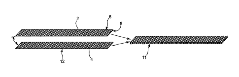
Figure 1A shows an end portion according to the present description, designed
to exhibit
improved short-term or long-term tissue fixation. Figure 1A shows two strips 2
and 4 of porous
materials (e.g., mesh), each having two major surfaces (6, 8, 10, and 12),
stacked against each
other to produce end portion 11 that includes two layers of open pore
material. The two layers
can be secured to each other by any fastening method or mechanism, such a by
sutures,
staples, rivets, adhesives, tack welds, thermal treatment of one or both
layers, etc. The use of
two layers of open pore materials for an end portion can improve strength of
the end portion and
can also improve short-term fixation by increasing the number of edge
extensions available to
contact tissue. The double layer construction is at the end portions of a
sling, and not
necessarily at a central support portion.
Figure 1A shows two pieces of open pore material being placed with major
surfaces
aligning in a width direction. If desired, alternate embodiments can place the
two strips at offset
positions so that edges do not align. Figures 1B and 1C illustrate a top view
and a side view of
two strips, 14 and 16, offset, and secured together by securement 18, which
may be, e.g., heat
treatment to melt polymer of strips 14 and 16, sutures, adhesive, etc.
= CA 02878309 2015-01-20
22
Still alternately, different sized strips could be used for end portions of
figures 1A and 1B
(or any other end portion described or illustrated herein), such as relatively
narrower open pore
materials in a width direction, to increase the number of edge extensions per
area of end
portion.
In a similar, alternate embodiment from that of figures 1A and 1B, an end
portion may be
of multiple layers, but based on a tubular construction. The end portion may
be a tubular piece
of open pore material folded flat against itself, or may maintain a somewhat
round, oblong, or
oval cross-section, with pores exposed at different directions around the
surface. A tubular end
portion may be made of an extruded porous material or a sheet connected at a
length-wise
seam. Optionally, slits may be cut (e.g., laterally, longitudinally,
diagonally, etc.) along the length
of a tubular end portion, to allow the end portion to conform to tissue or a
tissue path, to create
edges that improve friction between the end portion and tissue, and to allow
ingrowth between
tissue and the end portion.
Figure 2 illustrates another embodiment of a two-layer end portion of a
surgical implant,
such as a urethral sling. To further increase the likelihood of an end portion
having edge
extensions (e.g., sharp "tines" or "barbs") enter tissue upon implantation,
two layers of mesh
could be stacked as in figure 2, with one twisted layer. One porous strip of
an end portion can
spiral like a cork-screw or helix in regular or irregular lengths or
alternating or random directions,
resulting in increased contact and frictional engagement between mesh and
tissue. Figure 2
shows two strips 20 and 22 of a porous material (e.g., mesh). Strip 20 is
twisted, and then
stacked against strip 22, to produce end portion 24 that includes two layers
of open pore
material, one layer being twisted. As illustrated, one layer can be twisted
and one layer may be
laid flat, with the layers joined together as desired, e.g., by sutures,
staples, rivets, adhesives,
tack welds, thermal treatment of one or both layers, etc. By having one layer
twist relative to the
other layer, edge extensions or tines extend at every different angle from
edges of twisted mesh
CA 02878309 2015-01-20
=
23
20, increasing contact between edge extensions and tissue. When the implant is
installed, and
(optionally) a plastic sheath over the end portion is removed, the end portion
will open within a
tissue path and desirably engage tissue to prevent short-term movement of the
end portion or
sling.
Figure 3 illustrates another embodiment of a multi-layer end portion of a
surgical implant,
such as a urethral sling. To further increase the likelihood of the mesh to
have sharp "tines"
contacting tissue, three layers of mesh are stacked as in figure 3 with two
twisted layers. Figure
3 shows one central strip 34, and top and bottom strips 32 and 36, each of a
porous material
(e.g., mesh). Top and bottom strips 32 and 36 are twisted and then stacked
against center strip
34, to produce end portion 38 that includes three layers of open pore
material, with a center
layer one twisted top layer and one twisted bottom layer. Each of the layers
32, 34, and 36, may
be of the same or different materials, dimensions, pore and strand sizes (for
a mesh),
properties, etc. Twisted layers 32 and 36 may be of similar twisted
orientations, each being
twisted regularly in the same direction and at similar lengths, or in
alternating directions or
dissimilar lengths, or with irregular twist-lengths. So that top and bottom
layers 32 and 36 twist
and lay flat against a major surface of central strip 34, top and bottom
layers 32 and 36 can be
joined to central strip 34 as desired, e.g., by sutures, staples, rivets,
adhesives, tack welds,
thermal treatment of one or both layers, etc. Two twisted layers cause edge
extensions or tines
of twisted top and bottom layers 32 and 36 to extend at different angles to
increase the number
of edge extensions that contact tissue.
Figure 4 illustrates another embodiment of a multi-layer end portion of a
surgical implant,
such as a urethral sling. To further increase the likelihood of the implants
having have sharp end
extensions or "tines" contact tissue, two layers of twisted open pore mesh are
stacked against
each other in figure 4. Twisted strips 42 and 44, each of a porous material
(e.g., mesh), are
twisted and then stacked and attached together to produce end portion 46 that
includes the two
CA 02878309 2015-01-20
24
twisted layers of open pore material. Each of the layers 42 and 44 may be of
the same or
different sizes and materials. Twisted layers 42 and 44 may be of similar
twisted orientations,
each being twisted regularly in the same direction and at similar lengths, or
in alternating
directions or dissimilar or random twist-lengths, or with irregular twist-
lengths. Layers 40 and 42
can be twisted, laid flat, and then joined by any fastening mechanism or
technique, such as by
suture, staple, rivet, adhesive, tack weld, thermal treatment of one or both
layers, etc. By having
two twisted layers, edge extensions or tines of each of the two twisted layers
42 and 44 can
extend at different angles to increase the number of edge extensions that
contact tissue.
Figures 5, 6, and 6A, illustrate end portions of polymeric open pore material
for use in an
end portion, wherein the material is stiffened and biased to a non-flat
orientation, shape, or
form. Figure 5 shows end portion 48 in the form of an open pore material
(e.g., mesh) that
exhibits a wave-form in its natural state. End portion 48 has been treated or
produced to exhibit
a natural bias for this wave-form, e.g., by heat-forming, thermoforming,
molding, or coating with
a stiffening material. A force applied in opposite directions at each end of
end portion 48 would
reduce the wave-form and at least partially straighten the material, but upon
release of the force
the wave-form would return. This form causes the end portion to be biased
toward the wave-
form, and when the end portion is installed within a tissue path, the bias
will cause portions of
the wave-form end portion to exert pressure against tissue defining the tissue
path, increasing
frictional contact between the end portion and the tissue.
Figure 6A shows end portion 50 in the form of an open pore material (e.g.,
mesh) that
exhibits a three-dimensional twisted helical, screw, or spiral form in a
natural state. End portion
50 has been treated or produced (e.g., molded) to exhibit a natural bias for
this form, e.g., by
heat-forming, molding, coating with a stiffening material, etc. A force
applied opposite to the
twisted direction of end portion 50 may reduce the degree or number of twists,
and at least
partially straighten the end portion, but upon release of the force the twists
would substantially
CA 02878309 2015-01-20
return. This stiffened form causes the end portion to be biased to include the
twists. When the
end portion is installed within a tissue path, the bias will cause portions of
the twisted-form to
exert pressure against tissue defining the tissue path, increasing frictional
contact between the
end portion and the tissue.
Figure 6B shows end portion 51 in the form of an open pore material (e.g.,
mesh) that
exhibits a lateral curve form in a natural state. End portion 51 has been
treated or produced
(e.g., molded) to exhibit a natural bias for this curled form, e.g., by heat-
forming, molding,
coating with a stiffening material, etc. A force applied opposite to the
curled form may reduce
the degree of the curl and at least partially straighten the end portion, but
upon release of the
force the curl would substantially return.
Figures 5, 6 A, and 6B illustrate wave and spiral forms of end portions, but
other forms
would be useful as well for increasing force between an installed end portion
and tissue of a
tissue path. Further, while figures 5 and 6 illustrate single layer end
portions, a shaped end
portion (e.g., a heat formed end portion) could be used in combination with
multiple layer end
portions, if desired, such as an any one of the end portions described herein
or as illustrated,
e.g., at figures 1, 2, 3, 4, etc.
Figure 60 shows an implant that includes a central support portion that
exhibits a
stiffened, non-flat, curved, natural state. Implant 54 includes end portions
52, which may be flat,
non-flat, reinforced, multi-layer, etc., and central support portion 56.
Central support portion 56
has been treated or produced (e.g., molded) to exhibit a natural bias for a
curved form, e.g., by
heat-forming, molding, coating with a stiffening material, etc., as discussed
herein for producing
a non-flat end portion. A force applied opposite to the curled form may reduce
the degree of the
curl and at least partially straighten the end portion, but upon release of
the force the curl would
substantially return. The curved form of central support portion 56 may be of
a shape or form
adapted to a particular tissue such as the bladder, urethra, vagina, corpus
spongiosum, BC
CA 02878309 2015-01-20
26
muscle, etc., to allow the central support portion to more closely align with
a tissue upon
implantation.
Figures 7 A, 7B, and 70, illustrate exemplary urethral slings having
tensioning members
and widened central support portions (widened in a single direction or
"unilaterally"). Referring
to figure 7A, urethral sling 60 includes end portions 64, widened central
support portion 62, and
tensioning members 66. A tensioning member such as member 66 may be, e.g., a
suture, heat-
treated open pore material of end portions 64, adhesive, or the like,
resulting in reduced length-
wise elasticity of end portions 64. As illustrated, tensioning member 66 is
shown to be a suture
attached at multiple points 68, by, e.g., adhesive, thermal welding, sonic
welding, adhesive,
knots, or the like.
Referring to figure 7B, urethral sling 70 includes end portions 74, widened
central
support portion 72, and tensioning members 76, which may be, e.g., a suture,
heat-treated open
pore material of end portions 74, adhesive, or the like, resulting in reduced
length-wise elasticity
of end portions 74. As illustrated, tensioning members 76 are shown to be a
sutures attached at
multiple points 78, by, e.g., adhesive, thermal welding, sonic welding,
adhesive, knots, or the
like. Edge extension reinforcement 75 is shown to be present along each of the
side edges of
the opposing end portions 74.
Referring to figure 70, urethral sling 80 includes end portions 84, widened
central
support portion 82, and tensioning members 86, which may be, e.g., a suture,
heat-treated open
pore material of end portions 84, adhesive, or the like, resulting in reduced
length-wise elasticity
of end portions 84. As illustrated, tensioning members 86 are shown to be a
sutures attached at
a single attachment point 88 per suture, e.g., adhesive, thermal welding,
sonic welding,
adhesive, knot, or the like. Edge extension reinforcement 85 is shown to be
present along each
of the side edges of the opposing end portions 84.
CA 02878309 2015-01-20
27
Figures 7D, 7E, and 7F illustrate still other embodiments of urethral slings
of the
invention, each of which illustrates a urethral sling having a widened (bi-
laterally) central support
portion. Figure 7D shows sling 90 comprising widened central support portion
92, load-transition
portions 93, and end portions 94. Central support portion 92 and load-
transition portions 93 are
each of a single piece of material, and are connected to end portions 94 by
attachments 96,
extending the width of end portions 94. Attachments 96 may be, e.g., heat-
treated areas of
melted polymeric material of end portions 94, central support portion 92, or
both. Alternately,
attachments 96 may be sutures, adhesive, or the like.
Figure 7E shows sling 100 comprising widened central support portion 102, load-
transition portions 103, and end portions 104. Central support portion 102 and
load- transition
portions 103 are of a single piece of material and are connected to end
portions 104 by
attachments 106. Attachments 106 are illustrated to be polymeric rivets or
adhesive, but may
alternately be another type of attachment such as sutures or melted polymeric
implant material.
Sutures 105 extend along end portions 104, two sutures 105 per end portion
104. Sutures 105
are attached to end portions 104 at multiple attachment points 107.
Figure 7F shows sling 110 comprising widened central support portion 112, load-
transition portions 113, and end portions 114. Central support portion 112,
load-transition
portions 113, and end portions 114 are all of as single piece of material,
such as a woven
polymeric (e.g., polypropylene) mesh. Two sutures 115 extend along the entire
length of implant
110, including end portions 114, central support portion 112, and load-
transfer portions 113.
Sutures 115 are attached to implant 110 at multiple attachment points 117.
Referring to figure 8, implant 140 includes support portion 148 and end
portion 141. End
portion 141 is an open pore material such as a mesh that includes solid
portions (e.g.,
interwoven strands) 142 and apertures or pores 144 defined by solid portions
142. Edges 143
and 145 include edge extensions 146, directed with a slant away from support
portion 148.
CA 02878309 2015-01-20
28
Edge extensions 146 are illustrated as cut strands of material at the uneven
edge of the open
pore material defined by cutting (or forming) the open pore material along a
line that includes
adjacent pores. Extensions 146 are, e.g., cut strands of material that extend
from the open pore
material to define edges 143 and 145.
Referring to figure 9, implant 130 includes support portion 128 and end
portion 131. End
portion 131 is an open pore material that includes solid portions 122 and
apertures or pores 124
defined by solid portions 122. Edges 133 and 135 include edge extensions 126,
directed with a
slant away from support portion 128. Edge extensions 126 are illustrated as
portions of solid
material at the uneven edge of the open pore material defined by cutting or
forming the open
pore material along a line that includes adjacent pores. Extensions 126 are
the material that
extends from the open pore material to define edges 133 and 135.
Referring to figure 10, implant 160 includes central support portion 162 and
integral end
portions 164. As illustrated, implant 160 is of a single piece of material,
such as a mesh, cut as
one piece to the illustrated shape. Each of end portions 164 includes cuts
("slits" or "slots") 166
that extend laterally, partially across the width of each end portion 164, and
that are located at
multiple locations along the lengths of each of the two end portions 164. Each
cut 166 exposes
strands of mesh that can contact tissue upon installation and increase
frictional forces between
tissue and implant. Each cut 166 also allows an end portion 164 to conform in
shape to a tissue
path. Cuts 166 are lateral, but one or more longitudinal or diagonal cuts in
end portions 164 may
be used as an alternate to illustrated lateral cuts 166. Implant 160 also
includes cuts 168
extending longitudinally across portions of central support portion 162, to
allow central support
portion 162 to conform to tissue being supported. Cuts 168 are longitudinal,
but one or more
lateral or diagonal cuts in central support portion 162 may be used as an
alternate to illustrated
longitudinal cuts 168.
CA 02878309 2015-01-20
29
Figures 7A, 7B, 70, 7D, 7E, and 7F, and figures 8, 9, and 10, do not
specifically show
certain features end portions as described herein, e.g., multi-layer, heat-
shaped or formed,
coated to take a form of a wave, twist, or curl, etc. According to the
invention, however, any of
these features may be included in the end portions of slings of figures 7A,
7B, 7C, 7D, 7E, 7F,
8, 9, and 10.
Referring to figure 11, an exemplary embodiment of a urethral sling assembly
is
depicted. Sling assembly 210 includes sling end portions 220 and 221, and end
connectors 212,
which engage with free ends of right hand and left hand sling implantation
tools (not shown).
End connectors (or "dilators") 212 can be shaped to dilate right and left
passages through body
tissue formed by curved needles of right and left hand implantation tools in a
transobturator
procedure. While not specifically illustrated, a sling as illustrated by
figure 11 may include
features of end portions 220 and 221, as described herein, such as multiple
layers, stiffening for
shaping, features of edge extensions, etc.
Sling assembly 210 comprises a urethral sling with central support portion
240, and end
portions 242 and 240 enclosed within protective sheaths 222 and extending from
sling end
connectors 212 to open sheath ends 226 and 228. Protective sheaths 222 can be
constructed
of a flexible thin transparent plastic film that enables visual examination of
urethral sling end
portions 220 and 221, and are sufficiently lubricious to pass easily through
tissue passageways
of a patient formed using sling implantation tools. Sheaths 222 can include
sheath indicia or tear
scores, perforations, or holes for assisting a surgeon in orienting urethral
sling assembly 210
relative to a urethra or other pelvic tissue during installation. The sling
implant portion of
assembly 210 can be left in place chronically following implantation.
According to still other embodiments of the invention, ease of use of a needle
may
improve by application of a coating to the needle to reduce friction between a
needle and tissue,
for improved passage through tissue in creating a tissue path. Coatings can
include parylene,
CA 02878309 2015-01-20
Teflon (e.g., PTFE), hydrophilic low friction coatings, etc. Alternately or
in addition, plastic
sheaths such as sheaths 222 of figure 11, may be coated to reduce friction
between sheaths
111 and tissue, upon installation, and allow sheaths 222 to move through
tissue with less force.
Various embodiments of tools and implants described herein result in
advantages in
pelvic procedures, irrespective of gender. Materials of an implant such as
type of mesh,
materials useful for a mesh, geometry of a mesh, shape of a mesh, and
placement of a mesh,
can result in useful or improved short or long term fixation of a pelvic
implant. Materials,
implants, and related methods described herein may provide improved support
for pelvic tissue
such as the bladder, bladder neck, urethra, tissue supportive of the urethra,
etc., from the
position of the floor of the lower pelvic area. This provides resilience upon
downward pressure
being placed on the pelvic tissue and there will be a push up from the sling
when in the proper
position.
The invention also relates to surgical assemblies, systems, or kits, that
include an
implant as described herein, including any one or any combination of the
described features.
The implant may be useful for installation to treat a pelvic condition such as
incontinence. An
exemplary kit or assembly can include a urethral sling and one or two surgical
instruments, each
instrument having a handle portion, a needle portion having substantial
structure in three
dimensions, and a distal region. A needle portion of one of the tools can be
sized and shaped to
extend between an incision substantially adjacent the obturator foramen on the
patient's right
side and a medial incision. The assembly also has a second surgical instrument
for use on a left
side of a patient.
Exemplary transobturator methods may be useful for installing a urethral
sling, and may
include steps of creating a medial incision at the external male perineum or
female vaginal,
creating two external opposing lateral incisions substantially adjacent the
patient's left and right
obturator foramen, and installing a urethral sling as described herein, end
portions of which
CA 02878309 2015-01-20
31
traverse the obturator foramen. The sling may be placed using one or more
surgical installation
tools, by installing end portions of the sling between the medial and the
lateral incisions and
passing through the obturator foramen. The end portion may be pushed through
the tissue path
at the leading edge of a needle, or may be pulled through the needle path
using a trailing edge
of the needle.
In more detail, an exemplary transobturator method for installing a urethral
sling in a
male anatomy can include a steps of creating a medial incision at the exterior
perineum,
creating an external lateral incision substantially adjacent the patient's
obturator foramen,
providing a surgical instrument having substantial structure in three
dimensions, and providing
an implant for treating the incontinence (a urethral sling), as described. The
three-dimensional
region of the needle may be passed between the incisions and then the implant
can be
associated with the instrument, e.g., at the end of the three-dimensional
region. For example,
the needle may be passed from the lateral incision through the obturator
foramen and to the
medial incision, and the implant can be associated with the tip of the needle
extending from the
medial incision. The needle can then be pulled back through the incisions to
pull the end portion
of the implant from the medial incision, through the obturator foramen, and to
the lateral incision.
Alternately, the implant can be associated with the needle before passing the
needle between
incisions. The needle, with the end portion of an implant associated with the
needle tip, may
then be passed between incisions, such as from the medial incision, through
the obturator
foramen, and then through the lateral incision. This can be done on both the
right side and the
left side.
In other embodiments of a transobturator method, a single needle may be useful
to
place left and right end portions both left and right sides of a patient. A
single left-handed needle
(alternately a single right-handed needle) can be used to place a right side
of the sling on a
patient's right side, using a transobturator tissue path between a perineal
incision and a patient's
CA 02878309 2015-01-20
32
right-side lateral incision. In the same procedure, the same left-handed
needle may also be
used to place the opposite end portion on the patient's left side. While the
left- handed needle is
not optimal for placement at the patient's left side, it can be effective.
Systems or kits of the
invention can include a single left- or right-handed needle with an implant,
for surgical implant
according to this method.
By still other implantation methods, a variation of a "transobturator" method
(considered
for the present description to be a "transobturator method") includes a method
of inserting an
implant through a medial, perineal incision and attaching an end portion of
the implant to the
obturator membrane. The anchor traverses or otherwise attaches to the
obturator membrane.
Other features of the inventive methods described herein can be incorporated
into such a
technique, such as placement of the urethral sling below the BC or CS,
approximation of the
urethra to improve continence (without the need for compression of the
urethra), etc. This
method avoids the need for lateral incisions.
To improve continence, the sling can be placed to support the urethra, by
directly
contacting the urethra or by indirectly supporting the urethra by contacting
tissue supportive of
the urethra, such as the corpus spongiosum (CS) or bulbous cavernosum (BC)
muscle. See,
Assignee's copending United States Patent application no. 11/347,047 which has
been issued
as U.S. Patent No. 7,914,437, entitled "TRANSOBTURATOR METHODS FOR INSTALLING
SLING TO TREAT INCONTINENCE, AND RELATED DEVICES," filed on even date
herewith.
Placement of an implant below the corpus spongiosum or below the bulbous
cavernosum muscle may provide certain advantages that would provide for
approximating the
BC muscle once the ends of the implant are tensioned so as to approximate
pelvic tissue to a
target and optimum position for support of the urethra, bladder, or supportive
tissue, and hence
provide continence relief. In one example embodiment, the proposed pressure is
about 500
grams of force, but this may vary depending on the severity of incontinence of
the patient and is
CA 02878309 2015-01-20
33
not limited to this stated amount. According to different embodiments of
methods of treating
incontinence in a male anatomy, an implant can be placed below the BC muscle
or below the
corpus spongiosum, and can be tensioned via pulling end portions to
approximate the BC
muscle or CS, to place such tissue in a the final position that improves
continence. Optionally,
the implant may be sutured to the BC muscle or CS.
An implant such as a urethral sling can be installed as described herein with
the
assistance of surgical equipment, instruments, or tools that will be
understood to be of
assistance in performing the present surgical methods. Examples of surgical
tools that may be
useful include tools of the type described herein and in U.S. Pat. No.
6,911,003 and U.S.
Published Application No. 2003/0171644A1, which generally include right and
left-handed
opposing helical installation tools.
Exemplary surgical tools can comprise a needle sized and shaped to either a)
initially
extend through an incision substantially adjacent a patient's obturator
foramen and then through
the obturator foramen to a medial incision, or b) initially extend through a
medial incision and
subsequently through the obturator foramen and then to an incision
substantially adjacent a
patient's obturator foramen. Preferably, the needle comprises a pair of ends
having surfaces for
affording association with either an implantable sling material or a removable
handle. In one
embodiment, a needle is sized and shaped for use on either the patient's right
side or left side
(not both).
Embodiments of installation tools can include a substantially straight spacer
portion
emerging from an end of the handle portion preferably along a longitudinal
axis of the handle.
This helps afford convenient passage of the needle using an ergonomic wrist
roll adopted by
some surgeons.
A three-dimensional region of a needle can include a structure that can be
described as
a variable spiral or helix portion extending from the distal end of a straight
spacer portion. A
CA 02878309 2015-01-20
34
spiral portion can be variable as the angle of the spiral portion changes
between the beginning
of the spiral (e.g., the end of the spacer) and the distal end of the needle.
The shape of the
spiral portion can be designed to avoid over-insertion of the needle into the
body, which helps
avoid damage to the sensitive structures in this region of the body.
A useful needle can have dimension and shape features particularly designed
for male
or female anatomy, such as for installation using a male transobturator
procedure. The
dimensions and shape features of the tool allow the needle to extend from a
lateral incision
adjacent the anterior side of the pubic bone, through the obturator foramen
portion of the pubic
bone, to a position on the posterior side of the pubic bone, and to then
emerge from a medial
incision made between the patient's obturator foramen incisions. Alternate
needles may be
shaped to extend along the same tissue path in the opposite direction,
entering at the medial
incision and exiting at the lateral incision. A large number of different
sizes, shapes, and
dimensions of needles are suitable for the present invention.
In certain embodiments, a tool includes a handle or a portion of a handle may
exhibit a
non-circular form when viewed along the longitudinal axis of the handle. The
non- circular cross-
section can be, e.g., an oval, rectangle, rhombus, etc., having one dimension
"width" that is
greater than the dimension perpendicular to that "width." The non-circular
form will provide
surfaces on the handle for a surgeon to place pressure onto and to achieve a
grip. The non-
circular cross-sectional form also defines a midplane that is a plane that
includes the
longitudinal axis of the handle and extends along the widest dimension of the
handle when
viewed in cross section.
According to embodiments of the invention, a needle distal end of a tool
(measured at
the tip of the needle distal end) may be located at a position in space
relative to the handle
midplane and longitudinal axis, to provide the user with an ergonomic
advantage. The
ergonomic advantage may relate to useful or optimized (e.g., increased)
amounts of force and
CA 02878309 2015-01-20
control that can be applied at the needle distal end during the transobturator
installation
procedure, meaning amounts of force, sensitivity, and control that the user
will have over the
needle distal end when manipulating the handle using the midplane for leverage
or grasping. As
an example, a needle distal end may be located at an angle relative to the
midplane to provide
an ergonomic strength advantage or control advantage to a surgeon during
particularly risky or
sensitive portions of a surgical procedure, such as portions of a surgical
procedure that involve
using the needle distal end to dissect a tissue path through or near sensitive
organs or tissues,
e.g., traversing the obturator foramen. The angle "x" (see figures 12A and
12B) between the
needle distal end and the midplane may provide the surgeon with the use of
maximum hand or
wrist strength and maximum control and precision during manipulation of the
needle distal end
through a sensitive or risky tissue path, when applying pressure to a handle
having a midplane.
For transobturator procedures, the angle may be from 20 to 70 degrees, e.g.,
from 25 to 50
degrees, such as from 30 to 40 degrees from the midplane when viewed along
longitudinal axis
231. See also Assignee's copending United States Patent Application serial No.
11/347,553
which has been issued as U.S. Patent No. 7,422,557, entitled "NEEDLE DESIGN
FOR MALE
TRANSOBTURATOR SLING," filed on even date herewith.
Figures 12A and 12B illustrate two views of a tool useful according to the
invention.
Figure 12A illustrates a view of tool 230 along a longitudinal axis of the
tool. Figure 12B
illustrates a side view of tool 230. Tool 230 includes handle 232 and a needle
extending
longitudinally from an end of handle 232 along longitudinal axis 231 of the
handle and tool. The
needle includes spacer 234 and three-dimensional region 236 which may be
considered to be a
helix, a variable helix, or a spiral, etc. Diameter 238 can be larger than
diameters of relevant
prior art tools, and may be, for example, in the range from 2 to 5
centimeters, e.g., about 2.4
inches. Length 242 of spacer 234 can be any desired length, with an exemplary
length 242
being in the range from 1 to 5 inches, e.g., from 1.75 to 2.25 inches. Length
240 of three-
CA 02878309 2015-01-20
36
dimensional region 236 can be any desired length, such as in the range from
2.25 to 5
centimeters, e.g., from 2.4 to 2.5 inches. Angle y is approximately 45
degrees, and angle x is
approximately 30 degrees, but may be otherwise, such as in the range from 20
to 70 degrees,
or from 30 to 60 degrees. Needle end portion 244, which includes a length of
about one inch at
the end of the needle, is curved up until engaging portion 249, which is
straight.
Figure 12B shows an axis of needle end portion, line 252, or a plane defined
by the
needle end portion that is substantially orthogonal to the longitudinal axis
of handle 232. Distal
end portion 244 can define either a line or a plane, depending on, e.g.,
whether the distal end
portion is straight or curved. In figure 12A, distal end portion 244 includes
a curve, and as such
defines a plane including needle distal end 250. This plane, illustrated as
line 252, is
substantially orthogonal to the longitudinal axis of tool 30. Radial distance
251 of tool 230 can
be as desired, e.g., in the range from about from 0.7 to 1.4 inches, e.g.,
from 0.9 to 1.1 inch for
a male transobturator tool. Also shown at figures 12A, needle end portion 244,
which includes a
length of about one inch at the end of the needle, is curved up until engaging
portion 249, which
is straight.
Other modifications may also be useful for a tool, especially for use in an
transobturator
installation procedure in the male anatomy. Passing a needle in a male anatomy
may on
average be more difficult than in a female anatomy due to on-average greater
muscle mass to
pass through the obturator foramen, and due to the need to pass the needle
through the
perineal membrane versus a vaginal incision as in a woman. To assist in needle
passage in the
male anatomy, dimensions of size and shape of a three-dimensional portion of a
needle may be
increased or otherwise modified. A handle may be made to have a wider
dimension, or modified
shape or form, to allow improved grip and torque. Figure 13, for example,
illustrates an example
of a tool that has a handle with a non-conventional grip. Tool 270 includes
needle 274 and
handle 272. Handle 272 includes a grip 276, that is oriented along an axis
different from needle
CA 02878309 2015-01-20
37
shaft 274. This arrangement may allow a desirable orientation of the handle
for use during a
transobturator procedure.
The scope of the claims that follow is not limited by the embodiments set
forth in the
description. The claims should be given the broadest purposive construction
consistent with the
description as a whole.