Note: Descriptions are shown in the official language in which they were submitted.
CA 02878396 2015-01-05
WO 2013/028274
PCT/US2012/046329
SYSTEM AND METHOD FOR NEUROMODULATION
Priority: This application claims the benefit of U.S Provisional Application
No. 61/506,164,
filed July I I, 2011 (Attorney Docket IAC-1200), U.S Provisional Application
No.
61/551,418, filed October 25, 2011 (Attorney Docket IAC-1210), U.S.
Provisional
Application No. 61/584,812, filed January 9.2012 (Attorney Docket IAC-1220),
U.S.
Provisional Application No. 61/601,501, filed February 21, 2012 (Attorney
Docket IAC-
1230), U.S. Provisional Application No. 61/613,433, filed March 20, 2012
(Attorney Docket
1AC-1240), and U.S. Provisional Application No. 61/639,982,.ffled April 29,
2012 (Attorney
Docket IAC- I 250). Each of the foregoing applications is incorporated herein
by reference.
TECHNICAL FIELD OF THE INVENTION
The present application generally relates to systems and methods for
neuromodulation
using stimulation elements disposed within the vasculature.
BACKGROUND
Acute heart failure syndromes (Al-IFS) are serious conditions resulting in
millions of
hospitalizations each year. Well documented in the literature are causal links
between
declining renal function or myocardial injury during AFIFS hospitalization and
poor
prognosis. Heart failure resulting from myocardial ischemic insult or
tachycardia precipitates
complex alterations in autonomic tone, neurohormonal activation, and the
inflammatory
metabolic state. These changes in autonomic tone are typically manifested by
increased heart
rate and a reduction in heart rate variability. In the setting of an acute
exacerbation of heart
failure; the dramatically elevated heart rate is frequently accompanied by
hypotension. The
critical role of treating the autonomic nervous system dysfunction observed in
HF has long
been recognized (with inotropic agents and beta-blockers). Recently, specific
neuromodulation of the parasympathetic cardiac nerve inputs has shown
s4mificant
therapeutic benefit. Cleland JG, Bristow MR, Erdmann E, Remme WJ, Swedberg K,
- 1 -
CA 02878396 2015-01-05
WO 2013/028274
PCT/US2012/046329
Waagstein F. Beta-blocking agents in heart ,failure. Should they be used and
how? Eur Heart
J 1996;17: 1629-39; De Ferrari GM, Crijns 11J, Borgurefe M, et al. Chronic
yaps nerve
stimulation: a new and promising therapeutic approach fin. chronic heart
fttilure. Eur Heart J
2011;32:847-55.
However, in the case of AHFS associated with congestive symptoms and reduced
blood pressure (BP), the negative inotropic effects of lone parasympathetic
intervention or
beta-blockade can severely limit their utility. In the face of' hypotension,
sympathetic tone
must be maintained in order to assure adequate left ventricular (LV)
contractility. Arland IS,
Fisher LD, Chiang YT, et al. Changes in brain natriuretic peptide and
norepinephrine over
/line and mortality and morbidity in the Faisal-Ian Heart l'ailttre Trial (Val-
Heb7).
Circulation 2003;107:1278-83. Animal studies have demonstrated positive
inotropic effects
(increased LV pressure and cardiac output without change in systemic vascular
resistance)
when selectively stimulating certain cardiac efferent sympathetic nerves.
Zarse M, Plisiene
J, Mischke K, et al. Selective increase o/. cardiac neuronal sympathetic tone:
a catheter-
based access to modulate left ventricular contractility. j Am Coll Cardiol
2005;46:1354-9;
Meyer C, Rana OR, Saygili E, et al. Augmentation of lefi ventricular
contractility by cardiac
sympathetic neural stimulation. Circulation 2010;121:1286-94.
BRIEF DESCRIPTION OF THE DRAWINGS
Fig. I graphically represents stimulation effects achievable using the
disclosed system
and method.
Fig. 2A is a top, cross-section view of the superior vena cava illustrating a
target
electrode region for delivery of therapy to parasypathetic and sympathetic
targets.
Fig. 2B is similar to Fig. 2A and schematically shows electrodes positioned to
deliver
therapy to the parasympathetic and sympathetic targets.
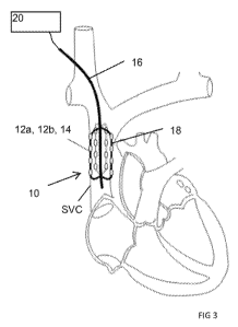
Fig. 3 schematically illustrates a therapy device disposed with the superior
vena cava
to position electrodes between the superior vena cava's bifurcation and the
atrium of the
heart.
Fig. 4 schematically illustrates an embodiment of a control system for a
neuromodulation system.
_ 2 -
CA 02878396 2015-01-05
WO 2013/028274 PCT/US2012/046329
Fig. 5 is a graphical representation illustrating control of normal
cardiovascular
function by the autonomic nervous system's cardiovascular control system.
Fig. 6 is a graphical representation illustrating an adjusted cardiovascular
control
system achieved through the addition of the disclosed neuromodulation system
to the
autonomic nervous system's cardiovascular control system.
DETAILED DESCRIPTION
The present application discloses methods and systems for treating autonomic
imbalance in a patient by energizing a first therapeutic element disposed in a
superior vena
cava of the patient to deliver therapy to a parasympathetic nerve fiber such
as a yaws nerve,
and energizing a second therapeutic element disposed within the superior vena
cava to
deliver therapy to a sympathetic cardiac nerve fiber. A neuromodulation system
includes a
parasympathetic therapy element adapted for positioning within a blood vessel,
a sympathetic
therapy element adapted for positioning with the blood vessel; and a
stimulator. The
stimulator is configured to energize the parasympathetic therapy element to
deliver
parasympathetic therapy to a parasympathetic nerve fiber disposed external to
the blood
vessel and energize the sympathetic therapy element within the blood vessel to
deliver
sympathetic therapy to a sympathetic nerve fiber disposed external to the
blood vessel. In
disclosed embodiments, delivery of the parasympathetic and sympathetic therapy
can be used
to decrease the patient's heart rate and while elevating or maintaining the
blood pressure of
the patient.
Studies conducted by the inventors have elucidated distinct and precise sites
in the
superior vena cava (SVC) where neurostimulation selectively results in
modulation of boih
cardiac parasympathetic and sympathetic nerves. These anatomic locations were
demonstrated using minimally invasive, vascular procedures. These studies
demonstrated
that independent cardiac parasympathetic and sympathetic stimulation is
achievable from
fully intravascular locations within the SVC. The results of these studies
consistently
demonstrated that parasympathetic neuromodulation through vagus nerve
stimulation to
decrease heart rate (HR) with attendant effect on blood pressure (BP) is
simple,
- 3 -
CA 02878396 2015-01-05
WO 2013/028274
PCT/US2012/046329
straightforward and repeatable. The studies also revealed that sympathetic
neuromodulation
for the purpose of increasing BP with attendant effect on HR could also be
accomplished in a
straightforward manner.
The present inventors have achieved rapid, acute parasympathetic and
sympathetic
modulation of cardiac hemodynamics in humans using intravascular stimulation
of the vagus
nerve and cardiac sympathetic branches from within the SVC. In those studies,
stimulation
parameters of 5-I5mA, 20 Hz, and 0.5ms pulse width were shown to be effective.
Although other investigators have separately stimulated parasympathetic or
sympathetic nerves to cardiac effect using surgically-based approaches, no
previous
approaches have demonstrated sinuthaneous and selective modulation of both
autonomic
inputs from intravascular locations. This unprecedented advantage of
instantaneous and
complete flexibility in management of HR and BP, together with an easy-to-use,
minimally
invasive approach will provide substantial therapeutic benefit.
The disclosed system can provide a broad spectrum of clinically relevant
control
through its ability to modulate both HR and BP. in patients that require a
decrease in HR and
BP, such as those with diastolic heart failure and preserved ejection fraction
that present with
elevated HR and BP, pure parasympathetic stimulation is provided (lower left
hand box of
Fig,. I). Similarly, for those patients requiring pure sympathetic
stimulation, such as for
elevation of heart rate and blood pressure (upper right hand box) the system
provides that
capability. But in many cases of acute decompensation, particularly in those
patients
approaching or in cardiouenic shock or with cardiorenal syndrome, pure
parasympathetic or
pure sympathetic stimulation could have potential detrimental. effects. These
patients often
present with both hypotension and tachycardia. Pure parasympathetic
stimulation could
worsen this situation by simultaneously decreasing heart rate while
potentially reducing
blood pressure resulting in inadequate systemic perfusion. On the other hand,
pure
sympathetic stimulation, while supporting the blood pressure, could further
drive the existent
tachycardia to extreme levels. Ideally, under the condition of hypotensiOn and
tachycardia
due to these forms of heart failure, one would want to provide support of the
blood pressure
to provide for adequate systemic perfusion while simultaneously reducing
tachycardia and
even lowering the heart rate further to allow adequate cycle length to
optimize the stroke
- 4 -
CA 02878396 2015-01-05
WO 2013/028274 PCT/US2012/046329
volume, preserving or improving cardiac output (upper left hand box), a
treatment achievable
using the disclosed system.
Ultimately, a combination of autonomic modulation based on hemodynamic
feedback
of both HR and BP would provide optimal therapy.
While discussed in connection with acute heart failure syndrome, the disclosed
system and methods may be used to provide acute autonomic neuromodulation in
patients
suffering from other conditions, including, but not limited to acute
myocardial infarction,
pulmonary embolism, hemorrhage, autonomic dysfunction, systemic inflammatory
response,
syndrome (SIRS), sepsis, as well as post-surgery autonomic dysfunction.
Moreover,
principles disclosed herein may further be implemented using an implantable
system,
including one in which the electrodes are chronically disposed or anchored in
the SVC at
positions determined to deliver the disclosed parasympathetic and/or
sympathetic stimulus.
An implantable system may have an implantable stimulator, such as one
implanted at an
intravascular or extravascular (e.g. subcutaneous) site, or a stimulator that
is positioned
outside the body for wirelessly activating the electrodes. Applications for a
chronic system
include treatment of patients suffering from chronic heart failure, or
autonomic dysfunction
associated with other conditions including those listed above.
Accordingly, the present inventors have conceived of a system that is suitable
for
each type of neuromodulation represented in Fig. 1, including delivery of
independent and
simultaneous stimulation of parasympathetic and sympathetic cardiac nerves to
achieve a
simultaneous reduction in HR and increase in BP that results in an increase in
cardiac output.
Referring to Fig. 3, the neuromodulation system comprises a therapy device 10
having one or
more intravascular therapeutic elements I 2a, 12b. The therapy device 10
positions the
therapeutic elements the SVC, where they are selectively energized to
modulate nerve
fibers located outside the vasculature. The therapeutic elements are arranged
such that some
of the therapeutic elements (referred to herein as the parasympathetic
therapeutic elements
12a) direct energy to parasympathetic cardiac nerve Fibers from within the
SVC, while
different ones of the therapeutic elements (referred to as the sympathetic
therapeutic elements
12b) direct energy to sympathetic cardiac nerve .fibers, also from within the
SVC. See Fig.
2B. Because percutaneous advancement of a catheter to the SVC is a. simple and
- 5 -
CA 02878396 2015-01-05
WO 2013/028274
PCT/US2012/046329
straightforward procedure, the ability to position both parasympathetic and
sympathetic -
therapeutic elements within the SVC is highly advantageous.
In preferred embodiments, the therapeutic elements I 2a, b are electrodes 14,
although
it is contemplated that other forms of therapeutic elements (including, but
not limited to,
ultrasound, thermal, or optical elements') may instead be used. The
therapeutic elements are
positioned on a flexible therapy device such as a catheter or other flexible
elongate carrier 1 6,
allowing advancement of the therapeutic elements from a percutaneous access
site to the
SVC. The therapy device includes an anchoring structure 18 expandable within
the
vasculature for biasing the electrodes in contact with the interior surface
of' the blood vessel
so as to optimize conduction of neuromodulation energy from the electrodes to
the target
nerve fibers.
The therapy device or catheter and its corresponding electrodes and anchoring
structure may take a variety of forms. Reference is made to commonly-owned
Application
Nos. PCT/US12/35712 ("Neuromodulation Systems and Methods for Treating Acute
Heart
____________________________________________ Failure Syndromes"; Atty Docket:
1AC-1010), U.S. , filed July 11, 2012
("Catheter System for Acute Neuromodulation; Any Docket: 1AC-1201), each of
which is
fully incorporated herein by reference. These applications describe exemplary
electrode and
catheter systems for use in acute neuromodulation which may be used or adapted
for use with
the disclosed neuromodulation system. Electrodes disclosed in U.S. Application
No.
13/281,399 entitled Intravascular Electrodes and Anchoring Devices for
Transvenous
Stimulation, may also be adapted for use with the disclosed system.
A preferred therapy device for use in the disclosed method utilizes an
integrated
design, from which stimulus may be directed from a single intravascular
therapy device to
two or more nerve targets. A device of this type may include a single flexible
support 16 or
catheter supporting multiple electrodes 18 or electrode arrays which may be
independently
activated to stimulate a different nerve target. In this type of embodiment,
the multiple
electrodes (i.e. those used for parasympathetic stimulation and those used for
sympathetic
stimulation) may be supported by a common support or electrode carrying member
I 8 that
biases the electrodes into contact with the vessel wall. For example, the
electrode carrying
member 18 might be formed of two or more longitudinal splines carried by the
support 16 in
- 6 -
CA 02878396 2015-01-05
WO 2013/028274
PCT/US2012/046329
an arrangement of the type disclosed in the prior applications incorporated
herein
(schematically shown in Fig. 3). With this design, the parasympathetic
stimulation
electrodes may be a bipolar arrangement of electrodes longitudinally arranged
on a first
spline, and the sympathetic stimulation electrodes may be a bipolar
arrangement of
electrodes longitudinally arranged on a second spline of the catheter. As
another example,
the parasympathetic stimulation electrodes and the sympathetic stimulation
electrodes may
be positioned on a common expandable sleeve formed of mesh, laser cut tubing,
or other
structures used for endoluminal electrode supports or stents.
The electrode carrying member 18 may include' multiple splines or regions
having
electrode arrays. This arrangement allows a mapping procedure to be conducted
upon
placement of the catheter within the SVC, such that the splines/regions whose
electrodes
produce the most optimal parasympathetic and sympathetic response may be
determined and
used for treatment. In other words, mapping may be used to determine which of
multiple
electrodes or electrode arrays will be the parasympathetic stimulation
electrodes or arrays,
and which will be the sympathetic stimulation electrodes or arrays.
In other embodiments, the electrode carrying member supports a first
electrode,
electrode array, or electrode pair for parasympathetic use, and a second
electrode, electrode
array, or pair for sympathetic use, together with means for independently or
simultaneously
adjusting the positions of the first and second arrays during mapping. Other
electrode
arrangements may be used, including separate catheters (e.g. telescoping or
parallel
catheters) for sympathetic and parasympathetic stimulation, with each catheter
having
longitudinally spaced electrodes. In these and the prior examples; independent
bipolar
electrodes, bipolar electrodes sharing a common pole, or unipolar electrodes
may be used -
with indifferent electrodes in the unipolar embodiments positioned elsewhere
on the catheter
or in/on the patient.
An external stimulator 20 energizes the electrodes using stimulation
parameters
selected to capture the target nerve fibers and to achieve the desired
neuromodulation.
Suitable stimulation parameters are 5-15mA, 20 Hz, and 0.5ms pulse width,
although other
stimulation parameters may alternatively be used. Feedback to the stimulator
is provided by
one or more diagnostic sensors. The catheter and stimulator may operate as a
closed-loop
- 7 -
CA 02878396 2015-01-05
WO 2013/028274
PCT/US2012/046329
system, allowing simulation parameters to be automatically determined and/or
dynamically
controlled in response to information sensed by the sensors and/or derived
from sensor
feedback. Suitable sensed or derived hemodynamic parameters may include
central venous
pressure (CVP), pulmonary capillary wedge pressure (PCWP), cardiac index,
derivations of
vascular resistance, heart rate, blood pressure (arterial). Other parameters
may include
CO/Cl, and cardiac filling pressures. For some parameters such as CVP,
feedback may be
generated using sensors mounted on the electrode-carrying member or extending
through the
lumen of' its catheter.
Electrode Position
The therapy device 10 positions the electrodes 14 or other therapy elements
such that
simultaneous sympathetic and parasympathetic stimulation may be carried out
using
parasympathetic stimulation electrodes disposed in the postero-lateral segment
of the mid to
cranial portion of the Superior Vella Cava, and sympathetic stimulation
electrodes disposed
in the postero-medial segment of the mid to cranial portion of the Superior
Vena Cava.
In preferred methods, the electrode positions in the SVC from which the
parasympathetic (vagus) and cardiac sympathetic nerve branches may be
stimulated reside in
an approximately 120- 270 degree circumferential band centered on the
posterior wall of the
vessel. In other words, referring to Nu. 2A, if mid-anterior MA is considered
to be at 0
degrees (6 o'clock in Hu. 2A below), proceeding clockwise, electrodes may be
positioned on
the vessel wall within a region that extends along the vessel wall from 45 to
315 degrees. In
other embodiments, electrodes may be positioned on the vessel wall within a
region that
extends along the vessel wall from 120-240 degrees as identified in Fig. 2A.
The
electrode(S) 12a used for parasympathetic stimulation is/are preferably
positioned on the
postero-lateral side, and the sympathetic electrode(s) I 2b is/are positioned
on the postero-
medial side as shown in Fie,. 2B. In some embodiments, the electrodes are
disposed in the
same horizontal plane as shown in Fig. 2B, although in other embodiments the
electrodes
may be longitudinally offset from one another. Stimulation electrodes are
preferably
positioned away from portions of the SVC wall that are proximate to
extravascular nerves
whose stimulation would produce undesirable effects. One such collateral
stimulation Zone
is disposed on the anterior-lateral wall as shown in Fig. 2A.
- 8 -
CA 02878396 2015-01-05
WO 2013/028274
PCT/US2012/046329
As shown in Fig. 3, the electrodes 14 are positioned in the portion of the SVC
disposed between the SVC's bifurcation and the atrium of the heart.
Control System
Fig. 4 schematically illustrates one embodiment of a neuromodulation system,
including a control system 100 suitable for carrying out the therapy disclosed
herein. The
neuromodulation system includes a therapeutic catheter (labeled
"NeuroCatheter" in the
drawing) having therapeutic elements, such as electrode arrays, and
optionally, patient and
system diagnostic elements; pressure sensors, flow sensors, other hemodynamic
sensors;
other patient condition sensors, and system condition sensors such as position
sensors,
system connection sensors or other system error condition monitoring sensors.
The
neuromodulation system also includes an external stimulator, (labeled
"NeuroModulator" in
the drawing.). The external stimulator has a clinician user interface and
functions to provide
therapeutic stimulation outputs to the therapeutic catheter; therapeutic
outputs that are
dynamically controlled in a closed-loop manner in response to information from
one or more
of the diagnostic elements. The diagnostic elements include sensors for
patient
hemodynamic feedback such as heart rate (HR), blood pressure (BP), and other
suitable
sensed or derived hemodynamic parameters (which may include central venous
pressure
(CVP), pulmonary capillary wedge pressure (PC\VP), cardiac index, derivations
of vascular
resistance, cardiac output, and cardiac filling pressures); sensors and/or
analyzers to
determine other patient conditions such as cardiac arrhythmia, cardiac
capture, respiration, or
patient movement: and other sensors and analyzers to monitor system conditions
for error,
malfunction or unsafe state (referred to as "safety monitoring") that should
be indicated to
the clinician and/or result in termination of stimulation. Together, these
system components
form a control system that is capable of safely balancing both parasympathetic
and
sympathetic tone to achieve the clinically desired HR and BP conditions, just
as in the native
autonomic nervous system.
The unique advantage of this autonomic system modulation is the utilization of
simultaneous and selective modulation of both parasympathetic and sympathetic
inputs
- 9 -
CA 02878396 2015-01-05
WO 2013/028274
PCT/US2012/046329
directly to the heart from distinct sites completely in the vasculature, but
ideally within a
common blood vessel. The complete flexibility in the management of FIR and BP
in
combination with a minimally invasive, percutaneous approach to access the
direct
autonomic inputs to the heart provides a substantial advantage in the
treatment of clinical
conditions such as acute heart failure syndrome (AHFS). Normal cardiovascular
function is
controlled by the autonomic nervous system's cardiovascular control system, a
negative
feedback system, in which increased BP and cardiac output increases afferent
activity which
inhibits sympathetic activity and activates parasympathetic activity, while
decreased BP and
cardiac output decreases afferent activity, resulting in the opposite effect,
as shown in Fig. 5.
In the case of decompensated heart failure, however, since the heart is
damaged, the effective
transfer function of the heart is perturbed, as cardiac output is depressed
despite higher HR
which leads to decompensation of the entire cardiovascular system, and the
negative
feedback cardiovascular control system can no longer function appropriately.
The failing
heart is being driven with sympathetic excitation due to the lowered BP and
cardiac output
sensed by the afferent nerves, which leads to further diminished cardiac
output¨effectively,
the system now operates at a point of &compensation due to the changed
transfer function of
the failing heart. The neuromodulation system has the ability to alter the
inputs directly uf
the heart in an immediate, minimally invasive manner so that the failing heart
can be
controlled in a manner more suitable for its current condition. This
immediately changes the
operating point of the cardiovascular control system to a clinically
appropriate point, where
treatment of the acute decompensation, such as with inotropic agents or
diuresis, can be
safely conducted with the autonomic system operating at a more suitable state.
The addition
of the neuromodulation system allows a new, adjusted cardiovascular control
system as
depicted in Fig. 6. Furthermore, the closed-loop control options afforded by
simultaneous
and selective parasympathetic and sympathetic stimulation allows the
neuromodulation
system to adapt as the patient's condition improves and the operating point of
the system
moves away from the decompensated state. The neuromodulation system can then
minimize
its contribution to the heart's direct neural inputs as the system begins
functioning more
normally.
-10-
CA 02878396 2015-01-05
WO 2013/028274
PCT/US2012/046329
Utilizing this control system, the neuromodulation system provides two primary
functions: a continuous safety-monitored, closed-loop control to modulate
heart rate (HR.)
and blood pressure (BP) with user-specified boundaries for the ultimate
purpose of
controlling patient hemodynamics; and an automatic parasympathetic and
sympathetic
response mapping function for the ultimate purpose of selecting the ideal
electrodes to
stimulate target nerves.
The control system, shown in Fig. 4, contains a Parasympathetic Control
function, a
Sympathetic Control function, a Safety Monitoring function, a Parasympathetic
Stimulation
Output function, a Sympathetic Stimulation Output function, an Electrode
Switching
function, and a number of other system feedback elements, consisting of
sensors, analyzers
and detectors of various system and patient conditions. The control system
elements or
functions can be implemented individually as or any combination of electronic
circuitry,
computer subsystems, computer software, mechanical subsystems, ultrasound
subsystems,
magnetic subsystems, electromagnetic subsystems, optical subsystems, and a
variety of
sensors or detectors including, but not limited to, electromechanical sensors,
electrochemical
sensors, thermal sensors, and infrared sensors. The control system elements or
functions
communicate with each other by direct physical means (electrically wired
connection,
mechanical interaction) or other indirect means (such as wireless RF, visible
light, infrared,
sound, ultrasound).
The Parasympathetic and Sympathetic Output functions generate the therapeutic
stimuli that can be, but are not limited to, electrical pulses. These two
output functions can
generate independent therapeutic levels (for example, electrical currents,
voltages, and pulse
widths), timing (frequencies, triggers or gates to other timing such as ECG
events, the latter
of which might be used, for example, to initiate stimulation during the atrial
refractory
period), and polarity (as applicable). The two output functions allow
independent
parasympathetic and sympathetic therapeutic outputs to be generated and
delivered to the
therapy catheter's therapeutic elements, described as electrodes.
The -Electrode Switching function provides the means to connect the
Parasympathetic
and Sympathetic Output functions to the desired electrodes on the therapy
catheter's
- 11 -
CA 02878396 2015-01-05
WO 2013/028274
PCT/US2012/046329
electrode array so as to capture the tartlet cardiac nerves fibers (i.e., the
parasympathetic
nerve fibers to the Parasympathetic Output function and the sympathetic nerve
fibers to the
Sympathetic Output function). The selection of which connection or connections
to make is
determined during the response mapping procedure, described later in this
application.
The Parasympathetic. and Sympathetic Control functions implement the system's
overall function based on user inputs (target HR and BP boundaries, or
immediate output
disable) and feedback from patient sensed or hemodynamic parameters, as well
as system
diagnostic conditions for safety monitoring. The Parasympathetic and
Sympathetic Control
functions directly govern the therapeutic output from Parasympathetic and
Sympathetic
Output functions, respectively, by controlling the therapeutic levels, timing,
polarity, as well
as the ability to disable the outputs. The Control functions are responsible
for, at a minimum,
the two primary functions of the neuromodulation system: the closed-loop
modulation of HR
and BP, as well as the response mapping function. En one example, the
Parasympathetic and
Sympathetic Control functions implement closed loop modulation utilizing the
user-targeted
HR and BP boundaries, as well as the feedback from actual HR and BP. Also, in
other
examples, additional sensed and/or derived hemodynamic parameters (such as
flow rates,
cardiac output, etc. described above) can also be determined by the system and
used in
addition to, or in place of, HR and BP. The transfer function implemented by
the
Parasympathetic and Sympathetic Control functions can be linear or non-linear
in nature.
For example, although the FIR and BP feedback response may be linear within a
given range
of modulation, non-linear response may occur at other points. Also, an input
from the Safety
Monitoring function may require a non-linear response to protect patient
safety.
The Safety Monitoring function receives inputs from the various patient and
system
diagnostic functions for the purpose of monitoring the safety of the system.
The Safety
Monitoring function can output to the Parasympathetic and Sympathetic Control
functions to
alter the therapeutic outputs to the patient and/or initiate a clinician alarm
or indicator. The
purpose of these outputs is to ensure that the neuromodulation system's
therapeutic outputs
are providing stimulation to the patient only when the system believes the
state of the patient
and system monitored conditions are in a known defined state, described in
this applications
as "safe". For example, a patient condition that may be monitored by the
Safety Monitoring
- -
CA 02878396 2015-01-05
WO 2013/028274
PCT/US2012/046329
function is the presence of inadvertent atrial capture by the therapeutic
neurostimulation.
This state would be undesirable from a clinical perspective because the
therapeutic
stimulation intended for nerve capture is capturing myocardium, and the Safety
Monitoring
function would, in this example, communicate to the immediately disable the
therapeutic
= 5 output from the system. Another example of a patient condition would
be the cardiac ECG
to control the timing of the therapeutic outputs with respect to the cardiac
cycle such as
synchronizing stimulation to the heart. Another example of a patient condition
would be any
suitable sensed or derived hemodynamic parameter (such as flow rates, cardiac
output, etc.
described above) that is clinically unsafe and should result in either an
alarm or disabling of
therapeutic output. There are also a variety of system conditions that shall
be monitored by
the Safety Monitoring function, includim4, but not limited to, a connection
failure to the
therapy catheter, monitoring central venous pressure to determine if the
therapy catheter
position is anatomically correct, and an external stimulator malfunction (such
as electrical
circuit failure, computer malfunction, software malfunction, mechanical
malfunction, etc.)
Any system condition that can be sensed or derived from the system's sensors
is monitored
by the Safety Monitoring function.
The neuromodulation system also contains patient and system feedback elements
that
sense, measure or derive various patient and system conditions and provide
this information
to both the Parasympathetic and Sympathetic Control functions and the Safety
Monitoring
function. These feedback elements include sensors on the therapy catheter such
as pressure
sensors, flow sensors, thermal sensors, P02 sensors, mechanical interacting
component,
magnetic components, as well as the therapeutic electrodes and additional
sensing electrodes.
In addition, clinical sensors used directly on the patient such as arterial
pressure transducers,
ECG electrodes, and other hemodynamic monitors can be utilized and connected
to the
external stimulator. For example, feedback of arterial blood pressure and
heart rate are, key
to the performance of the neuromodulation system. An Arterial Blood Pressure
Sensor
function that would be connected to a standard arterial line pressure
transducer can be
utilized to determine BP and HR for the control system. Therapy catheter
electrodes or
surface ECG electrodes can be connected to an ECG Analyzer function that would
derive
ECG parameters such as HR. P and R-wave timing, refractory timing, and
presence of
cardiac arrhythmias, such as tachycardia or fibrillation, can be utilized as
inputs to the system
- 13-
CA 02878396 2015-01-05
WO 2013/028274
PCT/US2012/046329
or for safety monitoring. Other Hemodynamic Sensors can be used to sense or
derive
hemodynamic parameters (such as flow rates, cardiac output, etc. described
above) can be
used both for closed-loop control, as well as safety monitoring. A Central
Venous Pressure
Sensor is disclosed to provide feedback both on the therapy catheter's
position, as well as
hemodynamic feedback that can be utilized as part of the closed-loop control
system. A
Cardiac Capture Detector function can be utilized to check if the
neurostimulation therapy is
unintentionally capturing the atrium due to incorrect catheter position and
may induce
arrhythmia. A Catheter Connection Detector that is comprised of, for example,
a magnetic or
proximity sensor can be used to assure the therapy catheter and external
stimulator
connection integrity, and a Catheter Position Sensor can be utilized to assure
that the catheter
anatomic placement is stable during system usage. Other Safety Monitoring
Sensors may
also be provided throughout the system to detect malfunctions (such as
electrical circuit
failure, computer malfunction, software malfunction, mechanical malfunction,
etc.) or other
unsafe.
Method
An exemplary method focusing an integrated device to achieve hemodynamic
control
will next be described. This method is particularly useful for decreasing
heart rate and
increasing or maintaining blood pressure in the treatment of acute heart
failure syndrome.
First, the integrated catheter is percutaneously delivered to the superior
vena cava
(e.g. using access through the femoral vein, subclavian, or internal jugular
vein). The
electrode carrying member is positioned in the SVC between the bifurcation and
the top of
the atrium, and the electrodes are brought into contact with the surrounding
walls of the
'SVC, preferably such that the electrodes contact the posterior wall of the
SVC. Electrode
contact is preferably achieved by expanding the electrode carrying member
within the SVC,
as described in the prior applications incorporated herein.
Mapping is performed to identify the optimal electrode location. This mapping
may
be either manually controlled by the clinician or automatically controlled
utilizing the
external stimulator and electrode carrying members on the catheter. Where the
electrode
carrying member supports multiple arrays of electrodes, each array is
independently
- 14-
CA 02878396 2015-01-05
WO 2013/028274
PCT/US2012/046329
energized and the response measured until the optimal array for the
parasympathetic
stimulation is identified and until the optimal array for the sympathetic
stimulation is
identified. Where the electrode carrying member supports a single array for
parasympathetic
use and a single array for sympathetic use, the arrays are energized and the
response
measured. The arrays may be repositioned and the test repeated until the
optimal positions
for the parasympathetic and sympathetic arrays are identified. In either case,
mapping
includes a parasympathetic mapping step in which electrodes on the postero-
lateral segment
of the SVC are energized, and a sympathetic mapping step in which electrodes
on the
postero-medial segment of the SVC are energized.
During the parasympathetic mapping step, heart rate is monitored prior to and
then
durinu eneruization of the electrodes disposed on the SVC's postero-lateral
segment. If the
heart rate during stimulation does not decrease by at least a threshold
amount, for example at
least 5%, a second parasympathetic site is selected by repositioning the
parasympathetic
array or by energizing another array located on the postero-lateral wall of
the SVC. The
process is repeated until a stimulation site is determined that will decrease
the heart rate by at
least the threshold amount (which in this example is 5%).
During the sympathetic mapping step, heart rate and/or blood pressure is
monitored
prior to and during energization of the electrodes on the postero-medial
segment. If the heart
rate and/or blood pressure during stimulation does not increase by at least a
threshold
amount, for example 5%, a second sympathetic site is selected by repositioning
the
sympathetic array or by energizing another array located on the postero-medial
wall of the
SVC. The process is repeated until a stimulation site is determined that will
increase the
blood pressure by at least the threshold amount (which in this example is 5%).
Note that
even where the desired therapy is to lower HR and sustain or elevate BP,
identification of a
target sympathetic stimulation site might still include monitoring for an
increase in heart rate
by at least a threshold amount during ene4zation of the electrodes being
positioned for
sympathetic stimulation. This is because an elevation in heart rate during
sympathetic
mapping confirms that sympathetic cardiac nerves are beimg captured by the
sympathetic side
stimulus.
- 15 -
CA 02878396 2015-01-05
WO 2013/028274
PCT/US2012/046329
The parasympathetic and sympathetic mapping steps may be performed
simultaneously or separately. Where each of these steps is performed
separately, during the
sympathetic mapping step, the electrodes on the postero-lateral segment of the
SVC are
preferably not energized, and likewise during the parasympathetic mapping step
electrodes
on the postero-medial segment of the SVC are preferably not energized. An
alternative
sympathetic mapping step, which is performed after the parasympathetic
stimulation site has
been identified, involves conducting sympathetic mapping while simultaneously
delivering
parasympathetic stimulation from the identified parasympathetic stimulation
site. In this
example, a sympathetic electrode site might be chosen that allows the
patient's blood
pressure to be maintained despite the decreased HR associated with the
parasympathetic
stimulation.
Mapping may further include adjusting the stimulation parameters (e.g..
amplitude,
frequency and pulse width) and observing the response while the electrodes
remain at a given
electrode location so as to identify the optimal stimulation parameters.
. In some embodiments, mapping is automatic, such that the user sets the
target heart
rate and blood pressure values and the system selects the stimulation
parameters and/or
electrode position based on the measured response to stimulus during mapping.
Once the parasympathetic and sympathetic electrodes are determined to be in
suitable
positions to achieve the desired stimulation of the target nerves, treatment
is initiated. The
parasympathetic and sympathetic electrodes may be energized simultaneously, or
the
parasympathetic and sympathetic stimulation may be alternated. Where the
parasympathetic
and sympathetic therapeutic energy is delivered at separate times, it may
occur at separate .
times asynchronously to each other, or separately, but synchronized to each
other. The
stimulation parameters for the parasympathetic and sympathetic electrodes may
be the same
or they may be different.
For treatment of acute heart failure syndrome, the neuromodulation may be used
to
= lower the patient's heart rate and raise or maintain the patient's blood
pressure. Target heart
rates fall in a range of 30¨ 180 beats per minute (bpm), preferably 45 ¨ 140
bpm, and most
preferably 60 ¨ 100 bpm. Target systolic blood pressures, with the patient in
a supine
- 16
CA 02878396 2015-01-05
WO 2013/028274
PCT/US2012/046329
position, fall in a range o170 ¨ 180 mmHg, preferably.80 ¨ 150 mmHg, and most
preferably
90¨ 120 mmHg.
By providing simultaneous stimulation of parasympathetic nerves and
sympathetic
cardiac nerve fibers, the system may operate such that stimulation of one
system (e.g. the
parasympathetic or sympathetic nervous.system) augments or mollifies the other
system in a
manner that produces the desired physiologic outcome. One particular benefit
of such a
system is its ability to respond to detection of a diminished physiological
response resulting
from adaptation of the autonomic nervous system (ANS) to a particular set of
stimulation
parameters. For example, in use the system is set up to deliver
parasympathetic and
sympathetic stimulations to produce a desired range of heart rate and blood
pressure. It is
recognized that there are various combinations of the sympathetic and
parasympathetic
stimulus that will achieve the target heart rate and blood pressure ranges. As
the body adapts
to the particular combination of stimuli being delivered by the system, the
system will sense
a diminution of the physiological response, and will thus begin to apply a
different
combination of parasympathetic and sympathetic stimulation that will produce
the target HR
and BP ranges. Alternatively, the system may be programmed to periodically
shift from one
combination of parasympathetic and sympathetic stimulation parameters to
another
combination, so as to avoid adaptation by the ANS. Where the catheter includes
multiple
electrode arrays, the system might further be programmed to periodically alter
which of the
arrays is used to deliver stimulus, as yet another safeguard against
adaptation.
Referring main to Fig. I above, the system may alternatively be operated to
decrease
both heart rate and blood pressure by solely or primarily energizing the
parasympathetic
electrodes. One suitable application for this mode is the treatment of
tachycardia. In another
mode of operation, the pulse generator is used to solely or primarily energize
the sympathetic
electrodes in order to increase blood pressure, such as for treatment of
hypotension.
While in this application use of the sympathetic stimulation has focused on
use of the
sympathetic stimulation to maintain or elevate blood pressure, in other
alternative methods
sympathetic electrodes in the SVC may instead be used to stimulate sympathetic
cardiac
nerves that are primarily associated with chronotropic or dromotropic effects.
Experimental Results
-17-
CA 02878396 2015-01-05
WO 2013/028274
PCT/US2012/046329
In an experimental arrangement, separate catheters were positioned in the SVC
at the
positions described in the preceding paragraphs. Stimulus was delivered at 20
Hz, Pulse
Width=0.5ms, and Amplitude of 10 mA for each catheter. Stimulus was performed
on each
catheter simultaneously from electrodes (4 mm separation) in a vertical plane.
The distance
.5 between the two catheters was about 1-2 cm and at an estimated 45
degrees circular.
As discussed above, similar electrode arrangements can be achieved through a
variety
of different catheter designs, including but not limited to electrode
placement on the splines
of a catheter of the type described in the incorporated applications, allowing
the
parasympathetic stimulation electrodes and the sympathetic stimulation
electrodes to reside
on a single catheter and yet to be placed at distinct, separate, areas for the
optimal stimulation
of parasympathetic and the sympathetic nerves. The figures discussed above
illustrate the
positioning of such a catheter with the SVC.
The animal study was designed to evaluate the hemodynamic effects of
simultaneously modulating both parasympathetic and sympathetic cardiac
efferent nerves via
IS intravascular stimulation at distinct sites in the superior vena cava
(SVC).
In an initial study using two canines, a 12Fr sheath was placed via the
Seldinger
technique in the right .femoral vein and a second I2Fr sheath was positioned
in left femoral
vein. Separate sheaths were employed in these experiments in order to maximize
stability
and minimize interaction between catheters. A 7Fr and 6Fr sheath were
introduced into the
right and left femoral arteries, respectively. Arterial access allowed left
ventriculogram
acquisition and continuous BP monitoring. Two guide catheters containing 8Fr
standard
quadripolar stimulation catheters were introduced from the femoral veins and
employed to
identify the optimal region for parasympathetic and sympathetic stimulation in
the SVC,
which was easily achieved. Once the stimulation site was located, the
catheters were
maintained in the same position for the duration of the study.
In both canine subjects, dual channel, simultaneous neurostimulation was
applied to
each catheter. Stimulation durations of approximately 1-2 minutes were
repeated to assure
reproducibility of hemodynamic effect. Pharmacologic intervention intended to
block
parasympathetic effect was also administered during stimulation (Atropine,
0.5ma IV).
- 18-
CA 02878396 2015-01-05
WO 2013/028274
PCT/US2012/046329
Based on the findings from the first canine experiment, the second canine
experiment
was modified to include increased time period continuous stimulation,
measurement of
ejection fraction, and the use of beta blockade (propranolol) to confirm
sympathetic
stimulation effect. Therefore, the second canine was subjected to continuous
dual channel
stimulation for I hour with continuous monitoring. Also, in the second canine,
the Atropine
was followed 5 minutes later by beta-blocker propranolol (3mg IV for two
doses). Finally, in
the second canine, a pigtail catheter was placed in the left ventricle and
ventriculograms for
the purpose of measuring ejection fraction obtained at baseline and during
dual stimulation.
The first canine experiment (Test 41) resulted in the finding that the novel,
fully
intravascular, simultaneous, dual channel stimulation of both parasympathetic
and
sympathetic cardiac efferent nerves can be achieved, and results in remarkable
HR decrease
logether with BP elevation. Also, administration of Atropine confirmed that
the resulting HR
decrease was solely due to the parasympathetic stimulation.
The second canine experiment (Test #2) easily confirmed the same findings as
seen in
Test #1, in addition to successful I hour continuous stimulation, an increase
in ejection
fraction, and the use of beta blockade confirmed sympathetic stimulation
effect.
Three additional canine experiments were then conducted to confirm the
findings as
seen in the first two experiments. The same procedure was utilized, and in
addition, direct
cardiac output measurements were taken in Tests #4 and Test #5 utilizing an
invasive flow
measurement catheter.
The five consecutive canine experiments provided consistent confirmation that
independent control of HR and BP was achieved. In particular, lower HR
simultaneous with
higher BP was consistently demonstrated. The effects were demonstrated acutely
and over
the course of an hour. Ejection fraction and cardiac output were improved.
Targeted drug
use demonstrated the target neuromodulation effects.
The animal study was designed to evaluate the hemodynamic effects of
simultaneously modulating both parasympathetic and sympathetic cardiac
efferent nerves via
intravascular stimulation at distinct sites in the superior vena cava (SVC).
The experimental
results are summarized as follows:
- I 9 -
CA 02878396 2015-01-05
WO 2013/028274
PCT/US2012/046329
= In all five (5) canines, parasympathetic stimulation-only resulted in a
significant
decrease in HR with attendant effects on .BP and sympathetic stimulation-only
demonstrated an increase in HR and BP as seen in earlier studies
= In all five (5) canines, simultaneous parasympathetic and sympathetic
dual
stimulation resulted in a decrease of FIR and an increase or maintenance in BP
= After cessation of stimulation, all fiv6 canines returned to baseline
hemodynamic
parameters within 1-3 minutes
= In two of two canines in which the measurement was taken, the ejection
fraction
improved measurably during dual stimulation
= In two of two canines in which the measurement was taken, the cardiac output
was
preserved or improved measurably during dual stimulation despite the reduction
in
HR
= In four of four canines in which the measurement was taken, the HR and BP
response
was maintained for 1 hour of stimulation and when stimulation was
discontinued, the
HR returned to baseline within 10 seconds with baseline BP returning within 1-
3
minutes
= In all five (5) canines, Atropine administration eliminated stimulation-
induced HR
reduction confirming selective parasympathetic modulation
= Also, in four of four canines in which the measurement was taken,
propranolol
administration mitigated stimulation-induced BP response confirming selective
sympathetic modulation
- 20 -
CA 02878396 2015-01-05
WO 2013/028274 PCT/US2012/046329
The results of the five canine studies are set forth in Table I below:
Table I. Stimulation Data
Parasympathetic Sympathetic Dual Stim Dual Stim
Atropine Propanolol
'
, Test # Stim Stim = (after 1 hour) Effect
Effect
487 bpm . t 2 bpm 4 87 bpm .
A HR .
452% *1% 453%
1 11
t 7 mmHg t 24 mmHg t 25 mmHg
A BP
*4% It 15% *16%
4 63 bpm t 3 bpm 435 bpm 4 46 bpm
A HR
447% 43% 426% 435%
2' 14 ti
4 6 mmHg t 25 mmHg t 35 mmHg t 29 mmHg
A BP
43% *31% *36% it 27%
494 bpm t 39 bpm 4 58 bpm 4 79 bpm
A HR
4 61% * 26% 4 37% 4 48%
3 ' 11 i .
4 35 mmHg t 18 mmHg t 31 mmHg t 14 mmHg
A BP = 16% *9% *14% . *7%
..
_
4- 53 bpm it 11 bpm 4 54 bpm 4 44 bpm
A HR
4 35% 't 7% 4 38% 4 30%
4" i 1(
4 3 mmHg t 16 mmHg t 14 mmHg t 50 mmHg,
A BP 41% It 7% It 7% *23%
4 48 bpm t 60 bpm 444 bpm 4 25 bpm
A HR
4 38% t 55% 4 34% _____ 4 19%
1/ I
iii
4 23 mmHg t 13 mmHg t 30 mmHg t 29 mmHg
ABP
I 411% *7% it 15% *14%
I. Ejection Fraction: No stim=67 /0, with stim=72 /0
5 ii. Ejection Fraction: No stim-7-41 A), with stim=55%
Cardiac Output: No stim=4.6 Umin, with stim=5.1 Umin
iii. Cardiac Output: No stim=5.9 Umin, with stim=6.8 Umin
_ ? 1 _