Note: Descriptions are shown in the official language in which they were submitted.
81784945
MICROINJECTION CATHETER
BACKGROUND OF THE INVENTION
Cell therapy has shown great promise in the treatment of a wide range of
neurological diseases,
including Parkinson's disease (PD), Huntington's disease, and stroke.
To date, cell therapies have been delivered to the human brain with a
stereotactically inserted
straight cannula. While effective for small animal experimental models,
straight cannula
transplantation strategies present significant challenges when scaled-up for
human therapy. The
human brain is 800 to 2300 times larger than that of rodents used for
preclinical research. With a
straight cannula, cell delivery to the larger target volumes of human brain
requires several
independent brain penetrations. Some patients with PD have received up to 16
separate
penetrations for transplantation to the putamen. Every transcortical brain
penetration injures
normal brain tissue and threatens hemorrhagic stroke.
In one approach to translational scale-up, very large numbers of cells can be
delivered to a single
location or along a short segment of the cannula tract. Unfortunately, the
implantation of a large
mass of cells within a confined location can severely impair graft viability,
resulting in necrosis
at the center of the transplant.
Another approach has been to insert a large host catheter, which is comprised
of a number of
internal passages, or lumens for the advancement of micro-catheters. These
internal passages
within the host catheter exit at specified distal orifice locations around the
distal end to allow the
delivery of a media to a desired target area. Using this approach, the host
catheter is inserted into
the center of the desired target, or delivery area in the patient. Then, the
micro-catheters are
inserted into the various lumens, where multiple doses can be delivered to
each of the distal
orifice locations along the elongate member. This method allows a larger
target area to be
covered without the need for multiple cranial penetrations. The introduction
of a relatively large
host catheter displaces a larger amount of tissue and the use of multiple
micro catheters makes
the ability to deliver a metered injection more difficult due to their
variable lengths.
1
CA 2878510 2019-12-18
81784945
A problem with at least some prior systems is that the delivered media may not
stay at the
desired delivery site. In a phenomenon called reflux, a portion of the media
may flow back up
the penetration shaft, significantly reducing the amount of media that remains
at the treatment
site. Larger injection volumes worsen the reflux of infused materials along
the penetration
tract making cell dosing unpredictable in terms of numbers as well as final
graft location.
In most clinical trials, a syringe is used to deliver cells through the
inserted cannula. Unless
the syringe is kept in constant motion, the cells naturally sediment to the
most dependent
location, usually the end attached to cannula. Thus, the first partial
injection volume from a
syringe may contain far more cells than those dispensed later, further
contributing to
.. unpredictable variability of cell dosing. The use of a syringe having a
larger diameter than the
catheter main lumen may make it difficult to control the volume of each dose,
and may
subject the cells to shear and other mechanical forces that result in the
decrease of cell
viability for cell transplantation.
BRIEF SUMMARY OF THE INVENTION
An insertion device for delivering media inside a patient includes an outer
guide tube having a
closed end configured for insertion into patient tissue. The outer guide tube
defines a side port
in a wall of the guide tube near the closed end. The insertion device also
includes an inner
guide tube nested within the outer guide tube and movable axially within the
outer guide tube.
The inner guide tube includes a deflector at an end within the outer guide
tube. The device
also includes a catheter nested within the inner guide tube and movable
axially within the
inner guide tube. The catheter has a dispensing end that defines one more
dispensing holes for
dispensing the media. The deflector of the inner guide tube is positionable in
relation to the
opening of the outer guide tube such that it deflects the dispensing end of
the catheter outward
through the opening in the outer guide tube when the catheter is advanced
axially within the
inner guide tube.
According to one aspect of the present invention, there is provided an
insertion device for
delivering media inside a patient, the insertion device comprising: an outer
guide tube
having a closed end configured for insertion into patient tissue, the outer
guide tube defining
a side port in a wall of the outer guide tube near the closed end; an inner
guide tube nested
within the outer guide tube and movable axially within the outer guide tube,
the inner guide
2
Date Recue/Date Received 2020-08-04
81784945
tube including a deflector at an end within the outer guide tube, the inner
guide tube
configured to be disposed within the outer guide tube in a first position or a
second
position, wherein when the inner guide tube is disposed in the first position,
the side port is
covered by the inner guide tube, and wherein when the inner guide tube is
disposed in the
second position, the side port is uncovered; and a catheter nested within the
inner guide tube
and movable axially within the inner guide tube and including an insertion
end; wherein when
the inner guide tube is disposed in the second position, the deflector of the
inner guide tube
is aligned with the side port of the outer guide tube, and wherein the
deflector alignment
with the side port deflects the insertion end of the catheter outward through
the side port in
the outer guide tube when the catheter is advanced axially within the inner
guide tube.
According to another aspect of the present invention, there is provided an
insertion device for
delivering media inside a patient, the insertion device comprising: an outer
guide tube having
a closed end configured for insertion into patient tissue, the outer guide
tube defining a side
port in a wall of the outer guide tube near the closed end; an inner guide
tube nested within
the outer guide tube and movable axially within the outer guide tube, the
inner guide tube
including a deflector at an end within the outer guide tube, the inner guide
tube configured to
be disposed within the outer guide tube in a first position or a second
position, wherein when
the inner guide tube is disposed in the first position, the side port is
covered by the inner guide
tube, and wherein when the inner guide tube is disposed in the second
position, the side port is
uncovered; a catheter nested within the inner guide tube and movable axially
within the
inner guide tube and including an insertion end; and a therapeutic delivery
cannula nested
within the catheter and having a distal end for insertion into patient tissue;
wherein when
the inner guide tube is disposed in the second position, the deflector of the
inner guide tube is
aligned with the side port of the outer guide tube, and wherein the deflector
alignment with
the side port deflects the insertion end of the catheter outward through the
side port in the
outer guide tube when the catheter is advanced axially within the inner guide
tube; and
wherein the therapeutic delivery cannula has sufficient flexibility that when
the distal end of
the therapeutic delivery cannula is advanced beyond the insertion end of the
catheter, the
direction of travel of the distal end of the therapeutic delivery cannula is
set by the catheter,
and wherein the therapeutic delivery cannula has sufficient rigidity that the
distal end of the
2a
Date Recue/Date Received 2020-08-04
81784945
therapeutic delivery cannula travels in a substantially straight path through
the patient tissue
after emerging from the insertion end of the catheter.
According to still another aspect of the present invention, there is provided
an insertion device
for delivering media inside a patient, the insertion device comprising: a
guide tube configured
for insertion into patient tissue and having a longitudinal axis, the guide
tube comprising an
inner guide tube and an outer guide tube, the outer guide tube having a closed
end configured
for insertion into patient tissue, the outer guide tube defining a side port
in a wall of the outer
guide tube proximate to the closed end; a catheter nested within the guide
tube and movable
axially within the inner guide tube and including an insertion end, wherein
the inner guide
tube includes a mechanism for deflecting the insertion end of the catheter
away from the
longitudinal axis of the guide tube and into patient tissue, the inner guide
tube configured to
be disposed within the outer guide tube in a first position or a second
position, wherein when
the inner guide tube is disposed in the first position, the side port is
covered by the inner guide
tube, and wherein when the inner guide tube is disposed in the second
position, the side port is
uncovered; and a therapeutic delivery cannula nested within the catheter and
having a distal
end for insertion into patient tissue; wherein when the inner guide tube is
disposed in the
second position, the mechanism of the inner guide tube is aligned with the
side port of the
outer guide tube, and wherein the mechanism alignment with the side port
deflects the
insertion end of the catheter outward through the side port in the outer guide
tube when the
catheter is advanced axially within the inner guide tube, and wherein the
therapeutic delivery
cannula has sufficient flexibility that when the distal end of the therapeutic
delivery cannula
is advanced beyond the insertion end of the catheter, the direction of travel
of the distal end of
the therapeutic delivery cannula is set by the catheter, and wherein the
therapeutic delivery
cannula has sufficient rigidity that the distal end of the therapeutic
delivery cannula travels in
a substantially straight path through the patient tissue after emerging from
the insertion end of
the catheter.
BRIEF DESCRIPTION OF THE DRAWINGS
FIG. 1 illustrates an insertion device in accordance with an embodiment of the
invention.
2b
Date Recue/Date Received 2020-08-04
81784945
FIG. 2 shows an embodiment of the insertion device of FIG. 1 incorporated with
a stereotactic
frame for animal surgery.
FIG. 3 illustrates additional components that may be used in accordance with
embodiments.
FIG. 4 illustrates the protrusion of a catheter from an outer guide tube
through a side port, in
an experimental embodiment.
FIG. 5 also shows the protrusion of a catheter from an outer guide tube
through a side port, in
accordance with embodiments.
2c
Date Recue/Date Received 2020-08-04
CA 02878510 2015-01-06
WO 2014/018871 PCT/US2013/052301
FIGS. 6A-6D illustrate the operation of the insertion device of FIG. 1 in more
detail.
FIG. 7 illustrates the dispensing of media from a catheter into brain tissue,
in accordance with
embodiments.
FIGS. 8A and 8B illustrate dispensing of media to a second treatment site,
according to
embodiments.
FIG. 9 illustrates the use of a single penetration of a skull to reach the
putamen of a brain, in
accordance with embodiments.
FIG. 10 illustrates a "tree-like" pattern of treatment sites that may be
reached through a single
penetration into patient tissue, in accordance with embodiments.
FIGS. 11A and 11B illustrate an example embodiment of reloading a catheter,
and dispensing
media from the reloaded catheter.
FIG. 12 illustrates an insertion device in accordance with another embodiment
of the invention.
FIG. 13 is a rendering of an actual insertion of a delivery device similar to
the delivery device of
FIG. 12 into an Agarose gel, simulating insertion into patient tissue.
FIG. 14 illustrates a delivery device according to another embodiment of the
invention.
FIG. 15 shows an embodiment in which a therapeutic delivery cannula includes
openings.
DETAILED DESCRIPTION OF THE INVENTION
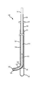
FIG. 1 illustrates an insertion device 100 in accordance with an embodiment of
the invention.
Example insertion device 100 includes a set of three tubes that assemble in a
nested manner. An
outer guide tube 101 may be rigid or semi-rigid, and may be made for example
of stainless steel,
polyetheretherketone (PEEK), or another suitable material. It should be noted
that FIG. 1 is not
necessarily to scale. The diameters of the components are exaggerated for
clarity of illustration.
Outer guide tube 101 may have an outer diameter suitable for the intended use
of insertion
device 100. In one example embodiment intended for placement of media into the
human brain,
outer guide tube 101 has an outer diameter of about 2.4 mm and an inner
diameter of about 1.8
mm, although it is to be understood that other sizes may be used for treatment
of the human
brain, and may be especially helpful in the treatment of other human organs,
or in the treatment
of animals which may be larger or smaller than humans. In another example
embodiment, outer
guide tube 101 has an outer diameter of about 2.11 mm, an inner diameter of
about 1.6 mm, and
a length of about 38 cm or more.
3
CA 02878510 2015-01-06
WO 2014/018871 PCT/US2013/052301
Outer guide tube 101 includes a side port 102 placed a distance from distal
end 103 of outer
guide tube 101. In one example embodiment, side port 102 is placed about 4 mm.
from closed
distal end 103, but other suitable dimensions may be used.
An inner guide tube 104 nests within outer guide tube 101. Inner guide tube
104 may also be
.. rigid or semi-rigid, and may be made for example of stainless steel, nylon-
12, or another suitable
material. In one example embodiment, inner guide tube 104 has an outer
diameter of about 1.65
mm and an inner diameter of about 1.19 mm, although other suitable dimension
may be used. In
another example embodiment, inner guide tube 104 has an outer diameter of
about 1.47 mm, an
inner diameter of about 1.07 mm, and a length of about 43 cm or more. Inner
guide tube 104 is
movable at least axially within outer guide tube 101, so that inner guide tube
can cover or
uncover (close or open) side port 102. Inner guide tube 104 may be lockable in
the open and
closed positions with respect to outer guide tube 101, for example using a
Tuohy Borst adapter
or other device (not shown). Inner guide tube 104 also includes a deflector
105, the purpose of
which is explained in more detail below.
.. A. flexible delivery catheter 106 translates within inner guide tube 104.
Catheter 106 may be
made, for example, of nylon-12, nylon-11, or another suitable material. In one
example
embodiment, catheter 106 has an outer diameter of about 1.0 mm and an inner
diameter of about
0.4 mm, but other suitable dimensions may be used. Catheter 106 may be
lockable with respect
to inner guide tube 104, for example using another Tuohy Borst adapter or
other device (not
shown). A depth stop 107 may be provided to prevent inadvertent deployment of
catheter 106
beyond its intended range.
At an insertion end 108 of catheter 106, one or more dispensing holes are
provided for
dispensing media 110. Any suitable number and arrangement of dispensing holes
may be used.
In the vicinity of insertion end 108, catheter 106 is preferably pre-formed
into a curved shape. A
.. heat set or other procedure may be used to pre-form insertion end 108. For
example, in one
example embodiment having a catheter made of nylon-1.2, the catheter was heat
set using a lab
oven for approximately 15 minutes after the glass transition temperature (170
F) was achieved.
This process provided the necessary environment to shape the catheter to the
desired
specifications. The catheter was both straightened and bent at the insertion
end. A metal rod
mandrel was placed in the inner lumen of the catheter excluding the last 1.5cm
from the distal
end. This distal portion was then fixated to the desired radius of curvature
(0.5cm). After the
allotted time, the catheter was removed from the oven yielding a cell delivery
catheter with
various radii of curvatures near the distal end.
4
CA 02878510 2015-01-06
WO 2014/018871 PCT/US2013/052301
During use, inner guide tube 104 may be positioned to align deflector 105 with
side port 102.
The combination of deflector 105 and the preset bend in catheter 106 causes
catheter 106 to
emerge from outer guide tube 101 approximately perpendicularly. In other
embodiments,
different deflector shapes and different preset bends may be used to achieve
other angles of
emergence. The system may be configured for any suitable maximum deployment of
catheter
106. In one example embodiment, catheter 106 can protrude from outer guide
tube 101 by up to
2.0 cm. Deflector 105 may be an angled, radiused, or have another suitable
shape to accomplish
the deflection of catheter 106.
A plunger 111 fits within catheter 106. Plunger 111 may be made from any
suitable material, for
example a Ti6A14V (Grade 5 Titanium) wire. In one example embodiment, plunger
111 has an
outer diameter of 0.4 mm and a flattened end, but other suitable sizes and
shapes may be used.
For example, the end of plunger 111 may be rounded, tapered, pointed, or have
any other
workable shape. A close fit between the walls of the catheter and the plunger
wire may provide
a nearly gas-tight seal to allow both aspiration and dispensing of fluids. Dip
coating of the distal
end of the plunger wire may further enhance this seal. For safety, a torquer
(not shown) at the
catheter proximal end prevents inadvertent movement of the plunger wire. To
allow translational
movement of the plunger wire, the user must open this plunger lock.
Preferably, catheter 106
can be fixed at any deployed distance by closing the catheter lock while still
allowing the user to
manipulate the plunger wire by opening the plunger lock. Thus, advancement of
the plunger
wire does not result in movement of the catheter.
To use insertion device 100, the user preferably plans an insertion trajectory
into the tissue of the
patient. Where feasible, the insertion may be planned and accomplished using
stereotactic
surgery techniques. For example, FIG. 2 shows an embodiment of insertion
device 100
incorporated with a stereotactic frame 201 for animal surgery.
Whatever means are used to guide the insertion, at least outer guide tube 101
and inner guide
tube 104 are inserted into the patient tissue to the desired insertion depth.
Preferably, inner guide
tube 104 is positioned so that side port 102 is closed during the insertion.
Once the desired
insertion depth is reached, the position of outer guide tube 101 may be
locked. Catheter 106 and
plunger 111 may be present within inner guide tube 104 during the insertion or
may be inserted
later.
Catheter 106 is loaded at any convenient time with media 110 to be dispensed
to the patient
tissue. For example media 110 may be a suspension of cells to be used for cell
therapy. Side
port 102 is opened by withdrawing inner guide tube 104, and inner guide tube
104 is preferably
5
CA 02878510 2015-01-06
WO 2014/018871 PCT/US2013/052301
locked with respect to outer guide tube 101. Catheter 106 (along with plunger
111) is advanced
so that insertion end 108 emerges through side port 102. When the desired
protrusion distance is
reached, catheter 106 is preferably locked in relation to inner guide tube
104. Plunger 111 may
then be unlocked and advanced through inner guide tube 104 to force media 110
out through
dispensing holes 109.
Once the desired dose has been delivered to the first treatment site, the
process may be reversed
to withdraw catheter 106 back into inner guide tube 104, and to close side
port 102. Outer guide
tube 101 may then be repositioned for delivery of media to a second treatment
site. During the
repositioning, outer guide tube 101 may be translated along its longitudinal
axis (either advanced
.. or retracted), rotated about its longitudinal axis, or moved in a
combination of translation and
rotation. When outer guide tube is in the desired position for dispensing
media to a second
treatment site, inner guide tube 104 is retracted to open side port 102,
catheter 106 is advanced,
and plunger 111 is advanced to dispense media 110 to the second treatment
site.
This process may be repeated, so that a number of different treatment sites
are accessed through
a single original penetration of outer guide tube 101 into the patient tissue.
In some
embodiments, this process may be called radially branched deployment, and can
result in
delivery of media in a precise "tree-like" pattern within the patient tissue.
It will be recognized that variations on this basic procedure are possible.
For example, while
outer guide tube 101 is held in a single orientation, catheter 106 may be
deployed to multiple
distances with media 110 being dispensed at each of the multiple distances. In
another variation,
catheter 106 may be withdrawn and re-loaded with media 110, and re-inserted
into inner guide
tube 104 during the procedure. In another variation, catheter 106 may be
replaced with a second
pre-loaded catheter, so that some treatment sites receive media from a first
catheter and some
receive media from a second catheter, while still requiring only one
penetration of patient tissue
by outer guide tube 101. This technique may be especially useful for complex
treatment
volumes. A variety of catheters may be provided having different preset bends,
and different
catheters may be used as needed to reach different parts of the treatment
volume.
FIG. 3 illustrates additional components that may be used in accordance with
embodiments. In
this example, a first Tuohy Borst adapter 301 is positioned to lock and unlock
inner guide tube
104 in relation to outer guide tube 101, and a second Tuohy Borst adapter 302
is positioned to
lock and unlock catheter 106 in relation to inner guide tube 104.
6
CA 02878510 2015-01-06
WO 2014/018871 PCT/US2013/052301
FIG. 4 illustrates the protrusion of catheter 106 from outer guide tube 101
through side port 102,
in an experimental embodiment. FIG. 5 also shows the protrusion of catheter
106 from outer
guide tube 101 through side port 102, in another view.
FIGS. 6A-6D illustrate the operation of insertion device 100 in more detail. A
deployed position
is shown in FIG 6A and FIG. 6B, in which catheter 106 protrudes from outer
guide tube 101
and inner guide tube 104 through side port 102. A retracted position is also
shown in FIG. 6C,
where side port 102 is open, but catheter 106 has been retracted within inner
guide tube 104.
Finally, FIG. 6D shows a closed position, where inner guide tube 104 has been
advanced so that
side port 102 is closed.
FIG. 7 illustrates the dispensing of media 110 from catheter 106 into brain
tissue, in accordance
with embodiments.
FIGS. 8A and 8B illustrate dispensing of media 110 to a second treatment site,
according to
embodiments. In FIG. 8A, catheter 106 has been retracted after dispensing
media 110 to a first
treatment site, and outer guide tube 101 is rotated. In some embodiments,
inner guide tube 104
may also be advanced to close side port 102 during the rotation. In FIG. 8B,
outer guide tube
101 has been rotated about its longitudinal axis, for dispensing a second dose
of media 110 to a
second treatment site. Inner guide tube 104 has been retracted to open side
port 102, and
catheter 106 has been extended, and additional media 801 is dispensed to a
second treatment site.
FIG. 9 illustrates the use of a single penetration 901 of the skull 902 to
reach the putamen 903 of
a brain 904. FIG. 10 illustrates a "tree-like" pattern of treatment sites that
may be reached
through a single penetration of outer guide tube 101 into patient tissue.
Catheter 106 may be
placed in several different rotational positions and several depths, resulting
in a relatively large
number of treatment sites accessible through the single penetration. The
treatment volume may
have a radius or up to 2 cm or more.
As was mentioned above, a close fit between the walls of catheter 106 and
plunger 111 may
provide a nearly gas-tight seal to allow aspiration of media 110. Accordingly,
one method of
loading catheter 106 is to insert plunger 111 into catheter 106, and advance
plunger 111 until it
has reached closed insertion end 108. This system can then be placed into a
media or solution,
and plunger 111 withdrawn to any location along the catheters long axis,
creating a suction force
(negative pressure) that will drive, or load, the media from the one or more
dispensing holes 109
into the main lumen of catheter 106. This process is illustrated in FIG. 11A.
Once the operator
has loaded the appropriate dose, depending on how far plunger 111 has been
withdrawn from the
7
CA 02878510 2015-01-06
WO 2014/018871 PCT/US2013/052301
long axes of the main lumen of catheter 106, the media 110 can then be placed
into the
appropriate target location in the patient as previously described and shown
in FIG. 11B.
One advantage of an insertion device according to embodiments, as compared
with previous
systems, may be that reflux of the dispensed media is significantly reduced.
In an experimental
evaluation, an insertion device embodying the invention was used to inject 10
ul of Allura Red
AC dye into an agarose gel that mimics the gross structural characteristics of
brain. The catheter
was deployed 7 cm below the gel surface, and dye was infused using a plunger
advancement rate
of 2 [11/min. No dye was observed to reflux up the tract created by the outer
guide tube. Reflux
was arrested at the transition point between the deployed catheter and the
outer guide tube side
port. This surprising resistance to reflux may be a consequence of the
directional change of the
tract and the larger caliber of the outer guide tube. By contrast, delivery of
10 jul of dye through
a 20G cannula-syringe system inserted to the same depth resulted in
significant reflux of dye.
This difference in reflux was also observed when MRI was used to track the
delivery of a
suspension of paramagnetic beads to an agarose gel contained within a model
skull.
An insertion device according to embodiments of the invention may promote cell
viability when
the device is used to deliver cells to a treatment site. The system has a
comparatively short and
uniform flow path for cells, so that few opportunities arise for shear forces
induced by the flow
of media 110 to injure cells in the media. Additionally, the small diameter of
plunger 111
enables the precise dispensing of very small quantities of media 110 to a
large number of sites.
For example, in one experimental embodiment, the volume of media 110 delivered
by 1 cm of
plunger travel was only 1.36 111. Thus comparatively few cells compete for the
available oxygen
at any particular treatment site.
FIG. 12 illustrates an insertion device 1200 in accordance with another
embodiment of the
invention. Insertion device 1200 has several components similar to components
of insertion
device 100 discussed above, and these components are designated with the same
reference
numerals. For example, outer guide tube 101 may be rigid or semi-rigid, and
may be made for of
stainless steel, polyetberetherketone (PEEK), or another suitable material.
Outer guide tube 101
includes a side port 102, and a closed distal end 103 for insertion into
tissue such as brain tissue.
In one example embodiment, outer guide tube has an outer diameter of about 2.0
mm, an inner
diameter of about 1.5 m, and a length of about 38 cm, although different sizes
may be used for
different applications.
Inner guide tube 104 nests within outer guide tube 101. Inner guide tube 104
may also be rigid
or semi-rigid, and may be made for example of stainless steel, nylon-12, or
another suitable
8
CA 02878510 2015-01-06
WO 2014/018871 PCT/US2013/052301
material. In one example embodiment, inner guide tube 104 has an outer
diameter of about 1.47
mm, an inner diameter of about 1.07 mm, and a length of about 43 cm or more,
although other
suitable dimensions may be used. Inner guide tube 104 is movable at least
axially within outer
guide tube 101, so that inner guide tube can cover or uncover (close or open)
side port 102.
Inner guide tube 104 may be lockable in the open and closed positions with
respect to outer
guide tube 101, for example using a Tuohy Borst adapter or other device (not
shown). Inner
guide tube 104 also includes a deflector 105.
A flexible delivery catheter 1201 translates within inner guide tube 104.
Catheter 1201 may be
made, for example, of nylon-12, nylon-11, or another suitable material. in one
example
embodiment, catheter 106 has an outer diameter of about 0.40 mm and an inner
diameter of
about 0.38 mm, but other suitable dimensions may be used. Catheter 1201 may be
lockable with
respect to inner guide tube 104, for example using another Tuohy Borst adapter
or other device
(not shown). A depth stop may be provided to prevent inadvertent deployment of
catheter 1201
beyond its intended range. Similar to catheter 106 described above, delivery
catheter 1201
preferably is formed into a curved shape near insertion end 1202, using a heat
set or other
operation.
in contrast to catheter 106, delivery catheter 1201 is open at insertion end
1202, and serves as a
conduit for a therapeutic delivery cannula 1203. Therapeutic delivery cannula
1203 may be, for
example a step cannula as shown in FIG. 12, having a step 1204 in its diameter
near distal end
1205, and suitable for delivering biologics to a brain or other tissue via
convection enhanced
delivery. However, it will be recognized that a wide variety of other cannula
types and delivery
methods may be used. Therapeutic delivery cannula 1203 is preferably made of a
semi-rigid
material such as Nitinol, and is flexible enough that it can be guided by
catheter 1201. However,
therapeutic delivery cannula 1203 preferably does not include a pre-formed
curved shape, so that
therapeutic delivery cannula 1203 tends to maintain a straight configuration
unless acted on by
another element such as catheter 1201. in one example embodiment, therapeutic
delivery
cannula is a step cannula made of Nitinol having an outside diameter of 0.375
mm over much of
its length, an outside diameter of 0.150 mm below step 1204 at distal end
1205, and an inner
diameter of 0.0625 mm.
To use insertion device 1200, the user preferably plans an insertion
trajectory into the tissue of
the patient. Where feasible, the insertion may be planned and accomplished
using stereotactic
surgery techniques as described above.
9
CA 02878510 2015-01-06
WO 2014/018871 PCT/US2013/052301
Whatever means are used to guide the insertion, at least outer guide tube 101
and inner guide
tube 104 are inserted into the patient tissue to the desired insertion depth.
Preferably, inner guide
tube 104 is positioned so that side port 102 is closed during the insertion.
Once the desired
insertion depth is reached, the position of outer guide tube 101 may be
locked. Catheter 1201
and therapeutic delivery cannula 1203 may be present within inner guide tube
104 during the
insertion or may be inserted later.
Therapeutic delivery cannula 1203 is loaded at any convenient time with media
to be dispensed
to the patient tissue. Side port 102 is opened by withdrawing inner guide tube
104, and inner
guide tube 104 is preferably locked with respect to outer guide tube 101.
Catheter 1201
(possibly along with therapeutic delivery cannula 1203) is advanced so that
dispensing end 1202
emerges through side port 102. In contrast to the use of delivery device 100
described above,
catheter 1201 may be advanced only a relatively small distance from inner
guide tube 104. The
distance is sufficient to establish a desired delivery angle 0 of delivery end
1202 with respect to
outer guide tube 101, but the distance is small enough to avoid any
appreciable "sweep" of
catheter 1201 through the tissue. Sweep may occur, for example, when a curved
catheter is
advanced into tissue and, due to its curvature and stiffness, tends to sweep
through the tissue
transverse to the longitudinal axis of the catheter, rather than following the
longitudinal axis
narrowly through the tissue. Because catheter 1201 is advanced only a small
distance, it has
little or no opportunity to sweep through the tissue.
Once the desired protrusion distance is reached, catheter 1201 is preferably
locked in relation to
inner guide tube 104. Therapeutic delivery cannula 1203 is then advanced
through catheter 1201
to reach the relatively distant treatment site. The curve of catheter 1201
directs therapeutic
delivery cannula 1203 in the direction of angle 0, but once therapeutic
delivery cannula 1203
emerges from catheter 1201, catheter 1201 no longer influences the direction
of travel of
therapeutic delivery cannula 1203. Because therapeutic delivery cannula 1203
is semi-rigid and
is straight in its natural condition, it tends to proceed through the tissue
along a straight line,
without a tendency to sweep. A therapeutic agent may then be delivered through
therapeutic
delivery cannula 1203 by any suitable means, for example by convection
enhanced delivery.
Once the desired dose has been delivered to a first treatment site, the
process may be reversed to
withdraw therapeutic delivery cannula 1203 and catheter 1201 back into inner
guide tube 104,
and to close side port 102. Outer guide tube may then be repositioned for
delivery of media to a
second treatment site. During the repositioning, outer guide tube 101 may be
translated along its
longitudinal axis (either advanced or retracted), rotated about its
longitudinal axis, or moved in a
CA 02878510 2015-01-06
WO 2014/018871 PCT/US2013/052301
combination of translation and rotation. When outer guide tube 104 is in the
desired position for
dispensing media to a second treatment site, inner guide tube 104 is retracted
to open side port
102, catheter 1201 is advanced, and therapeutic delivery cannula 1203 is
advanced to another
treatment site where a therapeutic agent is delivered.
This process may be repeated, so that a number of different treatment sites
are accessed through
a single original penetration of outer guide tube 101 into the patient tissue.
FIG. 13 is a
rendering of an actual insertion of a delivery device similar to insertion
device 1200 into an
Agarose gel, simulating insertion into patient tissue.
While delivery of a therapeutic agent through therapeutic delivery cannula
1203 may be
accomplished by any suitable method, in some embodiments a plunger similar to
plunger 111
discussed above may be used. FIG. 14 illustrates a delivery device 1400
according to this
embodiment. Delivery device 1400 is similar to insertion device 1200 in
construction and use,
with the addition of plunger 1401 within therapeutic delivery cannula 1203.
Thus, delivery
device 1400 includes five nested elements: outer guide tube 101, inner guide
tube 104, delivery
catheter 1201, therapeutic delivery cannula 1203, and finally plunger 1401. As
before, the use of
plunger 1401 enables very accurate dosing of a therapeutic agent, because the
plunger is of a
relatively small diameter and moves a relatively large distance during
dispensing, allowing very
fine control of the amount of agent dispensed.
It will also be recognized that steps in the operation of the delivery device
described above may
be performed in any suitable order. For example, catheter 1201 may be inserted
into inner guide
tube 104 and advanced through side port 102 before therapeutic delivery
cannula 1203 (and
plunger 1401 if present) is inserted into catheter 1201. Alternatively,
catheter 1201, therapeutic
delivery cannula 1203, and plunger 1401 (if present) could be advanced
together to the extent of
the advancement of catheter 1201. Catheter 1201 may then be fixed, and
therapeutic delivery
.. cannula 1203 and plunger 1401 advanced beyond the insertion end of catheter
1201 to reach the
treatment site. Many other sequences are possible.
Therapeutic delivery cannula 1203 may be withdrawn and re-loaded in a manner
similar to
catheter 106 described above, or in any other suitable manner.
While therapeutic delivery cannula 1203 is shown as open ended, this is not a
requirement. For
example, therapeutic delivery cannula could have a closed end similar to
catheter 106 discussed
above, including side openings similar openings 109. This arrangement may be
especially
suitable for delivering a suspension of cells for cell therapy as discussed
above. FIG. 15 shows
such an embodiment, in which therapeutic delivery cannula 1501 includes
openings 1502.
11
CA 02878510 2015-01-06
WO 2014/018871 PCT/US2013/052301
In addition to the delivery of therapeutic agents described in the above
embodiments, other
embodiments may enable delivery of other kinds of therapy. For example, an
electric therapeutic
such as an electrode may be delivered in place of or through therapeutic
delivery cannula 1203.
Embodiments of the invention have now been described in detail for the
purposes of clarity and
understanding. However, those skilled in the art will appreciate that certain
changes and
modifications may be practiced within the scope of the appended claims. It is
to be understood
that all workable combination of the components and features described herein
are considered to
be disclosed.
12