Note: Descriptions are shown in the official language in which they were submitted.
MICROWAVE ABLATION CATHETER AND METHOD OF UTILIZING THE SAME
BACKGROUND
Technical Field
[0001] The present disclosure relates to a microwave ablation catheter
and method of
utilizing the same. More particularly, the present disclosure relates to a
microwave ablation
catheter that is positionable through one or more branched luminal networks of
a patient for
treating tissue.
Description of Related Art
[0002] Microwave ablation may be utilized for treating various maladies,
e.g., nodules,
of different organs like the liver, brain, heart, lung and kidney. When a
nodule is found, for
example, within a lung, several factors are considered in making a diagnosis.
For example, a
biopsy of the nodule may be taken using a biopsy tool under CT guidance. If
the biopsy reveals
that the nodule is malignant, it may prove useful to ablate the nodule. In
this instance, microwave
ablation, which typically includes transmitting microwave energy to a
percutaneous needle, may
be utilized to ablate the nodule. Under certain surgical scenarios, certain
current percutaneous
methods of microwave ablation procedures can result in pneumothoraces (air
leaks) and a
collection of air in the space around the lungs which if not appreciated by
the clinician can
ultimately lead to collapse of the lung or a portion thereof.
[0003] Endobronchial navigation uses CT image data to create a navigation
plan to
facilitate advancing a navigation catheter (or other suitable device) through
a bronchoscope and
a branch of the bronchus of a patient to the nodule. Electromagnetic tracking
may also may be
utilized in conjunction with the CT data to facilitate guiding the navigation
catheter through the
branch of the bronchus to the nodule. In certain instances, the navigation
catheter may be
CA 2878570 2019-06-25
=
2
positioned within one of the airways of the branched luminal networks adjacent
to or within the
nodule or point of interest to provide access for one or more tools. Once the
navigation catheter
is in position, fluoroscopy may be used to visualize biopsy tools, such as,
for example, biopsy
brushes, needle brushes and biopsy forceps as they are passed through the
navigation catheter and
into the lung and to the nodule or point of interest.
SUMMARY
[0004] As can be appreciated, a microwave ablation catheter that
is positionable through
one or more branched luminal networks of a patient to treat tissue may prove
useful in the surgical
arena.
[0005] Aspects of the present disclosure are described in detail
with reference to the
drawing figures wherein like reference numerals identify similar or identical
elements. As used
herein, the term "distal" refers to the portion that is being described which
is further from a user,
while the term "proximal" refers to the portion that is being described which
is closer to a user.
[0006] An aspect of the present disclosure provides a microwave
ablation catheter. The
microwave ablation catheter includes a coaxial cable that is connected at its
proximal end to a
microwave energy source and at its distal end to a distal radiating section.
The coaxial cable
includes inner and outer conductors and a dielectric positioned therebetween.
The inner
conductor extends distally past the outer conductor and is in sealed
engagement with the distal
radiating section. A balun is formed in part from a conductive material
electrically connected to
the outer conductor of the coaxial cable and extends along at least a portion
of the coaxial cable.
The conductive material has a braided configuration and is covered by at least
one insulative
material. The insulative material covering the conductive material may be
polyethylene
terephthalate. An outer sheath may be provided and configured to surround the
coaxial cable.
CA 2878570 2019-06-25
3
[0007] The balun may further include an insulator substantially between
the conductive
layer and the outer conductor. A portion of the outer conductor may be removed
to form a feedgap
between the distal radiating section and the balun.
[0008] The microwave ablation catheter may include a multi-lumen housing
configured
to receive the coaxial cable, distal radiating section, and balun. The multi-
lumen housing includes
a hub at a proximal end thereof The hub includes a plurality of ports.
[0009] One of the plurality of ports may be an electrical port that is
configured to provide
electrical communication between the coaxial cable and the microwave energy
source.
Moreover, two of the plurality of ports are a fluid intake port and a fluid
return port configured to
provide respective ingress and egress of a coolant to and from the multi-lumen
housing for cooling
the distal radiating section of the coaxial cable.
[0010] The multi-lumen housing may include a four lumen configuration
having two
lumens dedicated for communication with a respective one of the fluid intake
and return ports
and two lumens dedicated to support the coaxial cable including the balun.
Alternatively, the
multi-lumen housing may include a five lumen configuration having four lumens
two of which
being dedicated for communication with the fluid intake port and two of which
being dedicated
for communication with the fluid return port and one lumen designated to
support the coaxial
cable including the balun.
[0011] An aspect of the present disclosure provides a microwave ablation
catheter. The
microwave ablation catheter includes a coaxial cable that is connected at its
proximal end to a
microwave energy source and at its distal end to a distal radiating section.
The coaxial cable
includes inner and outer conductors and a dielectric positioned therebetween.
The inner
conductor extends distally past the outer conductor and in sealed engagement
with the distal
radiating section. The outer conductor has a braided configuration to
facilitate movement of the
CA 2878570 2019-06-25
4
microwave ablation catheter through a branched lumina] network of a patient.
An outer sheath
may be provided and configured to surround the coaxial cable. One or more
temperature sensors
may be disposed at the distal radiating section and may be configured to
measure a temperature
of target tissue while the distal radiating section is energized. The
temperature sensor(s) may be
configured to communicate with a temperature sensor system that is in operable
communication
with the microwave energy source.
[0012] The microwave ablation catheter may include a balun that is formed
in part from
a conductive material electrically connected to the outer conductor of the
coaxial cable and
extending along at least a portion of the coaxial cable. The conductive
material may have a
braided configuration and covered by at least one insulative material. The
insulative material
covering the conductive material may be polyethylene terephthalate. An
insulator may be
provided substantially between the conductive layer and the outer conductor. A
portion of the
outer conductor may be removed to form a feedgap between the distal radiating
section and the
balun.
[0013] The microwave ablation catheter may include a multi-lumen housing
configured
to receive the coaxial cable, distal radiating section, and balun. The multi-
lumen housing includes
a hub at a proximal end thereof The hub includes a plurality of ports.
[0014] One of the plurality of ports may be an electrical port that is
configured to provide
electrical communication between the coaxial cable and the microwave energy
source.
Moreover, two of the plurality of ports are a fluid intake port and a fluid
return port configured to
provide respective ingress and egress of a coolant to and from the multi-lumen
housing for cooling
the distal radiating section of the coaxial cable.
[0015] The multi-lumen housing may include a four lumen configuration
having two
lumens dedicated for communication with a respective one of the fluid intake
and return ports
CA 2878570 2019-06-25
5
and two lumens dedicated to support the coaxial cable including the balun.
Alternatively, the
multi-lumen housing may include a five lumen configuration having four lumens
two of which
being dedicated for communication with the fluid intake port and two of which
being dedicated
for communication with the fluid return port and one lumen designated to
support the coaxial
cable including the balun.
BRIEF DESCRIPTION OF THE DRAWINGS
[0016] Various embodiments of the present disclosure are described
hereinbelow with
references to the drawings, wherein:
[0017] Fig. 1 is a perspective view of a microwave ablation system
including a
microwave ablation catheter assembly configured for use with a microwave
ablation system
according to an embodiment of the instant disclosure;
[0018] Fig. 2 is a front view of an embodiment of a lumen configuration
configured for
use with the microwave catheter assembly shown in Fig. 1;
[0019] Fig. 3A is a front view of an another embodiment of a lumen
configuration
configured for use with the microwave catheter assembly shown in Fig. 1;
[0020] Fig 3B is a front view of an another embodiment of a lumen
configuration
configured for use with the microwave catheter assembly shown in Fig. 1;
[0021] Fig 3C is a front view of an another embodiment of a lumen
configuration
configured for use with the microwave catheter assembly shown in Fig. 1,
whereby the lumen
supporting the coaxial microwave structure also communicates cooling fluid
with inflow or
outflow ports;
[0022] Fig. 4 is a perspective view of a distal end of a microwave
ablation catheter
configured for use with the microwave ablation assembly shown in Fig. 1;
CA 2878570 2019-06-25
6
[0023] Fig. 5 is a cross-sectional view taken along line section 5-5 in
Fig. 4;
[0024] Fig. 6 is a screen shot of a CT based luminal navigation system in
accordance with
an embodiment of the present disclosure;
[0025] Fig. 7 is a perspective view of a microwave ablation system and
luminal
navigation system configured for use the microwave ablation catheter assembly
shown in Fig. 1
and microwave ablation catheter shown in Fig. 2 in accordance with an
embodiment of the present
disclosure;
[0026] Fig. 8 is a side view of a luminal catheter delivery assembly
including an extended
working channel and locatable guide catheter in accordance with an embodiment
of the present
disclosure;
[0027] Fig. 9 is a partial, perspective view of a distal end of the
locatable guide catheter
shown in Fig. 8;
[0028] Fig. 10 is a side view of the extended working channel shown in
Fig. 8 with the
microwave ablation catheter extending from a distal end thereof;
[0029] Fig. 11 is a screen shot of a CT based luminal navigation system in
accordance
with an embodiment of the present disclosure;
[0030] Fig. 12A is a schematic, plan view of the extended working channel
positioned
within a bronchoscope prior to being positioned within a trachea of a patient;
[0031] Fig. 12B is a schematic, plan view of the bronchoscope shown in
Fig. 12A
positioned within the trachea of the patient with the extended working channel
extending distally
therefrom;
[0032] Fig. 12C is a partial, cutaway view of the extended working channel
and locatable
guide positioned within the bronchoscope;
CA 2878570 2019-06-25
7
[0033] Fig. 13A is a schematic, plan view of the bronchoscope positioned
within the
trachea of the patient with the extended working channel extending distally
therefrom;
[0034] Fig. 13B is a partial, cutaway view of the extended working channel
and a biopsy
tool positioned within the bronchoscope;
100351 Fig. 14 is a schematic, plan view of the bronchoscope positioned
within the
trachea of the patient with the extended working channel removed from the
bronchoscope;
[0036] Fig. 15A is a schematic, plan view of the bronchoscope positioned
within the
trachea of the patient with an extended working channel according to an
alternate embodiment
extending distally therefrom;
[0037] Fig. 15B is a partial, cutaway view of the extended working channel
shown in Fig.
15A positioned within the bronchoscope;
[0038] Fig. 16A is a schematic, plan view of the bronchoscope positioned
within the
trachea of the patient with the extended working channel shown in Fig. 15A
extending distally
therefrom;
[0039] Fig. 16B is a schematic, plan view of the bronchoscope positioned
within the
trachea of the patient with the extended working channel shown in Fig. 15A
extending distally
therefrom and adjacent target tissue;
100401 Fig. 16C is a partial, cutaway view of the extended working channel
and the
microwave ablation catheter shown in Fig. 2 coupled to one another and
positioned within the
bronchoscope;
[0041] Fig. 16D is a cross-sectional view taken along line section 16D-16D
in Fig. 16C;
[0042] Fig. 17 is a schematic, plan view of another embodiment of the
extended working
shown in Figs. 9 and 15A with the extended working channel positioned within
the lung of a
patient and having a balloon coupled thereto in an deflated configuration;
CA 2878570 2019-06-25
4
8
[0043] Fig. 18 is an enlarged area of detail of Fig, 17 and
showing the balloon in an
inflated configuration;
[0044] Fig. 19A is a schematic, plan view of an alternate
embodiment of a balun
configured for use with the microwave ablation catheter shown in Fig. 2 with
the balun shown in
an expanded configuration;
[0045] Fig. 19B is a schematic, plan view of the balun shown in
Fig. 19A in an non-
expanded configuration;
[0046] Fig. 20 is a schematic, plan view of a distal tip
configuration that may be utilized
with the microwave ablation catheter assembly shown in Fig. 1, the microwave
ablation catheter
shown in Fig. 2 or the extended working channel shown in Fig. 15A;
[0047] Fig. 21 is a schematic, plan view of an alternate
embodiment of the extended
working channel shown in Fig. 15A;
[0048] Fig. 22 is a schematic, plan view of yet another embodiment
of the extended
working channel shown in Fig. 15A;
[0049] Fig. 23 is a perspective view of an alternate embodiment of
the luminal navigation
system shown in Fig. 7;
[0050] Fig. 24 is a partial, cutaway view of another embodiment of
the microwave
ablation catheter shown in Fig. 1;
[0051] Fig. 25 is a cross-sectional view taken along line section
25-25 in Fig. 24;
[0052] Fig. 26 is a cross-sectional view taken along line section
26-26 in Fig. 24;
[0053] Fig. 27 is a partial, cutaway view of yet another
embodiment of the microwave
ablation catheter shown in Fig. 1;
[0054] Fig. 28 is a schematic, plan view of still yet another
embodiment of the microwave
ablation catheter shown in Fig. 1;
CA 2878570 2019-06-25
9
[0055] Fig. 29 is a schematic, plan view illustrating a circulation
feedback loop that is
configured for use with the extended working channels shown in Figs. 15A, 17
and 21, and the
microwave ablation catheter shown in Figs. 1, 24 and 27-28;
[0056] Fig. 30 is a schematic, plan view of still yet another embodiment
of the extended
working channel shown in Fig. 15A;
[0057] Fig. 31 is a schematic, plan view of still yet another embodiment
of the extended
working channel shown in Fig. 15A with the microwave ablation catheter shown
in Fig. 2 in a
retracted configuration;
[0058] Fig. 32 is a schematic, plan view of the extended working channel
shown in Fig.
31 with the microwave ablation catheter shown in an extended configuration;
[0059] Fig. 33 is a schematic, plan view of still yet another embodiment
of the extended
working channel shown in Fig. 15A;
[0060] Fig. 34 is a schematic, plan view of still yet another embodiment
of the extended
working channel shown in Fig. 15A with the extended working channel shown in a
non-expanded
configuration;
[0061] Fig. 35 is a schematic, plan view of the extended working channel
shown in Fig.
34 in an expanded configuration;
[0062] Fig. 36A is a front view of an alternate embodiment of the
microwave ablation
catheter shown in Fig. 2 including a conductive balloon coupled thereto and
shown in a deflated
configuration;
[0063] Fig. 36B is a front view of the microwave catheter shown in Fig.
36A with the
conductive balloon shown in an inflated configuration;
CA 2878570 2019-06-25
10
[0064] Fig. 37A is a front view of an alternate embodiment of the
microwave ablation
catheter shown in Fig. 2 including a plurality of thermally conductive fins
coupled thereto and
shown in a non-deployed configuration;
[0065] Fig. 37B is a front view of the microwave catheter shown in Fig.
37A with the
plurality of thermally conductive fins shown in a deployed configuration;
[0066] Fig. 38 is a schematic, plan view of still yet another embodiment
of the extended
working channel shown in Fig. 15A;
[0067] Fig. 39A is a schematic, plan view of an alternate embodiment of
the microwave
ablation catheter shown in Fig. 2 including a balloon coupled thereto and
shown in a deflated
configuration;
[0068] Fig. 39B is a schematic, plan view of the microwave catheter shown
in Fig. 39A
with the balloon shown in an inflated configuration;
[0069] Fig. 40A is a schematic, plan view of various fiducial markers
configured for use
with the microwave ablation system shown in Fig. 7, wherein the fiducial
markers are shown
adjacent target tissue that has not been ablated;
[0070] Fig. 40B is a schematic, plan view of the fiducial markers shown
in Fig. 40A,
wherein the fiducial markers are shown adjacent target tissue that has been
ablated;
[0071] Fig. 41 is a schematic, plan view of a guide wire including a
plurality of
thermocouples configured for use with the microwave ablation system shown in
Fig. 7;
[0072] Fig. 42 is a perspective view of an electrical measurement system
configured for
use with the microwave ablation system shown in Fig. 7;
[0073] Fig. 43 is a schematic, plan view of a feedback configuration
configured for use
with the microwave ablation system shown in Fig. 7;
CA 2878570 2019-06-25
4
11
[0074] Fig. 44 is a schematic, plan view of an another embodiment
of a feedback
configuration configured for use with the microwave ablation system shown in
Fig. 7;
[0075] Fig. 45 is schematic, plan view of a yet another embodiment
of a feedback
configuration configured for use with the microwave ablation system shown in
Fig. 7;
[0076] Fig. 46A is a fluoroscopic images of a patient, having a
catheter placed therein;
and
[0077] Fig. 46B is a virtual fluoroscopic image of a patient
depicting a target.
DETAILED DESCRIPTION
[0078] Detailed embodiments of the present disclosure are
disclosed herein; however, the
disclosed embodiments are merely examples of the disclosure, which may be
embodied in various
forms. Therefore, specific structural and functional details disclosed herein
are not to be
interpreted as limiting, but merely as a basis for the claims and as a
representative basis for
teaching one skilled in the art to variously employ the present disclosure in
virtually any
appropriately detailed structure.
[0079] As can be appreciated an energy device, such as a microwave
ablation catheter,
that is positionable through one or more branched luminal networks of a
patient to treat tissue
may prove useful in the surgical arena and the present disclosure is directed
to such apparatus,
systems and methods. Access to lumeninal networks may be pereutaneous or
through natural
orifice. In the case of natural orifice, an endobronchial approach may be
particularly useful in
the treatment of lung disease. Targets, navigation, access and treatment may
be planned pre-
procedurally using a combination of imaging and/or planning software. In
accordance with these
aspects of the present disclosure the planning software may offer custom
guidance using pre-
procedure images). Navigation of the luminal network may be accomplished using
image-
CA 2878570 2019-06-25
12
guidance. These image-guidance systems may be separate or integrated with the
energy device
or a separate access tool and may include MRI, CT, fluoroscopy, ultrasound,
electrical impedance
tomography, optical, and device tracking systems. Methodologies for locating
the separate or
integrated to the energy device or a separate access tool include EM, IR,
echolocation, optical,
and others. Tracking systems may integrated to imaging device, where tracking
is done in virtual
space or fused with preoperative or live images. In some cases the treatment
target may be
directly accessed from within the lumen, such as for the treatment of the
endobronchial wall for
COPD, Asthma, lung cancer, etc. In other cases, the energy device and/or an
additional access
tool may be required to pierce the lumen and extend into other tissues to
reach the target, such as
for the treatment of disease within the parenchyma. Final localization and
confirmation of energy
device placement may be performed with imaging and/or navigational guidance
using the
modalities listed above. The energy device has the ability to deliver an
energy field for treatment
(including but not limited to electromagnetic fields) and may have the ability
to monitor treatment
during energy application. The monitoring of the treatment may include
thermometry, electrical
impedance, radiometry, density measurement, optical absorption, hydration,
ultrasound, and
others. Additionally or alternatively treatment may be monitored from within
the lumen or
extracorporeally using an additional device or the image-guidance modalities
described above.
After treatment, the energy device and/or an additional device may have the
ability to confirm
adequate treatment was performed, employing at least the techniques described
above with
respect to treatment monitoring. Further, treatment confirmation may be from
within the lumen
or extracorporeal. The long term treatment performance may be performed with
imaging which
may be integrated into a follow-up software application.
[0080] One
embodiment of the present disclosure is directed, in part, to a microwave
ablation catheter that is positionable through one or more branched luminal
networks of a patient
CA 2878570 2019-06-25
13
to treat tissue. The microwave ablation catheter is part of an ablation system
that includes a
microwave energy source and a planning and navigation system for the placement
of the catheter
at a desired location within the luminal network. Further, the system includes
imaging modalities
that can be employed to confirm placement of the catheter and the effect of
the application of
energy. The microwave catheter itself may include the capability to aide in
the confirmation of
the placement within the tissue to be treated, or additional devices may be
used in combination
with the microwave catheter to confirm placement within the tissue to be
treated. Still further,
one or more thermocouples or temperature sensors on the microwave catheter
detect the
temperature of the microwave catheter or the tissue surrounding the catheter
and enable
monitoring of the microwave catheter temperature and the tissue temperature
during and after
treatment both for safety purposes and for dosage and treatment pattern
monitoring purposes. The
microwave catheter may also assist in the access to the target tissue, either
intraluminal or outside
the lumen. The microwave catheter may also assist in the monitoring of the
treatment through
various measurement techniques and may also be used for treatment
confirmation, in addition to
assistance from other monitoring and confirmation devices.
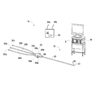
[00811 Figs. 1-5 depict various aspects of a microwave ablation system 10
(system 10).
The system 10, as show in Fig. 1 includes a microwave ablation catheter
assembly 12 (assembly
12) configured to house a microwave ablation catheter 14 (ablation catheter
14) (shown in Fig.
4). Assembly 12 and ablation catheter 14 are configured to couple to a
microwave energy source
(energy source 16) that is configured to transmit microwave energy to the
catheter 14 to treat
target tissue, e.g., lung tissue.
[0082] The assembly 12 shown in Fig. 1 is configured to receive the
ablation catheter 14
and to provide a pathway for a cooling medium to circulate within the assembly
12 and cool the
ablation catheter 14 when the ablation catheter 14 is energized. With these
purposes in mind,
CA 2878570 2019-06-25
14
assembly 12 is formed by overmolding plastic to form a generally elongated
housing 23 having
an outer sheath 18 (Fig. 2) and a plurality of lumens 19a, 19b, and 19c
extending from a proximal
end 20 to a distal end 22 that includes a relatively pointed or appropriately
rounded distal tip 21.
A hub portion 24 is provided at the proximal end 20 and includes ports 26a,
26b, 26c that couple
to corresponding distal ends (not explicitly shown) of connection tubes 28a,
28b, 28c. Connection
tubes 28a, 28c include respective proximal ends 30a, 30c that are configured
to releasably couple
either directly or indirectly to a fluid source 32 including hoses 31a, 3 lb
that provide one or more
suitable cooling mediums (e.g., water, saline, air or combination thereof) to
the ablation catheter
14. In
embodiments, the fluid source 32 may be a component of a cooling system that
is
disclosed in U.S. Patent No. 9,101,344 (Attorney Docket No. H-IL-00083),
entitled
"Recirculating Cooling System for Energy Delivery Device". A proximal end 30b
of connection
tube 28b is configured to couple either directly or indirectly to the energy
source 16 to energize
the ablation catheter 14. An optional pair of wings 34a, 34b may be provided
at the proximal end
20 of the assembly 12. The wings 34a, 34b may extend laterally from respective
right and left
sides of the proximal end 20 and may be configured to rest on a patient or to
be grasped by a
clinician for manipulation of the assembly 12.
100831 The
ports 26a, 26c of the assembly 12 are in fluid communication with
corresponding lumens 19a, 19c of the plurality of lumens 18 provided within
the assembly 12
(Fig. 2) and are configured to provide one of the aforementioned cooling
mediums to the assembly
12. In an embodiment, such as the embodiment illustrated in Fig. 2, port 26a
is an outflow port
and provides a point of egress for the cooling medium from outflow lumen 19a
and port 26c is an
inflow port and provides point of ingress for the cooling medium into the
inflow lumen 19c.
CA 2878570 2019-06-25
15
[0084] Fig. 3A illustrates an alternate lumen configuration that may be
utilized with the
assembly 12. In this embodiment, two outflow lumens 19a' and one inflow lumen
19c' are
provided and are in fluid communication with the respective ports 26a, 26c.
[0085] Fig. 3B illustrates an alternate lumen configuration that may be
utilized with the
assembly 12. In this embodiment, two outflow lumens 19a' and one inflow lumen
19c' are
provided and are in fluid communication with the respective ports 26a, 26c.
Additionally, the
lumen supporting the coaxial microwave structure is also used for either fluid
inflow or outflow.
[0086] Fig. 3C illustrates an alternate lumen configuration similar to
Fig. 3a and 3b that
may be utilized with the assembly 12. In this embodiment, two outflow lumens
19a' and two
inflow lumens 19c' are provided and are in fluid communication with the
respective ports 26a,
26c.
[0087] A third lumen 19b is provided within the assembly 12 and is
configured to support
the ablation catheter 14 when the ablation catheter 14 is coupled to the
assembly 12. In the
embodiment illustrated in Fig. 2, the outflow and inflow lumens 19a, 19c are
formed above the
lumen 19b. In the embodiment illustrated in Fig. 3A, the lumen 19b is centered
between the
outflow lumens 19a and inflow lumens 19c to provide two opposing outflow
lumens 19a and two
opposing inflow lumens 19c around the lumen 19b. In the embodiments
illustrated in Figs 3A
and 3B, the lumen 19b is centered between the outflow lumens 19a and inflow
lumen 19c to
provide two opposing outflow lumens 19a and one opposing inflow lumen 19c
around the lumen
19b. The lumen configurations illustrated in Figs. 2 and 3A-3C provide the
assembly 12 with the
needed flexibility to move within the relatively thin conductive airways
(and/or vessels) in the
branch of the bronchus.
[0088] In an embodiment, the assembly 12 may include a 4 lumen
configuration (not
shown). In this embodiment, three (3) outer lumens (e.g., a combination of
outflow and inflow
CA 2878570 2019-06-25
16
lumens 19a, 19c, respectively) may be equally spaced around a center lumen
(e.g., lumen 19b)
that is configured to support the ablation catheter 14 when the ablation
catheter 14 is coupled to
the assembly 12. In one particular embodiment, the three (3) outer lumens may
be configured to
include two (2) inflow lumens 19c and one (1) outflow lumen 19a (or vice
versa).
[0089] The outflow and inflow lumens 19a, 19c extend a predetermined
distance within
the assembly 12 and can function with various coolant feedback protocols
(e.g., open or closed
feedback protocols). In the embodiments illustrated in Figs. 2 and 3A-3C, the
inflow lumens 19c
extend distally of the outflow lumens 19a to allow an adequate amount of
cooling medium to
circulate around the ablation catheter 14. It should be understood, regardless
of the number of or
configuration of lumens, space not filled within the lumen supporting the
coaxial cable and
radiating section may be used for additional fluid ingress or egress to
improve fluid flow and
directly cool through intimate fluid contact the coaxial microwave structures.
In addition to
supporting the ablation catheter, the lumen 19b may also support additional
outflow or inflow of
coolant, whereby lumen 19b may couple to connection tubes 28a, 28c and their
respective
proximal ends 30a, 30c.
[0090] Referring now to Figs. 4 and 5, the ablation catheter 14 is
illustrated. Ablation
catheter 14 includes a coaxial cable 36. Coaxial cable 36 includes a proximal
end 38 that couples
to port 26b (shown in Fig. 1) that provides electrical connection to the inner
conductor 40 and
outer conductor 48 of the coaxial cable 36 and the energy source 16.
[0091] A distal radiating section 42 is provided at a distal end 44 of the
coaxial cable 36
and is configured to receive the inner conductor 40, as best seen in Fig. 5.
The distal radiating
section 42 may be formed from any suitable material. In embodiments, the
distal radiating section
42 may formed from ceramic or metal, e.g., copper, gold, silver, etc. The
distal radiating section
42 may include any suitable configuration including but not limited to a blunt
configuration, flat
CA 2878570 2019-06-25
17
configuration, hemispherical configuration, pointed configuration, bar-bell
configuration, tissue
piercing configuration, etc. The distal radiating section 42 may couple to the
distal end 44 of the
coaxial cable via soldering, ultrasonic welding, adhesive, or the like. In one
embodiment the
distal radiating section 42 is sealed to the inner conductor 40 and a
dielectric 50 to prevent fluid
from contacting the inner conductor 40. As an alternative, the seal may be
just between the inner
conductor 40 and the dielectric 50.
[0092] An
outer conductor 48 is braided and extends along the dielectric 50 positioned
between the inner and outer conductors 40, 48, respectively (Fig. 5). As
defined herein braided
means made by intertwining three or more strands, and while described as a
braid, the actual
construction is not so limited and may include other formations of outer
conductors of coaxial
cables as would be understood by those of ordinary skill in the art. One
advantage of a braided
configuration of the outer conductor 48 is that it provides the ablation
catheter 14 with the
flexibility to move within the relatively narrow luminal structures such as
the airways of the lungs
of a patient. Additionally, through the use of flat wire braiding and follow
on braid compression
with an appropriately sized die, the cross sectional dimension of the braided
conductor may be
minimized significantly in comparison to other conductive structures, such as
a drawn copper
tubing, while maintain an acceptable electrical performance.
[0093] A choke
or balun 52 is formed in part of a conductive layer 51 that extends along
a portion of the coaxial cable 36. The conductive layer 51 may be a braided
material of similar
construction as the outer conductor 48 and is connected to the outer conductor
48. Specifically,
a portion of the outer conductor 48 is shorted (e.g., soldered, interbraided
or otherwise affixed) to
a proximal portion 54 of the conductive layer 51.
[0094] The
balun 52 also includes an insulative layer 56, which may be formed of a
polytetrafluoroethylene (PTFE). The insulative layer 56 is generally formed
between the
CA 2878570 2019-06-25
18
conductive material 52 and the outer conductor 48. The insulative layer 56
extends distally past
a distal end of the conductive material 52. The insulative layer 56 and its
orientation extending
beyond the conductive layer can be adjusted during manufacture to control the
overall phase,
energy field profile, and temperature response of the coaxial cable 36.
[0095] The outer conductor 48 extends distally beyond the insulative layer
56. A portion
of the outer conductor 48 is removed to expose the dielectric 50 of the
coaxial cable 36 and form
a feedgap 58. The feedgap 58 is located distally from the balun 52 and
proximal of and
immediately adjacent the distal radiating section 42. The feedgap 58 and
distal radiating section
42 are located and dimensioned to achieve a specific radiation pattern for the
ablation catheter
14.
[0096] The ablation catheter 14 may optionally include an outer sheath 62
that extends to
the proximal end 54 of the balun 52. Alternatively, no outer sheath 62 is
employed and just a thin
layer of insulative material 60 (e.g., a layer of polyethylene terephthalate
(PET)) may be used to
cover a portion of the outer conductor 48, and the balun 52 up to the point
the insulative layer 56
extends beyond the conductive layer 51 of the balun 52 (Fig. 5). In yet a
further embodiment the
layer of PET 60 may be configured to extend proximally along the length of the
coaxial cable 36
to assist in maintaining the braided configuration of the outer conductor 48
and conductive layer
51. As will be appreciated by those of skill in the art, removal of the outer
sheath 62 and replacing
it with a thin material, either along the length of the coaxial cable 36 or
just at the balun 52
increases the flexibility of the ablation catheter 14. This added flexibility
is beneficial for
enabling greater ranges of movement when the ablation catheter 14 is used in
luminal networks
having small diameters and having a branched structure of multiple sharp
turns, as will be
described in greater detail below.
CA 2878570 2019-06-25
19
[0097] The
flexibility of the ablation catheter 14 can be altered to accommodate a
specific surgical procedure, a specific luminal structure, specific target
tissue, a clinician's
preference, etc. For example, in an embodiment, it may prove advantageous to
have an ablation
catheter 14 that is very flexible for movement through the relatively narrow
airway of the lungs
of a patient. Alternatively, it may prove advantageous to have an ablation
catheter 14 that is only
slightly flexible, e.g., where the ablation catheter 14 is needed to pierce or
puncture target tissue.
Still further, to achieve the desired amount of flexibility it may be
desirable to form the balun 52
in a manner consistent with the disclosure of U.S. Patent No. 9,119,650
(Attorney Docket No. H-
IL-00077 (1988-77) entitled "Microwave Energy-Delivery Device and System".
Still further,
although the microwave ablation catheter described here may be specific, it
should be understood
to those of skill in the art that other microwave ablation catheter
embodiments, either simplified
or more complex in structural detail, may be employed without departing from
the scope of the
instant disclosure.
[0098] In
embodiments, a temperature monitoring system 3 (Fig. 1), e.g., microwave
thermometry, may be utilized with the ablation catheter 14 to observe/monitor
tissue temperatures
in or adjacent an ablation zone. In an embodiment, for example, one or more
temperature sensors
"TS" may be provided on the ablation catheter 14, e.g., adjacent the distal
radiating section 42 (as
shown in Fig. 5) and may be configured to measure tissue temperatures in or
adjacent an ablation
zone. The temperature monitoring system 3 can be, for example, a radiometry
system, a
thermocouple based system, or any other tissue temperature monitoring system
known in the art.
The temperature monitoring system 3 may be incorporated into the energy source
16 to provide
feedback to the energy source, or alternatively be housed in a separate box
providing audible or
visual feedback to the clinician during use of the ablation catheter 14. In
either embodiment, the
temperature monitoring system 3 may be configured to provide tissue
temperature and ablation
CA 2878570 2019-06-25
20
zone temperature information to the energy source 16 (or other suitable
control system). In
embodiments, temperature sensors 3 may be included along the coaxial cable 36,
or along
assembly 12 (described with reference to Fig. 1), or along the EWC 90 to
provide a greater array
of temperature data collection points and greater detail on the temperature of
the tissue following
application of energy.
100991 In at least one embodiment, the tissue temperature and/or ablation
zone
temperature information may be correlated to specific known ablation zone
sizes or
configurations that have been gathered through empirical testing and stored in
one or more data
look-up tables and stored in memory of the temperature sensing monitoring
system 3 and/or the
energy source 16. The data look-up tables may be accessible by a processor of
the temperature
sensing monitoring system 3 and/or the energy source 16 and accessed by the
processor while the
distal radiating section 42 is energized and treating target tissue. In this
embodiment, the
temperature sensors "TS" provide tissue temperature and/or ablation zone
temperature to the
microprocessor which then compares the tissue temperature and/or ablation zone
temperature to
the known ablation zone sizes stored in the data look-up tables. The
microprocessor may then
send a command signal to one or more modules of the temperature sensing
monitoring system 3
and/or the energy source 16 to automatically adjust the microwave energy
output to the distal
radiating section 42. Alternatively, a manual adjustment protocol may be
utilized to control the
microwave energy output to the distal radiating section 42. In this
embodiment, the
microprocessor may be configured to provide one or more indications (e.g.,
visual, audio and/or
tactile indications) to a user when a particular tissue temperature and/or
ablation zone temperature
is matched to a corresponding ablation zone diameter or configuration.
[00100] System 10, depicted in Fig. 1 is configured to treat tissue, and as
further set forth
in Fig. 7 enables a method of identifying target tissue (hereinafter simply
referred to as "a target")
CA 2878570 2019-06-25
21
utilizing computed tomographic (CT) images, and once identified further
enables the use of a
navigation or guidance system to place the catheter assembly 12 or other tools
at the target. CT
data facilitates the planning of a pathway to an identified target as well as
providing the ability to
navigate through the body to the target location, this includes a preoperative
and an operative
component (i.e., pathway planning and pathway navigation).
[00101] The pathway planning phase includes three general steps. The first
step involves
using software for generating and viewing a three-dimensional model of the
bronchial airway tree
("BT") and viewing the CT data to identify targets. The second step involves
using the software
for selection of a pathway on the BT, either automatically, semi-
automatically, or manually, if
desired. The third step involves an automatic segmentation of the pathway(s)
into a set of
waypoints along the path that can be visualized on a display. It is to be
understood that the airways
are being used herein as an example of a branched luminal network. Hence, the
term "BT" is
being used in a general sense to represent any such luminal network (e.g., the
circulatory system,
or the gastro-intestional tract, etc.)
[00102] Using a software graphical interface 64 as shown in Fig. 6,
generating and viewing
a BT, starts with importing CT scan images of a patient's lungs into the
software. The software
processes the CT scans and assembles them into a three-dimensional CT volume
by arranging the
scans in the order they were taken and spacing them apart according to the
setting on the CT when
they were taken. The software uses the newly-constructed CT volume to generate
a three-
dimensional map, or BT, of the airways. The software then displays a
representation of the three-
dimensional map 66 on the software graphical interface 64. A user may be
presented with various
views to identify masses or tumors that the medical professional would like to
biopsy or treat, and
to which the medical professional would like to use the system 10 to navigate.
CA 2878570 2019-06-25
22
[00103] Next, the software selects a pathway to a target, e.g., target 68
identified by a
medical professional. In one embodiment, the software includes an algorithm
that does this by
beginning at the selected target and following lumina back to the entry point.
The software then
selects a point in the airways nearest the target. The pathway to the target
may be determined
using airway diameter.
[00104] After the pathway has been determined, or concurrently with the
pathway
determination, the suggested pathway is displayed for user review. This
pathway is the path from
the trachea to the target that the software has determined the medical
professional is to follow for
treating the patient. This pathway may be accepted, rejected, or altered by
the medical
professional. Having identified a pathway in the BT connecting the trachea in
a CT image with
a target, the pathway is exported for use by system 10 to place a catheter and
tools at the target
for biopsy of the target and eventually treatment if necessary. Additional
methods of determining
a pathway from CT images are described in commonly assigned U.S. Patent No.
9,459,770
(Attorney Docket No. H-IL-00087 (1988-00087)) entitled "Pathway Planning
System and
Method".
[00105] Fig. 7 shows a patient "P" lying on an operating table 70 and
connected to a system
enabling navigation along the determined pathway within the luminal network to
achieve access
to the identified target. A bronchoscope 72 is inserted into the patient's
lungs. Bronchoscope 72
is connected to monitoring equipment 74, and typically includes a source of
illumination and a
video imaging system. In certain cases, the devices of the present disclosure
may be used without
a bronchoscope, as will be described below. System 10 monitors the position of
the patient "P",
thereby defining a set of reference coordinates. Specifically, system 10
utilizes a six degrees-of-
freedom electromagnetic position measuring system according to the teachings
of U.S. Pat. No.
6,188,355 and published PCT Application Nos. WO 00/10456 and WO 01/67035. A
transmitter
CA 2878570 2019-06-25
23
arrangement 76 is implemented as a board or mat positioned beneath patient
"P." A plurality of
sensors 78 are interconnected with a tracking module 80 which derives the
location of each sensor
78 in 6 DOF (degrees of freedom). One or more of the reference sensors 78
(e.g., 3 sensors 78)
are attached to the chest of patient "P" and their 6 DOF coordinates sent to a
computer 82 (which
includes the software) where they are used to calculate the patient coordinate
frame of reference.
[00106] Fig. 8 depicts a positioning assembly 84, constructed and operative
according to
the teachings of the present disclosure. Positioning assembly 84 includes a
locatable guide 86
which has a steerable distal tip 88, an extended working channel 90 and, at
its proximal end, a
control handle 92.
[00107] There are several methods of steering the extended working channel
90. In a first
method, a single direction of deflection may be employed. Alternatively, a
multi-directional
steering mechanism with a manual direction selector may be employed to allow
selection of a
steering direction by the practitioner without necessitating rotation of the
catheter body. With
multi-directional steering four elongated tensioning elements ("steering
wires") 98a are
implemented as pairs of wires formed from a single long wire extending from
handle 92 to distal
tip 88. Steering wires 98a are bent over part of a base 98b and return to
handle 92. Steering wires
98a are deployed such that tension on each wire individually will steer the
distal tip 88 towards a
predefined lateral direction. In the case of four steering wires 98a, the
directions are chosen to be
opposite directions along two perpendicular axes. In other words, the four
steering wires 98a are
deployed such that each wire, when actuated alone, causes deflection of the
distal tip 98 in a
different one of four predefined directions separated substantially by
multiples of 90 .
[00108] Locatable guide 86 is inserted into the extended working channel 90
within which
it is locked in position by a locking mechanism 94. A position sensor element
96 of system 10 is
CA 2878570 2019-06-25
24
integrated with the distal tip 88 of the locatable guide 86 and allows
monitoring of the tip position
and orientation (6 DOF) relative to the reference coordinate system.
1001091 In embodiments, locatable guide 86 may have a curved or hooked
configuration
as shown in Fig. 10. This alternative is currently marketed by Covidien LP
under the name
EDGE . In such a system, it is the extended working channel 90 that is formed
with a curved
tip 91. Differing amounts of pre-curve implemented in the extended working
channel 90 can be
used, however, common curvatures include 45, 90, and 180 degrees. The 180
degree extending
working channel 90 has been found particular useful for directing the
locatable guide 86 to
posterior portions of the upper lobe of the lung which can be particularly
difficult to navigate.
The locatable guide 86 is inserted into the extended working channel 90 such
that the position
sensor 96 projects from the distal tip 88 of the extended working channel 90.
The extended
working channel 90 and the locatable guide 86 are locked together such that
they are advanced
together into the lung passages of the patient "P." In this embodiment, the
extended working
channel 90 may include a steering mechanism similar to the one already
described above. As can
be appreciated, certain modifications may need to be made to the extended
working channel 90
in order for the extended working channel to function as intended.
[00110] In embodiments, an integrated radial ultrasound probe "US" (Fig.
10) may be
provided on the extended working channel 90, the locatable guide 86, catheter
assembly 12 and/or
the ablation catheter 14. For illustrative purposes, the ultrasound probe "US"
is shown disposed
on the extended working channel 90 and the locatable guide 86. The ultrasound
probe "US" may
be configured to provide ultrasound feedback to one or more modules of the
system 10 during
navigation and insertion of the ablation catheter 14 to facilitate positioning
the ablation catheter
14 adjacent target tissue. As will be appreciated a US probe may also be used
without the
extended working channel but in conjunction with an endoscope for imaging
central lesions that
CA 2878570 2019-06-25
25
would be accessible to the endoscope. Furthermore, the US probe may be used to
monitor
treatment progression and/or confirm treatment completion.
[00111] As noted above, the present disclosure employs CT data (images) for
the route
planning phase. CT data is also used for the navigation phase. Specifically,
the CT system of
coordinates is matched with the patient system of coordinates; this is
commonly known as
registration. Registration is generally performed by identifying locations in
both the CT and on
or inside the body, and measuring their coordinates in both systems. Manual,
semi-automatic or
automatic registration can be utilized with the system 10. For purposes
herein, the system 10 is
described in terms of use with automatic registration. Reference is made to
commonly assigned
U.S. Patent Application Publication No. 2011/0085720, for a more detailed
description of
automatic registration techniques.
[00112] The automatic registration method includes moving locatable guide
86 containing
position sensor 96 within a branched structure of a patient "P." Data
pertaining to locations of the
position sensor 96 while the position sensor 96 is moving through the branched
structure is
recorded using the transmitter arrangement 80. A shape resulting from the data
is compared to
an interior geometry of passages of the three-dimensional model of the
branched stmcture. And,
a location correlation between the shape and the three-dimensional model based
on the
comparison is determined.
[00113] In addition to the foregoing, the software of the system 10
identifies non-tissue
space (e.g. air filled cavities) in the three-dimensional model. Thereafter,
the software records
position data of the position sensor 96 of the locatable guide 86 as the
locatable guide 86 is moved
through one or more lumens of the branched structure. Further, the software
aligns an image
representing a location of the locatable guide 86 with an image of the three-
dimensional model
CA 2878570 2019-06-25
26
based on the recorded position data and an assumption that the locatable guide
86 remains located
in non-tissue space in the branched structure.
[00114] Once in place in the patient "P," a screen 93 will be displayed by
the software on
the monitoring equipment 74 (Fig. 11). The right image is the actual
bronchoscopic image 95
generated by the bronchoscope 72. Initially there is no image displayed in the
left image 97, this
will be a virtual bronchoscopy generated from the CT image data once
registration is complete.
[00115] Starting with the locatable guide 86, and specifically the position
sensor 96
approximately 3-4 cm above the main carina, as viewed through the bronchoscope
72, the
bronchoscope 72 is advanced into both the right and left lungs to, for
example, the fourth
generation of the lung passages. By traversing these segments of the lungs,
sufficient data is
collected as described above such that registration can be accomplished.
[00116] Now that the targets have been identified, the pathway planned, the
bronchoscope
72 including locatable guide 86 inserted into the patient "P," and the virtual
bronchoscopy image
registered with the image data of the bronchoscope 72, the system 10 is ready
to navigate the
position sensor 96 to the target 68 within the patient's lungs. The computer
80 provides a display
similar to that shown in Fig. 11 identifying the target 68 and depicting the
virtual bronchoscopy
image 99. Appearing in each of the images on the display is the pathway from
the current location
of the position sensor 96 to the target 68. This is the pathway that was
established during the
pathway planning phase discussed above. The pathway may be represented, for
example, by a
colored line. Also appearing in each image is a representation of the distal
tip 88 of the locatable
guide 86 and position sensor 96. Once the pathway is established, a clinician
may utilize system
to treat the target tissue 68.
[00117] Operation of the system 10 to treat target tissue is described with
reference to Figs.
12A-16C. It is assumed the pathway to the target 68 had been ascertained via
the methods
CA 2878570 2019-06-25
27
described above. After, advancing the bronchoscope 72 including the extended
working channel
90 and the locatable guide 86 to a point of being wedged within the luminal
network, the extended
working channel and locatable guide are further advanced along the identified
pathway to the
target 68 (see Figs. 12A-12C).
[00118] In some cases the target tissue may be directly accessed from
within the lumen
(such as for the treatment of the endobronchial wall for COPD, Asthma, lung
cancer, etc.),
however in other instances, the target is not in direct contact with the BT
and use of the locatable
guide alone does not achieve access to the target. Additional access tools may
be required to
cross the lumen and access the target tissue (such as for the treatment of
disease within the
parenchyma).
[00119] Final localization and confirmation of the locatable guide or
access tool with
extended working channel may be performed with imaging and/or navigational
guidance (this
may include the same or different combinations of imaging and navigation
techniques listed
above).
[00120] Once the locatable guide 86 or an additional access tool has
successfully been
navigated to the target 68 location, the locatable guide 86 or access tool may
be removed, leaving
the extended working channel 90 in place as a guide channel for a biopsy tool
84 to the target 68
location (Figs. 13A-13B). The medical tools may be biopsy tools that can be
used to sample the
target 68. Details of this system are included in U.S. Patent No. 7,233,820.
[00121] Once the locatable guide 86 has successfully been navigated to the
target 68
location, the locatable guide 86 may be removed, leaving the extended working
channel 90 in
place as a guide channel for bringing a tool 84 to the target 68 location
(Figs. 13A-13B). The
medical tools may be biopsy tools that can be used to sample the target 68.
These samples are
retrieved and sent to pathology for analysis to determine if treatment of the
target is necessary.
CA 2878570 2019-06-25
28
The biopsy analysis can happen in real time after the biopsy procedure such
that the ablation can
be performed immediately, or there can be some period of time, e.g., hours,
days, weeks, between
the time when the biopsy is taken and when the ablation procedure is
performed.
[00122] If it is determined that the target 68 requires treatment (e.g.,
ablation), the
assembly 12 including the ablation catheter 14 may be positioned through the
bronchoscope 72
and the extended working channel 90 to enable treatment. Placement of the
assembly may occur
after the extended working channel 90 has been navigated to the target 68, or
the extended
working channel 90 may be navigated with the assembly 12 to reach the target
68. This second
option may require a sensor providing 6 DOF positioning within either the
extended working
channel 90 or the assembly 12. As noted above, the braided configuration of
the outer conductor
48 and the conductive layer 51 of the balun 52 in combination with the lumen
configurations
depicted in Figs. 2-3, provides the assembly 12 with the needed flexibility to
move within the
relatively narrow airways.
[00123] In embodiments, the target tissue "T" may be pierced or penetrated
to allow
placement of the distal radiating section 42 within the target 68 (e.g.,
centered within the mass
for treatment). For example, a guide wire, piercing tool, a biopsy tool 84 or
the distal end 21 of
the assembly 12 (described with reference to Fig. 1) may be utilized to pierce
or penetrate the
target 68. In the instance where the guide wire or piercing tool is utilized
to penetrate or pierce
tissue, the guide wire or piercing tool may passed through the extended
working channel 90 to
penetrate the target 68. Once pierced, the extended working channel 90 may be
held in place and
the guide wire or piercing tool removed to allow the assembly 12, housing the
ablation catheter
14, to be inserted into the opening created by the tool or the guide wire in
the target 68.
Alternatively, while the guide wire or piercing tool is in the target 68, the
extended working
channel 90 may be extended to place the distal end of the extended working
channel 90 within
CA 2878570 2019-06-25
29
the opening created in the target 68. Following placement of the extended
working channel 90
within the target 68, the guide wire or piercing tool can be removed to allow
for insertion of the
assembly 12 including ablation catheter 14. This second method helps assure
proper placement
of the ablation catheter 14, housed within the assembly 12, into the target
68.
[00124] One or more imaging modalities may be utilized to confirm that the
ablation
catheter 14 has been properly positioned (e.g. within the target 68.) For
example, computer
tomography (CT), ultrasound, fluoroscopy, and other imaging modalities may be
utilized
individually or in combination with one another to confirm that the ablation
catheter 14 has been
properly positioned within the target 68. One methodology employing both CT
and fluoroscopy
imaging modalities is described in commonly assigned U.S. Patent No. 9,278,203
entitled "CT-
Enhanced Fluoroscopy".
[00125] Yet a further alternative method of ablation catheter 14 placement
confirmation is
disclosed herein. Fig. 46A represents a live fluoroscopic image depicting the
placement of an
extended working channel 90 and an ablation assembly 12 or biopsy tool 84
extending therefrom,
after performing one of the navigation procedures described herein. Fig. 46B
is a virtual
fluoroscopic image depicting the same patient and displaying a target 68
thereon. The virtual
fluoroscopic image is generated from the same CT data used in both the
planning and navigation
methods described above. The CT data is manipulated to create a computer model
of a
fluoroscopic image of the patient. The target 68 is the same target 68
identified in the planning
phase, and the location of the target 68 in the virtual fluoroscopic image
corresponds to the
location of the target identified by the clinician during planning.
[00126] The virtual fluoroscopic image and the live fluoroscopic image may
be registered
to one another. This may be done using, for example, one or more fiducial
markers placed either
prior to the CT scan and that will also appear on the fluoroscopic image, or
by identifying
CA 2878570 2019-06-25
30
landmarks within the physiology that may act as fiducial markers (e.g.,
curvature and spacing of
the rib cage). The two images, the live fluoroscopic image and the static
virtual fluoroscopic
image provide the clinician with the ability to compare placement of the
extended working
channel 90 and the ablation assembly 12 with the location of the target 68.
This may be done in
either a side by side comparison mode as shown in Figs. 46A and 46B. For
example, in Fig.
46A, the live fluoroscopic image, a mass 67 that has been identified as the
target 68 during the
planning phase may only be lightly visible under fluoroscopy, often soft
tissue is difficult to
discern in fluoroscopic images, but by comparing the location of the extended
working channel
90 and the ablation assembly 12 as shown in Fig. 46A to the location of the
target 68 shown in
Fig. 46B, the necessary adjustments to positioning for proper ablation can be
readily ascertained.
1001271 Alternatively, where the live and the virtual fluoroscopic images
are registered to
one another, comparison may be made by overlaying the virtual image (Fig. 46B)
over the live
image (Fig. 46 A) such that a composite image is created. This composite image
then depicts the
relative position of the target 68 to the placement of the ablation assembly
12 and extended
working channel 90. By continuing live fluoroscopy visualization of the
placement of the
extended working channel 90 and/or the ablation assembly 12, or a biopsy tool
84 into the target
68 is enabled, thus enabling the clinician to actually see the proper
placement into a target 68 in
real time using a combination of a live fluoroscopic image and an overlaid
virtual fluoroscopic
image. Once placement of the ablation catheter 14 is confirmed within the
target 68, microwave
energy can be transmitted to the ablation catheter 14 to treat the target 68.
1001281 Following treatment of the target 68, one of the aforementioned
imaging
modalities may be utilized to confirm that a suitable ablation zone has been
formed around the
target 68 and to determine whether additional application of energy are
necessary. These steps
of treating and imaging may be repeated iteratively until a determination is
made that the target
CA 2878570 2019-06-25
31
has been successfully ablated. Moreover, the methodology described above using
the imaging
modalities to confirm the extent of treatment and determine whether additional
application of
energy is necessary can be combined with the radiometry and temperature
sensing techniques
described above to both confirm what is depicted by the imaging modality and
to assist in
determining treatment cessation points.
[00129] In an embodiment, such as, for example, when the target 68 is
relatively close to
a distal end of the bronchoscope 72, the extended working channel 90 may be
removed (Fig. 14),
or not used at all, and the bronchoscope 72 kept in place to visually guide
access tools and the
assembly 12 including the ablation catheter 14 to target 68. Alternately, the
extended working
channel 90 and accompanying access tools may be placed without use of the
bronchoscope 72,
or the bronchoscope 72 can be removed after placement of the extended working
channel 90 in
combination with access tools at the target 68 and kept in place and the
assembly 12 including
the ablation catheter 14 can be extended through the extended working channel
90 to treat the
target 68.
[00130] As noted above, temperature monitoring system 3 can be used to
determine and
monitor temperature of the target tissue 68, ablation zone size, etc. In
embodiments, the
temperature monitoring system 3 can incorporated into one or more components
(e.g., software
graphical interface 64) that are configured for use with the system 10.
[00131] In embodiments, placement of the extended working channel 90 and/or
the
ablation catheter 14 within the luminal network may accomplished without the
use of the
aforementioned pathway planning and pathway navigation methods. In this
instance, computer
tomography, ultrasound and/or fluoroscopy mat be utilized to facilitate
positioning the extended
working channel 90, and/or access tools and/or the ablation catheter 14 within
the luminal
network.
CA 2878570 2019-06-25
32
[00132] In embodiments, the distal radiating section 42 may be covered by a
temperature
sensitive "wax" material "W" that melts when energy is applied to the inner
conductor 20, thereby
absorbing heat from the distal radiating section 42 by changing phase.
[00133] Moreover, in place of fluid cooling the distal radiation section 42
may be frozen
to create an ice formation therearound. When the distal radiating section is
energized, the ice
turns to gas which may result in high heat dissipation, which, in turn, cools
the distal radiating
section 42.
[00134] Further, in accordance with the instant disclosure, it may prove
advantageous to
utilize the ablation catheter 14 without the assembly 12. In this particular
embodiment, the
extended working channel 90 may be modified to provide for fluid cooling of
the ablation catheter
14, for example one of the aforementioned lumen and port configurations and a
closed distal tip.
As can be appreciated, one or more other modifications may also have to be
made to the extended
working channel 90 in order for the extended working channel 90 to function as
intended herein.
[00135] Figs. 15A-15B illustrate an extending working channel 190 having a
closed distal
end and a modified catheter assembly 12 inserted therein. Rather than a closed
distal end as
shown in Fig. 1, the catheter assembly 12 has an open distal end. A space
between the inner
surface of the extended working channel 190 and the catheter assembly 12
establishes a fluid
inflow lumen 119a. A fluid outflow lumen 119c is exposed by the opening of the
distal end of
the catheter assembly 12. The lumens 119a and 119c allow for cooling fluid to
flow in the
extended working channel 190 and catheter assembly 12 to cool an the ablation
catheter 14
located within the catheter assembly 12. A cross section of the extended
working channel 190
with modified catheter assembly 12 is shown in Fig. 16D. The catheter assembly
12 may
optionally include a position sensor 96 such that the catheter assembly 12
acts as a locatable guide
86 (Fig. 12) to assist in the positioning of the extended working channel at a
target 68. The
CA 2878570 2019-06-25
33
extended working channel 190 may be formed to meet the flexibility criteria
described above.
Alternatively, the extended working channel may be placed as described above
using a locatable
guide 86 Thereafter, the locatable guide 86 may be removed and the extended
working channel
190 kept in place. With the locatable guide 86 removed, the modified catheter
assembly 12 and
ablation catheter 14 may be positioned within the extended working channel 190
(Fig. 16A) and
energized to form an ablation zone "AB" suitable for treating target 68 (Fig.
16B). Fig. 16C
shows yet another optional configuration, where the ablation catheter 14 is
placed into the
extended working channel 190 without any assembly following placement of the
extended
working channel and removal of the locatable guide 86. Water may be circulated
within the
extended working channel 190 to cool the distal radiating section in a manner
as described above.
[00136] As can be appreciated, a result of the flexible assembly 12
including the ablation
catheter 14 being inserted endobrachially is that the likelihood of
pneumothoraces occurring is
greatly reduced by navigating through the luminal branches of the lung.
Moreover, the ability of
the system 10 to create a pathway to target tissue takes the guess work out of
positioning the
locatable guide, the extended working channel and the assembly 12 including
the ablation catheter
14.
[00137] From the foregoing and with reference to the various figure
drawings, those
skilled in the art will appreciate that certain modifications can also be made
to the present
disclosure without departing from the scope of the same. For example, one or
modifications may
be made in the way of device delivery and placement; device cooling and
antenna buffering; and
sensor feedback. The following are a variety of non-limiting examples of such
modifications
considered within the scope of the present disclosure.
CA 2878570 2019-06-25
34
I. Device Delivery and Placement
[00138] In accordance with the instant disclosure, various methods may be
utilized to
deliver the ablation catheter 14 and/or the extended working channel 90/190
into a desired
location in the target tissue 68.
[00139] For example, to address the occurrence of bleeding within the
patient as a result
of biopsy or ablation, the bronchoscope may be employed to create tamponade;
that is, the
bronchoscope can be wedged into the bronchus to stop the bleeding at points
the bronchoscope
can reach. However, in accordance with the instant disclosure, the extended
working channels
90/190 could be navigated to the target 68 and one or more expandable members
may be provided
on the extended working channels 90/190 to create tamponade. The expandable
member, e.g., a
balloon, can be inflated to stop bleeding at these remote locations.
[00140] Specifically, Figs. 17 and 18 illustrate the extended working
channels 90/190
including a balloon "B" that is positioned on an exterior surface of the
extended working channels
90/190. The balloon "B" is initially in a deflated configuration (Fig. 17) for
navigating the
extended working channel 90/190 through a conductive airway and positioning
the extended
working channels 90/190 adjacent the target 68. Subsequently, the balloon is
inflated for
anchoring the extended working channel 90/190 in place and to create a
tamponade (Fig. 18).
[00141] In the embodiment where the balloon "B" is provided on the extended
working
channel 90, one or more lumens may be provided on the extended working channel
90 and may
be in fluid communication with the balloon "B" to provide one or more suitable
fluids from the
fluid source 32 to the balloon "B" to move the balloon "B" from the inflated
configuration to the
deflated configuration (and vice versa). Moreover, in this embodiment, the
balloon "B" may be
configured to control local lung properties which change with respiration. For
example, the
relative permittivity of deflated lung tissue at 2450 MHz is 48 and the
relative permittivity of
CA 2878570 2019-06-25
35
inflated lung tissue at the same frequency is 20; this large permittivity
range makes it difficult to
tune an antenna to a single frequency. It has been found through empirical
testing that by adding
the balloon "B," the lung can be locally isolated during an inflated or
deflated state to produce
one or more desired properties, e.g., electrical and thermal. Specifically,
thermal conductivity
changes with inflation and deflation of the lungs. For example, if local
respiration was stopped
with the lung inflated and the ablation catheter 14 was matched to the target
68 with a relative
permittivity of 45, heating can be focused thermally and electrically to the
target 68. Likewise,
if the lung were fixed in a deflated configuration, more lung tissue could be
thermally treated to
produce additional margin around the target 68.
[00142] Figs. 19A-19B illustrate an ablation catheter 214 according to
another
embodiment of the present disclosure. Ablation catheter 214 is similar to
ablation catheter 14.
Accordingly, only those features unique to ablation catheter 214 are described
in detail. An
expandable balun 252 is provided on a coaxial cable 236. The balun 252
functions in a manner
as described above with respect to the balun 52. Unlike balun 52, however, the
balun 252 is
expandable (air/fluid pressure) and configured to provide the functions of the
balloon "B" as
described above.
[00143] One or more lumens (not shown) may be provided on the ablation
catheter 214
and configured to receive one or more suitable fluids from the fluid source 32
to move the balun
252 between the deflated and inflated configurations, see Figs. 19A-19B.
Alternatively, the
lumens 19a, 19c of the assembly 12 may be in fluid communication with the
balun 252 and
configured to provide one or more suitable fluids from the fluid source 32 to
the balun 252 to
move the balun 252 between inflated and deflated configurations. As can be
appreciated, other
methods and/or devices may be utilized to move the balun 252 between inflated
and deflated
configurations.
CA 2878570 2019-06-25
36
[00144] Fig. 20 illustrates an extended working channel 290 according to
another
embodiment of the instant disclosure. In this embodiment, a closed distal tip
291 is energizable
for penetrating tissue "T." Specifically, an electrode 292 may be coupled at
the distal tip 291 of
the extending working channel 290. The electrode 291 may be in electrical
communication with
the energy source 16 via one or more leads or wires 293 that extend within the
extended working
channel 290. The electrode 292 may be configured for monopolar operation. A
return pad (not
shown) may be positioned on a patient and utilized as a return electrode.
Alternatively, a second
electrode (not shown) can be provided on the extended working channel 290 to
create a bipolar
electrode configuration. In use, when the electrode 291 is energized, the
distal tip 291 may be
utilized to penetrate tissue to facilitate positioning the extended working
channel 290 adjacent
target tissue.
[00145] Fig. 21 illustrates an extended working channel 390 according to
another
embodiment of the instant disclosure. The extended working channel 390
includes a closed distal
end and at least one water filled lumen or chamber (e.g., a lumen 319a of the
cooling water loop
utilized to cool the distal radiating section 42) that includes a piston
assembly 321 including a
base 323 and a needle 325 extending distally from the base and through an
aperture (not shown)
at a distal end of the lumen 319a. A seal (not shown) may be provided within
the aperture of the
lumen 319a to maintain the pressure within the lumen. An optional seal 327 may
be provided at
a distal tip of the extended working channel 390 and may be configured to
maintain a fluid tight
seal. The piston assembly 321 is movable within the lumen 319a to move the
needle 325 from a
retracted configuration to an extended configuration (shown in phantom in Fig.
21) through the
seal 327. In the extended configuration, the needle 325 may be utilized to
anchor the extended
working channel 390 to tissue and/or penetrate tissue.
CA 2878570 2019-06-25
37
[00146] In use, water may be provided to the extended working channel 390
to move the
needle 325 to the extended configuration for penetrating tissue; this may be
done prior to
energizing the distal radiating section 42 and/or when the distal radiating
section 42 is energized.
Thus, the cooling water loop serves a dual purpose (cooling of the distal
radiating section and
extension of the needle 325) and may eliminate the need for a separate
push/pull member or
sheath.
[00147] Fig. 22 illustrates an extended working channel 490 according to
another
embodiment of the instant disclosure. The extended working channel 490
includes an open distal
end and an electrode 492 operably coupled thereto. Electrode 492 is similar to
the electrode 292
illustrated in Fig. 20. Unlike electrode 292, however, electrode 492 may
extend along an outer
peripheral surface of the extended working channel 490. Additionally, a pair
of upright electrode
extensions 494a. 494b may be provided on the electrode 492 and configured to
function as a
monopolar pencil to treat tissue.
[00148] The electrode 492 may be in electrical communication with the
energy source 16
via one or more leads or wires 493 that extend within the extended working
channel 490. The
electrode 492 may be configured for monopolar operation. A return pad (not
shown) may be
positioned on a patient and utilized as a return electrode. Alternatively, a
second electrode (not
shown) can be provided on the extended working channel 490 to create a bipolar
electrode
configuration. In use, after tissue has been ablated, the upright extensions
494a, 494 may be
utilized to transmit microwave energy (or RF) to neighboring tissue. After the
tissue has been
treated, the upright extensions 494a, 494b may be utilized to scrape the
electrosurgically treated
tissue. As can be appreciated, having the electrode 492 on the extended
working channel 490,
allows a user to treat tissue with the electrode 492 while leaving ablation
catheter 14 in place
within the extended working channel 490.
CA 2878570 2019-06-25
38
[00149] Fig. 23 illustrates a head-up display 81 (e.g., Google glasses)
that communicates
with the guidance system for providing a virtual internal image to a
clinician. The virtual internal
image includes information pertaining to planning the pathway to the target 68
and for guiding
and navigating one of the aforementioned tools, extended working channels and
the locatable
guides through the lungs of a patient "P." The head-up display 81 may include
one or more
electromagnetic sensors 83 for providing a position of the head-up display 81
relative to a patient
"P" for projecting the virtual internal image into a clinician's view of the
patient "P" with the
proper orientation.
IL Device Cooling and Antenna Buffering
[00150] The following embodiments are configured to protect a patient from
unintended
heating from the coaxial cable 36 and/or the distal radiating section 42
and/or configured to
provide dielectric buffering to the distal radiating section 42.
[00151] Figs. 24-26 illustrate an assembly 512 according to an embodiment
of the instant
disclosure. Assembly 512 is similar to assembly 12. Accordingly, only those
features unique to
assembly 512 are described in detail.
[00152] A partition 511 is provided within the housing 523 adjacent the
distal end of the
assembly 512 to provide a chamber 514 that is configured to isolate the distal
radiating section
542 from the rest of the coaxial cable 536. A dielectric (e.g. ceramic,
hydrogel, etc.) 513 is
provided within the chamber 514 to cover the distal radiating section 542 and
is configured to
cool the distal radiating section 542 and the inner conductor 540 when
contacted by fluid being
transmitted through the lumens 519a, 519c and into contact with the partition
511. In accordance
with the instant disclosure, the dielectric 513 is capable of withstanding
heat without changing
properties to buffer the distal radiating section 542 and create a separate
active cooling system
around the coaxial cable 536. This reduces, if not eliminates, phase changes
around the distal
CA 2878570 2019-06-25
39
radiating section 542 during activation thereof and may reduce the active
cooling requirements
on the coaxial cable 536.
[00153] Fig. 27 illustrates an assembly 612 according to an embodiment of
the instant
disclosure. A plurality of ceramic elements 613 extend at least partially
along the coaxial cable
636 and form a nested configuration. The ceramic elements 613 serve as a heat
sink to cool a
distal radiating section 642 and an inner conductor 640. The ceramic elements
613 may be
actuatable to move from a relaxed configuration wherein the plurality of
ceramic elements 613
are spaced apart from one another (as shown in Fig. 27) to allow the coaxial
cable 636 to flex, to
a compressed configuration wherein the ceramic elements 613 are moved towards
one another to
increase cooling of the distal radiating section 642 and the inner conductor
640, and to secure the
position of the location of the assembly. A pair pull wire 617 operably
couples to the ceramic
elements 613 and is configured to move the ceramic elements 613 to the
compressed
configuration.
[00154] Fig. 28 illustrates an extended working channel 790 according to an
embodiment
of the instant disclosure. The extended working channel 790 functions as a
structural thermal
sink that is configured to sink heat either by itself or in conjunction with a
cooling fluid. In the
embodiment illustrated in Fig. 28, the extended working channel 790 is formed
from a material
that is a good thermal conductor to pull away heat from the distal radiating
section 742. A heat
sink 791 is operably coupled to a proximal end 793 of the extended working
channel 790. For
example, lumens 719a, 719c (shown in phantom) extend to a proximal end of a
balun 752 to cool
the proximal end 793 of the extended working channel 790. In this particular
embodiment, the
fluid may flow up to the proximal end of the balun 752 and turn around; this
would keep the
extended working channel 790 cool at the proximal end 793. Conduction is
utilized to move cool
air through a distal end of the extending working channel 790 distal to the
balun 752 to the cooled
CA 2878570 2019-06-25
40
proximal end 793 of the extended working channel 790 proximal to the balun
752. Additionally
or alternatively, a ceramic paste "CP" may at least partially cover the distal
radiating section 742
and may serve as a dielectric buffer to provide static cooling of the distal
radiating section 742.
Use of the ceramic paste "CP" may allow the extended working channel 790 to be
formed without
the lumens 719a, 7 l 9c, which, in turn, would allow the extended working
channel 790 to remain
flexible while providing static cooling and/or buffering.
[00155] Fig. 29 illustrates an extended working channel 890 according to an
embodiment
of the present disclosure. By using a vacuum pump to pull water through a the
extended working
channel 890, the boiling point of the water circulating through the extended
working channel 890
can be lowered. At this pressure water boils at about body temperature and the
boiling water will
rapidly vaporize and the change of phase results in cooling of the fluid and
components adjacent
to it and create an additional cooling effect for an ablation catheter 814. To
this end, a vacuum
pump 33 operably couples to a fluid return port (not shown) on the extended
working channel to
pressurize a fluid circulating through lumens 819c for lowering a boiling
point of the fluid
circulating through the lumens 819c. In embodiments, an air-mist mixture may
be utilized as the
cooling medium and circulated through the lumens 819a, 819c; this embodiment
takes advantage
of the large energy needed to change phase from liquid to vapor, even where
temperature remains
constant.
[00156] Fig. 30 illustrates an extended working channel 990. The extended
working
channel 990 may include a two lumen configurations (not explicitly shown). In
this embodiment,
one lumen is dedicated for communication with a fluid intake port (not shown)
of the extended
working channel 990 and one lumen dedicated to support the ablation catheter
914. Unlike the
previous disclosed lumen configurations, the fluid intake port and the lumen
are configured for
an open loop cooling protocol. The open loop cooling protocol may improve
fluid flow within
CA 2878570 2019-06-25
41
the extended working channel 990. Moreover, energy delivery and microwave
energy absorption
may be improved by hydrating the target. Further, the open loop cooling
protocol may be
combined with expandable balloon "B" and/or expandable balun 252 to lock the
extended
working channel 990 in place, which, in turn, may increase dielectric
buffering around the distal
radiating section 942.
[00157] In embodiments, the extended working channel 990 may include a
fluid return
port and a corresponding third lumen that is configured to provide suction for
suctioning the
cooling fluid dispensed from the extended working channel 990; this may
provide a user with the
ability to perform a Bronchoalveolar Lavage (BAL) at the end of the microwave
ablation
procedure, i.e., by stopping fluid flow and sucking the fluid back to retrieve
one or more tissue
samples.
[00158] Figs. 31-32 illustrate an extended working channel 1090 according
to another
embodiment of the present disclosure. In this embodiment, the extended working
channel 1090
may be utilized as a thermal and electrical control by extending the distal
radiating section 1042
through a seal structure 1091 that is provided at a distal end of the extended
working channel
1090. The seal structure 1091 is configured for sealed engagement with the
distal radiating
section 1042 to maintain a fluid tight seal when the distal radiating section
1042 is extended
therethrough for treating tissue.
[00159] Fig. 33 illustrates an extended working channel 1190 according to
another
embodiment of the present disclosure. In this embodiment, no flow fluid
buffering is utilized to
cool the distal radiating section 1142. With this purpose in mind, a chamber
1191 is provided at
a distal end of the extended working channel 1190 and is not in fluid
communication with lumens
1119a, 1119c. The chamber 1191 surrounds the distal radiating section 1142 and
configured to
receive a high boiling point liquid (e.g., water, saline, etc.) being therein
to cool the distal radiating
CA 2878570 2019-06-25
42
section 1142. In this embodiment seal members 1121a, 1121b may be optionally
provided at
distal ends of the lumens 1119a, 1119c and are configured to maintain the high
boiling point
liquid within the chamber 1191. The higher boiling point liquid in changer
1191 absorbs heat
generated by the distal radiating section 1142 and transfers it to the fluid
circulated through
lumens 1119a and 1119c.
[00160] Figs. 34 and 35 illustrate an extended working channel 1290
according to another
embodiment of the instant disclosure. In this embodiment, a heat sink 1291
having an accordion
configuration is coupled to a distal end of the extended working channel 1290.
The heat sink
1291 is configured to couple to the distal radiating section 1242 via one or
more suitable coupling
methods when the distal radiating section 1242 is extended through the
extended working channel
1290. In the illustrated embodiment, for example, a seal (not shown) may be
provided at a distal
end of the extended working channel 1290 and may be configured to releasably
engage (via a
press or friction fit) the distal radiating section 1242 as the distal
radiating section is extended
from the extended working channel 1290 (Fig. 34). As the heat sink heats, it
begins to extend
distally away from the extended working channel 1290 bringing the distal
radiating section 1242
coupled thereto with it. In the extended configuration, the distal radiating
section 1242 will have
been moved away from surrounding tissue, which, in turn, may reduce collateral
damage to the
surrounding tissue (Fig. 35).
[00161] Figs. 36A and 36B illustrate an ablation catheter 1314 according to
an
embodiment of the instant disclosure. In the embodiment illustrated in Figs.
36A and 36B, a heat
sink is created with the walls of a lung ("LW"), which, typically, include a
temperature in the
range of about 37 C. To this end, a thermally conductive balloon 1321 is
positioned adjacent a
distal radiating section (not explicitly shown) of the ablation catheter 1314
and is expandable (via
one or more of the aforementioned lumen configurations) to dissipate heat from
the distal
CA 2878570 2019-06-25
43
radiating section into the wall of a lung "LW" of patient. Specifically, when
the distal radiating
section is energized, the conductive balloon 1321 is inflated and expands into
contact with the
wall of the lung "LW," which, in turn sinks the heat absorbed by the thermally
conductive balloon
1321.
[00162] Alternatively, a plurality of thermally conductive fins 1323 (Figs.
37A-37B) may
be positioned adjacent the distal radiating section. In this embodiment, the
fins 1323 are
expandable to absorb and dissipate heat from the distal radiating section when
the distal radiating
section is energized. In the embodiment illustrated in Figs. 37A-37B, the fins
1323 are formed
from a shape memory metal that is configured to move to an expanded
configuration when heated
as a result of the distal radiating section being energized. Once expanded,
airflow may be
introduced into the bronchus and across the plurality of thettnally conductive
fins 1323 to cool
the conductive fins 1323, which. in turn, will cool the distal radiating
section.
[00163] Fig. 38 illustrates an extended working channel 1490 according to
an embodiment
of the instant disclosure. In this embodiment, the extended working channel
1490 includes a
proximal end 1491 including a diameter "D1 that is larger than a tapered
distal end 1492 that
includes a diameter "D2." The larger diameter D1 of the proximal end 1491
allows for more
cooling for a given length of extended working channel 1490. In accordance
with the instant
disclosure, the diameter "D 1 " of the proximal end 1491 should be large
enough to minimize
coolant pressure drop but small enough to fit in airways.
[00164] Figs. 39A-39B illustrate an ablation catheter 1514 according to an
embodiment of
the instant disclosure. Specifically, a balloon 1515 may be positioned
adjacent the radiating
section 1542 (and/or the balun not shown) and may be in fluid communication
with the lumens
(not explicitly shown) within the ablation catheter 1514. The balloon 1515 is
movable from a
deflated configuration (Fig. 39A) for extending the ablation catheter 1514
through an extended
CA 2878570 2019-06-25
44
working channel 1590 to an inflated configuration (Fig. 39B). In the inflated
configuration, the
balloon 1515 may serve to expand a buffering volume, i.e., there is more
volume to heat.
Moreover, the balloon 1515 may be configured to anchor the distal radiating
section 1542 in an
airway of the lung. Further, the balloon 1515 may be configured to increase
flow rate around the
balun of the ablation catheter 1514.
III. Sensor Feedback
[00165] The following embodiments are configured to provide sensor and/or
visual
feedback to the system 10 or physician relating device placement (e.g., the
extended working
channel 90/190, the catheter assembly 12 and/or the ablation catheter 14),
tissue environment,
ablation progress, device performance, safety, etc.
[00166] In accordance with the instant disclosure, one or more feedback
mechanisms may
be utilized with the instant disclosure. For example, Figs. 40A-40B illustrate
various fiducial
markers that may be detectable by the system 10. Any of the aforementioned
extended working
channels that include an open distal end, e.g., the working channel 90, may be
utilized as a conduit
for the placement of one or more fiducial markers within the patient following
removal of the
locatable guide 86. These markers can be used for a variety of purposes
including identifying
tumors and lesions for follow-up analysis and monitoring, to identify
locations that biopsy
sampling has been undertaken, and to identify the boundaries or the center of
a tumor or lesion
for application of treatment. Other uses will be understood by those of skill
in the art as falling
within the scope of the present disclosure.
[00167] In embodiments, the fiducial markers may be formed from a shape
memory alloy
"SM." In this embodiment, the fiducial markers "SM" are configured to change
shape when
heated to a predetermined temperature. Additionally or alternatively, the
fiducial markers may
be formed from poloxamers "PM." Poloxamers can be transformed from liquid to
solid using
CA 2878570 2019-06-25
45
energy from the distal radiating section of the ablation catheter, e.g.,
distal radiating section 42.
Once in the body, the fiducial markers "PM" cool to body temp and transform
back to liquid and
are dissolved in the bloodstream. In solid form, the fiducial markers "PM" may
be visible under
CT, ultrasound, and other imaging modalities to reveal the real time growth of
the ablation zone
"AZ."
[001681 Fig. 41 illustrates another feedback mechanism that may be utilized
with the
system 10. In this embodiment, a guide wire 73 that is positionable within one
of the
aforementioned extended working channels (e.g., the extended working channel
90) and
deployable therefrom may be utilized for measuring a temperature of the
aforementioned distal
radiating sections (e.g., distal radiating section 42). The guide wire 73
includes at least one
thermocouple 75 at a distal end thereof. The thermocouples 75 may be
configured to capture
temperature measurements when deployed from the extended working channel. The
thermal
couples 75 may be in communication with a microcontroller of the energy source
16 to monitor
rate of change of the temperature of or surrounding the distal radiating
section 42; the rate of
change can be analyzed to correlate with a specific ablation size. In
embodiments, the guide wire
73 may be utilized to deploy the ablation catheter 14 from the extended
working channel 90.
[00169] Figs. 42-43 illustrate another feedback mechanism that may be
utilized with the
system 10. In the embodiment illustrate in Fig. 42, the system 10 is capable
of detecting
placement of an ablation catheter 1642 in healthy vs. tumor tissue or if
bleeding occurs along the
ablation catheter 1642. With this purpose in mind, one or more electrodes 1641
(two electrodes
1641 shown in Fig. 42) are provided adjacent a distal radiating section 1642
and are configured
to detect data pertaining to the target tissue prior to, during or after
activation of the distal radiating
section 1642. The data pertaining to tissue may include electrical properties
of the tissue, e.g.,
RF impedance.
CA 2878570 2019-06-25
46
[00170] In embodiments, the electrodes 1641 can be utilized to capture
dielectric
measurements of the surrounding tissue to ensure placement in tumor tissue.
The amount and
type of buffering of the distal radiating section 1642 will play a role in how
well the electrodes
1641 can capture these measurements. With either of the RF or dielectric
measurement types, a
controller 17 (or another system 23, e.g., a laptop) connected to the ablation
catheter 1614 will be
needed to capture and analyze the data to interpret to the user. After the
data is analyzed, the
controller 17 provides the relevant information to a user, e.g., on a display
37.
[00171] In embodiments, the controller 17 may be configured to perform S-
parameter (Fig.
43) analysis between input and output ports of the microwave energy source. In
this embodiment,
the S-parameter analysis is utilized to determine ablation size "AZ", to
control operation of the
energy source 16 and/or to detect damage to the distal radiating section 1642
in real-time.
[00172] In embodiments, one or more sensor configurations may be utilized
with the
system 10. For example, a hydration sensor "HS" (see Fig. 43 for example) may
be utilized to
measure the water content of the tissue at some distance from distal radiating
section 42 to monitor
ablation progress and/or completion. In this instance, the extended working
channel 90 may be
utilized to position the "HS" at a predetermined point away from where the
distal radiating section
42 is going to be positioned. As moisture is driven out of the tissue, the
sensor "HS" tracks the
rate of change and can tell the user when the ablation is complete. Dielectric
properties can be
directly correlated with hydration levels of the tissue.
[00173] Moreover, one or more fiber optic cables "FC" may through the
extended working
channel 90 for positioning adjacent to target tissue for providing a visual
perspective of the target
tissue to a clinician. Alternately, the fiber optic cable "FC" may be provided
adjacent to the distal
radiating section 42 (see Fig. 5 for example). In this embodiment, one or more
lenses (not shown)
may be provided adjacent to the distal radiating section 42 and coupled to a
distal end of the fiber
CA 2878570 2019-06-25
47
optic cable "FC." Further, one or more force sensor "FS" configured to provide
feedback on
force being applied by the distal radiating section 42 to penetrate tissue. In
this instance, the force
sensor "FS" may be operably coupled adjacent the distal radiating section (see
Fig. 5 for
example).
[00174] In embodiments, one or more chemical sensor "CS" may be configured
to detect
one or ore chemicals of tissue prior to, during or after activation of the
distal radiating section 42
(see Fig. 5 for example). In this embodiment, the chemical sensor "CS" may be
in operable
communication with the microcontroller 17 that is configured to detect
chemicals associated with
the target tissue, e.g., acids and proteins. The chemicals detected may be
correlated to a
progression of thermal ablation growth and stored in one or more data look-up
tables (not shown)
that is accessible to the microcontroller 17.
[00175] Fig. 44 illustrates a method of placement configuration for various
sensor
configurations. Specifically, alternate airways may be utilized to deploy
sensors (e.g., acoustic,
thermocouples, electrical sensors, etc). In one particular embodiment, the
ablation catheter 14
may be extended through the extended working channel 90 and positioned in
between two
opposing sensors, e.g., acoustic sensors "AS" that are positioned in opposite
airways. During
operation of the distal radiating section 42, a ping across the airways can be
generated to measure
tissue properties, e.g., measure impedance, dielectric or temperature.
[00176] Fig. 45 illustrates another feedback mechanism that may be utilized
with the
system 10. In this embodiment, two antennas for ablation (e.g.,
procedural/completeness)
monitoring are provided, a sensor patch 1840 and a distal radiating section
1842 of an ablation
catheter 1814 (shown not positioned within an extended working channel for
clarity). Sensor
patch 1840 is positionable on a patient and configured to calibrate the
ablation catheter 1814 prior
to treating tissue and determine when the tissue has been adequately ablated.
The sensor patch
CA 2878570 2019-06-25
48
1840 is in operable communication with controller 17 configured to monitor the
amount of power
received by the sensor patch 1840 as the distal radiating section 1842 is
energized. The graph
indicates received power at the sensor patch 1840 during both calibration
(points A-B) and an
ablation cycle (points C-D). The calibration cycle baselines transmission
path. As ablation
progresses, transmission path between distal radiating section 1842 and sensor
patch 1840
becomes less lossy due to desiccation resulting in increasing received power.
Ablation
completeness is determined by amount of increased power received above
calibration. For
example, 1.5 cm ablation zone "AZ" increases power to sensor patch 1840 by
approximately
15%. In an embodiment, when the power at the sensor patch 1840 reaches the
calibration level
or surpasses the calibration level, the microcontroller 17 automatically shuts
power off to ablation
catheter 1814.
[00177] While
several embodiments of the disclosure have been shown in the drawings, it
is not intended that the disclosure be limited thereto, as it is intended that
the disclosure be as
broad in scope as the art will allow and that the specification be read
likewise. Therefore, the
above description should not be construed as limiting, but merely as
exemplifications of particular
embodiments. Those skilled in the art will envision other modifications within
the scope and
spirit of the claims appended hereto.
CA 2878570 2019-06-25