Note: Descriptions are shown in the official language in which they were submitted.
CA 02878982 2015-01-12
WO 2014/013026 PCT/EP2013/065214
TRANSPOSITION-MEDIATED IDENTIFICATION OF SPECIFIC BINDING
OR FUNCTIONAL PROTEINS
Inventor: Ulf Grawunder
BACKGROUND OF THE INVENTION
Field of the Invention
(a) Technologies for the identification of specific functional and binding
proteins
[0001] The discovery of target-specific proteins, including antibodies and
fragments thereof, is of
significant commercial interest, because the selection of highly selective
functional proteins or
binding proteins, including antibodies and fragments thereof, has a high
potential for the
development of new biological entities (NBEs) with novel therapeutic
properties that very
specifically integrate, or interfere with biological processes, and therefore
are predicted to
display lower side-effect profiles than conventional new chemical entities
(NCEs). In that
respect, particularly the development of highly target-specific, therapeutic
antibodies, and
antibody-based therapeutics, have paved the way to completely novel therapies
with improved
efficacy. As a consequence, therapeutic monoclonal antibodies represent the
fastest growing
segment in the development of new drugs over the last decade, and presently
generate about
USD 50 billion global revenues, which accounts for a significant share of the
total global
market of pharmaceutical drugs.
[0002] Therefore, efficient and innovative technologies, that allow the
discovery of highly potent, but
also well tolerated therapeutic proteins, in particular antibody-based
therapeutics, are in high
demand.
[0003] In order to identify a protein with a desired functionality or a
specific binding property, as is
the case for antibodies, it is required to generate, to functionally express
and to screen large,
diverse collections, or libraries of proteins, including antibodies and
fragments thereof, for
desired functional properties or target binding specificity. A number of
technologies have
been developed over the past twenty years, which allow expression of diverse
protein libraries
either in host cells, or on viral and phage particles and methods for their
high-throughput
screening and/or panning toward a desired functional property, or binding
phenotype.
[0004] Standard, state-of-the-art technologies to achieve identification of
target-specific binders or
proteins with desired functional properties include, e.g. phage-display,
retroviral display,
bacterial display, yeast display and various mammalian cell display
technologies, in
combination with solid surface binding (panning) and/or other enrichment
techniques. All of
these technologies are covered by various patents and pending patent
applications.
CA 02878982 2015-01-12
WO 2014/013026 PCT/EP2013/065214
-2-
100051 While phage and prokaryotic display systems have been established and
are widely adopted in
the biotech industry and in academia for the identification of target-specific
binders, including
antibody fragments (Hoogenboom, Nature Biotechnology 23, 1105-1116 (2005)),
they suffer
from a variety of limitations, including the inability to express full-length
versions of larger
proteins, including full-length antibodies, the lack of proper post-
translational modification,
the lack of proper folding by vertebrate chaperones, and, in the case of
antibodies, an
artificially enforced heavy and light chain combination. Therefore, in case of
antibody
discovery by these methods, "reformatting" into full-length antibodies and
mammalian cell
expression is required. Due to the above-mentioned limitations this frequently
results in
antibodies with unfavorable biophysical properties (e.g. low stability,
tendency to aggregate,
diminished affinity), limiting the therapeutic and diagnostic potential of
such proteins. This,
on one hand, leads to significant attrition rates in the development of lead
molecules generated
by these methods, and, on the other hand, requires significant effort to
correct the biophysical
and molecular liabilities in these proteins for further downstream drug
development.
[0006] Therefore, protein and antibody discovery technologies have been
developed using lower
eukaryotic (e.g. yeast) and, more recently, also mammalian cell expression
systems for the
identification of proteins with desired properties, as these technologies
allow (i) expression of
larger, full-length proteins, including full-length antibodies, (ii) better or
normal post-
translational modification, and, (iii) in case of antibodies, proper heavy-
light chain pairing
(Beerli & Rader, mAbs 2, 365-378 (2010)). This, in aggregate, selects for
proteins with
favorable biophysical properties that have a higher potential in drug
development and
therapeutic use.
[0007] Although expression and screening of proteins in vertebrate cells would
be most desirable,
because vertebrate cells (e.g. hamster CHO, human HEK-293, or chicken DT40
cells) are
preferred expression systems for the production of larger therapeutic
proteins, such as
antibodies, these technologies are currently also associated with a number of
limitations,
which has lead to a slow adoption of these technologies in academia and
industry.
[0008] First, vertebrate cells are not as efficiently and stably genetically
modified, as, e.g. prokaryotic
or lower eukaryotic cells like yeast. Therefore, its remains a challenge to
generate diverse
(complex) enough vertebrate cell based proteins libraries, from which
candidates with desired
properties or highest binding affinities can be identified. Second, in order
to efficiently isolate
proteins with desired properties, usually iterative rounds of cell enrichment
are required.
Vertebrate expression either by transient transfection of plasmids (Higuchi et
al J. Immunol.
Methods 202, 193-204(1997) ), or transient viral expression systems, like
sindbis or vaccinia
virus (Beerli et al. PNAS 105, 14336-14341 (2008), and W002102885) do not
allow multiple
CA 02878982 2015-01-12
WO 2014/013026 PCT/EP2013/065214
- 3 -
rounds of cell selection required to efficiently enrich highly specific
proteins, and these
methods are therefore either restricted to screening of small, pre-enriched
libraries of proteins,
or they do require tedious virus isolation/cell re-infection cycles.
[0009] In order to achieve stable expression of binding proteins and
antibodies in vertebrate cells, that
do allow multiple rounds of selections based on stable genotype-phenotype
coupling,
technologies have been developed, utilizing specific recombinases (flp/frt
recombinase
system, Zhou et al. mAbs 5, 508-518 (2010)), or retroviral vectors
(W02009109368).
However, the flp/frt recombination is a low-efficient system for stable
integration of genes
into vertebrate host cell genomes and therefore, again, only applicable to
small, pre-selected
libraries, or the optimization of selected protein or antibody candidates.
[0010] In comparison to the flp/frt recombinase system, retroviral vectors
allow more efficient stable
genetic modification of vertebrate host cells and the generation of more
complex cellular
libraries. However, (i) they are restricted to only selected permissible cell
lines, (ii) they
represent a biosafety risk, when human cells are utilized, (iii) retroviral
expression vectors are
subject to unwanted mutagenesis of the library sequences due to low-fidelity
reverse
transcription, (iv) retroviral vectors do not allow integration of genomic
expression cassettes
with intact intron/exon structure, due to splicing of the retroviral genome
prior to packaging of
the vector into retroviral particles, (v) retroviruses are subject to
uncontrollable and
unfavorable homologous recombination of library sequences during packaging of
the viral
genomes, (vi) are subject to retroviral silencing, and (vii) require a tedious
two-step
packaging-cell transfection / host-cell infection procedure. All these
limitations represent
significant challenges and linitations, and introduce significant complexities
for the utility of
retroviral vector based approaches in generating high-quality/high complexity
vertebrate cell
libraries for efficient target-specific protein, or antibody discovery.
[0011] Therefore, clearly a need exists for a more efficient, more
controllable and straightforward
technology that allows the generation of high-quality and highly complex
vertebrate cell based
libraries expressing diverse libraries of proteins including antibodies and
fragments thereof
from which proteins with highly specific function and/or binding properties
and high affinities
can be isolated.
(b) Transposases/Transposition:
[0012] Transposons, or transposable elements (TEs), are genetic elements with
the capability to
stably integrate into host cell genomes, a process that is called
transposition (Ivics et al.
Mobile DNA 1, 25 (2010)) (incorporated herein by reference in its entirety).
TEs were already
postulated in the 1950s by Barbara McClintock in genetic studies with maize,
but the first
functional models for transposition have been described for bacterial TEs at
the end of the
CA 02878982 2015-01-12
WO 2014/013026 PCT/EP2013/065214
- 4 -
1970s (Shapiro, PNAS 76, 1933-1937 (1979)) (incorporated herein by reference
in its
entirety).
[0013] Meanwhile it is clear that TEs are present in the genome of every
organism, and genomic
sequencing has revealed that approximately 45% of the human genome is
transposon derived
(International Human Genome Sequencing Consortium Nature 409: 860-921 (2001))
(incorporated herein by reference in its entirety). However, as opposed to
invertebrates, where
functional (or autonomous) TEs have been identified (Fig. la), humans and most
higher
vertebrates do not contain functional TEs. It has been hypothesized that
evolutionary selective
pressure against the mutagenic potential of TEs lead to their functional
inactivation millions of
years ago during evolution.
[0014] Autonomous TEs comprise DNA that encodes a transposase enzyme located
in between two
inverted terminal repeat sequences (ITRs), which are recognized by the
transposase enzyme
encoded in between the ITRs and which can catalyze the transposition of the TE
into any
double stranded DNA sequence (FIG. la). There are two different classes of
transposons:
class I, or retrotransposons, that mobilize via an RNA intermediate and a
"copy-and-paste"
mechanism (FIG. 2b), and class II, or DNA transposons, that mobilize via
excision-
integration, or a "cut-and-paste" mechanism (FIG. 2a) (Ivics et al. Nat.
Methods 6, 415-
422(2009) ) (incorporated herein by reference in its entirety).
[0015] Bacterial, lower eukaryotic (e.g. yeast) and invertebrate transposons
appear to be largely
species specific, and cannot be used for efficient transposition of DNA in
vertebrate cells.
Only, after a first active transposon had been artificially reconstructed by
sequence shuffling
of inactive TEs from fish, which was therefore called "Sleeping Beauty" (Ivies
et al. Cell 91,
501-510 (1997)) (incorporated herein by reference in its entirety), did it
become possible to
successfully achieve DNA integration by transposition into vertebrate cells,
including human
cells. Sleeping Beauty is a class II DNA transposon belonging to the
Tcl/mariner family of
transposons (Ni et al. Briefings Funct. Genomics Proteomics 7, 444-453 (2008))
(incorporated
herein by reference in its entirety). In the meantime, additional functional
transposons have
been identified or reconstructed from different species, including Drosophila,
frog and even
human genomes, that all have been shown to allow DNA transposition into
vertebrate and also
human host cell genomes (FIG. 3). Each of these transposons, have advantages
and
disadvantages that are related to transposition efficiency, stability of
expression, genetic
payload capacity, etc.
[0016] To date, transposon-mediated technologies for the expression of diverse
libraries of proteins,
including antibodies and fragments thereof, in vertebrate host cells for the
isolation of target
CA 02878982 2015-01-12
WO 2014/013026 PCT/EP2013/065214
- 5 -
specific, functional binding proteins, including antibodies and fragments
thereof, have not
been disclosed in the prior art.
BRIEF SUMMARY OF THE INVENTION
100171 The method disclosed herein describes a novel technology offering
unparalleled efficiency,
flexibility, utility and speed for the discovery and optimization of
polypeptides having a
desired binding specificity and/or functionality, including antigen-binding
molecules such as
antibodies and fragments thereof, for desired functional and/or binding
phenotypes. The novel
method is based on transposable constructs and diverse DNA libraries cloned
into
transposable vectors and their transfection into host cells by concomitant
transient expression
of a functional transposase enzyme. This ensures an efficient, stable
introduction of the
transposon-based expression vectors into vertebrate host cells in one step,
which can then be
screened for a desired functional or binding phenotype of the expressed
proteins, after which
the relevant coding sequences for the expressed proteins, including antibodies
and fragments
thereof, can be identified by standard cloning and DNA sequencing techniques.
[0018] In one embodiment, the invention is broadly directed to a method for
identifying a polypeptide
having a desired binding specificity or functionality, comprising:
[0019] (i) generating a diverse collection of polynucleotides encoding
polypeptides having
different binding specificities or functionalities, wherein said
polynucleotides comprise a
sequence coding for a polypeptide disposed between first and second inverted
terminal repeat
sequences that are recognized by and functional with a least one transposase
enzyme;
[0020] (ii) introducing the diverse collection of polynucleotides of (i) into
host cells;
[0021] (iii) expressing at least one transposase enzyme functional with said
inverted terminal repeat
sequences in said host cells so that said diverse collection of
polynucleotides is integrated into
the host cell genome to provide a host cell population that expresses said
diverse collection of
polynucleotides encoding
polypeptides having different binding specificities or
functionalities;
[0022] (iv) screening said host cells to identify a host cell expressing a
polypeptide having a desired
binding specificity or functionality; and
[0023] (v) isolating the polynucleotide sequence encoding said polypeptide
from said host cell.
In a preferred embodiment, the polynucleotides are DNA molecules. In one
embodiment, the
diverse collection of polynucleotides comprises a ligand-binding sequence of a
receptor, or a
target binding sequence of a binding molecule. In
a preferred embodiment, the
polynucleotides comprise a sequence encoding an antigen-binding molecule, such
as an
antibody VH or VL domain, or an antigen-binding fragment thereof, or antibody
heavy or
CA 02878982 2015-01-12
WO 2014/013026 PCT/EP2013/065214
- 6 -
light chains that are full-length (i.e., which include the constant region).
In certain
embodiments, the polynucleotides may comprise a sequence encoding both a VH
and VL
region, or both antibody heavy and light chains. In another embodiment, the
polynucleotides
comprise a sequence encoding a single-chain Fv or a Fab domain.
In one embodiment, the diverse collection of polynucleotides is generated by
subjecting V
region gene sequences to PCR under mutagenizing conditions, for example, by
PCR
amplification of V region repertoires from vertebrate B cells. In another
embodiment, the
diverse collection of polynucleotides is generated by gene synthesis (e.g., by
randomization of
sequences encoding a polypeptide having known binding specificity and/or
functionality). In
one useful embodiment, the diverse collection of polynucleotides comprises
plasmid vectors.
In another useful embodiment, the diverse collection of polynucleotides
comprises double-
stranded DNA PCR amplicons. The plasmid vectors may comprise a sequence
encoding a
marker gene, such as a fluorescent marker, a cell surface marker, or a
selectable marker. The
marker gene sequence may be upstream or downstream of the sequence encoding
the
polypeptide having a binding specificity or functionality, but between the
inverted terminal
repeat sequences. Alternatively, the marker gene sequence may be downstream of
said
sequence encoding a polypeptide having binding specificity or functionality
and separated by
an internal ribosomal entry site.
[0024] In some embodiments, the diverse collection of polynucleotides encode a
plurality of antigen-
binding molecules of a vertebrate, such as a mammal, e.g., a human.
[0025] In one embodiment, step (ii) of the method comprises introducing into
host cells
polynucleotides comprising sequences encoding immunoglobulin VH or VL regions,
or
antigen-binding fragments thereof, and wherein said VH and VL region sequences
are encoded
on separate vectors. In another embodiment, step (ii) of the method of the
invention
comprises introducing into host cells polynucleotides comprising sequences
encoding full-
length immunoglobulin heavy or light chains, or antigen-binding fragments
thereof, wherein
said full-length heavy and light chain sequences are on separate vectors. The
vectors may be
introduced into the host cells simultaneously or sequentially. In another
embodiment,
sequences encoding VH and VL regions or full-length heavy and light chains are
introduced
into host cells on the same vector. In the event that the VH and VL sequences
or the full-length
antibody heavy and light chain sequences are introduced into the host cells on
different
vectors, it is useful for the inverted terminal repeat sequences on each
vector to be recognized
by and functional with different transposase enzymes.
[0026] The host cells are preferably vertebrate cells, and preferably
mammalian cells, such as rodent
or human cells. Lymphoid cells, e.g, B cells, are particularly useful. B cells
may be
CA 02878982 2015-01-12
WO 2014/013026 PCT/EP2013/065214
- 7 -
progenitor B cells or precursor B cells such as, for example, Abelson-Murine
Leukemia virus
transformed progenitor B cells or precursor B cells and early, immunoglobulin-
null EBV
transformed human proB and preB cells. Other useful host cells include B cell
lines such as
Sp2/0 cells, NSO cells, X63 cells, and Ag8653 cells, or common mammalian cell
lines such as
CHO cells, Per.C6 cells, BHK cells, and 293 cells.
[0027] In one embodiment of the method of the invention, the expressing step
(iii) comprises
introducing into said host cells an expression vector encoding a transposase
enzyme that
recognizes and is functional with at least one inverted terminal repeat
sequence in the
polynucleotides. The vector encoding the transposase enzyme may be introduced
into the host
cells concurrently with or prior or subsequent to the diverse collection of
polynucleotides. In
one embodiment, the transposase enzyme is transiently expressed in said host
cell.
Alternatively, the expressing step (iii) may comprise inducing an inducible
expression system
that is stably integrated into the host cell genome, such as, for example, a
tetracycline-
inducible or tamoxifen-inducible system. In a preferred embodiment, step (iii)
comprises
expressing in the host cell(s) a vector comprising a functional Sleeping
Beauty transposase or
a functional PiggyBac transposase. In one useful embodiment, step (iii)
comprises expressing
in said host cell a vector comprising SEQ ID NO:11. In another useful
embodiment, the
vector encodes SEQ ID NO:12, or a sequence with at least 95% amino acid
sequence
homology and having the same or similar inverted terminal repeat sequence
specificity.
[0028] In another useful embodiment, step (iii) comprises expressing in said
host cell a vector
comprising SEQ ID NO:17. In another useful embodiment, the vector encodes SEQ
ID
NO:18, or a sequence with at least 95% amino acid sequence homology and having
the same
or similar inverted terminal repeat sequence specificity.
[0029] In one embodiment of the method of the invention, the screening step
(iv) comprises magnetic
activated cell sorting (MACS), fluorescence activated cell sorting (FACS),
panning against
molecules immobilized on a solid surface panning, selection for binding to
cell-membrane
associated molecules incorporated into a cellular, natural or artificially
reconstituted lipid
bilayer membrane, or high-throughput screening of individual cell clones in
multi-well format
for a desired functional or binding phenotype. In one embodiment, the
screening step (iv)
comprises screening to identify polypeptides having a desired target-binding
specificity or
functionality. In a preferred embodiment, the screening step (iv) comprises
screening to
identify antigen-binding molecules having a desired antigen specificity. In
one useful
embodiment, the screening step further comprises screening to identify antigen-
binding
molecules having one or more desired functional properties. The screening step
(iv) may
CA 02878982 2015-01-12
WO 2014/013026 PCT/EP2013/065214
- 8 -
comprise multiple cell enrichment cycles with host cell expansion between
individual cell
enrichment cycles.
[0030] In one embodiment of the method of the invention, the step (v) of
isolating the polynucleotide
sequence encoding the polypeptide having a desired binding specificity or
functionality
comprises genomic or RT-PCR amplification or next-generation deep sequencing.
In one
useful embodiment, the polynucleotide sequence isolated in step (v) is
subjected to affinity
optimization. This can be done by subjecting the isolated polynucleotide
sequence to PCR or
RT-PCR under mutagenizing conditions. In another useful embodiment, the
mutagenized
sequence is then further subjected to steps (i)-(v) of the method of the
invention. In a
preferred embodiment, the polynucleotide sequence obtained in (v) comprises a
sequence
encoding a VH or VL region of an antibody, or an antigen-binding fragment
thereof, and
wherein said antibody optimization comprises introducing one or more mutations
into a
complementarity determining region or framework region of said VH or VI,
100311 In one useful embodiment, the inverted terminal repeat sequences are
from the PiggyBac
transposon system and are recognized by and functional with the PiggyBac
transposase. In
one embodiment, the sequence encoding the upstream PiggyBac inverted terminal
repeat
sequence comprises SEQ ID NO: 1. In another embodiment, the sequence encoding
the
downstream PiggyBac inverted terminal repeat sequence comprises SEQ ID NO:2.
[0032] In another useful embodiment, the inverted terminal repeat sequences
are from the Sleeping
Beauty transposon system and are recognized by and functional with the
Sleeping Beauty
transposase. In one embodiment, the sequence encoding the upstream Sleeping
Beauty
inverted terminal repeat sequence comprises SEQ ID NO:14. In another
embodiment, the
sequence encoding the downstream Sleeping Beauty inverted terminal repeat
sequence
comprises SEQ ID NO:15.
[0033] In one embodiment of the invention, the polynucleotides comprise VH or
VL region sequences
encoding a sequence derived from a human anti-TNF alpha antibody. In one
embodiment, the
human anti-TNF alpha antibody is D2E7.
[0034] In a useful embodiment, step (iii) comprises introducing into said host
cell a vector
comprising a sequence encoding a functional PiggyBac transposase. In one
embodiment the
vector comprises SEQ ID NO:11. In another embodiment, the vector encodes SEQ
ID NO:12,
or a sequence with at least 95% amino acid sequence homology and having the
same or
similar inverted terminal repeat sequence specificity.
[0035] In another useful embodiment, step (iii) comprises expressing in said
host cell a vector
comprising SEQ ID NO:17. In another useful embodiment, the vector encodes SEQ
ID
CA 02878982 2015-01-12
WO 2014/013026 PCT/EP2013/065214
- 9 -
N0:18, or a sequence with at least 95% amino acid sequence homology and having
the same
or similar inverted terminal repeat sequence specificity.
[0036] In preferred embodiments, the inverted terminal repeat sequences are
recognized by and
functional with at least one transposase selected from the group consisting
of: PiggyBac,
Sleeping Beauty, Frog Prince, Himarl, Passport, Minos, hAT, Toll, Tol2, Ac/Ds,
PIF,
Harbinger, Harbinger3-DR, and Hsmarl .
[0037] The present invention is further directed to a library of
polynucleotide molecules encoding
polypeptides having different binding specificities or functionalities,
comprising a plurality of
polynucleotide molecules, wherein said polynucleotide molecules comprise a
sequence
encoding a polypeptide having a binding specificity or functionality disposed
between
inverted terminal repeat sequences that are recognized by and functional with
at least one
transposase enzyme. Preferably the polynucleotides are DNA.molecules and
comprise a
ligand-binding sequence of a receptor or a target-binding sequence of a
binding molecule. In
a particularly preferred embodiment, the library comprises polynucleotides,
wherein each
polynucleotide comprises a sequence encoding an antigen-binding sequence of an
antibody.
In one embodiment, the library comprises polynucleotides encoding a VH or VL
region of an
antibody or an antigen-binding fragment thereof. Alternatively, the
polynucleotides may
encode a VH region and a VL region. In a preferred embodiment, the
polynucleotides of the
library comprise a sequence encoding a full-length antibody heavy or light
chain (i.e.,
including the constant region) or an antigen-binding fragment thereof
Alternatively, the
polynucleotides may encode both a full-length immunoglobulin heavy and light
chain. In
other embodiments, the polynucleotides of the library comprise a sequence
encoding a single-
chain Fv or a Fab domain. In preferred embodiments, the polynucleotides of the
library are in
the form of plasmids or double stranded DNA PCR amplicons. In certain
embodiments, the
plasmids of the library comprise a marker gene. In another embodiment, the
plasmids
comprise a sequence encoding a transposase enzyme that recognizes and is
functional with the
inverted terminal repeat sequences. In one embodiment, the library of the
invention comprises
polynucleotides that encode the full-length immunoglobulin heavy chain
including the natural
intron/exon structure of an antibody heavy chain. The full-length
immunoglobulin heavy
chain may comprise the endogenous membrane anchor domain.
[0038] The present invention is also directed to a method for generating a
library of transposable
polynucleotides encoding polypeptides having different binding specificities
or functionality,
comprising (i) generating a diverse collection of polynucleotides comprising
sequences
encoding polypeptides having different binding specificities or
functionalities, wherein said
polynucleotides comprise a sequence encoding polypeptide having a binding
specificity or
CA 02878982 2015-01-12
WO 2014/013026 PCT/EP2013/065214
- 10 -
functionality disposed between inverted terminal repeat sequences that are
recognized by and
functional with a least one transposase enzyme.
[0039] The present invention is also directed to a vector comprising a
sequence encoding a VH or VL
region of an antibody, or antigen-binding portion thereof, disposed between
inverted terminal
repeat sequences that are recognized by and functional with at least one
transposase enzyme.
In certain embodiments, the vector encodes a full-length heavy or light chain
of an
immunoglobulin. Preferably, the sequence encoding the VH or VL or the heavy or
light chain
is a randomized sequence generated by, for example, PCR amplification under
mutagenizing
conditions or gene synthesis. In one embodiment, the vector comprises inverted
terminal
repeat sequences that are recognized by and functional with the PiggyBac
transposase. In
another embodiment, the inverted terminal repeat sequences are recognized by
and functional
with the Sleeping Beauty transposase. In one embodiment, the vector comprises
a VH or VL
region sequence derived from an anti-TNF alpha antibody such as, for example,
D2E7. In
certain embodiments, the vector comprises at least one sequence selected from
the group
consisting of: SEQ ID NO:5, SEQ ID NO:8, SEQ ID NO:9, SEQ ID NO:10, SEQ ID
NO:11,
SEQ ID NO:13, SEQ ID NO:16, SEQ ID NO:17, and SEQ ID NO:19, SEQ ID NO: 20, SEQ
ID NO:21, SEQ ID NO:22, SEQ ID NO:23, SEQ ID NO:24, SEQ ID NO:32, SEQ ID
NO:33,
SEQ ID NO:34, SEQ ID NO:35, SEQ ID NO:36, SEQ ID NO:37, SEQ ID NO:38, SEQ ID
NO:39, SEQ ID NO:40, and SEQ ID NO:41.
[0040] The present invention is also directed to a host cell comprising a
vector of the invention as
described above. In a preferred embodiment, the host cell further comprises an
expression
vector comprising a sequence encoding a transposase that recognizes and is
functional with at
least one inverted terminal repeat sequence in the vector encoding said VH or
VL region
sequence.
[0041] The present invention is still further directed to antigen-binding
molecules, e.g., antibodies,
produced by a method comprising claim 1.
[0042] The present invention is also directed to a method for generating a
population of host cells
capable of expressing polypeptides having different binding specificities or
functionalities,
comprising:
[0043] (i) generating a diverse collection of polynucleotides comprising
sequences encoding
polypeptides having different binding specificities or functionalities,
wherein said
polynucleotides comprise a sequence encoding a polypeptide having a binding
specificity or
functionality disposed between inverted terminal repeat sequences that are
recognized by and
functional with a least one transposase enzyme; and
[0044] (ii) introducing said diverse collection of polynucleotides into
host cells.
CA 02878982 2015-01-12
WO 2014/013026 PCT/EP2013/065214
- 11 -
BRIEF DESCRIPTION OF THE DRAWINGS/FIGURES
[0045] FIG. 1: a.) This drawing depicts the configuration of an autonomous
transposable element
(TE), which can transpose or "jump" into any target DNA sequence. The key
components of a
TE are an active transposase enzyme that recognizes the inverted terminal
repeats (ITRs)
flanking the transposase enzyme itself up- and downstream of its sequence. TEs
catalyze
either the copying or the excision of the TE, and the integration in unrelated
target DNA
sequences. b.) This drawing depicts the configuration of a transposon vector
system, in which
the expression of an active transposase enzyme is effected by an expression
vector that is not
coupled to the TE itself Instead, the TE may contain any sequence(s), or
gene(s) of interest
that is/are cloned in between the up- and downstream ITRs. Integration of the
TE containing
any sequence(s), or gene(s) of interest (e.g. a DNA library encoding a library
of proteins) may
integrate into unrelated target DNA sequences, if the transposase enzyme
expression is
provided in trans, e.g. by a separate transposase expression construct, as
depicted here.
[0046] FIG. 2: a) This drawing depicts the two different ways how TEs can
"jump" or transpose into
unrelated target DNA. For group II transposons, the transposase enzyme in a
first step
recognizes the ITRs of the transposable element and catalyzes the excision of
the TE from
DNA. In a second step, the excised TE is inserted into unrelated target DNA
sequence, which
is also catalyzed by the transposase enzyme. This results in a "cut-and-paste"
mechanism of
transposition. For group I transposons (shown in b.) the coding information of
the TE is first
replicated (e.g. transcribed and reverse transcribed, in the case of
retrotransposons) and the
replicated TE then integrates into unrelated target DNA sequence, which is
catalyzed by the
transposase enzyme. This results in a "copy-and-paste" mechanism of
transposition.
[0047] FIG. 3: This figure provides an overview of active transposase enzymes
that have been
identified and/or reconstructed from dormant, inactive TEs, and that have been
shown to be
able to confer transposition in various vertebrate and also human cells, as
provided in the
table. The table has been adapted from Table I of publication Ni et al.
Briefings Functional
Genomics Proteomics 7, 444-453 (2008) (incorporated herein by reference in its
entirety)
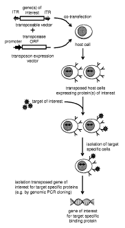
100481 FIG. 4: This figure outlines the principle of the method disclosed
herein, for the isolation of
coding information for proteins, including antibodies and fragments thereof,
with a desired
function, e.g. the binding to a target of interest, as depicted here. The
gene(s) of interest, e.g. a
diverse transposable DNA library encoding proteins, including antibody
polypeptide chains or
fragments thereof, that is cloned in between inverted terminal repeats (ITRs)
of a transposable
CA 02878982 2015-01-12
WO 2014/013026 PCT/EP2013/065214
- 12 -
construct is introduced into a vertebrate host cell together with an
expression vector for an
active transposase enzyme (see top of the drawing). The expression of the
transposon enzyme
in said host cells in trans and the presence of the gene(s) of interest cloned
in between ITRs
that can be recognized by the transposase enzyme allows the stable integration
of the ITR-
flanked gene(s) of interest into the genome of the host cells, which can then
stably express the
protein(s) of interest encoded by the genes of interest. The cellular library
expressing the
protein(s) of interest can then be screened for a desired functionality of the
expressed proteins,
e.g., but not limited to the binding to a target protein of interest, as
depicted here. By means of
cell separation technqiues known in the art, e.g. MACS or FACS, the cells
expressing the
protein(s) of interest with the desired phenotype and which therefore contain
the
corresponding genotype, can be isolated and the coding information for the
gene(s) of interest
can be retrieve from the isolated cells by cloning techniques known in the
art, e.g. but not
limited to genomic PCR cloning, as depicted here.
[0049] FIGs. 5a) and 5b): This drawing outlines the cloning strategy for the
generation of a
transposable human immunoglobulin (Ig) kappa light chain (LC) expression
vector, as
described in Example 1. FIG. 5 a.) depicts the cloning strategy for the
insertion of 5'- and 3'-
ITRs from the PiggyBac transposon into the mammalian expression vector pIRES-
EGFP
(Invitrogen, Carlsbad, CA, USA), which already contains the strong mammalian
cell promoter
element pCMV(IE) (immediate early promoter of CMV), and intron/polyA signals
for strong
mammalian host cell expression. In addition, downstream of the ClaI, EcoRV,
NotI, EcoRI
containing multiple cloning site, into which gene(s) of interest can be
cloned, pIRES-EGFP
contains an internal ribosomal entry site (IRES) with a downstream ORF of
enhanced green
fluorescent protein (EGFP), which effects the coupling of expression of
gene(s) of interest
cloned upstream of the IRES. Bacterial functional elements (ampicillin
resistance gene, amp')
and a bacterial origin of replication (Col El) for amplification and selection
of the plasmid in
E. coli are depicted as well. The resulting PiggyBac ITRs containing plasmid
is designated
pIRES-EGFP-T1T2. FIG 5b) then depicts the insertion of a gene synthesized
human Ig kappa
LC into the unique EcoRV restriction enzyme site of pIRES-EGFP-T1-T2, which
positions
the human Ig kappa LC upstream of the IRES-EGFP cassette, and thereby couples
the
expression of the human Ig kappa LC to EGFP marker gene expression. The
insertion of the
human Ig kappa LC results in transposable human Ig kappa LC expression vector
pIRES-
EGFP-T1T2-IgL. The drawings show selected unique restriction enzyme sites in
the plasmids,
as well as selected duplicated sites resulting from cloning steps.
CA 02878982 2015-01-12
WO 2014/013026 PCT/EP2013/065214
- 13 -
[0050] FIG. 6: This drawing outlines the cloning of a transposable human
immunoglobulin (Ig)
gamma 1 heavy chain (HC) expression vector, which can be generated by exchange
of the
human Ig kappa LC open reading frame (ORF) against the ORF for a human Ig
gamma 1 HC
ORF. The design of the final Ig gamma 1 HC ORF is similar, also with regard to
the
engineering of a unique Eco47III restriction enzyme site separating the
variable (V) from the
constant (C) coding regions, which allows the exchange of a single antibody V
coding region
against a diverse library of antibody V coding regions, as described in
Example 3.
[0051] Fig. 7: This drawing depicts the cloning of a mammalian PiggyBac
transposase enzyme
expression vector, as described in the Example 4, using pCDNA3.1(+) hygro as
the backbone
of the mammalian expression vector, into which the gene synthesized ORF from
PiggyBac
transposase is cloned into the unique EcoRV restriction enzyme site of
pCDNA3.1(+) hygro,
resulting in PiggyBac transposon expressin vector pCDNA3.1(+) hygro-
PB.expression vector
pCDNA3.1(+) hygro-PB. Also in this drawing the relative position of other
mammalian
functional elements (CMV-IE promoter, BGH-polyA signal, SV40-polyA segment,
hygromycinB ORF) and bacterial functional elements (ampicillin resistance
gene, ampR,
origin of replication, ColE1), as well as selected relevant restriction enzyme
recognition sites
are shown.
[0052] Fig. 8: This drawing depicts the cloning of a Sleeping Beauty
transposable human
immunoglobulin kappa light chain (Ig-kappa LC) expression vector, as described
in Example
5. The cloning can be performed by sequentially replacing the PiggyBac 5' and
3' ITRs with
Sleeping Beauty 5' and 3'ITRs in construct pIRES-EGFP-T1T2-IgL. Also in this
drawing the
relative position of other mammalian functional elements (CMV-IE promoter, BGH-
polyA
signal, 5V40-polyA segment, hygromycinB ORF) and bacterial functional elements
(ampicillin resistance gene, ampR, origin of replication, ColE1), as well as
selected relevant
restriction enzyme recognition sites are shown.
[0053] Fig. 9: This drawing depicts the cloning of a mammalian Sleeping Beauty
transposase enzyme
expression vector, as described in the Example 6, using pCDNA3.1(+) hygro as
the backbone
of the mammalian expression vector, into which the gene synthesized ORF from
Sleeping
Beauty transposase is cloned into the unique EcoRV restriction enzyme site of
pCDNA3.1(+)
hygro, resulting in Sleeping Beauty transposon expression vector pCDNA3.1(+)
hygro-SB.
Also in this drawing the relative position of other mammalian functional
elements (CMV-IE
promoter, BGH-polyA signal, 5V40-polyA segment, hygromycinB ORF) and bacterial
CA 02878982 2015-01-12
WO 2014/013026 PCT/EP2013/065214
- 14 -
functional elements (ampicillin resistance gene, ampR, origin of replication,
ColE1), as well
as selected relevant restriction enzyme recognition sites are shown.
[0054] Fig. 10: This drawing shows the arrangement of functional elements and
position of selected
unique restriction enzyme sites within the gene-synthesized DNA fragments 1.)
and 2.) that
were utilized in Example 4, in order to clone both "empty" IgH chain
expression vectors
allowing transposition utilizing either PiggyBac or Sleeping Beauty
transposase. The origin of
the functional elements is disclosed in detail in the description of the
Example.
[0055] Fig. 11: This drawing shows the final design and plasmid map of the
transposable expression
vectors for human, membrane bound Ig-gammal heavy chains (left) and human Ig
kappa light
chains (right). For the IgH expression vector, the VH-coding region may be
replaced by VH
coding regions of any other monoclonal antibody, or by a VH-gene library,
using unique
restriction enzyme sites NotI and NheI, flanking the VH coding region in this
vector. For the
IgL expression vector, the VL-coding region may be replaced by VL coding
regions of any
other monoclonal antibody, or by a VL-gene library, using unique restriction
enzyme sites
NotI and BsiWI, flanking the VL coding region in this vector. The 8 vector
constructions for
PiggyBac and Sleeping Beauty transposable IgH and IgL vectors, disclosed in
detail in
Example 4 all share this general design. The two vector maps displayed here
correspond to the
vector maps of pPB-EGFP-HC-Ac10 (left) and pPB-EGFP-LC-Ac10 (right), and the
additional vectors for hBUl 2 heavy chain (HC) or light chain (LC), containing
either
PiggyBac or Sleeping Beauty ITRs, are provided in the tables below. Sequences
of all vectors
in this figure are provided in Example 4.
[0056] Fig. 12: This figure shows two dimensional FACS dot-plots, in which the
surface expression
of human IgG from transfected and transposed IgHC and IgLC expression vectors
is detected
on the surface of 63-12 A-MuLV transformed murine proB cells derived from RAG-
2-
deficient mice. d2 post TF means that the FACS analysis was performed 2 days
after
transfection of vector constructs into 63-12 cells. The FACS plots in the left-
hand column
represents negative and positive controls for the transfection. NC=mock
electroporation of
cells without plasmid DNA. pEGFP-N3=transfection control with pEGFP-N3 control
vector,
which controls for the transfection efficiency by rendering transfected cells
green. The second
column from the left shows FACS plots from 63-12 cells co-transfected with
either PiggyBac-
transposase vector, pPB-EGFP-HC-Ac10, pPB-EGFP-LC-Ac10 vectors (top row), or
PiggyBac-transposase vector, pPB-EGFP-HC-hBU12, pPB-EGFP-LC-hBU12 vectors
(middle
row), or with Sleeping Beauty-transposase vector, pSB-EGFP-HC-Ac10, pSB-EGFP-
LC-
Ac10 vectors (bottom row). The second-left column labeled "d2 post TF" shows
the analysis
CA 02878982 2015-01-12
WO 2014/013026 PCT/EP2013/065214
- 15 -
for cells expressing IgG on the cell surface (Y-axis) and EGFP expression (X-
axis) two days
post co-transfection of the vectors as mentioned above. Surface IgG and EGFP
double
positive cells were FACS sorted as indicated by the rectangular gate. The
second-right column
labeled "d9 lx sorted" shows the analysis of surface IgG and EGFP expression
in the cell
population that was sorted at day 2 after transfection, analyzed in the same
way. Sorting gates
for the second FACS sort are also provided as rectangular gates. The rightmost
column
labeled "d16 2x sorted" shows the analysis of surface IgG and EGFP expression
of the cell
populations that had been re-sorted at day 9 after transfection, and analyzed
in the same way
for surface IgG and EGFP expression as in the previous experiments.
[0057] Fig. 13: This figure depicts the demonstration that proB cells
expressing CD30-specific IgG
on the surface of 63-12 cells can specifically be stained and detected by CD30
antigen, and
that the CD30-specific cells be detected and re-isolated from a large
population of cells
expressing surface IgG of unrelated specificity (here CD19), in which the CD30-
specific cells
have been spiked in with decreasing frequency. The FACS dot-plot on top shows
the detection
of IgG (via anti-kappaLC staining) and CD30 binding (via CD30-antigen
staining) on the
surface of the positive control cells, which are 63-12 cells stably transposed
and 2x sorted for
expressing human anti-CD30 IgG, clone Ac10 on the cell surface. As expected, a
quite pure
population (97.3%) of IgG-positive/CD30-reactive was detectable in the upper
right quadrant
of the FACs-dot-plot. The numbers on top of each FACS-plot indicates the
number of live
cells based on FSC/SSC gating that were acquired in each experiment. The
middle row shows
the FACS analysis for IgG-positive/CD30 reactive cells detectable in a
background of
IgGpositive/CD19 specific cells. The number above the number of events
indicates the
dilution factor of anti-CD30 specific IgG positive cells that were used for
the generation of the
"spiked-in" population of anti-CD30 mAb IgG positive cells in a background of
anti-CD19
mAb IgG positive cells. The sorting gates are indicated that were used to
specifically isolate
IgG-positive/CD30 antigen reactive cells from the spiked-in populations.
Larger numbers of
events needed to be acquired in order to allow detection and isolation of the
IgG-
positive/CD30 cells at higher dilutions. The lower row of FACS plots then
shows the re-
analysis of sorted cells after the cells had been expanded for 12 days for the
same parameters
(IgG-expression & CD30 antigen specificity).
[0058] Fig. 14: This figure shows the cloning of a transposable vector for a
human Ig-gammal heavy
chain (HC) in genomic configuration. The linear fragment on top represents the
human
gammal exon and introns for membrane-bound Ig-gammal-HC, with flanking NheI
and
BstBI restriction sites added to allow ligation into Ig-gammal HC cDNA vector
pPB-EGFP-
CA 02878982 2015-01-12
WO 2014/013026 PCT/EP2013/065214
- 16 -
HC-Ac10. H-designates the Hinge-region exon, M1 and M2 represent the exons
encoding the
trans-membrane region of surface expressed Ig heavy chain. With a simple one-
step ligation
the cDNA C-gammal region of the transposable human heavy chain vector is
replaced by its
genomic counterpart as indicated in the figure. Using this strategy, the VH
coding region will
be ligated in-frame to the CH1 coding exon of human C-gammal.
[0059] Fig. 15: This figure shows the sequence and overall design of the kappa
light chain library.
CDR3 coding region is underlined. Useful restriction sites are indicated.
[0060] Fig. 16: This figure shows the sequence and overall design of the gamma
heavy chain library,
showing as an example the library fragment randomized using the NNK4
randomization
strategy. The gamma heavy chain library fragments randomized using the NNK6,
NNK8 and
NNK10 randomization strategies differ only in the number of randomized amino
acid residues
in the HCDR3 region. HCDR3 coding region is underlined. The ARG codon encodes
Lysine
and Arginine. Useful restriction sites are indicated.
[0061] Fig. 17: This figure shows the digestion of PCR templates prior to
amplification with primers.
(A) Digestion of pUC57_Jkappa2-Ckappa with the restriction endonuclease ScaI
produces a
blunt-ended DNA fragment ideal for priming with the primer LCDR3-NNK6-F. (B)
Digestion
of pUC57_JH4 with the restricion endonuclease DrdI produces a DNA fragment
ideal for
priming with the primers HCDR3-NNK4-F, HCDR3-NNK6-F, HCDR3-NNK8-F, and
HCDR3-NNK10-F
[0062] Fig. 18: This figure shows the electropherograms spanning the
randomized LCDR3 and
HCDR3 region of the PCR amplicons generated to diversify the LCDR3 region by
the NNK-6
approach for Vkappa (A), and the HCDR3 region by the NNK4-approach for VH, as
disclosed
in Examples 12 and 13, respectively.
DETAILED DESCRIPTION OF THE INVENTION
[0063] Definitions
[0064] As used herein, "diverse collection" means a plurality of variants or
mutants of particular
functional or binding proteins exhibiting differences in the encoding
nucleotide sequences or
in the primary amino acid sequences, which define different functionalities or
binding
properties.
CA 02878982 2015-01-12
WO 2014/013026 PCT/EP2013/065214
- 17 -
[0065] As used herein, "library" means a plurality of polynucleotides encoding
polypeptides
having different binding specificities and/or functionalities. In certain
embodiments,
the library may comprise polynucleotides encoding at least 102, at least 103,
at least
104, at least 105, at least 106, at least 107, at least 108, or at least 109
unique
polypeptides, such as, for example, full-length antibody heavy or light chains
or VH
or VL domains.
100661 As used herein, "inverted terminal repeat sequence" or "ITR" means a
sequence identified at
the 5' or 3' termini of transposable elements that are recognized by
transposases and which
mediate the transposition of the ITRs including intervening coding information
from one
DNA construct or locus to another DNA construct or locus.
[0067] As used herein, "transposase" means an enzyme that has the capacity to
recognize and to bind
to ITRs and to mediate the mobilization of a transposable element from one
target DNA
sequence to another target DNA sequence.
[0068] As used herein, "antigen binding molecule" refers in its broadest sense
to a molecule that
specifically binds an antigenic determinant. A non-limiting example of an
antigen binding
molecule is an antibody or fragment thereof that retains antigen-specific
binding. By
"specifically binds" is meant that the binding is selective for the antigen
and can be
discriminated from unwanted or nonspecific interactions.
[0069] As used herein, the term "antibody" is intended to include whole
antibody molecules,
including monoclonal, polyclonal and multispecific (e.g., bispecific)
antibodies, as well as
antibody fragments having an Fc region and retaining binding specificity, and
fusion proteins
that include a region equivalent to the Fc region of an immunoglobulin and
that retain binding
specificity. Also encompassed are antibody fragments that retain binding
specificity
including, but not limited to, VH fragments, VL fragments, Fab fragments,
F(ab')2 fragments,
scFv fragments, Fv fragments, minibodies, diabodies, triabodies, and
tetrabodies (see, e.g.,
Hudson and Souriau, Nature Med. 9: 129-134 (2003)) (incorporated by reference
in its
entirety).
[0070] An embodiment of the invention disclosed herein is a method for the
identification of specific
functional or binding polypeptides, including, but not limited to antibody
chains or fragments
thereof (Fig. 4), which comprises:
i. cloning of diverse transposable DNA libraries encoding proteins,
including antibody
polypeptide chains or fragments thereof, in between inverted terminal repeats
(ITRs)
derived from transposable elements and recognizable by and functional with at
least one
transposase enzyme,
CA 02878982 2015-01-12
WO 2014/013026 PCT/EP2013/065214
- 18 -
ii. introduction of one or more diverse transposable DNA libraries of step
(i) into vertebrate
host cells by standard methods known in the art,
iii. providing temporary expression of at least one functional transposase
enzyme in said
vertebrate host cells in trans, such that said one or more diverse
transposable DNA
libraries are stably integrated into the vertebrate host cell genomes, thereby
providing a
vertebrate host cell population that then stably expresses diverse libraries
of proteins,
including antibody chains or fragments thereof,
iv. screening of said diverse cellular libraries, stably expressing
proteins, including
antibodies or fragments thereof, for a desired functional or binding phenotype
by
methods known in the art,
v. optionally, including iterative enrichment cycles with the stably
genetically modified
vertebrate host cells for a desired binding or functional phenotype, and
vi. isolation of the corresponding genes from the enriched host cells
encoding the desired
binding or functional phenotype by standard cloning methods, known in the art,
for
instance, but not limited to, PCR (polymerase chain reaction), using primers
specific for
the sequences contained in the one or more transposed DNA library constructs.
[0071] A preferred embodiment of step (i) is to generate diverse transposable
DNA libraries either by
gene synthesis, or by polymerase chain reaction (PCR) using appropriate
primers for the
amplification of diverse protein coding regions, and DNA templates comprising
a diversity of
binding proteins, including antibodies, or fragments thereof, by methods known
in the art.
[0072] For the generation of diverse antibody libraries, a diverse collection
of antibody heavy and
light chain sequences may be generated by standard gene synthesis in which the
V region
coding sequences may be randomized at certain positions, e.g. but not limited
to, any or all of
the complementarity determining regions (CDRs) of the antibody heavy or light
chain V-
regions. The diversity can be restricted to individual CDRs of the V-regions,
or to a particular
or several framework positions, and/or to particular positions in one or more
of the CDR
regions. The V regions with designed variations, as described above, can be
synthesized as a
fragment encoding entire antibody heavy or light chains that are flanked by
inverted terminal
repeats functional for at least one desired transposase enzyme. Preferably,
the DNA library
containing diverse variable domains encoding V regions for antibody heavy or
light chains is
generated, and flanked by appropriate cloning sites, including but not limited
to restriction
enzyme recognition sites, that are compatible with cloning sites in antibody
heavy or light
chain expression vectors. Useful transposon expression systems for use in the
methods of the
invention include, for example, the PiggyBac transposon system as described,
for example, in
US Pat. Nos. 6,218,185; 6,551,825; 6,962,810; 7,105,343; and 7,932,088 (the
entire contents
CA 02878982 2015-01-12
WO 2014/013026 PCT/EP2013/065214
- 19 -
of each of which are hereby incorporated by reference) and the Sleeping Beauty
transposon
system as described in US Pat. Nos. 6,489,458; 7,148,203; 7,160,682; US
2011117072; US
2004 077572; and US 2006 252140 (the entire contents of each of which are
hereby
incorporated by reference.)
[0073] Diverse antibody heavy and light chain libraries may also be obtained
from B cell populations
isolated from desired vertebrate species, preferably humans, and preferably
from cellular
compartments containing B cells, e.g., but not limited to peripheral or cord
blood, and
lymphoid organs like bone marrow, spleen, tonsils and lymph-node tissues. In
this case,
diverse antibody V region sequences for antibody heavy and light chains can be
isolated by
RT-PCR or by genomic PCR using antibody heavy and light chain specific
degenerate PCR
primer pairs, that can amplify the majority of V-region families by providing
upstream
primers that bind to homologous sequences upstream of, or within leader
sequences, upstream
of or within V-region frameworks, and by providing downstream primers that
bind in regions
of homology within or downstream of the J joining gene segment of variable
domain coding
regions, or within or downstream of the coding regions of the constant regions
of antibody
heavy or light chains.
[0074] The PCR primer sets utilized for the amplification of diverse variable
coding regions may be
flanked by appropriate cloning sites, e.g. but not limited to restriction
enzyme recognition
sites, that are compatible with cloning sites in antibody heavy or light chain
expression
vectors.
[0075] The transposable DNA libraries of step (i) encoding diverse proteins,
including antibodies and
antibody fragments thereof, can be provided in the form of plasmid libraries,
in which the
gene-synthesized or the PCR amplified transposable DNA libraries are cloned
using
appropriate cloning sites, as mentioned above. Alternatively, the transposable
DNA libraries
encoding diverse libraries of binding proteins, such as antibodies and
fragments thereof, can
be provided in form of linear, double-stranded DNA constructs, directly as a
result of DNA
synthesis, or as a result of PCR amplification. The latter approach of
providing the
transposable DNA libraries as linear double-stranded DNA PCR amplicons, that
have not
been cloned into expression vectors or plasmids (in comparison to all other
vertebrate cell
expression systems) has the advantage that the maximum molecular complexity of
the
transposable DNA libraries is maintained and not compromised by a limited
cloning or
ligation efficiency into an expression vector. In contrast, cloning by
ligation, or otherwise, into
plasmid expression or shuttle vectors is a necessary intermediate for all
other plasmid-based or
viral vector based vertebrate cell expression systems.
CA 02878982 2015-01-12
WO 2014/013026 PCT/EP2013/065214
- 20 -
[0076] However, the use of plasmid-based transposon expression vectors
containing the diverse
transposable DNA libraries encoding diverse binding proteins, including
antibodies and
antibody fragments thereof, has the advantage that these expression vectors
can be engineered
to contain additional functional elements, that allow the screening, or,
alternatively, the
selection for stably transposed vertebrate host cells for the stable
integration of the transposon
expression vector in transposed vertebrate host cells.
[0077] This is achieved by providing in operable linkage to the diverse
transposable DNA libraries,
i.e. cloned into the transposon expression vectors in cis, expression
cassettes for marker genes
including., but not limited to, fluorescent marker proteins (e.g. green,
yellow, red, or blue
fluorescent proteins, and enhanced versions thereof, as known in the art), or
expression
cassettes for cell surface markers including, but not limited to, CD markers,
against which
specific diagnostic antibodies or other diagnostic tools are available.
[0078] Alternatively, expression cassettes for selectable markers, that allow
selection of transposed
vertebrate host cells for antibiotic resistance, including, but not limited
to, puromycin,
hygromycinB, bleomycin, neomycin resistance, can be provided in operable
linkage to the
diverse transposable DNA libraries, i.e. cloned into the transposon expression
vectors in cis.
[0079] The operable linkage can be achieved by cloning of said expression
cassettes for marker genes
or antibiotic resistance markers, either up- or downstream of the coding
regions comprising
said diverse transposable DNA libraries, but within the inverted terminal
repeats of the
transposon vector.
[0080] Alternatively, the operable linkage can be achieved by cloning of the
coding regions for said
marker or antibiotic resistance genes downstream of the coding regions
comprising said
diverse transposable DNA libraries, but separated by internal ribosomal entry
site (IRES)
sequences, that ensure transcriptional coupling of the expression of said
diverse transposable
DNA libraries with said marker or antibiotic resistance genes, and thereby
allowing the
screening for or selection of stably transposed vertebrate host cells.
[0081] In step (ii) of the method disclosed herein, said diverse transposable
DNA libraries encoding
diverse libraries of proteins, including antibodies and fragments thereof, are
introduced into
desired vertebrate host cells by methods known in the art to efficiently
transfer DNA across
vertebrate cell membranes, including., but not limited to, DNA-transfection
using liposomes,
Calcium phosphate, DEAE-dextran, polyethyleneimide (PEI) magnetic particles,
or by
protoplast fusion, mechanical transfection, including physical, or ballistic
methods (gene gun),
or by nucleofection. Any of the above-mentioned methods and other appropriate
methods to
transfer DNA into vertebrate host cells may be used individually, or in
combination for step
(ii) of the method disclosed herein.
CA 02878982 2015-01-12
WO 2014/013026 PCT/EP2013/065214
- 21 -
[0082] In the case of dimeric proteins, including, but not limited to,
antibodies and fragments thereof,
it is a useful embodiment of the method disclosed herein to introduce diverse
transposable
DNA libraries and/or transposon vectors for antibody heavy or light chains
contained in
separate transposable vectors, which can independently be introduced into the
vertebrate host
cells. This either allows the sequential introduction of diverse transposable
DNA libraries for
antibody heavy or light chains into said cells, or their simultaneous
introduction of diverse
transposable DNA libraries for antibody heavy or light chains, which, in
either case, allows
the random shuffling of any antibody heavy with any antibody light chain
encoded by the at
least two separate diverse transposable DNA libraries.
[0083] Another useful embodiment of the previous embodiment is to utilize
separate transposon
vectors and/or diverse DNA transposable libraries for antibody heavy and light
chains, where
said constructs or libraries are contained on transposable vectors recognized
by different
transposase enzymes (Fig. 3). This allows the independent transposition of
antibody heavy
and antibody light chain constructs without interference between the two
different transposase
enzymes, as one transposable vector is only recognized and transposed by its
specific
transposase enzyme. In case of sequential transposition of transposable
vectors or DNA
libraries encoding antibody heavy or light chains, the advantage of utilizing
different
transposase enzymes with different ITR sequences is, that upon the second
transposition
event, the first already stably transposed construct is not again mobilized
for further
transposition.
[0084] This embodiment also allows the discovery of antibodies by the method
of guided selection
(Guo-Qiang et al. Methods MoL Biol. 562, 133-142 (2009)) (incorporated herein
by reference
in its entirety). Guided selection can e.g. be used for the conversion of any
non-human
antibody specific for a desired target/epitope specificity and with a desired
functionality into a
fully human antibody, where the same target/epitope specificity and
functionality is preserved.
The principle of guided selection entails the expression of a single antibody
chain (heavy or
light chain) of a reference (the "guiding") antibody, in combination with a
diverse library of
the complementary antibody chains (i.e. light, or heavy chain, respectively),
and screening of
these heavy-light chain combinations for the desired functional or binding
phenotype. This
way, the first antibody chain, "guides" the selection of one or more
complementary antibody
chains from the diverse library for the desired functional or binding
phenotype. Once the one
or more novel complementary antibody chains are isolated, they can be cloned
in expression
vectors and again be used to "guide" the selection of the second,
complementary antibody
chain from a diverse antibody chain library. The end-result of this two-step
process is that
both original antibody heavy and light chains of a reference antibody are
replaced by
CA 02878982 2015-01-12
WO 2014/013026 PCT/EP2013/065214
- 22 -
unrelated and novel antibody chain sequences from the diverse libraries, but
where the novel
antibody heavy-light chain combination exhibits the same, or similar
functional or binding
properties of the original reference antibody. Therefore, this method requires
the ability to
independently express antibody heavy and light chain constructs or libraries
in the vertebrate
host cells, which can be achieved by the preferred embodiment to provide
antibody heavy and
light chain expression cassettes in different transposable vector systems,
recognized by
different transposon enzymes.
[0085] However, diverse transposable DNA libraries can also be constructed in
a way, that the coding
regions for multimeric proteins, including antibodies and fragments thereof,
are contained in
the same transposon vector, i.e. where the expression of the at least two
different subunits of a
multimeric protein, for example VH and VL regions or full-length heavy and
light chains,
isoperably linked by cloning of the respective expression cassettes or coding
regions into the
same transposable vector.
[0086] Useful vertebrate host cells for the introduction of transposable
constructs and/or transposable
DNA libraries of step (ii) are cells from vertebrate species that can be or
that are immortalized
and that can be cultured in appropriate cell culture media and under
conditions known in the
art. These include, but are not limited to, cells from e.g. frogs, fish,
avians, but preferably
from mammalian species, including, but not limited to, cells from rodents,
ruminants, non-
human primate species and humans, with cells from rodent or human origin being
preferred.
[0087] Useful cell types from the above-mentioned species include, but are not
limited to cells of the
lymphoid lineage, which can be cultured in suspension and at high densities,
with B-lineage
derived cells being preferred, as they endogenously express all the required
proteins, factors,
chaperones, and post-translational enzymes for optimal expression of many
proteins, in
particular of antibodies, or antibody-based proteins. Of B-lineage derived
vertebrate cells,
those are preferred that represent early differentiation stages, and are known
as progenitor
(pro) or precursor (pre) B cells, because said pro- or preB cells in most
cases do not express
endogenous antibody chains that could interfere with exogenous or heterologous
antibody
chain expression that are part of the method disclosed herein.
[0088] Useful pro- and pre- B lineage cells from rodent origin are Abelson-
Murine Leukemia virus
(A-MuLV) transformed proB and preB cells (Alt et al. Cell 27, 381-390(1981)
(incorporated
herein by reference in its entirety)) that express all necessary components
for antibody
expression and also for their proper surface deposition, including the B cell
receptor
components Ig-alpha (CD79a, or mb-1), and Ig-beta (CD79b, or B-29) (Hombach et
al.
Nature 343, 760-762 (1990)) (incorporated herein by reference in its
entirety), but as
mentioned above, mostly lack the expression of endogenous antibody or
immunoglobulin
CA 02878982 2015-01-12
WO 2014/013026 PCT/EP2013/065214
- 23 -
chains. Here, A-MuLV transformed pro- and preB cells are preferred that are
derived from
mouse mutants, including, but not limited to, mouse mutants defective in
recombination
activating gene-1 (RAG-1), or recombination activating gene-2 (RAG-2), or
animals carrying
other mutations in genes required for V(D)J recombination, e.g. XRCC4, DNA-
ligase IV,
Ku70, or Ku80, Artemis, DNA-dependent protein kinase, catalytic subunit (DNA-
PIcs), and
thus lack the ability to normally express of endogenous antibody polypeptides.
[0089] Additional useful types of progenitor (pro) and precursor (pre) B
lineage cells are early,
immunoglobulin-null (Ig-null) EBV transformed human proB and preB cells
(Kubagawa et al.
PNAS 85, 875-879( 1988) ) (incorporated herein by reference in its entirety)
that also express
all the required factors for expression, post-translational modification and
surface expression
of exogenous antibodies (including CD79a and CD79b).
[0090] Other host cells of the B lineage can be used, that represent plasma
cell differentiation stages
of the B cell lineage, preferably, but not limited to Ig-null myeloma cell
lines, like Sp2/0,
NSO, X63, Ag8653, and other myeloma and plasmacytoma cells, known in the art.
Optionally, these cell lines may be stably transfected or stably genetically
modified by other
means than transfection, in order to over-express B cell receptor components
Ig-alpha
(CD79a, or mb-1), and Ig-beta (CD79b, or B-29), in case optimal surface
deposition of
exogenously expressed antibodies is desired.
[0091] Other, non-lymphoid mammalian cells lines, including but not limited
to, industry-standard
antibody expression host cells, including, but not limited to, CHO cells,
Per.C6 cells, BHK
cells and 293 cells may be used as host cells for the method disclosed herein,
and each of
these cells may optionally also be stably transfected or stably genetically
modified to over-
express B cell receptor components Ig-alpha (CD79a, or mb-1), and Ig-beta
(CD79b, or B-29),
in case optimal surface deposition of exogenously expressed antibodies is
desired.
[0092] Essentially, any vertebrate host cell, which is transfectable, can be
used for the method
disclosed herein, which represents a major advantage in comparison to any
viral expression
systems, such as., but not limited to vaccinia virus, retroviral, adenoviral,
or sindbis virus
expression systems, because the method disclosed herein exhibits no host cell
restriction due
to virus tropism for certain species or cell types, and furthermore can be
used with all
vertebrate cells, including human cells, at the lowest biosafety level, adding
to its general
utility.
[0093] Step (iii) of the method disclosed herein results in the stable genetic
modification of desired
vertebrate host cells with the transfected transposable constructs of step
(ii) by temporary, or
transient expression of a functional transposase enzyme, such that a stable
population of
CA 02878982 2015-01-12
WO 2014/013026 PCT/EP2013/065214
- 24 -
vertebrate host cells is generated that expresses diverse libraries of
proteins encoded by said
constructs.
[0094] A useful embodiment of step (iii) is to transiently introduce into the
host cells, preferably by
co-transfection, as described above, a vertebrate expression vector encoding a
functional
transposase enzyme together with said at least one diverse transposable DNA
library. It is to
be understood that transient co-transfection or co-integration of a
transposase expression
vector can either be performed simultaneously, or shortly before or after the
transfer of the
transposable constructs and/or diverse transposable DNA libraries into the
vertebrate host
cells, such that the transiently expressed transposase can optimally use the
transiently
introduced transposable vectors of step (ii) for the integration of the
transposable DNA library
into the vertebrate host cell genome.
[0095] Another useful embodiment of step (iii) is to effect the stable
integration of the introduced
transposable vectors and/or transposable DNA libraries of step (ii) by
transiently expressing a
functional transposase enzyme by means of an inducible expression system known
in the art,
that is already stably integrated into the vertebrate host cell genome. Such
inducible and
transient expression of a functional transposase may be achieved by e.g.,
tetracycline
inducible (tet-on/tet-off) or tamoxifen-inducible promoter systems known in
the art. In this
case, only the one or more transposable vector or DNA library needs to be
introduced into the
host cell genome, and the stable transposition of the constructs and the
stable expression of the
proteins encoded by the one or more transposable vector or DNA library is
effected by the
transiently switched on expression of the functional transposase enzyme in the
host cells.
[0096] Step (iv) of the method disclosed herein effects the isolation of
transposed vertebrate host
cells expressing proteins with a desired functionality or binding phenotype.
[0097] A preferred embodiment of step (iv) is to screen for and to isolate the
transposed host cells of
step (iii) expressing desired proteins, including antibodies and fragments
thereof, with target-
binding assays and by means of standard cell separation techniques, like
magnetic activated
cell sorting (MACS) or high-speed fluorescence activated cell sorting (FACS)
known in the
art. Especially, in a first enrichment step of a specific population of
transposed vertebrate host
cells, where large number of cells need to be processed, it is preferred to
isolate target specific
cells from a large number of non-specific cells by MACS-based techniques.
[0098] Particularly, for additional and iterative cell enrichment cycles, FACS
enrichment is preferred,
as potentially fewer numbers of cells need to be processed, and because multi
channel flow
cytometry allows the simultaneous enrichment of functionalities, including.,
but not limited to,
binding to a specific target of more than one species, or the specific
screening for particular
epitopes using epitope-specific competing antibodies in the FACS screen.
CA 02878982 2015-01-12
WO 2014/013026 PCT/EP2013/065214
- 25 -
[0099] If proteins, including antibodies and fragments thereof, are to be
discovered that interact with
soluble binding partners, these binding partners are preferably labeled with
specific labels or
tags, such as but not limited to biotin, myc, or HA-tags known in the art,
that can be detected
by secondary reagents, e.g. but not limited to, streptavidin or antibodies,
that themselves are
labeled magnetically (for MACS based cell enrichment) or with fluorochromes
(for FACS
based cell enrichment), so that the cell separation techniques can be applied.
[00100] If proteins, including antibodies and fragments thereof are to be
discovered against membrane
bound proteins, which cannot easily be expressed as soluble proteins, like
e.g. but not limited
to, tetraspannins, 7-transmembranbe spanners (like G-coupled protein coupled
receptors), or
ion-channels, these may be expressed in viral particles, or overexpressed in
specific cell lines,
which are then used for labeling or panning methods known in the art, which
can enrich the
vertebrate host cells expressing the proteins from the transposed constructs,
including
antibodies and fragments thereof
[00101] Due to the stable genotype-phenotype coupling in the stably transposed
vertebrate host cell
population, a useful embodiment of step (v) is to repeat cell enrichment
cycles for a desired
functional or binding phenotype, until a distinct population of cells is
obtained that is
associated with a desired functional or binding phenotype. Optionally,
individual cell clones
can be isolated e.g., but not limited to, by single-cell sorting using flow
cytometry technology,
or by limiting dilution, in order to recover the transposed DNA information
from individual
cell clones that are coupled to a particular, desired functional or binding
phenotype.
[00102] For the identification of functional target-specific antibodies it is
often favorable to not only
screen and to select for a particular binding phenotype, but to additionally
screen for
additional functional properties of target specific antibodies, in particular
antagonistic or
agonistic effects in biological assay.
[00103] Therefore, it is desirable to be able to efficiently "switch" cell
membrane bound antibody
expression to secreted antibody expression in the vertebrate host cells with
sufficient yields, in
order to produce enough quantity of a particular antibody clone for functional
assays.
[00104] In natural B lineage cells the switch from membrane bound to secreted
antibody expression
occurs via a mechanism of alternative splicing, in which in preB and B cells
an alternative
splice donor near the 3' end of the last heavy chain constant region exon is
preferentially
spliced to a splice acceptor of a membrane anchor exon downstream of the heavy
chain
constant regions exons. This way, an antibody heavy chain is produced in B
cells with an
extended C-terminal, membrane spanning domain, that anchors the heavy chain
and thereby
the entire heavy-light chain containing antibody in the cell membrane. The C-
terminal,
membrane spanning domain also interacts non-covalently with the membrane
spanning
CA 02878982 2015-01-12
WO 2014/013026 PCT/EP2013/065214
- 26 -
components Ig-alpha (CD79a or mb-1) and Ig-beta (CD79b or B29), which likely
results in
better membrane anchoring and higher surface immunoglobulin expression in B
lineage cells.
[00105] Once, a B cell differentiates further to the plasma cell stage, the
alternative splicing does not
occur any more and the alternative splice donor near the 3' end of the last
heavy chain constant
region is no longer recognized or utilized, and the mRNA template is
terminated downstream
of the heavy chain constant region stop codon, and a heavy chain of a secreted
antibody is
translated.
[00106] In order to exploit this natural mechanism of alternative splicing and
"switching" from
membrane bound to secreted expression of expressed antibodies, it is a useful
embodiment of
the method disclosed herein to construct the transposable vectors and diverse
DNA libraries
encoding proteins, including antibodies or fragments thereof, in such a way
that the natural
intron/exon structure of a constant antibody heavy chain, including the exons
encoding the
membrane spanning domains is maintained. This embodiment represents a clear
advantage
against retroviral expression systems, as the retroviral vector genome is
already spliced before
it is packaged into a retroviral particle and stably transduced into the host
cell genome.
[00107] Other viral vector systems may be restricted in the length of the DNA
insert that can be
incorporated into the vectors, thereby precluding the cloning of larger
genomic regions into
such expression vectors and thereby preventing the exploitation of the natural
"switching"
from membrane-bound to secreted antibody expression by alternative splicing.
Certain
transposons (e.g. To12, see Fig. 3), have been characterized to be able to
efficiently transpose
more than 10 kb DNA fragments into vertebrate host cells without any loss in
transposition
efficiency (Kawakami Genome Biol. 8, Suppl I, S7 (2007) ) (incorporated herein
by reference
in its entirety). Therefore, it is a useful embodiment of the method disclosed
herein to
construct transposable expression vectors comprising genomic exon/intron
structures for
better and proper expression and for the natural regulation switching from
membrane bound to
secreted antibody expression. The methods of the invention are useful to
transpose DNA
fragments at least 5kb, at least 6kb, at least 7kb, at least 8kb, at least 9
kb, at least 10 kb in size
into host cell genomes.
[00108] The differentiation of earlier B lineage differentiation stage that
favors membrane bound
antibody expression, to a later, plasma cell stage, that favors secreted
antibody expression can
be induced by B cell differentiation factors, such as, but not limited to,
CD40/IL4 triggering,
or stimulation by mitogens, such as, but not limited to, lipopolysaccharide
(LPS), or other
polyclonal activators, Staph. aureus Cowan (SAC) strain activators, and CpG
nucleotides, or
any combination thereof
CA 02878982 2015-01-12
WO 2014/013026 PCT/EP2013/065214
- 27 -
[00109] Preferrably, this differentiation is effected in transformed cells, in
which the proliferation can
artificially be inhibited, such that proper B cell differentiation can again
occur, as it has been
described for A-MuLV transformed murine preB cells, in which the Abelson
tyrosine kinase
is specifically inhibited by the tyrosine inhibitor Gleevec (Muljo et al. Nat.
Immunol 4,31-37
(2003)) (incorporated herein by reference in its entirety). Therefore, it is a
preferred
embodiment to utilize Ig-null A-MuLV transformed murine preB cells for the
method, which
by treatment with Gleevec, can again differentiate to more mature B cell
stages, including
plasma cells, which then secrete sufficient amounts of secreted antibody for
additional
functional testing on the basis of alternative splicing of genomic heavy chain
expression
constructs. It is a preferred embodiment of the method disclosed herein, to
further improve
such B-lineage cell differentiation by stable overexpression of anti-apoptotic
factors, known in
the art, including, but not limited to, bc1-2 or bel-xL.
[00110] After step (iv), the enrichment of transposed vertebrate host cells as
described above has been
performed, optionally, additional cell enrichments according to the above-
mentioned methods
may be performed (step (v)), until cell populations, or individual cells are
isolated expressing
proteins, including antibodies and fragments thereof, with desired functional
and/or binding
properties.
[00111] Step (vi) of the method disclosed herein is then performed in order to
isolate the relevant
coding information contained in the transposed vertebrate host cells, isolated
for a desired
functional and/or binding property.
[00112] A useful embodiment of step (vi) for the isolation cloning and
sequencing of the relevant
coding information for a desired functional or binding protein, including an
antibody or
antibody fragment thereof, contained in the isolated cells, is to utilize
genomic or RT-PCR
amplification with specific primer pairs for the relevant coding information
comprised in the
transposed DNA constructs, and to sequence the genomic or RT-PCR amplicons
either
directly, or after sub-cloning into sequencing vectors, known in the art,
e.g., but not limited
into TA- or Gateway-cloning vectors.
[00113] Cloning and sequencing of the relevant coding information for a
desired functional or binding
protein, including an antibody or antibody fragment thereof, as described in
the previous
paragraph by genomic or RT-PCR amplification can also be performed with
transposable IgH
and IgL expression vector, such that the binding protein coding region cannot
only be
identified, but at the same time also be expressed upon stable transposition
into mammalian
host cells as disclosed herein. For the expression of secreted antibodies this
would only
require the use of transposable Ig heavy chain expression vectors lacking the
IgH
transmembrane spanning coding region.
CA 02878982 2015-01-12
WO 2014/013026 PCT/EP2013/065214
- 28 -
[00114] Another useful embodiment of step (vi) is to subject the enriched cell
populations of steps (iv)
or (v), which exhibit a desired functional or binding phenotype to next-
generation ("deep")
sequencing (Reddy et al. Nat. Biotech. 28, 965-969 (2010)) (incorporated
herein by
reference in its entirety), in order to retrieve directly and in one step a
representative set of
several thousands of sequences for the coding information contained in the
transposed DNA
constructs. Based on a bioinformatics analysis of the relative frequency of
sequences
identified from the enriched cell populations, it allows a prediction about
which sequences
encoded a functional or binding protein, including an antibody or fragment
thereof (Reddy et
al. Nat. Biotech. 28, 965-969 (2010)) (incorporated herein by reference in its
entirety).
Statistically overrepresented sequences are then resynthesized and cloned into
expression
vector for expression as recombinant proteins, antibodies or fragments
thereof, in order to
characterize them functionally and for their binding properties. This method
can significantly
accelerate the identification of relevant sequences within a functionally and
phenotypically
enriched cell population, that expresses proteins with functional or target
specific properties.
[00115] Yet another useful embodiment of the method disclosed herein is to
utilize transposition-
mediated vertebrate cell expression of proteins, including antibodies or
fragments thereof, for
the mutagenesis and optimization of desired proteins, including the affinity
optimization of
antibodies and fragments thereof
[00116] This can be achieved by isolating the genes encoding the proteins,
including antibody chains
or fragments thereof, from transposed vertebrate cell populations enriched for
a desired
binding or functional phenotype according to the methods disclosed in step
(iv), such as but
not limited to, by genomic PCR or RT-PCR amplification under mutagenizing
conditions,
know in the art. The mutagenized sequences can then be re-cloned into
transposition vectors
and then again be transposed into vertebrate host cells, in order to subject
them to screening
according to the methods disclosed herein, for improved functional or binding
properties.
[00117] In one useful embodiment of this approach, specific primers are used
that allow the PCR
amplification under mutagenizing conditions of complete transposed constructs,
including the
flanking ITRs.
[00118] By this method a mutagenized PCR amplicon containing a defined average
frequency of
random mutations is generated from the functionally or phenotypically selected
transposed
cells. Said PCR amplicon with controlled mutations (variations) of the
original templates can
now directly be re-transposed into new vertebrate host cells, according to
preferred
embodiments disclosed in the methods applicable in step (ii).
[00119] The main advantage of this method over other approaches of genetically
modifying vertebrate
cells is, that with this technology no time-consuming re-cloning of the
mutagenized PCR
CA 02878982 2015-01-12
WO 2014/013026 PCT/EP2013/065214
- 29 -
amplicons and time consuming quality control of the mutagenized sequences into
expression
vectors is required, which is a mandatory requirement in all other plasmid-
based or viral
expression systems, if a mutagenized sequence shall be subjected to another
round of
screening.
[00120] Because transposition of DNA only requires the presence of ITRs
flanking the coding region
of genes of interest, PCR-amplified mutagenized PCR amplicons can directly be
re-introduced
and re-transposed into novel vertebrate host cells for expression and
screening for improved
properties and/or affinity matured mutants.
[00121] Taken together, the methods disclosed herein, of utilizing TEs for the
stable genetic
modification of vertebrate host cells with transposable constructs and/or
diverse transposable
DNA libraries encoding proteins, including antibodies and fragments thereof,
offers
unparalleled efficiency, flexibility, utility and speed for the discovery and
optimization of said
proteins for optimal desired functional or binding phenotypes.
Examples:
Example 1: Instruction for cloning of basic PiggyBac transposable light chain
expression vector for human antibody kappa light chains compatible with
the PiggyBac transposase enzyme
1001221 A basic transposable expression vector for human kappa light chains
can be generated by
cloning of the ITRs from the PiggyBac transposon up and downstream of a human
immunoglobulin kappa light chain expression cassette.
[00123] For this, as a first step, the minimal sequences for the up- and
downstream ITRs of the
PiggyBac transposon can be derived from pXLBacII (published in US 7,105,343)
(incorporated herein by reference in its entirety) and can be gene synthesized
with flanking
restriction enzyme sites for cloning into the mammalian expression vector
pIRES-EGFP
(PT3157-5, order #6064-1, Invitrogen-Life Technologies, Carlsbad, CA, USA)
[00124] The upstream PiggyBac ITR sequence with the 5' terminal repeat has to
be gene synthesized
with flanking MunI restriction enzyme sequence, compatible with a unique Muni
restriction
enzyme site in pIRES-EGFP, and additional four random nucleotides (in
lowercase letters)
allowing proper restriction enzyme digestion. This sequence is provided in Seq-
ID1.
[00125] The downstream PiggyBac ITR sequence with the 3' terminal repeat has
to be gene
synthesized with flanking XhoI restriction enzyme sequence compatible with a
unique XhoI
restriction enzyme site in pIRES-EGFP, and additional four random nucleotides
allowing
CA 02878982 2015-01-12
WO 2014/013026 PCT/EP2013/065214
- 30 -
proper restriction enzyme digestion. This sequence is provided in Seq-ID2.Upon
MunI
restriction enzyme digestion of the gene synthesized Seq-ID1, the DNA fragment
can be
ligated into Muni linearized pIRES-EGFP, generating pIRES-EGFP-TR1 according
to
standard methods, known in the art. The proper orientation of the insert can
be verified by
diagnostic restriction enzyme digestion, and/or by DNA sequencing of the
cloned construct
(Fig. 5a).
[00126] In a next step gene synthesized and XhoI digested DNA fragment Seq-
1D2, can be ligated into
XhoI linearized pIRES-EGFP-T1 (Fig. 5a) by standard methods known in the art,
in order to
generate pIRES-EGFP-T1T2, containing both PiggyBac ITRs up and downstream of
the
IRES-EGFP expression cassette (Fig. 5b). The proper orientation of the insert
can be verified
by diagnostic restriction enzyme digestion, and/or by DNA sequencing of the
cloned construct
(Fig. 5b).
[00127] For the cloning of a human immunoglobulin kappa light chain into the
vector pIRES-EGFP-
T1T2, the human Ig kappa light chain from human anti-TNF-alpha specific
antibody D2E7
can be synthesized, which can be retrieved from European patent application EP
1 285 930 A2
(incorporated herein by reference in its entirety).
[00128] The coding region for human Ig kappa light chain of human anti-TNF-
alpha specific antibody
D2E7, in which the V region of D2E7 is fused in frame to a Vkl -27 leader
sequence
(Genbank entry: X63398.1, which is the closest germ-line gene V-kappa family
member V-
kappa of D2E7), and to the human kappa constant region (Genbank entry: J00241)
has the
following nucleotide sequence, which is provided in Seq-1D3.
[00129] The nucleotide sequence of Seq-1D3 translates in the amino acid
sequence Seq-ID4.The DNA
fragment Seq-1D3 encoding the D2E7 Ig kappa light chain can be gene
synthesized and
directly ligated by blunt-ended ligation into the unique EcoRV restriction
enzyme site (which
is also a blunt cutter), by methods know in the art, resulting in construct
pIRES-EGFP-T1T2-
IgL (Fig. 5b)
[00130] Seq-1D3 has been engineered to contain a unique Eco47III restriction
enzyme site in between
the V-kappa and the C-kappa coding regions (highlighted in boldface and
underlined), which
allows the replacement of V-kappa regions in this construct against other V-
kappa regions or
V-kappa libraries, using a unique restriction enzyme upstream of V-kappa
coding region in
the construct, together with Eco47III. The proper orientation of the kappa
light chain insert
can be verified by diagnostic restriction enzyme digestion, and/or by DNA
sequencing of the
cloned construct (Fig. 5b).
[00131] The entire sequence for the transposable human antibody kappa light
chain vector pIRES-
EGFP-T1T2-IgL is provided in sequence Seq-IDS.
CA 02878982 2015-01-12
WO 2014/013026 PCT/EP2013/065214
-31 -
[00132] Sequences referred to in this Example 1:
[00133] Seq-ID1 (327 bp long PiggyBac 5'-ITR sequence. The MunI restriction
enzyme sites at each
end are underlined and typed in boldface print, the random nucleotide
additions at the termini
are printed in lowercase)
[00134] Seq- ID2 (264 bp long PiggyBac 3'-ITR sequence. The XhoI restriction
enzyme sites at each
end are underlined and typed in boldface print, the random nucleotide
additions at the termini
are printed in lowercase)
[00135] Seq-1D3 (711 bp long Ig-kappaLC coding region of anti-TNF-alpha-
specific mAb D2E7)
[00136] Seq-1D4 (236 amino acids long sequence of anti-TNF-alpha-specific mAb
D2E7)
[00137] Seq-IDS (6436bp long DNA sequence of PiggyBac transposable Ig-kappa-LC
expression
vector pIRES-EGFP-T1T2-IgL)
Example 2: Instruction for cloning of a basic
PiggyBac transposable heavy chain expression vector for membrane
spanning human antibody gammal heavy chains
1001381 In order to clone a transposable Ig heavy chain expression vector, the
kappa light chain ORF
from pIRES-EGFP- T1T2-IgL (Seq-IDS) needs to be exchanged with an ORF encoding
a
fully human IgG1 heavy chain coding region.
[00139] For the replacement of the human kappa light chain in vector pIRES-
EGFP- T1T2-IgL by a
human immunoglobulin gamma-1 heavy chain, the VH region of antibody D2E7,
which is
specific for human TNF-alpha (see: EP 1 285 930 A2) (incorporated herein by
reference in its
entirety) can be synthesized. For this, a leader sequence of a close germ-line
VH3-region
family member is fused in frame to the the VH region of antibody D2E7, which
then is fused
in frame to the coding region of a human gammal constant region (Genbank:
J00228)
including the membrane spanning exons (Genbank: X52847). In order to be able
to replace the
human Ig kappa light chain from pIRES-EGFP- T1T2-IgL, unique ClaI and NotI
restriction
enzyme sites need to be present at the 5' and the 3' end of the sequence
(underlined),
respectively. Additionally, four nucleotides flanking the restriction enzyme
sites (highlighted
in lowercase letters at the ends of the sequence) allow proper restriction
enzyme digestion of
the gene-synthesized DNA fragment and ligation into the ClaI-NotI linearized
pIRES-EGFP-
T1T2-IgL backbone, according to standard methods. The sequence that needs to
be gene
synthesized is provided in Seq-1D6.
CA 02878982 2015-01-12
WO 2014/013026 PCT/EP2013/065214
- 32 -
[00140] From the start codon in position 11 of Seq-1D6 (highlighted in
boldface print), this nucleotide
sequence translates to the human IgG1 heavy chain of anti-TNF-alpha specific
clone D2E7
(see: EP 1 285 930 A2) (incorporated herein by reference in its entirety), but
including the
human gammal transmembrane exons M1 and M2. The protein translation of Seq-1D6
is
provided in Seq-ID7.The DNA fragment Seq-1D6 encoding the D2E7 Ig gamma-1
heavy
chain can then be double-digested by ClaI and NotI restriction enzymes and
directionally
ligated into ClaI and NotI linearized pIRES-EGFP- T1T2-IgL, resulting in
construct pIRES-
EGFP- T1T2-IgH (Fig. 6)
[00141] Seq-1D6 has also been engineered to contain a unique Eco47III
restriction enzyme site in
between the V-heavy variable and the C-gammal constant coding regions
(highlighted in
boldface and underlined in Seq-1D7), which allows the replacement of V-heavy
regions in this
construct against other V-heavy regions or V-heavy libraries, using a unique
restriction
enzyme upstream of V-heavy coding region in the construct, together with
Eco47III. The
correct ligation of the insert can be verified by diagnostic restriction
enzyme digestion, and/or
by DNA sequencing of the cloned construct (Fig. 6).
[00142] The entire sequence for the transposable human antibody gamma-1 heavy
chain vector
pIRES-EGFP- T1T2-IgH is provided in sequence Seq-1D8
[00143] Examples 1 and 2 provide instructions for the cloning of basic
PiggyBac transposable
expression vectors for human antibody kappa light and human gamma-1 heavy
chains
(membrane bound form) and therefore for full-length, membrane bound human
IgGl, that can
be utilized for the reduction to practice of the invention.
[00144] Sequences referred to in this Example 2:
[00145] Seq-1D6 (1642 bp long DNA fragment containing the coding region for
membrane bound Ig-
gammal-HC of anti-TNF-alpha-specific mAb D2E7)
[00146] Seq-1D7 (539 amino acids long sequence of membrane bound Ig-gammal-HC
of anti-
TNFalpha antibody
[00147] Seq-1D8 (7341bp long DNA sequence of PiggyBac transposable human Ig-
gammal-
membrane-HC expression vector pIRES-EGFP-T1T2-IgH)
CA 02878982 2015-01-12
WO 2014/013026 PCT/EP2013/065214
- 33 -
Example 3: Instructions for cloning of basic transposable light chain
expression
vector for human antibody kappa light chains compatible with the
Sleeping Beauty transposase enzyme
1001481 In order to transpose human immunoglobulin heavy and light chain
expression vectors
contained in a transposable vector independently into host cells, a
transposable
immunoglobulin light chain construct with different inverted terminal repeat
(ITR) sequences
can be constructed that are recognized by the Sleeping Beauty transposase.
[00149] For this, the human Ig-kappa light chain expression vector pIRES-EGFP-
T1T2-IgL (Seq-ID-
5) of example 1 can be used to replace the 5' and 3' ITRs of the PiggyBac
transposon system,
contained in this vector, with the 5' and 3' ITRs of the Sleeping Beauty
transposon system.
The sequences for the Sleeping Beauty 5'ITR and 3'ITR, recognized and
functional with the
Sleeping Beauty transposase, can be retrieved from patent document
US7160682B1/US2003154500A1.
[00150] The upstream Sleeping beauty ITR sequence with the 5' terminal repeat
has to be gene
synthesized with flanking MunI restriction enzyme sequences, allowing the
replacement of the
MunI flanked PiggyBac 5'ITR in construct pIRES-EGFP-T1T2-IgL (Seq-ID-5) of
example 1
by the Sleeping Beauty 5'ITR sequence. This sequence is provided as Seq-ID14
below, at the
end of this Example.
[00151] The downstream Sleeping beauty ITR sequence with the 3' terminal
repeat (also published in
U57160682B1/U52003154500A1) has to be gene synthesized with flanking XhoI
restriction
enzyme sequences, allowing the replacement of the XhoI flanked PiggyBac 3' ITR
in construct
pIRES-EGFP-T1T2-IgL (Seq-ID-5) of example 1 by the Sleeping beauty 3'ITR
sequence.
This sequence is as provided in Seq-ID15 below, at the end of this Example
(XhoI restriction
enzyme sites are highlighted in boldface print and 4 additional flanking
random nucleotides,
allowing proper restriction enzyme digestion of the gene synthesized fragment,
are indicated
in lowercase letters):
[00152] In a first step, the Muni-flanked PiggyBac 5'ITR of construct pIRES-
EGFP-T1T2-IgL (Seq-
ID-5) has to be replaced by the Sleeping Beauty 5'ITR by digesting pIRES-EGFP-
T1T2-IgL
(Seq-ID-5) with MunI restriction enzyme and by ligating the MunI digested gene-
synthesized
fragment from Seq-ID-14 into the MunI linearized vector backbone of pIRES-EGFP-
T1T2-
IgL (Seq-ID-5). The correct orientation of Sleeping Beauty 5'ITR can be
checked by
diagnostic restriction enzyme digestions and/or DNA sequencing. The resulting
plasmid is
called pIRES-EGFP-sbTl-pbT2-IgL (Fig. 8).
CA 02878982 2015-01-12
WO 2014/013026 PCT/EP2013/065214
- 34 -
[00153] In a second step, the XhoI-flanked PiggyBac 3 'ITR of construct still
contained in pIRES-
EGFP-sbTl-pbT2-IgL has to be replaced by the Sleeping Beauty 3'ITR by
digesting pIRES-
EGFP-sbTl-pbT2-IgL with XhoI restriction enzyme and by ligating the XhoI
digested gene-
synthesized fragment from Seq-ID-15 into the XhoI linearized vector backbone
of pIRES-
EGFP-sbTl-pbT2-IgL. The correct orientation of Sleeping Beauty 3'ITR can be
checked by
diagnostic restriction enzyme digestions and/or DNA sequencing. The resulting
plasmid is
called pIRES-EGFP-sbT1T2-IgL (Fig. 8).
[00154] The entire sequence of the Ig-kappa LC expression vector pIRES-EGFP-
sbT1T2-IgL
transposable by the Sleeping Beauty transposase is provided in Seq-ID-16
below, at the end of
this Example.
[00155] Sequences referred to in this Example 3:
[00156] Seq-ID14 (246 bp long DNA fragment containing the 5'ITR of the
Sleeping Beauty transposon
system. Flanking Muni restriction enzyme sites are printed in boldface and
underlined)
[00157] Seq-ID15 (248 bp long DNA fragment containing the 3'ITR of the
Sleeping Beauty transposon
system)
[00158] Seq-ID16 (6339bp long DNA sequence of Sleeping Beauty transposable Ig-
kappa-LC
expression vector pIRES-EGFP-sbT1T2-IgL)
Example 4: Cloning of PiggyBac and Sleeping Beauty transposable vectors for
membrane bound human IgGi
[00159] In addition to the cloning instructions for basic PiggyBac
transposable IgH and IgL expression
vectors provided in Examples 1 and 2, and the construction of a basic Sleeping
Beauty
transposable IgL expression vector provided in Example 3, additionally cloning
of improved
PiggyBac and Sleeping Beauty transposable IgH and IgL expresison vectors for a
chimeric
anti-humanCD30 mAb and for a humanized anti-humanCD19 mAb has been performed,
in
order to reduce the invention to practice.
[00160] For this, in a first step, the following two gene fragments have been
synthesized
(commissioned to Genscript, Piscataway, NJ, USA):
[00161] 1.) A 4975 bp DNA fragment containing an expression cassette, in which
the expression of a
human membrane bound IgGi heavy chain is driven by the EF1-alpha promoter
(basepairs 1-
1335 of Clontech expression vector pEF1-alpha-IRES, Cat-No. #631970), and in
which the
CA 02878982 2015-01-12
WO 2014/013026 PCT/EP2013/065214
- 35 -
expression of Ig chains is linked to EGFP expression via an internal ribosomal
entry site
(IRES). The DNA sequences for the IRES and EGFP regions were derived from
pIRES-EGFP
(basepairs 1299-1884 and 1905-2621, respectively, of Clontech expression
vector pIRES-
EGFP (Cat.-No. #6064-1, Life Technologies ). In addition, the synthesized DNA
fragment
contained a chimeric intron positioned in between the Ig constant coding
region and the IRES
sequence, whose sequence was derived from pCI mammalian expression vector
(basepairs
857-989. od Promega, Cat.-No. #E1731). At the 3' end of the expression
cassette the
synthesized fragment contained a bovine growth hormone polyadenylation signal
(BGH-
polyA), whose sequence was derived from pCDNA3.1-hygro(+) expression vector
(basepairs
1021-1235 of Invitrogen-Life Technologies, Cat.-No. #V870-20). The expression
cassette was
flanked up-and downstream by PiggyBac transposon ITRs already disclosed in Seq-
ID1 and
Seq-ID2 further above.
[00162] A map of the elements and their arrangement in the gene-synthesized
DNA fragment is
provided in Fig. 10, including additionally added unique restriction enzyme
sites that can be
used to excise or to replace any of the functional elements of the expression
cassette.
[00163] The sequence of the 4975 bp long gene-synthesized fragment is provided
as Seq-ID20 below,
at the end of this Example.
[00164] It shall be noted here that the gene synthesized expression cassette
for human IgH chains
provided in Seq-1D20, on purpose, did not yet contain the coding region for a
VH domain,
such that the construct can be used for the insertion of any desired VH coding
region and/or VH
coding gene library using unique restriction enzyme sites NotI and NheI. This
construct
therefore is designated "empty" Ig-gammal-HC expression cassette.
[00165] 2.) In order to provide a plasmid backbone for the transposable
expression cassette of Seq-
ID20, a 2774 bp long DNA fragment had been gene synthesized (performed by
Genscript,
Piscataway, NJ, USA) that contained a bacterial ColE1 on and an ampicillin
resistance gene.
The sequence information for these plasmid backbone components were derived
from the
plasmid backbone of expression vector pCI (Promega, Cat.-No. #E1731). The
synthetic gene
fragment additionally contained 5' and 3' ITRs of the Sleeping Beauty
transposon, already
disclosed in Seq-1D14 and Seq-1D15, respectively. This fragment needed to be
circularized
and could be propagated in E.coli as an autonomous plasmid, due to the
presence of the ColE1
on and the ampicillin resistence gene.
[00166] A map of the elements and their arrangement in the gene-synthesized
DNA fragment is
provided in Fig. 10, including position of additionally added unique
restriction enzyme sites
that can be used to excise or to replace any of the functional elements of the
expression vector.
CA 02878982 2015-01-12
WO 2014/013026 PCT/EP2013/065214
- 36 -
[00167] The sequence of the 2774 bp long gene-synthesized fragment is provided
as Seq-1D21 below
at the end of this Example:
[00168] These two gene fragments allowed the construction of both PiggyBac and
Sleeping Beauty
transposable vectors by ligating fragments from these vectors, upon digestion
with different
restriction enzymes, followed by ligation, as follows:
[00169] The PiggyBac transposable vector was cloned by ligating EcoRI-ClaI
fragments from Seq-
ID20 and Seq-1D21, such that the resulting construct contains the entire
PiggyBac ITR-
flanked expression cassette of Seq-ID20, and the ColEl-amp containing backbone
without the
Sleeping Beauty ITRs of Sq-1D21. Conversely, the ligation of XbaI-MluI
fragments from Seq-
ID20 and Seq-ID21 resulted in the ligation of the expression cassette without
the PiggyBac
ITRs into the linearized plasmid backbone of Seq-1D21 still containing the
Sleeping Beauty
ITRs. Miniprep plasmids resulting from the two ligations were analyzed by
diagnostic
restiction enzyme digestions using a mixture of XhoI-NheI-BamHI and in
addition with PvuI
restriction enzymes, in order to identify correctly ligated plasmids. One
selected DNA clone
of each ligation was retransformed into E.coli to generate a DNA maxiprep,
which was
verified by DNA sequencing using sequencing primers allowing sequencing of the
entire
plasmid sequence.
[00170] The entire sequences of PiggyBac and Sleeping beauty transposable
vectors (containing the
"empty" human gammal-HC expression cassette) generated as described above and
verified
by DNA sequencing is provided as Seq-1D22 (PiggyBac transposable vector) and
Seq-1D23
(Sleeping Beauty transposable vector) below, at the end of this Example.
[00171] VH and VL coding regions of chimeric anti-human CD30 antibody
brentuximab (clone Ac10)
could be retrieved from sequences 1 and 9 of patent application
US2008213289A1, and are
provided below as Seq-1D24 and Seq-1D25, respectively.
[00172] VH and VL coding regions of humanized anti-human CD19 antibody hBU12
were retrieved
from patent document US 8,242,252 B2 as sequence variants HF and LG,
respectively, and
are provided in Seq-1D26 and Seq-1D27 further below, at the end of the
Example.
[00173] In order to allow construction of final PiggyBac and Sleeping Beauty
transposable anti-CD30
and anti-CD19 IgHC expression vectors, the DNA fragments for the VH domains
were
designed to have flanking NheI and NotI restriction enzyme sites. The
nucleotide sequence
encoding the VH of anti-CD30 antibody brentuximab (clone Ad l 0) has
additionally been
modified to also contain a leader sequence for mammalian cell expression. The
DNA
sequences of the NotI-NheI fragments encoding the VH of anti-CD30 and anti-
CD19 mAbs
are provided in Seq-1D28 and Seq-1D29 at the end of this Example. The DNA
fragments had
CA 02878982 2015-01-12
WO 2014/013026 PCT/EP2013/065214
- 37 -
been gene synthesized by GeneArt, Regensburg, Germany (NotI and NheI sites are
underlined).
[00174] In order to generate anti-CD30 and anti-CD19 IgH chain expression
vectors that are
transposable with either PiggyBac or Sleeping Beauty transposase, the NotI-
NheI digested
fragments of Seq-1D28 Seq-1D29 were ligated into NotI-NheI linearized vectors
disclosed in
Seq-1D22 or SeqID23, respectively. This resulted in the generation of four
vectors containing
a fully functional heavy chain (HC) of anti-CD30 mAb brentuximab (clone Ad l
0) and of anti-
CD19 mAb hBU12 and the constructs were designated: pPB-EGFP-HC-Ac10, pPB-EGFP-
HC-hBU12, pSB-EGFP-HC-Ac10, and pSB-EGFP-HC-hBU12 and their vector maps are
provided in Fig. 11. These vectors have specifically been designed to allow
surface expression
of the heavy chains, and, upon co-exprssion of light chains, surface IgG
expression. However,
simple omission of the coding region of for the membrane spanning region of
teh Ig heavy
chains would result in transposable expression vectors for secreted IgG.
[00175] In order to generate anti-CD30 and anti-CD19 IgL chain expression
vectors that are
transposable with either PiggyBac or Sleeping Beauty transposase, the IgH
constant region
genes from the vectors disclosed in Seq-1D22 and Seq-1D23 needed to be
replaced with IgL
chain coding regions of anti-CD30 and anti-CD19 antibodies. This was achieved
by gene
synthesizing gene fragments containing the VL coding regions as disclosed in
Seq-1D25 and
Seq-1D27 fused in-frame to a human constant kappa light chain coding region,
with a leader
sequence at the 5' end and flanked by NotI-BstBI cloning sites that allow the
ligation of the
NotI-BstBI digested fragment into NotI-BstBI linearized vectors disclosed in
Seq-1D22 and
Seq-1D23, thereby replacing the IgH constant coding region of Seq-1D22 and Seq-
1D23 with
the IgL coding regions of anti-CD30 mAb Ac10 and anti-CD19 mAb hBU12.
[00176] The gene-fragments containing the IgL coding regions of anti-CD30 mAb
Ad l 0 and anti-
CD19 mAb hBU12, with leader sequence and flanked by NotI-BstBI cloning sites
is disclosed
in Seq-1D30 and Seq-1D31 below, at the end of the Example. The gene synthesis
of these
DNA fragments was performed by Genscript (Piscataway, NJ, USA).
[00177] In order to generate anti-CD30 and anti-CD19 IgL chain expression
vectors that are
transposable with either PiggyBac or Sleeping Beauty transposase, NotI-BstBI
digested
fragments of Seq-1D30 and Seq-ID31 had been ligated into NotI-BstBI linearized
vectors
disclosed in Seq-1D22 or SeqID23. The resulting four vectors were called: pPB-
EGFP-LC-
Ac10, pPB-EGFP-LC-hBU12, pSB-EGFP-LC-Ac10, and pSB-EGFP-LC-hBU12 and their
vector maps are provided in Fig. 11.
[00178] Complete sequences of the PiggyBac and Sleeping beauty anti-CD30 and
anti-CD19 IgH and
IgL constructs (eight combinations) are provided in Seq-ID-32 (pPB-EGFP-HC-
Ac10), Seq-
CA 02878982 2015-01-12
WO 2014/013026 PCT/EP2013/065214
- 38 -
ID-33 (pPB-EGFP-HC-hBU12), Seq-ID-34 (pSB-EGFP-HC-Ac10), Seq-ID-35 (pSB-EGFP-
HC-hBU12), and in Seq-ID-36 (pPB-EGFP-LC-Ac10), Seq-ID-37 (pPB-EGFP-LC-hBU12),
Seq-ID-38 pSB-EGFP-LC-Ac10), and Seq-ID-39 (pSB-EGFP-LC-hBU12) below, at the
end
of this Example
[00179] Sequences referred to in this Example:
[00180] Seq-1D20 (4975 bp long DNA sequence containing a PiggyBac ITR-flanked
expression
cassette for membrane spanning human Ig-gammal heavy chains)
[00181] Seq-1D21 (2774 bp long DNA sequence containing vector backbone
components Co1E1 and
ampicillin resistance flanked by 5' and 3' ITRs of Sleeping Beauty)
[00182] Seq-1D22 (7242 bp long sequence of PiggyBac transposable "empty" human
gammal-HC
vector)
[00183] Seq-1D23 (7146 bp long sequence of Sleeping Beauty transposable
"empty" human gammal-
HC vector)
[00184] SeqID-24 (351 bp long VH coding region of anti-human CD30 antibody
brentuximab)
[00185] Seq-1D25 (333 bp long VL coding region of anti-human CD30 antibody
brentuximab)
[00186] Seq-1D26 (417 bp long VH coding region of anti-human CD19 mAb huB12,
including leader)
[00187] Seq-1D27 (375 bp long VL coding region of anti-human CD19 mAb huB12,
including leader)
[00188] Seq-1D28 (423 bp long DNA fragment, containing NotI-NheI-flanked VH
coding region of the
VH domain of anti-human CD30 mAb brentuximab)
[00189] Seq-1D29 (432 bp long DNA fragment, containing NotI-NheI-flanked VH
coding region of the
VH domain of anti-human CD19 mAb hBU12)
[00190] Seq-1D30 (733 bp long DNA fragment containing IgL coding region of
anti-CD30 mAb Ac10
and flanked by NotI and BstBI restriction enzyme sites)
[00191] Seq-1D31 (718 bp long DNA fragment containing IgL coding region of
anti-CD19 mAb
hBU12 and flanked by NotI and BstBI restriction enzyme sites)
[00192] Seq-1D32 (7645 bp sequence of pPB-EGFP-HC-Ac10
[00193] Seq-1D33 (7654 bp sequence of pPB-EGFP-HC-hBU12)
[00194] Seq-1D34 (7549 bp sequence of pSB-EGFP-HC-Ac10)
[00195] Seq-1D35 (7558 bp sequence of pSB-EGFP-HC-hBU12)
[00196] Seq-1D36 (6742 bp long sequence of pPB-EGFP-LC-Ac10)
[00197] Seq-1D37 (6727 bp long sequence of pPB-EGFP-LC-hBU12)
[00198] Seq-1D38 (6646 bp long sequence of pSB-EGFP-LC-Ac10)
[00199] Seq-1D39 (6631 bp long sequence of pSB-EGFP-LC-hBU12)
CA 02878982 2015-01-12
WO 2014/013026 PCT/EP2013/065214
- 39 -
Example 5: Instructions for cloning of a PiggyBac transposase expression
vector
[00200] The ORF of functional PiggyBac transposase enzyme can be retrieved
from US Patent US
7,105,343 Bl(incorporated herein by reference in its entirety) and is provided
in Seq-ID11
below, at the end of this Example. The DNA sequence of Seq-ID11 translates
into the amino
acid Seq-ID12 also provided at the end of this Example.
[00201] In order to generate a vertebrate cell expression vector for the
PiggyBac transposase enzyme,
this ORF can be gene synthesized and cloned as a blunt ended DNA into the
unique, blunt-
cutting restriction enzyme site EcoRV in the standard vertebrate cell
expression vector
pCDNA3.1-hygro(+) (catalogue # V870-20, Invitrogen, Carlsbad, CA, USA), by
methods
know in the art. The correct ligation of the PiggyBac ORF, relative to the
pCDNA3 promoter
can be verified by diagnostic restriction enzyme digestion, and/or by DNA
sequencing of the
cloned PiggyBac expression construct pCDNA3.1-hygro(+)-PB (Fig. 7). The
construction of a
PiggyBac expresison vector was performed as described herein and the vector
design was
verified by diagnostic restriction enzyme digestion, and DNA sequencing.
[00202] The sequence of the PiggyBac expression construct pCDNA3.1-hygro(+)-PB
is provided as
Seq-ID 13, below at the end of this Example
[00203] Sequences referred to in this Example 5:
[00204] Seq-ID11 (ORF of PiggyBac transposase)
[00205] Seq-ID12 (amino acid sequence of PiggyBac transposase)
[00206] Seq-ID13 (pCDNA3.1-hygro(+)-PiggyBac expression vector)
Example 6: Instructions for cloning of a Sleeping Beauty transposase
expression vector
wan The open reading frame (ORF) of the Sleeping Beauty transposase enzyme can
be found in
patent reference U57160682B1/U52003154500A1. The sequence is provided in Seq-
1D17,
below at the end of this Example. This DNA sequence of Seq-1D17 translates
into the amino
acid sequence of Seq-1D18, also provided at the end of this Example, further
below.
[00208] In order to generate a vertebrate cell expression vector for the
Sleeping Beauty transposase
enzyme, this ORF can be gene synthesized and cloned as a blunt ended DNA into
the unique,
blunt-cutting restriction enzyme site EcoRV in the standard vertebrate cell
expression vector
CA 02878982 2015-01-12
WO 2014/013026 PCT/EP2013/065214
- 40 -
pCDNA3.1-hygro(+) (catalogue # V870-20, Invitrogen, Carlsbad, CA, USA), by
methods
know in the art. The correct ligation of the Sleeping Beauty ORF, relative to
the pCDNA3
promoter can be verified by diagnostic restriction enzyme digestion, and/or by
DNA
sequencing of the cloned Sleeping Beauty expression construct pCDNA3.1-
hygro(+)-SB (Fig.
9).
[00209] The sequence of the Sleeping Beauty expression construct pCDNA3.1-
hygro(+)-SB is
provided in Seq-ID19, below, at the end of this Example.
[00210] The construction of a Sleeping Beauty expression vector was performed
as described herein
and the vector design was verified by diagnostic restriction enzyme digestion,
and DNA
sequencing. The coding regions for PiggyBac and Sleeping Beauty transposase
enzymes had
been gene synthesized by Genscript, Piscataway, NJ. With the eight different
transposable IgH
and IgL expression vectors for PiggyBac and Sleeping Beauty transposases, and
the
pCDNA3.1-hygro(+) expression vectors for PiggyBac and Sleeping Beauty
transposase
enzymes, all vectors have been generated that allow the expression of anti-
CD30 and anti-
CD19 antibodies on the cell surface of mammalian cells.
[00211] Sequences referred to in this Example 6:
[00212] Seq-ID17 (ORF of Sleeping Beauty transposase enzyme)
[00213] Seq-ID18 (amino acid sequence of Sleeping Beauty transposase)
[00214] Seq-ID19 (DNA sequence of Sleeping Beauty expression vector pCDNA3.1-
hygro(+)-SB)
Example 7: Generation of murine preB cells stably expressing membrane
bound human IgG from stably transposed expression vectors
1002151 In order to demonstrate stable expression of human IgG antibodies in
mammalian cells,
transposable human IgH and IgL expression constructs have been transfected
into Abelson
murine leukemia virus (A-MuLV) transformed proB cell line 63-12, originally
derived from
RAG-2 deficient mice (Shinkai et al. (1992) Cell 68, 855-867) and therefore
unable to initiate
V(D)J recombination. This host cell line represents a B cell lineage
lymphocyte cell type that
expresses all cellular components for optimal membrane bound antibody
expression, including
the B cell receptor co-factors Ig-alpha (CD79a or mb-1) and Ig-beta (CD79b or
B29) that
interact with the transmembrane spanning amino acids of membrane bound
immunoglobulin.
Therefore, these cells optimally anchor IgG molecules with a trans-membrane
spanning region
in the cell surface membrane. 63-12 cells were grown in in static culture in
suspension using
CA 02878982 2015-01-12
WO 2014/013026 PCT/EP2013/065214
- 41 -
IMDM medium supplemented with 2 % FCS, 0.03% PrimatoneTM RL-UF (Sheffield
Bioscience), 2 mM L-glutamine, 50[LM 2-mercaptoethanol, at 37 C in a
humidified incubator
and a 10% CO2 atmosphere. For the co-transfection of the transposable IgH and
IgL
expression vectors (Example 4) with a transposon expression vector (Examples 5
or 6), the
cells were passaged 24 hours prior to transfection and seeded at a density of
5x10⁵ cells/ml, in
order to allow the cells to enter into log-phase growth until the time-point
of transfection.
[00216] At the day of transfection, 63-12 cells were harvested by
centrifugation and resuspended in
RPMI 1640 medium without any supplements or serum at a density of 5x10⁶
cells/ml. 400111
of this cell suspension (corresponding to 2x10⁶ cells) were transferred into
0.4 cm
electroporation cuvettes (BioRad order #165-2081) and mixed with 400111 of
RPMI 1640
medium containing the desired plasmid DNA (or a mixture of plasmids). Cells
were then
transfected using a BioRad Gene Pulser II at 950[LF/300V settings and
incubated for 5 min at
room temperature after a single electroporation pulse. After this, the cells
were transferred into
ml IMDM-based growth medium and the cells were centrifuged once, in order to
remove
cell debris and DNA from the electroporation, before the cells were
transferred into IMDM-
based growth medium for recovery and expression of proteins from transfected
plasmids.
[00217] The electroporation settings have been determined as the most optimal
transfection conditions
for A-MuLV transformed proB cell line 63-12 that routinely resulted in
transient transfection
efficiencies ranging between 30-40%. The result of such a transfection by
electroporation is
documented in the FACS analyses depicted in Fig. 12, where the transfection
controls, two
days post transfection, are depicted on the left column panels. The negative
control (labeled
NC), that was mock-electroporated without DNA, as expected, does not show any
green
fluorescent cells, whereas the transfection control that was transfected with
151Lig pEGFP-N3
plasmid (Clontech, order #6080-1), showed that 38.8% of the cells were
transiently
transfected, as detected by cells expressing enhanced green fluorescent
protein (see cell in
lower right quadrant). As expected, the transfection controls do not show any
Ig-kappa signal,
because none of the transfection controls was transfected with an Ig-
expression construct.
[00218] For transposition of IgH and IgL expresison vectors, 63-12 cells were
also transfected by
electroporation with a mix of Slag each of a transposable IgH expression
vector, Slag of a
transposable IgL expression vector, and Slag of an expression vector allowing
expression of a
transposase mediating the transposition of the IgH and IgL expression vector.
The result of
this transfection is also shown in Fig. 12.
[00219] Expression of human IgG on the surface of the cells was detected by a
biotinylated anti-
human kappa light chain specific antibody (Affymetrix, ebioscience, order #13-
9970-82)
detected with streptavidin-allophycocyanin (strep-APC) (Affymetrix,
ebioscience, order #17-
CA 02878982 2015-01-12
WO 2014/013026 PCT/EP2013/065214
- 42 -
4317-82), and is shown on the Y-axis of the FACS dot-plots. As can be seen in
Fig. 10, the
measurements depicted in the second column from left show the analysis of
cells transfected
with IgH+IgL+transposase expression vectors two days after electroporation
(labeled "d2 post
TF"). The FACS analysis after two days of transfection showed that between
1.8% and 2.8%
of the cells express human IgH+IgL on the cell surface, because IgL expression
on the cell
surface can only be detected, if IgH chains are co-expressed in the cells,
such that a complete
IgG can be expressed on the surface of the cells.
[00220] From this data it can be inferred that if ca. 38% of the cells are
transiently transfected, ca. 5-
7.5% of these cells have been co-transfected with both IgH and IgL expression
vectors. From
this experiment it is concluded that the transposable IgH and IgL expression
constructs allow
high-level expression of human IgG on the surface of murine A-MuLV transformed
proB
cells, which is comparable to surface IgG signals obtained by staining of
human peripheral B
lymphocytes with the same antibody staining reagents (data not shown).
[00221] As expected the cells showing IgG expression also displayed EGFP
expression, because the
EGFP expression was transcriptionally coupled to IgH or IgL expression via
IRES sequences.
However, the EGFP expression was significantly lower, as compared to the EGFP
expression
from the pEGFP-N3 control plasmid, which is expected, as the EGFP expression
in pEGFP-
N3 is directly driven by a strong constitutive promoter, whereas EGFP
expression in the
transposable IgH and IgL expression vectors is effected by transcriptional
coupling to the IgH
and IgL coding region using an internal ribosomal entry site (IRES).
Nevertheless, as
expected, cells displaying higher IgG expression also displayed higher EGFP
signals (leading
to a slightly diagonal Ig-kappa /EGFP population), which clearly
demonstrates, that both
expression levels are coupled.
[00222] When cells were analyzed without cell sorting after one week of
transfection, the EGFP signal
in pEGFP-N3 control transfections was no longer detectable (data not shown),
showing that
the cells do not stably integrate expression constructs at any significant
frequency. In contrast,
a low ca. 1-2% IgG-EGFP double-positive population of cells was maintained in
cells that
have been co-transfected with transposable IgH&IgL vectors together with a
transposase
expression vector, already indicating that ca. 3-6% of the cell transiently
transfected cells
stably integrate simultaneously the transposable IgH and IgL expression
vectors into their
genome (data not shown).
[00223] In order to enrich for these stably transposed cells, Ig-kappa light
chain and EGFP double
positive cells have been FACS sorted at day 2 post transfection, as indicated
by the sorting
gates (black rectangles in the second left column FACS dot plots). Each 5'000
cells falling
into this gate have been sorted from the PiggyBac transpositions with IgH&IgL
of anti-CD30
CA 02878982 2015-01-12
WO 2014/013026 PCT/EP2013/065214
- 43 -
mAb Ac10 (top row), and of anti-CD19 mAb hBU12 (middle row), and 3'000 cells
have been
sorted from the Sleeping Beauty transposition with IgH&IgL of anti-CD3 0 mAb
Acl 0
(bottom row), as indicated in Fig. 12..
[00224] The FACS-sorted cells were expanded for one week (representing day 9
post transfection),
and were re-analyzed again for surface IgG expression by detection of IgG with
an anti-kappa
light chain antibody, as described above. As can be seen in Fig. 10 (second
column from the
right), over 30%, 50% and 5% of the one-time sorted cells stably expressed IgG
on the cell
surface, while these cells, as expected, were also EGFP-positive. This
demonstrates that a
significant percentage of transiently IgH & IgL co-transfected cells stably
maintain IgG
expression. The PiggyBac mediated transpositions in this experimental set-up
appear to have
occurred with about 6-10-fold higher efficiency than the Sleeping Beauty
mediated
transpositions.
[00225] A couple of additional conclusions can be drawn from this
transposition experiment: First,
from the IgG/EGFP double positive cells sorted on day two post transfection,
about 75% of
cells remained stably EGFP+ in the PiggyBac transpositions, as ca. 40% and 30%
of the PB-
Ac10 and PB-hBU12 transposition generate EGFP+ cells lacking IgG surface
expression.
These cells most likely have stably transposed only one of the two
transposable IgH and IgL
expression vectors, which does not allow for surface IgG expression, but
sufficient to render
the cells EGFP-positive. This also means that from the ca. 38% originally
transiently
transfected cells, at least 5% are stably transposed with at least one
transposable Ig expression
vector.
[00226] The numbers of stably transposed cells for the Sleeping Beauty
transposition were lower, than
those of the PiggyBac transposition, and after a first round of FACS sorting
of
IgH+IgL+transposase co-transfected cells, only about 5% of stably IgG
expressing cells was
obtained. However, if also the stably EGFP positive cells are considered,
about 9% of stably
transposed cells were obtained after the first FACS sorting cycle using
Sleeping Beauty
transposase.
[00227] When these stably IgG-positive and IgH/IgL transposed cells cells were
FACS sorted again,
over 99% stably IgG expressing cells were obtained (Fig. 12, rightmost
column), and the
stable expression phenotype was maintained for over four weeks, without any
change in the
percentage of IgG+ cells (data not shown). Therefore, it is concluded that the
transposable
expression vectors for human IgH and IgL chains, as disclosed in this
invention, are functional
and can stably be integrated into a mammalian host cell genome with high
efficiency.
CA 02878982 2015-01-12
WO 2014/013026 PCT/EP2013/065214
- 44 -
Example 8: Enrichment of stably IgG transposed and IgG expressing cells via
specific antigen binding
[00228] In order to demonstrate that human IgG expressing proB cells,
generated by transposition of
IgH and IgL expression vectors, as disclosed in Example 7 above, can be used
for the isolation
of antigen-specific cells, decreasing numbers of the proB cell line 63-12,
expressing anti-
CD30 mAb (see d16, 2x sorted, 63-12+PB-Ac10, of Fig. 12) were mixed with proB
cell line
63-12 expressing anti-CD19 mAb (see d16, 2x sorted, 63-12+PB-hBU12, of Fig.
12) at ratios
10-2, 10-3, 10-4, 10-5, 10-6 (see Fig. 13). A total of 107 cells were stained
in 1 ml PBS,
supplemented with 2%FCS, for 30 min on ice, with the following reagents:
0.1[tg 6xHis-tagged, recombinant human CD30 (Sino Biological Inc., Bejing,
China,
order #10777-H08H), and
10111 mouse anti-human Ig-kappaLC-APC labeled antibody (Life Technologies,
Invitrogen, order #MH10515).
[00229] After these primary reagents were removed from the cells, by
centrifugation and washing in
PBS, 2% FCS, a secondary staining was performed in 1 ml PBS, supplemented with
2%FCS,
for 30 min on ice, with:
0.1 [tg biotinylated anti-His-tag antibody (IBA Life Sciences, Gottingen,
Germany,
order #2-1590-001)
-After this secondary reagent was removed from the cells, by centrifugation
and
washing in PBS, 2% FCS, the CD30-6xHis/anti-His-tag-bio combination was
detected
by staining in 1 ml PBS, 2% FCS, for 30 min on ice, with:
1/500 diluted streptavidin-Phycoerythrin (strep-PE) (Affymetrix ebioscience,
order
#12-4317-87) reagent.
[00230] After the final FACS staining, the cells were again washed twice in
ice-cold PBS, 2%FCS,
and then resuspended in 1.0 ml PBS, 2% FCS, after which the cells were
subjected to FACS
analysis and cell sorting of Ig-kappaLC/CD30 positive cells (see Fig. 13).
[00231] As can be seen from the results disclosed in Fig. 13, a specific
population of IgG positive and
anti-CD30 reactive cells is detectable in the upper-right quadrant of the FACS-
plots of the
positive control, and as expected the intensity of the FACS signal for surface
IgG (detected
via anti-Ig-kappa-APC) correlates with the FACS signal for anti-CD30 resulting
in a diagonal
staining pattern for this population.
[00232] In the mixtures of anti-CD30 mAb expressing cells with anti-CD19 mAb
expressing cells, the
level of dilution of the specific anti-CD30 mAb expressing cells is very well
reflected by the
frequency of CD30 antigen specific cells in the upper right quadrant and the
stringently
CA 02878982 2015-01-12
WO 2014/013026 PCT/EP2013/065214
- 45 -
defined FACS sorting gate (black square in the upper right quadrant). The very
rare events
corresponding to CD30-reactive/IgG positive cells upon increased dilution of
the specific cells
(1:10'000, 1:100'000, 1:1'000'000) are hardly visible on the printouts of the
FACS-dot-plots,
even, if increasing numbers of events were acquired, as indicated above the
individual dot
plots. However, the frequency of CD30 detectable cells correlated well with
their frequency as
expected from the dilution factor. From this result it is concluded that the
display and antigen-
specific detection of cells expressing an antigen-specific antibody by means
of transposition
mediated human IgG expression on the surface of mammalian and proB cells, as
shown here,
can reliably be performed.
[00233] The bottom row of FACS dot-plots in Fig.13 shows the re-analysis of
the FACS sorted cells
from the different spike-in dilution experiments. As can be seen from the
results, the re-
analysis of the cells FACS sorted from the 1:100, 1:1'000 and 1:10'000
dilution resulted in
almost the same cell population being enriched, which showing ca. 90-95%
antigen reactive
cells. The FACS sorted cells from the 1:100'000 dilution contained a small,
additional
population that did not fall into the gate of IgG-positive/CD30 reactive
cells, but also in this
experiment ca. 85% of the FACS sorted cells were antigen-specific IgG-
expressing cells.
Surprisingly, the highest purity of cells, with regard to CD30-reactivity and
IgG expression,
resulted from the FACS sort, in which only 1 in 1'000'000 had been CD30
antigen specific,
and where only ca. 14 cells had been sorted. This can only be explained that
her almost clonal
effects need to be considered such that the sort was not a mixture from IgG-
positive-CD30
reactive cells, but rather a few clones that all represented IgG-positive-CD30
reactive cell
clones.
[00234] Nevertheless, the results of the specific antigen-mediated staining
and identification of
antigen-specific antibody expressing cells and their successful enrichment by
preparative
FACS-mediated cell sorting clearly demonstrates the feasibility of the method
disclosed
herein for the isolation of cells expressing antibodies with a desired binding
phenotype.
Example 9: Instruction for the generation and use of transposable IgH
expression vectors that can be used to switch from membrane bound to
secreted antibody expression
[00235] The transposable Ig expression vectors disclosed in Examples 1 to 4
only allow expression of
human IgG on the surface of mammalian cells, such that the binding phenotype
of antibodies
can readily be identified and enriched for by antigen-binding to the cells, by
means of FACS,
CA 02878982 2015-01-12
WO 2014/013026 PCT/EP2013/065214
- 46 -
as exemplified in Example 8, or by cell-panning or batch enrichment methods
(e.g. magnetic
bead activated cell sorting, MACS). However, it is often desired to rapidly
analyze the
antigen-binding properties of a given antibody displayed by a cell also as a
secreted antibody
in solution. While it is possible to PCR-amplify the relevant VH and VL coding
regions of an
antigen-specific cell clone into expression vectors for secreted IgG
expression, this approach
is time consuming and labour intensive.
[00236] In the detailed description of the invention it is already disclosed
that transposable IgH
expression constructs can be employed that exploit the natural "switch" from
membrane
bound to secreted antibody expression, based on alternative splicing of
genomic IgH chain
constructs.
[00237] This switch from membrane bound to secreted antibody expression can be
achieved as
follows:
[00238] Instead of a cDNA-based expression cassette for human Ig-gammal heavy
chains, the original
genomic organization of human Ig-gammal gene locus needs to be cloned into the
IgH
expression vectors as disclosed before in Example 4. The sequence of the
entire
immunoglobulin gene locus in germline configuration can be retrieved from
contig
NTO10168 of the human genome project, which covers the human Ig heavy chain
locus
located on chromosome 14. The human Ig-gammal heavy chain gene locus starting
from the
first amino acid of the CH1 domain at the 5' end to 500 bp downstream of the
last stop codon
of the second membrane-spanning exon gammal-M2 at the 3' end has a length of
5807 base
pairs and displays no internal NheI or BstBI sites. Therefore, this gene locus
can be
synthesized with flanking NheI and BstBI sites, that can be used for
directional cloning. Such
a gene synthesized fragment can then directly be used to replace the cDNA
coding region of
the membrane-bound gammal-constant coding region in pPB-EGFP-HC-Ac10 (Seq-
1D32)
[00239] The DNA sequence of a genomic human Ig-gammal fragment to be
synthesized is provided
in Seq-1D40 below, at the end of this Example (5'-NheI and 3'-BstBI sites are
highlightes in
boldface print).
[00240] The organization of the exon and introns of the human Ig-gammal-heavy
chain germline
locus, including their membrane spanning exons Ml and M2 is depicted in Fig.
14. The
coding and non-coding regions in this genomic gene fragment left in its
original genomic
configuration are supposed to contain all required cis-regulatory elements to
allow alternative
splicing of an Ig-gammal mRNA depending on the differentiation stage of the B-
lineage cells,
in which the mRNA is processed (Peterson et al. (2002) Mol. Cell. Biol. 22,
5606-5615). The
cloning of fragment Seq-1D40 into a transposable Ig-gammal HC expression
vector can be
performed by replacing the C-gammal coding region in pPB-EGFP-HC-Ac10 by
digesting
CA 02878982 2015-01-12
WO 2014/013026 PCT/EP2013/065214
- 47 -
pPB-EGFP-HC-Ac10 with NheI and BstBI restriction enzymes and ligating the
genomic
fragment of Seq-1D40 as a NheI-BstBI digested fragment into the NheI-BstBI
linearized
vector fragment of pPB-EGFP-HC-Ac10.
[00241] The result of this ligation is shown schematically in Fig. 14, and the
sequence of the construct
is provided in Seq-1D41 below at the end of this Example.
[00242] A-MuLV transformed proB cells, like 63-12 cells, represent a suitable
cell line to exploit the
natural mechanism of alternative splicing of a genomic Ig-gammal HC construct,
as it is
possible to effect phenotypic differentiation of these cells to more mature B-
lineage cells, if
the transforming activity of the Abl-kinase is inhibited. This can
specifically be achieved with
the Abl-kinase inhibitor Gleevec (also known as Imatinib, or STI-571) (Muljo
and Schlissel
(2003) Nature Immunol. 4, 31-37). However, if A-MuLV transformed proB cells
are treated
with Gleevec, they not only initiate phenotypic differentiation to more mature
B lineage
stages, but this process is also associated with an induction of apoptosis
(unpublished
observation). This can be prevented by first establishing a 63-12 A-MuLV
transformed cell
line that is stably transfected with a bc1-2 expression vector.
[00243] The mouse bc1-2 mRNA sequence can be found in NCBI-Genbank entry NM
009741, and
has the following sequence Seq-1D42, shown below at the end of this Example.
This open
reading frame translates into the following amino acid sequence Seq-1D43, also
provided
below, at the end of the Example.
[00244] In order to generate a mammalian bc1-2 expression vector, the murine
bc1-2 coding region can
be gene synthesized with flanking KpnI and XhoI restriction enzyme sites that
are not present
in the coding region of bc1-2 and a KpnI-XhoI double digested gene-synthesized
DNA
fragment can be ligated into pCDNA3.1-hygro(+) described further above in
order to generate
a mammalian expression vector for bc1-2 that can stably be transfected into 63-
12 cells in
order to select for stable bc1-2 transfectants.
[00245] The entire sequence of the pCDNA3.1-hygro(+) expression vector
containing the murine bc1-2
gene inserted into the KpnI and XhoI restriction sites of the multiple cloning
site is provided
as Seq-1D44 below, at the end of this Example.
[00246] In order to facilitate the generation of stable transfectants, this
vector can e.g. be linearized
outside of the expression cassettes for bc1-2 or hygromycinB using the enzyme
FspI, that
linearizes the vector in the bacterial ampicillin resistance gene. 20 [tg of
such a linearized
vector can be transfected into 2x10⁶ 63-12 cells by electroporation at
950[LF/300V exactly as
disclosed further above for the transfection of transposable vectors.
Following electroporation,
the cells can then be diluted in 100m1 growth medium and plated into five 96-
well plates with
each 200W/well, which will result in the plating of ca. 4x10³ cells/well.
CA 02878982 2015-01-12
WO 2014/013026 PCT/EP2013/065214
- 48 -
[00247] Stable transfectants can then selected by adding 800 g/m1 hygromycinB
48 hours post
transfection. Individual stably transfected cell clones, of which 20-100 can
be expected from
such an experiment, can then be obtained 2-3 weeks later. Stable bc1-2
transfected cell clones
are best functionally tested for their ability to protect cells from
apoptosis, by measuring the
survival of individual clones upon exposure of 0.1 to 1 mM Gleevec (Imatinib,
or STI-571).
Once a 63-12 stable bc1-2 transfectant is identified that has high resistance
to Gleevec
(Imatinib, or STI-571), this clone can be utilized as a host cell for
expression of human IgG
from transposable genomic Ig-gammal HC and Ig-kappa LC expression vectors,
e.g. utilizing
vectors Seq-1D41 and Seq-1D36.
[00248] These vector can be co-transfected with PiggyBac expression vector
(Seq-1D13) into the
stably Bc1-2 transfected 63-12 cells, and stably transposed and IgG expressing
cells can be
established as described further above (equivalent to Example 7). Because proB
cells
represent a differentiation stage, where endogenous immunoglobulin is
expressed as
membrane bound immunoglobulin, it can be expected that also the Ig-HC
expressed from a
transposable Ig-gammal HC expression vector in genomic configuration will be
expressed as
membrane bound version.
[00249] However, if the cells are treated with 0.1 to 1 mM Gleevec (Imatinib,
or STI-571), the Abl-
kinase encoded by the A-MuLV is specifically inhibited, the cells are no
longer transformed
and continue their intrinsic differentiation program to more mature B cell
differentiation
stages. In vitro, this differentiation is independent of functionally
expressed Ig proteins
(Grawunder et al. (1995) Int. Immunol. 7, 1915-1925). It has even been shown
that in vitro
differentiation of non-transformed proB cells renders them responsive to T
cell derived anti-
1L4 and CD40 stimulation, upon which the cells even differentiate into plasma
cell stage cells
undergoing class-switch recombinationand where they can be fused with myeloma
cells to
generate hybridomas (Rolink et al. (1996) Immunity 5, 319-330).
[00250] This means that also A-MuLV transformed 63-12 cells, which are
rendered resistant to
apoptosis by stable expression of bc1-2 can be differentiated into cells of
the plasma cell stage
upon treatment with Gleevec and simultaneous incubation with 10 g/m1 agonistic
anti-CD40
antibody, and 100U/ml recombinant IL4, exactly as described in Rolink et al.
(1996)
Immunity 5, 319-330.
[00251] This treatment will induce a change in the cellular differentiation
program, that will change
the cellular alternative splicing program from membrane bound IgG expression
to secreted
IgG expression from an Ig HC expression construct in genomic organization.
This will allow
the production of secreted antibody from replica plated cell clones identified
and isolated by
surface display and antigen binding, without the need to re-clone VH and VL
coding regions
CA 02878982 2015-01-12
WO 2014/013026 PCT/EP2013/065214
- 49 -
from selected cell clones and without the need to to ligate them into
expression vectors for
secreted IgG antibodies. This is a functional feature of the vectors that
cannot easily be
incorporated in most mammalian cell expression system, particularly not into
many virus-
based expression systems, in which such extended genomic expression vectors
cannot easily
be inserted.
[00252] Sequences referred to in this Example 9:
[00253] Seq-1D40 (5812 bp long genomic human Ig-gammal -heavy chain gene)
[00254] Seq-1D41 (transposable Ig-gammal-HC expression vector in genomic
configuration)
1002551 Seq-1D42 (murine bc1-2 coding region)
[00256] Seq-1D43 (amino acid sequence of murine Bc1-2 protein
[00257] Seq-1D44 (pCDNA3.1-hygro( )-bc12 mammalian expression vector)
Example 10: Instruction for the generation of vectors encoding basic human
antibody heavy and light chain libraries as PiggyBac transposable vectors.
[00258] In order to generate simple transposable DNA libraries encoding human
antibody heavy and
light chain libraries, only the VL and VH regions from transposable vectors
pIRES-EGFP-
T1T2-IgL of Example 2 and pIRES-EGFP-T1T2-IgH of Example 3, respectively, need
to be
replaced. This can be done by gene synthesizing human VH and VL coding regions
flanked by
ClaI and Eco47III restriction enzyme sites, and by allowing nucleotide
variations in certain
HCDR and LCDR positions, as provided in Seq-1D9, which encodes libraries for
variable
heavy chain domains, and Seq-ID-10, which encodes libraries for variable light
chain
domains, and which are provided at the end of this Example. Both of these
sequences contain
a stretch of N-sequences in the HCDR3 (boldface), and LCDR3 (boldface),
respectively. Both
Seq-1D9 and Seq-ID10 sequences are flanked by ClaI and Eco47III restriction
enzymes
(underlined), respectively, including four nucleotides flanking the
restriction enzyme sites
(highlighted in lowercase letters at the ends of the sequence), allowing
proper restriction
enzyme digestion of the gene-synthesized DNA fragments and directed ligation
into ClaI-
Eco47III linearized pIRES-EGFP-T1T2-IgH and pIRES-EGFP-T1T2-IgL backbones,
respectively.
CA 02878982 2015-01-12
WO 2014/013026 PCT/EP2013/065214
- 50 -
[00259] This way, diverse transposable DNA libraries, encoding antibody heavy
and light chains on
separate vectors, in which the expression of the antibody chains are
transcriptionally and
therefore operably linked to a green fluorescent marker protein can be
generated.
[00260] Seq-1D9 (VL domain coding region with variable N-sequence variation at
positions encoding
LCDR3)
[00261] Seq-ID10 (VH domain coding region with variable N-sequence variation
at positions
encoding HCDR3)
Example 11: Instructions for the generation of a basic Sleeping Beauty
transposable human Ig-kappa light chain expression library
[00262] In order to generate a diverse Sleeping Beauty transposable DNA
library encoding human
antibody light chain libraries, the VL region of Sleeping Beauty transposable
vector pIRES-
EGFP-sbT1T2-IgL of Example 5 needs to be replaced with a diverse VL gene
repertoire. This
can be done by gene synthesizing of human VL coding regions flanked by ClaI
and Eco47III
restriction enzyme sites, and by allowing nucleotide variations in certain
HCDR and LCDR
positions, as already provided in Seq-ID-10 above. The Seq-ID10 sequence is
flanked by ClaI
and Eco47III restriction enzymes allowing directed ligation into ClaI-Eco47III
linearized
pIRES-EGFP-sbT1T2-IgL. This way a Sleeping Beauty transposable DNA library
encoding
diverse human antibody light chain can be generated.
[00263] This way, diverse transposable DNA libraries, encoding antibody heavy
and light chains on
separate vectors, in which the expression of the antibody chains are
transcriptionally and
therefore operably linked to a green fluorescent marker protein can be
generated.
Example 12: Cloning of a transposable IgL chain expression library
1002641 A V-kappa light chain library with randomized LCDR3 region was
constructed as described
below. Six amino acid residues were randomized, i.e. encoded by the codon NNK
(N = any
nucleotide; K = T or G), which accomodates each of the 20 amino acids. The
library was
based on germline human Vkappal -5 and Jkappa2 gene segments and was
randomized
between the conserved cysteine at the end of the framework 3 region and the
Jkappa2-based
framework 4 region as follows: Gln-Gln-(NNK)6-Thr. The sequence and overall
design of the
kappa light chain library is shown in Figure 15.
CA 02878982 2015-01-12
WO 2014/013026 PCT/EP2013/065214
-51 -
[00265] A linear DNA molecule encoding the kappa light chain library was
generated by PCR. For
this, two templates were generated by total gene synthesis (performed by
GenScript,
Piscataway, NJ, USA). On one hand, a synthetic construct was generated
comprising the
Vkappal -5 gene segment cloned into the EcoRV site of pUC57 (Genscript order
#SD1176),
pUC57_Vkappal -5 (SEQ-1D45); on the other hand, a synthetic construct was
generated
comprising the Jkappa2 gene segment fused to the Ckappa coding region cloned
into the
EcoRV site of pUC57, pUC57_Jkappa2-Ckappa (SEQ-1D46).
[00266] A first linear DNA comprising the Vkappal -5 gene segment was PCR
amplified from
pUC57_Vkappal -5 using the primers pUC57-1 (5'-CGT TGT AAA ACG ACG GCC AG-3')
and LCDR3-B (5 '-CTG TTG GCA GTA ATA AGT TGC-3'). A second linear DNA
comprising the randomized CDR3 region (Gln-Gln-(NNK)6-Thr), the Jkappa2 gene
segment
and the Ckappa constant region was amplified from pUC57_Jkappa2-Ckappa using
the
primers LCDR3-NNK6-F (5'-GCA ACT TAT TAC TGC CAA CAG NNK NNK NNK NNK
NNK NNK ACT TTT GGC CAG GGG ACC AAG-3') and pUC57-2 (5'-TCA CAC AGG
AAA CAG CTA TG-3'). To prevent introduction of a sequence bias due to priming
of the
randomized region of the primer LCDR3-NNK6-F on pUC57_Jkappa2-Ckappa, the
plasmid
was first linearized by digestion with the restriction enzyme ScaI (Figure
17A).
[00267] The resulting DNA molecules (SEQ-1D47 and SEQ-1D48) displayed an
overlap of 21bp and
were assembled by PCR overlap extension using the primers pUC57-1 and pUC57-2,
generating a DNA molecule comprising the kappa light chain library flanked by
NotI and
AsuII (=BstBI) restriction sites as shown in Figure 15. The PCR amplicon of
the V-kappa
light chain library was subjected to PCR-fragment sequencing, and the result
shown in Fig.
18, demonstrate that indeed the expected diversity was introduced as designed
in the positions
of the LCDR3, as evidenced by overlapping electropherogram signals in the
randomized
positions. This PCR fragment was digested with the restriction endonucleases
NotI and BstBI
(an isoschizomer of AsuII) and cloned into the PiggyBac-transposable vector
pPB-EGFP_HC-
gl (SEQ-1D049, resulting in a library consisting of 5.2x 10⁷ independent
clones. The size of
this library can easily be increased by a factor 10 by scaling up the ligation
reaction.
[00268] Light chain libraries incorporating distinct randomization designs, or
comprising Vkappa and
Jkappa gene segments other than the ones used in this example, can be produced
the same
way. Likewise, the strategy described here can be employed for the production
of Vlambda
light chain libraries.
[00269] Sequences referred to in this Example 12:
[00270] SEQ-1D45 (pUC57_Vkappal -5)
CA 02878982 2015-01-12
WO 2014/013026 PCT/EP2013/065214
- 52 -
[00271] SEQ-1D46 (pUC57 Jkappa2-C-kappa)
[00272] SEQ-1D47 (Vkappal -5 PCR product)
[00273] SEQ-1D48 (NNK6-Jkappa2-C-kappa PCR product
Example 13: Cloning of transposable IgH chain expression libraries with
variable HCDR3 length
[00274] A human gammal heavy chain library with randomized HCDR3 region was
constructed as
described below. Several amino acid residues were randomized, i.e. encoded by
the codon
NNK (N = any nucleotide; K = T or G), which accomodates each of the 20 amino
acids. The
library was based on the VH3-30 and JH4 gene segments and was randomized
between the
conserved Cysteine residue at the end of the framework 3 region and the JH4-
based framework
4 region as follows: Ala-Lys/Arg-(NNK)n-Asp-NNK. Various HCDR3 lengths were
explored,
with n = 4, 6, 8, or 10 (NNK4, NNK6, NNK8, and NNK10 randomization). The
sequence and
overall design of the gamma heavy chain library is shown in Figure 16.
[00275] A linear DNA molecule encoding the heavy chain variable region (VH)
library was generated
by PCR. For this, two templates were generated by total gene synthesis
(performed by
GenScript, Piscataway, NJ, USA). On one hand, a synthetic construct was
generated
comprising the VH3-30 gene segment cloned into the EcoRV site of pUC57,
pUC57_VH3-30
(SEQ-1D49); on the other hand, a synthetic construct was generated comprising
the JH4 gene
segment cloned into the EcoRV site of pUC57, pUC57 JH4 (SEQ-1D50).
[00276] A first linear DNA comprising the VH3-30 gene segment was PCR
amplified from
pUC57_VH3-30 using the primers VH3-30-F (5'-GAT ATC CAA TGC GGC CGC ATG -3')
and HCDR3-B (5'-CGC ACA GTA ATA CAC AGC CGT G -3'). Additional linear DNA
molecules comprising the randomized HCDR3 regions Ala-Lys/Arg-(NNK)4-Asp-NNK,
Ala-
Lys/Arg-(NNK)6-Asp-NNK, Ala-Lys/Arg-(NNK)8-Asp-NNK, or Ala-Lys/Arg-(NNK)10-Asp-
NNK fused to the JH4 gene segment were amplified from pUC57 _JH4 using,
respectively, the
primers HCDR3-NNK4-F (5'-CAC GGC TGT GTA TTA CTG TGC GAR GNN KNN KNN
KNN KGA CNN KTG GGG CCA AGG AAC CCT GGT C-3'), HCDR3-NNK6-F (5'-CAC
GGC TGT GTA TTA CTG TGC GAR GNN KNN KNN KNN KNN KNN KGA CNN KTG
GGG CCA AGG AAC CCT GGT C-3'), HCDR3-NNK8-F (5'-CAC GGC TGT GTA TTA
CTG TGC GAR GNN KNN KNN KNN KNN KNN KNN KNN KGA CNN KTG GGG CCA
AGG AAC CCT GGT C-3'), or HDR3-NNK1O-F (5'-CAC GGC TGT GTA TTA CTG TGC
GAR GNN KNN KNN KNN KNN KNN KNN KNN KNN KNN KGA CNN KTG GGG
CA 02878982 2015-01-12
WO 2014/013026 PCT/EP2013/065214
- 53 -
CCA AGG AAC CCT GGT C-3') in combination with the primer pUC57-3 (5'-CAG GTT
TCC CGA CTG GAA AG-3'). To prevent introduction of a sequence bias due to
priming of
the randomized region of the primers HCDR3-NNK4-F, HCDR3-NNK6-F, HCDR3-NNK8-F
and HCDR3-NNK1O-F on pUC57_JH4, the plasmid was first linearized by digestion
with the
restriction enzyme DrdI (Figure 17B).
[00277] The resulting VH3-30 PCR product (SEQ-ID51) displayed an overlap of
22bp with the NNK4-
JH4, NNK6-JH4, NNK8-JH4 and NNK10-JH4 PCR products (SEQ-1D52 to 55), and was
assembled with each by PCR overlap extension in 4 separate reactions, using
the primers VH3-
30-F and pUC57-3. The resulting DNA molecules comprised the VH library flanked
by NotI
and NheI restriction sites as shown in Figure 16. All PCR amplicons obtained
from the PCRs
employing the NNK4-JH4, NNK6-JH4, NNK8-JH4 and NNK10-JH4 degenerate oligos
were
subjected to direct DNA sequencing, and it was confirmed that the designed
randomization of
the HCDR3 positions was obtained, as expected. This is shown by way of example
in Fig. 18
(B), where the electropherogram of the region spanning the HCDR3 is provided.
The
randomized positions show expected sequence peak overlays demonstrating the
nucleotide
diversity in these positions (Fig. 18). The 4 different VH library DNAs were
mixed in
equimolar ratio, digested with the restriction endonucleases NotI and NheI and
cloned into the
PiggyBac-transposable vector pPB-EGFP_HC-gammal (SEQ-ID22), upstream of the
gammal heavy chain constant region, resulting in a library consisting of 3.7x
107 independent
clones. The size of this library can easily be increased by a factor 10 by
scaling up the ligation
reaction.
[00278] Heavy chain libraries incorporating distinct randomization designs, or
comprising VH and JH
gene segments other than the ones used in this example, can be produced the
same way.
[00279] DNA sequences referred to in this Example 13:
[00280] SEQ-1D49 (pUC57_VH3-30)
[00281] SEQ-1D50 (pUC57_JH4)
[00282] SEQ-1D51 (VH3-30 PCR product)
[00283] SEQ-1D52 (NNK4-JH4 PCR product)
[00284] SEQ-1D53 (NNK6-JH4 PCR product)
[00285] SEQ-1D54 (NNK8-JH4 PCR product)
[00286] SEQ-1D55 (NNK10-JH4 PCR product)
CA 02878982 2015-01-12
WO 2014/013026 PCT/EP2013/065214
- 54 -
Example 14: Identification of variable light and heavy chain coding regions
from antigen-reactive, enriched and stably transposed host cells
[00287] Due to the stable integration of the transposable expression vectors
encoding antibody heavy
and light chains in the host, the variable heavy and light chain coding
regions can be re-
isolated in a straightforward way by standard PCR amplification followed by
direct
sequencing of the PCR amplicons or, upon re-cloning, from re-cloned plasmid
vectors. For
this, isolated cells or cell clones, expressing antigen-specific antibodies
are centrifuged for 5
minutes at 1200x g. Total RNA is isolated from these cells using TRIzol
reagent (Sigma-
Aldrich). First strand cDNA can be synthesized with PowerScript (Clontech-Life
Technologies) using an oligio-dT primer. The light chain coding regions can
then amplified
by PCR using the primers SP-F (5'-GAG GAG GAG GCG GCC GCC ATG AAT TTT GGA
C-3') and CK-rev (5'-GAG GAG GAG TTC GAA AGC GCT AAC ACT CTC-3'), which
will result in a PCR amplicon of ca. 740 bp, depending on length of the V-
kappa region
contained in the PCR amplicon. If desired, this PCR amplicon can be digested
with restriction
endonucleases NotI and BstBI, and cloned into the vector pPB-EGFP_HC-gl (Seq-
1D22), in
order to subclone individual clones of the PCR amplicon for further sequence
identification.
Individual V-kappa region clones can then be subjected to sequencing using the
primer pPB-
seq13 (5'-GGC CAG CTT GGC ACT TGA TG-3'), binding in the EF1-alpha promoter,
upstream of the cloned V-coding region.
[00288] The heavy chain variable regions can be PCR amplified on cDNA,
generated as above, using
the primers SP-F (5'-GAG GAG GAG GCG GCC GCC ATG AAT TTT GGA C-3') and CG-
revseq-1 (5'- GTT CGG GGA AGT AGT CCT TG-3') that will result in a PCR
amplicon of
ca. 530 bp expected size, depending in the length of the VH-region contained
in the PCR
amplicon. If desired, this PCR amplicon can be digested with restriction
endonucleases NotI
and NheI, and cloned into the vector pPB-EGFP_HC-gl (Seq-1D22). Individual
clones are
then subjected to sequencing of the VH-region using the primer pPB-seq13 (5'-
GGC CAG
CTT GGC ACT TGA TG-3'), binding in the EF1-alpha promoter, upstream of the
cloned V-
coding region.
CA 02878982 2015-01-12
WO 2014/013026 PCT/EP2013/065214
- 55 -
[00289] EMBODIMENTS OF THE INVENTION
1. A method for identifying a polypeptide having a desired binding
specificity or functionality,
comprising:
(i) generating a diverse collection of polynucleotides encoding
polypeptides having different
binding specificities or functionalities, wherein said polynucleotides
comprise a sequence coding for
a polypeptide disposed between first and second inverted terminal repeat
sequences that are
recognized by and functional with a least one transposase enzyme;
(ii) introducing the diverse collection of polynucleotides of (i) into host
cells;
(iii) expressing at least one transposase enzyme functional with said
inverted terminal repeat
sequences in said host cells so that said diverse collection of
polynucleotides is integrated into the
host cell genome to provide a host cell population that expresses said diverse
collection of
polynucleotides encoding polypeptides having different binding specificities
or functionalities;
(iv) screening said host cells to identify a host cell expressing a
polypeptide having a desired
binding specificity or functionality; and
(v) isolating the polynucleotide sequence encoding said polypeptide from
said host cell.
2. A method according to 1, wherein said polynucleotides are DNA molecules.
3. A method according to 1, wherein said polynucleotides comprise a ligand-
binding sequence of
a receptor or a target-binding sequence of a binding molecule.
4. A method according to 1, wherein said polynucleotides comprise an
antigen-binding sequence
of an antibody.
A method according to 1, wherein said polynucleotides comprise a sequence
encoding a VH
or VL region of an antibody, or an antigen-binding fragment thereof
6. A method according to 1, wherein said polynucleotides comprise a
sequence encoding an
antibody VH region and an antibody VL region.
7. A method according to 1, wherein said polynucleotides comprise a
sequence encoding a full-
length immunoglobulin heavy chain or light chain, or an antigen-binding
fragment thereof
CA 02878982 2015-01-12
WO 2014/013026 PCT/EP2013/065214
- 56 -
8. A method according to 1, wherein said polynucleotides comprise a
sequence encoding a
single-chain Fv or a Fab domain.
9. A method according to 1, wherein generating said diverse collection of
polynucleotides
comprises subjecting V region gene sequences to PCR under mutagenizing
conditions.
10. A method according to 1, wherein generating said diverse collection of
polynucleotides
comprises gene synthesis.
11. A method according to 1, wherein generating said diverse collection of
polynucleotides
comprises PCR amplification of V region repertoires from vertebrate B cells.
12. A method according to 1, wherein said diverse collection of
polynucleotides comprises
plasmid vectors.
13. A method according to 1, wherein said diverse collection of
polynucleotides comprises
double-stranded DNA PCR amplicons.
14. A method according to 4, wherein said antigen-binding sequence is of a
vertebrate.
15. A method according to 4, wherein said antigen-binding sequence is
mammalian.
16. A method according to 4, wherein said antigen-binding sequence is
human.
17. A method according to 12, wherein said plasmid vectors further encode a
marker gene.
18. A method according to 17, wherein said marker is selected from the
group consisting of: a
fluorescent marker, a cell surface marker and a selectable marker.
19. A method according to 17, wherein said marker gene sequence is upstream
or downstream of
the sequence encoding the polypeptide having a binding specificity or
functionality, but between the
inverted terminal repeat sequences.
20. A method according to 17, wherein said marker gene sequence is
downstream of said
sequence encoding a polypeptide having binding specificity or functionality
and separated by an
internal ribosomal entry site.
21. A method according to 1, wherein step (ii) comprises introducing into
said host cells
polynucleotides comprising sequences encoding immunoglobulin VH or VL regions,
or antigen-
CA 02878982 2015-01-12
WO 2014/013026 PCT/EP2013/065214
- 57 -
binding fragments thereof, and wherein said VH and VL region sequences are
encoded on separate
vectors.
22. A method according to 21, wherein step (ii) comprises introducing into
said host cells
polynucleotides comprising sequences encoding full-length immunoglobulin heavy
or light chains, or
antigen-binding fragments thereof, wherein said full-length heavy and light
chain sequences are on
separate vectors.
23. A method according to 21, wherein said vectors comprising VH sequences
and said vectors
comprising VL sequences are introduced into said host cells simultaneously.
24. A method according to 21, wherein said vectors comprising VH sequences
and said vectors
comprising VL sequences are introduced into said host cells sequentially.
25. A method according to 1, wherein step (ii) comprises introducing into
said host cells a vector
comprising sequences encoding antibody VH and VL chains.
26. A method according to 1, wherein step (ii) comprises introducing into
said host cells a vector
comprising sequences encoding a full-length immunoglobulin heavy chain and a
full-length
immunoglobulin light chain.
27. A method according to 21, wherein said vector comprising the VH
sequence comprises
inverted terminal repeat sequences that are recognized by a different
transposase enzyme than the
inverted terminal repeat sequences in the vector comprising the VL sequence.
28. A method according to 1, wherein the host cells of step (ii) are
vertebrate cells.
29. A method according to 28, wherein said host cells are mammalian.
30. A method according to 29, wherein said host cells are human or rodent
cells.
31. A method according to 28, wherein said vertebrate host cells are
lymphoid cells.
32. A method according to 31, wherein said host cells are B cells.
33. A method according to 32, wherein said host cells are progenitor B
cells or precursor B cells.
34. A method according to 33, wherein said host cells are selected from the
group consisting of:
Abelson-Murine Leukemia virus transformed progenitor B cells or precursor B
cells and early,
immunoglobulin-null EBV transformed human proB and preB cells.
CA 02878982 2015-01-12
WO 2014/013026 PCT/EP2013/065214
- 58 -
35. A method according to 32, wherein said host cells are selected from the
group consisting of:
Sp2/0 cells, NSO cells, X63 cells, and Ag8653 cells.
36. A method according to 29, wherein said host cells are selected from the
group consisting of:
CHO cells, Per.C6 cells, BHK cells, and 293 cells.
37. A method according to 1, wherein said expressing step (iii) comprises
introducing into said
host cells an expression vector encoding a transposase enzyme that recognizes
and is functional with
an least one inverted terminal repeat sequence.
38. A method according to 37, wherein said vector encoding said transposase
enzyme is
introduced into said host cells concurrently with or prior or subsequent to
the diverse collection of
polynucleotides.
39. A method according to 37, wherein said transposase enzyme is
transiently expressed in said
host cell.
40. A method according to 1, wherein said expressing step (iii) comprises
inducing an inducible
expression system that is stably integrated into the host cell genome.
41. A method according to 40, wherein said inducible expression system is
tetracycline-inducible
or tamoxifen-inducible.
42. A method according to 1, wherein said screening step (iv) comprises
magnetic activated cell
sorting (MACS), fluorescence activated cell sorting (FACS), panning against
molecules immobilized
on a solid surface panning, selection for binding to cell-membrane associated
molecules incorporated
into a cellular, natural or artificially reconstituted lipid bilayer membrane,
or high-throughput
screening of individual cell clones in multi-well format for a desired
functional or binding phenotype.
43. A method according to 1, wherein said screening step (iv) comprises
screening to identify
polypeptides having a desired target-binding specificity or functionality.
44. A method according to 1, wherein said screening step (iv) comprises
screening to identify
antigen-binding molecules having a desired antigen specificity.
45. A method according to 44, wherein said screening step further comprises
screening to identify
antigen-binding molecules having one or more desired functional properties.
46. A method according to 1, wherein said screening step (iv) comprises
multiple cell enrichment
cycles with host cell expansion between individual cell enrichment cycles.
CA 02878982 2015-01-12
WO 2014/013026 PCT/EP2013/065214
- 59 -
47. A method according to 1, wherein said step (v) of isolating the
polynucleotide sequence
encoding the polypeptide having a desired binding specificity or functionality
comprises genomic or
RT-PCR amplification or next-generation deep sequencing.
48. A method according to 1, further comprising (vi) affinity optimizing
the polynucleotide
sequence obtained in (v).
49. A method according to 48, wherein said affinity optimization comprises
genomic PCR or RT-
PCR under mutagenizing conditions.
50. A method according to 49, further comprising subjecting the mutagenized
sequences to steps
(i)-(v) of claim 1.
51. A method according to 1, wherein said inverted terminal repeat
sequences are from the
PiggyBac transposon system.
52. A method according to 51, wherein the sequence encoding the upstream
PiggyBac inverted
terminal repeat sequence comprises SEQ ID NO:l.
53. A method according to 51, wherein the sequence encoding the downstream
PiggyBac inverted
terminal repeat sequence comprises SEQ ID NO:2.
54. A method according to 5, wherein said VH or VL region sequences encode
a sequence
derived from a human anti-TNF alpha antibody.
55. A method according to 54, wherein said human anti-TNF alpha antibody is
D2E7.
56. A method according to 55, wherein the VH and VL regions of D2E7 are
encoded by separate
transposable vectors.
57. A method according to 56, wherein said vector comprising said VL region
sequence
comprises SEQ ID NO:5.
58. A method according to 56, wherein said vector comprising said VH region
sequence
comprises SEQ ID NO:8.
59. A method according to 56, wherein said vector comprising said VH region
sequence
comprises a randomized sequence as set forth in SEQ ID NO:9.
CA 02878982 2015-01-12
WO 2014/013026 PCT/EP2013/065214
- 60 -
60. A method according to 56, wherein said vector comprising said VL region
sequence
comprises a randomized sequence as set forth in SEQ ID NO:10.
61. A method according to 1, wherein step (iii) comprises introducing into
said host cell a vector
comprising a sequence encoding a functional PiggyBac transposase.
62. A method according to 61, wherein said vector comprises SEQ ID NO:11.
63. A method according to 61, wherein said vector encodes SEQ ID NO:12, or
a sequence with at
least 95% amino acid sequence homology and having the same or similar inverted
terminal repeat
sequence specificity.
64. A method according to claiml, wherein said inverted terminal repeat
sequences are
recognized by and functional with at least one transposase selected from the
group consisting of:
PiggyBac, Sleeping Beauty, Frog Prince, Himarl, Passport, Minos, hAT, Toll,
To12, Ac/Ds, PIF,
Harbinger, Harbinger3-DR, and Hsmarl.
65. A library of polynucleotide molecules encoding polypeptides having
different binding
specificities or functionalities, comprising a plurality of polynucleotide
molecules, wherein said
polynucleotide molecules comprise a sequence encoding a polypeptide having a
binding specificity or
functionality disposed between inverted terminal repeat sequences that are
recognized by and
functional with at least one transposase enzyme.
66. A library according to 65, wherein said polynucleotides are
DNA.molecules.
67. A library according to 65, wherein said polynucleotides comprise a
ligand-binding sequence
of a receptor or a target-binding sequence of a binding molecule.
68. A library according to 65, wherein said polynucleotides comprise at
least one sequence
encoding an antigen-binding sequence of an antibody.
69 A library according to 65, wherein said polynucleotides comprise a
sequence encoding a VH
or VL region of an antibody or an antigen-binding fragment thereof
70. A library according to 65, wherein said polynucleotides comprise a
sequence encoding an
antibody VH region and an antibody VL region.
71. A library according to 65, wherein said polynucleotides comprise a
sequence encoding a full-
length immunoglobulin heavy chain or light chain, or an antigen-binding
fragment thereof
CA 02878982 2015-01-12
WO 2014/013026 PCT/EP2013/065214
- 61 -
72. A library according to 65, wherein said polynucleotides comprise a
sequence encoding a
single-chain Fv or a Fab domain.
73. A library according to 65, wherein said polynucleotide molecules are
plasmids.
74. A library according to 65, wherein said polynucleotide molecules are
double stranded DNA
PCR amplicons.
75. A library according to 73, wherein said plasmids further comprise a
sequence encoding a
marker gene.
76. A library according to 73, wherein said plasmids further comprise a
sequence encoding a
transposase enzyme that recognizes and is functional with the inverted
terminal repeat sequences.
77. A method for generating a library of transposable polynucleotides
encoding polypeptides
having different binding specificities or functionality, comprising:
(i) generating a diverse collection of polynucleotides comprising
sequences encoding
polypeptides having different binding specificities or functionalities,
wherein said polynucleotides
comprise a sequence encoding polypeptide having a binding specificity or
functionalitydisposed
between inverted terminal repeat sequences that are recognized by and
functional with a least one
transposase enzyme.
78. A vector comprising a sequence encoding a VH or VL region of an
antibody, or antigen-
binding portion thereof, disposed between inverted terminal repeat sequences
that are recognized by
and functional with at least one transposase enzyme.
79. A vector according to 78,comprising a sequence encoding a full-length
heavy or light chain of
an immunoglobulin.
80. A vector according to 78, wherein said VH or VL region sequence is
randomized.
81. A vector according to 78, wherein said inverted terminal repeat
sequences are recognized by
and functional with the PiggyBac transposase.
82. A vector according to 78, wherein said VH or VL region sequence is
derived from an anti-
TNF alpha antibody.
83. A vector according to 82, wherein said antibody is D2E7.
CA 02878982 2015-01-12
WO 2014/013026 PCT/EP2013/065214
- 62 -
84. A vector according to 78, comprising at least one sequence selected
from the group consisting
of: SEQ ID NO:5, SEQ ID NO:8, SEQ ID NO:9, SEQ ID NO:10, SEQ ID NO:11, SEQ ID
NO:13,
SEQ ID NO:16, SEQ ID NO:17, and SEQ ID NO:19.
85. A host cell comprising a vector according to any one of claims 78-84.
86. A host cell according to 85 further comprising an expression vector
comprising a sequence
encoding a transposase that recognizes and is functional with at least one
inverted terminal repeat
sequence in the vector encoding said VH or VL region sequence.
87. An antibody produced by a method comprising claim 1.
88. A method according to 1, wherein said inverted terminal repeat
sequences are from the
Sleeping Beauty transposon system.
89. A method according to 88, wherein the sequence encoding the upstream
Sleeping Beauty
inverted terminal repeat sequence comprises SEQ ID NO:14.
90. A method according to 88, wherein the sequence encoding the downstream
Sleeping Beauty
inverted terminal repeat sequence comprises SEQ ID NO:15.
91 A method according to 88, wherein step (iii) comprises expressing in
said host cell a vector
comprising a functional Sleeping Beauty transposase.
92. A method according to 48, wherein said polynucleotide sequence obtained
in (v) comprises a
sequence encoding a VH or VL region of an antibody, or an antigen-binding
fragment thereof, and
wherein said antibody optimization comprises introducing one or more mutations
into a
complementarity determining region or framework region of said VH or VL.
93. A library according to 71, wherein said full-length immunoglobulin
heavy chain comprises
the natural intron/exon structure of an antibody heavy chain.
94. A library according to 93, wherein said full-length immunoglobulin
heavy chain comprises
the endogenous membrane anchor domain.
95. A method for generating a population of host cells capable of
expressing polypeptides having
different binding specificities or functionalities, comprising:
(i) generating a diverse collection of polynucleotides comprising
sequences encoding
polypeptides having different binding specificities or functionalities,
wherein said polynucleotides
CA 02878982 2015-01-12
WO 2014/013026 PCT/EP2013/065214
- 63 -
comprise a sequence encoding a polypeptide having a binding specificity or
functionality disposed
between inverted terminal repeat sequences that are recognized by and
functional with a least one
transposase enzyme; and
(ii) introducing said diverse collection of polynucleotides into host
cells.
96. A vector according to 78, wherein said inverted terminal repeat
sequences are recognized by
and functional with the Sleeping Beauty transposase
97. A method according to 91, wherein step (iii) comprises expressing in
said host cell a vector
comprising SEQ ID NO:17.
98. A method according to 91, wherein said vector encodes SEQ ID NO:18, or
a sequence with at
least 95% amino acid sequence homology and having the same or similar inverted
terminal repeat
sequence specificity.