Note: Descriptions are shown in the official language in which they were submitted.
CA 02880030 2015-01-23
WO 2014/018907
PCT/US2013/052362
ATRIAL APPENDAGE CLOSURE DEVICE AND RELATED METHODS
RELATED APPLICATIONS
[0001] This application claims priority from U.S. Provisional Application
Serial Nos.
61/676,157, filed July 26, 2012, and 61/791,147, filed March 15, 2013, the
entire disclosures
of which are incorporated herein by this reference.
TECHNICAL FIELD
[0002] The presently-disclosed subject matter relates to an atrial appendage
closure
device. In particular, the presently-disclosed subject matter relates to an
atrial appendage
closure device having an insertion rod and an occluding member, which, in a
deployed
position, is configured to provide a seal between a left atrial appendage and
a left atrium of a
heart.
BACKGROUND
[0003] In the United States, there are approximately three million patients
with atrial
fibrillation (AF), and this number is expected to increase to five million by
2040. AF is an
irregular sinus rhythm and atrial dysrhythmia, which results in a rapid,
irregular, and
unsynchronized contraction of the atrium. In AF, blood is not washed from the
left atrial
appendage and it stagnates and tends to clot inside the heart. However, these
clots are prone
to leaving the heart and embolizing to different organs in the body. For
example, it has been
observed that the clots frequently leave the heart and enter the cerebral
vessels, resulting in
an embolic stroke. Indeed, patients with AF are at a significantly increased
risk of stroke, and
it is estimated that patients with AF have, on average, 5 to 6 times greater
probability of
having a stroke (5-15% annualized risk of stroke) and 18 times greater
probability of having
an embolic event. This risk of stroke with AF only increases with age, with up
to 30% of all
CA 02880030 2015-01-23
WO 2014/018907
PCT/US2013/052362
2
strokes in elderly patients occurring due to AF, and with, overall, at least
100,000 strokes per
year being attributed to AF in the United States alone.
[0004] Medical and ablation therapies have been used to attempt to eliminate
AF, but
most patients continue to remain in AF after therapy. In this regard, current
treatment of AF
often includes anticoagulation therapy with warfarin, which has been reported
to reduce the
risk of stroke by 62%, but requires close monitoring to prevent bleeding
complications that
may otherwise result in mortality. In fact, even with close attention to
warfarin dosing, life-
threatening bleeding complications, intracerebral bleeding, or death still
occurs in 1-2.5% of
these patients every year, with the highest risk of warfarin complications
being in elderly
patients, who are also at the highest risk of stroke due to AF. Due to this
risk, it is estimated
that 40% to 65% of elderly patients with AF and at an increased risk of stroke
are not
receiving anticoagulant therapy with warfarin. However, it has further been
estimated that
35% of patients with AF who are not treated with anticoagulants will likely
have a stroke
during their lifetime.
[0005] Antiplatelet therapy with aspirin has been proposed as a possible
alternative to
warfarin therapy, but to date has not proven to be very effective. Similarly,
combination
therapy with aspirin and clopidogrel has also not proven to be as effective in
preventing clot
formation as warfarin. New pharmaceutical agents aimed at factor Xa and
thrombin
inhibition anticoagulant agents, such as Pradaxa0 (Boehringer Ingelheim Pharma
GmbH &
Co. KG; dabigatran etexilate) have provided similar reductions in stroke rates
and less
monitoring when compared to warfarin. Nevertheless, many of these agents,
including
Pradaxa0 are contraindicated for patients over 75, have been shown to still
result in bleeding
complications, and still require compliance from elderly patients who often
forget to take
their oral medications.
CA 02880030 2015-01-23
WO 2014/018907
PCT/US2013/052362
3
[0006] To overcome these limitations of pharmaceutical agent-based therapies
for
treating AF, catheter-based left atrial appendage occluder devices, such as
AMPLATZERO
(AGA Medical Corporation), PLAATOO (EV3 Inc.), and WATCHMAN (Atritech, Inc.),
as well as other devices such as the TIGERPAWO system (LAAx, Inc.) and
ATRICLIPO
(AtriCure, Inc.), have recently been developed. Initial reports regarding the
use of these
device-based therapies to block the left atrial appendage have provided good
results, and
have shown that the devices can reduce hemorrhagic stroke as compared to
warfarin therapy.
However, recent clinical trials with these devices have also shown an
associated increase in
ischemic stroke, which is in addition to the fact that the implantation of the
devices requires a
delivery catheter to puncture the atrial septum as well as barbs for anchoring
the devices,
both of which can lead to several complications including puncturing of the
left atrium.
Moreover, these current left atrial appendage closure devices are not always
completely
effective in sealing off the left atrial appendage due to patient-to-patient
variability in left
atrial appendage sizes, thus leading to embolic clots. Further, it is also
possible that any
foreign material in the left atrial appendage may also cause thrombus
formation.
Accordingly, an atrial appendage closure device that avoids the adverse events
common with
current catheter-based left atrial appendage occluder devices or common with
current
pharmaceutical therapies would be both highly-desirable and beneficial.
SUMMARY
[0007] The presently-disclosed subject matter meets some or all of the above-
identified
needs, as will become evident to those of ordinary skill in the art after a
study of information
provided in this document.
[0008] This summary describes several embodiments of the presently-disclosed
subject
matter, and in many cases lists variations and permutations of these
embodiments. This
summary is merely exemplary of the numerous and varied embodiments. Mention of
one or
CA 02880030 2015-01-23
WO 2014/018907
PCT/US2013/052362
4
more representative features of a given embodiment is likewise exemplary. Such
an
embodiment can typically exist with or without the feature(s) mentioned;
likewise, those
features can be applied to other embodiments of the presently-disclosed
subject matter,
whether listed in this summary or not. To avoid excessive repetition, this
summary does not
list or suggest all possible combinations of such features.
[0009] The presently-disclosed subject matter includes an atrial appendage
closure
device. In particular, the presently-disclosed subject matter includes an
atrial appendage
closure device having an insertion rod and an occluding member, which, in a
deployed
position, is configured to provide a seal between a left atrial appendage and
a left atrium of a
heart.
[0010] In one exemplary embodiment of the presently-disclosed subject matter,
an atrial
appendage closure device is provided that comprises an insertion rod having a
first end and a
second end. The atrial appendage closure device further includes an occluding
member that
is connected to the first end of the insertion rod. The occluding member
includes an outer
surface and an inner surface. Also included in the atrial appendage closure
device is an
anchoring member that is connected to or otherwise attached to the insertion
rod for securing
the device to a wall of the left atrial appendage.
[0011] The insertion rods of the exemplary atrial appendage closure devices
are generally
constructed from a metal or plastic material to provide an insertion rod
having a sufficient
amount of strength to allow it to be inserted into the wall of an atrial
appendage of a heart
and retain its shape. In this regard, the insertion rod can be in the form of
a solid insertion
rod (e.g., a wire) or can be in the form of a tube-like structure where the
insertion rod defines
a hollow interior cavity and an opening at the second end of the insertion
rod. In some
embodiments, such a hollow insertion rod can further define a plurality of
fenestrations that
are in fluid communication with the hollow interior cavity and the opening at
the second end
CA 02880030 2015-01-23
WO 2014/018907
PCT/US2013/052362
of the insertion rod, such that, upon using the occluding member to seal off a
left atrial
appendage, the fenestrations can be used to remove blood or fluid from the
left atrial
appendage.
[0012] With regard to the occluding member, the occluding member is moveable
between a retracted position and a deployed position. The occluding member is
typically
comprised of a flexible material or membrane that is supported by a plurality
of ribs radiating
from the center of the occluding member to thereby provide a flexible
structure that is
capable of being moved between the retracted position and the deployed
position. In the
retracted position, the occluding member is positioned adjacent to and extends
along the
length of the first end of the insertion rod. In the deployed position,
however, the occluding
member generally assumes an umbrella-like shape, such that the occluding
member is then
configured to provide a seal between a left atrial appendage and a left atrium
of a heart. In
this regard, in some embodiments, in a deployed position, the outer surface of
the occluding
member assumes a concave shape and the inner surface of the occluding member
assumes a
convex shape. In other embodiments, in a deployed position, the outer surface
of the
occluding member assumes a convex shape and the inner surface of the occluding
member
assumes a flattened shape. In some embodiments, to further assist in sealing
off the left atrial
appendage from the left atria of a heart, the occluding member further
includes a hooked
portion at each end of the outer surface of the occluding member.
[0013] To further facilitate the use of the atrial appendix closure devices of
the presently-
disclosed subject matter and promote the integration of the devices into the
heart of a subject,
the outer surface, the inner surface, or both the outer surface and the inner
surface of the
occluding member are coated with an extracellular matrix In some embodiments,
to facilitate
the use of the devices and promote their integration, the outer surface, the
inner surface, or
CA 02880030 2015-01-23
WO 2014/018907
PCT/US2013/052362
6
both the outer surface and the inner surface of the occluding member are
coated with a
growth factor.
[0014] With regard to the anchoring members included in the exemplary atrial
appendage
closure devices of the presently-disclosed subject matter, each anchoring
member is generally
connected to and configured to slide along the insertion rod. In certain
embodiments, the
anchoring member is in the form of a bolt that can be slid along the insertion
rod and against
the wall of an atrial appendage before being locked in place to secure the
device to the wall
of the left atrial appendage. In other embodiments, and similar to the
occluding member, the
anchoring member is movable between a retracted position and a deployed
position, and has
an umbrella-like shape with the proximal surface of the anchoring member being
flat and the
distal surface of the anchoring member having a convex shape.
[0015] Further provided by the presently-disclosed subject matter are methods
of
occluding a left atrial appendage. In one exemplary implementation of a method
of
occluding a left atrial appendage, a closure device is first provided that
includes: an insertion
rod having a first end and a second end; and an occluding member that has an
outer surface
and an inner surface and is connected to the first end of the insertion rod,
with the occluding
member being moveable between a retracted position and a deployed position.
Upon
providing the closure device, the occluding member and a portion of the
insertion rod is then
inserted into the left atrial appendage and into a left atrium of a heart by
piercing the wall of
the left atrial appendage and inserting the occluding member and the portion
of the insertion
rod while the occluding member is in a retracted position. Once inserted, the
occluding
member is then deployed inside the left atrium, such that the occluding member
is now
configured to provide a seal between the left atrial appendage and the left
atrium of the heart.
Subsequently, the inner surface of the occluding member is pulled toward the
tip of the left
atrial appendage, and the left atrial appendage is collapsed against and
secured to the inner
CA 02880030 2015-01-23
WO 2014/018907
PCT/US2013/052362
7
surface of the occluding member. In certain implementations that make use of
an anchoring
member in the closure devices, securing the inner surface of the occluding
member against
the wall of the left atrial appendage is then further accomplished by securing
the anchoring
member against the wall of the left atrial appendage opposite the inner
surface of the
occluding member to thereby provide a seal between a left atrial appendage and
the left
atrium of the heart.
[0016] Further features and advantages of the presently-disclosed subject
matter will
become evident to those of ordinary skill in the art after a study of the
description, figures,
and non-limiting examples in this document.
BRIEF DESCRIPTION OF THE DRAWINGS
[0017] FIG. 1 is a schematic representation of a cross-section of a human
heart showing
an atrial appendage closure device made in accordance with the presently-
disclosed subject
matter and inserted into the left atrial appendage of the heart;
[0018] FIGS. 2A-2B are side views of an atrial appendage closure device made
in
accordance with the presently-disclosed subject matter, and showing the device
in a retracted
position (FIG. 2A) and in a deployed position (FIG. 2B);
[0019] FIG. 3 is a schematic representation of an exemplary method of
occluding a left
atrial appendage in accordance with the presently-disclosed subject matter, in
which an atrial
appendage closure device of the presently-disclosed subject matter is deployed
to provide a
seal between the left atrial appendage and the left atrium of a heart;
[0020] FIG. 4 is a schematic representation similar to FIG. 3, but further
showing the
wall of the left atrial appendage partially collapsed against the inner
surface of the occluding
member of the atrial appendage closure device;
CA 02880030 2015-01-23
WO 2014/018907
PCT/US2013/052362
8
[0021] FIG. 5 is a schematic representation similar to FIGS. 3-4, but further
showing the
wall of the left atrial appendage completely collapsed against the inner
surface of the
occluding member of the atrial appendage closure device;
[0022] FIG. 6 is a schematic representation similar to FIGS. 3-5, but further
showing an
epithelial cell layer coating the outer surface of the occluding member of the
atrial appendage
closure device;
[0023] FIG. 7 is a schematic representation of another exemplary method of
occluding a
left atrial appendage in accordance with the presently-disclosed subject
matter, in which
another atrial appendage closure device of the presently-disclosed subject
matter is used to
collapse a left atrial appendage;
[0024] FIG. 8 is a schematic representation similar to FIG. 7, but further
showing the left
atrial appendage fully collapsed;
[0025] FIG. 9 is a side view of the left atrial appendage closure device used
in
accordance with the methods depicted in FIGS. 7 and 8;
[0026] FIG. 10 is a front view of the left atrial appendage closure device
shown in FIGS.
7-9, but further illustrating an extracellular matrix and growth factors
coating the outer
surface of the occluding member of the device;
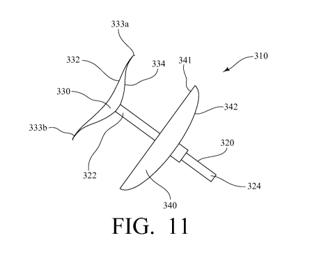
[0027] FIG. 11 is a side view of another atrial appendage closure device of
the presently-
disclosed subject matter, and showing the further device in a deployed
position; and
[0028] FIGS. 12A-12N are a series of schematic representations showing another
exemplary method of occluding a left atrial appendage in accordance with the
presently-
disclosed subject matter, in which the atrial appendage closure device of FIG.
11 is used to
provide a seal between the left atrial appendage and the left atrium of a
heart.
CA 02880030 2015-01-23
WO 2014/018907
PCT/US2013/052362
9
DESCRIPTION OF EXEMPLARY EMBODIMENTS
[0029] The presently-disclosed subject matter is an atrial appendage closure
device and,
more particularly, an atrial appendage closure device having an insertion rod
and an
occluding member, which, in a deployed position, is configured to occlude and
provide a seal
between a left atrial appendage and a left atrium of a heart.
[0030] Referring first to FIG. 1 and FIGS. 2A-2B, an exemplary atrial
appendage closure
device 10 made in accordance with the presently-disclosed subject matter
includes an
insertion rod 20 having a first end 22 and a second end 24. The atrial
appendage closure
device 10 further includes an occluding member 30 that is connected to the
first end 22 of the
insertion rod 20. The occluding member 30 includes an outer surface 32 and an
inner surface
34. Also included in the atrial appendage closure device 10 is an anchoring
member 40 in the
form of a bolt that is connected to or otherwise attached to the insertion rod
20 for securing
the device 10 to the left atrial appendage 100 of a heart 120.
[0031] With regard to the occluding member 30, and referring more specifically
now to
FIGS. 2A-2B, the occluding member 30 is moveable between a retracted position
and a
deployed position. As shown in FIG. 2A, in the retracted position, the
occluding member 30
is positioned adjacent to and is attached to the first end 22 of the insertion
rod 20, such that
the outer surface 32 and the inner surface 34 of the occluding member 30
extend along a
length of the insertion rod 20. However, as shown in FIG. 2B, in the deployed
position, the
insertion rod 20 assumes an umbrella-like shape with the outer surface 32 of
the occluding
member 30 assuming a concave shape and the inner surface 34 of the occluding
member 30
assuming a convex shape, such that the occluding member 30 is then configured
to provide a
seal between the left atrial appendage and the left atrium of the heart. Of
course, it is also
contemplated that, upon deployment, the outer surface and the inner surface of
the occluding
member can be constructed such that the surfaces of the occluding member
assume various
CA 02880030 2015-01-23
WO 2014/018907
PCT/US2013/052362
other shapes to accommodate the anatomy of a particular heart and/or to
accommodate a
desired application. For example, in certain embodiments, an occluding member
can include
an outer surface that is convex and an inner surface that is concave. As
another example, in a
further embodiment, an occluding member 230 of another exemplary atrial
appendage
closure device 210 includes an occluding member 230 that, upon deployment, has
a convex
outer surface 232 and a substantially flat inner surface 234, as shown in
FIGS. 7-9.
[0032] Regardless of the ultimate configuration of the occluding member upon
deployment, and as perhaps best shown in FIG. 2B, the occluding member 30 is
typically
comprised of a flexible material or membrane 38 that is supported by a
plurality of ribs 39
and/or a reinforcing mesh to thereby provide a flexible structure that is
capable of being
moved between a retracted and deployed position, but yet is still sufficiently
rigid such that
the occluding member 30 can provide an effective seal between the left atrial
appendage and
left atrium of a heart and will not collapse into the left atrial appendage
upon being exposed
to the blood flow in the heart and the pressure generated by the left atrium.
In some
embodiments, the occluding member is comprised of a plastic, a metal, a shape
memory
alloy, such as Nitinol, or combinations thereof
[0033] With regard to the insertion rod 20 of the exemplary atrial appendage
closure
device 10, the insertion rod 20 is in the form of a solid rod and is generally
constructed from
a metal or plastic material to provide an insertion rod having a sufficient
strength to allow it
to be inserted into the wall of an atrial appendage of a heart and retain its
shape. However, as
a refinement to the atrial appendage closure devices of the presently-
disclosed subject matter
and, in particular, to the insertion rods of the devices, in a further
embodiment, an atrial
appendage closure device 210 is provided where the insertion rod 220 defines a
hollow
interior cavity 272 and an opening 274 at the second end 224 of the insertion
rod, as shown
in FIGS. 7-9. The hollow insertion rod 220 further defines a plurality of
fenestrations 270
CA 02880030 2015-01-23
WO 2014/018907
PCT/US2013/052362
11
that are in fluid communication with the hollow interior cavity 272 and the
opening 274 at
the second end 224 of the insertion rod 220. In this regard, upon insertion of
the device
210, the deployment of the occluding member 230, and the securing of the inner
surface 234
and outer surface 232 of the occluding member 230 to provide a seal between
the left atrial
appendage 100 and the left atria of a heart, a vacuum can be applied to the
opening 274 and
the fenestrations 270 can be used to remove blood from the left atrial
appendage 100 while
also facilitating the collapsing of the left atrial appendage 100 against the
inner surface 234
of the occluding member 230, as shown best in FIGS. 7-8. Similarly, by making
use of the
opening 274 and the fenestrations 270, radio-opaque dyes can be injected
through the
insertion rod 220 to check the positioning of the device 210 and the integrity
of the seal
provided by the device 210.
[0034] Referring now to FIG. 10, to further facilitate the use of the atrial
appendage
closure device 210, the outer surface 232 of the occluding member 230 is
coated with an
extracellular matrix 238 and growth factors 237 using methods known to those
of ordinary
skill in the art. As used herein, the term "growth factor" is used to refer to
a substance
capable of stimulating cellular growth, proliferation, and cellular
differentiation. Such growth
factors include, but are not limited to, vascular endothelial growth factor
(VEGF), basic
fibroblast growth factor (bFGF), insulin-like growth factor (IGF), placental
growth factor
(PIGF), Angl, platelet derived growth factor-BB (PDGF-BB), and transforming
growth
factor 13 (TGF- p), and combinations thereof Of course, as would be recognized
by those of
ordinary skill in the art, various other materials and biological molecules
can be attached to
or used to coat a atrial appendage closure device of the presently-disclosed
subject matter,
and can be selected for a particular application as desired.
[0035] The term "extracellular matrix" is used herein to refer to the
extracellular network
of polysaccharides and proteins that typically serve as structural elements to
the cells and
CA 02880030 2015-01-23
WO 2014/018907
PCT/US2013/052362
12
tissues of a body and that provide a supporting and attachment surface for
epithelial cells. In
this regard, the term "extracellular matrix" is inclusive of the collection of
polysaccharides
and proteins that make up the extracellular matrix, but is further used to
refer to the
individual polysaccharides and proteins that make up the extracellular matrix,
as well as the
cells, such as fibroblasts and chondrocytes, that contribute to the
development of the
extracellular matrix. Exemplary polysaccharides and proteins of the
extracellular matrix
include, but are not limited to: proteoglycans, such as heparin sulfate,
chondroitin sulfate, and
keratin sulfate; non-proteoglycan polysaccharides, such as hyaluronic acid;
collagen; elastin;
fibronectin; and laminin.
[0036] Referring now to FIG. 6, by including an extracellular matrix (not
shown) on the
occluding member 30, the device 10 can be configured so as to promote
epithelialization or,
in other words, to promote the deposition of epithelial cells and the growth
of an epithelial
cell layer 36 over the surface of the device 10. In this regard, by promoting
epithelialization
over the device 10, the device 10 is kept from being directly exposed to the
circulating blood
within the heart of a subject and scar tissue formation, immune reactions, or
any other
adverse events commonly associated with the implantation of a foreign body
into a living
subject are thereby minimized or prevented. In some embodiments, the outer
surface of an
exemplary occluding member is coated with an extracellular matrix, growth
factors, or both
to promote epithelialization or the formation of an epithelial cell layer over
the entire surface
of the occluding member that is placed into direct contact with the left
atrium of a heart. Of
course, it is also contemplated that the inner surface of an exemplary
occluding member, or
any other portion of an exemplary atrial appendage closure device, can also be
coated with an
extracellular matrix or with growth factors without departing from the spirit
and scope of the
subject matter described herein. For example, with reference to FIGS. 3-6, it
is contemplated
that the insertion rod 20 of the device 10 can be coated with an extracellular
matrix such that,
CA 02880030 2015-01-23
WO 2014/018907
PCT/US2013/052362
13
upon insertion of the device 10, there is not an area of the device 10 where
cells are not able
to adhere or where a hole may be created between the device 10 and the
surrounding tissue,
which may then lead to blood stasis and blood clot formation.
[0037] Referring still to FIGS. 3-6, further provided by the presently-
disclosed subject
matter are methods of occluding a left atrial appendage that make use of the
exemplary atrial
appendage closure device 10 of the presently-disclosed subject matter. In one
exemplary
implementation of a method of occluding a left atrial appendage in accordance
with the
presently-disclosed subject matter, the atrial appendage closure device 10 is
first provided
and, while in a retracted position, the occluding member 30 and a portion of
the insertion rod
20 are inserted into the left atrial appendage 100 and the left atrium 110 by
piercing the wall
102 of the left atrial appendage 100 and then pushing the occluding member 30
and the
portion of the insertion rod 20 through the left atrial appendage 100 and into
the left atrium
110. By making use of an occluding member 30 that is moveable between a
retracted
position and a deployed position, the occluding member 30 of the device 10 can
advantageously be placed into its retracted position (shown in FIG. 2A) and
subsequently
inserted into the left atrial appendage of 100 of a heart by performing only a
minimally
invasive thoracotomy with a small (e.g., approximately 1 inch) incision,
similar to those used
in laparoscopic procedures. Of course, the device 10 can also be inserted as
part of other,
more invasive cardiac or vascular surgical procedures. However, without
wishing to be
bound by any particular theory, it is believed that by inserting the device 10
via a minimally
invasive thoracotomy, implantation complications are minimized, as small
thoracotomies
have been shown to have less complications than other procedures that involve
puncturing of
the atrial septum using catheter or guidewire techniques commonly utilized in
percutaneous
approaches.
CA 02880030 2015-01-23
WO 2014/018907
PCT/US2013/052362
14
[0038] Regardless of the particular surgical approach used to insert the
occluding
member 30 and the portion of the insertion rod 20, as shown in FIG. 3, once
the occluding
member 30 is inserted inside the left atrium 110, the occluding member 30 is
deployed such
that the outer surface 32 of the occluding member 30 assumes a concave shape,
the inner
surface 34 of the occluding member 30 assumes a convex shape, and the entirety
of the
occluding member 30 covers an area significantly beyond the opening of the
left atrial
appendage 100. Then, as shown in FIG. 4, while in a deployed position, the
occluding
member 30 and the remainder of the inserted portion of device 10 are pulled
towards the tip
104 of the left atrial appendage 100. As shown in FIG. 5, the left atrial
appendage 100 is
then collapsed, such as by using a vacuum or other mechanical force, and the
anchoring
member 40 is slid along the length of the insertion rod 20 and is attached to
the tip 104 of the
left atrial appendage 100 to thereby completely secure the inner surface 34 of
the occluding
member 30 against the wall 102 of the left atrial appendage 100 and to thereby
provide a
complete seal between the left atrial appendage 100 and the left atrium 110 of
the heart.
Lastly, a portion of the insertion rod 20, including the second end 24 of the
insertion rod 20 is
cut away or broken at a user-defined length adjacent to the anchoring member
40 (or can be
further filled with a filler to seal off the hollow interior if a hollow
insertion rod is used, such
as the hollow insertion rod shown in FIGS. 7-9).
[0039] Referring now to FIGS. 11 and 12A-12N, as a refinement to the atrial
appendage
closure devices and methods of the presently-disclosed subject matter, an
atrial appendage
closure device 310 is provided that, like the atrial appendage closure devices
10, 210 shown
in FIGS. 1-10, includes an insertion rod 320 having a first end 322 and a
second end 324.
The atrial appendage closure device 310 also includes an occluding member 330
having an
outer surface 332 and an inner surface 334, and an anchoring member 340 that
is connected
to or otherwise attached to the insertion rod 320 for securing the device 310
to a left atrial
CA 02880030 2015-01-23
WO 2014/018907
PCT/US2013/052362
appendage. Unlike the atrial appendage closure devices 10, 210 described
above, however,
the anchoring member 340 of the left atrial appendage closure device 310 is
not in the form
of a bolt, but rather has an umbrella-like shape, with the proximal surface
341 being
substantially flat and the distal surface 342 of the anchoring member 340
having a convex
shape. Additionally, the anchoring member 340 is movable between a retracted
position and
a deployed position, as best shown in FIG. 12H, and is configured to slide
along the length of
insertion rod 320. In this regard, upon the insertion of the device 310 and
the insertion of the
occluding member 330 into a heart to provide a seal between the left atrial
appendage and
the left atrium of the heart, the anchoring member 340 can be slid down the
insertion rod 320
and locked to thereby collapse the left atrial appendage 100, while the
increased surface area
of the anchoring member 340 provides the added benefit of preventing blood
from entering
into the left atrial appendage 300 and clotting even if the occluding member
330 inside the
left atrial appendage 100 does not completely seal off the left atrial
appendage 100 from the
left atrium 110.
[0040] Furthermore, with regard to the occluding member 330 of the left atrial
appendage
closure device 310, unlike the occluding members 30, 230 of the devices 10,
210, the
occluding member 330 includes a pair of hooked end portions 333a, 333b at
either end of the
outer surface 332 that assist in sealing off the left atrial appendage 300
from the left atria of a
heart. In this regard, it is contemplated that, in some embodiments, the
occluding member
330 can further include radiating members to provide horizontal stability and
circumferential
members to provide circumferential stability, and, in other embodiments, can
also have a
biconvex structure when fully expanded so that it seals of the left atrial
appendage while
displaying a slightly convex surface to the inside of the heart. Additionally,
it is contemplated
that the occluding member 330 can be injected with a liquid or gel to retain
its shape (e.g., a
liquid or gel that cures or solidifies after injection setting the shape), can
have a membrane or
CA 02880030 2015-01-23
WO 2014/018907
PCT/US2013/052362
16
structure that is textured (e.g., roughened, flecked, or sintered) to promote
formation of a
native lining to minimize thromboembolic events, or can be covered with a
fabric or
polymeric material to promote tissue ingrowth.
[0041] In use, the atrial appendage closure device 310 is generally used in a
method of
occluding a left atrial appendage by first providing and inserting a large
bore needle 500
through the wall 102 of a left atrial appendage 100, as shown in FIGS. 12A and
12B,
respectively. The portion of the atrial appendage closure device 310 that
includes only the
insertion rod 320 and the occluding member 330 is then provided and the
occluding member
330 is placed in a retracted position, as shown in FIG. 12C. The retracted
occluding member
330 is then inserted through the large bore needle 500 along with the first
end 322 of the
insertion rod 320 until the occluding member 330 is sufficiently placed in the
left atrium 110,
as shown in FIGS. 12D-12F. The occluding member 330 is then subsequently
deployed such
that the outer surface 332 of the occluding member 330 assumes a convex shape,
the inner
surface 334 of the occluding member 330 assumes a concave shape, and the
hooked end
portions 333a, 333b at either end of the outer surface 332 further seal off
the left atrial
appendage 100 from the left atrium 110, as shown in FIG. 12G. Upon the
placement of the
occluding member 330, the large bore needle 500 is then retracted from the
wall 102 of the
left atrial appendage 100, as shown in FIG. 12G.
[0042] Subsequent to retracting the needle 500 from the wall 102 of the left
atrial
appendage 100, the anchoring member 340 is then provided and placed in a
retracted
position, as shown in FIG. 12H. The anchoring member 340 is then placed onto
the second
end 324 of the insertion rod 320 and is slid along the length of the insertion
rod 320, as
shown in FIGS. 12I-12J. Upon exiting the large bore needle 500, the anchoring
member 340
is then re-deployed until the proximal surface 341 is substantially flat and
the distal surface
342 of the anchoring member 340 has a convex shape, as shown in FIGS. 12K-12L.
By
CA 02880030 2015-01-23
WO 2014/018907
PCT/US2013/052362
17
pushing the anchoring member against the wall 102 of the left atrial appendage
100 opposite
the inner surface 334 of the occluding member 330, the atrial appendage 100 is
then
collapsed and the large bore needle 500 is removed from the insertion rod 320,
as shown in
FIG. 12M. The insertion rod 320 is then cut away or otherwise broken adjacent
to the
anchoring member 340 to finish the occlusion of the left atrial appendage 100.
[0043] The above-described atrial appendage closure devices and related
methods of
occluding an atrial appendage, which allow for a left atrial appendage of a
heart to be
completely sealed off from the left atrium, are important both for preventing
clot formation
that may otherwise occur with atrial fibrillation and for minimizing surgery-
related
complications' that frequently occur in left atrial appendage occlusion
therapy. Further, the
devices of the presently-disclosed subject matter minimize the risk of
puncturing portions of
a heart during surgical placement as no barbs or similar anchoring mechanisms
are inserted
into the inside of the left atrial appendage. Moreover, the devices of the
presently-disclosed
subject matter can be provided in one size to thereby eliminate any patient-
to-patient
variability that is often observed with current atrial appendage closure
devices and, in
particular, pharmaceutical agent dosing. Thus, the atrial appendage closure
devices of the
presently-disclosed subject matter provide not only desirable alternatives to
current device-
or pharmaceutical agent-based therapies, with the added benefit that
complications arising
from the implantation of the device are minimized.
[0044] The presently-disclosed subject matter is further illustrated by the
following
specific but non-limiting examples. Some of the following examples are
prophetic,
notwithstanding the numerical values, results and/or data referred to and
contained in the
examples. Additionally, certain of the following examples may include
compilations of data
that are representative of data gathered at various times during the course of
development and
experimentation related to the presently-disclosed subject matter.
CA 02880030 2015-01-23
WO 2014/018907
PCT/US2013/052362
18
EXAMPLES
[0045] Example 1 - Prototyping and preliminary testing of atrial appendage
closure
device designs in pig hearts.
[0046] Eight candidate atrial appendage closure device designs are fabricated
at the
University of Louisville prototyping center for evaluation in pig hearts. Pig
hearts are
procured from slaughterhouses (Swift Slaughterhouse, Louisville) and the
prototype devices
are implanted. The atria of the porcine hearts are cut open and the efficacy
of the prototype
designs to completely occlude the left atrial appendage, the force needed to
pull out the atrial
appendage closure device (anchoring force), and the ease of deployment are
evaluated.
[0047] Upon analysis of the results from these experiments, it is observed
that the
candidate designs provide complete occlusion of the left atrial appendage,
with a pull out
force greater than 6N (i.e., approximately the same as a suture), and are
easily deployed into
the a left atrial appendage. The most promising candidate designs are then
selected for
cadaver testing and animal experiments.
[0048] Example 2 - Cadaver fit study.
[0049] An anatomical fit study is performed in human cadavers (45-120 kg,
n=4). The
candidate designs are implanted using a thoracoscopic procedure. Three ports
are positioned:
1 (5 mm) in the third intercostal space, 1 (10 mm) in the sixth space at the
median axillary
level, and 1 (10 mm) in the fifth space on the posterior axillary line. A
pericardiotomy is
performed parallel and posterior to the phrenic pedicle to expose the left
atrial appendage.
The Marshall ligament is then interrupted with electrocaudurization. Through
the inferior
port, the left atrial access retainer is subsequently activated with immediate
step insertion and
deployment of the occluding member of the device under transesophagial
echocardiography
guidance. The duration, ease of use, and complexity of the device is then
compared with
CA 02880030 2015-01-23
WO 2014/018907
PCT/US2013/052362
19
catheter-based left atrial appendage occlusion devices, and the anatomical
positioning, fit and
ease of occluding member deployment, and anchoring force of the devices is
further
evaluated.
[0050] Upon analysis of the results from these experiments, it is observed
that the atrial
closure devices: (1) provide complete occlusion of the left atrial appendage
with a pull out
force of greater than 6N; (2) can be implanted in less than 90 minutes; and
(3) are rated by
the surgeon as being considerably easier to insert and manipulate as compared
to current
catheter-based left atrial appendage occlusion techniques.
[0051] Example 3 - Acute Animal Study Surgical Procedures.
[0052] To begin the acute animal studies, test animals (60-100 kg, male,
Jersey calves)
first undergo a 14-day quarantine period. Then, the animals are anesthetized
with 1-5%
isoflurane and 100% oxygen, and a left thoracotomy is performed at the 5th
intercostal space
to provide access and exposure of the left atrial appendage. Heparin (200-300
units/kg via IV
central line) is administered and the atrial appendage closure device of the
presently-
disclosed subject matter is implanted as described herein above.
Echocardiography is
performed on each calf to verify anatomical positioning and fit of the closure
device.
Fluoroscopy is also performed to confirm anatomical positioning of the closure
device during
implantation. In this regard, a vascular sheath is placed in the carotid
artery, and an
angiography catheter may be placed in the left atrium for injection of
radiopaque dye (100-
150 mL, which may be repeated 3-5 times) for flow visualization during
fluoroscopy. After
the evaluation period, at necropsy, full gross examination of end organs is
completed, with
particular attention on the left atrial appendage area, where the device is
further visually
inspected for fit, positioning, and evidence of clots or defects.
CA 02880030 2015-01-23
WO 2014/018907
PCT/US2013/052362
[0053] Upon analysis of the results from these studies, it is again observed
that the
devices of the presently-disclosed subject matter: (1) provide complete
occlusion of left atrial
appendage with a pull out force of greater than 6N; (2) can be implanted in
less than 90
minutes; (3) are rated by the surgeon as being considerably easier to insert
and manipulate as
compared to current catheter-based left atrial appendage occlusion techniques;
and (4) allow
for blood loss to be less than 100 ml during implantation.
[0054] Example 4 - Chronic Animal Study Surgical Procedures.
[0055] For the chronic animal studies using the devices of the presently-
disclosed subject
matter, the quarantine, anesthesia, and implantation techniques used in the
acute studies are
again employed to place the device in the left atrial appendage of the test
animals (60-100 kg,
male, Jersey calves). In the chronic animal studies, echocardiography and
fluoroscopy are
performed at the beginning and end of the 14-day chronic study period.
Histopathological
analyses are performed on the device surface and the left atrial appendage to
quantify
endothelialization of device surface, tissue ingrowth, and device-related
injury
[0056] Upon analyzing the results from the chronic animal studies, it is
observed that the
devices of the presently-disclosed subject matter: (1) provide complete
occlusion of the left
atrial appendage with a pull out force greater than 6N without device fracture
or failure; (2)
are capable of being implanted in less than 90 minutes; (3) are rated by the
surgeon as being
considerably easier to insert and manipulate as compared to current catheter-
based left atrial
appendage occlusion techniques; (4) allow for blood loss to be less than 100
ml during
implantation; (5) exhibit no blood leaks at the device-left atrial appendage
junction over the
duration of the study; (6) show no visible device migration; (7) result in no
visible injury to
the myocardium or embolization in the end-organs in of the animals; and (8)
allow for full
endothelialization of the device surface with no or minimal histopathological
damage to the
CA 02880030 2015-01-23
WO 2014/018907
PCT/US2013/052362
21
myocardium, thus indicating that the devices of the presently-disclosed
subject matter can
effectively be used as part of a method for occluding the left atrial
appendage of a heart.
[0057] Throughout this document, various references are mentioned. All such
references
are incorporated herein by reference, including the references set forth in
the following list:
REFERENCES
1. Atrial Fibrillation Fact Sheet. February 2010. Centers for Disease Control
and Prevention.
3 Apr. 2012.
2. Go AS, Hylek EM, Phillips KA, Chang Y, Henault LE, Shelby IV and Singer DE.
Prevalence of Diagnosed Atrial Fibrillation in Adults: National Implications
for Rhythm
Management and Stroke Prevention: the Anticoagulation and Risk Factors in
Atrial
Fibrillation (Atria) Study. Journal of the American Medical Association
2001;285:2370-
2375.
3. Wolf PA, Abbott RD, and Kannel WB. Atrial fibrillation as an Independent
factor for
stroke: The Framingham study. Stroke; 1991; 22:983-988.
4. Pearce LA, Hart RG and Halperin M. Assessment of Three Schemes for
Stratifying
Stroke Risk in Patients with Nonvalyular Atrial Fibrillation. The American
Journal of
Medicine 2000;109:45-51.
5. Aronow WS. Management of the Older Person With Atrial Fibrillation. Journal
of
Gerontology: Medical Sciences 2002;57A:M352-M363.
6. Savelieva I, Bajpai A and Camm AJ. Stroke in atrial fibrillation: Update on
pathophysiology, new antithrombotic therapies, and evolution of procedures and
devices.
Annals of Medicine 2007;39:371-391.
7. Jais P, Haissaguerre M, Shah DC, Chouairi S, Gencel L, Hocini M, and
Clementy J. A
CA 02880030 2015-01-23
WO 2014/018907
PCT/US2013/052362
22
Focal Source of Atrial Fibrillation Treated by Discrete Radiofrequency
Ablation.
Circulation. 1997;95:572-576.
8. Go AS, Hylek EM, Borowsky LH, Phillips KA, Selby TV and Singer DE. Warfarin
Use
among Ambulatory Patients with Nonvalvular Atrial Fibrillation: The
Anticoagulation
and Risk Factors in Atrial Fibrillation (Atria) Study. Annals of Internal
Medicine
1999;131:927-934.
9. Mendelson G, and Aronow WS. Underutilization of Warfarin in Older Persons
with
Chronic Nonvalvular Atrial Fibrillation at High Risk for Developing Stroke.
Journal of
the American Geriatrics Society 1998;46:P1423-P1424.
10. Hart RG, Benavente 0, McBride R and Pearce LA. Antithrombotic Therapy To
Prevent
Stroke in Patients with Atrial Fibrillation: A Meta-Analysis. Annals of
Internal Medicine
1999;131:492-501.
11. Hylek EM, Go AS, Chang Y, Jensvold NG, Henault LE, Selby TV, et al. Effect
of
intensity of oral anticoagulation on stroke severity and mortality in atrial
fibrillation. N
Engl J Med. 2003;349:1019-26.
12. Nieuwlaat R, Capucci A, Lip GY, Olsson SB, Prins MH, Nieman FH, et al.;
Euro Heart
Survey Investigators. Antithrombotic treatment in real-life atrial
fibrillation patients: a
report from the Euro Heart Survey on Atrial Fibrillation. Eur Heart J.
2006;27:3018-26.
13. McCormick D, Gurwitz JH, Goldberg RJ, Becker R, Tate JP, Elwell A, et al.
Prevalence
and quality of warfarin use for patients with atrial fibrillation in the long-
term care
setting. Arch Intern Med. 2001;16:2458-63.
14. Sarawate C, Sikirica MV, Willey VJ, Bullano MF, Hauch 0. Monitoring
anticoagulation
in atrial fibrillation. J Thromb Thrombolysis. 2006;21:191-8.
15. Singer DE, Albers GW, Dalen JE, Go AS, Halperin JL, and Manning WJ.
Antithrombotic
therapy in atrial fibrillation: the Seventh ACCP Conference on Antithrombotic
and
Thrombolytic Therapy. Chest 2004;126:429S-56S.
CA 02880030 2015-01-23
WO 2014/018907
PCT/US2013/052362
23
16. Risk factors for stroke and efficacy of antithrombotic therapy in atrial
fibrillation.
Analysis of pooled data from five randomized controlled trials. Archives of
Internal
Medicine 1994;154:1449-57.
17. Atwood JE, and Albers GW. Anticoagulation and atrial fibrillation. Herz
1993;18:27-38
18. Gullov AL, Koefoed BG, and Petersen P. Bleeding During Warfarin and
Aspirin Therapy
in Patients With Atrial Fibrillation: The Afasak 2 Study. Archives of Internal
Medicine
1999;159:1322-1328.
19. Liu M, Counsell C, Sandercock P. Anticoagulants for preventing recurrence
following
ischaemic stroke or transient ischaemic attack. (Cochrane Review). In: The
Cochrane
Library, Issue 1, 2002. Oxford: Update Software.
20. Desbiens DA. Deciding on Anticoagulating the Oldest Old with Atrial
Fibrillation:
Insights from Cost-Effectiveness Analysis. JAGS 2002;50:863-869.
21. Aronow WS, Ahn C, Kronzon I, and Gutstein H. Incidence of new
thromboembolic
stroke in persons 62 years and older with chronic atrial fibrillation treated
with warfarin
versus aspirin. Journal of the American Geriatrics Society 1999;47:366-8.
22. Lip GY. Aspirin for Prevention of Stroke in Atrial Fibrillation. Stroke
2006;37:1640.
23. Garcia D, and Hylek E. Stroke prevention in elderly patients with atrial
fibrillation. The
Lancet 2007;370:460-461.
24. Lip GYH and Boos CJ. Antithrombotic treatment in atrial fibrillation.
Heart
2006;92:155-161.
25. Jailer AK. Warfarin reduced major stroke more than aspirin in elderly
patients with atrial
fibrillation in primary care. Evidence Based Medicine 2007;12:172.
26. Kamath S, Blann AD, Chin BS, Lip GY. A prospective randomized trial of
aspirin-
CA 02880030 2015-01-23
WO 2014/018907
PCT/US2013/052362
24
clopidogrel combination therapy and dose-adjusted warfarin on indices of
thrombogenesis and platelet activation in atrial fibrillation. J Am Coll
Cardiol.
2002;40:484-90.
27. Lorenzoni R, Lazzerini G, Cocci F, De Caterina R. Shortterm prevention of
thromboembolic complications in patients with atrial fibrillation with aspirin
plus
clopidogrel: the Clopidogrel-Aspirin Atrial Fibrillation (CLAAF) pilot study.
Am Heart J.
2004;148:e6.
28. ACTIVE Writing Group on behalf of the ACTIVE Investigators; Connolly S,
Pogue J,
Hart R, Pfeffer M, Hohnloser S, Chrolavicius S, et al. Clopidogrel plus
aspirin versus oral
anticoagulation for atrial fibrillation in the Atrial fibrillation Clopidogrel
Trial with
Irbesartan for prevention of Vascular Events (ACTIVE W): a randomized
controlled trial.
Lancet. 2006;367:1903-12.
29. Healey J, Hart R, Pogue J, Yusuf S, Pfeffer M, Hohnloser S, et al., on
behalf of The
ACTIVE-W Investigators. Effect of underlying risk of stroke on treatment
effects in the
ACTIVEW Trial. (Abstract). Eur Heart J. 2006;27 Supplement: Abstract P451.
30. Perzborn E, Roehrig S, Straub, A, Dagmar K, Mueck W, and Laux V.
Rivaroxaban: A
new oral factor Xa inhibitor. Arteriosclerosis, Thrombosis, and Vascular
Biology.
2010;30:376-381
31. Eriksson B, Quinlan D, Weitz J. Comparative Pharmacodynamics and
Pharmacokinetics
of oral direct thrombin and Factor Xa inhibitors in development. Clinical
Pharmacokinetics 2009: 48: 1-22.
32. Bayard YL, Ostermayer SH, Hein R, Skowasch M, Buscheck F, Baranowski A,
Heinisch
C, Sievert H. Percutaneous devices for stroke prevention. Cardiovascular
Revascularization Medicine. 2007:8:216-225.
33. Hanna IR, Kolm P, Martin R, Reisman M, Gray W and Block PC. Left atrial
structure
and function after percutaneous left atrial appendage transcatheter occlusion
(PLAATO):
Six-month echocardiographic follow-up. Journal of the American College of
Cardiology
CA 02880030 2015-01-23
WO 2014/018907
PCT/US2013/052362
2004;43:1868-72.
34. Nakai T, Lesh MD, Gerstenfeld EP, Virmani R, Jones R and Lee RJ.
Percutaneous Left
Atrial Appendage Occlusion (PLAATO) for Preventing Cardioembolism: First
Experience in Canine Model. Circulation 2002;105:2217-2222.
35. Ostermayer SH, Reisman M, Kramer PH, Matthews RV, Gray WA, Block PC, Omran
H,
Bartorelli AL, Bella PD, Mario CD, Pappone C, Casale PN, Moses JW, Poppas A,
Williams DO, Meier B, Skanes A, Teirstein PS, Lesh MD, Nakai T, Bayard Y,
Billinger
K, Trepels T, Krumsdorf U, and Sievert H. Percutaneous Left Atrial Appendage
Transcatheter Occlusion (PLAATO System) to Prevent Stroke in High-Risk
Patients
With Non-Rheumatic Atrial Fibrillation: Results From the International Multi-
Center
Feasibility Trials. Journal of the American College of Cardiology 2005;46:9-
14.
36. Sievert H, Lesh MD, Trepels T, Omran H, Bartorelli A, Bella PD, Nakai T,
Reisman M,
DiMario C, Block P, Kramer P, Fleschenberg D, Krumsdorf U, and Scherer D.
Percutaneous Left Atrial Appendage Transcatheter Occlusion to Prevent Stroke
in High-
Risk Patients With Atrial Fibrillation: Early Clinical Experience. Circulation
2002;105:1887-1889.
37. Fountain RB, Holmes DR, Chandrasekaran K, Packer D, Asirvatham S, Tassel
RV and
Turi Z. The Protect AF (Watchman Left Atrial Appendage System for Embolic
Protection in Patients with Atrial Fibrillation) Trial. American Heart Journal
2006;151:956-61.
38. Sick PB, Schuler G, Hauptmann KE, Grube E, Yakubov S, Turi ZG, Mishkel G,
Almany
S, and Holmes DR. Initial Worldwide Experience with the WATCHMAN Left Atrial
Appendage System for Stroke Prevention in Atrial Fibrillation. Journal of the
American
College of Cardiology 2007;49:1490-5.
39. Sievert H and Bayard YL. Percutaneous closure of the left atrial
appendage: A major step
forward. J Am Coll Cardiol Intv, 2009; 2:601-602.
40. Block PC. Watching the WATCHMAN. J Am Coll Cardiol, 2007; 49:1496-1497.
CA 02880030 2015-01-23
WO 2014/018907
PCT/US2013/052362
26
41. Maisel WH. Left atrial appendage occlusion - closure or just the
beginning. New England
Journal of Medicine, 2009.
42. Sick PB, Schuler G, Hauptmann KE, et al (April 2007). "Initial worldwide
experience
with the WATCHMAN left atrial appendage system for stroke prevention in atrial
fibrillation". J Am. Coll. Cardiol. 49 (13):1490-5
43. Onalan 0 and Crystal E. Left Atrial Appendage Exclusion for Stroke
Prevention in
Patients With Nonrheumatic Atrial Fibrillation. Stroke 2007;38:624-630.
44. Ailawadi G, Gerdisch MW, Harvey RL, Hooker RL, Damiano RJ Jr, Salamon T,
and
Mack MJ. Exclusion of the left atrial appendage with a novel device: early
results of a
multicenter trial. J Thorac Cardiovasc Surg. 2011;142(5):1002-9.
45. Payne KA, Huybrechts KF, Caro JJ, Craig Green TJ, Klittich WS. Long term
cost-of-
illness in stroke: an international review. Pharmacoeconomics. 2002;20:813-25.
46. Lafata JE, Martin SA, Kaatz S, Ward RE. The cost effectiveness of
different management
strategies for patients on chronic warfarin therapy. J Gen Intern Med.
2000;15:31-7.
[0058] It will be understood that various details of the presently disclosed
subject matter
can be changed without departing from the scope of the subject matter
disclosed herein.
Furthermore, the foregoing description is for the purpose of illustration
only, and not for the
purpose of limitation.