Note: Descriptions are shown in the official language in which they were submitted.
CA 02880110 2015-01-26
WO 2014/020150
PCT/EP2013/066275
ENDOSCOPIC BIOPSY INSTRUMENT, ENDOSCOPE, AND METHOD FOR
TAKING A BIOPSY SAMPLE
Technical Field of the Invention
The present invention relates to an endoscopic biopsy instrument, to
an endoscope comprising such an endoscopic biopsy instrument, as well as
to a method for taking a biopsy sample from a tissue of a subject.
Background Art
When evaluating various lesions and tumours, tissue samples may be
acquired using a biopsy instrument. If a suspected lesion or tumour is located
in or adjacent the gastrointestinal tract, an endoscopic biopsy instrument may
be used. An example of an endoscopic biopsy instrument is described in
US-5,865,724. This endoscopic biopsy instrument is generally comprised of a
forceps arranged at a distal end of two control wires, and a handle arranged
at a proximal end of the control wires, the control wires being enclosed in a
plastic sheath. When taking a tissue sample with this type of endoscopic bi-
opsy instrument, the wires in their sheath are inserted in a working channel
of
an endoscope, and the forceps are advanced to the site in the gastrointestinal
tract where the sample is to be taken. By operating the handle of the endo-
scopic biopsy instrument, the forceps may be manoeuvred to scoop out a
sample from the surface of the tissue.
For some diagnostic purposes the millimetre-sized sample thus re-
trievable is sufficient. However, for some types of lesions and tumours, such
a
small and superficial sample is inadequate for making a diagnosis. This is the
case, e.g., for submucosal tumours, such as gastrointestinal stromal tumours
(GIST). Since these tumours are located beneath the mucosa of the stomach
or intestines, the forceps of the above-mentioned type of endoscopic biopsy
instrument cannot reach into the tumour and retrieve a sufficiently large sam-
ple. Therefore, in order to get a sample that makes it possible to diagnose a
submucosal tumour, it is often necessary to retrieve the sample surgically.
Unfortunately, the fact that a patient has had surgery for diagnostic purposes
increases the risk of complications, and reduces the survival rate, even if a
malignant tumour is removed during subsequent therapeutic surgery. Thus, a
need for an improved endoscopic biopsy instrument, which makes it possible
2
to take larger samples and/or samples at a larger depth, e.g., beneath the
mucosa of the
gastrointestinal tract, remains.
US 2009/0118641 (to Van Dam et al.) disclosed a biopsy device which may have a
collection element comprising a helically shaped rod.
WO 2011/104692 (to Nevo) discloses a cryogenic biopsy instrument that
comprises a
helical collection element adapted to house a cryogenic needle.
US 2012/0197157 (to Ryan et al.) discloses a biopsy device which comprises a
coil for
collecting a tissue sample.
Summary of the Invention
It is an object of the invention to provide an endoscopic biopsy instrument,
which
overcomes the problems described above.
It is also an object of the invention to provide endoscope which overcomes
these problems.
A further object of the invention is to provide a method for taking a biopsy
sample from a
tissue of a subject, which overcomes the problems described above.
According to a first aspect of the invention, these and other objects are
achieved, in full
or at least in part, by an endoscopic biopsy instrument comprising:
a guide wire arranged in a sheath,
a drill device arranged at a first end of said guide wire, and
an actuator for actuating said drill device, said actuator being arranged at a
second end
of said guide wire,
wherein said drill device comprises an outer tube and an inner cutting device,
said inner
cutting device being slidable and rotatable inside said outer tube, said inner
cutting device
having a helical cutting edge. With such an endoscopic biopsy instrument, it
is possible to
take a biopsy.sample at a greater depth as compared to prior art instruments.
Further, a larger
sample may be taken. Especially for investigating submucosal tumours, it is
important to be
able to take the sample at a greater depth, since otherwise it may not be
possible to reach the
tumour, through the overlying tissue. For other types of tumours, it is useful
to get a larger
sample, since this may provide more diagnostic material.
The sheath may have a diameter of 1-5 mm in order to fit in a working channel
of an
endoscope. The diameter of the drill device may be adapted to the diameter of
the sheath, and
may be 0.5-4 mm, generally 1-2 mm.
In an embodiment, the outer tube is advancable over the inner cutting device
on an
outside of said inner cutting device. Thereby, the outer tube may
CA 2880110 2019-06-13
CA 02880110 2015-01-26
WO 2014/020150
PCT/EP2013/066275
3
be advanced over the inner cutting to enclose the sample cut out from the
tissue.
The inner cutting device of the drill device may comprise an inner core,
said helical cutting edge being formed on a helical flange surrounding said
inner core. In this manner, a rigid inner cutting device may be achieved,
which
may penetrate even tough tissues, such as the mucosa of the gastrointestinal
tract.
The inner cutting device may be made of metal. Metal is readily machi-
nable to the desired helical shape and may readily be sharpened to present a
cutting edge. Metal is particularly advantageous if the endoscopic biopsy in-
strument is used in an endoscope comprising an ultrasound probe, since
metal is visible in an ultrasonogram. Thus, the operator may, in an image ac-
quired by the ultrasound probe, for instance see how deep into the tissue the
inner cutting device has penetrated.
In an embodiment, the outer tube of the drill device has a cutting front
edge. In this manner, the sample may even more securely be cut out from the
tissue, should it not be completely cut off by the helical cutting edge of the
inner cutting device.
At the second end, the guide wire may comprise an outside thread,
and the actuator may comprise a rotatable portion having an inside thread,
the inside thread being engagable with the outside thread of the guide wire
for
advancing the guide wire inside said sheath. In this manner, an easily ma-
noeuvrable guide wire advancing device may be achieved. In one embodi-
ment, the operator may advance the guide wire by simply rotating the ro-
tatable portion between his or her thumb and index finger.
Alternatively, at the second end, the guide wire may comprise a
teethed portion, and the actuator may comprise a toothed gear, the toothed
gear being engagable with the teethed portion of the guide wire for advancing
the guide wire inside the sheath. This is another way of achieving an easily
manoeuvrable guide wire advancing device. In one embodiment, the operator
may advance the guide wire by simply rotating the toothed gear with his or
her index finger.
CA 02880110 2015-01-26
WO 2014/020150
PCT/EP2013/066275
4
The actuator may comprise a plunger arranged to advance said outer
tube of said drill device on an outside of said inner cutting device. In this
manner, an easily manoeuvrable outer tube advancing device may be
achieved.
The actuator may comprise a second rotatable portion arranged to ad-
vance said outer tube of said drill device on an outside of said inner cutting
device. This is another way of obtaining an easily manoeuvrable outer tube
advancing device.
According to a second aspect of the invention, these and other objects
are achieved, in full or at least in part, by an endoscope comprising:
an endoscopic insertion tube,
an imaging device arranged in said endoscopic insertion tube,
an endoscopic biopsy instrument of the invention arranged in said en-
doscopic insertion tube. With such an endoscope, the same advantages may
.. be achieved as with the endoscopic biopsy instrument according to the first
aspect of the invention. In the endoscope, the endoscopic biopsy instrument
may be embodied in the same ways as the endoscopic biopsy instrument ac-
cording to the first aspect of the invention, with the same advantages.
According to an embodiment, the imaging device is an ultrasound
probe. Ultrasonic imaging is particularly advantageous for evaluating submu-
cous tumours.
According to a third aspect of the invention, these and other objects
are achieved, in full or at least in part, by a method for taking a biopsy
sample
from a tissue of a subject, comprising:
providing an endoscopic biopsy instrument comprising a guide wire ar-
ranged in a sheath, a drill device arranged at a first end of said guide wire,
and an actuator for actuating said drill device, said actuator being arranged
at
a second end of said guide wire, wherein said drill device comprises an outer
tube and an inner cutting device, said inner cutting device being slidable and
.. rotatable inside said outer tube, said inner cutting device having a
helical cut-
ting edge,
inserting said first end of said guide wire into a body cavity of said sub-
ject,
CA 02880110 2015-01-26
WO 2014/020150
PCT/EP2013/066275
advancing said guide wire until said drill device is applied to a surface
of the tissue from which the biopsy sample is to be taken,
advancing said guide wire inside said sheath such that said inner cut-
ting device of said drill device is rotatingly bored into said tissue, such
that the
5 biopsy sample is cut out from said tissue,
enclosing said biopsy sample in said outer tube by a translational
movement of said outer tube in relation to said inner cutting device, and
retrieving said biopsy sample by withdrawing said first end of said
guide wire from said body cavity. With such a method, it is possible to take a
biopsy sample from a greater depth as compared to prior art methods. It is
also possible to take a larger sample.
The body cavity may be part of the gastrointestinal tract of the subject.
In a variant of the method, the outer tube is advanced over the inner
cutting device on an outside of the inner cutting device. In this way, the sam-
pie cut out by the inner cutting device is enclosed by the outer tube.
The endoscopic biopsy instrument may be inserted in an endoscopic
insertion tube of an endoscope, said endoscope comprising an imaging de-
vice arranged in said endoscopic insertion tube. In this way, the endoscopic
biopsy instrument may be securely guided to the site where the biopsy sam-
pie is to be taken.
Generally, the method of the invention may be varied in accordance
with the different embodiments of the first aspect of the invention, with the
same accompanying advantages.
Other objectives, features and advantages of the present invention will
appear from the following detailed disclosure, from the attached claims, as
well as from the drawings. It is noted that the invention relates to all
possible
combinations of features.
Generally, all terms used in the claims are to be interpreted according
to their ordinary meaning in the technical field, unless explicitly defined
other-
wise herein. All references to "a/an/the [element, device, component, means,
step, etc.]" are to be interpreted openly as referring to at least one
instance of
said element, device, component, means, step, etc., unless explicitly stated
otherwise. The steps of any method disclosed herein do not have to be per-
formed in the exact order disclosed, unless explicitly stated.
CA 02880110 2015-01-26
WO 2014/020150
PCT/EP2013/066275
6
As used herein, the term "comprising" and variations of that term are
not intended to exclude other additives, components, integers or steps.
Brief Description of the Drawings
The invention will be described in more detail with reference to the ap-
pended schematic drawings, which show an example of a presently preferred
embodiment of the invention.
Fig. 1 is a cross sectional view of an endoscopic biopsy instrument ac-
cording to an embodiment.
Fig. 2 is a detail view of an actuator of the endoscopic biopsy instru-
ment of Fig. 1.
Fig. 3 is a cross sectional view of an endoscopic biopsy instrument ac-
cording to a second embodiment.
Fig. 4 is a detail view of an inner cutting device of the endoscopic bi-
opsy instrument of Fig. 1 or Fig. 3.
Fig. 5 is a cross sectional view showing use of an endoscope accord-
ing to an embodiment.
Fig. 6 is a detail view of an actuator of an endoscopic biopsy instru-
ment according to a third embodiment in a first position.
Fig. 7 is a detail view of the actuator of Fig. 6 in a second position.
Detailed Description of Preferred Embodiments of the Invention
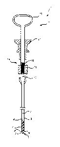
In Fig. 1, an endoscopic biopsy instrument 1 is shown. The endoscopic
biopsy instrument 1 comprises a guide wire 2 arranged in a sheath 3. At a
first, distal end 4 of the guide wire 2, a drill device 5 is arranged. The
drill de-
vice 5 comprises an outer tube 6 and an inner cutting device 7. The inner cut-
ting device 7 is slidable and rotatable inside the outer tube 6, and has a
heli-
cal cutting edge 8. The outer tube 6 is cylindrical and has a straight edge 9.
At a second, proximal end 10 of the guide wire 2, an actuator 11 com-
prising a handle 12 and a plunger 13 is arranged. The actuator 11 further
comprises a guide wire advancing device 14. The guide wire advancing de-
vice comprises a threaded portion 15 at the proximal end of the guide wire 2
having an outside thread, and a rotatable portion 16 or nut having an inside
thread. The threaded portion 15 of the guide wire 2 is engagable with the nut
CA 02880110 2015-01-26
WO 2014/020150
PCT/EP2013/066275
7
16, such that rotation of the nut 16 causes the guide wire to rotate and move
in its longitudinal direction, thus moving along the inside of the sheath 3.
As may be seen more clearly in Fig. 4, the inner cutting device 7 has
an inner core 17 surrounded by a helical flange 18. The helical cutting edge 8
of the inner cutting device 7 is formed on the helical flange 18. The helical
flange 18 forms more than one complete winding or turn around the inner
core 17. At the distal end 19 of the inner cutting device 7 a hook or
anchoring
portion 20 is formed on the helical flange 18.
With reference to Fig. 2, the actuator 11 further comprises a housing
21 in which the wire, via the nut 16, is retained at retaining points 22, and
a
sheath advancing device 23 in the form of bars 24 attached to the plunger 13.
Abutment portions 25 of the bars abut a cut-out edge of the sheath 3, such
that when the plunger 13 is depressed, the bars 23, via the abutment portions
24, push the sheath 3 outwardly from the housing 21. Thereby, the sheath 3
advances the outer tube 6 of the drill device 5 over the inner cutting device
7
on an outside of the inner cutting device 5.
In Fig. 3, an endoscopic biopsy instrument 101 according to a second
embodiment is shown. Except for the actuator 111, the endoscopic biopsy
instrument 101 in Fig. 3 has the same structure as the endoscopic biopsy in-
strument in Fig. 1. Like parts are in the embodiment in Fig. 3 marked with the
same reference numerals as used in Fig. 1, but with the addition of 100 on
each reference numeral.
Thus, the endoscopic biopsy instrument 101 comprises a guide wire
102 arranged in a sheath 103. At a distal end 104 of the guide wire 102 a
drill
device 105 of the same construction as the drill device 5 shown in Fig. 1 is
arranged. The inner cutting device 107 is of the same construction as that
shown in detail in Fig. 4. At the proximal end 110 of the guide wire 102, an
actuator 111 is arranged. The actuator 111 comprises a handle 112, a
plunger 113, and a guide wire advancing device 114. Different from the em-
bodiment shown in Figs. 1 and 2, the guide wire advancing device 114 com-
prises a teethed portion 115 at the proximal end of the guide wire 102, and a
toothed gear 116, which is engagable with the teethed portion 115 of the
guide wire 102. By rotating the toothed gear 116, the guide wire 102 may be
CA 02880110 2015-01-26
WO 2014/020150
PCT/EP2013/066275
8
rotated and advanced in its longitudinal direction, thus moving along the in-
side of the sheath 103.
In Fig. 6, an endoscopic biopsy instrument 201 according to a third
embodiment is shown. Except for the actuator 211, the endoscopic biopsy
instrument 201 in Fig. 6 has the same structure as the endoscopic biopsy in-
strument in Fig. 1. Like parts are in the embodiment in Fig. 6 marked with the
same reference numerals as used in Fig. 1, but with the addition of 200 on
each reference numeral.
The actuator 211 comprises a guide wire advancing device 214 which
similarly to the embodiment shown in Fig. 1 comprises a threaded portion 215
at the proximal end of the guide wire having an outside thread, and a first ro-
tatable portion 216 or nut having an inside thread. The threaded portion 215
of the guide wire is engagable with the wheel or nut 216, such that rotation
of
the nut 216 causes the guide wire to rotate and move in its longitudinal direc-
tion, thus moving along the inside of the sheath 203. The outer tube advanc-
ing device 223 differs from the one in the first embodiment in that it
comprises
a second rotatable portion in the form of a second wheel or nut 226. Rotation
of the second nut 226 causes a sheath advancing portion 228 connected to
two parallel guide bars 227 to travel in a distal direction from a position
shown
in Fig. 6 to a position shown in Fig. 7, thereby advancing the sheath 203. The
sheath 203 in turn advances the outer tube 206 of the drill device 205 over
the inner cutting device on an outside of the inner cutting device. In the em-
bodiment shown, the outer tube advancing device 223 additionally comprises
an end block 229 attached to the proximal end of the guide bars 227. As an
alternative to rotating the second nut 226, the end block 229 may be de-
pressed, thereby advancing the sheath advancing portion 228, which in turn
advances the sheath 203, and thereby the outer tube 206.
In all of the shown embodiments, the sheath is made of a medical
grade plastic material, whereas the outer tube and inner cutting device of the
drill device are made of medical grade metal.
An endoscopic biopsy instrument according to either embodiment may
be inserted in an endoscope 30 and used in the way schematically illustrated
in Fig. 5. An example of an endoscope with which the endoscopic biopsy in-
CA 02880110 2015-01-26
WO 2014/020150 PCT/EP2013/066275
9
strument of the invention may be used may be seen in EP-1 849 414. In the
following description of Fig. 5, the reference numerals used for the embodi-
ment shown in Fig. 6 are used, but the endoscopic biopsy instrument accord-
ing to the embodiment shown in Fig. 1 or Fig. 3 may just as well be used in
the same way.
The endoscopic biopsy instrument 201 is inserted in a working channel
of an endoscopic insertion tube 31 of the endoscope 30. A control unit 34 for
the endoscope 30 is arranged at a proximal end 35 of the endoscopic inser-
tion tube 31. At a distal end 36 of the insertion tube 31, an imaging device
in
the form of an ultrasound probe 37 or camera is arranged. Images captured
by the ultrasound probe 37 or camera may be displayed on a display 38.
In the illustration in Fig. 5, the endoscope is a gastroscope 30, which is
inserted through the mouth of a patient. The insertion tube 31 is advanced
trough the oesophagus, down into the stomach, until the distal end 36 of the
insertion tube 31 reaches the site which is to be investigated. Using the im-
ages acquired by the ultrasound probe 37 or camera, the operator may see
the investigated site on the display 38. Since the inner cutting device 207 is
made of metal, it is visible in an ultrasonogram. During insertion of the endo-
scopic insertion tube 31 to the site to be investigated, the inner cutting
device
207 is enclosed in the outer tube 206, such that the inner cutting device 207
does not injure the mucosa along the passage through the mouth, oesopha-
gus and stomach, and such that the sample taken is not soiled by tissue acci-
dentally caught by the inner cutting device 207 before the site to be investi-
gated is reached. When the desired location is reached, the distal end 19 of
the inner cutting device 207 of the endoscopic biopsy instrument 201 is ap-
plied to the surface of the mucosa, and the hook 20 is anchored in the mu-
cosa. By rotating the first nut 216, the operator advances the inner cutting
device 207, boring it into and through the mucosa, and into the lesion or tu-
mour to be evaluated. The inner cutting device 207 may be rotated a plurality
of turns, in order to get a large sample from the quite tough mucous tissue.
With a sufficiently long inner cutting device 207, the inner cutting device
207
may also be bored through the lesion or tumour, thereby also including some
overlying tissue in the sample. In this manner, a layered sample may be ob-
CA 02880110 2015-01-26
WO 2014/020150
PCT/EP2013/066275
tamed, thereby enabling orientation of the retrieved biopsy sample. Once the
inner cutting device 207 has been bored into the lesion or tumour to a depth
which the operator, guided by the images acquired by the ultrasound probe
37, considers is sufficient, the operator rotates the second nut 226, thereby
5 advancing the outer tube 206 of the drill device 205 on the outside of
the in-
ner cutting device 207. In this manner, the sample cut out and caught on the
flange 18 of the inner cutting device 207 is enclosed by the outer tube 206.
Subsequently, the endoscopic biopsy instrument is withdrawn from the endo-
scope. Once the drill device 205 is outside the body of the patient, the outer
10 tube 206 of the drill device 205 may be retracted, exposing the sample
on the
flange 18 of the inner cutting device 207. The sample is removed from the
inner cutting device 207 and prepared for microscopic examination. Gener-
ally, more than one sample will be taken by reinserting the endoscopic biopsy
instrument 201 in the endoscope 30, boring the inner cutting device 207 into
the tissue, enclosing the cut-out sample in the outer tube 206, and withdraw-
ing the endoscopic biopsy instrument 201 from the endoscope 30. This pro-
cedure may be repeated a number of times, until a desired number of biopsy
samples have been obtained. When the desired number of samples has been
retrieved, the endoscope may be withdrawn from the stomach, out through
the oesophagus and mouth.
The skilled person realises that a number of modifications of the em-
bodiments described herein are possible without departing from the scope of
the invention, which is defined in the appended claims.
For instance, although the endoscope shown in Fig. 5 is a gastro-
.. scope, the invention is equally applicable to other endoscopes, such as cob-
scopes, and bronchoscopes.
The imaging device may be a camera. For investigating many types of
tumours, a camera will be the first choice of imaging device. However, for in-
vestigating submucous tumours, the use of an ultrasound probe has been
shown to give better results (See, e.g., Thorlacius et al. Endoskopiskt
ultraljud
inom gastroenterologin. Lakartidningen. 17 November 2009, No. 47.). Still,
even when investigating submucosal tumours, the investigation may be
started using a fibre-optic endoscope with a camera, and if the results are
not
CA 02880110 2015-01-26
WO 2014/020150
PCT/EP2013/066275
11
satisfactory, the operator may switch to an endoscope with an ultrasound
probe.
The edge of the outer tube is straight and, in the embodiments de-
scribed above, not sharp. However, the edge may be made sharp, such that
the outer tube has a cutting edge.
The material of the sheath is chosen such that the sheath may easily
be inserted in the working channel of the endoscope in which it is to be sup-
ported, and such that the guide wire may be securely enclosed, yet freely
movable inside the sheath. Examples of suitable materials are medical grade
plastics materials, e.g., RIFE, FEP or polyolefin.
In the embodiments described above, the outer tube of the drill device
is made of metal, e.g. medical grade steel. However, other materials, such as
plastics may be used.
The inner cutting device is preferably made of metal, such as medical
grade steel, in order to make it visible in an ultrasonogram. However, if the
endoscope in which the endoscopic biopsy instrument is supported, uses an-
other type of imaging device, such as a camera, other materials may be used.
The length of the endoscopic biopsy instrument, or rather the length of
the guide wire, may be chosen depending on where in the body of the patient
the biopsy sample is to be taken. For instance, for use in a gastroscope, the
length of the guide wire may be approximately 160 cm.
Similarly, the length of the inner cutting device may be chosen depend-
ing on where the biopsy sample is to be taken. As non-limiting examples, a
length of 5-7 mm may be sufficient for taking a superficial biopsy, whereas a
.. biopsy of a submucosal tumour, such as GIST, may require a length of
10-12 mm, or even 15 mm.
In the embodiments described above, the inner cutting device has an
inner core. The inner cutting device may also be constructed without an inner
core, as long as the inner cutting device has a helical cutting edge and is
stiff
enough to penetrate into the tissue at the site of investigation.
In the embodiment shown in Fig. 6, there are two ways of manoeuvring
the outer tube advancing device 223, i.e. by rotation of the second nut 226 or
by depression of the end block 229. It may in many instances be advanta-
CA 02880110 2015-01-26
WO 2014/020150
PCT/EP2013/066275
12
geous to use rotation for advancing the outer tube 206, because experiments
show that if depression of the end block 229 is used, the operator may have a
tendency of slightly pulling back the instrument while depressing the end
block 229. This may lead to shearing of the biopsy. Therefore, the instrument
may very well be constructed without the end block 229. In such case, the
guide bars 227 need not extend all the way through the actuator 211, but
could be replaced by shorter guide bars or other guide elements guiding the
sheath advancing portion 228. However, the end block 229 and the longer
guide bars 227 may provide comfortable stability to the actuator 211.
In the method described above, when the sample has been cut out
from the tissue, the outer tube is advanced on the inner cutting device, such
that the sample is enclosed in the outer tube. Instead, the inner cutting
device
may be retracted into the outer tube. In other words, the inner cutting device
and the outer tube should be moved in translation relative to each other in
order to enclose the cut-out sample in the outer tube.
The endoscopic biopsy instrument is preferably a disposable instru-
ment, for single use. However, it may be constructed for multiple use, as long
as the materials chosen are suitable for the necessary sterilization, such as
autoclaving.
The endoscopic biopsy instrument, the endoscope, and the method
described above are particularly suitable for taking biopsy samples from sub-
mucous tumours. However, the invention may be used to advantage also for
taking biopsy samples from other tumours and lesions.