Note: Descriptions are shown in the official language in which they were submitted.
CA 02880200 2015-01-26
WO 2014/018663 PCT/US2013/051871
EXTRACTS ISOLATED FROM ELECTROPORATED AMBHIBIAN 00CYTES AND
USE THEREOF IN TREATING DISEASES AND DISORDERS
CROSS-REFERENCE TO RELATED APPLICATION
[0001] This application claims the benefit of priority from U.S.
provisional patent
application serial number 61/741,822, filed July 27, 2012, the entire
disclosure of which is
incorporated herein by reference.
FIELD OF INVENTION
[0002] The described invention relates to cellular reprogramming;
pharmaceutical
compositions for cellular reprogramming of differentiated cells containing
extracts isolated from
electroporated amphibian oocytes, and use of such pharmaceutical compositions
in regenerative
medicine.
BACKGROUND OF THE INVENTION
[0003] Human disease results from loss of organ function. Whether tissue
failure results
from infarction, infection, trauma, or congenital malfunction, the ideal
treatment would be
regrowth of a new organ or tissue to replace that which is lost or injured
(See, Alonso L. and
Fuchs E., Genes Dev., 2003; 17:1189-1200). Cell therapy is the transplantation
of live cells into
an organism in order to repair tissue or restore lost or defective functions
(See, Liras A., Journal
of Translational Medicine, 2010; 8:131-145). Stem cells are used for cell
therapy because of
their capability for unlimited self-renewal when cultured and their ability to
differentiate into the
specific cells required for repairing damaged or defective tissues or cells
(See, Medvedev S.P. et
al., Acta Naturae, 2010; 2(2):18-27 and Ahrlund-Richter L. et al., Cell Stem
Cell, 2009; 4:20-
26). Four classes of stem cells have been considered for use in cell therapy:
(1) embryonic stem
cells (ESCs); (2) adult stem cells (ASCs); (3) umbilical cord stem cells
(UCSCs); and (4)
induced pluripotent stem cells (iPSCs).
1
CA 02880200 2015-01-26
WO 2014/018663 PCT/US2013/051871
Embryonic Stem Cells (ESCs)
[0004] ESCs are isolated from the inner cell mass of pre-implantation
embryos (See,
Thomson J.A. et al., Science, 1998; 282:1145-1147). ESCs are pluripotent
(i.e., capable of
differentiating into virtually every cell type), easy to isolate, and highly
reproductive in culture
(See, Liras A., Journal of Translational Medicine, 2010; 8:131-145). However,
ESCs are an
allogeneic cell source and thus are prone to immunorejection.
Immunosuppressive drug
regimens have been employed to lessen the severity of the immune reaction, but
these regimens
simultaneously place the recipient at an increased risk of infection. The use
of ESCs further
provide the disadvantages of possibly differentiating into inadequate cell
types or of inducing
tumors, as well as the ethical concerns relating to the use of human embryos
for ESC derivation
(See, e.g., Jung Y. et al., Stem Cells, 2012; 30:42-47 and Liras A., Journal
of Translational
Medicine, 2010; 8:131-145).
Adult Stem Cells (ASCs)
[0005] Adult stem cells (ASCs) are undifferentiated cells occurring in
tissues and organs
of adult individuals, which can give rise to cells of the tissues and organs
from which they
originate (i.e., they are multipotent). For example, ASCs of the central
nervous system
differentiate into neurons, oligodendrocytes and astrocytes (See, Liras A.,
Journal of
Translational Medicine, 2010; 8:131-145). ASCs occur in most tissues,
including bone marrow,
adipose tissue, breast gland, gastrointestinal tract, central nervous system,
lung, peripheral blood,
dermis and the like. ASCs hold several advantages over ESCs. For example, the
use of ASCs
involves autologous transplantation (i.e., the donor and recipient are the
same individual), a
method less likely to induce immune rejection reactions. The use of ASCs also
poses no ethical
concerns, since these cells are derived from adult tissues and organs.
However, ASCs are
difficult to isolate, grow slowly, differentiate poorly in culture, are
difficult to produce in
adequate amounts for transplantation, behave differently depending on the
tissue source, show
telomere shorting, and often carry the genetic abnormalities inherited or
acquired by the donor
(See, e.g., Liras A., Journal of Translational Medicine, 2010; 8:131-145).
2
CA 02880200 2015-01-26
WO 2014/018663 PCT/US2013/051871
Umbilical Cord Stem Cells (UCSCs)
[0006] Umbilical cord stem cells (UCSCs) are a source of hematopoietic
stem cells and
progenitor cells for the treatment of a variety of malignant and non-malignant
disorders,
including acute and chronic myeloid and lymphoid leukemias, myelodysplastic
syndromes, solid
tumors, bone marrow failures, hemoglobinopathies, metabolic disorders,
leukodystrophies and
primary immunodeficiencies (See, Broxmeyer H.E., Cord Blood Hematopoietic Stem
Cell
Transplantation, StemBook, Copyright 2013 by the Massachusetts General
Hospital, Copyright
2008-2009 by the President and Fellows of Harvard University, I55N1940-3429).
UCSCs hold
an advantage over both ESCs and ASCs in that UCSCs are readily available
through cord blood
banks. However, the disadvantages of using UCSCs include, but are not limited
to, a limiting
numbers of cells collected from a single donor which can be suboptimal for
transplantation, the
slow speed of engraftment of neutrophils and platelets, and immune rejection
reactions
associated with the use of multiple cord blood units (See, Broxmeyer H.E.,
Cord Blood
Hematopoietic Stem Cell Transplantation, StemBook, Copyright 2013 by the
Massachusetts
General Hospital, Copyright 2008-2009 by the President and Fellows of Harvard
University,
I55N1940-3429).
Induced Pluripotent Stem Cells (iPSCs)
[0007] In 2006, it was reported that adult somatic cells could be
reprogrammed from
fully differentiated cells back to pluripotent stem cells by retroviral
delivery of four transcription
factors (Oct4, Sox2, K1f4 and Myc) (See, Takahashi K. and Yamanaka S., Cell,
2006; 126:663-
76). These cells, referred to as induced pluripotent stem cells or iPSCs,
closely resemble ESCs
in a broad spectrum of features. For example, iPSCs have the ability to
differentiate or mature
into the three primary groups of cells that form a human being: (i) ectoderm
cells (cells that
form the skin and nervous system); (ii) endoderm cells (cells that form the
gastrointestinal tract,
the respiratory tract, the liver, the pancreas and the endocrine glands); and
(iii) mesoderm cells
(cells that form bone, cartilage, muscle, connective tissue and the
circulatory system). (See, Cox
J.L. and Rizzino A., Experimental Biology and Medicine, 2010; 235:148-158).
iPSCs and ESCs
also share similar morphologies and growth characteristics and are equally
sensitive to growth
factors and signaling molecules. Like ESCs, iPSCS are easy to isolate and
highly reproductive
in culture, an advantage both ESCs and iPSCs hold over ASCs. However, unlike
both ESCs and
3
CA 02880200 2015-01-26
WO 2014/018663 PCT/US2013/051871
UCSCs, iPSCs are autologous and thus are not prone to immune-rejection. The
use of iPSCs can
further provide the advantage of a normal, stable karyotype within established
iPS cells, an
advantage iPSCs hold over both ESCs and ASCs. The use of iPSCs also bypasses
the ethical
issues surrounding the derivation and use of ESCs to cure disease (See, e.g.,
Jung Y. et al., Stem
Cells, 2012; 30:42-47 and Amabile A. and Meissner A, Trends in Molecular
Medicine, 2009;
15(2):59-68). Therefore, iPSCs are theoretically an ideal autologous cell
source for use in cell
therapies designed to treat chronic debilitating diseases that have escaped
remedial measures
from traditional allopathic approaches.
[0008] A number of different approaches have been devised to reprogram
somatic cells
into iPSCs. These approaches involve the shuttling of reprogramming factors
into somatic cells.
Such reprogramming factor delivery methods include: (i) integrating methods;
(ii) excisable
methods; (iii) nonintegrating methods; and (iv) DNA-free methods.
Integrating Methods
[0009] The first studies on iPSCs used constitutively active retroviral
vectors that stably
integrated into the host cell genome to introduce four genes, c-Myc, Klf4,
Oct4 and Sox2, the
minimal core set of genes required to generate iPSCs (See, Takahashi and
Yamanaka, Cell, 2006;
126:663-76 and Stadtfeld M. and Hochedlinger K., Genes Dev., 2010; 24:2239-
2263). However,
incomplete silencing of retroviral transgenes often results in partially
reprogrammed cells that
depend on exogenous factor expression and fail to activate the corresponding
endogenous genes
(See, Takahashi and Yamanaka, Cell, 2006; 126:663-76; and Stadtfeld M. and
Hochedlinger K.,
Genes Dev., 2010; 24:2239-2263). In addition, residual activity or
reactivation of viral
transgenes interferes with the developmental potential of iPSCs and frequently
leads to tumor
formation (See, Stadtfeld M. and Hochedlinger K., Genes Dev., 2010; 24:2239-
2263; and Okita
K. et al., Nature, 2007; 448:313-317). The risk of continued transgene
expression is exacerbated
when less-efficiently silenced constitutively active lentiviral vectors are
used (See, Stadtfeld M.
and Hochedlinger K., Genes Dev., 2010; 24:2239-2263; and Brambrink T. et al.,
Cell Stem Cell,
2008; 2:151-159). Continued transgene expression has been diminished by the
use of inducible
lentiviral vectors (See, Stadtfeld M. and Hochedlinger K. ., Genes Dev., 2010;
24:2239-2263;
and Brambrink T. et al. Cell Stem Cell, 2008; 2:151-159). However, inducible
lentiviral systems
4
CA 02880200 2015-01-26
WO 2014/018663 PCT/US2013/051871
have the disadvantage of requiring multiple integrations and transactivator
expression (See,
Stadtfeld M. and Hochedlinger K. Genes Dev., 2010; 24:2239-2263).
Excisable Methods
[0010] Cre protein is a site-specific DNA recombinase that can catalyze
recombination of
DNA between specific sites in the DNA of cells. These specific sites are known
as LoxP
sequences. Several laboratories have developed gene delivery vectors with
incorporated loxP
sites that can be excised from the host genome by transient expression of Cre
recombinase (See,
Stadtfeld M. and Hochedlinger K. Genes Dev., 2010; 24:2239-2263; and Kaji K.
et al., Nature,
2009; 458:771-775). Vectors with incorporated loxP sites enable the efficient
generation of
iPSCs from diverse cell types, especially when polycistronic vectors are
employed (See,
Stadtfeld M. and Hochedlinger K. Genes Dev., 2010; 24:2239-2263; and Chang
C.W. et al.,
Stem Cells, 2009; 27:1042-1049). However, short vector sequences which remain
in the host
cell DNA after excision can affect cellular function (See, Stadtfeld M. and
Hochedlinger K.
Genes Dev., 2010; 24:2239-2263).
[0011] Inducible pluripotent stem cells also have been generated with
transposons.
These mobile genetic elements can be introduced into and removed from the host
genome by the
transient expression of transposase (See, Stadtfeld M. and Hochedlinger K.
Genes Dev., 2010;
24:2239-2263; and Woltjen K. et al., Nature, 2009; 458:766-770). Although the
low error rate of
this approach provides for a seamless excision, laborious characterization of
integration sites in
iPSCs before and after transposon removal is required. The expression of
transposase also can
induce nonspecific alterations in the iPSC genome (See, Stadtfeld M. and
Hochedlinger K. Genes
Dev., 2010; 24:2239-2263).
Nonintegrating Methods
[0012] Integration-free iPSCs have been generated using adenoviral
vectors, plasmids,
polycistronic mini-circle vectors and self-replicating selectable episomes
(See, Stadtfeld M. and
Hochedlinger K. Genes Dev., 2010; 24:2239-2263; Stadtfeld M. et al., Science,
2008; 322:945-
949; Okita K. et al., Science, 2008; 322:949-953; Jia F. et al., Nat Methods,
2010; 7:197-199;
CA 02880200 2015-01-26
WO 2014/018663 PCT/US2013/051871
and Yu J. et al., Science, 2009; 324:797-801). These approaches have several
disadvantages,
including a low efficiency of iPSC generation (-0.001%) and occasional vector
integration into
the host genome (See, Stadtfeld M. and Hochedlinger K. Genes Dev., 2010;
24:2239-2263).
DNA-free Methods
[0013] Reprogramming of somatic cells also has been achieved without the
use of viral
vectors or plasmids. For example, iPSCs have been derived by delivering
reprogramming
factors as purified recombinant proteins or as whole-cell extracts isolated
from either embryonic
stem cells or human embryonic kidney 293 (HEK293) cells (See, Stadtfeld M. and
Hochedlinger
K. Genes Dev., 2010; 24:2239-2263; Zhou H. et al., Cell Stem Cell, 2009; 4:381-
384; Cho H.J.
et al., Blood, 2010; 116:386-395; and Kim D. et al., Cell Stem Cell, 2009;
4:472-476). However,
the efficiency of iPSCs generation by these approaches is low (-0.001%) and in
the case of the
recombinant protein approach, the addition of a histone deacetylase inhibitor
is required (See,
Stadtfeld M. and Hochedlinger K. Genes Dev., 2010; 24:2239-2263).
[0014] Likewise, iPSCs have been created by chemical compounds that
promote
reprogramming. A number of compounds have been identified that promote the
overexpression
of c-Myc, K1f4, Oct4 and Sox2 in somatic cells (See, Stadtfeld M. and
Hochedlinger K. Genes
Dev., 2010; 24:2239-2263; Desponts C. and Ding S., Methods Mol Biol, 2010;
636:207-218; and
Li W. and Ding S., Trends Pharmacol Sci, 2010; 31:36-45). Although providing a
reasonable
efficiency in the generation of iPSCs (-0.1-1%), these chemical compounds,
many of which are
known modulators of DNA and chromatin modification, act to decrease the number
of iPSC
clones generated while introducing genetic or epigenetic abnormalities into
resultant iPSCs.
See, Stadtfeld M. and Hochedlinger K. Genes Dev., 2010; 24:2239-2263.
[0015] Thus, the need exists to develop an efficient method to produce
cells that have the
properties of iPSCs but that are free from genetic or epigenetic abnormalities
and useful for
therapeutic applications. The described invention provides a method for the
non-viral
reprogramming of damaged and cancerous differentiated cells by administering a
composition
comprising a therapeutic amount of an extract of activated amphibian oocytes
comprising
6
CA 02880200 2015-01-26
WO 2014/018663 PCT/US2013/051871
microRNAs and proteins, which is effective to reprogram the damaged and
cancerous cells into
iPSC-like cells.
SUMMARY OF THE INVENTION
[0016] The present disclosure provides methods for preparing a
composition containing
extracts of activated amphibian oocytes and methods for treating a disease,
disorder, condition or
injury characterized by a damaged or a cancerous differentiated cell.
[0017] According to one aspect, the described invention provides a method
for preparing
a composition comprising extracts of activated amphibian oocytes comprising:
(a) providing a
suspension of oocytes harvested from an amphibian, in a buffered oocyte
washing solution in an
oocyte activation vessel; (b) applying an electroporation stimulus to the
suspended oocytes of (a)
in the oocyte activation vessel to produce a suspension of activated oocytes;
(c) combining an
aqueous energy solution with the suspension of activated oocytes to form an
aqueous suspension;
(d) incubating the aqueous suspension of (c) at an incubation temperature of
16 C to 20 C, for an
incubation time of about 2 to about 4 hours; (e) partitioning the incubated
combination of (d) to
obtain a portion external to the incubated activated oocytes (extra-oocyte
portion), and an
activated oocyte portion that includes the incubated activated oocytes of (d);
(f) separating the
extra-oocyte portion and the activated oocyte portion from each other; (g)
filtering the extra-
oocyte portion to produce an extra-oocyte composition; (h) rupturing the
activated oocyte portion
of (f) comprising a light fraction, a heavy fraction and a cytoplasmic
fraction; (i) separating the
cytoplasmic fraction from the light fraction and the heavy fraction to produce
a combination of
the light fraction and the heavy fraction; and (j) filtering the combination
of (i) to obtain an intra-
oocyte composition.
[0018] According to another aspect, the described invention provides a
method for
treating a disease, disorder, condition or injury characterized by a damaged
or cancerous
differentiated cell comprising: (a) preparing a composition by: (1) providing
a suspension of
oocytes harvested from an amphibian, in a buffered oocyte washing solution in
an oocyte
activation vessel; (2) applying an electroporation stimulus to the suspended
oocytes of (1) in the
oocyte activation vessel to produce a suspension of activated oocytes; (3)
combining an aqueous
7
CA 02880200 2015-01-26
WO 2014/018663 PCT/US2013/051871
energy solution with the suspension of activated oocytes to form an aqueous
suspension; (4)
incubating the aqueous suspension of (3) at an incubation temperature of 16 C
to 20 C, for an
incubation time of about 2 to about 4 hours; (5) partitioning the incubated
combination of (4) to
obtain a portion external to the incubated activated oocytes (extra-oocyte
portion), and an
activated oocyte portion that includes the incubated activated oocytes of (4);
(6) separating the
extra-oocyte portion and the activated oocyte portion from each other; (7)
filtering the extra-
oocyte portion to produce an extra-oocyte composition; (8) rupturing the
activated oocyte portion
of (6) to produce a light fraction, a heavy fraction and a cytoplasmic
fraction; (9) separating the
cytoplasmic fraction from the light fraction and the heavy fraction to produce
a combination of
the light fraction and the heavy fraction; and (10) filtering the combination
of (9) to obtain an
intra-oocyte composition; (b) formulating a pharmaceutical composition
comprising an equal
volume of the extra-oocyte composition and the intra-oocyte composition, and
optionally a
carrier; and (c) administering a therapeutic amount of the pharmaceutical
composition of (b) to a
subject in need thereof, wherein the therapeutic amount is effective to
reprogram the damaged or
cancerous cells into iPSC-like cells capable of differentiating into cells
capable of repairing the
damaged or cancerous cells, thereby treating the disease, disorder, injury or
condition.
[0019] According to one embodiment, the amphibian oocytes are Xenopus
laevis
oocytes.
[0020] According to one embodiment, the activation vessel is selected
from the group
consisting of a cell culture flask and an electroporation cuvette.
[0021] According to one embodiment, the electroporation stimulus is about
100 v/cm to
about 200 v/cm at about 25 ILIF to about 75 ILIF for about 0.3 msec to about
1.5 msec pulses for
about 5 to 10 pulses. According to another embodiment, the electroporation
stimulus is about
125 v/cm at about 50 ILIF for about 1 msec pulses at about 7 pulses.
[0022] According to one embodiment, the incubation temperature is 17 C.
[0023] According to one embodiment, the incubation time is 3 hours.
[0024] According to one embodiment, the light fraction comprises lipids.
8
CA 02880200 2015-01-26
WO 2014/018663 PCT/US2013/051871
[0025] According to one embodiment, the heavy fraction comprises yolk
particles.
[0026] According to one embodiment, the buffered oocyte washing solution
comprises
NaC1, HEPES, KC1, MgC12, NaHPO4 and penicillin/streptomycin. According to
another
embodiment, the buffered oocyte washing solution is about pH 7.4. According to
anther
embodiment, the buffered oocyte washing solution comprises about 82.5 mM NaC1,
about 5 mM
HEPES, about 2.5 mM KC1, about 1 mM MgC12, about 1 mM NaHPO4 and about 0.5%
penicillin/streptomycin.
[0027] According to one embodiment, the aqueous energy solution comprises
creatine
phosphate, adenosine-5'-triphosphate (ATP), and MgC12. According to another
embodiment, the
aqueous energy solution comprises about 7.5 mM creatine phosphate, about 1mM
adenosine-5'-
triphosphate (ATP) at pH 7.7, and about 1 mM MgC12. According to another
embodiment, the
aqueous energy solution is a 1:100 aqueous dilution.
[0028] According to one embodiment, the partitioning step is performed by
centrifugation.
[0029] According to one embodiment, the separating step is performed by a
syringe.
[0030] According to one embodiment, the filtering step is performed by a
filter.
According to another embodiment, the filter has a pore size of about 0.01 to
1 . According to
another embodiment, the filter has a pore size of about 0.2 .
[0031] According to one embodiment, the rupturing step is performed by
centrifugation.
[0032] According to one embodiment, the method further comprises
combining the
extra-oocyte portion with a mixture comprising a protease inhibitor and a
RNase inhibitor.
[0033] According to one embodiment, the method further comprises the step
of
combining the light fraction and the heavy fraction combination with a
protease inhibitor and a
RNase inhibitor.
[0034] According to one embodiment, the composition is a pharmaceutical
composition
comprising an equal volume of the extra-oocyte composition and the intra-
oocyte composition.
9
CA 02880200 2015-01-26
WO 2014/018663 PCT/US2013/051871
According to another embodiment, the pharmaceutical composition further
comprises a
pharmaceutically acceptable carrier. According to another embodiment, the
pharmaceutical
composition comprises: (a) a protein selected from the group consisting of
Gapd-prov,
prostaglandin D2 synthetase, hematopoietic b, phosphoglucomutase 1,
hypothetical protein
L0C100101274, hypothetical protein L0C398635, vitellogenin (VTG)-Al, short-VTG-
Al,
nucleoside diphosphate kinase Al, mg:bb02e05, adenosylhomocysteinase A, and a
combination
thereof and (b) an miRNA selected from the group consisting of hsa-miR-17-5p,
hsa-miR-18a,
hsa-miR-92a, hsa-miR-19b-1, hsa-miR-20a, mmu-miR-92a, mmu-miR-93, hsa-miR-367,
hsa-
miR-372, hsa-miR-373, and a combination thereof
[0035] According to one embodiment, the administering is parenterally.
According to
another embodiment, the administering is selected from the group consisting of
an
intraperitoneal injection, a subcutaneous injection, or an intramuscular
injection. According to
another embodiment, the injection is an intraperitoneal injection.
[0036] According to one embodiment, the differentiated cell is selected
from the group
consisting of a bone marrow cell, a fibroblast cell, an adipocyte, a
peripheral blood CD4+ T-
lymphocyte, a buccal cell, a cancer cell, and a senescent cell. According to
another embodiment,
the cancer cell is selected from the group consisting of a cervical carcinoma
cell, a breast
adenocarcinoma cell and a melanoma cell.
[0037] According to one embodiment, the disease, disorder, condition or
injury is
selected from the group consisting of cancer, traumatic brain injury,
traumatic alopecia, skin
wrinkling and aging. According to another embodiment, the cancer is selected
from the group
consisting of melanoma, cervical carcinoma and breast adenocarcinoma.
According to another
embodiment, the cancer is melanoma.
BRIEF DESCRIPTION OF THE DRAWINGS
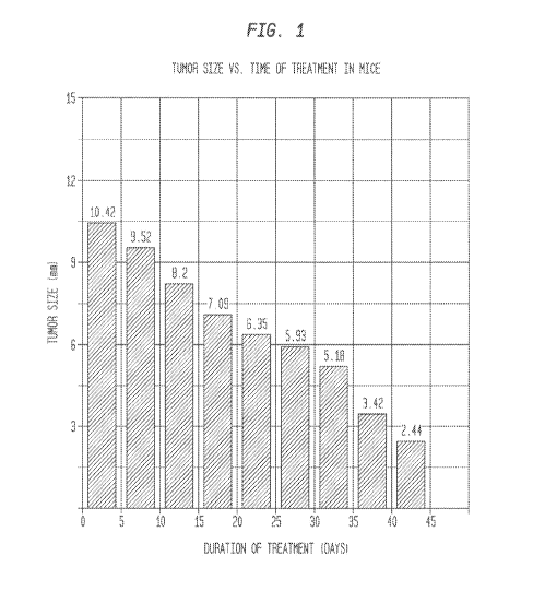
[0038] Figure 1 is a bar graph depicting the reduction in size of an
induced mouse foot
pad melanoma as a function of time of treatment with the pharmaceutical
composition of the
described invention.
CA 02880200 2015-01-26
WO 2014/018663 PCT/US2013/051871
[0039] Figure 2 is a photograph of a fully-developed mouse foot pad
melanoma three
weeks after inoculation with B 16 cells.
[0040] Figures 3, 4, 5, 6, 7, and 8 are photographs of a fully-developed
(40 day post-
inoculation) mouse foot pad melanoma after 0, 10, 20, 35, 40, and 45 days
treatment
respectively, with the pharmaceutical composition of the described invention.
[0041] Figure 9 depicts photomicrographs of COX-2 immunohistological
staining of
sections of a mouse foot pad melanoma taken at various times of treatment with
the
pharmaceutical composition of the described invention.
[0042] Figure 10 depicts photomicrographs of iNOS immunohistological
staining of
sections of a mouse foot pad melanoma taken at various times of treatment with
the
pharmaceutical composition of the described invention.
[0043] Figure 11 is a photograph of an early-stage mouse foot pad
melanoma one week
postinoculation.
[0044] Figure 12 is a photograph of the mouse foot pad melanoma of the
mouse of
Figure 12 after treatment with the pharmaceutical composition of the described
invention for 20
days.
[0045] Figures 13A to D are photographs of (A) injured mouse brains, (B)
healthy mouse
brains, (C) injured not treated mouse brains, and (D) injured and treated
mouse brains.
[0046] Figure 14 is a series of photographs that show the development ca.
two weeks
after injury and resolution of post-traumatic alopecia in a mouse after 45
days post-development
treatment.
[0047] Figure 15 is a series of photographs showing reduction in
chemically-induced
skin wrinkling in a mouse.
[0048] Figure 16A is a bar graph that shows the results of mouse
longevity studies. The
term "Bioquantine TM" is used to refer to the pharmaceutical composition of
the described
invention.
11
CA 02880200 2015-01-26
WO 2014/018663 PCT/US2013/051871
[0049] Figure 16B is a bar graph presenting the results of Drosophila
Melanogaster
longevity studies. The term "BioquantineTM" is used to refer to the
pharmaceutical composition
of the described invention.
[0050] Figure 17 is a series of photographs that show the expression of
pluripotency
markers by cells derived from human bone marrow stromal cells on d7 following
co-
electroporation with Xenopus laevis oocytes. (A)- (D) same field; (A) DAPI;
(B) Oct 3/4; (C),
Sox-2; (D), DAPI, Oct 3/4, and Sox-2 combined; (E)- (H) same field; (E) DAPI;
(F) Oct 3/4; (G)
Nanog; (H) DAPI, Oct 3/4, and Sox-2 combined; (I)- (1), same field; (1), DAPI;
(J) Rex-1; (K)
SSEA-1; (1) DAPI, Rex-1, and SSEA-1 combined.
[0051] Figure 18 is a series of photographs that show the expression of
pluripotency
markers by cells derived from BJ cells following co-electroporation with
Xenopus laevis oocytes.
(A) control cells (no co-electroporation); (B)-(C) same field, dS; (B) phase
contrast; (C) alkaline
phosphatase; (D)-(G) same field on dS; (D) DAPI; (E) Oct 3/4; (F) Nanog; (G)
DAPI, Oct 3/4,
and Nanog; (H)- (I) same field, d9; (H) phase contrast, (I) TRA-1-60; (J)- (K)
same field, d9; (J)
phase contrast; (K) Rex-1; (L)- (M) same, field, dl 1; (L) phase contrast; (M)
SSEA-1; (N)- (0)
same field, dS; (M) phase contrast; (N) Sox-2.
[0052] Figure 19 is a series of photographs that show the expression of
pluripotency
markers by cells derived from human pre-adipocytes (HPA) following co-
electroporation with
Xenopus oocytes. (A) duster of cells on d5 using phase contrast; (B) alkaline
phosphatase; (C)-
(D) same field at d5; (C) phase contrast; (D) Oct 3/4; (E)-(F) same field, d5;
(E) phase contrast;
(F) Nanog; (G)-(H), same field, d10; (G) phase contrast; (H) Sox-2; (I)- (J)
same field, d9; (I)
phase contrast; (J) TRA-1-60; ( K)- (L), same field, dl 1; (K) phase contrast,
(1) Rex-1; (M)- (N)
same field, d10; (M) phase contrast, (N) SSEA-1.
[0053] Figure 20 is a series of photographs that show the expression of
neural markers by
cells derived from human pre-adipocytes following culture for 8 days in
conditions that promote
neural progenitor differentiation by embryonic stem cells.
[0054] Figure 21 is a series of photographs that show cells derived from
human CD4+ T-
lymphocytes following co-electroporation with Xenopus laevis oocytes. (A)
control, no co-
12
CA 02880200 2015-01-26
WO 2014/018663 PCT/US2013/051871
electroporation; (B) no co-electroporation, culture on irradiated mouse
embryonic fibroblasts;
(C)-(D) cell culture on d5 following coelectroporation; (E)-(F) lower part of
cluster in (D); (G)-
(H) alkaline phosphatase on d9.
[0055] Figure 22 is a series of photographs that show the expression of
pluripotency
markers by cells derived from human CD4+ T-Lymphocytes (CD4TL) following co-
electroporation with Xenopus laevis oocytes. (A)-(B), same field, dl 0; (A)
phase contrast; (B)
Oct 3/4; (C)-(D) same field, dl 0; (C) phase contrast; (D) Nanog; (E)-(H) same
field, d5; (E)
DAPI; (F) Rex-1; (G) Sox-2; (H) DAPI, Rex-1, and Sox-2; (I)-(J) same field,
d9; (I) phase
contrast; (J) TRA-1-60; (K)-(L), same field, d10; (K) phase contrast; (L) SSEA-
1.
[0056] Figure 23 is a series of photographs that show colonies of cells
derived from
human buccal mucosa cells on 6 after co-electroporation with Xenopus laevis
oocytes. (A) grown
on irradiated mouse embryonic fibroblast substrate; (B) grown on StemAdhereTM
substrate.
[0057] Figure 24 is a series of photographs that show the expression of
human
pluripotency-associated factors by cells derived from human buccal mucosa
cells following co-
electroporation with Xenopus laevis oocytes. (A)-(B) same field, 96 h; (A)
phase contrast; (B)
Oct 3/4; (C)-(D) same field, dl 0; (C) phase contrast; (D) Nanog; (E)-(F) same
field, dl 0; (E)
phase contrast; (F) Sox-2; (G)-(H) same field, d9, (G) phase contrast; (H) TRA-
1-60; (I)-(J),
same field, dl 1; (I) phase contrast; (J) Rex-1; (K)-(L) same field, dl 1; (K)
phase contrast; (L)
SSEA-1.
[0058] Figure 25 is a series of photographs that show partial
dedifferentiation of HeLa
and MCF-7 cells following co-electroporation with Xenopus laevis oocytes. (A),
HeLa cells, no
co-electroporation; (B) HeLa cells grown on irradiated mouse embryonic
fibroblast cells, no co-
electroporation; (C) MCF-7 cells, no co-electroporation; (D) MCF-7 cells grown
on irradiated
mouse embryonic fibroblast cells, no co-electroporation; (E)-(H) cells derived
from HeLa cells
following co-electroporation with Xenopus laevis oocytes; (E)-(F), same field,
dl 1; (E) phase
contrast; (F) Oct 3/4; (G) phase contrast; (H) Oct 3/4; (I)-(L) MCF-7 cells
following co-
electroporation with Xenopus laevis oocytes; (G)-(H) same field, dl 1; (G)
phase contrast; (H)
Oct 3/4; (I)-(J) same field, dl 1; (I) phase contrast; (J) Nanog.
13
CA 02880200 2015-01-26
WO 2014/018663 PCT/US2013/051871
[0059] Figure 26 is a table containing the spectrometry results for 93
proteins.
[0060] Figure 27 is a bar graph that shows the distribution of hsa-miR-17-
5p inside and
outside Xenopus laevis oocytes before and after Bioquantine TM (BQ)
activation.
[0061] Figure 28 is a bar graph that shows the distribution of hsa-miR-
18a inside and
outside Xenopus laevis oocytes before and after Bioquantine TM (BQ)
activation.
[0062] Figure 29 is a bar graph that shows the distribution of hsa-miR-
19a inside and
outside Xenopus laevis oocytes before and after Bioquantine TM (BQ)
activation.
[0063] Figure 30 is a bar graph that shows the distribution of hsa-miR-
19b inside and
outside Xenopus laevis oocytes before and after Bioquantine TM (BQ)
activation.
[0064] Figure 31 is a bar graph that shows the distribution of hsa-miR-
20a inside and
outside Xenopus laevis oocytes before and after Bioquantine TM (BQ)
activation.
[0065] Figure 32 is a bar graph that shows the distribution of mmu-miR-
92a inside and
outside Xenopus laevis oocytes before and after Bioquantine TM (BQ)
activation.
[0066] Figure 33 is a bar graph that shows the distribution of mmu-miR-93
inside and
outside Xenopus laevis oocytes before and after Bioquantine TM (BQ)
activation.
[0067] Figure 34 is a bar graph that shows the distribution of hsa-miR-
367 inside and
outside Xenopus laevis oocytes before and after Bioquantine TM (BQ)
activation.
[0068] Figure 35 is a bar graph that shows the distribution of hsa-miR-
372 inside and
outside Xenopus laevis oocytes before and after Bioquantine TM (BQ)
activation.
[0069] Figure 36 is a bar graph that shows the distribution of hsa-miR-
373 inside and
outside Xenopus laevis oocytes before and after Bioquantine TM (BQ)
activation.
DETAILED DESCRIPTION OF THE INVENTION
[0070] The described invention can be better understood from the
following description
of exemplary embodiments, taken in conjunction with the accompanying figures
and drawings.
14
CA 02880200 2015-01-26
WO 2014/018663 PCT/US2013/051871
It should be apparent to those skilled in the art that the described
embodiments of the described
invention provided herein are merely exemplary and illustrative and not
limiting.
Definitions:
[0071] Various terms used throughout this specification shall have the
definitions set out
herein.
[0072] The term "adherent" as used herein refers to the act of sticking
to, clinging, or
staying attached.
[0073] The term "administer", "administering" or "to administer" as used
herein, refers
to the giving or supplying of a medication, including in vivo administration,
as well as
administration directly to tissue or cells ex vivo. Generally, compositions
may be administered
systemically either orally, bucally, parenterally, topically, by inhalation or
insufflation (i.e.,
through the mouth or through the nose) or rectally in dosage unit formulations
containing
conventional nontoxic pharmaceutically acceptable carriers, adjuvants and
vehicles as desired, or
may be locally administered by means such as, but not limited to, injection,
implantation,
grafting, topical application or parenterally.
[0074] The terms "agent" and "therapeutic agent" are used interchangeably
herein to
refer to a drug, molecule, composition, or other substance that provides a
therapeutic effect. The
term "active agent" as used herein, refers to the ingredient, component or
constituent of the
compositions of the described invention responsible for the intended
therapeutic effect.
[0075] The term "allogeneic" as used herein refers to being genetically
different although
belonging to or obtained from the same species.
[0076] The terms "apoptosis" or "programmed cell death" refer to a highly
regulated and
active process that contributes to biologic homeostasis comprised of a series
of biochemical
events that lead to a variety of morphological changes, including blebbing,
changes to the cell
membrane, such as loss of membrane asymmetry and attachment, cell shrinkage,
nuclear
fragmentation, chromatin condensation, and chromosomal DNA fragmentation,
without
damaging the organism.
CA 02880200 2015-01-26
WO 2014/018663 PCT/US2013/051871
[0077] The terms "residue" or "amino acid residue" or "amino acid" are
used
interchangeably to refer to an amino acid that is incorporated into a protein,
a polypeptide, or a
peptide, including, but not limited to, a naturally occurring amino acid and
known analogs of
natural amino acids that can function in a similar manner as naturally
occurring amino acids.
[0078] The term "attached" as used herein refers to being fastened,
fixed, joined,
connected, bound, adhered to or assembled with.
[0079] The term "autologous" as used herein means derived from the same
organism.The
term "biocompatible" as used herein refers to causing no clinically relevant
tissue irritation,
injury, toxic reaction, or immunological reaction to living tissue.
[0080] The term "biomarkers" (or "biosignatures") as used herein refers
to peptides,
proteins, nucleic acids, antibodies, genes, metabolites, or any other
substances used as indicators
of a biologic state. It is a characteristic that is measured objectively and
evaluated as a cellular
or molecular indicator of normal biologic processes, pathogenic processes, or
pharmacologic
responses to a therapeutic intervention.
[0081] The term "carrier" as used herein refer to a pharmaceutically
acceptable inert
agent or vehicle for delivering one or more active agents to a subject, and
often is referred to as
"excipient." The carrier must be of sufficiently high purity and of
sufficiently low toxicity to
render it suitable for administration to the subject being treated. The
carrier further should
maintain the stability and bioavailability of an active agent.
[0082] The term "cell" is used herein to refer to the structural and
functional unit of
living organisms and is the smallest unit of an organism classified as living.
[0083] The term "cellular senescence" as used herein refers to a stable
and long-term loss
of proliferative capacity, despite continued viability and metabolic activity.
The term
"replicative senescence" refers to the progressive shortening of telomeres of
a given cell with
replication. Senescence also can be induced in the absence of any detectable
telomere loss or
dysfunction, by a variety of conditions. This type of senescence has been
termed premature,
since it arises prior to the stage at which it is induced by telomere
shortening. Premature
senescence in vivo is believed to play a critical role in tumor suppression.
16
CA 02880200 2015-01-26
WO 2014/018663 PCT/US2013/051871
[0084] The term "compatible" as used herein means that the components of
a
composition are capable of being combined with each other in a manner such
that there is no
interaction that would substantially reduce the efficacy of the composition
under ordinary use
conditions.
[0085] The term "component" as used herein refers to a constituent part,
element or
ingredient.
[0086] The terms "composition" and "formulation" are used interchangeably
herein to
refer to a product of the described invention that comprises all active and
inert ingredients. The
term "active" refers to the ingredient, component or constituent of the
compositions of the
described invention responsible for the intended therapeutic effect. The terms
"pharmaceutical
formulation" or "pharmaceutical composition" as used herein refer to a
formulation or
composition that is employed to prevent, reduce in intensity, cure or
otherwise treat a target
condition or disease.
[0087] The term "condition" as used herein, refers to a variety of health
states and is
meant to include disorders or diseases caused by injury or any underlying
mechanism or
disorder.
[0088] The term "contact" and its various grammatical forms as used
herein refers to a
state or condition of touching or of immediate or local proximity. Contacting
a composition to a
target destination may occur by any means of administration known to the
skilled artisan.
[0089] The term "delay", "delaying", "delayed" or "to delay" as used
herein, refers to
stopping, detaining or hindering for a time; to cause to be slower or to occur
more slowly than
normal.
[0090] The term "derivative" as used herein means a compound that may be
produced
from another compound of similar structure in one or more steps. A
"derivative" or
"derivatives" of a peptide or a compound retains at least a degree of the
desired function of the
peptide or compound. Accordingly, an alternate term for "derivative" may be
"functional
derivative." Derivatives can include chemical modifications of the peptide,
such as akylation,
acylation, carbamylation, iodination or any modification that derivatizes the
peptide. Such
17
CA 02880200 2015-01-26
WO 2014/018663 PCT/US2013/051871
derivatized molecules include, for example, those molecules in which free
amino groups have
been derivatized to form amine hydrochlorides, p-toluene sulfonyl groups,
carbobenzoxy groups,
t-butyloxycarbonyl groups, chloroacetyl groups or formal groups. Free carboxyl
groups can be
derivatized to form salts, esters, amides, or hydrazides. Free hydroxyl groups
can be derivatized
to form 0-acyl or 0-alkyl derivatives. The imidazole nitrogen of histidine can
be derivatized to
form N-im-benzylhistidine. Also included as derivatives or analogues are those
peptides that
contain one or more naturally occurring amino acid derivative of the twenty
standard amino
acids, for example, 4-hydroxyproline, 5-hydroxylysine, 3-methylhistidine,
homoserine, ornithine
or carboxyglutamiate, and can include amino acids that are not linked by
peptide bonds. Such
peptide derivatives can be incorporated during synthesis of a peptide, or a
peptide can be
modified by wellknown chemical modification methods (see, e.g., Glazer et al.,
Chemical
Modification of Proteins, Selected Methods and Analytical Procedures, Elsevier
Biomedical
Press, New York (1975)).
[0091] The term "detectable marker" encompasses both selectable markers
and assay
markers. The term "selectable markers" refers to a variety of gene products to
which cells
transformed with an expression construct can be selected or screened,
including drug-resistance
markers, antigenic markers useful in fluorescence-activated cell sorting,
adherence markers such
as receptors for adherence ligands allowing selective adherence, and the like.
[0092] The term "detectable response" refers to any signal or response
that may be
detected in an assay, which may be performed with or without a detection
reagent. Detectable
responses include, but are not limited to, radioactive decay and energy (e.g.,
fluorescent,
ultraviolet, infrared, visible) emission, absorption, polarization,
fluorescence, phosphorescence,
transmission, reflection or resonance transfer. Detectable responses also
include
chromatographic mobility, turbidity, electrophoretic mobility, mass spectrum,
ultraviolet
spectrum, infrared spectrum, nuclear magnetic resonance spectrum and x-ray
diffraction.
Alternatively, a detectable response may be the result of an assay to measure
one or more
properties of a biologic material, such as melting point, density,
conductivity, surface acoustic
waves, catalytic activity or elemental composition. A "detection reagent" is
any molecule that
generates a detectable response indicative of the presence or absence of a
substance of interest.
18
CA 02880200 2015-01-26
WO 2014/018663 PCT/US2013/051871
Detection reagents include any of a variety of molecules, such as antibodies,
nucleic acid
sequences and enzymes. To facilitate detection, a detection reagent may
comprise a marker.
[0093] The term "differential label" as used herein generally refers to a
stain, dye,
marker, or antibody used to characterize or contrast structures, components or
proteins of a
single cell or organism.
[0094] The term "differentiation" as used herein refers to the process of
development
with an increase in the level of organization or complexity of a cell or
tissue, accompanied with a
more specialized function.
[0095] The term "disease" or "disorder" as used herein, refers to an
impairment of health
or a condition of abnormal functioning.
[0096] The term "fluorescence" as used herein refers to the result of a
three-state process
that occurs in certain molecules, generally referred to as "fluorophores" or
"fluorescent dyes,"
when a molecule or nanostructure relaxes to its ground state after being
electrically excited.
Stage 1 involves the excitation of a fluorophore through the absorption of
light energy; Stage 2
involves a transient excited lifetime with some loss of energy; and Stage 3
involves the return of
the fluorophore to its ground state accompanied by the emission of light.
[0097] The term "functional equivalent" or "functionally equivalent" are
used
interchangeably herein to refer to substances, molecules, polynucleotides,
proteins, peptides, or
polypeptides having similar or identical effects or use.
[0098] The term "gene" as used herein refers to a region of DNA that
controls a discrete
hereditary characteristic, usually corresponding to a single protein or RNA.
This definition
includes the entire functional unit, encompassing coding DNA sequences,
noncoding regulatory
DNA sequences and introns.
[0099] The term "Oct4" as used herein refers to the octamer-binding
transcription factor
4, also known as Oct3 and Pou5f1, which is involved in the self-renewal or
pluripotency of
undifferentiated cells. Oct4 is capable of inducing a pluripotent stem cell-
like state in
differentiated cells. Oct4 is used as a marker for undifferentiation of a
cell.
19
CA 02880200 2015-01-26
WO 2014/018663 PCT/US2013/051871
[00100] The term "Sox2" as used herein refers to the SRY (sex determining
region Y)-box
2 transcription factor which is involved in maintaining self-renewal or
pluripotency of
undifferentiated cells. Sox2 heterodimerizes with Oct4 and is capable of
inducing a pluripotent
stem cell-like state in differentiated cells. Sox2 is used as a marker for
undifferentiation of a
cell.
[00101] The term "K1f4" as used herein refers to the Kruppel-like factor 4
transcription
factor which is involved in the self-renewal or pluripotency of
undifferentiated cells. K1f4 is
capable of inducing a pluripotent stem cell-like state in differentiated
cells. K1f4 is used as a
marker for undifferentiation of a cell.
[00102] The term "Myc" as used herein refers to the transcription factor
that has been
linked to several cellular functions including cell-cycle regulation,
proliferation, growth,
differentiation and metabolism. Myc is involved in the self-renewal or
pluripotency of
undifferentiated cells. Myc is capable of inducing a pluripotent stem cell-
like state in
differentiated cells. Myc is used as a marker for undifferentiation of a cell.
[00103] The term "Nanog" as used herein refers to the transcription that
is involved in
maintaining self-renewal or pluripotency of undifferentiated cells. Nanog
works in concert with
other factors such as Oct4 and Sox2 and is capable of inducing a pluripotent
stem cell-like state
in differentiated cells. Nanog is used as a marker for undifferentiation of a
cell.
[00104] The term "improve" (or improving) as used herein refers to bring
into a more
desirable or excellent condition.
[00105] As used herein, the term "inflammation" refers to a response to
infection and
injury in which cells involved in detoxification and repair are mobilized to
the compromised site
by inflammatory mediators. Inflammation often is characterized by a strong
infiltration of
leukocytes at the site of inflammation, particularly neutrophils
(polymorphonuclear cells). These
cells promote tissue damage by releasing toxic substances at the vascular wall
or in uninjured
tissue.
[00106] Regardless of the initiating agent, the physiologic changes
accompanying acute
inflammation encompass four main features: (1) vasodilation, which results in
a net increase in
CA 02880200 2015-01-26
WO 2014/018663 PCT/US2013/051871
blood flow, is one of the earliest s physical responses to acute tissue
injury; (2) in response to
inflammatory stimuli, endothelial cells lining the venules contract, widening
the intracellular
junctions to produce gaps, leading to increased vascular permeability, which
permits leakage of
plasma proteins and blood cells out of blood vessels; (3) inflammation often
is characterized by a
strong infiltration of leukocytes at the site of inflammation, particularly
neutrophils
(polymorphonuclear cells). These cells promote tissue damage by releasing
toxic substances at
the vascular wall or in uninjured tissue; and (4) fever, produced by pyrogens
released from
leukocytes in response to specific stimuli.
[00107] During the inflammatory process, soluble inflammatory mediators of
the
inflammatory response work together with cellular components in a systemic
fashion in the
attempt to contain and eliminate the agents causing physical distress. The
terms "inflammatory"
or immuno-inflammatory" as used herein with respect to mediators refers to the
molecular
mediators of the inflammatory process. These soluble, diffusible molecules act
both locally at the
site of tissue damage and infection and at more distant sites. Some
inflammatory mediators are
activated by the inflammatory process, while others are synthesized and/or
released from cellular
sources in response to acute inflammation or by other soluble inflammatory
mediators. Examples
of inflammatory mediators of the inflammatory response include, but are not
limited to, plasma
proteases, complement, kinins, clotting and fibrinolytic proteins, lipid
mediators, prostaglandins,
leukotrienes, platelet-activating factor (PAF), peptides and amines,
including, but not limited to,
histamine, serotonin, and neuropeptides, proinflammatory cytokines, including,
but not limited
to, interleukin-1, interleukin-4, interleukin-6, interleukin-S, tumor necrosis
factor (TNF),
interferon-gamma, and interleukin 12.
[00108] The term "injury" as used herein, refers to damage or harm to a
structure or
function of the body caused by an outside agent or force, which may be
physical or chemical.
[00109] The term "isolate" and its various grammatical forms as used
herein refers to
placing, setting apart, or obtaining a protein, molecule, substance, nucleic
acid, peptide, cell or
particle, in a form essentially free from contaminants or other materials with
which it is
commonly associated, separate from its natural environment.
21
CA 02880200 2015-01-26
WO 2014/018663 PCT/US2013/051871
[00110] The term "labeling" as used herein refers to a process of
distinguishing a
compound, structure, protein, peptide, antibody, cell or cell component by
introducing a
traceable constituent. Common traceable constituents include, but are not
limited to, a
fluorescent antibody, a fluorophore, a dye or a fluorescent dye, a stain or a
fluorescent stain, a
marker, a fluorescent marker, a chemical stain, a differential stain, a
differential label, and a
radioisotope.
[00111] The terms "marker" or "cell surface marker" are used
interchangeably herein to
refer to an antigenic determinant or epitope found on the surface of a
specific type of cell. Cell
surface markers can facilitate the characterization of a cell type, its
identification, and eventually
its isolation. Cell sorting techniques are based on cellular biomarkers where
a cell surface
marker(s) may be used for either positive selection or negative selection,
i.e., for inclusion or
exclusion, from a cell population.
[00112] The term "microRNAs" (miRNAs) as used herein refers to a class of
small non-
coding RNAs (-22 nt), which normally function as negative regulators of target
mRNA
expression at the posttranscriptional level by binding to the 3'UTR of target
mRNAs through
base pairing, resulting in target mRNAs cleavage or translation inhibition
(Ambros V., Nature,
2004; 431:350-354; Bartel D. P., Cell, 2004; 116:281-297; Meister and Tuschl,
Nature, 2004;
431:343-349). Increasing evidence has shown that miRNAs play critical roles in
many key
biological processes, such as cell growth, tissue differentiation, cell
proliferation, embryonic
development, cell proliferation, and apoptosis (Esquela-Kerscher and Slack,
Nature Reviews
Cancer, 2006; 6:259-269). As such, the mutation of miRNAs, the dysfunction of
miRNA
biogenesis and the dysregulation of miRNAs and their targets may result to
various diseases,
such as cancers(Calin and Croce, Nature Reviews Cancer, 2006; 6:857-866;
Esquela-Kerscher
and Slack, Nature Reviews Cancer, 2006; 6:259-269), cardiovascular
disease(Latronico et al.,
Circ. Res, 2007; 101:1225-1236; van Rooij and Olson, J. Clin. Invest., 2007;
117:2369-2375),
schizophrenia(Hansen, et al.,PLos, 2007; 9:e873; Perkins et al., Genome
Biology, 2007; 8:R27),
renal function disorders(Williams, Cell. Mol. Life Sci., 2008; 65:545-562),
Tourette's
syndrome(Esau and Monia, Advanced Drug Delivery, 2007; 59:101-114),
psoriasis(Sonkoly et
al.,PLos, 2007: 7:e610), primary muscular disorders(Eisenberg et al.,PNAS,
2007: 104:17016-
17021), Fragile-X mental retardation syndrome(Fiore and Schratt, The
Scientific World Journal,
22
CA 02880200 2015-01-26
WO 2014/018663 PCT/US2013/051871
2007; 7:167-177), Polycythemia vera (Bruchova et al.,Experimental Hemaotlogy,
2007;
35:1657-1667), diabetes (Williams Cell. Mol. Life Sci., 2008; 65:545-562),
chronic
hepatitis(Murakami et al., Oncogene, 2006; 25:2537-2545), AIDS(Hariharan et
al., BBRC, 2005;
337:1214-1218), and obesity(Weiler et al., Gene Therapy, 2006; 13:496-502).
The mechanisms
of miRNAs implicated in diseases are very complex.
[00113] The term "modulate" as used herein means to regulate, alter,
adapt, or adjust to a
certain measure or proportion.
[00114] The term "multipotent" as used herein refers to a cell capable of
giving rise to a
limited number of cell types of a particular cell line.
[00115] The term "nucleic acid" is used herein to refer to a
deoxyribonucleotide or
ribonucleotide polymer in either single- or double-stranded form, and unless
otherwise limited,
encompasses known analogues having the essential nature of natural nucleotides
in that they
hybridize to single-stranded nucleic acids in a manner similar to naturally
occurring nucleotides
(e.g., peptide nucleic acids).
[00116] The term "nucleotide" is used herein to refer to a chemical
compound that
consists of a heterocyclic base, a sugar, and one or more phosphate groups. In
the most common
nucleotides, the base is a derivative of purine or pyrimidine, and the sugar
is the pentose
deoxyribose or ribose. Nucleotides are the monomers of nucleic acids, with
three or more
bonding together in order to form a nucleic acid. Nucleotides are the
structural units of RNA,
DNA, and several cofactors, including, but not limited to, CoA, FAD, DMN, NAD,
and NADP.
Purines include adenine (A), and guanine (G); pyrimidines include cytosine
(C), thymine (T),
and uracil (U).
[00117] The term "parenteral" as used herein, refers to introduction into
the body by way
of an injection (i.e., administration by injection), including, for example,
subcutaneously (i.e., an
injection beneath the skin), intramuscularly (i.e., an injection into a
muscle), intravenously (i.e.,
an injection into a vein), intrathecally (i.e., an injection into the space
around the spinal cord or
under the arachnoid membrane of the brain), intrasternal injection or infusion
techniques. A
parenterally administered composition is delivered using a needle, e.g., a
surgical needle. The
23
CA 02880200 2015-01-26
WO 2014/018663 PCT/US2013/051871
term "surgical needle" as used herein, refers to any needle adapted for
delivery of fluid (i.e.,
capable of flow) compositions into a selected anatomical structure. Injectable
preparations, such
as sterile injectable aqueous or oleaginous suspensions, may be formulated
according to the
known art using suitable dispersion or wetting agents and suspending agents.
[00118] The term "partition" and its various grammatical forms as used
herein, refers to
dividing or separating into parts or shares.
[00119] The term "peptide" is used herein to refer to two or more amino
acids joined by a
peptide bond.
[00120] The term "protein" is used herein to refer to a large complex
molecule or
polypeptide composed of amino acids. The sequence of the amino acids in the
protein is
determined by the sequence of the bases in the nucleic acid sequence that
encodes it.
[00121] The terms "peptide", "polypeptide" and "protein" also apply to
amino acid
polymers in which one or more amino acid residue is an artificial chemical
analogue of a
corresponding naturally occurring amino acid, as well as to naturally
occurring amino acid
polymers. The essential nature of such analogues of naturally occurring amino
acids is that,
when incorporated into a protein that protein is specifically reactive to
antibodies elicited to the
same protein but consisting entirely of naturally occurring amino acids. The
terms
"polypeptide", "peptide" and "protein" also are inclusive of modifications
including, but not
limited to, glycosylation, lipid attachment, sulfation, gamma-carboxylation of
glutamic acid
residues, hydroxylation and ADP-ribosylation. It will be appreciated, as is
well known and as
noted above, that polypeptides may not be entirely linear. For instance,
polypeptides may be
branched as a result of ubiquitination, and they may be circular, with or
without branching,
generally as a result of posttranslational events, including natural
processing event and events
brought about by human manipulation which do not occur naturally. Circular,
branched and
branched circular polypeptides may be synthesized by non-translation natural
process and by
entirely synthetic methods, as well.The term "pluripotent" as used herein
refers to the ability to
develop into multiple cells types, including all three embryonic lineages,
forming the body
organs, nervous system, skin, muscle and skeleton.
24
CA 02880200 2015-01-26
WO 2014/018663 PCT/US2013/051871
[00122] The term "portion" as used herein refers to a part of a whole
separated from or
integrated with it.
[00123] The term "prevent", "preventing", "prevented" or "to prevent" as
used herein,
refers to effectual stoppage of action or progress.
[00124] The term "progenitor cell" as used herein refers to an early
descendant of a stem
cell that can only differentiate, but can no longer renew itself. Progenitor
cells mature into
precursor cells that mature into mature (differentiated) phenotypes.
Hematopoietic progenitor
cells are referred to as colony-forming units (CFU) or colony-forming cells
(CFC). The specific
lineage of a progenitor cell is indicated by a suffix, such as, but not
limited to, CFU-E
(erythrocytic), CFU-F (fibroblastic), CFU-GM (granulocytic/macrophage), and
CFU-GEMM
(pluripotent hematopoietic progenitor).
[00125] The term "prolong", "prolonging", "prolonged" or "to prolong" as
used herein,
refers to lengthening in time, extent, scope or range.
[00126] The term "propagate" as used herein refers to reproduce, multiply,
or to increase
in number, amount or extent by any process.
[00127] The term "purification" as used herein refers to the process of
isolating or freeing
from foreign, extraneous, or objectionable elements.
[00128] The term "Reactive oxygen species" ("ROS"), such as free radicals
and
peroxides, as used herein refers to a class of molecules that are derived from
the metabolism of
oxygen and exist inherently in all aerobic organisms. The term "oxygen
radicals" as used herein
refers to any oxygen species that carries an unpaired electron (except free
oxygen). The transfer
of electrons to oxygen also may lead to the production of toxic free radical
species. The best
documented of these is the superoxide radical. Oxygen radicals, such as the
hydroxyl radical
(OH-) and the superoxide ion (02-) are very powerful oxidizing agents that
cause structural
damage to proteins, lipids and nucleic acids. The free radical superoxide
anion, a product of
normal cellular metabolism, is produced mainly in mitochondria because of
incomplete reduction
of oxygen. The superoxide radical, although unreactive compared with many
other radicals, may
CA 02880200 2015-01-26
WO 2014/018663 PCT/US2013/051871
be converted by biological systems into other more reactive species, such as
peroxyl (R00-),
alkoxyl (RO-) and hydroxyl (OH-) radicals.
[00129] The term "reduce", "reducing", "reduced" or "to reduce" as used
herein, refers to
a diminishing, a decrease in, an attenuation or abatement of the degree,
intensity, extent, size,
amount, density or number of.
[00130] The term "regeneration" or "regenerate" as used herein refers to a
process of
recreation, reconstitution, renewal, revival, restoration, differentiation and
growth to form a
tissue with characteristics that conform with a natural counterpart of the
tissue.
[00131] The term "relative" as used herein refers to something having, or
standing in,
some significant association to something else. The term "relative frequency"
as used herein
refers to the rate of occurrence of something having or standing in some
significant association
to the rate of occurrence of something else. For example, two cell types, X
cells and Y cells
occupy a given location. There are 5 X cells and 5 Y cells in that location.
The relative
frequency of cell type X is 5/10; the relative frequency of cell type Y is
5/10 in that location.
Following processing, there are 5 X cells, but only 1 Y cell in that location.
The relative
frequency of cell type X following processing is 5/6, and the relative
frequency of cell type Y
following processing is 1/6 in that location.
[00132] The term "repair" as used herein as a noun refers to any
correction, reinforcement,
reconditioning, remedy, making up for, making sound, renewal, mending,
patching, or the like
that restores function. When used as a verb, it means to correct, to
reinforce, to recondition, to
remedy, to make up for, to make sound, to renew, to mend, to patch or to
otherwise restore
function. In some embodiments "repair" includes full repair and partial
repair.
[00133] The term "stem cells" refers to undifferentiated cells having high
proliferative
potential with the ability to self-renew (make more stem cells by cell
division) that can generate
daughter cells that can undergo terminal differentiation into more than one
distinct cell
phenotype.
[00134] The term "stimulate" as used herein refers to activate, provoke,
or spur. The term
"stimulating agent" as used herein refers to a substance that exerts some
force or effect.
26
CA 02880200 2015-01-26
WO 2014/018663 PCT/US2013/051871
[00135] The term "syndrome" as used herein, refers to a pattern of
symptoms indicative of
some disease or condition.
[00136] The terms "subject" and "patient" are used interchangeably herein
to refer to
animal species of mammalian origin that may benefit from the administration of
a drug
composition or method of the described invention. Examples of subjects include
humans, and
other animals such as horses, pigs, cattle, dogs, cats, rabbits, mice, rats
and aquatic mammals.
[00137] The phrase "subject in need thereof' as used herein refers to a
subject suffering
from a disease, disorder, condition or injury characterized by damaged or
cancerous
differentiated cells that (i) will be administered a pharmaceutical
composition of the described
invention, (ii) is receiving a pharmaceutical composition of the described
invention; or (iii) has
received a pharmaceutical composition of the described invention, in order to
reprogram those
cells into iPSC-like cells and treat the condition, unless the context and
usage of the phrase
indicates otherwise.
[00138] The terms "therapeutic amount", "therapeutically effective amount"
and "amount
effective" are used interchangeably herein to refer to an amount of one or
more active agent(s)
that is sufficient to provide the intended benefit of treatment. Dosage levels
are based on a
variety of factors, including the type of injury, the age, sex, weight,
medical condition of the
patient, the severity of the condition, the route of administration and the
particular active agent
employed. The dosage regimen may vary widely, but can be determined routinely
by a
physician using standard methods.
[00139] The term "therapeutic effect" as used herein refers to a
consequence of treatment,
the results of which are judged to be desirable and beneficial. A therapeutic
effect may include,
directly or indirectly, the arrest, reduction, or elimination of a disease
manifestation. A
therapeutic effect also may include, directly or indirectly, the arrest
reduction or elimination of
the progression of a disease manifestation.
[00140] The term "treat", "treating" or "to treat" as used herein, refers
to accomplishing
one or more of the following: (a) reducing the severity of a disorder; (b)
limiting the
development of symptoms characteristic of a disorder being treated; (c)
limiting the worsening of
27
CA 02880200 2015-01-26
WO 2014/018663 PCT/US2013/051871
symptoms characteristic of a disorder being treated; (d) limiting the
recurrence of a disorder in
patients that previously had the disorder; and (e) limiting recurrence of
symptoms in patients that
were previously asymptomatic for the disorder. The term "treat", "treating" or
"to treat"
includes abrogating, substantially inhibiting, slowing or reversing the
progression of a disease,
condition or disorder, substantially ameliorating clinical or esthetical
symptoms of a condition,
substantially preventing the appearance of clinical or esthetical symptoms of
a disease, condition,
or disorder, and protecting from harmful or annoying symptoms.
[00141] The term "variant" as used herein refers to a peptide sequence
that varies at one or
more amino acid positions with respect to the reference peptide. A variant can
be a naturally-
occurring variant or can be the result of spontaneous, induced, or genetically
engineered
mutation(s) to the nucleic acid molecule encoding the variant peptide. A
variant peptide can also
be a chemically synthesized variant. A skilled artisan likewise can produce
polypeptide variants
having single or multiple amino acid substitutions, deletions, additions or
replacements. These
variants may include inter alia: (a) variants in which one or more amino acid
residues are
substituted with conservative or non-conservative amino acids; (b) variants in
which one or more
amino acids are added; (c) variants in which at least one amino acid includes
a substituent group;
(d) variants in which amino acid residues from one species are substituted for
the corresponding
residue in another species, either at conserved or non-conserved positions;
and (d) variants in
which a target protein is fused with another peptide or polypeptide such as a
fusion partner, a
protein tag or other chemical moiety, that may confer useful properties to the
target protein, such
as, for example, an epitope for an antibody. The techniques for obtaining such
variants,
including genetic (suppressions, deletions, mutations, etc.), chemical, and
enzymatic techniques
are known to the skilled artisan.
[00142] According to one aspect, the described invention provides
compositions obtained
from amphibian oocytes, preferably oocytes of Xenopus laevis. One such
composition is
designated an intra-oocyte composition; a second such composition is
designated as an extra-
oocyte composition.
28
CA 02880200 2015-01-26
WO 2014/018663 PCT/US2013/051871
[00143] The compositions of the described invention comprise extracts of
amphibian
oocytes containing, for example, proteins (polypeptides) and microRNAs
(miRNAs)
(polynucleotides), in combination with a solvent.
[00144] Exemplary proteins may include, but are not limited to, a Gapd-
prov protein, a
prostaglandin D2 (PGD2) synthetase protein, a hematopoietic b protein, a
phosphoglucomutase 1
protein, hypothetical protein L0C100101274, hypothetical protein L0C398635, a
vitellogenin
(VTG)-Al protein, a short-VTG-Al protein, a nucleoside diphosphate kinase Al
protein,
mg:bb02e05 and an adenosylhomocysteinase A protein. Without limitation, for
example,PGD2s
function as a neuromodulator as well as a trophic factor in the central
nervous system;
phosphoglucomutase (PGM) is a key enzyme in glucose metabolism; vitellogenin
is a female-
specific glucolipoprotein yolk precursor produced by all oviparous animals,
nucleoside
diphosphate kinase Al is believed to play a major role in the synthesis of
nucleoside
triphosphates other than ATP; and adenosylhomocysteine is a competitive
inhibitor of S-
adenosyl-L-methionine-dependent methyl transferase reactions, and may play a
key role in the
control of methylations via regulation of the intracellular concentration of
adenosylhomocysteine,
[00145] Exemplary microRNAs may include, without limitation, hsa-miR-17-
5p, hsa-nu/r-
18a, hsa-miR-92a, hsa-miR-19b-1, hsa-miR-20a, mmu-miR-92a, mmu-miR-93, hsa-miR-
367,
hsa-miR-372 and hsa-miR-373.
[00146] According to some embodiments, the compositions of the present
invention may
further include one or more compatible active ingredients which are aimed at
providing the
composition with an additional pharmaceutical effect.
[00147] The compositions of the present invention may be formulated as
aqueous
suspensions. A solution generally is considered as a homogeneous mixture of
two or more
substances; it is frequently, though not necessarily, a liquid. In a solution,
the molecules of the
solute (or dissolved substance) are uniformly distributed among those of the
solvent. A
suspension is a dispersion (mixture) in which a finely-divided species is
combined with another
species, with the former being so finely divided and mixed that it doesn't
rapidly settle out. In
everyday life, the most common suspensions are those of solids in liquid
water. Among the
29
CA 02880200 2015-01-26
WO 2014/018663 PCT/US2013/051871
acceptable vehicles and solvents that may be employed are water, Ringer's
solution, and isotonic
sodium chloride solution. In addition, sterile, fixed oils are conventionally
employed as a
solvent or suspending medium. For parenteral application, vehicles may consist
of solutions,
e.g., oily or aqueous solutions, as well as suspensions, emulsions, or
dispersions. Aqueous
suspensions may contain substances which increase the viscosity of the
suspension and include,
for example, sodium carboxymethyl cellulose, sorbitol and/or dextran.
Optionally, the
suspension may also contain stabilizers.
[00148] According to one embodiment, the compositions of the described
invention may
be prepared, for example, by a process that comprises: 1) providing a
suspension of amphibian
oocytes, harvested from an amphibian, in a buffered oocyte washing solution in
an oocyte
activation vessel; 2) applying an electroporation stimulus to the suspended
oocytes in the oocyte
activation vessel to produce a suspension of activated oocytes; 3) combining
an aqueous energy
solution with the suspension of activated oocytes; 4) incubating the
combination so obtained in
step 3) at an incubation temperature of 16 C to 20 C, for an incubation time
of about 2 to about
4 hours; 5) partitioning the incubated combination ( for example, using a
method based on
density), to obtain an extra-oocyte portion (that is, the portion external to
the incubated activated
oocytes), and an activated oocyte portion that includes the incubated
activated oocytes; and 6)
separating the extra-(activated)-oocyte and the activated oocyte portions from
each other.
[00149] According to another embodiment, the incubation temperature of
step 4) is 16 C,
17 C, 18 C, 19 C or 20 C.
[00150] According to one embodiment, the buffered oocyte washing solution
("OWS")
may include, but is not limited to, NaC1 (at 82.5 mM), HEPES (Sigma cat.
#H4034 at 5.0 mM),
KC1 (at 2.5 mM), MgC12 (at 1 mM), NaHPO4, (at 1.0 mM), and 0.5 %
penicillin/streptomycin,
adjusted to a pH of about 7.4. According to another embodiment, the OWS may
include, but is
not limited to, NaC1 (at 82.5 mM), KC1 (at 2.5 mM), MgC12 (at 1 mM), and
NaHPO4, (at 1.0
mM), adjusted to a pH of about 7.4 when used, for example, as a control in in
vivo studies.
[00151] According to one embodiment, the amphibian oocytes may be
suspended in
buffered OWS in an electroporation vessel. It is understood that any
convenient vessel can be
used as the electroporation vessel provided that it can accommodate the
electroporation
CA 02880200 2015-01-26
WO 2014/018663 PCT/US2013/051871
electrodes in a manner that allows delivery of electroporation stimulus to the
amphibian oocytes.
Standard T25 cell culture flasks and Gene Pulser electroporation cuvettes (Bio-
Rad cat. no. 165-
2088) are examples of suitable electroporation vessels. According to one
embodiment, the
electroporation electrodes may be diagonally opposed at a separation of about
6 cm. According
to one embodiment, the electroporation stimulus may be about 100 v/cm to about
200 v/cm at
about 25 ILIF to about 75 ILIF applied in about 0.3 msec to about 1.5 msec
pulses (time 30 constant
of 0.7 to 0.9 msec.) at about 5 to 10 pulses. According to another embodiment,
the
electroporation stimulus may be about 125 v/cm at about 50 ILIF applied in
about 0.3 msec to 1.5
msec pulses at about 7 pulses.
[00152] Aqueous energy solutions may be combined with the suspension of
activated
oocytes in order to provide, for example, chemicals or coenzymes necessary for
cellular
metabolism. According to one embodiment, the aqueous energy solution may
comprise about
7.5 mM creatine phosphate, about 1 mM adenosine-5'-triphosphate (ATP) at pH
7.7, and about 1
mM MgC12. According to another embodiment, the energy solution may be a 1:100
aqueous
dilution of creatine phosphate, ATP, and MgC12.
[00153] According to one embodiment, the combination of the aqueous energy
solution
with the suspension of activated oocytes may be incubated at an incubation
temperature of about
16 C to about 20 C for an incubation time of about 1 to 4 hours. According to
another
embodiment, the incubation temperature may be about 16 C. According to
another
embodiment, the incubation temperature may be about 17 C. According to
another
embodiment, the incubation temperature may be about 18 C. According to
another
embodiment, the incubation temperature may be about 19 C. According to
another
embodiment, the incubation temperature may be about 20 C. According to
another
embodiment, the incubation time may be at least about 2 hours but not more
than about 4 hours.
According to another embodiment, the incubation time may be about 3 hours.
[00154] According to one embodiment, the incubated combination that
includes activated
oocytes may be partitioned to obtain an extra-oocyte portion and an activated
oocyte portion that
contains activated, incubated amphibian oocytes. According to one embodiment,
partitioning
may be accomplished by methods based on differences in density, for example,
by
31
CA 02880200 2015-01-26
WO 2014/018663 PCT/US2013/051871
centrifugation. According to one embodiment, the partitioning includes, for
example, conditions
that do not rupture the activated incubated oocytes. According to another
embodiment, the
conditions include, but are not limited to, centrifugation at a force not
exceeding about 52 x g.
[00155] According to one embodiment, the extra-oocyte portion may be
separated from
the activated oocyte portion.
[00156] According to one embodiment, the extra-oocyte composition may be
obtained, for
example, by filtration. According to one embodiment, the extra-oocyte portion
may be filtered
through a fine filter. Filters can be obtained from Sigma, Fisher Scientific,
or other commercial
sources familiar to those skilled in the art. According to another embodiment,
the fine filter may
have a pore size from about 0.01 to 1 . According to another embodiment, the
fine filter may
have a pore size of about 0.02 . According to another embodiment, the fine
filter may have a
pore size of about 0.03 . According to another embodiment, the fine filter
may have a pore size
of about 0.04 . According to another embodiment, the fine filter may have a
pore size of about
0.05 . According to another embodiment, the fine filter may have a pore size
of about 0.06 .
According to another embodiment, the fine filter may have a pore size of about
0.07 .
According to another embodiment, the fine filter may have a pore size of about
0.08 .
According to another embodiment, the fine filter may have a pore size of about
0.09 .
According to another embodiment, the fine filter may have a pore size of about
0.1 . According
to another embodiment, the fine filter may have a pore size of about 0.2 .
According to another
embodiment, the fine filter may have a pore size of about 0.2 . According to
another
embodiment, the fine filter may have a pore size of about 0.3 . According to
another
embodiment, the fine filter may have a pore size of about 0.4 . According to
another
embodiment, the fine filter may have a pore size of about 0.5 . According to
another
embodiment, the fine filter may have a pore size of about 0.6 . According to
another
embodiment, the fine filter may have a pore size of about 0.7 . According to
another
embodiment, the fine filter may have a pore size of about 0.8 . According to
another
embodiment, the fine filter may have a pore size of about 0.9 . According to
another
embodiment, the fine filter may have a pore size of about 1.0 .
32
CA 02880200 2015-01-26
WO 2014/018663
PCT/US2013/051871
[00157]
According to another embodiment, the extra-oocyte portion may be combined
either before or after filtration with either a protease inhibitor (e.g. Sigma
cat# P8340) or a Rnase
inhibitor (e.g. SUPERase In Rnase, Applied Biosystems cat# A1V12694) to obtain
the extra-
oocyte composition of the described invention. According to another
embodiment, the extra-
oocyte portion may be combined either before or after filtration with both a
protease inhibitor
(e.g. Sigma cat# P8340) and an RNase inhibitor (e.g. SUPERase In RNase,
Applied Biosystems
cat# AM2694) to obtain the extra-oocyte composition of the described
invention. According to
another embodiment, the extra-oocyte portion is maintained at a temperature of
about 2 C to
8 C. According to another embodiment, the extra-oocyte portion is maintained
at a temperature
of about 4 C.
[00158]
According to one embodiment, the intra-oocyte composition may be obtained
from the activated oocyte portion by methods including, but not limited to,
centrifugation. For
example, the activated oocyte portion may be suspended in OWS and centrifuged
under
conditions that do not rupture the activated oocytes, but that provide a
"pellet" of activated
oocytes. After centrifugation, residual OWS then may be carefully removed from
the pellet of
activated oocytes by techniques well-known to those skilled in the art. The
pellet of activated
oocytes from which OWS has been removed may be centrifuged, for example, at
10,000 rpm at a
temperature below 20 C, to rupture the activated oocytes and provide three
fractions: a light
fraction, a heavy fraction, and a fraction of intermediate density. The light
fraction, which may
be two-phased, includes, for example, yolk proteins. The heavy fraction
includes, for example,
cell membranes and yolk particles. The fraction of intermediate density (i.e.,
the cytoplasmic
fraction), will become the intra-oocyte composition of the described
invention. The cytoplasmic
fraction is separated from the light and heavy fractions by techniques well-
known to those skilled
in the art. The cytoplasmic fraction may be combined with a protease inhibitor
(e.g. Sigma cat#
P8340) and an RNase inhibitor (e.g. SUPERase In RNase, Applied Biosystems cat#
AM2694).
The cytoplasmic fraction containing the inhibitors may be cooled at about 4 C
for up to about
one-half hour. The intra-oocyte composition of the described invention is
obtained by filtering
the cooled cytoplasmic fraction containing the inhibitors through a fine
filter, e.g., one having a
pore size of about 0.2 .
33
CA 02880200 2015-01-26
WO 2014/018663 PCT/US2013/051871
[00159] According to another aspect, the described invention provides a
pharmaceutical
composition comprising a therapeutic amount of the oocyte compositions of the
described
invention, which is effective to reprogram damaged or cancerous differentiated
cells into iPSC-
like cells that achieve regeneration, replacement, repair and/or rejuvenation
of the damaged or
cancerous differentiated cells and thereby treat the disease, condition,
injury or disorder being
characterized by the damaged or cancerous differentiated cells. Nonlimiting
examples of such
diseases, conditions, injuries or disorders include melanoma, traumatic brain
injury, post-
traumatic alopecia, and skin wrinkling.
[00160] The pharmaceutical composition can be formulated by mixing equal
volumes of
the extra-oocyte composition and the intra-oocyte compositions of the
described invention. The
pharmaceutical compositions comprise proteins and microRNAs. According to some
embodiments, the protein component can include protease-resistant forms of
aGapd-prov protein,
a prostaglandin D2 synthase protein, a hematopoietic b protein, a
phosphoglucomutase 1 protein,
hypothetical protein LOC100101274, hypothetical protein LOC398635, a
vitellogenin-Al
protein, a short-VTG -Al protein, a nucleoside diphosphate kinase Al protein,
a mg:bb02e05
protein, an adenosylhomocysteinase A protein and combinations thereof.
According to some
embodiments, the microRNA component can include, for example, hsa-miR-17-5p,
hsa-miR-
18a, hsa-miR-92a, hsa-miR-19b-1, hsa-miR-20a, mmu-miR-92a, mmu-miR-93, hsa-miR-
367,
hsa-miR-372 and hsa-miR-373. The pharmaceutical composition of the described
invention can
comprise about 5 mg/mL solid oocyte material, as determined by lyophilization
experiments.
[00161] According to some embodiments, the pharmaceutical compositions of
the present
invention may be formulated with an excipient or carrier. The carrier can be
inert, or it can
possess pharmaceutical benefits. The carrier can be liquid or solid and is
selected with the
planned manner of administration in mind to provide for the desired bulk,
consistency, etc., when
combined with an active and the other components of a given composition. The
term
"pharmaceutically acceptable carrier" as used herein refers to any
substantially non-toxic carrier
conventionally useful for administration of pharmaceuticals in which the
active component will
remain stable and bioavailable. In some embodiments, the pharmaceutically
acceptable carrier
of the compositions of the present invention include a release agent such as a
sustained release or
delayed release carrier. In such embodiments, the carrier can be any material
capable of
34
CA 02880200 2015-01-26
WO 2014/018663 PCT/US2013/051871
sustained or delayed release of the actives to provide a more efficient
administration, resulting in
less frequent and/or decreased dosage of the active ingredient, ease of
handling, and extended or
delayed effects. Non-limiting examples of such carriers include liposomes,
microsponges,
microspheres, or microcapsules of natural and synthetic polymers and the like.
Liposomes may
be formed from a variety of phospholipids such as cholesterol, stearylamines
or
phosphatidylcholines.
[00162] Additional pharmaceutical compositions of the present invention
can be readily
prepared using technology which is known in the art such as described in
Remington's
Pharmaceutical Sciences, 18th or 19th editions, published by the Mack
Publishing Company of
Easton, Pennsylvania, which is incorporated herein by reference.
[00163] The pharmaceutical composition may be constituted into any form
suitable for the
mode of administration selected. Exemplary routes of administration include,
but are not limited
to, parenteral (including subcutaneous), oral, inhalation, insufflation,
topical, buccal and rectal.
Compositions suitable for parenteral administration include sterile solutions,
emulsions and
suspensions. Oral administration include solid forms, such as pills, capsules,
granules, tablets,
and powders, and liquid forms, such as solutions, syrups, elixirs, and
suspensions. Compositions
suitable for inhalation and insufflation may take the form of an aerosolized
solution.
Compositions suitable for topical administration include creams, ointments and
dermal patches.
Compositions suitable for buccal administration may take the form of tablets
or lozenges.
Compositions suitable for rectal administration may take the form of
suppositories.
Formulations for administration may be provided using any formulation known in
the art and
appropriate for the route of administration. Such formulations may be as
provided using the
guidance of such resources as REMINGTON'S PHARMACEUTICAL SCIENCES, 18th ed.,
Mack Publishing Co., Easton, Pa. 1990.
Use to treat/inhibit progression of melanoma
[00164] According to one embodiment, the described invention provides a
method for
treating or inhibiting the progression of melanoma in a mammal. The method
includes the steps
of (a) preparing the extra-oocyte composition and the intra-oocyte composition
by acquiring the
CA 02880200 2015-01-26
WO 2014/018663 PCT/US2013/051871
oocytes; activating the oocytes; incubating the oocytes, partitioning the
incubated combination;
separating the extra-oocyte portion from the activated oocyte portion as
described above; (b)
formulating the pharmaceutical composition; and (c) administering a
therapeutically-effective
amount of the pharmaceutical composition of the described invention to a
mammal suffering
from melanoma. According to one embodiment, the administration may be by
injection or i.v.
drip. According to another embodiment, the injection may be, for example, an
intraperitoneal
injection, a subcutaneous injection, or an intramuscular injection. According
to another
embodiment, the injection may be an intraperitoneal injection. According to
another
embodiment, the efficacy of treating or inhibiting the progression of melanoma
in a mammal
may be demonstrated by, for example, a decrease in tumor mass with time of
treatment relative
to controls. See, e.g., Figure 1.
[00165] It is understood that the therapeutically-effective amount of the
pharmaceutical
composition will depend on the type of injury, the age, weight, sex, medical
condition of the
patient, the severity of the condition, the route of administration, and the
particular active agent
employed. Thus the dosage regimen may vary widely, but can be determined
routinely by a
physician using standard methods. According to one embodiment, the
therapeutically-effective
amount may vary about a mean of about 25 mg/kg body weight.
[00166] It is understood that, unlike treatment of acute conditions such
as bacterial
infections, successful systemic treatment of melanoma and other cancers may
require more than
a single, short-term course of treatment. Accordingly, according to one
embodiment, treatment
of melanoma with the pharmaceutical composition of the described invention may
involve
multiple administrations over a period of time. Those skilled in the art will
know to adjust the
frequency and duration of treatment, and also the amounts administered, based
on patient
tolerance and clinical evaluation of the regression of the disease in a
particular patient.
[00167] According to one embodiment, the described invention can replace
or supplement
methods of treating melanoma according to the then current standard of care,
such as surgery,
radiation and chemotherapy.
36
CA 02880200 2015-01-26
WO 2014/018663 PCT/US2013/051871
Use to treat TBI
[00168] According to another embodiment, the described invention provides
a method for
treating traumatic brain injury ("TBI") in a mammal. The method includes the
steps of (a)
preparing the extra-oocyte composition and the intra-oocyte composition by
acquiring the
oocytes; activating the oocytes; incubating the oocytes, partitioning the
incubated combination;
separating the extra-oocyte portion from the activated oocyte portion as
described above; (b)
formulating the pharmaceutical composition; and (c) administering a
therapeutically-effective
amount of the pharmaceutical composition of the described invention to a
mammal suffering
from TBI. According to one embodiment, the efficacy of treatment may be
demonstrated by rate
of restoration of TBI-induced memory loss, visual inspection of changes in
injured brains, and
resolution of TBI-induced P-amyloid plaques, relative to controls. See working
example 6.
According to another embodiment, administration may be by injection or i.v.
drip. According to
another embodiment, the injection may be, for example, an intraperitoneal
injection, a
subcutaneous injection, or an intramuscular injection. According to another
embodiment, the
injection may be an intraperitoneal injection.
[00169] It is understood that the therapeutically-effective amount of the
pharmaceutical
composition will depend on the type of injury, the age, weight, sex, medical
condition of the
patient, the severity of the condition, the route of administration, and the
particular active agent
employed. Thus the dosage regimen may vary widely, but can be determined
routinely by a
physician using standard methods. According to one embodiment, the
therapeutically-effective
amount may vary about a mean of about 25 mg/kg body weight.
[00170] It is understood that the disabilities resulting from TBI are
variable and recovery
is highly individualized. Those skilled in the art will know to adjust the
frequency and duration
of treatment, and also the amounts administered, based on the extent of the
injury, patient
tolerance and clinical evaluation of the regression of the disease or the
progress of recovery in a
particular patient. According to one embodiment, treatment of the TBI with the
pharmaceutical
composition of the described invention may involve multiple administrations
over a period of
time.
37
CA 02880200 2015-01-26
WO 2014/018663 PCT/US2013/051871
Use to treat trauma-induced alopecia
[00171] According to one embodiment, the described invention provides a
method for
treating trauma-induced alopecia. The method includes the steps of (a)
preparing the extra-
oocyte composition and the intra-oocyte composition by acquiring the oocytes;
activating the
oocytes; incubating the oocytes, partitioning the incubated combination;
separating the extra-
oocyte portion from the activated oocyte portion as described above; (b)
formulating the
pharmaceutical composition; and (c) administering to a mammal suffering from
trauma-induced
alopecia. According to one embodiment, administration may be by injection or
i.v. drip.
According to another embodiment, the injection may be, for example, an
intraperitoneal
injection, a subcutaneous injection, or an intramuscular injection. According
to another
embodiment, the injection may be an intraperitoneal injection.
[00172] It is understood that the therapeutically-effective amount of the
pharmaceutical
composition will depend on the type of injury, the age, weight, sex, medical
condition of the
patient, the severity of the condition, the route of administration, and the
particular active agent
employed. Thus the dosage regimen may vary widely, but can be determined
routinely by a
physician using standard methods. . According to one embodiment, the
therapeutically-effective
amount may vary about a mean of about 25 mg/kg body weight.
[00173] It is understood that the disabilities resulting from trauma-
induced alopecia are
variable and recovery is highly individualized. Those skilled in the art will
know to adjust the
frequency and duration of treatment, and also the amounts administered, based
on the extent of
the injury, patient tolerance and clinical evaluation of the regression of the
disease or the
progress of recovery in a particular patient. According to one embodiment,
treatment of
melanoma with the pharmaceutical composition of the described invention may
involve multiple
administrations over a period of time.
Use to treat aging skin
[00174] According to one embodiment, the described invention provides a
method of
treating cellular senescence in a mammal as exemplified by aging skin (skin-
wrinkling) The
method includes, but is not limited to, the steps of (a) preparing the extra-
oocyte composition
38
CA 02880200 2015-01-26
WO 2014/018663 PCT/US2013/051871
and the intra-oocyte composition by acquiring the oocytes; activating the
oocytes; incubating the
oocytes, partitioning the incubated combination; separating the extra-oocyte
portion from the
activated oocyte portion as described above; (b) formulating the
pharmaceutical composition;
and (c)administering a therapeutically-effective amount of the pharmaceutical
composition of
the described invention. According to one embodiment, administration may be by
injection or
i.v. drip. According to another embodiment, the injection may be, for example,
an
intraperitoneal injection, a subcutaneous injection, or an intramuscular
injection. According to
another embodiment, the injection may be an intraperitoneal injection.
[00175] It is understood that the therapeutically-effective amount of the
pharmaceutical
composition will depend on the type of injury, the age, weight, sex, medical
condition of the
patient, the severity of the condition, the route of administration, and the
particular active agent
employed. Thus the dosage regimen may vary widely, but can be determined
routinely by a
physician using standard methods. According to one embodiment, the
therapeutically-effective
amount may vary about a mean of about 25 mg/kg body weight.
[00176] It is understood that the skin-wrinkling in a patient is variable
and regression is
highly individualized. Those skilled in the art will know to adjust the
frequency and duration of
treatment, and also the amounts administered, based on the extent of
wrinkling, patient tolerance
and clinical evaluation of the regression of wrinkling in a particular
patient. According to one
embodiment, treatment of melanoma with the pharmaceutical composition of the
described
invention may involve multiple administrations over a period of time.
Use to prolong life expectancy
[00177] According to another embodiment, the described invention provides
a method for
increasing the life expectancy of a mammal or invertebrate, relative to
respective control cohorts
by effecting reprogramming of senescent and/or apoptotic cells. The method
includes the steps
of (a) preparing the extra-oocyte composition and the intra-oocyte composition
by acquiring the
oocytes; activating the oocytes; incubating the oocytes, partitioning the
incubated combination;
separating the extra-oocyte portion from the activated oocyte portion as
described above; (b)
formulating the pharmaceutical composition; and (c)administering the
pharmaceutical
39
CA 02880200 2015-01-26
WO 2014/018663 PCT/US2013/051871
composition of the described invention. According to one embodiment,
administration may be
by injection or i.v. drip. According to another embodiment, the injection may
be, for example,
an intraperitoneal injection, a subcutaneous injection, or an intramuscular
injection. According to
one embodiment the administering may be intraperitoneally to a mammal or in
the food of an
invertebrate.
[00178] Where a range of values is provided, it is understood that each
intervening value,
to the tenth of the unit of the lower limit unless the context clearly
dictates otherwise, between
the upper and lower limit of that range and any other stated or intervening
value in that stated
range is encompassed within the invention. The upper and lower limits of these
smaller ranges
which may independently be included in the smaller ranges is also encompassed
within the
invention, subject to any specifically excluded limit in the stated range.
Where the stated range
includes one or both of the limits, ranges excluding either both of those
included limits are also
included in the invention.
[00179] Unless defined otherwise, all technical and scientific terms used
herein have the
same meaning as commonly understood by one of ordinary skill in the art to
which this invention
belongs. Although any methods and materials similar or equivalent to those
described herein can
also be used in the practice or testing of the described invention, the
preferred methods and
materials are now described. All publications mentioned herein are
incorporated herein by
reference to disclose and described the methods and/or materials in connection
with which the
publications are cited.
[00180] It must be noted that as used herein and in the appended claims,
the singular
forms "a", "an", and "the" include plural references unless the context
clearly dictates otherwise.
All technical and scientific terms used herein have the same meaning.
[00181] The publications discussed herein are provided solely for their
disclosure prior to
the filing date of the present application and each is incorporated by
reference in its entirety.
Nothing herein is to be construed as an admission that the described invention
is not entitled to
antedate such publication by virtue of prior invention. Further, the dates of
publication provided
may be different from the actual publication dates which may need to be
independently
confirmed.
CA 02880200 2015-01-26
WO 2014/018663 PCT/US2013/051871
EXAMPLES
[00182] The following examples are put forth so as to provide those of
ordinary skill in
the art with a complete disclosure and description of how to make and use the
described
invention, and are not intended to limit the scope of what the inventors
regard as their invention
nor are they intended to represent that the experiments below are all or the
only experiments
performed. Efforts have been made to ensure accuracy with respect to numbers
used (e.g.
amounts, temperature, etc.) but some experimental errors and deviations should
be accounted for.
Unless indicated otherwise, parts are parts by weight, molecular weight is
weight average
molecular weight, temperature is in degrees Centigrade, and pressure is at or
near atmospheric.
Example 1: Preparation and Maintenance of Xenopus laevis oocytes
[00183] In this study, oocytes in the final stage of maturity were
collected from Xenopus
laevis.
[00184] South African clawed, egg-bearing frogs (Xenopus laevis, NASCO
cat#
LM00531, Fort Atkinson WI, USA) were adapted to the new environment for two
weeks at
about 18 C using a 12/12-hour light/dark cycle, and were kept in carbon-
filtered water
supplemented with 13.3 g/gallon sodium chloride. Animals were fed frog brittle
(NASCO cat#
5A02764LM). Water in containers was replaced on a daily basis. Eggs (oocytes)
were then
surgically harvested.
[00185] Prior to surgery, frogs were anesthetized in a plastic beaker
containing 1 L of
0.2% tricane solution (Sigma cat# A5040) for up to 20 min, then, placed on a
dissecting pan
filled with ice. A small incision (0.5 cm) was made through the skin layer and
then the muscle
layer. The bags of the ovaries were surgically removed and placed into
buffered oocyte washing
solution (OWS) containing 82.5 mM NaC1 (Sigma cat# S3014), 5.0 mM HEPES (Sigma
cat#
H4034), 2.5 mM KCI (Sigma cat# P5405), 1mM MgC12 (Sigma cat# M0250), 1.0 mM
Na2HPO,
(Sigma cat. #53264), and 0.5% penicillin/streptomycin. The pH was adjusted to
7.4.
[00186] Bags containing oocytes were disrupted with fine forceps and
rinsed multiple
times with OWS. After a final rinse, any remaining follicular cell layers were
digested by
placing the oocytes into a 0.2% collagenase type II solution (Worthington
Biochemical
41
CA 02880200 2015-01-26
WO 2014/018663 PCT/US2013/051871
Corporation cat#LS004176, Lakewood, NJ) for one hour or more at room
temperature. De-
folliculated oocytes were rinsed in OWS and then placed for overnight
incubation in a fresh
holding buffer (HB) containing 5 mM NaC1, 5.0 mM HEPES, 2.5 mM KCI, 1 mM
MgC12, 1.0
mM Na2HPO4, 0.5% penicillin/streptomycin, 1.0 mM CaC12 (Sigma cat# 223506),
2.5 mM
pyruvate, and 5% heat-inactivated horse serum (Sigma cat# H1138) titrated to
pH 7.4.
[00187] Recovered oocytes in the final stage of maturity were collected in
sterile 6-well
cell culture clusters (Costar cat# 3516) prefilled with HB and then incubated
at 17 C in a low-
temperature incubator for 24 hours before they were collected for
electroporation and preparation
of extra-oocyte and intra-oocyte portions.
Example 2: Preparation of Intra-Oocyte and Extra-Oocyte Compositions
[00188] In this study, oocytes collected in Example 1 were used to
prepared intra-oocyte
and extra-oocyte compositions. Intra-oocyte and extra-oocyte compositions were
separated in
order to maintain the two different phases and to interrupt the timed (3h)
process of
semiochemical emission.
[00189] Defolliculated Xenopus oocytes obtained by the procedure of
Example 1 were
rinsed 5 times in HEPES free and penicillin/streptomycin free OWS.
Approximately 1,000
oocytes were transferred to each of several sterile T25 cell culture flasks
containing 10 ml of
fresh OWS and equipped with two electrodes positioned diagonally at a
separation of 6 cm.
Oocytes were electroporated using the following parameters: 750 volts (125
v/cm), 50 F, 7
pulses, with time constant at 0.7-0.9 msec.
[00190] After electroporation, 100 IA of each of three stock energy
solutions (7.7 mM
creatine 25 phosphate, 1mM ATP at pH 7.7, and 1 mM MgC12) were added to each
flask
containing electroporated oocytes. Flasks were then placed in a low-
temperature incubator on an
orbital shaker at 17 C and rotated for 3hr.
[00191] Following incubation, the electroporated (i.e., activated) oocytes
were transferred
to 50 ml conical tubes and partitioned by centrifuging at 52 x g for 7 min.
Approximately 10 mL
of supernatant extra-oocyte portion was removed from each tube, combined with
500 IA of
42
CA 02880200 2015-01-26
WO 2014/018663 PCT/US2013/051871
SUPERase-In RNase inhibitor (Applied Biosystems cat# AM2694) at a final
concentration of 1
U/g1 and 100 gl (1:100 dilution) of protease inhibitor cocktail (Sigma cat#
P8340). The
combinations were kept on ice during the procedure. The chilled combinations
were filtered
cold through a pre-chilled 115 ml, 0.2 gm filter unit (Nalgene cat# 121-0020,
Rochester, N.Y.) to
obtain extra-oocyte composition.
[00192] In order to obtain the intra-oocyte composition, pellets of
activated oocytes from
each tube containing only activated oocytes (i.e. containing only activated
oocyte portion) were
gently suspended in OWS (by swirling) and then transferred into 12 ml
polypropylene adapter
tubes (Sarstedt). Tubes were centrifuged in a clinical centrifuge at 150 x g
for 30 seconds, then
at 700 x g for 30 seconds at 16 C. All excess buffered OWS was removed from
the top of the
packed oocytes in order to obtain a concentrated cytoplasm.
[00193] Tubes with oocytes were transferred onto a high speed (HS)
refrigerated
centrifuge and centrifuged at 10,000 rpm for 15 minutes at 16 C to rupture the
oocytes. After
HS centrifugation, tubes were placed in ice. HS centrifugation produced three
fractions: a light
fraction (a yellow lipid layer at the top of the tube); a heavy fraction at
the bottom of the tube
(heavy membranes and yolk particles); and a fraction of intermediate density
(the cytoplasmic
layer) between the light and heavy fractions.
[00194] The sides of the tubes were wiped with a tissue before piercing
with a 20G needle
at the bottom of each cytoplasmic fraction. The cytoplasmic fraction contains
essential
components of the intra-oocyte composition and was carefully removed by
syringe. The
cytoplasmic fractions were chilled on ice and combined with 500 gl of SUPERase-
In RNase
inhibitor (Applied Biosystems cat# AM2694) at a final concentration of 1 U/g1
and 100 gl (1:
100 dilution) of protease inhibitor cocktail (Sigma cat# P8340).
[00195] The combination was incubated on ice for 20 min., then filtered in
pre-cooled 115
mL, 0.2 gm filter units (Nalgene cat# 121-0020, Rochester, N.Y.) to obtain the
intra-oocyte
composition.
43
CA 02880200 2015-01-26
WO 2014/018663
PCT/US2013/051871
Example 3: Formulation and Analysis of Pharmaceutical Composition
[00196] In
this study, the intra-oocyte composition obtained in Example 2 was
formulated for intraperitoneal and subcutaneous injection.
[00197] The collected intra-Oocyte and extra-Oocyte compositions from
Example 2 were
combined in equal volumes (5m1+5m1) into sterile 10 mL glass serum vials. All
vials containing
the pharmaceutical composition were subsequently store in the dark at 4 C.
[00198] The concentration of solids in the pharmaceutical composition was
determined by
lyophilizing measured volumes of the pharmaceutical composition in pre-weight
lyophilization
vials. The concentration of solids was found to be 5 0.5 mg/mL.
[00199] The pharmaceutical composition obtained was tested for bacteria
using a Gram
Staining Kit (Fluka cat# 77730) according to manufacturer's protocol. The
pharmaceutical
composition was also tested for mycoplasma contamination using a PCR-based
Universal
Mycoplasma Detection Kit (ATCC cat# 30-1012K) according to manufacturer's
protocol.
Negative results were obtained from both the Gram Staining Kit and Universal
Mycoplasma
Detection Kit. Therefore, the pharmaceutical composition was deemed safe for
intraperitoneal
and subcutaneous injection.
Example 4: Treatment of Melanoma in a Mouse Foot Pad Model
[00200] Melanoma is a tumor derived from genetically altered epidermal
melanocytes that
arises because of complex interactions between genetic and environmental
factors. The
etiological pathogenesis of human melanoma is attributed to the combination of
genetic
predisposition and exposure to ultraviolet radiation (UVR). The transformation
of epidermal
melanocytes, and the progression from localized tumor to metastatic disease,
occurs in a
stepwise process resulting from the differential expression of genes. Four
critical molecular
phases in the development and progression of melanoma have been identified:
(1) onset of
genetic instability, (2) enhanced and inappropriate cellular proliferation,
(3) acquisition of
invasive and metastatic traits, and (4) promotion of tumor angiogenesis (See,
e.g., Sulaimon S.S.
and Kitchell B.E., J. Vet. Intern. Med., 2003; 17:760-772).
44
CA 02880200 2015-01-26
WO 2014/018663 PCT/US2013/051871
(1) Onset of genetic instability
[00201] Disrupting the genetic integrity of the melanocyte is a critical
event in the
development of melanoma. Factors known to alter the melanocyte genome,
resulting in a
genetically unstable melanocyte, include: infidelity of DNA replication;
defects in DNA repair;
generation of reactive oxygen species (ROS); and spontaneous deamination of
pyrimidines.
Cytogenetic evaluations of human melanomas have shown that important
chromosomal
aberrations occur on chromosomes 1, 6, 7, and 9. One frequently studied gene
shown to play a
role in the dysregulated proliferation of melanoma cells is the cyclin-
dependent kinase inhibitor
lA (CDKN1A) gene. This gene is located on chromosome 6 and is rearranged in
human
melanoma. CDKN1A is a potent inhibitor of cyclin-dependent kinases (CDKs).
CDKs are
necessary to regulate transitions between different phases of the cell cycle.
In melanoma cells,
CDKN1A control of CDKs (e.g., CDK2) is lost, resulting in dysregulated
proliferation and an
invasive phenotype (See, e.g., Sulaimon S.S. and Kitchell B.E., J. Vet.
Intern. Med., 2003;
17:760-772).
(2) Enhanced and inappropriate cellular proliferation
[00202] Deregulated proliferation of melanocytes is facilitated primary by
UVR. UVR
stimulates, among others, the generation of reactive oxygen species (ROS). The
ROS family
includes superoxide (02), hydrogen peroxide (H202), hydroxyl radical (OH),
hypochlorite
(HOC1), nitric oxide (NO) and sometimes singlet oxygen. Members of the ROS
family are
highly reactive and mediate the degradation of membranes, DNA strand breaks,
chromosomal
abnormalities, oxidative base modifications and enzyme deactivation. The
damage caused by
ROS leads to cellular dysfunction, cellular transformation and/or cell death
(See, e.g., Sulaimon
S.S. and Kitchell B.E., J. Vet. Intern. Med., 2003; 17:760-772).
(3) Acquisition of invasive and metastatic traits
[00203] In order for melanoma cells to metastasize, the cells must first
release themselves
from intercellular adhesive bonds. This is accomplished by secreting
proteolytic enzymes such
as matrix metalloproteinases. Once the cells leave the normal cellular
microenvironment and
migrate through the connective tissue matrix, they gain access to blood and
lymphatic vessels.
CA 02880200 2015-01-26
WO 2014/018663 PCT/US2013/051871
Once in the circulation, these metastatic melanoma (MM) cells must be able to
survive the
mechanical stress of the blood vasculature. Survival of MM cells in the
circulation is
accomplished by preventing apoptosis. MM cells have developed several
mechanisms to escape
death by apoptosis. For example, UVR induces the expression of COX-2 in MM
cells. COX-2
synthesizes prostaglandin E2 (PGE2) which in turn stimulates overexpression of
Bc1-2 protein.
Bc1-2 protein acts to bind and inhibit the pro-apoptotic proteins BCL-2
associated x protein
(Bax) and BCL-2 antagonist killer 1 protein (BAK) which blocks apoptosis (See,
Chipuk J.E.
and Green D.R., Trends in Cell Biology, 2008; 18(4):157-164 and Fosslien E.,
Ann. Clin. Lab.
Sci., 2000; 30(1):3-21). In addition, MM cells are known to express inducible
nitric oxide
synthase (iNOS). iNOS catalyzes the production of the inflammatory mediator
nitric oxide
(NO). NO has been shown to protect MM cells from apoptosis by nitrosylating
and inactivating
caspase 9, an essential protein in the apoptotic pathway (See, Ellerhorst J.A.
et al., Oncol. Rep.,
2010; 23(4):901-907; Salvucci O. et al., Cancer Res., 2001; 61:318-326 and
Torok N.J. et al.,
Cancer Res., 2002; 62:1648-1653).
[00204] Inflammation also has been implicated in the invasiveness of
melanoma cells and
their ability to metastasize. Inflammation generally is a protective response
elicited by injury or
destruction of tissues, which serves to destroy, dilute, or wall off both the
injurious agent and the
injured tissue. The classic signs of inflammation are heat, redness, swelling,
pain, and loss of
function. These are manifestations of the physiologic changes that occur
during the
inflammatory process. The three major components of this process are (1)
changes in the caliber
of blood vessels and the rate of blood flow through them (hemodynamic
changes); (2) increased
capillary permeability; and (3) leukocytic exudation (See, e.g., Paul,
Fundamentals of
Immunology).
[00205] Tumor cells, such as melanoma cells, are capable of producing
various cytokines
and chemokines that attract leukocytes. Leukocytes are capable of producing an
assorted array
of cytokines, cytotoxic mediators (e.g., NO), membrane perforating agents and
soluble mediators
of cell killing (e.g., TNF-a, interleukins and interferons) (See, Coussens
L.M. and Werb Z.,
Nature, 2002; 420:859-867). Tumor cells, such as melanoma cells, not only take
advantage of
the trophic factors made by inflammatory cells, but may also use the same
adhesion molecules,
chemokines and receptors to aid in migration and homing during distant
metastatic spread.
46
CA 02880200 2015-01-26
WO 2014/018663 PCT/US2013/051871
Evidence suggests that mechanisms used for homing of leukocytes may be
appropriated for the
dissemination of tumors via the bloodstream and lymphatics. (See, Coussens
L.M. and Werb Z.,
Nature, 2002; 420:859-867). For example, Selectins are adhesion receptors that
normally
recognize certain vascular mucin-type glycoproteins bearing the carbohydrate
structure sialyl-
Lewis X and facilitate leukocyte rolling along the blood vessels. Metastatic
progression of many
epithelial carcinomas, including melanoma cells, correlates with tumor
production of mucins
containing sialyl-Lewis X (See, Coussens L.M. and Werb Z., Nature, 2002;
420:859-867).
(4) Promotion of tumor angiogenesis
[00206] Like all tumors, MM cells are dependent on adequate vasculature.
Interactions
between stromal and melanomal cells play a critical role in the development of
neoangiogenesis
in MM. MM cell hypoxic signals induce the expression and release of angiogenic
factors
(vascular endothelial growth factor [VEGF], beta FGF [b-FGF], IL-8,
transforming growth
factors alpha and beta [TGF-a and TGF-b], and endothelial cell derived growth
factor) and a
concurrent decrease in the production of the angiogenic inhibitors
thrombospondin, interferon a
and b (IFN-a and IFN-b), and angiostatin. Angiogenic factors stimulate the
growth of new blood
vessels and allow the transport of tumor cells into systemic circulation. The
angiogenic
molecules VEGF and IL-8 appear to play the most important role in the
neoangiogenesis of MM
(See, e.g., Sulaimon S.S. and Kitchell B.E., J. Vet. Intern. Med., 2003;
17:760-772).
[00207] In this study, mice were used to test whether the pharmaceutical
composition of
Example 3 could reduce the size of a melanoma tumor.
Mice: Three to four week old, immunocompetent mice were purchased from Pet
World
Warehouse (Madison, WI). Males were separated from females and distributed 5
mice per cage.
The experimental and control groups consisted of 10 mice each. Animals were
kept on a normal
day-night cycle (L:D 12:12 h) and fed commercially available food consisting
of dried fruits,
grains and raw unsalted mixed nuts. Mice were adapted to the environment to
the age of
8weeks.
47
CA 02880200 2015-01-26
WO 2014/018663 PCT/US2013/051871
48
[00208] Induction of Melanoma: B16 melanoma cells were obtained from
American Type
Culture Collection (ATCC, cat# CRL-6323, Manassas, VA) and were received
frozen in vials.
Cells were thawed, washed, and grown at 37 C and 5 % CO2 in non-pyrogenic,
sterile, ventilated
(0.2 ), 25cm2 cell culture flasks (T25; Coming cat# 3056, Coming, NY, USA)
containing 5 mL
of high glucose DMEM (Millipore cat# SLM-220M) supplemented with 10% fetal
bovine serum
(FBS; ATCC cat# 30-2020), 1 mM sodium pyruvate (Sigma cat# P2256), 0.1mM non-
essential
amino acids (NEAA; Gibco cat# 11140), and 1 % penicillin (50 U/mL)/
streptomycin (50 ¨g/mL)
solution (1% penicillin/streptomycin; GIBCO cat# 15140).
[00209] B16 cells were detached by trypsinization at confluency, washed,
counted, and
diluted in phosphate buffered saline solution (pH 7.4) to a concentration of
106 cells/mL. Each
of 10 experimental mice were inoculated with 100 1 of the solution
(containing around 105
melanoma cells) by subcutaneous injection into either the left or right food
pad of each of the 10
mice in the experimental group. 100 1 of high glucose DMEM were
subcutaneously injected
into either the right or left foot pad of each of the 10 mice in the control
group. Palpable primary
tumors were detected in all mice in the experimental group between 12 and 14
days after
injection with B16 cells. An example of a fully-developed foot pad melanoma in
experimental
mouse #2 three weeks after injection is shown in Figure 2.
Treatment of Melanoma:
[00210] Beginning at post-tumor induction day 24 to day 28 (after melanoma
was fully
developed), each mouse in the experimental group received daily injections of
the
pharmaceutical composition of Example 3 for a period of 25 to up to 45 days,
depending on the
rate of tumor shrinkage in the individual animal.
[00211] Tumor shrinkage was observed in all mice in the experimental
group. The global
average tumor size as a function of days of treatment for all mice is depicted
in the bar chart of
Figure 1. A photographic record of tumor shrinkage as a function of days of
treatment for an
experimental mouse is provided by Figures 3 to 8 as shown in Table 1 which
follows:
48
CA 02880200 2015-01-26
WO 2014/018663 PCT/US2013/051871
49
Table 1
Figure Days of Treatment
3 0 (4 weeks post-induction)
4 10
20
6 35
7 40
8 45
Immunochemical Examination of Reduction of Expression of iNOS and COX-2 in
Treated Mice:
[00212] MM cells are known to express inducible nitric oxide synthase
(iNOS) which
catalyzes the production of the inflammatory mediator nitric oxide (NO). NO
has been shown to
protect MM cells from apoptosis by nitrosylating and inactivating caspase 9,
an essential protein
in the apoptotic pathway (See, Ellerhorst J.A. et al., Oncol. Rep., 2010;
23(4):901-907; Salvucci
O. et al., Cancer Res., 2001; 61:318-326 and Torok N.J. et al., Cancer Res.,
2002; 62:1648-
1653). Likewise, MM cells are known to overexpress COX-2, which synthesizes
prostaglandin
E2 (PGE2). PGE2 stimulates overexpression of Bc1-2 protein. Bc1-2 protein acts
to bind and
inhibit the pro-apoptotic proteins BCL-2 associated x protein (Bax) and BCL-2
antagonist killer
1 protein (BAK) which blocks apoptosis (See, Chipuk J.E. and Green D.R.,
Trends in Cell
Biology, 2008; 18(4):157-164 and Fosslien E., Ann. Clin. Lab. Sci., 2000;
30(1):3-21). Because
both iNOS and COX-2 are known to inhibit apoptosis in MM cells, these proteins
are used as
reliable biomarkers for the progression and invasiveness of melanoma cells.
[00213] Histological samples of the melanoma in an experimental mouse were
taken
during the course of treatment with the pharmaceutical composition of Example
3.
[00214] Formalin-fixed paraffin-embedded sections of mouse foot pad
melanoma tissue
were examined for iNOS and COX-2 expression by immunohistochemistry (IHC)
using an anti-
iNOS rabbit monoclonal antibody (1:50) (Labvision, CA, USA) and anti-COX-2
mouse
monoclonal antibody (1:50) (Transduction Laboratories, Lexington, KY).
49
CA 02880200 2015-01-26
WO 2014/018663 PCT/US2013/051871
[00215] Tissue sections were de-paraffinized and rehydrated, then placed
in a 0.01 M
citrate buffer, pH 6, and microwaved intermittently for a total of 20 min.
After cooling, the
slides were placed in 3% aqueous H202 for 30 min. An avidin-biotin-peroxidase
complex
(ABC) kit (Vectastain,Vector Laboratories) was then used for antigen
detection. After 30 min of
blocking in 1% BSA, the primary antibody was applied overnight at 8 C,
followed by a 30 min
incubation with secondary biotinylated antibody, and the ABC reagent.
[00216] The immunolabeling was developed with the chromogen 3-amino-9-
ethylcarbazole for 6 min. Hematoxylin was applied as a counter stain. A colon
carcinoma with
known COX2 and PPARG expression was chosen as a positive control. Normal
tissue samples
of the foot pad of control animals were considered as negative controls.
Immunolabeling was
scored separately for two variables: (1) number of iNOS and COX-2 positive
cells; and (2)
overall intensity of immunoreactivity of the positive cells. Briefly, scoring
for number of
positive cells was defined as follows: "0", <5% positive cells; "1 ", 5-25%
positive cells; "2",
25-75% positive cells; "3", greater than 75% positive cells. Intensity scoring
was defined as
follows: "0", no staining; "1", weak staining; "2", moderate staining; and
"3", intense staining.
[00217] The results of immunohistochemistry are also depicted in Figures 9
(COX-2
staining) and (iNOS staining) as shown in Table 2 as follows:
Table 2
Figures 9 and 10 Detailed Description
A No treatment
B 7 days treatment
C 14 days treatment
D 21 days treatment
E Control (healthy animal)
[00218] The decrease in the area density of stained areas in Figures 9 and
10 showed the
marked reduction in the expression of iNOS and COX-2.
CA 02880200 2015-01-26
WO 2014/018663 PCT/US2013/051871
51
Example 5: Inhibition of Melanoma Progression in a Mouse Foot Pad Model
[00219] In this study, mice were used to test whether the pharmaceutical
composition of
Example 3 could inhibit the progression of melanoma.
[00220] An individual experimental mouse was obtained, maintained, and
inoculated with
B16 cells as described in Example 4. An early-stage melanoma was visible seven
days after
inoculation with B16. Beginning on day 8, injections of 100 1 of the
pharmaceutical
composition of Example 3 were administered daily.
[00221] Development of the early stage melanoma can be observed in Figure
11. Figure
12 shows the foot pad of the same mouse after 20 days treatment. Comparison of
Figure 12 to
Figure 2 (21 days post-innoculation, no treatment) demonstrated the
effectiveness of the
pharmaceutical composition of Example 3 in inhibiting progression of the
melanoma.
Example 6: Treatment of traumatic Brain Injury (TBI)
[00222] Traumatic brain injury (TBI) is caused by a head injury such as a
blow to the
head, concussive forces, acceleration-deceleration forces, or a projectile
that can result in lasting
damage to the brain and affects up to 10 million patients worldwide each year.
It may occur both
when the skull fractures and the brain is directly penetrated (open head
injury) and also when the
skull remains intact but the brain still sustains damage (closed head injury).
[00223] TBI is graded as mild (meaning a brief change in mental status or
consciousness),
moderate, or severe (meaning an extended period of unconsciousness or amnesia
after the injury)
on the basis of the level of consciousness or Glasgow coma scale (GCS) score
after resuscitation.
The GCS scores eye opening (spontaneous = 4, to speech = 3, to pain = 3, none
= 1), motor
response (obeys = 6, localizes = 5, withdraws = 4, abnormal flexion = 3,
extensor response = 2,
none = 1), and verbal response (oriented = 5, confused = 4, inappropriate = 3,
incomprehensible
= 2, none = 1). Mild TBI (GCS 13-15) is in most cases a concussion and there
is full
neurological recovery, although many of these patients have short-term memory
and
concentration difficulties. In moderate TBI (GCS 9-13) the patient is
lethargic or stuporous, and
in severe injury (GCS 3-8) the patient is comatose, unable to open his or her
eyes or follow
commands. Patients with severe TBI (comatose) have a significant risk of
hypotension,
51
CA 02880200 2015-01-26
WO 2014/018663 PCT/US2013/051871
52
hypoxaemia, and brain swelling. If these sequelae are not prevented or treated
properly, they can
exacerbate brain damage and increase the risk of death.
[00224] Symptoms of TBI may include, but are not limited to, memory or
concentration
problems, dizziness or loss of coordination, slurred speech, sensory problems
(e.g., blurred
vision, ringing in the ears, etc.), headache, mood changes or mood swings,
depression,
anxiousness, and the like (See, Traumatic Brain Injury: Hope Through Research,
2002, the
National Institute of Neurological Disorders and Stroke (NINDS)).
[00225] TBI is characterized by two injury phases, primary and secondary.
The primary
brain injury is the direct injury to the brain cells incurred at the time of
the initial impact. This
results in a series of, biochemical processes leading to secondary brain
injury (See, e.g., Veenith
T. et al., World Journal of Emergency Surgery, 2009; 4:7-12). The secondary
brain injury is
caused by a dynamic interplay between ischemic, inflammatory and cytotoxic
processes. One of
the most significant factors causing secondary brain injury is the excessive
release of
excitotoxins such as glutamate and aspartate that occurs at the time of the
primary brain injury
(See, Veenith T. et al., World Journal of Emergency Surgery, 2009; 4:7-12),
which act on the N-
methyl-D-aspartate channel, altering cell wall permeability with an increase
in intracellular
calcium and sodium and activation of calcineurin and calmodulin. This
ultimately, leads to
destruction of the axon (See, Veenith T. et al., World Journal of Emergency
Surgery, 2009; 4:7-
12 and Smith D.H. et al., The Neuroscientist, 2000; 6:483-495). Potassium is
also released from
the cells, and, iIn an attempt to restrict the ionic imbalance, absorbed by
astrocytes causing
swelling of these cells and ultimately cell death (See, Veenith T. et al.,
World Journal of
Emergency Surgery, 2009; 4:7-12).
[00226] Apoptosis is recognized as an important factor in secondary brain
injury (See,
e.g., Rink A. et al., Am. J. Pathol., 1995; 47(6):1575-1583 and Veenith T. et
al., World Journal
of Emergency Surgery, 2009; 4:7-12). Cells undergoing apoptosis die without
membrane
rupture and therefore elicit less inflammatory reactions. This is in contrast
to the cells
undergoing necrosis (See, Tolias C.M. et al., NeuroRx, 2004; 1(1):71-9 and
Veenith T. et al.,
World Journal of Emergency Surgery, 2009; 4:7-12). This suggests that neuronal
apoptosis after
TBI may be a protective response by the brain in order to remove injured cells
without affecting
52
CA 02880200 2015-01-26
WO 2014/018663 PCT/US2013/051871
53
the remaining brain tissue (Raghupathi R., Brain Pathol., 2004; 14:215-222 and
Veenith T. et al.,
World Journal of Emergency Surgery, 2009; 4:7-12)
[00227] The apolipoprotein epsilon (APOE) gene is important in the
neuronal response of
the brain to injury and in the subsequent repair processes. There are three
different variants (82,
83, and 84) to this gene. The variant 84 is associated with a poor outcome in
cognitive
dysfunction and functionality following brain injury rehabilitation (Crawford
F.C. et al.,
Neurology, 2002; 58(7):1115-1118 and Veenith T. et al., World Journal of
Emergency Surgery,
2009; 4:7-12). It is also associated with a rapid cognitive decline in
Alzheimer's disease (Wilson
M. et al., Br. J. Anaesth., 2007; 99(1):43-48 and Veenith T. et al., World
Journal of Emergency
Surgery, 2009; 4:7-12).
[00228] In this study, mice were used to test whether the pharmaceutical
composition of
Example 3 could treat the symptoms associated with traumatic brain injury
(TBI).
Morris Water Maze Test:
[00229] The Morris water maze test (http://en.wikipedia.org/wiki/Morris
water navigation
task) was used to investigate the effect of treatment with the pharmaceutical
composition of
Example 3 on the rate at which animals recovered spatial memory after
suffering TBI.
[00230] Briefly, a video camera was placed above the center of a 180 cm
diameter circular
pool filled with water to capture images of the swimming animal for tracking
purposes to
determine the time and efficiency with which the animals could find a learned
escape platform
hidden 1.5 cm below the surface of the water, the location of which can
normally be identified
by a mouse only by reliance on spatial memory.
[00231] Mice: Ten mice, obtained and maintained as in Example 4, were
divided into
experimental and control groups, 5 mice per group. Prior to induction of TBI,
all mice were
trained to find the escape platform of the Morris water maze test so that the
rate of post-TBI
memory recovery could be determined.
[00232] Induction of TBI: The mice in the experimental group were briefly
anesthetized
with either in an exicator and then placed on the WDM platform. Animals were
immobilized
using magnetic clips.
53
CA 02880200 2015-01-26
WO 2014/018663 PCT/US2013/051871
54
[00233] A 200g weight was drop-released from a height of 4 cm; inducing a
focal blunt
injury over an intact skull of the mouse. The impact induced a closed head
injury with profound
neuroinflarnmatory response within the intrathecal compartment, including
bleeding and brain
swelling.
[00234] Treatment of TBI: The mice in the experimental group received
daily
intraperitoneal injections of 100 1 of the pharmaceutical composition of
Example 3 and were
evaluated for memory restoration using the Morris water maze test. Mice
suffering from TBI
and receiving daily administration of 100 1 of the pharmaceutical composition
of Example 3 for
to 45 days showed significant improvements in spacial memory, evidenced by the
time
required to find the escape platform in the Morris water maze test.
[00235] Healthy mice in the control group (no TBI, no treatment) found the
escape
platform (i.e. remembered surroundings), on average, 2.2 times faster than did
non-treated
animals inflicted with TBI. Treated animals inflicted with TBI found the
escape platform 3.8
times faster than did non-treated animals inflicted with TBI; faster than the
animals in the control
(uninjured) group.
[00236] The efficacy of the pharmaceutical composition of Example 3 in
treating the signs
and symptoms of TBI was further documented by visual inspection of the exposed
brains of
healthy (non-injured), injured and non-treated, and injured and treated mice.
A photographic
record of the visual inspection of the subject animals is presented in Figure
12 as shown in Table
3 as follows:
Table 3
Figure Detailed Description
13A Exposed brain of test animal about 5min. post-trauma
13B Exposed brain of healthy (non-traumatized) test animal
13C Exposed brain of a traumatized test animal 14 days post-
trauma
Exposed brain of traumatized test animal 14 days after treatment started
13D within one day of injury (inured animal treated for 14
days, then skull
opened)
54
CA 02880200 2015-01-26
WO 2014/018663 PCT/US2013/051871
[00237] The brain of Figure 13 D was judged to be indistinguishable from
the healthy
brain of Figure 13 B.
Example 7: Treatment of Post-Traumatic Alopecia Induced by Cranial Injury.
[00238] Traumatic alopecia (i.e., hair loss) can be caused by many
different types of
physical and chemical injury to the hair and scalp. These injuries often
result in the increased
destruction, the defective regeneration, or the defective formation of hair
follicles.
[00239] In this study, mice were used to test whether the pharmaceutical
composition of
Example 3 could treat post-traumatic brain injury alopecia.
[00240] In awareness of the possible occurrence of post-traumatic
alopecia, the mice in
the experimental group of Example 6 were examined for post-TBI (post-trauma)
hair loss. Figure
14 documents the development and resolution of post-TBI alopecia in an
experimental animal as
shown in Table 4 as follows:
Table 4
Figure Detailed Description
14A Experimental animal before infliction of TBI
14B Same experimental animal 10 days after infliction of TBI
Same experimental animal after 7 days treatment that began 14 days
14C post-TBI and continued for 21 days. Post-traumatic alopecia
was
reduced.
Example 8: Treatment of Skin Wrinkling
[00241] Facial muscles (also known as musculi facials, or mimetic
muscles), are a group
of striated muscles innervated by cranial nerve VII, also known as the facial
nerve. They are
subcutaneous (meaning just under the skin) muscles that control facial
expression. They
generally originate on bone, and insert on the skin of the face. A "facial
expression", which is a
form of nonverbal communication, results from one or more motions or positions
of the muscles
of the face. The muscles that allow this complex communication are located in
superficial
CA 02880200 2015-01-26
WO 2014/018663 PCT/US2013/051871
56
positions along the face, including muscles around the eyes, mouth, nose and
forehead, the scalp
and the neck (Table I.) The largest group of facial muscles is associated with
the
mouth. Smaller groups of muscles control movements of the eyebrows and
eyelids, the scalp,
the nose, and the external ear. During a spontaneous smile, for example, the
corners of the
mouth lift up through movement of the zygomaticus major muscle, and the eyes
crinkle, causing
"crows feet" through contraction of the orbicularis oculi muscle.
Table 5. Muscles of Facial Expression
Muscle Origin Insertion Action
Frontalis Galea aponeurotica Skin of eyebrows
Raises eyebrows, wrinkles forehead
and nose skin
Orbicularis oculi Frontal and Skin of eyelid Blinking,
squinting, forceful closing of
maxillary bone eyelids
Orbicularis oris Fibers of other Muscles and skin Closes and
protrudes lips
mouth muscles at angle of the
mouth
Platysma Pectoralis and Lower border of Depresses
mandible, draws angle of
deltoid fascia the mandible, mouth
downward, tightens skin of the
mouth skin and neck
muscle
[00242] A "wrinkle" is a ridge or crease of the skin surface caused by the
effects of facial
muscles. Wrinkling in skin, including, but not limited to, crows feet around
the eye, undereye
wrinkles, neck wrinkles, "smile lines", "parentheses lines", and wrinkles
around the lips, is
caused by a number of factors, including habitual facial expressions, aging,
sun damage,
smoking, and poor hydration. Wrinkles can be present as either fine surface
lines or deep
furrows.
[00243] Some subjects will do just about anything to reduce or eliminate
the appearance
of wrinkles. Consequently, a number of products and procedures have been
developed to
rejuvenate the appearance of skin. Many of these products and procedures have
undesirable side
effects.
56
CA 02880200 2015-01-26
WO 2014/018663 PCT/US2013/051871
57
[00244] In this study, mice were used to test whether the pharmaceutical
composition of
Example 3 could reduce skin wrinkling.
[00245] Squalene Monohydroperoxide: Topical application of squalene
monohydroxide
was used to induce skin wrinkling. Squalene-monohydroperoxide (Sq-00H) was
prepared from
squalene (sigma cat# S3626) using the experimental protocol described by
Chiba, K. et al.
(Experimental Dermatology, 1999; 8:471-479), slightly modified for our needs.
Briefly, 10 mL
of squalene in a 50 mL beaker was irradiated for 2h in a biosafety cabinet
using a Dennalight 80
UV Phototherapy UVB 311 phototherapy system (Munich, Germany), providing UV
radiation of
340-440 nm, positioned at a distance of 30 cm. UVB irradiation was carried out
using a
PLS9w/01 (DRH060) UVB light source (Philips, Aachen, Germany).
[00246] Protocol: Ten mice obtained and maintained as in Example 4 were
divided
evenly into two groups, experimental (5 mice) and control (5 mice).
Approximately 4 cm2 of
right lateral skin of all mice was depilated using Hibros depil sport
depilatory cream and sterile
MasterAmpTM Buccal Swab Brush (Epicentre Biotechnologies cat# MB100SP). To
induce skin
wrinkling in the experimental group, MasterAmpTM brushes soaked in Sq0OH were
used for
daily topical application of squalene-monohydroperoxide to exposed skin for up
to 3 weeks. In
the control group, 200 1 were applied daily to the exposed skin. On day
7,changes in the skin
were photographed using Nikon Coolpix 14.0 megapixel digital camera. Mice in
the
experimental group received daily intraperitoneal injections of 100 1 of the
pharmaceutical
composition of Example 3 for up to 45 days.
[00247] Figure 15A shows a mouse from the experimental group with
pronounced skin
wrinkling. Figure 15B shows the same mouse after 7 days treatment with the
pharmaceutical
composition of Example 3, in which skin wrinkling was reduced.
Example 9: Mouse Gerontology Study
[00248] Aging is considered to be a multifactorial process influenced by
both genetic and
environmental components. Although a number of different theories of aging
have been
proposed, none explains the aging process in its entirety (See, Mercado-Saenz
S. et al., Brazilian
Archives of Biology and Technology, 2010; 53(6):1319-1332). Despite the number
of theories,
57
CA 02880200 2015-01-26
WO 2014/018663 PCT/US2013/051871
58
it is generally accepted that aging primarily is associated with two
processes, progressive cell
degeneration and the loss of cell regenerative capacity. Progressive cell
degeneration is
principally related to the incomplete suppression of the production and
elimination of reactive
oxygen species (ROS) and to the glycosylation of proteins (See, Mercado-Saenz
S. et al.,
Brazilian Archives of Biology and Technology, 2010; 53(6):1319-1332). Loss of
cell
regenerative capacity is determined genetically, for example, by the
shortening of telomeres due
to the suppression of telomerase, the activation of a mechanism related to age
that stimulates heat
shock proteins, the accumulation of mutations in the genome of somatic cells
which leads to the
development of neoplasias and the decrease of organ functions, and by
processes of apoptosis
(See, Bushell W.C., Ann. NY Acad. Sci., 2005; 1057:28-49; Knaposwski J. et
al., J. Physiol.
Pharmacol., 2002; 53:135-146; Weng N.P. et al., Immunol. Rev., 1997; 160:43-54
and Mercado-
Saenz S. et al., Brazilian Archives of Biology and Technology, 2010;
53(6):1319-1332).
Experimental
[00249] In this study, mice were used to test whether the pharmaceutical
composition of
Example 3 could affect overall life expectancy.
[00250] Protocol: Twenty mice obtained and maintained as in Example 4 were
divided
into two groups, experimental and control, consisting of ten mice each. The
mice were
segregated by gender. Mice in each group received the same diet, described in
Example 4, and
were housed under the same conditions. Mice in the experimental group were
administered daily
intraperitoneal injections of 100 1 of the pharmaceutical composition of
Example 3. Mice in the
control group were administered daily injections of 100 1 of HEPES and
penicillin/streptomycin-free OWS.
[00251] Results: The results of this experiment are presented in Figure
16A. Mice that
received daily injections of the pharmaceutical composition of Example 3
survived on average,
about 50% longer than the mice in the control group. Notably, no local
inflammatory response
(e.g. abscesses) and no behavioral responses were observed in the mice that
received daily
injections of the pharmaceutical composition of Example 3.
58
CA 02880200 2015-01-26
WO 2014/018663 PCT/US2013/051871
59
Example 10: Invertebrate Gerontology Study
[00252] In this study, Drosophila melanogaster was used to test whether
the
pharmaceutical composition of Example 3 could affect overall life expectancy.
Drosophila
melanogaster is a well-established model system for human aging. The
conservation of human
genes in Drosophila melanogaster allows the functional analysis of orthologues
implicated in
human aging and age-related diseases (See, Brandt A. and Vilcinskas A., The
Fruit Fly
Drosophila melanogaster as a Model for Aging Research, Advances in Biochemical
Engineering/Biotechnology, DOI:10.1007/10 2013 193, Springer-Verlag Berlin
Heidelberg
2013). For example, Drosophila melanogaster models have been developed for a
variety of age-
related processes and disorders, including stem cell decline, Alzheimer's
disease, and
cardiovascular deterioration (See, Brandt A. and Vilcinskas A., The Fruit Fly
Drosophila
melanogaster as a Model for Aging Research, Advances in Biochemical
Engineering/Biotechnology, DOI:10.1007/10 2013 193, Springer-Verlag Berlin
Heidelberg
2013).
[00253] Drosophila melanogaster: Drosophila BioKit was purchased from
Carolina
Biological Company (CBC cat# 17-1960). Drosophila were cultured in glass
culture vessels
supplemented with formula 4-24 Instant Drosophila Medium (CBC cat# 17-3200).
Drosophila
were anesthetized in an empty vial (CBC cat#17-3120) using carbon dioxide
tablets (CBC cat#17-
3037). The anesthetized flies were placed in a row on a white note card and
examined with a
microscope at a magnification 15x. The sex of Drosophila was distinguished by
examination of
the genital organs using an optical microscope at a magnification 15x. Male
genitalia were
surrounded by heavy, dark bristles, which do not occur on the females. Using a
sorting brush
(CDC cat#17-3094) male Drosophila were separated from females. Male and female
Drosophila
were each separated into two gender-specific groups, experimental and control,
comprised of 100
Drosophila each.
[00254] Protocol: Drosophila in each of the experimental groups were
sustained on feed
comprised of lOg of 4-24 (Drosophila Medium, Carolina Biological Company, cat#
17-3200)
diluted in 10 mL of the pharmaceutical composition of Example 3. Drosophila in
the control
groups were sustained on Formula 4-24.
59
CA 02880200 2015-01-26
WO 2014/018663 PCT/US2013/051871
[00255] Results: The results of this experiment are presented in Figure
16B. The life span
of Drosophila sustained on feed supplemented with the pharmaceutical
composition of Example
3 was twice that of Drosphila sustained on feed that was not supplemented with
the
pharmaceutical composition of Example 3.
Example 11: Reprogramming of Normal and Cancerous Human Cells to iPSC-like
cells
[00256] In this study, normal human cells were electroporated with Xenopus
laevis
oocytes in the final stage of maturity in order to test whether the
pharmaceutical composition can
influence regulatory mechanisms involved in reprogramming differentiated human
cells into
iPSC-like cells.
[00257] Cell Lines: Human bone marrow stromal Cells (BMSCs) and stably
transfected
GFP-expressing BMSCs (BMSCGFp) were provided by Tulane University Center of
Gene
Therapy. Prior to release from the source, two trials of frozen, passage-1
cells were analyzed
over three passages for colony forming units, cell growth, and differentiation
into fat, bone, and
chondrocytes. The BMSC and BMSCGFp were cultured in Dulbecco's modified
Eagle's Medium
(DMEM; Sigma), supplemented with 10% fetal bovine serum (FBS; Gibco) and 1%
penicillin/streptomycin (Gibco) and cultured in 25 cm2 (T25) flasks at 37 C
with 5% CO2. At
day 4, the cultures were washed with phosphate buffered saline (PBS; Sigma) to
remove the non-
adherent cells and further expanded until about 80% confluence, when they were
harvested and
expanded in 75 cm2 flasks.
[00258] Human normal foreskin fibroblasts (BJ cells) from American Type
Culture
Collection (ATCC) were maintained at 37 C and 5% CO2 in T25 culture flasks in
5 ml of Eagle's
Minimum Essential Medium (EMEM; ATCC) supplemented with 10% PBS, 1 mM sodium
pyruvate, 0.1 mM nonessential amino acids (NEAA), and 1%
penicillin/streptomycin.
[00259] Human subcutaneous pre-adipocytes (HPA) from ScienCe II Research
laboratories were cultured at 37 C and 5% CO2 in T25 flasks coated with 0.01%
poly-lysine
(Sigma) and containing 5 ml of specially formulated pre-adipocyte medium (PAM;
ScienCells);
PAM was supplemented with 5% FBS, 1 mM sodium pyruvate, 0.1 mM NEAA, and 1%
penicillin/streptomycin.
CA 02880200 2015-01-26
WO 2014/018663 PCT/US2013/051871
61
[00260] Human peripheral blood CD4+ T-lymphocytes (CD4TLs) from Lonza
Group, ltd.
(pathogen-free poietics CD4TLs) were maintained as a cell suspension in T25
culture flasks at
37 C and 5% CO2 in 5 ml of lymphocyte growth medium-3 (LGM-3 , lonza Group
ltd.)
supplemented with 10% FBS, 1 mM sodium pyruvate, 0.1 mM non-essential amino
acids, 1%
penicillin/streptomycin, and 50 ng/ml recombinant human lnterleukin-4 (R&D
Systems).
[00261] Human buccal mucosa cells were obtained from healthy human
subjects
approximately 1 hour before the co-electroporation procedure. Subjects
abstained from drinking
coffee for 1 hour before collection. Subjects' mouths were rinsed twice with
listerine followed
by sterile distilled water before swabbing. Cells were collected by swabbing
firmly on the inside
of the cheek 20 times on both sides using a MasterAmpTM Buccal Swab Brush
(Epicentre
Biotechnologies). The brush holding cheek cells was placed into a 50 ml
centrifuge tube filled
with 20 ml of sterile filtered PBS (Sigma) containing 1%
penicillin/streptomycin. The sample
was vigorously twirled for 30 sec and then centrifuged at 200 x g for 7 min.
Pelleted cells were
resuspended in 5 ml of serum-free DMEM (ATCC) supplemented with 1 mM sodium
pyruvate,
0.1 mM NEAA, and 1% penicillin/streptomycin. Buccal mucosa cells were kept in
a refrigerator
at 4 C before use.
[00262] Human cervical carcinoma (Hela) cells (routinely maintained at the
Bioquark, Inc.
facility) were grown at 37 C and 5% CO2 in T25 flasks filled with 5 ml of
Eagle's essential
medium (ATCC) supplemented with 10% FBS, 1 mM sodium pyruvate, 0.1 mM NEAA,
and 1%
penicillin/streptomycin.
[00263] Human breast adenocarcinoma (MCF-7) cells from ATCC were
maintained in
Eagle's Minimum Essential Medium supplemented with 10% FBS, 1 mM sodium
pyruvate, 0.1
mM NEAA, 1% penicillin/streptomycin, and 0.01 mg/ml recombinant human insulin
(Eli Lilly; a
gift from North-Suburban Pharmacy, Skokie, IL). Irradiated mouse embryonic
fibroblasts
(iMEF; American R&D Systems) were grown at 37 C and 5% CO2 in non-pyrogenic,
sterile 25
cm2, 0.2 gm ventilated cell culture flasks (T25; Corning) containing 5 ml of
high glucose
DMEM (Millipore) supplemented with 10% FBS, 1 mM sodium pyruvate, 0.1 mM NEAA,
and
1% penicillin/streptomycin.
61
CA 02880200 2015-01-26
WO 2014/018663 PCT/US2013/051871
62
[00264] Co-electroporation of Xenopus laevis Oocytes with Human Cells:
Forty to fifty
fresh oocytes from suspensions obtained in Example 1 with >90% viability
(oocytes showing
abnormal pigment distribution or signs of damage of equatorial band, patchy
gray membranes
during the defolliculation process were discarded) were placed in sterile Gene
Pulser
electroporation cuvettes (Bio-Rad) prefilled with 400 IA of serum-free DMEM
containing 1.0
x105-1.5x10⁵cells/m1 of human cells in suspension. Cuvettes were filled to 800
IA with serum-
free DMEM and then placed into the shocking chamber. Co-electroporation of
frog oocytes with
the suspension of human cells was conducted using the following parameters:
150 v/cm/25 F/7
pulses, with time constant at 0.5-0.7 msec. After electroporation, cuvettes
containing oocytes
and the human cells were incubated at 17 C for three hours to recover. The
human cells were
transferred to T25 culture flasks containing iMEF feeder cells for culturing.
[00265] Culturing of Human Cells Following Co-electroporation: The co-
electroporated
human cells were cultured at 37 C on iMEF feeder cells in 0.1% gelatin-coated
(gelatin from
Sigma) T25 culture flasks containing 5 ml of specially formulated Embryomax
DMEM culture
medium (Millipore). Medium was supplemented with 15% FBS, 1mM sodium pyruvate,
0.1
mM NEAA, 1% penicillin/streptomycin, 100 M beta-mercaptoethanol (Gibco), and
1000 U/ml
ESGRQ (Millipore). To maintain the cells in an embryonic stem cell-like
state, 1000 U
ESGRQ per 1.0 ml of tissue culture media was required. After formation of
clusters, the human
cells were separated from the feeder cells using the differential
sedimentation technique
previously described by Doetschman (Doetschman T., Gene Targeting in Embryonic
Stem Cells:
A Laboratory Handbook, San Diego, CA, Academic Press, 2002), which removed >
99% of
contaminating feeder cells from the electroporated human cell suspension.
Trypsinized (trypsin
from Sigma) human cell cultures containing iMEFs were centrifuged at 200 x g,
resuspended in
ml of complete ES culture medium, and transferred to a new T25 cell culture
flask for 30
minutes at 37 C. Following incubation, the culture medium containing mostly
human cells was
transferred to a new T25 culture flask for 1 hour at 37 C to remove all
remaining fibroblast
feeders. Following the second incubation, the culture medium containing the
human cells was
removed, and the cells were counted, centrifuged again at 200 x g, and
resuspended in the ES
culture medium.
62
CA 02880200 2015-01-26
WO 2014/018663 PCT/US2013/051871
63
[00266] Subculturing: After separation from the feeder cells, the human
cells were plated
on T25 culture flasks containing either iMEF feeder cells or feeder-free
StemAdhereTM
pluripotency substrate (Primorigen Biosciences). Subcultured human cells were
grown in
NutriStemTm (Stem Gent).
[00267] Calculation of Reprogramming Efficacy: Fluorescent
immunohistochemically
detectable expression of the Nanog gene by cells derived from CD4T1s occurred
between 12 h-
24 h following co-electroporation with Xenopus laevis oocytes. This expression
preceded the
formation of tight iPSC-like clusters, making it possible to determine the
efficiency of
reprogramming by calculating the proportion of cells expressing Nanog gene.
The mean for the
reprogramming efficiency was calculated by counting the total number of Nanog-
positive cells
per specimen in each T25 flask (3-4 times), subtracting the number of
nonspecific binding sites
in the control flasks, dividing by the original number of cells having
undergone co-
electroporation and multiplying by 100%. The standard deviation of the mean
was also
calculated.
[00268] Cryopreservation of Reprogrammed Cells: Cells were cryopreserved
using a
standard slow-cooling freezing method (Peterson S. et al., Human Stem Cell
Manual, A
Laboratory Guide, Academic Press, 2007). One ml of cells was gently
resuspended in 1.5 ml
cryovials (Nalgene) containing 0.5 mL of 2X hES cell freezing medium (60% FBS,
20 % hES
cell culture medium, and 20% dimethyl sulfoxide). Cryovials were transferred
to 5100 Cryo 1 C
Freezing Container (Nalgene), refrigerated at -80 C overnight and then
rapidly transferred to
liquid nitrogen refrigeration units.
[00269] Trans-differentiation into Neuronal Progenitor Cells: After
formation of clusters,
reprogrammed cells derived from Human subcutaneous pre-adipocytes (HPA) were
separated
from the feeder layer using the Doetschman differential sedimentation
technique and were
dissociated enzymatically using collagenase IV (Sigma; 200 U/mL) for 30 min at
37 C
generating a cell suspension containing small cell aggregates and single
cells. Cell culture
conditions for growing neural progenitor cells (NPs) from embryonic stem cells
were employed
(Axell M.Z. et al., J. Neurosci. Methods, 2009; 184:275-284). The cells were
washed in warm
Neurobasal A medium (GibcoBRL/Invitrogen), pelleted and resuspended in pre-
warmed (37 C)
63
CA 02880200 2015-01-26
WO 2014/018663 PCT/US2013/051871
64
standard human embryonic stem cell culturing medium (hESC) supplemented with
following
growth factors and neuronal and other supplements: fibroblast growth factor-2
(10 ng/mL),
epidermal growth factor (20 ng/mL), 1% B27, 1% N2, 1% penicillin/streptomycin,
1% 1-
glutamine, 1% non-essential amino acids (NEAA), 0.2% beta-mercaptoethanol, and
20%
Knockout Serum Replacement (all media components from Gibco- BRL/Invitrogen).
The HPA-
derived cells in suspension were then seeded at high cell density (150-200x103
cells/cm2) onto
BD BioCoatTM and laminin-coated 150mm petri dishes (Becton Dickinson), and the
medium was
supplemented with hESC medium Embryomax0 DMEM culture medium (Millipore cat.
#SLM-
220-M, Danvers, MA, USA) and 4 ng/ml fibroblast growth factor-2. Proliferating
HPA-derived
neural progenitors were observed in 8-10 days. The neural rosettes were
dissociated by short (5-
min) collagenase IV treatment into single cells and re-seeded under the same
conditions, thus
generating a monolayer population of proliferating neural progenitors.
[00270] Qualitative Assessment of Colony Morphology: Assessment of colony
morphology (resemblance to iPSe colonies) was performed by Dr. Nikolai
Strelchenko, PhD of
the hESC Research Lab at Reproductive Genetics Institute, Chicago, IL, USA and
Dr. Arshak
Alexanian, VMD, PhD, of the Department of Neurosurgery, Neuroscience Research
Laboratories, Zablocki Veterans Affairs Medical Center and of Medical College
of Wisconsin,
Milwaukee, WI, USA.
[00271] Alkaline Phosphatase (AP) Staining and Fluorescent
Immunocytochemistry: AP
is a phenotypic marker of pluripotent stem cells (PSCs), including
undifferentiated embryonic
stem cells (ESCs), induced pluripotent stem cells (iPSCs), and embryonic germ
cells (EGCs).
While AP is expressed in most cell types, its expression is highly elevated in
PSCs. AP staining
has therefore been used to differentially stain PSCs to easily distinguish
them from mouse
embryonic fibroblasts (MEFs) used as feeders and parental fibroblasts commonly
used in
reprogramming experiments.
[00272] Histochemical staining for alkaline phosphatase (AP) was conducted
using the
Vector Blue Alkaline Phosphatase Substrate Kit III (Vector Laboratories,
Inc.). Expression of
several pluripotency factors was assayed using fluorescent
immunohistochemistry conducted at
room temperature. Samples from all populations of human cells in T25 culture
flasks went
64
CA 02880200 2015-01-26
WO 2014/018663 PCT/US2013/051871
through the following steps: (a) the growth medium was removed, (b) washed
three times with
PBS, (c) fixed in -10 C methanol, (d) washed three times with PBS, (e)
incubated for 20 min in
10% normal serum, (f) incubated for 60 min. in primary antibody diluted in
1.5% normal serum,
(g) washed three times with PBS, (h) incubated for 45 min. in the dark with
secondary antibody
diluted in 1.5% normal serum, (i) washed three times with PBS and left in 3rd
rinse, (j) examined
under an inverted-phase contrast fluorescent microscope, (k) PBS replaced with
the anti-fading
reagent 2% DABCO (Sigma), and (1) processed T25 flasks with specimens were
sealed with
parafilm, wrapped in aluminum foil and stored at 4 C.
[00273] The primary and secondary antibodies and normal sera (2.5 iug/mL)
included
polyclonal goat anti-Oct3/4 IgG, polyclonal goat anti-Nanog IgG, polyclonal
goat anti-Sox-2
IgG, monoclonal mouse anti-TRA-1-60 IgG, monoclonal mouse anti-SSEA-1 IgM,
polyclonal
goat anti-Rex-1 IgG, goat-anti mouse IgM-TR, donkey-anti-mouse IgG-FITC,
donkey anti-goat
IgG-FITC, donkey antigoat IgG-TR, normal donkey serum, and normal goat serum
(all from
Santa Cruz Biotechnology, Inc). Anti-sera to the following were used to
analyze formation of
neural progenitor cells: nestin (1:500 dilution, BD Pharmingen), beta-3
tubulin monoclonal
antibody (B3T; 10 ug/m1; Pierce antibodies), neural cell adhesion molecule
(NCAM), 1:500
dilution (Abcam), glial fibrillary acidic protein (GFAP, 1:250 dilution
(Abcam). DNA staining
was performed using 4',6-diamidino-2-phenylindole, 4',6-diamidinophenyl-indole
(DAPI; Santa
Cruz Biotechnology, Inc.).
[00274] Control Experiments: The control experiments described in the
following Table 6
were used to test for the effect of the presence of human cells, oocytes,
feeder cells, co-
electroporation, and the electroporate on reprogramming (expression of Nanog
detected using
fluorescent immunohistochemistry).
Table 6
Post-Electroporation
Electroporation Conditions
Incubation Conditions
Human Oocytes iMEF Electroporation Human Other
Nanog
Control
Cells* Cells* Cells*
Expression
a
Negative
b
Negative
CA 02880200 2015-01-26
WO 2014/018663 PCT/US2013/051871
66
'N/
Negative
Electroporate
'N/ 'N/ 'N/ from 0.4%
oocytes*
*NI (human cells Electroporate
'N/ electroporated 'N/ from 0.9%
separately) oocytes*
iMEF cells;
complete ES
'N/ 'N/
Negative
growth
media
*Approximately 105 of the following: bone marrow stromal cells, BJ cells,
human pre-
adipocytes, CD4TLs, human buccal mucosa cells, HeLa cells, MCF-7 cells (all
control
experiments were conducted separately with each human cell type)
Oocytes removed from the electroporate prior to incubation
Calculated using CD4+ T lymphocytes CD4TLs
Results:
[00275] Controls: Nanog was not detected in human cells from controls "a",
"b", "c',
and "f'. A small number of human cells from control "d", in which non-
electroporated human
cells were exposed for 3 hours to electroporate, expressed the Nanog gene
(reprogramming
efficiency of about 0.4%; calculated only for CD4TLs). A similarly low number
of human cells
from control "e" expressed the Nanog gene (0.9% efficiency, calculated only
for CD4TLs); in
this control, human cells were electroporated in the absence of oocytes and
then were exposed to
electroporate for 3 hours.
[00276] BMSC and BMSCGFp: Within one week of co-electroporation with
Xenopus
laevis oocytes, cells derived from human BMCs co-cultured with iMEF cells
expressed the
pluripotency-associated transcription factors Oct3/4, SOX-2, Nanog, Rex-1, and
SSEA-1 and
formed colonies resembling those known to form by iPSC in culture (Figure 17).
In separate
studies, BMSCGFp were co-electroporated with Xenopus oocytes and grown on iMEF
cells. The
resultant cell colonies resembled those of iPSCs and contained cells emitting
green fluorescence
(data not shown).
66
CA 02880200 2015-01-26
WO 2014/018663 PCT/US2013/051871
67
[00277] BJ Cells: Co-electroporation in the presence of Xenopus oocytes,
followed by
co-culture on iMEF feeder cells, resulted in reprogramming of BJ cells,
evidenced by a high
level of alkaline phosphatase activity and resemblance to iPSC in colony
morphology and the
expression of Oct3/4, Nanog, SOX-2, TRA-1-60, Rex-1, and SSEA-1 (Figure 18).
[00278] HPA Cells-Reprogramming, Cryopreservation, and Trans-
defferentiation:
After co-electroporation of HPA and co-culture on feeder cells, the human
cells formed colonies
morphologically similar to those of iPSC (Figure 19). The reprogrammed HPA-
derived cells
displayed strong alkaline phosphatase activity (Figure 19). The cells in these
colonies strongly
expressed Oct3/4, Nanog, SOX-2, TRA-1-60, Rex-1, and SSEA-1 (Figure 19).
[00279] One month after cryopreservation of the reprogrammed HPA-derived
cells, the
reprogrammed cells were thawed, resulting in 78% viability. By day 4 after
subculturing on fresh
feeder cells the reprogrammed HPA-derived cells formed secondary clusters
resembling those
formed by iPSC (data not shown).
[00280] Subculturing cells derived from HPA following co-electroporation
in conditions
that promote the neural differentiation of embryonic stem cells resulted in
formation of cells
expressing various immature and mature neural markers including nestin, NCAM,
B3T, and
GFAP (Figure 20).
[00281] CD4TL5-Reprogramming and Efficiency: Within 3 to 5 days after
transfer to
feeder cell layers following co-electroporation with Xenopus laevis oocytes,
the human CD4TLs
formed colonies similar to those formed by iPSC. Cells in these colonies had
high levels of
alkaline phosphatase activity (Figure 21) and strongly expressed Oct3/4,
Nanog, SOX-2, TRA-1-
60, Rex-1, and SSEA-1 (Figure 22).
[00282] Within 12 to 24 hours after co-electroporation with Xenopus laevis
oocytes, the
cells derived from human CD4TLs co-cultured with iMEF started to express the
Nanog gene.
During this time period, single cells and small iPSC-like clusters in which
individual cells could
be counted were present (data not shown). The proportion of cells expressing
Nanog and the
total number of cells were counted for calculation of reprogramming efficacy,
which was 23.4
3.5%.
67
CA 02880200 2015-01-26
WO 2014/018663 PCT/US2013/051871
68
[00283] Human Buccal Mucosa Cells: Freshly obtained human buccal mucosa
cells, co-
electroporated in the presence of Xenopus oocytes and cultured on iMEF and on
feeder cell-free
StemAdhereTM substrate, gave rise to cells that formed colonies similar to
those of iPSC (Figure
23). Cells in these colonies expressed Oct3/4, Nanog, SOX-2, TRA-1-60, Rex-1,
and SSEA-1
(Figure 24).
[00284] HeLa and MCF-7 Cells: Two human cancer cell lines, HeLa and MCF-7,
were
subjected to co-electroporation with Xenopus laevis oocytes followed by co-
culture on iMEF.
The cells derived from co-electroporation of these tumor cells showed partial
de-differentiation,
with formation of clusters and expression of Oct 3/4 (Hela-derived cells and
MCF-7-derived
cells) and Nanog (MCF-7-derived cells) (Figure 25). The cell clusters tended
to be smaller than
those derived from co-electroporation of non-tumor cells (data not shown).
Example 12: Identification of Proteins Involved in Reprogramming
[00285] In this study, protein expression from activated Xenopus laevis
oocytes was
analyzed and compared to protein expression from non-activated Xenopus laevis
oocytes in order
to identify proteins involved in reprogramming of cells to iPSC-like cells.
[00286] Protocol: Ninety-three proteins were investigated using standard
mass
spectrometry (MS) analysis. Peptide mixes obtained from in-gel trypsin digest
of total protein
pools from both activated and non-activated Xenopus laevis oocytes were
analyzed using a
nanoAcquity UPLC system coupled to a Synapt G2 HDMS mass spectrometer (Waters
Corp.,
Milford, MA). Peptides were separated on a 75 gm x 100 mm column with 1.7 um
C18 BEH
particles (Waters) using a 30 min. gradient of 5-35% acetonitrile with 0.1%
formic acid at a flow
rate of 0.3 t1/min and 35 C column temperature. For each sample, a data-
dependent analysis
(DDA) was conducted using a 0.7 sec MS scan followed by MS/MS acquisition on
the top three
ions with charge greater than one. MS/MS scans for each ion used an isolation
window of about
3 Da, a maximum of 2 sec per precursor, and dynamic exclusion for 120 sec
within 1.2 Da.
DDA data were converted to searchable files using ProteinLynx Global Server
2.4 (Waters
Corp.) and searched against the human IPI database v.3.79 (January 2011) using
Mascot server
2.2 with the following parameters: maximum one missed cleavage site,
carbamidomethylation at
68
CA 02880200 2015-01-26
WO 2014/018663 PCT/US2013/051871
69
Cys residues as fixed modification and Met oxidation, N-terminal acetylation,
Asn, Gln
deamidation as variable modifications. Precursor ion mass tolerance was set to
20 ppm, while
fragment mass tolerance was set to 0.2 Da. Acceptance criteria for protein
identification
required identification of at least two peptides for each protein with a
confidence interval
percentage (CI%) over 99.9%, corresponding to a false discovery rate of 0.1%.
[00287] Results: The results of this experiment are presented in Figure
26. Of the 93
proteins investigated, Gapd-prov protein, prostaglandin D2 synthase,
hematopoietic b,
phosphoglucomutase 1, hypothetical protein LOC100101274, hypothetical protein
LOC398635,
vitellogenin-Al, short-VTG -Al, nucleoside diphosphate kinase Al, mg:bb02e05
protein and
adenosylhomocysteinase A were identified as proteins present that may be
involved in
reprogramming of cells to iPSCs.
Example 13: Identification of MicroRNAs (miRNAs) Involved in Reprogramming
[00288] In this study, the distribution of miRNAs inside and outside
activated and non-
activated Xenopus laevis oocytes was analyzed in order to identify miRNAs
present that may be
involved in reprogramming of cells to iPSCs.
[00289] Total RNA Isolation: Total RNA was isolated from activated and non-
activated
Xenopus laevis oocytes using Trizol8LS reagent (LT cat#10296010) as per
manufacturer's
protocol.
[00290] Endogenous 18s rRNA Gene Expression Assay: Single-stranded cDNA
for 18s
rRNA analysis was synthesized using TaqMan reverse transcription reagent and
random
hexamers as described in the high capacity RNA to cDNA kit protocol (Applied
Biosystems cat#
4366593). TaqMan qPCR analysis for 18s rRNA was performed using eukaryotic
18s rRNA
Assay as described in the Applied Biosystems protocol for pre-developed TaqMan
assay
reagents (P/N 4323193 REV B). Two sets of qPCR reactions were performed per
sample using
either 5 1 or 15 1 of RT product.
[00291]TaqMan TaqMan MicroRNA (miRNA) qPCR Analysis: Single-stranded cDNA for
micro RNA profiling was synthesized form samples using the TaqMan MicroRNA
Reverse
Transcription Kit (P/N 4366593) as described in the Applied Biosystems
protocol "TaqMan
69
CA 02880200 2015-01-26
WO 2014/018663 PCT/US2013/051871
Small RNA Assays". Resulting reverse transcription product was used to perform
real-time PCR
reactions using TaqMan Universal PCR Master Mix, No AmpErase UNG (P/N
4324018) and
microRNA assays. MicroRNA assays were performed to detect 15 miRNAs believed
to be
involved in animal and human somatic cell reprogramming (See, Anokye-Danso F,
Trivedi CM,
Juhr D, Gupta M, Cui Z, Tian Y, Zhang Y, Yang W, Gruber PJ, Epstein JA,
Morrisey EE. Cell
Stem Cell, 2011; 8:376-388 and Wilson KD, Venkatasubrahmanyam S, Jia F, Sun N,
Butte AJ,
Wu JC. MicroRNA profiling of human-induced pluripotent stem cells. Stem Cells
and
Development. 2009;18(5):749-58). The 15 miRNAs were hsa-miR-17-5p, hsa-nu/r-
18a, hsa-
miR-92a, hsa-miR-19b-1, hsa-miR-20a, mmu-miR-92a, mmu-miR-93, hsa-miR-367, hsa-
miR-
372, hsa-miR-373, hsa-miR-106b, hsa-miR-302a, hsa-miR-302b, hsa-miR-302c and
hsa-miR-
302d. Real-time PCR reactions were performed on a 7900HT system (Applied
Biosystems).
[00292] Results: The results of this experiment are presented in Figures
27-36.
MicroRNAs hsa-miR-17-5p, hsa-nu/r-18a, hsa-miR-92a, hsa-miR-19b-1, hsa-miR-
20a, mmu-
miR-92a, mmu-miR-93, hsa-miR-367, hsa-miR-372 and hsa-miR-373 were positively
identified.
MicroRNAs hsa-miR-106b, hsa-miR-302a, hsa-miR-302b, hsa-miR-302c and hsa-miR-
302d
were not detected (data not shown). 18s rRNA was detected in all samples
tested (data not
shown).
[00293] While the present invention has been described with reference to
the specific
embodiments thereof, it should be understood by those skilled in the art that
various changes
may be made and equivalents may be substituted without departing from the true
spirit and scope
of the invention. In addition, many modifications may be made to adopt a
particular situation,
material, composition of matter, process, process step or steps, to the
objective spirit and scope
of the present invention. All such modifications are intended to be within the
scope of the claims
appended hereto.