Note: Descriptions are shown in the official language in which they were submitted.
- 1 -
SURGICAL TRAINING MODEL FOR LAPAROSCOPIC PROCEDURES
[0001] This paragraph intentionally removed.
FIELD OF THE INVENTION
[0002] This application is generally related to surgical training
tools, and in
particular, to simulated tissue structures and models for teaching and
practicing various
surgical techniques and procedures related but not limited to laparoscopic,
endoscopic and
minimally invasive surgery.
BACKGROUND OF THE INVENTION
[0003] Medical students as well as experienced doctors learning new
surgical
techniques must undergo extensive training before they are qualified to
perform surgery on
human patients. The training must teach proper techniques employing various
medical
devices for cutting, penetrating, clamping, grasping, stapling, cauterizing
and suturing a
variety of tissue types. The range of possibilities that a trainee may
encounter is great. For
example, different organs and patient anatomies and diseases are presented.
The thickness
and consistency of the various tissue layers will also vary from one part of
the body to the
next and from one patient to another. Different procedures demand different
skills.
Furthermore, the trainee must practice techniques in various anatomical
environs that are
influenced by factors such as the size and condition of the patient, the
adjacent anatomical
landscape and the types of targeted tissues and whether they are readily
accessible or
relatively inaccessible.
[0004] Numerous teaching aids, trainers, simulators and model organs
are
available for one or more aspects of surgical training. However, there is a
need for model
organs or simulated tissue elements that are likely to be encountered and that
CA 2880482 2019-03-20
CA 02880482 2015-01-28
WO 2014/052478
PCT/US2013/061728
- 2 -
can be used in practicing endoscopic and laparoscopic, minimally invasive
surgical
procedures. In laparoscopic or minimally invasive surgery, a small incision,
as small as
5-10 mm is made through which a trocar or cannula is inserted to access a body
cavity
and to create a channel for the insertion of a camera, such as a laparoscope.
The
camera provides a live video feed capturing images that are then displayed to
the
surgeon on one or more monitors. At least one additional small incision is
made
through which another trocar/cannula is inserted to create a pathway through
which
surgical instruments can be passed for performing procedures observed on the
monitor.
The targeted tissue location such as the abdomen is typically enlarged by
delivering
carbon dioxide gas to insufflate the body cavity and create a working space
large
enough to safely accommodate the scope and instruments used by the surgeon.
The
insufflation pressure in the tissue cavity is maintained by using specialized
trocars.
Laparoscopic surgery offers a number of advantages when compared with an open
procedure. These advantages include reduced pain, reduced blood and shorter
recovery times due to smaller incisions.
[0005]
Laparoscopic or endoscopic minimally invasive surgery requires an
increased level of skill compared to open surgery because the target tissue is
not
directly observed by the clinician. The target tissue is observed on monitors
displaying
a portion of the surgical site that is accessed through a small opening.
Therefore,
clinicians need to practice visually determining tissue planes, three-
dimensional depth
perception on a two-dimensional viewing screen, hand-to-hand transfer of
instruments,
suturing, precision cutting and tissue and instrument manipulation. Typically,
models
simulating a particular anatomy or procedure are placed in a simulated pelvic
trainer
where the anatomical model is obscured from direct visualization by the
practitioner.
Simulated pelvic trainers provide a functional, inexpensive and practical
means to train
surgeons and residents the basic skills and typical techniques used in
laparoscopic
surgery such as grasping, manipulating, cutting, knot tying, suturing,
stapling,
cauterizing as well as how to perform specific surgical procedures that
utilize these
basic skills. Simulated pelvic trainers are also effective sales tools for
demonstrating
medical devices required to perform these laparoscopic procedures.
CA 02880482 2015-01-28
WO 2014/052478 PCT/US2013/061728
- 3 -
[0006] One of the techniques mentioned above that requires practice in
laparoscopic or minimally invasive surgery is cutting and suturing. Therefore,
it is
desirable to present a model for practicing cutting and suturing. It is also
desirable to
have a model that not only simulates the particular anatomy but also presents
the
anatomy at a particular step or stage of the procedure or isolates a
particular step of a
procedure for the trainee to practice in a simulated laparoscopic environment.
The
model is then disposed inside a simulated laparoscopic environment such as a
laparoscopic trainer in which it is at least partially obscured from direct
visualization. A
camera and monitor provide visualization to the practitioner as in real
surgery. After a
technique is practiced, it is furthermore desirable that such a model permits
repeatable
practice with ease, speed and cost savings. In view of the above, it is an
object of this
invention to provide a surgical training device that realistically simulates
an anatomy,
isolates such anatomy and presents such an anatomy at a particular stage or
step of a
procedure that also enables repeatable practice. It has been demonstrated that
the use
of simulation trainers greatly enhances the skill levels of new laparoscopists
and are a
great tool to train future surgeons in a non-surgical setting. There is a need
for such
improved, realistic and effective surgical training models.
SUMMARY OF THE INVENTION
[0007] According to one aspect of the invention, a surgical training
device is
provided. The device includes a top cover connected to and spaced apart from a
base
to define an internal cavity between the top cover and the base. At least one
aperture
or a penetrable region for accessing the internal cavity is provided. A
laparoscopic
camera extends into the internal cavity and a video display is connected to
the
laparoscopic camera and configured to display to a user images captured by the
laparoscopic camera. A removable model is disposed inside the internal cavity.
The
model includes at least one simulated tissue portion connected to a plurality
of mounting
posts that are connected in spaced apart fashion to a base. Each mounting post
includes at least one notch formed in its outer surface and along the
longitudinal axis
and configured to hold the simulated tissue portion in the location of the at
least one
notch such that the simulated tissue portion is suspended by a distance from
the base.
CA 02880482 2015-01-28
WO 2014/052478 PCT/US2013/061728
- 4 -
[0008] According to another aspect of the invention, a surgical training
device
is provided. The device includes a base having an upper surface and a
plurality of
mounting posts connected to the base and extending upwardly from the upper
surface
of the base. Each mounting post has a proximal end connected to the base and a
tapered distal end. At least one substantially planar simulated tissue portion
having an
upper surface and a lower surface is provided. Apertures in the simulated
tissue portion
are connected to the mounting posts such that the simulated tissue portion is
suspended by the posts extending through the apertures. The simulated tissue
portion
is made of flexible and stretchable material such that it is mounted in
tension between
the plurality of mounting posts. The simulated tissue portion is penetrable
with surgical
instruments including a suture needle and scalpel. Also, the material is
configured to
hold sutures without propagating the point of penetration while the simulated
tissue
portion is held in tension on the posts. Each mounting post includes at least
one notch
equally spaced from one end of the post such that all the mounting posts have
notches
at the same height.
[0009] According to another aspect of the invention, a method for
surgical
training is provided. The method includes the step of providing a surgical
training model
comprising a base having an upper surface. The model includes a plurality of
mounting
posts connected to the base and extending upwardly from the upper surface of
the
base. Each mounting post has a proximal end connected to the base and a
tapered
distal end with the proximal end connected to the base. The method further
includes
the step of providing at least one substantially planar simulated tissue
structure having
an upper surface and a lower surface. The simulated tissue structure is
flexible and
stretchable. The method includes the step of mounting the at least one
simulated tissue
structure onto the mounting posts. The method includes the step of piercing
the
simulated tissue structure with the tapered distal ends of the mounting posts
to connect
the simulated tissue structure to the mounting posts with selectable tension
such that
the simulated tissue portion is suspended by the posts extending through
apertures.
The method includes stretching the simulated tissue between mounting posts.
The
method includes the step of providing apertures in the simulated tissue
structure. The
method includes the step of providing apertures in the simulated tissue
structure prior to
CA 02880482 2015-01-28
WO 2014/052478 PCT/US2013/061728
- 5 -
mounting the simulated tissue portion to the mounting posts. The method
includes the
step of providing apertures in the simulated tissue portion wherein the
apertures are
formed by piercing the simulated tissue structure with the mounting posts in
selected
locations along the simulated tissue structure. The method includes mounting
the at
least one planar simulated tissue portion at an angle with respect to the
base. The
method includes providing a plurality of notches in the mounting posts and
locating the
simulated tissue structure such that the simulated tissue structure is
retained within the
notches. The method further includes providing a second planar simulated
tissue
structure. The method further includes the step of mounting the second
simulated
tissue structure on the mounting posts. Wherein the step of mounting the at
least one
simulated tissue structure includes the step of selectively piercing the at
least one
simulated tissue structure with the distal ends of the mounting posts. Wherein
the step
of mounting the second simulated tissue structure and the at least one other
simulated
tissue structure, further includes the step of selectively piercing the at
least one
simulated tissue structure with the distal ends of the mounting posts. The
method
includes the step of mounting the second simulated tissue structure above the
first
simulated tissue structure. The method further includes the step of providing
a
laparoscopic trainer. The laparoscopic trainer includes a trainer base and a
trainer top
cover connected to and spaced apart from the base to define an internal
trainer cavity
between the top cover and the base. The laparoscopic trainer includes at least
one
aperture or a penetrable region for accessing the internal trainer cavity and
a
laparoscopic camera extending into and for viewing the internal trainer
cavity. A video
display connected to the laparoscopic camera and configured to display to a
user
images captured by the laparoscopic camera is further provided. The method
further
includes placing the surgical training model into the cavity of the
laparoscopic trainer
such that it is substantially obscured from view of the user. The method
further includes
providing a predetermined pathway on an upper surface of the at least one
simulated
tissue structure and cutting the simulated tissue structure along the
predetermined
pathway. The method includes cutting the at least one simulated tissue
structure with a
laparoscopic instrument to create an opening. The method includes
laparoscopically
suturing the opening closed. The method includes the step of providing a
simulated
CA 02880482 2015-01-28
WO 2014/052478 PCT/US2013/061728
- 6 -
tumor located between the second simulated tissue structure and the at least
one other
simulated tissue structure. The method includes the step of penetrating the
second
simulated tissue structure to access the tumor. The method includes the step
of
observing the surgical training model and procedure with the laparoscope. The
method
includes laparoscopically excising the tumor from the surgical training model.
The
method includes the step of suturing the at least one simulated tissue
structure and the
second simulated tissue structure. The method includes the step of mounting a
second
simulated tissue structure onto the mounting posts such that it is angled with
respect to
the at least one other simulated tissue structure. The method includes the
step of
stretching the at least one simulated tissue structure. Mounting posts that
wobble,
angu late or rotate polyaxially are provided. The method includes angulating
at least
one of the mounting posts upon contact with the at least one simulated tissue
portion
with a surgical instrument.
BRIEF DESCRIPTION OF THE DRAWINGS
[0010] FIG. 1 illustrates a top perspective view of a surgical
training device
according to the present invention.
[0011] FIG. 2 illustrates a top perspective, partially transparent
view of a
surgical training model according to the present invention.
[0012] FIG. 3 illustrates a top perspective view of a model without a
simulated
tissue portion according to the present invention.
[0013] FIG. 4 illustrates a top perspective, partially transparent
view of a
model with two tissue simulation portions according to the present invention.
DETAILED DESCRIPTION OF THE INVENTION
[0014] A surgical training device 10 that is configured to mimic the
torso of a
patient such as the abdominal region is shown in FIG. 1. The surgical training
device
provides a body cavity 12 substantially obscured from the user and configured
for
receiving simulated or live tissue or model organs or training model of the
like described
in this invention. The body cavity 12 is accessed via a tissue simulation
region 14 that
is penetrated by the user employing devices to practice surgical techniques on
the
- 7 -
tissue or organ model found located in the body cavity 12. Although the body
cavity 12 is
shown to be accessible through a tissue simulation region, a hand-assisted
access device or
single-site port device may be alternatively employed to access the body
cavity 12. An
exemplary surgical training device is described in U.S. Patent Application
Serial No.
13/248,449 entitled "Portable Laparoscopic Trainer" filed on September 29,
2011. The
surgical training device 10 is particularly well suited for practicing
laparoscopic or other
minimally invasive surgical procedures.
[0015] Still
referencing FIG. 1, the surgical training device 10 includes a top cover
16 connected to and spaced apart from a base 18 by at least one leg 20. FIG. 1
shows a
plurality of legs 20. The surgical training device 10 is configured to mimic
the torso of a
patient such as the abdominal region. The top cover 16 is representative of
the anterior
surface of the patient and the space between the top cover 16 and the base 18
is
representative of an interior of the patient or body cavity where organs
reside. The surgical
trainer 10 is a useful tool for teaching, practicing and demonstrating various
surgical
procedures and their related instruments in simulation of a patient undergoing
a surgical
procedure. Surgical instruments are inserted into the cavity 12 through the
tissue simulation
region 14 as well as through pre-established apertures 22 in the top cover 16.
Various tools
and techniques may be used to penetrate the top cover 16 to perform mock
procedures on
model organs placed between the top cover 16 and the base 18. The base 18
includes a
model-receiving area 24 or tray for staging or holding a simulated tissue
model or live tissue.
The model-receiving area 24 of the base 18 includes frame-like elements for
holding the
model (not shown) in place. To help retain simulated tissue model or live
organs on the base
18, a clip attached to a retractable wire is provided at locations 26. The
wire is extended and
then clipped to hold the tissue model in position substantially beneath the
tissue simulation
region 14. Other means for retaining the tissue model include a patch of hook-
and-loop type
fastening material (VELCRO ) affixed to the base 18 in the model-receiving
area 24 such
that it is removably connectable to a complementary piece of hook-and-loop
type fastening
material (VELCRO ) affixed to the model.
CA 2880482 2019-03-20
CA 02880482 2015-01-28
WO 2014/052478 PCT/US2013/061728
- 8 -
[0016] A video display monitor 28 that is hinged to the top cover 16 is
shown
in a closed orientation in FIG. 1. The video monitor 28 is connectable to a
variety of
visual systems for delivering an image to the monitor. For example, a
laparoscope
inserted through one of the pre-established apertures 22 or a webcam located
in the
cavity and used to observe the simulated procedure can be connected to the
video
monitor 28 and/or a mobile computing device to provide an image to the user.
Also,
audio recording or delivery means may also be provided and integrated with the
trainer
to provide audio and visual capabilities. Means for connecting a portable
memory
storage device such as a flash drive, smart phone, digital audio or video
player, or other
digital mobile device is also provided, to record training procedures and/or
play back
pre-recorded videos on the monitor for demonstration purposes. Of course,
connection
means for providing an audio visual output to a larger screen other than the
monitor is
provided. In another variation, the top cover 10 does not include a video
display but
includes means for supporting a laptop computer, a mobile digital device or
tablet such
as an IPAD and connecting it by wire or wirelessly to the trainer.
[0017] When assembled, the top cover 16 is positioned directly above the
base 18 with the legs 20 located substantially around the periphery and
interconnected
between the top cover 16 and base 18. The top cover 16 and base 18 are
substantially
the same shape and size and have substantially the same peripheral outline.
The
internal cavity is partially or entirely obscured from view. In the variation
shown in FIG.
1, the legs include openings to allow ambient light to illuminate the internal
cavity as
much as possible and also to advantageously provide as much weight reduction
as
possible for convenient portability. The top cover 16 is removable from the
legs 20
which in turn are removable or collapsible via hinges or the like with respect
to the base
18. Therefore, the unassembled trainer 10 has a reduced height that makes for
easier
portability. In essence, the surgical trainer 10 provides a simulated body
cavity 12 that
is obscured from the user. The body cavity 12 is configured to receive at
least one
surgical model accessible via at least one tissue simulation region 14 and/or
apertures
22 in the top cover 16 or sides through which the user may access the models
to
practice laparoscopic or endoscopic minimally invasive surgical techniques.
CA 02880482 2015-01-28
WO 2014/052478 PCT/US2013/061728
- 9 -
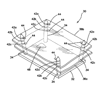
[01018] A surgical training model 30 according to the present invention
is
shown in FIG. 2. The model 30 is configured to be placed inside the surgical
training
device 10 described above or other surgical trainer similar to the one
described above.
The model 30 may also be used by itself without a laparoscopic trainer to
train or
practice certain procedures and surgical techniques. The model 30 includes a
base 32,
a plurality of posts 34, and at least one simulated tissue portion 36.
[0019] The base 32 of the model 30 is a platform that serves as a bottom
support for the rest of the model 30 and it is sized and configured such that
the model
30 does not tip over. The platform is made of any material such as metal or
plastic.
The base 32 is of sufficient heft to maintain the stability of the model 30 in
the upright
position while being manipulated by a user. The base 32 may include holes for
receiving posts 34. Alternatively, the posts 34 may be integrally form with
the base 32
as a unitary body. The model 30 is sized and configured to be placed into the
body
cavity 12 of the surgical trainer 10 in the location of the model receiving
area 24. The
underside of the base 32 is provided with means to affix the model 30 inside
the
surgical trainer 10. Such means to affix the model 30 inside the trainer 10
include but
are not limited to adhesive, suction cup, snap-fit, magnet, and a hook-and-
loop type
fastener material attached to the bottom surface of the base 32 and configured
to
connect with a complementary hook-and-loop type fastener material or adhesive
attached to the base 18 of the surgical trainer 30.
[0020] Still referencing FIG. 2, four posts 34 are connected to the base
32 of
the model 30 or, alternatively, the posts 34 are integrally formed with the
base 32. Each
post 34 is elongate and cylindrical in shape having a proximal end connected
to the
base 32 and a distal end that extends upwardly from the base 32. In one
variation, the
distal end includes a tapered section 38 that terminates at a blunt tip
surface 40 so as to
not injure a user but is sharp enough to puncture holes in simulated tissue.
In one
variation, as shown in FIG. 2, the distal end is conical or tapered and has
smoothly
curved, rounded or flat tip. Each post 34 includes at least one
circumferential notch 42
or cut that extends radially inwardly from the outer surface and into the post
34. In the
variation shown in FIG. 2, each post 34 includes three notches 42a, 42b, 42c
spaced
apart along the length of the post 34 although any number of notches may be
included
CA 02880482 2015-01-28
WO 2014/052478 PCT/US2013/061728
- 10 -
in the post 34. The notches 34 are perpendicular to the longitudinal axis of
the each
post 34. In one variation, all of the posts 34 have the same number of notches
34 in the
same locations or distances along the longitudinal axis. The posts 34 are
spaced apart
and located in substantially the four corners of the base 32. The posts 34 may
be
oriented perpendicular to the base 32 or angled outwardly as shown in FIG. 2
to help
retain a tensioned simulated tissue portion 36 or to allow for varied tension
in the
simulated tissue portion 36. In one variation, the posts are movable with
respect to the
base 32 such that their angle with respect to the base 32 can be selected by
the user in
order to vary the tension on the simulated tissue portion 26. In another
variation, the
angle of the posts 34 are not fixed but vary within constrained parameters
upon
manipulation of the connected simulated tissue portion 36 thereby increasing
the
difficulty for the clinician in performing the surgical technique. At least
one of the posts
34 angulates, shifts, tilts, wobbles or is movable with respect to base 32 in
response to
forces applied to the simulated tissue portion 36 by the practitioner. The
proximal end
of at least one post 34 is connected to the base 32 and configured such that
the post
angulates polyaxially or rotates polyaxially with respect to the base. In
another variation
at least one of the posts 34 is a flexible gooseneck which can be adjusted
with the
position being maintained by the gooseneck post 34 following the adjustment.
The
gooseneck post 34 is advantageous in adjusting the tension in the simulated
tissue
portion 36. The posts 34 are configured to support the simulated tissue
portion 36 and
to selectively locate and position the simulated tissue portion 36 in the
notches 34. If
the simulated tissue portion 36 is in the form of a sheet as shown in FIG. 2,
then the
thickness of the notches 42 is at least as thick as the thickness of the sheet
forming the
simulated tissue portion 36 such that the simulated tissue portion 36 is
supported within
and by the notches 42 and retained in the notches 42 along the posts 34 and
thereby
prevented from slipping or moving along the length of the post 34 as a
clinician
manipulates the simulated tissue portion 36. In one variation, the simulated
tissue
sheet 36 is approximately 0.05 inches thick and the notches are approximately
0.1
inches thick and the notches 42 are spaced apart by approximately 0.25 inches.
In
another variation, the notches 42 are thinner than the sheet 36 to slightly
compress the
sheet in position within the notch 42. For example, the notch 42 is
approximately 0.08
CA 02880482 2015-01-28
WO 2014/052478 PCT/US2013/061728
- 1 1 -
inches and the sheet is approximately 0.1 inches. One variation includes
mounting
posts that have notches that are formed at the same height. For example, a
post 34 is
approximately 4.0 inches long and includes first, second, third and fourth
notches
located at approximately 1.0 inch, 1.8 inches, 2.7 inches, and 3.7 inches,
respectively.
The outer diameter of the posts 34 are approximately 0.3 inches and the inner
diameter
of the posts 34 in the location of the notches is approximately 0.23 inches.
[0021] In one variation, the posts 34 are removable from the base 32.
The
base 32 includes four apertures and the posts 34 are passed into the apertures
from
underneath the base 32. Each post 34 is provided with a flange and each
aperture is
keyed for allowing the flanged post 34 to pass into the aperture. Once
inserted into the
aperture of the base 32, the post 34 is twisted relative to the base 32 to
lock the post 34
in position relative to the base 32. To remove the post 34, the post 34 is
twisted in the
opposite direction and pushed down through the aperture. The underside of the
base
32 includes an alcove provided with detents into which the posts 34 may be
snapped
into for flat storage of the model. Of course, rigid posts 34 may be
interchangeable with
flexible/movable ones.
[0022] Still referencing FIG. 2, the simulated tissue portion 36
includes a
sheet of simulated tissue material. In another variation, the simulated tissue
portion can
take the form and shape of a particular organ. The simulated tissue portion 36
is
connected to the posts 34 and in essence suspended from the upper surface of
the
base by a distance defined by the distance of the notch 42 to which the
simulated tissue
portion is attached. The simulated tissue portion 36 is free on all sides
except at the
points of support at the posts 34. The simulated tissue portion 36 is mounted
in tension
being slightly stretched between and connected to the posts 34. The tension of
the
sheet may be adjusted by angulating the posts 34 or by stretching and piercing
the
simulated tissue portion 36 in locations closer together along the simulated
tissue
portion. In one variation, the simulated tissue portion 36 is a sheet of
silicone. In
another variation the simulated tissue portion is a sheet of fabric or mesh
coated with
silicone on at least one side. The fabric or mesh is a 2-way or 4-way stretch
material
such as stretch nylon or spandex or a stretch nylon/spandex blend mesh or
fabric. The
fabric or mesh material is stretchable and porous and weighs approximately 79
grams
CA 02880482 2015-01-28
WO 2014/052478 PCT/US2013/061728
- 12 -
per square yard. The material of the sheet can be any polymeric material that
is flexible
and can stretch and may include a mesh or other reinforcement material or
fiber. The
silicone coating on the mesh provides a realistic tissue feel and may include
a textured
surface to provide the user with tactile feedback and to allow the user to
grab onto the
surface with graspers. The mesh, fabric, fiber or other filler material
provides
reinforcement to the silicone such that the sheet can hold a suture without
tearing or be
stretched without tearing when being manipulated or connected to the posts 34.
The
simulated tissue portion 36 may also be made of KRATON or other thermoplastic
elastomer.
[0023] In one variation, the simulated tissue portion 36 includes a
marking or
a predetermined pathway drawn on the upper surface of the at least one
simulated
tissue portion 36 with ink for example for the user to cut along. A shape may
also be
drawn which the user can practice cutting out. A pre-marked simulated tissue
portion
36 provides a starting point for the user. Also, a blank simulated tissue
portion 36
allows the user to draw their own line, path or shape on the simulated tissue
portion 36
that then the user can cut along employing laparoscopic scissors and
dissectors to
practice precision cuffing and then practice suturing the cut or opening
closed.
Furthermore, in one variation, the simulated tissue portion 36 includes pre-
formed
apertures 44 located along the perimeter at the four corners as shown in FIG.
2. These
apertures are approximately 0.125 inches in diameter and are set back from the
edges
by approximately 0.413 inches. The apertures 44 are located in the four
corners of the
sheet 36 and are used for mounting the simulated tissue portion 36 onto the
four posts
34 as shown. The simulated tissue portion 36 in the form of a sheet is
approximately 1
to 10 mm thick for example. In another variation, the simulated tissue portion
36 that is
formed in a sheet includes a textured upper surface and a smooth lower
surface. The
texturing can include protrusions or other realistic organ details. If
desired, the user
may flip the sheet such that the smooth surface is facing upwardly on the
posts. The
smooth surface may increase the difficulty in grasping and manipulating the
simulated
tissue portion with instruments. In another variation, the sheet of simulated
tissue 36
includes several pre-cut paths and/or holes which forces the user to maintain
tension on
CA 02880482 2015-01-28
WO 2014/052478 PCT/US2013/061728
- 13 -
the simulated tissue portion drawing opposite sides of the hole or pre-cut
path close
together for suturing.
[0024] In use, a user will mount at least one simulated tissue portion
36 onto
the posts 34. If the simulated tissue portion 36 includes preformed apertures
44 then
mounting the simulated tissue portion 36 includes placing the apertures 44
over each
post 34 and sliding the simulated tissue portion 36 to rest within one of the
at least one
notches 42 formed in the post 34. The simulated tissue portion 36 is mounted
on all
four posts 34. Fewer posts may be employed to suspend the simulated tissue
portion
36. The notches 42 advantageously permit the entire sheet 36 to be mounted at
an
angle such that one side or at least one corner of the simulated tissue
portion 36 is
mounted on a higher or lower notch relative to the other corners and posts.
For
example, one side of the simulated tissue portion 36 is connected to two posts
34 by
positioning the simulated tissue portion 36 along that first side to rest in
notches 42a
and the other side of the simulated tissue portion 36 is connected to two
posts 34 by
positioning the simulated tissue portion 36 along that second side to rest in
notches 42c
which are lower than notches 42a thereby angulating the simulated tissue
portion 36. If
the simulated tissue portion 36 is not provided with preformed apertures 44,
the tapered
distal ends 38 of the posts 34 can be used to puncture apertures 44 anywhere
into the
sheet 36. Hence, the tension in the simulated tissue portion 36 can be
selected by the
user when the user mounts the simulated tissue portion 36 onto the posts 34.
For
example, when the simulated tissue portion 36 is mounted by piercing an
aperture 44
into the simulated tissue portion 36, it can then be selectively stretched
making the
simulated tissue portion 36 as tense or loose as the user wishes before
piercing at least
a second aperture 44 to mount the simulated tissue portion on another post 34
and so
forth. The fabric reinforced silicone material prevents the aperture 44 from
propagating.
Multiple preformed apertures 44 can be included in the sheet 36 to provide
different
degrees of tension when the sheet is mounted using a specific set of preformed
apertures 44. As the simulated tissue portion 36 in the form of a sheet is
stretched over
a post, it then snaps into place inside one of the notches 42. The posts 34
may include
barbs, a shoulder or flange (not shown) extending outwardly from the outer
surface to
help retain the simulated tissue portion 36 in position together with or
without notches
CA 02880482 2015-01-28
WO 2014/052478 PCT/US2013/061728
- 14 -
42. The posts 34 allow the user to set the sheet to different tensions to
allow for
different levels of difficulty as well as different angles to represent
different structures or
locations within the body.
[0025] FIG. 3 shows a variation of the model 30 that includes more than
four
posts 34. In particular, there is a first or outer set of posts 34 and a
second inner set of
posts 46. There are four outer posts 34 and four inner posts 46 for a total of
eight
posts. The inner posts 46 are shorter relative to the outer posts 34. Both
sets of posts
are generally positioned in the four corners of the base 32 and adjacent to
each other.
Having two sets of posts allows greater variation or selectability in the
tension or angles
for mounting the simulated tissue portion 36. The second set of posts 46, like
the first
set of posts 34, includes notches 42 for positioning the simulated tissue
portion 36.
Although one notch 42 is shown in all of the posts 34, 46, the invention is
not so limited
and any number of notches at varying heights can be formed in the posts 34,
42. FIG. 3
does not illustrate the simulated tissue portion 36.
[0026] Turning now to FIG. 4, there is shown a model 30 according to the
present invention having two simulated tissue portions 36a, 36b mounted on the
posts
34. As shown the simulated tissue portions 36a, 36b are formed as sheets but
are not
so limited and may include shapes that simulate organs and other tissue
structures. A
first simulated tissue portion 36a is mounted onto the posts 34 and placed
into notches
42c and a second simulated tissue portion is shown mounted onto posts 34 and
placed
into notches 42a. Of course, the second sheet 36b can be placed into the same
notches as the first sheet 36a or angled in any manner with respect to the
first sheet
36a which may also be angled and placed in different notches. Placing the
sheets 36a,
36b in the same notches creates a layered tissue that can be used to mimic
muscle
tissue as found in the abdominal region. The sheets of simulated tissue 36 can
be any
color and include markings and vascular structures drawn on the simulated
tissue
structure 36 to mimic real tissue structures. The multiple sheets may all be
connected
together and retained with adhesive selectively applied in selected areas
between the
sheets. Although, two sheets 36a, 36b are shown, the invention is not limited
to the
number of sheets that can be mounted on the posts 34. The posts 34 can be
accordingly constructed to be longer and include more notches 42 to
accommodate
CA 02880482 2015-01-28
WO 2014/052478 PCT/US2013/061728
- 15 -
more sheets and a wider selection of angulations. FIG. 4 illustrates a
simulated tumor
48 located between the two sheets 36a, 36b. The tumor 48 can be attached to
one or
both of the layers 36a, 36b or not be attached. The clinician can practice
making an
incision in the second layer 36b to uncover the tumor 48, then practice
excising the
tumor 48 and then practice suturing the defect left behind in the first layer
36a if the
tumor 48 was attached to the first layer 36a and then practice suturing the
second layer
36b closed as well.
[0027] The model 30 is also suitable for use as a blunt dissection
model. The
simulated tissue sheet 36 for blunt dissection is made of silicone with no
fabric
reinforcement which allows the dissectors or trocars to puncture and separate
the
material. Multiple sheets may be layered together and attached together by
means of
silicone adhesive or thinner layers of silicone to allow for tissue
dissections and
separations of tissue planes.
[0028] The model 30 provides a realistic platform for presenting
simulated
tissue structures for training in a laparoscopic environment. As the clinician
practices
certain techniques such as cutting and suturing, the clinician will use
certain instruments
such as graspers, cutters, suture needles, sutures, laparoscopes, endoscopes,
trocars
and the like. When the simulated tissue structure that is supported on the
posts in the
model of the present invention is contacted with such instruments, the
simulated tissue
structure will give and flex under the force, deflecting a certain degree
depending upon
the tension with which it is mounted. This dynamism of the simulated tissue
structure
advantageously mimics real live tissue that gives way, moves and flexes upon
manipulation in real life. Also, cutting and suturing feels differently when
performed on
simulated tissue structure that is suspended, that is in tension and that
allows for a
certain amount of deflection. These simulation advantages are provided by the
model
30 of the present invention and are particularly useful when practicing
laparoscopic
surgical techniques that allow the user to fine tune depth perception and
tissue
manipulation skills while suturing, cutting and puncturing in a simulated
laparoscopic
environment.
[0029] While certain embodiments have been particularly shown and
described with reference to exemplary embodiments thereof, it will be
understood by
CA 02880482 2015-01-28
WO 2014/052478 PCT/US2013/061728
- 16 -
those of ordinary skill in the art that various changes in form and details
may be made
therein without departing from the spirit and scope thereof as defined by the
following
claims.