Note: Descriptions are shown in the official language in which they were submitted.
WO 2014/039782
PCT/US2013/058448
TITLE OF THE INVENTION
GENETICALLY MODIFIED MICE WHICH EXPRESS HUMAN CYTOKINES
AND METHODS OF USE THEREOF, INCLUDING ENGRAFTMENT
CROSS-REFERENCE TO RELATED APPLICATIONS
This application claims priority to U.S. Provisional Application Serial
No. 61/698,002, filed September 7, 2012, and to U.S. Provisional Application
Serial
No. 61/775,171, filed March 8, 2013.
BACKGROUND OF THE INVENTION
The aim of biomedical research is to gain a better understanding of
human physiology and to use this knowledge to prevent, treat or cure human
diseases.
Due to practical and ethical barriers to the experimentation on human
subjects, many
studies are conducted on small animal models, such as the mouse. However, mice
are
not people and the knowledge gained from animal experimentation is not always
applicable to humans. In this context, mice repopulated with a human hemato-
lymphoid system (HHLS) represent a useful small animal model for the study of
human hematopoiesis and immune function in vivo.
HHLS mice are generated by the transplantation of human
hematopoietic stem and progenitor cells (HSPCs) and/or human fetal tissues
into
recipient mice deficient in the innate and adaptive arms of the immune
response. The
first models of HHLS mice were developed in the late 1980s (Mosier et al.,
1988,
Nature 335:256-259; McCune et al., 1988, Science 241:1632-1639; Kamel-Reid and
Dick, 1988, Science 242:1706-1709), and have been undergoing a series of
improvements since then (Legrand et al., 2006, Journal of Immunology
176:2053-2058; Shultz et al., 2007, Nature Reviews Immunology 7:118-130). The
strains of mice currently used as recipients for human hematopoietic
engraftment
share three characteristics. First, they lack B and T cells due to the Scid
mutation in
the gene encoding the PRKDC protein (Mosier et al., 1988, Nature 335:256-259;
McCune et al., 1988, Science 241:1632-1639), or due to deletion of one of the
two
Rag genes
(Shultz et al., 2000, Journal of immunology 164:2496-2507; Traggiai et al.,
2004,
Science 304:104-107). Second, deletion or mutation of the 112rg gene that
encodes the
common gamma chain (ye) of cytokine receptors abolishes IL-15 signaling and
results
1
Date recue/ date received 2022-01-25
CA 02881468 2015-02-06
WO 2014/039782
PCT/US2013/058448
in the absence of NK cells (Traggiai et al., 2004, Science 304:104-107; Ito et
al. 2002,
Blood 100:3175-3182). Third, the interaction between the SIRPA receptor
expressed
on mouse macrophages and the CD47 ligand on human cells provides an inhibitory
signal to mouse macrophages and confers phagocytic tolerance for the human
xenograft (Takenaka et al., 2007, Nature Immunology 8:1313-1323; Takizawa &
Manz, 2007, Nature Immunology 8:1287-1289). Cross-species interaction between
SIRPA expressed on mouse cells and human CD47 is achieved when using the NOD
genetic background which contains a natural polymorphism in the Sirpa gene
(Takenaka et al., 2007, Nature Immunology 8:1313-1323; Takizawa & Manz, 2007,
Nature Immunology 8:1287-1289; Legrand et al., 2011, Proc Natl Acad Sci USA
108:13224-13229) or by BAC-transgenic expression of the human SIRPA gene
(Strowig et al., 2011, Proc Natl Acad Sci USA 108:13218-13223). High levels of
human hematopoietic cell engraftment, upon human HSPC transplantation, are
achieved when using NOD &id ye-I- (NOG (Ito et al. 2002, Blood 100:3175-3182)
or
NSG (Ishikawa et al., 2005, Blood 106:1565-1573)) or hSIRPAtg RAG2-/-y (SRG
(Strowig et al., 2011, Proc Natl Acad Sci USA 108:13218-13223)) mice as
recipients.
Although human multi-lineage hematopoietic development is observed
in these recipient strains, the terminal differentiation, homeostasis and/or
effector
function of most human cell types is sub-optimal. It has been hypothesized
that this
condition is due to reduced or absent cross-reactivity between cytokines
secreted by
mouse tissues and the human receptors expressed on hematopoietic cells (Manz,
2007, Immunity 26:537-541; Willinger et al., 2011, Trends in Immunology 32:321-
327). To circumvent this limitation, several strategies have been developed to
deliver
human cytokines in the mouse host. These methods include the injection of
recombinant cytokines (Lapidot et al., 1992, Science 255:1137-1141; van Lent
et al.,
2009, J. Immunol 183:7645-7655), lentiviral delivery of cytokine-encoding cDNA
(O'Connell et al., 2010, PloS One 5(8):e12009), hydrodynamic injection of
plasmid
DNA (Chen et al., 2009, Proc Natl Acad Sci USA 106:21783-21788), transgenic
expression of cDNA (Nicolini et al., et al., 2004, Leukemia 18(2):341-347;
Brehm et
al., 2012, Blood 119:2778-2788; Takagi et al., 2012, Blood 119:2768-2777) or
knock-
in replacement of cytokine-encoding genes (Rongvaux et al., 2011, Proc Natl
Acad
Sci USA 108:2378-2383; Willinger et al., 2011, Proc Natl Acad Sci USA 108:2390-
2395; Rathinam et al., 2011, Blood 118:3119-3128). The later method has the
advantage of more physiological expression of the human gene. Furthermore, if
the
2
CA 02881468 2015-02-06
WO 2014/039782
PCT/US2013/058448
human cytokine is not fully cross-reactive on the mouse receptor, it can
induce a
defect in mouse cell populations and confer an additional competitive
advantage to
human cells. Using a knock-in gene replacement strategy, humanization of the
gene
encoding thrombopoietin (Tpo) resulted in better maintenance of functional
human
hematopoietic stem cells and increased engraftment in the bone marrow
(Rongvaux et
al., 2011, Proc Natl Acad Sci USA 108:2378-2383); replacement of the genes
encoding interleukin-3 and GM-CSF (113 and OP) induced the loss of mouse lung
alveolar macrophages (AM) and the development of functional human AM
(Willinger
et al., 2011, Proc Natl Acad Sci USA 108:2390-2395); and substitution of the
Csfl
gene, which encodes M-CSF, resulted in increased numbers of human monocytes in
multiple tissues (Rathinam et al., 2011, Blood 118:3119-3128).
Human and mouse hemato-lymphoid systems differ in many aspects
(Haley, 2003, Toxicology 188:49-71; Mestas & Hughes, 2004, J Immunol 172:2731-
2738). One of the major differences between the two species lies in their
white blood
cell (WBC) differential. Human blood is rich in myeloid cells that represent
50-75%
of total WBCs. In contrast, mouse blood is dominated by lymphocytes and only
20-
30% of WBCs are of myeloid lineages. This species difference, whose functional
and
evolutionary significance is not understood, is not recapitulated in
conventional
HHI,S mice such as NOG/1\1-SG or SRG. Indeed, human myeloid development is
particularly defective in these hosts, with myeloid cells representing only 5-
10% of
human WBCs.
One application of mice with functional human immune systems is the
development and testing of human vaccines. Historically, the induction of
immune
responses in vivo has been relatively inefficient (2004, Traggiai et al.,
Science
304:104-107; 2002, Ito et al., Blood 100:3175-3182; 2005, Ishikawa et al.,
Blood
106:1565-1573; 2005, Shultz et al., J Immunol 174:6477-6489; 2006, Baenziger
et al.,
Proc Natl Acad Sci USA 103:15951-15956). Several studies have reported
successful
pathogen-specific immune responses upon infection. Although it was reported
that
around 50% of mice produced virus-specific 1gM and IgG upon dengue virus
infection (2007, Kuruvilla et al. Virology 369:143-152), other studies
reported
frequencies below 20% of mice producing antigen-specific IgM and IgG after HIV
and EBV infection (2006, Baenziger et al., Proc Natl Acad Sci USA 103:15951-
15956; 2008, Yajima et al., J Infect Dis 198:673-682). Upon immunization with
adjuvant and antigen, class switching of antigen-specific immunoglobulins is
also
3
CA 02881468 2015-02-06
WO 2014/039782
PCT/US2013/058448
historically inefficient with only a fraction of immunized animals showing
antigen
specific IgG responses (2004, Traggiai et al., Science 304:104-107; 2002, Ito
et al.,
Blood 100:3175-3182; 2005, Ishikawa etal., Blood 106:1565-1573; 2005, Shultz
et
al., J Immunol 174:6477-6489; 2009, Watanabe etal., Int Immunol 21:843-858;
2010,
Becker et al., PLoS ONE 5). These studies included NSG and BALB/c RAG2-1 7,1
mice and different adjuvant/antigen combinations.
There is a need in the art for humanized non-human animals able to
support and sustain engraftment with human hematopoietic cells. The present
invention addresses this unmet need in the art.
SUMMARY OF THE INVENTION
The invention relates generally to genetically modified non-human
animals expressing at least one of human M-CSF, human IL-3, human GM-CSF,
human SIRPA or human TPO, as well as to their methods of use. Thus, in one
embodiment, the invention is a genetically modified non-human animal
comprising a
genome comprising at least one nucleic acid encoding at least one of the group
consisting of human M-CSF, human IL-3, human GM-CSF, human SIRPA and
human TPO, where the at least one nucleic acid is operably linked to a
promoter, and
where the animal expresses at least one polypeptide selected from the group
consisting of human M-CSF, human IL-3, human GM-CSF, human SIRPA and
human TPO. In another embodiment, the invention is a genetically modified non-
human animal, comprising a genome comprising a nucleic acid encoding human M-
CSF, a nucleic acid encoding human IL-3, a nucleic acid encoding human GM-CSF,
a
nucleic acid encoding human SIRPA and a nucleic acid encoding human TPO, where
each of the nucleic acids encoding human M-CSF, human IL-3, human GM-CSF,
human SIRPA and human TPO is operably linked to a promoter, and where the
animal expresses human M-CSF polypeptide, human IL-3 polypeptide, human GM-
CSF polypeptide, human SIRPA polypeptide and human TPO polypeptide. In some
embodiments, the genetically modified non-human animal is immunodeficient. In
some embodiments, the genetically modified non-human animal does not express
recombination activating gene 2 (Rag-2-/-). In some embodiments, the
genetically
modified non-human animal does not express IL2 receptor gamma chain (gamma
chain-/-). In some embodiments, the genetically modified non-human animal does
not
express Rag-2 and the genetically modified non-human animal does not express
IL2
4
CA 02881468 2015-02-06
WO 2014/039782
PCT/US2013/058448
receptor gamma chain (Rag-2-/- gamma chain-/-). In some embodiments, the
genetically modified non-human animal is a rodent. In some embodiments, the
genetically modified non-human animal is a mouse. In one embodiment, the
genetically modified non-human animal also includes at least one human
hematopoietic cell. In one embodiment, the genetically modified non-human
animal
also includes at least one human cancer cell. In some embodiments, the human
cancer
cell is a leukemia cell or a melanoma cell.
In another embodiment, the invention is a method of hematopoietic
stem and progenitor cell (HSPC) engraftment in a genetically modified non-
human
animal, where the animal expresses at least one of the group consisting of
human M-
CSF, human IL-3, human GM-CSF, human SIRPA and human TPO, the method
comprising the step of: administering at least one HSPC to the genetically
modified
animal expressing at least one of the group consisting of human M-CSF, human
IL-3,
human GM-CSF, human SIRPA and human TPO. In some embodiments, the HSPC is
a human HSPC. In one embodiment, the genetically modified non-human animal is
a
rodent. In one embodiment, the genetically modified non-human animal is a
mouse.
In one embodiment, the genetically modified non-human animal is
immunodeficient.
In one embodiment, the genetically modified immunodeficient non-human animal
does not express recombination activating gene 2 (Rag-2-/-). In one
embodiment, the
genetically modified immunodcficient non-human animal does not express
endogenous IL2 receptor (gamma chain-/-). In one embodiment, the genetically
modified immunodeficient non-human animal does not express endogenous Rag-2
and does not express endogenous gamma chain (Rag-2-/- gamma chain-/-). In one
embodiment, the genetically modified animal comprises a human cancer cell. In
one
embodiment, the human cancer cell is a leukemia cell or a melanoma cell.
In another embodiment, the invention is a genetically modified Rag-2-
/-, gamma chain-/- mouse having a genome comprising at least one nucleic acid
encoding at least one of the group consisting of human M-CSF, human IL-3,
human
GM-CSF, human SIRPA and human TPO, where the at least one nucleic acid is
operably linked to at least one promoter, where the mouse expresses at least
one
polypeptide selected from the group consisting of human M-CSF, human IL-3,
human
GM-CSF, human SIRPA and human TPO. In one embodiment, the genetically
modified non-human animal comprises a genome having a nucleic acid encoding
human M-CSF, a nucleic acid encoding human IL-3, a nucleic acid encoding human
5
CA 02881468 2015-02-06
WO 2014/039782
PCT/US2013/058448
GM-CSF, a nucleic acid encoding human SIRPA and a nucleic acid encoding human
TPO, where each of the nucleic acids encoding human M-CSF, human IL-3, human
GM-CSF, human SIRPA and human TPO is operably linked to a promoter, and where
the animal expresses human M-CSF polypeptide, human IL-3 polypeptide, human
GM-CSF polypeptide, human SIRPA polypeptide and human TPO polypeptide. In
one embodiment, the genetically modified non-human animal is a rodent. In one
embodiment, the genetically modified non-human animal is a mouse. In one
embodiment, the genetically modified non-human animal comprises a human
hematopoietic cell. In one embodiment, the genetically modified non-human
animal
.. comprises a human cancer cell. In some embodiments, the human cancer cell
is a
leukemia cell or a melanoma cell.
BRIEF DESCRIPTION OF THE DRAWINGS
The following detailed description of preferred embodiments of the
.. invention will be better understood when read in conjunction with the
appended
drawings. For the purpose of illustrating the invention, there are shown in
the
drawings embodiments which are presently preferred. It should be understood,
however, that the invention is not limited to the precise arrangements and
instnirnentalities of the embodiments shown in the drawings.
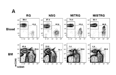
Figure 1, comprising Figures 1A-1E, depicts the results of experiments
showing that MISTRG mice support high levels of human hematopoietic
engraftment.
X-ray pre-conditioned newborn mice of the indicated strains were engrafted by
intra-
hepatic injection of 100,000 human fetal liver-(FL-)CD34' cells. Human
engraftment
levels (hCD45' cells) were measured in the blood 7-9 weeks later, and in the
BM 10-
12 weeks later. (Figure 1A) Representative flow cytometry analysis of the
frequency
of mouse and human CD45 cells in the blood and BM of the indicated recipient
mice. Numbers next to gated areas indicate percentages among total CD45 cells.
(Figure 1B) Combined data of blood engraftment levels (% hCD45' cells) from 19
independent experiments. In each experiment, a single FL-CD34 cell sample was
.. split and injected into mice of the respective strains. Each symbol
represents an
individual mouse and the red bars indicate mean values (n=56-155; ns, not
sipificant;
* p<0.05 Tukey test (see Figure 6 for a complete statistical analysis). The
gray
horizontal line indicates 10% hCD45' cells. (Figure IC) Engraftmcnt levels in
the
BM of a representative subset of mice (Figure 6C) from panel (Figure 6B) (n=12-
16;
6
CA 02881468 2015-02-06
WO 2014/039782
PCT/US2013/058448
* p<0.05 Tukey test; see also Figures 6D-6E). (Figure 1D) Representative flow
cytometry analysis of hCD45 cell engraftment in the blood and BM 3 months
after
intra-hepatic injection of 200,000 FL-CD34 cells into non-irradiated newborn
MISTRG mice. (Figure 1E) Human CD45 cell engraftment levels in the blood and
BM of MISTRG mice transplanted as in (Figure 1D) (n=16). In this case, the BM
of
all mice (including mice with blood hCD45' < 10%) are shown.
Figure 2, comprising Figures 2A-2K, depicts the results of experiments
showing that MISTRG mice support efficient myeloid development and maintenance
in lymphoid and non-lymphoid tissues. (Figure 2A) Percentages of human myeloid
cells (hCD33) among human hematopoietic cells (hCD45-) in the blood of the
indicated recipient mice, engrafted as newborns by intra-hepatic injection of
FL-
CD34 cells after X-ray preconditioning. Each symbol represents an individual
mouse
and the red bars indicate mean values (n=20-113; statistical analysis is shown
in
Figure 7A). (Figure 2B) Human WBC composition in the same mice (n=20-113
mice/group; n=8 human donors; error bars indicate SEM). (Figure 2C)
lmmunohistological staining of human myeloid cells (hC1)68') in non-lymphoid
tissues of the indicated recipient mice. The black bar represents 20 um, and
the
images shown are representative of at least three mice analyzed per group.
(Figure 2D
and Figure 2E) Representative flow cytometry analysis (Figure 2D) and
frequencies
(Figure 2E) of human monocyte subsets, identified by expression of CD14 and
CD16
among hCD45'CD33 cells in the blood of recipient mice (n=8-12 mice/group;
error
bars indicate SEM). (Figure 2F and Figure 2G) Cytokine production by human
monocytes isolated from the BM of MITRG recipients and stimulated in vitro
with
LPS (Figure 2F) or R848 (Figure 2G) (error bars indicate SD of triplicates;
representative of 3 independent experiments). (Figure 2H) In vitro
phagocytosis of
GFP-expressing E.coli by human cells present in the blood of MITRG mice (n=7).
(Figures 21, 2J, 2K) In vivo cytokine production measured by ELISA in the
serum or
by RT-PCR in the lung of mice treated with LPS (Figure I; 90 min, n=15-18), or
infected with Listeria monocytogenes (Figure 2J; day 2, n=6-15) or influenza
A/PR8
H1N1 (Figure 2K; day 3, n=3-5). (Figures 2A, 2J, 2K) p-values calculated by
one-
way ANOVA followed by Tukey posthoc test (* p<0.05); (Figure 21) p-value
calculated by unpaired Student's t-test on logl 0-transformed values.
Figure 3, comprising Figures 3A-3I, depicts the results of experiments
showing that MISTRG mice efficiently support the development and function of
7
CA 02881468 2015-02-06
WO 2014/039782
PCT/US2013/058448
human NK cells. (Figure 3A) Quantitative RT-PCR analysis of human IL-15 and IL-
15Ra mRNA expression in the liver of engrafted NSG, MITRG, and MISTRG mice
(n=7-8; p-values calculated by one-way ANOVA; *, p<0.05 Tukey post hoc test).
Expression was normalized to mouse Hprt. (Figure 3B) Quantitative RT-PCR
analysis of human IL-15 and IL-15Ra mRNA expression in human cell populations
purified from bone marrow of engrafted MITRG (n=4-5, error bars indicate SEM).
Expression was normalized to human HPRT and is shown relative to hCD14 hCD16-
cells. (Figure 3C and Figure 3D) Representative flow cytometry analysis (gated
on
hCD45 'mCD45- cells, lymphocyte gate; numbers next to outlined areas indicate
percentages of cells) (Figure 3C) and absolute number or frequency (Figure 3D)
of
human NK cells (hNKp46' hCD3-) in engrafted NSG, MITRG, and MISTRG (n=8-
16; p-values calculated by one-way ANOVA; *, p<0.05 Tukey post hoc test).
(Figure
3E) Absolute number of human liver NK (hNKp46'hCD3-) and T cells (hCD3
shown as control) from engrafted MISTRG mice either left untreated or treated
for 3
consecutive days with liposome-encapsulated clodronate to deplete phagocytic
cells
(n=8; p-value calculated by unpaired Student's t-test; ns, not significant).
(Figure 3F)
Labeled LCL721.221 (HLA class I negative) and LCL721.45 (class I positive)
cells
were injected i.v. in a 1:1 ratio, and the proportions of HLA class I positive
or
negative, among labeled cells recovered 12 hours later in the spleen, were
used to
calculate specific NK cell cytotoxicity (n=8, p-value calculated by unpaired
student's
t-test). (Figure 3G) Quantitative RT-PCR analysis of human IFNy mRNA
expression
in the liver of NSG and MISTRG mice 2 days after Listeria infection (n=8-9, p-
value
calculated by unpaired student's t-test). Expression was normalized to mouse
Hprt.
(Figure 3H and Figure 31) Representative flow cytometry analysis (Figure 3H)
and
frequency (Figure 31) of IFNy-expressing and degranulating (CD107a) human
liver
NK cells from either uninfected or Listeria-infected NSG and MISTRG mice (n=4-
11;
p-value calculated by one-way ANOVA). Results are combined from two (Figures
3A, 3E-31), three (Figure 3B), or four (Figures 3C, 3D) experiments.
Figure 4, comprising Figures 4A-4F, depicts the results of experiments
showing that human myeloid cells in MISTRG infiltrate a tumor and support its
growth. The human melanoma cell line Me290 was implanted in the flank of
engrafted or non-engrafted NSG and MISTRG mice. Some mice were treated with
the
VEGF-inhibitor AvastinTm. The tumors were measured and dissected for analysis
11
8
CA 02881468 2015-02-06
WO 2014/039782
PCT/US2013/058448
days later. (Figure 4A) Infiltration of human hematopoietic cells in the
tumor,
determined by the expression of mRNA encoding human hematopoietic (PTPRC,
encoding CD45) and myeloid (ITGAII, encoding CD1 1 b) markers (n=6-7; p-value
calculated by unpaired Student's t-test). (Figure 4B and Figure 4D)
Representative
immunohistochemistry pictures of human myeloid cell markers in tumors from
NSG,
MISTRG and patients. (Figure 4C) Quantification of the density of CD163+ cells
(n=3
samples/group, 3 slides counted/sample). (Figure 4E and Figure 4F)
Representative
pictures (Figure 4E) and volume (Figure 4F) of the tumors in the indicated
groups of
mice (n=7-24 mice/group). p-values were calculated by Student's t-test (Figure
4A) or
by one-way ANOVA (Figures 4C, 4E) followed by Tukey posthoc test (* p<0.05).
Figure 5 depicts cytokines involved in HSC function and myeloid
development. Schematic representation of hematopoietic stem cell development
into
myeloid cells and non-exhaustive list of cytokines known to regulate this
process.
Shading indicates the percentages of amino acid identity between human and
mouse
cytokines. The percentage of amino acid identity is the most objective measure
of
protein conservation between species, but it does not always correlate with
functional
inter-species cross-reactivity in vivo. Black rectangles indicate cytokines
that are
genetically humanized in MISTRG. HSC, hematopoietic stem cell; MPP,
multipotent
progenitor; CMP, common myeloid progenitor; GMP, granulocyte/macrophage
progenitor; MEP, megakaryocyte/erythrocyte progenitor.
Figure 6, comprising Figures 6A-6E, depicts the results of statistical
analysis of engraftment levels in recipient mice. (Figure 6A) Statistical
analysis (one-
way ANOVA followed by Tukey post-hoc test; ns, not significant) of the data
presented in Figure 1A (percentage of hCD45 cells in the blood of recipient
mice).
(Figure 6B) Numbers of recipient mice that reach an engraftment level of at
least 10%
hCD45 cells in the blood 7-9 weeks after transplantation. (Figure 6C) Blood
engraftment levels of the mice used in Figure IC for analysis of the BM.
(Figure 6D)
Statistical analysis, similar to (Figure 6A), of the data presented in Figure
IC
(percentage of hCD45+ cells in the BM of recipient mice). (Figure 6E) Absolute
numbers of hCD45' cells in the BM (2 femurs and 2 tibias) of recipient mice
shown
in Figure IC. The reduced numbers of cells in the BM of MISTRG is due to the
smaller size of the mice at that age (10-12 weeks post-transplantation) and is
caused
by the first clinical signs of anemia described in detail in Figure 10.
9
CA 02881468 2015-02-06
WO 2014/039782
PCT/US2013/058448
Figure 7, comprising Figures 7A-7H, depicts the results of experiments
assessing enhanced human myeloid development in MISTRG mice. (Figure 7A)
Statistical analysis (one-way ANOVA followed by Tukey post-hoc test; ns, not
significant) of the data presented in Figure 2A (percentage of hCD33' cells in
the
blood of recipient mice). (Figure 7B and Figure 7C) Frequencies (Figure 7B)
and
statistical analysis (Figure 7C) of human myeloid cells (hCD33') in the BM of
recipient mice. (Figure 7D) Representative flow cytometry analysis of human
lymphoid and myeloid lineages in the blood of MISTRG. (Figure 7E and Figure
7F)
Representative flow cytometry analysis of human monocytes (CD33h1SSC'0CD66-)
and granulocytes (CD33 'SSChiCD66') in the BM (Figure 7E) and blood (Figure
7F)
of MISTRG and human donor. (Figure 7G and Figure 7H) Absolute numbers of
human myeloid cells (hCD33-) in the lung (Figure 7G) and liver (Figure 7H) of
recipient mice (n=8-12; p-values calculated by one-way ANOVA followed by Tukey
posthoc test, * p<0.05).
Figure 8, comprising Figures 8A and 8B, depicts the results of
experiments showing enhanced development of human monocyte subsets in MISTRG
mice. (Figure 8A) Representative flow cytometry analysis of human monocyte
subsets, identified by expression of CD14 and CD16 among hCD45H CD33 cells in
the BM, spleen, lung and liver of the indicated recipient mice. (Figure 8B)
Frequencies (error bars represent SEM) among hCD33 cells and absolute numbers
of
monocyte subsets in the lung and liver of recipient mice (n=12 mice/group; p-
values
calculated by one-way ANOVA; *, p<0.05 Tukey post hoc test).
Figure 9, comprising Figures 9A and 9B, depicts the results of
experiments showing that human monocyte subsets are similar in MISTRG and in
human donors. Extended immunophenotype of the indicated subsets of human
monocytes in the blood (Figure 9A) and BM (Figure 9B) of MISTRG recipients and
human donor. Staining with isotype control antibodies and specific antibodies
is
shown.
Figure 10, comprising Figures 10A-1M, depicts the results of
experiments showing that human myeloid cells breach human-to-mouse phagocytic
tolerance. (Figure 10A) CFSE-labeled mouse RBCs were transferred into the
indicated mice and the frequency of labeled cells was measured at the
indicated time
points. (Figure 103) Engrafted MISTRG were pre-treated or not with clodronate
to
deplete phagocytic cells and CFSE-labeled mouse RBCs were transferred and
CA 02881468 2015-02-06
WO 2014/039782
PCT/US2013/058448
monitored as in (Figure 10A) (p-value, clodronate-effect measured by repeated
measure ANOVA for days 1-3). These results show that transferred mouse RBCs
are
rapidly cleared in vivo by phagocytic cells that are present in MISTRG but not
in
NSG. (Figure 10C) RBC counts in the blood of non-engrafted mice (n=9-15) or 8-
10
weeks after engraftment with human FL-CD34 cells (n=11-37). p-values indicate
comparison between non-engrafted and engrafted mice of each genotype
(Student's
unpaired t test). (Figure 10D) Correlation between human engraftment levels
(percentage of hCD45- cells in the blood) and RBC counts (n=13-22). (Figure
10E)
Flow cytometry analysis of mouse (mTer119 ) and human (hCD235a-) erythroid
cells
in the blood of non-engrafted or engrafted MISTRG, showing that almost all
erythroid
cells in the blood of engrafted MISTRG are of mouse origin, and human
erythroid
cells are barely detectable. (Figure 10F) Representative pictures and spleen
weight of
engrafted mice of the indicated strains (n=3-22), showing splenomegaly in
engrafted
MISTRG mice. Spleens from Balb/c mice were used as a control (p-value, one-way
ANOVA; *, p<0.05 compared to all other groups, Tukey posthoc test). (Figure
10G)
Histological section of the spleen of engrafted N SG and MISTRG stained with
H&E,
illustrating the enlargement of the red pulp in MISTRG mice with splenomegaly.
(Figure 10H) Flow cytometry analysis of mouse erythroid progenitors
(mTerl 19 'mCD71 which represent up to 80% of the cells in the spleen of
engrafted
MISTRG. (Figure 101) Blood smears of non-engrafted and engrafted MISTRG
illustrate enrichment in reticulocytes. Taken together, these results strongly
suggest
that anemia in MISTRG results from the absence of human-to-mouse phagocytic
tolerance, and massive extra-medullary mouse erythropoiesis fails to
compensate for
the destruction of mRBCs. Results are representative of at least 5 mice
examined in
each group (Figures 10C, 10E-10I) and 2 independent experiments (Figures 10A,
10B).
Figure 11, comprising Figures 11A and 11B, depicts the results of
experiments showing that MISTRG mice provide human IL-15/ IL-15Ra. (Figure
11A) Quantitative RT-PCR analysis of human IL-15 and 1L-15Ra mRNA expression
in the lung of engrafted NSG, MITRG, and MISTRG mice (n=7-8; p-values
calculated by one-way ANOVA; *, p<0.05 Tukey post hoc test). Expression was
normalized to mouse Ifprt. (Figure 11B) Flow cytometry analysis of IL-15Ra
expression on human cell populations (hCD45 'mCD45-) from blood of engrafted
MISTRG mice (representative of n=4). Histograms represent staining with
isotype
11
CA 02881468 2015-02-06
WO 2014/039782
PCT/US2013/058448
control or with IL-15Ra antibody, respectively. Results are representative of
or
combined from two experiments.
Figure 12, comprising Figures 12A and 12B, depicts the results of
experiments showing enhanced human NK cell development in MISTRG mice.
(Figure 12A and Figure 12B) Frequency (Figure 12A) and absolute number (Figure
12B) of human NK cells (hNKp46 hCD3-) in engrafted NSG, MITRG, and MISTRG
mice (n=8-16; p-values calculated by one-way ANOVA; *, p<0.05 Tukey post hoc
test). Results are combined from four experiments.
Figure 13, comprising Figures 13A-13F, depicts the results of
experiments showing that bona fide human NK cells exhibiting enhanced
maturation
are present in MISTRG mice. (Figure 13A) Flow cytometry analysis of CD94 and
CD161 expression on human blood NK cells from a human donor and engrafted
MISTRG (n=3). Histograms represent staining with isotype control Abs or with
CD94/CD161 Abs. (Figure 13B) Flow cytometry analysis of KIR expression on
human blood NK cells from a human donor or from engrafted MISTRG mice (n=3).
Numbers indicate frequencies of KIR' cells. (Figure 13C and Figure 131)) CD16
surface expression on human NK cells from engrafted NSG, MITRG, and MISTRG
mice (n=4-8; p-values calculated by one-way ANOVA; *, p<0.05 Tukey post hoc
test) (Figure 13F, and Figure 13F) Tiftracellular perforin expression by human
liver
NK (hNKp46 11CD3-) and T cells (hCD3') from engrafted NSG and MISTRG mice
(n=3; p-value calculated by unpaired Student's t-test). MFI, mean fluorescence
intensity. Results are representative of or combined from one (Figure 13A and
Figure
13B), two (Figure 13E and Figure 13F), or four (Figure 13C and Figure 13D)
experiments.
Figure 14 depicts the results of experiments showing the effect of
human monocyte/macrophage depletion on human NK cell homeostasis in MISTRG
mice. Engrafted MISTRG mice were left untreated or treated for 3 consecutive
days
with liposome-encapsulated clodronate to deplete phagocytic cells. Flow
cytometry
analysis of human monocytes/macrophages (upper panel, gated on hCD33' cells)
and
NK cells (hNKp46' hCD3-) in liver (n=8) is shown. Results are representative
of two
experiments. In 1 out of 8 mice, the clodronate-depletion of
monocytes/macrophages
was not effective, and no reduction in NK cell number was observed in that
mouse.
Figure 15 depicts the results of experiments showing
immunohistochemistry of human myeloid cells infiltrating melanoma.
Representative
12
CA 02881468 2015-02-06
WO 2014/039782
PCT/US2013/058448
immunohistochemistry staining of human myeloid cells in tumors from NSG,
MISTRG or human patients. Three subject per group, and 3 pictures per subject
are
shown.
Figure 16 shows a comparison of engraftment levels and immune cell
development and function in recipient mice with single gene replacement, in
NSG,
MISTRG and in humans.
Figure 17, comprising Figure 17A-17D, depicts the results of
experiments demonstrating that samples isolated from patients with AML, CMML
and MDS can be engrafted in MISTRG. (Figure 17A) Characteristics of the
samples
used (including type of disease and genetic abnormality found in patient
samples),
experimental protocol (method of cell purification, number of cells injected
per mouse
and time post-transplantation at which mice were analyzed) and engraftment
results
(including number of mice with detectable human engraftment, percentage of
human
hematopoietic CD45+ cells and myeloid CD33+ cells, and genomic abnormality
observed in human cells isolated from the mice). (Figure 17B) Representative
flow
cytometry analysis of the granularity (SSC) of myeloid CD33+ cells isolated
from a
mouse transplanted with RAEB I patient or with normal donor cells, showing
deficient granularity in RAEB I samples. (Figure 17C) Representative fish
analysis of
human cells isolated from mice transplanted with RAER TT sample and showing
absence of chromosome 5q. (Figure 17D) Caryotype of human cells isolated from
mice transplanted with CMML sample and showing deletion in chromosome 6.
DETAILED DESCRIPTION
The invention relates generally to a genetically modified non-human
animal expressing at least one of human M-CSF, human IL-3, human GM-CSF,
human SIRPA or human TPO. The invention also relates to methods of generating
and methods of using the genetically modified non-human animals described
herein.
In some embodiments, the genetically modified non-human animal is a mouse. In
some embodiments, the genetically modified non-human animal described herein
is
engrafted with human hematopoietic cells. In various embodiments, the human
hematopoietic cell engrafted, genetically modified non-human animals of the
invention are useful for the in vivo evaluation of the growth and
differentiation of
hematopoietic and immune cells, for the in vivo evaluation of human
hematopoiesis,
for the in vivo evaluation of cancer cells, for the in vivo assessment of an
immune
13
CA 02881468 2015-02-06
WO 2014/039782
PCT/US2013/058448
response, for the in vivo evaluation of vaccines and vaccination regimens, for
the use
in testing the effect of agents that modulate cancer cell growth or survival,
for the in
vivo evaluation of a treatment of cancer, for the in vivo production and
collection of
immune mediators, including human antibodies, and for use in testing the
effect of
agents that modulate hematopoietic and immune cell function.
Definitions
Unless defined otherwise, all technical and scientific terms used herein
have the same meaning as commonly understood by one of ordinary skill in the
art to
which this invention belongs. Such terms are found defined and used in context
in
various standard references illustratively including J. Sambrook and D. W.
Russell,
Molecular Cloning: A Laboratory Manual, Cold Spring Harbor Laboratory Press;
3rd
Ed., 2001; F. M. Ausubel, Ed., Short Protocols in Molecular Biology, Current
Protocols; 5th Ed., 2002; B. Alberts et al., Molecular Biology of the Cell,
4th Ed.,
Garland, 2002; D. L. Nelson and M M Cox, Lebninger Principles of Biochemistry,
4th Ed., W.H. Freeman & Company, 2004; and Herdewijn, P. (Ed.),
Oligonucleotide
Synthesis: Methods and Applications, Methods in Molecular Biology, Humana
Press,
2004. Although any methods and materials similar or equivalent to those
described
herein can be used in the practice or testing of tile present invention, the
preferred
methods and materials are described.
As used herein, each of the following terms has the meaning associated
with it in this section.
The articles "a" and "an" are used herein to refer to one or to more
than one (i.e., to at least one) of the grammatical object of the article. By
way of
example, "an element" means one element or more than one element.
"About" as used herein when referring to a measurable value such as
an amount, a temporal duration, and the like, is meant to encompass variations
of
20% or 10%, more preferably 5%, even more preferably 1%, and still more
preferably 0.1% from the specified value, as such variations are appropriate
to
perform the disclosed methods.
The term "abnormal" when used in the context of organisms, tissues,
cells or components thereof, refers to those organisms, tissues, cells or
components
thereof that differ in at least one observable or detectable characteristic
(e.g., age,
treatment, time of day, etc.) from those organisms, tissues, cells or
components
14
CA 02881468 2015-02-06
WO 2014/039782
PCT/US2013/058448
thereof that display the "normal" (expected) respective characteristic.
Characteristics
which are normal or expected for one cell or tissue type, might be abnormal
for a
different cell or tissue type.
The term "antibody," as used herein, refers to an immunoglobulin
molecule which is able to specifically bind to a specific epitope on an
antigen.
Antibodies can be intact immunoglobulins derived from natural sources or from
recombinant sources and can be immunoreactive portions of intact
immunoglobulins.
The antibodies in the present invention may exist in a variety of forms
including, for
example, polyelonal antibodies, monoclonal antibodies, intracellular
antibodies
("intrabodies"), Fv, Fab and F(ab)2, as well as single chain antibodies
(scFv), heavy
chain antibodies, such as camelid antibodies, and humanized antibodies (Harlow
et
al., 1999, Using Antibodies: A Laboratory Manual, Cold Spring Harbor
Laboratory
Press, NY; Harlow et al., 1989, Antibodies: A Laboratory Manual, Cold Spring
Harbor, New York; Houston et al., 1988, Proc. Natl. Acad. Sci. USA 85:5879-
5883;
Bird et al., 1988, Science 242:423-426).
the term -cancer" as used herein is defined as disease characterized by
the uncontrolled proliferation and/or growth of aberrant cells. Cancer cells
can spread
locally or through the bloodstream and lymphatic system to other parts of the
body.
Cancer as here herein includes both solid tumors and fiematopoietic
malignancies
Examples of various cancers amenable to the invention include, but are not
limited to,
breast cancer, prostate cancer, ovarian cancer, cervical cancer, skin cancer,
pancreatic
cancer, colorectal cancer, renal cancer, liver cancer, bone cancer, brain
cancer,
lymphoma, leukemia, lung cancer, myeloidysplastic syndromes,
myeloproliferative
disorders and the like.
"Constitutive" expression is a state in which a gene product is
produced in a living cell under most or all physiological conditions of the
cell.
A "coding region" of a gene consists of the nucleotide residues of the
coding strand of the gene and the nucleotides of the non-coding strand of the
gene
which are homologous with or complementary to, respectively, the coding region
of
an mRNA molecule which is produced by transcription of the gene.
A "coding region" of a mRNA molecule also consists of the nucleotide
residues of the mRNA molecule which are matched with an anti-codon region of a
transfer RNA molecule during translation of the mRNA molecule or which encode
a
stop codon. The coding region may thus include nucleotide residues comprising
CA 02881468 2015-02-06
WO 2014/039782
PCT/US2013/058448
codons for amino acid residues which are not present in the mature protein
encoded
by the mRNA molecule (e.g., amino acid residues in a protein export signal
sequence).
A "disease" is a state of health of an animal wherein the animal cannot
maintain homeostasis, and wherein if the disease is not ameliorated then the
animal's
health continues to deteriorate.
In contrast, a "disorder" in an animal is a state of health in which the
animal is able to maintain homeostasis, but in which the animal's state of
health is
less favorable than it would be in the absence of the disorder. Left
untreated, a
disorder does not necessarily cause a further decrease in the animal's state
of health.
A disease or disorder is "alleviated" if the severity of a symptom of the
disease or disorder, the frequency with which such a symptom is experienced by
a
patient, or both, is reduced.
An "effective amount" or "therapeutically effective amount" of a
.. compound is that amount of compound which is sufficient to provide a
beneficial
effect to the subject to which the compound is administered. An -effective
amount" of
a delivery vehicle is that amount sufficient to effectively bind or deliver a
compound.
"Encoding" refers to the inherent property of specific sequences of
nucleotides in a polynucleotide, such as a gene, a cDNA, or an mRNA, to serve
as
templates for synthesis of other polymers and macromolecules in biological
processes
having either a defined sequence of nucleotides (i.e., rRNA, tRNA and mRNA) or
a
defined sequence of amino acids and the biological properties resulting
therefrom.
Thus, a gene encodes a protein if transcription and translation of mRNA
corresponding to that gene produces the protein in a cell or other biological
system.
Both the coding strand, the nucleotide sequence of which is identical to the
mRNA
sequence and is usually provided in sequence listings, and the non-coding
strand, used
as the template for transcription of a gene or cDNA, can be referred to as
encoding the
protein or other product of that gene or cDNA.
As used herein "endogenous" refers to any material from or produced
inside an organism, cell, tissue or system.
As used herein, the term "exogenous" refers to any material introduced
from or produced outside an organism, cell, tissue or system.
The terms "expression construct" and "expression cassette" are used
herein to refer to a double-stranded recombinant DNA molecule containing a
desired
16
CA 02881468 2015-02-06
WO 2014/039782
PCT/US2013/058448
nucleic acid human coding sequence and containing one or more regulatory
elements
necessary or desirable for the expression of the operably linked coding
sequence.
As used herein, the term "fragment," as applied to a nucleic acid or
polypeptide, refers to a subsequence of a larger nucleic acid or polypeptide.
A
"fragment" of a nucleic acid can be at least about 15 nucleotides in length;
for
example, at least about 50 nucleotides to about 100 nucleotides; at least
about 100 to
about 500 nucleotides, at least about 500 to about 1000 nucleotides, at least
about
1000 nucleotides to about 1500 nucleotides; or about 1500 nucleotides to about
2500
nucleotides; or about 2500 nucleotides (and any integer value in between). A
"fragment" of a polypeptide can be at least about 15 nucleotides in length;
for
example, at least about 50 amino acids to about 100 amino acids; at least
about 100 to
about 500 amino acids, at least about 500 to about 1000 amino acids, at least
about
1000 amino acids to about 1500 amino acids; or about 1500 amino acids to about
2500 amino acids; or about 2500 amino acids (and any integer value in
between).
As used herein, the terms "gene" and "recombinant gene" refer to
nucleic acid molecules comprising an open reading frame encoding a
polypeptide.
Such natural allelic variations can typically result in 1-5% variance in the
nucleotide
sequence of a given gene. Alternative alleles can be identified by sequencing
the gene
of interest in a number of different individuals. This can be readily carried
out by
.. using hybridization probes to identify the same genetic locus in a variety
of
individuals. Any and all such nucleotide variations and resulting amino acid
polymorphisms or variations that are the result of natural allelic variation
and that do
not alter the functional activity are intended to be within the scope of the
invention.
"Homologous" as used herein, refers to the subunit sequence similarity
between two polymeric molecules, e.g. between two nucleic acid molecules,
e.g., two
DNA molecules or two RNA molecules, or between two polypeptide molecules.
When a subunit position in both of the two molecules is occupied by the same
monomeric subunit, e.g., if a position in each of two DNA molecules is
occupied by
adenine, then they arc homologous at that position. The homology between two
sequences is a direct function of the number of matching or homologous
positions,
e.g. if half (e.g., five positions in a polymer ten subunits in length) of the
positions in
two compound sequences are homologous then the two sequences are 50%
homologous, if 90% of the positions, e.g. 9 of 10, are matched or homologous,
the
17
CA 02881468 2015-02-06
WO 2014/039782
PCT/US2013/058448
two sequences share 90% homology. By way of example, the DNA sequences 5'-
ATTGCC-3' and 5'-TATGGC-3' share 50% homology.
The terms "human hematopoietic stem and progenitor cells" and
"human HSPC" as used herein, refer to human self-renewing multipotent
hematopoietic stem cells and hematopoietic progenitor cells.
"Inducible" expression is a state in which a gene product is produced
in a living cell in response to the presence of a signal in the cell.
As used herein, an "instructional material" includes a publication, a
recording, a diagram, or any other medium of expression which can be used to
communicate the usefulness of a compound, composition, vector, or delivery
system
of the invention in the kit for effecting alleviation of the various diseases
or disorders
recited herein. Optionally, or alternately, the instructional material can
describe one or
more methods of alleviating the diseases or disorders in a cell or a tissue of
a
mammal. The instructional material of the kit of the invention can, for
example, be
affixed to a container which contains the identified compound, composition,
vector, or
delivery system of the invention or be shipped together with a container which
contains the identified compound, composition, vector, or delivery system.
Alternatively, the instructional material can be shipped separately from the
container
with the intention that the instructional material and the compound be used
cooperatively by the recipient.
The term "operably linked" as used herein refers to a polynucleotide in
functional relationship with a second polynucleotide. By describing two
polynucleotides as "operably linked" is meant that a single-stranded or double-
stranded nucleic acid moiety comprises the two polynucleotides arranged within
the
nucleic acid moiety in such a manner that at least one of the two
polynucleotides is
able to exert a physiological effect by which it is characterized, upon the
other. By
way of example, a promoter operably linked to the coding region of a gene is
able to
promote transcription of the coding region. Preferably, when the nucleic acid
encoding the desired protein further comprises a promoter/regulatory sequence,
the
promoter/regulatory sequence is positioned at the 5' end of the desired
protein coding
sequence such that it drives expression of the desired protein in a cell.
Together, the
nucleic acid encoding the desired protein and its promoter/regulatory sequence
comprise a "transgene."
18
CA 02881468 2015-02-06
WO 2014/039782
PCT/US2013/058448
The term "polynucleotide" as used herein is defined as a chain of
nucleotides. Furthermore, nucleic acids are polymers of nucleotides. Thus,
nucleic
acids and polynucleotides as used herein are interchangeable. One skilled in
the art
has the general knowledge that nucleic acids are polynucleotides, which can be
hydrolyzed into the monomeric "nucleotides." The monomeric nucleotides can be
hydrolyzed into nucleosides. As used herein polynucleotides include, but are
not
limited to, all nucleic acid sequences which are obtained by any means
available in
the art, including, without limitation, recombinant means, i.e., the cloning
of nucleic
acid sequences from a recombinant library or a cell genome, using ordinary
cloning
technology and PCR, and the like, and by synthetic means.
As used herein, the terms "peptide," "polypeptide," and "protein" are
used interchangeably, and refer to a compound comprised of amino acid residues
covalently linked by peptide bonds. A protein or peptide must contain at least
two
amino acids, and no limitation is placed on the maximum number of amino acids
that
can comprise a protein's or peptide's sequence. Polypeptides include any
peptide or
protein comprising two or more amino acids joined to each other by peptide
bonds.
As used herein, the term refers to both short chains, which also commonly are
referred
to in the art as peptides, oligopeptides and oligomers, for example, and to
longer
chains, which generally are referred to in the art as proteins, of which there
are many
types. Volypeptides" include, for example, biologically active fragments,
substantially homologous polypeptides, oligopeptides, homodimers,
heterodimers,
variants of polypeptides, modified polypeptides, derivatives, analogs, fusion
proteins,
among others. The polypeptides include natural peptides, recombinant peptides,
synthetic peptides, or a combination thereof The term "peptide" typically
refers to
short polypeptides. The term "protein" typically refers to large polypeptides.
The term "progeny" as used herein refers to a descendent or offspring
and includes the differentiated or undifferentiated decedent cell derived from
a parent
cell. In one usage, the term progeny refers to a descendent cell which is
genetically
identical to the parent. In another use, the term progeny refers to a
descendent cell
which is genetically and phenotypically identical to the parent. In yet
another usage,
the term progeny refers to a descendent cell that has differentiated from the
parent
cell.
The term "promoter" as used herein refers to a DNA sequence
operably linked to a nucleic acid sequence to be transcribed such as a nucleic
acid
19
CA 02881468 2015-02-06
WO 2014/039782
PCT/US2013/058448
sequence encoding a desired molecule. A promoter is generally positioned
upstream
of a nucleic acid sequence to be transcribed and provides a site for specific
binding by
RNA polymerasc and other transcription factors. In specific embodiments, a
promoter
is generally positioned upstream of the nucleic acid sequence transcribed to
produce
the desired molecule, and provides a site for specific binding by RNA
polymerase and
other transcription factors. An included promoter can be a constitutive
promoter or
can provide inducible expression; and can provide ubiquitous, tissue-specific
or cell-
type specific expression.
Ranges: throughout this disclosure, various aspects of the invention
can be presented in a range format. It should be understood that the
description in
range format is merely for convenience and brevity and should not be construed
as an
inflexible limitation on the scope of the invention. Accordingly, the
description of a
range should be considered to have specifically disclosed all the possible
subranges as
well as individual numerical values within that range. For example,
description of a
range such as from 1 to 6 should be considered to have specifically disclosed
subranges such as from 1 to 3, from 1 to 4, from 1 to 5, from 2 to 4, from 2
to 6, from
3 to 6 etc., as well as individual numbers within that range, for example, 1,
2, 2.7, 3,
4, 5, 5.3, and 6. This applies regardless of the breadth of the range.
A "recombinant polypeptide" is one, which is produced upon
expression of a recombinant polynucleotidc.
The term "regulatory element" as used herein refers to a nucleotide
sequence which controls some aspect of the expression of nucleic acid
sequences.
Exemplary regulatory elements illustratively include an enhancer, an internal
ribosome entry site (IRES), an intron; an origin of replication, a
polyadenylation
.. signal (pA), a promoter, an enhancer, a transcription termination sequence,
and an
upstream regulatory domain, which contribute to the replication,
transcription, post-
transcriptional processing of a nucleic acid sequence. Those of ordinary skill
in the art
are capable of selecting and using these and other regulatory elements in an
expression construct with no more than routine experimentation. Expression
constructs can be generated recombinantly or synthetically using well-known
methodology.
By the term "specifically binds," as used herein with respect to an
antibody, is meant an antibody which recognizes a specific antigen, but does
not
substantially recognize or bind other molecules in a sample. For example, an
antibody
CA 02881468 2015-02-06
WO 2014/039782
PCT/US2013/058448
that specifically binds to an antigen from one species may also bind to that
antigen
from one or more species. But, such cross-species reactivity does not itself
alter the
classification of an antibody as specific. In another example, an antibody
that
specifically binds to an antigen may also bind to different allelic forms of
the antigen.
However, such cross reactivity does not itself alter the classification of an
antibody as
specific.
In some instances, the terms "specific binding" or "specifically
binding", can be used in reference to the interaction of an antibody, a
protein, or a
peptide with a second chemical species, to mean that the interaction is
dependent
upon the presence of a particular structure (e.g., an antigenic determinant or
epitope)
on the chemical species; for example, an antibody recognizes and binds to a
specific
protein structure rather than to proteins generally. If an antibody is
specific for epitope
"A", the presence of a molecule containing epitope A (or free, unlabeled A),
in a
reaction containing labeled "A" and the antibody, will reduce the amount of
labeled A
bound to the antibody.
By the term -synthetic antibody" as used herein, is meant an antibody
which is generated using recombinant DNA technology, such as, for example, an
antibody expressed by a bacteriophage as described herein. The term should
also be
construed to mean an antibody which has been generated by the synthesis of a
DNA
molecule encoding the antibody and which DNA molecule expresses an antibody
protein, or an amino acid sequence specifying the antibody, wherein the DNA or
amino acid sequence has been obtained using synthetic DNA or amino acid
sequence
technology which is available and well known in the art.
"Variant" as the term is used herein, is a nucleic acid sequence or a
peptide sequence that differs in sequence from a reference nucleic acid
sequence or
peptide sequence respectively, but retains essential biological properties of
the
reference molecule. Changes in the sequence of a nucleic acid variant may not
alter
the amino acid sequence of a peptide encoded by the reference nucleic acid, or
may
result in amino acid substitutions, additions, deletions, fusions and
truncations.
Changes in the sequence of peptide variants are typically limited or
conservative, so
that the sequences of the reference peptide and the variant are closely
similar overall
and, in many regions, identical. A variant and reference peptide can differ in
amino
acid sequence by one or more substitutions, additions, deletions in any
combination.
A variant of a nucleic acid or peptide can be a naturally occurring such as an
allelic
21
CA 02881468 2015-02-06
WO 2014/039782
PCT/US2013/058448
variant, or can be a variant that is not known to occur naturally. Non-
naturally
occurring variants of nucleic acids and peptides may be made by mutagenesis
techniques or by direct synthesis.
As used herein, the term "genetically modified" means an animal, the
germ cells of which comprise an exogenous human nucleic acid or human nucleic
acid sequence. By way of non-limiting examples a genetically modified animal
can be
a transgenic animal or a knock-in animal, so long as the animal comprises a
human
nucleic acid sequence.
As used herein, "knock-in" is meant a genetic modification that
replaces the genetic information encoded at a chromosomal locus in a non-human
animal with a different DNA sequence.
Description
The invention relates to a genetically modified non-human animal
expressing human M-CSF, human IL-3/GM-CSF, human SIRPA and human TPO
(herein referred to as MIST). the invention also relates to methods of
generating and
methods of using the genetically modified non-human animals described herein.
In
some embodiments, the genetically modified non-human animal is a mouse. In
some
embodiments, the genetically modified non-human animal is an immunodeficient
mouse. In a particular embodiment, the immunodeficient mouse is a RAG2- yej-
mouse. In another particular embodiment, the genetically modified non-human
animal
of the invention expresses human M-CSF, human IL-3/GM-CSF, and human TPO
and does not express RAG2 or 7, (referred to herein as MITRG). In another
particular
embodiment, the genetically modified non-human animal of the invention
expresses
human M-CSF, human IL-3/GM-CSF, human SIRPA and human TPO and does not
express RAG2 or ye (referred to herein as MISTRG). In some embodiments, the
genetically modified non-human animals described herein are engrafted with a
human
bennatopoietic cell.
In various embodiments, the human hematopoietic cell engrafted,
genetically modified non-human animals of the invention are useful for the in
vivo
evaluation of the growth and differentiation of hematopoietic and immune
cells, for
the in vivo evaluation of human hematopoiesis, for the in vivo evaluation of
cancer
cells, for the in vivo assessment of an immune response, for the in vivo
evaluation of
vaccines and vaccination regimens, for the use in testing the effect of agents
that
22
CA 02881468 2015-02-06
WO 2014/039782
PCT/US2013/058448
modulate cancer cell growth or survival, for the in vivo evaluation of a
treatment of
cancer, for the in vivo production and collection of immune mediators,
including
human antibodies, and for use in testing the effect of agents that modulate
hematopoietic and immune cell function.
Genetically Modified Non-Human Animals
The invention includes a genetically modified non-human animal that
expresses at least one of human M-CSF, human IL-3/GM-CSF, human SIRPA,
human TPO, and any combination thereof. In some embodiments, the genetically
modified non-human animal that expresses a human nucleic acid also expresses
the
corresponding non-human animal nucleic acid. In other embodiments, the
genetically
modified non-human animal that expresses a human nucleic acid does not also
express the corresponding non-human animal nucleic acid. In some embodiments,
the
genetically modified animal is an animal having one or more genes knocked out
to
render the animal an immunodeficient animal, as elsewhere described herein. To
create a genetically modified non-human animal, a nucleic acid encoding a
human
protein can be incorporated into a recombinant expression vector in a form
suitable
for expression of the human protein in a non-human host cell. In various
embodiments, the recombinant expression vector includes one or more regulatory
sequences operatively linked to the nucleic acid encoding the human protein in
a
manner which allows for transcription of the nucleic acid into mRNA and
translation
of the mRNA into the human protein. The term "regulatory sequence" is art-
recognized and intended to include promoters, enhancers and other expression
control
elements (e.g., polyadenylation signals). Such regulatory sequences are known
to
those skilled in the art and are described in 1990, Goeddel, Gene Expression
Technology: Methods in Enzymology 185, Academic Press, San Diego, Calif. It
should be understood that the design of the expression vector may depend on
such
factors as the choice of the host cell to be transfected and/or the amount of
human
protein to be expressed.
A genetically modified animal can be created, for example, by
introducing a nucleic acid encoding the human protein (typically linked to
appropriate
regulatory elements, such as a constitutive or tissue-specific enhancer) into
an oocyte,
e.g., by microinjection, and allowing the oocyte to develop in a female foster
animal.
Intronic sequences and polyadenylation signals can also be included in the
transgene
23
CA 02881468 2015-02-06
WO 2014/039782
PCT/US2013/058448
to increase the efficiency of expression of the transgene. Methods for
generating
genetically modified animals, particularly animals such as mice, have become
conventional in the art and are described, for example, in U.S. Pat. Nos.
4,736,866
and 4,870,009 and 1986, Hogan et al., A Laboratory Manual, Cold Spring Harbor,
N.Y., Cold Spring Harbor Laboratory. A genetically modified founder animal can
be
used to breed additional animals carrying the transgene. Genetically modified
animals
carrying a transgene encoding the human protein of the invention can further
be bred
to other genetically modified animals carrying other transgenes, or be bred to
knockout animals, e.g., a knockout animal that does not express one or more of
its
genes. In various embodiments, the genetically modified animal of the
invention is a
mouse, a rat or a rabbit.
In some embodiments, the genetically modified animal of the invention
expresses one or more human nucleic acids from the non-human animal's native
promoter and native regulatory elements. In other embodiments, the genetically
modified animal of the invention expresses a human nucleic acid from the
native
human promoter and native regulatory elements. The skilled artisan will
understand
that the genetically modified animal of the invention includes genetically
modified
animals that express at least one human nucleic acid from any promoter.
Examples of
promoters useful in the invention include, but are not limited to, DNA pol TT
promoter, PGK promoter, ubiquitin promoter, albumin promoter, globin promoter,
ovalbumin promoter, 5V40 early promoter, the Rous sarcoma virus (RSV)
promoter,
retroviral LTR and lentiviral LTR. Promoter and enhancer expression systems
useful
in the invention also include inducible and/or tissue-specific expression
systems.
In some embodiments, the invention includes genetically modified
immunodeficient animals having a genome that includes a nucleic acid encoding
a
human polypeptide operably linked to a promoter, wherein the animal expresses
the
encoded human polypeptide. In various embodiments, the invention includes
genetically modified immunodeficient non-human animals having a genome that
comprises an expression cassette that includes a nucleic acid encoding at
least one
human polypeptide, wherein the nucleic acid is operably linked to a promoter
and a
polyadenylation signal and further contains an intron, and wherein the animal
expresses the encoded human polypeptide.
In various embodiments, various methods are used to introduce a
human nucleic acid sequence into an immunodeficient animal to produce a
genetically
24
CA 02881468 2015-02-06
WO 2014/039782
PCT/US2013/058448
modified immunodeficient animal that expresses a human gene. Such techniques
are
well-known in the art and include, but are not limited to, pronuclear
microinjection,
transformation of embryonic stem cells, homologous recombination and knock-in
techniques. Methods for generating genetically modified animals that can be
used
include, but are not limited to, those described in Sundberg and Ichiki (2006,
Genetically Engineered Mice Handbook, CRC Press), Hofker and van Deursen
(2002,
Genetically modified Mouse Methods and Protocols, Humana Press), Joyner (2000,
Gene Targeting: A Practical Approach, Oxford University Press), Turksen (2002,
Embryonic stem cells: Methods and Protocols in Methods Mol Biol., Humana
Press),
Meyer et al. (2010, Proc. Nat. Acad. Sci. USA 107:15022-15026), and Gibson
(2004,
A Primer Of Genome Science 20d ed. Sunderland, Massachusetts: Sinauer), U.S.
Pat.
No. 6,586,251, Rathinam et al. (2011, Blood 118:3119-28), Willinger et al.,
(2011,
Proc Natl Acad Sci USA, 108:2390-2395), Rongvaux et al., (2011, Proc Natl Acad
Sci USA, 108:2378-83) and Valenzuela et al. (2003, Nat Biot 21:652-659).
In some embodiments, the compositions and methods of the invention
comprise genetically modified immunodeficient animals deficient in B cell
and/or 1'
cell number and/or function, alone, or in combination with a deficiency in NK
cell
number and/or function (for example, due to an IL2 receptor gamma chain
deficiency
-/-
(i.e., yo )), and having a genome that comprises a human nucleic acid operably
linked
to a promoter, wherein the animal expresses the encoded human polypeptide. The
generation of the genetically modified animal of the invention can be achieved
by
methods such as DNA injection of an expression construct into a
preimplantation
embryo or by use of stem cells, such as embryonic stem (ES) cells or induced
pluripotent stem (iPS) cells.
In one embodiment, the human nucleic acid is expressed by the native
regulatory elements of the human gene. In other embodiments, the human nucleic
acid
is expressed by the native regulatory elements of the non-human animal. In
other
embodiments, human nucleic acid is expressed from a ubiquitous promoter.
Nonlimiting examples of ubiquitous promoters useful in the expression
construct of
the compositions and methods of the invention include, a 3-phosphoglycerate
kinase
(PGK-1) promoter, a beta-actin promoter, a R05A26 promoter, a heat shock
protein
70 (Hsp70) promoter, an EF-1 alpha gene encoding elongation factor 1 alpha
(EF1)
promoter, an eukaryotic initiation factor 4A (e1F-4A1) promoter, a
chloramphenicol
acetyltransferase (CAT) promoter and a CMV (cytomegalovirus) promoter.
CA 02881468 2015-02-06
WO 2014/039782
PCT/US2013/058448
In other embodiments, the human nucleic acid is expressed from a
tissue-specific promoter. Nonlimiting examples of tissue-specific promoters
useful in
the expression construct of the compositions and methods of the invention
include a
promoter of a gene expressed in the hematopoietic system, such as a M-CSF
.. promoter, an IL-3 promoter, a GM-CSF promoter, a SIRPA promoter, a TPO
promoter, an IFN-I3 promoter, a Wiskott-Aldrich syndrome protein (WASP)
promoter,
a CD45 (also called leukocyte common antigen) promoter, a Flt-1 promoter, an
endoglin (CD105) promoter and an ICAM-2 (Intracellular Adhesion Molecule 2)
promoter. These and other promoters useful in the compositions and methods of
the
.. invention are known in the art as exemplified in Abboud et al. (2003, J.
Histochem &
Cytochem. 51:941-949), Schoipp et al. (1996, NAR 24:1787-1788), McBumey et al.
(1994, Devel. Dynamics, 200:278-293) and Majumder et al. (1996, Blood 87:3203-
3211). Further to comprising a promoter, one or more additional regulatory
elements,
such as an enhancer element or intron sequence, is included in various
embodiments
.. of the invention. Examples of enhancers useful in the compositions and
methods of
the invention include, but arc not limited to, a cytomegalovirus (CMV) early
enhancer
element and an SV40 enhancer element. Examples of intron sequences useful in
the
compositions and methods of the invention include, but are not limited to, the
beta
globin intron or a generic intron. Other additional regulatory elements useful
in some
embodiments of the invention include, but are not limited to, a transcription
termination sequence and an mRNA polyadenylation (pA) sequence.
In some embodiments, the methods of introduction of the human
nucleic acid expression construct into a preimplantation embryo include
linearization
of the expression construct before it is injected into a preimplantation
embryo. In
.. preferred embodiments, the expression construct is injected into fertilized
oocytes.
Fertilized oocytes can be collected from superovulated females the day after
mating
and injected with the expression construct. The injected oocytes are either
cultured
overnight or transferred directly into oviducts of 0.5-day p.c. pseudopregnant
females.
Methods for superovulation, harvesting of oocytes, expression construct
injection and
.. embryo transfer are known in the art and described in Manipulating the
Mouse
Embryo (2002, A Laboratory Manual, 3rd edition, Cold Spring Harbor Laboratory
Press). Offspring can be evaluated for the presence of the introduced nucleic
acid by
DNA analysis (e.g., PCR, Southern blot, DNA sequencing, etc.) or by protein
analysis
(e.g., ELISA, Western blot, etc.).
26
CA 02881468 2015-02-06
WO 2014/039782
PCT/US2013/058448
In other embodiments, the expression construct may be transfected into
stem cells (ES cells or iPS cells) using well-known methods, such as
electroporation,
calcium-phosphate precipitation and lipofection. The cells can be evaluated
for the
presence of the introduced nucleic acid by DNA analysis (e.g., PCR, Southern
blot,
DNA sequencing, etc.) or by protein analysis (e.g., ELISA, Western blot,
etc.). Cells
determined to have incorporated the expression construct can then be
microinjected
into preimplantation embryos. For a detailed description of methods known in
the art
useful for the compositions and methods of the invention, see Nagy et al.,
(2002,
Manipulating the Mouse Embryo: A Laboratory Manual, 3rd edition, Cold Spring
Harbor Laboratory Press), Nagy et al. (1990, Development 110:815-821), U.S.
Pat.
No. 7,576,259, U.S. Pat. No. 7,659,442, U.S. Pat. No. 7,294,754, and Kraus et
al.
(2010, Genesis 48:394-399).
The genetically modified non-human animals of the invention can be
crossed to immunodeficient animal to create an immunodeficient animal
expressing at
least one human nucleic acid. Various embodiments of the invention provide
genetically modified animals that include a human nucleic acid in
substantially all of
their cells, as well as genetically modified animals that include a human
nucleic acid
in some, but not all their cells. One or multiple copies, adjacent or distant
to one
another, of the human nucleic acid may be integrated into the genome of the
cells of
the genetically modified animals.
In some embodiments, the invention is a genetically modified non-
human mouse engrafted with at least one human hematopoietic cell. In other
embodiments, the invention is a method of engrafting human hematopoietic cells
in a
genetically modified non-human animal. The engrafted human hematopoietic cells
useful in the compositions and methods of the invention include any human
hematopoietic cell. Non-limiting examples of human hematopoietic cells useful
in the
invention include, but are not limited to, HSC, HSPC, leukemia initiating
cells (LIC),
and hematopoietic cells of any lineage at any stage of differentiation,
including
terminally differentiated hematopoietic cells of any lineage. Such
hematopoietic cells
can be derived from any tissue or location of a human donor, including, but
not
limited to, bone marrow, peripheral blood, liver, fetal liver, or umbilical
cord blood.
Such hematopoietic cells can be isolated from any human donor, including
healthy
donors, as well as donors with disease, such as cancer, including leukemia.
27
CA 02881468 2015-02-06
WO 2014/039782
PCT/US2013/058448
In other embodiments, the invention is a method of engrafting human
hematopoietic cells in a genetically modified non-human animal. In some
embodiments, the genetically modified non-human animal into which human
hematopoietic cells are engrafted is an immunodeficient animal. Engraftment of
hematopoietic cells in the genetically modified animal of the invention is
characterized by the presence of human hematopoietic cells in the engrafted
animal.
In particular embodiments, engraftment of hematopoietic cells in an
immunodeficient
animal is characterized by the presence of differentiated human hematopoietic
cells in
the engrafted animal in which hematopoietic cells are provided, as compared
with
appropriate control animals.
In some embodiments, the animals of the invention are transplanted
with human cancer cells (e.g., human solid tumors, etc.) in addition to human
hematopoietic cells. In various embodiments, the human cancer cells can be a
cancer
cell line or primary human cancer cell isolated from a patient, from any of
many
different types of cancer (including, by way of non-limiting examples,
melanoma,
breast cancer, lung cancer, etc.) In some embodiments, the human cancer cell
and the
HSPC are isolated from the same patient and transplanted into the same non-
human
animal.
The genetically modified non-human animals provided in various
embodiments of the present invention have various utilities such as, but not
limited to,
for use as models of growth and differentiation of hematopoietic cells, for
the in vivo
evaluation of human hematopoiesis, for the in vivo evaluation of cancer cells,
for in
vivo study of an immune response, for in vivo evaluation of vaccines and
vaccination
regimens, for the use in testing the effect of agents that modulate cancer
cell growth
or survival, for the in vivo evaluation of a treatment of cancer, for in vivo
production
and collection of immune mediators, such as an antibody, and for use in
testing the
effect of agents that affect hematopoietic and immune cell function.
Engraftment of human hematopoietic cells in genetically modified
and/or immunodeficient non-human animals has traditionally required
conditioning
prior to administration of the hematopoietic cells, either sub-lethal
irradiation of the
recipient animal with high frequency electromagnetic radiation, generally
using
gamma or X-ray radiation, or treatment with a radiomimetic drug such as
busulfan or
nitrogen mustard. Conditioning is believed to reduce numbers of host
hematopoietic
cells, create appropriate microenvironmental factors for engraftment of human
28
CA 02881468 2015-02-06
WO 2014/039782
PCT/US2013/058448
hematopoietic cells, and/or create microenvironmental niches for engraftment
of
human hematopoietic cells. Standard methods for conditioning are known in the
art,
such as described herein and in J. Hayakawa et al, 2009, Stem Cells, 27(1):175-
182.
Methods for engraftment of human hematopoietic cells in immunodeficient
animals
.. are provided according to embodiments of the present invention which
include
providing human hematopoietic cells to the immunodeficient animals, with or
without
irradiating the animals prior to administration of the hematopoietic cells.
Methods for
engraftment of human hematopoietic cells in immunodeficient animals are
provided
according to embodiments of the present invention which include providing
human
.. hematopoietic cells to the genetically modified non-human animals of the
invention,
with or without, administering a radiomimetic drug, such as busulfan or
nitrogen
mustard, to the animals prior to administration of the hematopoietic cells.
In some embodiments, the methods of hematopoietic cell engraftment
in a genetically modified non-human animal according to embodiments of the
present
invention include providing human hematopoietic cells to a genetically
modified
animal of the invention as elsewhere described here. In some embodiments, the
genetically modified non-human animal of the invention is an immunodeficient
animal that is deficient in non-human B cell number and/or function, non-human
T
cell number and/or function, and/or non-human NK cell number and/or function.
In
other embodiments, the immunodeficient animal has severe combined immune
deficiency (SCID). SCID refers to a condition characterized by the absence of
T cells
and lack of B cell function. Examples of SCID include: X-linked SCID, which is
characterized by gamma chain gene mutations in the IL2RG gene and the
lymphocyte
phenotype T(-) B(+) NK(-); and autosomal recessive SCID characterized by Jak3
gene mutations and the lymphocyte phenotype T(-) B(+) NK(-), ADA gene
mutations
and the lymphocyte phenotype T(-) B(-) NK(-), IL-7R alpha-chain mutations and
the
lymphocyte phenotype T(-) B(+) NK(+), CD3 delta or epsilon mutations and the
lymphocyte phenotype T(-) B(+) NK(+), RAG1/RAG2 mutations and the lymphocyte
phenotype T(-) B(-) NK(+), Artemis gene mutations and the lymphocyte phenotype
T(-) B(-) NK(+), CD45 gene mutations and the lymphocyte phenotype T(-) B(+)
NK(+),In some embodiments, the genetically modified non-human animal of the
invention is RAG17-.
In some embodiments, the methods of hematopoietic cell engraftment
in a genetically modified animal according to embodiments of the present
invention
29
CA 02881468 2015-02-06
WO 2014/039782
PCT/US2013/058448
include providing human hematopoietic cell to in a genetically modified non-
human
animal having the severe combined immunodeficiency mutation (Prkdc'd),
commonly referred to as the scid mutation. The scid mutation is well-known and
located on mouse chromosome 16 as described in Bosma et al. (1989,
Immunogenetics 29:54-56). Mice homozygous for the scid mutation are
characterized
by an absence of functional T cells and B cells, lymphopenia, hypoglobulinemia
and a
normal hematopoietic microenvironment. The scid mutation can be detected, for
example, by detection of markers of the scid mutation using well-known
methods.
In other embodiments, the methods of hematopoietic cell engraftment
in a genetically modified animal according to embodiments of the present
invention
include providing human hematopoietic cells to genetically modified
immunodeficient non-human animal having an IL2 receptor gamma chain
deficiency,
either alone, or in combination with, the severe combined immunodeficiency
(scid)
mutation. The term "IL2 receptor gamma chain deficiency" refers to decreased
IL2
receptor gamma chain. Decreased IL2 receptor gamma chain can be due to gene
deletion or mutation. Decreased 1L2 receptor gamma chain can be detected, for
example, by detection of IL2 receptor gamma chain gene deletion or mutation
and/or
detection of decreased IL2 receptor gamma chain expression using well-known
methods.
In addition to the naturally occurring human nucleic acid and amino
acid sequences, the term encompasses variants of human nucleic acid and amino
acid
sequences As used herein, the term "variant" defines either an isolated
naturally
occurring genetic mutant of a human or a recombinantly prepared variation of a
human, each of which contain one or more mutations compared with the
corresponding wild-type human. For example, such mutations can be one or more
amino acid substitutions, additions, and/or deletions. The term "variant" also
includes
non-human orthologues. In some embodiments, a variant polypeptide of the
present
invention has at least 90%, 91%, 92%, 93%, 94%, 95%, 96%, 97%, 98% or 99%
identity to a wild-type human polypeptidc.
The percent identity between two sequences is determined using
techniques as those described elsewhere herein. Mutations can be introduced
using
standard molecular biology techniques, such as site-directed mutagenesis and
PCR-
mediated mutagenesis. One of skill in the art will recognize that one or more
amino
CA 02881468 2015-02-06
WO 2014/039782
PCT/US2013/058448
acid mutations can be introduced without altering the functional properties of
human
proteins.
Conservative amino acid substitutions can be made in human proteins
to produce human protein variants. Conservative amino acid substitutions are
art
recognized substitutions of one amino acid for another amino acid having
similar
characteristics. For example, each amino acid may be described as having one
or
more of the following characteristics: electropositive, electronegative,
aliphatic,
aromatic, polar, hydrophobic and hydrophilic. A conservative substitution is a
substitution of one amino acid having a specified structural or functional
characteristic for another amino acid having the same characteristic. Acidic
amino
acids include aspartate, glutamate; basic amino acids include histidine,
lysine,
arginine; aliphatic amino acids include isoleucine, leucine and valine;
aromatic amino
acids include phenylalanine, glycine, tyrosine and tryptophan; polar amino
acids
include aspartate, glutamate, histidine, lysine, asparagine, glutamine,
arginine, swine,
threonine and tyrosine; and hydrophobic amino acids include alanine, cysteine,
phenylalanine, glycine, isoleucine, leucine, methionine, proline, valine and
tryptophan; and conservative substitutions include substitution among amino
acids
within each group. Amino acids may also be described in terms of relative
size,
al an in e, cysteine, aspartate, glycine, asparagine, proline, tlireonine,
serine, val in e, all
typically considered to be small.
Human variants can include synthetic amino acid analogs, amino acid
derivatives and/or non-standard amino acids, illustratively including, without
limitation, alpha-aminobutyric acid, citrulline, canavanine, cyanoalanine,
diaminobutyric acid, diaminopimelic acid, dihydroxy-phenylalanine, djenkolic
acid,
homoarginine, hydroxyproline, norleucine, norvaline, 3-phosphoserine,
homoserine,
5-hydroxytryptophan, 1-methylhistidine, methylhistidine, and ornithine.
Human variants are encoded by nucleic acids having a high degree of
identity with a nucleic acid encoding a wild-type human. The complement of a
nucleic acid encoding a human variant specifically hybridizes with a nucleic
acid
encoding a wild-type human under high stringency conditions.
The term "nucleic acid" refers to RNA or DNA molecules having more
than one nucleotide in any form including single-stranded, double-stranded,
oligonucleotide or polynucleotide. The term "nucleotide sequence" refers to
the
31
CA 02881468 2015-02-06
WO 2014/039782
PCT/US2013/058448
ordering of nucleotides in an oligonucleotide or polynucleotide in a single-
stranded
form of nucleic acid.
Nucleic acids encoding a human variant can be isolated or generated
recombinantly or synthetically using well-known methodology.
Isolation of human hematopoietic cells, administration of the human
hematopoietic cells to a host animal and methods for assessing engraftment
thereof
are well-known in the art. Hematopoietic cells for administration to host
animal can
be obtained from any tissue containing hematopoietic cells such as, but not
limited to,
umbilical cord blood, bone marrow, peripheral blood, cytokine or chemotherapy-
mobilized peripheral blood and fetal liver. Hematopoietic cells can be
administered
into newborn or adult animals by administration via various routes, such as,
but not
limited to, intravenous, intrahepatic, intraperitoneal, intrafemoral and/or
intratibial.
Engraftment of human hematopoietic cells in the genetically modified
animal of the invention can be assessed by any of various methods, such as,
but not
limited to, flow cytometric analysis of cells in the animals to which the
human
hematopoietic cells are administered at one or more time points following the
administration of hematopoietic cells.
Exemplary methods of isolating human hematopoietic cells, of
administering human hematopoietic cells to a host animal, and of assessing
engraftment of the human hematopoietic cells in the host animal are described
herein
and in Pearson et al. (2008, Curr. Protoc. Immunol. 81:1-15), Ito et al.
(2002, Blood
100:3175-3182), Traggiai et al. (2004, Science 304:104-107), Ishikawa et al.
(2005,
Blood 106:1565-1573), Shultz et al. (2005, J. Immunol. 174:6477-6489) and
Holyoake et al. (1999, Exp Hematol. 27:1418-27).
In some embodiments of the invention, the human hematopoietic cells
are isolated from an original source material to obtain a population of cells
enriched
for a particular hematopoietic cell population (e.g., HSCs, HSPCs, LICs, CD34-
h,
CD34-, lineage specific marker, etc.). The isolated hematopoietic cells may or
may
not be a pure population. In one embodiment, hematopoietic cells useful in the
compositions and methods of the invention are depleted of cells having a
particular
marker, such as CD34. In another embodiment, hematopoietic cells useful in the
compositions and methods of the invention are enriched by selection for a
marker,
such as CD34. In some embodiments, hematopoietic cells useful in the
compositions
and methods of the invention are a population of cells in which CD34+ cells
32
CA 02881468 2015-02-06
WO 2014/039782
PCT/US2013/058448
constitute about 1-100% of the cells, although in certain embodiments, a
population of
cells in which CD34+ cells constitute fewer than 1% of total cells can also be
used. In
certain embodiments, the hematopoietic cells useful in the compositions and
methods
of the invention are a T cell-depleted population of cells in which CD34+
cells make
up about 1-3% of total cells, a lineage-depleted population of cells in which
CD34+
cells make up about 50% of total cells, or a CD34+ positive selected
population of
cells in which CD34+ cells make up about 90% of total cells.
The number of hematopoietic cells administered is not considered
limiting with regard to the generation of a human hematopoietic and/or immune
system in a genetically modified non-human animal expressing at least one
human
gene. Thus, by way of non-limiting example, the number of hematopoietic cells
administered can range from about 1X10³to about 1X10⁷, although in various
embodiments, more or fewer can also be used. By way of another non-limiting
example, the number of HSPCs administered can range from about 3X10³ to about
1X10⁶ CD34+ cells when the recipient is a mouse, although in various
embodiments,
more or fewer can also be used. For other species of recipient, the number of
cells that
need to be administered can be determined using only routine experimentation.
Generally, engraftment can be considered successful when the number
(or percentage) of human hernatopoietic cells present in the genetically
modified non-
human animal is greater than the number (or percentage) of human cells that
were
administered to the non-human animal, at a point in time beyond the lifespan
of the
administered human hematopoietic cells. Detection of the progeny of the
administered
hematopoietic cells can be achieved by detection of human DNA in the recipient
animal, for example, or by detection of intact human hematopoietic cells, such
as by
the detection of the human cell surface marker, such as CD45 for example.
Serial
transfer of human hematopoietic cells from a first recipient into a secondary
recipient,
and engraftment of human hematopoietic cells in the second recipient, is a
further
optional test of engraftment in the primary recipient. Engraftment can be
detected by
flow cytometry as 0.05% or greater human CD45+ cells in the blood, spleen or
bone
marrow at 1-4 months after administration of the human hematopoietic cells. A
cytokine (e.g., GM-CSF) can be used to mobilize stem cells, for example, as
described in Watanabe (1997, Bone Marrow Transplantation 19:1175-1181).
EXPERIMENTAL EXAMPLES
33
CA 02881468 2015-02-06
WO 2014/039782
PCT/US2013/058448
The invention is further described in detail by reference to the
following experimental examples. These examples are provided for purposes of
illustration only, and are not intended to be limiting unless otherwise
specified. Thus,
the invention should in no way be construed as being limited to the following
examples, but rather, should be construed to encompass any and all variations
which
become evident as a result of the teaching provided herein.
Without further description, it is believed that one of ordinary skill in
the art can, using the preceding description and the following illustrative
examples,
make and utilize the compounds of the present invention and practice the
claimed
methods. The following working examples therefore, specifically point out the
preferred embodiments of the present invention, and are not to be construed as
limiting in any way the remainder of the disclosure.
Example 1: Functional Innate Immune Responses and Solid Tumor Support in
Human-Hemato-Lymphoid System Mice
As described herein, mice repopulated with a human hemato-lymphoid
system (HHLS) represent a powerful tool for predictive human preclinical in
vivo
research. A major limitation of current HHLS mice is the defective development
of
human cells critical for innate immunity. Here, a novel mouse strain is
reported in
which multiple genes encoding cytokines are genetically humanized. These
humanized cytokines act synergistically to efficiently support human
hematopoiesis
and the development and function of human monocytes/macrophages and NK cells.
In
a tumor microenvironment, human macrophages acquire an immunosuppressive
phenotype and support the growth of a human cancer. With a more complete and
functional human innate immune system, this novel model of HHLS mice has
exceptional potential to facilitate the study of physiology and pathology of
human
innate immunity in vivo.
Monocytes and macrophages are major cellular components of the
innate immune response (Auffray et al., 2009, Annual review of immunology 27,
669). On the one hand, these cells are capable of sensing an infection and of
mediating direct anti-microbial functions, by diverse mechanisms such as
phagocytosis or the secretion of pro-inflammatory factors. On the other band,
monocytes/macrophages can acquire immunosuppressivc functions, important for
the
resolution of inflammation and for tissue repair. Furthermore, these anti-
inflammatory
34
CA 02881468 2015-02-06
WO 2014/039782
PCT/US2013/058448
properties can be co-opted by tumor-infiltrating macrophages and provide a
survival
advantage to evolving tumors through a diversity of mechanisms (Allavena and
Mantovani, 2012, Clinical and experimental immunology 167, 195; Qian and
Pollard,
2010, Cell 141, 39).
Small animal models such as mice are frequently used to study in vivo
mammalian immune responses. However, fundamental differences in immune
function exist between species (Mestas and Hughes, 2004, Journal of Immunology
172, 2731; Rongvaux et al., 2013, Annual review of immunology 31, 635). In
particular, major phenotypic and functional species-specific differences exist
among
monocytes/macrophages populations and generally, knowledge gained from mouse
studies is only partly applicable to humans (Auffray et al., 2009, Annual
review of
immunology 27, 669; Rongvaux et al., 2013, Annual review of immunology 31,
635;
Chow et al., 2011. Nature reviews Immunology 11, 788). One promising approach
to
study the specificities of human hematopoietic and immune function in vivo
consists
in using mice carrying a human hemato-lymphoid system (HHLS) (Rongvaux et al.,
2013, Annual review of immunology 31, 635; Shultz et al., 2012, Nature reviews
Immunology 12, 786). However, the development and function of several human
immune cell types, such as monocytes/macrophages and NK cells, is largely
defective
in current HHT,S mice (Rongvaux et al., 2013, Annual review of immunology 31,
635). These defects are most likely due to reduced cross-reactivity of mouse
cytokines
on the corresponding human receptors (Manz, 2007, Immunity 26, 537). To
circumvent this limitation, a strategy was developed to replace mouse genes
encoding
cytokines by their human counterpart (Willinger et al., 2011, Trends in
immunology
32, 321) and this approach resulted in significant improvements in the
development
and function of individual human cell types (Figure 16) (Rathinam et al.,
2011, Blood
118, 3119; Willinger et al., 2011, Proceedings of the National Academy of
Sciences
108, 2390; Rongvaux et al., 2011, Proceedings of the National Academy of
Sciences
108, 2378).
Hematopoiesis is a tightly regulated developmental process in which
multipotent hematopoietic stem cells differentiate into more committed
progenitors
and then into mature blood cells (Kondo et al., 2003, Annual review of
immunology
21, 759; Doulatov et al., 2012, Cell stem cell 10, 120). This process requires
specific
cytokines that support successive developmental steps (Figure 5). Perhaps
synergy
between multiple humanized cytokines would be required to fully recapitulate
human
CA 02881468 2015-02-06
WO 2014/039782
PCT/US2013/058448
myelopoiesis in the mouse. Thus, a novel mouse strain, named MISTRG, was
generated in which the genes encoding M-CSF (Rathinam et al., 2011, Blood 118,
3119), 1L-3/GM-CSF (Willinger et al., 2011, Proceedings of the National
Academy of
Sciences 108, 2390) and TPO (Rongvaux et al., 2011, Proceedings of the
National
Academy of Sciences 108, 2378) were replaced by their human counterparts
(Willinger et al., 2011, Trends in immunology 32, 321) in the hSIRPAtg RAG2-/-
IL-
2R7-/- background (Traggiai et al., 2004, Science 304, 104; Strowig et al.,
2011,
Proceedings of the National Academy of Sciences 108, 13218).
Newborn MISTRG mice and their littermates MITRG (lacking the
hSIRPA transgene) were sublethally irradiated and transplanted with human
fetal
liver-derived CD34+ cells, following a standard protocol (Traggiai et al.,
2004,
Science 304, 104). RAG2-/- IL2-Ry-/- (RG) mice that share the same genetic
background but lack all the humanized alleles, and commercially available NOD-
Scid
IL2-Ry-/- (NSG) mice served as controls. Blood engraftment levels (hCD45+ cell
percentage; (Figures IA and 1B; and Figure 6A) were lower in RG and higher in
NSG
recipients as previously reported (Strowig et al., 2011, Proceedings of the
National
Academy of Sciences 108, 13218; Brehm et al., 2010, Clinical immunology 135,
84).
The percentage of blood hCD45+ cells was similar in MISTRG and in NSG. Blood
en graftment was also significantly increased in MITRG compared to RG,
suggesting
that the combined humanization of genes overcomes the need to induce
phagocytic
tolerance through SIRPa/CD47 cross-reactivity (Strowig et al., 2011,
Proceedings of
the National Academy of Sciences 108, 13218; Takenaka et al., 2007, Nature
immunology 8, 1313; Legrand et al., 2011, Proceedings of the National Academy
of
Sciences 108, 13224), possibly by weakening the mouse innate response. Mice
with at
least 10% human CD45+ cells in the blood were selected for further
experimentation
(Figure 6B). In the bone marrow (BM), the percentages of hCD45+ cells exceeded
90% and reached up to 99% in the majority of both MISTRG recipients (Figures
lA
and 1C; and Figures 6C to 6E), and the high efficiency of engraftment in the
BM was
independent of SIRPa/CD47 interaction. To test the capacity of humanized
cytokines
to support human hematopoiesis in more competitive conditions, human CD34+
cells
were transplanted into non-irradiated MISTRG. This protocol resulted in human
CD45+ cells in the blood and BM of all recipients (Figures ID and 1 E) and
remarkably, half of the mice showed chimerism as high as the highest levels
measured
in recipients engrafted after X-ray pre-conditioning (compare Figure lE to
Figures 1B
36
CA 02881468 2015-02-06
WO 2014/039782
PCT/US2013/058448
and 1C). The data described herein show that the genetic replacement of
multiple
cytokines in MISTRG creates a microenvironment in which human hematopoiesis
can
almost completely displace mouse hcmatopoiesis in the bone marrow, and obviate
the
need for pathology-inducing irradiation.
Next, the capacity of MISTRG mice to support human myelopoiesis
was assessed. Human myeloid cells (hCD33+) were present in significantly
higher
proportions in the blood and bone marrow of MISTRG compared to RG and NSG
(Figure 2A; and Figures 7A to 7C). The increased proportion of myeloid cells
in
MISTRG resulted in a blood composition that resembles the physiological
composition of human blood, which is rich in myeloid cells and radically
different
from that of lymphoid-rich mouse blood (Mestas and Hughes, 2004, Journal of
Immunology 172, 2731; Rongvaux et al., 2013, Annual review of immunology 31,
6354) (Figure 2B; and Figure 7D). While both monocytes (CD33hiSSCloCD66-) and
granulocytes (CD33+SSChiCD66+) were present in the BM (Figure 7E), human
myeloid cell populations in peripheral blood were composed mostly of monocytes
(Figure 7F), suggesting that the terminal differentiation and egress from the
BM or
peripheral survival of human granulocytes is still suboptimal in this mouse
environment. Importantly however, human myeloid cells were present in high
numbers in non-lymphoid tissues such as lung, liver and colon of MISTRG as
shown
by immunohistochemistry (hCD68+ cells; (Figure 2C) or by flow cytometry
(hCD33+; (Figures 7G and 7H), and significantly exceeded human myeloid cell
numbers found in NSG mice by a factor of ¨10.
In humans, three subsets of monocytes have been phenotypically and
functionally described, based on the expression of the CD14 and CD16 markers
(Auffray et al., 2009, Annual review of immunology 27, 669; Cros et al., 2010,
Immunity 33, 375). All three subpopulations of human monocytes (CD14+CD16-,
CD14+CD16+and CD14dimCD16+) were present in the lymphoid and non-lymphoid
tissues, such as lung and liver, of MISTRG (Figures 2D and 2E; and Figures 8A
and
8B). In contrast in NSG, in addition to the lower frequency of myeloid cells,
only
CD14+CD16- and to some extent CD14+CD16+ monocytes could be consistently
detected, while CD14dimCD16+ cells were only marginally represented. The
extended immunophenotype (CD33, CD11b, CD115, CD62L and CX3CR1) of the
monocyte subpopulations found in MISTRG compared closely to the equivalent
subsets in human peripheral blood (Figure 9). Human CD14+CD16- and
37
CA 02881468 2015-02-06
WO 2014/039782
PCT/US2013/058448
CD14+CD16+ monocytes isolated from the BM of MITRG produced high levels of
inflammatory cytokines in response to TLR4 and TLR7/8 ligands (LPS and R848,
respectively) (Figures 2F and sG). In an in vitro assay performed on VVBCs of
MITRG, both CD14+CD16- and CD14+CD16+ cells had a high capacity to
phagocytose GFP-expressing E. coli, while CD14dimCD16+ monocytes had limited
phagocytic ability (Figure 2H), again reflecting the physiological properties
of the
corresponding subpopulations in human blood (Cros et al., 2010, Immunity 33,
375).
When challenged in vivo with LPS or infected with the bacterial and viral
human
pathogens Listeria monocytogenes and influenza A, respectively, MISTRG mice
responded with robust production of human inflammatory cytokines (INFa, IL-6
and
respectively), while NSG mice showed significantly lower, about one log
lower, responses (Figures 21 to 2K). These results demonstrate that the human
monocyte subsets that develop in MISTRG are functional in vitro and in vivo.
However, a drawback of the presence of functional human phagocytic cells in
the
mouse is a breach of human-to-mouse phagocytic tolerance, to which mouse RBCs
are particularly susceptible (Figure 10A and 1013). This destruction of mouse
RBCs
resulted in anemia (Figures 10C to 101) and limited the lifespan of engrafted
mice to
10-12 weeks (MISTRG) or 12-16 weeks (MITRG).
Myeloid cells can support the development and differentiation of other
immune cells through the production of cytokines. Whether the myeloid
compartment
of MISTRG mice was a source of human cytokines, such as IL-15, was assessed.
Consistent with this notion, it was found that mRNA expression of human IL-15
and
IL-15Ra was increased by a factor of greater than 10 in MISTRG when compared
to
NSG (Figure 3A; and Figure 11A). To define in more detail the cellular source
of
human IL-15/ IL-15Ra in MISTRG, the abundance of human IL-15 and IL-15Ra
transcripts in purified human cell populations was measured. Expression of
human IL-
15Ra mRNA was higher in human myeloid cells (hCD33+) than in non-myeloid cells
(hCD33-) (Figure 3B). In particular, CD14+CD16+ monocytes showed an enrichment
of both IL-15 and 1L-15Ra transcripts (Figure 3B). The expression of human 1L-
15Ra
protein on the surface of human myeloid cells from MISTRG was confirmed by
flow
cytometry (Figure 11B).
Based on these findings, whether MISTRG mice support the
development of human immune cells dependent on IL-15 trans-presentation, such
as
NK cells (Ma et al., 2006, Annual review of immunology 24, 657; Soderquest et
al.,
38
CA 02881468 2015-02-06
WO 2014/039782
PCT/US2013/058448
2011, Blood 117, 4511), was assessed. The efficient development of human NK
cells
in current HHLS mouse models requires the exogenous pharmacologic delivery of
human IL-15/1L-15Ra (Huntington et al., 2009, Journal of experimental medicine
206, 25; Chen et al., 2009, Proceedings of the National Academy of Sciences
106,
21783; Pek et al., 2011, Immunobiology 216, 218)23-25) since mouse IL-15 is
not
sufficient to support human NK cells in vivo. As previously reported
(Huntington et
al., 2009, Journal of experimental medicine 206, 25; Chen et al., 2009,
Proceedings of
the National Academy of Sciences 106, 21783; Pek et al., 2011, Immunobiology
216,
218), very few human NK cells (hNKp46+hCD3-) were observed in engrafted NSG
(Figures 3C and 3D; and Figures 12A and 12B). In contrast, human NK cells were
readily detected in multiple tissues of engrafted MISTRG and were increased by
a
factor of ¨10 compared to NSG (Figure 3C and 3D; and Figure 12A and 12B).
Apart
from the bone marrow, MITRG had less human NK cells than MISTRG, which is
most likely due to the previously reported requirement for human SIRPct for
the
survival of human NK cells in the periphery (Legrand et al., 2011, Proceedings
of the
National Academy of Sciences 108, 13224). the hNKp46+hCD3- cells in MIS ERG'
mice represented bona fide NK cells because they expressed the typical NK cell
surface markers CD94, CD161, and killer inhibitory receptors (KIRs) closely
mimicking human controls (Figures 12A and 12B). Tn addition to its effect on
development, IL-15 also promotes the maturation of NK cells. Consistently, it
was
found that surface expression of the maturation marker CD16 and the amounts of
the
lytic granule protein perforM were higher on NK cells from MISTRG compared to
NSG (Figure 13C to 13F).
The cellular source of IL-15 trans-presentation in vivo in humans is
currently unknown, but human myeloid cells can support human NK cell
proliferation
in vitro (Huntington et al., 2009, Journal of experimental medicine 206, 25).
To test if
trans-presentation of human IL-15 by human monocytes/macrophages underlies the
improved human NK cell development in MISTRG, the mice were treated with
liposome-encapsulated clodronate to deplete phagocytic cells (Figure 14). The
depletion of phagocytic cells also induced a significant reduction of human NK
cells
(Figure 3E), suggesting that human monocytesimacrophages are indeed a critical
cell
type that trans-presents IL-15 to support human NK cell homeostasis in vivo.
NK cells participate in the innate defense against pathogens by killing
cells that lack the expression of MHC class I (missing-self) (Raulet, 2006,
Seminars
39
CA 02881468 2015-02-06
WO 2014/039782
PCT/US2013/058448
in immunology 18, 145), and by producing the key cytokine IFN7 (Vivier et al.,
2008,
Nature immunology 9, 503). Consistent with higher perforin expression (Figures
13E
and 13F), significantly enhanced NK cell cytotoxic activity against human
cells
lacking MHC class I was observed in vivo in MISTRG compared to NSG (Figure
3F).
NK cells are an early source of IFN7 after Listeria infection. Accordingly, it
was
found that expression of human IFN7 mRNA in the liver was more than 10-fold
higher in MISTRG than in NSG two days post-infection (Figure 3G). At single-
cell
resolution, NK cells from Listeria-infected MISTRG showed production of human
IF1N7 without ex vivo restimulation (Figure 3H), at frequencies significantly
higher
than in NSG (Figure 31). NK cells in MISTRG also had lytic activity
(degranulation)
after Listeria infection, as shown by plasma membrane exposure of CD107a
(Figure
3H). Overall, MISTRG via efficient production of human myeloid cells support
the
development, differentiation, and function of human NK cells, thereby
overcoming
one major limitation of current HHLS mouse models.
Next, the role of human myeloid cells in the context of a tumor
microenvironment was assessed. Therefore, the human melanoma cell line Me290
was used as a tumor model (Valmori et al., 1998, Journal of immunology 160,
1750).
Clinical observations show that myeloid cells infiltrate tumors in several
solid tumors,
and high densities of infiltrating macrophages correlate with poor patient
prognosis in
most types of cancer (Qian and Pollard, 2010, Cell 141, 39; Coussens et al.,
Science
339, 286; Egeblad et al., 2010, Developmental cell 18, 884; Nelson and
Bissell, 2006,
Annual review of cell and developmental biology 22, 287; Bingle et al., 2002,
T
Journal of pathology 196, 254). Accordingly, higher human myeloid cell
infiltration
was detected in tumors in MISTRG than in NSG, as shown by the expression of
human PTPRC and ITGAM mRNA (encoding respectively CD45 and CD11b)
(Figure 4A). Closely resembling human tumors in patients, cells expressing the
macrophage markers CD163 and CD14 were abundant in tumors in MISTRG, but
were almost undetectable in the same tumors in NSG (Figures 4B and 4C; and
Figure
15). Most of the CD163+ cells also expressed low levels of HLA-DR and high
levels
of CD206 (Figure 4B and 4D), an immunophenotype generally associated with "M2-
like" macrophages (Hao et al., 2012, Clinical & developmental immunology 2012,
948098; Tang, 2013, Cancer Lett 332, 3).
The M2 subtype of macrophages promotes tumor progression via a
variety of effector mechanisms, including proliferative signals to cancer
cells, anti-
CA 02881468 2015-02-06
WO 2014/039782
PCT/US2013/058448
apoptotic signals, pro-angiogenic activity, enabling cancer cell egress from
primary
tumors and formation of metastasis (Qian and Pollard, 2010, Cell 141, 39;
Coussens
et at., Science 339, 286; Egeblad et al., 2010, Developmental cell 18, 884).
Macrophage infiltration in tumors could promote tumor growth in MISTRG was
.. assessed. Remarkably, it was observed that the size of the tumors in CD34+-
engrafted
MISTRG, which are heavily infiltrated by human CD163+ HLA-DRlow CD206+
macrophages, was significantly greater than tumors in NSG, which are not
infiltrated
by human macrophages and are the same small size seen in non-engrafted NSG or
MISTRG mice (Figures 4E and 4F). One of the mechanisms by which macrophages
support tumor growth is through the production of cytokines or enzymes that
promote
vascularization and immune suppression. VEGF is an important polyfunctional
tumor-supporting molecule (Kandalaft et al., Current topics in microbiology
and
immunology 344, 129; Motz and Coukos, Immunity 39, 61), and to test whether
this
factor was involved in tumor growth in MISTRG, the mice were treated with the
human-VEGF inhibitor AvastinTM. This treatment completely reversed the tumor-
growth phenotype (Figure 4F), demonstrating that myeloid cells in MISTRG
support
melanoma growth through a VEGF-dependent mechanism. Overall, these results
show that MISTRG mice recapitulate the role of human macrophages in tumor
development and fulfill a critical need for models allowing studies of the
interaction
.. between human tumors and human macrophages in vivo, especially at onset of
tumor
development.
The data described here have demonstrated that the provision of
multiple human cytokines in MISTRG mice resulted in synergistic effects
(Figure 16)
on human hematopoiesis and on direct or indirect support for human immune cell
function. The MISTRG model of HHLS mice offers a unique opportunity to study
human innate immune responses in vivo.
The materials and methods are now described.
.. Mouse strains
The generation of mice with knockin replacement of the genes
encoding TPO, IL-3/GM-CSF and M-CSF or with BAC-transgenic expression of
human SIRPa in the RAG2-/-yc-/- Balb/c x 129 genetic background was reported
(Rathinam et al., 2011, Blood 118, 3119; Willinger et al., 2011, Proceedings
of the
41
CA 02881468 2015-02-06
WO 2014/039782
PCT/US2013/058448
National Academy of Sciences 108, 2390; Rongvaux et al., 2011, Proceedings of
the
National Academy of Sciences 108, 2378; Strowig et al., 2011, Proceedings of
the
National Academy of Sciences 108, 13218). These strains were crossbred to
obtain
MITRG (M-CSFh/hIL-3/GM-CSFh/hTPOURAG2-/-1c-/-) and MISTRG (M-
CSFh/hIL-3/GM-CSFh/hhSIRPAtgTP01i/hRAG2-/-yc-/-) mice. Those mice are
viable, healthy and fertile. The mice were maintained under specific pathogen
free
conditions with continuous treatment with enrofloxacin in the drinking water
(Baytril,
0.27 mg/ml). NOD Scid 7c-/-(NSG) mice were obtained from Jackson Laboratory.
Human HSPC preparation and engraftment into recipient mice
Recipient mice were engrafted with human hematopoietic stem and
progenitor cells as described (Rathinam et al., 2011, Blood 118, 3119;
Willinger et al.,
2011, Proceedings of the National Academy of Sciences 108, 2390; Rongvaux et
al.,
2011, Proceedings of the National Academy of Sciences 108, 2378; Traggiai et
al.,
2004, Science 304, 104; Strowig et al., 2011, Proceedings of the National
Academy of
Sciences 108, 13218). Fetal liver samples were cut in small fragments, treated
for 45
min at 37 C with Collagenase D (Roche, 100 ng/mL) and a cell suspension was
prepared. Human CD34+ cells were purified by density gradient centrifugation
(Lymphocyte Separation Medium, MP Riomedicals) followed by positive
immunomagnetic selection with anti-human CD34 microbeads (Miltenyi Biotec).
Cells were frozen in FBS containing 10% DMSO and kept in liquid nitrogen.
For engraftment, newborn pups (within first 2 days of life) were
sublethally irradiated (X-ray irradiation; RG, 2 x 180 cGy 4 h apart; NSG, 1 x
100
cGy; MISTRG, 1 x 150 cGy) and 100,000 FL-CD34+ cells in 20 L of PBS were
injected into the liver with a 22-gauge needle (Hamilton Company). In specific
experiments (Figures 1D and 1E), 200,000-300,000 cells were injected into non-
irradiated MISTRG newborn recipients. The mice were bled 7-9 weeks later and
the
percentage of human CD45+ cells was measured by flow cytometry. Mice in which
human CD45+ cells represented at least 5% (RU) or 10% (N SG, MITRG and
MISTRG) of the total (mouse and human combined) CD45+ populations were
selected for further experimentation. The mice were sacrificed or used for
experiments 9-12 weeks after transplantation.
42
CA 02881468 2015-02-06
WO 2014/039782
PCT/US2013/058448
All experiments were performed in compliance with Yale University
Human Investigation Committee and Yale Institutional Animal Care and Use
Committee protocols.
Immunophenotypic analysis of human cell populations
To prepare WBCs, heparinized blood was treated twice with ACK
lysis buffer to eliminate RBCs. Single cell suspension of the spleen and bone
marrow
(flushed from the femur and tibia) were treated with ACK lysis buffer. Liver
and lung
leukocytes were isolated by mechanically dissociating and digesting tissues
with 100
U/m1collagenase IV and 0.02 mg/m1DNase I (Sigma) for lb at 37 C, followed by
density gradient centrifugation.
For FACS analysis, antibodies against the following antigens were
used:
Mouse antigens: CD45 (clone 30-F11), CD71 (RI7217), Ter119
Human antigens: CD1c (BDCA1, clone L161), CD3 (UCHT1), CD1lb
(ICK1-44), CD1 lc (3.9), CD14 (M5E2), CD16 (368), CD19 (H1B19), CD33
(WM53), CD45 (HI30), CD62L (DREG-56), CD66 (ASL-32), CD94 (DX22),
CD107a (H4A3), CD115 (9-4D2-1E4), CD123 (6H6), CD141 (BDCA3, M80),
CD161 (HP-3610), CD235a (HI264), CD303 (IDCA2, 201A), NKp46 (9E2), TT,-
15Ra (JM7A4), CX3CR1 (2A9-1), HLA-A,B,C (W6/32), HLA-DR (L243), 1FN7
(B27) KIR2DL1/S1 (HP-MA4), KIR2DL2/L3 (DX27), KIR3DL1 (DX9), perforin
(dG9).
Human lineage cocktail: CD3, CD15, CD19, CD56, NKp46
All antibodies were obtained from Biolegend, BD Biosciences or
Miltenyi Biotec. Data were acquired with FACSDiva on a LSRII flow cytometer
(BD
Biosciences) and analyzed with FlowJo software.
For histological analysis, spleen, lung, liver and colon tissues were
fixed overnight in IHC zinc fixative (BD Biosciences) or 4% paraformaldehyde
and
embedded in paraffin. Sections were stained with hematoxylin and eosin, or
with anti-
human CD68 antibody (clone PGM1) followed by a HRP-conjugated secondary
antibody and revealed with the peroxidase substrate 3, 3'-diaminobenzidine.
Phagocytosis assay in vitro
43
CA 02881468 2015-02-06
WO 2014/039782
PCT/US2013/058448
E. Coll expressing GFP were grown in LB medium overnight at 37 C
to an 0D600 of 1.5-1.8, at which point the bacteria were diluted and grown for
1-2
hours to an 0D600 of approximately 1Ø The E. Coli were washed three times
with
PBS and incubated with WBCs from MITRG mice for 4 hours at 37 C in a volume of
200 1 with about 2x10⁸ E. Coli per 1x10⁷ WBCs. After the incubation, the cells
were
washed with PBS and analyzed by flow cytometry.
TLR stimulation in vitro and infection in vivo
Human monocyte subsets were isolated from the BM of mice. Briefly,
BM cells were recovered and pooled from the hind legs and the spine of six
mice.
Human CD33+ cells were enriched by magnetic isolation (EasySep CD33 selection
kit, StemCell Technologies). CD14+CD16- and CD14+CD16+ subsets were purified
on a FACSAria cell sorter (BD Biosciences). 100,000 cells in 200 ttl media
were
cultivated overnight in the presence of the TLR4 ligand LPS (E. Coli 0111:B4,
Sigma-Aldrich, 100 ng/ml) or the TLR7/8 ligand R848 (Invivogen, 10 jig/m1).
For in vivo stimulation, 35 jig of LI'S (E. Coli 0111:B4, Sigma-
Aldrich) in 100 1 PBS were injected intra-peritoneally and the serum was
collected
90 minutes later.
Mice were infected with 3x10³ colony-forming units (CFU) of I,isteria
monocytogenes (strain 10403S) by intravenous injection. Forty-eight hours
after
infection, sera and tissues were harvested for ELISA and qPCR, respectively.
Liver
lymphocytes from uninfected or infected mice were incubated at 37C /5% CO2 for
4
hours in medium containing monensin (GolgiStop, BD Biosciences) and anti-human
CD107a antibody. Cells were then stained for surface antigens, permeabilized
using
Cytofix/Cytoperm kit (BD Biosciences), and stained for intracellular human
IFNy.
Mice were infected intranasally with 2 x 10⁴ PFU of influenza A /PR8
(H1N1) virus, and lungs were harvested on day 3 postinfection for qPCR
analysis.
Cytokine concentrations (human TNFa, TL-6 and IL-1 p) in mouse
scrum and in culture supernatants were measured using EL1SA MAX Standard kits
(Biolegend), following the manufacturer's instructions.
RBC analysis
RBC counts were measured on a Hemavet 950 (Drew Scientific).
Blood smears were stained with Wright-Giemsa. For mouse RBC transfer
44
CA 02881468 2015-02-06
WO 2014/039782
PCT/US2013/058448
experiments, blood was obtained from RG mice, labeled with CFSE (20 IuM, 15
minutes at 37 C), washed three times with PBS and 200 p1 of labeled RBCs were
injected by retro-orbital intravenous injection. The mice were bled 5 minutes
later to
determine the initial frequency (Day 0, 100%) of CFSE-positive cells among
Ter119+
cells by flow cytometry. They were then bled at the indicated time points and
the
maintenance of CFSE-labeled Ten 19+ cells was calculated as a percentage of
Day 0
values.
Depletion of phagocytic cells in vivo
Phagocytic cells were depleted by intravenous retro-orbital injection of
100 IA of clodronate-loaded liposomes (Van Rooijen and Sanders, 1994, Journal
of
immunological methods 174, 83). Clodronate-liposomes were injected 3 times
daily
and human NK cells in mouse liver were analyzed 24h after the last injection.
For
RBC phagocytosis assay, clodronate-liposomes were injected 3 days and 1 day
prior
to transfer of CFSE-labeled RBCs.
Quantitative RT-PCR
Total RNA was extracted from tissues or purified cells with TRIzol
reagent (Invitrogen) according to the manufacturer's instnictions and used for
cDNA
.. synthesis with the SuperScript First-Strand Synthesis System (Invitrogen).
Quantitative RT-PCR was performed on a 7500 Fast Real-Time PCR system with
primer-probe sets purchased from ABI. Expression values were calculated using
the
comparative threshold cycle method and normalized to mouse Hprt or human HPRT,
as indicated.
In vivo NK cell cytotoxicity assays
Human NK cell cytotoxicity in vivo was determined following a
previously reported protocol (Strowig et al., 2010, Blood 116,4158).
LCL721.221
(HLA class I negative) and LCL721.45 (class I positive) cells were mixed in a
1:1
ratio, labeled with CellTrace Violet (Invitrogen) and injected intravenously
(1x10⁷
cells/mouse) into engrafted NSG or MISTRG mice. Mice were sacrificed 12 hours
later and single cell suspension of the spleens were prepared and analyzed by
flow
cytometry. The proportions of HLA class 1 positive and negative among violet
cells
CA 02881468 2015-02-06
WO 2014/039782
PCT/US2013/058448
were measured and specific lysis was calculated as (MHC class I positive ¨ MHC
class I negative) x 100 / MHC class I positive.
Tumorigenesis
The human melanoma cell line Me290 (Valmori et al., 1998, Journal of
immunology 160, 1750) was grown to ¨90% confluency and the cells (-7 million
cells per mouse) were injected subcutaneously under anesthesia in the flank of
the
mouse. For some experiments, the mice were treated every other day, starting
on the
day of tumor implantation, with the anti-human VEGF antibody AvastinTM (Roche;
100 ug intravenously). The size of the tumors was measured 11 days later and
the
volume calculated using the following formula: Volume = 0.5 * Length2 * Width.
Patients and mouse tissues were frozen in Optimum Cutting
Temperature (OCT, Sakura Finetek). Cryosections (7 um) were consecutively
treated
with Triton-100X 0.1% for 15 min, Hyaluronidase 0.03% for 15 min, Background
Buster (Innovex bioscience) for 15 min, Fc Receptor Block (Innovex bioscience)
for
15 mm and Background Buster for an additional 15 min. 'The sections were then
stained with primary antibodies, diluted in PBS supplemented with 5% BSA and
0.01% Saponin for lh at room temperature, washed and stained with the
secondary
antibodies at room temperature for 40 minutes. Nuclei were stained with 4',6-
diamidino-2-phenylindole (1 luglmL) for 2 mm.
Primary antibodies: human CD14 (1:200, UCHM1, AbD Serotec);
human CD163 (1:200, EDHu-1, AbD Serotec); human CD206 (1:100, 15-2, AbD
Serotec); human HLA-DR (1:100, LN3, Biolegend). For CD163/CD206 combined
staining, both antibodies were labeled with Alexa Fluor 488 or 568 Antibody
Labeling Kit (Molecular Probes) prior tissue staining.
Secondary antibodies: goat anti-rat Alexa Fluor 568; goat anti-mouse
Alexa Fluor 488; goat anti-mouse Alexa Fluor 588 or goat anti-mouse Alexa
Fluor
647 (1:700, Molecular Probes).
lmmunofluorescence imaging was performed on an Eclipse Ti inverted
microscope system (Nikon Instruments Inc.) operated via NIS-Element Ar
software
(Nikon Instruments Inc).
For quantification of the density of CD163+ cell infiltration, tumors
from 3 different melanoma patients, 3 N SG and 3 M1STRG were selected. From
each
tumor, 3 cryosections were stained for human CD163. From each stained section
3
46
CA 02881468 2015-02-06
WO 2014/039782
PCT/US2013/058448
representative pictures were acquired, totaling 27 representative pictures
from each
group (Patients, MISTRG and NSG). For each picture, CD163+ cells were counted
using the NIS-Element Ar software (Niko Instruments Inc.). Each picture was
analyzed using the "split channels + overlay" display and by zooming
simultaneously
on each separate channel and on the overlay.
Statistical analysis
Statistical analysis was performed with the GraphPad Prism 5
software, using one-way ANOVA followed by Tukey post hoc test, two-tailed
unpaired Student's t-test or repeated measure ANOVA.
Example 2: Human myeloid neoplasms can be engrafted in MISTRG
Myeloid leukemia is a form of cancer that affects cells of the myeloid
lineage. Myeloid leukemias are classified in different types, including acute
myeloid
leukemia (AML), myeloproliferative disorder (MPD), chronic myelo-monocytic
leukemia (CMML) and myclodysplastic syndrome (MDS). the risk of developing
myeloid leukemias increases with age and the incidence of these diseases is
likely to
increase with ageing of the population. Although therapeutic and supportive
care
approaches are available ill the clinic, abetter understanding of this group
of diseases
and novel therapies are needed.
One of the methods used to study human leukemias relies on the xeno-
transplantation of patient samples into immunodeficient mice. However,
currently
available recipient mice are not optimal for this purpose: only a subset of
AML
samples can be engrafted successfully; and robust engraftment of MPD, CMML or
MDS (including RCUD, RAEB I and RAEB II) has not been reported so far. Thus,
optimized strains of recipient mice are needed for better engraftment of human
myeloid leukemia.
It is demonstrated herein that MISTRG supports better engraftment of
human hematopoictic cells, leading to the almost complete replacement of mouse
hematopoiesis by human hematopoiesis in the bone marrow. It is also shown
herein
that samples isolated from patients with AML, CMML and MDS can be engrafted in
MISTRG (Figure 17).
Therefore, the genetically modified non-human animals described
herein represent a novel in vivo animal model of human myeloid leukemia that
will be
47
WO 2014/039782
PCT/US2013/058448
useful to (i) study the cellular and molecular pathogenesis of the disease;
(ii) to
identify biomarkers with predictive or prognostic value; (iii) to identify
novel targets
for therapies; and (iv) to test therapies in a pre-clinical and patient-
specific setting.
While this invention has been disclosed with reference to specific
embodiments, it is apparent that other embodiments and variations of this
invention
may be devised by others skilled in the art without departing from the true
spirit and
scope of the invention. The appended claims are intended to be construed to
include
all such embodiments and equivalent variations.
48
Date Recue/Date Received 2020-12-18