Note: Descriptions are shown in the official language in which they were submitted.
CA 02881841 2015-02-11
WO 2014/031902
PCT/US2013/056280
SPECTRALLY ENCODED MICROBEADS AND METHODS AND DEVICES FOR MAKING AND
USING SAME
CROSS-REFERENCE TO RELATED APPLICATIONS
[0001] This application claims of benefit of U.S. Provisional Patent
Application No.
61/692,618, filed August 23, 2012, which application is incorporated herein by
reference in its entirety.
STATEMENT REGARDING FEDERALLY SPONSORED RESEARCH
[0002] The invention was made with government support under Grant No. DE-
ACO2-
05CH11231 awarded by the U.S. Department of Energy. The government has certain
rights in the invention.
INTRODUCTION
[0003] Over the past several years, advances in biomedical research
technology have
driven an unprecedented explosion of genomic and proteomic data, yet the
challenge
of translating new biomarkers of disease into actionable diagnostics and
therapeutics
remains daunting. To both validate and deploy the vast numbers of recent
discoveries
into clinical practice requires new approaches to multiplexing and high-
throughput
biomarker analysis. Despite intense research, few practically available cost-
effective
assays for multiplexing exist, and new approaches are needed. Beyond
diagnostics,
advances in multiplexing may have impact on basic research and development
systems, including combinatorial drug discovery.
[0004] Multiplexed assays require that individual probes be reliably
identified and
tracked throughout an experiment. This identification and tracking is often
done using
planar arrays, where the identity of each probe is encoded by its physical
position. An
alternative approach uses encoded beads, where each probe is attached to a
separate
bead that is uniquely identifiable.
[0005] Bead-based assays offer faster reaction kinetics, increased assay
flexibility,
and improved reproducibility and reduced costs due to the ability to attach
probes to
multiple particles at once. However, technical challenges in bead encoding
have
limited their practical application to date. Existing encoding methods
generally fall
into two categories: spatial encoding and spectral encoding. Spatial encoding
schemes
CA 02881841 2015-02-11
WO 2014/031902
PCT/US2013/056280
create graphical patterns or bar codes in the particle material in a variety
of ways.
However, spatial methods face difficulties in cost-effective fabrication,
often require
large particles to generate large code sets, and have slower and more
challenging code
readout than existing spectral methods due to orientation requirements.
[0006] Spectral encoding schemes incorporate mixtures of photoluminescent
materials such as lanthanides, quantum dots (QDs), or fluorescent dyes that
emit light
at different wavelengths to generate uniquely identifiable signatures. These
schemes
allow identification of codes in any orientation and are compatible with
conventional
bead synthesis procedures and standard detection optics, making them
particularly
attractive. Despite the promise of spectral encoding schemes, technical
challenges
have limited their practical code capacity. Organic dyes have broad emission
spectra,
narrow Stokes shifts, and limited photostability, making it difficult to
deconvolve
spectral signatures from multiple dyes and reducing the usable lifetime of the
codes.
Quantum dots offer relatively narrow and tunable excitation spectra, and have
therefore been the subject of considerable recent interest for encoding
schemes.
However, QDs have complicated photophysics and can undergo energy transfer and
re-absorption when tightly packed together. These effects limit the number of
optical
codes that can be created, due to re-absorption losses at higher
concentrations in the
beads. As a result, the largest experimentally produced spectral code sets
from organic
dyes or quantum dots have fallen far short of theoretical expectations. The
best known
commercial system, Luminex , has been limited to 500 unique codes and code
sets
synthesized in the literature have been even smaller.
[0007] Accordingly, there exists a need in the art for improved
multiplexing and high-
throughput biomarker analysis techniques and tools. The present disclosure
addresses
this need and provides related advantages.
SUMMARY
[0008] Spectrally encoded microbeads and methods and devices for making and
using
spectrally encoded microbeads are provided. The disclosed methods and devices
facilitate the preparation and use of microbeads containing multiple
lanthanide
nanoparticles, which microbeads have uniquely identifiable spectral codes. The
disclosed microbeads, and the methods and devices for making and using same,
find
use in multiplexing and high-throughput biomarker analysis.
2
CA 02881841 2015-02-11
WO 2014/031902
PCT/US2013/056280
[0009] The present disclosure provides a population of polymeric microbeads
embedded with at least two different lanthanide nanoparticles, the population
including: a plurality of polymeric microbeads, wherein each polymeric
microbead of
the plurality is embedded with at least two lanthanide nanoparticles having
different
luminescence spectra, and wherein the relative concentrations of the first and
second
lanthanide nanoparticles are substantially equal among the polymeric
microbeads of
the population.
[0010] In some instances, the luminescence intensity level variation among
all the
members of the population is no greater than about 15 percent.
[0011] Any of the aforementioned populations may have a luminescence
intensity
level variation among all the members of the population that is no greater
than about 5
percent.
[0012] Any of the aforementioned populations may have polymeric microbeads,
wherein each polymeric microbead of the population has a diameter of less than
500
p m.
[0013] Any of the aforementioned populations may have polymeric microbeads,
wherein each polymeric microbead of the population has a diameter of less than
100
p m.
[0014] Any of the aforementioned populations may have polymeric microbeads,
wherein each polymeric microbead of the population has a diameter of less than
50
p m.
[0015] Any of the aforementioned populations may have polymeric microbeads,
wherein the diameter variation among all the members of the population is no
greater
than about 5 percent.
[0016] Any of the aforementioned populations may have polymeric microbeads,
wherein one or more of the lanthanide nanoparticles includes bismuth.
[0017] The present disclosure also provides a set of populations of
polymeric
microbeads embedded with at least two different lanthanide nanoparticles, the
set of
populations of polymeric microbeads including: a first population of polymeric
microbeads, wherein each polymeric microbead of the first population is
embedded
with at least a first lanthanide nanoparticle and a second lanthanide
nanoparticle;
wherein the first and second lanthanide nanoparticles comprise different
lanthanides;
and wherein the relative concentrations of the first and second lanthanide
nanoparticles are substantially equal among the polymeric microbeads of the
first
3
CA 02881841 2015-02-11
WO 2014/031902
PCT/US2013/056280
population; and a second population of polymeric microbeads, wherein each
polymeric microbead in the second population is embedded with at least the
first
lanthanide nanoparticle and the second lanthanide nanoparticle; and wherein
the
relative concentrations of the first and second lanthanide nanoparticles are
substantially equal among the polymeric microbeads of the second population;
wherein the concentration of at least one of the first and second lanthanide
nanoparticles is different between the polymeric microbeads of the first
population
and second population.
[0018] In some instances, the concentration of one of the first and second
lanthanide
nanoparticles is substantially equal for each polymeric microbead of the first
population and second population of the set of populations.
[0019] In some instances, the concentration of the first lanthanide
nanoparticle in the
first population is a known percentage of the concentration of the first
lanthanide
nanoparticle in the second population of the set of populations, wherein the
known
percentage is other than 100 percent.
[0020] Any of the aforementioned sets of populations may have polymeric
microbeads, wherein the luminescence intensity level variation among all the
members of the first population is no greater than about 15 percent.
[0021] Any of the aforementioned sets of populations may have polymeric
microbeads, wherein the luminescence intensity level variation among all the
members of the first population is no greater than about 5 percent.
[0022] Any of the aforementioned sets of populations may have polymeric
microbeads, wherein the luminescence intensity level variation among all the
members of the second population is no greater than about 15 percent.
[0023] Any of the aforementioned sets of populations may have polymeric
microbeads, wherein the luminescence intensity level variation among all the
members of the second population is no greater than about 5 percent.
[0024] Any of the aforementioned sets of populations may have polymeric
microbeads, wherein each polymeric microbead has a diameter of less than 500 p
m.
[0025] Any of the aforementioned sets of populations may have polymeric
microbeads, wherein each polymeric microbead has a diameter of less than 100 p
m.
[0026] Any of the aforementioned sets of populations may have polymeric
microbeads, wherein each polymeric microbead has a diameter of less than 50 p
m.
4
CA 02881841 2015-02-11
WO 2014/031902
PCT/US2013/056280
[0027] Any of the aforementioned sets of populations may have polymeric
microbeads, wherein the diameter variation among all the members of the set of
populations is no greater than about 5 percent.
[0028] Any of the aforementioned sets of populations may have polymeric
microbeads, wherein each polymeric microbead comprises 3 to 10 lanthanide
nanoparticles, wherein each lanthanide nanoparticle has a different
luminescence
spectra.
[0029] Any of the aforementioned sets of populations may have polymeric
microbeads, wherein each polymeric microbead comprises an upconverting
lanthanide nanoparticle.
[0030] Any of the aforementioned sets of populations may have polymeric
microbeads, wherein each polymeric microbead comprises a downconverting
lanthanide nanoparticle.
[0031] Any of the aforementioned sets of populations may include 24 or more
different populations of polymeric microbeads.
[0032] Any of the aforementioned sets of populations may include 64 or more
different populations of polymeric microbeads.
[0033] Any of the aforementioned sets of populations may have polymeric
microbeads, wherein the lanthanide nanoparticles comprise bismuth.
[0034] The present disclosure also provides a set of populations of
polymeric
microbeads embedded with at least two different lanthanide nanoparticles, the
set of
populations of polymeric microbeads being produced using a method including:
mixing at least two fluids into a first solution, wherein each fluid includes
a
polymerizable component and a lanthanide nanoparticle having a different
luminescence spectra; forming a first plurality of droplets from the first
solution;
subjecting the first plurality of droplets to polymerization conditions,
thereby
producing a first population of polymeric microbeads embedded with at least
two
different lanthanide nanoparticles; mixing the at least two fluids into a
second
solution, wherein the concentration of at least one of the different
lanthanide
nanoparticles is different between the first solution and the second solution;
forming a
second plurality of droplets from the second solution; and subjecting the
second
plurality of droplets to polymerization conditions, thereby producing a second
population of polymeric microbeads embedded with the at least two different
lanthanide nanoparticles, wherein the concentration of at least one of the at
least two
CA 02881841 2015-02-11
WO 2014/031902
PCT/US2013/056280
different lanthanide nanoparticles is different between the polymeric
microbeads of
the first population and the polymeric microbeads of the second population.
[0035] In some instances, the concentration of one of the first and second
lanthanide
nanoparticles is substantially equal for each polymeric microbead of the first
population and second population of the set of populations.
[0036] In some instances, the concentration of the first lanthanide
nanoparticle in the
first population is a known percentage of the concentration of the first
lanthanide
nanoparticle in the second population of the set of populations, wherein the
known
percentage is other than 100 percent.
[0037] The present disclosure also provides a microfluidic device, the
device
including: a flow channel having an inlet side and an outlet side; at least
two inlets
positioned toward the inlet side of the flow channel, wherein the inlets are
configured
to fluidly communicate with the flow channel; a mixing element positioned in
the
flow channel downstream of the at least two inlets; an input configured to
hold a
carrier fluid and fluidly communicate with the flow channel, wherein the input
configured to hold a carrier fluid is configured to fluidly communicate with a
portion
of the flow channel downstream of the mixing element; and an outlet located at
the
outlet side of the flow channel, wherein the outlet is configured to fluidly
communicate with the flow channel and is positioned downstream of the portion
of
the flow channel with which the input configured to hold a carrier fluid is
configured
to fluidly communicate.
[0038] In some instances, the microfluidic device includes a sample
collection
element in fluid communication with the outlet, and located downstream of the
outlet.
[0039] Any of the aforementioned microfluidic devices may include a
plurality of
valves, including valves separating each of the at least two inlets from the
flow
channel respectively.
[0040] Any of the aforementioned microfluidic devices may include a valve
between
the mixing element and the portion of the flow channel with which the input
configured to hold a carrier fluid is configured to fluidly communicate,
wherein the
valve between the mixing element and the portion of the flow channel with
which the
input configured to hold a carrier fluid is configured to fluidly communicate,
when
closed, prevents fluid contained in the mixing element from contacting fluid
from the
input configured to hold a hydrophobic carrier fluid. In some instances, such
microfluidic devices may include a waste outlet located between the mixing
element
6
CA 02881841 2015-02-11
WO 2014/031902
PCT/US2013/056280
and the valve between the mixing element and the portion of the flow channel
with
which the input configured to hold a carrier fluid is configured to fluidly
communicate. In some instances, one or more of the plurality of valves is
configured
to be actuated pneumatically.
[0041] For any of the aforementioned microfluidic devices, the carrier
fluid may be a
hydrophilic carrier fluid or a hydrophobic carrier fluid. Where the carrier
fluid is a
hydrophilic carrier fluid, the microfluidic devices may also include an input
which is
configured to hold a hydrophobic carrier fluid and fluidly communicate with
the flow
channel and is located upstream of the mixing element. Where the carrier fluid
is a
hydrophilic carrier fluid, the input configured to hold a hydrophilic carrier
fluid may
include an on-chip resistor. Where the carrier fluid is a hydrophilic carrier
fluid, the
input configured to hold a hydrophobic carrier fluid may include an on-chip
resistor.
One or more of the above features may be combined in a single microfluidic
device.
[0042] Where the carrier fluid is a hydrophobic carrier fluid, the
microfluidic device
may also include an input which is configured to hold a hydrophilic carrier
fluid and
fluidly communicate with the flow channel and is located upstream of the
mixing
element. Where the carrier fluid is a hydrophobic carrier fluid, the input
configured to
hold a hydrophilic carrier fluid may include an on-chip resistor. Where the
carrier
fluid is a hydrophobic carrier fluid, the input configured to hold a
hydrophobic carrier
fluid may include an on-chip resistor. One or more of the above features may
be
combined in a single microfluidic device.
[0043] For any of the aforementioned microfluidic devices the mixing
element may
include a staggered herringbone mixer.
[0044] Any of the aforementioned microfluidic devices may be fabricated by
multi-
layer soft lithography.
[0045] Any of the aforementioned microfluidic devices may be fully
automated.
[0046] The present disclosure also provides a system including any of the
aforementioned microfluidic devices, and a chamber, wherein the device is
positioned
in the chamber and exposed to nitrogen gas therein.
[0047] The present disclosure also provides a system including any of the
aforementioned microfluidic devices, and a UV generating element positioned to
expose a portion of the flow channel to UV radiation, wherein the portion to
be
exposed to UV radiation is downstream of the portion of the flow channel with
which
7
CA 02881841 2015-02-11
WO 2014/031902
PCT/US2013/056280
the input configured to hold a carrier fluid is configured to fluidly
communicate and
upstream of the outlet.
[0048] The present disclosure also provides a system including any of the
aforementioned microfluidic devices, and a plurality of inlet containers,
wherein each
inlet container is configured to fluidly communicate to a different one of the
at least
two inlets, and wherein each inlet container comprises a fluid comprising a
different
lanthanide nanoparticle and a polymerizable component. In some instances, the
system includes a plurality of pumps, wherein the plurality of inlet
containers is
configured to fluidly communicate with the plurality of pumps. In some
instances, the
inlet containers include capillary tubing having a length that is at least
1000 times
greater than the internal diameter of the capillary tubing. In some instances,
the
containers are positioned in a chamber and exposed to nitrogen gas therein. In
some
instances, the plurality of containers includes 2 to 10 lanthanide
nanoparticles,
wherein each lanthanide nanoparticle has a different luminescence spectra. In
some
instances, the lanthanide nanoparticle contained in each fluid is present at a
concentration of 1 mg/mL to 250 mg/mL. One or more of the above features may
be
combined in a single system.
[0049] The present disclosure also provides a microfluidic device, the
device
including: a flow channel having an inlet side and an outlet side, wherein a
portion of
the flow channel is configured as a zig-zag mixer; at least two inlets
positioned
toward the inlet side of the flow channel, wherein the at least two inlets are
configured
to fluidly communicate with the flow channel; an input configured to hold a
carrier
fluid configured to fluidly communicate with the flow channel, wherein the
input
configured to hold a carrier fluid is configured to fluidly communicate with a
portion
of the flow channel downstream of the at least two inlets and upstream of the
portion
of the flow channel configured as a zig-zag mixture; and an outlet located at
the outlet
side of the flow channel, downstream of the portion of the flow channel
configured as
a zig-zag mixer. In some instances, the carrier fluid is a hydrophobic carrier
fluid. In
other cases, the carrier fluid is a hydrophilic carrier fluid.
[0050] The present disclosure also provides a method for producing a
polymeric
microbead comprising at least two different lanthanide nanoparticles, the
method
including: mixing at least two fluids into a solution, wherein each fluid
comprises a
polymerizable component and a different lanthanide nanoparticle; forming a
droplet
from the solution; and subjecting the droplet to polymerization conditions,
thereby
8
CA 02881841 2015-02-11
WO 2014/031902
PCT/US2013/056280
producing a polymeric microbead comprising at least two different lanthanide
nanoparticles, wherein the above steps are performed on any of the
aforementioned
microfluidic devices or with any of the aforementioned systems. In some
instances the
polymerization conditions include exposing the droplet to UV radiation. In
some
instances, the polymerization conditions include exposing the droplet to a
temperature
sufficient to initiate polymerization of the polymerizable component.
[0051] For any of the aforementioned methods, each fluid may have an
approximately
equivalent total concentration of lanthanide nanoparticles.
[0052] For any of the aforementioned methods, the relative concentration of
each
different lanthanide nanoparticle in the polymeric microbead may be controlled
by
adjusting the relative flow rates of the at least two fluids. The relative
flow rates may
be determined based at least in part upon solving the coupled flow equations:
P ¨ P
ix
Eqn. 1 Qn n m
Rn , and
Eqn. 2=
ot R mix
where
Qn is the flow rate for each input;
Pn is the pressure for each input;
Qtot is total flow rate;
Pillix is the pressure at the point where the fluids are mixed;
Rmix is the resistance at the point where the fluids are mixed; and
Rn is the resistance of each input.
[0053] For any of the aforementioned methods, the droplet size may be
modulated by
adjusting the pressure at a T-junction used to form the droplet.
[0054] The present disclosure also provides a method for producing a
population of
polymeric microbeads including a plurality of different lanthanide
nanoparticles, the
method including: mixing at least two fluids into a solution, wherein each
fluid
comprises a polymerizable component and a different lanthanide nanoparticle;
forming a first plurality of droplets from the solution; and subjecting the
first plurality
9
CA 02881841 2015-02-11
WO 2014/031902
PCT/US2013/056280
of droplets to polymerization conditions, thereby producing a first plurality
of
polymeric microbeads embedded with at least two different lanthanide
nanoparticles,
wherein the relative concentrations of the lanthanide nanoparticles are
substantially
equal among the polymeric microbeads of the first plurality of polymeric
microbeads.
In some instances, the method includes mixing the at least two fluids into a
second
solution, wherein the concentration of at least one of the different
lanthanide
nanoparticles in the second solution is different than in (i); forming a
second plurality
of droplets from the second solution; and subjecting the second plurality of
droplets to
polymerization conditions, thereby producing a second plurality of polymeric
microbeads embedded with at least two different lanthanide nanoparticles,
wherein
the relative concentrations of the lanthanide nanoparticles are substantially
equal
among the polymeric microbeads of the second plurality of polymeric
microbeads. In
some instances, the concentration of at least one of the different lanthanide
nanoparticles in the second solution is substantially equal to that in (i). In
some
instances, the concentration of one of the different lanthanide nanoparticles
in the first
plurality of polymeric microbeads is a known percentage of the concentration
of the
first lanthanide nanoparticle in the second plurality of polymeric microbeads,
wherein
the known percentage is other than 100 percent. In some instances, the
polymerization
conditions include exposing the first plurality of droplets to UV radiation.
In some
instances, the polymerization conditions include exposing the second plurality
of
droplets to UV radiation. In some instances, the polymerization conditions
include
exposing the first plurality of droplets to a temperature sufficient to
initiate
polymerization of the polymerizable component. In some instances, the
polymerization conditions comprise exposing the second plurality of droplets
to a
temperature sufficient to initiate polymerization of the polymerizable
component.
[0055] Any of the aforementioned methods for producing a population of
polymeric
microbeads may produce a population of polymeric microbeads such that the
luminescence intensity level variation among all the members of the first
plurality of
polymeric microbeads is no greater than about 15 percent.
[0056] Any of the aforementioned methods for producing a population of
polymeric
microbeads may produce a population of polymeric microbeads such that the
luminescence intensity level variation among all the members of the first
plurality of
polymeric microbeads is no greater than about 5 percent.
CA 02881841 2015-02-11
WO 2014/031902
PCT/US2013/056280
[0057] Any of the aforementioned methods for producing a population of
polymeric
microbeads may produce a population of polymeric microbeads such that the
luminescence intensity level variation among all the members of the second
plurality
of polymeric microbeads is no greater than about 15 percent.
[0058] Any of the aforementioned methods for producing a population of
polymeric
microbeads may produce a population of polymeric microbeads such that the
luminescence intensity level variation among all the members of the second
plurality
of polymeric microbeads is no greater than about 5 percent.
[0059] The present disclosure also provides a method of imaging spectrally
encoded
microbeads, the method including: illuminating one or more microbeads selected
from any of the aforementioned populations of microbeads with a light source;
detecting luminescence emission from the microbead in a plurality of spectral
bands;
and determining the intensities of each different lanthanide nanoparticle
present in the
microbead using linear unmixing. In some instances, the spectral bands are
defined by
a plurality of emission filters that pass the characteristic emission peaks of
each
lanthanide nanoparticle. In some instances, the light source is a deep UV
light source.
In some instances, the light source is near IR light source.
[0060] The present disclosure also provides a system, including: a
microfluidic device
including one or more inlet ports; a flow channel configured for fluid
communication
with the one or more inlet ports, wherein the flow channel is sized and shaped
to
provide a monolayer of polymeric microbeads in the flow channel; a sieve valve
positioned in or downstream of the flow channel, wherein the sieve valve is
configured to allow fluid flow through the flow channel while retaining the
polymeric
microbeads in the flow channel; and one or more outlet ports configured for
fluid
communication with the flow channel; and a light source configured to
illuminate a
portion of the flow channel. In some instances, the light source is a deep UV
light
source. In some instances, the light source is a near IR light source. Any of
the above
systems may include a camera configured to collect an image of the illuminated
portion of the flow channel. Any of the above systems may include a display
configured to display an image of the illuminated portion of the flow channel.
BRIEF DESCRIPTION OF THE DRAWINGS
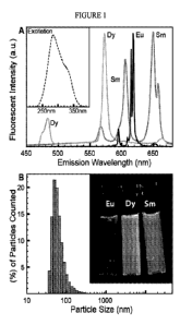
[0061] FIG. 1 provides an emission spectra (Panel A) for each of
nanoparticles (Dy-,
Eu-, and Sm- doped Yo soflio i5VO4) when excited at 285 nm. Excitation spectra
of all
11
CA 02881841 2015-02-11
WO 2014/031902
PCT/US2013/056280
three nanoparticles (inset) are nearly identical. FIG. 1 also provides a
histogram
(Panel B) showing particle size distribution for nanoparticles (as measured by
dynamic light scattering), and a photograph of vials of nanoparticle
suspensions
illuminated with a UV lamp (inset).
[0062] FIG. 2 provides schematics and related images of a microfluidic bead
synthesizer according to some embodiments of the present disclosure and a
method of
using same. (Panel A) Stage 1 of bead synthesis. Mixtures of lanthanide
nanoparticles
(Eu alone, Eu/Sm, Eu/Dy) suspended in pre-polymer bead mixture flow into a
microfluidic device at controlled ratios and are mixed on chip using a mixing
element
(an exemplary staggered herringbone mixer) is depicted. (Panel B) Stage 2 of
bead
synthesis. A hydrophilic carrier fluid (e.g., water pushes) the lanthanide
mixture
towards a T-junction containing a continuously flowing hydrophobic carrier
fluid
(e.g., oil) stream, producing droplets (shown in microscope image, inset).
Droplets are
polymerized into beads by exposing them to polymerization conditions (e.g.,
via
illumination with UV light) and collected for later use. (Panel C) CAD drawing
of the
flow channels of the device showing the lanthanide inputs and the exemplary
herringbone mixer, an input configured to hold a hydrophilic carrier fluid (an
exemplary water input is depicted) and resistor, an input configured to hold a
hydrophobic carrier fluid (e.g., an oil input) and bead output. (Panel D)
Photograph of
the bead synthesizer microfluidic device with food coloring in the channels
and a
dime for scale. Flow channels are colored as in panel C; control lines used to
open
and close on-chip valves are identified with "*".
[0063] FIG. 3 provides schematics and related images of a bead imaging
setup
according to some embodiments of the present disclosure. (Panel A) Photograph
of
microfluidic imaging device with flow channels shown in black and control
channels
shown in grey (and identified by "*"). Beads are injected into a 55 p m wide
serpentine channel for imaging (photograph, inset); sieve valves at the end of
the
channel retain beads while permitting fluid flow to facilitate channel
loading. Inputs
(Injection 1(Inj1), Bead In (BdIn), Injection 2 (Inj2)) and outputs (Waste 1
(W1),
Waste 2 (W2), Bead Out (BdOut)) at either end of the device provide
bidirectional
flow. (Panel B) Schematic of a microscopy system used for imaging beads. Light
from a suitable light source (e.g., a full-spectrum 300 W Xenon arc lamp) is
collected
(L1), reflected off a 400 nm long pass filter (M1) to reject visible light,
and passed
through a shutter (Si) and an excitation filter wheel (to switch between UV
and
12
CA 02881841 2015-02-11
WO 2014/031902
PCT/US2013/056280
visible transillumination) before being focused (L2) into a deep UV liquid
light guide.
The other end of the liquid light guide is mounted on the condenser mount of a
Nikon Ti microscope, where the light is collimated by a fused silica lens
(L3) and
projected onto the sample. Emitted light from the sample is collected by a
Plan Apo
4x/0.2 NA objective (L4), with a UV blocking filter placed between the sample
and
the objective. Emitted light is filtered through an emission filter wheel
mounted
beneath the objective before being focused onto the camera.
[0064] FIG. 4 provides images and graphical data associated with an
analysis
procedure according to some embodiments of the present disclosure. (Panel A)
Raw
data in each of six luminescence channels. Data are scaled linearly. (Panel B)
Reference spectra used for unmixing. (Panel C) Left: Linearly scaled black and
white
images of unmixed data from each channel (Dy, Eu, and Sm), with black set to
the
minimum intensity in the image and white set to the maximum intensity. Right:
Bright
field image of the same field of view. (Panel D) False color overlay of Dy,
Eu, and
Sm luminescence with scaling as in Panel C (shown here in greyscale).
[0065] FIG. 5 provides a graphical representation of a 24 code matrix
according to
some embodiments of the present disclosure. (Panel A) Scatter plot of the
median
Dy/Eu and Sm/Eu luminescence ratios for 10 filled serpentines (1926 beads),
with
points false colored according to their Sm/Eu and Dy/Eu ratios (shown here in
greyscale). Each point represents one bead. Grey ellipses around each code
cluster
illustrate three- and four-sigma contours derived from fitting a Gaussian
mixture
model to the data. Histograms of bead ratios in the Dy/Eu and Sm/Eu channels
(black)
and their corresponding Gaussian fits (grey fit lines) are shown along each
axis; these
histograms group all codes together. Inset: Measured cluster centroids and
their
corresponding programmed intensity ratios; the root mean square deviation
between
the programmed ratios and the measured ratios is 0.014. (Panel B) Standard
deviations calculated from Gaussian fits to the bead ratio histograms in Panel
A as a
function of ratio (filled circles, identified with "*" for Dy and "+" for Sm).
Square
symbols illustrate the statistical standard deviation determined from
replicated
imaging of the same serpentine of beads.
[0066] FIG. 6 provides a graph showing the reproducibility of lanthanide
nanoparticle synthesis according to some embodiments of the present
disclosure. Each
individual batch of lanthanide nanoparticle suspensions were diluted 1:500 in
DI
water from the concentrated stock solutions. A luminescence emission spectrum
(400-
13
CA 02881841 2015-02-11
WO 2014/031902
PCT/US2013/056280
800 nm) was obtained using a FluoroMax-3 spectrofluorometer for all of these
diluted
stock solutions. The excitation was the same for all solutions (285 nm through
a 3-nm
slit width excitation monochromator) and the emission parameters were also
held
constant for all emitters (3-nm emission slit width, 1-nm increment steps, and
0.1 sec
integration time at each step) with the exception of the Europium lanthanide
nanoparticles which, due to their brightness, had slit widths of 1 nm at both
monochromators. For each emitter shown, the left column shows the emission
spectra
of each individual batch synthesized as a stacked plot: (Panel A) Sm, 4
batches,
(Panel B) Dy, 3 batches, and (Panel C) Eu, 5 batches. In the right column, the
normalized emission spectra for all batches are shown as an overlay for each
emitter.
Typically, only one color is observed in the overlaid spectra since the high
reproducibility of the batches results in several spectra that are coincident.
[0067] FIG. 7 provides images showing compatibility of exemplary spectrally
encoded beads according to some embodiments of the present disclosure with
commonly used visible fluorophores. A sample of the 24-code beads were imaged
using a Chroma Sedat quad filter set (#89000), with a Lambda XL lamp,
C001SNAPTM HQ2 camera, 10x / 0.3 NA objective, and 1 sec exposure time for
each
luminescence channel. All four channel combinations were imaged, and the
corresponding images are labeled with the excitation and emission centers of
the filter
sets. The 402/455 (DAPI channel) image shows weak luminescence; the other
channels show negligible luminescence with the luminescence in the Cy5 channel
being undetectable.
[0068] FIG. 8 provides scatter plots of two different batches of
synthesized beads
(Bead Set 1 (grey) and Bead Set 2 (Black)) according to some embodiments of
the
present disclosure. The two batches were synthesized on different dates and
imaged
on the same date. The Set 1 batch of beads is missing one code due to a
computer
ei-ror.
[0069] FIG. 9 provides a graph comparing programmed ratios (black) and
measured
code centroids and three sigma error ellipses for the Set 1 ("*") and Set 2
("+") beads
referenced in FIG. 8.
[0070] FIG. 10 provides a schematic of peptide synthesis on spectrally
encoded
beads.
[0071] FIG. 11 provides false color images demonstrating that embedded
codes are
robust to peptide synthesis conditions. (Panel A) Image showing beads
containing
14
CA 02881841 2015-02-11
WO 2014/031902
PCT/US2013/056280
either Eu alone (blue) or a combination of Eu and Dy (yellow) after on-bead
synthesis
of either FLAG peptide (Eu beads, blue) or myc peptide (Eu/Dy beads, yellow).
(Panel B) Image showing binding of Alexa-647-labeled anti-FLAG antibody (red)
and
Alexa-555-labeled anti-myc antibody (green) to encoded beads.
[0072] FIG. 12 provides a photograph showing the microfluidic bead reactor
utilized
for the immunoassays described in Example 4. The device features input and
output
channels (BeadIn, BeadOut) for loading beads into a single main reaction
chamber, as
well as 4 additional input channels (R1-R4) and a single output channel for
introducing reagents into the reaction chamber. Flow is controlled by 4 valves
that
control reagent inputs, a pump (P1) that controls reagent flow rates, and 2
sieve valves
(SvIn, SvOut) that retain beads during reagent exchanges.
[0073] FIG. 13 provides a table showing the results of liquid
chromatography¨mass
spectrometry/mass spectrometry (LC-MS/MS) analysis of peptides cleaved from
spectrally encoded microbeads.
[0074] FIG. 14 provides a graph showing relative Dy signal present within a
4-code
bead set before and after peptide synthesis.
[0075] FIG. 15 demonstrates single bead release through the use of a single
constriction in a microfluidic channel. (Panel A) Photograph of a linear
serpentine
channel for imaging of spectrally encoded beads in a first-in first-out linear
array.
(Panel B) Montage of images from a movie showing release of a single bead.
(Panel
C) Graph showing changes in intensity at the exit of the serpentine channel as
a
function of time. Increases in intensity indicate the passage of individual
beads at
fairly regular intervals.
[0076] FIG. 16 provides a luminescence spectra of Tm:YV04 with and without
bismuth. Tm loading is 1% in both cases and bismuth is incorporated at ¨15-20%
replacement of Yttrium (Y).
[0077] FIG. 17 provides a luminescence spectra of Er:YV04 with and without
bismuth (Bi). Er loading is 5% in both cases and bismuth is incorporated at
¨15-20%
replacement of Yttrium (Y).
[0078] FIG. 18 provides an emission scan of Tm:YV04 with varying Tm%. The
emission scan shows adjustment of the Tm % loading to arrive at a preferred
value of
1% Tm:YV04. Nanoparticle concentrations in these solutions are approximately
constant (up to 5% variance in concentration).
CA 02881841 2015-02-11
WO 2014/031902
PCT/US2013/056280
[0079] FIG. 19 provides a graph showing a comparison of Er:YV04
luminescence
(normalized to 1M counts). The graph shows adjustment of the Er % loading to
arrive
at a preferred value of 5% Er:YV04. Nanoparticle concentrations in these
solutions
are approximately constant (up to 5% variance in concentration).
[0080] FIG. 20 provides a luminescence spectra of the CeTb:LaPO4
nanophosphor.
[0081] FIG. 21 provides a graph showing the results of a particle size
analysis of the
CeTb:LaPO4 nanophosphor.
[0082] FIG. 22 provides emission spectra for commercially available PEG-
diamine
and the PEG-DAM synthesized from it. As a comparison, a commercially available
PEG-600 is included to illustrate an acceptable level of autofluorescence for
a PEG.
[0083] FIG. 23 provides emission spectra showing a comparison of the
autofluorescence between the commercially available PEG-diamine and the PEG-
diamine synthesized from PEG2K as described herein.
[0084] FIG. 24 provides a proton NMR readout showing the synthesis of PEG2K-
monoacrylamide-monoBoc.
DEFINITIONS
[0085] As used herein, the term "lanthanide nanoparticle" refers to a
nanoparticle
which includes a lanthanide and a host lattice.
[0086] As used herein, the term "lanthanide" refers to Ce, Pr, Nd, Sm, Eu,
Gd, Tb,
Dy, Ho, Er, Tm, Yb, La, combinations thereof, compounds containing Ce, Pr, Nd,
Sm, Eu, Gd, Tb, Dy, Ho, Er, Tm, Yb, La and combinations thereof, and ions of
Ce,
Pr, Nd, Sm, Eu, Gd, Tb, Dy, Ho, Er, Tm, Yb, La and combinations thereof.
[0087] As used herein, the term "nanoparticle" refers to a particle having
one or more
dimensions (e.g., diameter) of less than 1000 nm, e.g., about 500 nm or less,
about
100 nm or less, about 50 nm or less, about 10 nm or less, about 5 nm or less,
or about
1 nm or less. For example, a nanoparticle may have one or more dimensions
(e.g.,
diameter) of from less than 1000 nm to about 500 nm, from about 500 nm to
about
100 nm, from about 100 nm to about 10 nm, from about 50 nm to about 10 nm,
from
about 10 nm to about 5 nm, or from about 5 nm to about 1 nm. Nanoparticles may
have a generally spherical shape or a non-spherical shape.
[0088] As used herein, the term "microbead" refers to a particle having one
or more
dimensions (e.g., diameter) of about 1000 p m or less, e.g., about 500 p m or
less,
about 100 p m or less, about 50 p m or less, about 10 p m or less, or about 5
p m or less.
16
CA 02881841 2015-02-11
WO 2014/031902
PCT/US2013/056280
For example, a microbead may have one or more dimensions (e.g., diameter) of
from
about 1000 p m to about 1 p m, from about 500 p m to about 1 p m, from about
100 p m
to about 1 p m, from about 50 p m to about 1 p m, from about 10 p m to about 1
p m, or
from about 5 p m to about 1 p m. Microbeads may have a generally spherical
shape or
a non-spherical shape.
[0089] It will be appreciated that throughout this present disclosure
reference is made
to amino acids according to the single letter or three letter code. For the
reader's
convenience, the single and three letter amino acid code is provided below:
G Glycine Gly P Proline Pro
A Alanine Ala V Valine Val
L Leucine Leu I Isoleucine Ile
M Methionine Met C Cysteine Cys
F Phenylalanine Phe Y Tyrosine Tyr
W Tryptophan Trp H Histidine His
K Lysine Lys R Arginine Arg
Q Glutamine Gln N Asparagine Asn
E Glutamic Acid Glu D Aspartic Acid Asp
S Serine Ser T Threonine Thr
[0090] Reference to "peptide" herein is meant to encompass a polymer of
amino acids
linked by native amide bonds and/or non-native amide bonds.
[0091] It should be understood that as used throughout, and unless
specifically
indicated otherwise, the term "amino acid" is used herein in its broadest
sense, and
includes naturally occurring amino acids as well as non-naturally occurring
amino
acids, including amino acid analogs and derivatives. The latter includes
molecules
containing an amino acid moiety. One skilled in the art will recognize, in
view of this
broad definition, that reference herein to an amino acid includes, for
example,
naturally occurring proteogenic L-amino acids; D-amino acids; chemically
modified
amino acids such as amino acid analogs and derivatives; naturally occurring
nonproteogenic amino acids such as norleucine, p-alanine, omithine, etc.; and
chemically synthesized compounds having properties known in the art to be
characteristic of amino acids. As used herein, the term "proteogenic"
indicates that the
amino acid can be incorporated into a peptide, polypeptide, or protein in a
cell
through a metabolic pathway.
17
CA 02881841 2015-02-11
WO 2014/031902
PCT/US2013/056280
[0092] Before the present invention is further described, it is to be
understood that
this invention is not limited to particular embodiments described, as such
may, of
course, vary. It is also to be understood that the terminology used herein is
for the
purpose of describing particular embodiments only, and is not intended to be
limiting,
since the scope of the present invention will be limited only by the appended
claims.
[0093] Where a range of values is provided, it is understood that each
intervening
value, to the tenth of the unit of the lower limit unless the context clearly
dictates
otherwise, between the upper and lower limit of that range and any other
stated or
intervening value in that stated range, is encompassed within the invention.
The upper
and lower limits of these smaller ranges may independently be included in the
smaller
ranges, and are also encompassed within the invention, subject to any
specifically
excluded limit in the stated range. Where the stated range includes one or
both of the
limits, ranges excluding either or both of those included limits are also
included in the
invention.
[0094] Unless defined otherwise, all technical and scientific terms used
herein have
the same meaning as commonly understood by one of ordinary skill in the art to
which this invention belongs. Although any methods and materials similar or
equivalent to those described herein can also be used in the practice or
testing of the
present invention, the preferred methods and materials are now described. All
publications mentioned herein are incorporated herein by reference to disclose
and
describe the methods and/or materials in connection with which the
publications are
cited.
[0095] It must be noted that as used herein and in the appended claims, the
singular
forms "a," "and," and "the" include plural referents unless the context
clearly dictates
otherwise. Thus, for example, reference to "a lanthanide nanoparticle"
includes a
plurality of such lanthanide nanoparticles and reference to "the microbead"
includes
reference to one or more microbeads and equivalents thereof known to those
skilled in
the art, and so forth.
[0096] It is further noted that the claims may be drafted to exclude any
recited
element. As such, this statement is intended to serve as antecedent basis for
use of
such exclusive terminology as "solely," "only" and the like in connection with
the
recitation of claim elements, or use of a "negative" limitation.
18
CA 02881841 2015-02-11
WO 2014/031902
PCT/US2013/056280
[0097] To the extent any definition of a term defined herein conflicts with
a definition
of a term in an application or reference incorporated by reference herein, the
instant
application shall control.
[0098] The publications discussed herein are provided solely for their
disclosure prior
to the filing date of the present application. Nothing herein is to be
construed as an
admission that the present invention is not entitled to antedate such
publication by
virtue of prior invention. Further, the dates of publication provided may be
different
from the actual publication dates which may need to be independently
confirmed.
[0099] As will be apparent to those of skill in the art upon reading this
disclosure,
each of the individual embodiments described and illustrated herein has
discrete
components and features which may be readily separated from or combined with
the
features of any of the other several embodiments without departing from the
scope or
spirit of the present invention. Any recited method can be carried out in the
order of
events recited or in any other order which is logically possible. This is
intended to
provide support for all such combinations.
DETAILED DESCRIPTION
[00100] The present disclosure provides spectrally encoded microbeads and
methods
and devices for making and using spectrally encoded microbeads. The disclosed
methods and devices facilitate the preparation and use of microbeads
containing
multiple lanthanide nanoparticles, which microbeads have uniquely identifiable
spectral codes. The disclosed microbeads, and the methods and devices for
making
and using same, find use in multiplexing and high-throughput biomarker
analysis.
Lanthanide Nanoparticles for Use in Spectrally Encoded Microbeads
[00101] The spectrally encoded microbeads of the present disclosure
generally include
two or more different lanthanide nanoparticles. Suitable lanthanide
nanoparticles for
incorporation into the spectrally encoded microbeads include nanoparticles
including
a lanthanide and a host lattice.
[00102] Lanthanides which may be incorporated into the disclosed lanthanide
nanoparticles include, for example, Ce, Pr, Nd, Sm, Eu, Gd, Tb, Dy, Ho, Er,
Tm, Yb,
La, combinations thereof, compounds containing Ce, Pr, Nd, Sm, Eu, Gd, Tb, Dy,
Ho,
Er, Tm, Yb, La and combinations thereof, and ions of Ce, Pr, Nd, Sm, Eu, Gd,
Tb,
Dy, Ho, Er, Tm, Yb, La and combinations thereof.
19
CA 02881841 2015-02-11
WO 2014/031902
PCT/US2013/056280
[00103] A variety of suitable nano-crystal host lattices which may be
utilized in the
disclosed lanthanide nanoparticles are known in the art. For example,
lanthanide
dopants may be incorporated into a host lattice to provide lanthanide-doped
yttrium
orthovanadate (YV04), lanthanide-doped oxide, lanthanide-doped fluoride,
lanthanide-doped chloride, lanthanide-doped bromide, lanthanide-doped iodide,
lanthanide-doped lanthanum phosphate, and lanthanide-doped strontium borates
(e.g.,
SrB407, SrB6O10 and Sr4B14025), among others.
[00104] Lanthanide nanoparticles according to the present disclosure may be
prepared
using methods known in the art or as described herein. An exemplary lanthanide
nanoparticle synthesis scheme utilizing yttrium orthovanadate (YV04) as the
host
lattice is described in Example 1 below. Additional lanthanide nanoparticle
preparation methods and materials are described, for example, in Xu et al.
(2004)
Solid State Communications, 130:465-468; Choi et al. (2010) Journal of
Luminescence, 130:549-553; and Wang et al. (2008) Angewandte Chemie-
International Edition, 47:906-909; the disclosure of each of which is
incorporated by
reference herein.
[00105] Lanthanide nanoparticles according to the present disclosure may be
configured as up-converting or down-converting lanthanide nanoparticles using
methods known in the art. Suitable up-converting lanthanide nanoparticles may
include, for example, NaGdF4: Tm; NaGdF4: Ln; NaGdF4Yb; NaGdF4Er; NaGdF4Yb,
Er; NaYF4:Er; NaYF4:Yb; NaYF4:Er,Yb; NaYF4:Tm,Yb; LaF3:Yb,Tm; LaF3:Yb,Er;
and LaF3:Yb,Ho nanoparticles. Suitable down-converting lanthanide
nanoparticles
may include, for example, YV04:Eu; YV04:Dy; and YV04:Sm nanoparticles. It
should be noted that the above referenced lanthanides may be incorporated into
the
nanoparticles as their respective ions.
[00106] Materials may be added during preparation of the lanthanide
nanoparticles to
increase their UV absorption. For example, in some embodiments bismuth is
incorporated into the lanthanide nanoparticles to increase their UV
absorption.
[00107] In some embodiments, lanthanide nanoparticles as disclosed herein
may be
modified (e.g., covered or coated) in a suitable material to facilitate
formation of a
stable colloid suspension of the lanthanide nanoparticles in a carrier fluid.
Suitable
materials may include materials which prevent aggregation of the lanthanide
nanoparticles in the carrier fluid (e.g., H20) and/or facilitate maintenance
of a nano-
particle form of the lanthanide nanoparticles. For example, suitable materials
which
CA 02881841 2015-02-11
WO 2014/031902
PCT/US2013/056280
may be used to cover or coat the lanthanide nanoparticles may include
polyethyleneimine (PEI), polyacrylic acid (PAA), sodium citrate, or citric
acid.
Polyethyleneimine (PEI) may be suitable for use, e.g., as a coating material
in order to
make the nanophosphors more compatible with a monomer mixture bearing free
amines.
[00108] The lanthanide nanoparticles described herein may be incorporated
into
microbeads, e.g., polymeric microbeads, to provide spectrally encoded
microbeads as
discussed in greater detail below.
Spectrally Encoded Microbeads
[00109] The spectrally encoded microbeads of the present disclosure
generally include
two or more different lanthanide nanoparticles as discussed herein and one or
more
polymers, copolymers or combinations thereof.
[00110] A variety of polymers may be utilized in the lanthanide
nanoparticles
described herein. Suitable polymers may be selected which can evenly and
irreversibly entrap the lanthanide nanoparticle materials within a polymer
matrix.
Suitable polymers may include, for example, poly(ethylene glycol) (PEG),
polystyrene, polyethylene, poly acrylic acid, poly(methyl methacrylate)
(PMMA),
polysaccharides, and copolymers or combinations thereof.
[00111] In some embodiments, suitable polymers are those which are capable
of
forming microbeads as a result of a polymerization process, e.g., a thermal-
or photo-
initiated polymerization process. Such polymers may include, for example,
polyacrylate (e.g., poly (PEG-diacrylate)), polyacrylamide (e.g., PEG-
diacrylamide),
polymethacrylate, polymethacrylamide, polystyrene, polythiol-ene,
polyurethane,
epoxy resin, polysaccharide (e.g., agarose), as well as copolymers or
combinations of
two or more of the above. Suitable polymers may also include
polyurethanes/polyureas, polysiloxanes, organosiloxanes, polyethers (e.g.,
polyethylene glycol (PEG)), polyvinylpyrrolidones (PVP), vinyl ethers, vinyl
acetates,
polyimides, polysulfones, polyamic acids, polyamides, polycarbonates,
polyesters,
and copolymers or combinations of two or more of the above.
[00112] It should be noted that the above polymers may be provided in
monomer form
during the microbead preparation process, and these monomers may be
polymerized
to form the above polymers, copolymers or combinations thereof in the
spectrally
encoded microbeads of the present disclosure. Suitable monomers may include
those
21
CA 02881841 2015-02-11
WO 2014/031902
PCT/US2013/056280
which can be polymerized in situ alone or with a cross-linking agent to form a
cross-
linked resin. Additional monomers which may be utilized in the lanthanide
nanoparticles described herein may include, e.g., monomers which are capable
of
participating in thiol-ene thiol-yne reactions, e.g., pentaerythritol
tetrakis(3-
mercaptopropionate) (TT); diallyl phthalate (DAP); 1,3,5,-trially1-1,3,5-
triazine-
2,4,6(1H,3H,5H)-trione (TTT); 1,7-octadiyne (OY); mercaptoacetic acid (MA);
allylamine (AA), pentaerythritol triallyl ether (PTE) and propargylamine (PA).
These
monomers find use, for example, in photo-initiated polymerization processes.
For
additional discussion of thiol-ene thiol-yne reactions and monomers suitable
for use
therein, see, e.g., Prasath et al. (2010) Polym. Chem., 1: 685-692, the
disclosure of
which is incorporated by reference herein.
[00113] In some embodiments, a suitable monomer for use in preparation of
the
disclosed microbeads is selected from a PEG diacrylamide (PEG-DAM), a PEG
monoacrylamide-monoamine (PEG-AM) and a PEG-monoacrylamide-monoBoc. A
PEG-monoacrylamide-monoBoc may find particular use when the microbead is to be
used as a substrate in a downstream peptide synthesis reaction.
[00114] In some embodiments, the present disclosure is directed to specific
populations of spectrally encoded microbeads, for example, a population of
polymeric
microbeads embedded with at least two different lanthanide nanoparticles,
wherein
the population includes a plurality of polymeric microbeads, wherein each
polymeric
microbead of the plurality is embedded with at least two lanthanide
nanoparticles
having different luminescence spectra, and wherein the relative concentrations
of the
first and second lanthanide nanoparticles are substantially equal (e.g., not
significantly
different) among the polymeric microbeads of the population.
[00115] In some embodiments, a set of populations of polymeric microbeads
embedded with at least two different lanthanide nanoparticles is provided, the
set of
populations of polymeric microbeads including a first population of polymeric
microbeads, wherein each polymeric microbead of the first population is
embedded
with at least a first lanthanide nanoparticle and a second lanthanide
nanoparticle;
wherein the first and second lanthanide nanoparticles comprise different
lanthanides;
and wherein the relative concentrations of the first and second lanthanide
nanoparticles are substantially equal (e.g., not significantly different)
among the
polymeric microbeads of the first population; and a second population of
polymeric
microbeads, wherein each polymeric microbead in the second population is
embedded
22
CA 02881841 2015-02-11
WO 2014/031902
PCT/US2013/056280
with at least the first lanthanide nanoparticle and the second lanthanide
nanoparticle;
and wherein the relative concentrations of the first and second lanthanide
nanoparticles are substantially equal (e.g., not significantly different)
among the
polymeric microbeads of the second population; wherein the concentration of at
least
one of the first and second lanthanide nanoparticles is different between the
polymeric
microbeads of the first population and second population.
[00116] In some embodiments, the concentration of the first lanthanide
nanoparticle is
substantially equal (e.g., not significantly different) for each polymeric
microbead of
the first population and second population. By providing a set of populations
of
polymeric microbeads wherein the concentration of a first lanthanide
nanoparticle is
substantially equal (e.g., not significantly different) for each polymeric
microbead of
the first population and second population, an internal lanthanide
nanoparticle
standard may be provided. It should be noted that an internal lanthanide
nanoparticle
standard may be provided wherein the concentration of the lanthanide
nanoparticle
standard is not substantially equal across all populations of the set. For
example, a
lanthanide nanoparticle standard could have a concentration of approximately X
for
each member of a first population in a set and a concentration of
approximately Y for
each member of a second population in the set, wherein Y is a known percentage
of X
other than 100 percent, e.g., 10 percent, 50 percent, 150 percent or 200
percent.
[00117] The devices and methods disclosed herein allow for the precise
control of the
concentration of the lanthanide nanoparticles in the spectrally encoded
polymeric
microbeads of the present disclosure. Accordingly, a population of polymeric
microbeads may be provided such that the relative concentrations of at least
two
different lanthanide nanoparticles are substantially equal (e.g., not
significantly
different) among the polymeric microbeads of the population. In other words,
the
population of polymeric microbeads may be provided such that each polymeric
microbead in the population has at least substantially the same ratio of two
or more
lanthanide nanoparticles as the other polymeric microbeads in the population.
[00118] This precision allows a population of spectrally encoded microbeads
to be
provided such that the luminescence intensity level variation among all the
members
of the population is no greater than about 25 percent, e.g., no greater than
about 20
percent, no greater than about15 percent, no greater than about 10 percent, no
greater
than about 5 percent, no greater than about 4 percent, no greater than about 3
percent,
no greater than about 2 percent, or no greater than about 1 percent. In some
23
CA 02881841 2015-02-11
WO 2014/031902
PCT/US2013/056280
embodiments, the luminescence intensity level variation among all the members
of
the population is from about 25 percent to about 1 percent, e.g., from about
20 percent
to about 1 percent, from about 15 percent to about 1 percent, from about 10
percent to
about 1 percent, from about 5 percent to about 1 percent, from about 4 percent
to
about 1 percent, from about 3 percent to about 1 percent, or from about 2
percent to
about 1 percent.
[00119] By precisely providing unique, identifiable spectral codes using
multiple
lanthanide nanoparticles a number of uniquely identifiable polymeric microbead
populations may be provided. In some embodiments, a set of populations of
polymeric microbeads as described herein includes 2 or more different
populations of
polymeric microbeads, e.g., 5 or more, 10 or more, 20 or more, 30 or more, 40
or
more, 50 or more, 60 or more, 70 or more, 80 or more, 90 or more, or 100 or
more
different populations of polymeric microbeads, wherein the polymeric
microbeads for
each population include a different spectral code when compared with the
polymeric
microbeads of the other populations in the set. In some embodiments, a set of
populations of polymeric microbeads as described herein includes 102 or more,
103 or
more, 104 or more, 105 or more, 106 or more, or 107 or more different
populations of
polymeric microbeads, wherein the polymeric microbeads for each population
include
a different spectral code when compared with the polymeric microbeads of the
other
populations in the set. For example, in some embodiments, a set of populations
of
polymeric microbeads as described herein includes from about 2 to about 10,
from
about 10 to about 20, from about 20 to about 30, from about 30 to about 40,
from
about 40 to about 50, from about 50 to about 60, from about 60 to about 70,
from
about 70 to about 80, from about 80 to about 90, or from about 90 to about 100
different populations of polymeric microbeads, wherein the polymeric
microbeads for
each population include a different spectral code when compared with the
polymeric
microbeads of the other populations in the set. As a further example, in some
embodiments, a set of populations of polymeric microbeads as described herein
includes from about 10 to about 107, from about 102 to about 107, from about
103 to
about 107, from about 104 to about 107, from about 105 to about 107, or from
about 106
to about 107 different populations of polymeric microbeads, wherein the
polymeric
microbeads for each population include a different spectral code when compared
with
the polymeric microbeads of the other populations in the set. The number of
different
identifiable populations may be calculated by taking the number of resolvable
24
CA 02881841 2015-02-11
WO 2014/031902
PCT/US2013/056280
luminescence levels to the power of the number of different lanthanide
nanoparticles.
For example, using 6 different lanthanide nanoparticles with 10 resolvable
levels
each, the number of different, uniquely identifiable populations is 106 or
1,000,000.
This code space can be increased as discussed in greater detail below, by
utilizing
both up-converting and down-converting lanthanide nanoparticles.
[00120] The polymeric microbead populations described herein may be
provided in a
variety of population sizes. For example, a polymeric microbead population as
described herein may include 5 or more, 10 or more, 100 or more, 500 or more,
1000
or more, 1500 or more or 2000 or more polymeric microbeads. In some
embodiments,
a polymeric microbead population as described herein includes from about 5 to
about
2000, from about 10 to about 2000, from about 100 to about 2000, from about
500 to
about 2000, from about 1000 to about 2000 or from about 1500 to about 2000
polymeric microbeads.
[00121] The polymeric microbeads disclosed herein may include two or more
different
lanthanide nanoparticles, e.g., 3 or more, 4 or more, 5 or more, 6 or more, 7
or more,
8 or more, 9 or more, or 10 or more, wherein each lanthanide nanoparticle has
a
different luminescence spectra. For example, in some embodiments, the
polymeric
microbeads disclosed herein may include from 2 to 10, from 3 to 10, from 4 to
10,
from 5 to 10, from 6 to 10, from 7 to 10, from 8 to 10, or from 9 to 10
lanthanide
nanoparticles, wherein each lanthanide nanoparticle has a different
luminescence
spectra.
[00122] The spectrally encoded polymeric microbeads of the present
disclosure have
one or more dimensions (e.g., diameter) of about 1000 p m or less, e.g., about
500 p m
or less, about 100 p m or less, about 50 p m or less, about 10 p m or less, or
about 5 p m
or less. For example, a spectrally encoded microbead may have one or more
dimensions (e.g., diameter) of from about 1000 p m to about 1 p m, from about
500
p m to about 1 p m, from about 100 p m to about 1 p m, from about 50 p m to
about 1
p m, from about 10 p m to about 1 p m, or from about 5 p m to about 1 p m. The
spectrally encoded polymeric microbeads may have a generally spherical shape
or a
non-spherical shape.
[00123] For populations of spectrally encoded polymeric microbeads, each
member of
the population may have approximately the same one or more dimensions, e.g.,
one or
more dimensions as listed above. In some embodiments, the members of a
population
of spectrally encoded polymeric microbeads have a diameter such that the
diameter
CA 02881841 2015-02-11
WO 2014/031902
PCT/US2013/056280
variation among all the members of the population is no greater than about 10
percent,
e.g., no greater than about 5 percent, no greater than about 1 percent, no
greater than
about 0.1 percent, or no greater than about 0.01 percent. In some embodiments,
the
diameter variation among all the members of the population is from about 10
percent
to about 1 percent, e.g., from about 5 percent to about 1 percent, from about
4 percent
to about 1 percent, from about 3 percent to about 1 percent, or from about 2
percent to
about 1 percent. In some embodiments, the diameter variation among all the
members
of the population is from about 5 percent to about 0.01 percent, e.g., from
about 4
percent to about 0.01 percent, from about 3 percent to about 0.01 percent,
from about
2 percent to about 0.01 percent, from about 1 percent to about 0.01 percent,
or from
about 0.1 percent to about to about 0.01 percent.
[00124] Spectrally encoded polymeric microbeads according to the present
disclosure
may include or be modified to include one or more reactive functional groups
for the
attachment of a molecule or molecules to the spectrally encoded polymeric
microbeads. For example, monomers containing a single acrylate group and a
functional group (thiol, amine, hydroxyl, carboxylic acid) can be added before
polymerization to yield a microbead with functionality suitable for the
attachment of
an additional molecule or molecules to the microbeads subsequent to
polymerization.
An exemplary molecule would be hydroxy-PEG-acrylate. As an additional example,
in a thiol-ene polymerization system, such reactive functional groups may be
provided
when the spectrally encoded polymeric microbeads are formed using functional
monomers containing carboxylate (mercaptoacetic acid), hydroxyl
(pentaerythritol
triallyl ether), or amine (allylamine) moieties. See, e.g., Prasath et al.
(2010) Polym.
Chem., 1: 685-692, the disclosure of which is incorporated by reference
herein.
Spectrally encoded polymeric microbeads including one or more reactive
functional
groups, e.g., carboxyl groups, hydroxyl groups or amine groups, can be used
for the
attachment of one or more molecules, e.g., nucleic acids, peptides, or
subunits thereof,
to the spectrally encoded polymeric microbeads described herein.
Methods of Making Spectrally Encoded Microbeads
[00125] The present disclosure provides methods for producing spectrally
encoded
microbeads as described herein. These methods may be conducted using
microfluidic
devices as described in greater detail below. In some embodiments, a
population of
polymeric microbeads including two or more different lanthanide nanoparticles
is
26
CA 02881841 2015-02-11
WO 2014/031902
PCT/US2013/056280
provided. The method may include, for example: (i) mixing at least two fluids
into a
first solution, wherein each fluid comprises a polymerizable component (e.g.,
a
polymer or monomer) and a different lanthanide nanoparticle; (ii) forming
droplets
from the solution; and (iii) subjecting the droplets to polymerization
conditions,
thereby producing a first set of polymeric microbeads embedded with at least
two
different lanthanide nanoparticles, wherein the relative concentrations of the
lanthanide nanoparticles are substantially equal (e.g., not significantly
different)
among the polymeric microbeads of the first set.
[00126] In some embodiments, the method may include the following
additional steps
(iv) mixing the at least two fluids into a second solution, wherein the
concentration of
at least one of the different lanthanide nanoparticles in the second solution
is different
than in (i) above; (v) forming droplets from the solution; and (vi) subjecting
the
droplets to polymerization conditions, thereby producing a second set of
polymeric
microbeads embedded with at least two different lanthanide nanoparticles,
wherein
the relative concentrations of the lanthanide nanoparticles are substantially
equal (e.g.,
not significantly different) among the polymeric microbeads of the second set.
[00127] In some embodiments, the concentration of at least one of the
different
lanthanide nanoparticles in the second solution is substantially equal (e.g.,
not
significantly different) to that in (i) above.
[00128] The above mixing steps may be implemented using a variety of "on-
chip" and
"off-chip" mixing elements as described in greater detail below.
[00129] The step of forming droplets from the solution may include, for
example,
contacting the solution (which may be hydrophilic due to the presence of a
hydrophilic carrier fluid, e.g., water) with a hydrophobic carrier fluid
(e.g., mineral oil
or water-immiscible organic solvent, e.g. octanol) such that droplets are
formed. This
may be accomplished, for example, by introducing the solution into a flowing
stream
including the hydrophobic carrier fluid. Alternatively, a hydrophobic carrier
fluid
(e.g., mineral oil or water-immiscible organic solvent, e.g. octanol) can be
used to
form the solution, and droplets can be formed by contacting the hydrophobic
carrier
fluid with a hydrophilic carrier fluid (e.g., water). This may be
accomplished, for
example, by introducing the hydrophobic carrier fluid into a flowing stream
including
the hydrophilic carrier fluid.
[00130] Any suitable device and/or method for droplet formation may be
utilized to
form droplets in the context of the present disclosure, including, e.g., the
utilization of
27
CA 02881841 2015-02-11
WO 2014/031902
PCT/US2013/056280
flow focusing nozzles. See, e.g., Ward et al. (2005) Electrophoresis, 26:3716-
3724,
the disclosure of which is incorporated by reference herein.
[00131] The droplet size may be modulated by adjusting the pressure used to
form the
droplet, e.g., at the interface of the solution and the hydrophobic carrier
fluid. In
addition, droplet size may be modulated by adjusting the geometry, e.g., size
and
shape, of the microfluidic device channels.
[00132] One or more stabilizers or surfactants may be added to one or more
of the
carrier fluids to prevent droplet merging and sticking of droplets to the
walls of the
microfluidic device. Suitable surfactants may include, for example, Abil EM90
(a
silicon based emulsifier; CAS No. 144243-53-8) and SpanTM 80 (CAS No. 1338-43-
8), among others.
[00133] The step of subjecting the droplets to polymerization conditions
may include,
for example, exposing the droplets to UV radiation or elevated temperatures to
initiate
polymerization. Other suitable polymerization conditions are known in the art
and
may be selected provided that they are compatible with the polymers and/or
monomer
components to be polymerized, e.g., thiol-ene polymerization, redox-initiated
polymerization, and controlled radical polymerization by Reversible Addition-
Fragmentation chain Transfer (RAFT), Atom Transfer Radical Polymerization
(ATRP) or Nitroxide-Mediated Polymerization (NMP). See also, e.g., Piskin E.
et al.
(1994) J. of Biomaterials Science ¨ Polymer Edition 5:451-471; the disclosure
of
which is incorporated by reference herein.
[00134] In some embodiments, droplets are exposed to radiation (e.g., UV
radiation)
by localizing the radiation (e.g., UV radiation) exposure onto a microfluidic
device
(as discussed in greater detail below) such that the droplets are only
irradiated after
they have been formed on the microfluidic device and before they exit the
microfluidic device. Radiation (e.g., UV radiation) localization may be
achieved using
an inverted microscope by mounting the microfluidic device on the microscope
stage.
For example, UV illumination may occur through the objective onto a very small
area
and an additional aperture within the microscope UV light path may further
restrict
the UV irradiation to a specific area of the microfluidic device.
[00135] Where a polymerization method is utilized to form the spectrally
encoded
polymeric microbeads, a suitable polymerization initiator (e.g., a
photoinitiator or
thermal initiator) may be utilized which is compatible with the polymerizable
components and the polymerization conditions. For example, where a UV
28
CA 02881841 2015-02-11
WO 2014/031902
PCT/US2013/056280
polymerization process is utilized, a suitable initiator may include a
compound that,
when exposed to UV light, undergoes a photoreaction, producing reactive
species that
are capable of initiating polymerization. Exemplary photoinitiators may
include, e.g.,
acetophenones, benzyl and benzoin compounds, benzophenone, cationic
photoinitiators, and thioxanthones. In some embodiments, a photoinitiator such
as 2-
hydroxy-4' -(2-hydroxyethoxy)-2-methylpropiophenone (IrgacureC) 2959) is
utilized.
Suitable thermal initiators may include, for example, azo compounds, peroxides
or
hydroperoxides, persulfates, and the like.
[00136] Methods which do not require polymerization may also be used to
form the
spectrally encoded polymeric microbeads. For example, a polymer precipitation
method may be utilized in which a pre-formed polymer (e.g., a mid- to high-
molecular weight polymer) is dissolved in a suitable solvent (e.g., water)
along with
dispersed lanthanide nanoparticles. Droplets of this solution can be formed by
introducing the solution into an immiscible carrier fluid (e.g., a hydrophobic
carrier
fluid, e.g., mineral oil). The immiscible carrier fluid and polymer should be
selected
such that the polymer does not dissolve in the immiscible carrier fluid, and
the
immiscible carrier fluid is capable of accepting the solvent leaching from the
droplet
as the polymeric microbead is formed through precipitation. Additional solvent-
immiscible carrier fluid combinations may include, e.g., dichloromethane as a
solvent
and poly(vinyl alcohol) (PVA) as an immiscible carrier fluid. Microbead
preparation
methods utilizing a dichloromethane-poly(vinyl alcohol) (PVA) combination are
described, for example, in Berkland et al. (2002) Journal of Controlled
Release,
73:59-74, and Berkland et al. (2004) Journal of Controlled Release, 94:129-
141, the
disclosure of each of which is incorporated by reference herein.
[00137] The steps of mixing at least two fluids can occur either before or
after droplet
formation depending on the particular microfluidic device architecture
utilized. For
example, where a herringbone type mixing architecture is utilized the two
fluids may
be mixed prior to droplet formation. Alternatively, where a zig-zag type
mixing
architecture is utilized droplets containing unmixed lanthanide nanoparticles
may be
formed and subsequently mixed to distribute the lanthanide nanoparticles
within a
droplet.
[00138] Accurate programming of spectral codes for the spectrally encoded
microbeads may be facilitated by precisely controlling the flow from each of
the
lanthanide nanoparticle fluid inputs. This may be accomplished, in part, by
solving
29
CA 02881841 2015-02-11
WO 2014/031902 PCT/US2013/056280
the coupled flow equations (1) and (2) to determine the pressure (Pa) from
each input
(n) to achieve the desired flow rates (Qn):
n P
(Eqn. 1): Q P mix
n
Rn
(Eqn. 2):Pmix
Qt0t =¨
R.
where Qtat is the total flow rate from all lanthanide inputs, P nax is the
pressure at the
inlet to the mixing channel where all lanthanide input streams come together,
Raw, is
the resistance of the mixing channel, and (Ra) is the resistance of each
input. The
methods described herein may be implemented using one or more microfluidic
devices as described in greater detail below, alone or in combination with one
or more
electronic control devices. The methods may be implemented via software stored
in a
computer readable medium and configured to run on the one or more electronic
control devices.
Microfluidic Devices and Systems for Preparing Spectrally Encoded Microbeads
[00139] The present disclosure provides microfluidic devices and systems
configured
for the preparation of spectrally encoded microbeads as described herein. In
some
embodiments, a microfluidic device according to the present disclosure
includes a
flow channel having an inlet side and an outlet side; at least two inlets
positioned
toward the inlet side of the flow channel, wherein the inlets are configured
to fluidly
communicate with the flow channel; a mixing element positioned in the flow
channel
downstream of the at least two inlets; an input configured to hold a
hydrophobic
carrier fluid and fluidly communicate with the flow channel, wherein the input
configured to hold a hydrophobic carrier fluid is configured to fluidly
communicate
with a portion of the flow channel downstream of the mixing element; and an
outlet
located at the outlet side of the flow channel, wherein the outlet is
configured to
fluidly communicate with the flow channel and is positioned downstream of the
portion of the flow channel with which the input configured to hold a
hydrophobic
carrier fluid is configured to fluidly communicate. Generally, an input
configured to
hold a hydrophobic carrier fluid will be made of a material that is compatible
with the
CA 02881841 2015-02-11
WO 2014/031902
PCT/US2013/056280
hydrophobic carrier fluid and which is sized and shaped to hold the
hydrophobic
carrier fluid.
[00140] The portion of the flow channel downstream of the mixing element
with which
the input configured to hold a hydrophobic carrier fluid is configured to
fluidly
communicate may be configured as a T-junction, e.g., as depicted in FIG. 2.
[00141] In other embodiments, a microfluidic device according to the
present
disclosure includes a flow channel having an inlet side and an outlet side; at
least two
inlets positioned toward the inlet side of the flow channel, wherein the
inlets are
configured to fluidly communicate with the flow channel; a mixing element
positioned in the flow channel downstream of the at least two inlets; an input
configured to hold a hydrophilic carrier fluid and fluidly communicate with
the flow
channel, wherein the input configured to hold a hydrophilic carrier fluid is
configured
to fluidly communicate with a portion of the flow channel downstream of the
mixing
element; and an outlet located at the outlet side of the flow channel, wherein
the outlet
is configured to fluidly communicate with the flow channel and is positioned
downstream of the portion of the flow channel with which the input configured
to
hold a hydrophilic carrier fluid is configured to fluidly communicate.
Generally, an
input configured to hold a hydrophilic carrier fluid will be made of a
material that is
compatible with the hydrophilic carrier fluid and which is sized and shaped to
hold
the hydrophilic carrier fluid.
[00142] The portion of the flow channel downstream of the mixing element
with which
the input configured to hold a hydrophilic carrier fluid is configured to
fluidly
communicate may be configured as a T-junction.
[00143] It should be noted that while FIG. 2 depicts an embodiment wherein
an input
configured to hold a hydrophobic carrier fluid is positioned downstream from
the
inlets, such a depiction is not intended to be limiting and the downstream
fluid input
may be configured to hold either a hydrophilic carrier fluid or a hydrophobic
carrier
fluid so as to provide a hydrophilic in hydrophobic droplet (e.g., a water-in-
oil
droplet) or a hydrophobic in hydrophilic droplet (e.g., an oil-in-water
droplet)
accordingly.
[00144] As discussed previously herein, droplet size may be modulated by
adjusting
the pressure at the interface (e.g., at a T-junction) of the hydrophilic and
hydrophobic
carrier fluids. In addition, droplet size may be modulated by adjusting the
geometry of
the microfluidic device channels.
31
CA 02881841 2015-02-11
WO 2014/031902
PCT/US2013/056280
[00145] In other embodiments, a microfluidic device is provided which
includes a flow
channel having an inlet side and an outlet side, wherein a portion of the flow
channel
is configured as a zig-zag mixer; at least two inlets positioned toward the
inlet side of
the flow channel, wherein the at least two inlets are configured to fluidly
communicate with the flow channel; an input configured to hold a hydrophobic
(or
hydrophilic) carrier fluid and fluidly communicate with the flow channel,
wherein the
input configured to hold a hydrophobic (or hydrophilic) carrier fluid input is
configured to fluidly communicate with a portion of the flow channel
downstream of
the at least two inlets and upstream of the portion of the flow channel
configured as a
zig-zag mixer; and an outlet located at the outlet side of the flow channel,
downstream
of the portion of the flow channel configured as a zig-zag mixer.
[00146] While the above embodiments are described with respect to "on-chip"
mixing
elements, i.e., mixing elements incorporated on or in the microfluidic device
itself, it
should be noted that mixing of the lanthanide nanoparticle inputs can also
occur "off-
chip", i.e., in a separate device (e.g., a separate microfluidic device). A
variety of
suitable mixing elements may be utilized as off-chip mixing elements,
including, e.g.,
rotary pump mixers and the like. Once mixed the lanthanide nanoparticle inputs
can
be returned to a microfluidic device (e.g., via a single input) for droplet
formation.
[00147] The functions of lanthanide nanoparticle input mixing and droplet
formation
may also be provided by connecting a plurality of microfluidic devices in
series. For
example, mixing of lanthanide nanoparticle inputs may occur in a first
microfluidic
device which is configured to communicate with a second microfluidic device
configured to provide droplet formation and microbead synthesis.
[00148] In some embodiments, a microfluidic device as described herein
includes an
input which is located upstream of one or more mixing elements and is
configured to
hold a hydrophilic (or hydrophobic) carrier fluid (e.g., a water input or an
oil input)
and fluidly communicate with the flow channel.
[00149] The inputs described herein may be configured to include on-chip
resistors
which facilitate control of the inputs. These on-chip resistors may be
optimized, for
example, for stable droplet production at the interface between a hydrophilic
carrier
fluid and a hydrophobic carrier fluid as described herein.
[00150] In some embodiments, a microfluidic device as described herein may
include
one or more valves positioned between a flow channel of the microfluidic
device and
one or more inlets and/or outlets of the microfluidic device. By opening,
closing or
32
CA 02881841 2015-02-11
WO 2014/031902
PCT/US2013/056280
modulating these one or more valves, fluid communication between the flow
channel
and the one or more inlets and/or outlets can be controlled. One or more
valves may
also be positioned between the flow channel and one or more of the inputs
configured
to hold a hydrophilic (or hydrophobic) carrier fluid and the input configured
to hold a
hydrophobic (or hydrophilic) carrier fluid as described herein. One or more of
the
valves may be configured for actuation via a variety of mechanisms, e.g.,
mechanical,
pneumatic, hydraulic, or a combination thereof. In some embodiments, the
opening,
closing and/or modulation of the valves is automatically controlled by a
suitable
electronic control device known in the art, such as a computer.
[00151] It should be noted that valveless systems may also be utilized. For
example,
flow channels may be pressurized to control the flow of the various inputs and
outputs
without the use of valves.
[00152] A microfluidic device as described herein may include a sample
collection
element in fluid communication with the outlet, and located downstream of the
outlet.
In some embodiments, a microfluidic device as described includes a waste
outlet
located between the mixing element and the valve between the mixing element
and
the portion of the flow channel with which the input configured to hold a
hydrophobic
(or hydrophilic) carrier fluid is configured to fluidly communicate.
[00153] Where the mixing element is positioned in the flow channel it may
be selected
from a variety of suitable structures designed to cause turbulent flow and/or
transverse
flow across the flow channel and thereby mix fluid streams originating from
the least
two inlets positioned toward the inlet side of the flow channel. These
structures may
be positioned on and/or in one or more walls of the flow channel and in some
embodiments may be etched or ablated into one or more walls of the flow
channel.
Suitable structures may include, for example, wells or trenches formed in one
or more
walls of the flow channel, or obstructions positioned in the flow channel. In
some
embodiments, the flow channel is configured to include a herringbone
configuration
of grooves or channels positioned in the flow channel and configured to mix
fluid
streams originating from the least two inlets positioned toward the inlet side
of the
flow channel. In some embodiments, the herringbone structure is a staggered
herringbone structure such as that depicted in FIG. 2, Panels A and B. In
addition
and/or as an alternative to the above passive mixing elements, active mixing
may be
utilized. Such active mixing elements may be positioned in or external to the
flow
channel and/or the microfluidic device as appropriate. Exemplary mixing
elements are
33
CA 02881841 2015-02-11
WO 2014/031902
PCT/US2013/056280
described in Lee et al. (2011) Int. J. MoL Sci. 12:3263-3287, the disclosure
of which
is incorporated by reference herein.
[00154] A microfluidic device according to the present disclosure may be
formed
using a variety of fabrication methods known in the art, including, e.g., wet
etching,
reactive ion etching, machining, photolithography, soft lithography (e.g.,
multi-layer
soft lithography), hot embossing, injection molding, laser ablation, in situ
construction, and plasma etching. An example of a suitable fabrication method
utilizing multi-layer soft lithography is provided in Example 2. Selection of
a suitable
fabrication method may also depend at least in part on the material substrate
to be
used in the fabrication.
[00155] Substrates which may find use in the fabrication of a microfluidic
device
according to the present disclosure include, for example, silicon, glass,
quartz,
polydimethylsiloxane (PDMS), polymethylmethacrylate (PMMA), thermoset
polyester (TPE), polycarbonate (PC), cyclic olefin copolymer (COC),
polystyrene
(PS), polyvinylcholoride (PVC), and polyethyleneterephthalate glycol (PETG).
See,
e.g., Fiorini G. S. and Chiu D. T. (2005) BioTechniques 38:429-446, the
disclosure of
which is incorporated by reference herein.
[00156] Operation of a microfluidic device according to the present
disclosure may be
automated such that operation of the device itself as well as the various
input sources
and related system components are automatically controlled by a suitable
electronic
control device known in the art, such as a computer, configured to run a
microbead
fabrication software program configured to implement the methods described
herein.
[00157] The microfluidic devices described herein may operate in
conjunction with
one or more additional components as part of one or more systems. For example,
a
system according to the present disclosure may include one or more
microfluidics
devices as described herein and a radiation generating element (e.g., a UV
generating
element) positioned to expose a portion of the flow channel to radiation
(e.g., UV
radiation) and thereby facilitate microbead polymerization, wherein the
portion to be
exposed to radiation is downstream of the portion of the flow channel with
which the
input configured to hold a hydrophobic (or hydrophilic) carrier fluid is
configured to
fluidly communicate and upstream of the outlet.
[00158] A system according to the present disclosure may also include a
plurality of
inlet containers, wherein each inlet container is configured to fluidly
communicate
with a different one of the at least two inlets, and wherein each inlet
container
34
CA 02881841 2015-02-11
WO 2014/031902
PCT/US2013/056280
includes a fluid including a different lanthanide nanoparticle or lanthanide
nanoparticle combination and a polymerizable component (e.g., a polymer or
monomer). The inlet containers may include capillary tubing for fluidically
communicating between the at least two inlets and the flow channel. In some
embodiments, the capillary tubing has a length that is substantially greater
than its
internal diameter, e.g., a length that is at least 100 times greater, at least
500 times
greater, or at least 1000 times greater than the internal diameter of the
capillary
tubing. A system according to the present disclosure may also include a
plurality of
pumps wherein the plurality of inlet containers is configured to fluidly
communicate
with the plurality of pumps.
[00159] In some embodiments, one or more microfluidics devices as described
herein,
including optionally the above inlet containers and/or pumps, are positioned
in a
chamber which is purged with an inert gas, e.g., nitrogen, for example, to
reduce
oxygen inhibition of the microbead formation (e.g., polymerization) process.
[00160] As discussed above, each inlet container may include a fluid
including a
different lanthanide nanoparticle or lanthanide nanoparticle combination. In
some
embodiments, the plurality of containers includes 2 to 10, e.g., 3 to 10, 4 to
10, 5 to
10, 6 to 10, 7 to 10, 8 to 10, or 9 to 10 lanthanide nanoparticles, wherein
each
lanthanide nanoparticle has a different luminescence spectra.
[00161] The lanthanide nanoparticle contained in each fluid may be present
at a
concentration of from about 1 mg/mL to about 250 mg/mL, e.g., from about 5
mg/mL
to about 250 mg/mL, from about 10 mg/mL to about 250 mg/mL, from about 20
mg/mL to about 250 mg/mL, from about 30 mg/mL to about 250 mg/mL, from about
40 mg/mL to about 250 mg/mL, from about 50 mg/mL to about 250 mg/mL, from
about 60 mg/mL to about 250 mg/mL, from about 70 mg/mL to about 250 mg/mL,
from about 80 mg/mL to about 250 mg/mL, from about 90 mg/mL to about 250
mg/mL, from about 100 mg/mL to about 250 mg/mL, from about 150 mg/mL to about
250 mg/mL, or from about 200 mg/mL to about 250 mg/mL.
Methods and Devices for Imaging Spectrally Encoded Microbeads
[00162] Methods and devices for imaging spectrally encoded microbeads are
also
provided by the present disclosure. Generally, such methods include steps of
illuminating a microbead including a plurality of different lanthanide
nanoparticles
with a suitable source of illumination; detecting luminescence emission from
the
CA 02881841 2015-02-11
WO 2014/031902
PCT/US2013/056280
microbead in a plurality of spectral bands; and determining the intensities of
each
different lanthanide nanoparticle present in the microbead using linear
unmixing. The
spectral bands may be defined by a plurality of emission filters that pass the
characteristic emission peaks of each lanthanide nanoparticle. In this regard,
it should
be noted that one or more filters may be configured to pass multiple emission
peaks.
Linear unmixing generally includes least squares fitting after background
subtraction
and flat-field correction of the microbead images. Linear unmixing and the
associated
image analysis are described in greater detail in Example 3.
[00163] The source of illumination should be selected such that it is
compatible with
the particular lanthanide nanoparticles present in the spectrally encoded
microbeads.
For example a deep ultraviolet (UV) light source may be selected for use with
down-
converting lanthanide nanoparticles while a near-infrared (IR) light source
may be
used with up-converting lanthanide nanoparticles.
[00164] The methods described herein may be implemented using one or more
microfluidic devices as described in greater detail below, alone or in
combination
with one or more electronic control devices. The methods may be implemented
via
software stored in a computer readable medium and configured to run on the one
or
more electronic control devices.
[00165] Suitable microfluidic devices for use in imaging spectrally encoded
microbeads include those which are capable of providing a monolayer of
spectrally
encoded microbeads for image acquisition by a microscope and/or camera.
Suitable
microfluidic devices may include, for example, one or more inlet ports; a flow
channel configured for fluid communication with the one or more inlet ports; a
sieve
valve positioned in or downstream of the flow channel, wherein the sieve valve
is
configured to allow fluid flow through the flow channel while retaining the
polymeric
microbeads in the flow channel; and one or more outlet ports configured for
fluid
communication with the flow channel. In some embodiments, e.g., as depicted in
FIG.
3, a flow channel including a serpentine portion is provided, wherein the
serpentine
portion is sized and shaped to provide an ordered, linear array of polymeric
microbeads.
[00166] Generally, the microfluidic devices for use in imaging the
spectrally encoded
microbeads may be prepared and operated using materials and methods similar to
those described above in the context of the microbead preparation devices,
provided
36
CA 02881841 2015-02-11
WO 2014/031902
PCT/US2013/056280
that they are compatible with the source of illumination selected for use
during the
imaging process.
[00167] A bead imaging system according to the present disclosure may
include one or
more microfluidic devices as described above and a light source configured to
illuminate the monolayer of beads, e.g., in a portion of the flow channel. As
discussed
above, the source of illumination should be selected such that it is
compatible with the
particular lanthanide nanoparticles present in the spectrally encoded
microbeads. Such
a system may also include a camera configured to collect an image of the
illuminated
portion of the flow channel.
[00168] A bead imaging microfluidic device and/or system according to the
present
disclosure may be automated, and in some embodiments may include a display
unit
configured to display an image of the spectrally encoded microbeads, e.g., as
positioned in the flow channel. The displayed image may include a false color
overlay
of lanthanide luminescence which is scaled and adjusted for microfluidic
device
autofluorescence.
Peptide Synthesis Using Spectrally Encoded Microbeads
[00169] Peptides may be used for a variety of purposes including, e.g., the
preparation
of epitope-specific antibodies, mapping of antibody epitopes and enzyme
binding
sites and the design of novel enzymes, drugs and vaccines. Accordingly,
methods
which provide for low-cost synthesis relative to commercially available
methods and
which can be used to synthesize multiple peptides simultaneously are of
interest.
Methods and devices for the solid-phase synthesis of predetermined peptide
sequences which utilize spectrally encoded microbeads as described herein are
provided. Generally, the methods include coupling a spectrally encoded
microbead,
e.g., as described herein, functionalized with one or more functional groups
(e.g.,
peptide-bond forming functional groups, such as amine functional groups), with
a first
amino acid (e.g., an N-terminal protected amino acid). Additional amino acids
can
then be added, e.g., using the synthesis scheme described herein, or using
standard
tert-butoxycarbonyl (Boc) or 9-fluorenylmethoxycarbonyl (Fmoc) synthesis
reagents,
procedures and reaction times, e.g., as described in Amblard et al., Mol
Biotechnol.
(2006) Jul. 33(3):239-54; Lloyd-Williams P. et al. (1997) Chemical approaches
to the
synthesis of peptides and proteins. Boca Raton: CRC Press. 278; Merrifield R.
B.
(1963) Solid phase peptide synthesis. I. The synthesis of a tetrapeptide.
Journal of the
37
CA 02881841 2015-02-11
WO 2014/031902
PCT/US2013/056280
American Chemical Society. 85, 2149-54; Carpino L. A. (1957) Oxidative
reactions of
hydrazines. Iv. Elimination of nitrogen from 1, 1-disubstituted-2-
arenesulfonhydrazides1-4. Journal of the American Chemical Society. 79, 4427-
31;
McKay F. C. and Albertson N. F. (1957) New amine-masking groups for peptide
synthesis. Journal of the American Chemical Society. 79, 4686-90; Anderson G.
W.
and McGregor A. C. (1957) T-butyloxycarbonylamino acids and their use in
peptide
synthesis. Journal of the American Chemical Society. 79, 6180-3; and Carpino
L. A.
and Han G. Y. (1972) 9-fluorenylmethoxycarbonyl amino-protecting group. The
Journal of Organic Chemistry. 37, 3404-9; the disclosure of each of which is
incorporated by reference herein. In some embodiments, a peptide synthesis
scheme
according to the present disclosure includes the incorporation of a cleavable
linker
(e.g., an aspartic acid-proline dipeptide linker) that can be cleaved under
orthogonal
conditions to peptide synthesis reactions.
[00170] In some embodiments of the disclosed methods, one or more of the
above
steps may be performed in a microfluidic device.
[00171] In some embodiments a peptide synthesis method according to the
present
disclosure includes steps of imaging a plurality of microbeads, identifying a
plurality
of spectral codes based on the imaging, and sorting one or more of the
microbeads
based on the identified spectral codes.
[00172] While the present disclosure describes microbeads containing
multiple
lanthanide nanoparticles, which microbeads have uniquely identifiable spectral
codes,
it should be noted that the devices and methods described herein may also find
use in
the preparation, analysis, and use of microbeads prepared using other encoding
techniques such as the incorporation of quantum dots, organic dyes, and the
like. For
example, while lanthanide nanoparticle containing microbeads may be preferred
for
various reasons as discussed herein, the peptide synthesis methods described
herein
are not limited to the use of such microbeads, and may instead utilize
microbeads
prepared using other encoding techniques such as the incorporation of quantum
dots,
organic dyes, and the like.
Single Bead Release for Microfluidic Sorting
[00173] In some embodiments of the disclosed methods and devices, it may be
advantageous to release microbeads one-at-a-time from a microfluidic channel.
For
example, in order to automate the sorting of beads for programmable peptide
38
CA 02881841 2015-02-11
WO 2014/031902
PCT/US2013/056280
synthesis, it may be advantageous to image arrays of beads and then release
the beads
one-at-a-time for downstream sorting. To achieve this type of release from a
microfluidic channel, the microfluidic channel may be sized and shaped to
include a
constriction at a terminal end of the microfluidic channel. In other words,
the
microfluidic channel may be tapered at a terminal end. In some embodiments,
the
channel begins to narrow at a distance of approximately 50 p m to 150 p m or
more
from the terminal end and continues to narrow until the terminal end, e.g., in
some
embodiments, the channel begins to narrow at a distance of from about 60 p m
to
about 140 p m, from about 70 p m to about 130 p m, from about 80 p m to about
120
p m, from about 90 p m to about 110 p m, or about 100 p m, from the terminal
end and
continues to narrow until the terminal end. In some embodiments, the terminal
end of
the microfluidic channel may be a T-junction or another type of junction
wherein the
microfluidic channel meets one or more additional microfluidic channels. In
some
embodiments, a first side and a second side of the microfluidic channel may
each
narrow between approximately 10 p m and 20 p m over any one of the above
distances, e.g., between about 11 p m and 19 p m, about 12 p m and 18 p m,
about 13
p m and 17 p m, about 14 p m and 16 p m, or about 15 p m.
[00174] The above constriction allows for the metering of beads one-by-one
at a
channel outlet. This ability to release beads one at a time may be beneficial
for a
variety of bead uses. Accordingly, this feature has broad applicability to
microfluidic
devices in general where bead sorting applications are of interest.
EXAMPLES
[00175] The following examples are put forth so as to provide those of
ordinary skill in
the art with a complete disclosure and description of how to make and use the
present
invention, and are not intended to limit the scope of what the inventors
regard as their
invention nor are they intended to represent that the experiments below are
all or the
only experiments performed. Efforts have been made to ensure accuracy with
respect
to numbers used (e.g. amounts, temperature, etc.) but some experimental errors
and
deviations should be accounted for. Unless indicated otherwise, parts are
parts by
weight, molecular weight is weight average molecular weight, temperature is in
degrees Celsius, and pressure is at or near atmospheric. Standard
abbreviations may
be used, e.g., s or sec, second(s); mm, minute(s); h or hr, hour(s); Mn,
number
average molecular weight; Mw, weight average molecular weight; and the like.
39
CA 02881841 2015-02-11
WO 2014/031902
PCT/US2013/056280
Example 1: Synthesis of Lanthanide Nanoparticles
[00176] Nanoparticles for use in preparation of the disclosed spectrally
encoded
microbeads were synthesized using a polymer-assisted hydrothermal approach
combined with microwave irradiation. Chemical reagents and polymers
[poly(ethylene glycol) (PEG) and poly(acrylic acid) (PAA)] for nanoparticle
synthesis
were purchased from Sigma-Aldrich (St. Louis, MO) and were used without
further
purification. Microwave synthesis was performed using a Biotage Initiator
(Biotage
AB, Uppsala, Sweden). Purification of the synthesized nanoparticles was
performed
by ultrafiltration using Amicon Ultra-15 centrifugal filter units with a
50,000 Dalton
Molecular Weight Cutoff (MWCO) (Millipore, Billerica, MA), resulting in
suspensions with a nanoparticle concentration of ¨50 mg/mL in water.
Luminescence
spectra were measured using a FluoroMax-3 (Horiba Scientific, Kyoto, Japan)
spectrofluorometer and the nanoparticle particle size distributions were
measured
using a Zetasizer Nano (Malvern Instruments, Malvern, UK).
[00177] Solutions (0.1 M) of the rare-earth (RE) dopants [Sm(NO3)3,
Dy(NO3)3,
Eu(NO3)31, Y(NO3)3, and Na3VO4 were prepared in advance. 14.2 mg of Bi(NO3)3
was added into 3 mL of a 10 w/w% solution of PEG (Mn ¨ 2,000). This solution
was
then rapidly dissolved through brief sonication before being heated to 70 C
in an oil
bath under magnetic stirring. A solution of Y(NO3)3 (800 p L) and the RE
solution
(e.g., Eu(NO3)3) (50 p L ) was premixed and then added drop-wise into the
stiffing
PEG solution. The PEG solution instantly turned white upon addition of the
Y+RE
mixture. This solution was stirred for 30 minutes, followed by the drop-wise
addition
of the Na3VO4 solution (950 p L). The suspension turned yellowish at this
stage and
the mixture was again stirred for 30 mm. The suspension was transferred into a
glass
vial suitable for microwave synthesis and was heated to 180 C at 15 bar for
60 min.
Upon removal from the microwave, the suspension was pure white. The material
was
pelleted in a 15-mL disposable centrifuge tube and the PEG supernatant was
removed.
The pellet was then re-suspended in 3 mL of deionized H20, to which was added
5
mL of a 10 w/w% PAA solution (Mn ¨ 1,400). This mixture was heated back up to
70
C and stirred for 10 min. The solution was pH adjusted to 7.5 using 5 N NaOH
and
stirred for an additional 30 mm. The suspension was then diluted 1:10 with
deionized
H20 and sonicated for 18 hours. After sonication, any larger phosphor
particles were
pelleted under centrifugation and the remaining translucent suspension was
filtered
CA 02881841 2015-02-11
WO 2014/031902
PCT/US2013/056280
consecutively through a 1 p m and 0.45 p m Polytetrafluoroethylene (PTFE)
filters
before being added to an ultracentrifugation filter unit for concentration and
the
removal of excess salts and polymers. After the entire reaction volume (-100
mL) had
been passed through the membrane, the retained nanoparticles were washed 4
times
with 15 mL of deionized water to exchange out the remaining solution. The
final
nanoparticle (NP) suspensions were white and milky in appearance and had a
nanoparticle concentration of about 50 mg/mL.
[00178] The product of the above synthesis is a crystalline YV04
nanoparticulate host
containing one of the trivalent rare earth dopants (Eu3+, Sm3+, or Dy3+),
resulting in
materials with unique emission spectra (FIG. 1, Panel A) when excited with UV
light
(FIG. 1, Panel A, inset). The nanoparticles have a size distribution from 30-
160 nm
(FIG. 1, Panel B) and are coated with poly(acrylic acid) to create stable
aqueous
suspensions (FIG. 1, Panel B, inset, illuminated with a UV lamp). Bismuth (at
a 5-
15% atomic replacement of yttrium) has also been incorporated into these
nanoparticles to increase their UV absorption. The nanoparticles may be
referred to
herein simply by the rare earth dopant present (e.g., Eu). To test the
reproducibility of
nanoparticle production, multiple batches of each nanoparticle were
synthesized and
their emission spectra were compared (FIG. 6). In all cases, these spectra
were
virtually identical, demonstrating the ability to consistently and
reproducibly produce
nanoparticles.
Example 2: Synthesis of Ratiometrically Encoded Polymeric Beads
Microfluidic Device Production
[00179] To incorporate the prepared lanthanide nanoparticles into solid
beads at
programmed ratios, a custom, fully-automated microfluidic device was designed
and
fabricated. Devices were fabricated in poly(dimethylsiloxane) (PDMS, RTV 615,
Momentive Performance Materials, Albany, NY) by Multi-Layer Soft Lithography
using 4" test-grade silicon wafers (University Wafer, South Boston, MA) coated
with
multiple layers of 5U8 (Microchem Corp., Newton, MA) and AZ50 XT photoresists
(Capitol Scientific, Austin, TX) patterned by standard photolithography
processes.
[00180] All photolithography masks were designed using AutoCAD (Autodesk,
San
Rafael, CA) and printed onto transparency film with a resolution of 30,000 dpi
(FineLine Imaging, Colorado Springs, CO). To improve adhesion of subsequent
photoresist layers, all wafers were first coated with a 5 p m layer of SU-8
2005
41
CA 02881841 2015-02-11
WO 2014/031902
PCT/US2013/056280
negative photoresist (Microchem Corp.) according to the manufacturer's
instructions.
All spin-coating steps were performed on a G3P-8 programmable spin coater
(Specialty Coating Systems, Indianapolis, IN). After each coating step wafers
were set
on a flat surface for 10 to 20 minutes to allow photoresist to relax
completely and
reduce surface irregularities, except for the initial 5 p m adhesion layer of
SU-8. All
photoresist baking steps were done on aluminum-top hot plates (H540A, Torrey
Pines
Scientific, Carlsbad CA). Mask alignment and photoresist exposure were done on
a
Quintel Q2001CT i-line mask aligner (Neutronix-Quintel, Morgan Hill, CA).
[00181] Bead synthesizer control molds were fabricated using SU-8 2025
photoresist
according to the manufacturer's instructions for creating ¨ 25 p m thick
channels.
Flow molds were constructed with five layers of photoresist, one using AZ50 XT
positive photoresist (Capitol Scientific, Austin, TX), the other four using
different
types of SU-8. Layers 1 and 2 were developed separately, but layers 3-5 (all
SU-8)
were developed together, after they had all been exposed, as this was found to
reduce
bubble formation, improve height uniformity, and allow for significantly
better
staggered herringbone fabrication. After the 5th layer, layers 3-5 were
developed for 6
min in SU-8 Developer, followed by hard baking for 2 hours at 165 C, with an
initial
ramp from 65 C to 165 C at 120 C/hr. The five layers were:
[00182] I) 5 p m thick SU-8 2005 layer for the high resistance push water
input. Spin-
coat: (1) 500rpm for 5s with 5s ramp (spread), (2) 2900rpm for 30s with 8s
ramp
(cast). Soft bake: 65 C 2 min/95 C 3 min/65 C 2 min. UV exposure: 7.4s at 18.4
mW/cm2. Post exposure bake: 65 C 2 min/95 C 3 min/65 C 2 min. Develop: 2 min
in SU-8 Developer (Microchem).
[00183] II) 45 p m thick AZ50 XT layer to create rounded channels at valve
locations.
Spin-coat: (1) 200rpm for 5s with is ramp (spread), (2) 1400rpm for 30s with
5s ramp
(cast), (3) 3400rpm for is with is ramp (edge bead removal). Soft bake: 65 C-
112 C
full speed ramp for 22 min. Rehydrate overnight. UV exposure: 20s x 4 with 20s
pauses in between at 18.4 mW/cm2. Develop: 1:3 solution of AZ Electronic
Materials
AZ400k developer (Capitol Scientific). Hard bake: ramp from 65 C to 190 C at
C/hr, remain at 190 C for 4 hrs.
[00184] III) 45 p m thick 5U8-2025 layer for the lanthanide inputs, mixer
channel, and
oil channels. Spin-coat: (1) 500rpm for lOs with 5s ramp (spread), (2) 1600rpm
for
30s with 3.6s ramp (cast). Soft bake: 65 C 2 min/95 C 10 min/65 C 2 min. UV
exposure: 13.1s at 18.4 mW/cm2. Post-exposure: 65 C 2 min/95 C 9 min/65 C 2
mm.
42
CA 02881841 2015-02-11
WO 2014/031902
PCT/US2013/056280
[00185] IV) 30 p m thick SU8-2025 layer on top of layer 3 in the mixer
channel and
downstream of the T-junction. Spin-coat: (1) 500rpm for lOs with 5s ramp
(spread),
(2) 3500rpm for 30s with lOs ramp (cast). Soft bake: 65 C 2 min/95 C 7 min/65
C 2
min. UV exposure: 14.3s at 18.4 mW/cm2. Post-exposure bake: 65 C 3 min/95 C 6
min/65 C 2 mm.
[00186] V) 35 pm thick 5U8-2025 layer on top of layer 4 on the mixing
channel for
the staggered herringbone grooves. Spin-coat: (1) 500rpm for lOs with 5s ramp
(spread), (2) 2500rpm for 30s with 6.7s ramp (cast). Soft bake: 65 C 2 min/95
C 7
min/65 C 2 min. UV exposure: 7s at 18.8 mW/cm2. Post-exposure bake: 65 C 2
min/95 C 6 min/65 C 2 mm.
[00187] All molds were silanized by exposure to trichloromethylsilane
(Sigma-
Aldrich, St. Louis, MO) vapors for 60 minutes. Each flow mold was then coated
with
a 4 mm thick layer of Momentive Materials RTV 615 (R.S. Hughes, Oakland, CA)
mixed at a ratio of 1:5 (cross-linker:elastomer) using a Thinky AR-250
planetary
centrifugal mixer (Thinky USA Inc, Laguna Hills, CA). This 4 mm thick layer
was
subsequently degassed in a vacuum chamber for 60 minutes. All control molds
and
slides for mounting the devices were spin coated with a ¨ 20 p m thick layer
of RTV
615 mixed at a ratio of 1:20 via a 2 step spin process: (1) 500 rpm for 5 s
with a 5 s
ramp (spread), and (2) 1900 rpm for 60 s with a 15 s ramp (cast). Flow molds,
control
molds, and coated slides were baked at 80 C for 1 hour, 40 minutes, and 20
minutes,
respectively. Following baking, PDMS flow layers were peeled from molds, cut
to the
appropriate size, punched with a drill press (Technical Innovations, Brazoria,
TX) at
inlet and outlet ports, and aligned to control layers (still remaining on the
molds). The
aligned devices were then baked for an additional hour before being cut from
the
molds, punched to create control access ports, and placed on the coated
slides. The
entire assembly was then baked at 80 C for 1-12 hours to finalize device
bonding.
Synthesis devices were mounted on regular microscope glass slides.
Microflaidic Device Operation
[00188] Valves in the microfluidic devices were actuated by 10 mm pneumatic
solenoid valves (Festo Corp., Hauppauge, NY) driven by an ethernet-based,
programmable fieldbus I/O system with digital output modules (750-841
Programmable Fieldbus Controller, 750-504 4-Channel Digital Output Module,
Wago
Corp., Germantown, WI). All fluids were injected into the microfluidic devices
using
43
CA 02881841 2015-02-11
WO 2014/031902
PCT/US2013/056280
pressure-driven flow from custom-made containers. Pressurized air to operate
the
valves and push fluids into the chips was supplied by a set of manual
precision
pressure regulators connected to the house air supply through a series of high
efficiency filters for oil and particulate removal. A custom software platform
written
in MATLAB@ (The MathWorks Inc., Natick, MA), with a graphical user interface,
allowed for real time control and script-driven automation of all aspects of
the chip
operation, for bead synthesis. The UV light source for droplet polymerization
was a
Leica@ EL6000 fluorescence excitation light source with a metal halide bulb
and
liquid light guide filtered by an Omega UV filter cube set #XF02-2 (80nm band
around 330nm).
Bead Synthesis
[00189] Encoded beads were generated by varying ratios of three pre-polymer
input
solutions each containing different lanthanide nanoparticles. The three
monomer input
solutions used in the microfluidic bead synthesizer all contained purified
water with
42.8% v/v 700 MW PEG-diacrylate (Sigma-Aldrich), 6% v/v 2-hydroxy-4' -(2-
hydroxyethoxy)-2-methylpropiophenone ("Irgacure@ 2959", a photoinitiator,
Sigma-
Aldrich) dissolved in methanol at 0.33 g/mL, and 5% v/v YV04:Eu (25 mg/mL).
One
of the input solutions also contained 21.3% v/v YV04:Dy (10 mg/mL) and one of
the
others contained 21.3% v/v YV04:Sm (10 mg/mL). Droplets were formed into a
continuous flowing stream of light mineral oil (Sigma-Aldrich) that contained
2% v/v
Abil@ EM90 (Evonik Industries, Germany) and 0.05% v/v SpanTM 80 (Sigma-
Aldrich) as surfactants to eliminate droplet merging and sticking to the PDMS
walls.
On device UV illumination was used to polymerize the droplets into beads
downstream of the T-junction.
[00190] All monomer-lanthanide mixtures were injected into the chip from
custom-
made containers using PEEKTM capillary tubing with an inner diameter of 65 p m
and
a length of 30.5 cm to provide high input resistance relative to the
resistance of the
staggered herringbone channel. This helps minimize any potential flow rate
errors due
to inaccuracies in measuring the resistance of the staggered herringbone
channel or
fluctuations in set pressures. On-chip resistors optimized for stable drop
production at
the T-junction with input pressures near the middle of the pressure regulator
range
were used on the oil and push water inputs. To reduce oxygen inhibition of the
PEG-
diacrylamide polymerization, these containers were pressurized with nitrogen
(95-
99% purity) supplied by a high-precision, high-speed, computer-controlled
pressure
44
CA 02881841 2015-02-11
WO 2014/031902
PCT/US2013/056280
regulator with eight independent output channels (MFCS-FLEX, 8 channels, 0-
1000
mBar range, Fluigent SA, Paris, France). For the same reason, the microfluidic
device
was surrounded by a 95-99% pure nitrogen atmosphere during operation. The high
gas permeability of PDMS ensures that the interior of the microfluidic device
will
equilibrate with this nitrogen atmosphere. The compressed nitrogen for these
purposes
was supplied by a membrane-based nitrogen generator (Membrane Module 210,
Generon IGS, Pittsburg, CA) fed from the building compressed air supply.
[00191] After polymerization, the beads were smaller than the droplets,
mostly due to
oxygen-driven cross-linking inhibition on the droplet surface, and this
reduction in
size was highly dependent on the UV dose delivered to the droplets (the lower
the
dose, the smaller the beads). For the experimental conditions described here,
the
typical diameter shrinkage was approximately 7 p m, resulting in beads of
approximately 46 p m +/- 1 p m. The measured error corresponded to
approximately
half a pixel in the image and thus the actual size variation of the beads is
likely
smaller. Sizes of beads were measured by fitting a circle to 3 user-selected
points on
the perimeter of the bead in a brightfield image using NIS-Elements (Nikon
Instruments, Melville, NY).
[00192] Precise flow control from each of the lanthanide inputs facilitates
the accurate
programming of spectral codes. This was accomplished by performing a
calibration
routine to directly measure relative hydraulic resistances and then solving
the coupled
flow equations (1) and (2) to determine the pressure (Pa) from each input (n)
to
achieve the desired flow rates (Qõ):
n P
(Eqn. 1): Q n P
mL
R n
(Eqn. 2): Q0t = ¨
R
[00193] Where Qtot is the total flow rate from all lanthanide inputs, Pmo,
is the pressure
at the inlet to the mixing channel where all lanthanide input streams come
together,
and Rmi, is the resistance of the mixing channel. The resistance of each input
(Ra) was
determined relative to a fixed reference standard, PEG-diacrylate with food
coloring,
flowed into one of the lanthanide inputs. The pressures at these two inputs
were set to
the same value and the flow rate ratio (Qn/Qõf) was determined by measuring
the
CA 02881841 2015-02-11
WO 2014/031902
PCT/US2013/056280
width taken up by each fluid in the channel. When the input pressures are
equal,
equation (1) reduces to:
Qn (Pn¨PõK _Rõf
(Eqn. 3):
Qref (Pref ¨13nuj Rref Rn
[00194] Rmix was determined by flowing lanthanide in pre-polymer at one of
the
lanthanide inputs at a fixed pressure and then measuring the pressure (P mix)
where the
inputs come together at the entrance to the mixing channel. Under these
conditions
Qtot = ,so from equations (1) and (2):
R Pr,
(Eqn. 4):
R nõx 13,õx
[00195] P mix was measured by opening the valve to a second lanthanide
input and
adjusting the pressure at this second input until there was no flow at this
second input.
Once the relative resistances for each of the lanthanide inputs (R,) and the
resistance
downstream of the inputs (Rmix,) is determined, the system of equations (1)
and (2)
can be solved to obtain the pressures needed for the desired flow rates of
each
lanthanide in monomer for accurately hitting each targeted spectral code.
[00196] The microfluidic bead synthesizer depicted in FIG. 2 operates in
two stages. In
stage one, the different lanthanides, suspended in poly(ethylene glycol)
diacrylate,
flow into the device and mix in a staggered herringbone mixer. During this
mixing,
the relative flow rates from the different lanthanide inputs determine the
relative
abundance of each lanthanide in a bead. In stage two, droplets are generated
by
flowing the lanthanide mixture into an oil stream at a T-junction, and then
polymerized into beads through on-chip UV illumination. After producing beads
with
each mixture, the mixing channel is flushed with high-pressure water to clear
the
channel, the pressures are adjusted to pre-programmed values automatically for
the
next code, and the process repeats. Precise control over lanthanide flow
rates, which is
achieved by setting pressures based on a set of coupled flow equations and
calibration
parameters (see above), facilitates the accurate development of targeted
spectral
codes. The device depicted in FIG. 2, Panels C and D, was prepared as
described
above and incorporates controls for up to 8 lanthanide inputs.
[00197] To test both the feasibility of using lanthanide nanoparticles to
create uniquely
identifiable spectral codes and the performance of the microfluidic bead
synthesizer, a
46
CA 02881841 2015-02-11
WO 2014/031902
PCT/US2013/056280
set of ratiometrically-encoded beads including varying levels of Dy and Sm and
a
constant level of Eu was synthesized. Codes were determined by the relative
ratios of
Dy/Eu and Sm/Eu, with the constant level of Eu providing an internal
normalization
to correct for spatial and temporal variations in either excitation intensity
or detection
efficiency. A two-dimensional grid of 24 ratiometric codes was synthesized
containing 6 distinct levels of both Dy and Sm based on preliminary
measurements
suggesting these codes would be distinguishable with high accuracy. In three
hours of
fully automated unattended device operation, a set of 24 spectral codes with
each code
including approximately 1500 beads was produced.
Example 3: Imaging of Spectrally Encoded Beads
Microfluidic Device Production
[00198] To measure the lanthanide luminescence ratios in the spectrally
encoded
beads, an additional custom microfluidic device was developed to create an
ordered
linear array of ¨190 beads within a narrow serpentine channel (FIG. 3, Panel
A)
covering a ¨1 mm2 area. Beads in the serpentine can be loaded and unloaded
using
on-chip valves in the fluidic circuit, allowing for efficient imaging of large
numbers
of beads.
[00199] All photolithography masks were designed using AutoCAD (Autodesk,
San
Rafael, CA) and printed onto transparency film with a resolution of 30,000 dpi
(FineLine Imaging, Colorado Springs, CO). To improve adhesion of subsequent
photoresist layers, all wafers were first coated with a 5 p m layer of SU-8
2005
negative photoresist (Microchem Corp.) according to the manufacturer's
instructions.
All spin-coating steps were performed on a G3P-8 programmable spin coater
(Specialty Coating Systems, Indianapolis, IN). After each coating step wafers
were set
on a flat surface for 10 to 20 minutes to allow photoresist to relax
completely and
reduce surface irregularities, except for the initial 5 p m adhesion layer of
SU-8. All
photoresist baking steps were done on aluminum-top hot plates (HS40A, Toney
Pines
Scientific, Carlsbad CA). Mask alignment and photoresist exposure were done on
a
Quintet Q2001CT i-line mask aligner (Neutronix-Quintel, Morgan Hill, CA).
[00200] Imaging device control molds were fabricated using SU-8 2025
according to
the manufacturer's instructions for creating ¨ 25 p m thick channels. Imaging
device
flow molds had the following layers:
47
CA 02881841 2015-02-11
WO 2014/031902
PCT/US2013/056280
[00201] I) ¨50 p m thick layer of AZ 50 XT photoresist to create rounded
channels at
valve locations. Spin-coat: (1) 200 rpm for 5 s with a 1 s ramp (spread), (2)
750 rpm
for 30 s with a 5 s ramp (cast), and (3) 2750 rpm for 1 s with a 1 s ramp
(edge bead
removal). Soft bake: 25 minutes with ramp between 65 C and 112 C at full
speed,
and allowed to cool to room temperature. Rehydrate overnight. UV exposure: 25
s x 3
at ¨18 mW/cm2. Develop: 1:3 solution of AZ AZ400k developer in water. Hard
bake:
Ramp from 65 C to 190 C at 10 C/hour, remaining at 190 C for 4 hours.
[00202] II) ¨50 p m thick layer of SU-8 2050 to create all flow channels.
This layer
was fabricated largely according to the manufacturer's instructions, although
it was
found that soft baking set to ramp between 65 C and 95 C (rather than simply
transferring wafers between hot plates set to 65 C and 95 C) helped prevent
formation of bubbles within the photoresist.
[00203] All molds were silanized by exposure to trichloromethylsilane
(Sigma-
Aldrich, St. Louis, MO) vapors for 60 minutes. Each flow mold was then coated
with
a 4 mm thick layer of Momentive Materials RTV 615 (R.S. Hughes, Oakland, CA)
mixed at a ratio of 1:5 (cross-linker:elastomer) using a Thinky AR-250
planetary
centrifugal mixer (Thinky USA Inc, Laguna Hills, CA). This 4 mm thick layer
was
subsequently degassed in a vacuum chamber for 60 minutes. All control molds
and
slides for mounting the devices were spin coated with a ¨ 20 p m thick layer
of RTV
615 mixed at a ratio of 1:20 via a 2 step spin process: (1) 500 rpm for 5 s
with a 5 s
ramp (spread), and (2) 1900 rpm for 60 s with a 15 s ramp (cast). Flow molds,
control
molds, and coated slides were baked at 80 C for 1 hour, 40 minutes, and 20
minutes,
respectively. Following baking, PDMS flow layers were peeled from molds, cut
to the
appropriate size, punched with a drill press (Technical Innovations, Brazoria,
TX) at
inlet and outlet ports, and aligned to control layers (still remaining on the
molds). The
aligned devices were then baked for an additional hour before being cut from
the
molds, punched to create control access ports, and placed on the coated
slides. The
entire assembly was then baked at 80 C for 1-12 hours to finalize device
bonding.
Imaging devices were mounted on cyclic olefin copolymer slides (COP480R, Pure
Slides LLC, Medford, MA) to minimize the fluorescence background.
Microflaidic Device Operation
[00204] Valves in the microfluidic devices were actuated by 10 mm pneumatic
solenoid valves (Festo Corp., Hauppauge, NY) driven by an ethernet-based,
48
CA 02881841 2015-02-11
WO 2014/031902
PCT/US2013/056280
programmable fieldbus I/O system with digital output modules (750-841
Programmable Fieldbus Controller, 750-504 4-Channel Digital Output Module,
Wago
Corp., Germantown, WI). All fluids were injected into the microfluidic devices
using
pressure-driven flow from custom-made containers. Pressurized air to operate
the
valves and push fluids into the chips was supplied by a set of manual
precision
pressure regulators connected to the house air supply through a series of high
efficiency filters for oil and particulate removal. A custom software platform
written
in MATLAB@ (The MathWorks Inc., Natick, MA), with a graphical user interface,
allowed for real time control and script-driven automation of all aspects of
the chip
operation, for the imaging chips.
[00205] "Sieve" valves positioned at the end of the serpentine channel of
the
microfluidic device permitted fluid flow while retaining beads, facilitating
pres sure-
driven packing of beads within the channel and maximizing imaging throughput.
Multiple output ports collected both buffer and bead wastes; fluid injection
ports at
either side of the device allowed flushing of the serpentine from either side
to clear
stuck particles. These devices are mounted on cyclic olefin polymer slides
(COP480R; Pure Slides, LLC., Medford, MA) to minimize autofluorescence; the
PDMS itself was not significantly autofluorescent. Device control lines were
pressurized to 25 psi (for fully sealing valves) and 35 psi (for sieve
valves).
[00206] Prior to loading, bead batches were washed ten times in a solution
of lx
Phosphate Buffered Saline (PBS) with 0.5% Tween and twice in a solution of lx
PBS
with 0.1% Tween before being diluted to a working concentration of 100-200
beads
per p L in lx PBS with 0.1% Tween. Bead solutions (¨ 25 pL) were loaded into
Tygon@ tubing (using a 1 mL syringe), and connected directly to the device.
Buffers
were stored in pressurized vials connected to the device via Tygon@ tubing.
During
serpentine loading, both bead and buffer inputs were pressurized at 3-5 psi
and excess
buffer was directed to the waste port. During imaging, buffer solution
pressure was
reduced to ¨1 psi to relax bead packing. After imaging, the output was
directed to a
separate bead waste port to collect imaged beads for further use.
Bead Imaging
[00207] Bead imaging was performed using a Nikon Ti microscope (Nikon
Instruments, Melville, NY) with a custom built illuminator. Because the
microscope
objectives are not transparent to the short wavelength UV illumination used to
excite
the lanthanides, a transillumination geometry as shown in FIG. 3 was used.
Light
49
CA 02881841 2015-02-11
WO 2014/031902
PCT/US2013/056280
from a full-spectrum 300W Xenon arc lamp (Newport, Irvine, CA) was collected,
reflected off a 400 nm long pass mirror (CVI Melles-Griot, Albequerque, NM) to
reject visible light, then passed through a shutter and an excitation filter
wheel (Sutter
Instrument Co., Novato, CA), before being focused into a 3 mm diameter deep UV
liquid light guide (Newport). The excitation wheel allowed switching between
UV
illumination for lanthanide excitation and visible light illumination for
finding beads
and imaging the device during bead loading. For UV imaging, the illumination
light
was filtered with a 292/27 excitation filter (Semrock, Rochester, NY) paired
with
UG11 absorptive glass (Newport). The illumination intensity at the sample was
¨12.5
mW/cm2. For visible light imaging the residual visible light reflected by the
400 nm
long pass mirror was used, which was further filtered with a 409 nm long pass
filter
(Semrock), infra-red reflective mirror (Edmund Optics), and an OD 1.0 neutral
density filter.
[00208] The other end of the liquid light guide was mounted on the
condenser mount
of a Nikon Ti microscope, where the light was collimated by a fused silica
lens
(Newport) and projected onto the sample. Emitted light from the sample was
collected
by a Plan Apo 4x/0.2NA (Nikon Instruments, Melville, NY) objective, with a UV
blocking filter (Edmund Optics, Barrington, NJ) placed between the objective
and the
sample. Emitted light was filtered through an emission filter wheel mounted
beneath
the objective before being focused onto the camera. An image stack was
collected
which included six different images acquired through the following filters
(all from
Semrock): 482/35, 510/84, 543/22, 572/15, 615/20, and 630/92. The filters used
here
were chosen using a Monte Carlo optimization procedure to select filters which
minimize the unmixing error. Typical exposure times were 5 seconds for the
first four
channels and 1 second for the last two. The camera used was an Andor DU-888
(Andor Technology, Belfast, Northern Ireland) operated in conventional readout
mode at 13MHz with 2x2 binning. The microscope and camera were controlled by
Micro-Manager software (Edelstein A, Amodaj N, Hoover K, Vale R, Stuurman N
(2010) in Current Protocols in Molecular Biology (John Wiley & Sons, Inc.).
[00209] Bead images for testing bead autofluorescence in conventional dye
channels
were acquired on a Nikon Ti microscope with a 10x / 0.3 Plan Fluor objective
and a
Coolsnap HQ2TM CCD camera (Photometrics, Tucson, AZ). Illumination was from a
Sutter XL lamp (Sutter Instrument Company, Novato, CA) and a Chroma 89000
filter
set (Chroma Technology, Bellows Falls, VA) was used to define the excitation
and
CA 02881841 2015-02-11
WO 2014/031902
PCT/US2013/056280
emission channels. The lamp was operated at full power and the exposure time
for
each image was 1 second.
Linear Unmixing and Image Analysis
[00210] All data analysis was performed with custom software written in
MATLAB .
Reference spectra for unmixing were acquired from beads doped with a single
lanthanide. These beads were spotted on quartz coverslips (to minimize
background
fluorescence) and an image stack was acquired as described above. The
background
was subtracted using a local background estimation procedure and the mean
luminescence of the beads in each channel was measured. The device background
spectrum was measured from a region of the microfluidic device where no beads
were
present. These reference spectra were then normalized so that each spectrum
summed
to one.
[00211] Before linear unmixing, the images of the beads in the serpentine
device were
corrected for camera bias and dark current by subtracting a dark image. Dark
images
were acquired by averaging 100 frames acquired with the same exposure times as
the
fluorescent images, but with the camera shutter closed. The image stack was
then flat-
field corrected by dividing each image by a corresponding flat-field image.
Flat-field
images were acquired by averaging 100 frames captured through each emission
filter
with white-light transmitted illumination and no sample present on the
microscope.
While the use of an internal standard corrected for variations in excitation
intensity
across the field of view, wavelength-dependent pixel response nonuinformity
was
monitored. Linear unmixing was then performed using standard least squares
analysis
to fit the intensity of each pixel of the measured image stack to a sum of the
reference
spectra times the abundance of each lanthanide. This unmixing process reduced
the
six-channel raw data to a four-channel image stack including background
fluorescence and Dy, Eu, and Sm luminescence.
[00212] Beads were then identified in the unmixed image by median filtering
the Eu
channel and performing adaptive local thresholding. The threshold parameters
were
adjusted to include as many pixels as possible in each bead while maintaining
separation between them. For each bead identified, the pixel by pixel ratio of
Dy to
Eu and Sm to Eu luminescence was calculated and the median Dy/Eu and Sm/Eu
luminescence ratio was recorded. To minimize the effect of wavelength-
dependent
pixel response nonuniformity on the CCD, only beads within the central 300 x
300
pixels on the CCD were analyzed. Because the data returned by linear unmixing
were
51
CA 02881841 2015-02-11
WO 2014/031902
PCT/US2013/056280
on an arbitrary scale, a variation of Iterative Closest Point matching was
used to
determine overall scaling factors along the Dy/Eu and Sm/Eu axes to best map
the
observed data to the programmed codes (Best PJ, McKay ND (1992) A Method for
Registration of 3-D Shapes. IEEE Trans Pattern Anal Mach Intell 14:239-256;
and
Segal AV, Haehnel D, Thrun S (2009) in Proceedings of Robotics: Science and
Systems (RSS)). Briefly, the algorithm works as follows: an initial
transformation is
determined that maps the brightest bead along each axis to the highest
programmed
level of that lanthanide. This transformation is applied to the data and the
closest
programmed level to each measured bead is determined. The transformation that
best
matches the measured beads to their closest programmed levels is determined,
and the
process is iterated until convergence. To account for small systematic errors
between
different serpentines, these scaling factors were determined separately for
each
serpentine. This systematic variation was largest along the Sm/Eu dimension,
and
correcting it reduced the overall CV by ¨0.6%. This correction was
statistically
significant as compared to resealing an equal number of subsets of the data
without
regard to which serpentines they originated from.
[00213] The Gaussian mixture model (GMM) was fit in MATLABC) and standard
deviation ellipses and numbers of standard deviations between points and
cluster
centroids were determined using the Cholesky decomposition of the covariance
matrix (Press WH, Teukolsky SA, Vetterling WT, Flannery BP (2007) Numerical
Recipes 3rd Edition: The Art of Scientific Computing (Cambridge University
Press).
3rd Ed.). Cross-validation was performed by splitting the bead data into ten
disjoint
sets, training the GMM on nine, and then testing the classification accuracy
on the
remaining test set. This was repeated for each of the ten test sets in turn.
Measurement errors were determined by replicate imaging of two different
serpentines of beads. For each bead, the mean and standard deviation of five
repeated
measurements were calculated. The standard deviations were then grouped by
lanthanide ratio and averaged to give the statistical error plotted in FIG. 5,
Panel B.
Imaging Results
[00214] Initial images of the beads showed that they are highly
monodisperse, with a
mean diameter of 46.4 1.0 p m. Images of these beads at commonly used
fluorescence wavelengths revealed minimal luminescence of the lanthanides in
the
52
CA 02881841 2015-02-11
WO 2014/031902
PCT/US2013/056280
fluorescein, Cy3, and Cy5 emission channels, indicating that these beads are
compatible with assays using these dyes for detection (FIG. 7).
[00215] Luminescence emission from the beads was detected in six spectral
bands
defined by emission filters chosen to pass the characteristic emission peaks
of each
lanthanide (Fig. 1, Panel A and FIG. 4, Panel A). The intensities of
individual
lanthanides were then determined by linear unmixing, which expresses the
measured
images as a sum of component images multiplied by each component's
characteristic
spectrum. Here, the three lanthanides Dy, Eu, and Sm were used, as well as the
autofluorescence of the microfluidic device within which the beads are held
(FIG. 4,
Panel B). The unmixing error (the difference between the measured images and
the
component images times their spectra) was <2% for a typical image set. A
typical set
of unmixed images is shown in FIG. 4, Panel C. To identify the lanthanide
ratios in
each bead, beads in the image were first identified by adaptive local
thresholding of
the Eu channel. For each identified bead (spanning ¨90 pixels), the Dy/Eu and
Sm/Eu
ratios were then calculated on a pixel by pixel basis, and median ratios for
each bead
were recorded.
Identification of Spectral Codes
[00216] To be practically useful, each code within an encoded bead set
should cluster
tightly around the predetermined, programmed ratios. The results of imaging 10
bead-
filled serpentines containing a representative sample of 1926 beads from the
24 code
set are shown in FIG. 5, Panel A. To determine cluster centroids for each
code, k-
means clustering was performed on the data, using the programmed values as the
starting cluster centroids. The synthesized beads for each code cluster
tightly, with the
measured values for each code agreeing very well with the targeted values
(inset FIG.
5, Panel A): the mean distance between the programmed ratios (0, 0.12, 0.27,
0.46,
0.70, and 1) and the measured ratios is only 0.014. Discounting the 0,0 code,
the mean
fractional error between the measured and programmed levels (distance from
programmed to measured divided by distance of programmed to origin) is 2.9%. A
second independently-generated code set shows similarly precise agreement with
the
programmed levels (FIGs. 8 and 9), indicating that the disclosed synthesizer
produces
spectrally-encoded beads with both high accuracy and repeatability. These code
sets
were synthesized independently with a three-week gap between the syntheses,
and
both code sets were imaged several additional weeks after the syntheses,
demonstrating bead and reagent stability.
53
CA 02881841 2015-02-11
WO 2014/031902
PCT/US2013/056280
[00217] Another important consideration for a robust encoding scheme is
that beads
from different codes cluster tightly together and far from other codes,
preventing
misidentification of beads. The root mean square (RMS) deviation of individual
beads
from their code centroid, calculated as above, is 4%. To quantify how
accurately
beads can be assigned to a code, a two-dimensional Gaussian was fit to each
code
cluster.
[00218] Understanding and minimizing errors is important in order to
maximize the
code space that can be achieved with a given encoding scheme. The distribution
of
measured bead ratios around their programmed values can result from both
errors in
bead synthesis and bead imaging. These synthesis and imaging errors, in turn,
are
composed of both a systematic, instrumental component as well as a
statistical, shot
noise (either photon or lanthanide nanoparticle number) component. To probe
the
relative contributions of these sources of error in the measurements, the mean
error
for each Dy/Eu level and each Sm/Eu level was calculated, independent of the
concentration of the other lanthanide. The histograms of the Dy/Eu and Sm/Eu
ratios
for all beads are shown at the edges of the scatter plot in FIG. 5, Panel A,
along with
Gaussian fits to the data. The widths (standard deviations) from these
Gaussian fits
are plotted (Fig. 5, Panel B), along with the statistical measurement error
(determined
by repeated imaging of the same serpentine of beads). The statistical
measurement
error accounts for roughly one half of the total error in the Dy/Eu channel
and
between one half and one third of the total error in the Sm/Eu channel. While
these
other sources of error can be further reduced, the fact that the results are
within 2-3
fold of the measurement shot noise limit indicates that these other errors are
relatively
small.
[00219] The described experiments have demonstrated a system designed to
precisely
generate beads containing ratiometric spectral codes using a microfluidic
device and
luminescent lanthanide nanoparticles. 24 uniquely identifiable codes have been
created containing a single reference level of Eu and 6 levels each of both Sm
and Dy.
Measurements of ¨2,000 beads from this code set establish both that the
measured
ratios closely match the desired programmed ratios and that these codes are
easily
distinguished from one another, validating the accuracy and precision of this
technique. Importantly, both this scheme and the device used to produce these
codes
can be extended to significantly larger code sets.
54
CA 02881841 2015-02-11
WO 2014/031902
PCT/US2013/056280
[00220] Exemplary embodiments of the disclosed devices are unique in
incorporating
automated on-chip mixing with multiple input streams while accurately
achieving
programmed ratiometric codes (error <3%), with low variation within a code
(4%),
and precise control over bead size (CV < 2.5%). An exemplary bead synthesizer
described herein incorporates eight lanthanide inputs and is scalable with
respect to
both the number of lanthanide inputs and the rate of bead synthesis.
[00221] The ultimate performance of a spectral encoding scheme depends both
on the
number of encoding species and the number of intensity levels of each that can
be
reliably distinguished. The number of distinguishable intensity levels is
inversely
proportional to deviations from the programmed error level; therefore,
minimization
of synthesis and measurement errors is necessary to maximize the code space.
These
results establish that beads can be synthesized with a mean deviation from the
programmed ratio of 2.9%. This number is significantly smaller than deviations
from
programmed intensities seen for QDs indicating that lanthanide nanoparticles
suffer
much less from energy transfer and re-absorption between particles.
[00222] The code set demonstrated above can be expanded through a
minimization of
code variation and the addition of other lanthanides. If, for example, it is
assumed that
the errors in intensity ratios are normally distributed, and require that the
midpoint
between any two programmed codes is at least four standard deviations from
each
other (corresponding to a misidentification probability of less than 10-4),
then the
current intra-code variation of 4% should allow the resolution of seven
intensity levels
for Dy/Eu and Sm/Eu. By reducing the total error to the statistical
measurement error
in FIG. 5, the number of resolvable levels would increase to 12 per lanthanide
while
maintaining a code-calling accuracy rate greater than 99.99%.
[00223] A number of lanthanide nanoparticles with different dopants and
distinct
emission spectra have been synthesized, including erbium, thulium, holmium,
and
cerium/terbium. By incorporating these lanthanide nanoparticles, the code
space size
can be increased to ¨76= 117,649. In addition to the discussed downconverting
(UV-
excited) YV04 nanophosphors, there are also upconverting lanthanide
nanophosphors
that emit in the visible region upon excitation in the near-IR. By utilizing
upconverting nanoparticles with the emitting species Dy, Er, Eu, Ho, Sm, Tb,
and Tm
an additional six ratiometric channels may be provided. By alternating
excitation
between UV and near-IR sources, it is therefore possible to separate the
spectra of
upconverting and downconverting nanocrystals. Such a system combining
CA 02881841 2015-02-11
WO 2014/031902
PCT/US2013/056280
upconverting and downconverting nanoparticles could have a code space as large
as
712, approximately 14 billion.
[00224] The methodology and device described here allows for efficient and
accurate
synthesis of spectrally encoded beads using microfluidics and lanthanide
nanoparticles. Given an expanded code space with additional lanthanide
nanoparticles, this platform enables a multitude of diverse assays, including
immuno-
diagnostics, small molecule library screening, and combinatorial synthesis
approaches.
[00225] While the present invention has been described with reference to
the specific
embodiments thereof, it should be understood by those skilled in the art that
various
changes may be made and equivalents may be substituted without departing from
the
true spirit and scope of the invention. In addition, many modifications may be
made to
adapt a particular situation, material, composition of matter, process,
process step or
steps, to the objective, spirit and scope of the present invention. All such
modifications are intended to be within the scope of the claims appended
hereto.
Example 4: Peptide Synthesis Using Lanthanide Nanoparticle Encoded Microbeads
Materials and Methods
[00226] Bead Production: Amine-functionalized polyacrylamide beads were
produced
using a microfluidic device as described previously herein. Beads containing
Europium alone were composed of purified water containing 27% v/v PEG-
diacrylamide, 27% v/v amine-functionalized PEG-diacrylamide, 8% v/v lithium
acylphosphinate (LAP) photoinitiator, and 5% v/v YV04:Eu (25 mg/mL). Beads
containing a mixture of Europium and Dysprosium were composed of the same
reagent mixture, with the addition of 16% v/v YV04:Dy.
[00227] Peptide Synthesis: Each peptide synthesis reaction started with ¨
200 p L of
packed encoded polymer beads suspended in a solution of lx PBS with 0.1%
Tween.
The first amino acid (Proline) was added to beads manually to optimize amino
acid
loading. For this process, amine-functionalized polyacrylamide beads were
loaded
into 10 mL polypropylene syringes for manual peptide synthesis (New England
Peptide; Gardner, MA). Beads were washed 3x with ¨ 5 mL of methanol, an
additional 2x with ¨ 5 mL of dimethylformamide (DMF), and allowed to swell in
dimethylformamide for 30 minutes. After swelling (and ejection of remaining
DMF),
beads were mixed with 5 mL of 0.1 M Fmoc-Pro-OH and 2.5 mL of 0.1 M 2-(1H-
56
CA 02881841 2015-02-11
WO 2014/031902
PCT/US2013/056280
Benzotriazole-1-y0-1,1,3,3 tetramethyluronium hexafluorophosphate (HBTU) and
0.4
M 4-methylmorpholine in DMF and incubated for an additional 30 minutes.
Following coupling, beads were washed an additional 3x in DMF and the Fmoc
protecting group on the nascent peptide chain was removed via exposure to 5 mL
of
20% 4-methylpiperidine in DMF for 2 minutes. Following deprotection, beads
were
again washed 3x in DMF, resuspended in DMF, and loaded into SymphonyTM peptide
synthesizer (Protein Technologies, Inc.) reaction vessels for automated
addition of the
remaining amino acids. For the amino acids in positions 2-7 within the peptide
chain
relative to the C-terminus, beads were first incubated with 3 volumes of 20%
piperidine in DMF and mixed for 2 minutes and 30 seconds. Following this
incubation, beads were washed 3x with DMF and then exposed to 2 volumes of 0.1
M
Fmoc-protected amino acid in DMF and 1 volume of 0.4 M 4-methylmorpholine in
DMF with mixing for 10 minutes. This washing and incubation process was
repeated
once more (referred to as "double coupling"), followed by a final 3x wash in
DMF.
[00228] For amino acids in positions 8-14, the coupling followed the double
coupling
procedure outlined in the previous paragraph with the exception that each
coupling
step was incubated for 20 minutes.
[00229] For the final amino acid, the 20 minute double coupling procedure
was
followed by a 5 minute incubation with a solution of 20% 4-methylpiperidine in
DMF
(to remove the final Fmoc protecting group), 3 30 second washes with methanol,
and
drying under nitrogen for 10 minutes.
[00230] Side Chain Deprotection: Dried beads were transferred from the
SymphonyTM
reaction vessels to 10 mL polypropylene syringes using a metal spatula. Beads
were
then incubated in 5 mL of a 95% trifluoroacetic acid, 2.5% triisopropylsilane,
and
2.5% water solution for 2 hours at room temperature on a shaking block to
cleave off
side chain protecting groups. At the end of this incubation, the TFA/TIS/water
solution was ejected to waste and beads were washed ¨5x with deionized water
and
an additional 3-4x with a solution of lx PBS with 0.1% Tween.
[00231] Peptide Cleavage from Beads: Beads for peptide cleavage and peptide
quality
control via mass spectrometry were transferred to a 1.5 mL polypropylene
Eppendorf
tube. Beads were then pelleted via centrifugation at maximum speed in a
benchtop
centrifuge for 1 minute and washed 2-3x in a solution of 1% formic acid, 50%
acetonitrile, and 50% water. Following the final wash, beads were resuspended
in
between 20 p L (for single bead cleavage experiments) and 1 mL of 1% formic
acid,
57
CA 02881841 2015-02-11
WO 2014/031902
PCT/US2013/056280
50% acetonitrile, and 50% water and incubated in a heat block at 95 C for 1
hour.
Beads were again pelleted via centrifugation at maximum speed and the
supernatant
(containing the cleaved peptides) was analyzed via LC-MS/MS mass spectrometry.
To assess the minimum number of beads required to generate sufficient samples
for
LC-MS/MS analysis, we prepared a suspension of beads in 1% formic acid, 50%
acetonitrile, and 50% water (as described above), generated a 1:2 dilution
series to
create samples with progressively smaller numbers of beads, and then counted
the
number of beads within each Eppendorf tube. Figures used to illustrate peptide
quality
and cleavage sites were generated using WebLogo software, Crooks et al.,
Genome
Research, 14:1188-1190 (2004).
[00232] Manual Immunoassays: Peptide-conjugated beads suspended in lx PBS
with
0.1% Tween were further diluted via addition of lx PBS with 0.1% Tween to a
final
concentration of about a 5-10 p L pellet of packed beads within a total volume
of 50
p L. 1 p L of either Alexa-555 conjugated anti-myc antibody, Alexa-647
conjugated
anti-FLAG antibody, or both antibodies were then added to each reaction, and
this
final mixture was protected from light and incubated on a nutating platform at
4 C
overnight (more than 12 hours). Following this incubation, beads were washed
3x
with lx PBS with 0.1% Tween via pelleting and resuspension, pipetted onto a
quartz
slide and covered with a quartz coverslip for imaging on an inverted
microscope. To
determine bead code identity, beads were imaged and analyzed as described
previously herein. To determine antibody loading, beads were imaged using
standard
Cy3 and Cy5 filter cube sets with a 100 ms exposure time in each channel.
[00233] On-chip immunoassays: On-chip immunoassays were conducted using the
same reagent solutions and incubation times as described above for the
standard
immunoassays.
[00234] Microfluidic Devices: PDMS molding masters and devices for the
microfluidic
bead reactor and bead imaging and release device were produced as described
previously herein.
Peptide Synthesis and Immunoassays
[00235] To demonstrate the feasibility of using spectrally encoded beads as
a substrate
for solid phase peptide synthesis, two different well-characterized epitope
tag peptides
(FLAG and myc) (EKQLISEEDL and DYKDDDDK respectively) were synthesized
on amine-functionalized polyacrylamide beads containing different combinations
of
58
CA 02881841 2015-02-11
WO 2014/031902
PCT/US2013/056280
lanthanide nanoparticles. The FLAG peptide was synthesized on beads containing
only Europium (Eu) nanoparticles, and the myc peptide was synthesized on beads
containing both Eu and Dysprosium (Dy) nanoparticles (FIG. 10). In both cases,
peptides were attached to the beads via an aspartic acid-proline dipeptide
linker that
can be cleaved under orthogonal conditions to peptide synthesis reactions.
This linker
remains conjugated to the beads during all peptide synthesis steps
(facilitating
downstream on-bead immunoassays) but can be cleaved to release peptides from
beads for quality control via mass spectrometry.
[00236] Following synthesis, the Eu beads coated with FLAG peptides and the
Eu/Dy
beads coated with myc peptides were mixed with Alexa-647-labeled anti-FLAG
antibodies and Alexa-555-labeled anti-myc antibodies, incubated overnight,
washed
to remove all unbound antibody, and then imaged at multiple wavelengths to
identify
the embedded codes (as described above) and with Cy3 and Cy5 filter sets to
image
bound labeled anti-FLAG and anti-myc antibodies. Both codes were easily
identified
post peptide synthesis, demonstrating that the embedded codes are robust to
the harsh
conditions required for peptide synthesis (FIG. 11, Panel A). In addition, the
images
demonstrated strong binding of labeled antibodies to the peptide-conjugated
beads,
with anti-FLAG antibodies binding only to the FLAG-conjugated Eu beads, and
anti-
myc antibodies binding only to the myc-conjugated Eu/Dy beads (FIG. 11, Panel
B).
Reactions took place both in standard reaction tubes and in a custom-made
microfluidic bead reactor fabricated from polydimethylsiloxane (PDMS) (FIG.
12).
[00237] In addition to the immuoassays described above, the peptides were
released
from the beads by boiling them in a solution of 1% formic acid, 50%
acetonitrile, and
50% water (which cleaves the aspartic acid-proline dipeptide linker) and then
evaluated for quality via mass spectrometry. The peptides synthesized on
spectrally
encoded beads were of comparable quality to peptides synthesized on
commercially
available TentaGelTm beads (FIG. 13). In addition, a dilution series was
performed
and it was determined that peptides could be detected from single beads, with
an
estimated peptide loading level of 50 fmol per bead.
[00238] Finally, a 4-code set of beads containing an identical amount of Eu
nanocrystals and varying amounts of Dy nanocrystals (0%, 33%, 66%, and 100%)
was synthesized and imaged before and after peptide synthesis to further probe
code
stability following synthesis. The relative amounts of Dy nanocrystals
remained
unchanged before and after synthesis (FIG. 14).
59
CA 02881841 2015-02-11
WO 2014/031902
PCT/US2013/056280
Single bead release for microfluidic sorting
[00239] In order to automate the sorting of beads for programmable peptide
synthesis,
it may be advantageous to image arrays of beads and then release the beads one-
at-a-
time for downstream sorting. This capability was demonstrated by including a
small
constriction (wherein each side of the channel narrows between 10 and 20 p m
over a
100 p m distance) at the outlet of a microfluidic, serpentine channel (FIG.
15).
Without intending to be bound by any particular theory, this small
constriction creates
a pressure instability (known as the Haine's jump instability) at the beads
pass
through the constriction, which leads to metering of beads one-by-one at the
channel
outlet. The ability to release beads one at a time may also be beneficial for
a variety of
bead uses outside of the peptide synthesis field. Accordingly, this feature
has broad
applicability to microfluidic devices in general.
Example 5: Synthesis and Characterization of Er:YV04 and Tm:YV04
Materials and Methods
[00240] Materials for Tm and Er:YV04 Synthesis: All chemical reagents and
polymers
[poly(ethylene glycol) (PEG) and poly(acrylic acid) (PAA)] for nanophosphor
synthesis were purchased from Sigma-Aldrich (St. Louis, MO) and were used
without
further purification. Microwave synthesis was performed using a Biotage
Initiator
(Biotage AB, Uppsala, Sweden). Purification of the synthesized nanophosphors
was
performed by ultrafiltration using Amicon Ultra-15 centrifugal filter units
with a
30,000 MWCO (Millipore, Billerica, MA), resulting in suspensions with a
nanophosphor concentration of ¨50 mg/mL in water. Luminescence spectra were
measured using a FluoroMax-3 (Horiba Scientific, Kyoto, Japan)
spectrofluorometer.
[00241] Typical synthesis of Tm:YV04 and Er:YV04: Solutions (0.1 M) of the
rare-
earth (RE) dopants [Tm(NO3)3, Er(NO3)31, Y(NO3)3, and Na3VO4 were prepared
beforehand. A solution of Y(NO3)3 and the rare earth solution was premixed
(950 p L
Y and 50 p L Tm for Tm:YV04, or 950 p L Y and 50 p L Er for EnYV04) and added
rapidly to 2 mL of a 10 w/w% solution of PEG (Mn ¨ 2,000) being stirred at 70
C in
an oil bath under magnetic stiffing. This solution was stirred for 20 minutes,
followed
by the drop-wise addition of the Na3VO4 solution (950 p L). The suspension
turned
yellowish at this stage and the mixture was again stirred for 30 mm. The
suspension
was transferred into a glass vial suitable for microwave synthesis and was
heated to
180 C at 15 bar for 90 mm. Upon removal from the microwave, the suspension
was
CA 02881841 2015-02-11
WO 2014/031902
PCT/US2013/056280
pure white. The material was pelleted in a 15-mL disposable centrifuge tube
and the
PEG supernatant was removed. The pellet was then re-suspended in 3 mL of
deionized H20, to which was added 5 mL of a 10 w/w% PAA solution (Mn ¨ 1,400).
This mixture was heated back up to 70 C and stirred for 10 mm. The solution
was pH
adjusted to 7.5 using 5 N NaOH and stirred for an additional 30 mm. The
suspension
was then diluted 1:10 with deionized H20 and sonic ated for 18 hours. After
sonication, any larger phosphor particles were pelleted under centrifugation
and the
remaining translucent suspension was filtered consecutively through 1 p m and
0.45
p m PTFE filters before being added to an ultracentrifugation filter unit for
concentration and the removal of excess salts and polymers. After the entire
reaction
volume (-100 mL) had been passed through the membrane, the retained
nanophosphors were washed 4 times with 15 mL of deionized water to exchange
out
the remaining solution. The final NP suspensions were white and milky in
appearance
and had a nanophosphor concentration of about 50 mg/mL.
Synthesis and characterization of Er:YV04 and Tm:YV04
[00242] Following on the successful synthesis and incorporation of Eu, Sm,
and Dy
phosphors into beads as discussed previously herein, a similar process was
sought for
additional rare earths Er and Tm, both of which can also be doped into a YV04
crystalline matrix to emit visible light when excited with ultraviolet light.
To varying
extents depending on the rare earth dopant (emitter) being used, the
incorporation of
bismuth into the lattice in addition to the rare earth dopant has a tendency
to diminish
the luminescence of that emitter. This was found to be the case for both Tm
and Er,
where their counterparts with bismuth synthesized using the protocols
disclosed
herein demonstrated relatively low luminescence. The non-bismuth material,
however, showed sufficient luminescence for both emitters (FIGs. 16 and 17) to
warrant further investigation. Originally, bismuth was incorporated to shift
the
excitation wavelength maximum from 280 nm to roughly 320 nm when bismuth is
sufficiently incorporated into the matrix. However, it was determined that by
using
the illumination optics disclosed herein to read the codes, the excitation
region around
280-300 nm was sufficiently covered such that the non-Bi-containing
nanophosphors
could be examined.
[00243] The loading levels of both Er and Tm were adjusted by synthesizing
small
batches of YV04 with increasing Tm or Er doping concentration in order to
observe a
doping level that maximized the luminescence. FIG. 18 shows this progression
for Tm
61
CA 02881841 2015-02-11
WO 2014/031902
PCT/US2013/056280
and FIG. 19 for Er. A preferred stoichiometry for Tm was 1% loading, or
Tm0i01Y0.99\704. Tm:YV04 suffers from autoquenching of the luminescence when
the
Tm loading gets too high, as was observed in going from 1% to 5% Tm doping.
[00244] FIG. 19 demonstrates that the inherent brightness or luminescence
of a
nanophosphor solution may be compared by examining the peak luminescent
intensity
against the background luminescence of the YV04 matrix. When higher Er
concentrations were plotted as non-normalized spectra, there is enough
variance in
solution concentration, particle size, etc. to show significant differences
between the
excitation spectra (not shown here). However, plotted as normalized spectra as
in
FIG. 19, it is easier to see that the luminescence of the Er peaks at 524 and
554 nm
increase as the Er doping is increased from 0.1% up to 5% and then additional
loading
(10 and 15 % dopant) has a negligible effect on the luminescence. For this
reason, 5%
loading was chosen as the preferred loading level for Er, or Er0.05Y0.95\704.
Example 6: Synthesis and Characterization of CeTb:LaPO4
Materials and Methods
[00245] To a 20 mL microwave synthesis vial containing 10 mL H20 was added
155
mg LaC13=7H20, 165 mg CeC13=7H20, and 57.6 mg TbC13.6H20. 370 mg of Na5P3010
was dissolved separately in 4 mL of H20 and then added dropwise to the
chlorides in
the microwave vial. The vial was capped and heated to 180 C for 60 mm. The
material was pelleted in a 15-mL disposable centrifuge tube and the
supernatant was
removed. The pellet was then re-suspended in 5 mL of deionized H20, to which
was
added 10 mL of a 10 w/w% PAA solution (Mn ¨ 1,400). This mixture was heated
back up to 70 C and stirred for 10 min. The solution was pH adjusted to 7.5
using 5
N NaOH and stirred for an additional 30 mm. The suspension was then diluted
1:10
with deionized H20 and sonic ated for 18 hours. After sonication, any larger
phosphor
particles were pelleted under centrifugation and the remaining translucent
suspension
was filtered consecutively through 1 p m and 0.45 p m PTFE filters before
being added
to an ultracentrifugation filter unit for concentration and the removal of
excess salts
and polymers. After the entire reaction volume (-100 mL) had been passed
through
the membrane, the retained nanophosphors were washed 4 times with 15 mL of
deionized water to exchange out the remaining solution. The final NP
suspensions
were white and milky in appearance and had a nanophosphor concentration of
about
200 mg/mL.
62
CA 02881841 2015-02-11
WO 2014/031902
PCT/US2013/056280
Characterization of CeTb:LaPO4
[00246] Figure 20 shows the excitation and emission spectra for the
CeTb:LaPO4
nanophosphor suspension with an excitation maximum of 275 nm and excitation
peaks at 490, 545 (most intense), 585, and 620 nm. Figure 21 shows the
particle size
distribution as measured by dynamic light scattering with an average particle
size of
34 nm.
Example 7: Monomer Development and Synthesis
[00247] As previously described herein poly(ethylene glycol) diacrylate
(PEG-DA)
was used as a monomer in the preparation of microbeads according to the
present
disclosure. While beads made from this monomer may work when evaluated for
start-to-finish feasibility and stability with respect to peptide synthesis
and the
subsequent biological assays to be performed, it is possible that the ester
bonds of
PEG-DA may be too acid- and base-sensitive to for such applications. For this
reason,
acrylamide bond formation was investigated as a possible alternative bead
chemistry
for use in peptide synthesis applications.
[00248] PEG diacrylamide (PEG-DAM) was initially prepared from a
commercially-
available PEG-diamine source. The formula for the original PEG diacrylate (PEG-
DA) and the initial synthesis scheme for the PEG diacrylamide (PEG-DAM) are
shown below:
0 0
i \
(Eqn. 5)
1
700 MW
PEG-DA
0
0 0
CI
/
(Eqn. 6) H2N/:3),NH2 0
n TEA I H \ H I
2,000 MW THF
PEG-DAM ("Bis")
[00249] The above synthesis of PEG-DAM from a commercially-available PEG-
diamine source was readily achieved. However, when tested with the beads, the
PEG-
63
CA 02881841 2015-02-11
WO 2014/031902 PCT/US2013/056280
DAM exhibited an unacceptably high autofluorescence when excited with
ultraviolet
light. The autofluorescence was observed for both the starting material (PEG-
diamine) and the final PEG-DAM product (FIG. 22). FIG. 22 provides emission
spectra for commercially available PEG-diamine and the PEG-DAM synthesized
from it. As a comparison, a commercially available PEG-600 is included to
illustrate
an acceptable level of autofluorescence for a PEG.
[00250] In view of the above results, it was determined that a new source
for this
monomer was needed. The following reaction scheme and synthesis procedure was
tested:
(Eqn. 7)
i 1) MsCI, TEA, DCM I ,_, NH2\
0 )-
HO VOH ______________________ ' H2N u µ in
2) NH4OH (sat NH3 in H20)
2000 MW
[00251] Synthesis of PEG2K-diamine from PEG2K: 20 g of PEG2K (2000 MW PEG)
was added to a 500-mL round bottom flask containing 200 mL dichloromethane.
8.37
mL (3 equiv) of triethylamine was added and the flask was chilled to 0 C
while being
stirred. 3.6 mL (2.2 equiv) of mesyl chloride was added dropwise and the
solution
was stirred overnight. About half of the dichloromethane was removed the next
morning by rotary evaporation and the solids (triethylamine hydrochloride
salt) were
filtered out using a Buchner funnel. Next, a small portion of diethyl ether
was added
to further precipitate more triethylamine salt and this was again filtered.
This filtrate
was then slowly poured into a 1-L Erlenmeyer flask with 600 mL diethyl ether
to
precipitate the PEG-dimesylate product. This was used in the next step without
further
purification. Using an aliquot of the crude dimesylate, 7.2 g of PEG-
dimesylate was
added to a 500-mL glass Nalgene serum bottle with a stir bar and 150 mL NH4OH
(conc). This was stirred in the sealed bottle for 4 days. The NH3(g) was
removed
under partial vacuum followed by the removal of ¨50 mL H20 after the NH3 had
been
removed. This solution was made basic (pH 12-13) with 5 N NaOH and was
transferred to a 250-mL separatory funnel where the aqueous layer was
extracted 8
times with 40 mL dichloromethane. The dichloromethane extracts were pooled and
dried over K2CO3 and the solution was transferred to a round bottom flask
where the
solvent was removed to yield ¨10-15 g of a yellowish liquid. This was added
dropwise into 350 mL of stirring diethyl ether to precipitate out the final
product. The
64
CA 02881841 2015-02-11
WO 2014/031902
PCT/US2013/056280
product was removed by filtration through a Buchner funnel and was dried under
vacuum until no ether was present. The final product was 6.0 g of a white
powder
(80% yield).
[00252] The PEG-diamine synthesized as described above exhibited lower
absorption
in solution by UV-vis as well as significantly less autofluorescence as
observed by the
spectrofluorometer. The comparison of autofluorescence is shown in FIG. 23.
[00253] The synthetic schemes for PEG-diamine to PEG-DAM (Eqn. 8), PEG-AM
(Eqn. 9), and PEG-monoacrylamide-monoBoc (Eqn. 10) are provided below:
0
0 0
\ CI
(Egli. 8) H2N (1
)s-',n NH2 NC)rVN
TEA H H
2,000 MW THF
PEG-DAM ("Bis")
0
CI 0
,\ 1.1 equiv II0\
(Eqn. 9) 2N NH2
TEA H
N ,n NH2
2,000 MW THF
PEG-AM ("monoamine")
0
1) Acryloyl CI (1.0 eq), DCM
(Eqn. 10) H2N-uNH2n
2) Boc20, TEA
2,000 MW
[00254] To obtain the PEG-DAM, PEG-diamine was reacted with acryloyl
chloride
(Eqn. 8). 2.0 g of PEG2K-diamine (with 2 equivalents of amine groups present
per
molecule of PEG) was added to a 250 mL round bottom flask along with 100 mL
tetrahydrofuran (THF). 1120 p L triethylamine was added and the solution was
chilled
to 0 C. 243 p L acryloyl chloride in 2 mL of THF was added dropwise to the
flask
and the mixture was stirred in the dark for 90 minutes with the flask
returning to room
temperature after the first 15 minutes. The mixture was then filtered to
remove the
triethylamine hydrochloride salt and the THF was removed using the rotary
evaporator. The residue was taken up into 75 mL dichloromethane and
transferred to a
separatory funnel. The organic layer was washed once with 100 mL of saturated
NaCl. The organic layer was saved and the brine layer was washed 4 times with
50
mL dichloromethane. All of the dichloromethane extracts were pooled and dried
over
K2CO3. The dichloromethane was removed by rotary evaporator until ¨10 mL of
CA 02881841 2015-02-11
WO 2014/031902
PCT/US2013/056280
residue remained in the flask. This was added dropwise into 300 mL of stirring
diethyl ether to precipitate the product. The product was isolated using a
Buchner
funnel and was dried under vacuum until no diethyl ether was detectable.
[00255] PEG-AM (Eqn. 9) was synthesized the same way as PEG-DAM except that
no
triethylamine is needed since the remaining amine on the PEG-diamine catalyzes
the
reaction. There is also a simpler work-up with no extraction for PEG-AM.
[00256] Some initial testing has indicated that there are times when the
nanoparticle
solutions tend to aggregate and become incompatible with the monomer mixture
before polymerization. This was observed occasionally with PEG-DA, but the
issues
were always resolved. This aggregation has also been observed with PEG-DAM and
to a greater extent with PEG-AM. The aggregation also appears to be more
prevalent
when PEG-AM is introduced with amines present. To address this issue, a
monomer
with a functional amine was suggested and successfully synthesized to test the
nanoparticle compatibility: one of these types of molecules is shown in (Eqn.
10) and
is called PEG-monoacrylamide-monoBoc where the Boc carbamate is protecting the
amine that may be responsible for the nanoparticle aggregation issues.
[00257] The synthesis of the PEG-monoacrylamide-monoBoc is the same as for
the
PEG-AM, but while still in solution, 2 equivalents of Boc20 are added to the
solution
and the solution is stirred overnight in the dark. This solution is extracted
with brine
to remove the triethylamine salt and then ultimately precipitated into diethyl
ether as
with all the other PEGs in this series of compounds. The NMR (FIG. 24) shows a
clean product with no free amines. Future testing will be performed to
determine
compatibility with the nanophosphors and the ability to produce polymerized
beads.
66