Note: Descriptions are shown in the official language in which they were submitted.
CA 02882095 2015-02-13
WO 2014/028274
PCT/US2013/053850
INTERNATIONAL APPLICATION
FOR
RAPID METHOD PRODUCTION HIGH PURITY CANCER STEM CELLS AND
POPULATION OF HIGH PURITY CANCER STEM CELLS
Priority benefit
[0001]The present application claims priority benefit from U.S. Provisional
Ser. No.
61/718,643, filed October 25, 2012, entitled, "Rapid Production of High Purity
Cancer
Stem Cells and Population of High Purity Cancer Stem Cells," which is hereby
incorporated herein in its entirety, and from U.S. Provisional Ser. No.
61/683,477, filed
August 15, 2012, entitled, "Rapid Method to Produce High Purity Cancer Stem
Cells
and Population of High Purity Cancer Stem Cells, which is also hereby
incorporated
herein in its entirety.
Field of the disclosure
[0002]The present disclosure relates to cancer stem cells, methods and
reagents for
cell purification, methods for stimulating immune response, and methods for
administration to subjects. The compositions and related methods can stimulate
immune response against antigens that are characteristic of neoplastic
disorders, or
against cells that express the antigens. Neoplastic disorders of the present
disclosure
include melanoma, liver cancer, gastric cancer, and ovarian cancer.
CA 02882095 2015-02-13
WO 2014/028274 PCT/US2013/053850
Background
[0003] In a solid tumor, a small percentage of the cells have the capacity to
initiate
tumors of the same histological heterogeneity as the parental tumor. These
cells are
called, "cancer stem cells." These are also known as tumor-initiating cells or
cancer-
initiating cells. Cancer stem cells can be defined by a cluster of properties.
First, they
have the capacity to renew themselves. Second, they are able to establish new
tumors
when transplanted. Third, they may be characterized as dormant or slowly
cycling (cell
cycle) tumor cells. Fourth, they may be responsible for resistance of tumors
to
chemotherapy or radiation therapy. Fifth, they depend on a particular
microenvironment
that maintains their ability to renew, and to give rise to more differentiated
progenitor
cells, where the environment maintains the undifferentiated state of the
cancer stem
cells. This microenvironment may include mesenchymal stem cells, tissue-
associated
fibroblasts, and endothelial cells. In the case of colon cancer stem cells,
for example,
this microenvironment includes the presence of tumor-associated
myofibroblasts.
(Schmidt et al (2011) Oncotarget. 2:313-320; Borovski et al (2011) Cancer Res.
71:634-
639; Korkaya et al (2011) J. Clin. Inv. 121:3804-3809). The ability to form
spheres with
in vitro culture, is yet another characteristic that can contribute to the
identification of a
particular cell as a cancer stem cell (Perego et al (2011) J. Inv. Dermatol.
11:546-547).
One non-limiting definition of cancer stem cells is, cells that are able to
reproduce the
full heterogeneity of the parental tumor and to grow continuously even after
multiple
passages (Civenni et al (2011) Cancer Res. 71:3098-3109).
[0004] Cancer stem cells have been shown to inhibit immune response, where the
inhibitory mechanisms included induction of T regulatory cells (Tregs), an
impairment of
T cell activation and proliferation (Wei et al (2010) Clin. Cancer Res. 16:461-
473).
[0005] Cancer stem cells establish and maintain tumor masses by their ability
to
continuously self-renew. In addition, tumor stem cells also migrate in what is
called an
epithelial-to-mesenchymal transition state. These features of self-renewal and
migratory or invasive characteristics are believed to be the main reasons for
cancer's
virulence (Greaves et al (2012) Clonal evolution in cancer. Nature. 481(7381):
p. 306-
13). In addition, cancer stem cells have immunosuppressive properties (Wu et
al (2010)
2
CA 02882095 2015-02-13
WO 2014/028274 PCT/US2013/053850
Glioma cancer stem cells induce immunosuppressive macrophages/microglia.
Neuro.
Oncol. 12:1113-1125). Thus, cancer stem cells have been explored as a target
for
anti-cancer therapy, for example, by reagents and methods that destroy the
cancer
stem cells.
[0006] Putative tumor stem cells have been identified in a number of solid
tumors based
on markers and serial transplantation xenograph assays performed in mice.
Several
surface markers can identify tumor stem cells in melanoma but the expression
of these
markers is variable from tumor to tumor when assayed after surgical section.
The
biomarker CD271, is a growth factor receptor associated with cells of neural
crest origin.
CD271 can be used to identify putative melanoma stem cells, where these
melanoma
stem cells may be propagated in a mouse model under serial dilution (Civenni
et al
(2011) Human CD271-positive melanoma stem cells associated with metastasis
establish tumor heterogeneity and long-term growth. Cancer Res. 71:3098-3109).
[0007] A characteristic of cells of the neural crest during embryonic
development is their
ability to migrate, a characteristic of mesenchymal cells. Melanoma cells that
retain
mesenchymal characteristics are an aggressive species of melanoma cell. CD146,
also
known as melanoma cell adhesion molecule (MCAM) and MUC18, is a marker of
melanoma progression (Schlagbauer-Wadl et al (1999) Influence of
MUC18/MCAM/CD146 expression on human melanoma growth and metastasis in SCID
mice. Int J Cancer. 81:951-955). CD146 (MCAM) is also expressed by normal
mesenchymal stem cells (Rusell et al (2010) Stem Cells. 28:788-798). The
co-expression of these two markers on the same cell indicates a very
aggressive form
of cancer stem cell.
[0008] Traditional approaches using non-cancer stem cell specific media have
been
labor intensive and lengthy, with an average production time of 3.8 months
(range 0.6 to
22.3 months, median 3.1). This resulted in delayed time to treatment with only
29% of
the patients who submitted a sample receiving therapy. Frequently, overgrowth
of
normal fibroblast required extensive manipulation by skilled technicians which
made the
process expensive. Bulk preparations lack large amounts of antigen from the
most
aggressive phenotypes, namely tumor initiating or cancer stem cells.
3
CA 02882095 2015-02-13
WO 2014/028274 PCT/US2013/053850
[0009] By isolating and propagating putative cancer stem cells from patient
tumor
samples to quantities necessary for loading dendritic cells the present
disclosure
provides benefits beyond the traditional approach.
Summary of the disclosure
[0010]The present disclosure provides reagents, including cells, and related
methods,
useful for administering to subjects with a neoplastic disorder. The reagents
and
methods encompass cancer stem cells of enhanced purity. Neoplastic disorder
encompasses melanoma, ovarian cancer, colorectal cancer, breast cancer, and
lung
cancer.
[0011]The present disclosure provides an isolated population of cells
originating from a
human melanoma tumor, wherein: (i) at least 30% of the cells in the population
express
CD146 and at least 30% of the cells in the population express CD271, or (ii)
wherein at
least 30% of the cells co-express CD146 and CD271, wherein the percent value
CYO is
defined as an average value over the population. Also, what is provided is the
above
isolated population of cells, wherein: the expression is at least 35%; and, co-
expression
is at least 35%. Also, what is provided is the above population of cells,
wherein: the
expression is at least 40%; and co-expression is at least 40%. Also, what is
provided is
the above population of cells, wherein: the expression is at least 45%; and,
co-
expression is at least 45%. In another aspect, what is provided is the above
population
of cells, wherein: the expression is at least 50%; and, co-expression is at
least 50%.
[0012]What is also contemplated is the above population of isolated cells,
wherein less
than 5% of the cells are contaminating cells, or wherein less than 2% of the
cells are
contaminating cells.
[0013] In vaccine embodiments, what is provided is a vaccine comprising
autologous
dendritic cells, wherein the dendritic cells are loaded with the above
isolated population
of cells of, and wherein the dendritic cells and the human tumor are from the
same
human subject.
4
CA 02882095 2015-02-13
WO 2014/028274 PCT/US2013/053850
[0014] What is provided is the above vaccine, wherein the population of cells,
prior to
loading on the dendritic cells, comprises radiation damage that prevents cell
division, or
comprises a nucleic acid cross-linking agent that prevents cell division.
[0015] In another vaccine embodiment, what is provided is a vaccine comprising
autologous dendritic cells, wherein the dendritic cells are loaded with at
least one of the
isolated population of cells originating from a human melanoma tumor, wherein:
(i) at
least 50% of the cells in the population express CD146 and at least 50% of the
cells in
the population express CD271, or (ii) wherein at least 50% of the cells co-
express
CD146 and CD271, wherein the percent value ("Yo) is defined as an average
value over
the population, and wherein the dendritic cells and the human tumor are from
the same
human subject.
[0016] What is provided is the above vaccine, wherein the population of cells,
prior to
loading on the dendritic cells, comprises radiation damage that prevents cell
division, or
comprises a nucleic acid cross-linking agent that prevents cell division.
[0017] What is provided is an isolated population of cells originating from a
human
melanoma tumor, wherein at least 30% of the cells in the population express
CD146
and at least 30% of the cells express CD271, or wherein at least 30% of the
cells
co-express CD146 and CD271, wherein the cells are prepared by a method
comprising
the steps of: Step i. Dispersing cells in a melanoma tumor sample, Step ii.
Culture on a
low adherent surface or on an ultra-low adherent surface, Step iii.
Sedimentation to
collect microspheres; and, Step iv. Dissociating cells from the microspheres.
[0018] What is further provided is the above method, further comprising the
step (Step
v.) of culturing in a culture medium on an adherent surface in order to expand
cells, to
produce a population of expanded cells.
[0019] What provided is the above method, wherein Step (ii) comprises culture
on a low
adherent surface, or wherein Step (ii) does not comprise culture on a low
adherent
surface, or wherein Step (ii) comprises culture on an ultra-low adherent
surface, or
wherein Step (ii) comprises culture on an ultra-low adherent surface and not
on a low
adherent surface.
CA 02882095 2015-02-13
WO 2014/028274 PCT/US2013/053850
[0020] What is provided is an isolated population of cells originating from a
human
melanoma tumor, wherein at least 30% of the cells in the population express
CD146
and at least 30% of the cells express CD271, or wherein at least 30% of the
cells
co-express CD146 and CD271, wherein the cells are prepared by a method
comprising
the steps of: Step i. Dispersing cells in a melanoma tumor sample, Step ii.
Culture on a
low adherent surface or on an ultra-low adherent surface, Step iii.
Sedimentation to
collect microspheres; and, Step iv. Dissociating cells from the microspheres.
[0021] What is provided are the above population of cells, wherein the
isolated
population of cells has at least one of: (i) down-regulated immunosuppressive
molecule;
(ii) up-regulated MHC-II; or (iii) down-regulated immunosuppressive molecule
and
up-regulated of MHC-II; as compared with expression that is detectable in the
cells in
Step i.
[0022] What is provided are the above cells, wherein the immunosuppressive
molecule
is at least one of indoleamine-pyrrole-2,3-dioxygenase, tumor growth factor-
beta, and
interleukin-10 (IL-10), and wherein the down-regulation is to a level that is
80% or lower,
as compared with expression (defined as 100%) that is detectable in Step i.
[0023] What is provided are the above cells, wherein the dispersing cells from
one or
both of the melanoma tumor sample and from the microspheres, comprises
treatment
with an added protease.
[0024] What is provided are the above cells, wherein culture on a low adherent
surface
is in the presence of basic fibroblast growth factor (bFGF).
[0025] What is provided is the above cells, wherein the culturing on a low
adherent
surface or ultra-low adherent surface comprises collecting any tumor stem cell
spheres
that have formed, wherein the collecting is performed every 2-3 days, with
resumed
culturing of the collected spheres in fresh medium on the low adherent
surface.
[0026] In vaccine embodiments, what is provided is a vaccine comprising
autologous
dendritic cells, loaded with the isolated population of cells, as disclosed
above, wherein
the dendritic cells and the human tumor are from the same human subject.
6
CA 02882095 2015-02-13
WO 2014/028274 PCT/US2013/053850
[0027] In other vaccine embodiments, what is provided is the above vaccine,
wherein
tumor cell division is prevented, prior to loading on dendritic cells, by
irradiating the
tumor cells or by adding a nucleic acid cross-linking agent to the tumor
cells.
[0028] What is provided is an isolated population of cells originating from a
human
melanoma tumor, wherein at least 30% of the cells in the population express
CD146
and at least 30% of the cells express CD271, or wherein at least 30% of the
cells
co-express CD146 and CD271, wherein the cells are prepared by a method
comprising
the steps of: Step i. Dispersing cells in a melanoma tumor sample, Step ii.
Culture on a
low adherent surface or ultra-low adherent surface, Step iii. Sedimentation to
collect
microspheres; and, Step iv. Dissociating cells from the microspheres, and Step
v.
Culturing in a culture medium on an adherent surface in order to expand cells,
to
produce a population of expanded cells.
[0029] What provided is the above method, wherein Step (ii) comprises culture
on a low
adherent surface, or wherein Step (ii) does not comprise culture on a low
adherent
surface, or wherein Step (ii) comprises culture on an ultra-low adherent
surface, or
wherein Step (ii) comprises culture on an ultra-low adherent surface and not
on a low
adherent surface.
[0030] What is provided are the above cells, wherein the isolated population
of cells has
at least one of: (i) down-regulated immunosuppressive molecule; (ii) up-
regulated
MHC-II; or (iii) down-regulated immunosuppressive molecule and up-regulated of
MHC-II; as compared with expression that is detectable in the cells in Step i.
[0031] What is provided are the above cells, wherein the immunosuppressive
molecule
is at least one of indoleamine-pyrrole-2,3-dioxygenase, tumor growth factor-
beta, and
interleukin-10 (IL-10), and wherein the down-regulation is to a level that is
80% or lower,
as compared with expression (defined as 100%) that is detectable in Step i.
[0032] What is provided are the above cells, wherein the dispersing cells from
one or
both of the melanoma tumor sample and from the microspheres, comprises
treatment
with an added protease.
7
CA 02882095 2015-02-13
WO 2014/028274 PCT/US2013/053850
[0033] What is provided are the above cells, wherein culture on a low adherent
surface
is in the presence of basic fibroblast growth factor (bFGF).
[0034] What is provided are the above cells wherein culturing on an adherent
surface in
order to expand cells is in a culture medium that contains bFGF.
[0035] What is provided is the above cells, wherein the culturing on a low
adherent
surface comprises collecting any tumor stem cell spheres that have formed,
wherein the
collecting is performed every 2-3 days, with resumed culturing of the
collected spheres
in fresh medium on the low adherent surface.
[0036] What is provided is the above cells, wherein the total time of
culturing on the
adherent surface is selected from a time frame that is 12-30 days,14-28 days,
or 18-24
days.
[0037] In vaccine embodiments, what is provided is a vaccine comprising
autologous
dendritic cells, loaded with the isolated population of cells, as disclosed
above, wherein
the dendritic cells and the human tumor are from the same human subject.
[0038] In other vaccine embodiments, what is provided is the above vaccine,
wherein
tumor cell division is prevented, prior to loading on dendritic cells, by
irradiating the
tumor cells or by adding a nucleic acid cross-linking agent to the tumor
cells.
[0039] In methods embodiments, what is provided is a method for stimulating an
antigen-specific immune response against one or more melanoma-specific
antigens,
comprising administering to a human subject comprising living melanoma cells,
a
vaccine comprising autologous dendritic cells that are loaded with the above
isolated
population of cells of, wherein the dendritic cells and the human tumor are
from the
same human subject. What is also provided is the above method, wherein the
melanoma-specific antigen is MAGE antigen.
[0040] In another methods embodiment, what is provided is a method for
producing
purified cancer stem cells, comprising the steps of: (a) immersing a cell
suspension,
previously acquired by dissociating cells of a tumor sample, in neuron stem
cell media
and culturing in ultra-low adherent container or in low adherent container;
(b) allowing
formation of cancer stem cell spheres; (c) recovering the cancer stem cell
spheres by
8
CA 02882095 2015-02-13
WO 2014/028274 PCT/US2013/053850
sedimentation to produce recovered spheres; (d) re-culturing the recovered
spheres;
(e) allowing the recovered spheres to associate with each other during said re-
culturing; (f) dissociating the associated spheres to yield a suspension of
single cells.
[0041] What provided is the above method, wherein Step (a) comprises culture
on a low
adherent container, or wherein Step (a) does not comprise culture on a low
adherent
container, or wherein Step (a) comprises culture on an ultra-low adherent
container, or
wherein Step (a) comprises culture on an ultra-low adherent container and not
on a low
adherent container.
[0042] Also, what is provided is above method, further comprising the step of
acquiring
a tumor sample prior to the step of dissociating the tumor sample to produce a
cell
suspension. Also, what is provided is above method, further comprising the
step of
establishing a proliferating adherent cell culture and expanding the cells.
[0043] The present invention provides an isolated population of cells
originating from a
human melanoma tumor, wherein at least 30% of the cells in the population
express
CD146 and wherein at least 30% of the cells in the population express CD146,
or
wherein at least 30% of the cells co-express CD146 and CD271, wherein the
percent
value is an average value over the population.
[0044] Also provided is the above population of cells, wherein at least 40% of
the cells in
the population express CD146 and at least 40% of the cells express CD271, or
wherein
at least 40% of the cells co-express CD146 and CD271, wherein the percent
value is
defined as an average value over the population.
[0045] Also provided is the above population of cells, wherein at least 50% of
the cells in
the population express CD146 and at least 50% of the cells express CD271, or
wherein
at least 50% of the cells co-express CD146 and CD271, wherein the percent
value is an
average value over the population.
[0046] Also provided are the above cells, wherein the culture on an adherent
surface
results in down-regulation of an immunosuppressive molecule in said population
of
expanded cells. Also provided are the above cells, wherein the culture on an
adherent
surface results in down-regulation of an immunosuppressive molecule, and (i)
wherein
9
CA 02882095 2015-02-13
WO 2014/028274 PCT/US2013/053850
the immunosuppressive molecule is at least one of indoleamine-pyrrole
2,3-dioxygenase, tumor growth factor-beta, and interleukin-10 (IL-10), and
(ii) wherein
the expression of the at least one immunosuppressive molecule prior to culture
on
adherent surface is 100%, and wherein down-regulation after culture on the
adherent
surface results in an expression that is at a level that is less than 80%,
less than 70%,
less than 60%, less than 50%, less than 40%, less than 30%, less than 20%,
less than
10%, or less than about 80%, less than about 70%, less than about 60%, less
than
about 50%, less than about 40%, less than about 30%, less than about 20%, less
than
about 10`)/0, and the like.
[0047] bFGF, or another growth factor, or bFGF in combination with one or more
growth
factors, can each be used at a concentration that is about 0.5 ng/mL, about
1.0 ng/mL,
about 2.0 ng/mL, about 5.0 ng/mL, about 10 ng/mL, about 12 ng/mL, about 15
ng/mL,
about 20 ng/mL, about 25 ng/mL, about 30 ng/mL, about 40 ng/mL, about 50
ng/mL, or
in the range of 0.5-1.0 ng/mL, 1-2 ng/mL, 2-4 ng/mL, 1-5 ng/mL, 5-10 ng/mL, 10-
12
ng/mL, 10-15 ng/mL, 15-20 ng/mL, 20-25 ng/mL, 25-30 ng/mL, 20-30 ng/mL, 30-40
ng/mL, and the like. What is also provided is exclusionary embodiments. For
example,
the present disclosure can exclude a method, and can exclude a medium, where
bFGF
occurs at 0.5 ng/mL, 1.0 ng/mL, 2.0 ng/mL, 5.0 ng/mL, 10 ng/mL, 12 ng/mL, 15
ng/mL,
20 ng/mL, 25 ng/mL, 30 ng/mL, 40 ng/mL, 50 ng/mL, or in the range of 0.5-1.0
ng/mL,
1-2 ng/mL, 2-4 ng/mL, 1-5 ng/mL, 5-10 ng/mL, 10-12 ng/mL, 10-15 ng/mL, 15-20
ng/mL, 20-25 ng/mL, 25-30 ng/mL, 20-30 ng/mL, 30-40 ng/mL, and the like. The
above
alternate embodiments, as well as the above exclusionary embodiments can be
applied
to a medium that is used with a non-adherent surface (or a very low-adherent
surface,
or an ultra-low adherent surface). Also, the above alternate embodiments, as
well as
the above exclusionary embodiments can be applied to a medium that is used
with an
adherent surface.
[0048] Furthermore, what is provided is the above cells, wherein none of the
media
used for culturing cells comprise an animal product. Also provided is the
above cells,
wherein the dispersing cells from one or both of the melanoma tumor sample,
and from
the microspheres, comprises treatment with an added protease. Also provided is
the
above cells, wherein the dispersing cells from the melanoma tumor sample,
comprises
CA 02882095 2015-02-13
WO 2014/028274 PCT/US2013/053850
added collagenase. What is also provided are the above cells, wherein the
dispersing
cells from the microspheres, comprises treatment with added trypsin.
[0049] The present disclosure provides an isolated population of cells,
wherein at least
20%, at least 30%, at least 40%, at least 50%, at least 60%, at least 70%, at
least 80%,
at least 90%, or at least 95% of the cells express CD146, or wherein at least
20%, at
least 30%, at least 40%, at least 50%, at least 60%, at least 70%, at least
80%, at least
90%, or at least 95% of the cells co-express CD271, or wherein at least 20%,
at least
30%, at least 40%, at least 50%, at least 60%, at least 70%, at least 80%, at
least 90%,
or at least 95% of the cell population expresses each of CD146 and CD271, in
at least
the same percentage, or wherein at least 20%, at least 30%, at least 40%, at
least 50%,
at least 60%, at least 70%, at least 80%, at least 90%, or at least 95% of the
cells
co-express both CD146 and CD271.
[0050] The disclosure encompasses the above isolated population of cells,
wherein the
cells are comprised by a sphere of cells, wherein the cells occur in the form
of a sphere
of cells, wherein the cells are not comprised by a sphere of cells, wherein
the cells are
not part of a sphere of cells, wherein the cells are in suspension, or wherein
the cells
are in a monolayer.
[0051] In another aspect, the disclosure encompasses the above isolated
population of
cells, wherein at least 10%, at least 20%, at least 30%, at least 40%, at
least 50%, at
least 60%, at least 70%, at least 80%, at least 90%, at least 95%, or at least
98%, of the
isolated population of cells are cancer stem cells. Moreover, the disclosure
provides the
isolated population that contains at least 1 cancer stem cell, at least 10
cancer stem
cells, at least 100 cancer stem cells, at least 1,000 cancer stem cells, at
least 2,000
cancer stem cells, at least 5,000 cancer stem cells, at least 10,000 cancer
stem cells, at
least 20,000 cancer stem cells, at least 50,000 cancer stem cells, at least
100,000
cancer stem cells, at least 1 x 10⁶ cancer stem cells, at least 10 x 106
cancer stem cells,
at least 100 x 106 cancer stem cells, at least 1 x 10⁹ cancer stem cells, at
least 10 x 109
cancer stem cells, at least 100 x 109 cancer stem cells, or at least 1 x 10¹²
cancer stem
cells.
[0052] What is contemplated by the present disclosure, is the above population
of cells,
11
CA 02882095 2015-02-13
WO 2014/028274 PCT/US2013/053850
that is capable of stimulating an effective immune response against a cell
expressing
MAGE antigen, wherein said isolated population is contacted to at least one
dendritic
cell, wherein said isolated population is processed in vivo by at least one
dendritic cell,
and wherein an effective immune response occurs in the subject in response to
administration of the at least one dendritic cell to a subject.
[0053] What is further embraced by the present disclosure, is the above
isolated
population of cells, that is capable of stimulating an effective immune
response against
a cell that is a melanoma cancer cell, a lung cancer cell, a breast cancer
cell, a
colorectal cancer cell, or a hepatocellular cancer cell, wherein said isolated
population is
contacted to at least one dendritic cell, wherein said isolated population is
processed in
vivo by at least one dendritic cell, and wherein an effective immune response
occurs in
the subject as a consequence of administering the at least one dendritic cell,
wherein
the dendritic cell is administered to a subject having melanoma, lung cancer,
breast
cancer, colorectal cancer, or hepatocellular cancer, respectively.
[0054] In another aspect, the disclosure provides the above isolated
population of cells,
wherein the effective immune response comprises one or more of: (a) cytotoxic
T cell
response against a cell of the respective tumor, (b) increased response as
measured
by intracellular cytokine staining assays, ELISPOT assays, or tetramer assays;
(c)
increased population number of antigen-specific CD8+ T cells, (d) increased
population
number of antigen-specific CD4+ T cells, (e) reduction in tumor burden by
RECIST
criteria, and (f) increased survival of the subject.
[0055] Furthermore, the disclosure provides the above isolated population of
cells of
wherein substantially all of the population express MAGE antigen; wherein
about 95%
of the population express MAGE antigen; wherein about 90% of the population
express
MAGE antigen; wherein about 80% of the population express MAGE antigen;
wherein
about 70% of the population express MAGE antigen; wherein about 60 (:)/0 of
the
population express MAGE antigen; wherein about 50% of the population express
MAGE antigen; wherein about 45% of the population express MAGE antigen; and,
wherein more than about 25% of the population express MAGE antigen.
12
CA 02882095 2015-02-13
WO 2014/028274 PCT/US2013/053850
[0056] In another composition of matter exemplary implementation, the present
disclosure encompasses an isolated population of cells, wherein at least 20%,
at least
30%, at least 40%, at least 50%, at least 60%, at least 70%, at least 80%, at
least 90%,
or at least 95%, of the cells expresses MAGE; wherein at least 20%, at least
30%, at
least 40%, at least 50%, at least 60%, at least 70%, at least 80%, at least
90%, or at
least 95% of the cells express CD146; or wherein at least 20%, at least 30%,
at least
40%, at least 50%, at least 60%, at least 70%, at least 80%, at least 90%, or
at least
95% of the cells co-express CD271; or wherein at least 20%, at least 30%, at
least
40%, at least 50%, at least 60%, at least 70%, at least 80%, at least 90%, or
at least
95% of the cell population expressed each of CD146 and CD271, in at least the
same
percentage, or wherein at least 20%, at least 30%, at least 40%, at least 50%,
at least
60%, at least 70%, at least 80%, at least 90%, or at least 95% of the cells co-
express
both CD146 and CD271.
Definitions
[0057]"Administration" as it applies to a human, mammal, mammalian subject,
animal,
veterinary subject, placebo subject, research subject, experimental subject,
cell, tissue,
organ, or biological fluid, refers without limitation to contact of an
exogenous ligand,
reagent, placebo, small molecule, pharmaceutical agent, therapeutic agent,
diagnostic
agent, or composition to the subject, cell, tissue, organ, or biological
fluid, and the like.
"Administration" can refer, e.g., to therapeutic, pharmacokinetic, diagnostic,
research,
placebo, and experimental methods. Treatment of a cell encompasses contact of
a
reagent to the cell, as well as contact of a reagent to a fluid, where the
fluid is in contact
with the cell. "Administration" also encompasses in vitro and ex vivo
treatments, e.g., of
a cell, by a reagent, diagnostic, binding composition, or by another cell.
[0058] An "agonist," as it relates to a ligand and receptor, comprises a
molecule,
combination of molecules, a complex, or a combination of reagents, that
stimulates the
receptor. For example, an agonist of granulocyte-macrophage colony stimulating
factor
(GM-CSF) can encompass GM-CSF, a mutein or derivative of GM-CSF, a peptide
mimetic of GM-CSF, a small molecule that mimics the biological function of GM-
CSF, or
an antibody that stimulates GM-CSF receptor. An antagonist, as it relates to a
ligand
13
CA 02882095 2015-02-13
WO 2014/028274 PCT/US2013/053850
and receptor, comprises a molecule, combination of molecules, or a complex,
that
inhibits, counteracts, downregulates, and/or desensitizes the receptor.
"Antagonist"
encompasses any reagent that inhibits a constitutive activity of the receptor.
A
constitutive activity is one that is manifest in the absence of a
ligand/receptor
interaction. "Antagonist" also encompasses any reagent that inhibits or
prevents a
stimulated (or regulated) activity of a receptor. By way of example, an
antagonist of GM-
CSF receptor includes, without implying any limitation, an antibody that binds
to the
ligand (GM-CSF) and prevents it from binding to the receptor, or an antibody
that binds
to the receptor and prevents the ligand from binding to the receptor, or where
the
antibody locks the receptor in an inactive conformation.
[0059] Unless expressly stated otherwise, or dictated otherwise by the
context, the term
"expression" encompasses the following. Expression encompasses the
biosynthesis of
mRNA, polypeptide biosynthesis, polypeptide activation, e.g., by post-
translational
modification, or an activation of expression by changing the subcellular
location or by
recruitment to chromatin. In other words, "increased expression" encompasses
increased biosynthesis, or increased activity that is caused by
phosphorylation, or an
increased activity that is caused by migration from the cytosol to the
nucleus.
[0060] Antigen presenting cells (APCs) are cells of the immune system used for
presenting antigen to T cells. APCs include dendritic cells, monocytes,
macrophages,
marginal zone Kupffer cells, microglia, Langerhans cells, T cells, and B cells
(see, e.g.,
Rodriguez-Pinto and Moreno (2005) Eur. J. Immunol. 35:1097-1105). Dendritic
cells
occur in at least two lineages. The first lineage encompasses pre-DC1, myeloid
DC1,
and mature DC1. The second lineage encompasses CD34++CD45RA- early progenitor
multipotent cells, CD34++CD45RA+ cells, CD34++CD45RA++ CD4+ IL-3Ralpha++ pro-
DC2
cells, CD4+CD11c- plasmacytoid pre-DC2 cells, lymphoid human DC2 plasmacytoid-
derived DC25, and mature DC25 (see, e.g., Gilliet and Liu (2002) J. Exp. Med.
195:695-
704; Bauer et al. (2001) J. Immunol. 166:5000-5007; Arpinati et al. (2000)
Blood
95:2484-2490; Kadowaki et al. (2001) J. Exp. Med. 194:863-869; Liu (2002)
Human
Immunology 63:1067-1071; McKenna et al. (2005) J. Virol. 79:17-27; Rossi and
Young
(2005) J. Immunol. 175:1373-1381; Banchereau and Palucka (2005) Nat. Rev.
Immunol. 5:296-306).
14
CA 02882095 2015-02-13
WO 2014/028274 PCT/US2013/053850
[0061] "Effective amount" encompasses, without limitation, an amount that can
ameliorate, reverse, mitigate, prevent, or diagnose a symptom or sign of a
medical
condition or disorder. Unless dictated otherwise, explicitly or by context, an
"effective
amount" is not limited to a minimal amount sufficient to ameliorate a
condition. The
severity of a disease or disorder, as well as the ability of a treatment to
prevent, treat, or
mitigate, the disease or disorder can be measured, without implying any
limitation, by a
biomarker or by a clinical parameter. Biomarkers include blood counts,
metabolite
levels in serum, urine, or cerebrospinal fluid, tumor cell counts, cancer stem
cell counts,
tumor levels. Tumor size and number can be determined by the RECIST criteria
(Eisenhauer et al. (2009) Eur. J. Cancer. 45:228-247). Expression markers
encompass
genetic expression of mRNA or gene amplification, expression of an antigen,
and
expression of a polypeptide. Clinical parameters include progression-free
survival
(PFS), 6-month PFS, disease-free survival (DFS), time to progression (TTP),
time to
distant metastasis (TDM), and overall survival, without implying any
limitation.
[0062] A composition that is "labeled" is detectable, either directly or
indirectly, by
spectroscopic, photochemical, biochemical, immunochemical, isotopic, or
chemical
methods. For example, useful labels include 32P3 33P3 35, 14C3 3H3 3
125.Istable isotopes,
epitope tags fluorescent dyes, electron-dense reagents, substrates, or
enzymes, e.g.,
as used in enzyme-linked immunoassays, or fluorettes (see, e.g., Rozinov and
Nolan
(1998) Chem. Biol. 5:713-728).
[0063] The term, "originating," as in, a population of cells that is,
"originating from a
human melanoma tumor," encompasses, without implying any limitation, a
population of
cells that originated from a single cell from the tumor, and where the
population of cells
was produced by culturing the single cell to produce, by way of cell division,
a
population of cells. Also encompassed, is a population of cells originating
from a
number (number greater than one) of cells from one tumor, and where the number
of
cells was cultured to produce, by way of cell division, a greater number of
cells. Also
encompassed, is a population of cells that originated from one or more cells
acquired
from one particular tumor in a patient, and also from one or more cells
acquired from a
different tumor from the same patient, where the eventually produced
population of cells
represents the combined tumor cells from all of the harvested tumors. The
number of
CA 02882095 2015-02-13
WO 2014/028274 PCT/US2013/053850
tumors harvested can be one, two, three, four, or more. The term, a population
of cells
"originating from a human melanoma tumor" encompasses using as a starting
cell, a
melanoma tumor cell that happens not to be residing in a tumor, that is,
starting with a
melanoma tumor cell occurs as a solitary cell, e.g., one that resides in the
lymphatic
system or in the circulatory system. The term, "originating," encompasses,
without
limitation, a harvested tumor cell that was subjected to purification by
removing
contaminating cells, subjected to culturing in a medium, subjected to storage
in a
refrigerator, subjected to expansion in a medium, subjected to in vitro
formation of one
or more spheres, and the like.
Immunology of cancer
[0064]Cancer is distinguished by the lack of effective immune response against
the
cancer. Lack of immune response can result, for example, from the fact that
many
tumor antigens are "self-antigens," from lack of expression of MHC by the
tumor cells
and consequent lack of presentation of tumor antigens by the tumor cells, from
the
association of macrophages with tumors where the macrophages express cytokines
that reduce immune response, and from the immunosuppressive activity of T
regulatory
cells (Tregs). Lack of immune response against tumors also results from the
fact that
tumor cells tend not to express molecules that stimulate innate immune
response, that
is, molecules that stimulate toll-like receptors (TLRs) or nucleotide-binding
oligomerization domain (NOD)-like receptors). Cancer encompasses solid tumors
as
well as the hematological cancers, such as the leukemias and the
myelodysplastic
syndromes.
[0065]Cancer can be classified as a disorder of the immune system. This
classification
is based on the fact that the immune system fails, at least in certain
segments of the
afflicted human population, to respond optimally to cancer. Cancer cells avoid
attack by
the immune system because of the following reasons. First, cancer cells
consist mainly
of self-antigens, in striking contrast to the situation with infectious
organisms. Some
antigens that are classified as cancer antigens, are actually normal antigens
that are
overexpressed, or normal antigens that have a mutation in only one or two
amino acids
in the polypeptide chain. Second, cancer cells down-regulate Major
Histocompatibility
16
CA 02882095 2015-02-13
WO 2014/028274 PCT/US2013/053850
Complex (MHC), and thus do not much present tumor cell-derived peptides by way
of
MHC. Third, cancer cells, and associated tumor-associated macrophages, express
cytokines that dampen immune response (see, e.g., Yu et al (2007) Nature Rev.
Immunol. 7:41-51). This dampening is caused, for example, by the secretion of
interleukin-10 (IL-10) by the cancer cells or by the associated macrophages.
Fourth,
unlike the situation with infections, cancer cells do not provide any immune
adjuvant.
Pathogens express a variety of naturally-occurring immune adjuvants, which
take the
form of toll-like receptor (TLR) agonists and NOD agonists (see, e.g.,
Kleinnijenhuis et
al (2011) Clin. Dev. Immunol. 405310 (12 pages)). Generally, optimal
activation of
dendritic cells requires contact of an immune adjuvant with one or more toll-
like
receptors (TLRs). This refers to TLRs that are expressed by the dendritic
cell. Hence,
it is not likely the case that any cancer cell, or cancer cell antigen,
without more, can
optimally activate any dendritic cell. And without activation of the dendritic
cell, contact
between the dendritic cell and T cells (immune synapse) fails to result in
optimal
activation of the T cell.
[0066] In exemplary implementations, the present disclosure encompasses
reagents
and methods for activating dendritic cells (DCs), with one or more immune
adjuvants,
such as a toll-like receptor (TLR) agonist, e.g., CpG-oligonucleotide (TLR9),
imiquimod
(TLR7), poly(I:C) (TLR3), glucopyranosyl lipid A (TLR4), murein (TLR2),
flagellin
(TLR5), as well as an adjuvant such as CD40 agonists, e.g., CD40-ligand, or
the
cytokine, interferon-gamma, prostaglandin E2, and the like. See, e.g., U.S.
Pat. No.
7,993,659 issued to Noelle et al; US 7,993,648 issued to Kedl et al; US
7,935,804
issued to Dubensky et al, each of which is incorporated herein by reference in
its
entirety. The present disclosure encompasses in vitro treatment of DCs with
one or
more of the above adjuvant reagents, or in addition, or alternatively,
administration of
the adjuvant to a human subject, animal subject, or veterinary subject.
[0067] The immune system encompasses cellular immunity, humoral immunity, and
complement response. Cellular immunity includes a network of cells and events
involving dendritic cells, CD8+ T cells (cytotoxic T cells; cytotoxic
lymphocytes), and
CD4+ T cells (helper T cells). Dendritic cells (DCs) acquire polypeptide
antigens, where
these antigens can be acquired from outside of the DC, or biosynthesized
inside of the
17
CA 02882095 2015-02-13
WO 2014/028274 PCT/US2013/053850
DC by an infecting organism. The DC processes the polypeptide, resulting in
peptides
of about ten amino acids in length, transfers the peptides to either MHC class
I or MHC
class II to form a complex, and shuttles the complex to the surface of the DC.
When a
DC bearing a MHC class 1/peptide complex contacts a CD8+ T cell, the result is
activation and proliferation of the CD8+ T cell. Regarding the role of MHC
class II, when
a DC bearing a MHC class II/peptide complex contacts a CD4+ T cell, the
outcome is
activation and proliferation of the CD4+ T cell (Munz et al. (2010) Curr.
Opin. Immunol.
22:89-93; Monaco (1995) J. Leukocyte Biol. 57:543-547; Robinson et al (2002)
Immunology 105:252-262). Although dendritic cells presenting antigen to a T
cell can
"activate" that T cell, the activated T cell might not be capable of mounting
an effective
immune response. Effective immune response by the CD8+ T cell often requires
prior
stimulation of the DC by one or more of a number of interactions. These
interactions
include direct contact of a CD4+ T cell to the DC (by way of contact the CD4+
T cell's
CD40 ligand to the DC's CD40 receptor), or direct contact of a TLR agonist to
one of the
dendritic cell's toll-like receptors (TLRs).
[0068]Humoral immunity refers to B cells and antibodies. B cells become
transformed
to plasma cells, and the plasma cells express and secrete antibodies. Naïve B
cells are
distinguished in that they do not express the marker CD27, while antigen-
specific
B cells do express CD27 (Perez-Andres et al. (2010) Cytometry Part B 78B
(Suppl. 1)
S47-S60). The secreted antibodies can subsequently bind to tumor antigens
residing
on the surface of tumor cells. The result is that the infected cells or tumor
cells become
tagged with the antibody. With binding of the antibody to the infected cell or
tumor cell,
the bound antibody mediates killing of the infected cell or tumor cell, where
killing is by
NK cells. Although NK cells are not configured to recognize specific target
antigens, in
the way that T cells are configured to recognize target antigens, the ability
of NK cells to
bind to the constant region of antibodies, enables NK cells to specifically
kill the cells
that are tagged with antibodies. The NK cell's recognition of the antibodies
is mediated
by Fc receptor (of the NK cell) binding to the Fc portion of the antibody.
This type of
killing is called, antibody-dependent cell cytotoxicity (ADCC). NK cells can
also kill cells
independent of the mechanism of ADCC, where this killing requires expression
of MHC
18
CA 02882095 2015-02-13
WO 2014/028274 PCT/US2013/053850
class Ito be lost or deficient in the target cell (see, e.g., Caligiuri (2008)
Blood 112:461-
469).
[0069]The present disclosure, in some exemplary implementations, provides
reagents
and methods to enhance NK cell-mediated killing of cancer stem cells. NK cells
can
mediate cytotoxicity against cancer stem cells (see, e.g., Jewett and Tseng
(2011) J.
Cancer. 2:443-457). Without wishing to be bound to any particular mechanism,
the
disclosure encompasses administration of cancer stem cell antigens, or
administering
dendritic cells loaded with cancer stem cell antigens, where the antigens
stimulate the
production of antibodies that specifically recognize one or more of the cancer
stem cell
antigens, and where the antibodies mediate ADCC. The phrase, loaded with
antigens,
refers to the ability of the dendritic cell to capture live cells, to capture
necrotic cells, to
capture dead cells, to capture polypeptides, or to capture peptides, and the
like.
Capture by cross-presentation is encompassed by the present disclosure. Also
encompassed, is the use of antigen-presenting cells that are not dendritic
cells, such as
macrophages or B cells (see, e.g., O'Neill et al (2004) Blood. 104:2235-2246;
Sabado
and Bhardwaj (2010) Immunotherapy. 2:37-56).
[0070]The technique of "delayed type hypersensitivity response" can be used to
distinguish between immune responses that mainly involve cellular immunity or
mainly
involve humoral immunity. A positive signal from the delayed type
hypersensitivity
response indicates a cellular response (see, e.g., Roychowdhury et al. (2005)
AAPS J.
E834-E846).
[0071]The disclosure encompasses differential trypsinization, for example,
treatment
using 0.25% trypsin for ten minutes. Also encompassed, is complete
trypsinization, for
example, incubating with 0.25% trypsin or 120 minutes (Liu et al (2012) PLoS
ONE.
7:e35720 (14 pages). In another aspect, the disclosure excludes reagents or
methods
that use added trypsin, that use differential trypsinization, or that use
complete
trypsinization. The disclosure encompasses reagents or methods, and also
excludes
one or more of the reagents or methods, as described in US2012/0122215 of
Edinger et
al; 2012/0020936 of Harira; 2011/0250182 of Abbot et al, which are each
incorporated
19
CA 02882095 2015-02-13
WO 2014/028274 PCT/US2013/053850
herein by reference in their entirety. SeIvan et al (2010) Melanoma Res.
20:280-292,
disclose reagents and methods for detaching adherent cells.
[0072]The disclosure provides pharmaceuticals, reagents, kits including
diagnostic kits,
that wherein the pharmaceuticals, reagents, and kits, comprise dendritic
cells,
antibodies, or antigens. What is also provided are methods for administering
compositions that comprise at least one dendritic cell and at least one
antigen, methods
for stimulating antibody formation, methods for stimulating ADCC, methods for
stimulating complement-dependent cytotoxicity, and methods and kits for
determining
patient suitability, for determining patient inclusion/exclusion criteria in
the context of a
clinical trial or ordinary medical treatment, and for predicting response to
the
pharmaceutical or reagent. Complement-dependent cytotoxicity is described
(see, e.g.,
Goodman et al. (1990) J. Clin. Oncol. 8:1083-1092; Cheson (2010) J. Clin.
Oncol.
28:3525-3530). The pharmaceutical compositions, reagents, and related methods,
of
the disclosure encompass CD83 positive dendritic cells, where CD83 is induced
by
loading with IFN-gamma-treated cancer cells. In a CD83 aspect of the
disclosure, the
CD83 is induced by at least 2%, at least 3%, at least 4%, 6%, 7%, 8%, 9%, 10%,
and
the like. In another aspect, what is excluded are DC reagents, or DC-related
methods,
where CD83 of dendritic cells is not detectably induced by loading with IFN-
gamma-
treated cancer cells. Media, labeled antibodies, cell culturing supplies, and
other
reagents are available from, e.g., Sigma-Aldrich, St. Louis, MO, Life
Technologies,
Carlsbad, CA, and GIBCO, Grand Island, NY. KO DMEM medium is "Knockout
Dulbecco's modified Eagle's medium." B27 medium is described, e.g., in Stevens
et al
(2009) Proc. Nat'l. Acad. Sci. 106:16568-16573, and Brewer et al (1993) J.
Neurosci.
Res. 35:567-576. Glutamax0 is L-alanyl-L-glutamine.
Loading dendritic cells
[0073]Dendritic cells (DCs) can be loaded with melanoma tumor cell antigen, DC
vaccines can be prepared, and DC vaccines can be administered to a human
subject by
one or more routes of administration. See, e.g., SeIvan et al (2008) Int. J.
Cancer.
122:1374-1383; Sabado and Bhardwaj (2010) Immunotherapy. 2:37-56; Hirschowitz
et
al (2004) J. Clin. Oncol. 22:2808-2815; O'Neill et al (2004) Blood. 104:2235-
2246;
CA 02882095 2015-02-13
WO 2014/028274 PCT/US2013/053850
Schwaab et al (2009) Clin. Cancer Res. 15:4986-4992; Zhong et al (2007) Clin.
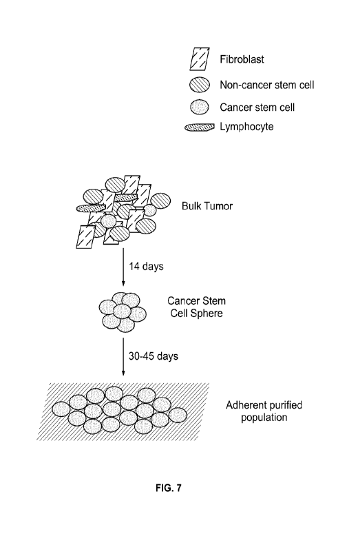
Cancer
Res. 13:5455-5462.
[0074] The present disclosure provides compositions and methods, where tumor
cells
are inactivated, e.g., by radiation, nucleic acid cross-linkers, polypeptide
linkers, or
combinations of these. One particular nucleic acid alkylator is beta-alanine,
N-(acridin-
9-y1), 2-[bis(2-chloroethyl)amino]ethyl ester. Exemplary cross-linkers, such
as
psoralens in combination with ultraviolet (UVA) irradiation, have the ability
to cross-link
DNA but to leave proteins unmodified. Nucleic acid targeting compound can be
4'-(4-
amino-2-oxa)buty1-4,5',8-trimethylpsoralen ("S-59"). Cells can be inactivated
with 150
micromolar of psoralen S-59 and 3 J/cm2 UVA light (FX 1019 irradiation device,
Baxter
Fenwal, Round Lake, IL). See, U.S. Pat. Nos. 7,833,775 of Dubensky and
7,691,393 of
Dubensky, which are incorporated herein by reference, in their entirety.
Tumor antigens
[0075]The present disclosure provides reagents and methods for stimulating
immune
response against a tumor antigen, for stimulating immune response against a
cell
expressing a tumor antigen, for administering to a human or veterinary
subject, and for
use in diagnosing a human or veterinary subject, and the like. The present
disclosure
provides a reagent, and related methods, for stimulating immune response
against a
cell that expresses one or more of, e.g., p53, MUC1, NY-ESO-1, c-myc,
surviving, p62,
cyclin B1, and Her2/neu (see, e.g., Reuschenbach et al (2009) Cancer Immunol.
Immunother. 58:1535-1554). In some exemplary implementations, the immune
response is against a cell that expresses said antigen or antigens, but not
necessarily
specific to that cell (the immune response is against other cells as well). In
in other
exemplary implementations, the immune response is against a cell that
expresses said
antigen or antigens, and where the immune response requires recognition of
said
antigen. In yet other exemplary implementations, the immune response is
against a cell
that expresses said antigen or antigens, and where the immune response does
not
require recognition of said antigen.
[0076]The present disclosure provides reagents and methods for stimulating
immune
response against a cell that expresses heat shock protein (HSP). Immunotherapy
21
CA 02882095 2015-02-13
WO 2014/028274 PCT/US2013/053850
against HSP is effective against colorectal cancer, melanoma, and renal cell
carcinoma
(see, e.g., Buonaguro et al (2011) Clinical Vaccine Immunol. 18:23-34). What
is
encompassed is reagent and method for stimulating immune response against
cancer-
testis antigen, or against differentiation antigen, or against an
overexpressed antigen, or
against tumor-associated carbohydrate antigen. In other exemplary
implementations,
reagent and method that stimulates immune response against a neoplastic
disorder that
expresses MUC1, an antigen that is associated with breast, colorectal,
gastric,
pancreatic, and ovarian cancer (Reuschenbach et al (2009) Cancer Immunol.
Immunother. 58:1535-1554). Also provided is reagent and method that stimulates
immune response against a neoplastic disorder that expresses p53, an antigen
associated with lung cancer, colorectal cancer, esophageal cancer, and ovarian
cancer
(Reuschenbach et al, supra). Moreover, what is provided is reagent and method
that
stimulates immune response against a neoplastic disorder that expresses
Her2/neu, an
antigen associated with breast, colorectal, and ovarian cancer. In exemplary
implementations, the present disclosure provides reagents and methods for
stimulating
immune response against the following antigen, or against a cell expressing
said
antigen, where the antigen is a MAGE family antigen. MAGE means, "melanoma
associated antigen." MAGE family antigens are associated with melanoma (SeIvan
et al
(2008) Int. J. Cancer. 122:1374-1383), as well as with hepatocellular
carcinoma (see,
e.g., Mou et al (2002) Brit. J. Cancer. 86:110-116), ovarian cancer (Zhang et
al (2010)
BMC Cancer. 10:163 (6-pages), non-small cell lung cancer (NSCLC) (Gridelli et
al
(2009) The Oncologist. 14:909-920; Sienel et al (2007) Clin. Cancer Res.
13:3840-
3847), and colorectal cancer (Toh et al (2009) Clin. Cancer Res. 15:7726-
7736).
[0077] CD133 is an antigen expressed by a variety of cancers, including
melanoma,
colorectal cancer, Ewing's sarcoma, hepatocellular cancer (HCC), non-small
cell lung
cancer (NSCLC), and ovarian cancer (Perego et al (2011) J. Inv. Dermatol.
11:546-547;
Cao, et al. (2011) BMC Gastroenterol. 11:71 (11 pages); Lorico and Rappa
(2011)
135039 (6 pages); Ferrandina et al (2009) BMC Cancer. 9:221 (9 pages)). The
present
disclosure provides a population of cancer stem cells that expresses CD133; at
least
one dendritic cell loaded with a cancer stem cell that expresses CD133;
methods for
preparing a dendritic cell loaded with a cancer stem cell that expresses
CD133; and
22
CA 02882095 2015-02-13
WO 2014/028274 PCT/US2013/053850
methods of administering at least one dendritic cell loaded with a cancer stem
cell that
expresses CD133 to a subject that has a cancer that expresses the CD133
biomarker.
[0078] Regarding the ABCB5 antigen, the present disclosure provides reagents
and
methods, where cancer stem cells expressing ABCB5 are contacted to, or loaded
onto,
dendritic cells, and where the loaded dendritic cells are administered to a
human
subject or animal that has an ABCB5-expressing cancer. ABCB5 expression is
associated, for example, with melanoma cancer stem cells (Schatton et al
(2010)
Cancer Res. 70:697-708), as well as with colorectal cancer (Wilson et al
(2011) Cancer
Res. 71:5307-5316). Related methods include inducing immune response against a
cell that expresses ABCB5, preferably, a cancer stem cell that expresses
ABCB5.
[0079] The present disclosure also encompasses reagents and methods, relating
to the
following antigens: aldehyde dehydrogenase (ALDH), for example ALDH1A3; ABCB1
(P-glycoprotein/MDR1); BCL2A1; SNAI2 (slug); ATM, CHEK1, and CHEK2. Also
encompassed are reagents and methods, relating to CD44, CD133, CD24, CD49f,
ESA;
CD166; and lineage panels. Lineage panels include CD45, CD31, CD3, CD64, CD10,
CD16, CD18, and GPA; CD45, CD31, CD140a, and Ter119; CD45, CD31 and CD140a.
Typically lineage panels include one or more of CD45, CD31, CD3, CD64, CD10,
CD16,
CD18, GPA, CD140a and Ten 19 (US 2011/0124032 of Diehn et al, which is hereby
incorporated by reference in its entirety).
[0080] What is also encompassed is reagent, and related methods, that are
specific for
stimulating immune response against one or more of the following antigens, or
against a
cell expressing one or more of the following antigens: MAGE-A subtypes, such
as,
MAGE-Al , MAGE-A2, MAGE-A3/6, MAGE-A4, and MAGE-Al2 (see, e.g., Sienel et al,
supra). In alternative exemplary implementations, the disclosure provides
reagents and
related methods that stimulate against intercellular adhesion molecule-1 (ICAM-
1), or
against a cell expressing ICAM-1, or against a neoplastic cell, or against a
cancer in a
subject, that is identified with one or more of ICAM-1. ICAM-1 associated
cancers
include melanoma, colon cancer, bladder cancer, lung cancer, pancreatic
cancer, and
hepatocellular carcinoma (Shih et al (2004) Korean J. Intern. Med. 19:48-52).
Moreover, the present disclosure provides reagents and methods for stimulating
23
CA 02882095 2015-02-13
WO 2014/028274 PCT/US2013/053850
immune response against the following antigen, or against a cell expressing
the
following antigen, or against a neoplastic cell, or against a cancer in a
subject that is
identified with sHLA-E. sHLA-E is a non-classical MHC class I molecule, that
is
associated with melanoma, colorectal cancer, and renal cancer (Allard et al
(2011)
PLoS One. 6:e21118 (9-pages). Also provide are reagents and method for
stimulating
response against the following antigen, or against a cell that expresses the
following
antigen, or against a neoplastic cell expressing the following antigen, or
against a
cancer in a subject that expresses the following antigen. The antigen is HERV-
K gag-
related NGO-Pr-54. This antigen is associated with ovarian cancer, prostate
cancer,
and leukemia (Ishida et al (2008) Cancer Immunol. 8:15 (10-pages)).
[0081] The present disclosure provides reagents and related methods for
stimulating
immune response against a neoplastic cell that is as follows, or against a
cancer in a
subject that is as follows. The exemplary implementations encompass, and are
not
limited to: (1) melanoma and colorectal; (2) melanoma and ovarian; (3)
melanoma and
lung; (4) melanoma and hepatic; (5) melanoma, colorectal, and ovarian; (6)
melanoma,
colorectal, and lung; (7) melanoma, colorectal, and hepatic; (8) melanoma,
lung, and
hepatic; (9) melanoma, ovarian, and lung; (10) melanoma, ovarian, and hepatic;
(11)
melanoma, ovarian, lung, and hepatic; (12) melanoma, colorectal, lung, and
hepatic;
(13) melanoma, colorectal, ovarian, and hepatic; (14) melanoma, colorectal,
ovarian,
and lung; and (15) melanoma, colorectal, ovarian, lung, and hepatic.
[0082] Exclusionary exemplary implementations are reagents and methods that do
not
induce, or have been shown to fail to induce, a pre-determined level of immune
response. The pre-determined level of immune response can be assessed, for
example, against one or more of a cancer cell that is melanoma cell,
colorectal cancer
cell, ovarian cancer cell, lung cancer cell, or hepatic cancer cell. By way of
a
non-limiting definition, the pre-determined level stimulation can be, for
example, a
stimulation that is less than 20%, less than 15%, less than 10%, less than 5%,
less than
2%, less than 1%, a maximal level. The maximal level can be in terms of
percent of
human subjects showing maximal response by RECIST criteria, in terms of
killing of
cancer stem cells in a human subject or experimental animal, in terms of
overall
survival, in terms of progression-free survival (PFS); in terms of time to
progression
24
CA 02882095 2015-02-13
WO 2014/028274 PCT/US2013/053850
(TTP), in terms of a maximal cytotoxic lymphocyte (CTL) response signal, in
terms of a
maximal ELISPOT assay signal, in terms of a maximal result from antibody
dependent
cell cytotoxicity (ADCC), in terms of T cell activation, in terms of T cell
expansion, in
terms of intracellular cytokine staining (ICS) assays, in terms of tetramer
assays, and
the like (see, e.g., Nomura et al (2008) Cytometry A. 73:984-991). For
example, in one
exemplary implementation, what is excluded are reagents and methods that
stimulate
less than 20% of a pre-determined maximal level. Clinical endpoints, such as
PFS,
TTP, time to distant metastasis, overall survival, and techniques for
interpreting these
endpoints, are detailed (Brody (2012) Clinical Trials:Study Design, Endpoints
and
Biomarkers. Elsevier, San Diego, CA), and are part of the present disclosure.
[0083] Reagents, methods, and techniques that are encompassed by the present
disclosure include, US 2011/0313229 of Sugaya et al, which concerns cancer
stem
cells, WO 2011/041453 of Weismann and Boiko, which concerns isolation of
melanoma
cancer stem cells, and US 2011/0286963 of Blot-Chabaud et al, which concerns
CD146. Each of these is hereby incorporated by reference in their entirety.
[0084] Non-adherent conditions, non-adherent plates, non-adherent coatings,
and the
like, can be provided by hydrophobic materials and by non-biofouling
materials, such as
polystyrenes, thin agar coating, siloxanes, fluorpolymers, polyethylenes, and
the like.
See, e.g., Tsai et al (2009) J. Biomater. Sci. Polym. Ed. 20:1611-1628; US
7,790,217
issued to Toreki et al, US 6,342,591 issued to Zamora et al, US 2011/0282005
of Jiang
et al, each of which is incorporated herein in its entirety.
Polyethyleneglycol (PEG) for
non-adhesion is disclosed by Kim et al (2006) Lab Chip. 6:1432-1437. Ultra-Low
Attachment surfaces include Corning Ultra-Low Attachment Surface (Corning,
Inc.) and
Thermo Scientific's Nunc HydroCell Surface.
[0085] In exemplary implementations, the disclosure provides additives that
can
promote non-adherent conditions, such as additives that are membrane
expanders,
tensioactive agents, Pluronic F-68, Tween-80, or polyvinylalcohol (PVA) (Sigma
Aldrich
catalogue, St. Louis, MO).
[0086] In exemplary implementations, down-regulation of indoleamine-pyrrole
2,3-
dioxygenase can be effected by sodium butyrate, COX-2 inhibitors, anti-sense
nucleic
CA 02882095 2015-02-13
WO 2014/028274 PCT/US2013/053850
acids, si-RNA, or micro-RNA. Down-regulation of IL-10 or TGF-beta can be
affected by
anti-sense nucleic acids, si-RNA, or micro-RNA. Liu et al (2011) FEBS Lett.
585:1963-
1968, discloses the use of micro-RNA to down-regulated IL-10 expression. Yu et
al
(2012) Carcinogenesis. 33:68-76, disclose the use of micro-RNA to decrease
efficacy of
transforming growth factor-beta (TGF-beta), by down-regulating TGF-beta
receptor.
Lang et al (2011) Biochim. Biophys. Res. Commun. 409:448-453, report the use
of
small interference RNA (siRNA) to inhibit TGF-beta expression.
[0087] In exemplary implementations, expansion procedure is conducted starting
with a
single cell. In other exemplary implementations, expansion procedure is
initiated with
about 10 cells, about 20 cells, about 50 cells, about 100 cells, about 200
cells, about
500 cells, about 1000 cells, about 2000 cells, about 5000 cells, about 10000
cells, about
20000 cells, about 50000 cells, and the like.
[0088] Exclusionary exemplary implementations are provided. Without implying
any
limitation, the reagents and method of the present disclosure can exclude a
population
of cells, a tissue, an organ, or a subject, and the like, where expression of
CD146 is less
than 50%, less than 40%, less than 30%, less than 20%, less than 15%, less
than 10%,
less than 5%, or less than 2%. Also, what can be excluded is a population of
cells, a
tissue, an organ, or a subject, and the like, where expression of CD271 is
less than
50%, less than 40%, less than 30%, less than 20%, less than 15%, less than
10%, less
than 5%, or less than 2%. In yet another exclusionary exemplary
implementation, what
can be excluded is a population of cells, a tissue, an organ, or a subject,
and the like,
where co-expression of both CD146 and CD271 (co-expression in exactly the same
cell, for each cell measured) is less than 50%, less than 40%, less than 30%,
less than
20%, less than 15%, less than 10%, less than 5%, or less than 2%. Also, what
can be
excluded is a population of cells, a tissue, an organ, or a subject, and the
like, where
expression of both CD146 and CD271 (either co-expressed in exactly the same
cell, or
merely both expressed in the entire population of cells) is less than 50%,
less than 40%,
less than 30%, less than 20%, less than 15%, less than 10%, less than 5%, or
less than
2%.
26
CA 02882095 2015-02-13
WO 2014/028274 PCT/US2013/053850
[0089]The term "co-express" refers to the situation where the indicated
markers, e.g.,
genes, polypeptides, antigens, and so on, are expressed in exactly the same
cell, and
also are expressed concurrently. Regarding concurrent co-expression, the time
frame
of co-expression can be about 5 minutes, about 30 minutes, about 1 hour, about
6 hours, about 12 hours, about 1 day, about 2 days, about 4 days, about 8
days, and so
on. The time frame of co-expression can be at least 1 minute, at least 5 min,
at least 10
min, at least 20 min, at least 60 min, at least 2 hours, at least 4 hours, at
least 6 hours,
at least 12 hours, at least 24 hours, at least 2 days, at least 3 days, at
least 4 days, at
least 8 days, at least one week, at least two weeks, and so on.
[0090]The present disclosure isolated population of cells originating from a
human
melanoma tumor, wherein at least 20% of the cells in the population express
CD146
and at least 20% of the cells express CD271, or wherein at least 20% of the
cells
co-express CD146 and CD271. Also provided, is isolated population of cells
originating
from a human melanoma tumor, wherein at least 30% of the cells in the
population
express CD146 and at least 30% of the cells express CD271, or wherein at least
40% of
the cells co-express CD146 and CD271. Also provided, is isolated population of
cells
originating from a human melanoma tumor, wherein at least 40% of the cells in
the
population express CD146 and at least 30% of the cells express CD271, or
wherein at
least 40% of the cells co-express CD146 and CD271. The present disclosure
encompasses, without limitation, a population of cells that occurs as a
monolayer or
other layer, a population of cells that occurs as a suspension, a population
of cells that
occurs as one or more spheres, and so on.
Populations of cells detectably expressing only one of CD146 or CD271
[0091]The present disclosure encompasses a population of cells that expresses
CD146
but not CD271. Also, the present disclosure encompasses a population of cells
that
expresses CD271 but not CD146. In embodiments, what is encompassed is a
population of cells where at least 20% of the cells express CD146, where at
least 30%,
at least 40%, at least 50%, at least 60%, at least 70%, at least 80%, at least
90%, at
least 95%, at least 98%, of the cells express CD146 but not CD271. Also, what
is
encompassed is a population of cells where at least 20% of the cells express
CD146,
27
CA 02882095 2015-02-13
WO 2014/028274 PCT/US2013/053850
where at least 30%, at least 40%, at least 50%, at least 60%, at least 70%, at
least
80%, at least 90%, at least 95%, at least 98%, of the cells express CD1271 but
not
CD146. What is encompassed are methods of culturing the above cells, methods
of
isolating the cells, methods of loading the cells on dendritic cells, vaccines
comprising
the above cells, vaccines comprising dendritic cells loaded with the above
cells,
methods for administering the vaccines to subject, and so on.
[0092]Melanoma cells that are CD146+/CD271- identify mesenchymal cancer cells,
and
melanoma cells that are CD146+/CD271+ identify cells that are cancer stem
cells with
mesenchymal characteristics. The present disclosure provides a population of
cells,
and related methods, wherein at least 20% of the cells in the population are
CD146+/CD271-, at least 22%, at least 24%, at least 26%, at least 28%, at
least 30%,
at least 34%, at least 38%, at least 42%, at least 46%, at least 50%, at least
54%, at
least 58%, at least 62%, at least 66%, at least 70%, at least 74%, at least
78%, or at
least 82%, of the cells are CD146+/CD271-. The present disclosure provides a
population of cells, and related methods, wherein at least 20% of the cells in
the
population are CD146-/CD271+, at least 22%, at least 24%, at least 26%, at
least 28%,
at least 30%, at least 34%, at least 38%, at least 42%, at least 46%, at least
50%, at
least 54%, at least 58%, at least 62%, at least 66%, at least 70%, at least
74%, at least
78%, or at least 82%, of the cells are CD146-/CD271+. In exclusionary
embodiments,
the present disclosure can exclude any cell population that fails to meet one
of the
above-disclosed percentages.
Exclusionary embodiments
[0093]What can be excluded is a single cell, a population of cells, a
population of cells
that occurs as a monolayer or other layer, a population of cells that occurs
as a
suspension, a population of cells that occurs as one or more spheres, and so
on, where
expression of CD146 occurs in less than 10% of the cells, occurs in less than
20%, less
than 30%, less than 40%, less than 50%, less than 60%, less than 70%, less
than 80%,
of the cells. What can be excluded is a single cell, a population of cells, a
population of
cells that occurs as a monolayer or other layer, a population of cells that
occurs as a
suspension, a population of cells that occurs as one or more spheres, and so
on, where
28
CA 02882095 2015-02-13
WO 2014/028274 PCT/US2013/053850
expression of CD271 occurs in less than 10% of the cells, occurs in less than
20%, less
than 30%, less than 40%, less than 50%, less than 60%, less than 70%, less
than 80%,
of the cells. What can be excluded is a single cell, a population of cells, a
population of
cells that occurs as a monolayer or other layer, a population of cells that
occurs as a
suspension, a population of cells that occurs as one or more spheres, and so
on, where
expression of each of CD146 and CD271 occurs in less than 10% of the cells,
occurs in
less than 20%, less than 30%, less than 40%, less than 50%, less than 60%,
less than
70%, less than 80%, of the cells. What can be excluded is a single cell, a
population of
cells, a population of cells that occurs as a monolayer or other layer, a
population of
cells that occurs as a suspension, a population of cells that occurs as one or
more
spheres, and so on, where co-expression of each of CD146 and CD271 occurs in
less
than 10% of the cells, occurs in less than 20%, less than 30%, less than 40%,
less than
50%, less than 60%, less than 70%, less than 80%, of the cells. In this
context,
co-expression means that, with analysis of a given particular cell, CD146 and
CD271
are both detectably expressed by that particular cell. What can be excluded is
any
population of melanoma cells, where over 1`)/0, over 2%, over 4%, over 5%,
over 10%,
over 15%, over 20%, over 30%, over 40%, over 50%, over 60%, over 70%, over
80%,
over 90%, of the melanoma cells are not melanoma cancer stem cells.
Brief descriptions of the figures
[0094]Figure 1. Flow cytometry characterization of melanoma stem cells at
various
stages of the purification process.
[0095]Figure 2. Table showing percentages of expression of CD146 and CD271 in
autologous melanoma cell lines.
[0096]Figure 3. Phenotype (CD146; CD271) of various melanoma cell lines.
[0097]Figure 4. Flow cytometry results (CD146; CD271) of melanoma stem cells
in
enzyme digest, melanoma stem cells prepared by standard method, and melanoma
cells prepared by sphere-generating method.
[0098]Figure 5. Flow cytometry results (CD146; CD271) (MHC Class II; MHC Class
I)
of melanoma cells subjected to various treatments.
29
CA 02882095 2015-02-13
WO 2014/028274 PCT/US2013/053850
[0099]Figure 6. Drawing of purification procedure using Method 1.
[00100] Figure 7. Drawing of purification procedure using Method 2.
[00101] Figure 8. Enrichment of cancer stem cells during purification process.
Figure 8A shows histogram. Figure 8B shows flow cytometry results.
[00102] Figure 9. Histogram comparing expression of: (1) CD146 and CD271 in
bulk
tumor cells, (2) "Cancer stem cells" of the present disclosure, and (3)
"Purified cell line"
produced by standard method.
[00103] As used herein, including the appended claims, the singular forms of
words
such as "a," "an," and "the" include their corresponding plural references
unless the
context clearly dictates otherwise. All references cited herein are
incorporated by
reference to the same extent as if each individual publication, patent,
published patent
application, and sequence listing, as well as figures and drawings in said
publications
and patent documents, was specifically and individually indicated to be
incorporated by
reference.
Further Description
Staging of cutaneous melanoma
[00104] The pharmaceutical or reagent of the disclosure can be administered to
melanoma patients, where melanoma is diagnosed at Stage I, Stage II, Stage
III, or
Stage IV (Mohr et al (2009) Ann. Oncology (Suppl. 6) vi14-vi21). Stage I, for
example,
refers to patients with primary melanomas without evidence of regional or
distant
metastasis. Stage II includes patients without evidence of lymphatic disease
or distant
metastases, where the patients are further characterized, e.g., by lesions
greater than
lmm and less than or equal to 2mm thick with ulceration of the overlying
epithelium, or
by lesions greater than 2mm and less than or equal to 4mm thick with
epithelial
ulceration. Stage III melanoma includes lesions with pathologically documented
involvement of regional lymph nodes or in-transit or satellite metastases,
where patients
may have, e.g., one, two, three, or four or more affected lymph nodes. Stage
IV
melanoma is defined by the presence of distant metastases, where the
metastasis is
located only in distant skin, subcutaneous tissues, or lymph nodes, where the
CA 02882095 2015-02-13
WO 2014/028274 PCT/US2013/053850
metastasis involves lung metastases, or where the metastasis involves all
other visceral
sites.
[00105] The disclosure encompasses methods for administration that are
preventative, that is, for use with subjects not yet or never diagnosed with a
melanoma.
What is encompassed are methods for administration where a subject had earlier
been
diagnosed with a melanoma, and had earlier been successfully treated to
eradicate the
melanoma (or had experienced a spontaneous complete remission), and where
following eradication the administration is used preventatively.
[00106] The disclosure provides a pharmaceutical composition or pharmaceutical
reagent, related methods of administration, and methods of treatment, that
result in
survival data with a hazard ratio (HR) of less than 1.0, HR less than 0.9, HR
less than
0.8, HR less than 0.7, HR less than 0.6, HR less than 0.5, HR less than 0.4,
HR less
than 0.3, and the like. The disclosure results in overall survival data,
progression-free
survival data, time to progression data, and so on. What is also provided is 6-
month
PFS of at least 40%, at least 50%, at least 60%, at least 70%, at least 75%,
at least
80%, at least 85%, at least 90%, at least 95%, and so on. Moreover, what is
provided is
6-month overall survival of at least 40%, at least 50%, at least 60%, at least
70%, at
least 75%, at least 80%, at least 85%, at least 90%, at least 95%, and so on.
Additionally, what is provided is 1-year (or 2-year) PFS of at least 40%, at
least 50%, at
least 60%, at least 70%, at least 75%, at least 80%, at least 85%, at least
90%, at least
95%, and so on. Moreover, what is provided is 1-year (or 2-year) overall
survival of at
least 40%, at least 50%, at least 60%, at least 70%, at least 75%, at least
80%, at least
85%, at least 90%, at least 95%, and so on (see, e.g., U.S. Dept. of Health
and Human
Services. Food and Drug Administration. Guidance for Industry. Clinical trial
endpoints
for the approval of cancer drugs and biologics (April 2005)).
Biomarkers and flow cytometry
[00107] Studies of melanoma cancer stem cells disclose reagents and methods
for
detecting markers, such as CD271 (Civenni et al (2011) Cancer Res. 71:3098-
3109),
CD146, CD146 (Perego et al (2010) J. Inv. Dermatol. 130:1877-1886); and ABCB5
(Schatton et al (2011) Cancer Res. 70:697-708). Other biomarkers of interest
include
31
CA 02882095 2015-02-13
WO 2014/028274 PCT/US2013/053850
CD20, CD133, CD44, CD90, CD24, EpCAM, ALDH1, and ABCB5 (see, e.g., Wang and
Jacob (2011) Genome Medicine. 3:11 (6 pages); Schlaak et al (2012) Oncotarget.
3:22-30).
[00108] Phenotypic characterization of the cell populations are performed
using
monoclonal antibodies against surface markers (BD Pharmingen San Diego, CA:
BD)
Pharmingen. CaliBRITE flow cytometry calibration (BD Pharmingen) is used prior
to
each run and the same instrument settings were used throughout the collection
of flow
cytometric data. Flow cytometry is conducted with a Beckton-Dickenson FAGS
Calibur0 flow cytometer. The number of polypeptides expressed by a cell can be
measured, for example, using fluorescent antibodies with quantitation by flow
cytometry
(see, e.g., Macey (2010) Flow Cytometry:Principles and Applications, Humana
Press;
Hawley (2010) Flow Cytometry Protocols (Methods in Molecular Biology) Humana
Press; Shapiro (2003) Practical Flow Cytometry, Wiley-Liss).
[00109] Characterization of cell lines of the present disclosure by flow
cytometry
demonstrated the enrichment for cells of mesenchymal and neural crest origin
(CD146
and CD271, respectively) which have been described as melanoma stem cell
markers.
Comparison of these cell lines versus the original bulk enzyme digest samples
demonstrated that they were enriched for either CD146 and/or CD271 (78.5
8.3%
versus 26.9 5.8%) after purification and expansion. Examination of 35/42
cell lines
used in a randomized phase II clinical trial revealed consistent expression
these
markers in the purified tumor cell lines (35.2 3.9% CD146+/CD271-, 41.5
4.3
CD146+/CD271+, 16.9 4.0 CD146-/CD271-, 6.4 1.9 CD146-/CD271+) . Using
these
cells as the antigen source in an autologous dendritic cell therapy resulted
in 50% 5-
year survival in patients with stage IV melanoma (n = 54). Using excess
cryopreserved
samples with our methods, we were able to reduce the production time to 2
months and
increase the success rate to 80%. This process also resulted in increasing the
purity of
the cancer stem cells from ¨70% to >90% based on these known cancer stem cell
markers. In addition, contaminating fibroblasts were eliminated with minimal
skilled
manipulations. This approach is suitable for automation and/or optimization
where a
closed and uniform systems may be constructed that support automation and
32
CA 02882095 2015-02-13
WO 2014/028274 PCT/US2013/053850
scalability. Scalability and optimization is often associated with reductions
in cost of
delivery and preparation, automation may also result in reduced cost of labor.
Spheres
[00110] The ability of cells to form spheres result, in part, from cell-
surface proteins
called, integrins." Homophilic integrins expressed on the cell's surface
ensure that cells
"stay together". Spheres are formed directly from enzyme digest (ED) which is
a single
cell suspension at the very beginning of a culture, or can be formed from
frozen sample
or an existing attached culture at any time. The enzyme digest seeding result
in this
spherical formations that incorporate the cells with the specific surface
properties.
Fibroblasts for example cannot be incorporated, eventually are faded out from
a culture
during gravitational feeding . The media used is lacking of molecules that
promote
adhesion in order to prevent the non-specific agglomeration of the cells not
having
homophilic proprieties and to prevent the adhesion to the culture vessel
surfaces. Such
adhesion molecules (CAMs) are commonly found in the animal or human serum,
therefore a media composition which is serum free is suitable.
[00111] In the serum free media culture, what is supplied by way of
supplements to the
media include any hormones, nutrients, mineral, and vitamins that are required
for
supporting growth and maintenance, or other desired aspects of cell physiology
and
function. In some instance one can stimulate and sustain the stem cell
proliferation with
the addition or adjustment of amount of growth factors that possess a
mitogenic activity,
e.g., such as FGF family and EGF.
[00112] Spheres of cells, including spheres of cancer stem cells, can be
characterized
in terms of biomarker expression by way of fixing and staining with labeled
antibodies,
followed by viewing with confocal microscopy (Weiswald et al (2010) Cancer.
10:106
(11 pages)). Spheres can be prepared, for example, from suspensions obtained
from
fresh tumors, or from cells adapted to grow as adherent cells, as documented
for the
case of melanoma cells (Perego et al (2011) J. Inv. Dermatol. 11:546-547).
Spheres
can be generated from a single cell, as shown by the fluorescent microscopy
images of
spheres (see, e.g., Cao et al (2011) BMC Gastroenterol. 11:71 (11 pages)). The
morphology of spheres, for example, large and irregular versus tiny and
compact, may
33
CA 02882095 2015-02-13
WO 2014/028274 PCT/US2013/053850
be influenced by the choice of medium (Mancini et al (2011) PLoS ONE. 6:e21320
(12
pages). Without implying any limitation, the present disclosure encompasses
the
methods and techniques, disclosed in the above references.
Time in culture
[00113] The present disclosure provides method for preparing melanoma cancer
stem
cells, where the total culturing time including time required for
manipulations such as
changing media, replating, centrifugation, and sedimenting, is less than 5
months, less
than 4 months, less than 3 months, less than 2 months, less than one month,
less than
150 days, less than 120 days, less than 90 days, less than 60 days, less than
30 days,
or less than 150 days (+/-20 days), less than 120 days (+/-20 days), less than
90 days
(+/-20 days), less than 60 days (+/-20 days), less than 30 days (+/-20 days).
In
exclusionary embodiments, the present disclosure can exclude any method for
preparing cancer stem cells, and any population of cancer stem cells prepared
by that
method, where time required for manipulation is greater than one of the time-
frames
disclosed above. What is provided is a time in adherent culture, that is
indicated by one
of the above time-frames. Also, what is a provided is a time in non-adherent
culture,
that is one of the above time-frames. Moreover, what is also provided is a
combined
time in adherent culture and in non-adherent culture, that is identified by
one of the
above time-frames.
Media of the present disclosure.
[00114] One formulation variation can use the B27 supplementation with HSA
(human
serum albumin), while another variant uses the B27 supplementation with BSA
(bovine
serum albumin). Table 1 discloses the serum free supplement composition is
exemplified with human albumin (see the final component in Table 1). In some
exemplary implementations, BSA can be used instead of human serum albumin
(HSA).
Table 11 discloses the components of the medium, RPM! 1640. "PSF" refers to
Pen
Strep and Fungizone." PSF is used as an antibiotic and anti-fungal agent.
"Neuroblast
stem cell media" is described in Table 8 (bovine component in the medium) and
in
Table 9 (made with human serum albumin; HSA). "NeuroBlast media" and
"NeuroBlast
Stem Cell media" are one and the same.
34
CA 02882095 2015-02-13
WO 2014/028274 PCT/US2013/053850
[00115] The present disclosure encompasses neuron stem cell media, methods for
using neuron stem cell medium, cells prepared with the use of neuron stem cell
media,
methods for administering said cells to subjects, and kits comprising neuron
stem cell
medium. For each individual component of the neuron stem cell medium, the
present
disclosure encompasses a range of concentrations that is +/-5% or less, +/-10%
or less,
-F/-15% or less, -F/-20% or less, +/-25% or less, -F/-30% or less, and the
like, with the
total volume remaining constant. The present disclosure encompasses neuron
stem
cell medium, with omission of one or more of the components. The disclosure
also
encompasses neuron stem cell medium, with one or more substitutions. General
examples of substitutions for cell media include, e.g., substituting sodium
phosphate for
potassium phosphate, sucrose for glycerol, cystine for cysteine, and the like.
Alternatives to basic fibroblast growth factor
[00116] With culture on non-adherent substrate, medium can optionally include
basic
fibroblast growth factor (bFGF), bFGF analogue, bFGF in combination with one
or more
other growth factors, or one or more growth factors with no added bFGF. Growth
factors include ENNA compounds (Burton et al (2010) Biochem. Soc. Trans.
38:1058-1061), bone morphogenic protein-2 (BMP-2), vascular endothelial growth
factor (VEGF), leukemia inhibitory factor (LIF), insulin growth factor-1 or -2
(IGF-1;
IGF-2), transforming growth factor-beta (TGF-beta), and the like.
[00117] The present disclosure provides, as an alternative to bFGF, any growth
factor
or ligand that acts through the MAPK (Mitogen-activated protein kinases). MAPK
was
originally called ERK (extracellular signal-regulated kinases).
[00118] The classic list of growth factors acting through this mechanism
includes FGF,
EGF, PDGF, NT3/4, BDNF, NGF, VEGF. In addition these also actt the same way:
TNF, IL-1, TGFb, FASL. These growth factors act primarily as mitogens.
[00119] Another mechanism of fast proliferation that can be exploited is
stimulating
PI3K-AKT pathway through receptor tyrosine kinases (such as EGF, IGF etc) and
GPCRs (G protein-coupled receptors).
CA 02882095 2015-02-13
WO 2014/028274 PCT/US2013/053850
[00120] In addition to natural ligands that stimulate the above pathways,
agents that
antagonize the inhibitors are to be considered for in-vitro use for
manufacturing. An
example of such inhibitor is the PTEN inhibitor that acts in the PI3K-AKT
system.
[00121] Other cancer treatments use agents against single targets in this
pathway.
Examples of single targets already used in therapy in vivo include, e.g., Raf
kinase
inhibitors sorafenib, SB590885, PLX4720, XL281, RAF265, LGX818, vemurafenib;
MEK
inhibitors : XL518, 0I-1040, PD035901, MEK162, selumetinib,
Trametinib(GSK1120212).
[00122] In contrast to these treatments the present disclosure uses the
(agonists)
ligands that promote the expansion of the cancer for in vitro manufacturing.
[00123] The morphogenic effect of ligands used in the present disclosure (such
as
FGF, EGF) contributes to preferential expansion of the cancer stem cell
population by
stimulating transcription factors such as Nanog, cKit, Sox2, Oct3/4 through
the
enumerated pathways.
[00124] One or more of the above natural and synthetic ligands, or any
combination
thereof, can be added to culture medium during non-adherent culture, e.g., in
a low
adherent flask, very low adherent flask, or ultra-low adherent flasks, or can
be added to
culture medium during adherent culture. The following concerns growth in
adherent
culture. bFGF is not absolutely needed during adherent culture, but it can
help maintain
the "stem cell" status of the cancer cells by stimulating transcription
factors such as
Nanog, cKit, Sox2, Oct3/4. Adversely, indiscriminate use or over-use of bFGF
may
enhance growth of a population that is not tumoral such as normal fibroblasts
or
epithelial cells. Therefore the use in the adherent stage should be limited in
time by
assessing the purity of the cancer cell population. The spherogenic step in
manufacturing will prevent the expansion of normal cell population and that is
the point
when growth factors can be used extensively and if impurification is
suspected, this step
(spherogenic) can be repeated.
[00125] Without implying any limitation, the main features of the present
disclosure
include: (1) Growing in non-adherent conditions, preferably with single growth
factors, or
combinations of growth factors, or antagonists of inhibitors; (2) Selecting
cancer step
36
CA 02882095 2015-02-13
WO 2014/028274 PCT/US2013/053850
cells by way of isolating spheroids (and not using any other method to isolate
the cancer
stem cells). Spheroids can be isolated by gravity methods, centrifugation,
filtering, and
so on; (3) Growing on adherent conditions, preferably with one or more growth
factors;
and (4) Optional repeating of the non-adherent process.
[00126]
Table 1. Neural Stem Cell Media
Component Final concentration in medium
(mg/L)
corticosterone 0.02
progesterone 0.005
retinol, all trans 0.1
retinol acetate 0.1
insulin 5
T3 0.002
Pluronic F68 0.25
lipoic acid 0.05
tocopherol 1
tocopherol acetate 1
linoleic acid 1
linolenic acid 1
catalase 2.5
glutathione reduced 1
superoxide dismutase 2.5
1-carnitine 2
ethanolamine 1
putrescine 10
selenium 0.01
human albumin 2500
/ / /
37
CA 02882095 2015-02-13
WO 2014/028274 PCT/US2013/053850
Table 2. R15-2XPSF (1L) Table 3. R15-1XPSF Table 4. R15
Antibiotic
(1L) free (1L)
RPMI-1640 800 mL 810 mL 820 mL
Heat inactivated FBS 75 mL 75 mL 75 mL
Iron-supplemented calf 75 mL 75 mL 75 mL
serum
L-glutamine 10 mL 10 mL 10 mL
Sodium pyruvate 10 mL 10 mL 10 mL
1 M HEPES 10 mL 10 mL 10 mL
Pen/Strp/Fung 20 mL 10 mL (zero mL)
(antibiotics)
Table 5. Transport media
R15-2XPSF (amount needed)
Gentamicyin (40 mg/mL) 1.2 microliters per mL R15-2XPSF
Table 6. Serum-free/antibiotic free (1 L)
RPMI-1640 970 mL
L-glutamine 10 mL
Sodium pyruvate 10 mL
1 M HEPES 10 mL
Table 7. Serum-free 2XPSF (1L)
RPMI-1640 950 mL
L-glutamine 10 mL
Sodium pyruvate 10 mL
1 M HEPES 10 mL
Pen/Strep/fung 20 mL
/ / /
38
CA 02882095 2015-02-13
WO 2014/028274
PCT/US2013/053850
Table 8. Neuroblast with BSA (IL)
KO DMEM/F12 970 mL
B-27 supplement 20 mL
ITS 10 mL
Glutamax 10 mL
FGF FGF must be added to a final concentration
of 5
ng/mL or 10 ng/mL right before using.
Table 9. Neuroblast with HSA (IL)
KO DMEM/F12 970 mL
B-27 supplement 20 mL
ITS 10 mL
Glutamax 10 mL
FGF FGF must be added to a final concentration
of 5
ng/mL or 10 ng/mL right before using.
Table 10. Cardioblast with B27 HSA based (1L)
KO DMEM/F12 970 mL
B-27 supplement 20 mL
ITS 10 mL
Glutamax 10 mL
T3 (0.1 mg/mL) 0.100 mL
Insulin 2 mL of 5 mg/mL stock or 10
micrograms/mL
Ascorbic acid 1 mL of 1000X stock or 20 micrograms/mL
FGF FGF must be added to a final
concentration of 10
ng/mL right before using.
EGF EGF must be added to a final concentration
of
2Ong/mL right before using.
39
CA 02882095 2015-02-13
WO 2014/028274
PCT/US2013/053850
Table 11. RPM! 1640 medium
Molecular Weight Concentration (mg/L) mM
Amino Acids
Glycine 75 10 0.133
L-Arginine 174 200 1.15
L-Asparagine 132 50 0.379
L-Aspartic acid 133 20 0.15
L-Cystine 2HCI 313 65 0.208
L-Glutamic Acid 147 20 0.136
L-Glutamine 146 300 2.05
L-Histidine 155 15 0.0968
L-Hydroxyproline 131 20 0.153
L-Isoleucine 131 50 0.382
L-Leucine 131 50 0.382
L-Lysine hydrochloride 183 40 0.219
L-Methionine 149 15 0.101
L-Phenylalanine 165 15 0.0909
L-Proline 115 20 0.174
L-Serine 105 30 0.286
L-Threonine 119 20 0.168
L-Tryptophan 204 5 0.0245
L-Tyrosine disodium salt dihydrate 261 29 0.111
L-Valine 117 20 0.171
Vitamins
Biotin 244 0.2 0.00082
Choline chloride 140 3 0.0214
D-Calcium pantothenate 477 0.25 0.000524
Folic Acid 441 1 0.00227
Niacinamide 122 1 0.0082
Para-Aminobenzoic Acid 137 1 0.0073
Pyridoxine hydrochloride 206 1 0.00485
Riboflavin 376 0.2 0.000532
Thiamine hydrochloride 337 1 0.00297
Vitamin B12 1355 0.005 0.0000037
idnositol 180 35 0.194
Inorganic Salts
Calcium nitrate (Ca(NO3)2 4H20) 236 100 0.424
Magnesium Sulfate (Mg504) (anhydrous) 120 48.84 0.407
Potassium Chloride (KCI) 75 400 5.33
Sodium Bicarbonate (NaHCO3) 84 2000 23.81
Sodium Chloride (NaCI) 58 6000 103.45
Sodium Phosphate dibasic (Na2HPO4) anhydrous 142 800 5.63
Other Components
D-Glucose (dextrose) 180 2000 11.11
Glutathione (reduced) 307 1 0.00326
Phenol Red 376.4 5 0.0133
CA 02882095 2015-02-13
WO 2014/028274 PCT/US2013/053850
Detailed descriptions of the figures
Figure 1
[00127] Melanoma stem cells with neuroendocrine and mesenchymal phenotypes are
enriched during the purification process using differential attachment and
serum
starvation methods. Representative flow cytometry plots for CD146 and CD271 at
the
beginning of the culture period (Enzyme Digest), at the point of partial
purity
(Intermediate), and after the purification was complete (Purified) are shown.
Normal
human dermal fibroblasts (NHDF) were included as a control. Summarized data
for
eight separately processed samples is shown. Values shown are averages SD.
Figure 2
[00128] The histogram discloses the percent of cells expressing CD146 and
CD271, for
cells at three different stages of preparation. The stages are enzyme digest,
intermediate, and purified. Table 12 shows Percentage of expression of CD146
and
CD271 in autologous melanoma cell lines used to load dendritic cells for
active specific
immunotherapy. N = 63 , for the individual patients comprising the histogram.
Table 12
Patient
Number CD146+/CD271- CD146+/CD271+ CD146-/CD271- CD146-/CD271+
1 17 82.7 0.3 0
2 77.3 4.1 18.5 0.1
3 13.3 76 5.9 4.8
4 1.7 96.7 0.7 1
33.3 64.6 1.8 0.3
6 32.6 67.4 0.1 0
7 46.5 52.7 0.8 0
8 35.9 53.4 9.7 1.1
9 47.8 50.8 1.4 0.1
11 14.6 47.4 27
11 86.8 8.2 4.9 0
12 77.4 19.6 2.7 0.3
13 0.2 2.6 34 63.3
14 0 6.7 26.1 67.3
12.6 70.9 8.3 8.2
16 46.4 48 4.7 0.9
17 71 20.1 8.6 0.2
18 59 25.9 15.1 0.1
19 58.1 34.4 7.2 0.3
47.3 48.5 3.8 0.4
41
CA 02882095 2015-02-13
WO 2014/028274
PCT/US2013/053850
21 64.1 22 13.3 0.5
22 57.3 38.6 4.1 0.1
23 56 43.5 0.5 0
24 48.4 43.1 8 0.6
25 18.8 76.8 2.4 3
26 27.8 40.7 30.1 1.4
27 79.2 9.7 11.1 0
28 23.5 20.3 42.4 13.8
29 55.9 41.8 2.2 0.1
30 76.4 11.9 11.7 0
31 24.5 58.1 12.2 5.2
32 31.7 45.9 18.7 3.8
33 0.3 0.9 89.8 9
34 79.2 10.5 10.3 0.1
35 47.3 50.5 2 0.3
36 64.2 18 17.2 0.3
37 21.4 25.7 45.5 7.4
38 0.2 57.8 1.6 40.4
39 46.1 50.3 3.6 0
40 46.4 52.2 1.4 0
41 64.2 30.9 4.8 0.1
42 0.3 0.5 67 32.2
43 58.5 35.9 5.6 0
44 30.1 24.4 38.4 7.1
45 68.6 30.2 1.1 0.1
46 21.4 14.7 56.6 7.4
47 54.3 33.4 9 3.3
48 0.2 51.3 0.4 48.2
49 21.8 72.9 0.6 4.7
50 7.8 70.8 4.9 16.5
51 69.9 14.6 15.2 0.4
52 35.5 63.6 0.9 0
53 50.7 48.3 0.8 0.2
54 59.5 22.2 15.6 2.8
55 14.3 2.7 78 5.1
56 19.6 78.2 1.4 0.8
57 17.2 80.3 2.4 0.1
58 29 68.6 2.4 0.1
59 31.2 42.2 21.2 5.5
60 24.7 74.4 0.5 0.5
61 38.9 52.7 2.1 6.3
62 33.6 65 1.1 0.3
63 53.8 18.9 20.3 7
Average 38.9 40.7 14 6.5
S.D. 24.2 24.7 19.6 14.3
42
CA 02882095 2015-02-13
WO 2014/028274 PCT/US2013/053850
Figure 3
[00129] Autologous melanoma cell lines used to load dendritic cells in active
specific
immunotherapy contained cells expressing antigens associated with neural crest
and
mesenchymal origins. Irradiated and cryopreserved purified autologous melanoma
cells
were assayed by flow cytometry for the expression of CD146 and CD271, N = 36,
values shown are SD.
Figure 4
[00130] Melanoma stem cell lines derived from either standard or sphere
generating
methodologies give rise to cells of the same phenotype. Cells from each of the
conditions shown in the figure were assayed by flow cytometry for CD146 and
CD271
measuring double positives. Figure 4 findings as compared to figure 3 findings
suggest
that in some instances there may at least one of a loss of cells that do not
form spheres
during incubation and an increase in CD271 expression.
Figure 5
[00131] Purified melanoma cell lines were placed in either neuronal stem cell
media or
standard serum containing expansion media (15%FBS/RPMI) for period of 7 days.
Afterwards, the cells were harvested by trypsinization in the case of the
adherent cells
and by simple collection of the cancer stem cell spheres. Cells from each of
the
conditions shown in the figure were simultaneously assayed by flow cytometry
for
CD146, CD271, MHC class I and MHC class II. Higher MHC II expression
stimulates
CD4 memory cells which can support and sustain an immune response by secretion
of
activation cytokines.
Figure 6
[00132] Schematic representation of the purification of melanoma cancer stem
cells
process using differential attachment and serum starvation (Method I). Bulk
tumor
representing enzymatically digested surgical tumor samples are incubated for 1-
3 days
in serum containing cell culture media then washed twice to remove
lymphocytes. The
attached mixture of fibroblasts, non¨cancer stem cells and cancer stem cells
are then
subjected to low serum cell culture conditions (range of 1-5% fetal bovine
serum) and a
43
CA 02882095 2015-02-13
WO 2014/028274 PCT/US2013/053850
series of differential attachment procedures over the course of an average of
120 days.
The differential attachment procedure consists of enzymatically detaching the
mixture of
cells from the substrate and plating onto new substratum (standard plasma
treated cell
culture flasks) for period of 5-20 minutes until 25-30% of the cell have
attached. The
non-attached cells are then transferred to a new flask and the attachment
procedure
repeated for a series of 4-6 times. This process take advantage of the
characteristic the
higher rate of attachment of fibroblasts compared to that of cancer cells.
Additionally,
the low serum conditions will inhibit the growth of contaminating fibroblast
and non-
cancer stem cells growth rates due to higher nutrient requirements of these
cells
compared to cancer stem cells.
Figure 7
[00133] Schematic representation of purification process using ultra-low
adherent stem
cell conditions to isolate cancer stem cells followed by adherent expansion
conditions
(Method 2). Polystyrene coated with a Corning proprietary material is, in
some
implementations, used to support ultra-low adherence. Poly-propylene has
structure-
inherited hydrophobic properties which also will support ultra-low adherence.
In
addition of Corning proprietary coatings, a thin agar coating
(polysaccharides),
polyamides or a siloxane may be used. Bulk tumor representing enzymatically
digested
surgical tumor samples are incubated in stem cell media under ultra-low
adherent or
adherent conditions to generated cancer stem cell spheres after 14 days.
Adherency
refers to cells that remain attached to the surface that they are growing on;
non-
attached cells will be removed during the washing and harvesting. Non adherent
condition refers to culture environment when the cells are not attached to a
substrate
other than a similar live cell. Hydrophobic materials or non-biofouling
treatments can be
used to achieve non-adherent conditions: agarose, poly-ethylene, fluoro-
polymers,
siloxanes. Conditions that can promote non-adherent conditions include lack of
serum
components or lack of peptides with terminations specific for integrins (RGD,
IKVAV,
YIGSR, RETTAWA etc) from media or substrates. Also, addition of membrane
expanders or tensioactive agents: Pluronic F-68, Tween80, Poly-Vinyl Alcohol
(PVA),
Poly-Ethylene Glycol (PEG) can promote non-adherent conditions. Hyaluronidase
which can act as a mobility enzyme in higher concentrations can cause cell
detachment.
44
CA 02882095 2015-02-13
WO 2014/028274 PCT/US2013/053850
It may also cause a lack of CD44 dependent anchorage. The skilled artisan in
the field
of mammalian cell culture can readily distinguish between culture flasks,
culture dishes,
and other culture containers, that are ultra-low adherent and those that are
low adherent.
[00134] This results in the removal of contaminating populations of cells such
as
lymphocytes and fibroblasts and non-cancer stem cell tumor cells. These
spheres are
enriched for CD146/CD271 positive cancer stem cell populations. The spheres
are then
dissociated either mechanically or enzymatically and plated onto adherent
surfaces and
allowed to replicate for a further 30-45 days. The cells are then harvested
for use in
immunotherapy as either whole cells or lysates.
Figure 8
[00135] Figure 8 discloses enrichment of cancer stem cells during purification
process.
Figure 8A shows flow cytometry results, and Figure 8B shows a historgram that
summarizes flow cytometry results. The results demonstrate that, for this
particular cell
preparation, in the enzyme digest (bulk tumor, open bars) the most prevalent
cell type is
CD146 minus/CD271 minus, and that at a purified step of the present
disclosure, the
most prevalent cell type is CD146 plus/CD271 minus. At the intermediate stage,
CD146
plus/CD271 cells and CD146 minus/CD271 plus cells occurred in roughly equal
percentages.
Figure 9
[00136] Figure 9 discloses expression of CD146 and CD271 in bulk tumor cells,
in
"cancer stem cells" of the present disclosure, and in "purified cell line"
produced by a
standard method.
[00137] Regarding Figure 9, in bulk tumor cells, the CD146 minus/CD271 minus
cells
(open segment of bar) account for the greatest proportion of cells, with CD146
minus/CD271 plus cells (downwards-slanted hatches in segment of bar)
accounting for
the next-most prevalent cells. In "cancer stem cells," the CD146 plus/CD271
plus cells
(criss-cross hatches in segment of bar) is the most prevalent type of cell. In
the
"purified cell line, CD146 plus/CD271 plus cells (criss-cross hatches in
segment of bar)
CA 02882095 2015-02-13
WO 2014/028274 PCT/US2013/053850
and the CD146 plus/CD271 minus cells (upwards-slanted hatches in segment of
bar)
are roughly equivalent in proportion, with the CD146 plus/CD271 plus cells
occurring at
a somewhat greater percentage than the CD146 plus/CD271 minus cells. Figure 9
represents the data from Table 13.
[00138]
Table 13. Data used for histogram of Figure 9
Cancer Stem Days in Yield CD146plus/ CD146plus/ CD146minus/
CD146minus/
Cell Derived culture x 10^6 CD271minus CD271plus CD271minus CD271plus
2352 39 125 3.04 93.39 0.55 3.02
2350 39 280 2.93 92.45 1.25 3.37
2266 39 92 14.52 77.84 5.7 2.44
Standard
Method Cell
Lines
2352 57 150 55.86 36.57 7.02 0.55
2350 76 200 19.95 61.73 9.52 8.8
2266 NA NA (not a previously established cell
line)
Examples
[00139] This present disclosure provides methods and reagents for isolating
and
expanding cancer stem cells of mesenchymal and neural crest origin from
biopsies of
melanoma samples for use in cell-based immunotherapy. Methodologies include
the
use of media formulations to isolate and then proliferate distinct populations
of tumor
stem cells with a neural crest and/or mesenchymal phenotype. CD146 is a marker
frequently found on mesenchymal cells and is associated with highly invasive
phenotype. CD271, neuronal growth factor receptor p75, is expressed by
neuronal
precursor cells and is expressed on melanoma initiating cells. What is
provided are
steps for isolating and expanding a type of cells that can be found in
metastatic
melanoma preparations where by these cells are either CD146+/CD271-,
CD146+/CD271+, or CD146-/CD271+. In addition, these cells may also be positive
for
CD44, Twist, Zeb1/2, Snail, Slug, SIP, CD133, CD166, CXCR4, Notch-1 and CD90
in
total or in part. The cells are cultivated under non-adherent conditions for
10-14 days in
stem cell media and then switched to adherent conditions under non-stem cell
46
CA 02882095 2015-02-13
WO 2014/028274 PCT/US2013/053850
expansion media. The final adherent step is to promote the up-regulation of
major
histocompatibility complexes and down-regulation of immunosuppressive
molecules like
indoleamine-pyrrole 253-dioxygenase, tumor growth factor beta and interieukin-
10. The
present disclosure uses these cells to immunize patients rather than bulk
tumor biopsies
or purified non-adherent cancer stem cells. Thus, the present disclosure uses
cells that
are identified as tumor forming as opposed to differentiated, non-
proliferating cell
populations which are the predominant cell in a bulk tumor biopsy or
immunosuppressive cancer stem cells that are still in the non-adherent
"spheroid" state.
SeIvan et al (2010) Melanoma Res. 20:280-292, disclose reagents and methods
for
processing melanoma tissue samples.
[00140] Cell-based immunotherapy is expected to be effective in the autologous
setting
due to presence of tumor-associated neo-antigens. However, the use of bulk
autologous tumor preparations have not yielded the expected clinical results
likely due
to the fact that population of cells present in bulk tumor are mainly
differentiated cells
with a low percentage of cells representing cancer stem cells. The techniques
of the
present disclosure demonstrate that tumor specimens must be processed in a
manner
that enriches for cancer stem cells to be most effective. In particular, the
process
results in the enrichment of cells that are either CD146+/CD271- (mesenchymal
cancer
cell), or CD271/CD146 (cancer stem cell with mesenchymal characteristics),
using the
sphere forming technique.
[00141] However, the cells must be then be attached to a substrate such as
cell culture
flask for the final proliferation steps before use in immunotherapy to guard
against the
immunosuppressive effects of cancer stem cells (Wei et al (2010) Glioma-
associated
cancer-initiating cells induce immunosuppression. Clin Cancer Res. 16:461-473;
Schatton et al (2010) Modulation of T-cell activation by malignant melanoma
initiating
cells. Cancer Res. 70:697-708).
[00142] The effect of expanding the cells on an adherent substrate such as a
standard
cell culture flask or similar surface is to up-regulate important immune
associated
proteins called major histocompatibility complexes (MHC class I and class II).
These
protein complexes are primary mechanism by which the immune system recognizes
47
CA 02882095 2015-02-13
WO 2014/028274 PCT/US2013/053850
and responses to cells that are either foreign or infected with viruses.
Cancer stem cells
down regulate those molecules while in the detached, spheroid phase of growth
and
up-regulate them while in the attached, expanding phase of growth. The act of
transferring the cancer stem cells from a non-adherent to an adherent state
during
expansion is expected to reduce or eliminate the ability of cancer stem cells
to suppress
an immune response. In an addition to MHC class I and class II proteins,
cancer stem
cells also up-regulate other immunosuppressive molecules such as transforming
growth
factor-beta (TGF-b), indoleamine-pyrrole 2,3-dioxygenase (IDO) and interleukin-
10 (IL-
10) (Jewett, A. and N.C. Tseng, Tumor induced inactivation of natural killer
cell cytotoxic
function; implication in growth, expansion and differentiation of cancer stem
cells. J.
Cancer, 2011. 2:443-457). Subsequently, these factors should be down-regulated
in
response to adherence to a substrate.
Percent down-regulation and percent up-regulation
[00143] The present disclosure provides cells, and related methods and
compositions,
wherein the culture on an adherent surface results in down-regulation of an
immunosuppressive molecule, and (i) wherein the immunosuppressive molecule is
at
least one of indoleamine-pyrrole-2,3-dioxygenase, tumor growth factor-beta,
and
interleukin-10 (IL-10), and (ii) wherein the expression of the at least one
immunosuppressive molecule prior to culture on adherent surface is 100%, and
wherein
down-regulation after culture on the adherent surface results in an expression
that is at
a level that is less than 80%, less than 70%, less than 60%, less than 50%,
less than
40%, less than 30%, as compared to the initial 100%.
[00144] Also, the present disclosure provides cells, and related methods and
compositions, wherein the culture on an adherent surface results in down-
regulation of
an immunosuppressive molecule, and (i) wherein the immunosuppressive molecule
is
indoleamine-pyrrole-2,3-dioxygenase and (ii) wherein the expression of the
immunosuppressive molecule prior to culture on adherent surface is 100%, and
wherein
down-regulation after culture on the adherent surface results in an expression
that is at
a level that is less than 80%, less than 70%, less than 60%, less than 50%,
less than
40%, less than 30%, as compared to the initial 100%.
48
CA 02882095 2015-02-13
WO 2014/028274 PCT/US2013/053850
[00145] What is also provided is cells, and related methods and compositions,
wherein
the culture on an adherent surface results in down-regulation of an
immunosuppressive
molecule, and (i) wherein the immunosuppressive molecule is tumor growth
factor-beta, and (ii) wherein the expression of the immunosuppressive molecule
prior
to culture on adherent surface is 100%, and wherein down-regulation after
culture on
the adherent surface results in an expression that is at a level that is less
than 80%,
less than 70%, less than 60%, less than 50%, less than 40%, less than 30%, as
compared to the initial 100%.
[00146] In another aspect, what is provided is cells, and related methods and
compositions, wherein the culture on an adherent surface results in down-
regulation of
an immunosuppressive molecule, and (i) wherein the immunosuppressive molecule
is
interleukin-10 (IL-10), and (ii) wherein the expression of the
immunosuppressive
molecule prior to culture on adherent surface is 100%, and wherein down-
regulation
after culture on the adherent surface results in an expression that is at a
level that is
less than 80%, less than 70%, less than 60%, less than 50%, less than 40%,
less than
30%, as compared to the initial 100%.
[00147] In embodiments, the up-regulation of a particular nucleic acid or
polypeptide is
detectable in at least 20% of a cell population, in at least 30%, in at least
40%, in at
least 50%, in at least 60%, in at least 70%, in at least 80%, in at least 90%,
of cell
population. Regarding down-regulation, the down-regulation of a particular
nucleic acid
or polypeptide is detectable in at least 20% of a cell population, in at least
30%, in at
least 40%, in at least 50%, in at least 60%, in at least 70%, in at least 80%,
in at least
90%, of a cell population.
[00148] In embodiments, the up-regulation of a particular nucleic acid or
polypeptide is
detectable in at least 20% of a population of cancer stem cells, in at least
30%, in at
least 40%, in at least 50%, in at least 60%, in at least 70%, in at least 80%,
in at least
90%, of a population of cancer stem cells. Regarding down-regulation, the
down-regulation of a particular nucleic acid or polypeptide is detectable in
at least 20%
of cell population, in at least 30%, in at least 40%, in at least 50%, in at
least 60%, in at
least 70%, in at least 80%, in at least 90%, of a population of cancer stem
cells.
49
CA 02882095 2015-02-13
WO 2014/028274 PCT/US2013/053850
[00149] The related methods and compositions, mentioned above, encompass
methods of cell culture, methods for loading cancer stem cells on dendritic
cells (DCs),
methods for preparing a vaccine, a composition that is a vaccine comprising
DCs
loaded with melanoma cancer stem cells, methods for administering the vaccine
to a
subject, to a subject at risk for melanoma, or to a subject that comprises
melanoma, and
methods for stimulating specific immune response against at least one
melanoma-specific antigen, methods for improving an objective endpoint as
measurable
by RECIST criteria, and methods for improving a clinical endpoint, such as
progression-free survival (PFS), time to distant metastasis (TDM), or overall
survival
(OS).
[00150] The present disclosure provides cells, and related methods and
compositions,
wherein the culture on an adherent surface results in up-regulation of MHC-I,
of MHC-II,
or of both MHC-I and MHC-II, and (ii) wherein the expression of the MHC-I, of
MHC-II,
or of both MHC-I and MHC-II, prior to culture on adherent surface is 100%, and
wherein
up-regulation after culture on the adherent surface results in an expression
that is at a
level that is at least 125%, at least 150%, at least 200% (2-fold increase),
at least 250%,
at least 300%, at least 400% (4-fold increase), at least 500%, as compared to
the initial
100%. MHC is major histocompatibility complex. Methods are available for
measuring
expression of MHC Class I, or of MHC Class II, and for quantifying the
expression as
up-regulation or as down-regulation (see, e.g., Pantel et al (1991) Cancer
Res.
51:4712-4715; Vertuani et al (2009) Cancer Immunol. Immunother. 58:653-664;
Yadav
et al (2009) J. Immunol. 182:39-43; Lollini et al (1998) Int. J. Cancer.
77:937-941).
[00151] A variety of non-limiting methods for detecting expression or for
detecting
up-regulation of indoleamine-pyrrole-2,3-dioxygenase (Orabona et al (2006)
Blood.
107:2846-2854, interleukin-10 (IL-10) (Hedrich and Bream (2010) Immunol. Res.
47:185-206), and tumor growth factor-beta (Kloen et al (1997) Cancer. 80:2230-
2239),
are cited.
[00152] The present disclosure provides prepared melanoma cells, provides
dendritic
cells loaded the prepared melanoma cells, and provides vaccine comprising
provides
dendritic cells loaded the prepared melanoma cells, wherein immunosuppression
is
CA 02882095 2015-02-13
WO 2014/028274 PCT/US2013/053850
reduced to less than 90% maximal immunosuppression, to less than 85%, to less
than
80%, to less than 75%, to less than 70%, to less than 60%, to less than 50%,
to less
than 40%, to less than 30%, and the like, of the maximal immunosuppression. In
this
context, "immunosuppression" refers to any immunosuppressive (tolerizing)
ability of
one or more melanoma antigens, or to a vaccine comprising dendritic cells
loaded with
purified melanoma antigens, or to a vaccine comprising dendritic cells loaded
with
processed melanoma cells, that is, where tolerance is raised against one or
more
melanoma-specific tumor antigen. Without implying any limitation, a vaccine of
the
present disclosure can comprise dendritic cells (DCs) loaded with spheres,
loaded with
a population of cells that comprises spheres, loaded with a population of
cells that was
derived from spheres and that were expanded on an adherent surface prior to
loading
on DCs, loaded with spheres that were subjected to homogenization or son
ication prior
to loading on DCs, loaded with a population of expanded cells that were
subjected to
homogenization or son ication prior to loading on DCs, and so on.
[00153] Enrichment of tumor stem cells with mesenchymal characteristics by in
vitro
cell culture techniques would make it possible to use these cells in a cell-
based
immunotherapy protocol. These methods can be used on samples from breast,
glioblastoma, mesothelioma, ovarian, lung, prostate, liver and colon cancer
biopsies.
[00154] Table 14 discloses the expression of common melanoma associate
antigens
on cell lines derived from standard methodology of differential attachment and
serum
starvation. See also, e.g., SeIvan et al (2008) Int. J. Cancer. 122:1374-1383;
SeIvan et
al (2010) Melanoma Res. 20:280-292.
Table 14. Expression of the indicated antigens in a selection of 94
melanoma cell lines. The number 94 represents 94 separate cell lines from
94 different patients.
Expression level
Antigens
(positive cell lines/total cell lines)
S100 28/94* (30`)/0)
HMB45
(gp-100) 71/94(76%)
Melan-A/Mart-1 77/94 (82%)
51
CA 02882095 2015-02-13
WO 2014/028274 PCT/US2013/053850
MAGE-1 47/94 (50%)
Tyrosinase 81/94 (86%)
Mel-5 (TRP-1) 60/94 (64%)
H LA-1 91/94 (97%)
H LA-2 78/94 (83%)
Fibroblast (1-25%) 4/94 (4%)
[00155] In exemplary implementations, the present disclosure provides an
isolated
population of cancer stem cells where at least 20%, at least 40%, at least
50%, at least
60%, at least 70%, at least 80%, at least 90%, at least 95%, at least 98%, of
the cells
are CD146+/CD271-, where about 0%, about 5%, about 10%, about 20%, about 40%,
about 60%, about 80%, about 90%, or at least 10%, at least 20%, at least 30%,
at least
40%, at least 50%, at least 60%, at least 70%, at least 80%, at least 90%, or
at least
95%, of the same cells express one or more of CD44, Twist, Zeb1/2, Snail,
Slug, SIP,
CD133, CD166, CXCR4, Notch-1, and CD90.
[00156] In exemplary implementations, the present disclosure provides an
isolated
population of cancer stem cells where at least 20%, at least 40%, at least
50%, at least
60%, at least 70%, at least 80%, at least 90%, at least 95%, at least 98%, of
the cells
are CD146-/CD271+, where about 0%, about 5%, about 10%, about 20%, about 40%,
about 60%, about 80%, about 90%, or at least 10%, at least 20%, at least 30%,
at least
40%, at least 50%, at least 60%, at least 70%, at least 80%, at least 90%, or
at least
95%, of the same cells express one or more of CD44, Twist, Zeb1/2, Snail,
Slug, SIP,
CD133, CD166, CXCR4, Notch-1, and CD90.
[00157] . In exemplary implementations, the present disclosure provides an
isolated
population of cancer stem cells where at least 20%, at least 40%, at least
50%, at least
60%, at least 70%, at least 80%, at least 90%, at least 95%, at least 98%, of
the cells
are CD146+/CD271+, where about 0%, about 5%, about 10%, about 20%, about 40%,
about 60%, about 80%, about 90%, or at least 10%, at least 20%, at least 30%,
at least
52
CA 02882095 2015-02-13
WO 2014/028274 PCT/US2013/053850
40%, at least 50%, at least 60%, at least 70%, at least 80%, at least 90%, or
at least
95%, of the same cells express one or more of CD44, Twist, Zeb1/2, Snail,
Slug, SIP,
CD133, CD166, CXCR4, Notch-1, and CD90.
[00158] In exemplary implementations, the isolated population of cancer stem
cells is
about 100 cells, about 1,000 cells, about 2,000 cells, about 5,000 cells,
about 10,000
cells, about 20,000 cells, about 50,000 cells, about 100,000 cells, about
200,000 cells,
about 500,000 cells, about 1 X 10⁶ cells, about 2 X 10⁶ cells, about 5 X 10⁶
cells, about
lox 106 cells, about 20 X 106 cells, about 50 X 106 cells, about 100 X 106
cells, about
200 X 106 cells, about 500 X 106 cells, about 1 X 10⁹ cells, about 2 X 10⁹
cells, about 5
X 109 cells, about 10 X 109 cells, about 20 X 109 cells, about 50 X 109 cells,
about 100 X
109 cells, and the like.
Methods
Method 1. Standard methodology for generating melanoma cancer cell lines
[00159] The methods used to generate purified tumor cells lines used in the
referenced
clinical trials were differential attachment and serum starvation whereby
fibroblasts and
normal stromal cells are eliminated (Dillman et al (1993) Establishing in
vitro cultures of
autologous tumor cells for use in active specific immunotherapy. J.
Immunother.
Emphasis Tumor Immunol. 14:65-69).
[00160] Surgical specimens of about at least a few hundred cells were obtained
after
pathological examination and processed into single-cell suspension by mincing
with
scalpels and collagenase digestion (enzyme digest). The resulting purified
cell cultures
were expanded to approximately 200 million cells and irradiated prior to
cryopreservation and storage in liquid nitrogen. Eligible patients under went
apheresis
to obtain monocytes for purification by elutriation. Purified monocytes were
then
differentiated into dendritic cells using 178ng/mL GM-CSF and 80 ng/mL IL-4
(Cell
Genetics) in AIMV (Invitrogen). The resulting dendritic cells were then
antigen loaded
with irradiated purified tumor cells (DC+TC). Patients received eight sub-
cutaneous
injections of DC+TC resuspended in 500 micrograms of GM-CSF. Those of ordinary
skill in the art, will understand that it is within the scope of this
disclosure that in some
53
CA 02882095 2015-02-13
WO 2014/028274 PCT/US2013/053850
instances it may be possible to augment or replace (in whole or in part) GM-
CSF with
at least one of a TLR agonist and CD40 ligand.
[00161] Expression of a panel of antigens expressed in the melanoma lines were
determined using immunocytochemical procedure. Briefly, cells were cultured in
8-chamber culture slides (Thermo Fisher) in the presence or absence of
1000IU/mL
IFN-c. After 72 hours, the cells were washed three times with phosphate
buffered
saline (PBS) and fixed in cold acetone. After blocking endogenous peroxidase,
the cells
were incubated with appropriate primary antibodies against the antigens
listed.
Immunohistochemistry was performed using biotinylated anti-mouse or rabbit
immunoglobulins, Super Sensitive enzyme-conjugated streptavidin labeling and
horse
radish peroxidase chromogen, and substrate kits (Biogenex, Fremont, CA). The
reactivity of the following anti-human polyclonal or mono- clonal antibodies
was
investigated with isotype-matched control antibody: 5-100 and HMB-45
(Biogenex),
Mel-2, Mel- 5, Mart-1 (Signet), Tyrosinase, Mage-1 (Thermo Scientific,
Waltham, MA),
Melan-A, HLA class I, and HLA class II (Dako, Carpinteria, CA).
Method 2. Cancer stem cell spheroid method of purification of cancer cell
lines
[00162] Surgical tumor samples were processed by mincing with scalpels and
collagenase digestion. The resulting cell suspensions were placed in neuron
stem cell
media (Neuroblast stem cell media, California Stem Cell, Irvine, CA) at 0.05-
0.2 million
cells/mL for 21 days in ultra-low adherent cell culture flasks (Corning).
During culture
on ultra-low adherent substrate, bFGF is not absolutely needed, however, bFGF
promotes a more rapid proliferation. The present disclosure provides
populations of
cells, populations of spheres, and related methods, where bFGF was not used
during
culture on ultra-low adherent substrate, and also where bFGF was in fact
included
during culture on ultra-low adherent substrate. The tumor stem cell spheres
were
recovered using sedimentation and re-cultured in fresh medium every two-three
days.
After the 21 day spheroid culture period, the spheres are dissociated by
enzymatic
trypsinization to yield a single celled suspension. The cells are then placed
in standard
cell culture flasks (Corning, Corning, NY) in RPM! medium containing 15% fetal
bovine
serum or animal product-free expansion media (Omega Scientific, Tarzana, CA)
and
54
CA 02882095 2015-02-13
WO 2014/028274 PCT/US2013/053850
expanded to establish proliferating adherent cell cultures. Other expansion
media
formulation can be used that provide for adequate nutrients to ensure rapid
expansion
of the adherent population of cancer cells.
[00163] Purified tumor cell culture, cancer stem cell sphere, and enzyme
digest
samples were assayed for the expression of one or more of following by flow
cytometry
after thawing from cryopreservation or during cell culturing: MHC class I, MHC
class II
and CD146 and CD271 (BD Biosciences, San Jose, CA). In addition, control
samples
of normal human dermal fibroblasts were also assayed. Cells were fixed in 4%
paraformaldehyde (Sigma-Aldrich, St. Louis, MO) for 15 minutes, washed twice
with
phosphate buffered saline (PBS) (Omega Scientific) and re-suspended at 1
million cells
per milliliter. The cells were stained with 10 uL of CD146 and CD271 or
isotype control
(BD Biosciences) for 30 minutes, washed with PBS and flow cytometry conducted
as
per manufacturing instructions on a bead-calibrating FAGS Calibur (BD
Biosciences).
[00164] In cell expansion exemplary implementations, cells subjected to
procedure is
about 1 cell, exactly 1 cell, about 10 cells, about 20 cells, about 50 cells,
100 cells,
about 1,000 cells, about 2,000 cells, about 5,000 cells, about 10,000 cells,
about 20,000
cells, about 50,000 cells, about 100,000 cells, about 200,000 cells, about
500,000 cells,
about 1 X 10⁶ cells, about 2 X 10⁶ cells, about 5 X 10⁶ cells, about 10 X 106
cells, about
20 X 106 cells, about 50 X 106 cells, about 100 X 106 cells, about 200 X 106
cells, about
500 X 106 cells, about 1 X 10⁹ cells, about 2 X 10⁹ cells, about 5 X 10⁹
cells, about 10 X
109 cells, about 20 X 109 cells, about 50 X 109 cells, about 100 X 109 cells,
and the like.
[00165] In some instances it may be useful to add one or more of the following
steps to
Method 2. An adherence step onto plasma treated tissue culture flasks
substantially
immediately following enzyme digestion of the tumor followed by a washing step
to
remove lymphocytes and debris that are not attached to the flask. An
incubation step,
wherein the washing step may be followed by incubation of the adhered mixture
of
cancer cells and normal cells in neuronal stem cell media which will produce
budding of
cancer stem cell spheres from the surface of the flask. A collection step
wherein the
budding cancer stem cells may be collected and placed in ultra-low adherent
conditions
for further propagation.
CA 02882095 2015-02-13
WO 2014/028274 PCT/US2013/053850
[00166] Modified method 2. Cancer stem cell spheroid method of purification of
cancer cell lines
[00167] The following non-limiting protocol is for processing and
characterizing tumor
cell lines that are generated by microsphere technique. Microsphere technique
is
disclosed below.
[00168] Step 1. Randomly select eight enzymatically digested melanoma tumor
samples.
[00169] Step 2. Thaw cryovials in a water bath set at 37 degrees C, resulting
in a cell
suspension. Add the suspension dropwise to 15 mL conical tubes containing 5%
RPM!
medium.
[00170] Step 3. Centrifuge at 1200 rpm for 5 minutes.
[00171] Step 4. Resuspend in 10 mL of Neuroblast media.
[00172] Step 5. Perform a cell count and viability test using a hemocytometer.
[00173] Step 6. Resuspend at 80,000 viable cells/mL in Neuroplast plus 10
ng/mL
bFGF and place in ultra-low adherent flasks at 0.52m1 per square centimeter.
[00174] Step 7. Every 2-3 days, centrifuge cells at 900 rpm for 5 min, and
replace with
fresh media. Repeat this for the first three media changes, then switch to
passive
sedimentation for the remaining culture period for a total of 21 days. Passive
sedimentation consists of transferring the cell suspension to a 50 mL conical
tube, and
placing the conical tube in a holder on a flat surface for 3-5 minutes.
Observe for
collection of microspheres at the bottom of the conical tube. Remove the
supernatant
and resuspend the cell pellet in Neuroblast plus 5% FBS supplemented with 10
ng/mL
bFGF. At the end of 21 days, perform the passive sedimentation.
[00175] Step 8. Remove the supernatant, and dissociated the cell pellet with
TrypLE
for 10 min with gentle pipetting. Perform a cell count and assess viability
using a
hemocytometer.
[00176] Step 9. Resuspend the cells at 20,000 to 30,000 viable cells per
square
centimeter in NeuroBlast plus 5% FBS supplemented with 10 ng/mL bFGF in
standard
56
CA 02882095 2015-02-13
WO 2014/028274 PCT/US2013/053850
adherent cell culture flasks. Maintain the cell cultures for 3-4 weeks while
changing the
media 2-3 times per week, depending on media usage. Take phase contrast
photographs periodically.
[00177] Step 10. At the end of the expansion period, passage the cells with
TrypLE
and perform cell counts.
[00178] Step 11. Samples from the prepared cells can be characterized as
follows. Fix
3-5 million cells by incubating in paraformaldehyde fixation, for flow
cytometry
characterization using antibodies against CD146 and CD271. Cells were also
stained
with either isotype IgG1-PE and IgG1-FITC, CD146-PE and CD271-FITC labeled
antibodies for 30 minutes in the dark at room temperature in PBS. The stained
cells are
centrifuged at 400 x g for 5 minutes and washed once with PBS. The cells are
then
resuspended in 0.4 mL of PBS and used for flow cytometry on a BD FACSCalibur0
instrument.
Administration to subjects
[00179] The dendritic cell vaccine is administered subcutaneously (SC). Each
dose
ranges from 5-20 million loaded DCs, repeated in a series of 8 doses. The
injections (4)
are given every week for the first month, and every month after the next 4
injections. In
alternative exemplary implementations, administration is once a week for 3
weeks then
once a month for 5 months for a total of 8 weeks. In some exemplary
implementations,
a boost adjuvant (GM-CSF) is given simultaneously with every dose. In
alternative
exemplary implementations, GM-CSF boost adjuvant is given, but not with every
single
dose. In other exemplary implementations, there is no GM-CSF boost adjuvant at
all.
[00180] Without limitation, dendritic cells (e.g., autologous or allogeneic
dendritic cells)
are contacted with cancer stem cell antigens as a cell lysate, acid elution,
cell extract,
partially purified antigens, purified antigens, isolated antigens, partially
purified peptides,
purified peptides, isolated peptides, synthetic peptides, or any combination
thereof. The
dendritic cells are then administered to a subject, for example, a subject
comprising a
cancer, or a control subject not comprising a cancer. In exemplary
implementations,
dendritic cells are contacted with, injected into, or administered, by one or
more of a
route that is subcutaneous, intranodal, intramuscular, intravenous,
intranasal, inhaled,
57
CA 02882095 2015-02-13
WO 2014/028274 PCT/US2013/053850
oral, by application to intestinal lumen, and the like (see, e.g., O'Neill et
al (2004) Blood.
104:2235-2246; Sabado and Bhardwaj (2010) lmmunotherapy. 2:37-56).
[00181] Thus, while there have shown and described and pointed out fundamental
novel features of the disclosure as applied to exemplary implementations
and/or
aspects thereof, it will be understood that various omissions,
reconfigurations and
substitutions, and changes in the form and details of the exemplary
implementations,
disclosure and aspects thereof, may be made by those skilled in the art
without
departing from the spirit of the disclosure and/or claims. For example, it is
expressly
intended that all combinations of those elements and/or method steps which
perform
substantially the same function in substantially the same way to achieve the
same
results are within the scope of the disclosure. Moreover, it should be
recognized that
structures and/or elements and/or method steps shown and/or described in
connection
with any disclosed form or implementation may be incorporated in any other
disclosed
or described or suggested form or implementation as a general matter of design
choice.
It is the intention, therefore, to not limit the scope of the disclosure. All
such
modifications are intended to be within the scope of the claims appended
hereto.
[00182] All publications, patents, patent applications and references cited in
this
specification are herein incorporated by this reference as if fully set forth
herein.
[00183] While method and apparatus have been described in terms of what are
presently considered to be the most practical and preferred implementation,
exemplars
and/or embodiments, it is to be understood that the disclosure need not be
limited to the
disclosed implementation, exemplars and/or embodiments. It is intended to
cover
various modifications and similar arrangements included within the spirit and
scope of
the claims, the scope of which should be accorded the broadest interpretation
so as to
encompass all such modifications and similar structures.
[00184] It should also be understood that a variety of changes may be made
without
departing from the essence of the invention. Such changes are also implicitly
included
in the description. They still fall within the scope of this disclosure. It
should be
understood that this disclosure is intended to yield a patent covering
numerous aspects
both independently and as an overall system and in both method and apparatus
modes.
58
CA 02882095 2015-02-13
WO 2014/028274 PCT/US2013/053850
[00185] Further, each of the various elements of the disclosure, exemplars,
aspects
thereof and claims may also be achieved in a variety of manners. This
disclosure
should be understood to encompass each such variation, be it a variation of
any
apparatus, a method or process, or even merely a variation of any element of
these.
[00186] Particularly, it should be understood that as the disclosure relates
to elements
claimed, the words for each element may be expressed by equivalent apparatus
terms
or method terms -- even if only the function or result is the same.
[00187] Such equivalent, broader, or even more generic terms should be
considered to
be encompassed in the description of each element or action. Such terms can be
substituted where desired to make explicit the implicitly broad coverage to
which this
invention is entitled.
[00188] It should be understood that all actions may be expressed as a means
for
taking that action or as an element which causes that action.
[00189] Similarly, each physical element disclosed should be understood to
encompass
a disclosure of the action which that physical element facilitates.
[00190] Any patents, publications, or other references mentioned in this
application for
patent are hereby incorporated by reference.
[00191] Finally, all references listed in the Information Disclosure Statement
or other
information statement filed with the application or thereafter are hereby
appended and
hereby incorporated by reference; however, as to each of the above, to the
extent that
such information or statements incorporated by reference might be considered
inconsistent with the patenting of claimed invention(s), such statements are
expressly
not to be considered as made by the applicant(s).
[00192] In this regard it should be understood that for practical reasons and
so as to
avoid adding potentially hundreds of claims, the applicant has presented
claims with
initial dependencies only.
[00193] Support should be understood to exist to the degree required under new
matter
laws -- including but not limited to 35 USC 132 or other such laws -- to
permit the
addition of any of the various dependencies or other elements presented under
one
59
CA 02882095 2015-02-13
WO 2014/028274 PCT/US2013/053850
independent claim or concept as dependencies or elements under any other
independent claim or concept.
[00194] To the extent that insubstantial substitutes are made, to the extent
that the
applicant did not in fact draft any claim so as to literally encompass any
particular
embodiment, and to the extent otherwise applicable, the applicant should not
be
understood to have in any way intended to or actually relinquished such
coverage as
the applicant simply may not have been able to anticipate all eventualities;
one skilled in
the art, should not be reasonably expected to have drafted a claim that would
have
literally encompassed such alternative embodiments.
[00195] Further, the use of the transitional phrase "comprising" is used to
maintain the
"open-end" claims herein, according to traditional claim interpretation. Thus,
unless the
context requires otherwise, it should be understood that the term "compromise"
or
variations such as "comprises" or "comprising", are intended to imply the
inclusion of a
stated element or step or group of elements or steps but not the exclusion of
any other
element or step or group of elements or steps.
[00196] Such terms should be interpreted in their most expansive forms so as
to afford
the applicant the broadest coverage legally permissible.
[00197] The Abstract is provided to comply with 37 CFR 1.72(b) to allow the
reader to
quickly ascertain the nature and gist of the technical disclosure. The
Abstract is
submitted with the understanding that it will not be used to interpret or
limit the scope or
meaning of the claims.