Note: Descriptions are shown in the official language in which they were submitted.
METHOD AND APPARATUS FOR TISSUE HARVESTING
CROSS-REFERENCE TO RELATED APPLICATION(S)
[000.11 The present application relates to and Claims priority from
US. Provisional
Patent Application Serial No. 61/682,969 filed August 14, 2012.
TECHNICAL FIELD
10002) The present disclosure relates to method and apparatus for
fluid-assisted
harvesting of small tissue specimens from a donor site.
BACKGROUND INFORMATION .
100031 Various approaches to tissue copying and grafting are being
developed, in
which small columns of tissue (microscopic tissue columns, or MTCs) are
removed from a
donor site and can be used in various procedures such as, e.g., beine
introduced into a
recipient site, implanted in a matrix, etc. Such approaches are described,
e.g., in International
Patent Publication No. WO 2009/146068.
10004j The MTCs are typically less than about 1 nun in diameter and their
removal is
well-tolerated by the donor site. For example, the holes formed in a donor
site by removal of
MTCs can heal rapidly with little or no visible scar or marking formed because
of the small
size of the holes and their being surrounded by healthy tissue. These columns
of living tissue
can nucleate and/or stimulate growt!, of new tissue. The small size of the
MTCs favors their
survival in various environments.
[0005] The MTCs can be harvested using a hollow needle. However,
they tend to be
fragile tissue samples that can be adversely affected by their surroundings
and handline,
they may be contaminated or mechanically stressed after being cut or otherwise
separated and
then removed from the donor site.. Accordingly, it is desirable to provide a
method and
apparatus for harvesting MTCs that facilitates their rapid extraction from a
donor site and
subsequent retrieval and storage without damaging them,
100061 Accordingly, there may be a need to address and/or overcome
at least some of
the issues indicated herein above.
1
CA 2882129 2019-12-13
CA 02882129 2015-02-13
WO 2014/028626
PCT/US2013/054955
SUMMARY OF EXEMPLARY EMBODIMENTS
100071 According to exemplary embodiments of the present disclosure,
method and
apparatus can be provided for harvesting small samples of biological tissue
(e.g. microscopic
tissue columns, or MTCsi that are typically less than about.! ram in width,
and may be longer
in length, The-removal of such small MTCs can. be well-tolerated by the donor
site. For
example, the small regions of damage in the donor site caused by removal or
the tissue
samples (e.g., MTCs) heal rapidly with little or no formation of visible
scars,
(00081 In certain exemplary embodiments of the present disclosure, the
method and
apparatus can facilitate harvesting MTCs that uses one or more hollow needles
to extract the
MTCs from a tissue donor site. For example, an apparatus can be provided that
includes one
or more hollow harvesting or 'coring' needles, preferably extending from a
housing. The
distal end of the needle is configured to penetrate the tissue, so that a
portion of tissue (an
MTC) will be cut away from the surrounding tissue by the needle tip and walls,
and located
in a distal portion of the hollow lumen of the needle, The MTC can be removed
front the
surrounding. tissue and remain in the lumen of the needle when the needle is
withdrawn_ An
inner diameter of the hollow needle can be less than about 1 mm in diameter,
e.g., between
about 0.15 mm and 0,5 mm, for cosmetic treatments involving skin, In further
exemplary
embodiments, larger diameters may be used to harvest samples from other
tissues or organs
that may be more tolerant of damage and/or for which visible scarring is not
problematic.
(00091 A conduit can be provided in the apparatus that is configured to
circulate a
fluid past a proximal end of each coring needle. The lumen of the hollow
needle can be in
fluid communication with the conduit. The flowing fluid helps to draw the MTC
up through
the lumen of the needle and into the fluid path after the MTC is separated
.from surrounding
tissue, where the Ntrc can then be surrounded by a protective fluid
environment.
1.0010] A filter arrangement that can include, e.g., a filter element, a
mesh basket, or
the like, can be provided in the flow path of the circulating fluid such that
the harvested
.MTCs within the flowing fluid can then be trapped in the filter arrangement
while the fluid
passes through. In certain exemplary embodiments of the present disclosure,
the filter
arrangement can be provided in a chamber, and a cap or cover can be provided
to facilitate
access to the harvested MTCs. A vent can optionally be provided to release air
that may be
entrained in the fluid during harvesting of the MTCs.
2
CA 02882129 2015-02-13
WO 2014/028626
PCT/US2013/054955
100111 According to further exemplary embodiments of the present
disclosure, the
fluid containing entrained MTCs can be directed by a delivery arrangement onto
a porous
dressing or substrate external to the site. For example, 11.4Tes can be
deposited directly from
the flowing liquid onto a porous dressing, and the dressing with MTCs can then
be applied
directly to a wound site. The delivery arrangement and substrate can be moved
relative to
one another such that MTCs are deposited over a particular region of the
dressing/substrate
during the harvesting procedure. in still limber exemplary embodiments of the
present.
disclosure, the porous dressing can be provided as part of the filter
arrangement.
100121 The fluid characteristics can be selected to provide a gentle
environment for
1.0 .the Mats, to prevent contamination, and/or to promote their viability
and growth. The fluid
can be temperature-controlled using conventional thermal control -systems. The
fluid can
contain a variety of substances, including saline, growth factors, buffers,
etc. Various sensors
and controllers can optionally be provided, e.g., to monitor and/or control
such parameters as
fluid temperature and flow rate, fluid composition, pressure at various
locations within the
apparatus, etc.
100131 An actuator such as a solenoid, a motor with a rotarOinear
converter, or the
like can be provided to direct the needles into the donor tissue and then
withdraw them, Such
actuators can be controlled using a conventional power source and controller
arrangement,
100141 According to additional exemplary embodiments of the present
disclosure, a
lower portion of the exemplary apparatus can be shaped to create a recess
between the tissue
surface and a lower surface of the apparatus. One or more &lets can be
provided in
communication with this enclosed space, and a source of low pressure can be
connected to
the ducts to pull the tissue surface upward, thereby stretching and
stabilizing the tissue to
facilitate penetration by the needles. An elevated pressure can optionally be
connected to the
.. ducts after penetration by the needles to push the tissue back down. In
certain embodiments,
the needles can be held stational), with respect to the lower surface of the
apparatus, and an
alternating low and high pressure can he applied to pull the tissue onto the
needles and then
pull it away from them,
100151 These and other objects, features and advantages of the present
disclosure will
.. become apparent upon reading the inflowing detailed description of
exemplary embodiments
of the present disclosure, when taken in conjunction with. the appended
drawings and
appended claims,
3
CA 02882129 2015-02-13
WO 2014/028626
PCT/US2013/054955
HUFF DESCRIPTION OF THE DRAWINGS
100161 Further objects, features and advantages of the disclosure will
become
apparent from the following detailed description taken in conjunction with the
accompanying
figures showing illustrative embodiments, results and/or features of the
exemplary
embodiments of the present disclosure, in which:
100171 FIG, IA is an illustration of an exemplary harvesting needle that
can be used
with exemplary embodiments of the present disclosure:
100.181 FIG. 1B is an illustration of the tip region of an exemplary
harvesting needle:
100191 FIG. 2 is a cross-sectional view of a diagram of an exemplary
apparatus for
harvesting tissue samples in accordance with exemplary embodiments of the
present
disclosure;
10020] FIG. 3A is a cross-sectional view of a diagram of the exemplary
apparatus for
harvesting tissue samples in accordance with further exemplary embodiments of
the present
disclosure, in one exemplary operation;
100211 FIG. 3B is a cross-sectional view of a diagram of the exemplary
apparatus for
harvesting tissue samples in accordance with yet further exemplary embodiments
of the
present disclosure in another exemplary operation; and
100221 FIGS. 4A anti 4.B are cross-sectional views of diagrams of an
exemplary
apparatus for harvesting tissue samples in operation in accordance with still
further
exemplary embodiments of the present disclosure, performing further exemplary
operations.
10023] Throughout the drawings, the same reference numerals and
characters, unless
otherwise stated, are used to denote like features, elements, components, or
portions of the
illustrated embodiments. Similar features may thus be described by the same
reference
numerals, which indicate to the Skilled reader that exchanges of features
between different
embodiments can .be done unless otherwise explicitly stated. Moreover, while
the present
disclosure will now be described in detail with reference to the figures, it
is done so in
connection with the illustrative embodiments and is not limited by the
particular
embodiments illustrated in the figures. It is intended that changes and
modifications can be
made to the described embodiments without departing from the true scope and
spirit of the
present disclosure as defined by the appended claims.
4
CA 02882129 2015-02-13
WO 2014/028626
PCT/US2013/054955
DETAILED DESCRIPTION OF THE PREFERRED EMBODIMENTS
100241 The present disclosure relates to a method and apparatus for
harvesting
microscopic tissue columns (MTCs) that uses one or more hollow needles to
extract the
MTCs from a tissue donor site. An apparatus can be provided that includes one
or more
hollow harvesting or 'coring' needles.
10025] An illustration of a side perspective view of an exemplary hollow
harvesting
needle 100 is provided in FIG. IA. The inner diameter of the needle 100 can be
selected to
approximately correspond to a particular diameter of a tissue sample or MTC to
he removed
from the donor site as described herein. For example, 18, 19 or 20 gauge
biopsy needles
(e.g., having an inner diameter of 0.838 mm, 0.686 min and 0.564 mm,
respectively) or the
like can be used to form the tube. In general, needles having a gauge size
between 18 and 30
or the equivalent can be used for cosmetic applications such as skin.
resurfacing. In general,
the inner diameter of such needle 100 (e.g., the diameter of the central
lumen) can be, e.g.,
between about 1 mm and about 0.15 mm, or preferably between about 0,5 mm and
0.15 mm,
Such smaller inner diameters can be used to separate and remove MTCs having a
similar
width from surrounding tissue. MTCs having such small widths may exhibit
desirable
viability, for example, because nutrients can more readily be transported
directly to more
cells in the MX. from surrounding environment. A hollow needle or tube 100
having a
slightly larger or smaller inner diameter can also be used in further
embodiments, e.g. based
.. on the type of tissue being harvested, if larger or smaller MTCs are
desired. For example,
larger diameters may be used to harvest samples from tissues or organs other
than skin that
may be more tolerant of damage and/or .for which visible scarring is not
problematic.
100261 The harvesting needle .100 shown in FIG. IA includes a distal end
that can be
formed as a plurality of piercing arrangements (e.g., including points)105. A
side view of a
distal end of the needle 100 is shown in FIG, 113. For example, the two
exemplary points 105
shown in FIG. IA can be formed by grinding flat bevels on opposite sides of
the needle 100
at an angle a relative to the long axis of the needle, as shown in FIG. 18.
The angle a can he,
e.g., between about 10' and about 250, or between about 10" and 20". Such
narrow tip angles
can facilitate penetration of the needle 100 into tissue and asevering.of
tissue within the
lumen of the needle 100 from adjacent tissue as the needle 100 is advanced. In
further
exemplar), embodiments, the distal end of the harvesting needle 100 can be
provided with
three or more points 105, e.g., by forming three or more angled flat bevels at
different
orientations, and optionally at different angles.
5
CA 02882129 2015-02-13
WO 2014/028626
PCT/US2013/054955
100271 The exemplary points 105 and associated beveled edges can
facilitate insertion
of the distal end of the needle 100 into donor-site tissue and removal of MTCs
therefrom.
For example, the distal end of the harvesting needle 100 can be configured to
penetrate the
- tissue, so that a -portion of tissue (an MTC) will be cut away from the
surrounding tissue by
the needle tips 105 and adjacent beveled edges, such that the MTC will be
located in the
hollow lumen of the needle 1.00. The needle 100 can be -formed of metal or
another
structurally rigid material, e.g., hypodermic stainless steel tubing or the
like. For example,
the needles .100 can be formed from a small biopsy needle or a similar
structure. .A portion of
the needle 100 can optionally be coated with a lubricant or low-friction
material, such as
Teflon , to further facilitate passage of the needle 100 through the donor
site tissue. In
certain exemplary embodiments of the present disclosure, a rotating motion can
be applied
around the longitudinal axis of the needle 100 during insertion to facilitate
penetration of the
needle 100 into the tissue andfor separation and removal of an MX from the
surrounding
tissue.
100281 Exemplary harvesting needles 100 were formed by grinding angled
bevels into
opposite sides of a surgical steel hypodermic needle to form two points, as
illustrated
schematically in FIGS. IA and I B. The bevel angle a was. about 12", Thin wall
hypodermic
needles of 19 and 22 gauge, and regular-wall needles of 25 and 30 gauge were
used. These
exemplary needles 100 were inserted into samples of pig and human skin tissue
to a depth of
the subcutaneous fat layer, and the penetration force was measured. The width
of the
resulting harvested MTCs was also measured. Data for this study is summarized
in Table 1
below.
Table 1: Mean diameter, D. of harvested MSTC and harvesting penetration force,
F, for needles of different gauges: regular wall (RW), thin wall (TW), outer
diameter (oti), inner diameter (id). Mean force was obtained from two users
and
four independent measurements per user. Mean diameter was obtained from five
independent measurements. Standard deviation of the mean is in parenthesis.
an-rstiur,-Novdie P
-Gauge od pm! id 7,-Anii D Ohl pig human
IOW 1070 810 820 (97) 1.3.6 12.29) 6.5 (1.00)
22TW 710 510 520 (45) 8.45 (1.58) - 4.8 (1,15)
2511W 510 250 380 (84) 8.24 (0.778) 4.8 (0.86)
3omv 311150 NA 4.3 (0.385) NA
[00291 in general, the width of a harvested MTC was observed to
correspond closely
with the inner (lumen) diameter of the harvesting needle 100. 'Insertion force
of any needle
6
CA 02882129 2015-02-13
WO 2014/028626
PCT/US2013/054955
into human tissue was about 50-60% of the force needed to hist:rt the same
needle into pig
=skin tissue. For typical needle sizes that may be used to harvest skin tissue
in humans, the
force measured to insert a single needle 100 was about 5-6 N. If a plurality
of needles 100
are inserted simultaneously, the total force required would, to a .first
approximation, be about
5N multiplied by the number of needles 100 being inserted. Such force data can
be used,
e.g., to estimate the force requirements for devices having a plurality of
harvesting needles
100, and can also set limits on how many such needles 100 can be inserted
using a reasonable
degree of force.
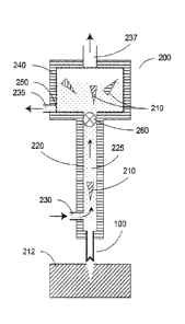
100301 A cross-sectional view of a diagram of an apparatus .200 in
accordance with
certain exemplary embodiments of the present disclosure is shown in FIG. 2.
The exemplary
apparatus .200 shown in FIG. 2 can include a housing 220 with a fluid conduit
225 provided
therein. One or more harvesting needles 100 can be coupled to the housing 220.
The thud
conduit 225 can be provided with at least one fluid inlet 230 and at least one
fluid outlet 235.
The fluid conduit 225 can be configured or structured such that a fluid can
flow therethrough;
e.g., the direction of fluid flow is indicated by the arrows in FIG. 2. A
proximal end of the
needle lumen can be in a .fluid communication with the conduit 225. For
example, the fluid
can flow past a proximal end of the harvesting needle 100, as shown in FIG. 2.
100311 In one exemplary procedure to harvest MTCs 210 from a donor
tissue 212, as
illustrated in FIG. 2, the exemplary apparatus 200 can be manipulated such
that the distal end
lone or more of the harvesting needles 100 penetrate the tissue 212 to a
particular depth.
The depth can be selected and/or controlled, e.g., by providing or adjusting a
particular
distance between the bottom of the housing 220 and the distal end of the one
or more needles
100. For example, a penetration depth can be selected that extends the distal
end done or
more of the harvesting needles 100 through the entire local thickness of the
derails to about
the depth of the subcutaneous fat layer, or optionally slightly into this fat
layer. 'Inserting the
needles 100 through the entire thickness of the dermis can provide an MTC 210
that has the
full length of the derrnis. Further, such exemplary depth can facilitate a
separation of the
MTC 210 from the surrounding tissue, because the proximal end of the needle
100 can cut
the MTC 210 away front the adjacent dermal tissue, and the MIC 210 can then be
fully
detached by tearing a small amount of subcutaneous fat at the bottom of the
MTC 210. Such
fatty tissue may be more easily separable than denser dermal tissue. After the
needle 100 is
withdrawn from the donor site tissue 212, an MTC that was separated from the
surrounding
tissue 212 can remain within the lumen of the needle 100.
CA 02882129 2015-02-13
WO 2014/028626
PCT/US2013/054955
100321 The fluid flowing through the conduit 225 can reduce pressure at
the proximal
end of the needle 100, which can facilitate removal of the WIC 210 from the
lumen of the
needle 100. The MTC 210 can be entrained in the flowing liquid, and carried
through the
conduit 225 and into a chamber 240. The flowing fluid can be withdrawn from
the fluid.
outlet 235, which can be provided as part of the chamber 240. MTCs that have
been
harvested as described herein can remain in the chamber 240. One or more
optional vents
237 can be provided in an upper portion of the chamber 240 (or conduit 225, if
no chamber is
provided) to allow any air entrained during the harvesting procedure to escape
from the
conduit, pathway, e.g., to prevent the chamber 240 from filling with air. For
example, a small
amount of air may be sucked in through the needle 100 along with an MTC 210
when the
needle 1.00 is withdrawn from the donor tissue 212.
[0033] in some exemplary embodiments of the present disclosure, the
conduit 225
can form a closed loop for the fluid flow or otherwise recirculate fluid
flowing through the
apparatus 200. For example, the fluid inlet 230 and outlet 235 shown in FIG. 2
can be
connected to the outlet and inlet, respectively, of a fluid pump arrangement
(not shown) or
the like.
100341 The pump arrangement can be or include an external pump or
similar device
configured to circulate fluid through the conduit 225. The fluid can be
provided from one. or
more reservoirs, and the pump arrangement and the conduit 225 can be
configured, connected
or structured such that the fluid leaving the chamber 240 via the outlet 235
can be discarded.
In further exemplary embodiments of the present disclosure, the fluid exiting
the outlet 235
can be recirculated through the conduit 225, e.g., in a closed-loop
configuration. One or
more sensors (e.g. pressure or flow rate sensors - not shown) can optionally
be provided in
the apparatus to facilitate control of the circulating fluid. In certain
exemplary embodiments
of the present disclosure. the pump arrangement can be or include a
peristaltic pump. The
flowing fluid can facilitate the removal of the MTCs 210 through the hollow
needle 100 and
into the fluid path. where the MTCs 210 are surrounded by a gentle fluid
environment.
[0035] A "trap" or filter arrangement 250 can be provided in the
apparatus to remove
harvested MTCs 210 from the circulating fluid and hold them for subsequent
transfer or
further processing. For example, an optional filter arrangement 250 can be
provided in the
chamber 240. e.g., near the outlet 235, to retain harvested MTCs within the
chamber during
the exemplary tissue harvesting procedure, as shown in FIG. 2. The filter
arrangement 250
can include, e.g., a Chamber or an enlarged region provided in the fluid
circulation path of the
8
CA 02882129 2015-02-13
WO 2014/028626
PCT/US2013/054955
conduit 225. The filter arrangement .2.50 can also .nc.ut.e a permeable filter
element, e.g. a
mesh, woven or porous material, basket, trap, or the like such that the
circulating fluid flows
at least partially through the chamber 240 and the filter element.
100361 A pore size or permeability of the filter arrangement 250 can be
selected to
fiwilitate the fluid flow therethrough while preventing the MTCs 210 from
doing so. For
example, the pore size can be less than about 200 microns, e.g., about 100
microns or less.
Such exemplary pore sizes can facilitate the flow of the circulating fluid
through the filter
arrangement 250 with a relatively little restriction, while being small enough
to trap and
retain the MTCs 210 that can be suspended in the flowing fluid. Accordingly,
the harvested
MTCs 210 can be retained in the trap while the fluid can flow therethrough,
and exit from the
filter arrangement 250, e.g., through the outlet 235.
100371 According to certain exemplary embodiments of the present
disclosure, the
filter arrangement 250 can include a porous dressing with holes or pores
sufficiently small to
trap MTCs 210 while facilitating or allowing the fluid to flow through it. The
dressing can be
15 'populated' with MICs after the exemplary harvesting procedure, and it
can be removed
from the apparatus and applied directly onto a wound site. Such dressing as
the filter element
can be used with any of the various embodiments described herein,
100381 in certain exemplary embodiments of the present disclosure, a
source of low
pressure (not shown) can optionally be provided in communication with the
conduit 225, e.g.,
20 to reduce pressure in the fluid conduit 225 and further facilitate fluid
flow and/or removal of
MTCs 210 from the harvesting needle 100. For example, the chamber 240 can be
configured
or structured to provide a headspace for a gas, such as air, above the filter
arrangement 250.
The source of low pressure can include, e.g., a vacuum pump, a low-pressure
line Or the like.
The low-pressure source can he in fluid communication with this headspace,
via a tube
25 or hose connected to an opening in the chamber 240, such as the vent 237
ShOWII in Fla, 2.
Other similar or equivalent exemplary configurations can also be provided to
generate a
reduced pressure in the conduit: 225 according to further exemplary
embodiments of the
present disclosure.
100391 According to further exemplary embodiments of the present
disclosure, the
30 exemplary apparatus 200 can include one or more control arrangements
(not shown). For
example, a pressure sensor can be provided at one or more locations within the
apparatus 200
to detect, e.g., the pressure within the fluid conduit 225 near the harvesting
needle 100 or a
9
CA 02882129 2015-02-13
WO 2014/028626
PCT/US2013/054955
pressure differential across the filter arrangement 250 to ascertain if the
filter arrangement.
250 is clogged and may be impeding fluid flow. Such exemplary sensors can be
provided in
communication with, e.g., a fluid pump arrangement and/or an optional low-
pressure source
as described herein, to control or adjust the operation of such components and
maintain
preferred conditions for the apparatus .200 during the exemplary operation.
Other exemplary
sensors that can be provided and can include, for example, temperature sensors
to monitor
and optionally control the fluid temperature, an optical sensor adjacent to or
within the
conduit 225 to detect a presence of M.Tes 210 flowing therethough, and/or one
or more
sensors configured to monitor characteristics of the fluid flowing through the
apparatus 200.
In further embodiments, a location sensor can be provided on or next to the
needle 100 or
within the apparatus 200 to detect a position of the needle 100 relative to
the bottom surface
ofthe housing 220, e.g., to track or monitor the penetration depth of the
needle 100 during
use. Such exemplary sensors and control arrangements, and/or a low-pressure
source, can be
used with any of the various embodiments described herein, including those
embodiments
illustrated in FIGS. 3 and 4.
100441 It still. further exemplary embodiments of the present
disclosure, a cauterizing
arrangement can be provided on one or more needles 100. For example, 'R.17
current can be
provided to one or more of the harvesting needles 100 in the apparatus 200.
The cauterizing
arrangement can be used to reduce or prevent bleeding during or after the
harvesting
procedure. For example, RP current can be applied to one or more of the
needles 100 after
the MTCs 210 have been withdrawn from the needle lumens, and before- the
needles 100 are
fully withdrawn from the tissue 2.12 to avoid damaging the MICs 210 while
cauterizing the
area around the removed volume of tissue.
100411 According to yet further exemplary embodiments of the present
disclosure,
one or more control valves (not shown) can optionally be provided at one or
more locations
in the conduit 225. For example, a valve 260 can be provided between the
proximal end of
the coring needle 100 and the chamber 240 and/or filter arrangement 250, as
shown in FIG. 2.
The valve 260 can be kept open during harvesting of tissue columns 210, to
allow and/or
facilitate fluid containing such MTCS 210 to flow therethroueh. The valve .260
can be
periodically and/or momentarily closed while fluid is circulating, e.g., while
the needle 100 is
not located within the tissue of the donor site 212, which can direct some
fluid entering the
inlet .230 through the coring needle 100 and out of the distal end thereof,
which can clean
and/or unblock the lumen of the needle 100.
CA 02882129 2015-02-13
WO 2014/028626
PCT/US2013/054955
100421 The fluid can be selected to provide a gentle environment for the
MTCs 210,
e.g., to prevent mechanical damage or contamination, and/or to promote their
viability and
growth. The fluid can be temperature-controlled using conventional thermal
control systems.
For example, the fluid can be provided from a source reservoir or container,
and the
temperature and/or other conditions of the fluid reservoir can be controlled
using
conventional control systems. The fluid can contain a variety of substances
including, for
example, saline, growth factors, butlers, etc. For example, the fluid can
contain supplemental
nutrients such as, e.g., amino acids, glucose, electrolytes, and/or oxygen to
promote or help
maintain viability of the harvested MTCs 210. The fluid can also include or
comprise a
conventional tissue culture medium, such as Dulbecco's Modified Eagle Medium,
F12, or the
Antibiotics (e.g., penicillin, streptomycin, or the like) and/or autifungal
agents (e.g.,
amphotericin or fluconazole) can optionally be provided in the fluid to help
disinfect the
MTCs 210 atler they are removed from the donor site 212.
100431 In the various exemplary embodiments described herein, the MTCs
210 can be
maintained in a controlled fluid environment from the time they are pulled up
from the
harvesting, needle(s) 100 and flow through the conduit 225 until they are
captured or
deposited on the filter arrangement 250, which can also be maintained within
the fluid.
Accordingly, the MTCs 210.am less likely to be damaged or Contaminated as
compared to.
e.g., other tissue removal devices that may expose removed tissue samples to
air and/or other
non-sterile surfaces,
100441 FIG. 3A shows a cross-sectional view of a diagram of an apparatus
$00 in
accordance with further exemplary embodiments of the present disclosure. The
apparatus
300 shown in FIG. 3A can be operated manually, and it has many features
similar to those
shown and described for the apparatus 200 in FIG. 2, but not *limited
to, the housing 220
.. with the fluid conduit 225, the harvesting needle(s) 100, the fluid inlet
230, the outlet 235, the
upper chamber 240, the optional vent 237, and the filter arrangement 250.
Certain
differences between the exemplary embodiments of the apparatus 200 illustrated
in FIG. 2
and the apparatus shown in FIG. 3A are described herein,
100451 For example, one or more of the harvesting needles 100 can be
attached or
affixed to a hub $10. The hub $10 can be provided, e.g., as .a shaped disc or
in another
geometry with one or more harvesting needles 100 affixed to it. The hub 310
can be
configured such that it can fit into a shaped recess in the housing 220, to
facilitate removal
and replacement of the harvesting needle(s).100 during or between harvesting
procedures. A
1 I
CA 02882129 2015-02-13
WO 2014/028626
PCT/US2013/054955
j.intrusion 'distance ofthe harvesting needle(s)1.00 beyond the bottom surface
of the
apparatus 300, Which can correspond to a penetration depth of the needle(s)
100 Into tissue,
can be adjusted using an adjusting arrangement such as, e.g., a threaded screw
coupler
.provided in the housing, or the like. In certain embodiments, one or more
needles IOU can be
provided with a hub 310, where a desired penetration depth of the needles 100
into the tissue
of the donor site can be determined or selected based on a predetermined
distance between
the hub 310 and the distal end of the needle(s) 100. A hub 310 such as that
shown in FIG.
3A, which can include one or more of the needles 100, can be used with any of
the various
exemplary embodiments described herein,
100461 The chamber 240 can be provided with a removable cap 320, or the
like, to
fitcilitate access to the interior of the chamber and removal of MTCs 210 that
may be trapped
or retained by the filter arrangement 250. For example, the exemplary
apparatus 300 can
include the filter arrangement 250 provided in the chamber 240, where the
filter arrangement
250 can be located between an end of the conduit 225 and the fluid outlet 235.
Such
Configuration facilitates the flow of fluid containing the harvested MTCs 210
through the
filter arrangement 250 and out of the outlet 235, where the MTCs 210 can be
retained by the
filter arrangement 250. Access to the MTCs 210 after they are harvested and
trapped can be
achieved, e.g., by removing the cap 320 from the chamber 240,
100471 According to additional exemplary embodiments of the present
disclosure, the
filter arrangement 250 and optionally the cap 320 can be provided, for
example, as a sterile
cartridge that can be inserted into the chamber 240 before harvesting MTCs
210, and can
later be removed with the harvested MTCs 2.10. In still further exemplary
embodiments of
the present disclosure, the filter arrangement 250 can be provided as a
removable "basket" or
the like that can be inserted, into the chamber 240, and removed with. trapped
MTCs 210 after
the harvesting procedure is completed.
100481 In an exemplary operation, similar to the exemplary operation of
the
exemplary apparatus 200 described herein, the exemplary apparatus 300 can be
pressed onto
a donor tissue site, such that the distal end of the harvesting needle 100
pierces the tissue and
separates an MTC 210 from the surrounding tissue. The fluid flowing through
the conduit
225 can facilitate withdrawal of the MTC 210 Crain the proximal end.of the
harvesting needle-
1 00 such that it flows with the Imuid through the conduit 225. The flowing
fluid can transport
the MTC 210 to the filter arrangement' 250, where the MTC 210 can be retained
by a mesh or
other filter element, while the fluid flows through the filter arrangement 250
and exits the
12
CA 02882129 2015-02-13
WO 2014/028626
PCT/US2013/054955
outlet 235, where it can optionally be recirculated. The apparatus 300 can be
withdrawn from
the donor site, and. inserted into another location to harvest a further MTC
210. This process
can be repeated a plurality of times to harvest a number of MTCs 210 from the
donor site.
After a sufficient number of MTCs 210 have been harvested, the filter
arrangement 250 (or a
portion thereof) containing the MTCs 210 can be removed from the apparatus 300
for further
handling or processing.
100491 Another exemplary apparatus 350 is shown in F)G. 3B that can
include several
features in common with the other exemplary apparatuses 200, 300, e.g., the
housing 220
with the fluid conduit 225, the harvesting needle(s) 100, and the fluid inlet
230. The
exemplary apparatus 350 illustrated in FIG, 3B can be provided with a delivery
arrangement
360 configured to direct at least a portion of the fluid flowing from the
inlet 230 and through
the conduit 225 onto a receiving substrate 370 (which can be or act as a
filter arrangement).
The delivery arrangement 360 can include rigid andfor flexible tubing, or the
like, which can
be connected to the conduit 225,
[00501 The receiving substrate 370 can be or include,
a filter element that can
trap MTCs 210 while allowing fluid from the conduit 225 to flow through or off
of the
substrate 370. In further exemplary embodiments of the present disclosure, the
substrate $70
can be or include a permeable or porous dressing material, which can act as a
filter element to
trap MTCs 210 thereon while allowing the fluid to pass through or flow off of
the substrate
370. In this exemplary manner, harvested MTCs 210 can be directly deposited
onto a
dressing or the like, and such dressing with the MTCs 2.10 can then be
transported or applied
directly to a wound site.
100511 The distal end of the delivery arrangement 360 can be
positiortable such that it
traverses a predetermined region of the substrate.370 during the harvesting
procedure, e.g.,
while fluid containing .MTCs 210 flows through the conduit 225 and out of the
distal end of
the delivery arrangement 360. For example, at least a portion of the delivery
arrangement
360 can be flexible, such that the distal end thereorcan be positioned and/or
moved over the
substrate 370 while the housing 220 containing the needle(s) 100 can be
advanced and
withdrawn over multiple locations of the donor site to harvest MTCs 210.
100521 In a further exemplary embodiment.of the present disclosure, the
distal end of
the delivery arrangement 360 can be held or maintained in a stationary
position, and the
13
CA 02882129 2015-02-13
WO 2014/028626
PCT/US2013/054955
substrate 370 can be controllably moved or translated relative to this distal
end such that
.MTC's 210 are deposited over a predetermined area of the substrate 370.
i00s31 The translation of the distal end of the delivery arrangement 360
relative to the
substrate 370 (or vice versa) can be performed, e.g., .using any one of
various translation
arrangements known in the art. Such positional translators can include, e.g.,
one or more
motors or actuators, various arms, supports, clamps, pivots, or the like,
along with any
sensors and/or controllers that may be used to control a rate and/or direction
of motion, limits
of motion or displacement, etc. For example, the relative motion of the distal
end of the
delivery arrangement 160 and the substrate 370 can be selected and/or
performed such that
IVETC.s 210 are deposited in a predetermined spacing, pattern or density on
the substrate 370..
The deposition geometry can be estimated in a straightforward manner based on
the
frequency at which the needle 100 is inserted into tissue to obtain a new MTC
210, together
with the speed and direction of the relative motion between the distal end of
the delivery
arrangement 360 and the substrate 370.
[00541 According to a further exemplary embodiment of the present
disclosure,
another exemplary apparatus can he provided, is shown in FIGS. 4A and 413 that
can include
the harvesting needle(s) 100 secured to the hub 310. The apparatus 400 shown
in FIGS, 4A
and 48 has many features similar to those shown and described for the
apparatus 200, 300
and/or 350 shown in FIG. 2, FIG. 3A and FIG. 3B, respectively. These features
include, e.g.,
the housing 220 with the fluid conduit 225, the harvesting neediets) 1.00, the
fluid inlet 230
and the outlet 235, the upper chamber 240, the optional vent .217, and the
filter arrangement
250. One or more harvesting needles 100 can be attached or affixed to the hub
310.
(00551 The exemplary apparatus 400 can include a base 420 that can be
slidably
engaged. with the housing 220, e.g., such that the housing 220 can move up and
down over a
particular distance relative to the base 420. One or more solenoid coils 430
can be coupled or
affixed to the base 420, and a solenoid core 435 can be located at least
partially within the
solenoid coil 430 and mechanically coupled to the housing 220. With such
exemplary
configuration, the solenoid(s) 430 can be configured to move the housing 220
and the
attached needles 100 up and down relative to the base 420, thereby inserting
and withdrawing
the needles .100 from the donor tissue 212. One or more 0-rines or .similar
sealing
arrangements can be provided to maintain a fluid-tight seal between the
housing 200 and the
hub 310, and also between the housing 220 and the base 420 when the housing
220 is
translated during operation of the apparatus 400. A linear bearing can
optionally be provided
14
CA 02882129 2015-02-13
WO 2014/028626
PCT/US2013/054955
to maintain support and alignment of the housing 220 within the base 420
during operation of
the apparatus 400,
100561 For example, the apparatus 400 of FIG. 4A shows the solenoids 430
which are
not activated. In this exemplary state, the harvesting needles 100 are
retracted so that they
are close to but not protruding from, a lower surface of the base 420. In
operation, the base
420 can be placed on the surface of the donor site tissue 212 to be harvested,
with the
solenoids 430 off, as shown in FIG. 4A. A pump arrangement or the like (not
shown) can be
activated to supply -fluid to the inlet 230 and circulate it through the
conduit 225, as described
herein.
[00571 The solenoids 430 can then be activated, such that the cores 435 are
drawn
downward, such that the housing 220 with mechanically coupled needles 110 are
also pulled
downward with respect to the base 420, as shown in FIG. 413. ,This exemplary
motion can
result in the harvesting needles 100 protruding beyond a lower surface of the
base 420,
causing the needles 110 to pierce the tissue 212 of the donor site and
separate MTCs 210
from the surrounding tissue 212õ as described herein. The MTCs 210 can then be
withdrawn
from the needles 100 such that they flow through the conduit 225 with the -
fluid and can be
deposited in the filter arrangement 250. The solenoids 430 can then be
deactivated, such that
the housing 220 rises relative to the base 420 (e.g., using springs or the
like to return the
housing to a raised position) and the needles 110 are withdrawn from the donor
site 212 and
back into the base 420õ as shown in FIG. 4A. This exemplary procedure can be
repeated at
different locations on the donor site 212 to harvest additional MTCs 210. In
an exemplary
operation, such apparatus 400 can be used to harvest the MTCs 210 at a
frequency between
about 0,5 and about 2 Hz, e.gõ with a time interval between successive
penetrations of about
0.5 to 2 seconds, Certain exemplary modifications may be developed to allow
faster
harvesting rates, and slower rates can also be used if desired.
100581 An adjusting arrangement such as, e.g., a screw-type adjuster or
a spacer that
can he attached to the base 420, can he provided to control the maximum
protrusion length of
the needles 110 from a lower surface of the base 420 (thereby controlling a
corresponding
maximum penetration depth of the needles 100 into the donor site tissue .212),
100591 In further exemplary embodiments of the present disclosure, other
types of
actuators can be used instead of or in addition to the solenoids 430. For
example, one or
more motors can be provided with a rotary/linear convener to convert rotary
motion to a
CA 02882129 2015-02-13
WO 2014/028626
PCT/US2013/054955
linear motion of the housing 220 relative to the base 420, e.g., at a
controlled .frequency
and/or particularexcursion distance. Other types of linear actuators can also
be used to
extend and withdraw the needles 100 from the tissue 212 beneath the apparatus
400.
100601 The base 420 of the exemplary apparatus 400 can be structured to
include a
.. recess 450 that can form an enclosed volume between the tissue surface 212
and. a lower
surface of the base 420 adjacent to the needles 100, as:shown in FIG. 4A. Such
exemplary
recess 450 can be formed, e.g., by providing the base 420 with a rim or edge
that can rest on
the donor site tissue 212 while a lower surface of the base 420 remains a
small distance above
the tissue surface. One or more vacuum ducts 410 can be provided in
communication with
the enclosed volume. Application of a low-pressure or vacuum source (not
shown) to the
vacuum duct(s) 410 can cause the surface of the donor site tissue 212 to be
pulled up into the
recess 450, as shown in FIG.4.8,
100611 This exemplary deformation can stretch the surface and provide
tension,
which may provide several benefits. For example, stretching the tissue surface
can
mechanically stabilize it such that the needles 100 can penetrate the
stretched tissue 212 more
easily than they may penetrate unstretched, resilient: tissue. Further, puling
the tissue surface
upward using. low pressure such that it contacts a lower surface of the base
420, as shown in
FIG, 4B, can facilitate an accurate insertion depth of the needles 100. In
certain
embodiments, the needles 100 can be in a fixed position relative to the base
420 such that
they remain protruding a small distance from the lower surface, as shown in
FIG. 48,
100621 Instead of foreim!..the needles 100 into the tissue 212, as
described herein, the
tissue 212 can be pulled up onto the needles 100 such that they pierce the
tissue 212, as
shown in FIG. 48, The low pressure can then be released to allow the tissue
212 to relax and
fall off the needles 110, optionally assisted with a positive pressure being
applied to the
vacuum ducts 410.An exemplary application of low (and/or optionally high)
pressure to the
vacuum ducts 410 can be done, for example, using a conventional pump
arrangement or other
source(s) of low and high pressure, together with an appropriate valve
arrangements to
control the application and release of pressure differences in the ducts 410.
The timing of
such pressure cycles can be coordinated with the activation/deactivation of
the solenoids 430,
Such exemplary- chamber 450 with the vacuum ducts 410 can also be used with
any of the
other exemplary embodiments=described herein.
16
CA 02882129 2015-02-13
WO 2014/028626
PCT/US2013/054955
100631 According to still further exemplary embodiments of the present
disclosure,
the surface of the donor site tissue 212 can be stretched or stabilized using
other procedures,
e.g., by manually stretching the surface with fingertips before inserting the
needles 100. In
yet further exemplary embodiments of the present disclosure, the donor site
tissue 2.12 can be
pre-cooled or partially frozen prior :to insertion of the harvesting needles
100, e.g., using
convective or conductive techniques such as a cryospray or contact with a
cooled object. The
exemplary cooling of the donor site tissue 212 can make it more rigid and
fheilitate insertion
of the harvesting needles 1.00. In still further embodiments a mechanical,
surface clamp or
spreader can be applied around the donor site region to stretch the tissue 212
before inserting
the needles 100. Such procedures can .be performed with any of the exemplary
devices and
methods described herein.
100641 The exemplary apparatuses 200, 300, 3:50, 400 can be provided
with various
numbers of the harvesting needles 1.00. For example, in addition to a single
one of the
needles 100, arrays of 4, 6, 8, 9, 12 or more of the needles 100 can be used,
and they can be
affixed to a hub $10 to facilitate insertion and removal of the needles 100
from the exemplary
apparatuses 200, 300, 350, 400 as a group. The needles 100 can be provided in
various
.geometrical arrangements such as, e,g., a:square or triangular pattern.
Providing a hub with a
larger number of needles can increase the efficiency and speed of harvesting
MTCs 210, as
more MTes 210 (one per needle 100) can be harvested with each insertion-and-
withdrawal
cycle of the needles 100. However, a very large number of needles 100 can
increase the
force-required to advance-all of the needles 100 into the donor site tissue
212 simultaneously,
and can increase the complexity of manufacturing the hub-needle component.
According still
additional exemplary embodiments of the present disclosure, the huh
arrangements can have
between about 4 and 25 needles coupled thereto.
100651 The needles 100 can be spaced apart an appropriate distance to
facilitate
harvesting of a large number of the MTC:s 210 from a donor site 212 while
maintaining
healthy tissue between the removed tissue samples 210 to promote rapid healing
of the donor
site 212, prevent formation of scars or markings, etc. For example, the
spacing between
adjacent needles 100 can be about 1-2 mm, or up to about 5 mm, Larger spacings
can be
used in certain embodiments, but this can require a correspondingly larger
width of the
overall apparatus to accommodate the larger hub. The Wits 210 can be harvested
over a
larger area of tissue 212 by moving the exemplary apparatuses 200, 300, 350,
400 to different
locations before each needle insertion procedure.
.17
100661 The exemplary embodiments described herein can include the
fluid conduit
225 that is substantially vertical. In further exemplary embodiments of the
present
disclosure, other orientations of the conduit 225 can be provided. For
example, the conduit
can be substantially horizontal, with the inlet 230 and the outlet 235 can be
provided at
opposing ends of such a conduit 225, and the proximal ends of the needles 100
protruding
into the conduit 225 such that the liquid flows past this end of the needles
100. Such an
exemplary configuration can also provide a simpler, e.g. linear, conduit
geometry that may be
easier to manulhcture andior clean, may result in fewer pressure drops along
the fluid path.
etc. Other exemplary orientations of the conduit 225 or shapes thereof, such
as a curved
conduit, can also be provided in still further exemplary embodiments of the
present
disclosure.
100671 According to still additional exemplary embodiments of the
present disclosure,
, at least two of the needles 100 can be separately actuated, e.g.,
such that they pierce the tissue
212 at different times. For example, two or more actuators can be coupled to
different ones
of the needlesõAlternatively, a singular actuator can he provided that is
configured to
advance different ones of the needles at different times. Such 'staggering' of
penetrations
can reduce the maximum force needed to advance the needles into the tissue.
10068] Other needle cross-sectional shapes can be used with the
various embodiments
desctibed herein to harvest the MTCs 210 having different geometric
characteristics.
Although circular cross-sections are most common, needles WO having oval,
square, or
triangular cross-sections, or combinations thereof in multi-needle devices,
can also be used.
100691 In further embodiments of the present disclosure, the
methods and apparatus
described herein can be applied to other tissues besides -skin tissue. Thus,
the tvITCs 210 can
be harvested from a variety of organs or tissue structures, which can
facilitate rapid healing of
a donor site while providing microscopic graft tissue suitable for placement
at recipient Sites,
on scatiblds, within biocompatible matrices, etc.
100701 It will thus be appreciated that those skilled in the an
will be able to devise
numerous systems, arrangements and methods which, although not explicitly
shown or
described herein, embody the principles of the present disclosure and are thus
within the
spirit and scope of the present disclosure.
18
CA 2882129 2019-12-13