Note: Descriptions are shown in the official language in which they were submitted.
APPARATUS AND METHODS FOR DRUG
DELIVERY USING MICRONEEDLES
Background
110011 The embodiments described herein relate generally to the field of
ophthalmic
therapies and more particularly to the use of a microneedle for delivery
and/or removal of a
substance, such as a fluid therapeutic agent into and/or from ocular tissues
for treatment of
the eye.
110021 Although needles have been used in transdermal and intraocular
drug delivery,
there remains a need for improved microneedle devices and methods,
particularly for
delivery of substances (e.g., drugs) into the targeted regions of the eye.
Many inflammatory
and proliferative diseases in the posterior region of the eye require long
term
pharmacological treatment. Examples of such diseases include macular
degeneration,
diabetic retinopathy, and uveitis. It is often difficult to deliver effective
doses of a drug to the
posterior region of the eye using conventional delivery methods such as
topical application,
which has poor efficacy, and systemic administration, which often causes
significant side
effects. For example, while eye drops are useful in treating conditions
affecting the exterior
surface of the eye or tissues at the front of the eye, the eye drops are not
significantly carried
to the back of the eye, as may be desired for the treatment of some of the
retinal diseases
listed above.
[10031 Although there have been advances in the past decade regarding
the utilization of
ocular injection and systemically delivered substances for the treatment of
ocular disorders,
obstacles still exist. For example, direct injection into the eye (e.g., into
a portion of the
sclera and/or the vitreous) using conventional 27 gauge or 30 gauge needles
and syringes can
be effective but often requires professional training and raises safety
concerns. Moreover,
the anatomy of the eye can make insertion of a conventional 27 gauge or 30
gauge needle
into ocular tissue challenging. For example, the eye has a lower modulus of
elasticity than
skin, and thus will deform more readily in response to an applied force
compared to
deformation of the skin in response to the same applied force. Accordingly,
conventional
1
CA 2882184 2019-10-31
needles that are designed to pierce skin or other tissue may not be suitable
for piercing ocular
tissue.
[1004] In addition, many known methods of direct injection of a drug
into the eye include
inserting a needle or a cannula at an acute angle relative to a surface of the
eye, which can
make controlling the depth of insertion challenging. For example, some such
methods
include controlling the angular orientation of the needle such that the
injected substance exits
the needle at a particular locations. Moreover, some known methods of
injecting substances
into ocular tissue include using complicated visualization system or sensors
to control the
placement of the needle or cannula.
[1005] Moreover, in some instances, such as when treating intraocular
tumors, tumors
seeds and/or precancerous tissue within the vitreous can be spread through the
passageway
defined by the insertion of the needle (i.e., a needle tract), which can
increase the risk of
complications from the tumor. Thus, known methods that result in multiple
needle tracts,
needle tracts having a large diameter and/or length can result in increased
risk of
complications.
[1006] In some instances, the relative size of the anatomy of the eye
can present
challenges to treatment of ocular disease. For example, in the treatment of
retinoblastoma in
pediatric cases, the target insertion site of a needle (e.g., the ciliary
body) is significantly
smaller than a corresponding target insertion site in an adult case. In such
instances, the
.. precise placement of the needle can present a challenge for physicians,
resulting in an
increased chance of tissue damage and an increase in cost of the procedure. In
addition, the
anatomy of the eye in pediatric cases can be such that a standard 27 gauge or
30 gauge needle
is too large, making insertion to a desired depth into ocular tissue a
challenge.
[1007] Thus, a need exists for improved methods and devices for
delivering substances to
ocular tissue.
2
CA 2882184 2019-10-31
Summary
[1008]
According to one aspect of the present disclosure, an object is to provide an
apparatus for use in administering a medicament, comprising:
a housing, a proximal end portion of the housing defining an opening
configured to
receive a portion of a medicament container therein, a distal end portion of
the housing
including a base surface configured to contact a surface of a target tissue;
and
a microneedle coupled to the distal end portion of the housing, a proximal end
portion of the microneedle in fluid communication with the medicament
container when the
portion of the medicament container is disposed within the opening, a distal
end portion of
the microneedle including a beveled surface that defines includes (i) a first
segment having a
first bevel surface arranged at a first angle relative to a centerline of a
lumen defined by the
microneedle, and (ii) a second segment having a second bevel surface arranged
at a second
angle relative to the centerline, the first angle being different than the
second angle, the first
segment having a length different than a length of the second segment, and at
least one of the
first bevel surface or the second bevel surface being curvilinear about an
axis normal to the
centerline of the lumen
[1008a] According to another aspect of the present disclosure, an object is to
provide an
apparatus for use in administering a medicament, comprising:
a housing, a proximal end portion of the housing defining an opening
configured to
receive a portion of a medicament container therein, a distal end portion of
the housing
including a base surface; and
a microneedle coupled to the housing, a proximal end portion of the
microneedle in
fluid communication with the medicament container when the portion of the
medicament
container is disposed within the opening, a distal end portion of the
microneedle including a
beveled surface disposed at an angle relative to a centerline of a lumen
defined by the
microneedle, and at least a portion of the beveled surface being curved about
an axis that is
normal to the centerline of the lumen.
[1008b] According to another aspect of the present disclosure, an object is to
provide an
apparatus for use in administering a medicament, comprising:
a medicament container, a proximal end portion of the medicament container
including a seal and a plunger configured to move the seal within the
medicament container;
3
Date Recue/Date Received 2020-05-22
a housing removably coupled to the medicament container such that at least a
distal
end portion of the medicament container is disposed within the housing, the
housing
including a base surface; and
a microneedle coupled to the housing such that a length of the microneedle
extends
beyond the base surface, a proximal end portion of the microneedle in fluid
communication
with the medicament container, a distal end portion of the microneedle
including a beveled
surface defined by a first plane at a first angle relative to a centerline of
a lumen defined by
the microneedle, at least a first portion of the beveled surface cut along a
second plane
different from the first plane, the second plane being rotated at a second
angle about the
centerline and relative to the first plane, a thickness of the microneedle
varying at least one of
circumferentially or linearly about the microneedle.
[10080 According to another aspect of the present disclosure, an object is to
provide a use
of a device to treat an eye of a patient, the device comprising:
a microneedle having a distal edge defined by a beveled surface of the
microneedle
configured to extend through the choroid of the eye, the beveled surface
defining a tip angle
of less than about 20 degrees, the beveled surface having a height such that
an opening
defined by the beveled surface is within at least one of a suprachoroidal
space or a lower
portion of the sclera,
wherein the device is configured to convey a substance from a cartridge
coupled to a
proximal end portion of the microneedle into the suprachoroidal space via the
opening
defined by the beveled surface.
According to another aspect of the present disclosure, an object is to provide
the use of the
apparatus such as the one described and/or illustrated in the present patent
specification, to
treat an eye of a patient, wherein the microneedle is configured to extend
through the choroid
of the eye such that an opening defined by the beveled surface is within at
least one of a
suprachoroidal space or a lower portion of the sclera; and the apparatus is
configured to
convey a substance into the suprachoroidal space via the opening defined by
the beveled
surface.
[1010] Other possible aspect(s), object(s), embodiment(s), variant(s) and/or
advantage(s) of
the present disclosure, all being preferred and/or optional, are briefly
summarized
hereinbelow.
3a
Date Recue/Date Received 2020-05-22
[1011] Indeed, devices and methods described herein relate generally to
intraocular
treatment and more particularly to the use of microneedles for treatment of
ocular tissue. In
some embodiments, a microneedle has a proximal end portion and a distal end
portion and
defines a lumen. The proximal end portion is configured to be coupled to a
cartridge to place
the lumen in fluid communication with the cartridge. The proximal end portion
includes a
base surface that is configured to be placed in contact with a surface of a
target tissue. The
distal end portion of the microneedle includes a beveled surface. The beveled
surface defines
a first bevel angle and a second bevel angle different from the first bevel
angle.
[1012] In some embodiments, a microneedle has a proximal end portion and a
distal end
portion and defines a lumen. The proximal end portion is configured to be
coupled to a
cartridge to place the lumen in fluid communication with the cartridge. The
proximal end
portion includes a base surface configured to contact a surface of a target
tissue. The distal
end portion includes a beveled surface defining a tip angle of less than about
20 degrees and
a ratio of a bevel height to a bevel width of less than about 2.5.
[1013] In some embodiments, a method for delivering a substance to a target
tissue of an
eye includes inserting a microneedle into an eye such that a distal edge
defined by a beveled
surface of the microneedle does not extend through the choroid of the eye. The
beveled
surface of the microneedle defines a tip angle of less than about 20 degrees.
The beveled
surface has a height such that an opening defined by the beveled surface is
within at least one
of a suprachoroidal space or a lower portion of the sclera. A substance is
conveyed from a
cartridge coupled to a proximal end portion of the microneedle into the
suprachoroidal space
via the opening defined by the beveled surface
Brief Description of the Drawings
[1014] FIG. 1 is a cross-sectional view of an illustration of the human eye.
[1015] FIG. 2 is a cross-sectional view of a portion of the human eye of FIG.
1 taken along
the line 2-2.
_____________________________________________________________________________
_
_
3b
Date Recue/Date Received 2020-05-22
CA 02882184 2015-02-13
WO 2014/036009 PCT/US2013/056863
[1016] FIGS. 3 and 4 arc cross-sectional views of a portion of the human
eye of FIG. 1
taken along the line 3-3, illustrating the suprachoroidal space without and
with, respectively,
the presence of a fluid.
[1017] FIG. 5 is a block diagram of a delivery device according to an
embodiment.
[1018] FIG. 6 is a front view illustration of a delivery device according
to an
embodiment.
[1019] FIG. 7 is a perspective view illustration of a portion of a
microneedle according to
an embodiment.
[1020] FIG. 8 is a side view illustration of a portion of a microneedle
according to
another embodiment.
[1021] FIG. 9 is a top view illustration of the portion of the microneedle
of FIG. 8.
[1022] FIG. 10 is a cross-sectional view of the portion of the microneedle
taken along the
line 10-10 in FIG. 9.
[1023] FIG. 11 is a side view illustration of a portion of a microneedle
according to
another embodiment.
[1024] FIG. 12 is a top view illustration of the portion of the microneedle
of FIG. 11.
[1025] FIG. 13 is side view illustration of a portion of a microneedle
according to another
embodiment.
[1026] FIGS. 14-16 are side view illustrations of various lumen and bevel
configurations
included in a microneedle according to various embodiments.
[1027] FIG. 17 is a schematic illustration of an infusion device in use,
according to an
embodiment.
[1028] FIG. 18 is an enlarged view of a portion of the human eye and a
portion of the
infusion device identified in FIG. 17 as region Z.
[1029] FIG. 19 is a schematic illustration of a microneedle in use,
according to an
embodiment.
4
CA 02882184 2015-02-13
WO 2014/036009 PCT/US2013/056863
[1030] FIG. 20 is a schematic illustration of a portion of the human eye
illustrating
certain dimensions.
[1031] FIG. 21 is a front view illustration of a delivery device according
to an
embodiment.
[1032] FIG. 22 is a schematic illustration of a delivery system according
to an
embodiment.
[1033] FIG. 23 is a schematic illustration of a kit including a delivery
device and at least
one microneedle according to an embodiment.
[1034] FIG. 24 is a schematic illustration of a microneedle array according
to an
embodiment.
[1035] FIG. 25 is a cross-sectional illustration of an eye with a
microneedle, according to
an embodiment, and a standard 30 gauge needle inserted into the vitreous.
[1036] FIG. 26 is a flow chart illustrating a method of delivering a drug
to a target ocular
tissue, according to an embodiment.
[1037] FIG. 27 is an image of a microneedle (shown in the middle), a 27
gauge standard
needle (shown at the top), and a 30 gauge standard needle (shown at the
bottom), according
to an embodiment.
[1038] FIG. 28 is an image of a microneedle (shown at the bottom) and 34
gauge
standard needle (shown at the top), according to an embodiment.
[1039] FIG. 29 is a set of images of human cadaver eyes prior to injection
(top panels)
and following injection (bottom panels) of triamcinolone with a 30 gauge
standard needle
(left panels) and a microneedle of the invention (right panels).
[1040] FIG. 30 is a graph showing the growth of WERT human retinoblastoma
cells
versus days in cell culture, following aspiration and passage of cells with a
26 gauge, 30
gauge or microneedle.
[1041] FIG. 31 is a set of images depicting stained WERI human
retinoblastoma cells
following aspiration and passage using 26 gauge, 30 gauge standard needle or a
microneedle
of the invention.
CA 02882184 2015-02-13
WO 2014/036009 PCT/US2013/056863
[1042] FIGS. 32 and 33 are schematic illustrations of a microneedle with
and without a
baffle in the chamber of the microneedle, respectively, according to an
embodiment.
[1043] FIG. 34 is a graph showing cell density of WER1 human retinoblastoma
cells
versus time, following aspiration and passage of cells with a standard needle
with or without
a baffle, or a microneedle with or without a baffle.
[1044] FIG. 35 are images of the rabbit eye. A. Vitreous seeds of
retinoblastoma (*) in
the rabbit model. B. The microneedle (arrow) is inserted at the pars plana. C.
The
microneedle (arrow) is inserted to its hub into the vitreous. D. After 3
weekly injections of
20 jig topotecan, the vitreous seeds disappeared.
[1045] FIG. 36 is a schematic depiction of a 30 gauge needle or a
microneedle with baffle
inserted into the pars plana of an enucleated eye (left panel); and a set of
images of
enucleated eyes stained with hematoxylin-eosin following aspiration of
retinoblastoma with a
30 gauge needle (top panes) or microneedle (bottom panes). Images were taken
at 25X
(middle panes) and 100X (right panes) magnification. Needle tracts are
indicated with black
arrows in the 100X images.
[1046] FIG. 37 is a bar graph showing vitreous seed score after control
(PBS), low-dose
topotecan (51aL/50iug topotecan), or high-dose topotecan (10ittL/50gg
topotecan) treatment in
a rabbit retinoblastoma model. Dosages were administered once per week, for
three weeks.
Vitreous seed score (i.e., no score, (+), (++), or (+++) corresponding to a
score of 0, 1, 2, or
3, respectively) was determined before and after topotecan treatment in each
animal, and the
average vitreous seed score in each group was calculated at both time points.
Vitreous seeds
were graded as 0 (no seed), 1 plus (+) with seeds filling less than 1/3 of the
vitreous; 2 plus
(++) with seeds filling 1/3-2/3 of the vitreous; and 3 plus (+++) with seeds
filling the entire
vitreous.
[1047] FIG. 38 is a bar graph depicting tumor area after administration of
control (PBS),
low-dose topotecan (5 L/50ug topotecan), or high-dose topotecan (10 L/50iug
topotecan) in
a rabbit retinoblastoma model. Dosages were administered once per week for
three weeks.
Tumor area was measured in mm2.
Detailed Description
[1048] In some embodiments, a microneedle for ocular drug delivery and/or
ocular tumor
removal includes a beveled surface. The beveled surface of the microneedle
defines a tip
6
CA 02882184 2015-02-13
WO 2014/036009 PCT/US2013/056863
angle of less than about 20 degrees and a ratio of a bevel height to a bevel
width of less than
about 2.5. The beveled microneedle, in one embodiment, allows for accurate and
reproducible drug delivery to the suprachoroidal space (SCS) of the eye. In
other
embodiments, the beveled microneedle is used in a pediatric ocular drug
delivery methods to
deliver one or more drugs to the vitreous of a pediatric eye. Advantageously,
the beveled
microneedle, when used in the pediatric eye, minimizes the length of the
needle tract, thereby
minimizing the opportunity for a tumor cell(s) to re-seed in the needle tract.
[1049] In some embodiments, a microneedle has a proximal end portion and a
distal end
portion and defines a lumen. The proximal end portion is configured to be
coupled to a
cartridge to place the lumen in fluid communication with the cartridge. The
proximal end
portion includes a base surface that is configured to be placed in contact
with a surface of a
target tissue. The distal end portion of the microneedle includes a beveled
surface. The
beveled surface defines a first bevel angle and a second bevel angle different
from the first
bevel angle. In some embodiments, the first bevel angle is less than the
second bevel angle.
In some embodiments, the first bevel angle is less than about 20 degrees and
the second bevel
angle is less than about 30 degrees.
[1050] In some embodiments, a microneedle has a proximal end portion and a
distal end
portion and defines a lumen. The proximal end portion is configured to be
coupled to a
cartridge to place the lumen in fluid communication with the cartridge. The
proximal end
portion includes a base surface that is configured to be placed in contact
with a surface of a
target tissue. The distal end portion of the microneedle includes a beveled
surface. The
beveled surface defines a tip angle of less than about 20 degrees and a ratio
of a bevel height
to a bevel width of less than about 2.5.
[1051] In some embodiments, a hollow microneedle and/or microneedle
assembly for
delivery of a drug to an eye is provided. In some embodiments, the hollow
microneedle
includes a distal end portion and a shaft extending from a cartridge housing.
The needle can
be disposed within a needle cap prior to use. A distal end of the microneedle
includes a bevel
that corresponds, at least partially, to a target location within the eye. The
cartridge housing
can receive a cartridge containing a therapeutic agent. In some embodiments,
the
microneedle is configured to allow the entire shaft or substantially the
entire shaft of the
microneedle to be inserted into the eye such that the distal end portion of
the microneedle is
disposed within the target location (e.g., the suprachoroidal space) of the
eye.
7
CA 02882184 2015-02-13
WO 2014/036009 PCT/US2013/056863
[10521 In some embodiments, a microneedle for delivery of a drug to a
pediatric eye is
provided. The microneedle may be a hollow microneedle, or a solid microneedle.
In some
embodiments, the microneedle includes a bevel and a shaft extending from a
base, and
defines a lumen. The microneedle is configured to facilitate the insertion of
the entire shaft
or substantially the entire shaft of the microneedle into the pediatric eye
such that a drug
formulation can be deposited, injected and/or infused in the vitreous of the
pediatric eye
without damaging the lens or retina.
[10531 In some embodiments, a method for delivering a substance to a target
tissue of an
eye includes inserting a microneedle into an eye such that a distal edge
defined by a beveled
surface of the microneedle does not extend through the choroid of the eye. The
beveled
surface of the microneedle defines a tip angle of less than about 20 degrees.
The beveled
surface has a height such that an opening defined by the beveled surface is
within at least one
of a suprachoroidal space or a lower portion of the sclera. A substance is
conveyed from a
cartridge coupled to a proximal end portion of the microneedle into the
suprachoroidal space
via the opening defined by the beveled surface.
[10541 In some embodiments, a method for delivering a drug to the
suprachoroidal space
of an eye includes inserting a distal end of a hollow microneedle into the
sclera, wherein the
entire shaft or substantially the entire shaft of the microneedle is inserted
into the eye at an
angle of approximately 90 degrees. Upon insertion, a drug is conveyed,
injected and/or
infused through the microneedle, through the sclera, and into the
suprachoroidal space
without damaging the lens, retina, or other ocular tissue.
[10551 In some embodiments, a method for delivering a drug to the
suprachoroidal space
of an eye includes inserting a distal end of a hollow microneedle into the
sclera, wherein the
entire shaft or substantially the entire shaft of the microneedle is inserted
into the eye at an
angle of approximately 90 degrees. Upon insertion, a drug is infused through
the
microneedle, through the sclera, and into the suprachoroidal space without
damaging the lens,
retina, or other ocular tissue. In a further embodiment, the microneedle, when
inserted into
the eye for suprachoroidal drug delivery, does not puncture the choroid.
[10561 In some embodiments, a method for delivering a drug to the vitreous
of a pediatric
eye includes inserting the distal end of any of the microneedles described
herein through the
ciliary body of the pediatric eye, wherein the entire shaft or substantially
the entire shaft of
8
CA 02882184 2015-02-13
WO 2014/036009 PCT/US2013/056863
the microneedle is inserted into the eye at an angle of approximately 90
degrees. A drug is
then injected and/or infused through the lumen of the microneedle into the
vitreous.
[10571 In some embodiments, a method for delivering a drug to the
suprachoroidal space
of an eye is provided. In some embodiments, the method comprises inserting a
distal end of a
hollow microneedle into the sclera or suprachoroidal space, wherein the entire
shaft or
substantially the entire shaft of the microneedle is inserted into the eye at
an angle of
approximately 90 degrees. Upon insertion, a drug is injected and/or infused
through the
microneedle into the suprachoroidal space without damaging the lens, retina,
and/or other
ocular tissue.
[1058] In some embodiments, a method for treating retinoblastoma includes
inserting a
distal end of a microneedle defining a lumen through the ciliary body of the
human eye,
wherein the entire shaft or substantially the entire shaft of the microneedle
is inserted into the
eye, and infusing a topotecan formulation through the microneedle and into the
vitreous of
the eye.
[1059] In some embodiments, the methods provided herein are used to deliver
a growth
factor to the vitreous of the eye, for example, a pediatric eye. In some
embodiments, the
growth factor is vascular endothelial growth factor (VEGF). In other
embodiments, the drug
delivered with the methods provided herein is a VEGF inhibitor. In some
embodiments, the
VEGF inhibitor is an antibody, e.g., bevacizumab. In still other embodiments,
both VEGF
and a VEGF inhibitor are delivered to the vitreous of a pediatric eye via any
of the methods
and microneedles described herein. In a further embodiment, the length of the
microneedle is
such that the entire shaft or substantially the entire shaft of the
microneedle is inserted into
the eye without damaging the lens or retina, or other ocular substructures.
[1060] In some embodiments, a method for decreasing the tumor size of an
intraocular
tumor in a patient includes infusing a topotecan formulation into an eye
having one or more
intraocular tumors, such as, for example, a retinoblastoma tumor. The
topotecan formulation
is infused using at least one microneedle provided herein. In some instances,
the patient in
need thereof is a pediatric patient. In one embodiment, the drug (e.g., a
chemotherapeutic
agent such as topotecan) is infused into the eye in an hourly, daily, or
weekly dosing regimen.
In one embodiment, the drug is infused into the eye once weekly. In a further
embodiment,
the drug is infused into the eye once weekly for two, three, four, five, or
six weeks. In
another embodiment, the drug is infused into the eye once weekly for three
weeks. In another
9
CA 02882184 2015-02-13
WO 2014/036009 PCT/US2013/056863
embodiment, the drug is infused into the eye at a dosage of about 10 iitg,
e.g., in a 50 tL
volume. In one embodiment, the tumor area is reduced to a greater extent in
comparison to
the reduction in tumor area that occurs when topotecan is infused using a 30
gauge needle.
[1061] In another aspect, a method for extraction of a biological tissue,
fluid, or
molecular sample from the vitreous, sclera or corneal stroma of a patient's
eye (e.g., a
pediatric eye) using any of the microneedles described herein is provided. In
a further
embodiment, the biological sample is a cancer cell or cells, for example, a
retinoblastoma cell
or cells. In a further embodiment, the extraction of the biological sample
does not result in
the accumulation of the biological tissue, fluid, or molecule in the needle
tract. In another
embodiment, the extraction of the biological sample results in less
accumulation of the
biological tissue, fluid, or molecule in comparison to the accumulation of the
biological
tissue, fluid, or molecule in the needle tract that occurs when the biological
sample is
extracted using a 27 gauge or 30 gauge needle.
[1062] In another embodiment, provided herein are methods for decreasing
the number of
vitreous seeds of an intraocular tumor in a patient, e.g., a retinoblastoma
tumor. In a further
embodiment, the number of vitreous tumor seeds in the patient is reduced to a
greater extent
compared to the reduction in the number of vitreous tumor seeds that are
present after
infusion of topotecan using a 30 gauge needle. In even a further embodiment,
the patient is a
pediatric patient.
[1063] In some embodiments, a microneedle, such as those described herein,
is
configured to be at least partially inserted into the sclera to deliver a
therapeutic agent to a
target region of the eye (e.g., the suprachoroidal space). The microneedles
described herein
include a bevel, which in comparison with bevels of standard needles, allows
for ease of
penetration into the sclera and/or suprachoroidal space with minimal
collateral damage. The
microneedles define a narrow lumen (e.g., greater than or equal to 30 gauge,
32 gauge, 34
gauge, 36 gauge, etc.) that can allow for suprachoroidal drug delivery while
minimizing the
diameter of the needle tract caused by the insertion of the microneedle. The
lumen, the
configuration of multiple bevel angles and the bevel aspect ratio of the
microneedles
described herein are distinct from the bevel included in standard 27 gauge and
30 gauge
needles. Moreover, the entire shaft (or substantially the entire shaft) of the
microneedle can
be inserted into the eye using the methods provided herein, allowing for less
uncertainty and
less variability in drug delivery. In one embodiment, the microneedle has a
length of about 4
mm compared to, for example, a length of about 10 mm for a 27 gauge and/or 30
gauge
CA 02882184 2015-02-13
WO 2014/036009 PCT/US2013/056863
needle. In such embodiments, the size of the microneedle can be more
appropriate for
insertion into the pediatric eye than the size of a 27 gauge and/or 30 gauge
needle.
[1064] In some embodiments, a microneedle defines a lumen and includes a
distal end
portion and a shaft extending from a cartridge housing. The microneedle can be
disposed
within a needle cap prior to use. The cartridge housing can receive a
cartridge containing a
therapeutic agent. The arrangement of the cartridge housing and the
microneedle can allow
the entire shaft or substantially the entire shaft of the microneedle to be
inserted into the eye.
In some embodiments, the arrangement of the distal end portion of the
microneedle can
correspond to a target tissue of the eye. For example, in some instances, the
entire shaft or
substantially the entire shaft can be inserted into the eye such that the
distal end portion of the
microneedle is disposed within the sclera or suprachoroidal space of the eye
without
damaging other ocular tissues. In some instances, the lumen of the microneedle
defines a
flow path through which a drug formulation is conveyed and/or infused when the
microneedle is disposed within the sclera or the suprachoroidal space. For
example, in some
instances, the distal end portion of the microneedle can be inserted into a
target region in or
near the sclera. The relatively expandable suprachoroidal space can have a
smaller resistance
to flow than the relatively incompressible surrounding tissue. Thus, as a drug
formulation is
conveyed and/or infused into the target region, the drug formulation can
naturally flow into
and expand the suprachoroidal space. As a result, the drug formulation can be
conveyed to
an anterior region of the eye (e.g., the choroid, retina, etc.) without
surgically accessing (e.g.,
cutting) the target region.
[1065] In some embodiments, the microneedle is hollow and defines a narrow
lumen.
The narrow lumen (e.g., greater than or equal to 32 gauge) of the microneedle
can allow for
drug delivery to the posterior segment of the eye, for example, to the
vitreous, as well as
aspiration of cellular material from the eye. The microneedle is much smaller
than standard
27 gauge and 30 gauge needles which are now commonly used for intraocular
injection.
Moreover, the entire shaft (or substantially the entire shaft) of the
microneedle is inserted into
the eye in the methods provided herein, allowing for less uncertainty and less
variability in
drug delivery. Such embodiments can be particularly useful in pediatric
patients, as the eyes
have a very short ciliary body (e.g., as described herein with reference to
Table 1 below).
The embodiments described herein can achieve greater reproducibility in drug
delivery and
reduce the risk of damage to the lens and/or retina when compared to
conventional needles.
11
CA 02882184 2015-02-13
WO 2014/036009 PCT/US2013/056863
[1066] In some
embodiments, a method includes inserting a hollow microneedle into an
eye of a patient at an insertion site; the microneedle has a tip end that
defines an opening.
Upon insertion, a triamcinolone composition (e.g., triamcinolone particles) is
delivered over a
period of time through the inserted microneedle and into the suprachoroidal
space of the eye.
During the time period the delivered drug formulation flows within the
suprachoroidal space
away from the insertion site. In some
embodiments, the composition comprises
triamcinolone or triamcinolone acetonide nanoparticles or microparticles.
In some
embodiments, the microparticles in the composition have a D50 of 2 !um or less
and/or a Dgo
of less than 10 gm.
[1067] In yet
another aspect, a method for delivering a drug into the vitreous of an eye is
provided. In some embodiments, the method includes inserting a distal end of a
microneedle
into a vitreous of a human eye, e.g., a pediatric eye, wherein the entire
shaft, or substantially
the entire shaft of the microneedle is inserted into the eye. The distal end
of the microneedle
includes a bevel that corresponds, at least partially, to a target location
within the eye. The
microneedle defines a lumen configured to provide a flow path for a drug when
the
microneedle is disposed within the vitreous. The method further includes
removing the
microneedle after a desired amount of drug is delivered.
[10681 In some
embodiments, a method for drug delivery to the pediatric eye is provided.
The method includes inserting a microneedle into the pediatric eye, so that
the entire shaft or
substantially the entire shaft of the microneedle is inserted into the
pediatric eye during drug
delivery. In this regard, the user (e.g., a doctor, nurse, etc.) of the device
is not required to
determine the depth of insertion of the device, which allows for greater
reproducibility in
drug delivery methods and/or cellular aspiration methods. Moreover, the bevel
structure
(e.g., bevel length and bevel angle) of the devices presented herein eliminate
or substantially
reduce damage to the lens and retina when inserting the device through the
ciliary body and
into the vitreous of the eye.
[1069] As used
in this specification, the singular forms "a," "an" and "the" include plural
referents unless the context clearly dictates otherwise. Thus, for example,
the term "a
member" is intended to mean a single member or a combination of members, "a
material" is
intended to mean one or more materials, or a combination thereof.
[1070] As used
herein, the words "proximal" and "distal" refer to the direction closer to
and away from, respectively, an operator (e.g., surgeon, physician, nurse,
technician, etc.)
12
CA 02882184 2015-02-13
WO 2014/036009 PCT/US2013/056863
who would insert the medical device into the patient, with the tip-end (i.e.,
distal end) of the
device inserted inside a patient's body first. Thus, for example, the end of a
microneedle
described herein first inserted inside the patient's body would be the distal
end, while the
opposite end of the microneedle (e.g., the end of the medical device being
manipulated by the
operator) would be the proximal end of the microneedle.
[10711 As used herein, a "set" can refer to multiple features or a singular
feature with
multiple parts. For example, when referring to set of walls, the set of walls
can be considered
as one wall with distinct portions, or the set of walls can be considered as
multiple walls.
[1072] As used herein, the terms "about" and "approximately" generally mean
plus or
minus 10% of the value stated. For example, about 0.5 would include 0.45 and
0.55, about
would include 9 to 11, about 1000 would include 900 to 1100.
[1073] The embodiments and methods described herein can be used to treat,
deliver
substances to and/or aspirate substances from, various target tissues in the
eye. For reference,
FIGS. 1-4 are a various cross-sectional views of a human eye 10. While
specific regions are
identified, those skilled in the art will recognize that the proceeding
identified regions do not
solely constitute the eye 10, rather the identified regions are presented as a
simplified
example suitable for the discussion of the embodiments herein. The eye 10
includes both an
anterior segment 12 (the portion of the eye in front of and including the
lens) and a posterior
segment 14 (the portion of the eye behind the lens). The anterior segment 12
is bounded by
the cornea 16 and the lens 18, while the posterior segment 14 is bounded by
the sclera 20 and
the lens 18. The anterior segment 12 is further subdivided into the anterior
chamber 22,
between the iris 24 and the cornea 16, and the posterior chamber 26, between
the lens 18 and
the iris 24. The cornea 16 and the sclera 20 collectively form a limbus 38 at
the point at
which they meet. The exposed portion of the sclera 20 on the anterior segment
12 of the eye
is protected by a clear membrane referred to as the conjunctiva 45 (see e.g.,
FIGS. 2 and 3).
Underlying the sclera 20 is the choroid 28 and the retina 27, collectively
referred to as
retinachoroidal tissue. A vitreous humour 30 (also referred to as the
"vitreous") is disposed
between a ciliary body 32 (including a ciliary muscle and a ciliary process)
and the retina 27.
The anterior portion of the retina 27 forms an ora serrata 34. The loose
connective tissue, or
potential space, between the choroid 28 and the sclera 20 is referred to as
the suprachoroid.
FIG. 2 illustrates the cornea 16, which is composed of the epithelium 40, the
Bowman's layer
41, the stroma 42, the Descemet's membrane 43, and the endothelium 44. FIG. 3
illustrates
the sclera 20 with surrounding Tenon's Capsule 46 or conjunctiva 45,
suprachoroidal space
13
CA 02882184 2015-02-13
WO 2014/036009 PCT/US2013/056863
36, choroid 28, and retina 27, substantially without fluid in the
suprachoroidal space 36 (i.e.,
the in this configuration, the space is "potential" suprachoroidal space). As
shown in FIG. 3,
the sclera 20 has a thickness between about 500 j_tm and 700 JIM. FIG. 4
illustrates the sclera
20 with the surrounding Tenon 's Capsule 46 or the conjunctiva 45,
suprachoroidal space 36,
choroid 28, and retina 27, with fluid 50 in the suprachoroidal space 36.
[1074] As used herein, the term "suprachoroidal space," which is synonymous
with
suprachoroid, or suprachoroidia, describes the space (or volume) and/or
potential space (or
potential volume) in the region of the eye 10 disposed between the sclera 20
and choroid 28.
This region primarily is composed of closely packed layers of long pigmented
processes
derived from each of the two adjacent tissues; however, a space can develop in
this region as
a result of fluid or other material buildup in the suprachoroidal space and
the adjacent tissues.
The suprachoroidal space can be expanded by fluid buildup because of some
disease state in
the eye or as a result of some trauma or surgical intervention. In some
embodiments, the
fluid buildup is intentionally created by the delivery, injection and/or
infusion of a drug
formulation into the suprachoroid to create and/or expand further the
suprachoroidal space 36
(i.e., by disposing a drug formulation therein). This volume may serve as a
pathway for
uveoscleral outflow (i.e., a natural process of the eye moving fluid from one
region of the eye
to the other through) and may become a space in instances of choroidal
detachment from the
sclera.
[1075] The dashed line in FIG. 1 represents the equator of the eye 10. In
some
embodiments, the insertion site of any of the microneedles and/or methods
described herein is
between the equator and the limbus 38 (i.e., in the anterior portion 12 of the
eye 10). For
example, in some embodiments, the insertion site is between about two
millimeters and 10
millimeters (mm) posterior to the limbus 38. In other embodiments, the
insertion site of the
microneedle is at about the equator of the eye 10. In still other embodiments,
the insertion
site is posterior the equator of the eye 10. In this manner, a drug
formulation can be
introduced (e.g., via the microneedle) into the suprachoroidal space 36 at the
site of the
insertion and can flow through the suprachoroidal space 36 away from the site
of insertion
during an infusion event (e.g., during injection).
[1076] FIG. 5 is block diagram illustrating a delivery (e.g., infusion,
injection) device 100
according to an embodiment. The delivery device 100 includes a delivery member
110, a
cartridge housing 130, and a cartridge 140. The delivery member 110 can be any
suitable
structure configured to puncture and/or pierce a target tissue of a patient,
and deliver a
14
CA 02882184 2015-02-13
WO 2014/036009 PCT/US2013/056863
substance to and/or away from the target tissue. For example, the delivery
member 110 can
be any of the microneedles of the types shown and described herein configured
to puncture
ocular tissue, deliver a substance thereto and/or remove a substance
therefrom. In some
embodiments, the shape and/or size of the delivery member 110 can correspond
with at least
a portion of a target tissue. For example, in some embodiments, the length of
the delivery
member 110 can correspond to a portion of ocular tissue such that when the
delivery member
110 is inserted into the ocular tissue, at least a portion of the delivery
member 110 is disposed
within the sclera or suprachoroidal space of the eye. In other embodiments, a
bevel geometry
(e.g., bevel angle, bevel height, bevel aspect ratio or the like) of the
delivery member 110 is
configured to easily pierce the target tissue and maintain an opening (not
shown) within a
desired region. The delivery member 110 is physically and/or fluidically
coupled to the
cartridge housing 130. More specifically, the cartridge housing 130 can
include a set of
annular walls that define an inner volume that is in fluid communication with
the delivery
member 110.
[10771 The cartridge housing 130 can be coupled to and/or receive the
cartridge 140 such
that at least a portion of the cartridge 140 is disposed within the cartridge
housing 130. The
cartridge 140 can be any suitable device configured to house or contain a drug
formulation
(e.g., a prophylactic agent, a therapeutic agent, a diagnostic agent or any of
the formulations
described herein). More specifically, the cartridge 140 can include a set of
walls that define
an inner volume within which a drug formulation is disposed. The cartridge 140
can be
moved between a first configuration and a second configuration to expel the
drug formulation
disposed within the inner volume. For example, in some embodiments, the
cartridge 140 can
be a prefilled syringe or the like.
[1078] In use, the delivery member 110 can be inserted into, for example,
an ocular tissue
such that at least a portion of the delivery member 110 is disposed within the
sclera or
suprachoroidal space of the eye. With the delivery member 110 disposed within
the eye, the
cartridge 140 can be moved within the inner volume of and/or relative to the
cartridge
housing 130 to place the inner volume of the cartridge 140 in fluid
communication with the
lumen defined by the delivery member 110. After the cartridge 140 is placed in
fluid
communication with the delivery member 110, the cartridge 140 can be moved
from the first
configuration to the second configuration to expel the drug formulation
(contained within the
inner volume) through the lumen of the delivery member 110. Thus, in this
manner, the
delivery device 100 can deliver a drug formulation to the suprachoroidal space
of the eye and
CA 02882184 2015-02-13
WO 2014/036009 PCT/US2013/056863
the drug formulation can flow within the suprachoroidal space to be delivered
to, for
example, the posterior region of the eye. In other embodiments, the delivery
device 100 can
deliver a drug formulation to any suitable location.
[1079] As shown in FIG. 5, in some embodiments, the delivery device 100 can
include a
cap 150 that is disposed about the delivery member 110 prior to the insertion
of the delivery
member 110 into the ocular tissue. In such embodiments, the cap 150 can be
configured to
maintain the sterility of the delivery member 110. Similarly stated, in some
embodiments,
the cap 150 can enclose the delivery member 110 such that delivery member 110
is sterile
prior to insertion into the ocular tissue.
[1080] FIG. 6 is schematic illustration of medicament delivery device 200
according to
an embodiment. The medicament delivery device 200 includes a microneedle 210,
a
cartridge housing 230, a cartridge 240, and a cap 250. The cap 250 is disposed
adjacent to
the cartridge housing 230 and is configured to house at least a portion of the
microneedle
210. In this manner, the cap 250 can maintain the sterility of the microneedle
210 prior to use
of the medicament delivery device 200. Therefore, a user (e.g., a doctor,
technician, nurse,
physician, ophthalmologist, etc.) can remove the cap 250 to expose at least a
portion of the
microneedle 210, as described in further detail herein.
[1081] The microneedle 210 can be any suitable device that is configured to
puncture, a
target tissue of a patient. For example, the microneedle 210 can be any of the
microneedles
described herein configured to puncture ocular tissue. In some embodiments,
the
microneedle 210 can be a 30 gauge microneedle, a 32 gauge microneedle or a 34
gauge
microneedle. In some embodiments, the shape and/or size of the microneedle 210
can
correspond with at least a portion of a target tissue. For example, in some
embodiments, the
length of the microneedle 210 can correspond with a portion of ocular tissue
such that when
the microneedle 210 is inserted into the ocular tissue, a portion of the
microneedle 210 is
disposed within the sclera or suprachoroidal space of the eye. In other
embodiments, a bevel
geometry (e.g., bevel angle, bevel height, bevel aspect ratio or the like) of
the microneedle
210 is shaped such that the distal tip of the microneedle 210 can easily
pierce the target tissue
and the opening (not shown) of the microneedle 210 can be maintained within a
desired
region during an injection event.
[1082] The microneedle 210 defines a lumen 214 that extends through a
proximal end
portion 211 and a distal end portion 212 of the microneedle 210. The distal
end portion 212
16
CA 02882184 2015-02-13
WO 2014/036009 PCT/US2013/056863
of the microneedle 210 can include a bevel or a sharpened tip configured to
puncture, pierce
and/or separate a target tissue of a patient (e.g., ocular tissue), as
described in further detail
herein. The proximal end portion 211 of the microneedle 210 is physically and
fluidically
coupled to the cartridge housing 230. In some embodiments, the microneedle 210
and the
cartridge housing 230 can be monolithically or unitarily formed. In other
embodiments, the
microneedle 210 can be physically coupled to the cartridge housing 230 via a
press fit, a
friction fit, a threaded coupling, an adhesive, and/or any other suitable
coupling means. In
this manner, the lumen 214 defined by the microneedle 210 can be placed in
fluid
communication with an inner volume 233 defined by the cartridge housing 230,
as described
in further detail herein.
[1083] The cartridge housing 230 has a proximal end portion 231 and a
distal end portion
232. The distal end portion 232 is physically and fluidically coupled to the
microneedle 210,
as described above. The proximal end portion 231 can be configured to receive
and/or be
coupled to the cartridge 240. More specifically, at least a portion of the
cartridge 240 can be
inserted through an opening 235 defined by the proximal end portion 231 of the
cartridge
housing 230 such that at least a portion of the cartridge 240 is disposed
within the inner
volume 233 of the cartridge housing 230.
[1084] The cartridge 240 includes a cartridge body 241 and a plunger 245.
The cartridge
body 241 has a proximal end portion 242 and a distal end portion 243 and
defines an inner
volume 244. The proximal end portion 242 of the cartridge body 241 is
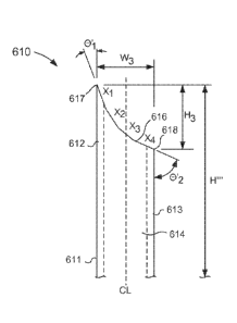
substantially open
such that the cartridge body 241 can movably receive at least a portion of the
plunger 245.
More specifically, at least a portion of the plunger 245 is disposed within
the inner volume
244 and can be moved between a first position (e.g., a proximal position) and
a second
position (e.g., a distal position). The plunger 245 includes a seal member 246
that forms a
friction fit with one or more surfaces of the cartridge body 241 that define
the inner volume
244. In this manner, the seal member 246 and the cartridge body 241 can form a
substantially fluid-tight seal that substantially isolates a portion of the
inner volume 244 that
is distal to the seal member 246 from a portion of the inner volume 244 that
is proximal to the
seal member 246, as described in further detail herein.
[1085] In some embodiments, the distal end portion 243 of the cartridge
body 241 can be
at least temporarily closed (e.g., at least temporarily fluidically sealed).
In this manner, the
inner volume 244 (e.g., the portion of the inner volume 244 between the seal
member 246
and the distal end portion 243) of the cartridge body 241 is fluidically
isolated from a volume
17
CA 02882184 2015-02-13
WO 2014/036009 PCT/US2013/056863
outside of the cartridge body 241. The inner volume 244 of the cartridge body
241 can
further house or contain a drug formulation of the compositions described
herein (e.g., a
prophylactic agent, a therapeutic agent, or a diagnostic agent). More
specifically, the drug
formulation is disposed within the inner volume 244 of the cartridge body 241
in a distal
position relative to the seal member 246. Thus, the drug formulation contained
within the
inner volume 244 is substantially fluidically isolated from a volume outside
of the container
body 241. In some embodiments, the inner volume 244 can contain a drug
formulation with
a volume of about 0.5 mL or less, for example about 0.1 mL to about 0.5 mL. In
other
embodiments, the inner volume 244 can contain a drug formulation with a volume
of about
0.1 mL. In still other embodiments, the inner volume 244 can contain a drug
formulation
with a volume greater the about 0.5 mL.
[1086] In some embodiments, the distal end portion 243 of the cartridge
body 241 can be
moved between a first configuration (e.g., a closed or sealed configuration)
and a second
configuration (e.g., an open configuration). Expanding further, the distal end
portion 243 of
the container body 241 can include a surface that can be deformed (e.g.,
punctured, broken,
opened, or otherwise reconfigured) to expel the drug formulation contained
with the inner
volume 244 of the cartridge body 241. For example, in some embodiments, the
cartridge 240
can be inserted into the cartridge housing 230 such that the deformable
surface of the distal
end portion 243 of the cartridge body 241 is placed in contact with the
proximal end portion
211 of the microneedle 210. In such embodiments, the proximal end portion 211
of the
microneedle 210 can extend from a surface of the cartridge housing 230 that
defines the inner
volume 233 such that when the cartridge 240 is disposed within the inner
volume 233, the
proximal end portion of the microneedle 210 pierces, breaks, or otherwise
reconfigures the
deformable portion of the cartridge body 241. In this manner, the lumen 214
defined by the
microneedle 210 can be placed in fluid communication with the inner volume 244
defined by
the cartridge body 241. Therefore, when the plunger 245 is moved from its
first position to
its second position relative to the cartridge body 241, the drug formulation
contained within
the inner volume 244 of the cartridge body 241 can be expelled through the
lumen 214
defined by the microneedle 210.
[10871 In other embodiments, however, the distal end portion 243 of the
cartridge body
241 can be fluidically coupled to the microneedle 210. In this manner, the
inner volume 244
(e.g., the portion of the inner volume 244 between the seal member 246 and the
distal end
portion 243) of the cartridge body 241 is fluidically coupled to a volume
outside of the
18
CA 02882184 2015-02-13
WO 2014/036009 PCT/US2013/056863
cartridge body 241 via the microneedle 210. For example, in some embodiments
the distal
end portion 243 can be devoid of a deformable portion or seal (e.g., a crimp
seal), and in use
the proximal end portion of the microneedle 210 need not pierce, break, or
otherwise a
surface prior to use.
[1088] In use, a user (e.g., a doctor, technician, nurse, physician,
ophthalmologist, etc.)
can remove the cap 250 to expose at least a portion of the microneedle 210 and
can
manipulate the infusion device 200 to insert the microneedle 210 into, for
example, an ocular
tissue. As described above, the length and/or the shape of the distal end
portion 212 of the
microneedle 210 (including, for example, a beveled surface) at least partially
corresponds
with the target tissue (e.g., the eye) such that the distal end portion 212 of
the microneedle
210 is disposed within a lower portion of the sclera and/or the suprachoroidal
space of the eye
after being inserted. More specifically, the distal end portion 212 of the
microneedle 210 is
configured to pierce the sclera of the eye and be disposed within the sclera
and/or
suprachoroidal space without substantially piercing the choroid of the eye.
[1089] With the microneedle 210 disposed within the eye, the cartridge 240
can be
moved within the inner volume 233 of the cartridge housing 230 to place the
inner volume
244 of the cartridge body 241 in fluid communication with the lumen 214
defined by the
microneedle 210. For example, in some embodiments, the proximal end portion
211 of the
microneedle 210 can pierce or otherwise reconfigure the proximal end portion
211 to move
the deformable surface from a sealed configuration to an unsealed or open
configuration.
Thus, the inner volume 244 of the cartridge body 241 is placed in fluid
communication with
the lumen 214 defined by the microneedle 210.
[10901 After the inner volume 244 of the cartridge body 241 is placed in
fluid
communication with the microneedle 210, the cartridge 240 can be moved from a
first
configuration (e.g., where the plunger 245 is disposed in its first position
relative to the
cartridge body 241) to a second configuration (e.g., where the plunger 245 is
disposed in its
second position relative to the cartridge body 241). With the seal member 246
forming a
substantially fluid-tight or leak-proof seal (e.g., a substantially hermetic
seal) with an inner
surface of the cartridge body 241, the movement of the plunger 245 to its
second position
expels the drug formulation (contained within the inner volume 244) through
the lumen 214
of the microneedle 210. Thus, the medicament delivery device 200 can deliver
the drug
formulation to the suprachoroidal space of the eye and the drug formulation
can flow within
the suprachoroidal space to be delivered to, for example, the posterior region
of the eye.
19
CA 02882184 2015-02-13
WO 2014/036009 PCT/US2013/056863
[1091] Although the proximal end portion 211 of the microneedle 210 is
described above
as piercing or otherwise reconfiguring the deformable surface of the distal
end portion 243 of
the cartridge body 241, in other embodiments, the microneedle 210 need not
physically
contact the cartridge body 241. For example, in some embodiments, the distal
end portion
243 of the cartridge body 241 can be in its closed configuration (e.g.,
undeformed
configuration) when the plunger 245 is moved relative to the cartridge body
241. In such
embodiments, the movement of the plunger 245 can increase the pressure within
the inner
volume 244 and the increase in pressure can move the deformable surface of the
distal end
portion 243 to its open configuration (e.g., deformed configuration). For
example, the
increase in pressure can open a valve or break (e.g., rupture) the deformable
surface. Thus,
the inner volume 244 can be placed in fluid communication with the lumen 214
of the
microneedle 210 and the medicament delivery device 200 can deliver the drug
formulation to
the target tissue.
[1092] Although the microneedle 210 is described above as being inserted
such that the
distal end portion 212 is at least partially disposed in the suprachoroidal
space, in other
instances, the microneedle 210 can be inserted into various other regions of
ocular tissue. For
example, in some instances, a the microneedle 210 can be inserted through the
ciliary body to
dispose, at least partially, the distal end portion 212 of the microneedle 210
in the vitreous of,
for example, a pediatric eye, as described in further detail herein.
[1093] Although not shown in FIG. 6, in some embodiments, the cartridge 240
can
include a cap configured to enclose the deformable surface of the cartridge
body 241. In such
embodiments, the cap can maintain the sterility of the deformable surface
and/or can prevent
deformation (e.g., breaking, puncturing, etc.) of the deformable surface prior
to use.
[1094] FIG. 7 is a schematic illustration of a microneedle 310 according to
an
embodiment. The microneedle 310 can be included in, for example, the
medicament delivery
device 200 described above with reference to FIG. 6, or any other delivery
system described
herein. The microneedle 310 can be configured to puncture, pierce and/or
penetrate a portion
of the eye to deliver a drug formulation to and/or remove a substance from a
target location,
such as, for example, the suprachoroidal space. The microneedle 310 includes a
proximal
end portion 311, a distal end portion 312, and a set of annular walls 313. The
microneedle
310 has a shaft length H' that can be any suitable length. For example, in
some
embodiments, the shaft length H' can substantially correspond to at least a
portion of the eye.
For example, in some embodiments, the shaft length H' can correspond to and/or
be within a
CA 02882184 2015-02-13
WO 2014/036009 PCT/US2013/056863
range of the thickness of the sclera (see e.g., FIGS. 3 and 4). Thus, in some
embodiments,
the shaft length H' can be any suitable length such that when the microneedle
is inserted into
the eye, the distal end portion 312 of the microneedle 310 is disposed within
the
suprachoroidal space without puncturing and/or extending through the choroid.
By way of
example, the microneedle 310 shaft length H' can be about 1000 pm or less,
about 900 pm or
less, about 850 [ini or less, about 800 pm or less, about 750 1..tm or less,
about 700 pm or less,
about 650 [tin or less, or about 600 [tm or less. In some embodiments, the
microneedle 310
shaft length H' can be about 750 [im. In other embodiments, the microneedle
310 shaft
length H' can be about 800 pm, or about 850 [tm, or about 900 pm, or about 950
pm, or about
1 mm.
[1095] In other embodiments, the microneedle 310 can have a shaft length H'
suitable for
use in treatment of other portions of the eye, such as, the vitreous. For
example, in some
embodiments, the microneedle 310 can have a shaft length H' of about 1 mm to
about 3 mm.
In another embodiments, the microneedle 310 can have a shaft length H' from
about 2.5 mm
to about 5.5 mm. In yet another embodiment, the microneedle 310 can have a
shaft length H'
from about 3 mm to about 4 mm.
[1096] The walls 313 define a lumen 314 that extends through the proximal
end portion
311 and the distal end portion 312. The proximal end portion 311 includes a
base (or hub)
319 and/or can be coupled to (e.g., physically and/or fluidically) any
suitable medical device.
For example, in some embodiments, the proximal end portion 311 can be
physically and
fluidically coupled to the cartridge housing 230, as described above with
reference to FIG. 6.
In other embodiments, the proximal end portion 311 can be indirectly coupled
to a medical
device or cartridge housing via any suitable intervening structure such as,
for example, a
Luer-Lok0 (or other locking mechanism) or sterile flexible tubing. In this
manner, the lumen
314 defined by the walls 313 of the microneedle 310 can be placed in fluid
communication
with a fluid source (e.g., the cartridge 240 described above or any other
suitable source) to
deliver a drug formulation to a target tissue.
[1097] The distal end portion 312 of the microneedle 310 defines an opening
315
configured to place the lumen 314 in fluid communication with a volume
substantially
outside the microneedle 310. The distal end portion 312 includes a bevel 316
(also referred
to herein as "beveled surface") with a distal edge 317 and a proximal edge
318. Similarly
stated, the distal end portion 312 includes a surface (i.e., the bevel 316)
that is slanted, sloped,
angled and/or inclined from an outer surface of the walls 313. Said another
way, the beveled
21
CA 02882184 2015-02-13
WO 2014/036009 PCT/US2013/056863
surface 316 intersects the outer surface of the walls 313 at one or more
angles to define one
or more sharp edges (e.g., the distal edge 317 and/or the proximal edge 318),
as defined
herein. This arrangement allows the distal end portion 312 of the microneedle
to pierce,
separate and/or deform the target tissue to facilitate penetration of the
shaft microneedle 310
therethrough. Similarly stated, the sharp distal edge 317 is configured to
pierce the target
tissue, such as ocular tissue, to facilitate defining a passageway within the
target tissue to the
desired location (e.g., the suprachoroidal space and/or the vitreous, as
described in further
detail herein).
[1098] As shown
in FIG. 7, the bevel 316 has a height H1 defined and/or bounded by the
distal edge 317 and the proximal edge 318. In other embodiments, the bevel 316
can have a
height H'1 that is defined between the distal edge 317 and an edge formed
between an inner
surface of the walls 313 that is circumferentially opposed to the distal edge
317 (e.g., an edge
formed by the bevel 316 and an inner surface of the walls 313 that is adjacent
to the proximal
edge 318). The height Hi and/or H'1 can be any height that prevents and/or
limits the
likelihood of piercing the lens, retina, and/or choroid when the entire shaft
length H' or
substantially the entire shaft length H' of the microneedle 310 is inserted
into the eye through
the sclera. In other instances, the height Hi and/or H'i of the bevel 316 can
prevent and/or
limit the likelihood of damaging the lens or other ocular tissue when the
entire shaft length H'
or substantially the entire shaft length H' is inserted into the eye through
the ciliary body
(e.g., such that the distal end portion 312 is disposed in the vitreous 30).
In yet other
embodiments, the height H1 and/or H'1 can be such that when the microneedle is
disposed
within the sclera, the opening 315 is at a desired location of the sclera
and/or suprachoroidal
space (see e.g., FIGS. 3 and 4). For example, if the bevel height Hi and/or
is too large, a
portion of the opening may be disposed outside of (e.g., above) the sclera
and/or into the
conjunctiva. Such positioning may result in the deposition of substances in an
undesirable
portion of the eye. Thus, in some embodiments, the bevel height FI1 and/or H'1
is such that
when the distal edge 317 is disposed within the suprachoroidal space and/or
adjacent the
choroid, the opening 315 does not extend beyond the innermost half of the
sclera, third of the
sclera, or quarter of the sclera. More particular, as described herein, in
some embodiments,
the microneedle can be inserted into the ocular tissue at an angle that is
between about 80
degrees and about 100 degrees relative to a tangential surface of the
insertion site of the eye.
When inserted in such an orientation and with the distal edge 317 disposed
within the
suprachoroidal space and/or adjacent the choroid, the bevel height Hi and/or
can be such
22
CA 02882184 2015-02-13
WO 2014/036009 PCT/US2013/056863
that the opening 315 does not extend beyond the innermost half of the sclera,
third of the
sclera, or quarter of the sclera.
[1099] For example, in some embodiments, the height H1 and/or H'1 of the
bevel 316 can
be about 500 [inn or less, about 450 wn or less, about 400 11M or less, about
350 [tm or less,
about 300 lam or less, about 250 [trn or less, about 200 pm or less, about 150
lam or less, or
about 100 lam or less. In other embodiments, the height H1 and/or H'1 of the
bevel 316 is
from about 50 vim to about 500 pm, from about 100 [tm to about 500 pm, from
about 150 1..tm
to about 500 pm, from about 200 wri to about 500 pm, from about 250 [tm to
about 500 pm,
from about 300 lam to about 500 ium, from about 50 lam to about 400 lam, from
about 100 pm
to about 400 pm, or from about 150 pm to about 400 tim. In some embodiments,
the height
H1 and/or H'1 of the bevel 316 can be about 485 ium. In still other
embodiments, the height
H1 and/or H'1 of the bevel 316 can be about 500 1..tm to about 1 mm. In
another embodiment,
the height H1 and/or H'1 of the bevel 316 is from about 600 ium to about 1 mm,
from about
700 [tm to about 1 mm, from about 800 tim to about 1 mm, from about 900 p.m to
about 1
mm, and/or any fraction there between.
[1100] While characterized above by the height H1 and/or H'1 of the bevel
316, in other
embodiments, the microneedle 310 can be characterized by a bevel angle, or
more
particularly, a tip angle relative to an axis defined by the lumen 314. In
some embodiments,
the tip angle can be selected to facilitate insertion of the microneedle 310
within the desired
type of tissue (e.g., ocular tissue). Similarly stated, in some embodiments,
the tip angle can
be selected to provide a sufficient "sharpness" such that the microneedle 310
can be inserted
into the eye (e.g., the sclera) while minimizing the deformation of the eye
resulting from the
force of insertion. For example, in some embodiments, the bevel angle can be
less than about
0.1 degree. In other embodiments, the bevel angle can be from approximately
0.1 degree to
approximately 1 degree. In still other embodiments, the bevel angle can be
from
approximately 1 degree to approximately 5 degrees, including any fraction of a
degree there
between. In some embodiments, the microneedle 310 has a bevel angle from about
0.1
degree to about 30 degrees or from about 1 degree to about 25 degrees or from
about 2
degrees to about 20 degrees or from about 10 degrees to about 20 degrees. In
some
embodiments, the tip angle can be less than about 18 degrees, 15 degrees or 12
degrees.
[1101] In yet other embodiments, the microneedle 310 can be characterized
by the bevel
height H1 (or H'1) to width W1 ratio, (i.e., the bevel aspect ratio). In some
embodiments, the
bevel width WI corresponds with an outer diameter of the microneedle 310. In
other
23
CA 02882184 2015-02-13
WO 2014/036009 PCT/US2013/056863
embodiments, the bevel width WI can be associated with a diameter that is
smaller than the
outer diameter of the microneedle 310 (e.g., the microneedle 310 can have an
outer diameter
that is tapered from the proximal end portion 311 to the distal end portion
3212). In some
embodiments, the bevel width Wi is from about 50 gm to about 500 gm, from
about 50 gm
to about 400 gm, from about 100 gm to about 400 gm, from about 200 gm to about
400 gm,
from about 200 gm to about 320 gm, or from about 100 gm to about 250 gm. As
such, the
microneedle 310 can have a bevel aspect ratio (height: width) is about 0.25:1,
about 0.5:1,
about 0.75:1, about 1:1, about 1.5:1, about 2:1, about 2.2:1, about 2.5:1 or
about 3:1.
[1102] In this manner, the arrangement of the bevel 316 (e.g., the bevel
can be such that
the distal edge 317 is sufficiently sharp such as to pierce a target tissue
and penetrate into
sclera, the suprachoroidal space or the vitreous without (I) substantially
causing the target
tissue to elastically deform or (ii) damaging internal structures of the eye
(e.g., the lens,
retina, choroid, etc.). Similarly stated, the arrangement of the bevel 316 can
be such that the
distal edge 317 is sufficiently sharp such that prior to piercing the target
tissue, the distal edge
317 does not substantially bend, compress, deform, or otherwise move the
target tissue.
Thus, the accuracy of insertion relative to the target tissue is increased and
a potential for
damaging surrounding tissue is minimized.
[1103] As described above, the microneedle 310 has a shaft length H'. The
shaft length
H' of the microneedle 310 is defined as the length from the distal edge 317 to
the hub or base
surface 319. In the drug delivery methods provided herein, the entire shaft
length H' or
substantially the entire shaft length H' of the microneedle 310 can be
inserted into the eye. In
this regard, the user need not determine the depth of insertion. The shaft
length H' is such
that when the microneedle 310 inserted through, for example, the ciliary body,
the risk of
piercing the lens or retina is greatly minimized or eliminated. Similarly, the
shaft length H' is
such that when the microneedle 310 is inserted through the sclera at about the
ocular
hemisphere (described above), the risk of piercing the choroid and/or retina
is greatly
minimized or eliminated. In this regard, the microneedles provided herein
achieve greater
reproducibility, and eliminate or substantially reduce uncertainty associated
with drug
delivery methods to the eye (e.g., to the suprachoroidal space and/or to the
vitreous). The
shaft length can be tailored depending on the age of the patient and the
desired tissue for drug
delivery or aspiration.
[1104] While the bevel 316 included in the microneedle 310 is shown in FIG.
7 as being
substantially linear (e.g., the beveled surface is straight or planar), in
other embodiments, a
24
CA 02882184 2015-02-13
WO 2014/036009 PCT/US2013/056863
microneedle can include a bevel and/or beveled surface that is substantially
curvilinear. For
example, FIGS. 8-10 are schematic illustrations of a microneedle 410 according
to another
embodiment. The microneedle 410 includes a proximal end portion 411, a distal
end portion
412, and a set of annular walls 413. The microneedle 410 has a shaft length H"
that can be
any suitable length. For example, in some embodiments, the shaft length H" can
substantially correspond to at least a portion of the eye. For example, in
some embodiments,
the shaft length H" can correspond to and/or be within a range of the
thickness of the sclera
(see e.g., FIGS. 3 and 4). In some instances, the shaft length H" can be
substantially similar
to or the same as the shaft length H' described above with reference to the
microneedle 310.
[1105] The walls 413 of the microneedle 410 define a lumen 414 extending
through the
proximal end portion 411 and the distal end portion 412. Furthermore, the
walls 413 can
have and/or can define a width W2 associated with an outer diameter and/or a
beveled surface
416 of the microneedle 410, as described in further detail herein. The
proximal end portion
411 can be substantially similar in form and function as the proximal end
portion 311
included in the microneedle 310 described above with reference to FIG. 7. In
this manner,
the proximal end portion 411 can be physically and fluidically coupled to any
suitable
medical device, as described above. The distal end portion 412 of the
microneedle 410
includes a bevel (or beveled surface) 416 having a distal edge 417 and a
proximal edge 418.
More particularly, as shown in FIGS. 8-10, the walls 413 of the microneedle
410 include a
first portion 420 that intersects a first end of the bevel 416 to form and/or
define the distal
edge 417, and a second portion 421 that intersects a second end of the bevel
416, opposite the
first end, to form and/or define the proximal edge 418. Similarly stated, the
first portion 420
and the second portion 421 of the walls are circumferentially opposed and as
such, the distal
edge 417 of the bevel 416 and the proximal edge 418 of the bevel 416 are
circumferentially
opposed. The distal end portion 412 of the microneedle 410 also defines an
opening 415. In
this manner, the distal end portion 412 is configured to place the lumen 414
in fluid
communication with a volume substantially outside the microneedle 410, such as
for
example, the suprachoroidal space.
[1106] As shown FIG. 8, the bevel 416 has a height H2 defined and/or
bounded by the
distal edge 417 and the proximal edge 418. In some embodiments, the height H2
of the bevel
416 is substantially similar in height to H1 of the bevel 316. In other
embodiments, the height
H2 of the bevel 416 can be any suitable height included in the range of
lengths described
above. For example, in some embodiments, the height H2 can be such that when
the
CA 02882184 2015-02-13
WO 2014/036009 PCT/US2013/056863
microneedle 410 is disposed within the sclera, the opening 415 is at a desired
location of the
sclera and/or suprachoroidal space (see e.g., FIGS. 3 and 4). For example, if
the bevel height
H2 is too large, a portion of the opening may be disposed outside of (e.g.,
above) the sclera
and/or into the conjunctiva. Such positioning may result in the deposition of
substances in an
undesirable portion of the eye. Thus, in some embodiments, the bevel height H2
is such that
when the distal edge 417 is disposed within the suprachoroidal space and/or
adjacent the
choroid, the opening 415 does not extend beyond the innermost half of the
sclera, third of the
sclera, or quarter of the sclera. More particular, as described herein, in
some embodiments,
the microneedle 410 can be inserted into the ocular tissue at an angle that is
between about 80
degrees and about 100 degrees relative to a tangential surface of the
insertion site of the eye.
When inserted in such an orientation and with the distal edge 417 disposed
within the
suprachoroidal space and/or adjacent the choroid, the bevel height H2 can be
such that the
opening 415 does not extend beyond the innermost half of the sclera, third of
the sclera, or
quarter of the sclera. Thus, by maintaining the bevel height H2 below a
desired threshold, the
length of the opening 415 can be such that the entire opening 415 is
maintained within a
desired portion of the sclera and/or suprachoroidal space.
[1107] In some embodiments, the height H2 of the bevel can be about 500 pm
to about 1
mm. In another embodiment, the height H2 of the bevel 416 is about 500 pm or
less, about
450 pm or less, about 400 pm or less, about 350 pm or less, about 300 pm or
less, about 250
pm or less, about 200 pm or less, about 150 pm or less, or about 100 pm or
less. In other
embodiments, the height H2 of the bevel 416 is from about 50 pm to about 500
prn, from
about 100 pm to about 500 pm, from about 150 pm to about 500 pm, from about
200 pm to
about 500 pm, from about 250 pm to about 500 pm, from about 300 pm to about
500 pm,
from about 50 pm to about 400 pm, from about 100 pm to about 400 pm, or from
about 150
pm to about 400 pm. The bevel height H2 can be any bevel height that prevents
piercing of
the lens, retina, or choroid when the distal end 412 of the microneedle 410 is
inserted through
the sclera, and the entire shaft length H" or substantially the entire shaft
length H" of the
microneedle 410 is inserted into the eye.
[1108] As described above, the microneedle 410 has the width W2. In some
embodiments, the width W2 can be from about 50 gm to about 500 pm, from about
50 pm to
about 400 gm, from about 100 pm to about 400 pm, from about 200 gm to about
400 pm, or
from about 100 gm to about 250 pm. In some embodiments, the microneedle 410
can be
characterized by, for example, a bevel height H2 to width W2 ratio (i.e., a
bevel aspect ratio
26
CA 02882184 2015-02-13
WO 2014/036009 PCT/US2013/056863
(height: width)). In some embodiments, the microneedle 410 can have a bevel
aspect ratio
that is about 0.25:1, about 0.5:1, about 0.75:1, about 1:1, about 1.5:1, about
2:1, about 2.2:1,
about 2.5:1, or about 3:1. In some embodiments, the microneedle 410 can have a
bevel
aspect ratio that is less than about 3.0:1, about 2.5:1, about 2.2:1, about
2.0:1 or about 1.5:1.
[1109] As shown in FIGS. 8 and 10, the bevel 416 is substantially
curvilinear (e.g., the
beveled surface is not planar). The bevel 416 can have any suitable radius of
curvature (or
radii of curvature). In some embodiments, the radius of curvature of the bevel
416 can be
substantially consistent or continuous between the distal edge 417 and the
proximal edge 418.
In other embodiments, the radius of curvature can vary along the height H2 of
the bevel 416.
For example, as shown in FIG. 10, the first portion 420 of the walls 413 can
have and/or can
be associated with an axis al that is substantially parallel to a centerline
CL of the
microneedle 410 and the second portion 421 of the walls 413 can have and/or
can be
associated with an axis az that is substantially parallel to the centerline CL
of the microneedle
410. The bevel 416 can be arranged such that a first bevel angle el (e.g., a
tip angle or distal
angle) is defined between the axis al and a line that is tangent to the bevel
416 at or near the
distal edge 417. Similarly, the bevel 416 can be arranged such that a second
bevel angle 02
(e.g., a proximal angle or an inside angle) is defined between the axis az and
a line that is
tangent to the bevel 416 at or near the proximal edge 418. In some
embodiments, the first
bevel angle 01 can be smaller than the second bevel angle 02. For example, in
some
embodiments, the first bevel angle 01 can be less than about 20 degrees and
the second bevel
angle 02 can be greater than about 30 degrees. In some embodiments, the first
bevel angle
01 can be less than 0.1 degree. In other embodiments, the first bevel angle 01
can be from
approximately 0.1 degree to approximately 1 degree. In still other
embodiments, the first
bevel angle 01 can be from approximately 1 degree to approximately 5 degrees,
including
any fraction of a degree therebetween. In one embodiment, the microneedle 410
can have a
first bevel angle 01 from about 0.1 degree to about 30 degrees, or from about
1 degree to
about 25 degrees, or from about 2 degrees to about 20 degrees, or from about
10 degrees to
about 20 degrees. In some embodiments, the first bevel angle 01 can be less
than about 20
degrees or less than about 18 degrees. In still other embodiments, the first
bevel angle 01 can
be about 12 degrees.
[1110] In some embodiments, the second bevel angle 02 can be from about 20
degrees to
about 30 degrees, or from about 20 degrees to about 45 degrees, or from about
20 degrees to
about 60 degrees, or from about 20 degrees to about 75 degrees, or from about
20 degrees to
27
CA 02882184 2015-02-13
WO 2014/036009 PCT/US2013/056863
about 90 degrees. In other embodiments, the second bevel angle 02 can be less
than 20
degrees yet still larger than the first bevel angle 01. In some embodiments,
the second bevel
angle 02 can be greater than about 30 degrees. In some embodiments, the second
bevel angle
02 can be between about 30 degrees and about 45 degrees. In other embodiments,
the second
bevel angle 02 can be between about 45 degrees and about 90 degrees.
[11111 As a result, arrangement of the bevel 416 at or near the distal edge
417 can define
a radius of curvature that is substantially larger than the arrangement of the
bevel 416 at or
near the proximal edge 418. In this manner, the bevel 416 and more
particularly, the distal
edge 417, can be sufficiently sharp (as described above), while the second
portion of the
bevel 417 can be configured to maintain the structural rigidity of the distal
end portion 412 of
the microneedle 410 and/or maintain the bevel height H2 at or below a desired
value. For
example, the first bevel angle 01 can be below a desired angle such that a
thickness of the
first portion 420 of the walls 413 is sufficiently thin to pierce ocular
tissue without
substantially bending, curving, deforming, and/or otherwise moving the ocular
tissue prior to
insertion. Conversely, the second bevel angle 02 can be increased (e.g.,
relative to the first
bevel angle 01) to an extent such that a thickness of the second portion 421
of the walls 413
is sufficiently thick to maintain the structural rigidity of the distal end
portion 412 of the
microneedle 410 and/or such that the bevel height H2 is at or below a desired
value.
[11121 As shown in FIG. 8, the shaft length H" of the microneedle 410 is
defined as the
length from the distal edge 417 to the hub 419. In the drug delivery methods
provided herein,
the entire shaft length H" or substantially the entire shaft length H" of the
microneedle 410
is inserted into the eye. In this regard, the user need not determine an
optimal depth of
microneedle insertion prior to insertion and/or employ visualization
techniques during the
insertion operation. The microneedle 410 can be defined by a shaft length H"
that, in some
embodiments, does not allow for piercing of the choroid when inserted into the
sclera of the
eye. In this regard, the microneedle 410 provided herein reduces variability
associated with
sclera and suprachoroidal drug delivery methods, and eliminates or
substantially reduces the
uncertainty associated with ocular drug delivery methods to the eye. In other
embodiments,
the microneedle 410 can be defined by a shaft length H" that does not allow
for piercing of
the lens when inserted into the vitreous of the eye. In this regard, the
microneedles 410
provided herein reduce variability associated with intravitreal drug delivery
methods, and
eliminates or substantially reduces the uncertainty associated with ocular
drug delivery
methods to the eye.
28
CA 02882184 2015-02-13
WO 2014/036009 PCT/US2013/056863
[1113] Although the microneedle 410 is shown in FIGS. 8-10 as including a
bevel 416 or
beveled surface that is substantially continuous, in other embodiments, a
microneedle can
have a bevel that is discontinuous and/or is defined in at least two planes.
Similarly stated,
although the microneedle is shown and described above as include a single
bevel surface, in
other embodiments, a microneedle can have multiple bevel surfaces. For
example, FIGS. 11
and 12 illustrate a microneedle 510 according to another embodiment. As shown
in FIG. 11,
the microneedle 510 has a proximal end portion 511 and a distal end portion
512 and defines
a lumen 514. The proximal end portion 511 can be substantially similar in form
and function
of the proximal end portion 311 of the microneedle 310 shown in FIG. 7. The
distal end
portion 512 includes and/or defines a bevel 516 having a distal edge 517 and a
proximal edge
518. As shown in FIG. 11, the bevel 516 can be substantially curvilinear. In
some
embodiments, the bevel 516 can include a radius of curvature that is
substantially similar to
the radius of curvature of the bevel 416 described above with reference to
FIGS. 8-10.
[1114] In some embodiments, the distal end portion 512 of the microneedle
510 can be
cut or formed in a plane that is different from (e.g., substantially
orthogonal to) the plane in
which the bevel 516 is profiled (e.g., as shown in FIG. 11). For example, as
shown in FIG.
12, the distal end portion 512 of the microneedle 510 can be cut along the
lines CI and C2.
As a result, the distal end portion 512 can be tapered between a first
portion, which is near
and/or adjacent to the proximal edge 518, and a second portion, which is near
and/or adjacent
to the distal edge 517. Thus, by cutting the distal end portion 512 along, for
example, the
lines C1 and C2 in FIG. 12, the distal edge 517 can be sharpened in two planes
(e.g., by the
bevel 516 and by the cuts along the lines CI and C2).
[1115] While the bevels 416 and 516 are shown as forming a substantially
smooth curve
or continuous surface, in other embodiments, a microneedle can include a bevel
that is
formed from any number of planes or segments (e.g., a three facet or a four
facet bevel). For
example, FIG. 13 is a schematic illustration of a microneedle 610 according to
an
embodiment. The microneedle 610 includes a proximal end portion 611, a distal
end portion
612, and a set of annular walls 613. The microneedle 610 can have a shaft
length H" that
can substantially correspond to at least a portion of the eye. For example, in
some
embodiments, the shaft length H" can correspond to and/or be within a range of
the
thickness of the sclera (see e.g., FIGS. 3 and 4). Thus, in some embodiments,
the shaft length
H' " can be any suitable length that does not allow for piercing of and/or
extending through
the choroid when the microneedle 610 is inserted into the posterior segment of
the eye, as
29
CA 02882184 2015-02-13
WO 2014/036009 PCT/US2013/056863
described above. In this regard, the microneedle 610 provided herein reduces
variability
associated with suprachoroidal drug delivery methods, and eliminates or
substantially reduces
the uncertainty associated with ocular drug delivery methods to and/or removal
of substances
from the eye. In some embodiments, the shaft length H" ' can be any of the
lengths described
above with reference to the microneedle 310 or the microneedle 410, or any
other shaft
lengths referenced herein.
[1116] The walls 613 define a lumen 614 extending through the proximal end
portion 611
and the distal end portion 612. The proximal end portion 611 can be
substantially similar in
form and function as the proximal end portion 311 included in the microneedle
310 described
above with reference to FIG. 7. In this manner, the proximal end portion 611
can be
physically and fluidically coupled to any suitable medical device, as
described above.
Although not shown in FIG. 13, in some embodiments, the proximal end portion
611 can be
coupled and/or can include a base or hub. As described herein, in use the base
or hub can
include a surface that substantially circumscribes the shaft of the
microneedle 610, and that
can contact a portion of the target tissue (e.g., a surface of the eye) during
use.
[1117] The distal end portion 612 of the microneedle 610 includes a bevel
or beveled
surface 616 having a distal edge 617 and a proximal edge 618. Similarly
stated, the distal
end portion 612 includes a surface (i.e., the bevel 316) that is slanted,
sloped, angled and/or
inclined from an outer surface of the walls 613. Said another way, the beveled
surface 616
intersects the outer surface of the walls 613 at one or more angles to define
one or more sharp
edges (e.g., the distal edge 617 and/or the proximal edge 618), as defined
herein. This
arrangement allows the distal end portion 612 of the microneedle to pierce,
separate and/or
deform the target tissue to facilitate penetration of the shaft microneedle
610 therethrough.
Similarly stated, the sharp distal edge 617 is configured to pierce the target
tissue, such as
ocular tissue, to facilitate defining a passageway within the target tissue to
the desired
location (e.g., the suprachoroidal space and/or the vitreous, as described
herein).
[1118] While not shown in FIG. 13, the distal end portion 612 can define an
opening
substantially similar to the opening 315 or the opening 415 described above.
In this manner,
the distal end portion 612 can be configured to place the lumen 614 in fluid
communication
with a volume substantially outside the microneedle 610, such as for example,
the
suprachoroidal space.
CA 02882184 2015-02-13
WO 2014/036009 PCT/US2013/056863
[1119] In some
embodiments, the microneedle 610 comprises multiple facets (also
referred to as segments). For example, as shown in FIG. 13, the bevel 616 can
include a first
segment X1, a second segment X2, a third segment X3, and a fourth segment X4
(i.e., a four
facet bevel). In another embodiment, the bevel includes a first segment X1, a
second segment
X2 and a third segment X3 (i.e., a three facet bevel). In yet other
embodiments, a microneedle
can include any number of facets or bevel portions.
[1120] In some
embodiments, the segments X1, X2, X3, and X4 can be substantially
similar in length to each other. In other embodiments, the segments X1, X2,
X3, and X4 can
be substantially different lengths. Furthermore, the segments Xi, X2, X3, and
X4 can be
configured to be substantially tangential to a radius of curvature (not shown)
of the bevel 616.
Therefore, as shown in FIG. 13, the segments X1, X2, X3, and X4 are arranged
at various
angles relative to a centerline CL of the microneedle 610 defined by the walls
613 of the
microneedle 610. Expanding further, the segment X1 can be arranged such that
the distal
edge 617 of the bevel 616 is sufficiently sharp (as described above) while the
segments X2,
X3, and X4 can be arranged to provide structural rigidity to the distal end
portion 612 of the
microneedle 610 and/or maintain a bevel height H3 (or a bevel aspect ratio)
within a desired
range.
[1121] In some
embodiments, the first segment Xi defines a first bevel angle O'i (e.g., a
tip angle or distal angle) and another segment (e.g., segment X4) defines a
second bevel angle
02 (e.g., a proximal angle or an inside angle). In some embodiments, the first
bevel angle
(/ I can be smaller than the second bevel angle CY2. For example, in some
embodiments, the
first bevel angle and/or
the second bevel angle (Y2 can be any angle or within any range
of angles described above with reference to the microneedle 410. As a result,
arrangement of
the bevel 616 at or near the distal edge 617 can be sufficiently sharp (as
described herein),
while the portion of the bevel 617 at or near the proximal edge 618 can be
configured to
maintain the structural rigidity of the microneedle 610 and/or maintain the
bevel height H3 at
or below a desired value. For example, the first bevel angle W1 can be such
that a thickness
of a first portion of the walls 613 is sufficiently thin to pierce ocular
tissue without
substantially bending, curving, deforming, and/or otherwise moving the ocular
tissue prior to
insertion. Conversely, the second bevel angle Cr2 can be larger than the first
bevel angle 0'1
to maintain the bevel height H3 and/or the bevel aspect ratio (H3 :W3) at or
below any of the
values specified herein.
31
CA 02882184 2015-02-13
WO 2014/036009 PCT/US2013/056863
[1122] In this manner, the height H3 can be such that when the microneedle
is disposed
within the sclera, the opening (not shown in FIG. 13) is at a desired location
of the sclera
and/or suprachoroidal space (see e.g., FIGS. 3 and 4). For example, if the
bevel height H3 is
too large, a portion of the opening may be disposed outside of (e.g., above)
the sclera and/or
into the conjunctiva. Such positioning may result in the deposition of
substances in an
undesirable portion of the eye. Thus, the multi-angle bevel arrangement
maintains the bevel
height H3 such that when the distal edge 617 is disposed within the
suprachoroidal space
and/or adjacent the choroid, the opening does not extend beyond the innermost
half of the
sclera, third of the sclera, or quarter of the sclera. More particular, as
described herein, in
some embodiments, the microneedle 610 can be inserted into the ocular tissue
at an angle that
is between about 80 degrees and about 100 degrees relative to a tangential
surface of the
insertion site of the eye. When inserted in such an orientation and with the
distal edge 617
disposed within the suprachoroidal space and/or adjacent the choroid, the
bevel height H3 can
be such that the opening does not extend beyond the innermost half of the
sclera, third of the
sclera, or quarter of the sclera.
[1123] While shown in FIG. 13 as including four segments X1, X2, X3, and
X4, in other
embodiments, a microneedle can include any number of segments configured at
any suitable
angle relative to an axis defined by a set of walls of the microneedle.
Moreover, in some
embodiments, a microneedle can include additional bevel segments that are
defined within a
different plane, such as the "chisel cut" bevel surfaces shown and described
in FIG. 12.
[1124] FIG. 14 is a schematic illustration of a microneedle 710 according
to an
embodiment. The microneedle 710 includes a proximal end portion 711, a distal
end portion
712, and a set of annular walls 713. The proximal end portion 711 can be
substantially
similar in form and function as the proximal end portion 311 included in the
microneedle 310
described above with reference to FIG. 7. In this manner, the proximal end
portion 711 can
be physically and fluidically coupled to any suitable medical device, as
described above. The
distal end portion 712 of the microneedle 710 includes a bevel 716. The bevel
716 can be
substantially similar to the bevel 316 included in the microneedle 310,
described above with
reference to FIG. 7, and/or any of the other bevels or beveled surfaces
described herein. The
distal end portion 712 of the microneedle 710 is also configured to define an
opening 715.
[1125] In some embodiments, the microneedle can have a shaft length (not
identified in
FIG. 14) that substantially corresponds to at least a portion of the eye. For
example, in some
embodiments, the shaft length can correspond to and/or be within a range of
the thickness of
32
CA 02882184 2015-02-13
WO 2014/036009 PCT/US2013/056863
the sclera (see e.g., FIGS. 3 and 4). Similarly stated, the microneedle 710
can have a shaft
length that does not allow for piercing of and/or passing through the choroid
when inserted
into the posterior segment of the eye, as described herein. In this regard,
the microneedle 710
provided herein reduces variability associated with scleral and suprachoroidal
drug delivery
methods, and eliminates or substantially reduces the uncertainty associated
with ocular drug
delivery methods to the eye. In other embodiments, the microneedle 710 can
have a shaft
length (not identified in FIG. 14) that does not allow for piercing of the
lens when inserted
into the posterior segment of the eye, as described above. In this regard, the
microneedle 710
provided herein reduces variability associated with intravitreal drug delivery
methods, and
eliminates or substantially reduces the uncertainty associated with ocular
drug delivery
methods to the eye.
[1126] The walls 713 define a lumen 714 extending through the proximal end
portion 711
and the distal end portion 712. The walls 713 have a thickness ti and define
an outer
diameter d1 of the microneedle 710 and an inner diameter d2 of the microneedle
710. In some
embodiments, the outer diameter d1 of the microneedle 710 is 30 gauge, 32
gauge, 34 gauge
or 36 gauge. In some embodiments, the outer diameter d1 can be from about 50
gm to about
500 gm, from about 50 gm to about 400 gm, from about 100 gm to about 400 gm,
from
about 200 gm to about 400 gm, or from about 100 p.m to about 250 gm. In some
embodiments, the inner diameter d2 can be at least partially associated with a
particle size of
a drug to be conveyed therethrough. For example, in some embodiments, the
inner diameter
d2 can be about 5 times the average particle size of a drug that is to be
conveyed
therethrough. In some embodiments, the inner diameter d2 can be between about
100 gm and
about 130 gm. In other embodiments, the inner diameter d2 can be between about
110 gm
and about 120 gm. In still other embodiments, the inner diameter d2 can be
less than 100 gm.
In yet other embodiments, the inner diameter d2 can be greater than 130 gm.
The opening
715 defined by the distal end portion 712 has a diameter d3 that is configured
to be
substantially greater than the inner diameter d2 and substantially less than
the outer diameter
d1.
[1127] While the bevel 716 is shown in FIG. 14 as being substantially
straight and/or
planar, in some embodiments, a microneedle can include a curvilinear bevel
and/or a multi-
faceted beveled surface. For example, the microneedle 410 shown and described
above has a
curved bevel, and can have an inner diameter, an outer diameter and/or a wall
thickness as
described herein with reference to the microneedle 710. As another example,
the
33
CA 02882184 2015-02-13
WO 2014/036009 PCT/US2013/056863
microneedle 610 shown and described above has a multi-faceted bevel, and can
have an inner
diameter, an outer diameter and/or a wall thickness as described herein with
reference to the
microneedle 710s. As yet another example, FIG. 15 is a schematic illustration
of a
microneedle 810 according to an embodiment. The microneedle 810 includes a
proximal end
portion 811, a distal end portion 812, and a set of annular walls 813. The
proximal end
portion 811 can be substantially similar in form and function as the proximal
end portion 311
included in the microneedle 310 described above with reference to FIG. 7. In
this manner,
the proximal end portion 811 can be physically and fluidically coupled to any
suitable
medical device, as described above. The distal end portion 812 of the
microneedle 810
includes a bevel 816 that is substantially curvilinear. In some embodiments,
the bevel 816
can be substantially similar to the bevel 416 included in the microneedle 410,
described
above with reference to FIGS. 8-10. The distal end portion 812 of the
microneedle 810 is
also configured to define an opening 815. The microneedle 810 can have a shaft
length (not
shown) that does not allow for piercing of the choroid when inserted into the
posterior
segment of the eye, as described above. In this regard, the microneedle 810
provided herein
reduces variability associated with suprachoroidal drug delivery methods, and
eliminates or
substantially reduces the uncertainty associated with ocular drug delivery
methods to the eye.
In other embodiments, the microneedle 810 can have a shaft length (not shown)
that does not
allow for piercing of the lens when inserted into the posterior segment of the
eye, as
described above. In this regard, the microneedle 810 provided herein reduces
variability
associated with intravitreal drug delivery methods, and eliminates or
substantially reduces the
uncertainty associated with ocular drug delivery methods to the eye.
[1128] The walls 813 define a lumen 814 extending through the proximal end
portion 811
and the distal end portion 812. The walls 813 have a thickness ti and define
an outer
diameter d1 of the microneedle 810 and an inner diameter d2 of the microneedle
810. In some
embodiments, the outer diameter d1 of the microneedle 810 is 30 gauge, 32
gauge, 34 gauge
or 36 gauge. The opening 815 defined by the curvilinear bevel 816 has a length
h that is
configured to be greater than the inner diameter d2 and less than the outer
diameter di.
[1129] While walls 813 are shown in FIG. 15 as having the same thickness th
in other
embodiments, a thickness of a set of walls can be varied circumferentially
about the
microneedle. For example, FIG. 16 is a schematic illustration of a microneedle
910
according to an embodiment. The microneedle 910 includes a proximal end
portion 911, a
distal end portion 912, and a set of annular walls 913. The proximal end
portion 911 can be
34
CA 02882184 2015-02-13
WO 2014/036009 PCT/US2013/056863
substantially similar in form and function as the proximal end portion 311
included in the
microneedle 310 described above with reference to FIG. 7. The distal end
portion 912 of the
microneedle 910 includes a bevel 916 having a distal edge 917 and a proximal
edge 918. The
bevel 916 can be substantially similar to the bevel 316 included in the
microneedle 310,
described above with reference to FIG. 7. The distal end portion 912 of the
microneedle 910
is also configured to define an opening 915. The microneedle 910 can have a
shaft length
(not shown) that does not allow for piercing of the choroid when inserted into
the posterior
segment of the eye, as described above. In this regard, the microneedle 910
provided herein
reduces variability associated with suprachoroidal drug delivery methods, and
eliminates or
substantially reduces the uncertainty associated with ocular drug delivery
methods to the eye.
In other embodiments, the microneedle 910 can have a shaft length (not shown)
that does not
allow for piercing of the lens when inserted into the posterior segment of the
eye, as
described above. In this regard, the microneedle 910 provided herein reduces
variability
associated with intravitreal drug delivery methods, and eliminates or
substantially reduces the
uncertainty associated with ocular drug delivery methods to the eye.
[1130] The annular walls 913 define a lumen 914 extending through the
proximal end
portion 911 and the distal end portion 912. The walls 913 define an outer
diameter di of the
microneedle 910 and an inner diameter d2 of the microneedle 910, as described
above in
reference to FIG. 14. The opening 915, defined by the distal end portion 912,
has a diameter
d3 that is configured to be substantially greater than the inner diameter d2
and substantially
less than the outer diameter di.
[1131] As shown in FIG. 16, the walls 913 have a first thickness t2 and a
second thickness
t3. Expanding further, the thickness of the walls 913 can vary relative to an
axis defined
between a point (not shown) on the distal edge 917 and a point (not shown) of
the proximal
edge 918. For example, as shown, a first portion of the walls 913 that
intersects a first end of
the bevel 916 to form and/or define the distal edge 917 has the first
thickness t2. A second
portion of the walls 913 that intersects a second end of the bevel 916,
opposite the first end,
to form and/or define the proximal edge 918 has the second thickness t3. As
show, the first
thickness t2 is greater than the second thickness ti. In this manner, the
first thickness t2 can be
sufficiently thick such that the bevel 916 can have a desired angle and
surface area at the
distal edge 917. Furthermore, the second thickness t3 can be sufficiently thin
such that the
diameter d3 is maintained.
CA 02882184 2015-02-13
WO 2014/036009 PCT/US2013/056863
[11321 While the first thickness t2 and the second thickness -6 are shown
in FIG. 16 as
being associated with the distal edge 917 and the proximal edge 918, in other
embodiments, a
microneedle can have a wall configured to have a thickness associated with a
proximal edge
that is thicker than a thickness associated with a distal edge. In such
embodiments, the
greater thickness at the proximal edge can be increase and/or maintain
structural rigidity at a
distal end portion of the microneedle. Furthermore, the smaller thickness at
the distal edge
can facilitate the insertion of the distal edge into ocular tissue without
substantially elastically
deforming the adjacent tissue (as described herein).
[11331 Any of the embodiments described above with reference to FIGS. 5-16
can be
suitable for suprachoroidal drug delivery. For example, FIGS. 17 and 18
illustrate a
medicament delivery device 1000 being used to deliver a drug formulation to
the
suprachoroidal space of an eye according to an embodiment. The medicament
delivery
device 1000 can be substantially similar to or the same as the medicament
delivery device
200 described above with reference to FIG. 3, or any other delivery devices
shown herein. In
this manner, the infusion device 1000 includes a microneedle 1010, a cartridge
housing 1030,
and a cartridge 1040. The microneedle 1010 can be, for example, any of the
microneedles
described herein.
[11341 In some embodiments, a method of delivering a drug formulation to
target ocular
tissue includes inserting the microneedle 1010 into the sclera 20 of the eye
10 and advancing
the microneedle 1010 through the sclera 20 such that the entire or
substantially the entire
shaft length (H' or H") of the microneedle 1010 between a proximal end portion
1011 and a
distal end portion 1012 is disposed in the eye 10 (as shown in FIG. 17). More
specifically,
the microneedle 1010 can be inserted into the ocular tissue (e.g., the sclera)
such that a base
surface of the proximal end portion of the microneedle 1010 or a base surface
of the cartridge
is placed in contact with the ocular tissue. Thus, the entire shaft length of
the microneedle
1010 can be inserted into the eye. The microneedle 1010 can have any suitable
shaft length,
such as the shaft lengths H' (see microneedle 310), H" (see microneedle
410)and H" (see
microneedle 610) described herein. Moreover, the base can substantially
circumscribe the
shaft. Thus, in some embodiments, as shown in FIG. 17, the microneedle 1010
can be
inserted into the eye 10 at an angle that is between about 80 degrees and
about 100 degrees
relative to a surface of the insertion site of the eye. In some embodiments,
the microneedle
1010 can be inserted into the eye 10 at an angle of about 90 degrees, and such
that a portion
of the base that circumscribes the shaft is in contact with the surface of the
eye 10.
36
CA 02882184 2015-02-13
WO 2014/036009 PCT/US2013/056863
[1135] As shown in FIG. 18, the shaft length of the microneedle 1010 is
such that the
distal end portion 1012 of the microneedle 1010 is disposed within a lower
portion 20a of the
sclera 20 and/or the suprachoroidal space 36. Furthermore, the shaft length of
the
microneedle 1010 is such that the distal end portion 1012 and more
specifically, a distal edge
1017, does not substantially pierce, extend through and/or deform the choroid
28. The shaft
length can be any suitable length as described herein.
[1136] Moreover, the distal end portion 1012 of the microneedle 1010
includes a beveled
surface, which can be similar to any of the beveled surfaces described herein.
In particular,
the beveled surface is configured such that the distal end opening (not
identified) of the
microneedle 1010 is at a desired location of the sclera 20 and/or
suprachoroidal space 36.
Thus, in some embodiments, the bevel height and/or the bevel aspect ratio is
such that when
the distal edge 1017 is disposed within the suprachoroidal space and/or
adjacent the choroid,
the opening does not extend outside of the lower portion 20a of the sclera 20.
The lower
portion 20a of the sclera 20 can include the lower half of the sclera 20,
third of the sclera 20,
or quarter of the sclera 20. More particularly, because the microneedle 1010
can be inserted
into the ocular tissue at an angle that is between about 80 degrees and about
100 degrees
relative to a tangential surface of the insertion site of the eye, the
circumferential angular
orientation (i.e., the angle of rotation about the axis of the microneedle)
need not be
controlled.
[1137] With the distal end portion 1012 of the microneedle 1010 disposed in
the lower
portion 20a of the sclera 20 and/or the suprachoroidal space 36, in one
embodiment, the
method further includes delivering a drug formulation through a lumen 1014
defined by the
microneedle 1010 and into the lower portion 20a of the sclera 20 and/or the
suprachoroidal
space 36. More specifically, with the distal end portion 1012 of the
microneedle 1010
disposed in the lower portion 20a of the sclera and/or the suprachoroidal
space 36, the
cartridge 1040 can be moved within the cartridge housing 1030, as described in
detail with
reference to FIG. 5. Furthermore, a plunger 1045 included in the cartridge
1040 can be
moved relative to a cartridge body (not shown in FIGS. 17 and 18) to increase
a pressure
within an inner volume of the cartridge body, as indicated by the arrow AA in
FIG. 17. The
increase in pressure can be such that a drug formulation disposed within the
inner volume of
the cartridge is expelled through the lumen 1014 defined by the microneedle
1010, as
indicated by the arrow BB in FIG. 18.
37
CA 02882184 2015-02-13
WO 2014/036009 PCT/US2013/056863
[11381 With the distal end portion 1012 of the microneedle 1010 disposed
within the
lower portion 20a of the sclera 20 and/or the suprachoroidal space 36, the
drug formulation
can be expelled from the lumen 1014 of the microneedle 1010 to substantially
expand a
volume of the suprachoroidal space 36, as described above. In this manner, the
drug can flow
circumferentially within the suprachoroidal space 36 to be delivered to, for
example, the
posterior region of the eye 10, as indicated by the arrows CC in FIG. 18. The
drug
formulation can be any suitable medicament suitable to treat, for example,
ocular disease, as
described in further detail below. With the medicament delivered to the
suprachoroidal space
36 the method includes moving the medicament delivery device 1000 in a
direction opposite
the arrow AA in FIG. 17 to remove the microneedle 1010 from the eye 10. As
described
above, the arrangement of the microneedle 1010 is such that the amount of
damage to
surrounding tissue (e.g., the retina or choroid) due to the insertion of the
microneedle 1010 is
substantially reduced or eliminated.
[1139] Although FIGS. 17 and 18 illustrate the entire microneedle 1010
being inserted
into the eye 10 and into the suprachoroidal space. However, in other
embodiments, only a
portion of a microneedle 1010 need be inserted into the portion of the eye 10.
For example,
in some embodiments, only a bevel portion of a microneedle is disposed within
ocular tissue,
for example, through the sclera to place a lumen of the microneedle in fluid
communication
with the suprachoroidal space.
[1140] Some of the microneedles and methods described herein can be used to
deliver a
drug to the suprachoroidal space of the eye. Injection may be performed by
inserting the
microneedle anterior to, at, or posterior to, the equator of the eye. For
example, in some
embodiments, the microneedle is inserted approximately 0.3 mm to 0.6 mm
posterior to the
limbus. In the methods provided herein, the entire shaft of the microneedle or
substantially
the entire shaft of the microneedle is inserted into the eye without damaging
the retina or
lens.
[1141] In some embodiments, the eye is a pediatric eye. In the methods
described herein,
the entire microneedle shaft length or substantially the entire microneedle
shaft length (H' or
H", see FIGS. 7 and 8) is inserted into the eye, for example, into the
vitreous or sclera, and
the drug is infused through the microneedle into the vitreous or
suprachoroidal space. In this
regard, the microneedle shaft length controls the depth of microneedle
insertion. This control
mechanism allows for the user of the microneedle to achieve reliable and
reproducible drug
delivery to the vitreous or suprachoroidal space without further optimization.
Furthermore,
38
CA 02882184 2015-02-13
WO 2014/036009 PCT/US2013/056863
the shaft length can be reduced any suitable amount in accordance with
pediatric ocular
anatomy.
[1142] Although shown in FIGS. 17 and 18 as delivering a substance to the
suprachoroidal space, in some embodiments, the microneedles described herein
are used to
deliver one or more drugs (e.g., VEGF inhibitor, topoisomerase inhibitor,
diagnostic agent)to
the vitreous of an eye of a pediatric patient in need thereof In the methods
described herein,
the entire microneedle shaft or substantially the entire microneedle shaft (H'
or H", see
FIGS. 7 and 8) is inserted into the pediatric eye and the drug is delivered,
injected and/or
infused through the microneedle into the vitreous. In this regard, the
microneedle shaft
length controls the depth of microneedle insertion. This built in control
mechanism allows
for the user of the microneedle to achieve reliable and reproducible drug
delivery to the
vitreous. In one embodiment, the microneedle used in the methods provided
herein includes
a multi-angle bevel, such as, for example, a three facet bevel or a four facet
bevel, as
described above.
[1143] Provided herein are methods for treating retinoblastoma in a patient
in need
thereof, and in particular, a pediatric patient in need thereof.
Retinoblastoma is one of the
most common primary intraocular tumor in children and one of the five most
common
childhood cancers. There is approximately 1 child born with retinoblastoma in
18,000 live
births in the United States. A retinoblastoma tumor may be heritable and
bilateral, resulting
from a second mutation in developing retinal cells in an infant who harbors a
germline
mutation in the retinoblastoma gene. The tumor may grow within the retina and
develop
neovascularization, extending toward the vitreous (endophytic growth pattern),
toward the
choroid (exophytic growth pattern), a combination of both, or diffusely
infiltrate the retina.
Retinoblastoma often forms spheroids of tumor and invades the avascular
vitreous, in a
process known as vitreous seeding. Therefore, a type of therapy which will
locally deliver
chemotherapeutic agents, including to vitreous tumor seeds, is desirable.
[1144] Intra-arterial chemotherapy (IAC) has been utilized via radiologic
guided trans-
arterial cannulization of the ophthalmic artery. More recently, trans-arterial
chemotherapy of
retinoblastoma using a microcatheter has been employed; however, this method
requires
multiple injections and is associated with risks such as stroke. Moreover, the
technique has
been shown to be ineffective in approximately 1/3 of cases. Reasons for
failure of
chemoreduction and IAC include most importantly failure to control vitreous
seeds, followed
by resistant well differentiated tumor, intraretinal tumor and subretinal
tumor. Although a
39
CA 02882184 2015-02-13
WO 2014/036009 PCT/US2013/056863
recent preliminary study showed evidence of retinoblastoma tumor control,
including control
of vitreous seeds with intravitreal injections of methotrexate using a 30
gauge needle, there
are risks of seeding the tumor when utilizing a 30 gauge needle (Kivela et al.
Intravitreal
methotrexate for retinoblastoma. Ophthalmology 2011; 118, 1689-6; Karcioglu et
al. Tumor
seeding in ocular fine needle aspiration biopsy. Ophthalmology 1985; 92, 1763-
1767).
Accordingly, a local delivery method that reduces or eliminates the risks of
tumor seeding is
desirable in the field of retinoblastoma treatment and therapy.
[1145] In some embodiments, a method for treating retinoblastoma is
provided,
comprising inserting at least one microneedle into the vitreous of the eye of
the patient (e.g.,
a pediatric patient), and delivering, injecting and/or infusing a drug
formulation into the
vitreous of the eye through the microneedle. In a further embodiment, the drug
formulation
comprises an effective amount of a topoisomerase inhibitor, e.g., topotecan.
Any of the
microneedle embodiments described herein can be configured to deliver an
effective amount
of a suitable medicament for the treatment of retinoblastoma, or other ocular
cancer. For
example, any of the embodiments described herein can deliver vincristine,
paclitaxel,
cisplatin, carboplatin, teniposide, etoposide, cyclophosphamide, ifosfamide,
doxorubicin,
idrubicin, topotecan, and/or any other suitable medicament. In one embodiment,
topotecan is
delivered to the vitreous with one of the methods described herein. In some
instances, the
patient is a pediatric patient.
[1146] In one embodiment, provided herein are methods for decreasing the
size of an
intraocular retinoblastoma tumor. In one embodiment, the method comprises
infusing an
effective amount of topotecan into an eye of the patient, wherein the eye
comprises one or
more retinoblastoma tumors, and the effective amount of topotecan is infused
into the
vitreous through the at least one microneedle. In a further embodiment, the
patient is a
pediatric patient. In one embodiment, the drug is infused into the vitreous
according to an
hourly, daily, or weekly dosing regimen. In one embodiment, the drug is
infused into the eye
once weekly. In a further embodiment, the drug is infused into the eye once
weekly for two,
three, four, five, or six weeks. In a further embodiment, the drug is infused
into the eye once
weekly for three weeks.
[1147] The dosage of topotecan can vary, as appreciated by those of skill
in the art. For
example, in one embodiment, the topotecan is administered at a dose of between
about
1 iug/501uL and about 100 iug/501uL per dose. In a further embodiment,
topotecan is
administered at a dosage of about 1 lug, about Slug, about 10iug, about 20iug,
about 50 jig,
CA 02882184 2015-02-13
WO 2014/036009 PCT/US2013/056863
about 75 jug, or about 100)tg. In a further embodiment, topotecan is
administered at a dose of
about 1 On. In one embodiment, when the microneedle is used to infuse
topotecan, the
tumor area is reduced to a greater extent in comparison to the reduction in
tumor area that
occurs when the same dosage and dosing regimen of topotecan is infused into
the vitreous
using a 30 gauge needle.
[1148] In another embodiment, when the microneedle is used to infuse
topotecan or other
chemotherapeutic agent, the number of vitreous tumor seeds is reduced to a
greater extent
compared to the reduction in the number of vitreous tumor seeds that are
present after
infusion of topotecan using a 30 gauge needle under the same dosing regimen.
[1149] In one embodiment, the method comprises inserting a microneedle
through the
ciliary body of the pediatric eye wherein the entire shaft or substantially
the entire shaft of the
microneedle is inserted into the eye, and infusing a drug into the vitreous.
In a further
embodiment, the eye is a pediatric eye.
[1150] For example, in one embodiment, a method for treating an intraocular
tumor in a
child is provided (see FIGS. 19 and 20). As shown in Table 1 below and in the
corresponding FIGS. 19 and 20, the length of the ciliary body 32 of a child is
substantially
smaller than the length of a ciliary body of an adult. Thus, the target
location for the insertion
of the microneedle into the sclera 20 is substantially reduced in the
pediatric eye.
Table 1
Age Length of Ciliary Body (mm) Z1 .. Limbus to Sclerotomy Distance
(mm) Z2
<6 months 2.23+0.06 Nasal
1.5**
2.48+0.07 Temporal
6 to 12 2.69+0.01 Nasal
months 2.96+0.14 Temporal 2.0
1 year to 2 2.98+0.09 Nasal
2.5
years 3.15+0.09 Temporal
2 years to 6 3.25+0.11 Nasal
3.0
years 3.85+0.12 Temporal
Adult 3.64+0.11 Nasal
3.5
4.32+0.13 Temporal
[1151] In this manner, the arrangement of any of the embodiments described
above with
reference to FIGS. 5-16 is such that the embodiments are suitable intravitreal
drug delivery.
Therefore, in some embodiments, the method includes inserting a microneedle
1110 into the
41
CA 02882184 2015-02-13
WO 2014/036009 PCT/US2013/056863
sclera 20 of the eye 10 and advancing the microneedle 1110 through the ciliary
body 32 such
that the entire or substantially the entire shaft (H' or H") of the
microneedle 1110 is disposed
in the eye 10, as indicated by the arrow DD in FIG. 19. The method further
includes
delivering a medicament through a lumen (not shown) defined by the microneedle
1110 and
into the vitreous 30. The medicament can be any suitable medicament suitable
to treat, for
example, retinoblastoma. With the medicament delivered to the vitreous the
method includes
moving the microneedle 1110 in a direction opposite the arrow DD to remove the
microneedle 1110 from the eye 10.
[1152] In some embodiments, the microneedle 1110 is configured such that
the tract
produced by the insertion of the microneedle 1110 is sufficiently small so
that tumor (e.g.,
retinoblastoma) seeds within the vitreous 30 cannot substantially move within
the tract.
Therefore, the risk of seeding (e.g., spreading) the tumor is greatly reduced
if not eliminated
all together. Furthermore, the microneedle 1110 is fabricated such that the
amount of damage
to surrounding tissue (e.g., the lens 18 or retina 34) due to the insertion of
the microneedle
1110 is substantially reduced or eliminated. For example, in some embodiments,
the
microneedle 1110 can include a bevel of the types shown and described herein,
which can
contribute to and/or result in a reduced needle tract.
[1153] FIG. 19 illustrates the entire microneedle 1110 being inserted into
the eye 10 and
into the vitreous. However, in other embodiments, only a portion of a
microneedle need be
inserted into the portion of the eye 10. For example, in some embodiments,
only a bevel
portion of a microneedle is disposed within ocular tissue, for example, within
the sclera, the
suprachoroidal space, and/or the vitreous.
[1154] Embodiments 1-47 relate to microneedles for use with the pediatric
eye.
[1155] Embodiment 1. A microneedle for delivery of a drug to a pediatric
eye
comprising:
a bevel, shaft extending from a base, and a lumen;
means for controllably inserting the entire shaft or substantially the entire
shaft of the
microneedle into the pediatric eye; and
means for depositing a drug formulation in the vitreous without damaging the
lens or
retina of the eye.
[1156] Embodiment 2. The microneedle of embodiment 1, wherein the lumen is
a 32
gauge lumen or smaller.
42
CA 02882184 2015-02-13
WO 2014/036009 PCT/US2013/056863
[11571 Embodiment 3. The microneedle of embodiment 1 or 2, wherein the drug
formulation comprises VEGF, a VEGF inhibitor, or a combination thereof.
[1158] Embodiment 4. The microneedle of embodiment 3, wherein the VEGF
inhibitor
is bevacizumab, ranibizumab, pegaptanib, aflibercept or a combination thereof
[1159] Embodiment 5. The microneedle of embodiment 1 or 2, wherein the drug
formulation comprises a topoisomerase inhibitor.
[1160] Embodiment 6. The microneedle of embodiment 5, wherein the
topoisomerase
inhibitor is topotecan.
[1161] Embodiment 7. The microneedle of any one of embodiments 1-6, wherein
the
microneedle extends from the base at an angle of about 90 degrees to provide
approximately
perpendicular insertion of the microneedle into the surface of the ciliary
body.
[1162] Embodiment 8. The microneedle of any one of embodiments 1-7, wherein
the
bevel height is about 1 mm.
[1163] Embodiment 9. The microneedle of any one of embodiments 1-8, wherein
the
microneedle shaft is about 2.5 mm to about 4.5 mm.
[1164] Embodiment 10. The microneedle of any one of embodiments 1-9,
wherein the
microneedle shaft is about 3 mm.
[1165] Embodiment 11. The microneedle of any one of embodiments 1-10,
wherein the
bevel is a three facet bevel.
[1166] Embodiment 12. A microneedle for delivery of a drug to a pediatric
eye
comprising:
a bevel, a shaft extending from a base, and a lumen;
means for controllably inserting the entire shaft or substantially the entire
shaft of the
microneedle into the pediatric eye, wherein the length of the bevel is 450 p.m
or less; and
means for depositing a drug formulation in the vitreous without damaging the
lens or
retina of the eye.
[1167] Embodiment 13. The microneedle of embodiment 12, wherein the lumen
is a 32
gauge or lumen or a lumen smaller than 32 gauge.
43
CA 02882184 2015-02-13
WO 2014/036009 PCT/US2013/056863
[1168] Embodiment 14. The microneedle of embodiment 12 or 13, wherein the
drug
formulation comprises VEGF, a VEGF inhibitor, or a combination thereof.
[1169] Embodiment 15. The microneedle of embodiment 14, wherein the VEGF
inhibitor is bevacizumab, ranibizumab, pegaptanib, aflibercept or a
combination thereof
[1170] Embodiment 16. The microneedle of embodiment 12 or 13, wherein the
drug
formulation comprises a topoisomerase inhibitor.
[1171] Embodiment 17. The microneedle of embodiment 16, wherein the
topoisomerase
inhibitor is topotecan.
[1172] Embodiment 18. The microneedle of any one of embodiments 12-17,
wherein
the microneedle extends from the base at an angle of about 90 degrees to
provide
approximately perpendicular insertion of the microneedle into the surface of
the ciliary body.
[1173] Embodiment 19. The microneedle of any one of embodiments 12-18,
wherein
the bevel height is about 1 mm.
[1174] Embodiment 20. The microneedle of any one of embodiments 12-19,
wherein
the microneedle shaft is about 2.5 mm to about 4.5 mm.
[1175] Embodiment 21. The microneedle of any one of embodiments 12-20,
wherein
the microneedle shaft is about 3 mm.
[1176] Embodiment 22. The microneedle of any one of embodiments 12-21,
wherein
the bevel is a three facet bevel.
[1177] Embodiment 23. The microneedle of embodiment 1 or 12, wherein the
microneedle is a hollow microneedle.
[1178] Embodiment 24. The microneedle of any one of embodiments 12-21,
wherein
the bevel height is about 400 tm or less, or about 350 mm, or about 300 [tm or
less.
[1179] Embodiment 25. A method for delivering a drug to a pediatric eye,
comprising:
inserting the distal end of the microneedle of any one of embodiments 1-24
through the
ciliary body of the pediatric eye, wherein the entire shaft or substantially
the entire shaft of
the microneedle is inserted into the eye an angle of approximately 90 degrees,
and
upon insertion, the lens and retina are not damaged, and
44
CA 02882184 2015-02-13
WO 2014/036009 PCT/US2013/056863
infusing a drug through the microneedle into the vitreous.
[1180] Embodiment 26. The method of embodiment 25, wherein the lumen is a
32
gauge lumen or smaller.
[1181] Embodiment 27. The method of embodiment 25 or 26, wherein the drug
formulation comprises VEGF, a VEGF inhibitor, or a combination thereof.
[1182] Embodiment 28. The method of embodiment 27, wherein the VEGF
inhibitor is
bevacizumab, ranibizumab, pegaptanib, aflibercept or a combination thereof.
[1183] Embodiment 29. The method of embodiment 25 or 26, wherein the drug
formulation comprises a topoisomerase inhibitor.
[1184] Embodiment 30. The method of embodiment 29, wherein the
topoisomerase
inhibitor is topotecan.
[1185] Embodiment 31. The method of any one of embodiments 25-30, wherein
the
microneedle extends from the base at an angle of about 90 degrees to provide
approximately
perpendicular insertion of the microneedle into the surface of the ciliary
body.
[1186] Embodiment 32. The method of any one of embodiments 25-31, wherein
the
bevel height is about 1 mm or less, about 500 um or less, about 450 um or
less, about 4001..tm
or less or about 350 um or less.
[1187] Embodiment 33. The method of any one of embodiments 25-32, wherein
the
microneedle shaft is about 2.5 mm to about 4.5 mm.
[1188] Embodiment 34. The method of any one of embodiments 25-33, wherein
the
microneedle shaft is about 3 mm.
[1189] Embodiment 35. The method of any one of embodiments 25-34, wherein
the
bevel is a three facet bevel.
[1190] Embodiment 36. A method of extraction from a tissue of the eye
comprising:
inserting at least one microneedle of any one of embodiments 1-24 into the
vitreous,
and
withdrawing a biological fluid, tissue, or molecule sample from the sclera or
corneal
stroma with the at least one microneedle.
CA 02882184 2015-02-13
WO 2014/036009 PCT/US2013/056863
[1191] Embodiment 37. The method of embodiment 36, wherein the biological
sample
is retinoblastoma.
[1192] Embodiment 38. The method of embodiment 37, wherein the method does
not
result in the accumulation of retinoblastoma cells in the needle tract.
[1193] Embodiment 39. A method of treating retinoblastoma in a patient
comprising
inserting at least one microneedle of embodiments 1-24 into the vitreous of
the eye of the
patient, and infusing a drug through the microneedle into the vitreous of the
eye.
[1194] Embodiment 40. The method of embodiment 39, wherein the drug is
topotecan.
[1195] Embodiment 41. The method of embodiment 39 or 40, wherein the
patient is a
pediatric patient.
[1196] Embodiment 42. A method of decreasing the tumor area of an
intraocular
retinoblastoma tumor, the method comprising infusing topotecan into an eye
having one or
more retinoblastoma tumor, wherein topotecan is infused using at least one
microneedle of
any one of embodiments 1-24.
[1197] Embodiment 43. A method of reducing the number of vitreous seeds
associated
with an intraocular retinoblastoma tumor, the method comprising infusing
topotecan into an
eye having one or more retinoblastoma tumor, wherein topotecan is infused
using at least one
microneedle of any one of embodiments 1-24.
[1198] Embodiment 44. The method of embodiment 42 or 43, wherein the
topotecan is
infused into the eye in a weekly dosing regimen.
[1199] Embodiment 45. The method of embodiment 42 or 43, wherein the
intraocular
tumor is present in the eye of a pediatric subject.
[1200] Embodiment 46. The method of embodiment 42, wherein the tumor area
is
reduced to a greater extent in comparison to the reduction in tumor area that
occurs when
topotecan is infused using a 30 gauge needle.
46
CA 02882184 2015-02-13
WO 2014/036009 PCT/US2013/056863
[1201] Embodiment 47. The method of embodiment 43, wherein the number of
vitreous
seeds is reduced to a greater extent compared to the reduction in the number
of vitreous seeds
that are present after infusion of topotecan using a 30 gauge needle.
[1202] While the embodiments and methods herein describe delivering a
medicament to a
target tissue, in other embodiments, the embodiments described herein can be
configured to
facilitate a biopsy or other aspiration procedure. Some biopsy techniques
include creation of
a scleral flap and suturing of the scleral flap, two procedures that put the
eye at risk for
perforation and tumor seeding. In some instances, fine needle aspiration
biopsy of the tumor
with commercially available 27 and 30 gauge needles and methods can still
result in tumor
seeding. This tumor seeding increases the risk for metastasis and mortality
from the tumor.
Therefore, in some instances, a microneedle can be inserted into the eye in
order to extract a
biological tissue, fluid, or molecule from the sclera or corneal stroma.
[12031 In other instances, a microneedle can be inserted into the vitreous
of an eye to core
a target tissue such as, for example, a tumor. In some instances, the tumor is
a
retinoblastoma. The arrangement of the embodiments described herein can be
such that the
extraction of the biological tissue, fluid, or molecule sample results in less
accumulation of
the biological tissue, fluid, or molecule in comparison to the accumulation of
the biological
tissue, fluid, or molecule in the needle tract that occurs following a
extraction of a biological
tissue, fluid, or molecule sample using a 30 gauge needle. For example, in
some
embodiments, a retinoblastoma is extracted from an eye and less retinoblastoma
seeds
accumulate in the needle tract in comparison to extraction of retinoblastoma
using a 30 gauge
needle. In one embodiment, retinoblastoma cells are not present in the needle
tract following
extraction of a retinoblastoma tumor, or portion thereof, using one of the
microneedles
described herein.
[1204] Any of the embodiments described herein can be used in any suitable
system
and/or with any suitable method for administration of a fluid drug formulation
to, or
withdrawal of fluid from, one or more biological tissues. For example, FIG. 21
illustrates
medicament delivery device 1200 that includes a microneedle 1210 according to
an
embodiment. The microneedle 1210 defines a lumen 1214 through which a fluid
drug
formulation can be delivered to the eye or through which a biological fluid
can be withdrawn
from the eye. The microneedle 1210 has a proximal end portion 1211 and a
distal end
portion 1212. The distal end portion 1212 includes and/or otherwise defines a
beveled tip of
the types shown and described herein, and defines an opening 1215 of the lumen
1214. The
47
CA 02882184 2015-02-13
WO 2014/036009 PCT/US2013/056863
microneedle 1210 is configured extend from a drug housing 1230 defining an
inner volume
1233 for containing a fluid drug formulation. The inner volume 1233 is in
operable
communication (e.g., fluid communication) with the lumen 1214 defined by the
microneedle
1210.
[1205] As shown, in some embodiments, the inner volume 1233 can include two
fluid
drug formulations 1238 and 1239 for injection into the eye. Although not
shown, in other
embodiments, more than two or less than two fluid drug formulations can be
disposed in the
inner volume 1233. In some instances, the first fluid drug formulation 1238
can have a
different viscosity than the second fluid drug formulation 1239, such that the
fluid drug
formulations can be injected in series through the lumen 1214 and out the
opening 1215 of
the microneedle into the biological tissue. In one embodiment, the first fluid
drug
formulation 1238 has a greater viscosity than that of the second fluid drug
formulation 1239
such that the second fluid drug formulation 1239 facilitates delivery of the
first fluid drug
formulation 1238 to the target biological tissue.
[1206] Any of the embodiments described herein can be included in any
suitable infusion
and/or aspiration system. For example, FIG. 22 illustrates system 1360
includes a drug
housing 1330 that defines an inner volume 1333, a plunger 1345, and a
microneedle 1310
extending from the syringe in fluid communication with the reservoir. The
system 1360
further includes a sensor 1363 and pressure feedback control system 1360
operably connected
to the drug housing 1330. The pressure feedback control system 1360 includes a
pressure
monitor 1362 and digital computing pressure feedback control 1361. The
pressure feedback
control system 1360 can further include one or more valves, pumps, sensors,
actuators,
microprocessors, and/or memories (not shown in FIG. 22). For example, in an
embodiment,
the pressure feedback control system 1360 can include a valve that
automatically closes in
response to the pressure reaching a pre-determined value.
[1207] The pressure feedback control systems 1360 can be used to monitor
and control
the pressure being applied to the drug housing 1330 during the insertion
process, thereby
monitoring and controlling the position of the microneedle 1310 in the
biological tissue. In
one embodiment, the pressure feedback control system 1360 is used to monitor
and control
the pressure being applied to the plunger 1345 during the injection process.
In another
embodiment, the pressure feedback control system 1360 is used for combinations
of the
foregoing. For example, injection into the suprachoroidal space generally
requires a lower
pressure than injection into the sclera. Thus, the pressure feedback control
system 1360 can
48
=
be utilized to facilitate placement of the microneedle 1310 and injection of
the fluid drug
formulation into the desired target biological tissue (e.g., by injecting some
nominal amount
of fluid to determine the position and proper placement of the microneedle).
[1208] Although not shown, any other type of control system or combination
thereof can be
used to control the transport of drug formulation or biological fluid through
the hollow
microneedle 1310. For instance, in one embodiment, the system 1360 can include
a
micropump, a microvalve, and a positioner, with a microprocessor programmed to
control a
pump or valve to control the rate of delivery of a drug formulation through
the microneedle
1310 and into the ocular tissue. The flow through a microneedle 1310 may be
driven by
diffusion, capillary action, mechanical displacement, electrosmosis,
electrophoresis,
convection, or other driving forces. Devices and microneedle designs can be
tailored using
known pumps and other devices to utilize these drivers. In one embodiment, the
system 1360
can further include an iontophoretic apparatus, similar to that described in
U.S. Patent
6,319,240 to Beck (for example), for enhancing the delivery of the drug
formulation to the
ocular tissue. In another embodiment, the system 1360 can further include a
flowmeter or
other means to monitor flow through the microneedle 1310 and to coordinate use
of the
pumps and valves.
[1209] The flow of drug formulation or biological fluid can be regulated using
various valves
or gates known in the art. The valve may be one which can be selectively and
repeatedly
opened and closed, or it may be a single-use type, such as a fracturable
barrier. Other valves
or gates used in the system 1360 can be activated thermally,
electrochemically, mechanically,
or magnetically to selectively initiate, modulate, or stop the flow of
material through the
microneedle 1310. In one embodiment, the flow is controlled with a rate-
limiting membrane
acting as the valve.
[1210] Any of the embodiments described herein can be included in any suitable
kit and/or
packaging. In some embodiments, portions of a kit can be packaged together or
separately.
For example, FIG. 23 illustrates a kit 1470 according to an embodiment. The
kit 1470
includes a sterile package 1471 having one or more microneedles 1410 and one
or more
infusion devices 1472 (e.g., syringes) disposed therein. The infusion device
1472 defines a
fluid drug reservoir (not shown) into which a drug formulation optionally can
be pre-loaded.
Alternatively, in other embodiments, the kit 1470 can include one or more
adapters (not
shown) either in place of or in addition to the infusion device 1472 to
facilitate attachment of
the microneedle 1410 to the packaged infusion device 1472 or any other
conventional
49
CA 2882184 2019-10-31
CA 02882184 2015-02-13
WO 2014/036009 PCT/US2013/056863
infusion device and/or syringe. The one or more microneedles 1410 in the kit
1471 may
comprise a range of different lengths or geometries in accordance with the
embodiments
described herein.
[1211] In some embodiments, any number of microneedles can be stored in,
for example,
an array or the like. For example, FIG. 24 illustrates a top view and a front
view of a
microneedle array 1580 according to an embodiment. The microneedle array 1580
includes
an annular-shaped base 1581 having an array of microneedles 1510 extending
therefrom. A
fluid drug reservoir (not shown) may be incorporated directly into the annular-
shaped base
1581 or in fluid connection thereto, such that a fluid drug formulation may be
injected
through the annular array of microneedles 1510. Each of the microneedles
included in the
array of microneedles 1510 can include one or more beveled distal ends, as
described herein.
In one embodiment, the annular array 1581 of microneedles 1510 has a diameter
substantially
similar to the diameter of the cornea. For example, if the array 1581 diameter
is somewhat
smaller than the diameter of the cornea, then the microneedle array 1581 can
be positioned to
make injections into the cornea along the corneal edge of the limbus.
Similarly, if the array
1581 diameter is somewhat larger than the diameter of the cornea, then the
microneedle array
1581 can be positioned to make injections into the conjunctiva, sclera or
subconjunctival
space along the sclera edge of the limbus. In this manner, the microneedle
array 1580 can be
a component of treatments of glaucoma, especially for targeting of the
trabecular meshwork.
[1212] Any of the embodiments described herein can be used in any suitable
method for
administering a drug into an eye of a patient. In some embodiments, a method
can include
inserting a microneedle into an outer tissue of the eye and a inserting a
fluid drug formulation
through the channel of the microneedle and into the outer tissue of the eye.
The outer tissue
includes but is not limited to the sclera, cornea, corneal stroma, choroid,
suprachoroidal
space, conjunctiva, subconjunctival space, and subretinal space. The
microneedle systems
can also be used to deliver drug to tissues and sites proximal to the outer
tissue, including
trabecular meshwork, ciliary body, aqueous humour or vitreous humour. In some
embodiments, the fluid drug formulation released from the microneedle into the
outer tissue
subsequently spreads to one or more tissues proximal to the outer tissue. For
example, the
fluid drug formulation may subsequently spread to the trabecular meshwork, the
inter photo
receptor space between the rod and cone outer segments and/or the pigment
epithelium, the
aqueous humour or vitreous humour, or the ciliary muscle. As used herein, the
term "spread"
refers to transport or movement of the fluid away from the initial site of
injection, where the
CA 02882184 2015-02-13
WO 2014/036009 PCT/US2013/056863
movement may occur due to one or more forces, including diffusion. In other
embodiments,
the fluid drug formulation remains substantially at the site of injection and
does not
substantially spread to other tissues at the time of injection. In such
embodiments, there may
be subsequent movement of the drug from the site of injection after the
injection.
[1213] In some embodiments, a method can further include using a pressure-
guided
feedback system to determine the location of the opening of the microneedle in
the eye and/or
for controlling the fluid drug formulation infusion. For example, the method
can include a
pressure-guided feedback system including measuring (i) the pressure applied
to the
microneedle during its insertion into the eye and/or (ii) the pressure of the
fluid drug
formulation during its infusion into the sclera, cornea, corneal stroma,
choroid,
suprachoroidal space, conjunctiva, or subretinal space. In an embodiment, a
drop in the
pressure resisting infusion of the fluid drug formulation is used to signal
that the microneedle
is inserted an amount effective to place the opening of the microneedle in
fluid
communication with the suprachoroidal space.
[12141 In another aspect, a method can include aspirating a fluid from an
eye of a patient.
The method can include inserting at least one hollow microneedle into the
tissue of the eye at
an insertion site, and aspirating fluid from the insertion site into the at
least one microneedle.
For example, the at least one hollow microneedle may be inserted into the
sclera of the eye at
an insertion site to remove fluid from the suprachoroidal space. Aspiration of
fluid from the
eye can be particularly advantageous prior to surgical intervention to
determine appropriate
therapeutic treatment. For example, in an embodiment the aspiration and
analysis of fluid
from the suprachoroidal space of a patient may be desirable prior to
conducting retinal
reattachment surgery to identify appropriate cytokine and/or inflammatory
mediators for
individual treatment prior to reattachment of the retina. As another example,
the at least one
hollow microneedle can be inserted into the cornea of the eye at an insertion
site to remove
fluid from the cornea and/or from the anterior chamber.
[1215] FIG. 26 is a flowchart illustrating a method 1690 of delivering an
effective
amount of a drug to a target ocular tissue, according to an embodiment. The
method 1690
includes inserting a microneedle into an eye such that a distal edge defined
by a beveled
surface of the microneedle does not extend through the choroid of the eye, at
1691. The
microneedle can be any of the microneedles described herein (e.g., the
microneedles 310,
410, 510, 610, 710, 810, 910, 1010, 1110, 1210, 1310, 1410, and/or 1510). As
such, the
microneedle can have a proximal end portion that is configured to be operably
coupled to a
51
CA 02882184 2015-02-13
WO 2014/036009 PCT/US2013/056863
delivery device (e.g., the medicament delivery devices 200, 1000, and/or 1200)
and a distal
end portion that includes and/or forms the beveled surface that defines an
opening. In some
embodiments, the microneedle can include a beveled surface that is
substantially similar to
the beveled surface 416 of the microneedle 410 (see e.g., FIGS. 8-10) and/or
the beveled
surface 616 of the microneedle 610. Thus, the beveled surface can define a tip
angle that can
be less than, for example, about 20 degrees and an inside angle that can be
greater than the tip
angle.
[1216] As described above with reference to the microneedles 410, 610 and
1010, the
beveled surface can have a height such that when the microneedle is inserted
into the eye, the
beveled surface is within at least one of a suprachoroidal space or a lower
portion of the
sclera (see e.g., FIG. 18). In some embodiments, the arrangement of the
microneedle can be
such that the entire shaft or substantially the entire shaft of the
microneedle is disposed within
the ocular tissue. Similarly stated, the microneedle can be inserted into the
eye to a place a
surface of the infusion device in contact with a surface of the eye. In some
embodiments, the
microneedle can be inserted into the ocular tissue at an angle that is between
about 80 degrees
and about 100 degrees relative to a tangential surface of the insertion site
of the eye. In some
embodiments, the microneedle can be inserted into the ocular tissue at about
90 degrees
relative to the tangential surface of the insertion site of the eye.
Furthermore, the
microneedle can be inserted into the ocular tissue at any suitable angular
orientation relative
to a centerline of the microneedle. In other words, the beveled surface can be
in any radial
orientation relative to the tangential surface of the insertion site of the
eye.
[1217] With the proximal end portion of the microneedle operably coupled
to, for
example, a medicament delivery device, a substance is conveyed from a
cartridge coupled to
the proximal end portion of the microneedle and into the suprachoroidal space
via the
opening defined by the beveled surface, at 1692. More specifically, the
cartridge (e.g., the
cartridge 240 included in the medicament delivery device 200 of FIG. 6) can be
manipulated
(e.g., by a plunger or the like) within an infusion device to increase a
pressure within an inner
volume of the cartridge. The increase in pressure can be such that a substance
(e.g., a drug
formulation) disposed within the inner volume of the cartridge is expelled
through the
opening defined by the microneedle. With the beveled surface disposed within
the lower
portion of the sclera and/or the suprachoroidal space, the substance can be
expelled from the
opening to substantially expand a volume of the suprachoroidal space, as
described above. In
this manner, the substance can flow circumferentially within the
suprachoroidal space to be
52
CA 02882184 2015-02-13
WO 2014/036009 PCT/US2013/056863
delivered to a target ocular tissue. In some embodiments, the substance can be
any suitable
medicament suitable to treat, for example, ocular disease, as described in
further detail below.
[1218] EXAMPLES
[1219] The embodiments described herein are further illustrated by
reference to the
following examples. However, it should be noted that these examples, like the
embodiments
described above, are illustrative and are not to be construed as restricting
the scope of the
embodiments in any way.
Example 1. Inhibition of retinoblastoma cell growth using microneedle
injection
[1220] The delivery of substances to the eye and the inhibition of the
growth of
retinoblastoma cells using the microneedles described herein compared to
standard needles
was evaluated. FIG. 25 is a schematic depiction of the microneedle 1610 of the
invention
versus a 30 gauge needle, both inserted into an eye. Specifically, the
microneedle 1610 and
the 30 gauge needle are shown inserted into the sclera, through the ciliary
body, and into the
vitreous.
[1221] FIG. 27 shows a microneedle such as those described herein (shown in
the
middle) in comparison to 28 gauge (shown at the top) and 30 gauge (showed at
the bottom)
standard needles. FIG. 28 shows the (shown at the bottom) in comparison to a
34 gauge
standard needle (shown at the top). The microneedle is shorter (4mm) and
narrower than
standard 26 and 30 gauge needles, each of which have a length of approximately
10 mm.
Thus, the size of the microneedle is more appropriate for the pediatric eye
than 26 gauge or
30 gauge needles. In addition, the microneedle has a shallower bevel (aspect
ratio) with a
smaller opening than the standard 34 gauge needle.
[1222] Human cadaver eyes were used to compare 30 gauge standard needle
(FIG. 29,
left panels) and microneedle (FIG. 29, right panels) injections of
triamcinolone. As shown in
FIG. 29, the microneedle is inserted into the cadaver eye and extends
minimally into the
vitreous (FIG. 29, top right). Although the microneedle extends minimally into
the vitreous,
triamcinolone was still successfully administered into the vitreous (FIG. 29,
bottom right).
[1223] The number of cells that are allowed passage and that survived with
the use of 34
gauge microneedles, in comparison to 26 or 30 gauge standard needles, was
assessed. WERI
human retinoblastoma (WERI-Rb) cells were aspirated and injected into culture
media. At
days 0, 2, 4, 6, 8, and 10 after aspiration and re-plating, the WERI-Rb cells
that were viable
53
CA 02882184 2015-02-13
WO 2014/036009 PCT/US2013/056863
were counted in triplicate. As shown in FIG. 30, the use of the 34 gauge
microneedle
significantly decreased the spread of WER1-Rb cells in comparison to 26 gauge
or 30 gauge
standard needles. Morphology and concentration of WER1-Rb cells after
aspiration and
passage in the 26 gauge, 30 gauge, and microneedle groups are shown in FIG.
31.
Example 2. Inhibition of retinoblastoma cell growth using microneedle with
baffle
[1224] FIG. 32 is an illustration of a microneedle 1710 without a baffle
according to an
embodiment. FIG. 33 is an illustration of a microneedle 1810 with a baffle
1876 (e.g.,
inserted into the hub of the microneedle 1810), which creates a chamber that
traps aspirated
cells. A similar baffled microneedle 1810 was prepared. The baffle 1876 was
made from a
thin sheet of plastic which cut in the shape of a circle that snugly fit into
the hub of the
microneedle 1810. Approximately 8 holes of about 50 to150 nms in diameter were
made in
the baffle 1876 with the 34 gauge microneedle 1810.
[1225] The baffle 1876 was inserted into the microneedle 1810, which allows
for drug to
pass through via the channels present in the baffle 1876. In order to assess
the spread of
retinoblastoma cells from needles, human WERI-Rb cells were aspirated and re-
plated into
96 well plates using a standard 34 gauge, a standard 34 gauge with a baffle,
the microneedle
1710, and the microneedle 1810 with the baffle 1876. The least amount of
retinoblastoma
cell growth occurred with the microneedle 1810 with the baffle 1876, followed
by the 34
gauge needle with baffle, the microneedle 1710, and finally the 34 gauge
needle (FIG. 34).
Example 3. Targeted drug delivery using microneedle
[1226] An experiment was conducted in order to assess whether microneedles
are
suitable for targeted drug delivery to vitreous seeds in a rabbit
retinoblastoma model. New
Zealand white rabbits received subretinal injections of retinoblastoma cells,
which resulted in
the establishment of tumors in the subretinal space near the optic nerve and
in the vitreous
(Kang and Grossniklaus, J Biomed Biotech 2011, Article ID 394730). In this
model, similar
to human retinoblastoma, vitreous seeds of viable tumor are present (indicated
in FIG. 35 by
the star in the top left). Topotecan was injected into the eye via a
microneedle, which was
inserted at the pars plana (indicated in FIG. 35 by the arrow in the top
right) to its hub
(indicated in FIG. 35 by the arrow in the bottom left). Twenty lag of
topotecan was injected
weekly for 3 weeks. As shown in FIG. 35 (bottom right), after 3 weekly
injections of
topotecan with the microneedle, the vitreous seeds have disappeared.
54
CA 02882184 2015-02-13
WO 2014/036009 PCT/US2013/056863
Example 4. Aspiration of retinoblastoma cells using a microneedle.
[1227] Either a 30 gauge needle or a microneedle with baffle was used to
aspirate
retinoblastoma tumors in enucleated eyes. An enucleated eye and a schematic
depiction of
the 30 gauge needle and the microneedle are shown in the left panel of FIG.
36.
[1228] Following aspiration, enucleated eyes were prepared and sectioned,
and sections
were stained with hematoxylin-eosin. As shown in FIG. 36, tumor cells were
readily visible
within the 30 gauge needle tract in the sclera (FIG. 36, top two panels),
whereas tumor cells
were not evident within the needle tract in the sclera from the microneedle
with baffle (FIG.
36, bottom two panels). Thus, a microneedle with baffle can be used to
aspirate
retinoblastoma cells without causing tumor cells to be present in the needle
tract following
aspiration.
Example 5. Topotecan delivery via a microneedle reduces vitreous seeds and
tumor
area in a rabbit retinoblastoma model.
[1229] New Zealand white rabbits received subretinal injections of
retinoblastoma cells
in order to establish tumors in the subretinal space near the optic nerve and
in the vitreous
space, as described above in Example 3. Fifty iut of PBS (control), 5ugl50uL
of topotecan
("low dose"), or l0lug/50uL of topotecan ("high dose") were injected once
weekly for three
weeks into the eye. Injections were conducted via insertion of a microneedle
at the pars
plana up to the microneedle hub.
[1230] In order to enumerate vitreous seeds before and after topotecan
treatment, fundus
examination was conducted just prior to the first injection and one week after
the third
injection. Vitreous seeds were graded as a 1 plus (+) if seeds filled less
than one third of the
vitreous; 2 plus (++) if seeds filled one third to two thirds of the vitreous;
and 3 plus (+++) if
seeds filled the entire vitreous (i.e., two thirds of the vitreous up to the
entire vitreous). Scores
of +, ++, and +++ were expressed as scores of 1, 2, and 3, respectively, and
the scores for
rabbits in each group were averaged. As shown in FIG. 37, both low and high
doses of
topotecan delivered via microneedle reduced vitreous seed score. The high dose
of topotecan
delivered via microneedle resulted in a significant reduction in vitreous
seeds after weekly
treatment.
[1231] One week after the third injection of topotecan, animals were
euthanized and eyes
were removed. All enucleated eyes were fixed in 10% formalin, dehydrated in
increasing
CA 02882184 2015-02-13
WO 2014/036009 PCT/US2013/056863
concentrations of alcohol, and cleared in xylene. Serial sections of 8 microns
were prepared,
and every third slide was stained with hematoxylin-eosin. Five sections with
the largest tumor
area in each eye were photographed at 40X magnification (DP10; Olympus, Tokyo,
Japan).
Tumor size was determined with ImageJ software (developed by Wayne Rasband,
National
Institutes of Health, Bethesda, MD) and expressed as tumor area in mm2. As
shown in FIG.
38, topotecan injected via a microneedle decreased tumor area. In particular,
three weekly
treatments of high dose (i.e., 10iag/50uL) topotecan significantly reduced the
tumor area in
comparison to control treatment or low dose topotecan treatment.
[1232] Although the embodiments have been described above as being used
with a given
set of drug formulations to treat specific ocular diseases, the embodiments
described herein
can be used with any suitable drug formulation to treat any suitable ocular
disease. Non-
limiting examples of ocular diseases include uveitis, glaucoma, diabetic
macular edema or
retinopathy, macular degeneration, retinoblastoma, and genetic diseases. The
methods
described herein are particularly useful for the local delivery of drugs that
need to be
administered to the posterior region of the eye, for example the
retinochoroidal tissue,
macula, and optic nerve in the posterior segment of the eye. In one
embodiment, the delivery
methods and devices described herein may be used in gene-based therapy
applications. For
example, the methods may administer a fluid drug formulation into the
suprachoroidal space
to deliver select DNA, RNA, or oligonucleoti des to targeted ocular tissues.
[1233] The microneedles can be used to target delivery to specific tissues
or regions
within the eye or in neighboring tissue. In various embodiments, the methods
may be
designed for drug delivery specifically to the sclera, the choroid, the
Brach's membrane, the
retinal pigment epithelium, the subretinal space, the retina, the macula, the
optic disk, the
optic nerve, the ciliary body, the trabecular meshwork, the aqueous humor, the
vitreous
humor, and other ocular tissue or neighboring tissue in need of treatment.
[1234] A wide range of drugs may be formulated for delivery to ocular
tissues with the
present microneedle devices and methods. Moreover, any of the delivery devices
and/or
methods described herein can involve, include and/or contain any of the drugs
described
herein. For example, in some embodiments, the cartridge 240 or any other
cartridge or
medicament container described herein can contain any of the drugs and/or
formulations
described herein. As used herein, the term "drug" refers to any prophylactic,
therapeutic, or
diagnostic agent (e.g., a contrast agent). The drug may be selected from
suitable proteins,
peptides and fragments thereof, which can be naturally occurring, synthesized
or
56
recombinantly produced. Representative examples of types of drugs for delivery
to ocular
tissues include antibodies, anti-viral agents, chemotherapeutic agents (e.g.,
topoisomerase
inhibitors), analgesics, anesthetics, aptamers, antihistamines, anti-
inflammatory agents, and
anti-neoplastic agents. In one embodiment, the drug is triamcinolone or
triamcinolone
acetonide.
112351 The term "antibody" is intended to refer broadly to any immunologic
binding agent
such as IgG, IgM, IgA, IgD and IgE. An antibody can be monoclonal or
polyclonal, and in
one embodiment, is a humanized antibody. The term "antibody" is also used to
refer to any
antibody-like molecule that has an antigen binding region, and includes
antibody fragments
such as Fab', Fab, F(ab')2, single domain antibodies (DABs), Fv, scFv (single
chain Fv), and
engineering multivalent antibody fragments such as dibodies, tribodies and
multibodies. The
techniques for preparing and using various antibody-based constructs and
fragments are well
known in the art (see, e.g., Antibodies: A Laboratory Manual, Cold Spring
Harbor
Laboratory, 1988; for example).
112361 Non-limiting examples of specific drugs and classes of drugs include 0-
adrenoceptor
antagonists (e.g., carteolol, cetamolol, betaxolol, levobunolol, metipranolol,
timolol), miotics
(e.g., pilocarpine, carbachol, physostigmine), sympathomimetics (e.g.,
adrenaline,
dipivefrine), carbonic anhydrase inhibitors (e.g., acetazolamide,
dorzolamide), topoisomerase
inhibitors (e.g., topotecan, irinotecan, camptothecin, lamellarin D,
etoposide, teniposide,
doxorubicin, mitoxantrone, amsacrine), prostaglandins, anti-microbial
compounds, including
anti-bacterials and anti-fungals (e.g., chloramphenicol, chlortetracycline,
ciprofloxacin,
framycetin, fusidic acid, gentamicin, neomycin, norfloxacin, ofloxacin,
polymyxin,
propamidine, tetracycline, tobramycin, quinolines), anti-viral compounds
(e.g., acyclovir,
cidofovir, idoxuridine, interferons), aldose reductase inhibitors, anti-
inflammatory and/or
anti-allergy compounds (e.g., steroidal compounds such as betamethasone,
clobetasone,
dexamethasone, fluorometholone, hydrocortisone, prednisolone and non-steroidal
compounds such as antazoline, bromfenac, diclofenac, indomethacin, lodoxamide,
saprofen,
sodium cromoglycate), artificial tear/dry eye therapies, local anesthetics
(e.g., amethocaine,
lignocaine, oxbuprocaine, proxymetacaine), cyclosporine, diclofenac,
urogastrone and
.. growth factors such as epidermal growth factor, mydriatics and
cycloplegics, mitomycin C,
and collagenase inhibitors and treatments of age-related macular degeneration
such as
pegagtanib sodium, ranibizumab, aflibercept and bevacizumab.
57
CA 2882184 2019-10-31
[1237] In one embodiment, the drug is an integrin antagonist, a selectin
antagonist, an
adhesion molecule antagonist (e.g., intercellular adhesion molecule (ICAM)-1,
ICAM-2,
ICAM-3, platelet endothelial adhesion molecule (PCAM), vascular cell adhesion
molecule
(VCAM)), a leukocyte adhesion-inducing cytokine or growth factor antagonist
(e.g., tumor
necrosis factor-a (TNF-a), interleukin-10 (IL-10), monocyte chemotatic protein-
1 (MCP-1),
or a vascular endothelial growth factor (VEGF)), as described in U.S. Patent
No. 6,524,581
to Adamis, for example. In some embodiments, a vascular endothelial growth
factor (VEGF)
inhibitor is administered with one of the microneedles described herein.
[1238] In some embodiments, two drugs are delivered by the methods described
herein. The
compounds may be administered in one formulation, or administered serially, in
two separate
formulations. For example, both a VEGF inhibitor and VEGF are provided. In
some
embodiments, the VEGF inhibitor is an antibody, for example a humanized
monoclonal
antibody. In further embodiments, the VEGF antibody is bevacizumab. In another
embodiment, the VEGF inhibitor is ranibizumab, aflibercept or pegaptanib. In
still other
embodiments, the devices and methods described herein can be used to deliver
one or more
of the following VEGF antagonists: AL8326, 2C3 antibody, AT001 antibody,
HyBEV,
bevacizumab (Avastin), ANG3070, APX003 antibody, APX004 antibody, ponatinib
(AP24534), BDM-E, VGX100 antibody (VGX100 CIRCADIAN), VGX200 (c-fos induced
growth factor monoclonal antibody), VGX300, COSMIX, DLX903/1008 antibody,
ENMD2076, Sutent (sunitinib malate), INDUS815C, R84 antibody, KDO19, NM3,
allogenic
mesenchymal precursor cells combined with an anti-VEGF agent or antibody,
MGCD265,
MG516, VEGF-Receptor kinase inhibitors, MP0260, NT503, anti-DLL4NEGF
bispecific
antibody, PAN90806, Palomid 529, BD0801 antibody, XV615, lucitanib (AL3810,
E3810),
AMG706 (motesanib diphosphate), AAV2-sFLT01, soluble Flt1 receptor, Cediranib
(Recentin), AV-951 (Tivozanib, KRN-951), Stivarga (regorafenib), Volasertib
(BI6727),
CEP11981, KH903, Lenvatinib (E7080), terameprocol (EM1421), ranibizumab
(Lucentis),
Votrient (pazopanib hydrochloride), PF00337210, PRS050, SPO1 (curcumin),
Carboxyamidotriazole orotate, hydroxychloroquine, linifanib (ABT869, RG3635),
Iluvien
(fluocinolone acetonide), ALG1001, AGN150998, DARPin MP0112, AMG386, ponatinib
(AP24534), AVA101, Vargatef (nintedanib), BMS690514, KH902, golvatinib
(E7050),
Afinitor (everolimus), Dovitinib lactate (TKI258, CHIR258), ORA101, ORA102,
Axitinib
(Inlyta, AG013736), Plitidepsin (Aplidin), Lenvatinib mesylate, P1C299,
aflibercept
(Zaltrap, Eylea), pegaptanib sodium (Macugen, LI900015), Visudyne
(verteporfin),
58
CA 2882184 2019-10-31
CA 02882184 2015-02-13
WO 2014/036009 PCT/US2013/056863
bucillamine (Rimatil, Lamin, Brimani, Lamit, Boomiq), R3 antibody, AT001/r84
antibody,
troponin (BLS0597), EG3306, vatalanib (PTK787), Bmab100, GSK2136773, Anti-
VEGFR
Alterase, Avila, CEP7055, CLT009, ESBA903, HuMax-VEGF antibody, GW654652,
HMPL010, GEM220, HYB676, JNJ17029259, TAK593, XtendVEGF antibody, Nova21012,
Nova21013, CP564959, Smart Anti-VEGF antibody, AG028262, AG13958, CVX241,
SU14813, PRS055, PG501, PG545, PTI101, TG100948, ICS283, XL647, enzastaurin
hydrochloride (LY317615), BC194, quinolines, COT601M06.1, C0T604M06.2,
MabionVEGF, SIR-Spheres coupled to anti-VEGF or VEGF-R antibody, Apatinib
(YN968D1), and AL3818. In addition, delivery of a VEGF inhibitor or VEGF
antagonist
using the microneedle devices and methods disclosed herein may be combined
with one or
more agents listed herein or with other agents known in the art.
[1239] In one embodiment, the devices and methods described herein are used
for
delivery of an effective amount of a VEGF antagonist to the suprachoroidal
space of the eye
of a patient in need thereof
[1240] In a further embodiment, the VEGF antagonist is used to treat,
prevent and/or
ameliorate diabetic macular edema, visual impairment due to diabetic macular
edema,
diabetic retinopathy, dry eye syndrome (inflammation and corneal tissue damage
of dry Eye),
neovascular (wet) age-related macular degeneration (AMD)), ocular
neovascularization,
retinal detachment, a retinal disorder, retinitis pigmentosa, retinal vein
occlusion, branch
retinal vein occlusion, central retinal vein occlusion, eye cancer, subfoveal
neovascular age-
related macular degeneration, macular edema, macular edema associated with
branch retinal
vein occlusion, macular edema following retinal vein occlusion, macular edema
with retinal
vein occlusion (RVO)
[1241] In one another embodiment, the devices and methods described herein
are used
for delivery of an effective amount of a VEGF antagonist to the suprachoroidal
space of the
eye of a patient in need thereof In a further embodiment, the VEGF antagonist
is used to
treat, prevent and or ameliorate a disease or disorder selected from leukemia,
relapsed/refractory leukemia, acute lymphoblastic leukemia, acute myelogenous
leukemia,
relapsed or refractory acute myeloid leukemia, atopic dermatitis, recurrent or
metastatic
carcinoma of the urothelium, advanced urothelial carcinoma, blood disorders,
myelofibrosis,
brain tumor, glioblastoma, glioma, meningioma, cancer, carcinomatous
meningitis
(neoplastic meningitis), choroidal neovascularization (CNV), subfoveal
choroidal
neovascularization, chronic lymphocytic leukemia, chronic myelogenous
leukemia, refractory
59
CA 02882184 2015-02-13
WO 2014/036009 PCT/US2013/056863
chronic myelogenous leukemia, degenerative nerve diseases, ncurodegenerative
diseases,
endometrial cancer, neurofibromatosis type II, head and neck cancer,
hematological
malignancies, Kaposi's Sarcoma, hepatocellular carcinoma, lung cancer, macular
degeneration, age related macular degeneration, exudative age-related macular
degeneration,
multiple myeloma, relapsed or refractory multiple myeloma, multiple sclerosis,
myopia,
pathological myopia, neuroendocrine tumor, carcinoid tumor, neuroendocrine
tumor, non-
Hodgkin's Lymphoma, Diffuse Large B-Cell Lymphoma, corneal graft rejection,
osteoarthritis, recurrent symptomatic malignant ascites, peripheral T-cell
lymphoma,
androgen independent psoriasis, pulmonary fibrosis, idiopathic pulmonary
fibrosis,
respiratory diseases, rheumatoid arthritis, sarcoma, alveolar soft part
sarcoma, soft tissue
sarcoma, scleroderma/systemic sclerosis, solid tumors, refractory germ cell
tumors, thyroid
cancer, differentiated or medullar thyroid cancer, and West Syndrome
(Infantile Spasm).
[12421 In
certain embodiments, the drug delivered to the suprachoroidal space using the
devices and methods disclosed herein is rapamycin (Sirolimus, Rapamune). In
one
embodiment, the devices (e.g., microneedle devices) and methods disclosed
herein are used
in conjunction with rapamycin to treat, prevent and/or ameliorate a wide range
of diseases or
disorders including, but not limited to: abdominal neoplasms, acquired
immunodeficiency
syndrome, acute coronary syndrome, acute lymphoblastic leukemia, acute
myelocytic
leukemia, acute n on-I ymphobl astic leukemia,
adenocarcinom a, adenoma,
adenomyoepith eli om a, adn ex al diseases, an ap I astic astro cytom a, an
aplasti c large cell
lymphoma, anaplastic plasmacytoma, anemia, angina pectoris, angioimmunoblastic
lymphadenopathy with dysproteinemia, angiomyolipoma, arterial occlusive
diseases,
arteriosclerosis, astrocytoma, atherosclerosis, autoimmune diseases, B-cell
lymphomas, blood
coagulation disorders, blood protein disorders, bone cancer, bone marrow
diseases, brain
diseases, brain neoplasms, breast beoplasms, bronchial neoplasms, carcinoid
syndrome,
carcinoid Tumor, carcinoma, squamous cell carcinoma, central nervous system
diseases,
central nervous system neoplasms, choroid diseases, choroid plexus neoplasms,
choroidal
neovascularization, choroiditis, chronic lymphocytic leukemia, chronic myeloid
leukemia,
chronic myelomonocytic leukemia, chronic myeloproliferative disorders, chronic
neutrophilic
leukemia, clear cell renal cell carcinoma, colonic diseases, colonic
neoplasms, colorectal
neoplasms, coronary artery disease, coronary disease, coronary Occlusion,
coronary
restenosis, coronary stenosis, coronary thrombosis, cutaneous T-cell lymphoma,
diabetes
mellitus, digestive system neoplasms, dry eye syndromes, car diseases, edema,
endocrine
gland neoplasms, endocrine system diseases, en dom etri al neoplasms, En dom
etri al strom al
CA 02882184 2015-02-13
WO 2014/036009 PCT/US2013/056863
tumors, Ewing's sarcoma, exanthema, eye neoplasms, fibrosis, follicular
lymphoma,
gastrointestinal diseases, gastrointestinal neoplasms, genital neoplasms,
glioblastoma, glioma,
gliosarcoma, graft vs host disease, hematologic diseases, hematologic
neoplasms,
hemorrhagic disorders, hemostatic disorders, Hodgkin disease, Hodgkin
lymphoma,
homologous wasting disease, immunoblastic lymphadenopathy, immunologic
deficiency
syndromes, immunoproliferative disorders, infarction, inflammation, intestinal
diseases,
intestinal neoplasms, ischemia, kidney cancer, kidney diseases, kidney
neoplasms, leukemia,
B-Cell, leukemia, lymphoid, liver cancer, liver diseases, lung diseases,
lymphatic diseases,
lymphoblastic lymphoma, lymphoma, macular degeneration, macular edema,
melanoma,
mouth neoplasms, multiple myeloma, myelodysplastic syndromes, myelofibrosis,
myeloproliferative disorders, neuroectodermal tumors, neuroendocrine tumors,
neuroepithelioma, neurofibroma, renal cancer, respiratory tract diseases,
retinal degeneration,
retinal diseases, retinal neoplasms, retinoblastoma, rhabdomyosarcoma,
thoracic neoplasms,
uveitis, vascular diseases, Waldenstrom Macroglobulinemia, and wet macular
degeneration.
In addition, delivery of rapamycin using the microneedle devices and methods
disclosed
herein may be combined with one or more agents listed herein or with other
agents known in
the art.
[12431 In one embodiment, the drug delivered to ocular tissue, for example
the sclera or
suprachoroidal space, using the microneedle devices and methods disclosed
herein reduces,
inhibits, prevents and/or ameliorates inflammation. Examples of drugs that
reduce, inhibit,
prevent and/or ameliorate inflammation include (but are not limited to): 19AV
Agonists,
19GJ agonists, 2MD Analogs, 4SC101, 4SC102, 57-57, 5-HT2 Receptor Antagonist,
64G12,
A804598, A967079, AAD2004, AB1010, AB224050, abatacept, Abegrin, Aabevac,
AbGn134, AbGn168, Abki, ABN912, ABR215062, ABR224050, Abrammune, Abreva,
ABS15, ABS4, ABS6, ABT122, ABT325, ABT494, ABT874, ABT963, ABXIL8, ABXRB2,
AC430, Accenetra, Acdeam, ACE772, Acebid, Acebloc, aceclofenac, acetaminophen,
chlorzoxazone, serrapeptase, tizanidine hydrochloride, betadex, Aceclogesic
Plus, Aceclon,
Acecloren, Aceclorism, acecrona, Aceffein, acemetacin, Acenac, Acenterine,
Acetal-SP,
ibuprofen, Acetyl-G, acetylsalicylate dl-lysine, acetylsalicylic acid, Acicot,
Acifine, Acik,
Aclocen, Acloflam-P, Aclomore, Aclon, A-CQ, ACS15, actarit, Actemra, Acthelea
liofilizado, Actifast, Actimab-B, Actiquim, Actirin, Actis PLUS, activated
leukocyte cell
adhesion molecule antibody, Acular X, AD452, adalimumab, ADAMTS5 Inhibitor,
ADC1001, Adco-Diclofenac, Adco-Indomethacin, Adco-Mcloxicam, Adco-Naproxen,
Adco-
Piroxicam, Adcort, Adco-Sulindac, adenosine triphosphate disodium,
AdenosineA2a
61
CA 02882184 2015-02-13
WO 2014/036009 PCT/US2013/056863
Receptor Agonist, Adimod, Adinos, Adioct, Adiodol, Adipoplus, adipose derived
stem and/or
regenerative cells, Adizcn. Adpep, Advacan, Advagraf, Advel, Adwiflam, AEB071,
Acntal,
Afenac, Affen Plus, Afiancen, Afinitor, Aflamin, Aflazacort, Aflogen, Afloxan,
AFM15,
AFM16, AFM17, AFM23, Afpred-Dexa, AFX200, AG011, Agafen, aganirsen, AGI1096,
Agidex, AGS010, Agudol, A-Hydrocort, AIK1, AIN457, Airtal, AIT110, AJM300,
ajulemic
acid, AK106, AL-24-2A1, AL4-1A1, Ala Cori, Alanz, Albumin immune-globulin,
alclometasone dipropionate, ALD518, aldesleukin, Aldoderma, alefacept,
alemtuzumab,
Alequel, Alergolon, Alergosone, Aletraxon, Alfenac, Algason, Algin vek coat,
Algioflex,
Algirex, Algivin Plus, alicaforsen sodium, Alin, Alinia, Aliviodol, Aliviosin,
alkaline
phosphatase, ALKS6931, allantoin, Allbupen, Allmol, Allochrysine, allogeneic
endothelial
cells, allogeneic mesenchymal precursor cells, allogeneic mesenchymal stem
cells,
alminoprofen, alpha 1 antitrypsin, Alpha 7 nicotinic agonists, alpha amylase,
alpha
chymotrypsin, alpha fetoprotein, alpha linolenic acid, Alpha-l-antitrypsin,
A1pha2Beta1
Integrin Inhibitors, Alphacort, Alphafen, alpha-hexidine, alpha-trypsin,
Alphintem,
Alpinamed mobility omega 3, Alpoxen, AL-Revl, Alterase, ALX0061, ALX0761,
ALXN1007, ALXN1102, AM3840, AM3876, AMAB, AMAP102, Amason, Ambene,
AmbezimG, amcinonide, AME133v, Amecin, Ameloteks, A-Methapred, Amevive,
AMG108, AMG139, AMG162, AMG181, AMG191, AMG220, AMG623, AM0674,
AMG714, AMG719, AMG729, AMG827, Amidol, amifampridine phosphate, Amifenac,
Amimethacin, amiprilose hydrochloride, Amiprofen, Ammophos, Amoflam, AMP110,
Ampikyy, Ampion, ampiroxicam, amtolmetin guacil, AMX256, AN6415, ANA004,
ANA506, Anabu, Anacen, Anaflam, Anaflex ACT, Anaida, anakinra, Analgen
Artritis,
Anapan, Anaprox, Anavan, Anax, Anco, andrographis, Aneol, Anergix, Anervax.RA,
Anflene, ANG797, Anilixin, Anmerushin, Annexin 1 peptides, annexin A5,
Anodyne,
Ansaid, Anspirin, Antarene, Anti BST2 antibody, Anti C5a MAb, Anti ILT7
antibody, Anti
VLA1 antibody, Anti-alphall antibody, Anti-CD4 802-2, Anti-CD86 Monoclonal
Antibody,
Anti-chemokine, Anti-DC-SIGN, Anti-HMGB-1 MAb, Anti-IL-18 Mab, Anti-IL-1R MAb,
Anti-IL-1R MAb, Anti-IL23 BRISTOL, Anti-inflammatory Peptides, Anti-
interleukin 1Beta
antibody, Anti-LIGHT antibody, Anti-LIGHT antibody, Anti-MIF Antibody, Anti-
MIF
Antibody, Anti-miR181a, antioxidant inflammation modulators, Antiphlaminc,
AntiRAGE
MAb, antithrombin 111, Anti-TIRC-7 MAb, Anusol-HC, Anyfen, AP105, AP1089,
AF'1189,
AP401, AP501, apazone, APD334, Apentac, APG103, Apidonc, apilimod mesylate,
Apitac,
Apitoxin, Apizel, APN Inhibitor, apo-Azathioprine, Apo-Dexamethasone, ApoE
mimetics,
ApoFasL, apo-Indomethacin , apo-mefenamic, apo-methotrexate, apo-nabumetone,
Apo-
Napro-NA, apo-Naproxen, aponidin, apo-Phenylbutazone, apo-Piroxicam, apo-
Sulin, Apo-
62
CA 02882184 2015-02-13
WO 2014/036009 PCT/US2013/056863
Tenoxicam, apo-Tiaprofenic, Apranax, apremilast, apricoxib, Aprofen, Aprose,
Aproxen,
AF'X001 antibody, APX007 antibody, AF'YO201, AqvoDex, AQX108, AQX1125,
AQX131135, AQX140, AQX150, AQX200, AQX356, AQXMN100, AQXMN106,
ARA290, Arava, Arcalyst, Arcoxia, Arechin, Arflur, ARG098, ARG301, arginine
aescin,
arginine deiminase (pegylated), ARGX109 antibody, ARGX110, Arheuma,
Aristocort,
Aristospan, Ark-AP, ARN4026, Arofen, Aroff EZ, Arolef, Arotal, Arpibru,
Arpimune, Arpu
Shuangxin, ARQ101, Arrestin SP, Arrox, ARRY162, ARRY371797, ARRY614, ARRY872,
ART621, Artamin, Arthfree, Artho Tech, Arthrexin, Arthrispray, Arthrotec,
Arthrovas,
Artifit, Artigo, Artin, Artinor, Artisid, Artoflex, Artren Hipergel, Artridol,
Artrilase,
Artrocaptin, Artrodiet, Artrofen, Artropan, Artrosil, Artrosilene, Artrotin,
Artrox, Artyflam,
Arzerra, AS604850, AS605858, Asacol, ASA-Grindeks, Asazipam, Aseclo, ASF1096,
ASF1096, ASK8007, ASKP1240, ASLAN003, Asmo ID, Asonep, ASP015K, ASP2408,
ASP2409, Aspagin, Aspeol, Aspicam, Aspirimex, aspirin, AST120, astaxanthin,
AstroCort,
Aszes, AT002 antibody, AT007, AT008 antibody, AT008 antibody, AT010, AT1001,
atacicept, Ataspin, Atepadene, Atgam, ATG-Fresenius, Athrofen, ATIO03,
atiprimod,
ATL1222, ATN103, ATN192, ATR107, Atri, Atrmin, Atrosab antibody, ATX3105,
AU801,
auranofin, Aurobin, Auropan, Aurothio, aurotioprol, autologous adipose derived
regenerative
cells, Autonec, Avandia, AVE9897, AVE9940, Avelox, Avent, AVI3378, Avloquin,
AVP13546, AVP13748, AVP28225, AVX002, Axcel Diclofenac, Axcel Papain, Axen,
AZ17, AZ175, Azacortid, AZA-DR, Azafrine, Azamun, Azanin, Azap, Azapin,
Azapren,
Azaprin, Azaram, Azasan, azathioprine, AZD0275, AZD0902, AZD2315, AZD5672,
AZD6703, AZD7140, AZD8309, AZD8566, AZD9056, Azet, Azintrel, azithromycin, Az-
od,
Azofit, Azolid, Azoran, Azulene, Azulfidine, Azulfin, B1 antagonists,
Baclonet, BAF312,
BAFF Inhibitor, Bages, Baily S.P., Baleston, Balsolone, baminercept alfa,
bardoxolone
methyl, baricitinib, Barotase, Basecam, basiliximab, Baxmune, Baxo, BAY869766,
BB2827,
BCX34, BCX4208, Becfine, Beclate-C, Beclate-N, Beclolab Q, beclomethasone
dipropionate, Beclorhin, Becmet-CG, Begita, Begti, belatacept, belimumab,
Belosalic,
Bemetson, Ben, Benevat, Benexam, Benflogin, Benisan, Benlysta, Benlysta,
benorilate,
Benoson, benoxaprofen, Bentol, benzydamine hydrochloride, Benzymin, Beofenac,
Berafen,
Berinert, Berlofen, Bertanel, Bestamine, Bestofen, Beta Nicip, Betacort,
Betacorten G,
Betafoam, bcta-glucan, Betalar, Beta-M, Bctamed, Betamesol, betamethasone,
betamethasone dipropionate, betamethasone sodium, betamethasone sodium
phosphate,
betamethasone valerate, Betane, Betanex, Betapanthen, Betapar, Betapred,
Betason,
Betasonate, Betasone, Betatrinta, Betaval, Betazon, Betazone, Betesil,
Betnecort, Betnesol,
Betnovate, Bextra, BFPC13, BFPC18, BFPC21, BFPT6864, BG12, BG9924, BI695500,
63
CA 02882184 2015-02-13
WO 2014/036009 PCT/US2013/056863
BI695501, BIAI2, Big-Joint-D, BIIB023 antibody, Bi-ksikam, Bingo, BioBcc, Bio-
Cartilagc,
Bio-C-Sinkki, Biodexonc, Biofcnac, Biorcucam, Biosonc, Biosporin, BIRB796,
Bitnoval,
Bitvio, Bivigam, BKT140, BKTP46, BL2030, BL3030, BL4020, BL6040, BL7060,
BLI1300, blisibimod, Blokium B12, Blokium Gesic, Blokium, BMS066, BMS345541,
BMS470539, BMS561392, BMS566419, BMS582949, BMS587101, BMS817399,
BMS936557, BMS945429, BMS-A, BN006, BN007, BNP166, Bonacort, Bonas, bone
marrow stromal cell antigen 2 antibody, Bonflex, Bonifen, Boomich Borbit,
Bosong,
BRO2001, BR3-FC, Bradykinin B1 Receptor Antagonist, Bredinin, Brexecam,
Brexin,
Brexodin, briakinumab, Brimani, briobacept, Bristaflam, Britten, Broben,
brodalumab,
Broen-C, bromelains, Bromelin, Bronax, Bropain, Brosiral, Bruace, Brufadol,
Brufen,
Brugel, Brukil, Brusil, BT061, BTI9, BTK kinase inhibitors, BTT1023 antibody,
BTT1507,
bucillamine, Bucillate, Buco Reigis, bucolome, Budenofalk, budesonide, Budex,
Bufect,
Bufencon, Bukwang Ketoprofen, Bunide, Bunofen, Busilvex, busulfan, Busulfex,
Busulipo,
Butartrol, Butarut B12, Butasona, Butazolidin, Butesone, Butidiona, BVX10,
BXL628,
BYM338, B-Zone, Cl esterase inhibitor, C243, c4462, c5997, C5aQb, c7198,
c9101, C9709,
c9787, CAB101, cadhcrin 11 antibody, cacrulomycin A, CAL263, Calcort,
Calmatcl,
CAM3001, Camelid Antibodies, Camlox, Camola, Campath, Camrox, Camtenam,
canakinumab, candida albicans antigen, Candin, cannabidiol, CAP1.1, CAP1.2,
CAP2.1,
CAP2.2, CAP3.1, CAP3.2, Careram, Carimune, Cariodent, Cartifix, CartiJoint,
Cartilago,
Cartisafe-DN, Cartishine, Cartivit, Cartril-S, Carudol, CaspaCIDe, CaspaCIDe,
Casyn,
CAT1004, CAT1902, CAT2200, Cataflam, Cathepsin S inhibitor, Catlep, CB0114,
CB2
agonist, CC0478765, CC10004, CC10015, CC1088, CC11050, CC13097, CC15965,
CC16057, CC220, CC292, CC401, CC5048, CC509, CC7085, CC930, CCR1 Antagonist,
CCR6 Inhibitor, CCR7 Antagonist, CCRL2 antagonist, CCX025, CCX354, CCX634, CD
Diclofenac, CD102, CD103 Antibody, CD103 Antibody, CD137 antibody, CD16
antibody,
CD18 antibody, CD19 antibody, CD1d Antibody, CD20 antibody, CD200Fc, CD209
antibody, CD24, CD3 antibody, CD30 antibody, CD32A antibody, CD32B antibody,
CD4
antibody, CD40 ligand, CD44 antibody, CD64 antibody, CDC839, CDC998, CDIM4,
CDIM9, CDK9-Inhibitor, CDP146, CDP323, CDP484, CDP6038, CDP870, CDX1135,
CDX301, CE224535, Ceancl, Ccbcdcx, Ccbutid, Ccclonac, Cccx, CEL2000, Cclact,
Cclbexx, Ce[cox, Celebiox, Cclebrcx, Celebrin, Celccox, celecoxib, Celedol,
Celestone,
Celevex, Celex, CELG4, Cell adhesion molecule antagonists, CellCept, Cellmune,
Celosti,
Celoxib, Celprot, Celudex, cenicriviroc mesylate, cenplace1-1, CEP11004,
CEP37247,
CEP37248, Cephyr, Ceprofen, Certican, certolizumab pegol, Cetofenid,
Cetoprofeno,
cetylpyridinium chloride, CF101, CF402, CF502, CG57008, CGEN15001, CGEN15021,
64
CA 02882184 2015-02-13
WO 2014/036009 PCT/US2013/056863
CGEN15051, CGEN15091, CGEN25017, CGEN25068, CGEN40, CGEN54, CGEN768,
CGEN855, CGI1746, CG1560, CG1676, Cgtx-Peptides, CH1504, CH4051, CH4446,
chaperonin 10, chemokine C-C motif ligand 2, chemokine C-C motif ligand 2
antibody,
chemokine C-C motif ligand 5 antibody, chemokine C-C motif receptor 2
antibody,
chemokine C-C motif receptor 4 antibody, chemokine C-X-C motif ligand 10
antibody,
chemokine C-X-C motif ligand 12 aptamer, Chemotaxis Inhibitor, Chillmetacin,
chitinase 3-
like 1, Chlocodemin, Chloquin, chlorhexidine gluconate, chloroquine phosphate,
choline
magnesium trisalicylate, chondroitin sulfate, Chondroscart, CHR3620, CHR4432,
CHR5154,
Chrysalin, Chuanxinlian, Chymapra, Chymotase, chymotrypsin, Chytmutrip, CI202,
C13 02,
Cicloderm-C, Ciclopren, Cicporal, Cilamin, Cimzia, cinchophen, cinmetacin,
cinnoxicam,
Cinoderm, Cinolone-S, Cinryze, Cipcorlin, cipemastat, Cipol-N, Cipridanol,
Cipzen, Citax F,
Citogan, Citoken T, Civamide, CJ042794, CJ14877, c-Kit monoclonal antibody,
cladribine,
Clafen, Clanza, Claversal, clazakizumab, Clearoid, Clease, Clevegen, Clevian,
Clidol,
Clindac, Clinoril, Cliptol, Clobenate, Clobequad, clobetasol butyrate,
clobetasol propionate,
Clodol, clofarabine, Clofen, Clofenal LP, Clolar, Clonac, Clongamma, clonixin
lysine,
Clotasoce, Clovacort, Clovana, Cloxin, CLT001, CLT008, C-MAF Inhibitor,
CMPX1023,
Cnac, CND0201, CN11493, CNT0136, CNT0148, CNT01959, Cobefen, CoBenCoDerm,
Cobix, Cofenac, Cofenac, C0G241, C0L179, colchicine, Colchicum Dispert,
Colchimax,
Colcibra, Coledes A, Colesol, Colifoam, Colirest, collagen, type V, Comcort,
complement
component (3b/4b) receptor 1, Complement Component Cls Inhibitors, complement
component C3, complement factor 5a receptor antibody, complement factor 5a
receptor
antibody, complement factor D antibody, Condrosulf, Condrotec, Condrothin,
conestat alfa,
connective tissue growth factor antibody, Coolpan, Copaxone, Copiron,
Cordefla, Corhydron,
Cort S, Cortan, Cortate, Cort-Dome, Cortecetine, Cortef, Corteroid, Corticap,
Corticas,
Cortic-DS, corticotropin, Cortiderm, Cortidex, Cortiflam, Cortinet M,
Cortinil, Cortipyren B,
Cortiran, Cortis, Cortisolu, cortisone acetate, Cortival, Cortone acetate,
Cortopin, Cortoral,
Cortril, Cortypiren, Cosamine, Cosone, cosyntropin, COT Kinase Inhibitor,
Cotilam,
Cotrisone, Cotson, Covox, Cox B, COX-2/5-LO Inhibitors, Coxeton, Coxflam,
Coxicam,
Coxitor, Coxtral, Coxypar, CP195543, CP412245, CP424174, CP461, CP629933,
CP690550,
CP751871, CPSI2364, C-quin, CR039, CR074, CR106, CRA102, CRAC channel
inhibitor,
CRACM Ion Channel Inhibitor, Cratisone, CRB15, CRC4273, CRC4342, C-reactive
protein
2-methoxyethyl phosphorothioate oligonucleotide, CreaVax-RA, CRH modulators,
critic-aid,
Crocam, Crohnsvax, Cromoglycic acid, cromolyn sodium, Cronocorteroid,
Cronodicasone,
CRTX803, CRx119, CRx139, CRx150, CS502, CS670, CS706, CSF1R Kinase Inhibitors,
CSL324, CSL718, CSL742, CT112, CT1501R, CT200, CT2008, CT2009, CT3, CT335,
CA 02882184 2015-02-13
WO 2014/036009 PCT/US2013/056863
CT340, C15357, CT637, CTP05, CTP10, CT-P13, CTP17, Cuprenil, Cupriminc,
Cuprindo,
Cupripcn, Curaquin, Cutfcn, CWF0808, CWP271, CX1020, CX1030, CX1040, CX5011,
Cx611, Cx621, Cx911, CXC chemokine receptor 4 antibody, CXCL13 antibodies,
CXCR3
antagonists, CXCR4 antagonist, Cyathus 1104 B, Cyclo-2, Cyclocort,
cyclooxygenase-2
inhibitor, cyclophosphamide, Cyclorine, Cyclosporin A Prodrug, Cyclosporin
analogue A,
cyclosporine, Cyrevia, Cyrin CLARIS, CYT007TNFQb, CYT0131L1bQb, CYT015IL17Qb,
CYT020TNFQb, CYT107, CYT387, CYT99007, cytokine inhibitors, Cytopan, Cytoreg,
CZC24832, D1927, D9421C, daclizumab, danazol, Danilase, Dantes, Danzen,
dapsone,
Dase-D, Daypro, Daypro Alta, Dayrun, Dazen, DB295, DBTP2, D-Cort, DD1, DD3,
DE096,
DE098, Debio0406, Debio0512, Debio0615, Debio0618, Debio1036, Decaderm,
Decadrale,
Decadron, Decadronal, Decalon, Decan, Decason, Decdan, Decilone, Declophen,
Decopen,
Decorex, Decorten, Dedema, Dedron, Deexa, Defcort, De-flam, Deflamat, Deflan,
Deflanil,
Deflaren, Deflaz, deflazacort, Defnac, Defnalone, Defnil, Defosalic, Defsure,
Defza,
Dehydrocortison, Dekort. Delagil, delcasertib, delmitide, Delphicort,
Deltacorsolone,
Deltacortril, Deltafluorene, Deltasolone, Deltasone, Deltastab, Deltonin,
Demarin, Demisone,
Denebola, denileukin diftitox, denosumab, Denzo, Depocortin, Depo-mcdrol,
Depomethotrexate, Depopred, Deposet, Depyrin, Derinase, Dermol, Dermolar,
Dermonate,
Dermosone, Dersone, Desketo, desonide, desoxycorticosterone acetate, Deswon,
Dexa,
Dexabene, Dexacip, Dexacort, Dexacortisone, Dexacotisil, Dexadic, Dexadrin,
Dexadron,
Dexafar, Dexahil, Dexalab, Dexalaf, Dexalet, Dexalgen, Dexallion, Dexalocal,
Dexalone,
Dexa-M, Dexamecortin, Dexamed, Dexamedis, Dexameral, Dexameta, Dexamethasone,
dexamethasone acetate, dexamethasone palmitate, dexamethasone phosphate,
dexamethasone
sodium metasulfobenzoate, dexamethasone sodium phosphate, Dexamine,
Dexapanthen,
Dexa-S, Dexason, Dexatab, Dexatopic, Dexaval, Dexaven, Dexazolidin, Dexazona,
Dexazone, Dexcor, Dexibu, dexibuprofen, Dexico, Dexifen, Deximune,
dexketoprofen,
dexketoprofen trometamol, Dexmark, Dexomet, Dexon I, Dexonalin, Dexonex,
Dexony,
Dexoptifen, Dexpin, Dextan-Plus, dextran sulfate, Dezacor, Dfz, diacerein,
Diannexin,
Diastone, Dicarol, Dicasone, Dicknol, Diclo, Diclobon, Diclobonse,
Diclobonzox, Diclofast,
Diclofen, diclofenac, diclofenac beta-dimethylaminoethanol, diclofenac deanol,
diclofenac
dicthylamine, diclofenac epolamine, diclofenac potassium, diclofenac resinatc,
diclofenac
sodium, Diclogen AGIO, Diclogen Plus, Diclokim, Diclomcd, Diclo-NA, Diclonac,
Dicloramin, Dicloran, Dicloreum, Diclorism, Diclotcc, Diclovit, DicIowa!,
Diclozcm, Dico P,
Dicofen, Dicoliv, Dicorsone, Dicron, Dicser, Difena, Diffutab, diflunisal,
dilmapimod,
Dilora, dimethyl sulfone, Dinac, D-Indomethacin, Dioxaflex Protect, Dipagesic,
Dipenopen,
Dipexin, Dipro AS, Diprobeta, Diprobetasone, Diproklenat, Dipromet, Dipronova,
66
CA 02882184 2015-02-13
WO 2014/036009 PCT/US2013/056863
Diprosone, Diprovatc, Diproxcn, Disarmin, Discr, Disopain, Dispain, Dispercam,
Distamine,
Dizox, DLT303, DLT404, DM199, DM99, DM19523, dnaJP1, DNX02070, DNX04042,
DNX2000, DNX4000, docosanol, Docz-6, Dolamide, Dolaren, Dolchis, Dolex,
Dolflam,
Dolfre, Dolgit, Dolmax, Dolmina, Dolo Ketazon, Dolobest, Dolobid, Doloc,
Dolocam,
Dolocartigen, Dolofit, Dolokind, Dolomed, Dolonac, Dolonex, Dolotren, Dolozen,
Dolquine,
Dom0100, Dom0400, Dom0800, Domet, Dometon, Dominadol, Dongipap, Donica,
Dontisanin, doramapimod, Dorixina Relax, Dormelox, Dorzine Plus, Doxatar,
Doxtran, DP
NEC, DP4577, DP50, DP6221, D-Penamine, DPIV/APN Inhibitors, DR1 Inhibitors,
DR4
Inhibitors, DRA161, DRA162, Drenex, DRF4848, DRL15725, Drossadin, DSP, Duexis,
Duo-Decadron, Duoflex, Duonase, DV1079, DV1179, DWJ425, DWP422, Dymol, DYN15,
Dynapar, Dysmen, E5090, E6070, Easy Dayz, Ebetrexat, EBI007, ECO286, EC0565,
EC0746, Ecax, echinacea purpurea extrack, EC-Naprosyn, Econac, Ecosprin 300,
Ecosprin
300, Ecridoxan, eculizumab, Edecam, efalizumab, Efcortesol, Effigel, Eflagen,
Efridol,
EGFR Antibody, EGS21, eIF5A1 siRNA, Ekarzin, elafin, Eldoflam, Elidel,
Eliflam, Elisone,
Elmes, Elmetacin, ELND001, ELND004, elocalcitol, Elocom, elsibucol, Emanzen,
Emcort,
Emifen, Emifenac, emorfazonc, Empynasc, emricasan, Emtor, Enable, Enbrcl,
Enccid,
EncorStat, Encortolon, Encorton, Endase, Endogesic, Endoxan, Enkorten, Ensera,
Entocort,
Enzylan, Epanova, Eparang, Epatec, Epicotil, epidermal growth factor receptor
2 antibody,
epidermal growth factor receptor antibody, Epidixone, Epidron, Epiklin, EPPA1,
epratuzumab, Equi0, Erac, Erazon, ERB041, ERB196, Erdon, EryDex, escherichia
coli
enterotoxin B subunit, Escin, E-Selectin Antagonists, Esfenac, ESN603,
esonarimod,
Esprofen, estetrol, Estopein, Estrogen Receptor beta agonist, etanercept,
etaracizumab,
ETC001, ethanol propolis extrack, ETI511, etiprednol dicloacetate, Etodin,
Etodine, Etodol,
etodolac, Etody, etofenamate, Etol Fort, Etolac, Etopin, etoricoxib, Etorix,
Etosafe, Etova,
Etozox, Etura, Eucob, Eufans, eukaryotic translation initiation factor 5A
oligonucleotide,
Eunac, Eurocox, Eurogesic, everolimus, Evinopon, EVT401, Exaflam, EXEL9953,
Exicort,
Expen, Extra Feverlet, Extrapan, Extrauma, Exudase, F16, F991, Falcam, Falcol,
Falzy,
Farbovil, Farcomethacin, Farnerate, Farnezone, Farnezone, Farotrin, fas
antibody, Fastflam,
FasTRACK, Fastum, Fauldmetro, FcgammaRIA antibody, FE301, Febrofen, Febrofid,
felbinac, Feldcnc, Feldcx, Feloran, Fclxicam, Fcnac, Fenacop, Fcnadol,
Fenaflan, Fenamic,
Fenaren, Fenaton, Fenbid, fcnbufen, Fengshi Gutong, Fenicort, Fcnopinc,
fcnoprofen
calcium, Fcnopron, Fcnris, Fensupp, Fcnxicam, fcpradinol, Ferovisc, Feverlet,
fczakinumab,
FG3019, FHT401, FHTCT4, FID114657, figitumumab, Filexi, filgrastim, Fillase,
Final,
Findoxin, fingolimod hydrochloride, firategrast, Firdapse, Fisiodar, Fivasa,
FK778, Flacoxto,
Fladalgin, Flagon, Flamar, Flamcid, Flamfort, Flamide, Flaminase, Flamirex
Gesic, Flanid,
67
CA 02882184 2015-02-13
WO 2014/036009 PCT/US2013/056863
Flanzen, Flarcn, Flaren, Flash Act, Flavonoid Anti-inflammatory Molecule,
Flebogamma
D1F, Flenac. Flex, Flexafen 400, Flexi, Flexidol, Flexium, Flexon, Flexono,
Flogene,
Flogiatrin B12, Flogomin, Flogoral, Flogosan, Flogoter, Flo-Pred, Flosteron,
Flotrip Forte,
Flt3 inhibitors, fluasterone, Flucam, Flucinar, fludrocortisone acetate,
flufenamate aluminum,
flumethasone, Flumidon, flunixin, fluocinolone, fluocinolone acetonide,
fluocinonide,
fluocortolone, Fluonid, fluorometholone, Flur, flurbiprofen, Fluribec,
Flurometholone, Flutal,
fluticasone, fluticasone propionate, Flutizone, Fluzone, FM101 antibody, fms-
related tyrosine
kinase 1 antibody, Folitrax, fontolizumab, formic acid, Fortecortin, Fospeg,
fostamatinib
disodium, FP1069, FP13XX, FPA008, FPA031, FPT025, FR104, FR167653, Framebin,
Frime, Froben, Frolix, FROUNT Inhibitors, Fubifen PAP, Fucole ibuprofen,
Fulamotol,
Fulpen, Fungifin, Furotalgin, fusidate sodium, FX002, FX141L, FX201, FX300,
FX87L,
Galectin modulators, gallium maltolate, Gamimune N, Gammagard, Gamma-I.V.,
GammaQuin, Gamma-Venin, Gamunex, Garzen, Gaspirin, Gattex, GBR500, GBR500
antibody, GBT009, G-CSF, GED0301, GED0414, Gefenec, Gelofen, Genepril,
Gengraf,
Genimune, Geniquin, Genotropin, Genz29155, Gerbin, Gerbin, gevokizumab,
GF01564600,
Gilenia, Gilenya, givinostat, GL0050, GL2045, glatiramer acetate, Globulin,
Glortho Forte,
Glovalox, Glovenin-I, GLPG0259, GLPG0555, GLPG0634, GLPG0778, GLPG0974, Gluco,
Glucocerin, glucosamine, glucosamine hydrochloride, glucosamine sulfate,
Glucotin, Gludex,
Glutilage, GLY079, GLY145, Glycanic, Glycefort up, Glygesic, Glysopep, GMCSF
Antibody, GMI1010, GMI1011, GMI1043, GMR321, GN4001, Goanna Salve, Goflex,
gold
sodium thiomalate, golimumab, GP2013, GPCR modulator, GPR15 Antagonist, GPR183
antagonist, GPR32 antagonist, GPR83 antagonist, G-protein Coupled Receptor
Antagonists,
Graceptor, Graftac, granulocyte colony-stimulating factor antibody,
granulocyte-macrophage
colony-stimulating factor antibody, Gravx, GRC4039, Grelyse, GS101, GS9973,
GSC100,
GSK1605786, GSK1827771, GSK2136525, GSK2941266, GSK315234, GSK681323,
GT146, GT442, Gucixiaotong, Gufisera, Gupisone, gusperimus hydrochloride,
GW274150,
GW3333, GW406381, GW856553, GWB78, GXP04, Gynestrel, Haloart, halopredone
acetate, Haloxin, HANALL, Hanall Soludacortin, Havisco, Hawon Bucillamin,
HB802,
HC31496, HCQ 200, HD104, HD203, HD205, HDAC inhibitor, HE2500, HE3177, HE3413,
Hccoria, Hectomitacin, Hcfasolon, Helen, Hclenil, HcmaMax, Hcmatom,
hematopoietic stem
cells, Hematrol, Hemncr, Hemril, heparinoid, Heptax, HER2 Antibody, Herponil,
hESC
Derived Dendritic Cells, hESC Derived Hematopoietic stem cells, Hespercorbin,
Hexacorton,
Hexadrol, hexetidine, Hexoderm, Hexoderm Salic, HF0220, HF1020, HFT-401, hG-
CSFR
ED Fc, Hibema, high mobility group box 1 antibody, Hiloneed, Hinocam, hirudin,
Hirudoid,
Hison, Histamine H4 Receptor Antagonist, Hitenercept, Hizentra, HL036, HL161,
68
CA 02882184 2015-02-13
WO 2014/036009 PCT/US2013/056863
HMPL001, HMPL004, HMPL004, HMPL011, HMPL342, HMPL692, honey bee venom,
Hongqiang, Hotemin, HF'H116, HTI101, HuCAL Antibody, Human adipose mesenchymal
stem cells, anti-MHC class II monoclonal antibody, Human immunoglobulin, Human
Placenta Tissue Hydrolysate, HuMaxCD4, HuMax-TAC, Humetone, Humicade, Humira,
Huons Betamethasone sodium phosphate, Huons dexamethasone sodium phosphate,
Huons
Piroxicam, Huons Talniflumate, Hurofen, Huruma, Huvap, HuZAF, HX02, Hyalogel,
hyaluronate sodium, hyaluronic acid, hyaluronidase, Hyaron, Hycocin, Hycort,
Hy-Cortisone,
hydrocortisone, hydrocortisone acetate, hydrocortisone butyrate,
hydrocortisone
hemisuccinate, hydrocortisone sodium phosphate, hydrocortisone sodium
succinate,
Hydrocortistab, Hydrocortone, Hydrolin, Hydroquine, Hydro-Rx, Hydrosone HIKMA,
hydroxychloroquine, hydroxychloroquine sulfate, Hylase Dessau, HyMEX, Hypen,
HyQ,
Hysonate, HZN602, I.M.75, IAP Inhibitors, Ibalgin, Ibalgin, Ibex, ibrutinib,
IBsolvM1R, Ibu,
Ibucon, Ibudolor, Ibufen, Ibuflam, Ibuflex, Ibugesic, Ibu-Hepa, Ibukim,
Ibumal, Ibunal,
Ibupental, Ibupril, Ibuprof, ibuprofen, Ibuscent, Ibusoft, Ibusuki Penjeong,
Ibususpen,
Ibutard, Ibutop, Ibutop, Ibutrex, IC487892, ichthammol, ICRAC Blocker,
IDEC131,
IDECCE9.1, Ides, Idicin, Idizone, IDN6556, Idomethine, IDR1, Idyl SR, Ifen,
iguratimod,
IK6002, IKK-beta inhibitor, IL17 Antagonist, IL-17 Inhibitor, 1L-17RC, IL 1 8,
IL1Hyl,
IL1R1, 1L-23 Adnectin, IL23 Inhibitor, IL23 Receptor Antagonist, IL-31 mAb, IL-
6
Inhibitor, IL6Qh, Ilacox, fiaris, ilodecakin, ILV094, ILV095, Imaxetil,
IMD0560, IMD2560,
Imesel Plus, Iminoral, Immodin, IMMU103, IMMU106, Immucept, Immufine, Immunex
Syrup, immunoglobulin, immunoglobulin G, Immunoprin, ImmunoRel, Immurin,
IM08400,
IMP731 antibody, Implanta, Imunocell, Imuran, Imurek, Imusafe, Imusporin,
Imutrex,
IN0701, Inal, INCB039110, INCB18424, INCB28050, INCB3284, INCB3344, Indexon,
Indic, Indo, Indo-A, Indobid, Indo-Bros, Indocaf, Indocarsil, Indocid,
Indocin, Indomehotpas,
Indomen, Indomet, Indometacin, indomethacin, Indomethasone, Indometin,
Indomin,
Indopal, Indoron, Indotroxin, INDUS 830, INDUS83030, Infladase, Inflamac,
Inflammasome
inhibitor, Inflavis, Inflaxen, Inflectra, infliximab, Ingalipt, Inicox dp,
Inmecin, Inmunoartro,
Innamit, InnoD06006, IN07997, Inocin, 1noten, Inovan, Inpra, Inside Pap,
Insider-P,
Instacyl, Instracool, Intafenac, Intaflam, Inteban, Inteban Spansule,
integrin, alpha 1
antibody, integrin, alpha 2 antibody, Intenurse, interferon alfa, interferon
beta-la, interferon
gamma, interferon gamma antibody, lnterking, interleukin 1 Hyl, interleukin 1
antibody,
interleukin 1 receptor antibody, interleukin 1, beta antibody, interleukin 10,
interleukin 10
antibody, interleukin 12, interleukin 12 antibody, interleukin 13 antibody,
interleukin 15
antibody, interleukin 17 antibody, interleukin 17 receptor C, interleukin 18,
interleukin 18
binding protein, interleukin 18 antibody, interleukin 2 receptor, alpha
antibody, interleukin 20
69
CA 02882184 2015-02-13
WO 2014/036009 PCT/US2013/056863
antibody, Interleukin 21 mAb, interleukin 23 aptamer, interleukin 31 antibody,
interleukin 34,
Interleukin 6 Inhibitor, interleukin 6 antibody, interleukin 6 receptor
antibody, interleukin 7,
interleukin 7 receptor antibody, interleukin 8, interleukin 8 antibody,
interleukin-18 antibody,
Intradex, Intragam P, Intragesic, Intraglobin F, Intratect, Inze1, Iomab B,
IOR-T3,
IP751, IPH2201, IPH2301, IPH24, IPH33, IP1145, Ipocort, IPP201007, I-Profen,
Iprox,
Ipson, Iputon, IRAK4 Inhibitor, Iremod, Irtonpyson, IRX3, IRX5183, ISA247,
ISIS104838,
ISIS2302, ISISCRPRx, Ismafron, IsoQC inhibitor, Isox, ITF2357, Iveegam EN,
Ivepred,
IVIG-SN, INV001, Izilox, J607Y, J775Y, JAK Inhibitor, JAK3 inhibitor, JAK3
kinase
inhibitor, JI3292, JI4135, Jinan Lida, JNJ10329670, 1NJ18003414, JNJ26528398,
1NJ27390467, 1NJ28838017, 1NJ31001958, 1NJ38518168, 1NJ39758979, JNJ40346527,
1NJ7777120, JNT-Plus, Joflam, Joint Glucosamin, Jointec, Jointstem, Joinup,
JPE1375,
JSM10292, JSM7717, JSM8757, JTE051, JTE052, JTE522, JTE607, Jusgo, K412, K832,
Kaflam, KAHR101, KAHR102, KAI9803, Kalymin, Kam Predsol, Kameton, KANAb071,
Kappaproct, KAR2581, KAR3000, KAR3166, KAR4000, KAR4139, KAR4141, KB002,
KB003, KD7332, KE298, keliximab, Kemanat, Kemrox, Kenacort, Kenalog, Kenaxir,
Kenketsu Venoglobulin-IH, Keplat, Ketalgipan, Keto Pine, Keto, Ketobos,
Ketofan, Ketofen,
Ketolgan, Ketonal, Ketoplus Kata Plasma, ketoprofen, Ketores, Ketorin,
ketorolac, ketorolac
tromethamine, Ketoselect, Ketotop, Ketovail, Ketricin, Ketroc, Ketum, Keyi,
Keyven,
KF24345, K-Fenac, K-Fenak, K-Gesic, Kifadene, Kilcort, Kildrol, KIM127,
Kimotab,
Kinase Inhibitor 4SC, Kinase N, Kincort, Kindorase, Kineret, Kineto, Kitadol,
Kitex, Kitolac,
KLK1 Inhibitor, Klofen-L, Klotaren, KLS-40or, KLS-40ra, K1V1277, Knavon,
Kodolo
orabase, Kohakusanin, Koide, Koidexa, Kolbet, Konac, Kondro, Kondromin,
Konshien,
Kontab, Kordexa, Kosa, Kotase, KPE06001, KRP107, KRP203, KRX211, KRX252,
KSB302, K-Sep, Kv 1.3 Blocker, Kv1.3 4SC, Kv1.3 inhibitor, KVK702, Kynol,
L156602,
Labizone, Labohydro, Labopen, Lacoxa, Lamin, Lamit, Lanfetil, laquinimod,
larazotide
acetate, LAS186323, LAS187247, LAS41002, Laticort, LBEC0101, LCP3301, LCP-
Siro,
LCP-Tacro, LCsA, LDP392, Leap-S, Ledercort, Lederfen, Lederlon, Lederspan,
Lefenine,
leflunomide, Leflux, Lefno, Lefra, Leftose, Lefumide, Lefunodin, Lefva,
lenalidomide,
lenercept, LentiRA, LE015520, Leodase, Leukine, Leukocyte function-associated
antigen-1
antagonist, leukocyte immunoglobulin-like receptor, subfamily A, member 4
antibody,
Leukothera, leuprolide acetate, levalbuterol, levomenthol, LFA-1 Antagonist,
LFA451,
LFA703, LFA878, LG106, LG267 Inhibitors, LG688 Inhibitors, LGD5552, Li Life,
LidaMantle, Lidex, lidocaine, lidocaine hydrochloride, Lignocaine
hydrochloride, LIM0723,
LIM5310, Limethason, Limus, Limustin, Lindac, Linfonex, Linola acute, Lipcy,
lisofylline,
Listran, Liver X Receptor modulator, Lizak, UP1207, LJP920, Lobafen, Lobu,
Locafluo,
CA 02882184 2015-02-13
WO 2014/036009 PCT/US2013/056863
Localyn, Locaseptil-Nco, Locprcn, Lodine, Lodotra, Lofcdic, Loflam, Lofnac,
Lolcam,
Lonac, lonazolac calcium, Loprofen, Loracort, Lorcam, Lorfenamin, Lorinden
Lotio,
Lorncrat, lornoxicam, Lorox, losmapimod, loteprednol etabonate, Loteprednol,
Lotirac, Low
Molecular Ganoderma Lucidum Polysaccharide, Loxafen, Loxfenine, Loxicam,
Loxofen,
Loxonal, Loxonin, loxoprofen sodium, Loxoron, LP183A1, LP183A2, LP204A1,
LPCN1019, LT1942, LT1964, LTNS101, LTNS103, LTNS106, LTNS108, LTS1115,
LTZMP001, Lubor, lumiracoxib, Lumitect, LX2311, LX2931, LX2932, LY2127399,
LY2189102, LY2439821, LY294002, LY3009104, LY309887, LY333013, lymphocyte
activation gene 3 antibody, Lymphoglobuline, Lyser, lysine aspirin, Lysobact,
Lysoflam,
Lysozyme hydrochloride, M3000, M834, M923, mAb hG-CSF, MABP1, macrophage
migration inhibitory factor antibody, Maitongna, Majamil prolongatum, major
histocompatibility complex class II DR antibody, major histocompatibility
complex class II
antibody, Malidens, Malival, mannan-binding lectin, mannan-binding lectin-
associated serine
protease-2 antibody, MapKap Kinase 2 Inhibitor, maraviroc, Marlex, masitinib,
Maso,
MASP2 antibody, MAT304, Matrix Metalloprotease Inhibitor, mavrilimumab,
Maxiflam,
Maxilase, Maximus, Maxisona, Maxius, Maxpro, Maxrcl, Maxsulid, Maxy12, Maxy30,
MAXY4, Maxy735, Maxy740, Mayfenamic, MB11040, MBPY003b, MCAF5352A,
McCam, McRofy, MCS18, MD707, MDAM, MDcort, MDR06155, MDT012, Mebicam,
Mebuton, meclofenamate sodium, Meclophen, Mecox, Medacomb, Medafen, Medamol,
Medesone, MEDI2070, MEDI5117, MEDI541, MEDI552, MEDI571, Medicox, Medifen,
Medisolu, Medixon, Mednisol, Medrol, Medrolon, medroxyprogesterone acetate,
Mefalgin,
mefenamic acid, Mefenix, Mefentan, Meflen, Mefnetra forte, Meftagesic-DT,
Meftal,
Megakaryocyte Growth and Development Factor, Megaspas, Megaster, megestrol
acetate,
Meite, Meksun, Melbrex, Melcam, Melcam, Melflam, Melic, Melica, Melix,
Melocam,
Melocox, Mel-One, Meloprol, Melosteral, Melox, Meloxan, Meloxcam, Meloxic,
Meloxicam, Meloxifen, Meloxin, Meloxiv, Melpred, Melpros, Melurjin, Menamin,
Menisone, Menthomketo, Menthoneurin, Mentocin, Mepa, Mepharen, meprednisone,
Mepresso, Mepsolone, mercaptopurine, Mervan, Mesadoron, mesalamine, Mesasal,
Mesatec,
Mesenchymal Precursor Cells, mesenchymal stem cell, Mesipol, Mesren, Mesulan,
Mesulid,
Metacin, Mctadaxan, Metaflcx, Metalcaptasc, metalloenzyme inhibitors,
Mctapred, Mctax,
Mctaz, Meted, Metcdic, Methacin, Methaderm, Methasone, Methotrax,
mcthotrexate,
mahotrexate sodium, Methprcd, Methyl prednisolonc acetate, methyl salicylatc,
methyl
sulphonyl methane, Methylon, Methylpred, methylprednisolone,
methylprednisolone acetate,
methylprednisolone sodium succinate, methylprednisolone succin ate,
Methylprednisolone,
Methysol, Metindol, Metoart, Metoject, Metolate, Metoral, Metosyn, Metotab,
Metracin,
71
CA 02882184 2015-02-13
WO 2014/036009 PCT/US2013/056863
Metrex, mctronidazole, Metypred, Mcvamox, Mcvedal, Mcvilox, Mevin SR, Mexilal,
Mexpharm, Mext, Mcxtran, MF280, M-FasL, MHC class II beta chain peptide,
Micar,
Miclofen, Miclofenac, Micofenolato Mofetil, Micosone, Microdase, microRNA 181a-
2
oligonucleotide, MIF Inhibitors, MIFQb, MIKA-Ketoprofen, Mikametan,
milodistim, Miltax,
Minafen, Minalfen, Minalfene, Minesulin, Minocort, Mioflex, Miolox, Miprofen,
Miridacin,
Mirloks, Misoclo, Misofenac, MISTB03, MISTB04, Mitilor, mizoribine, MK0359,
MK0812,
MK0873, MK2 Inhibitors, MK50, MK8457, MK8808, MKC204, MLN0002, MLN0415,
MLN1202, MLN273, MLN3126, MLN3701, MLN3897, MLNM002, MM093, MM7XX,
MN8001, Mobic, Mobicam, Mobicox, Mobifen Plus, Mobilat, Mobitil, Mocox,
Modigraf,
Modrasone, Modulin, Mofecept, Mofetyl, mofezolac sodium, Mofilet, Molace,
molgramostim, Molslide, Momekin, Momen Gele, Moment 100, Momesone, Momesun,
Mometamed, mometasone, mometasone furoate, Monimate, monosodium alpha-luminol,
Mopik, MOR103, MOR104, MOR105, M0R208 antibody, MORAb022, Moricam,
morniflumate, Mosuolit, Motoral, Movaxin, Mover, Movex, Movix, Movoxicam, Mox
Forte,
Moxen, moxifloxacin hydrochloride, Mozobil, MP, MP0210, MP0270, MP1000,
MP1031,
MP196, MP435, MPA, mPGES-1 inhibitor, MPSS, MRX7EAT, MSL, MT203, MT204,
mTOR Inhibitor, MTRX1011A, Mucolase, Multicort, MultiStem, muramidase,
muramidase,
muramidase hydrochloride, muromonab-CD3, Muslax, Muspinil, Mutaze, Muvera,
MX68,
Mycept, Mycocell, Mycocept, Mycofenolatmofetil Actavis, Mycofet, Mycofit,
Mycolate,
Mycoldosa, Mycomun, Myconol, mycophenolate mofetil, mycophenolate sodium,
mycophenolic acid, Mycotil, myeloid progenitor cells, Myfenax, Myfetil,
Myfortic, Mygraft,
Myochrysine, Myocrisin, Myprodol, Mysone, nab-Cyclosporine, Nabentac,
nabiximols,
Nabton, Nabuco, Nabucox, Nabuflam, Nabumet, nabumetone, Nabuton, Nac Plus,
Nacta,
Nacton, Nadium, Naklofen SR, NAL1207, NAL1216, NAL1219, NAL1268, NAL8202,
Nalfon, Nalgesin S, namilumab, Namsafe, nandrolone, Nanocort, Nanogam,
Nanosomal
Tacrolimus, Napageln, Napilac, Naprelan, Napro, Naprodil, Napronax, Napropal,
Naproson,
Naprosyn, Naproval, Naprox, naproxen, naproxen sodium, Naproxin, Naprozen,
Narbon,
Narexsin, Naril, Nasida, natalizumab, Naxdom, Naxen, Naxin, Nazovel, NC2300,
ND07,
NDC01352, Nebumetone, NecLipGCSF, Necsulide, Necsunim, Nelsid-S, Neo
Clobenate,
Nco Swiflox FC, Ncocoflan, Nco-Drol, Neo-Eblimon, Neo-Hydro, Ncoplanta,
Neoporine,
Neoprcol, Ncoprox, Ncoral, Neotrexatc, Ncozen, Ncpra, Nestacort, Ncumega,
Ncupogcn,
Ncuprcx, Neurofenac, Neurogesic, Neurolab, Ncurotcradol, Ncuroxicam, Ncutalin,
neutrazumab, Neuzym, New Panazox, Newfenstop, NewGam, Newmafen, Newmatal,
Newsicam, NEX1285, sFcRIIB, Nextomab, NF-kappaB Inhibitor, NF-kB inhibitor,
NGD20001, NHP554B, NHP554P, NI0101 antibody, NI0401, NI0501 antibody, NI0701,
72
CA 02882184 2015-02-13
WO 2014/036009 PCT/US2013/056863
NI071, NI1201 antibody, NI1401, Nicip, Niconas, Nicool, NiCord, Nicox,
Niflumatc, Nigaz,
Nikam, Nilitis, Nimacc, Nimaid, Nimark-P, Nimaz, Nimcct Juicy, Nimc, Nimcd,
Nimcpast,
nimesulide, Nimesulix, Nimesulon, Nimica Plus, Nimkul, Nimlin, Nimnat,
Nimodol,
Nimpidase, Nimsaid-S, Nimser, Nimsy-SP, Nimupep, Nimusol, Nimutal, Nimuwin,
Nimvon-
S, Nincort, Niofen, Nipan, Nipent, Nise, Nisolone, Nisopred, Nisoprex,
Nisulid,
nitazoxanide, Nitcon, nitric oxide, Nizhvisal B, Nizon, NL, NMR1947, NN8209,
NN8210,
NN8226, NN8555, NN8765, NN8828, NNC014100000100, NNC051869, Noak, Nodevex,
Nodia, Nofenac, Noflagma, Noflam, Noflamen, Noflux, Non-antibacterial
Tetracyclines,
Nonpiron, Nopain, Normferon, Notpel, Notritis, Novacort, Novagent, Novarin,
Novigesic,
NOXA12, NOXD19, Noxen, Noxon, NPI1302a-3, NPI1342, NPI1387, NPI1390, NPRCS1,
NPRCS2, NPRCS3, NPRCS4, NPRCS5, NPRCS6, NPS3, NPS4, nPT-ery, NU3450, nuclear
factor NF-kappa-B p65 subunit oligonucleotide, Nucort, Nulojix, Numed-Plus,
Nurokind
Ortho, Nusone-H, Nutrikemia, Nuvion, NV07alpha, NX001, Nyclobate, Nyox, Nysa,
Obarcort, 002417, 0C2286, ocaratuzumab, OCTSG815, Oedemase, Oedemase-D,
ofatumumab, Ofgy1-0, Ofvista, OHR118, OKi, Okifen, Oksamen, Olai, olokizumab,
Omcprose E, Omnacortil, Omnccd, Omniclor, Omnigcl, Omniwcl, oncrccpt, 0N04057,
ON S1210, 0NS1220, Ontac Plus, Ontak, 0NX0914, 0PC6535, opebacan, OPN101,
OPN201, 0PN302, 0PN305, OPN401, oprelvekin, 0PT66, Optifer, Optiftur,
OptiMIRA,
Orabase Hca, Oradexon, Oraflex, OralFenac, Oralog, Oralpred, Ora-sed, Orasone,
orBec,
Orbone forte, Orcl, ORE10002, ORE10002, Orencia, 0rg214007, 0rg217993,
0rg219517,
Org223119, 0rg37663, 0rg39141, 0rg48762, 0rg48775, Orgadrone, Ormoxen, Orofen
Plus,
Oromylase Biogaran, Orthal Forte, Ortho Flex, Orthoclone OKT3, Orthofen,
Orthoflam,
Orthogesic, Orthoglu, Ortho-II, Orthomac, Ortho-Plus, Ortinims, Ortofen,
Orudis, Oruvail,
0S2, Oscart, Osmetone, Ospain, Ossilife, Ostelox, Osteluc, Osteocerin,
osteopontin, Osteral,
otelixizumab, Otipax, Ou Ning, OvaSave, 0X40 Ligand Antibody, Oxa, Oxagesic
CB,
Oxalgin DP, oxaprozin, OXCQ, Oxeno, Oxib MD, Oxibut, Oxicam, Oxiklorin,
Oximal,
Oxynal, oxyphenbutazone, Oxyphenbutazone, ozoralizumab, P13 peptide, P1639,
P21, P2X7
Antagonists, p38 Alpha Inhibitor, p38 Antagonist, p38 MAP kinase inhibitor,
p38a1pha MAP
Kinase Inhibitor, P7 peptide, P7170, P979, PA401, PA517, Pabi-dexamethasone,
PAC,
PAC10649, paclitaxcl, Painoxam, Paldon, Palima, pamapimod, Pamatase,
Panafcort,
F'anafcortclonc, Pancwin, PanGraf, Panimun Bioral, Panmesonc, Panodin SR,
Panslay,
Panzem, Pan= NCD, PAP1, papain, Papirzin, Pappcn K Pap, Paptinim-D,
paquinimod,
PAR2 Antagonist, Paracetamol, Paradic, Parafen TAJ, Paramidin, Paranac,
Parapar, Parci,
parecoxib, Parixam, Parry-S, Partaject Busulfan, pateclizumab, Paxceed,
PB10032, PBI1101,
PBI1308, PBI1393, PBI1607, PBI1737, PBI2856, PBI4419, PBI4419, P-Cam,
PCI31523,
73
CA 02882184 2015-02-13
WO 2014/036009 PCT/US2013/056863
PC132765, PCI34051, PCI45261, PC145292, PC145308, PD360324, PD360324, PDA001,
F'DE4 inhibitor, PDE-IV Inhibitor, PDL241 antibody, PDL252, Pcdiapred, Pcfree,
pegacaristim, Peganix, Peg-Interleukin 12, pegsunercept, Pegsunercept,
PEGylated arginine
deiminase, peldesine, pelubiprofen, Penacle, penicillamine, Penostop, Pental
gin, Pentasa,
Pentaud, pentostatin, Peon, Pepdase, Pepser, Peptirase, Pepzen, Pepzol,
Percutalgine,
Periochip, Peroxisome Proliferator Activated Receptor gamma modulators,
Petizene,
PF00344600, PF04171327, PF04236921, PF04308515, PF05230905, PF05280586,
PF251802, PF3475952, PF3491390, PF3644022, PF4629991, PF4856880, PF5212367,
PF5230896, PF547659, PF755616, PF9184, PG27, PG562, PG760564, PG8395,
PGE3935199, PGE527667, PH5, PH797804, PHA408, Pharmaniaga Mefenamic acid,
Pharmaniaga Meloxicam, Pheldin, Phenocept, phenylbutazone, PHY702, PI3K delta
inhibitor, PI3K Gamma/Delta Inhibitor, PI3K Inhibitor, Picalm, pidotimod,
piketoprofen,
Pilelife, Pilopil, Pilovate, pimecrolimus, Pipethanen, Piractam, Pirexyl,
Pirobet, Piroc,
Pirocam, Pirofel, Pirogel, Piromed, Pirosol, Pirox, Piroxen, Piroxicam,
piroxicam betadex,
Piroxifar, Piroxil, Piroxim, Pixim, Pixykine, PKC Theta Inhibitor, PL3100,
PL5100
Diclofenac, Placenta Polypeptidc, Plaquenil, plcrixafor, Plocfcn, PLR14,
PLR18, Plutin,
PLX3397, PLX5622, PLX647, PLX-BMT, pms-Diclofenac, pms-lbuprofen, pms-
Leflunomide, pms-Meloxi cam, pms-Piroxi cam, pms-Prednisolone, pms-
Sulfasalazine, pms-
Tiaprofenic, PMX53, PN0615, PN100, PN951, podofilox, P0L6326, Polcortolon,
Polyderm,
Polygam S/D, Polyphlogin, Poncif, Ponstan, Ponstil Forte, Porine-A Neoral,
Potaba,
potassium aminobenzoate, Potencort, Povidone, povidone iodine, pralnacasan,
Prandin,
Prebel, Precodil, Precortisyl Forte, Precortyl, Predfoam, Predicort,
Predicorten, Predilab,
Predilone, Predmetil, Predmix, Predna, Prednesol, Predni, prednicarbate,
Prednicort,
Prednidib, Prednifarma, Prednilasca, prednisolone, prednisolone acetate,
prednisolone
sodium phosphate, prednisolone sodium succinate, prednisolone sodium
succinate,
prednisone, prednisone acetate, Prednitop, Prednol-L, Prednox, Predone,
Predonema, Predsol,
Predsolone, Predsone, Predval, Preflam, PreIon, Prenaxol, Prenolone,
Preservex, Preservin,
Presol, Preson, Prexige, Priliximab, Primacort, Primmuno, Primofenac,
prinaberel, Privigen,
Prixam, Probuxil, Procarne, Prochymal, Procider-EF, Proctocir, Prodase, Prodel
B, Prodent,
Prodcnt Verde, Procpa, Profccom, Profcnac L, Profcnid, Profcnol, Proflam,
Proflcx, Progcsic
Z, proglumetacin, proglumetacin maleatc, Prograf, Prolase, Prolixan,
promethazine
hydrochloride, Promostem, Promune, PronaB, pronase, Pronat, Prongs, Pronison,
Prontoflam,
Propaderm-L, Propodezas, Propolisol, Proponol, propyl nicotinate, Prostaloc,
Prostapol,
Protacin, Protase, Protease Inhibitors, Protectan, Proteinase Activated
Receptor 2 Inhibitor,
Protofen, Protrin, Proxalyoc, Proxidol, Proxigel, Proxil, Proxym, Prozym,
PRT062070,
74
CA 02882184 2015-02-13
WO 2014/036009 PCT/US2013/056863
PRT2607, PRTX100, PRTX200, PRX106, PRX167700, Prysolone, PS031291, PS375179,
F'S386113, PS540446, F'S608504, F'S826957, PS873266, Psorid, PT, PT17, PTL101,
P-
Transfer Factor peptides, PTX3, Pulminiq, Pulsonid, Purazen, Pursin, PVS40200,
PX101,
PX106491, PX114, PXS2000, PXS2076, PYM60001, Pyralvex, Pyranim,
pyrazinobutazone,
Pyrenol, Pyricam, Pyrodex, Pyroxi-Kid, QAX576, Qianbobiyan, QPI1002, QR440,
qT3,
Quiacort, Quidofil, R107s, R125224, R1295, R132811, R1487, R1503, R1524,
R1628, R333,
R348, R548, R7277, R788, rabeximod, Radix Isatidis, Radofen, Raipeck,
Rambazole,
Randazima, Rapacan, Rapamune, Raptiva, Ravax, Rayos, RDEA119, RDEA436, RDP58,
Reactine, Rebif, REC200, Recartix-DN, receptor for advanced glycation end
products
antibody, Reclast, Reclofen, recombinant HSA-TIMP-2, recombinant human
alkaline
Phosphatase, recombinant Interferon Gamma, Recominant human alkaline
phosphatase,
Reconil, Rectagel HC, Recticin, Recto Menaderm, Rectos, Redipred, Redolet,
Refastin,
Regenica, REGN88, Relafen, Relaxib, Relev, Relex, Relifen, Relifex, Relitch,
Rematof,
remestemce1-1, Remesulidum, Remicade, Remsima, Remsima, Remsima, ReN1869,
Renacept, Renfor, Renodapt, Renodapt-S, Renta, Reosan, Repare-AR, Reparilexin,
reparixin,
Repertaxin, Rcpisprin, Resochin, Rcsol, resolvin El, Rcsurgil, Re-tin-colloid,
Rctoz,
Reumacap, Reumacon, Reumadolor, Reumador, Reumanisal, Reumazin, Reumel,
Reumotec,
Reuquinol, revamilast, Revascor, Reviroc, Revlimid, Revmoksikam, Rewalk,
Rexalgan,
RG2077, RG3421, RG4934 antibody, RG7416, RG7624, Rheila, Rheoma, Rheprox,
Rheudenolone, Rheufen, Rheugesic, Rheuma cid, Rheumacort, Rheumatrex,
Rheumesser,
Rheumid, Rheumon, Rheumox, Rheuoxib, Rhewlin, Rhucin, RhuDex, Rhulef, Ribox,
Ribunal, Ridaura, rifaximin, rilonacept, rimacalib, Rimase, Rimate, Rimatil,
Rimesid,
risedronate sodium, Ritamine, Rito, Rituxan, rituximab, RNS60, R01138452,
Ro313948,
R03244794, R05310074, Rob803, Rocamix, Rocas, Rofeb, rofecoxib, Rofee,
Rofewal,
Roficip Plus, Rojepen, Rokam, Rolodiquim, Romacox Fort, Romatim, romazarit,
Ronaben,
ronacaleret, Ronoxcin, ROR Gamma T Antagonist, ROR gamma t inverse agonists,
Rosecin,
rosiglitazone, Rosmarinic acid, Rotan, Rotec, Rothacin, Roxam, Roxib, Roxicam,
Roxopro,
Roxygin DT, RP54745, RPI78, RPI78M, RPI78MN, RPIMN, RQ00000007, RQ00000008,
RTA402, R-Tyflam, Rubicalm, Rubifen, Ruma pap, Rumalef, Rumidol, Rumifen,
Runomex,
rusalatide acetate, ruxolitinib, RWJ445380, RX10001, Rycloser MR, Rydol, SIP
Receptor
Agonists, SiP Receptor Modulators, S1P1 Agonist, S1P1 receptor agonist, S2474,
S3013,
SA237, SA6541, Saaz, S-adenosyl-L-methionine-sulfate-p-toluene sulfonate,
Sala, Salazidin,
Salazine, Salazopyrin, Salcon, Salicam, salsalate, Sameron, SAN300, Sanaven,
Sandimmun,
Sandoglobulin, Sanexon, SangCya, SAR153191, SAR302503, SAR479746, Sarapep,
sargramostim, Sativex, Savantac, Save, Saxizon, Sazo, 5B1578, SB210396,
5B217969,
CA 02882184 2015-02-13
WO 2014/036009 PCT/US2013/056863
SB242235, SB273005, SB281832, SB683698, SB751689, SB1087, SC080036, SC12267,
SC409, Scaflam, SCD ketoprofen, SCI0323, SCI0469, SD-15, SD281, SDP051
antibody,
Sd-rxRNA, secukinumab, Sedase, Sedilax, Sefdene, Seizyme, SEL113, Seladin,
Selecox,
selectin P ligand antibody, Glucocorticoid Receptor Agonist, Selectofen,
Selektine, SelK1
antibody, Seloxx, Selspot, Selzen, Selzenta, Selzentry, semapimod, semapimod
hydrochloride, semparatide, Semparatide, Senafen, Sendipen, Senterlic,
SEP119249,
Sepdase, Septirose, Seractil, Serafen-P, Serase, Seratid D, Seratiopeptidase,
Serato-M,
Seratoma Forte, Serazyme, Serezon, Sero, Serodase, Serpicam, Serra,
serrapeptase, Serratin,
Serratiopeptidase, Serrazyme, Servisone, Seven E P, 5GI1252, SGN30, SGN70,
SGX203,
shark cartilage extrack, Sheril, Shield, Shifazen, Shifazen-Fort, Shincort,
Shincort, Shiosol,
ShK186, Shuanghuangxiaoyan, SI615, SI636, Sigmasporin, Sigmasporin, SIM916,
Simpone,
Simulect, Sinacort, Sinalgia, Sinapol, Sinatrol, Sinsia, siponimod, Sirolim,
sirolimus,
Siropan, Sirota, Sirova, sirukumab, Sistal Forte, 5KF105685, 5KF105809,
5KF106615,
SKF86002, Skinalar, Skynim, Skytrip, SLAM family member 7 antibody, Slo-indo,
SM101,
SM201 antibody, SM401, SMAD family member 7 oligonucleotide, SMART Anti-IL-12
Antibody, SMP114, 5N0030908, 5N0070131, sodium aurothiomalate, sodium
chondroitin
sulfate, sodium deoxyribonucleotide, sodium gualenate, sodium naproxen, sodium
salicylate,
Sodixen, Sofeo, Soleton, Solhidrol, Solicam, Soliky, Soliris, Sol-Melcort,
Solomet, Solondo,
Solone, Solu-Cort, Solu-Cortef, Solu-Decortin H, Solufen, Solu-Ket, Solumark,
Solu-Medrol,
Solupred, Somaigen, somatropin, Sonap, Sone, sonepeizumab, Sonexa, Sonim,
Sonim P,
Soonil, Soral, Sorenil, sotrastaurin acetate, SP-10, SP600125, Spanidin, SP-
Cortil, SPD550,
Spedace, sperm adhesion molecule 1, Spictol, spleen tyrosine kinase
oligonucleotide, Sporin,
S-prin, SPWF1501, SQ641, 5Q922, SR318B, 5R9025, 5RT2104, 55R150106, 55R180575,
SSSO7 antibody, 5T1959, 5TA5326, stabilin 1 antibody, Stacort, Stalogesic,
stanozolol,
Staren, Starmelox, Stedex IND-SWIFT, Stelara, Stemin, Stenirol, Sterapred,
Steriderm S,
Steno, Sterisone, Steron, stichodactyla helianthus peptide, Stickzenol A,
Stiefcortil, Stimulan,
STNM01, Store Operated Calcium Channel (SOCC) Modulator, 5TP432, STP900,
Stratasin,
Stridimmune, Strigraf, SU Medrol, Subreum, Subuton, Succicort, Succimed,
Sulan, Sulcolon,
Sulfasalazin Heyl, Sulfasalazin, sulfasalazine, Sulfovit, Sulidac, Sulide,
sulindac, Sulindex,
Sulinton, Sulphafinc, Sumilu, 51JN597, Suprafcn, Suprctic, Supsidinc, Surgam,
Surgamine,
Surugamu, Suspen, Suton, Suvenyl, Suwci, SW Dexasone, Syk Family Kinasc
Inhibitor,
Syn1002, Synacran, Synacthen, Synalar C, Synalar, Synavivc, Synercort,
Sypresta, T cell
cytokine-inducing surface molecule antibody, T cell receptor antibody, T5224,
T5226,
TA101, TA112, TA383, TA5493, tabalumab, Tacedin, Tacgraf, TACIFc5, Tacrobell,
Tacrograf, Tacrol, tacrolimus, Tadekinig alpha, Tadolak, TAFA93, Tafirol
Artro, Taizen,
76
CA 02882184 2015-02-13
WO 2014/036009 PCT/US2013/056863
TAK603, TAK715, TAK783, Takfa, Taksta, talarozolc, Talfin, Talmain,
talmapimod,
Talmca, Talnif, talniflumate, Tabs, Talpain, Talumat, Tamaigen, Tamceton,
Tamezon,
Tandrilax, tannins, Tannosynt, Tantum, tanzisertib, Tapain-beta, Tapoein,
Tarenac,
tarenflurbil, Tarimus, Tarproxen, Tauxib, Tazomust, TBR652, TC5619, T-cell,
immune
regulator 1, ATPase, H+ transporting, lysosomal VO subunit A3 antibody, TCK1,
T-cort, T-
Dexa, Tecelac, Tecon, teduglutide, Teecort, Tegeline, Tementil, temoporfin,
Tencam,
Tendrone, Tenefuse, Tenfly, tenidap sodium, Tenocam, Tenoflex, Tenoksan,
Tenotil,
tenoxicam, Tenoxim, Tepadina, Teracort, Teradol, tetomilast, TG0054, TG1060,
TG20,
TG20, tgAAC94, Th1/Th2 Cytokine Synthase Inhibitor, Th-17 cell inhibitors,
Thalido,
thalidomide, Thalomid, Themisera, Thenil, Therafectin, Therapyace, thiarabine,
Thiazolopyrimidines, thioctic acid, thiotepa, THR090717, THR0921, Threenofen,
Thrombate
III, Thymic peptide, Thymodepressin, Thymogam, Thymoglobulin, Thymoglobuline,
Thymoject thymic peptides, thymomodulin, thymopentin, thymopolypetides,
tiaprofenic acid,
tibezonium iodide, Ticoflex, tilmacoxib, Tilur, T-immune, Timocon, Tiorase,
Tissop,
TKB662, TL011, TLR4 antagonists, TLR8 inhibitor, TM120, TM400, TMX302, TNF
Alpha
inhibitor, TNF alpha-TNF receptor antagonist, TNF antibody, TNF receptor
superfamily
antagonists, TNF TWEAK Bi-Specific, TNF-Kinoid, TNFQb, TNFR1 antagonist,
TNR001,
TNX100, TNX224, TNX336, TNX558, tocilizumab, tofacitinib, Tokuhon happ,
TOL101,
TOL102, Tolectin, ToleriMab, Tolerostem, Tolindol, toll-like receptor 4
antibody, toll-like
receptor antibody, tolmetin sodium, Tongkeeper, Tonmex, Topflame, Topicort,
Topleucon,
Topnac, Toppin Ichthammol, toralizumab, Toraren, Torcoxia, Toroxx, Tory,
Toselac,
Totaryl, Touch-med, Touchron, Tovok, Toxic apis, Toyolyzom, TP4179, TPCA1,
TPI526,
TR14035, Tradil Fort, Traficet-EN, Tramace, tramadol hydrochloride, tranilast,
Transimune,
Transporina, Tratul, Trexall, Triacort, Triakort, Trialon, Triam,
triamcinolone, triamcinolone
acetate, triamcinolone acetonide, triamcinolone acetonide acetate,
triamcinolone
hexacetonide, Triamcort, Triamsicort, Trianex, Tricin, Tricort, Tricortone,
TricOs T,
Triderm, Trilac, Trilisate, Trinocort, Trinolone, Triolex, triptolide,
Trisfen, Trivaris,
TRK170, TRK530, Trocade, trolamine salicylate, Trolovol, Trosera, Trosera D,
Troycort,
TRX1 antibody, TRX4, Trymoto, Trymoto-A, TT301, TT302, TT32, TT32, TT33,
TTI314,
tumor necrosis factor, tumor necrosis factor 2-methoxyethyl phosphorothioate
oligonucleotide, tumor necrosis factor antibody, tumor necrosis factor kinoid,
tumor necrosis
factor oligonucicotide, tumor necrosis factor receptor superfamily, member 1B
antibody,
tumor necrosis factor receptor superfamily1B oligonucleotide, tumor necrosis
factor
superfamily, member 12 antibody, tumor necrosis factor superfamily, member 4
antibody,
tumor protein p53 oligonucleotide, tumour necrosis factor alpha antibody,
TuNEX, TXA127,
77
CA 02882184 2015-02-13
WO 2014/036009 PCT/US2013/056863
TX-RAD, TYK2 inhibitors, Tysabri, ubidecarenone, Ucerase, ulodesine, Ultiflam,
Ultrafastin, Ultrafen, UltraIan, U-Nice-B, Uniplus, Unitrexate, Unizen,
Uphaxicam,
UR13870, UR5269, UR67767, Uremol-HC, Urigon, U-Ritis, ustekinumab, V85546,
Valcib,
Valcox, valdecoxib, Valdez, Valdixx, Valdy, Valentac, Valoxib, Valtune, Valus
AT, Valz,
Valzer, Vamid, Vantal, Vantelin, YAP-1 SSA() Inhibitor, vapaliximab,
varespladib methyl,
Varicosin, Varidase, vascular adhesion protein-1 antibody, VB110, VB120,
VB201,
VBY285, Vectra-P, vedolizumab, Vefren, VEGFR-1 Antibody, Veldona, veltuzumab,
Vendexine, Venimmun N, Venoforte, Venoglobulin-IH, Venozel, Veral, Verax,
vercirnon,
vero-Dexamethasone, Vero-Kladribin, Vetazone, VGX1027, VGX750, Vibex MTX,
vidofludimus, Vifenac, Vimovo, Vimultisa, Vincort, Vingraf, Vioform-HC, Vioxl,
Vioxx,
Virobron, visilizumab, Vivaglobin, Vivalde Plus, Vivian-A, VLST002, VLST003,
VLST004,
VLST005, VLST007, Voalla, voclosporin, Vokam, Vokmor, Volmax, Volna-K,
Voltadol,
Voltagesic, Voltanase, Voltanec, Voltaren, Voltarile, Voltic, Voren,
vorsetuzumab, Votan-
SR, VR909, VRA002, VRP1008, VRS826, VRS826, VT111, VT214, VT224, VT310,
VT346, VT362, VTX763, Vurdon, VX30 antibody, VX467, VX5, VX509, VX702, VX740,
VX745, VX745, VX850, W54011, Walacort, Walix, WC3027, Wilgraf, Winflam,
Winmol,
Winpred, Winsolve, Wintogeno, WIP901, Woncox, WSB711 antibody, VVSB712
antibody,
WSB735, WSB961, X071NAB, X083NAB, Xantomicin Forte, Xedenol, Xefo, Xefocam,
Xenar, Xepol, X-Flam, Xibra, Xicam, Xicotil, Xifaxan, XL499, XmAb5483,
XmAb5485,
XmAb5574, XmAb5871, X0MA052, Xpress, XPro1595, XtendTNF, XToll, Xtra, Xylex-H,
Xynofen SR, Yang Shu-IVIG, YHB14112, YM974, Youfeline, Youfenac, Yuma,
Yumerol,
Yuroben, YY piroxicam, Z104657A, Zacy, Zaltokin, zaltoprofen, Zap70 Inhibitor,
Zeepain,
Zeloxim Fort, Zema-Pak, Zempack, Zempred, Zenapax, Zenas, Zenol, Zenos,
Zenoxone,
Zerax, Zerocam, Zerospasm, ZFNs, zinc oxide, Zipsor, ziralimumab, Zitis, Zix-
S, Zocort,
Zodixam, Zoftadex, zoledronic acid, Zolfin, Zolterol, Zopyrin, Zoralone,
ZORprin, Zortress,
ZP1848, zucapsaicin, Zunovate, Zwitterionic polysaccharides, ZY1400, Zybodies,
Zycel,
Zyrofen, Zyrogen Inhibitors, Zyser, Zytrim, and Zywin-Forte. In addition, the
anti-
inflammatory drugs, as listed above, may be combined with one or more agents
listed above
or herein or with other agents known in the art.
[1244] In one embodiment, a drug that reduces, inhibits, prevents and/or
ameliorates
inflammation, for example, one of the drugs provided above, is delivered to
the
suprachoroidal space of the eye using the microneedle devices and methods
disclosed herein,
and is used to treat, prevent and/or ameliorate a disease or disorder selected
from arthritis,
degenerative arthritis, psoriatic arthritis, arthritic disorders, arthritic
pain, arthrosis,
78
CA 02882184 2015-02-13
WO 2014/036009 PCT/US2013/056863
autoimmune arthritis, autoimmune diseases, autoimmune disorders, axial
spondyloarthritis,
chronic prosthetic joint infection, collagen induced arthritis,
ostcoarthritis, rheumatoid
arthritis, senile arthritis, seronegative oligoarthritis of the knee, allergic
and autoimmune
inflammatory diseases, inflammatory diseases, inflammatory disorders, collagen
diseases,
discoid Lupus Erythematosus, immune deficiencies, immune diseases, immune
disorders,
immunodeficiency diseases, immunodeficiency disorders, immunoglobulin (IgG2)
deficiency, immunoglobulin deficiency, Inflammation, Lambert-Eaton myasthenia
syndrome,
polymyositis, dermatomyositis, polyneuritis, post-operative ocular
inflammation,
polychondritis, sporadic inclusion body myositis, Systemic Lupus
Erythematosus, T cell
deficiency, TNF-receptor associated periodic syndrome, tropical spastic
paraparesis,
Wegener Granulomatosis, X-linked severe combined immunodeficiency disease,
Behcet's
disease, Crohn's disease, Crohn's Fistula, cutaneous Lupus Erythematosus,
acute
inflammation, acute inflammatory edema, adrenocortical insufficiency, cerebral
inflammation, chronic lung inflammation, corticoid-responsive inflammatory
skin disorders,
cutaneous inflammation, dermal inflammation, dry skin inflammatory disease,
ear edema, ear
inflammation, glossitis, inflammatory bowel disease, inflammatory degenerative
disease,
inflammatory disorders of the eye and/or ear, inflammatory lesions in fungal
infections,
inflammatory lesions, inflammatory pain, inflammatory skin diseases or
disorders, mouth and
gum inflammation, mouth and throat inflammation, musculoskeletal disorders,
otitis, pelvic
inflammatory disease, perianal inflammation, post operative inflammation,
pulmonary
inflammation, rectal inflammation, refractory idiopathic inflammatory
myopathies,
seborrhoeic dermatitis, swelling, aphthous ulcerations, chronic polyarthritis,
juvenile
rheumatoid arthritis, rheumatic diseases, Sjogren's syndrome, opthalmic for
Sjogren's
syndrome, transplant rejection, acute allograft rejection, chronic graft
rejection, graft versus
host disease, humoral rejection in heart transplantation, humoral rejection in
kidney
transplantation, organ rejection in renal transplantation, solid organ
transplant rejection,
bronchiolitis obliterans after lung transplantation, rejection of bone marrow
transplant,
chronic lung transplant rejection, Corneal graft rejection, delayed graft
function in kidney
transplantation, heart transplant rejection, Homotransplantation rejection,
immune rejection
of hESC-derived therapeutic grafts, kidney transplant rejection, liver
transplant rejection,
lung transplant rejection, organ rejection, pancreatic islet transplantation
rejection in type I
diabetes, renal transplant rejection and xcnograft rejection.
[12451 In one embodiment, the drug delivered to the suprachoroidal space
using the
microneedle devices and methods disclosed herein treats, prevents, and/or
ameliorates
79
CA 02882184 2015-02-13
WO 2014/036009 PCT/US2013/056863
macular degeneration (e.g., age related macular degeneration, dry age related
macular
degeneration, exudative age-related macular degeneration, geographic atrophy
associated
with age related macular degeneration, neovascular (wet) age-related macular
degeneration,
neovascular maculopathy and age related macular degeneration, occult with no
classic
choroidal neovascularization (CNV) in age-related macular degeneration,
Stargardt's disease,
Subfoveal wet Age-Related macular degeneration, and Vitreomacular Adhesion
(VMA)
associated with Neovascular Age Related macular degeneration). Examples of
drugs that
treat, prevent and/or ameliorate macular degeneration that can be used in
conjunction with the
devices and methods described herein include, but are not limited to: A0003,
A36 peptide,
AAV2-sFLT01 , ACE041, ACU02, ACU3223, ACU4429, AdPEDF, aflibercept, AG13958,
aganirsen, AGN150998, AGN745, AL39324, AL78898A, AL8309B, ALN-VEG01,
alprostadil, A1v11101, amyloid beta antibody, anecortave acetate, Anti-VEGFR-2
Alterase,
Aptocine, APX003, ARC1905, ARC1905 with Lucentis, ATG3, ATP-binding cassette,
sub-
family A, member 4 gene, ATXS10, Avastin with Visudyne, AVT101, AVT2,
bertilimumab,
bevacizumab with verteporfin, bevasiranib sodium, bevasiranib sodium; with
ranibizumab,
brimonidinc tartrate, BVA301, canakinumab, Cand5, Cand5 with Lucentis,
CERE140, ciliary
neurotrophic factor, CLT009, CNT02476, collagen monoclonal antibody,
complement
component 5 aptamer (pegylated), complement component 5 aptamer (pegylated)
with
ranibizumab, complement component C3, complement factor B antibody, complement
factor
D antibody, copper oxide with lutein, vitamin C, vitamin E, and zinc oxide,
dalantercept,
DE109, dexamethasone with ranibizumab and verteporfin, disitertide, DNA damage
inducible transcript 4 oligonucleotide, E10030, E10030 with Lucentis, EC400,
eculizumab,
EGP, EHT204, embryonic stem cells, human stem cells, endoglin monoclonal
antibody,
EphB4 RTK Inhibitor, EphB4 Soluble Receptor, ESBA1008, ETX6991, Evizon,
Eyebar,
EyePromise Five, Eyevi, Eylea, F200, FCFD4514S, fenretinide, fluocinolone
acetonide,
fluocinolone acetonide with ranibizumab, fms-related tyrosine kinase 1
oligonucleotide, fins-
related tyrosine kinase 1 oligonucleotide with kinase insert domain receptor
169,
fosbretabulin tromethamine, Gamunex, GEM220, GS101, GSK933776, HC31496, Human
n-
CoDeR, HYB676, IBI-20089 with Lucentis, iCo-008, Iconl, I-Gold, Ilaris,
Iluvien, Iluvien
with Lucentis, immunoglobulins, integrin alpha5betal immunoglobulin fragments,
Integrin
inhibitor, IRIS Lutein, I-Sense Ocushield, Isonep, isopropyl unoprostone,
JPE1375,
JSM6427, KH902, LentiVue, LFG316, LP590, LP01010AM, Lucentis, Lucentis with
Visudyne, Lutein ekstra, Lutein with myrtillus extrack, Lutein with
zeaxanthin, M200, M200
with Lucentis, Macugen, MCI 101, MCT355, mecamylamine, Microplasmin, motexafin
lutetium, MP0112, NADPH oxidase inhibitors, Neoretna, neurotrophin 4 gene,
Nova21012,
CA 02882184 2015-02-13
WO 2014/036009 PCT/US2013/056863
Nova21013, NT501, NT503, Nutri-Stulln, ocriplasmin, OcuXan, Oftan Macula,
Optrin,
0RA102 with Avastin, P144, P17, Palomid 529, PAN90806, Panzem, Panzem, PARP
Inhibitors, pazopanib hydrochloride, pegaptanib sodium, PF4523655, PG11047,
piribedil,
platelet-derived growth factor beta polypeptide aptamer (pegylated), platelet-
derived growth
factor beta polypeptide aptamer (pegylated) with ranibizumab, PLG101,
PMX20005,
PMX53, POT4, PRS055, PTK787, ranibizumab, ranibizumab with triamcinolone
acetonide,
ranibizumabwith verteporfin, ranibizumab with volociximab, RD27, Rescula,
Retaane, retinal
pigment epithelial cells, RetinoStat, RG7417, RN6G, RT101, RTU007, SB267268,
serpin
peptidase inhibitor, clade F, member 1 gene, shark cartilage extrack, Shefl,
SIR1046,
SIR1076, Sirna027, sirolimus, SMTD004, Snelvit, SOD Mimetics, Soliris,
sonepcizumab,
squalamine lactate, 5T602, StarGen, T2TrpRS, TA106, talaporfin sodium,
Tauroursodeoxycholic acid , TG100801, TKI , TLCx99, TRC093, TRC105,
triamcinolone
acetonide with verteporfin, Trivastal Retard, TT30, Ursa, ursodiol, Vangiolux,
VAR10200,
vascular endothelial growth factor antibody, vascular endothelial growth
factor B, vascular
endothelial growth factor kinoid, vascular endothelial growth factor
oligonucleotide, VAST
Compounds, vatalanib, VEGF Inhibitor, vertcporfin, Visudync, Visudync with
Luccntis and
dexamethasone, Visudyne with triamcinolone acetonide, Vivis, volociximab,
Votrient,
XV615, zeaxanthin, ZFP TF, zinc-monocysteine and Zybrestat. In one embodiment,
one or
more of the macular degeneration treating drugs described above is combined
with one or
more agents listed above or herein or with other agents known in the art.
[1246] In one embodiment, the methods and devices provided hererin are used
to delivery
triamcinolone or triamcinolone acetonide to the suprachoroidal space of an eye
of a patient in
need thereof. In a further embodiment, the triamcinolone or triamcinolone
acetonide is
delivered for the treatment of sympathetic ophthalmia, temporal arteritis,
uveitis and/or
ocular inflammatory conditions. In one embodiment, triamcinolone or
triamcinolone
acetonide is delivered to the suprachoroidal space of the eye in a patient in
need of treatment
of sympathetic opthalmia with the methods and devices described herein. In
another
embodiment, triamcinolone or triamcinolone acetonide is delivered to the
suprachoroidal
space of the eye in a patient in need of treatment of temporal arteritis with
the methods and
devices described herein. In yet another embodiment, triamcinolone or
triamcinolone
acetonide is delivered to the suprachoroidal space of the eye in a patient in
need of treatment
of uveitis, with the methods and devices described herein. In another
embodiment,
triamcinolone or triamcinolone acctonide is delivered to the suprachoroidal
space of the eye
81
CA 02882184 2015-02-13
WO 2014/036009 PCT/US2013/056863
in a patient in need of treatment of one or more ocular inflammatory
conditions, with the
methods and devices described herein.
[1247] The triamcinolone composition provided herein, in one embodiment, is
a
suspension comprising microparticles or nanoparticles of triamcinolone or
triamcinolone
acetonide. The microparticles, in one embodiment, have a D50 of about 3 um or
less. In a
further embodiment, the D50 is about 2 um. In another embodiment, the D50 is
about 2 um or
less. In even another embodiment, the D50 is about 1000 nm or less. The
microparticles, in
one embodiment, have a D99 of about 10 pm or less. In another embodiment, the
D99 is about
um. In another embodiment, the D99 is less than about 10 um or less than about
9 um or
less.
[1248] In one embodiment, the triamcinolone composition comprises
triamcinolone
microparticles. In a further embodiment, the composition comprises polysorbate
80. In
another embodiment, the triamcinolone composition comprises one or more of
CaC12, MgCl2,
sodium acetate and sodium citrate. In one embodiment, the composition
comprises
polysorbate 80 at a w/v% of 0.02% or about 0.02%, 0.015% or about 0.015%.
[1249] In certain embodiments the drug delivered to ocular tissues using
the microneedle
devices and methods disclosed herein treats, prevents, and/or ameliorates
fibrosis (e.g.
myelofibrosis, fibrosis in diabetic nephropathy, cystic fibrosis, scarring,
and skin fibrosis).
[1250] In one embodiment, a drug that treats, prevents and/or ameliorates
fibrosis is used
in conjunction with the devices and methods described herein, and is delivered
to the
suprachoroidal space of the eye. In a further embodiment, the drug is
Actimmune with
Pirfenidone, ACUHTR028, AlphaVBeta5, aminobenzoate potassium, amyloid P,
ANG1122,
ANG1170, ANG3062, ANG3281, ANG3298, ANG4011, Anti-CTGF RNAi, Aplidin,
astragalus membranaceus extrack with salvia and schisandra chinensis,
atherosclerotic plaque
blocker, Azol, AZX100, BB3, connective tissue growth factor antibody , CT140,
danazol,
Esbriet, EXC001, EXC002, EXC003, EXC004, EXC005, F647, FG3019, Fibrocorin,
Follistatin, FT011, Galectin-3 inhibitors, GKT137831, GMCT01, GMCT02, GRMD01,
GRMD02, GRN510, Heberon Alfa R, interferon alfa-2b, interferon gamma-lb with
pirfenidone, ITMN520, JKB119, JKB121, JKB122, KRX168, LPA1 receptor
antagonist,
MGN4220, MIA2, microRNA 29a oligonucleotide, MMI0100, noscapine, PBI4050,
PBI4419, PDGFR inhibitor, PF-06473871, PGN0052, Pirespa, Pirfenex,
pirfenidone,
plitidepsin, PRM151, Px102, PYN17, PYN22 with PYN17, Relivergen, rhPTX2 Fusion
82
CA 02882184 2015-02-13
WO 2014/036009 PCT/US2013/056863
Proteins, RXI109, secretin, STX100, TGF-beta Inhibitor, transforming growth
factor, beta
receptor 2 oligonucleotide, VA999260 or XV615. In one embodiment, one or more
of the
fibrosis treating drugs described above is combined with one or more agents
listed above or
herein or with other agents known in the art.
[1251] In one embodiment, a drug that treats, prevents and/or ameliorates
diabetic
macular edema is used in conjunction with the devices and methods described
herein, and is
delivered to the suprachoroidal space of the eye. In a further embodiment, the
drug is
AKB9778, bevasiranib sodium, Cand5, choline fenofibrate, Cortiject, c-raf 2-
methoxyethyl
phosphorothioate oligonucleotide, DE109, dexamethasone, DNA damage inducible
transcript
4 oligonucleotide, F0V2304, iCo007, KH902, MP0112, NCX434, Optina, Ozurdex,
PF4523655, SAR1118, sirolimus, SK0503 or TriLipix. In one embodiment, one or
more of
the diabetic macular edema treating drugs described above is combined with one
or more
agents listed above or herein or with other agents known in the art.
[1252] In one embodiment, a drug that treats, prevents and/or ameliorates
macular edema
is used in conjunction with the devices and methods described herein, and is
delivered to the
suprachoroidal space of the eye. In a further embodiment, the drug is
denufosol tetrasodium,
dexamethasone, ecallantide, pcgaptanib sodium, ranibizumab or triamcinolone.
In addition,
the drugs delivered to ocular tissues using the microneedle devices and
methods disclosed
herein which treat, prevent, and/or ameliorate macular edema, as listed above,
may be
combined with one or more agents listed above or herein or with other agents
known in the
art.
[1253] In one embodiment, a drug that treats, prevents andlor ameliorates
ocular
hypertension is used in conjunction with the devices and methods described
herein and is
delivered to the suprachoroidal space of the eye. In a further embodiment, the
drug is 2-MeS-
beta gamma-CC12-ATP, Aceta Diazol, acetazolamide, Aristomol, Arteoptic,
AZD4017,
Betalmic, betaxolol hydrochloride, Betimol, Betoptie S, Brimodin, Brimonal,
brimonidine,
brimonidine tartrate, Brinidin, Cake, carteolol hydrochloride, Cosopt, CS088,
DE092,
DE104, DE111, dorzolamide, dorzolamide hydrochloride, Dorzolamide
hydrochloride with
Timolol maleate, Droptimol, Fortinol, Glaumol, Hypadil, Ismotic, isopropyl
unoprostone,
isosorbide, Latalux, latanoprost, Latanoprost with Timolol maleate,
levobunolol
hydrochloride, Lotensin, Mannigen, mannitol, metipranolol, mifepristone,
Mikelan, Minims
Metipranolol, Mirol, nipradilol, Nor Tenz, Ocupress, olmesartan, Ophtalol,
pilocarpine
nitrate, Piobaj, Rescula, RU486, Rysmon TG, SAD448, Saflutan, Shemol,
Taflotan,
83
CA 02882184 2015-02-13
WO 2014/036009 PCT/US2013/056863
tafluprost, tafluprost with timolol, Thiaboot, Timocomod, timolol, Timolol
Actavis, timolol
hemihydrate, timolol maleate, Travast, travoprost, Unilat, Xalacom, Xalatan or
Zomilol. In
addition, the drugs delivered to ocular tissues using the microneedle devices
and methods
disclosed herein which treat, prevent, and/or ameliorate ocular hypertension,
as listed above,
may be combined with one or more agents listed above or herein or with other
agents known
in the art.
[1254] In certain embodiments one or more drugs may be delivered to ocular
tissues
and/or into the suprachoroidal space via the microneedle device described
herein. Delivery
of one or more drugs into the suprachoroidal space using the microneedle
device described
herein may be accomplished by using one or more microneedles. In addition,
combinations
of one of more drugs may be delivered to the suprachoroidal space using the
microneedle
device described herein in combination with delivery of one or more drugs via
intravitreal
(IVT) administration (e.g., intravitreal injection, intravitreal implant or
eye drops). Methods
of IVT administration are well known in the art. Examples of drugs that can be
administered
via IVT include, but are not limited to: A0003, A0006, Acedolone, AdPEDF,
aflibercept,
AG13958, aganirsen, AGN208397, AKB9778, AL78898A, amyloid P, Angiogenesis
Inhibitor Gene Therapy, ARC1905, Aurocort, bevasiranib sodium, brimonidine,
Brimonidinc,
brimonidine tartrate, bromfenac sodium, Cand5, CERE140, Ciganclor, CLT001,
CLT003,
CLT004, CLT005, complement component 5 aptamer (pegylated), complement factor
D
antibody, Cortiject, c-raf 2-methoxyethyl phosphorothioate oligonucleotide,
cyclosporine,
triamcinolone, DE109, denufosol tetrasodium, dexamethasone, dexamethasone
phosphate,
disitertide, DNA damage inducible transcript 4 oligonucleotide, E10030,
ecallantide,
EG3306, Eos013, ESBA1008, ESBA105, Eylea, FCFD4514S, fluocinolone acetonide,
fms-
related tyrosine kinase 1 oligonucleotide, fomivirsen sodium, fosbretabulin
tromethamine,
F0V2301, F0V2501, ganciclovir, ganciclovir sodium, GS101, GS156,
hyaluronidase,
IBI20089, iCo007, Iluvien, INS37217, Isonep, JSM6427, Kalbitor, KH902,
lerdelimumab,
LFG316, Lucentis, M200, Macugen, Makyueido, Microplasmin, MK0140, MP0112,
NCX434, neurotrophin 4 gene, 0C10X, ocriplasmin, ORA102, Ozurdex, P144, P17,
Palomid
529, pazopanib hydrochloride, pegaptanib sodium, Plasma Kallikrein Inhibitors,
platelet-
derived growth factor beta polypeptide aptamer (pegylated), POT4, PRM167,
PRS055,
QPI1007, ranibizumab, resveratrol, Retilone, retinal pigment epithelium-
specific protein
65kDa gene, Raiser( rod derived cone viability factor, RPE65 Gene Therapy,
RPGR Gene
Therapy, RTP801, Sd-rxRNA, serpin peptidase inhibitor clade F member 1 gene,
Sirna027,
sirolimus, sonepcizumab, SRT501, STP601, TG100948, Trabio, triamcinolone,
triamcinolone
84
CA 02882184 2015-02-13
WO 2014/036009 PCT/US2013/056863
acetonide, Trivaris, tumor necrosis factor antibody, VEGF/rGel-Op,
verteporfin, Visudyne,
Vitrasc, Vitrasert, Vitravene, Vitreals, volociximab, Votrient, XG102, Xibrom,
XV615, and
Zybrestat. Accordingly, the methods of the present invention include
administrating via 1VT
one or more of the drugs listed above in combination with one or more drugs
disclosed herein
administered into the suprachoroidal space using the microneedle device
described herein.
[1255] In one embodiment, the drug is formulated for storage and delivery
via the
microneedle device described herein. The "drug formulation" is a formulation
of a drug,
which typically includes one or more pharmaceutically acceptable excipient
materials known
in the art. The term "excipient" refers to any non-active ingredient of the
formulation
intended to facilitate handling, stability, dispersibility, wettability,
release kinetics, and/or
injection of the drug. In one embodiment, the excipient may include or consist
of water or
saline.
[12561 In one embodiment, the fluid drug formulation includes
microparticles or
nanoparticles, either of which includes at least one drug. Desirably, the
microparticles or
nanoparticles provide for the controlled release of drug into the ocular
tissue. As used herein,
the term "microparticle" encompasses microspheres, microcapsules,
microparticles, and
beads, having a number average diameter of 1 to 100 ium, most preferably 1 to
25 gm. The
term "nanoparticles" are particles having a number average diameter of 1 to
1000 nm.
Microparticles may or may not be spherical in shape. "Microcapsules" are
defined as
microparticles having an outer shell surrounding a core of another material.
The core can be
liquid, gel, solid, gas, or a combination thereof. In one case, the
microcapsule may be a
"microbubble" having an outer shell surrounding a core of gas, wherein the
drug is disposed
on the surface of the outer shell, in the outer shell itself, or in the core.
(Microbubbles may
be respond to accoustic vibrations as known in the art for diagnosis or to
burst the
microbubble to release its payload at/into a select ocular tissue site.)
"Microspheres" can be
solid spheres, can be porous and include a sponge-like or honeycomb structure
formed by
pores or voids in a matrix material or shell, or can include multiple discrete
voids in a matrix
material or shell. The microparticle or nanoparticles may further include a
matrix material.
The shell or matrix material may be a polymer, amino acid, saccharride, or
other material
known in the art of microencapsulation.
[1257] The drug-containing microparticles or nanoparticles may be suspended
in an
aqueous or non-aqueous liquid vehicle. The liquid vehicle may be a
pharmaceutically
acceptable aqueous solution, and optionally may further include a surfactant.
The
CA 02882184 2015-02-13
WO 2014/036009 PCT/US2013/056863
microparticles or nanoparticles of drug themselves may include an excipient
material, such as
a polymer, a polysaccharide, a surfactant, etc., which are known in the art to
control the
kinetics of drug release from particles.
[1258] In one
embodiment, the fluid drug formulation further includes an agent effective
to degrade collagen or GAG fibers in the sclera, which may enhance
penetration/release of
the drug into the ocular tissues. This agent may be, for example, an enzyme,
such a
hyaluronidase, a collagenase, or a combination thereof. In a variation of this
method, the
enzyme is administered to the ocular tissue in a separate step from¨preceding
or
following¨infusion of the drug. The enzyme and drug are administered at the
same site.
[12591 In
another embodiment, the drug formulation is one which undergoes a phase
change upon administration. For instance, a liquid drug formulation may be
injected through
hollow microneedles into the suprachoroidal space, where it then gels and the
drug diffuses
out from the gel for controlled release.
[12601 The
embodiments described herein can be formed or constructed of one or more
biocompatible materials. For example, any of the microneedles shown and
described herein
can be constructed of a substantially rigid material such that the microneedle
does not
substantially deform when used according to the methods described herein.
Examples of
suitable biocompatible materials include metals, glasses, ceramics, polymers,
and/or
nanotubes (e.g., carbon nanotubcs). Examples of suitable metals include
pharmaceutical
grade stainless steel, gold, titanium, nickel, iron, platinum, tin, chromium,
copper, and alloys
thereof. The polymer may be biodegradable or non-biodegradable. Examples of
suitable
biodegradable polymers include polylacti des, polyglycoli des, polylactide-co-
glycolides
(PLGA), polyanhydrides, polyorthoesters,
polyetheresters, polycaprolactones,
polyesteramides, poly(butyric acid), poly(valeric acid), polyurethanes and
copolymers and
blends thereof. Examples of non-biodegradable polymers include nylons,
polyesters,
polycarbonates, polyacrylates, polymers of ethylene-vinyl acetates and other
acyl substituted
cellulose acetates, non-degradable polyurethanes, polystyrenes, polyvinyl
chloride, polyvinyl
fluoride, poly(vinyl imidazole), chlorosulphonate polyolefins, polyethylene
oxide, blends and
copolymers thereof.
[12611 The
microneedles described herein can be fabricated by a variety of methods. For
example, in some embodiments, the hollow microneedle is fabricated using a
laser or similar
optical energy source. In one example, a microneedle and/or microcannula may
be cut using
86
CA 02882184 2015-02-13
WO 2014/036009 PCT/US2013/056863
a laser to represent the desired microneedle length. The laser may also be use
to shape single
or multiple tip openings. Single or multiple cuts may be performed on a single
microcannula
to shape the desired microneedle structure. In one example, the microcannula
may be made
of metal such as stainless steel and cut using a laser with a wavelength in
the infrared region
of the light spectrum (0.7-300 um). Further refinement may be performed using
metal
electropolishing techniques familiar to those in the field. In another
embodiment, the
microneedle length and optional bevel can be formed by a physical grinding
process, which,
for example, may include grinding a metal cannula against a moving abrasive
surface. The
fabrication process may further include precision grinding, micro-bead jet
blasting and/or
ultrasonic cleaning to form the shape of the desired precise tip of the
microneedle.
[1262] While various embodiments have been described above, it should be
understood
that they have been presented by way of example only, and not limitation.
Where methods
described above indicate certain events occurring in certain order, the
ordering of certain
events may be modified. Additionally, certain of the events may be performed
concurrently
in a parallel process when possible, as well as performed sequentially as
described above
[1263] Where schematics and/or embodiments described above indicate certain
components arranged in certain orientations or positions, the arrangement of
components may
be modified. Similarly, where methods and/or events described above indicate
certain events
and/or procedures occurring in certain order, the ordering of certain events
and/or procedures
may be modified. While the embodiments have been particularly shown and
described, it
will be understood that various changes in form and details may be made.
[1264] For example, although the microneedles are shown and described
herein as being
substantially linear (i.e., having a linear center line) and being
substantially rigid, in other
embodiments a microneedle, such as, for example, the microneedles 310, 410 and
610 can be
curved and/or can define a substantially curved lumen therethrough. In yet
other
embodiments, any of the microneedles described herein (e.g., the microneedles
310, 410 and
610) can be flexible.
[1265] Although various embodiments have been described as having
particular features
and/or combinations of components, other embodiments are possible having a
combination of
any features and/or components from any of embodiments as discussed above.
[1266] For example, although the microneedles 310, 410, 510 and 610 can
have the
diameters and wall thickness as specified by the microneedle 710 shown and
described
87
CA 02882184 2015-02-13
WO 2014/036009 PCT/US2013/056863
above. Moreover, any of the microneedles described herein can have a variable-
thickness
wall, similar to that shown in the microneedle 910.
88