Note: Descriptions are shown in the official language in which they were submitted.
CA 02882543 2015-06-03
SYSTEMS AND METHODS FOR ESTIMATING BLOOD FLOW
CHARACTERISTICS FROM VESSEL GEOMETRY AND PHYSIOLOGY
RELATED APPLICATION
[001]
FIELD OF THE INVENTION
[002] Various embodiments of the present disclosure relate generally to
medical imaging and related methods. More specifically, particular embodiments
of the present disclosure relate to systems and methods for estimating patient-
specific blood flow characteristics from vessel geometry and physiology.
BACKGROUND
[003] A functional assessment of arterial capacity is important for
treatment planning to address patient needs. Recent studies have demonstrated
that hemodynamic characteristics, such as Fractional Flow Reserve (FFR), are
important indicators to determine the optimal treatment for a patient with
arterial
disease. Conventional assessments of these hemodynamic characteristics use
invasive catheterizations to directly measure blood flow characteristics, such
as
pressure and flow velocity. However, despite the important clinical
information
that is gathered, these invasive measurement techniques present severe risks
to
the patient and significant costs to the healthcare system.
[004] To address the risks and costs associated with invasive
measurement, a new generation of noninvasive tests have been developed to
assess blood flow characteristics. These noninvasive tests use patient imaging
(such as computed
1
CA 02882543 2015-02-19
WO 2014/042899 PCT/US2013/057546
tomography (CT)) to determine a patient-specific geometric model of the blood
vessels and this model is used computationally to simulate the blood flow
using
computational fluid dynamics (CFD) with appropriate physiological boundary
conditions and parameters. Examples of inputs to these patient-specific
boundary
conditions include the patient's blood pressure, blood viscosity and the
expected
demand of blood from the supplied tissue (derived from scaling laws and a mass
estimation of the supplied tissue from the patient imaging). Although these
simulation-based estimations of blood flow characteristics have demonstrated a
level
of fidelity comparable to direct (invasive) measurements of the same quantity
of
interest, physical simulations demand a substantial computational burden that
can
make these virtual, noninvasive tests difficult to execute in a real-time
clinical
environment. Consequently, the present disclosure describes new approaches for
performing rapid, noninvasive estimations of blood flow characteristics that
are
computationally inexpensive.
SUMMARY
[005] Systems and methods are disclosed for deriving a patient-specific
geometric model of a patient's blood vessels, and combining this geometry with
the
patient-specific physiological information and boundary conditions. Combined,
these
data may be used to estimate the patient's blood flow characteristics and
predict
clinically relevant quantities of interest (e.g., FFR). The presently
disclosed systems
and methods offer advantages over physics-based simulation of blood flow to
compute the quantity of interest, such as by instead using machine learning to
predict the results of a physics-based simulation. In one embodiment,
disclosed
systems and methods involve two phases: first, a training phase in which a
machine
learning system is trained to predict one or more blood flow characteristics;
and
2
CA 02882543 2016-01-08
second, a production phase in which the machine learning system is used to
produce one or more blood flow characteristics and clinically relevant
quantities of
interest. In the case of predicting multiple blood flow characteristics, this
machine
learning system can be applied for each blood flow characteristic and quantity
of
interest.
[006] According to one embodiment, a method is disclosed for determining
patient-specific blood flow characteristics. The method comprising acquiring,
for
each of a plurality of individuals, a geometric model and blood flow
characteristics
of at least part of the individual's vascular system; executing, using at
least one
computer system, a machine learning algorithm on the geometric model and blood
flow characteristics for each of the plurality of individuals; identifying,
using the
machine learning algorithm, for each of the plurality of individuals, a
plurality of
points in the geometric model of the individual that correspond to features
predictive of blood flow characteristics of the individual; acquiring, for a
patient, a
geometric model of at least part of the patient's vascular system; and using
the
identified features to determine a blood flow characteristic of the patient
for at least
one point in the patient's geometric model.
[007] According to another embodiment, a system is disclosed for
estimating patient-specific blood flow characteristics. The system comprising
a
data storage device storing instructions for determining patient-specific
blood flow
characteristics; and a processor configured to execute the instructions to
perform
a method as disclosed herein.
3
CA 2882543 2017-04-04
[007a] According to another embodiment, a non-transitory computer-
readable medium storing instructions that, when executed by a computer, cause
the computer to perform a method as disclosed herein.
[007b] In one aspect, there is provided a method for determining patient-
specific blood flow characteristics, the method comprising: acquiring, for
each of a
plurality of individuals, a geometric model and blood flow characteristics of
at least
part of the individual's vascular system; executing an unsupervised machine
learning algorithm on the geometric model and blood flow characteristics for
each
of the plurality of individuals; identifying, using the unsupervised machine
learning
algorithm, features predictive of blood flow characteristics corresponding to
a
plurality of points in the geometric models; acquiring, for a patient, a
geometric
model of at least part of the patient's vascular system; and using the
identified
features to determine a blood flow characteristic of the patient for at least
one
point in the patient's geometric model.
[007c] In another aspect, there is provided a system for estimating patient-
specific blood flow characteristics, the system comprising: a data storage
device
storing instructions for determining patient-specific blood flow
characteristics; and
a processor configured to execute the instructions to perform a method
including
the steps of: acquiring, for each of a plurality of individuals, a geometric
model and
blood flow characteristics of at least part of the individual's vascular
system;
executing an unsupervised machine learning algorithm on the geometric model
and blood flow characteristics for each of the plurality of individuals;
identifying,
4
using the unsupervised machine learning algorithm, features predictive of
blood
flow characteristics corresponding to a plurality of points in the geometric
models;
acquiring, for a patient, a geometric model of at least part of the patient's
vascular
system; and using the identified features to determine a blood flow
characteristic
of the patient for at least one point in the patient's geometric model.
[007d] In another aspect, there is provided a non-transitory computer-
readable medium storing instructions that, when executed by a computer, cause
the computer to perform a method including: acquiring, for each of a plurality
of
individuals, a geometric model and blood flow characteristics of at least part
of the
individual's vascular system; executing an unsupervised machine learning
algorithm on the geometric model and blood flow characteristics for each of
the
plurality of individuals; identifying, using the unsupervised machine learning
algorithm, features predictive of blood flow characteristics corresponding to
a
plurality of points in the geometric models; acquiring, for a patient, a
geometric
model of at least part of the patient's vascular system; and using the
identified
features to determine a blood flow characteristic of the patient for at least
one
point in the patient's geometric model.
4a
CA 2832543 2017-07-21
BRIEF DESCRIPTION OF THE DRAWINGS
[010] The accompanying drawings, which are incorporated in and
constitute a part of this specification, illustrate various exemplary
embodiments
and together with the description, serve to explain the principles of the
disclosed
embodiments.
[011] FIG. 1 is a block diagram of an exemplary system and network for
estimating patient-specific blood flow characteristics from vessel geometry
and
physiological information, according to an exemplary embodiment of the present
disclosure.
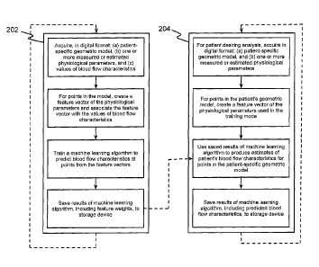
[012] FIG. 2 is a block diagram of an exemplary method for estimating
patient-specific blood flow characteristics from vessel geometry and
physiological
information, according to an exemplary embodiment of the present disclosure.
4b
CA 2832543 2017-07-21
CA 02882543 2015-02-19
WO 2014/042899 PCT/US2013/057546
DESCRIPTION OF THE EMBODIMENTS
[013] Reference will now be made in detail to the exemplary embodiments of
the disclosure, examples of which are illustrated in the accompanying
drawings.
Wherever possible, the same reference numbers will be used throughout the
drawings to refer to the same or like parts.
[014] The present disclosure describes certain principles and embodiments
for providing advantages over physics-based simulation of blood flow to
compute
patient-specific blood flow characteristics and clinically relevant quantities
of interest.
Namely, the presently disclosed systems and methods may incorporate machine
learning techniques to predict the results of a physics-based simulation. For
example, the present disclosure describes an exemplary, less processing-
intensive
technique, which may involve modeling the fractional flow reserve (FFR) as a
function of a patient's vascular cross-sectional area, diseased length, and
boundary
conditions. The cross-sectional area may be calculated based on lumen segment
and plaque segment, among other things. The diseased length may be calculated
based on plaque segment and stenosis location, among other things. The
boundary
conditions may reflect patient-specific physiology, such as coronary flow
(estimated
from myocardial mass), outlet area, and hyperemic assumptions, to reflect that
different patients have different geometry and physiologic responses.
[015] In one embodiment, fractional flow reserve may be modeled as a
function of a patient's boundary conditions (f(BCs)), and a function of a
patient's
vascular geometry (g(areaReductions)). Although the patient's geometry may be
described as a function of "areaReductions," it should be appreciated that
this term
refers, not just to changes in patient's vascular cross-sectional area, but to
any
physical or geometric characteristics affecting a patient's blood flow. In one
CA 02882543 2015-02-19
WO 2014/042899 PCT/US2013/057546
embodiment, FFR can be predicted by optimizing the functions "f" and "g" such
that
the difference between the estimated FFR (FFRc-r_scaiingLaw) and the measured
FFR
(mFFR) is minimized. In other words, machine learning techniques can be used
to
solve for the functions that cause the estimated FFR to approximate the
measured
FFR. In one embodiment, the measured FFR may be calculated by traditional
catheterized methods or by modern, computational fluid dynamics (CFD)
techniques.
In one embodiment, one or more machine learning algorithms may be used to
optimize the functions of boundary conditions and patient geometry for
hundreds or
even thousands of patients, such that estimates for FFR can reliably
approximate
measured FFR values. Thus, FFR values calculated by CFD techniques can be
valuable for training the machine learning algorithms.
[016] Referring now to the figures, FIG. 1 depicts a block diagram of an
exemplary system and network for estimating patient-specific blood flow
characteristics from vessel geometry and physiological information.
Specifically,
FIG. 1 depicts a plurality of physicians 102 and third party providers 104,
any of
whom may be connected to an electronic network 100, such as the Internet,
through
one or more computers, servers, and/or handheld mobile devices. Physicians 102
and/or third party providers 104 may create or otherwise obtain images of one
or
more patients' cardiac and/or vascular systems. The physicians 102 and/or
third
party providers 104 may also obtain any combination of patient-specific
information,
such as age, medical history, blood pressure, blood viscosity, etc. Physicians
102
and/or third party providers 104 may transmit the cardiac/vascular images
and/or
patient-specific information to server systems 106 over the electronic network
100.
Server systems 106 may include storage devices for storing images and data
received from physicians 102 and/or third party providers 104. Sever systems
106
6
CA 02882543 2015-02-19
WO 2014/042899 PCT/US2013/057546
may also include processing devices for processing images and data stored in
the
storage devices.
[017] FIG. 2 is a block diagram of an exemplary method for estimating
patient-specific blood flow characteristics from vessel geometry and
physiological
information, according to an exemplary embodiment of the present disclosure.
The
method of FIG. 2 may be performed by server systems 106, based on information
received from physicians 102 and/or third party providers 104 over electronic
network 100.
[018] In one embodiment, the method of FIG. 2 may include a training
method 202, for training one or more machine learning algorithms based on
numerous patients' blood flow characteristic estimates, and a production
method 204
for using the machine learning algorithm results to predict a particular
patient's blood
flow characteristics.
[019] In one embodiment, training method 202 may be performed based on
FFR estimates generating using CFD techniques for hundreds of patients.
Training
method 202 may involve acquiring, for each of a plurality of individuals,
e.g., in digital
format: (a) a patient-specific geometric model, (b) one or more measured or
estimated physiological parameters, and (c) values of blood flow
characteristics.
Training method 202 may then involve, for one or more points in each patient's
model, creating a feature vector of the patients' physiological parameters and
associating the feature vector with the values of blood flow characteristics.
For
example, training method 202 may associate an estimated FFR with every point
in a
patient's geometric model. Training method 202 may then train a machine
learning
algorithm (e.g., using processing devices of server systems 106) to predict
blood
flow characteristics at each point of a geometric model, based on the feature
vectors
7
CA 02882543 2015-02-19
WO 2014/042899 PCT/US2013/057546
and blood flow characteristics. Training method 202 may then save the results
of the
machine learning algorithm, including feature weights, in a storage device of
server
systems 106. The stored feature weights may define the extent to which patient
features or geometry are predictive of certain blood flow characteristics.
[020] In one embodiment, the production method 204 may involve estimating
FFR values for a particular patient, based on results of executing training
method
202. In one embodiment, production method 204 may include acquiring, e.g. in
digital format: (a) a patient-specific geometric model, and (b) one or more
measured
or estimated physiological parameters. For multiple points in the patient's
geometric
model, production method 204 may involve creating a feature vector of the
physiological parameters used in the training mode. Production method 204 may
then use saved results of the machine learning algorithm to produce estimates
of the
patient's blood flow characteristics for each point in the patient-specific
geometric
model. Finally, production method 204 may include saving the results of the
machine learning algorithm, including predicted blood flow characteristics, to
a
storage device of server systems 106.
[021] Described below are general and specific exemplary embodiments for
implementing a training mode and a production mode of machine learning for
predicting patient-specific blood flow characteristics, e.g. using server
systems 106
based on images and data received from physicians 102 and/or third party
providers
104 over electronic network 100.
GENERAL EMBODIMENT
[022] In a general embodiment, server systems 106 may perform a training
mode based on images and data received from physicians 102 and/or third party
providers 104 over electronic network 100. Specifically, for one or more
patients,
8
CA 02882543 2015-02-19
WO 2014/042899 PCT/US2013/057546
server systems 106 may acquire a digital representation (e.g., the memory or
digital
storage [e.g., hard drive, network drive] of a computational device such as a
computer, laptop, DSP, server, etc.) of the following items: (a) a patient-
specific
model of the geometry for one or more of the patient's blood vessels; (b) a
list of one
or more measured or estimated physiological or phenotypic parameters of the
patient; and/or (c) measurements, estimations or simulated values of all blood
flow
characteristic being targeted for prediction. In one embodiment, the patient-
specific
model of the geometry may be represented by a list of points in space
(possibly with
a list of neighbors for each point) in which the space can be mapped to
spatial units
between points (e.g., millimeters). In one embodiment, the list of one or more
measured or estimated physiological or phenotypic parameters of the patient
may
include blood pressure, blood viscosity, patient age, patient gender, mass of
the
supplied tissue, etc. These patient-specific parameters may be global (e.g.,
blood
pressure) or local (e.g., estimated density of the vessel wall at a particular
location).
[023] For every point in the patient-specific geometric model for which there
is a measured, estimated or simulated value of the blood flow characteristic,
server
systems 106 may then create a feature vector for that point. The feature
vector may
be a numerical description of the patient-specific geometry at that point and
estimates of physiological or phenotypic parameters of the patient. The
feature
vector may contain both global and local physiological or phenotypic
parameters,
where: for global parameters, all points have the same numerical value; and
for
local parameters, the value(s) may change at different points in the feature
vector.
Server systems 106 may then associate this feature vector with the measured,
estimated or simulated value of the blood flow characteristic at this point.
9
CA 02882543 2015-02-19
WO 2014/042899 PCT/US2013/057546
[024] Server systems 106 may then train a machine learning algorithm to
predict the blood flow characteristics at the points from the feature vectors
at the
points. Examples of machine learning algorithms that can perform this task are
support vector machines (SVMs), multi-layer perceptrons (MLPs), and
multivariate
regression (MVR) (e.g., weighted linear or logistic regression). Server
systems 106
may then save the results of the machine learning algorithm (e.g., feature
weights) to
a digital representation (e.g., the memory or digital storage [e.g., hard
drive, network
drive] of a computational device such as a computer, laptop, DSP, server,
etc.).
[025] Also in a general embodiment, server systems 106 may perform a
production mode based on images and data received from physicians 102 and/or
third party providers 104 over electronic network 100. For a patient on whom a
blood flow analysis is to be performed, server systems 106 may acquire a
digital
representation (e.g., the memory or digital storage [e.g., hard drive, network
drive] of
a computational device such as a computer, laptop, DSP, server, etc.) of (a) a
patient-specific model of the geometry for one or more of the patient's blood
vessels;
and (b) a list of one or more estimates of physiological or phenotypic
parameters of
the patient. In one embodiment, the patient-specific model of the geometry for
one
or more of the patient's blood vessels may be represented as a list of points
in space
(possibly with a list of neighbors for each point) in which the space can be
mapped to
spatial units between points (e.g., millimeters). The list of one or more
estimates of
physiological or phenotypic parameters of the patient, may include blood
pressure,
blood viscosity, patient age, patient gender, the mass of the supplied tissue,
etc.
These parameters may be global (e.g., blood pressure) or local (e.g.,
estimated
density of the vessel wall at a location). This list of parameters must be the
same as
the list used in the training mode.
CA 02882543 2015-02-19
WO 2014/042899 PCT/US2013/057546
[026] For every point in the patient-specific geometric model, server systems
106 may create a feature vector that consists of a numerical description of
the
geometry and estimates of physiological or phenotypic parameters of the
patient.
Global physiological or phenotypic parameters can be used in the feature
vector of
all points and local physiological or phenotypic parameters can change in the
feature
vector of different points. These feature vectors may represent the same
parameters
used in the training mode. Server systems 106 may then use the saved results
of
the machine learning algorithm produced in the training mode (e.g., feature
weights)
to produce estimates of the blood flow characteristics at each point in the
patient-
specific geometric model. These estimates may be produced using the same
machine learning algorithm technique used in the training mode (e.g., the SVM,
MLP, MVR technique). Server systems 106 may also save the predicted blood flow
characteristics for each point to a digital representation (e.g., the memory
or digital
storage [e.g., hard drive, network drive] of a computational device such as a
computer, laptop, DSP, server, etc.).
EXEMPLARY EMBODIMENT
[027] In one exemplary embodiment, server systems 106 may perform a
training mode based on images and data received from physicians 102 and/or
third
party providers 104 over electronic network 100. Specifically, for one or more
patients, server systems 106 may acquire a digital representation (e.g., the
memory
or digital storage [e.g., hard drive, network drive] of a computational device
such as a
computer, laptop, DSP, server, etc.) of (a) a patient-specific model of the
geometry
for the patient's ascending aorta and coronary artery tree; (b) a list of
measured or
estimated physiological or phenotypic parameters of the patient; and (c)
measurements of the FFR when available.
11
CA 02882543 2015-02-19
WO 2014/042899 PCT/US2013/057546
[028] In one embodiment, the patient-specific model of the geometry for the
patient's ascending aorta and coronary artery tree may be represented as a
list of
points in space (possibly with a list of neighbors for each point) in which
the space
can be mapped to spatial units between points (e.g., millimeters). This model
may
be derived by performing a cardiac CT imaging study of the patient during the
end
diastole phase of the cardiac cycle. The resulting CT images may then be
segmented manually or automatically to identify voxels belonging to the aorta
and to
the lumen of the coronary arteries. Once all relevant voxels are identified,
the
geometric model can be derived (e.g., using marching cubes).
[029] In one embodiment, the list of measured or estimated physiological or
phenotypic parameters of the patient may be obtained and may include: (i)
systolic
and diastolic blood pressures; (ii) heart rate; (iii) hematocrit level; (iv)
patient age,
gender, height, weight, general health status (presence or absence of
diabetes,
current medications); (v) lifestyle characteristics: smoker/non-smoker; and/or
(vi)
myocardial mass (may be derived by segmenting the myocardium obtained during
the CT imaging study and then calculating the volume in the image; the mass is
then
computed using the computed volume and an estimated density (1.05g/mL) of the
myocardial mass.
[030] In one embodiment, measurements of the FFR may be obtained when
available. If the measured FFR value is not available at a given spatial
location in
the patient-specific geometric model, then a numerically computed value of the
FFR
at the point may be used. The numerically computed values may be obtained from
a
previous CFD simulation using the same geometric model and patient-specific
boundary conditions derived from the physiological and phenotypic parameters
listed
above.
12
CA 02882543 2015-02-19
WO 2014/042899 PCT/US2013/057546
[031] For every point in the patient-specific geometric model for which there
is a measured, estimated or simulated value of the blood flow characteristics,
server
systems 106 may create a feature vector for that point that contains a
numerical
description of physiological or phenotypic parameters of the patient and a
description
of the local geometry. Specifically the feature vector may contain: (i)
systolic and
diastolic blood pressures; (ii) heart rate; (iii) blood properties including:
plasma, red
blood cells (erythrocytes), hematocrit, white blood cells (leukocytes) and
platelets
(thrombocytes), viscosity, yield stress; (iv) patient age, gender, height,
weight, etc.;
(v) diseases: presence or absence of diabetes, myocardial infarction,
malignant and
rheumatic conditions, peripheral vascular conditions, etc.; (vi) lifestyle
characteristics: presence or absence of current medications/drugs, smoker/non-
smoker; (vii) characteristics of the aortic geometry (Cross-sectional area of
the aortic
inlet and outlet, Surface area and volume of the aorta, Minimum, maximum, and
average cross-sectional area, etc.); (viii) characteristics of the coronary
branch
geometry; and (ix) one or more feature sets.
[032] In one embodiment, the characteristics of the coronary branch
geometry may include: (i) volumes of the aorta upstream/downstream of the
coronary branch point; (ii) cross-sectional area of the coronary/aorta
bifurcation
point, i.e., inlet to the coronary branch; (iii) total number of vessel
bifurcations, and
the number of upstream/downstream vessel bifurcations; (iv) average, minimum,
and
maximum upstream/downstream cross-sectional areas; (v) distances (along the
vessel centerline) to the centerline point of minimum and maximum
upstream/downstream cross-sectional areas; (vi) cross-sectional of and
distance
(along the vessel centerline) to the nearest upstream/downstream vessel
bifurcation;
(vii) cross-sectional area of and distance (along the vessel centerline) to
the nearest
13
CA 02882543 2015-02-19
WO 2014/042899 PCT/US2013/057546
coronary outlet and aortic inlet/outlet; (viii) cross-sectional areas and
distances
(along the vessel centerline) to the downstream coronary outlets with the
smallest/largest cross-sectional areas; (ix) upstream/downstream volumes of
the
coronary vessels; and (x) upstream/downstream volume fractions of the coronary
vessel with a cross-sectional area below a user-specified tolerance.
[033] In one embodiment, a first feature set may define cross-sectional area
features, such as a cross-sectional lumen area along the coronary centerline,
a
powered cross-sectional lumen area, a ratio of lumen cross-sectional area with
respect to the main ostia (LM, RCA), a powered ratio of lumen cross-sectional
area
with respect to the main ostia, a degree of tapering in cross-sectional lumen
area
along the centerline, locations of stenotic lesions, lengths of stenotic
lesions,
location and number of lesions corresponding to 50%, 75%, 90% area reduction,
distance from stenotic lesion to the main ostia, and/or irregularity (or
circularity) of
cross-sectional lumen boundary.
[034] In one embodiment, the cross-sectional lumen area along the coronary
centerline may be calculated by extracting a centerline from constructed
geometry,
smoothing the centerline if necessary, and computing cross-sectional area at
each
centerline point and map it to corresponding surface and volume mesh points.
In
one embodiment, the powered cross-sectional lumen area can be determined from
various source of scaling laws. In one embodiment, the ratio of lumen cross-
sectional area with respect to the main ostia (LM, RCA) can be calculated by
measuring cross-sectional area at the LM ostium, normalizing cross-sectional
area of
the left coronary by LM ostium area, measuring cross-sectional area at the RCA
ostium, and normalizing cross-sectional area of the right coronary by RCA
ostium
area. In one embodiment, the powered ratio of lumen cross-sectional area with
14
CA 02882543 2015-02-19
WO 2014/042899 PCT/US2013/057546
respect to the main ostia can be determined from various source of scaling
laws. In
one embodiment, the degree of tapering in cross-sectional lumen area along the
centerline can be calculated by sampling centerline points within a certain
interval
(e.g., twice the diameter of the vessel) and compute a slope of linearly-
fitted cross-
sectional area. In one embodiment, the location of stenotic lesions can be
calculated
by detecting minima of cross-sectional area curve, detecting locations where
first
derivative of area curve is zero and second derivative is positive, and
computing
distance (parametric arc length of centerline) from the main ostium. In one
embodiment, the lengths of stenotic lesions can be calculated by computing the
proximal and distal locations from the stenotic lesion, where cross-sectional
area is
recovered.
[035] In one embodiment, another feature set may include intensity features
that define, for example, intensity change along the centerline (slope of
linearly-fitted
intensity variation). In one embodiment, another feature set may include
surface
features that define, for example, 3D surface curvature of geometry (Gaussian,
maximum, minimum, mean). In one embodiment, another feature set may include
volume features that define, for example, a ratio of total coronary volume
compared
to myocardial volume. In one embodiment, another feature set may include
centerline features that define, for example, curvature (bending) of coronary
centerline, e.g., by computing Frenet curvature:
[036] K = -Ip'13, where p is coordinate of centerline
[037] or by computing an inverse of the radius of circumscribed circle along
the centerline points. Curvature (bending) of coronary centerline may also be
calculated based on tortuosity (non-planarity) of coronary centerline, e.g.,
by
computing Frenet torsion :
CA 02882543 2015-02-19
WO 2014/042899 PCT/US2013/057546
(P'xP")13-
[038] , where p is coordinate of centerline
lp,x73-12
[039] In one embodiment, another feature set may include a SYNTAX
scoring feature, including, for example, an existence of aorto ostial lesion,
detection
of a lesion located at the origin of the coronary from the aorta; and/or
dominance (left
or right).
[040] In one embodiment, another feature set may include a simplified
physics feature, e.g., including a fractional flow reserve value derived from
Hagen-
Poisseille flow assumption (Resistance¨Area-2). For example, in one
embodiment,
server systems 106 may compute the cross-sectional area of the origin (LM
ostium
or RCA ostium) of the coronary from the aorta (A0) with aortic pressure (P0);
compute cross-sectional area of coronary vessel (Ai) at each sampled interval
(L,);
determine the amount of coronary flow in each segment of vessel using
resistance
boundary condition under hyperemic assumption (Qi); estimate resistance at
each
sampled location (R1) based on:
8/4/.=
' p[041] Ri =a = --4 i, where:
TrZi
[042] Nominal value = dynamic viscosiy of blood, ai = 1.0, 3i = 0, yi =
2.0 (Hagen ¨ Poisseille).
[043] Server systems 106 may estimate pressure drop (LP) as APi =
-
QiRi and compute FFR at each sampled location as FFR, = Po-EAP k. Locations of
Po
cross-sectional area minima or intervals smaller than vessel radius may be
used for
sampling locations. Server systems 106 may interpolate FFR along the
centerline
using FFRi, project FFR values to 3D surface mesh node, and vary ai, . y, and
obtain new sets of FFR estimation as necessary for training, such as by using
the
feature sets defined above to perturb parameters where a1,,G1 can be a
function of
16
CA 02882543 2015-02-19
WO 2014/042899 PCT/US2013/057546
the diseased length, degree of stenosis and tapering ratio to account for
tapered
vessel; and Qi can be determined by summing distributed flow of each outlet on
the
basis of the same scaling law as the resistance boundary condition
(outlet resistance a outlet area-1.35). However, a new scaling law and
hyperemic
assumption can be adopted, and this feature vector may be associated with the
measurement or simulated value of the FFR at that point. Server systems 106
may
also train a linear SVM to predict the blood flow characteristics at the
points from the
feature vectors at the points; and save the results of the SVM as a digital
representation (e.g., the memory or digital storage [e.g., hard drive, network
drive] of
a computational device such as a computer, laptop, DSP, server, etc.).
[044] In an exemplary production mode, servers systems 106 may, for a
target patient, acquire in digital representation (e.g., the memory or digital
storage
(e.g., hard drive, network drive) of a computational device such as a
computer,
laptop, DSP, server, etc.): (a) a patient-specific model of the geometry for
the
patient's ascending aorta and coronary artery tree; and (b) a list of
physiological and
phenotypic parameters of the patient obtained during training mode. In one
embodiment, the patient-specific model of the geometry for the patient's
ascending
aorta and coronary artery tree may be represented as a list of points in space
(possibly with a list of neighbors for each point) in which the space can be
mapped to
spatial units between points (e.g., millimeters). This model may be derived by
performing a cardiac CT imaging of the patient in the end diastole phase of
the
cardiac cycle. This image then may be segmented manually or automatically to
identify voxels belonging to the aorta and the lumen of the coronary arteries.
Once
the voxels are identified, the geometric model can be derived (e.g., using
marching
cubes). The process for generating the patient-specific model of the geometry
may
17
CA 02882543 2015-02-19
WO 2014/042899 PCT/US2013/057546
be the same as in the training mode. For every point in the patient-specific
geometric model, the server systems 106 may create a feature vector for that
point
that consists of a numerical description of the geometry at that point and
estimates of
physiological or phenotypic parameters of the patient. These features may be
the
same as the quantities used in the training mode. The server systems 106 may
then
use the saved results of the machine learning algorithm produced in the
training
mode (e.g., feature weights) to produce estimates of the FFR at each point in
the
patient-specific geometric model. These estimates may be produced using the
same
linear SVM technique used in the training mode. The server systems 106 may
save
the predicted FFR values for each point to a digital representation (e.g., the
memory
or digital storage [e.g., hard drive, network drive] of a computational device
such as a
computer, laptop, DSP, server, etc.).
[045] In one embodiment, the above factors (i) thru (viii) ("Systolic and
diastolic blood pressures" thru "Characteristics of the coronary branch
geometry")
may be considered global features, which are applicable to all points within a
given
patient's geometric model. Also, items (ix) thru (xv) ("Feature Set I: Cross-
sectional
area feature" thru "Feature Set VII: Simplified Physics feature") may be
considered
features that are local to specific points within a given patient's geometric
model. In
addition, features (i) thru (vi) may be considered variables within the
function of
boundary conditions, f(BCs), while features (vii) thru (xv) may be considered
variables within the function of geometry, g(areaReductions), on that page. It
will be
appreciated that any combination of those features, modified by any desired
weighting scheme, may be incorporated into a machine learning algorithm
executed
according to the disclosed embodiments.
18
CA 02882543 2015-06-03
[046] Other embodiments of the invention will be apparent to those skilled
in the art from consideration of the specification and practice of the
invention
disclosed herein. The invention, rather, is defined by the claims.
19