Note: Descriptions are shown in the official language in which they were submitted.
CA 02882561 2015-02-19
WO 2014/031612 PCT/US2013/055749
ASSAYS, SYSTEMS, AND METHODS FOR OBTAINING PERSONALIZED
ANABOLIC PROFILES
CROSS REFERENCE TO RELATED APPLICATIONS
[0001] This application claims the benefit under 35 U.S.C. 119(e) of
U.S.
Provisional Application No. 61/684,959 filed August 20, 2012, the content of
which is
incorporated herein by reference in its entirety.
TECHNICAL FIELD
[0002] The inventions provided herein generally relate to assays,
systems, and kits for
generating a personalized anabolic profile, which can be used in diagnostics,
therapeutic
and/or nutritional decision support. The inventions also relate to methods for
selecting a
treatment regimen for a subject determined to have a musculoskeletal disease
or disorder.
BACKGROUND OF THE DISCLOSURE
[0003] Loss of muscle mass is increasingly common in the aging population
and is
present in a variety of debilitating muscle wasting associated disease. It can
also be
associated with chemotherapy, and weight loss. Muscle wasting and bone loss
can reduce
quality of life, increase risk for mortality and pose a substantial burden on
the healthcare
system. For example, in industrialized countries (e.g., North America, Europe,
Japan), the
overall prevalence of cachexia (due to any disease) is growing and currently
about 1%, i.e.,
about nine million patients. Musculoskeletal wasting includes both muscle
decline and/or
bone loss. Muscle decline indications include, for example, HIV associated
wasting, cachexia
and muscular dystrophy. Bone loss indications include, for example, HIV
associated bone
loss, osteopenia and osteoporosis. For HIV/AIDS, the United States has a 0.6%
rate of
prevalence, with approximately 1.2 million HIV infected individuals. For
cachexia, in the
United States, cancer cachexia affects more than 1.3 million people (-30% of
all individuals
with cachexia) (C.C.M.H. Group, 2012). The total number of individuals
inflicted with
cachexia is ¨ 4 million people in the US. For muscular dystrophy, about 500 -
600 male
newborns are usually diagnosed with muscular dystrophy each year in the US.
Osteoporosis
is a major health risk for about 28 million Americans. In the United States,
about 10 million
individuals have osteoporosis and about 18 million more have low bone mass
(NIAMS,
2012).
1
CA 02882561 2015-02-19
WO 2014/031612 PCT/US2013/055749
[0004] Current diagnostic and treatment procedures do not distinguish
between
different types and/or causes of musculoskeletal decline and do not provide
guidance for
anabolic therapy. For example, existing diagnostics include measurement of
creatine kinase
(CK) and phosphocreatine (PCr) in muscle biopsies as a measure of muscle
decline. Urine
urea is used to infer rapid loss of muscle. Electromyography (EMG) is used to
measure
neuromuscular function using a surface electrode. Standardized reference
values for body
composition (e.g., National Health and Nutrition Examination Survey (NHANES))
are used
in assessing loss of skeletal muscle. Body composition assessments through the
use of
computed tomography (CT) image analysis or dual-energy X-ray absorptiometry
(DXA)
can be used to quantify loss of skeletal muscle or bone, respectively. While
there is a desire
to use less invasive procedures such as CT or DXA, these less-invasive
procedures (e.g., CT
or DXA) do not provide information about muscle function or personalized
anabolic
therapeutic options.
[0005] The current standard of care for muscle wasting is to provide
nutritional
supplementation and anabolic supplements such as testosterone, analogs of
testosterone (e.g.,
DHT) growth hormone or analogs of growth hormone. The current standard of care
for bone
loss is vitamin D supplementation, the use of bisphosphonates and/or bone
resorption
antagonists. The relative anabolic efficacy of these compounds -on a patient
specific level- is
unknown and not currently part of routine care. Human genetic variation and
life history
influence, often unpredictably, the response to therapeutic intervention. For
example, HIV
progression and response to drugs can be influenced by genetic polymorphisms
(A. Telenti et
al., 2008 Annu. Rev. Pharmacol. Toxicol. 48: 227). Cancer cachexia can vary in
severity and
response (Tan B. H. et al. 2011 J. Genet 90: 165). Muscular dystrophy can vary
in
progression and responsiveness (Pegoraro E. et al., 2010 Neurology 76: 219).
Additionally,
although anabolic compounds promote mass gains in muscle and/or bone, they do
so with
varying efficacy that depends on many factors, including age (See, e.g.,
Banerjee C. et al.,
2011 Immun. Aging 8:5). These diagnostic-treatment deficiencies limit
treatment options to
help guide patient and disease specific treatment.
[0006] In 2010 over 18 million people in the United States were estimated
to suffer
from age-associated decline in muscle mass and function (e.g., sarcopenia).
Although
anabolic supplementation has been used in treating sarcopenia, HIV muscle
wasting and
cachexia, anabolic responses vary in individuals and there are consistently 15-
25% of
individuals that do not benefit and are in effect non-responders. See, e.g.,
Montano et al.
2
CA 02882561 2015-02-19
WO 2014/031612 PCT/US2013/055749
Journal of Clinical Endocrinology & Metabolism, 2007, 92 (7): 2793-2802;
Sardar et al. HIV
Clin Trials, 2010 July-Aug, 11(4): 220-229; and Houtman et al., Anal Chim
Acta, 2009,
637(1-2): 247-258. The financial burden on healthcare is predicted to increase
as the US
population ages, in part due to loss in muscle mass and the associated
increased risk for
functional decline. This anticipated burden poses an immediate challenge to
identify effective
diagnostic tools that better match patients with treatment options.
Accordingly, there is a
need for development of an assay or a diagnostic test that can be used to
provide a more
personalized guidance on anabolic therapy, thus improving the treatment
outcome and/or
response rate.
SUMMARY
[0007] Current diagnostic and treatment procedures do not distinguish
between
different types and/or causes of musculoskeletal decline and do not provide
guidance for
anabolic therapy specific to an individual. For example, there are no
predicate products (e.g.,
FDA approved medical device) or commercially-available kits, which can be used
to guide a
personalized or stratified anabolic therapy. Conventional diagnostics
typically measure
muscle loss, and do not indicate type of deficit or responsiveness to anabolic
alternatives.
Accordingly, there is a need for development of assays, methods and/or kits
that can provide
a personalized or stratified, and scalable, anabolic guide to maintain muscle
health and/or
optimize treatment of musculoskeletal wasting, thus improving health outcomes
and reducing
healthcare costs associated with musculoskeletal decline in muscle and/or bone
disease as
well as in aging.
[0008] The inventions provided herein generally relate to assays,
methods, systems,
and kits, e.g., for profiling anabolic responses of a subject or a population
subgroup to a panel
of anabolic agents or compositions selected to maintain and/or increase muscle
and/or bone
growth. The assays, methods, systems, and kits described herein, in part, rely
on ranking
relative efficacies of the anabolic agents or compositions in stimulating
muscle and/or bone
growth of subject-specific cells (i.e., patient-specific cells) or a panel of
cells representing
different population subgroups, thereby generating a personalized or
stratified diagnostic
report. In some embodiments, the panel of cells representing different
population subgroups
can be stratified based one or a plurality of (e.g., at least two or more)
feature(s) (e.g.,
phenotypic feature(s) including, but not limited to, age, gender, body mass
index (BMI),
condition, and/or ethnicity) of the population subgroups. Thus, an anabolic
agent or
composition can be selected, recommended and/or optionally administered to a
subject or a
3
CA 02882561 2015-02-19
WO 2014/031612 PCT/US2013/055749
patient in need thereof based on the personalized or stratified diagnostic
analysis. In some
embodiments, an anabolic agent or composition can be selected, recommended
and/or
optionally administered to a subject or patient based on a subject-specific or
patient-specific
anabolic profile generated using his /or own biopsy and/or blood sample. In
alternative
embodiments, an anabolic agent or composition can be selected, recommended
and/or
optionally administered to a subject or patient based on a stratified anabolic
profile generated
using tissue specimens of a matching population subgroup as the subject,
wherein the subject
is matched or associated to an appropriate population subgroup based on one or
a plurality of
pre-determined feature(s) such as phenotypic feature(s). By way of example
only, for a 30-
year old diabetic Caucasian woman who is in need of muscle augmentation and/or
mitigation
of muscle or bone loss, an optimal anabolic agent or composition can be
selected for her
based on a personalized anabolic profiling (which requires her own biopsy or
blood sample)
and/or a stratified anabolic profiling of a population subgroup of about 30-
year old (e.g., 25-
35-year old) diabetic Caucasian women.
[0009] Unlike the existing diagnostic methods such as CT and MRI, or
blood
biomarkers such as creatine phosphor-kinase (CPK), the generated functional
anabolic
profiles can provide information about muscle and/or bone function in response
to a variety
of anabolic agents or compositions, which can in turn be used to make
diagnostic, and/or
therapeutic or prophylactic decisions. For example, in some embodiments, the
generated
functional anabolic profiles can be used to diagnose an anabolic deficiency,
and/or a defect in
and/or an imbalance between anabolic growth pathway(s) in a subject. In some
embodiments,
the generated functional anabolic profiles can be used to identify and/or
optimize a
therapeutic or nutritional option for treatment of a muscle wasting-associated
disease or
disorder. In other embodiments, the generated functional anabolic profiles can
be used to
identity and/or optimize a prophylactic option to prevent or mitigate muscle
loss and to
optimize and maintain muscle heath. Accordingly, methods for diagnosing,
treating and/or
preventing muscle wasting or a musculoskeletal disease or disorder in a
subject are also
provided herein.
[0010] In one aspect, provided herein relates to cell-based assays, e.g.,
which can be
carried out to obtain personalized or stratified anabolic profiles. The assay
comprises: (a)
contacting a population of musculoskeletal cells or precursor cells thereof
with a plurality of
test compositions each comprising at least one agent selected to increase
and/or maintain
muscle and/or bone growth, to profile anabolic responses of the cells to the
test compositions;
4
CA 02882561 2015-02-19
WO 2014/031612 PCT/US2013/055749
(b) subjecting the musculoskeletal cells or precursor cells thereof to at
least one analysis,
including, e.g., at least two analyses, to quantify muscle growth and/or bone
growth of the
musculoskeletal cells or precursor cells in response to the test compositions;
and (c) ranking
anabolic efficacy of the plurality of the test compositions based on the
quantified muscle
growth and/or bone growth, thereby providing anabolic profiles for muscle
and/or bone
growth of the assayed cells.
[0011] In some embodiments, the musculoskeletal cells or precursor cells
thereof for
use in the assay described herein can be obtained or derived from a muscle
biopsy or a blood
sample of a subject or patient seeking for an anabolic treatment or
supplement. In one
embodiment, muscle stem cells derived from a subject's biopsy or blood sample
can be
subjected to the assay described herein. Thus, the generated anabolic profile
for muscle
growth and/or bone growth can be personalized to the specific subject or
patient.
[0012] In alternative embodiments, the musculoskeletal cells or precursor
cells
thereof for use in the assay described herein can be obtained or derived from
cells or tissue
specimens representing one or more different population subgroups. The cells
or tissue
specimens representing one or more different population subgroups can be
obtained from a
cell or tissue depository. In these embodiments, a stratified anabolic profile
of a population
subgroup that shares at least one or more features with a subject seeking for
an anabolic
treatment and/or supplement can be used to determine an optimal treatment
and/or
supplement for the subject. Examples of a feature can be a phenotypic feature
for population
stratification including, but not limited to, age groups, gender, ethnicity,
body types, body
mass index (BMI), blood types, activity levels, a condition such as chronic or
acute diseases
and/or psychophysiological disorders, genetic polymorphisms, diet, drug
resistance, treatment
regime such as chemotherapy, drastic/abnormal weight loss, geographical
location, and any
combinations thereof In some embodiments, the stratification can be performed
based on age
and gender.
[0013] In some embodiments, the musculoskeletal cells or precursor cells
thereof for
use in the assay described herein can be obtained or derived from a biological
sample (e.g.,
but not limited to, a muscle biopsy and/or blood sample) of subjects or
individuals who are
determined to suffer from or have a risk for muscle loss and/or bone loss
(e.g., a
musculoskeletal disease or disorder). Examples of subjects or individuals who
are at risk for a
musculoskeletal disease or disorder include, but are not limited to, athletes,
aging individuals,
individuals having a chronic disease or disorder (e.g., but not limited to,
cancer, chronic
CA 02882561 2015-02-19
WO 2014/031612 PCT/US2013/055749
obstructive pulmonary disease (COPD), chronic kidney disease (CKD), chronic
liver failure
(CLF), and chronic infections), individuals suffering from malnutrition, or
any combinations
thereof. Examples of a musculoskeletal disease or disorder include, but are
not limited to,
muscle loss, muscle wasting, muscle wasting associated with HIV infection,
muscle wasting
in cancer survivors, cachexia, muscular dystrophy, osteopenia, osteoporosis,
sarcopenia, an
age-related musculoskeletal disease or disorder, or a musculoskeletal disease
or disorder
associated with anabolic resistance, a musculoskeletal disease or disorder
associated with
excessive weight loss, or any combinations thereof
[0014] In some embodiments, the musculoskeletal cells or precursor cells
thereof for
use in the assay described herein can be obtained or derived from a biological
sample of
subjects or individuals who have previously shown non-responsiveness or
resistance to at
least one or more anabolic agents.
[0015] In some embodiments, the musculoskeletal cells or precursor cells
thereof for
use in the assay described herein can be obtained or derived from a biological
sample of
subjects or individuals who are seeking to maintain and/or enhance muscle
and/or bone
health.
[0016] In order to determine specific anabolic responses (e.g., muscle
growth or bone
growth) of the musculoskeletal cells or precursor cells thereof to one or more
test
compositions, the cells are cultured in appropriate conditions optimized for
each specific
anabolic response (e.g., muscle growth or bone growth). For example, to
determine the
muscle growth-response of the musculoskeletal cells or precursor cells thereof
to a plurality
of test compositions, it can be desirable to culture and/or maintain the cells
in a muscle cell-
specific condition (e.g., a condition optimal to muscle cell differentiation)
during the contact
with the test compositions. In some embodiments, the muscle cell-specific
condition can
include culturing in a substrate material with a defined stiffness optimal to
muscle cell
differentiation. For example, the muscle cell-specific condition can include
culturing in a
substrate material with a defined stiffness of about 5 kPa to about 50 kPa, or
about 10 kPa to
about 20 kPa.
[0017] Similarly, to determine the bone growth-response of subject-
specific cells to a
plurality of test compositions, it can be desirable to culture and/or maintain
the
musculoskeletal cells or precursor cells thereof in a bone cell-specific
condition (e.g., a
condition optimal to bone cell differentiation) during the contact with the
test compositions.
6
CA 02882561 2015-02-19
WO 2014/031612 PCT/US2013/055749
In some embodiments, the bone cell-specific condition can include culturing in
a substrate
material with a defined stiffness optimal to bone differentiation. For
example, the bone cell-
specific condition can include culturing in a substrate material with a
defined stiffness of
about 10 kPa to about 150 kPa, or about 20 kPa to about 100 kPa. While not
necessary, in
some embodiments, the bone cell-specific condition can further include
culturing in the
presence of a bone formation-inducing agent. Examples of a bone formation-
inducing agent
can include, but are not limited to, bone morphogenic factor (BMP) (e.g., BMP-
1, BMP-2,
BMP-3, BMP-4, BMP-5 and BMP-6), transforming growth factor (TGF), insulin-like
growth
factor (IGF), basic fibroblast growth factor (bFBF), osteogenic protein (OP)
(e.g., OP-1, OP-
2 and OP-3), osteogenic factors, osteoconductive factors, osteoinductive
factors, and any
combinations thereof In one embodiment, the bone formation-inducing agent can
include
bone morphogenetic protein-2 (BMP-2).
[0018] Quantitation of anabolic responses (e.g., muscle growth and/or
bone growth)
can be performed by any methods known in the art. For example, muscle growth
of a subset
of the musculoskeletal or precursor cells thereof in response to a test
composition can be
quantified based on a distribution of the number of nuclei per cell. The
number of nuclei per
cell can be determined, for example, by cell imaging. Thus, in one embodiment,
an increase
in muscle growth of the musculoskeletal or precursor cells thereof induced by
a test
composition can be quantified by an increase in the number of multi-nucleated
cells formed
by fusion of the musculoskeletal cells or precursor cells thereof (e.g.,
mononucleated muscle
cells), as compared to a condition without the test composition.
[0019] Bone growth can be characterized by differentiation of muscle
cells or bone
precursor cells thereof to bone cells. For example, in some embodiments, an
increase in bone
growth of the musculoskeletal or precursor cells thereof induced by a test
composition can be
characterized by an increase in the number of bone cells differentiated from
the
musculoskeletal cells or precursor cells thereof (e.g., muscle cells or bone
precursor cells
thereof), as compared to a condition without the test composition. Any art-
recognized
methods can be used to characterize bone differentiation. For example, in one
embodiment,
the bone cells can be identified by detecting expression of a bone marker. An
exemplary bone
marker includes, but not limited to, alkaline phosphatase (ALP), type I
collagen propetides,
osetocalcin, and any combinations thereof.
[0020] The test compositions used in the assay described herein can each
independently comprise one or more agents selected to increase and/or maintain
muscle
7
CA 02882561 2015-02-19
WO 2014/031612 PCT/US2013/055749
and/or bone growth. In some embodiments, at least some of the test
compositions can
comprise two or more agents selected to increase and/or maintain muscle and/or
bone growth.
The agent(s) included in the test compositions can include a therapeutic agent
that has already
been indicated for anabolic treatment (e.g., FDA-approved anabolic drugs or
over-the-counter
anabolic drugs), off-label FDA-approved drugs or over-the-counter drugs, an
anabolic
supplement, a candidate agent to be assessed for its anabolic efficacy, or any
combinations
thereof. Thus, in some embodiments, the assays described herein can be used to
identify a
novel anabolic compound or a novel combination of anabolic compounds suitable
for a
subject's or a population subgroup's musculoskeletal condition. In other
embodiments, the
assays described herein can be used to select or optimize a treatment regimen
for a subject
with a musculoskeletal condition, e.g., selecting a specific anabolic agent or
combination
therapy that stimulate muscle and bone growth in the subject, and/or
optimizing the dosage
and/or administration schedule of the selected anabolic agent(s) for a
personalized treatment.
Accordingly, in some embodiments, the assay can further comprise identifying
or selecting at
least one of the test compositions for administration to the subject, wherein
the at least one of
the test compositions is selected based on the rankings of their anabolic
efficacies in the
assay. The selected test composition for administration to the subject can
provide a
therapeutic effect for treatment of a musculoskeletal disease or disorder in a
subject, or a
prophylactic effect for optimizing and maintaining muscle health in a subject.
[0021] In some embodiments, the agent(s) included in the test
compositions can
include a molecule that is involved in an anabolic growth pathway. Examples of
an anabolic
growth pathway can include, but are not limited to, an amino acid pathway, an
androgen
receptor (AR): testosterone (T) pathway, a Wnt pathway, a calcium pathway, an
IGF
pathway, an insulin pathway, a follistatin pathway, a growth hormone pathway,
an adhesion
G-protein coupled receptor (GPCR) pathway, a myostatin pathway, and a FGF
pathway. By
way of example only, if the musculoskeletal or precursor cells thereof
corresponding to a
subject or a population subgroup do not respond to a subset of test
compositions associated
with a specific anabolic growth pathway, the subject can be diagnosed for
having a defect in
the specific anabolic pathway, or a disease or disorder associated with the
defective anabolic
pathway. Accordingly, in some embodiments, the assay can further comprise
identifying or
diagnosing an anabolic deficiency or a defect in or an imbalance among
anabolic pathways in
a subject or population subgroup based on the anabolic responses of the
respective cells to the
test compositions.
8
CA 02882561 2015-02-19
WO 2014/031612 PCT/US2013/055749
[0022] In some embodiments, the assays described herein can be employed
as part of
a clinical decision support to optimize or select a treatment regimen for a
subject determined
to have, or have a risk for, a musculoskeletal disease or disorder.
Accordingly, another aspect
provided herein relates to a method of optimizing or selecting a treatment
regimen for a
subject determined to have, or have a risk for, a musculoskeletal disease or
disorder. The
method comprises subjecting the musculoskeletal cells or precursor cells
thereof obtained or
derived from a subject determined to have, or have a risk for, a
musculoskeletal disease or
disorder, or a group of individuals sharing a similar background and symptoms
as the subject,
to one or more embodiments of the assay described herein. The test
compositions can be
ranked based on its efficacy to stimulate muscle and/or bone growth as
determined in the
assay. If some of the test compositions show an anabolic efficacy above a pre-
determined
threshold (e.g., anabolic response of the musculoskeletal or precursor cells
thereof in the
absence of the test composition), at least one of those test compositions can
be selected,
based on their ranking in the assay described herein, for administration to
the subject. If none
of the test compositions demonstrates an anabolic efficacy above the pre-
determined
threshold, none of the test compositions is selected or recommended for the
treatment.
[0023] Methods of treating a subject determined to have, or have a risk
for, a
musculoskeletal disease or disorder are also provided herein. In one aspect,
the method
comprises subjecting the musculoskeletal cells or precursor cells thereof
obtained or derived
from a subject determined to have, or have a risk for, a musculoskeletal
disease or disorder,
or a group of individuals sharing a similar background and symptoms as the
subject, to one or
more embodiments of the assay described herein. If any of the test
compositions
demonstrates an anabolic efficacy above a certain threshold (e.g., anabolic
response of the
subject-specific cells in the absence of the test composition), at least one
of those test
compositions can be selected based on its ranking in the assay to treat the
subject. In such
embodiments, the method can further comprise prescribing or administering an
effective
amount of the selected test composition to the subject. On the other hand, if
none of the test
compositions demonstrates an anabolic efficacy above the threshold, none of
the test
compositions is selected or recommended for the treatment.
[0024] In another aspect, a method of treating a subject determined to
have, or have a
risk for, a musculoskeletal disease or disorder comprises administering to a
subject
determined to have, or have a risk for, a musculoskeletal disease or disorder,
an effective
amount of a test composition selected based on its ranking in the assay
described herein. In
9
CA 02882561 2015-02-19
WO 2014/031612 PCT/US2013/055749
some embodiments, the method can further comprise performing the assay with
the
musculoskeletal cells or precursor cells thereof obtained or derived from the
subject, or a
group of individuals sharing a similar background and symptoms as the subject.
[0025] In some embodiments, the assays described herein can be employed
as part of
a preventive care for individuals seeking to mitigate or prevent loss in
muscle and bone, e.g.,
on a routine basis to extend health-span. Accordingly, methods of preventing a
musculoskeletal disease or disorder in a subject, or maintaining or increasing
muscle and/or
bone mass in a subject are also provided herein. In one aspect, the method
comprises
subjecting the musculoskeletal cells or precursor cells thereof obtained or
derived from a
subject determined to have a muscle and/or bone loss, or experience a symptom
associated
with an onset of a muscle and/or bone loss, or from a group of individuals
sharing a similar
background and symptoms as the subject, to one or more embodiments of the
assay described
herein. If any of the test compositions indicates a reduction or delay in the
onset of muscle
and/or bone loss, at least one of those test compositions can be selected
based on its ranking
in the assay as a preventative supplement. In such embodiments, the method can
further
comprise prescribing or administering an effective amount of the selected test
composition to
the subject. On the other hand, if none of the test compositions indicates a
reduction or delay
in the onset of muscle and bone loss, none of the test compositions is
selected or
recommended as a preventative supplement.
[0026] In another aspect, the method of preventing a musculoskeletal
disease or
disorder in a subject, or maintaining or increasing muscle and/or bone mass in
a subject
comprises administering to a subject determined to have a loss in muscle
and/or bone, or
experience a symptom associated with an onset of a loss in muscle and/or bone,
an effective
amount of a test composition selected based on its ranking in the assay
described herein. In
some embodiments, the method can further comprise performing the assay with
the
musculoskeletal cells or precursor cells thereof obtained or derived from the
subject, or a
group of individuals sharing a similar background and symptoms as the subject.
[0027] In some embodiments of the methods of various aspects described
herein, the
composition that works best for a particular population of individuals with
respect to the
muscle and/or bone growth as determined from a stratification profile based
upon using the
assay described herein can be selected and administered to the subject. In
other embodiments,
other factors such as side effects and/or price of the drug, and/or other
drugs that the subject
is taking can be considered when selecting the test composition for treating
the subject. In
CA 02882561 2015-02-19
WO 2014/031612 PCT/US2013/055749
such embodiments, the test composition with a lower rank and an anabolic
efficacy above a
pre-determined threshold (e.g., anabolic response of the musculoskeletal or
precursor cells
thereof in the absence of the test composition) can be selected and
administered to the subject
instead.
[0028] Not only can the anabolic profiles generated by the assay described
herein
provide personalized or stratified information about which test composition
indicates a higher
anabolic efficacy for a specific subject or a subset of population, but it can
also determine
anabolic resistance of the specific subject or the subset of population. For
example, if a subset
of the test compositions associated with a specific anabolic pathway score a
low rank and/or
do not reach a pre-determined threshold value of anabolic efficacy, it
indicates that the
specific subject or the subset of population can develop an anabolic
resistance to the
molecules associated with the specific anabolic pathway. Accordingly, methods
for
determining an anabolic resistance in a subject or a subset of populations are
also provided
herein. The method comprises subjecting the musculoskeletal cells or precursor
cells thereof
obtained or derived from a subject or a subset of populations to one or more
embodiments of
the assay described herein. When the anabolic efficacy of at least one of the
test compositions
is determined to be below a pre-determined threshold, it indicates that the
subject is or the
subset of the population are non-responsive or resistant to the at least one
of the test
compositions.
[0029] In some embodiments, the methods of various aspects described
herein do not
necessarily require a biological sample from a subject to perform the assay as
described
herein. Instead, a database comprising anabolic profiles for a plurality of
population
subgroups stratified by at least one feature such as phenotypic feature can be
created and
established. Thus, a subject seeking an anabolic treatment can be matched to
one of the
population subgroups in the database based on at least one feature such as
phenotypic feature
(e.g., age, gender, ethnicity, condition and/or BMI), thereby selecting an
anabolic agent based
on the rankings of the anabolic agents in the matching population subgroup.
Accordingly, in
another aspect, provided herein is a method of selecting an anabolic agent for
a subject in
need of anabolic augmentation and/or mitigation of muscle and/or bone loss.
The method
comprises (a) creating a database comprising anabolic information for a
plurality of
population subgroups stratified by at least one feature, wherein the anabolic
information for
each of the population subgroups comprises rankings of a plurality of anabolic
agents based
on their anabolic efficacy in each of the population subgroups; and (b)
mapping a subject
11
CA 02882561 2015-02-19
WO 2014/031612 PCT/US2013/055749
who is in need of anabolic augmentation or muscle loss reduction to one of the
plurality of
the population subgroups based on the at least one phenotypic feature, thereby
selecting at
least one anabolic agent for the subject based on the ranking of the anabolic
agents in the
matching population subgroup. Examples of a feature can be a phenotypic
feature to stratify
the population subgroups including, but not limited to, age groups, gender,
ethnicity,
condition, body types, body mass index (BMI), blood types, activity levels,
chronic diseases,
acute diseases, genetic polymorphisms, diet, drug resistance, treatment regime
such as
chemotherapy, drastic/abnormal weight loss, geographical location, and any
combinations
thereof. In some embodiments, the subject can be mapped or associated to one
of the
population subgroups based on age and gender.
[0030] In some embodiments, the method can further comprise administering
to the
subject the selected anabolic agent. Accordingly, provided herein is also a
method of treating
a subject who is in need of anabolic augmentation and/or mitigation of muscle
and/or bone
loss, which comprises administering at least one selected anabolic agent to
the subject,
wherein the at least one selected anabolic agent is determined based on a
process comprising:
(a) providing a database comprising anabolic information for a plurality of
population
subgroups stratified by at least one feature such as a phenotypic feature,
wherein the anabolic
information for each of the population subgroups comprises rankings of a
plurality of
anabolic agents based on their anabolic efficacy in each of the population
subgroups; and (b)
mapping the subject to one of the plurality of the population subgroups based
on the at least
one feature such as the phenotypic feature, thereby selecting the at least one
anabolic agent
for the subject based on the ranking of the anabolic agents in the matching
population
subgroup.
[0031] In some embodiments, the anabolic efficacy of the anabolic agents
can be
determined based on the effect of the anabolic agents on fusion of muscle
precursor cells to
form multi-nucleated cells. Additionally or alternatively, the anabolic
efficacy of the anabolic
agents can be determined based on the effect of the anabolic agents on
differentiation of
muscle cells or bone precursor cells to bone cells. Accordingly, in some
embodiments, the
database can be created by a method comprising: (a) for each of the plurality
of the
population subgroups, quantifying muscle growth and/or bone growth of the
musculoskeletal
cells or precursor cells thereof obtained or derived from the population
subgroup, upon the
contact of the musculoskeletal cells or precursor cells thereof with the
plurality of the
anabolic agents; and (b) ranking anabolic efficacy of the plurality of the
anabolic agents
12
CA 02882561 2015-02-19
WO 2014/031612 PCT/US2013/055749
based on the quantified muscle growth and/or bone growth for each of the
plurality of the
population subgroups.
[0032] The methods of any aspects described herein can be used to
facilitate the
treatment and/or prevention of any musculoskeletal disorder or disease.
Examples of a
musculoskeletal disorder or disease can include, but are not limited to,
muscle loss, muscle
wasting, muscle wasting associated with HIV infection, muscle wasting in
cancer survivors,
cachexia, muscular dystrophy, osteopenia, osteoporosis, sarcopenia, an age-
related
musculoskeletal disease or disorder, or a musculoskeletal disease or disorder
associated with
anabolic resistance, a musculoskeletal disease or disorder associated with
excessive weight
loss, or any combinations thereof.
[0033] In some embodiments, the subjects amenable to the methods of any
aspects
described herein can include, but are not limited to, individuals suffering or
having a risk for
a musculoskeletal disease or disorder, athletes, aging individuals,
individuals having a
chronic disease or disorder (e.g., but not limited to, cancer, chronic
obstructive pulmonary
disease (COPD), chronic kidney disease (CKD), chronic liver failure (CLF), and
chronic
infections), individuals suffering from malnutrition, individuals afflicted
with HIV infection,
cancer survivors, individuals showing excessive weight loss, individuals that
have previously
shown non-responsiveness or resistance to at least one or more anabolic
agents, or any
combinations thereof
[0034] Computer systems for use in any aspects of the assays and/or
methods
described herein are also provided. For example, one embodiment provided
herein is a
computer system for generating anabolic profiles for at least one or more
subjects. The
computer system comprises: (a) a determination module configured to receive at
least one or
more samples each comprising a population of the musculoskeletal cells or
precursor cells
thereof and perform the following steps: (i) contacting the musculoskeletal
cells or precursor
cells thereof with a plurality of test compositions each comprising at least
one agent selected
to increase and/or maintain muscle and/or bone growth; and (ii) subjecting the
musculoskeletal cells or precursor cells thereof to at least one analysis
(including, e.g., at
least two analyses) to quantify muscle growth and/or bone growth of the
musculoskeletal
cells or precursor cells thereof in response to the test compositions; (b) a
storage device
configured to store data output from said determination module; and (c) an
analysis module
configured to raffl( anabolic efficacy of the test compositions based on the
data output from
said determination module; and (d) a display module for displaying a content
based in part on
13
CA 02882561 2015-02-19
WO 2014/031612
PCT/US2013/055749
the data output from said determination module. The content displayed in the
display module
can comprise a signal indicative of at least a partial ranking of the anabolic
efficacy of the
test compositions, or a signal indicative of at least one or more test
compositions
recommended for the subject's treatment, or a signal indicative of no test
composition
recommended for the subject.
[0035] A
sample received by the determination module can contain musculoskeletal
cells or precursor cells thereof obtained or derived from a biological sample
(e.g., a muscle
biopsy or a blood sample) of a subject who is seeking an anabolic treatment.
Alternatively, a
sample can contain musculoskeletal cells or precursor cells thereof obtained
or derived from
a panel of tissue specimens or cells representing one or more different
population subgroups.
The panel of tissue specimens or cells representing one or more different
population
subgroups can be obtained from a tissue or cell depository. In some
embodiments, the
musculoskeletal cells or precursor cells thereof can contain cells from
individuals that share
at least one feature such as a phenotypic feature (e.g., but not limited to,
age, gender, BMI,
condition, and ethnicity). For example, in one embodiment, the musculoskeletal
cells or
precursor cells thereof can contain cells from different population subgroups
chacterized by
age or age groups and gender. In some embodiments, a sample can contain
musculoskeletal
cells or precursor cells thereof obtained or derived from a subject who is
determined to have
or have a risk for a musculoskeletal disease or disorder described herein.
[0036] The
determination module can be configured in any manner to accommodate
different types of analyses selected to quantify muscle growth and/or bone
growth of the
musculoskeletal or precursor cells thereof. In some embodiments, the
determination module
can be configured to determine the number of multi-nucleated cells formed by
fusion of
mononucleated musculoskeletal cells or precursor cells thereof for quantifying
muscle
growth. For example, the determination module can be configured to include a
microscope
and an imaging system that permit examining and/or capturing images of the
musculoskeletal
cells or precursor cells thereof for muscle growth analysis (e.g., quantifying
formation of
multi-nucleated cells and/or fusion of mononucleated muscle cells). In some
embodiments,
the determination module can be further configured to determine the number of
bone cells
differentiated from the musculoskeletal cells or precursor cells thereof
(e.g., muscle cells or
bone precursor cells) for quantifying bone growth. By way of example only, the
determination module can be configured to perform immunostaining, protein
expression
analysis, and/or nucleic acid expression analysis on the cells, e.g., to
detect the bone cells
14
CA 02882561 2015-02-19
WO 2014/031612 PCT/US2013/055749
based on expression of a bone marker. In one embodiment, the bone marker is
alkaline
phosphatase (ALP). Other examples of a bone marker can include, but are not
limited to type
I collagen propetides and/or osetocalcin. The images and/or data collected by
the
determination module can be stored in the storage device for subsequent
analyses.
[0037] In some embodiments, the analysis module can comprise at least one
image
analysis algorithm to quantify muscle growth and/or bone growth based on the
images of
cells captured by the determination module and stored in the storage device.
The image
analysis algorithm can be programmed to quantify the number of multi-nucleated
cells
formed by fusion of mononucleated musculoskeletal cells or precursor cells
thereof in each
image. Alternatively or additionally, the image analysis algorithm can be
programmed to
quantify the number of bone cells present in each image, e.g., based on
expression of a bone
marker described herein.
[0038] In some embodiments, the analysis module can further comprise a
comparison
algorithm adapted to compare the data output from the determination module
with reference
data stored on the storage device. The reference data can include anabolic
data (e.g., muscle
and bone growth) from a negative control (e.g., in the absence of the test
composition(s));
anabolic data (e.g., muscle and bone growth) from a positive control (e.g., in
the presence of
an anabolic agent that is well known to stimulate muscle and/or bone growth);
anabolic data
(e.g., muscle and bone growth) of one or more subjects from at least one
previous time point;
and/or anabolic data (e.g., muscle and bone growth) of one or more normal
healthy subjects
without any known muscle or bone loss.
[0039] A computer readable physical medium having computer readable
instructions
recorded thereon to define software modules for implementing a method on a
computer is
also described herein. The computer readable storage medium comprises: (a)
instructions for
analyzing the data stored on a storage device that in part comprises data
indicative of
anabolic responses of musculoskeletal cells or precursor cells thereof to a
plurality of test
compositions comprising at least one agent selected to increase and/or
maintain muscle
and/or bone growth; wherein the data analysis ranks anabolic efficacy of the
test
compositions based on the data stored on the storage device; and (b)
instructions for
displaying a content based in part on the data stored on the storage device.
The content to be
displayed can comprise a signal indicative of at least a partial ranking of
the anabolic efficacy
of the test compositions, or a signal indicative of at least one test
composition recommended
CA 02882561 2015-02-19
WO 2014/031612 PCT/US2013/055749
for the subject's treatment, or a signal indicative of no test composition
recommended for the
subject.
[0040] Provided herein is also a processor-readable medium including
instructions
that, when executed by a processing device, cause the processing device to
perform a method
comprising: (a) receiving subject-specific information comprising at least one
feature such as
a phenotypic feature; (b) mapping, by the processing device, a subject to one
of a plurality of
population subgroups in a database based on the at least one feature such as
the phenotypic
feature, wherein the database comprises anabolic information for the plurality
of the
population subgroups stratified or characterized by the at least one feature
such as the
phenotypic feature, and wherein the anabolic information for each of the
population
subgroups comprises rankings of a plurality of anabolic agents based on their
anabolic
efficacy in each of the population subgroups; and (c) displaying a content
based in part on the
anabolic information of the matching population subgroup, wherein the content
comprises a
signal indicative of at least a partial ranking of the anabolic efficacy of
the anabolic agents, or
a signal indicative of at least one anabolic agent recommended for the
subject, or a signal
indicative of no anabolic agent recommended for the subject. The content can
be displayed
on a screen, a monitor, or paper. In some embodiments, the processing device
can be a
personal digital assistant (PDA), smart-phone, cellular telephone, a computer,
a tablet PC, or
any combinations thereof
[0041] A further aspect provides kits that can be used in the assays,
systems, and
methods of any aspects described herein. For example, in some embodiments, the
kits can be
used to generate a personalized diagnostic report that ranks each subject's
response to the test
compositions. In other embodiments, the kits can be used as diagnostic kits
for optimizing or
selecting an anabolic treatment of a musculoskeletal disease or disorder. In
one embodiment,
a kit comprises (a) a plurality of test compositions each comprising at least
one agent selected
to maintain and/or increase muscle and/or bone growth; (b) a first container
containing a first
substrate material optimized for muscle growth and/or differentiation; and
optionally (c) a
second container containing a second substrate material optimized for bone
growth and/or
differentiation.
[0042] In some embodiments, the first substrate material and the second
substrate
material can be pre-aliquoted or disposed into individual wells of a micro-
titer plate for cell
culture. In other embodiments, the first substrate material and the second
substrate material
can be contained in a vial or a tube.
16
CA 02882561 2015-02-19
WO 2014/031612 PCT/US2013/055749
[0043] In some embodiments, each of the test compositions can be pre-
distributed
into individual wells of a micro-titer plate for cell culture. In some
embodiments, the test
compositions can be each pre-mixed into individual aliquots of the first and
second substrate
material.
[0044] In some embodiments, the kit can further comprise at least one
micro-titer
plate. In some embodiments, the kit can further comprise at least one reagent,
e.g., but not
limited to, cell culture medium, a cell stain (e.g., DAPI), an agent for
detecting a bone marker
(e.g., an antibody to a bone marker such as ALP).
[0045] In some embodiments, the kit can further comprise an agent to
facilitate
purification or isolation of muscle cells or precursor cells thereof from a
subject's specimen
(e.g., a muscle biopsy or a blood sample). For example, anti-CD45 and anti-
CD46 magnetic
beads can be included in the kit for use in purification or isolation of
muscle cells from a
muscle biopsy. In another embodiment, the kit can be used with a blood sample.
Using
induced pluripotent stem (iPS) cell technology, blood cell-derived muscle and
bone cells are
then used to generate patient specific muscle and bone cells for ex vivo
therapeutics. In these
embodiments, the kit can further comprise stem cell differentiation factors to
generate iPS
cells.
BRIEF DESCRIPTION OF THE DRAWINGS
[0046] Figure 1 is a schematic of an exemplary protocol for obtaining
muscle
anabolic profile using muscle cells from individual subjects. In step 1, cells
are purified using
CD45 and CD56-identifying molecules. Cells are generally all mononucleated
(one nucleus
per cell, as indicated in the image and graph). In step 2, cells are plated in
a specific ECM
scaffold and treated with an array of anabolic compounds. About 48 hours
later, cells are
stained with DAPI and nuclei are then visualized and digitally recorded to
quantify fusion
index and determine rank of response for each subject.
[0047] Figures 2A-2B shows partial results of an exemplary muscle
anabolic or
myogenic screen of known and novel anabolic compounds that promote muscle
growth. In
Figure 2A, the left panel indicates results of a test plate with a grid of
wells that contain
muscle cells simultaneously exposed to different compounds from an FDA library
and other
compound libraries. The number within each well is a score indicating the
potency of each
compound in stimulating muscle growth. The right panel shows an example of one
well, D14,
which displays a potency index or fusion index of 4.0 (compared with untreated
0.0), a metric
17
CA 02882561 2015-02-19
WO 2014/031612 PCT/US2013/055749
that indicates cells with 3 or more nuclei (nuclei are shown in darker shades
within cells
appearing translucent). Figure 2B shows examples of anabolic compounds (e.g.,
pro-muscle
compounds) identified in the muscle anabolic (myogenic) screen, and the
corresponding well
identification in the plate as shown in Figure 2A.
[0048] Figure 3 shows an example of brightfield images of muscle cells in
response
to an anabolic compound promoting bone growth. Cells were treated with media
(Negative
control), BMP-2 (Positive control), or a combination of a compound (e.g.,
compound 92) and
BMP-2 for about 48 hours, and then assayed for ALP staining. The left panel is
an image
showing no ALP signal from a negative control. The center panel is an image
showing
induction of ALP in positive-control cells, e.g., cells in the presence of BMP-
2. The right
panel is an image showing robust induction of ALP in the presence of compound
92.
[0049] Figures 4A-4B show a schematic of an exemplary protocol for high-
throughput screening of small molecules to investigate BMP-2 induced promotion
of
osteoblast formation. Figure 4A shows an overview of C2C12 cells switched to
differentiation media (DM) (low serum) and that received either no BMP-2
(negative
control), BMP-2 only (positive control), or BMP-2+compound*. The level of
alkaline
phosphatase (ALP) activity measured colorimetrically was scored in all wells
using three
criteria (see Exemplary materials and methods in the Examples section).
Compounds that
enhanced ALP expression were considered for further analysis. Figure 4B is a
schematic of
5405 compounds tested on C2C12 cells in a 384-well plate format, in duplicate
for BMP-2
induced ALP expression*. ALP intensity images were acquired using a Digilab
Plate reader.
Images were analyzed using three independent criteria and considered for
secondary
screening and validation in the MC3T3 pre-osteoblast cell line. Enhancement of
the mRNA
and protein levels of the mature osteoblast markers RunX2 and osterix was
tested. *Plates
were run in duplicate.
[0050] Figure 5 is a Venn diagram depiction of the 3 analysis approaches.
ImageJ
analysis was used to find compounds that were three standard deviations above
the positive
controls. 211 compounds were identified with ImageJ. Digilab analysis was used
to find
compounds that were in the 95th percentile and 31 compounds were identified
under this
category. The compounds were also analyzed by visual inspection with 44 noted.
Of these, 18
compounds were common to all three analyses. Functional categories of the 18
compounds
are indicated in the inset box.
18
CA 02882561 2015-02-19
WO 2014/031612 PCT/US2013/055749
[0051] Figures 6A-6B show that rapamycin and FK-506 increase BMP-2
induced
phosphorylation of Smad 1/5/8. MC3T3 pre-osteoblast cells were plated at a
density of 8x10⁵
cells per 9.6 cm2 well. BMP-2 was added to differentiate the cells with
rapamycin (Figure
6A) or FK-506 (Figure 6B) at a concentration of 100 ng/mL. Total protein was
collected at 5
and 10 min and analyzed by western blot with antibodies to phospho-Smad 1/5/8
and total
Smad 1/5/8. Graphs are shown as a ratio of phosphorylated and total Smad 1/5/8
and
compared to untreated samples. Shown are representative results (average of
duplicates) of at
least three independent experiments. (* indicates p-value=0.001).
[0052] Figures 7A-7B show that rapamycin and FK-506 increase BMP-2
induced
Runx2 and Osx transcripts. MC3T3 pre-osteoblast cells were plated at a density
of 8x10⁵
cells per 9.6 cm2 well. BMP-2 was added to differentiate the cells with
rapamycin (Figure
7A) or FK-506 (Figure 7B) at a concentration of 100 ng/mL. Cells were
harvested at 6 h and
24 h time points, RNA was purified and target transcripts Runx-2 and Osx were
analyzed by
qRT-PCR. UT = untreated and Stim = stimulated with the indicated compound.
Values
represent fold change compared to untreated samples after normalization using
the AACT
method. Shown are representative results (average of duplicates) of at least
three independent
experiments. (* indicates p-value = 0.001 for both Runx2 and Osx).
[0053] Figures 8A-8B show that rapamycin and FK-506 induce osteoblast
differentiation independently of BMP-2. The same experiments were done as
shown in
Figures 6A-6B and Figures 7A-7B, except rapamycin and FK-506 were added to the
MC-
3T3 cells without BMP-2. (Figure 8A) Phosphorylation of Smad 1/5/8 was
observed at 5, 10
and 30 min after stimulation and compared to total Smad 1/5/8 levels via
western blot. The
graph represents the ratio of P-Smad to total Smad and is representative of 3
independent
experiments. (Figure 8B) RNA was collected 24 h after simulation and analyzed
by qRT-
PCR for Runx2 and Osx levels. The values for the graph were determine by AACT
and
compared to the untreated sample and were normalized to 18S. This was
representative of
three independent experiments (* indicates p-value = 0.02 for both Runx2 and
Osx).
[0054] Figures 9A-9B show that FK-506 induces late differentiation
markers. MC-
3T3 cells were plated at a density of 8x10⁵ cells per 9.6 cm2 well and treated
with 100 ng/mL
FK-506 in the presence or absence of 100 ng/mL BMP-2. (Figure 9A) Media were
replaced
with fresh compound stimulation every two days and then RNA was collected and
purified
for qRT-PCR of Ocn mRNA transcripts on day 14. Values in the graph represent
fold change
compared to untreated samples after normalization using the AACT method. Shown
are
19
CA 02882561 2015-02-19
WO 2014/031612 PCT/US2013/055749
representative results (average of duplicates) of at least three independent
experiments. (*
indicates p-value=0.001). (Figure 9B). Media were replaced with fresh compound
simulation
every two days and then stained on day 21 with Alizarin-Red. Pictures are
representative of
three independent experiments. Quantification was done using ImageJ (*
indicates p-
values=0.02).
[0055] Figures 10A-10B show that TGFI3 inhibits osteoblast
differentiation and
rapamycin rescues this inhibition while increasing induction of Smad 7
transcripts. MC3T3
cells were treated with media containing 1 ng/mL of TGFI31 for 24 h and then
the media were
replaced with BMP-2 or BMP-2 with 100 ng/mL rapamycin. RNA was collected 24 h
afterwards and analyzed by qRT-PCR and the AACT method. (Figure 10A) Values of
Runx2
and Osx transcripts are represented as fold change compared with untreated
samples (*
indicates p-value = 0.02). (Figure 10B) Values of Smad 7 transcripts. Shown
are
representative results (average of duplicates) of at least three independent
experiments. (*
indicates p-value=0.03).
[0056] Figure 11 is a graphical model of role for rapamycin and FK-506
promoting
osteoblastogenesis.
[0057] Figure 12 is a block diagram showing an example of a system for
determining
anabolic profiles from a population of musculoskeletal cells or precursor
cells thereof
obtained from at least one subject.
[0058] Figure 13 is a block diagram showing exemplary instructions on a
computer
readable medium for assessing anabolic profiles of a subject, e.g., to
optimize or select a
treatment regimen for the subject determined to have a musculoskeletal disease
or disorder.
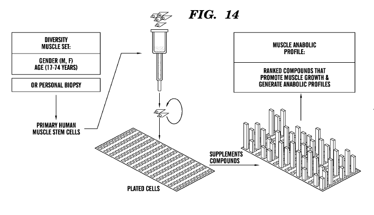
[0059] Figure 14 is a schematic diagram showing an exemplary process of
generating
a personalized anabolic profile based on a personal biopsy or a panel of cells
representing a
diverse set of individuals or a panel of cells representing a population
subgroup that shares at
least one feature such as a phenotypic feature (e.g., but not limited to, age,
gender, condition,
and ethnicity) with a subject in need of muscle augmentation or mitigation of
muscle and/or
bone loss.
DETAILED DESCRIPTION OF THE INVENTION
CA 02882561 2015-02-19
WO 2014/031612 PCT/US2013/055749
[0060] Human genetic variation and life history influence, often
unpredictably, the
response to therapeutic intervention for treatment of a musculoskeletal
disease or disorder,
such as HIV-associated musculoskeletal disease or disorder. Current diagnostic
and treatment
procedures do not distinguish between different types and/or causes of
musculoskeletal
decline and do not provide guidance for anabolic therapy specific to an
individual. For
example, current diagnostic options for evaluating muscle loss include
measuring blood
levels of creatine kinase (CK), which is an indirect measure of muscle loss
that may occur in
response to many non-muscle pathologies. Additional diagnostics include body
composition
analysis using CT or MRI imaging to qualitatively evaluate muscle tissue. The
current
standard of care for muscle wasting is to provide nutritional supplementation
and off-label
prescriptions for anabolic agents. While existing diagnostics are used to
identify muscle loss,
they do not provide targeted decision support for patients to evaluate which,
among the many
treatment options available, are more effective for their unique anabolic
needs. Accordingly,
there is a need for development of assays and/or kits that can provide a more
personalized,
and scalable, anabolic guide to optimize treatment of musculoskeletal wasting,
improve
health outcomes and/or reduce healthcare costs associated with musculoskeletal
decline in
muscle and bone disease as well as in aging.
[0061] Various aspects provided herein generally relate to assays,
methods, systems,
and kits, e.g., for profiling anabolic responses such as skeletal muscle and
bone cell growth of
individuals (e.g., a mammalian subject such as a human) or different
population subgroups in
response to a panel of anabolic compounds. The assays, methods, systems, and
kits described
herein are, in part, based on ranking relative abilities of various anabolic
compounds
(including candidate agents or compositions to be assessed for their anabolic
effects) to
stimulate muscle and/or bone growth of subject-specific cells or patient-
specific cells, e.g.,
collected from a biological sample (e.g., a muscle microbiopsy or a blood
sample), or of a
panel of cells representing different population subgroups, thus generating a
personalized or
stratified anabolic diagnostic report. In some embodiments, the panel of cells
representing
different population subgroups can be stratified based one or a plurality of
(e.g., at least two
or more) feature(s) of the population subgroups. Examples of a feature can be
a phenotypic
feature for population stratification including, but not limited to, age
groups, gender,
ethnicity, body types, body mass index (BMI), blood types, condition, activity
levels, chronic
diseases, acute diseases, genetic polymorphisms, diet, drug resistance,
treatment regime such
as chemotherapy, drastic/abnormal weight loss, geographical location, and any
combinations
21
CA 02882561 2015-02-19
WO 2014/031612 PCT/US2013/055749
thereof. The generated subject-specific or stratified anabolic profiles can be
used to make
therapeutic decisions, e.g., selecting a test composition for treating and/or
preventing muscle
and/or bone loss or a musculoskeletal disease or disorder in the subject,
based on the ranking
of the test composition in one or more embodiments of the assay described
herein. Thus, in
some embodiments, an anabolic agent or composition can be selected,
recommended and/or
optionally administered to a subject or patient based on a subject-specific or
patient-specific
anabolic profile generated using his /or own biopsy and/or blood sample.
Alternatively, an
anabolic agent or composition can be selected, recommended, and/or taken or
otherwise
administered to a subject or patient based on a stratified anabolic profile
generated using
tissue specimens of a matching population subgroup as the subject, based on
one or a
plurality of pre-determined feature such as phenotypic feature(s), e.g., but
not limited to, age,
gender, ethnicity, condition, and/or body mass index (BMI). By way of example
only, for a
30-year old diabetic Caucasian woman who is in need of muscle augmentation
and/or
mitigation of muscle or bone loss, an optimal anabolic agent or composition
can be selected
for this woman, based on a personalized anabolic profiling (which requires her
own biopsy or
blood sample), and/or a stratified anabolic profiling of a population subgroup
of about 30-
year old (e.g., 25-35-year old) diabetic Caucasian women. Factors can also be
based on
whether the subject is taking the anabolic agent in response to the subject's
circumstance or
condition such as chemotherapy or massive and sudden weight loss.
[0062] Unlike the existing diagnostic methods such as CT and MRI or blood
biomarkers such as creatine phosphor-kinase (CPK), embodiments of the assays,
methods,
systems, and kits described herein can provide information about muscle and/or
bone
function in response to a variety of test compositions, which can be in turn
used to make
diagnostic, and/or therapeutic or prophylactic decisions. For example, in some
embodiments,
the generated functional anabolic profiles can be used to diagnose an anabolic
deficiency,
and/or a defect in and/or an imbalance among anabolic growth pathway(s) in a
subject. In
some embodiments, the generated functional anabolic profiles can be used to
identify and/or
optimize a therapeutic option for treatment of a muscle wasting-associated
disease or
disorder. In other embodiments, the generated functional anabolic profiles can
be used to
identity and/or optimize a prophylactic option to prevent or mitigate muscle
loss and to
optimize and maintain muscle heath. Accordingly, methods for diagnosing,
treating and/or
preventing muscle wasting or a musculoskeletal disease or disorder in a
subject are also
22
CA 02882561 2015-02-19
WO 2014/031612 PCT/US2013/055749
provided herein. Methods for determining a risk for anabolic resistance or
potential anabolic
resistance in a subject are also described herein.
Cell-based assays
[0063] One aspect provided herein relates to cell-based assays using
musculoskeletal
cells or precursor cells thereof to generate anabolic profiles specific for
individual subjects
(personalized anabolic profiles) or representing different population
subgroups (stratified
anabolic profiles). The assay comprises: (a) contacting a population of
musculoskeletal cells
or precursor cells thereof with a plurality of test compositions (e.g., at
least two or more test
compositions) to profile anabolic responses of the cells to the test
compositions, wherein each
of the test compositions comprises at least one agent selected to maintain
and/or increase
muscle and/or bone growth; (b) subjecting the musculoskeletal cells or
precursor cells thereof
to at least one analysis to quantify muscle growth or bone growth of the
musculoskeletal
cells or precursor cells thereof in response to the test compositions; and (c)
ranking anabolic
efficacies of the plurality of the test compositions based on the quantified
muscle growth
and/or bone growth, thereby providing anabolic profiles (e.g., muscle anabolic
profiles and/or
bone anabolic profiles) of the assayed cells. In some embodiments, the
musculoskeletal cells
or precursor cells thereof are subjected to at least two analyses to quantify
their muscle
growth and bone growth in response to the test compositions.
[0064] As used herein, the term "anabolic profile" refers to anabolic
responses of the
musculoskeletal cells or precursor cells thereof to a variety of anabolic
agents or
compositions. The anabolic responses of the musculoskeletal cells or precursor
cells thereof
can be characterized by quantifying muscle growth and/or bone growth of the
cells as
described in detail below. In some embodiments, muscle growth of the
musculoskeletal cells
or precursor cells thereof can be characterized by formation of multi-
nucleated muscle cells
(e.g., individual muscle cells each containing at least two or more nuclei).
In some
embodiments, bone growth of the musculoskeletal cells or precursor cells
thereof can be
characterized by formation of bone cells.
[0065] In some embodiments, the assay can be used to generate
personalized anabolic
profiles for individual subjects or patients. Accordingly, in some
embodiments, the
musculoskeletal cells or precursor cells thereof for use in the assay
described herein can be
obtained or derived from a biological sample of a subject or patient seeking
for an anabolic
23
CA 02882561 2015-02-19
WO 2014/031612 PCT/US2013/055749
treatment or supplement. For example, the musculoskeletal cells or precursor
cells thereof
can be obtained or derived from a muscle biopsy or microbiopsy and/or a blood
sample of a
subject or patient. In one embodiment, muscle stem cells derived from a
subject's biopsy or
blood sample can be subjected to the assay described herein. Thus, the
generated anabolic
profile for muscle growth and/or bone growth can be personalized to the
specific subject or
patient.
[0066] In some embodiments, the assay can be used to generate stratified
anabolic
profiles for distinct population subgroups. In these embodiments, the
musculoskeletal cells or
precursor cells thereof for use in the assay described herein can be obtained
or derived from
cells or tissue specimens representing one or different population subgroups.
The cells or
tissue specimens representing one or more different population subgroups can
be obtained
from a cell or tissue depository. As used herein, the term "population
subgroups" refers to
subsets or subgroups of a population stratified by at least one or more
(including, e.g., at least
two, at least three, at least four or more) feature of the population.
Examples of a feature can
be a phenotypic feature for population stratification including, but not
limited to, age or age
groups, gender, ethnicity or races, body types, weights, heights, body mass
index (BMI),
blood types, activity levels (e.g., sedentary lifestyle or work such as a
secretary in office vs.
active lifestyle or work such as an athlete), a condition such as chronic or
acute diseases (e.g.,
but not limited to, diabetes, cancer, osteoporosis, HIV infection, infection,
musculoskeletal
diseases or disorders, metabolic diseases or disorders, and
psychophysiological disorders),
genetic polymorphisms, diet (e.g., but not limited to, vegetarian, and gluten-
free), living
habits (e.g., but not limited to, smoking and alcohols), drug resistance,
treatment regime such
as chemotherapy, drastic weight loss, geographical location (e.g., individuals
living in the
west coast vs. east coast of the United States, or individuals living in the
United States vs. in
Asian countries) and environmental factors associated therewith, and any
combinations
thereof. By way of example only, in one embodiment, population subgroups can
be stratified
by age or age groups, gender, ethnicity, body mass index (BMI), and any
combinations
thereof. In some embodiments, a stratified anabolic profile of a population
subgroup that
shares at least one or more phenotypic features with a subject seeking for an
anabolic
treatment and/or supplement can be used to determine an optimal treatment
and/or
supplement for the subject. One can create bands or population subgroups based
upon sex
(gender) and age. The age groupings can be 20 years, 15 years, 10 years, 5
years, etc. One
24
CA 02882561 2015-02-19
WO 2014/031612 PCT/US2013/055749
can group women based upon whether they are in a child-bearing years or not,
pregnant or
not, under 21 years, orver 50 years of age, etc.
[0067] As used herein, the term "contacting" refers to any suitable means
for
delivering, or exposing, an agent or a test composition to at least one cell
in vitro. Exemplary
delivery methods include, but are not limited to, direct delivery to cell
culture medium,
delivery to an in vitro substrate material (e.g., an extracellular matrix
(ECM) scaffold) in
which cells are seeded, e.g., via perfusion or injection, or other delivery
method well known
to one skilled in the art. In one embodiment, a test composition is added to
the cell culture
medium in which the musculoskeletal cells or precursor cells thereof are
cultured. In another
embodiment, a test composition is distributed or mixed into a substrate
material (e.g., an
ECM scaffold) in which the musculoskeletal cells or precursor cells thereof
are placed. The
term "treatment" or "treated" as used herein, with respect to exposing cells
to an agent, e.g.,
cells treated with an agent, is used herein interchangeably with the term
"contacting."
[0068] The contact of the musculoskeletal cells or precursor cells
thereof with a
plurality of test compositions (e.g., 2, 3, 4, 5, 6, 7, 8, 9, 10, 20, 30, 40,
50, 100, 150, 200, 300,
400, or more) can be performed in vitro in any assay container. For example,
in one
embodiment, the musculoskeletal cells or precursor cells thereof are placed in
a multi-well
microtiter plate (e.g., 48-well plate, 96-well plate, 384-well plate or 1536-
well plate), wherein
the cells in each well are contacted with one test composition, except where
the cells are used
as a negative control (e.g., in the absence of a test composition).
[0069] In order to determine specific anabolic responses (e.g., muscle
growth and/or
bone growth) of the musculoskeletal cells or precursor cells thereof to one or
more test
compositions, the cells can be cultured in separate conditions optimized for
muscle growth
and/or bone growth. For example, to determine the muscle growth-response of
the
musculoskeletal cells or precursor cells thereof to a plurality of test
compositions, it can be
desirable to culture and/or maintain the cells in a muscle cell-specific
condition (e.g., a
condition optimal to muscle differentiation) during the contact with the test
compositions. In
some embodiments, the muscle cell-specific condition can include culturing in
a substrate
material, which is further described in detail below, for example, with a
defined stiffness
optimal to muscle differentiation such as a stiffness of about 5kPa to about
50 kPa, or about
kPa to about 20 kPa.
CA 02882561 2015-02-19
WO 2014/031612 PCT/US2013/055749
[0070] To determine the bone growth-response of the musculoskeletal cells
or
precursor cells thereof to a plurality of test compositions, it can be
desirable to culture and/or
maintain the cells in a bone cell-specific condition (e.g., a condition
optimal to bone
differentiation) during the contact with the test compositions. In some
embodiments, the bone
cell-specific condition can include culturing in a substrate material, which
is further described
in detail below, e.g., with a defined stiffness optimal to bone
differentiation such as a defined
stiffness of about 10 kPa to about 150 kPa, or about 20 kPa to about 100 kPa.
While not
necessary, in some embodiments, the bone cell-specific condition can further
include
culturing in the presence of a bone formation-inducing agent. An exemplary
bone formation-
inducing agent includes, but not limited to, bone morphogenetic protein-2 (BMP-
2).
[0071] The musculoskeletal cells or precursor cells thereof can be
contacted with
different test compositions for any period of time, e.g., minutes, hours, or
days. In some
embodiments, the population of the musculoskeletal cells or precursor cells
thereof can be
contacted with a plurality of test compositions for at least about 6 hours, at
least about 12
hours, at least about 18 hours, at least about 24 hours, at least about 2
days, at least about 3
days, at least about 4 days, at least about 5 days, at least about 6 days, at
least about 7 days or
longer. In some embodiments, the population of musculoskeletal cells or
precursor cells
thereof can be contacted with a plurality of test compositions for at least
about 48 hours or
longer. In some embodiments, the population of musculoskeletal cells or
precursor cells
thereof can be contacted with a plurality of test compositions for at least
about 1 week, at
least about 2 weeks, at least about 3 weeks, at least about 4 weeks or longer.
[0072] In some embodiments, the musculoskeletal cells or precursor cells
thereof can
be contacted with a test composition for an amount of time sufficient to
increase muscle
growth, e.g., by at least about 10% or more, as compared to the
musculoskeletal cells or
precursor cells thereof in the absence of the test composition. For example,
in some
embodiments, the musculoskeletal cells or precursor cells thereof can be
contacted with a test
composition for an amount of time sufficient to increase muscle growth by at
least about
10%, at least about 20%, at least about 30%, at least about 40%, at least
about 50%, at least
about 60%, at least about 70%, at least about 80%, at least about 90%, at
least about 95% or
more, as compared to the musculoskeletal cells or precursor cells thereof in
the absence of the
test composition. In some embodiments, the musculoskeletal cells or precursor
cells thereof
can be contacted with a test composition for an amount of time sufficient to
increase muscle
growth by at least about 1.5-fold, at least about 2-fold, at least about 3-
fold, at least about 4-
26
CA 02882561 2015-02-19
WO 2014/031612 PCT/US2013/055749
fold, at least about 5-fold, at least about 6-fold, or longer, as compared to
the musculoskeletal
cells or precursor cells thereof in the absence of the test composition.
Methods for
determining muscle growth are known in the art, for example, but not limited
to, microscopic
imaging, nucleic staining, cell staining, protein expression, nucleic acid
expression, and any
combinations thereof By way of example only, muscle growth can be detected by
examining
the morphological change of mononucleated muscle cells or muscle precursor
cells to form
multi-nucleated muscle cells (e.g., muscle cells each independently having at
least 2 nuclei, at
least 3 nuclei, or more). In some embodiments, the nuclei of the cells can be
stained in situ
and imaged using a high throughput analysis instrument, e.g., a microscopic
imaging system.
Thus, in one embodiment, the muscle growth of the musculoskeletal cells or
precursor cells
thereof induced by a test composition can be quantified by an increase in the
number of
multi-nucleated cells formed by fusion of the musculoskeletal cells or
precursor cells thereof,
as compared to muscle growth in the absence of the test composition. Nuclei
distribution
(e.g., the number of nucleic per cell distribution) can be generated for each
test composition
and the relative ranks of the test compositions for promoting muscle growth
can then be
ranked to generate a muscle anabolic profile.
[0073] In some embodiments, the musculoskeletal cells or precursor cells
thereof can
be contacted with a test composition for an amount of time sufficient to
increase bone
growth, e.g., by at least about 10% or more, as compared to the
musculoskeletal cells or
precursor cells thereof in the absence of the test composition. For example,
in some
embodiments, the musculoskeletal cells or precursor cells thereof can be
contacted with a test
compositions for an amount of time sufficient to increase bone growth by at
least about 10%,
at least about 20%, at least about 30%, at least about 40%, at least about
50%, at least about
60%, at least about 70%, at least about 80%, at least about 90%, at least
about 95% or longer,
as compared to the musculoskeletal cells or precursor cells thereof in the
absence of the test
composition. In some embodiments, the musculoskeletal cells or precursor cells
thereof can
be contacted with a test composition for an amount of time sufficient to
increase bone growth
by at least about 1.5-fold, at least about 2-fold, at least about 3-fold, at
least about 4-fold, at
least about 5-fold, at least about 6-fold, or longer, as compared to the
musculoskeletal cells or
precursor cells thereof in the absence of the test composition. By way of
example only, bone
growth can be determined by detection of bone or osteoblast phenotype cells
differentiated
from precursor cells thereof such as muscle cells, muscle stem cells, and/or
bone precursor
cells. Thus, in one embodiment, the bone growth of the musculoskeletal cells
or precursor
27
CA 02882561 2015-02-19
WO 2014/031612 PCT/US2013/055749
cells thereof induced by a test composition can be quantified by an increase
in the number of
bone or osteoblast phenotype cells differentiated from the musculoskeletal
cells or precursor
cells thereof (e.g., muscle cells, muscle stem cells, and/or bone precursor
cells), as compared
to bone growth in the absence of the test composition. Methods of identifying
bone or
osteoblast phenotype cells are known in the art, and such exemplary methods
are described in
detail below, including, e.g., detection of a bone or osteoblast phenotype
cell by expression of
a bone marker (e.g., but not limited to, alkaline phosphatase).
[0074] In accordance with various embodiments of the assay described
herein, the
anabolic efficacies of the plurality of the test compositions are ranked based
on the abilities
of the test compositions to stimulate muscle growth and/or bone growth of the
musculoskeletal cells or precursor cells thereof. The ranking of the anabolic
efficacies of the
test compositions can be performed by any art-recognized algorithms, which can
vary with
the methods used to quantify the muscle and/or bone growth. For example, in
the analysis of
muscle cell proliferation and/or differentiation, images of the cells treated
with various test
compositions can be analyzed, e.g., with imaging analysis programs such as
ImageJ or
MATHLAB, for the presence or absence of multi-nucleated muscle cells (e.g., 2
or more
nuclei in a cell) formed from mononucleated cells. A fusion or anabolic
efficacy index can be
determined for each test composition, e.g., based on the number of multi-
nucleated cells (e.g.,
2 or more nuclei in a cell). In some embodiments, a fusion or anabolic
efficacy index can be
defined as a ratio of the total number of nuclei involved in cells having at
least 2 nuclei,
including, e.g., at least 3 nuclei, to the total number of nuclei present in
all of the cells
(including both mononucleated and multi-nucleated cells). For example, as
shown in Figure
2A, a higher fusion or anabolic efficacy index determined for a test
composition indicates
that the test composition is capable of inducing a larger fraction of
mononucleated cells to
fuse together to form multi-nucleated cells. Accordingly, based on the fusion
index
determined for each test composition, an anabolic rank of the test
compositions can be
performed based on the abilities of the test compositions to stimulate muscle
cell proliferation
or differentiation.
[0075] In some embodiments, a fusion or anabolic efficacy index can be
defined as
the frequency of cells with two or more nuclei per cell, including three or
more nucleic per
cell. Accordingly, in some embodiments, a fusion distribution can be computed
to determine
the anabolic rank of the test compositions. By way of example only and not
construed to be
limiting, a fusion distribution can be determined as follows: Prior to contact
with a test
28
CA 02882561 2015-02-19
WO 2014/031612 PCT/US2013/055749
composition or an anabolic agent, the number of musculoskeletal or precursor
cells thereof in
the assay can be determined by any art-recognized method, for example, by high-
throughput
digital cell imaging with phase contrast microscopy. In addition, the number
of nucleus or
nuclei within each cell can be determined and/or tabulated. See, e.g., the
example fusion
distribution corresponding to step 1 (prior to addition of a test composition)
of Figure 1. The
initial number of nucleus per cell prior to the anabolic treatment (e.g.,
prior to the contact of
the musculoskeletal cells or precursor cells thereof with a test composition
or an anabolic
agent) is generally 1.0, i.e., one nucleus per cell. The corresponding nuclear
frequency
distribution (or fusion distribution) would therefore be 100% for one
nucleus/cell, and 0% for
two or more nuclei/cell (e.g., 0% for two nuclei/cell, 0 % for three
nuclei/cell, ...0% for ten+
nuclei or more per cell. After anabolic treatment, e.g., for a pre-determined
period of time,
e.g., at least about 6 hours or longer, the process of determining cell number
and the number
of nuclei per cell can be repeated and displayed as a frequency distribution
of 1-10+
nuclei/cell. See, e.g., the example fusion distribution corresponding to step
2 (after addition
of a test composition) of Figure 1. The distribution of nuclei per cell can
represent the
anabolic signature for the tested composition or anabolic agent/composition.
Each test
composition or anabolic agent/composition can have a unique anabolic
signature.
Accordingly, in these embodiments, the anabolic raffl( of the test
compositions can be
computed based on anabolic treatment of the musculoskeletal cells or precursor
cells thereof
and computing the frequency of cells with two or more nuclei per cell, which
can be
compared to the untreated cells (a baseline). In some embodiments, a higher
threshold value
of a fusion index, e.g., frequency of cells with three or more nuclei per
cell, can be used to
minimize random variation. Accordingly, in some embodiments, the anabolic
raffl( of the test
compositions can be computed as a function of the frequency of cells with
three or more
nucleic per cell. The highest frequency would be defined as highest rank = 1,
the second
highest frequency as rank = 2, etc. In cases where the rank is equivalent, the
tie can be broken
based on the higher median number of nuclei/cell in the anabolic signature,
albeit in this
example they have the same frequency of three or more nuclei per cell.
[0076] In the analysis of bone cell proliferation and/or differentiation,
a ranking of the
test compositions with respect to their individual abilities to stimulate bone
cell proliferation
or differentiation can be performed. In some embodiments, the ranking of the
test
compositions can be determined based on expression of at least one bone marker
(e.g., ALP)
in the cells.
29
CA 02882561 2015-02-19
WO 2014/031612 PCT/US2013/055749
[0077] In some embodiments where muscle growth and bone growth are both
quantified, two separate anabolic profiles or anabolic ranking of the test
compositions (e.g.,
one based on the test composition's ability to induce muscle proliferation
and/or
differentiation, and another based on the test composition's ability to induce
bone
proliferation and/or differentiation) can be generated. Alternatively, a
combined anabolic
profile or anabolic ranking of the test compositions can be generated. For
example, a
weighted average of the muscle growth and bone growth can be used to generate
a combined
anabolic profile or anabolic ranking of the test compositions.
[0078] In one embodiment, the cell-based assay can comprise (a)
contacting muscle
cells or muscle precursor cells thereof, which are CD45 negative and CD56
positive and are
depleted of fibroblasts, with an array of test compositions each comprising at
least one
anabolic agent in the presence of growth media; (b) quantifying muscle growth
of the cells in
response to the test compositions by detecting the number of multi-nucleated
cells formed
from mononucleated cells, thereby generating a distribution of the number of
nuclei per cell
distribution; and (c) ranking the test compositions based on their efficacy in
promoting
muscle growth (cell fusion). The muscle cells or muscle precursor cells (e.g.,
muscle stem
cells) for use in the assay can be isolated from a muscle tissue or biopsy of
a subject, or can
be provided as banked specimens, and plated in a multi-well culture plate
(e.g., a 384 well
plate). In some embodiments, it can be desirable to prepare duplicates or
triplicates for each
test composition. In some embodiments where personal biopsy-derived cells are
used, the
growth media can comprise autologous serum derived from a blood sample.
Musculoskeletal cells or precursor cells thereof and collection and
preparation of the same
[0079] Musculoskeletal cells or precursor cells thereof used in one or
more
embodiments of the assays, methods, and systems described herein can generally
be collected
from one or more subjects, or can include established cell lines. In some
embodiments where
the assay is used to generate a personalized anabolic profile, the
musculoskeletal cells or
precursor cells thereof used in the assays, methods, and systems described
herein can be
collected from a single subject, e.g., who is seeking for personalized
guidance on an anabolic
treatment. Accordingly, in these embodiments, the musculoskeletal cells or
precursor cells
thereof used in the assay, method and/or system described herein are subject-
specific, and can
be obtained or derived from a biological sample of the subject. For example,
subject-specific
CA 02882561 2015-02-19
WO 2014/031612 PCT/US2013/055749
musculoskeletal cells or precursor cells thereof can be obtained or derived
from a muscle
biopsy and/or a blood sample of the subject.
[0080] In some embodiments, the musculoskeletal cells or precursor cells
thereof
used in one or more embodiments of the assays, methods, and systems described
herein can
be obtained or derived from cells or tissue specimens representing at least
one or more
population subgroups, including, e.g., 1, 2, 3, 4, 5, 6, 7, 8, 9, 10, 20, 30,
40, 50, 75, 100, 200,
300, 400, 500 or more population subgroups. The cells or tissue specimens can
be obtained
from a cell or tissue depository and should be generally well-documented such
that they can
be stratified to represent different population subgroups based on one or a
plurality of
features of the population. Examples of the features can include phenotypic
features for
population stratification including, but not limited to, age or age groups,
gender, ethnicity or
races, body types, weights, heights, body mass index (BMI), blood types,
activity levels (e.g.,
sedentary lifestyle or work such as a secretary in office vs. active lifestyle
or work such as an
athlete), chronic or acute diseases (e.g., but not limited to, diabetes,
cancer, osteoporosis, HIV
infection, infection, musculoskeletal diseases or disorders, metabolic
diseases or disorders,
and psychophysiological disorders), genetic polymorphisms, diet (e.g., but not
limited to,
vegetarian, and gluten-free), living habits (e.g., but not limited to, smoking
and alcohols),
drug resistance, treatment regime such as chemotherapy, drastic/abnormal
weight loss,
geographical location (e.g., individuals living in the west coast vs. east
coast of the United
States, or individuals living in the United States vs. in Asian countries) and
environmental
factors associated therewith, and any combinations thereof. In these
embodiments, distinct
anabolic profiles for different population subgroups (stratified anabolic
profiles) can be
generated using one or more embodiments of the assays described herein. If a
subject is
seeking for guidance on an anabolic treatment, the subject can be matched with
one of the
stratified population subgroups based on at least one or more features such as
phenotypic
features as described above and be recommended with an anabolic agent based on
its
respective ranking in a stratified anabolic profile of the matching population
subgroup.
[0081] In some embodiments, the musculoskeletal cells or precursor cells
thereof for
use in the assay, method, system and/or kit described herein can be obtained
or derived from
a biological sample (e.g., but not limited to, a muscle biopsy and/or blood
sample) of subjects
or individuals who are determined to suffer from or have a risk for muscle
loss and/or bone
loss (e.g., a musculoskeletal disease or disorder). Examples of subjects or
individuals who are
at risk for a musculoskeletal disease or disorder include, but are not limited
to, athletes, aging
31
CA 02882561 2015-02-19
WO 2014/031612 PCT/US2013/055749
individuals, individuals having a chronic disease or disorder (e.g., but not
limited to, cancer,
chronic obstructive pulmonary disease (COPD), chronic kidney disease (CKD),
chronic liver
failure (CLF), and chronic infections), individuals suffering from
malnutrition, or any
combinations thereof Examples of a musculoskeletal disease or disorder
include, but are not
limited to, muscle loss, muscle wasting, muscle wasting associated with HIV
infection,
muscle wasting in cancer survivors, cachexia, muscular dystrophy, osteopenia,
osteoporosis,
sarcopenia, an age-related musculoskeletal disease or disorder, or a
musculoskeletal disease
or disorder associated with anabolic resistance, a musculoskeletal disease or
disorder
associated with excessive weight loss, or any combinations thereof
[0082] In some embodiments, the musculoskeletal cells or precursor cells
thereof for
use in the assay, method, system and/or kit described herein can be obtained
or derived from
a biological sample of subjects or individuals who have previously shown non-
responsiveness or resistance to at least one or more anabolic agents.
[0083] In some embodiments, the musculoskeletal cells or precursor cells
thereof for
use in the assay, method, system and/or kit described herein can be obtained
or derived from
a biological sample of subjects or individuals who are seeking to maintain
and/or enhance
muscle and/or bone health.
[0084] In some embodiments, the musculoskeletal cells or precursor cells
thereof for
use in the assay, method, system and/or kit described herein can be obtained
or derived from
a biological sample of subjects or individuals who are normal healthy subjects
without any
known bone and/or muscle loss.
[0085] As used herein, the term "musculoskeletal cells" generally refers
to cells
associated with muscle, bone, cartilage, connective tissue or any combinations
thereof. In
some embodiments, musculoskeletal cells used in the assays, methods, systems
and/or kits
described herein can include muscle cells. Depending on the type of
musculoskeletal disease
or disorder, and/or the location of symptoms which can include pain or
weakness, the
musculoskeletal cells can be obtained or derived from a muscle biopsy of any
tissue (e.g.,
heart tissue, leg muscles such as quandriceps, and arm muscles such as biceps,
shoulder
muscles such as deltoid) in a subject.
[0086] As used herein, the term "precursor cells thereof" generally
refers to precursor
cells or stem cells that can be differentiated into musculoskeletal cells,
including, but are not
limited to, muscle precursor cells, bone precursor cells, and/or any cells
that can be
32
CA 02882561 2015-02-19
WO 2014/031612 PCT/US2013/055749
differentiated into muscle cells or bone cells. Muscle precursor cells can
encompass any cells
that differentiate towards the myogenic lineage upon treatment with at least
one known
myoblast-promoting (e.g., but not limited to, bFGF, aFGF, IGF-1, and NGF). In
one
embodiment, muscle precursor cells can include mononucleated myoblasts. Bone
precursor
cells can encompass cells that differentiate towards the osteoblast lineage
upon treatment
with at least one known osteoblast-promoting agents (e.g., but not limited to
type I collagen,
fibrinogen, fibrin, fibrinogen, osteocalcin, osteonectin, TGF-I3, Vitamin D3,
basic fibroblast
growth factor, or bone morphogenic protein 2). In one embodiment, bone
precursor cells can
include osteoprogenitor cells or preosteoblasts. Without limitations, muscle
precursor cells
and/or bone precursor cells can be obtained or derived from a muscle biopsy
and/or from
peripheral blood progenitor cells or stem cells, e.g., using induced
pluripotent cell technology
known in the art. Accordingly, in some embodiments, a blood sample can be
collected from a
subject as a source of musculoskeletal cells or precursor cells thereof in one
or more
embodiments of the assays, methods, and/or systems described herein.
[0087] In some embodiments, the musculoskeletal cells or precursor cells
thereof for
use in the assays, methods, kits, and/or systems described herein can be pre-
selected for
CD45 negative and/or CD56-positive. Without wishing to be bound by theory, the
cells that
are CD45-negative and CD56-positive can be muscle-derived stem cells.
Accordingly, in
some embodiments, muscle-derived stem cells or muscle precursor stem cells can
be used in
the assays, methods, kits and/or systems described herein. Methods for
isolating muscle-
derived stem cells from a muscle biopsy are known in the art. See, e.g.,
Sharifiaghdas et al.,
Urol J. 2011 Winter; 8(1):54-9 or Example 4 for example methods to prepare
human muscle-
derived stem cells or muscle precursor stem cells) from a muscle biopsy. For
example, CD45-
positive musculoskeletal cells or precursor cells thereof can be first removed
from the
population of cells using magnetic beads coated with CD45-binding molecules.
The
remaining CD45-negative musculoskeletal cells or precursor cells thereof can
be further
positively selected for CD56-positive cells, e.g., using magnetic beads coated
with CD56-
binding molecules, which are considered as muscle-derived stem cells or muscle
precursor
stem cells.
[0088] In some embodiments, the musculoskeletal cells or precursor cells
thereof for
use in the assays, methods, kits, and/or systems described herein exclude
fibroblasts. The
fibroblasts can be removed from the population of musculoskeletal or precursor
cells thereof
using any methods known in the art. For example, fibroblasts can be removed
from the
33
CA 02882561 2015-02-19
WO 2014/031612 PCT/US2013/055749
population of cells using magnetic beads and/or by performing a fibroblast
plate adherence
depletion. An exemplary fibroblast plate adherence depletion assay includes
plating the cell
population to a non-ECM-coated plate where fibroblasts can attach to the plate
in the absence
of the ECM, while other cells cannot. Thus, the non-attached musculoskeletal
or precursor
cells thereof can be collected, e.g., by centrifugation.
[0089] Collections of a biopsy or a blood sample for at least one
analysis performed
in the assays and/or systems described herein are well known to those skilled
in the art. The
sample can be obtained by removing a sample of cells from a subject, but can
also be
accomplished by using previously isolated cells (e.g. isolated by another
person). In addition,
the cells can be freshly collected or a previously collected sample.
[0090] In some embodiments, the sample for use in one or more embodiments
of the
assay or system described herein can be a frozen sample, e.g., a frozen tissue
or blood
sample. The frozen sample can be thawed before employing assays and systems
described
herein. After thawing, a frozen sample can be centrifuged before being
subjected to assays
and systems described herein.
[0091] To collect a muscle tissue, by way of example only, a patient's
muscle biopsy,
e.g., a percutaneous muscle biopsy, can be performed by a trained medical
personnel. To
collect a blood sample, by way of example only, the patient's blood can be
drawn by trained
medical personnel directly into anti-coagulants such as citrate, EDTA PGE, and
theophylline.
The whole blood can be separated into the plasma portion, the cells, and
platelets portion by
refrigerated centrifugation at 3500 g for 2 minutes. After centrifugation, the
supernatant is the
plasma and the pellet is RBC. Since platelets have a tendency to adhere to
glass, it is
preferred that the collection tube be siliconized. Another method of isolating
red blood cells
(RBCs) is described in Best, CA et al., 2003, J. Lipid Research, 44:612-620.
In order to
collect peripheral blood stem cell from a blood sample of a subject, prior to
the collection, the
subject can be given a medication to promote the growth and release of stem
cells from the
bone into the blood. The stem cells are then collected using a special machine
called a cell
separator.
Cell culture and substrate materials employed in the assays, systems or kits
described herein
[0092] In order to determine different anabolic responses (e.g., muscle
growth and/or
bone growth) of the musculoskeletal cells or precursor cells thereof to one or
more test
34
CA 02882561 2015-02-19
WO 2014/031612 PCT/US2013/055749
compositions, in some embodiments, the cells can be cultured and/or maintained
in separate
conditions optimized for promoting muscle cell growth/proliferation and/or
bone cell
growth/proliferation, prior to and/or during the contact with the test
compositions. For
example, the musculoskeletal cells or precursor cells thereof can be cultured
in a growth
medium optimized to mimic the original microenvironment of a subject or pooled
subjects
within a population subgroup from whom the cells were collected. Without
wishing to be
bound by theory, serum from individual subjects can have different effects on
muscle growth
and/or bone growth. By way of example only, it has been previously shown in
mouse models
that transfer of serum from younger subjects to older subjects can accelerate
muscle
regeneration and conversely, serum from older subjects to younger subjects can
inhibit
muscle growth. Accordingly, in some embodiments, serum collected from a blood
sample
from a subject for personalized biopsy or from pooled subjects within a
population subgroup
can be added to the growth medium to re-create the microenvironment of the
host. The serum
can be added to the growth medium in any amount. For example, the serum can be
added to
reach at least about 1 % or higher by volume in the growth medium, including,
e.g., but not
limited to, at least about 5% , at least about 10%, at least about 20%, at
least about 30% or
more, by volume in the growth medium. In one embodiment, the growth medium has
about
10% (by volume) of serum.
[0093] While not necessary, the growth medium in which the
musculoskeletal cells or
precursor cells thereof are cultured prior to or after addition of the test
compositions
described herein can have the same or different composition.
[0094] In some embodiments, the cells can be additionally or
alternatively cultured
and/or maintained in a separate substrate material specific to muscle
differentiation/growth
and bone differentiation/growth, prior to and/or during the contact with the
test compositions.
[0095] The term "substrate material" as used herein refers to a material
that facilitates
muscle and/or bone growth and/or differentiation. The substrate material can
comprise a
synthetic material, a natural material, or both. In some embodiments, the
substrate material
can comprise at least one biocompatible material that facilitates, permits, or
enhances
deposition of new tissue matrix (e.g., muscle- or bone-related matrices). In
some
embodiments, the substrate material can comprise mineralized materials, such
as calcium
sulfate or calcium phosphate, a biopolymer, a metal, allograft muscle tissue,
autograft muscle
tissue, demineralized bone matrix, or any combinations thereof
CA 02882561 2015-02-19
WO 2014/031612 PCT/US2013/055749
[0096] In one embodiment, the substrate material includes extracellular
matrix.
Extracellular matrix (ECM) for culture of muscle cells or precursor cells
thereof obtained
from a subject can include collagen, non-collagenous glycoproteins,
proteoglycans, or any
combinations thereof The extracellular matrix can include any art-recognized
molecules
such as growth factor (e.g., fibroblast growth factor) to mimic the native
environment of a
musculoskeletal tissue for promotion of muscle or bone differentiation and/or
growth. See,
e.g., Kjaer M. (2004) Physiol. Rev. 84: 649, for typical ECM components in a
muscle.
[0097] The substrate material can be planar in shape or form a scaffold.
In one
embodiment, the substrate material employed in the assays, systems or kits for
culturing
musculoskeletal cells or precursor cells thereof forms a scaffold. The term
"scaffold" as used
herein generally refers to a 3-dimensional supporting structure that promotes
muscle and/or
bone growth. Non-limiting examples of a scaffold includes a gel, a solid, a
matrix, a
hydrogel, a sponge, a mesh, a membrane and any combinations thereof In some
embodiments, the scaffold substrate can be porous.
[0098] In one embodiment, the substrate material for use in the assays,
systems and
kits described herein includes an ECM scaffold, e.g., a scaffold comprising at
least one type
of extracellular matrix molecules, fibers, and/or fibrils, including at least
two, at least three, at
least four or more extracellular matrix molecules, fibers, and/or fibrils.
[0099] In some embodiments, the substrate material for use in the assays,
systems and
kits described herein can include Matrigel and/or Engelbreth-Holm-Swarm
sarcoma ECM.
[00100] Muscle cell-specific culture conditions: The stiffness of the
substrate material
can also affect anabolic responses of cells to a test composition. Thus, it is
desirable to have a
substrate material with a defined stiffness optimal to promote muscle and/or
bone growth
and/or differentiation. For example, to determine muscle growth-responses of
musculoskeletal cells or precursor cells thereof to a test composition, in
some embodiments,
the cells can be cultured and/or maintained, prior to and/or during the
contact with a test
composition, in a substrate material with a defined stiffness optimal to
promote muscle cell
proliferation and/or differentiation, e.g., a defined stiffness of about 5kPa
to about 50 kPa, or
about 10 kPa to about 20 kPa. In some embodiments, the stiffness of the
substrate material
for stimulation of muscle cell proliferation and/or differentiation can vary
with the
microenvironment of the tissue from which the musculoskeletal cells or
precursor cells
thereof were collected or derived.
36
CA 02882561 2015-02-19
WO 2014/031612 PCT/US2013/055749
[00101] In some embodiments, the musculoskeletal or precursor cells
thereof (e.g., but
not limited to, muscle precursor stem cells) can be cultured in a growth
medium adapted to
induce cell differentiation. For example, the musculoskeletal or precursor
cells thereof (e.g.,
but not limited to, muscle precursor stem cells) can be cultured in a growth
medium adapted
to induce cell fusion to form multi-nucleated muscle cells, and/or to induce
differentiation of
non-differentiated stem cells or muscle precursor cells to form muscle cells.
In some
embodiments, the growth medium can comprise a serum purified or collected from
a blood
sample of a specific subject or pooled subjects within a population subgroup
as described
above.
[00102] Bone cell-specific culture conditions: Similarly, to determine
bone growth-
responses of musculoskeletal cells or precursor cells thereof to a test
composition, in some
embodiments, the cells can be cultured and/or maintained, prior to and/or
during the contact
with a test composition, in a substrate material with a defined stiffness
optimal to promote
bone cell proliferation and/or differentiation, e.g., a defined stiffness of
about 10 kPa to about
150 kPa, or about 20 kPa to about 100 kPa. In some embodiments, the stiffness
of the
substrate material for stimulation of bone cell proliferation and/or
differentiation can vary
with the microenvironment of the tissue from which the musculoskeletal cells
or precursor
cells thereof were collected or derived.
[00103] In some embodiments, the stiffness, composition and/or structure
of the
substrate material can be adjusted such that the substrate material is
osteoconductive,
osteoinductive, osteogenic, or any combinations thereof. For example, while
not necessary, in
some embodiments, the substrate material for stimulation of bone growth and/or
differentiation can include an osteoconductive, osteoinductive, osteogenic
agent, e.g., bone
morphogenetic protein-2 (BMP-2), any derivatives thereof, or any art-
recognized bone-
inducing agents such as the agents disclosed in U.S. Pat. No. 7,897,588, the
content of which
is incorporated by reference.
[00104] As used herein, the term "osteoconductive" generally refers to the
ability of a
material or agent to facilitate the migration of osteogenic cells within a
substrate material. In
some embodiments, the porosity of the substrate material can affect its
osteoconductivity. As
used herein, the term "osteoinductive" generally refers to the ability to
induce non-
differentiated stem cells or osteoprogenitor cells (osteoblasts), which is a
component of
osseous (bone) tissue, to differentiate into osteoblasts. The simplest test of
osteoinductivity is
the ability to induce the formation of bone in tissue locations such as
muscle, which do not
37
CA 02882561 2015-02-19
WO 2014/031612 PCT/US2013/055749
normally form bone (ectopic bone growth). It is generally understood that a
substrate material
can be made osteoinductive by adding growth factors such as rhBMP-2
(recombinant human
bone morphogenic protein-2) to it. The mineralization and the addition of
growth factors can
affect the osteoinductivity of the substrate material. As used herein, the
term "osteogenic"
generally refers to the ability of forming new bone cells or tissue.
Osteogenesis is the process
of laying down new bone material using osteoblasts. Osteoblasts build bone by
producing
osteoid to form an osteoid matrix, which is composed mainly of Type I
collagen. Osseous
tissue comprises the osteoid matrix and minerals (mostly with calcium
phosphate) that form
the chemical arrangement termed calcium hydroxyapatite. Osteoblasts are
typically
responsible for mineralization of the osteoid matrix to form osseous tissue.
Without wishing
to be bound by a theory, the osteoconductivity and osteoinductivity of the
substrate material
can have an impact on osteogenesis.
1001051 In some embodiments, the musculoskeletal cells or precursor cells
thereof
(e.g., but not limited to, muscle or bone precursor stem cells) can be
cultured in a growth
medium adapted to induce cell differentiation. For example, the
musculoskeletal or precursor
cells thereof (e.g., but not limited to, muscle or bone precursor stem cells)
can be cultured in
a growth medium adapted to differentiate muscle cells, bone precursor cells,
non-
differentiated stem cells to become bone cells. In some embodiments, the
growth medium can
comprise a serum purified or collected from a blood sample of a specific
subject or pooled
subjects within a population subgroup as described above.
[00106] While not necessary, in some embodiments, the musculoskeletal
cells or
precursor cells thereof can be cultured in the presence of a bone formation-
inducing agent.
Examples of a bone formation-inducing agent can include, but are not limited
to, bone
morphogenic factor (BMP) (e.g., BMP-1, BMP-2, BMP-3, BMP-4, BMP-5 and BMP-6),
transforming growth factor (TGF), insulin-like growth factor (IGF), basic
fibroblast growth
factor (bFBF), osteogenic protein (OP) (e.g., OP-1, OP-2 and OP-3), osteogenic
factors,
osteoconductive factors, osteoinductive factors, and any combinations thereof.
In one
embodiment, the bone formation-inducing agent can include bone morphogenetic
protein-2
(BMP-2).
[00107] In vitro culture of muscle cells and precursor cells thereof for
muscle cell
proliferation/differentiation or bone cell proliferation/differentiation are
known in the art. A
skilled artisan can optimize muscle cell-specific culture conditions and bone
cell-specific
culture conditions for musculoskeletal cells or precursor cells thereof.
38
CA 02882561 2015-02-19
WO 2014/031612 PCT/US2013/055749
[00108] In some embodiments, rather than harvesting muscle cells from a
subject's
muscle biopsy for use in the assays, systems, and/or kits described herein,
stem cells
collected from a subject (e.g., a subject's blood sample) can be used in the
assays, methods,
systems and/or kits described herein and be cultured in an appropriate
condition suitable for
muscle cell differentiation or bone cell differentiation. Methods for
generation of muscle cells
from stem cells are known in the art. See, e.g., Salani S et al. "Generation
of skeletal muscle
cells from embryonic and induced pluripotent stem cells as an in vitro model
and for therapy
of muscular dystrophies" (2012) J Cell Mol Med. 16 :1353; Yoshida Y and
Yamanaka S.
"Recent Stem Cell Advances: Induced Pluripotent Stem Cells for Disease
Modeling and Stem
Cell¨Based Regeneration Circulation." (2010) Circulation. 122: 80; and
Bilousova G. et al.,
"Osteoblasts derived from induced pluripotent stem cells form calcified
structures in
scaffolds both in vitro and in vivo." (2011) Stem Cells. 29: 206-16.
Muscle growth of musculoskeletal cells or precursor cells thereof (e.g.,
mononucleated
muscle cells, muscle precursor cells thereof including blood-derived cells)
and exemplary
methods/analyses to quantifj; muscle growth
[00109] In one or more embodiments of the assays described herein, the
musculoskeletal cells or precursor cells thereof after contact with a
plurality of test
compositions can be subjected to at least one or more analyses to quantify
muscle growth of
the cells in response to the test compositions. In one embodiment, the
musculoskeletal cells
or precursor cells thereof include mononucleated muscle cells. The
mononucleated muscle
cells, myoblasts, are uniquely different from other cells in the body in a
number of ways: 1)
myoblasts naturally differentiate to form muscle tubules capable of muscle
contraction, 2)
when myoblasts fuse to form myotubes, these cells become post mitotic (stop
dividing) with
maturation, and 3) as myotubes, the cells express large amounts of protein
which is produced
in the cells due to multinucleation. Accordingly, as used herein, the term
"muscle growth," in
some embodiments, refers to an increase in differentiation of mononucleated
muscle cells,
myoblasts, e.g., collected from a muscle biopsy, into myotubes, or multi-
nucleated muscle
cells. For example, the term "muscle growth" refers to an increase in the
number of
mononucleated muscle cells, myoblasts, differentiated into multi-nucleated
muscle cells by at
least about 10%, at least about 20%, at least about 30%, at least about 40%,
at least about
50%, at least about 60%, at least about 70%, at least about 80%, at least
about 90%, at least
about 95%, or more, as compared to the number of myoblasts in the absence of a
test
39
CA 02882561 2015-02-19
WO 2014/031612 PCT/US2013/055749
composition described herein. In other embodiments, the term "muscle growth"
refers to an
increase in proliferation of myoblasts. For example, the proliferation of
myoblasts is
increased by at least about 10%, at least about 20%, at least about 30%, at
least about 40%, at
least about 50%, at least about 60%, at least about 70%, at least about 80%,
at least about
90%, at least about 95%, or more, as compared to the number of myoblasts in
the absence of
a test composition described herein. In some embodiments, the term "muscle
growth" refers
to both an increase in proliferation of myoblasts (e.g., by at least about
10%) and an increase
in differentiation of the mononucleated myoblasts into multi-nucleated muscle
cells (e.g., by
at least about 10% in cell number), as compared to the number of myoblasts in
the absence of
a test composition described herein.
[00110] Any art-recognized methods and/or analyses to quantify muscle
growth of
myoblasts or blood-derived muscle cells in vitro can be employed in one or
more
embodiments of the assay described herein. For example, the muscle growth of
myoblasts in
vitro can be determined by examining the formation of multinucleated muscle
cells
(myotubes) by fusion of mononucleated muscle cells (myoblasts), e.g., by
microscopy and/or
histological methods.
[00111] Alternatively, the muscle growth of myoblasts in vitro can be
determined by
detecting one or more proteins and/or genes involved in muscle cell
differentiation. Genes,
such as muscle creatine kinase, troponin, caveolin 3, a-actin, and myosin, are
reported to be
expressed predominantly in the skeletal muscles. A family of transcription
factors specifically
expressed in the muscles, including myoD, myogenin, myf-5, and MRF-
4/herculin/myf-6,
have been cloned. These factors are phosphorylated nuclear proteins containing
a helix-loop-
helix (bHLH) motif, as required for both dimerization and DNA binding, and are
believed to
be determinants of the cell-specific differentiation program (Olson and Klein
(1994), Genes
& Dev. 8:1-8). When one of these factors is introduced into non-myogenic
cells,
differentiation into mature muscle cells is initiated (Weintraub et al.
(1991), Science 251:761-
766). The myoD family, a group of transcription factors, has been found to
direct muscle
formation, inhibit proliferation, activate differentiation and induce a
contractile phenotype.
While myoD and myf-5 are expressed within the proliferating myoblasts,
myogenin and
MRF-4 are not expressed until the myoblasts withdraw from the cell cycle in
response to
mitogen withdrawal. Based on these findings, it was demonstrated that myogenin
and MRF-4
activate and maintain the expression of muscle-specific genes (Emerson (1993),
Curr. Opin.
Genet. Dev. 3:265-274), while myoD and myf-5 are thought to play a role in the
proliferation
CA 02882561 2015-02-19
WO 2014/031612 PCT/US2013/055749
of myoblasts. Other cell-cycle regulatory proteins, such as RB (Shiio et al.
(1996), Oncogene
12:1837-1845, Wang et al. (1997), Cancer Research 57:351-354), p21 (Guo et al.
(1995),
Mol. Cell Biol. 15:3823-3829), cyclin D, cdk2, cdk4 (Kiess et al. (1995),
Oncogene 10:159-
166) and tumor suppressor gene p53 (Soddu et al. (1996), J. Cell Biol. 134:193-
204) are
involved in the muscle cell differentiation program. Recently, caveolin 3
(Song et al. (1996),
J. Cell Biol. 271:15160-15165), a-dystroglycan (Kostrominova and Tanzer
(1995), J. Cell
Biochem. 58:527-534) and DNA methyltransferases (Takagi et al. (1995), Eur. J.
Biochem.
231:282-291) have been shown to play positive roles in myogenic
differentiation. Methods
for detecting expression of proteins and/or genes in cells are known in the
art and can be used
herein to detect the presence or absence of any protein and/or genes that
induce myoblast
proliferation and/or differentiation. Other proteins and/or genes involved in
myocyte
differentiation, e.g., the ones discussed in U.S. Pat. No. 6,670,450, can also
be employed to
quantify muscle growth in vitro.
Bone growth of musculoskeletal cells or precursor cells thereof (e.g., muscle
cells, or bone
precursor cells thereof including blood-derived cells) and exemplary
methods/analyses to
quantifj; bone growth
[00112] In one or more embodiments of the assay described herein, the
musculoskeletal cells or precursor cells thereof after contact with a
plurality of test
compositions can be subjected to at least one or more analyses to quantify in
vitro bone
growth of the cells in response to the test compositions. As used herein, the
term "bone
growth," in some embodiments, refers to an increase in differentiation of
muscle cells or
precursor cells thereof into bone or osteoblast phenotype cells. For example,
the term "bone
growth" refers to an increase in the number of muscle cells or bone precursor
cells
differentiated into bone or osteoblast phenotype cells by at least about 10%,
at least about
20%, at least about 30%, at least about 40%, at least about 50%, at least
about 60%, at least
about 70%, at least about 80%, at least about 90%, at least about 95%, or
more, as compared
to the cells in the absence of a test composition described herein. In some
embodiments, the
term "bone growth" can further encompass an increase in proliferation of
osteoblast
phenotype cells. For example, the proliferation of osteoblast phenotype cells
can be increased
by at least about 10%, at least about 20%, at least about 30%, at least about
40%, at least
about 50%, at least about 60%, at least about 70%, at least about 80%, at
least about 90%, at
least about 95%, or more, as compared to the cells in the absence of a test
composition
41
CA 02882561 2015-02-19
WO 2014/031612 PCT/US2013/055749
described herein. In some embodiments, the term "bone growth" refers to both
an increase
in differentiation of the muscle cells and/or bone precursor cells into bone
or osteoblast
phenotype cells (e.g., by at least about 10% in cell number) and an increase
in proliferation of
the osteoblast phenotype cells (e.g., by at least about 10%), as compared to
the cells in the
absence of a test composition described herein.
[00113] As used herein, the term "osteoblast phenotype cells" refers to
cells displaying
at least one phenotype or characteristic associated with osteoblasts. The
osteoblast phenotype
cells including terminally or non-terminally differentiated cells can be
derived from bone
precursor cells thereof including stem cells. Examples of osteoblast-
associated phenotype or
characteristic include but are not limited to, cuboidal morphology of the
cells, production of
alkaline phosphatase (ALP), production of type I collagen production,
production of
osteocalcine, production of mineralized extracellular matrix, expression of
one or more
specific marker transcripts, such as but not limited to, AP-1 family members,
Runx2, Fra-2,
alkaline phosphatase, osteocalcin, I3-catenin, CCAAT/enhancer binding protein
(C/EBP), and
ATF4, or any combinations thereof. In some embodiments, the osteoblast
phenotype cells can
advance in differentiation all the way to terminal differentiation (e.g., with
the production of
a mineralized extracellular matrix), and these cells are termed as "terminally
differentiated
osteoblasts" herein.
[00114] Accordingly, in order to quantify the bone growth in
musculoskeletal cells or
precursor cells thereof after contact with a test composition, methods for
determining the
activity of alkaline phosphatase (ALP), the expression of type I collagen, and
osteocalcine,
and any mineralization of the extracellular matrix can be employed in one or
more
embodiments of the assay or systems described herein.
[00115] In one embodiment, the musculoskeletal cells or precursor cells
thereof after
contact with a test composition can be subjected to an analysis of ALP level
or activity
expressed by the osteoblast phenotype cells. For example, alkaline phosphatase
(ALP)
activity can be determined by using para-nitrophenol phosphatase as a
substrate, using the
technique described by L. Lecoeur and J. P. Ouhayoun, Biomaterials 18, 989-993
(1997). The
quantity of para-nitrophenol formed upon hydrolysis of the substrate can be
determined by
measuring the absorbance at 410 nm, which is converted into nanomoles of
enzyme using a
calibration curve established on the basis of known concentrations of para-
nitrophenol. A test
can also be performed for detecting in situ the activity of alkaline
phosphatase on cell
cultures fixed with formaldehyde, using a kit for semi-quantitative
histochemical detection of
42
CA 02882561 2015-02-19
WO 2014/031612 PCT/US2013/055749
alkaline phosphatase (kits sold by Sigma Chemical Co., reference 86R).
Alkaline phosphatase
activity can then be visualized on the cell mat by a reddish color.
Alternatively, ALP level
can be determined by staining as described in the Examples.
[00116] In some embodiments, the musculoskeletal cells or precursor cells
thereof
after contact with a test composition can be subjected to an analysis of
collagen production,
osteocalcine expression, or both. For example, the presence of collagens
(e.g., of type I and
of type II) or osteocalcine can be determined by an immunoassay, e.g.,
antibody-based assay
and immunostaining, and/or gene expression measurements (e.g., PCR, and/or
quantitative
PCR).
[00117] In some embodiments, the musculoskeletal cells or precursor cells
thereof
after contact with a test composition can be subjected to an analysis of
possible
mineralization of the extracellular matrix. For example, mineralization of the
extracellular
matrix can be detected by using the von Kossa stain test implemented using the
technique
described by Cheng et al., Endocrinology 134: 277-285 (1994).
[00118] In some embodiments, the musculoskeletal cells or precursor cells
thereof
after contact with a test composition can be subjected to an analysis to
determine expression
of at least one art-recognized marker for osteoblast differentiation, e.g.,
but not limited to,
AP-1 family members, Runx2, Fra-2, alkaline phosphatase, osteocalcin, I3-
catenin,
CCAAT/enhancer binding protein (C/EBP), and ATF4, or to determine a gene
expression
profile associated with osteoblast differentiation. Additional bone markers,
e.g., as described
in U.S. Pat. App. No. US 2004/0101818, can be utilized in one or more
embodiments of the
assay or system described herein to quantify the presence or absence of bone
growth of the
musculoskeletal cells or precursor cells thereof in response to a test
composition. For
example, the presence of bone growth of the musculoskeletal cells or precursor
cells thereof
in response to a test composition can be indicated by an increase in
expression of at least one
art-recognized marker for osteoblast differentiation, e.g., but not limited
to, AP-1 family
members, Runx2, Fra-2, alkaline phosphatase, osteocalcin, I3-catenin,
CCAAT/enhancer
binding protein (C/EBP), and ATF4, by at least about 10%, at least about 20%,
at least about
30%, at least about 40%, at least about 50%, at least about 60%, at least
about 70%, at least
about 80%, at least about 90%, at least about 95% or more, as compared to the
cells in the
absence of the test composition.
43
CA 02882561 2015-02-19
WO 2014/031612 PCT/US2013/055749
Exemplary methods of determining expression level of markers for
differentiated muscle cells
and/or bone cells
[00119] Measuring protein expression: Various methods known in the art can
be
used to determine expression of markers or genes specific for differentiated
muscle cells and
bone cells as described earlier. In some embodiments, the protein expression
level of markers
or genes specific for differentiated muscle cells or bone cells as described
earlier can be
measured to quantify muscle growth or bone growth, respectively, of
musculoskeletal cells or
precursor cells thereof in response to a test composition. By way of example
only, the levels
of protein markers can be measured by contacting a test sample with an
antibody-based
binding moiety that specifically binds to at least one of the markers specific
for differentiated
muscle or bone cells, or to a fragment thereof Formation of the antibody-
protein complex is
then detected by a variety of methods known in the art.
[00120] The term "antibody-based binding moiety" or "antibody" can include
immunoglobulin molecules and immunologically active determinants of
immunoglobulin
molecules, e.g., molecules that contain an antigen binding site which
specifically binds
(immunoreacts with) to the markers specific for differentiated muscle or bone
cells. The term
"antibody-based binding moiety" is intended to include whole antibodies, e.g.,
of any isotype
(IgG, IgA, IgM, IgE, etc), and includes fragments thereof which are also
specifically reactive
with the markers described herein specific for differentiated muscle or bone
cells. Antibodies
can be fragmented using conventional techniques. Thus, the term includes
segments of
proteolytically-cleaved or recombinantly-prepared portions of an antibody
molecule that are
capable of selectively reacting with a certain protein. Non-limiting examples
of such
proteolytic and/or recombinant fragments include Fab, F(ab')2, Fab', Fv, dAbs
and single
chain antibodies (scFv) containing a VL and VH domain joined by a peptide
linker. The
scFv's can be covalently or non-covalently linked to form antibodies having
two or more
binding sites. Thus, "antibody-base binding moiety" includes polyclonal,
monoclonal, or
other purified preparations of antibodies and recombinant antibodies. The term
"antibody-
base binding moiety" is further intended to include humanized antibodies,
bispecific
antibodies, and chimeric molecules having at least one antigen binding
determinant derived
from an antibody molecule. In some embodiments, the antibody-based binding
moiety can be
detectably labeled.
[00121] "Labeled antibody", as used herein, includes antibodies that are
labeled by a
detectable means and include, but are not limited to, antibodies that are
enzymatically,
44
CA 02882561 2015-02-19
WO 2014/031612 PCT/US2013/055749
radioactively, fluorescently, and chemiluminescently labeled. Antibodies can
also be labeled
with a detectable tag, such as c-Myc, HA, VSV-G, HSV, FLAG, V5, or HIS. The
detection
and quantification of the marker proteins in test samples correlate to the
intensity of the
signal emitted from the detectably labeled antibody.
[00122] In some embodiments, the antibody-based binding moiety can be
detectably
labeled by linking the antibody to an enzyme. The enzyme, in turn, when
exposed to its
substrate, will react with the substrate in such a manner as to produce a
chemical moiety
which can be detected, for example, by spectrophotometric, fluorometric or by
visual means.
Enzymes which can be used to detectably label the antibodies against the
marker protein
specific for differentiated muscle or bone cells can include, but are not
limited to, malate
dehydrogenase, staphylococcal nuclease, delta-V-steroid isomerase, yeast
alcohol
dehydrogenase, alpha-glycerophosphate dehydrogenase, triose phosphate
isomerase,
horseradish peroxidase, alkaline phosphatase, asparaginase, glucose oxidase,
beta-
galactosidase, ribonuclease, urease, catalase, glucose-VI-phosphate
dehydrogenase,
glucoamylase and acetylcholinesterase.
[00123] Detection can also be accomplished using any of a variety of other
immunoassays. For example, by radioactively labeling an antibody, it is
possible to detect the
antibody through the use of radioimmune assays. The radioactive isotope can be
detected by
such means as the use of a gamma counter or a scintillation counter or by
autoradiography.
Isotopes which are particularly useful for the purpose of the present
invention are 3H, 13115
35,
14C, and 1251.
[00124] It is also possible to label an antibody with a fluorescent
compound. When the
fluorescently labeled antibody is exposed to light of the proper wavelength,
its presence can
then be detected due to fluorescence. Examples of the most commonly used
fluorescent
labeling compounds include, but not limited to, CYE dyes, fluorescein
isothiocyanate,
rhodamine, phycoerytherin, phycocyanin, allophycocyanin, o-phthaldehyde and
fluorescamine.
[00125] An antibody can also be detectably labeled using fluorescence
emitting metals
such as 152Eu, or others of the lanthanide series. These metals can be
attached to the antibody
using such metal chelating groups as diethylenetriaminepentaacetic acid (DTPA)
or
ethylenediaminetetraacetic acid (EDTA).
CA 02882561 2015-02-19
WO 2014/031612 PCT/US2013/055749
[00126] An antibody also can be detectably labeled by coupling it to a
chemiluminescent compound. The presence of the chemiluminescent-antibody is
then
determined by detecting the presence of luminescence that arises during the
course of a
chemical reaction. Examples of chemiluminescent labeling compounds can
include, but not
limited to, luminol, luciferin, isoluminol, theromatic acridinium ester,
imidazole, acridinium
salt and oxalate ester.
[00127] Without limitations, levels of the marker specific for
differentiated muscle or
bone cells can be detected by immunoassays, such as enzyme linked
immunoabsorbant assay
(ELISA), radioimmunoassay (RIA), Immunoradiometric assay (IRMA), Western
blotting,
immunocytochemistry or immunohistochemistry, each of which are described in
more detail
below. In some embodiments, immunoassays such as ELISA or RIA can be used for
determining expression levels of the markes specific for muscle and/or bone
cells. Antibody
arrays or protein chips can also be employed, see for example U.S. Patent
Application Nos:
2003/0013208A1; 2002/0155493A1; 2003/0017515 and U.S. Patent Nos: 6,329,209;
6,365,418, which are herein incorporated by reference in their entirety.
Commercially
available antibodies and/or immunoassays (such as ELISA) for detecting
specific markers for
differentiated muscle or bone cells can be used in the assays and/or systems
described herein.
[00128] Immunoassays: The most common enzyme immunoassay is the "Enzyme-
Linked Immunosorbent Assay (ELISA)." ELISA is a technique for detecting and
measuring
the concentration of an antigen using a labeled (e.g. enzyme linked) form of
the antibody.
There are different forms of ELISA, which are well known to those skilled in
the art. The
standard techniques known in the art for ELISA are described in "Methods in
Immunodiagnosis", 2nd Edition, Rose and Bigazzi, eds. John Wiley & Sons, 1980;
Campbell
et al., "Methods and Immunology", W. A. Benjamin, Inc., 1964; and Oellerich,
M. 1984, J.
Clin. Chem. Clin. Biochem., 22:895-904.
[00129] In a "sandwich ELISA", an antibody (e.g. anti-enzyme) is linked to
a solid
phase (i.e. a microtiter plate) and exposed to a biological sample containing
antigen (e.g.
enzyme). The solid phase is then washed to remove unbound antigen. A labeled
antibody
(e.g. enzyme linked) is then bound to the bound-antigen (if present) forming
an antibody-
antigen-antibody sandwich. Examples of enzymes that can be linked to the
antibody are
alkaline phosphatase, horseradish peroxidase, luciferase, urease, and B-
galactosidase. The
enzyme linked antibody reacts with a substrate to generate a colored reaction
product that can
be measured.
46
CA 02882561 2015-02-19
WO 2014/031612 PCT/US2013/055749
[00130] In a "competitive ELISA", antibody is incubated with a sample
containing
antigen (i.e. enzyme). The antigen-antibody mixture is then contacted with a
solid phase (e.g.
a microtiter plate) that is coated with antigen (i.e., enzyme). The more
antigen present in the
sample, the less free antibody that will be available to bind to the solid
phase. A labeled (e.g.,
enzyme linked) secondary antibody is then added to the solid phase to
determine the amount
of primary antibody bound to the solid phase.
[00131] In an "immunohistochemistry assay" a test sample is tested for
specific
proteins by exposing the test sample to antibodies that are specific for the
protein that is being
assayed. The antibodies are then visualized by any of a number of methods to
determine the
presence and amount of the protein present. Examples of methods used to
visualize
antibodies are, for example, through enzymes linked to the antibodies (e.g.,
luciferase,
alkaline phosphatase, horseradish peroxidase, or beta-galactosidase), or
chemical methods
(e.g., DAB/Substrate chromagen). The sample is then analyzed microscopically,
for example,
by light microscopy of a sample stained with a stain that is detected in the
visible spectrum,
using any of a variety of such staining methods and reagents known to those
skilled in the art.
[00132] Alternatively, "Radioimmunoassays" can be employed. A
radioimmunoassay
is a technique for detecting and measuring the concentration of an antigen
using a labeled
(e.g., radioactively or fluorescently labeled) form of the antigen. Examples
of radioactive
labels for antigens include 3H,
and 1251. The concentration of antigen enzyme in a test
sample or a biological sample can be measured by having the antigen in the
biological sample
compete with the labeled (e.g. radioactively) antigen for binding to an
antibody to the
antigen. To ensure competitive binding between the labeled antigen and the
unlabeled
antigen, the labeled antigen is present in a concentration sufficient to
saturate the binding
sites of the antibody. The higher the concentration of antigen in the sample,
the lower the
concentration of labeled antigen that will bind to the antibody.
[00133] In a radioimmunoassay, to determine the concentration of labeled
antigen
bound to antibody, the antigen-antibody complex must be separated from the
free antigen.
One method for separating the antigen-antibody complex from the free antigen
is by
precipitating the antigen-antibody complex with an anti-isotype antiserum.
Another method
for separating the antigen-antibody complex from the free antigen is by
performing a "solid-
phase radioimmunoassay" where the antibody is linked (e.g., covalently) to
Sepharose beads,
polystyrene wells, polyvinylchloride wells, or microtiter wells. By comparing
the
concentration of labeled antigen bound to antibody to a standard curve based
on samples
47
CA 02882561 2015-02-19
WO 2014/031612 PCT/US2013/055749
having a known concentration of antigen, the concentration of antigen in the
biological
sample can be determined.
[00134] An "Immunoradiometric assay" (IRMA) is an immunoassay in which the
antibody reagent is radioactively labeled. An IRMA requires the production of
a multivalent
antigen conjugate, by techniques such as conjugation to a protein e.g., rabbit
serum albumin
(RSA). The multivalent antigen conjugate must have at least 2 antigen residues
per molecule
and the antigen residues must be of sufficient distance apart to allow binding
by at least two
antibodies to the antigen. For example, in an IRMA the multivalent antigen
conjugate can be
attached to a solid surface such as a plastic sphere. Unlabeled "sample"
antigen and antibody
to antigen which is radioactively labeled are added to a test tube containing
the multivalent
antigen conjugate coated sphere. The antigen in the sample competes with the
multivalent
antigen conjugate for antigen antibody binding sites. After an appropriate
incubation period,
the unbound reactants are removed by washing and the amount of radioactivity
on the solid
phase is determined. The amount of bound radioactive antibody is inversely
proportional to
the concentration of antigen in the sample.
[00135] In some embodiments, Western blotting (Towbin et at., Proc. Nat.
Acad. Sci.
76:4350 (1979)) can be used to measure expression levels of specific markers
for
differentiated muscle or bone cells, wherein a suitably treated sample is run
on an SDS-
PAGE gel before being transferred to a solid support, such as a nitrocellulose
filter.
Detectably labeled anti- enzyme antibodies can then be used to assess enzyme
levels, where
the intensity of the signal from the detectable label corresponds to the
amount of enzyme
present. Levels can be quantified, for example by densitometry.
[00136] In addition to immunoassays, the expression level of at least one
of the
serum/plasma biomarkers can be determined by mass spectrometry such as
MALDI/TOF
(time-of-flight), SELDI/TOF, liquid chromatography-mass spectrometry (LC-MS),
gas
chromatography-mass spectrometry (GC-MS), high performance liquid
chromatography-
mass spectrometry (HPLC-MS), capillary electrophoresis-mass spectrometry,
nuclear
magnetic resonance spectrometry, or tandem mass spectrometry (e.g., MS/MS,
MS/MS/MS,
ESI-MS/MS, etc.). See for example, U.S. Patent Application Nos: 20030199001,
20030134304, 20030077616, which are herein incorporated by reference. Mass
spectrometry
methods are well known in the art and have been used to quantify and/or
identify molecules
(see, e.g., Li et al. (2000) Tibtech 18:151-160; Rowley et al. (2000) Methods
20: 383-397;
and Kuster and Mann (1998) Curr. Opin. Structural Biol. 8: 393-400).
48
CA 02882561 2015-02-19
WO 2014/031612 PCT/US2013/055749
[00137] In certain embodiments, a gas phase ion spectrophotometer is used.
In other
embodiments, laser-desorption/ionization mass spectrometry is used to analyze
the sample.
Modern laser desorption/ionization mass spectrometry ("LDI-MS") can be
practiced in two
main variations: matrix assisted laser desorption/ionization ("MALDI") mass
spectrometry
and surface-enhanced laser desorption/ionization ("SELDI"). In MALDI, the
analyte is
mixed with a solution containing a matrix, and a drop of the liquid is placed
on the surface of
a substrate. The matrix solution then co-crystallizes with the biological
molecules. The
substrate is inserted into the mass spectrometer. Laser energy is directed to
the substrate
surface where it desorbs and ionizes the biological molecules without
significantly
fragmenting them. See, e.g., U.S. Pat. No. 5,118,937 (Hillenkamp et al.), and
U.S. Pat. No.
5,045,694 (Beavis & Chait).
[00138] In SELDI, the substrate surface is modified so that it is an
active participant in
the desorption process. In one variant, the surface is derivatized with
adsorbent and/or
capture reagents that selectively bind the protein of interest. In another
variant, the surface is
derivatized with energy absorbing molecules that are not desorbed when struck
with the laser.
In another variant, the surface is derivatized with molecules that bind the
protein of interest
and that contain a photolytic bond that is broken upon application of the
laser. In each of
these methods, the derivatizing agent generally is localized to a specific
location on the
substrate surface where the sample is applied. See, e.g., U.S. Pat. No.
5,719,060 and WO
98/59361. The two methods can be combined by, for example, using a SELDI
affinity
surface to capture an analyte and adding matrix-containing liquid to the
captured analyte to
provide the energy absorbing material.
[00139] For additional information regarding mass spectrometers, see,
e.g., Principles
of Instrumental Analysis, 3rd edition., Skoog, Saunders College Publishing,
Philadelphia,
1985; and Kirk-Othmer Encyclopedia of Chemical Technology, 4th ed. Vol.
15 (John
Wiley & Sons, New York 1995), pp. 1071-1094. Software programs such as the
Biomarker
Wizard program (Ciphergen Biosystems, Inc., Fremont, Calif.) can be used to
aid in
analyzing mass spectra, e.g., comparing the signal strength of peak values
from spectra of a
test subject sample and a control sample (e.g., a normal healthy person). The
mass
spectrometers and their techniques are well known to those of skill in the
art.
[00140] Measuring mRNA expression: In some embodiments, the mRNA
expression
of specific markers for differentiated muscle or bone cells can be measured to
quantify
muscle growth or bone growth, respectively, of the musculoskeletal cells or
precursor cells
49
CA 02882561 2015-02-19
WO 2014/031612 PCT/US2013/055749
thereof in response to a test composition. Real time PCR is an amplification
technique that
can be used to determine expression levels of mRNA corresponding to a protein
of interest.
(See, e.g., Gibson et al., Genome Research 6:995-1001, 1996; Heid et al.,
Genome Research
6:986-994, 1996). Real-time PCR evaluates the level of PCR product
accumulation during
amplification. This technique permits quantitative evaluation of mRNA levels
in multiple
samples. For mRNA levels, mRNA can be extracted from a biological sample, e.g.
cultured
cells after contact with a test composition, and cDNA is prepared using
standard techniques.
Real-time PCR can be performed, for example, using a Perkin Elmer/Applied
Biosystems
(Foster City, Calif.) 7700 Prism instrument. Matching primers and fluorescent
probes can be
designed for genes of interest using, for example, the primer express program
provided by
Perkin Elmer/Applied Biosystems (Foster City, Calif.). Optimal concentrations
of primers
and probes can be initially determined by those of ordinary skill in the art,
and control (for
example, beta-actin) primers and probes can be obtained commercially from, for
example,
Perkin Elmer/Applied Biosystems (Foster City, Calif.). To quantitate the
amount of the
specific nucleic acid of interest in a sample, a standard curve is generated
using a control.
Standard curves can be generated using the Ct values determined in the real-
time PCR, which
are related to the initial concentration of the nucleic acid of interest used
in the assay.
Standard dilutions ranging from 101-106 copies of the gene of interest are
generally sufficient.
In addition, a standard curve is generated for the control sequence. This
permits
standardization of initial content of the nucleic acid of interest in a test
sample to the amount
of control for comparison purposes.
[00141] Methods of real-time quantitative PCR using TaqMan probes are well
known
in the art. Detailed protocols for real-time quantitative PCR are provided,
for example, for
RNA in: Gibson et al., 1996, A novel method for real time quantitative RT-PCR.
Genome
Res., 10:995-1001; and for DNA in: Heid et al., 1996, Real time quantitative
PCR. Genome
Res., 10:986-994.
[00142] The TaqMan based assays use a fluorogenic oligonucleotide probe
that
contains a 5' fluorescent dye and a 3' quenching agent. The probe hybridizes
to a PCR
product, but cannot itself be extended due to a blocking agent at the 3' end.
When the PCR
product is amplified in subsequent cycles, the 5' nuclease activity of the
polymerase, for
example, AmpliTaq, results in the cleavage of the TaqMan probe. This cleavage
separates the
5' fluorescent dye and the 3' quenching agent, thereby resulting in an
increase in fluorescence
as a function of amplification (see, for example, Perkin-Elmer).
CA 02882561 2015-02-19
WO 2014/031612
PCT/US2013/055749
[00143] In another embodiment, detection of RNA transcripts can be
achieved by
Northern blotting, wherein a preparation of RNA is run on a denaturing agarose
gel, and
transferred to a suitable support, such as activated cellulose, nitrocellulose
or glass or nylon
membranes. Labeled (e.g., radiolabeled) cDNA or RNA is then hybridized to the
preparation,
washed and analyzed by methods such as autoradiography.
[00144] Detection of RNA transcripts can further be accomplished using
known
amplification methods. For example, mRNA can be reverse-transcribed into cDNA
followed
by polymerase chain reaction (RT-PCR); or, to use a single enzyme for both
steps as
described in U.S. Pat. No. 5,322,770, or reverse transcribe mRNA into cDNA
followed by
symmetric gap lipase chain reaction (RT-AGLCR) as described by R. L. Marshall,
et al.,
PCR Methods and Applications 4: 80-84 (1994). One suitable method for
detecting enzyme
mRNA transcripts is described in reference Pabic et al. Hepatology, 37(5):
1056-1066, 2003,
which is herein incorporated by reference.
[00145] In situ hybridization visualization can also be employed, wherein
a
radioactively labeled antisense RNA probe is hybridized with target biomarkers
in a test
sample, washed, cleaved with RNase and exposed to a sensitive emulsion for
autoradiography. The samples can be stained with haematoxylin to demonstrate
the
histological composition of the sample, and dark field imaging with a suitable
light filter
shows the developed emulsion. Non-radioactive labels such as digoxigenin can
also be used.
[00146] Alternatively, mRNA expression can be detected on a DNA array,
chip or a
microarray. Oligonucleotides corresponding to enzyme are immobilized on a chip
which is
then hybridized with labeled nucleic acids of a test sample obtained from a
patient. Positive
hybridization signal is obtained with the sample containing biomarker
transcripts. Methods
of preparing DNA arrays and their use are well known in the art. (See, for
example U.S.
Patent Nos: 6,618,6796; 6,379,897; 6,664,377; 6,451,536; 548,257; U.S.
20030157485 and
Schena et al. 1995 Science 20:467-470; Gerhold et al. 1999 Trends in Biochem.
Sci. 24, 168-
173; and Lennon et al. 2000 Drug discovery Today 5: 59-65, which are herein
incorporated
by reference in their entirety). Serial Analysis of Gene Expression (SAGE) can
also be
performed (See for example U.S. Patent Application 20030215858).
Methods for using the assays, systems, and/kits described herein
51
CA 02882561 2015-02-19
WO 2014/031612 PCT/US2013/055749
[00147] Physiologic response to anabolic use is complex and varies with a
number of
phenotypic and/or genetic factors. See, e.g., Montano et al., Ageing Res Rev
2011, 2011,
10(2): 216-224. For example, physiologic response to anabolic use can be age-
dependent.
Anabolic use is recognized to increase muscle mass in both young and older
individuals.
With the progressive aging of the human population, there is a decline in
muscle mass,
strength and function, a phenomenon that has motivated the use of anabolics,
even among
those individuals with subclinical decline. While there are biomarkers in
serum that can be
associated with aging and anabolic use (e.g., testosterone use), and despite
similar gains in
muscle strength and mass, older men differed from younger men in the serum
response
profile of these selected biomarkers. In accordance with embodiments of
various aspects
described herein, the assays, methods, systems and/or kits described herein
are developed for
generating personalized or stratified anabolic profiles that can account for
the existence of
complex mechanisms for anabolic response (including, e.g., anabolic
resistance), age-
sensitivity, as well as other environmental/personalized factors that can
influence biomarker
robustness. For example, based on a personalized or stratified anabolic
profiling (which
indicates relative rankings of anabolic efficacies of the test compositions
comprising one or
more anabolic agents), a test composition can be specifically selected for
treatment and/or
prevention of a musculoskeletal disease or disorder in a subject or a
population subgroup.
[00148] Personalized medicine is part of a continuum of care, from one-
size-fits-all, to
population stratification into subgroups that share features, to individuals
as personalized
individuals with unique genetic polymorphisms, phenotypic characteristics
and/or
environmental life histories. Personalized medicine is a treatment model in
which patients
can benefit from treatment protocols that are tailored to their unique
profiles.
[00149] In some embodiments, a personalized muscle anabolic profiling can
be
generated by stratifying a cohort into subpopulations, for example, based on
at least one or
more stratification features, including, e.g., but not limited to, genetic
polymorphisms,
phenotypic characteristics and/or environmental life histories, to identify
differential anabolic
response among different subpopulations. Examples of stratification features
include, but are
not limited to, age groups, gender, ethnicity (e.g., Caucasians vs. Asians or
African
Americans), body types, body mass index (BMI), blood types, activity levels
(e.g., sedentary
work or lifestyle vs. athletic or active lifestyle), chronic and/or acute
diseases or disorders,
diet or nutrition, habits (e.g., smoking, alcoholic), drug resistance,
treatment regime such as
chemotherapy, drastic/abnormal weight loss, genetic polymorphisms,
geographical locations,
52
CA 02882561 2015-02-19
WO 2014/031612 PCT/US2013/055749
and any combinations thereof The anabolic profiles of these strata can be
generated using
one or more embodiments of the assay described herein, where the
musculoskeletal or
precursor cells thereof used in the assay include a panel of cells
representing different
population subgroups. By way of example only, in one embodiment, muscle
precursor stem
cells (MPCs) from distinct individuals (e.g., young vs. old, male vs. female),
can be screened
against a selected panel of test compositions or anabolic agents chosen to
achieve broad
coverage of known anabolic pathways as described herein. The muscle and/or
bone growth
response of the MPCs for each test composition or anabolic agent can be
quantitatively
measured using one or more embodiments as described above, thereby generating
an anabolic
growth diagnostic report for subpopulations, for example, based on age and
sex. An
individual can then be mapped to a specific population subgroup based on
certain
characteristics of the individual, and obtain an anabolic profile representing
the population
subgroup to which the individual is belonged, without performing a personal
biopsy. By way
of example only, an individual can be mapped to a specific population subgroup
based on
age, gender, and/or ethnicity. By integrating patient specific information
into a treatment
selection process for subjects who are in need of muscle augmentation and/or
mitigation of
muscle and/or bone loss (including ones who are in need for treatment of a
musculoskeletal
disease or disorder), a stratified diagnostics and therapeutics can be
provided to complement
standard-of-care based guidelines.
1001501 In some embodiments, a more personalized anabolic profiling can be
generated based on a muscle biopsy- or blood sample- derived muscle and/or
bone precursor
cells thereof specific for an individual.
[00151] In some embodiments, an individual can be mapped to a population
subgroup
to first identify a subset of anabolic agents that the individual are likely
to respond. A more
personalized anabolic profiling specific for the subset of anabolic agents can
be generated
based on the individual's muscle biopsy- or blood sample-derived muscle and/or
bone
precursor cells thereof.
[00152] In some embodiments, stratification profiles can be prepared as
discussed
above. These stratification profiles can be used in the sale of anabolic
products. Thus, for
example, one particular product may work better for women over 50 years old
than another
product, which might be better for women under 50 years old. This information
could be
included on the product in a health food store where products can be displayed
by such
characteristics. The information could also be provided by a smart phone
application.
53
CA 02882561 2015-02-19
WO 2014/031612 PCT/US2013/055749
[00153] Accordingly, another aspect provided herein relates to a method of
optimizing
or selecting a treatment regimen for a subject determined to have, or have a
risk for, a
musculoskeletal disease or disorder, where the method comprises performing one
or more
embodiments of the assay described herein. For example, the method can
comprise subjecting
the musculoskeletal cells or precursor cells thereof to one or more
embodiments of the
assays, systems and/or kits described herein, wherein the musculoskeletal
cells or precursor
cells thereof can be obtained or derived from (i) a subject determined to
have, or have a risk
for, a musculoskeletal disease or disorder; or from (ii) a group of
individuals sharing a similar
background and symptoms as the subject (based on at least one or at least two
or more
features such as phenotypic features described herein). The test compositions
can be ranked
based on its efficacy to stimulate muscle and/or bone growth as determined in
the assay. In
one embodiment, the test composition can be ranked based on its efficacy to
stimulate muscle
growth, for example, characterized by a fusion index as described herein. If
some of the test
compositions show an anabolic efficacy above a pre-determined anabolic
threshold or a level
of a control or reference (e.g., anabolic response of the musculoskeletal or
precursor cells
thereof in the absence of the test composition), at least one of those test
compositions can be
selected, based on their ranking in the assay described herein, for
administration to the
subject. If none of the test compositions demonstrates an anabolic efficacy
above the pre-
determined threshold, none of the test compositions is selected or recommended
for the
treatment.
[00154] In some embodiments of various aspects described herein where
anabolic
efficacy of a test composition is determined based on the ability of the test
composition to
induce fusion of the musculoskeletal cells or precursor cells thereof to form
multi-nucleated
cells, the anabolic threshold can be defined as the frequency of three or more
nuclei per at
least a subset of the cells used in the assay. In one embodiment, the anabolic
threshold can be
defined as the frequency of three or more nuclei per ¨100 cells, based on
analysis of ¨1000
cells in replicate. Cells with one nucleus per cell or two nuclei per cell
would be excluded and
considered as sub-threshold random variation.
[00155] By employing one or more embodiments of the assays, systems and/or
kits
described herein, selection or optimization of a treatment regimen can
include, but is not
limited to, selection of a specific anabolic agent or combination therapy that
stimulate muscle
and/or bone growth in the subject, optimization of the dosage and/or
administration schedule
of the selected anabolic agent(s) for a personalized treatment, or both.
54
CA 02882561 2015-02-19
WO 2014/031612 PCT/US2013/055749
[00156] Methods of treating a subject determined to have, or have a risk
for, a
musculoskeletal disease or disorder are also provided herein. In one
embodiment, the method
comprises performing one or more embodiments of the assay described herein.
For example,
the method can comprise subjecting the musculoskeletal cells or precursor
cells thereof to
one or more embodiments of the assays, systems and/or kits described herein,
wherein the
musculoskeletal cells or precursor cells thereof can be obtained or derived
from (i) a subject
determined to have, or have a risk for, a musculoskeletal disease or disorder;
or from (ii) a
group of individuals sharing a similar background and symptoms as the subject
(based on at
least one or at least two or more features such as phenotypic features
described herein). The
test compositions can be ranked based on its efficacy to stimulate muscle
and/or bone growth
as determined in the assay. In one embodiment, the test composition can be
ranked based on
its efficacy to stimulate muscle growth, for example, characterized by a
fusion index as
described herein. If some of the test compositions show an anabolic efficacy
above a pre-
determined threshold or a level of a control or reference (e.g., anabolic
response of the
musculoskeletal or precursor cells thereof in the absence of the test
composition), at least one
of those test compositions can be selected, based on their ranking in the
assay described
herein, to treat the subject. In such embodiments, the method can further
comprise
prescribing or administering an effective amount of the selected test
composition to the
subject. However, if none of the test compositions demonstrates an anabolic
efficacy above
the pre-determined threshold, none of the test compositions is selected or
recommended for
the treatment.
[00157] In another embodiment, the method of treating a subject determined
to have,
or have a risk for, a musculoskeletal disease or disorder can comprise
prescribing or
administering an effective amount of a test composition to the subject who is
determined to
have or have a risk for a musculoskeletal disease or disorder, wherein the
test composition
was selected based upon its ranking in one or more embodiments of the assay
described
herein. The term "effective amount" as used herein refers to an amount
sufficient to increase
the muscle growth and/or bone growth as described herein, by at least about
10% or higher,
as compared to the muscle growth and/or bone growth without administration of
the test
composition.
[00158] The terms "treatment" and "treating" as used herein, with respect
to treatment
of a disease, means preventing the progression of the disease, or altering the
course of the
disorder (for example, but are not limited to, slowing the progression of the
disorder), or
CA 02882561 2015-02-19
WO 2014/031612
PCT/US2013/055749
reversing a symptom of the disorder or reducing one or more symptoms and/or
one or more
biochemical markers in a subject, preventing one or more symptoms from
worsening or
progressing, promoting recovery or improving prognosis. For example, in the
case of treating
a musculoskeletal disease or disorder, therapeutic treatment refers to
alleviation of at least
one symptom associated with the musculoskeletal disease or disorder.
Measurable lessening
includes any statistically significant decline in a measurable marker or
symptom, such as a
reduction in muscle wasting and/or bone loss, and/or an increase in muscle
and/or bone
strength, after treatment. In one embodiment, at least one symptom of a
musculoskeletal
disease or disorder is alleviated by at least about 10%, at least about 15%,
at least about 20%,
at least about 30%, at least about 40%, or at least about 50%. In another
embodiment, at least
one symptom is alleviated by more than 50%, e.g., at least about 60%, or at
least about 70%.
In one embodiment, at least one symptom is alleviated by at least about 80%,
at least about
90% or greater, as compared to a control (e.g., a subject having the same
condition as the
treated subject is administered without the test composition, or a subject
whose anabolic
profile does not recommend the test composition is administered with the test
composition).
In one embodiment, at least one marker associated with a musculoskeletal
disease or disorder
(e.g., creatine kinase) is alleviated by at least about 10%, at least about
15%, at least about
20%, at least about 30%, at least about 40%, or at least about 50%. In another
embodiment, at
least one marker associated with a musculoskeletal disease or disorder (e.g.,
creatine kinase)
is alleviated by more than 50%, e.g., at least about 60%, or at least about
70%. In one
embodiment, at least one marker associated with a musculoskeletal disease or
disorder (e.g.,
creatine kinase) is alleviated by at least about 80%, at least about 90% or
greater, as
compared to a control (e.g., a subject having the same condition as the
treated subject is
administered without the test composition, or a subject whose anabolic profile
does not
recommend the test composition is administered with the test composition).
[00159] While
some embodiments of the assays described herein can be employed as
part of treatment of a musculoskeletal disease or disorder, other embodiments
of the assays
described herein can be employed as part of preventive care in individuals
seeking to mitigate
or prevent loss in muscle and bone, e.g., on a routine basis to extend health-
span.
Accordingly, methods of preventing a musculoskeletal disease or disorder in a
subject are
also provided herein. As used herein, the term "preventing" with respect to a
condition or
disorder refers to delaying or preventing the onset of a musculoskeletal
disease or disorder
described herein, or the onset of a muscle and/or bone loss, e.g., in a
subject at risk of having
56
CA 02882561 2015-02-19
WO 2014/031612 PCT/US2013/055749
a musculoskeletal disease or disorder, and/or a muscle and/or bone loss. In
some
embodiments, "preventing" a condition can also encompass inhibiting,
decreasing, or slowing
the progression or severity of the condition, e.g., in a subject being
diagnosed with the
condition. The onset, the progression or severity of such disorder or
condition can be
determined by detecting an increase in at least one symptom associated with
the condition, or
a decrease in the muscle/bone mass and/or function affected by the condition.
Such detection
methods for any muscle and/or bone loss as well as musculoskeletal disease or
condition are
known in the art, e.g., by imaging (e.g., X-ray, MRI, CT scan),
electromyography, and
blood/urine test for measuring expression levels of disorder-specific
biomarkers (e.g.,
creatine kinase, urea).
[00160] In one embodiment, the method of preventing a musculoskeletal
disease or
disorder in a subject, or maintaining or increasing muscle and/or bone mass in
a subject can
comprise performing one or more embodiments of the assay described herein. For
example,
the method can comprise subjecting the musculoskeletal cells or precursor
cells thereof to
one or more embodiments of the assays, systems and/or kits described herein,
wherein the
musculoskeletal cells or precursor cells thereof can be obtained or derived
from (i) a subject
determined to have, or have a risk for, a muscle and/or bone loss, or
experience at least one
symptom associated with an onset of a muscle and/or bone loss; or from (ii) a
group of
individuals sharing a similar background and symptoms as the subject (based on
at least one
or at least two or more features such as phenotypic features described
herein). The test
compositions can be ranked based on its efficacy to stimulate muscle and/or
bone growth as
determined in the assay. In one embodiment, the test composition can be ranked
based on its
efficacy to stimulate muscle growth, for example, characterized by a fusion
index as
described herein. If some of the test compositions show an anabolic efficacy
above a pre-
determined threshold or a level of a control or reference (e.g., anabolic
response of the
musculoskeletal or precursor cells thereof in the absence of the test
composition), it indicates
that a subset of the test composition can reduce or delay muscle and/or bone
loss. In these
embodiments, at least one of those test compositions can be selected and
recommended,
based on their ranking in the assay described herein, as a preventative care
or supplement. In
such embodiments, the method can further comprise prescribing or administering
an effective
amount of the selected test composition to the subject. However, if none of
the test
compositions indicates a reduction or delay in muscle and bone loss, none of
the test
compositions is selected or recommended.
57
CA 02882561 2015-02-19
WO 2014/031612 PCT/US2013/055749
[00161] In another embodiment, the method of preventing a musculoskeletal
disease
or disorder in a subject, or maintaining or increasing muscle and/or bone mass
can comprise
prescribing or administering an effective amount of a test composition to the
subject who is
determined to have a loss in muscle and/or bone, or experience at least one
symptom
associated with an onset of a loss in muscle and/or bone, wherein the test
composition was
selected based upon its ranking in one or more embodiments of the assay
described herein. In
one embodiment, the effective amount administered to a subject is an amount
sufficient to
slow down a decrease in muscle and/or bone mass by at least about 10%, at
least about 20%,
at least about 30%, at least about 40%, at least about 50%, at least about
60%, at least about
70%, at least about 80%, at least about 90%, or higher, as compared to the
muscle and/or
bone loss without administration of the selected test composition. In one
embodiment, the
effective amount administered to a subject is an amount sufficient to maintain
the muscle
and/or bone condition (e.g., no significant change in the muscle and/or bone
condition) over a
period of time, e.g., at least about 1 month, at least about 2 months, at
least about 3 months, at
least about 6 months, or longer.
[00162] In some embodiments of the methods of various aspects described
herein, the
composition with the highest rank (i.e., highest anabolic efficacy, if the
test compositions are
ranked in a descending order of anabolic efficacy) with respect to the muscle
and/or bone
growth as determined in the assay described herein can be selected and
administered to the
subject. Stated another way, in some embodiments, the composition that works
best for a
particular population of individuals with respect to muscle and/or bone growth
as determined
from a stratification profile based upon using the assay described herein can
be selected and
administered to the subject. In other embodiments, other factors such as side
effects and/or
price of the drug, and/or other drugs that the subject is taking can be
considered when
selecting the test composition for treating the subject. In such embodiments,
the test
composition with a lower rank and an anabolic efficacy above a pre-determined
threshold
(e.g., anabolic response of the musculoskeletal or precursor cells thereof in
the absence of the
test composition) can be selected and administered to the subject instead.
[00163] In some embodiments of the methods of any aspects described
herein, a
skilled practitioner (e.g., a clinical advisor) can provide insight into the
analysis of pro-
anabolic compounds influencing muscle growth and any potential contra-
indications of
selected compounds, based on their rank in the resultant anabolic profile. Not
only can the
anabolic profiles generated by the assays, systems and/or kits described
herein provide
58
CA 02882561 2015-02-19
WO 2014/031612 PCT/US2013/055749
personalized or stratified information about which test composition indicates
a higher
anabolic efficacy for a specific subject or a subset of population, but it can
also determine
anabolic resistance of the specific subject or the subset of population. For
example, if a subset
of the test compositions associated with a specific anabolic pathway score a
low rank
(corresponding to low anabolic efficacy if the test compositions are ranked in
a descending
order of anabolic efficacy) and/or do not reach a pre-determined threshold
value of anabolic
efficacy, it indicates that the specific subject or the subset of population
can develop an
anabolic resistance to the molecules associated with the specific anabolic
pathway, and/or can
experience an imbalance among the anabolic pathways. Accordingly, methods for
determining an anabolic resistance in a subject or a subset of populations are
also provided
herein. The method comprises subjecting the musculoskeletal cells or precursor
cells thereof
obtained or derived from a subject or a subset of populations to one or more
embodiments of
the assays, systems, and/or kits described herein. When the anabolic efficacy
of at least one
of the test compositions is determined to be below a pre-determined threshold,
it indicates
that the subject is or the subset of the population are non-responsive or
resistant to the at least
one of the test compositions.
[00164] In some embodiments, the methods of various aspects described
herein do not
necessarily require a biological sample from a subject to perform the assay as
described
herein. Instead, a database comprising anabolic profiles for a plurality of
population
subgroups stratified by at least one feature such as phenotypic feature as
described herein can
be created and established. Thus, a subject seeking an anabolic treatment or
supplement can
be matched or associated to one of the population subgroups in the database
based on at least
one feature such as phenotypic feature (e.g., age, gender, ethnicity,
condition, and/or BMI),
thereby selecting an anabolic agent based on the rankings of the anabolic
agents for the
matching population subgroup. Accordingly, in another aspect, provided herein
is a method
of selecting an anabolic agent for a subject in need of anabolic augmentation
and/or
mitigation of muscle and/or bone loss. The method comprises (a) providing or
creating a
database comprising anabolic information for a plurality of population
subgroups stratified or
characterized by at least one feature, wherein the anabolic information for
each of the
population subgroups comprises rankings of a plurality of anabolic agents
based on their
anabolic efficacy in each of the population subgroups; and (b) mapping or
associating a
subject who is in need of anabolic augmentation or muscle loss reduction to
one of the
plurality of the population subgroups based on the at least one feature such
as phenotypic
59
CA 02882561 2015-02-19
WO 2014/031612 PCT/US2013/055749
feature, thereby selecting at least one anabolic agent for the subject based
on the ranking of
the anabolic agents in the matching population subgroup.
1001651 In some embodiments, the method can further comprise providing a
computer
system or processing device, the computer system or processing device
including a processor
and associated memory, a user input component and an output component. In
these
embodiments, the method can further comprise connecting the computer system or
processing device to the database. Examples of the computer or processor
device include, but
are not limited to, a personal digital assistant (PDA), smart-phone, cellular
phone, a
computer, a tablet PC, and any combination thereof.
[00166] In some embodiments, the method can further comprise, prior to the
mapping
or association, inputting into the computer system or processing device at
least one feature
associated with the subject in need of an anabolic treatment (e.g., a subject
who is in need of
anabolic augmentation or mitigation of muscle loss or bone loss).
[00167] In some embodiments, the mapping or association of the subject to
a specific
population subgroup can further comprise searching the database for anabolic
information of
an associated population subgroup characterized by the input feature. Examples
of a subject's
feature can include phenotypic features for population stratification
including, but not limited
to, age or age groups, gender, ethnicity or races, body types, condition,
weights, heights, body
mass index (BMI), blood types, activity levels (e.g., sedentary lifestyle or
work such as a
secretary in office vs. active lifestyle or work such as an athlete), chronic
or acute diseases
(e.g., but not limited to, diabetes, cancer, osteoporosis, HIV infection,
infection,
musculoskeletal diseases or disorders, metabolic diseases or disorders, and
psychophysiological disorders), genetic polymorphisms, diet (e.g., but not
limited to,
vegetarian, and gluten-free), living habits (e.g., but not limited to, smoking
and alcohols),
drug resistance, treatment regime such as chemotherapy, drastic/abnormal
weight loss,
geographical location (e.g., individuals living in the west coast vs. east
coast of the United
States, or individuals living in the United States vs. in Asian countries) and
environmental
factors associated therewith, and any combinations thereof.
[00168] In some embodiments, the method can further comprise selecting the
at least
one anabolic agent for the subject based on the ranking of the anabolic agents
in the
associated population subgroup. In some embodiments, the method can further
administering
to the subject the selected anabolic agent. Accordingly, provided herein is
also a method of
CA 02882561 2015-02-19
WO 2014/031612 PCT/US2013/055749
treating a subject who is in need of anabolic augmentation and/or mitigation
of muscle and/or
bone loss, which comprises administering at least one selected anabolic agent
to the subject,
wherein the at least one selected anabolic agent is determined based on a
process comprising:
(a) providing a database comprising anabolic information for a plurality of
population
subgroups stratified or characterized by at least one feature such as a
phenotypic feature,
wherein the anabolic information for each of the population subgroups
comprises rankings of
a plurality of anabolic agents based on their anabolic efficacy in each of the
population
subgroups; and (b) mapping the subject to one of the plurality of the
population subgroups
based on the at least one feature such as the phenotypic feature, thereby
selecting the at least
one anabolic agent for the subject based on the ranking of the anabolic agents
in the matching
population subgroup.
[00169] In some embodiments, the stratification profiles can be used in
the sale of
anabolic agents or products. Thus, for example, one particular anabolic agent
or product may
work better for women over 50 years old than another product, which might be
better for
women under 50 years old. As another example, a specific anabolic agent or
product (e.g., a
product that does not require an insulin pathway) may work better for a
diabetic subject than
another product which requires an insulin pathway. This kind of information
can be included
on the packaging of the anabolic agent or product in a health food store where
products can
be displayed by such characteristics. This kind of information can also be
included on the
packaging of prescription or over-the-counter anabolic drugs in a pharmacy
store. The
information could also be provided by a smart phone application.
[00170] In some embodiments, the anabolic efficacy of the anabolic agents
can be
determined based on the effect of the anabolic agents on fusion of muscle
precursor cells to
form multi-nucleated cells. Additionally or alternatively, the anabolic
efficacy of the anabolic
agents can be determined based on the effect of the anabolic agents on
differentiation of
muscle cells or bone precursor cells to bone cells. Accordingly, in some
embodiments, the
database can be created by a method comprising: (a) for each of the plurality
of the
population subgroups, quantifying muscle growth and/or bone growth of the
musculoskeletal
cells or precursor cells thereof obtained or derived from the population
subgroup, upon the
contact of the musculoskeletal cells or precursor cells thereof with the
plurality of the
anabolic agents; and (b) ranking anabolic efficacy of the plurality of the
anabolic agents
based on the quantified muscle growth and/or bone growth for each of the
plurality of the
population subgroups.
61
CA 02882561 2015-02-19
WO 2014/031612 PCT/US2013/055749
[00171] The database used in the method described herein can comprise
anabolic
profiles of at least 2 or more population subgroups, including, e.g., 2, 3, 4,
5, 6, 7, 8, 9, 10,
20, 30, 40, 50, 60, 70, 80, 90, 100, 150, 200, 250, 300, 350, 400, 450, 500 or
more distinct
population subgroups, depending on the size of the database (e.g., the number
and/or
diversity of the individuals). For example, the population of individuals can
be stratified into
two subgroups by gender (i.e., male vs. female), and each subgroup can be
further stratified
into smaller groups based on age or age groups, ethnicity, condition and/or
body mass index
(BMI). In some embodiments, the population of individuals can be stratified
based on disease
symptoms experienced by the individuals. In some embodiments, the population
of
individuals can be stratified based on a treatment regime such as chemotherapy
taken by the
subject.
[00172] In some embodiments, the subjects amenable to the methods of any
aspects
described herein can include, but are not limited to, individuals suffering or
having a risk for
a musculoskeletal disease or disorder, athletes, aging individuals,
individuals having a
chronic disease or disorder (e.g., but not limited to, cancer, chronic
obstructive pulmonary
disease (COPD), chronic kidney disease (CKD), chronic liver failure (CLF), and
chronic
infections), individuals suffering from malnutrition, individuals afflicted
with HIV infection,
cancer survivors, individuals showing excessive weight loss, individuals that
have previously
shown non-responsiveness or resistance to at least one or more anabolic
agents, or any
combinations thereof
[00173] The agent(s) included in the test compositions can include a
therapeutic agent
that has already been indicated for anabolic treatment, and/or a candidate
agent to be assessed
for its anabolic efficacy. The test compositions used in the assay described
herein can each
independently comprise one or more agents selected to increase and/or maintain
muscle
and/or bone growth. In some embodiments, at least some of the test
compositions can
comprise two or more agents selected to increase and/or maintain muscle and/or
bone growth.
The agent(s) included in the test compositions can include a therapeutic agent
that has already
been indicated for anabolic treatment (e.g., FDA-approved anabolic drugs or
over-the-counter
anabolic drugs), off-label FDA-approved drugs or over-the-counter drugs, an
anabolic
supplement, a candidate agent to be assessed for its anabolic efficacy, or any
combinations
thereof. As such, while in some embodiments, the assays described herein can
be used to
select or optimize a treatment regimen for a subject with a musculoskeletal
condition, in
some embodiments, the assays described herein can be used to identify a novel
anabolic
62
CA 02882561 2015-02-19
WO 2014/031612 PCT/US2013/055749
compound specific for a subject's or a population subgroup's musculoskeletal
condition, e.g.,
as shown in Example 3.
Selection of subjects with a musculoskeletal disease or disorder
[00174] In accordance with some embodiments described herein, subjects
amenable to
assays, methods, compositions, and kits described herein are subjects who are
in need of
muscle augmentation and/or mitigation of muscle and/or bone loss. As used
herein, by the
term "muscle augmentation" is meant increasing the muscle mass and/or
strength. In some
embodiments, the muscle mass and/or strength can be increased by at least
about 10%, at
least about 20%, at least about 30%, at least about 40%, at least about 50%,
at least about
60%, at least about 70%, at least about 80%, at least about 90% or higher,
when a subject is
administered with an anabolic agent selected based on its anabolic efficacy as
determined in
the assay described herein, as compared to treatment without the selected
anabolic agent. In
some embodiments, the term "muscle augmentation" can also refer to decreasing,
reducing or
alleviating at least one symptom associated with a loss in muscle mass and/or
strength. For
example, an increased muscle mass and/or strength can reduce a decline in
functional
mobility due to a loss in the muscle mass. Thus, in some embodiments, muscle
augmentation
can be characterized by an increase in functional mobility.
[00175] In some embodiments, the muscle loss experienced by the subject
can be due
to aging. Accordingly, in some embodiments, the subject who is in need of
muscle
augmentation and/or mitigation of muscle and/or bone loss can be an aging
subject. Loss of
muscle mass is increasingly common in aging. There are currently 70 million
individuals
born in the United States (US) from 1946 to 1964 (age 49-to-67) referred to as
the "baby
boomers" and 40.3 million people who are 65+ years old. As the US population
ages, there is
an increasing prevalence of muscle loss, raising the risk for frailty,
declines in functional
mobility, and early mortality. The National Health and Nutrition Examination
Survey
(NHANES) estimates a prevalence of sarcopenia (decline in muscle mass and
function) of
approximately 7-10%.
[00176] Age-associated declines in muscle mass and muscle strength is
increasingly
common. Skeletal muscle accounts for 40-50% of total body mass, and is
critical for both
mobility and bioenergetic metabolism. Muscle homeostasis represents a dynamic
balance
between anabolism and catabolism of muscle, mediated by the actions of growth
factors and
63
CA 02882561 2015-02-19
WO 2014/031612 PCT/US2013/055749
cytokines, as well as their respective regulators, with a potential role for
vasculature in
providing growth maintenance cues (e.g., insulin). The loss of muscle mass
and/or strength is
a universal feature of aging. In many cases, a loss in muscle mass can raise
the risk for frailty
and thus impair functional capacity leading to disability and even early
mortality.
Accordingly, in some embodiments, the subject who is in need of muscle
augmentation
and/or mitigation of muscle and/or bone loss can be an aging subject who is
seeking to
mitigate age-associated functional decline.
[00177] The term "sarcopenia" as used herein is used to describe the
wasting effects of
age on skeletal muscle, typically characterized by a loss of muscle mass
and/or function,
metabolic dysregulation, and/or an overall increase in vulnerability to
stressors, e.g., in the
context of co-morbidities.
[00178] In some embodiments, the subject who is in need of muscle
augmentation
and/or mitigation of muscle and/or bone loss can be a subject suffering from
malnutrition.
Malnutrition can contribute to muscle loss. For example, there can be an
imbalance between
energy intake and energy expenditure, with a growing deficit in protein
intake. Accordingly,
in some embodiments, the assays, methods, systems, and/or kit described herein
can be used
to identify and/or optimize a nutritional option for treatment of a muscle
loss and/or bone
loss. Without wishing to be bound by theory, this anabolic resistance can be
due to a
"desynchronization" of pathways for protein utilization and thus result in
loss in muscle
mass. The anabolic profiles as generated in the assay described herein can
facilitate
determining one or more specific pathways that appear to be desynchronized
from others or
that are defective or non-responsive and thus identify a more effective
approach to counter
muscle loss.
[00179] In some embodiments, the subject who is in need of muscle
augmentation
and/or mitigation of muscle and/or bone loss can be an athlete who needs to
strengthen and/or
build more muscles.
[00180] In some embodiments, the subject who is in need of muscle
augmentation
and/or mitigation of muscle and/or bone loss can be diagnosed with or
suspected of having or
developing a musculoskeletal disease or disorder. The phrase "suspected of
having or
developing a musculoskeletal disease or disorder" refers to a subject that
presents one or
more symptoms indicative of a risk for a musculoskeletal disease or disorder
(e.g., muscle
wasting, bone loss, fatigue, pain, tenderness, impairment in mobility, soft
tissue swelling, or
64
CA 02882561 2015-02-19
WO 2014/031612 PCT/US2013/055749
bony swelling etc.). Accordingly, in some embodiments, subjects that have been
diagnosed or
suspected of having or developing with a musculoskeletal disease or disorder
are selected
prior to subjecting them to the assays, methods, compositions and kits
described herein. In
some embodiments, a subject selected for the assays, methods, compositions and
kits
described herein is being treated for the diagnosed musculoskeletal disease or
disorder. For
example, where a subject with a musculoskeletal disease or disorder is being
administered
with a therapeutic agent for treatment of the musculoskeletal disease or
disorder, the subject
can be selected for the assays, methods, and kits described herein, e.g., for
optimizing the
current treatment regimen (e.g., dosage and administration frequency) and/or
selecting an
alternative treatment regimen (e.g., a different anabolic agent) to increase
the therapeutic
outcome. Accordingly, a subject amenable to the assays, methods and/or
compositions
described herein is specifically selected for any musculoskeletal disease or
disorder before
performing the assays and/or methods described herein and/or administering the
compositions described herein.
[00181] By "musculoskeletal disease or disorder" is meant a disease or
disorder of the
muscles, ligaments, bones, joints, cartilage, and/or other connective tissue.
Among the most
commonly-occurring musculoskeletal disorders are various forms of arthritis,
e.g.,
osteoarthritis, rheumatoid arthritis, juvenile rheumatoid arthritis, and gout.
Other
musculoskeletal disorders include acquired hyperostosis syndrome, acromegaly,
ankylosing
spondylitis, Behcet's disease, bone diseases, bursitis, cartilage diseases,
chronic fatigue
syndrome, compartment syndromes, congenital hypothyroidism, congenital
myopathies,
dentigerous cyst, dermatomyositis, diffuse idiopathic skeletal hyperostosis,
Dupuytren's
contracture, eosinophilia-myalgia syndrome, fasciitis, Felty's syndrome,
fibromyalgia, hallux
valgus, infectious arthritis, joint diseases, Kabuki make-up syndrome, Legg-
Perthes disease,
lupus, Lyme disease, Melas syndrome, metabolic bone diseases, mitochondrial
myopathies,
mixed connective tissue disease, muscular diseases, muscular dystrophies,
musculoskeletal
abnormalities, myositis, myositis ossificans, necrotizing fasciitis,
neurogenic arthropathy,
osteitis deformans, osteochondritis, osteomalacia, osteomyelitis,
osteonecrosis, osteoporosis,
Paget's disease, Pierre Robin syndrome, polymyalgia rheumatica, polymyositis,
postpoliomyelitis syndrome, pseudogout, psoriatic arthritis, reactive
arthritis, Reiter disease,
relapsing polychondritis, renal osteodystrophy, rhabdomyolysis, rheumatic
diseases,
rheumatic fever, scleroderma, Sever's disease (calceneal apophysitis),
Sjogren's syndrome,
spinal diseases, spinal stenosis, Still's disease, synovitis,
temporomandibular joint disorders,
CA 02882561 2015-02-19
WO 2014/031612
PCT/US2013/055749
tendinopathy, tennis elbow, tenosynovitis, Tietze's syndrome, and Wegener's
granulomatosis,
muscle wasting associated with HIV-infection, cachexia, muscular dystrophy,
osteopenia,
sarcopenia, an age-related musculoskeletal disease or disorder, or a
musculoskeletal disease
or disorder associated with anabolic assistance.
[00182] In
some embodiments, a musculoskeletal disease or disorder amenable to the
methods of treatment described herein is associated with an immune response.
In some
embodiments, a musculoskeletal disease or disorder amenable to the methods of
treatment
described herein includes muscle wasting associated with HIV infection. In
some
embodiments, a musculoskeletal disease or disorder amenable to the methods of
treatment
described herein includes cachexia, muscular dystrophy, osteopenia,
osteoporosis,
sarcopenia, an age-related musculoskeletal disease or disorder, or a
musculoskeletal disease
or disorder associated with anabolic resistance.
[00183] In
some embodiments, a musculoskeletal disease or disorder amenable to the
methods of treatment described herein can include a "muscle wasting associated
disorder or
condition," which can encompass any disorders or conditions in which muscle
wasting or loss
of muscle is one of the symptoms or is one of the primary symptoms. For
example, loss of
lean muscles can be a comorbid condition in multiple chronic, acute and/or
psychophysiological diseases or disorders such as muscular dystrophy, spinal
cord injury,
neurodegenerative diseases, anorexia, sarcopenia, cachexia, cancer cachexia,
HIV-associated
weight loss, inflammatory sepsis, muscular atrophy due to immobilization,
prolonged bed
rest, or weightlessness, and the like, as well as disorders in which an
abnormally high fat-to-
muscle ratio is implicated in a disease or pre-disease state, e.g., Type II
diabetes or Syndrome
X. Accordingly, in some embodiments, subjects who are in need of muscle
augmentation
and/or mitigation of muscle and/or bone loss are subjects having a comorbid
condition of
increased risk for wasting/cachexia as a side effect of a chronic disease
state. In some
embodiments, the subjects with a managed chronic disease but suffering from or
having a
risk for wasting/cachexia can be amenable to the methods described herein,
thereby
determining an optimum treatment option for them to maintain and/or increase
their muscle
mass.
[00184] The
term "cachexia" as used herein is derived from the Greek meaning "bad
condition". This condition is generally recognized as a metabolic syndrome
associated with
underlying illness and substantial loss in muscle mass with or without loss in
fat mass.
Cachexia can be a severe condition in association with diseases such as
cancer, chronic
66
CA 02882561 2015-02-19
WO 2014/031612 PCT/US2013/055749
obstructive pulmonary disease (COPD) chronic kidney disease (CKD), chronic
liver failure
(CLF) and chronic infections. In the case of cancer cachexia, the syndrome can
include loss
in muscle and optionally fat mass that cannot be restored by nutritional
support. Mortality
associated with cachexia generally ranges between 20-40%. Protein catabolism
in cachexia
can offset gains in muscle due to anabolic activity. The initial protein
targets that are
catabolized in muscle appear to be the myofilaments, a threadlike structure
that contains
myofibrils composed of striated muscle fibers. Single nucleotide polymorphisms
(SNPs) in
IL-1, IL-6, IL-10 have been discussed to be associated with prevalence of
cachexia. The
1082G allele in the IL-10 promoter has been discussed to be associated with a
procachectic
genotype and reduced survival, presumably due to altered IL-10 levels. In ¨40%
of cancer
patients that are obese, there is an observed sarcopenic underlaying
phenotype, sarcopenic
obesity. Sarcopenic obesity is also observed in other conditions such as Type
2 Diabetes
Mellitus (T2DM). This phenomenon can represent a vicious cycle wherein adipose
tissue
produces inflammatory factors that drive muscle catabolic pathways. Without
wishing to be
bound by theory, several signaling pathways have been discussed to contribute
to loss in
muscle mass associated with cachexia, including, but are not limited to, (1)
the ubiquitin
proteasome system, (2) the calpain protease system, (3) the lysosomal
proteolysis pathway,
(4) increased myostatin expression, (5) reduced MyoD expression and reduced
IGF1
expression, and any combinations thereof.
1001851 In some embodiments, subjects who are amenable to the assays,
methods,
compositions and kits described herein are subjects who have shown anabolic
non-
responsiveness or resistance to at least one or more (including, e.g., at
least two, at least three
or more) anabolic agents previously administered to them. Patient response to
anabolic agents
is variable and at present there is no way to identify patients in advance
that are likely to
respond favorably (or not) to a particular treatment. In 2011, consumers spent
approximately
$1.6 billion on prescription testosterone therapies. However, nearly 20% of
patients may not
respond fully to testosterone supplementation. Similarly, the use of human
growth hormone
(GH) has also been characterized by considerable variability in muscle growth
response.
Factors that can contribute to treatment variability include, but are not
limited to, when the
treatment is initiated, the severity of disease, genetic variation in anabolic
signaling
intermediates (e.g., functional polymorphisms) and/or genetic variability in
physical function
capacity in young subjects and older subjects. Embodiments of various aspects
described
67
CA 02882561 2015-02-19
WO 2014/031612 PCT/US2013/055749
herein provide information on muscle anabolic profiling, which can be in turn
used in
decision support to better match patients with optimally effective treatment
options.
[00186] Methods for assessing the condition of muscle and bone in a
subject are
known in the art. For example, plasma creatine phosphokinase (CPK) is measured
by ELISA,
with confirmatory cases using muscle biopsy to measure CPK and phosphocreatine
(PCr) by
magnetic resonance spectroscopy (MRS) to measure muscle wasting. Urine urea is
used to
infer rapid loss of muscle. Electromyography (EMG) is used to measure
neuromuscular
function using a surface electrode. Standardized reference values for body
composition (e.g.,
NHANES) are used in assessing loss of skeletal muscle. Body composition
assessments
through the use of CT image analysis or dual x-ray absorptiometry (DXA) may be
used to
quantify loss of skeletal muscle or bone, respectively.
[00187] Additionally, currently available techniques for the noninvasive
assessment of
the skeleton for the diagnosis of a musculoskeletal disease or disorder in
various tissues (e.g.,
hip, spine, trabecular bone, peripheral skeleton) or the evaluation of an
increased risk of
fracture include dual x-ray absorptiometry (DXA), e.g., for measuring bone
mineral density
(Eastell et al. (1998) New Engl J. Med 338:736-746); quantitative computed
tomography
(QCT) (Cann (1988) Radiology 166:509-522); peripheral DXA (PDXA) (Patel et al.
(1999) J
Clin Densitom 2:397-401); peripheral QCT (PQCT) (Gluer et. al. (1997) Semin
Nucl Med
27:229-247); x-ray image absorptiometry (RA) (Gluer et. al. (1997) Semin Nucl
Med 27:229-
247); and quantitative ultrasound (QUS) (Njeh et al. "Quantitative Ultrasound:
Assessment of
Osteoporosis and Bone Status" 1999, Martin-Dunitz, London England; U.S. Pat.
No.
6,077,224, incorporated herein by reference). (See, also, WO 9945845; WO
99/08597; and
U.S. Pat. No. 6,246,745).
[00188] In some embodiments, subjects amenable to assays, methods,
compositions
and kits described herein are subjects that have been diagnosed with or
suspected of having
or developing a musculoskeletal disease or disorder described herein, e.g.,
HIV infection-
associated muscle wasting.
[00189] In some embodiments, subjects amenable to assays, methods,
compositions
and kits described herein are subjects that have been diagnosed with or
suspected of having
or developing anabolic resistance (e.g., which develops with aging and/or
defects or
"desynchronization" of pathways for protein utilization and/or muscle growth)
that
represents a barrier to therapeutic intervention.
68
CA 02882561 2015-02-19
WO 2014/031612 PCT/US2013/055749
[00190] In some embodiments, the subject selected for the assays, methods,
compositions and kits described herein have been in remission from a
musculoskeletal
disorder and is now diagnosed with a relapse or a predisposition to a relapse.
In other
embodiments, the subject selected for the assays, methods and compositions
described herein
have been diagnosed with a musculoskeletal disease or disorder and is
currently taking at
least one anabolic agent.
[00191] As used herein, a "subject" can mean a human or an animal.
Examples of
subjects include primates (e.g., humans, and monkeys). Generally, the animal
is a vertebrate
such as a primate, rodent, domestic animal or game animal. Primates include
chimpanzees,
cynomologous monkeys, spider monkeys, and macaques, e.g., Rhesus. Rodents
include
mice, rats, woodchucks, ferrets, rabbits and hamsters. A patient or a subject
includes any
subset of the foregoing, e.g., all of the above, or includes one or more
groups or species such
as humans, primates or rodents. In certain embodiments of the aspects
described herein, the
subject is a mammal, e.g., a primate, e.g., a human. The terms, "patient",
"individual", and
"subject" are used interchangeably herein.
[00192] In some embodiments, the human subjects amenable to the assays,
methods,
compositions and kits described herein can be of any age. In some embodiments,
the human
subjects amenable to the assays, methods, compositions and kits described can
be at an age of
at least 18 years old. In other embodiments, human subjects below 18 years can
also be
subjected to the assays, methods, compositions and kits described herein.
Treatment regimen comprising a test composition selected based on its ranking
in an assay
described herein
[00193] A selected test composition in a treatment regimen can be
administered
together via a single dosage form or by separate administration of each active
ingredient or
agent encompassed by the selected test composition. In certain embodiments,
the selected test
composition can be administered together in a single dosage form. For example,
the single
dosage form can be administered as a single tablet, pill, capsule for oral
administration or a
solution for parenteral administration. Alternatively, the selected test
composition comprising
more than one anabolic agents can be administered as separate components,
e.g., as separate
tablets or solutions. In some embodiments where the selected test composition
is
administered in separate components, different components can be administrated
by the same
69
CA 02882561 2015-02-19
WO 2014/031612 PCT/US2013/055749
or different routes. For example, one component can be administered by
intravenous or
intramuscular injection while another component can be administered orally, or
vice versa.
Alternatively, for example, all the separate components can be administered
together by the
same route, e.g., but not limited to, intravenous or intramuscular injection
or by oral
administration.
[00194] In some embodiments, the treatment regimen can further comprise
life-style
advice, including, e.g., prescribing an exercise regime, dietary advice,
and/or administering
another pharmaceutical agent effective in treatment of a musculoskeletal
disease or disorder.
Examples of the test compositions and/or anabolic agents
1001951 The term "test composition" as used herein refers to a composition
comprising
at least one or more anabolic agents. As used herein, the term "anabolic
agent" refers to an
agent selected to increase and/or maintain muscle and/or bone growth in at
least one subject
or at least one population subgroup. In some embodiments, the anabolic agents
included in
the test compositions can be agents that have been indicated for anabolic
treatment. In some
embodiments, the anabolic agents included in the test compositions can be off-
label FDA-
approved and/or over-the-counter drugs or supplements. In some embodiments,
the anabolic
agents included in the test compositions can be candidate agents for anabolic
treatment. In
some embodiments, the anabolic agents included in the test compositions can be
anabolic/nutritional supplements, e.g., which can be found in a health food
store. The
anabolic agent can include, but are not limited to, nutritional
supplementation, FDA-approved
drugs, NIH compounds, over-the-counter drugs, off-label prescriptions of
anabolic agents,
pharmaceutical development-stage compounds, and any combinations thereof
Examples of
the anabolic agents include, but are not limited to, vitamin supplements
(e.g., but not limited
to, Vitamin D), protein or peptide combinations (e.g., but not limited to,
whey protein, casein
protein, soy protein, egg-white protein, hemp seed, rice protein, pea
protein), branched-chain
amino acids (e.g., leucine, isoleucine, and valine), glutamine, essential
fatty acids (e.g., alpha-
linolenic acid and linoleic acid), amino acids, prohormones, creatine, weight
loss products,
testosterone boosters (e.g., Fenugreek, Eurycoma longifolia, D-aspartic acid,
boron, L-
carnitine, Tribulus terrestris), synthetic anabolic steroids or anabolic-
androgenic steroids
(e.g., testosterone propionate, testosterone enanthate, testosterone
cypionate, testosterone,
nandrolone decanoate, nandrolone phenpropionate, oxandrolone, oxymetholone,
CA 02882561 2015-02-19
WO 2014/031612 PCT/US2013/055749
methytestosterone, danazol, fluxymesterone, methandrostenolone, boldenone
undecylenate,
stanozolol, and fluoxymesterone), natural forms of anabolic steroids (e.g.,
but not limited to,
methyltestosterone or android, DHT, and DHEA), growth factors or hormones,
IGF1,
retinoids, resveratrol, catabolic antagonists, natural or synthetic small
molecules, anti-fibrotic
compounds, selective androgen receptor modulators (SARMs), anabolic pathway
modulators
(e.g., 114, 115), and any combinations thereof In some embodiments, the test
compositions
can comprise inflammatory activators (e.g., but not limited to, ceramide) to
identify anabolic
deficiencies.
[00196] In some embodiments, the anabolic agents can be pro-anaoblic on
muscle. In
some embodiments, the anabolic agents can be pro-anabolic on bone.
[00197] In some embodiments, the anabolic agents can be ligands that are
involved in
at least one or more anabolic pathways including, but not limited to, androgen
pathway,
androgen receptor: testosterone pathway, insulin/IGF-1 pathway, amino acid
transport,
prostaglandin G-protein coupled, the Activin receptor, the growth hormone
pathway, Wnt
pathway, calcium pathway, follistatin pathway, adhesion GPCR pathway,
myostatin pathway,
FGF pathway, NFkB pathway, and any combinations thereof Most or all of these
pathways
have ligands that are commercially available as FDA-approved drugs or over-the-
counter
(OTC) supplements, and can be used as anabolic agents in the assays, methods,
systems
and/or kits described herein.
[00198] For example, in some embodiments, at least one of the anabolic
agents can
include an amino acid or branched-chain amino acid (e.g., leucine, isoleucine,
and valine),
including its derivative or variant thereof, that is involved in the amino
acid pathway. Amino
acids, such as leucine, can activate mTOR through different solute carrier
(SLC) family of
transporters. Activated mTOR can then induce p70S6k, which can promote protein
synthesis.
Branched-chain amino acids (BCAA) (e.g., Leucine, isoleucine, valine) and
leucine use
different SLC transporters. Leucine has been discussed to increase myogenesis.
[00199] In some embodiments, at least one of the anabolic agents can
include an
anabolic steroid and/or an androgen receptor modulator. An example of an
anabolic steroid
can be testosterone or any derivative or variant thereof that is involved in
the androgen
receptor (AR): testosterone (T) pathway. An exemplary derivative or variant of
the
testosterone can be dihydrotestosterone (DHT). Testosterone can stimulate
muscle growth in
vivo and also in vitro. Muscle precursor stem cells isolated from muscle
biopsies display
71
CA 02882561 2015-02-19
WO 2014/031612 PCT/US2013/055749
increased myogenic activity when treated with T or DHT. Testosterone can
promote growth
through multiple pathways. For example, in some embodiments, testosterone can
bind
androgen receptor (AR), which as a dimer has DNA binding activity. In some
embodiments,
testosterone can also suppress Reddl, an inducer of a TSC complex that can
suppress
mTORC1. In some embodiments, testosterone can suppress GSK3, releasing beta-
catenin:TCF/LEF driven growth. In some embodiments, testosterone can stimulate
Akt, in
effect de-repressing mTORC1, through suppression of the TSC complex. The
mTORC1
kinase can then induce S6K1, which can promote protein synthesis.
[00200] In some embodiments, at least one of the anabolic agents can
include a Wnt
ligand (e.g., Wnt5a, Wnt5b, and Wnt7a). Wnt ligands (e.g., Wnt5a, Wnt5b, and
Wnt7a) can
activate Dshl, a suppressor of GSK3, thereby liberating beta-catenin:TCF/LEF
driven
growth. In some embodiments, Wnt3a can negatively regulate muscle growth and
stimulate
fibrosis through upregulation of TGFb. In these embodiments, Wnt3a may not be
considered
as an anabolic agent.
[00201] In some embodiments, at least one of the anabolic agents can
include a ligand
that activates calcium pathway. Examples of ligands that can activate calcium
pathway
include, but are not limited to, essential fatty acids (e.g., omega-6 fatty
acid), prostaglandin
F2a (PGF-2a), interleukin-4 (IL-4). PGF-2a and the calcium activated NFATc
pathway can
promote muscle fusion and growth. Increases in intracellular calcium can
trigger the
activation of a phosphatase, calcinuerin. The phosphatase can then
dephosphorylate NFAT,
which is localized in the cytoplasm. Dephosophorylation of NFAT can then allow
for
translocation into the nucleus and activation of target genes. Without wishing
to be bound by
theory, in muscle, the NFATc2 isoform can regulate muscle, as knockout mice
have smaller
myotubes. A target gene of NFATc2 can be interleukin-4 (IL-4). IL-4 likely
plays a director
role in muscle growth since antibodies against IL-4 can suppress muscle
differentiation,
while IL-4 addition to muscle cells from the NFATc2 knockout mouse (which do
not express
IL-4) can be stimulated to undergo fusion and growth. IL-4 receptor-alpha
knockout mice
also have defective muscle growth. Prostaglandin F2a can also activate NFATc2
and
promote muscle growth, but the pathway for induced muscle growth can be
independent of
IL-4. Prostaglandins are derived from arachidonic acid, which is an omega-6
fatty acid that is
often found in phospholipids of cellular membranes. PGF2a is one of many other
fatty acids
stimulated by inflammatory conditions. Therefore, regulated inflammation can
stimulate
muscle growth, while excessive or chronic inflammation can promote catabolism.
72
CA 02882561 2015-02-19
WO 2014/031612 PCT/US2013/055749
[00202] In some embodiments, at least one of the anabolic agents can
include IGF1 or
a derivative or variant thereof. IGF can induce P13 K, which in turn can
induce PDK1, which
can activate Akt driven suppression of TSC complex, thus promoting mTORC1
activity.
Activation of mTORC1 can then induce S6K1, which can promote protein
synthesis. IGF1
can be provided by CCR2 recruited macrophages in vivo to stimulate muscle
regeneration in
response to acute injury.
[00203] In some embodiments, at least one of the anabolic agents can
include insulin
or a derivative or variant thereof While IGF1 can stimulate muscle synthesis,
insulin can
inhibit muscle breakdown and promote uptake of amino acids including BCAAs
(e.g.,
Leucine). Without wishing to be bound by theory, insulin appears to operate
downstream of
mTORC1, in contrast with BCAAs which operate upstream of mTORC1. Anabolic
resistance
may in part be explained by attenuated response to AAs and reduced insulin
sensitivity.
[00204] In some embodiments, at least one of the anabolic agents can
include
follistatin or a derivative or variant thereof Without wishing to be bound by
theory, follistatin
can promote growth by competing with the negative regulator Myostatin for
binding to the
activin receptor IIB (ActRIIB). Follistatin can also likely induce Akt.
[00205] In some embodiments, at least one of the anabolic agents can
include a peptide
hormone that stimulates muscle growth. For example, growth hormone (GH), also
called
somatotropin, is a peptide hormone that stimulates muscle growth. GH can have
direct
effects through activation of tyrosine kinase signaling, as well as indirect
effects through
stimulation of IGF1. In terms of direct effects, the growth hormone receptor
can associate
with the protein-tyrosine kinase JAK2, which when activated by receptor
coupling can
stimulate JAK2 phosphorylation of the insulin receptor substrate (IRS)
protein, resulting in
mTOR kinase activation and subsequent up-regulation of the protein synthesis
machinery.
Growth hormone can also appear to activate the IGF-independent NFATc2 pathway
to
stimulate muscle fusion. In terms of indirect effects, growth hormone can
stimulate the
production of IGF1 in the liver in vivo. There is cross-talk between the
insulin, IGF1 and GH
pathways.
[00206] In some embodiments, at least one of the anabolic agents can
include a
phospholipid or a derivative or variant thereof that can be recognized by an
adhesion G-
protein coupled receptor (GPCR) such as BAll . For example, phosphatidylserine
can be an
exemplary phospholipid for use as an anabolic agent. Without wishing to be
bound by theory,
73
CA 02882561 2015-02-19
WO 2014/031612 PCT/US2013/055749
cell death triggered by apoptosis can appear to stimulate myoblast fusion
through a
phagocytic pathway. Cell death is one outcome of muscle injury and may be a
signaling link
to promote muscle regeneration. Typically, with cell death, phagocytosis is
activated.
Phagocytosis a process by which phagocytic cells engulf dead cells. When cells
are dying
from apoptosis they express a phospholipid, phosphatidylserine (PtSer) on
their outer
membrane. This phospholipid can be recognized by an adhesion G-protein coupled
receptor
(GPCR) such as BAI1 initiating a signaling cascade. This signaling cascade can
ultimately
stimulate myoblast fusion. This stimulation can occur during development, as
well as during
muscle regeneration and repair. Without wishing to be bound by theory, events
in the
apopotosis induced myoblast signaling can occur as follows: binding of
phosphatidylserine
by BAI1 can stimulate the ELMO protein to recruit Dock180 to the plasma
membrane. Once
at the plasma membrane, complexes of ELMO/Dock180 can stimulate the GTPase
protein
Racl. Racl activity can then promote phagocytosis, or in the case of
myoblasts, can promote
fusion of healthy muscle cells. Apoptotic cells can catalyze muscle fusion,
but do not appear
to directly participate in the muscle fusion process.
[00207] In some embodiments, at least one of the anabolic agents can
include an
antibody or a soluble receptor against myostatin. Myostatin binding to the Act
R2b receptor
can induce Smad activity, which subsequently inhibit Akt, thereby promoting
TSC complex
inhibition of mTORC1. Myostatin knockout mice have shown a substantial gain in
muscle
mass, although there may not be a proportional increase in muscle strength.
[00208] In some embodiments, at least one of the anabolic agents can
include an anti-
inflammatory molecule. Inflammatory cytokines such as TNF-alpha are shown to
be elevated
in cachexia and can activate NFkB pathway. Activation of NFkB (p50:p65) can
induce
atrogenes MuRF1 and MAfbx. Activation of NFkB can also result in nuclear
translocation of
a cytoplasmic precursor. Once in the nucleus, NFkB can activate several target
genes,
including atrogenes, which can promote muscle breakdown. Accordingly, without
wishing to
be bound by theory, inhibition of NFkB activation can reduce muscle breakdown.
[00209] In some embodiments, at least one of the anabolic agents can
include a FGF
inhibitor. During aging, FGF2 can increase in muscle stem cells that reside
within the muscle
stem cell niche, leading to a break in quiescence, eventual loss of self-
renewal function and
ultimate diminution of the stem cell pool.
74
CA 02882561 2015-02-19
WO 2014/031612 PCT/US2013/055749
[00210] The test composition used in the assay, methods, systems and/kits
described
herein can comprise one or more anabolic agents. In some embodiments, the test
composition
can comprise more than one (e.g., 2, 3, 4, 5, or more) anabolic agents. In
these embodiments,
more than one anabolic agents can be included in the test composition to
assess any adjuvant
effect, which can be additive or synergistic. The term "additive" as used
herein in the context
of one agent has an additive effect on a second agent, refers to an increase
in effectiveness of
a first agent in the presence of a second agent as compared to the use of the
first agent alone.
Stated in another way, the second agent can function as an agent which
enhances the
physiological response of an organ or organism to the presence of a first
agent. Thus, a
second agent will increase the effectiveness of the first agent by increasing
an individual's
response to the presence of the first agent. The term "synergy" or
"synergistic" as used herein
refers to the interaction of two or more agents so that their combined effect
is greater than
each of their individual effects at the same dose alone.
[00211] In some embodiments, the anabolic agent(s) included in the test
composition
can include a protein, peptide, nucleic acid (e.g., but not limited to DNA,
RNA, shRNA,
siRNA, miRNA, and modified RNA), aptamer, antibody or a portion thereof,
antibody-like
molecule, small molecule, or any combination thereof.
[00212] In some embodiments, the anabolic agent(s) included in the test
composition
can include a known therapeutic agent or a candidate agent for anabolic
treatment (e.g.,
reducing muscle and/or bone loss; and/or inducing muscle and/or bone growth).
For example,
the anabolic agents included in the test composition can encompass FDA-
approved
compounds, natural-like molecules or synthetic small molecules from various
chemical
libraries.
[00213] In some embodiments, the anabolic agent(s) included in the test
composition
can include one or more compounds as shown in Figure 2B. In some embodiments,
the
anabolic agent(s) included in the test composition can include one or more
compound classes
as shown in Figure 2B.
[00214] In some embodiments, a test composition can comprise at least one
agent that
has been indicated for stimulating muscle and/or bone growth, e.g., a known
anabolic agent
such as acidic and basic fibroblast growth factors (aFGF and bFGF); epidermal
growth factor
(EGF); insulin-like growth factor-1 (IGF-1); platelet derived growth factor
(PDGF);
transforming growth factor 0 or a (TGF-I3 or TGF-a); and nerve growth factor
(NGF); an
CA 02882561 2015-02-19
WO 2014/031612 PCT/US2013/055749
agent commonly administered for treatment of a musculoskeletal disease or
disorder, e.g.,
non-steroidal anti-inflammatory drugs (NSAIDs) such as indomethacin,
ketoprofen,
ibuprofen, acetylsalicylic acid (ASA), and flurbiprofen, local analgesic
therapies,
corticosteroid; immunosuppressors (e.g., but not limited to, rapamycin and FK-
506);
nutritional supplements and anabolic supplements such as testosterone and
analogs thereof
(e.g., DHT), growth hormone and analog thereof, vitamin D, bisphosphonates,
bone
resorption antagonists (e.g., denosumab), and any art-recognized agents for
treatment of a
musculoskeletal disease or disorder, e.g., the ones described in U.S. Pat.
App. No. US
2007/0213308, and US 2008/0119426, and U.S. Pat. No. 7897588, the contents of
which are
incorporated herein by reference.
[00215] One embodiment of a panel of the test compositions used to produce
optimized and personalized or stratified anabolic profiles in one or more
embodiments of the
assays, methods, systems and/or kits described herein is shown in Table 1
below. This
exemplary panel of the test compositions is provided as an illustrative
example and is not
construed to be limiting. In some embodiments, the panel of the test
compositions used in the
assays, methods, systems and/or kits described herein can comprise at least
one or any
combinations of the following classes, including, but not limited to, growth
factors or
hormones (e.g., but not limited to molecules 1-12 in Table 1), synthetic
anabolic steroids
(e.g., but not limited to molecules 14-32 in Table 1), natural anabolic
steroids (e.g., but not
limited to molecules 33-35 in Table 1), catabolic antagonists (e.g., but not
limited to
molecules 36-44, 56-58 in Table 1), vitamins (e.g., but not limited to
molecule 45 in Table 1),
synthetic or natural small molecules (e.g., but not limited to, molecules 46-
48, and 114 in
Table 1), amino acids, protein or peptide combinations (e.g., but not limited
to, molecules 49-
55 in Table 1), anti-fibrotic molecules (e.g., but not limited to molecule 61
in Table 1),
selective androgen receptor modulators (SARMs) (e.g., but not limited to
molecules 70-77 in
Table 1), inflammatory activators to identify deficiencies (e.g., but not
limited to molecules
81 and 83 in Table 1), other inhibitors (e.g., but not limited to, molecules
108-109 in Table
1), known or potential anabolic pathway modulators (e.g., but not limited to,
molecules 112-
113 in Table 1).
[00216] Table 1: An exemplary panel of test compositions used in one or
more
embodiments of the assay, method, system and/or kit described herein.
Insulin/Slin
2 HGH/Somatotropin,
76
CA 02882561 2015-02-19
WO 2014/031612
PCT/US2013/055749
3 HGH/Somatotropin derivatives
GHRP-2
GHRP-6
6 hexarelin
7 HGF Frag 176-191
8 IGF DES(1-3)
9 IGF-1 LR3
MGF
11 Modified GRF 1-29
12 PEG-MGF
13 oxymetholone/Anadrol-50
14 Boldenone (Equipoise/EQ)
/5 Methenolone (Primobolan)
16 Drostanolone (Masteron)
17 Testosterone blend (Sustanon/Omnadren)
18 Testosterone enanthate
19 Methandrostenolone (Dianabol/D-bol)
Chlorodehydromethyl testosterone (Turinabol)
21 Oxymetholone/Anadrol-50
22 Nandrol/nandrolone
23 decanoate/deca-durabolin/deca,
24 oxandrin/oxandralone/anavar,
danazol,
26 trenbolone acetate/Tren A/Fina,
27 Sustanon5000/250/mixed
28 testo
propionate/phenylpropionate/isocaproate/decanoate,
29 testo/testosterone cypionate,
testoterone/androgel
37 stanozolol/Winstrol/Winny/Win-V,
32 Fluoxymesterone (Halotestin)
3$ Methyltestosterone/Android
34 DHT
DHEA
36 Rap amycin
37 FK506
38 Pentoxyphilin
39 Thalidomoide
GSH/Glutathione
41 NAC
42 NSAIDs
EPA
44 JQ1
vitamins (e.g., Vitamin D)
77
CA 02882561 2015-02-19
WO 2014/031612
PCT/US2013/055749
46 creatine,
47 L-carnitine,
48 L-glutamine
49 Protein powder (whey, egg, soy, rice)
50 EAA/9,
BCAA/Leucine+Isoleucine+Valine),
52 Leucine,
5$ Leucine metabolite HMB,
54 Arginine
55 RGD peptide: Cyclo [Arg-Gly-Asp-D-Phe-Val]
56 Follistatin
57 Follistatin 344
58 FLRG-1 (Follistatin related gene)
59 Erythropoietin
60 CRH
61 Losartan
62 calstabin1/2
63 Halofuginone
64 Clenbuterol
65 tibolone
66 Des-Acyl Ghrelin
67 ghrelin
68 zilpaterol
69 Resveratrol
70 Andarine,
Ostarine,
72 AC-262356
7$ BMS-564929
74 LGD-4033, LGD-2226, LGD-2941
75 propionalide,
76 S-40542,
77 MK-0773
78 10-Hydroxycamptothecin
79 AG-1296
80 Tyrosine kinase inhibitor: Beclomethasone
81 Dihydroceramide(prodrug)/Ceramide
82 CMLD004378
83 Ebselen
84 CDK/GSK3b inhibitor: Indirubin, Indirubin-3'-
monoxime
85 GASP-1 (GDF assoc serum protein)
86 Mycophenolic acid/Mycophenolate mofetil(prodrug)
87 Parthenolide
88 Prostaglandin F2alpha (PGF2a), 17-phPGF2a,17-
78
CA 02882561 2015-02-19
WO 2014/031612 PCT/US2013/055749
phenyl trinor PGF2a
89 Retinoids (1st gen:Retinoic acid/13-cis Retinoic
acid/9-cis Retinoic acid; 2nd gen: Etretinate; 3rd
gen:Bexarotene; synthetic:4-
Hydroxyphenylretinamide; Derivatives: AM-580;
TTNPB)
90 ROLIPRAM
91 Forskolin
92 phytoandrogens (daidzein, gutta-percha
triterpenoids)
93 Xenoandrogens (modified tocopherols, modified
nicotinamide)
94 Phytoecdysteroids (25S)-20,22-0-(R-
ethylidene)inokosterone
95 ursolic acid
96 Wnt7a
97 Clemastine fumarate
98 Suramin sodium
99 Procainamide
/00 Fluphenazine 2HC1
/01 Mevastatin
102 Sotalol HC1/formoterol/salmeterol
103 Ipamorelin
104 CJC-1295
105 Thymosin beta 4 (TB-500)
106 Sermorelin
107 CoQ
108 Activin A
109 MSTN
110 IL-4
/// Calcium
112 PtdSer
113 zVAD
114 Arachidonic Acid (ARA)
[00217] Due to an increasingly diverse population showing different
anabolic
responsiveness, a panel of anabolic agents can be selected for pharmacologic
diversity in
order to achieve a broad anabolic landscape, and thus provide a diagnostic
tool to gauge
therapeutic effectiveness by measuring anabolic activity in muscle cells or
muscle stem cells
based on an in vitro cell-based assay described herein. By identifying patient
subgroups who
are more likely to benefit from a particular treatment or by identifying a
personalized
treatment for a specific subject, muscle anabolic diagnostics described herein
can provide
79
CA 02882561 2015-02-19
WO 2014/031612
PCT/US2013/055749
treatment options as decision support to maintain muscle mass and to counter
muscle loss,
thus reducing risk for functional decline.
[00218] One skilled in the art would be able to readily determine
recommended dosage
levels for the selected test composition and/or anabolic agents included
therein, e.g., based on
the anabolic response of the musculoskeletal cells or precursor cells thereof
to different
concentrations of the anabolic agents used in one or more embodiments of the
assay
described herein, and/or by consulting appropriate references such as drug
package inserts,
FDA guidelines, and the Physician's Desk Reference. One of skill in the art
can readily adjust
dosage, depending on a number of factors such as types and/or potency of
anabolic agents,
severity of a musculoskeletal disease or disorder, physical condition of a
subject (e.g., ages,
genders, and weights), administration routes, other medications taken by a
subject, and any
combinations thereof
[00219] In some embodiments, the test compositions can be present in a
growth
medium or differentiation medium added to the musculoskeletal cells or
precursor cells
thereof at a concentration of about 1 nM to about 500 M, about 10 nM to about
400 M,
about 100 nM to about 300 M, about 1 M to about 200 M, about 5 M to about
100 M.
In some embodiments, the test compositions can be present in a growth medium
or
differentiation medium added to the musculoskeletal cells or precursor cells
thereof at a
concentration of at least about 1 M, at least about 5 M, at least about 10
M, at least about
25 M, at least about 50 M or more.
Pharmaceutical compositions for treatment/prevention of a musculoskeletal
disease or
disorder, or for muscle augmentation or mitigation of muscle and/or bone loss
[00220] For in vivo administration to subjects with the selected test
composition or
anabolic agent/composition for treatment/prevention of a musculoskeletal
disease or disorder,
or for muscle augmentation or mitigation of muscle and/or bone loss, one
aspect provided
herein relates to pharmaceutical compositions comprising a therapeutically
effective amount
of a selected test composition or anabolic agent/composition (based on its
ranking in the
assay described herein) or a pharmaceutically acceptable salt thereof, and a
pharmaceutically
acceptable carrier.
[00221] In various embodiments, the therapeutically effective amount of a
selected test
composition and/or anabolic agent is sufficient to decrease at least one
symptom of a
CA 02882561 2015-02-19
WO 2014/031612 PCT/US2013/055749
musculoskeletal disease or disorder (e.g., muscle wasting/bone loss) by at
least about 10%, at
least about 15%, at least about 20%, at least about 30%, at least about 40%,
or at least about
50%. In another embodiment, the therapeutically effective amount of a selected
test
composition and/or anabolic agent is sufficient to decrease at least one
symptom of a
musculoskeletal disease or disorder (e.g., muscle wasting/bone loss) by more
than 50%, e.g.,
at least about 60%, or at least about 70%. In one embodiment, the
therapeutically effective
amount of a selected test composition and/or anabolic agent is sufficient to
decrease at least
one symptom of a musculoskeletal disease or disorder (e.g., muscle
wasting/bone loss) by at
least about 80%, at least about 90% or greater, as compared to a control
(e.g., a subject
having the same condition as the treated subject is administered without the
test composition,
or a subject whose anabolic profile does not recommend the test composition is
administered
with the test composition). In one embodiment, the therapeutically effective
amount of a
selected test composition and/or anabolic agent is sufficient to decrease at
least one marker
associated with a musculoskeletal disease or disorder (e.g., creatine kinase)
by at least about
10%, at least about 15%, at least about 20%, at least about 30%, at least
about 40%, or at
least about 50%. In another embodiment, the therapeutically effective amount
of a selected
test composition and/or anabolic agent is sufficient to decrease at least one
marker associated
with a musculoskeletal disease or disorder (e.g., creatine kinase) by more
than 50%, e.g., at
least about 60%, or at least about 70%. In one embodiment, the therapeutically
effective
amount of a selected test composition and/or anabolic agent is sufficient to
decrease at least
one marker associated with a musculoskeletal disease or disorder (e.g.,
creatine kinase) by at
least about 80%, at least about 90% or greater, as compared to a control
(e.g., a subject
having the same condition as the treated subject is administered without the
test composition,
or a subject whose anabolic profile does not recommend the test composition is
administered
with the test composition).
[00222] In various embodiments, the therapeutically effective amount of a
selected test
composition and/or anabolic agent is sufficient to increase muscle and/or bone
mass by at
least about 10%, at least about 15%, at least about 20%, at least about 30%,
at least about
40%, or at least about 50%. In another embodiment, the therapeutically
effective amount of a
selected test composition and/or anabolic agent is sufficient to increase
muscle and/or bone
mass by more than 50%, e.g., at least about 60%, or at least about 70%. In one
embodiment,
the therapeutically effective amount of a selected test composition and/or
anabolic agent is
sufficient to increase muscle and/or bone mass by at least about 80%, at least
about 90% or
81
CA 02882561 2015-02-19
WO 2014/031612 PCT/US2013/055749
greater, as compared to a control (e.g., a subject having the same condition
as the treated
subject is administered without the test composition, or a subject whose
anabolic profile does
not recommend the test composition is administered with the test composition).
[00223] As used herein, the term "pharmaceutically acceptable" refers to
those
compounds, materials, compositions, and/or dosage forms which are, within the
scope of
sound medical judgment, suitable for use in contact with the tissues of human
beings and
animals without excessive toxicity, irritation, allergic response, or other
problem or
complication, commensurate with a reasonable benefit/risk ratio.
[00224] As used herein, the term "pharmaceutically acceptable carrier"
means a
pharmaceutically-acceptable material, composition or vehicle, such as a liquid
or solid filler,
diluent, excipient, manufacturing aid (e.g., lubricant, talc magnesium,
calcium or zinc
stearate, or steric acid), or solvent encapsulating material, involved in
carrying or transporting
the subject compound from one organ, or portion of the body, to another organ,
or portion of
the body. Each carrier must be "acceptable" in the sense of being compatible
with the other
ingredients of the formulation and not injurious to the patient. Some examples
of materials
which can serve as pharmaceutically-acceptable carriers include: (i) sugars,
such as lactose,
glucose and sucrose; (ii) starches, such as corn starch and potato starch;
(iii) cellulose, and its
derivatives, such as sodium carboxymethyl cellulose, methylcellulose, ethyl
cellulose,
microcrystalline cellulose and cellulose acetate; (iv) powdered tragacanth;
(v) malt; (vi)
gelatin; (vii) lubricating agents, such as magnesium stearate, sodium lauryl
sulfate and talc;
(viii) excipients, such as cocoa butter and suppository waxes; (ix) oils, such
as peanut oil,
cottonseed oil, safflower oil, sesame oil, olive oil, corn oil and soybean
oil; (x) glycols, such
as propylene glycol; (xi) polyols, such as glycerin, sorbitol, mannitol and
polyethylene glycol
(PEG); (xii) esters, such as ethyl oleate and ethyl laurate; (xiii) agar;
(xiv) buffering agents,
such as magnesium hydroxide and aluminum hydroxide; (xv) alginic acid; (xvi)
pyrogen-free
water; (xvii) isotonic saline; (xviii) Ringer's solution; (xix) ethyl alcohol;
(xx) pH buffered
solutions; (xxi) polyesters, polycarbonates and/or polyanhydrides; (xxii)
bulking agents, such
as polypeptides and amino acids (xxiii) serum component, such as serum
albumin, HDL and
LDL; (xxiv) C2-C12 alchols, such as ethanol; and (xxv) other non-toxic
compatible
substances employed in pharmaceutical formulations. Wetting agents, coloring
agents,
release agents, coating agents, sweetening agents, flavoring agents, perfuming
agents,
preservative and antioxidants can also be present in the formulation.
82
CA 02882561 2015-02-19
WO 2014/031612 PCT/US2013/055749
[00225] Pharmaceutically acceptable carriers can vary in a composition of
the
invention, depending on the administration route and formulation. For example,
the
pharmaceutically acceptable composition of the invention can be delivered via
injection.
These routes for administration (delivery) include, but are not limited to,
subcutaneous or
parenteral including intravenous, intraarterial, intramuscular,
intraperitoneal, intramyocardial,
and infusion techniques. In one embodiment, the pharmaceutical acceptable
composition is in
a form that is suitable for injection. In another embodiment, the
pharmaceutical composition
is formulated for delivery by a catheter.
[00226] When administering a pharmaceutical composition of the invention
parenterally, it will be generally formulated in a unit dosage injectable form
(solution,
suspension, emulsion). The pharmaceutical formulations suitable for injection
include sterile
aqueous solutions or dispersions. The carrier can be a solvent or dispersing
medium
containing, for example, water, cell culture medium, buffers (e.g., phosphate
buffered saline),
polyol (for example, glycerol, propylene glycol, liquid polyethylene glycol,
and the like),
suitable mixtures thereof. In some embodiments, the pharmaceutical carrier can
be a buffered
solution (e.g. PBS).
[00227] In some embodiments, the pharmaceutical composition can be
formulated in
an emulsion or a gel.
[00228] In some embodiments, the pharmaceutical compositions described
herein can
be formulated for oral administration or for inhalation. For oral
administration, suitable
dosage forms can include tablets, troches, cachets, caplets, and capsules,
including hard and
soft gelatin capsules.
[00229] In some embodiments, the pharmaceutical compositions can be
formulated for
sustained release. In some embodiments, the pharmaceutical compositions can be
formulated
in controlled-release drug-delivery systems.
[00230] Additionally, various additives which enhance the stability,
sterility, and
isotonicity of the compositions, including antimicrobial preservatives,
antioxidants, chelating
agents, and buffers, can be added. Prevention of the action of microorganisms
can be ensured
by various antibacterial and antifungal agents, for example, parabens,
chlorobutanol, phenol,
sorbic acid, and the like. In many cases, it may be desirable to include
isotonic agents, for
example, sugars, sodium chloride, and the like.
83
CA 02882561 2015-02-19
WO 2014/031612 PCT/US2013/055749
[00231] The compositions can also contain auxiliary substances such as
wetting or
emulsifying agents, pH buffering agents, gelling or viscosity enhancing
additives,
preservatives, colors, binders, and the like, depending upon the route of
administration and
the preparation desired. Standard texts, such as "REMINGTON'S PHARMACEUTICAL
SCIENCE", 17th edition, 1985, incorporated herein by reference, may be
consulted to
prepare suitable preparations, without undue experimentation. With respect to
compositions
of the invention, however, any vehicle, diluent, or additive used should have
to be
biocompatible with the selected test composition and/or anabolic agents.
[00232] The pharmaceutical compositions can be isotonic, i.e., they can
have the same
osmotic pressure as blood and lacrimal fluid. The desired isotonicity of the
compositions of
the invention can be accomplished using sodium chloride, or other
pharmaceutically
acceptable agents such as dextrose, boric acid, sodium tartrate, propylene
glycol or other
inorganic or organic solutes. In one embodiment, sodium chloride is used in
buffers
containing sodium ions.
[00233] Viscosity of the compositions can be maintained at the selected
level using a
pharmaceutically acceptable thickening agent. In one embodiment,
methylcellulose is used
because it is readily and economically available and is easy to work with.
Other suitable
thickening agents include, for example, xanthan gum, carboxymethyl cellulose,
hydroxypropyl cellulose, carbomer, and the like. The preferred concentration
of the thickener
will depend upon the agent selected. The important point is to use an amount
which will
achieve the selected viscosity. Viscous compositions are normally prepared
from solutions by
the addition of such thickening agents.
[00234] Typically, any additives (in addition to the selected test
composition and/or
anabolic agents) can be present in an amount of 0.001 to 50 wt % solution,
e.g., in a buffered
solution (e.g., phosphate buffered saline), and the active ingredient is
present in the order of
micrograms to milligrams to grams, such as about 0.0001 to about 5 wt %, about
0.0001 to
about 1 wt %, about 0.0001 to about 0.05 wt % or about 0.001 to about 20 wt %,
about 0.01
to about 10 wt %, and about 0.05 to about 5 wt %. For any therapeutic
composition to be
administered to a subject in need thereof, and for any particular method of
administration, it
is preferred to determine toxicity, such as by determining the lethal dose
(LD) and LD50 in a
suitable animal model e.g., rodent such as mouse; and, the dosage of the
composition(s),
concentration of components therein and timing of administering the
composition(s), which
84
CA 02882561 2015-02-19
WO 2014/031612 PCT/US2013/055749
elicit a suitable response. Such determinations do not require undue
experimentation from the
knowledge of the skilled artisan.
[00235] Those skilled in the art will recognize that the components of the
compositions
should be selected to be biocompatible with respect to the active agent, e.g.,
the selected test
composition and/or anabolic agents. This will present no problem to those
skilled in chemical
and pharmaceutical principles, or problems can be readily avoided by reference
to standard
texts or by simple experiments (not involving undue experimentation).
[00236] The compositions of the invention can be prepared by mixing the
ingredients
following generally-accepted procedures. For example, the ingredients can be
mixed in an
appropriate pharmaceutically acceptable carrier and the mixture can be
adjusted to the final
concentration and viscosity by the addition of water or thickening agent and
possibly a buffer
to control pH or an additional solute to control tonicity. Generally the pH
can vary from
about 3 to about 7.5. In some embodiments, the pH of the composition can be
about 6.5 to
about 7.5. Compositions can be administered in dosages and by techniques well
known to
those skilled in the medical and veterinary arts taking into consideration
such factors as the
age, sex, weight, and condition of the particular patient, and the composition
form used for
administration (e.g., liquid). Dosages for humans or other mammals can be
determined
without undue experimentation by a skilled artisan.
Systems and computer readable media
[00237] A further aspect provided herein relates to systems (and computer
readable
physical storage media for causing computer systems) to perform one or more
embodiments
of the assay described herein, e.g., for profiling anabolic responses of a
subject or a
population subgroup to a plurality of test compositions, and/or for optimizing
or selecting a
treatment regimen for a subject with a musculoskeletal disease or disorder
based on the
subject-specific (personalized) or subject-matched (stratification) anabolic
profile.
[00238] A system for generating anabolic profiles for at least one or more
subjects is
provided herein. The system comprises a determination module configured to
receive at least
one or more samples each comprising a population of the musculoskeletal cells
or precursor
cells thereof, and subject the cells to at least one analysis or at least two
analyses to quantify
at least one or more anabolic responses (e.g., muscle and/or bone cell
proliferation and/or
differentiation) of the cells in response to test compositions exposed to the
cells.
CA 02882561 2015-02-19
WO 2014/031612 PCT/US2013/055749
[00239] The system can further comprise a computer system, the computer
system
including a processor and associated memory including instructions that, when
executed by
the processor, cause the processor to control operation of the determination
module to
perform that least one analysis on one or more samples.
[00240] In some embodiments, before the musculoskeletal cells or precursor
cells
thereof are received in the determination module, the cells can have been
placed in an assay
container (e.g., a micro-titer plate) and contacted with a plurality of test
compositions
described herein. Accordingly, in these embodiments, the determination module
is configured
to receive, e.g., a plate of musculoskeletal cells or precursor cells thereof
treated with
different test compositions.
[00241] In other embodiments, the determination module can be configured
to contact
the cells with a plurality of test compositions described herein before
subjecting them to the
analyses. Accordingly, in some embodiments, the determination module can be
configured to
receive at least one or more samples each comprising a population of the
musculoskeletal
cells or precursor cells thereof and perform the following steps: (i)
contacting the
musculoskeletal cells or precursor cells thereof with a plurality of test
compositions each
comprising at least one agent selected to increase and/or maintain muscle
and/or bone
growth; and (ii) subjecting the musculoskeletal cells or precursor cells
thereof to at least one
analysis (including, e.g., at least two analyses) to quantify muscle growth
and/or bone growth
of the musculoskeletal cells or precursor cells thereof in response to the
test compositions;
[00242] A sample received by the determination module can contain
musculoskeletal
cells or precursor cells thereof obtained or derived from a biological sample
(e.g., a muscle
biopsy or a blood sample) of a subject who is seeking an anabolic treatment.
Alternatively, a
sample can contain musculoskeletal cells or precursor cells thereof obtained
or derived from
a panel of tissue specimens or cells representing one or more different
population subgroups.
The panel of tissue specimens or cells representing one or more different
population
subgroups can be obtained from a tissue or cell depository. In some
embodiments, the
musculoskeletal cells or precursor cells thereof can contain cells from
individuals that share
at least one feature such as a phenotypic feature (e.g., but not limited to,
age, gender, BMI,
condition, and ethnicity). In some embodiments, a sample can contain
musculoskeletal cells
or precursor cells thereof obtained or derived from a subject who is
determined to have or
have a risk for a musculoskeletal disease or disorder described herein.
86
CA 02882561 2015-02-19
WO 2014/031612 PCT/US2013/055749
[00243] The determination module can be configured in any manner to
accommodate
different types of analyses selected to quantify muscle growth and/or bone
growth of the
musculoskeletal or precursor cells thereof. In some embodiments, the
determination module
can be configured to determine the number of multi-nucleated cells formed by
fusion of
mononucleated musculoskeletal cells or precursor cells thereof for quantifying
muscle
growth. For example, the determination module can be configured to include a
microscope
and an imaging system that permit examining and/or capturing images of the
phenotypes
and/or morphology of the musculoskeletal cells or precursor cells thereof for
muscle growth
analysis (e.g., quantifying formation of multi-nucleated cells and/or fusion
of mononucleated
muscle cells).
[00244] In some embodiments, the determination module can be further
configured to
determine the number of bone cells differentiated from the musculoskeletal
cells or precursor
cells thereof (e.g., muscle cells or bone precursor cells) for quantifying
bone growth. By way
of example only, the determination module can be configured to perform
immunostaining,
protein expression analysis, and/or nucleic acid expression analysis on the
cells, e.g., to detect
the bone cells based on expression of a bone marker. In one embodiment, the
bone marker is
alkaline phosphatase (ALP). Other examples of a bone marker can include, but
are not
limited to type I collagen propetides and/or osetocalcin. The images and/or
data collected by
the determination module can be stored in the storage device for subsequent
analyses.
[00245] In one embodiment, bone cell proliferation and/or differentiation
can be
quantified by imaging the cells after they are stained for a bone cell marker,
e.g., alkaline
phosphatase (ALP), to determine the number of cells that express a bone
marker. In such
embodiments, the determination module can be configured to include an
automated
immunohistochemistry apparatus that performs cell immunostaining. Examples of
such
automated immunohistochemistry apparatus are commercially available, for
example such
Autostainers 360, 480, 720 and Labvision PT module machines from LabVision
Corporation,
which are disclosed in U.S. Patents 7,435,383; 6,998,270; 6,746,851,
6,735,531; 6,349,264;
and 5,839; 091 which are incorporated herein in their entirety by reference.
Other
commercially available automated immunohistochemistry instruments are also
encompassed
for use in the present invention, for example, but not are limited BONDTM
Automated
Immunohistochemistry & In Situ Hybridization System, Automate slide loader
from GTI
vision. Automated analysis of immunohistochemistry can be performed by
commercially
available systems such as, for example, IHC Scorer and Path EX, which can be
combined
87
CA 02882561 2015-02-19
WO 2014/031612
PCT/US2013/055749
with the Applied spectral Images (ASI) CytoLab view, also available from GTI
vision or
Applied Spectral Imaging (ASI) which can all be integrated into data sharing
systems such
as, for example, Laboratory Information System (LIS), which incorporates
Picture Archive
Communication System (PACS), also available from Applied Spectral Imaging
(ASI) (see
world-wide-web: spectral-imaging.com). Other a determination module can be an
automated
immunohistochemistry systems such as NexESO automated immunohistochemistry
(IHC)
slide staining system or BenchMark LT automated IHC instrument from Ventana
Discovery SA, which can be combined with VIASTM image analysis system also
available
Ventana Discovery. BioGenex Super Sensitive MultiLink Detection Systems, in
either
manual or automated protocols can also be used as the determination module,
e.g., using the
BioGenex Automated Staining Systems. Such systems can be combined with a
BioGenex
automated staining systems, the i6000TM (and its predecessor, the OptiMax0
Plus), which is
geared for the Clinical Diagnostics lab, and the GenoMx 6000TM, for Drug
Discovery labs.
Both systems BioGenex systems perform "All-in-One, All-at-Once" functions for
cell and
tissue testing, such as Immunohistochemistry (IHC) and In Situ Hybridization
(ISH).
[00246] In
other embodiments, other non-imaging or more quantitative methods, e.g.,
but not limited to, polymerase chain reaction (PCR) method, Western blot, and
ELISA, can
be used to determine the presence or absence of muscle or bone cell
proliferation and/or
differentiation, e.g., based on expression of a muscle or bone marker. In
these embodiments,
the determination module can further comprise an amplification device (e.g., a
PCR
machine), a robotic module (e.g., to perform transfer of a sample from one
chamber to
another, and/or to add a reagent to a sample), a signal detection device
(e.g., a
spectrophotometer), and any combinations thereof Exemplary systems for
automated protein
expression analysis of a specific muscle and/or bone marker, can include, for
example, but
not limited to, Mass Spectrometry systems including MALDI-TOF, or Matrix
Assisted Laser
Desorption Ionization - Time of Flight systems; SELDI-TOF-MS ProteinChip array
profiling
systems, e.g. Machines with Ciphergen Protein Biology System IITM software;
systems for
analyzing gene expression data (see for example U.S. 2003/0194711); systems
for array
based expression analysis, for example HT array systems and cartridge array
systems
available from Affymetrix (Santa Clara, CA 95051) AutoLoader, Complete
GeneChip0
Instrument System, Fluidics Station 450, Hybridization Oven 645, QC Toolbox
Software Kit
, Scanner 3000 7G, Scanner 3000 7G plus Targeted Genotyping System, Scanner
3000 7G
Whole-Genome Association System, GeneTitanTm Instrument, GeneChip0 Array
Station,
88
CA 02882561 2015-02-19
WO 2014/031612 PCT/US2013/055749
HT Array; an automated ELISA system (e.g. DSXO or DS20 form Dynax, Chantilly,
VA or
the ENEASYSTEM III , Triturus0, The Mago0 Plus); Densitometers (e.g. X-Rite-
508-
Spectro Densitometer0, The HYRYSTM 2 densitometer); automated Fluorescence in
situ
hybridization systems (see for example, United States Patent 6,136,540); 2D
gel imaging
systems coupled with 2-D imaging software; microplate readers; Fluorescence
activated cell
sorters (FACS) (e.g. Flow Cytometer FACSVantage SE, Becton Dickinson); radio
isotope
analyzers (e.g. scintillation counters).
[00247] Alternatively, determination modules 40 for determining the
presence or absence
of muscle or bone cell proliferation and/or differentiation, e.g., based on
expression of a
muscle or bone marker may include known systems for automated detection of
nucleotide
sequences (i.e. RNA expression) of corresponding muscle and/or bone markers,
including
sequence analysis including but not limited to Hitachi FMBIO and Hitachi
FMBIO II
Fluorescent Scanners (available from Hitachi Genetic Systems, Alameda,
California);
Spectrumedix SCE 9610 Fully Automated 96-Capillary Electrophoresis Genetic
Analysis
Systems (available from SpectruMedix LLC, State College, Pennsylvania); ABI
PRISM 377
DNA Sequencer, ABI 373 DNA Sequencer, ABI PRISM 310 Genetic Analyzer, ABI
PRISM 3100 Genetic Analyzer, and ABI PRISM 3700 DNA Analyzer (available from
Applied Biosystems, Foster City, California); Molecular Dynamics FluorlmagerTM
575, SI
Fluorescent Scanners, and Molecular Dynamics FluorlmagerTM 595 Fluorescent
Scanners
(available from Amersham Biosciences UK Limited, Little Chalfont,
Buckinghamshire,
England); GenomyxSCTM DNA Sequencing System (available from Genomyx
Corporation
(Foster City, California); and Pharmacia ALFTM DNA Sequencer and Pharmacia
ALFexpressTM (available from Amersham Biosciences UK Limited, Little Chalfont,
Buckinghamshire, England).
[00248] Embodiments of the system described herein also comprises a
storage device
configured to store data output from the determination module; and a display
module for
displaying a content based in part on the data output from said determination
module and/or
an analysis module. The content displayed in the display module can comprise a
signal
indicative of a partial or entire ranking of the anabolic efficacy of the test
compositions, or a
signal indicative of at least one test composition recommended for the
subject's treatment, or
a signal indicative of no test composition recommended for the subject.
[00249] The storage device can be separated from the computer system
and/or located
remotely over a network. In these embodiments, the computer system can control
a remote
89
CA 02882561 2015-02-19
WO 2014/031612 PCT/US2013/055749
system to access data stored in the storage device and/or save data to the
storage device.
Alternatively or additionally, the storage device can be integrated within the
local computer
system.
[00250] In some embodiments, the system and/or computer system can further
comprise an analysis module configured to raffl( anabolic efficacy of the test
compositions as
a function of the data output from said determination module.
[00251] In some embodiments, the analysis module can comprise at least one
image
analysis algorithm to quantify muscle growth and/or bone growth based on the
images of
cells captured by the determination module and stored in the storage device.
The image
analysis algorithm can be programmed to quantify the number of multi-nucleated
cells
formed by fusion of mononucleated musculoskeletal cells or precursor cells
thereof in each
image and compute the corresponding fusion index or fusion distribution as
described above.
Alternatively or additionally, the image analysis algorithm can be programmed
to quantify
the number of bone cells present in each image, e.g., based on expression of a
bone marker
described herein.
[00252] In some embodiments, the analysis module can further comprise a
comparison
algorithm adapted to compare the data output from the determination module
with reference
data stored on the storage device. The reference data can include anabolic
data (e.g., muscle
and/or bone growth) from a negative control (e.g., in the absence of the test
composition(s)),
anabolic data (e.g., muscle and/or bone growth) from a positive control (e.g.,
in the presence
of an anabolic agent that is known to stimulate muscle and/or bone cell
proliferation and/or
differentiation), anabolic data (e.g., muscle and/or bone growth) of one or
more subjects from
at least one previous time point; and/or anabolic data (e.g., muscle and/or
bone growth) of
one or more normal healthy subjects without any known muscle or bone loss.
[00253] A computer readable physical storage medium having computer
readable
instructions recorded thereon to define software modules for implementing a
method on a
computer is also described herein. The computer readable storage medium
comprises: (a)
instructions for analyzing the data stored on a storage device that in part
comprises data
indicative of anabolic responses of musculoskeletal cells or precursor cells
thereof to a
plurality of test compositions comprising at least one agent selected to
maintain and/or
increase at least muscle or bone growth; wherein the data analysis ranks
anabolic efficacy of
the test compositions based on the data stored on the storage device; and (b)
instructions for
CA 02882561 2015-02-19
WO 2014/031612 PCT/US2013/055749
displaying a content based in part on the data stored on the storage device.
In some
embodiments, the content to be displayed can comprise a signal indicative of
at least a partial
ranking of the anabolic efficacy of the test compositions. In some
embodiments, the content
to be displayed can comprise a signal indicative of at least one test
composition
recommended for the subject's treatment. In other embodiments, the content to
be displayed
can comprise a signal indicative of no test composition recommended for the
subject.
[00254] Embodiments of the systems have been described through functional
modules, which are defined by computer executable instructions recorded on
computer
readable media and which cause a computer to perform method steps when
executed. The
modules have been segregated by function for the sake of clarity. However, it
should be
understood that the modules need not correspond to discrete blocks of code and
the
described functions can be carried out by the execution of various code
portions stored on
various media and executed at various times. Furthermore, it should be
appreciated that the
modules may perform other functions, thus the modules are not limited to
having any
particular functions or set of functions.
[00255] The computer readable media can be any available tangible, non-
transitory
media that can be accessed by a computer. Computer readable media includes
volatile and
nonvolatile, removable and non-removable tangible media implemented in any
method or
technology for storage of information such as computer readable instructions,
data structures,
program modules or other data. Computer readable media includes, but is not
limited to,
RAM (random access memory), ROM (read only memory), EPROM (eraseable
programmable read only memory), EEPROM (electrically eraseable programmable
read only
memory), flash memory or other memory technology, CD-ROM (compact disc read
only
memory), DVDs (digital versatile disks) or other optical storage media,
magnetic cassettes,
magnetic tape, magnetic disk storage or other magnetic storage media, other
types of volatile
and non-volatile memory, and any other tangible medium which can be used to
store the
desired information and which can accessed by a computer including and any
suitable
combination of the foregoing.
[00256] In some embodiments, the computer readable storage media 200 can
include
the "cloud" system, in which a user can store data on a remote server, and
later access the
data or perform further analysis of the data from the remote server. For
example, the database
comprising anabolic information for a plurality of population subgroups
stratified or
characterized by at least one feature such as phenotypic feature as described
herein can be
91
CA 02882561 2015-02-19
WO 2014/031612
PCT/US2013/055749
stored in the "cloud" system, which can be later retrieved to a computer
system or other
processing device such as a tablet PC or mobile phone in accordance with the
instructions
contained in the computer readable storage media, wherein the anabolic
information for each
of the population subgroups can comprise rankings of a plurality of anabolic
agents based on
their anabolic efficacy in each of the population subgroups. By way of example
only, in a
health food store, a customer can use such stratification profile database to
choose an
anabolic/nutritional supplement, for example, by matching his/her phenotypic
profile (e.g.,
age, gender, ethnicity, condition, and/or BMI) to one of the population
subgroups stored in
the database. Based on the anabolic profile of the associated population
subgroup, the
corresponding anabolic/nutritional supplement can then be recommended for the
customer. In
one embodiment, the anabolic/nutritional supplement can be recommended based
on the
customer's age and gender. Alternatively, a mobile phone application program
can be
developed to permit access of the stratification profile database and to
output a recommended
anabolic product based on the input of the subject's information such as at
least one
phenotypic feature, including, but not limited to, age, gender, ethnicity,
condition, and/or
BMI, and association of the subject's information to one of the population
subgroups.
[00257]
Computer-readable data embodied on one or more computer-readable media,
or computer readable medium 200, may define instructions, for example, as part
of one or
more programs, that, as a result of being executed by a computer, instruct the
computer to
perform one or more of the functions described herein (e.g., in relation to
system 10, or
computer readable medium 200), and/or various embodiments, variations and
combinations
thereof. Such instructions may be written in any of a plurality of programming
languages, for
example, Java, J#, Visual Basic, C, C#, C++, Fortran, Pascal, Eiffel, Basic,
COBOL
assembly language, and the like, or any of a variety of combinations thereof.
The computer-
readable media on which such instructions are embodied may reside on one or
more of the
components of either of system 10, or computer readable medium 200 described
herein, may
be distributed across one or more of such components, and may be in transition
there
between.
[00258] The
computer-readable media can be transportable such that the instructions
stored thereon can be loaded onto any computer resource to implement various
aspects
described herein. In addition, it should be appreciated that the instructions
stored on the
computer readable media, or computer-readable medium 200, described above, are
not
limited to instructions embodied as part of an application program running on
a host
92
CA 02882561 2015-02-19
WO 2014/031612 PCT/US2013/055749
computer. Rather, the instructions may be embodied as any type of computer
code (e.g.,
software or microcode) that can be employed to program a computer to implement
various
aspects described herein. The computer executable instructions may be written
in a suitable
computer language or combination of several languages. Basic computational
biology
methods are known to those of ordinary skill in the art and are described in,
for example,
Setubal and Meidanis et al., Introduction to Computational Biology Methods
(PWS
Publishing Company, Boston, 1997); Salzberg, Searles, Kasif, (Ed.),
Computational
Methods in Molecular Biology, (Elsevier, Amsterdam, 1998); Rashidi and
Buehler,
Bioinformatics Basics: Application in Biological Science and Medicine (CRC
Press, London,
2000) and Ouelette and Bzevanis Bioinformatics: A Practical Guide for Analysis
of Gene and
Proteins (Wiley & Sons, Inc., 2nd ed., 2001).
[00259] The functional modules of certain embodiments described herein can
include a
determination module, a storage device, and a display module. In some
embodiments, certain
embodiments described herein can further include an analysis module. The
functional
modules can be executed on one, or multiple, computers, or by using one, or
multiple,
computer networks. The determination module 40 has computer executable
instructions to
provide sequence information in computer readable form.
[00260] Information about anabolic responses (e.g., muscle and/or bone
cell
proliferation and/or differentiation) of cells to a plurality of test
compositions determined in
the determination module can be read by the storage device. As used herein the
"storage
device" 30 is intended to include any suitable computing or processing
apparatus or other
device configured or adapted for storing data or information. Examples of
electronic
apparatus suitable for use with the present invention include stand-alone
computing
apparatus, data telecommunications networks, including local area networks
(LAN), wide
area networks (WAN), Internet, Intranet, and Extranet, and local and
distributed computer
processing systems. Storage devices 30 also include, but are not limited to:
magnetic storage
media, such as floppy discs, hard disc storage media, magnetic tape, optical
storage media
such as CD-ROM, DVD, electronic storage media such as RAM, ROM, EPROM, EEPROM
and the like, general hard disks and hybrids of these categories such as
magnetic/optical
storage media. The storage device 30 is adapted or configured for having
recorded thereon
information about anabolic responses. Such information may be provided in
digital form that
can be transmitted and read electronically, e.g., via the Internet, on
diskette, via USB
(universal serial bus) or via any other suitable mode of communication, e.g.,
the "cloud".
93
CA 02882561 2015-02-19
WO 2014/031612 PCT/US2013/055749
[00261] As used herein, "information about anabolic responses of cells"
refers to any
information about muscle cell proliferation, growth and/or differentiation,
bone cell
proliferation, growth and/or differentiation, or both, including but not
limited to images and
scoring indices/indicators of cell morphology/phenotype of muscle cells and/or
bone cells,
information related to expression of at least one muscle cell-marker or at
least one bone cell-
specific marker in the cells (e.g., in protein level or in mRNA level),
information related to
specific molecules (e.g., cytokines, extracellular matrix molecules, growth
factors, MMPs)
secreted by muscle cells or bone cells, and any combinations thereof Moreover,
information
"related to" anabolic responses of cells includes functions of muscle cells
(e.g., contractility)
or bone cells, detection of the presence or absence of a muscle- or bone-
specific marker or
specific molecules secreted by muscle cells or bone cells (e.g., presence or
absence of an
amino acid sequence, nucleotide sequence, or post translational modification),
determination
of the concentration of a muscle- or bone-specific marker or specific
molecules secreted by
muscle cells or bone cells in the sample (e.g., amino acid sequence levels, or
nucleotide
(RNA or DNA) expression levels, or level of post translational modification),
and the like. In
some embodiments, the information related to anabolic responses also includes
arithmetic
manipulation of expression levels of at least two or more specific cell
markers.
[00262] As used herein, "stored" refers to a process for encoding
information on the
storage device 30. Those skilled in the art can readily adopt any of the
presently known
methods for recording information on known media to generate manufactures
comprising the
information of anabolic responses.
[00263] A variety of software programs and formats can be used to store
the
information of anabolic responses on the storage device. Any number of data
processor
structuring formats (e.g., text file or database) can be employed to obtain or
create a medium
having recorded thereon the information of anabolic responses.
[00264] By providing in computer-readable form information about anabolic
responses
of cells to a plurality of test compositions, one can use the information in
the readable form in
the analysis module 80 to rank anabolic efficacy of the test compositions,
e.g., based on the
ability of each individual test composition to stimulate muscle and/or bone
cell proliferation
and/or differentiation. For example, in the analysis of muscle cell
proliferation and/or
differentiation, images of the cells treated with various test compositions
can be analyzed,
e.g., with imaging analysis programs such as ImageJ or MATHLAB, for the
presence or
absence of multi-nucleated muscle cells (e.g., more than 2 nuclei in a cell)
formed from
94
CA 02882561 2015-02-19
WO 2014/031612 PCT/US2013/055749
mononucleated cells. The analysis module can assign a fusion or anabolic
efficacy index to
each test composition, e.g., based on the number of multi-nucleated cells
(e.g., more than 2
nuclei in a cell). In some embodiments, a fusion or anabolic efficacy index is
a ratio of the
number of nuclei involved in cells with at least 2 nuclei to total number of
nuclei of all the
cells. For example, as shown in Figure 2A, a higher fusion or anabolic
efficacy index
assigned to a test composition indicates that the test composition is capable
of inducing a
larger fraction of mononucleated cells to fuse together to form multi-
nucleated cells.
Accordingly, based on the fusion index or fusion distribution determined for
each test
composition as described above, the analysis module can provide a ranking of
the anabolic
efficacy of the test compositions to stimulate muscle cell proliferation or
differentiation. The
analysis module can also be configured to raffl( the test compositions based
on a quantifier as
determined by other methods for quantifying muscle growth as described herein.
[00265] In the analysis of bone cell proliferation and/or differentiation,
the analysis
module can provide a ranking of the test compositions with respect to their
individual
abilities to stimulate bone cell proliferation and/or differentiation. The
analysis module can
be configured to rank the test compositions based on bone growth as determined
by various
methods for quantifying bone growth as described herein. In one embodiment,
the ranking of
the test compositions can be determined based on expression of at least one
bone marker
(e.g., ALP) in the cells upon the contact of the cells with the test
compositions.
[00266] While the analysis module can be configured to output two separate
anabolic
profiles or anabolic ranking of the test compositions (e.g., one based on the
test
composition's ability to induce muscle proliferation and/or differentiation,
and another based
on the test composition's ability to induce bone proliferation and/or
differentiation), in some
embodiments, the analysis module can be further configured to generate a
combined anabolic
profile or anabolic ranking of the test compositions based on the two separate
anabolic
profiles or anabolic rankings of the test compositions. For example, in some
embodiments,
the analysis module can be configured to compute a weighted average of the
muscle growth
and bone growth for generating a combined anabolic profile or anabolic ranking
of the test
compositions.
[00267] In some embodiments, by providing in computer-readable form
information
about anabolic responses of cells to a plurality of test compositions, one can
use the
information in the readable form in the analysis module 80 and compare with
the reference
data within the storage device 30. In some embodiments, search programs can be
used to
CA 02882561 2015-02-19
WO 2014/031612 PCT/US2013/055749
perform comparison of the determined anabolic profile with the reference data.
The
comparison made in computer-readable form provides a computer readable
comparison result
which can be processed by a variety of means. Content 140 based on the
comparison result
can be retrieved from the determination module 40 or the analysis module 80,
or it can be
displayed on a display module 110.
[00268] In one embodiment, the reference data stored in the storage device
30 to be
read by the determination module 40 or the analysis module 80 includes
information about
anabolic responses from a control, e.g., of the same type as the subject or
the population
subgroup to be tested. Alternatively, the reference data includes a database,
e.g., anabolic
profiles of a population of various subjects (e.g., subjects having various
types of a
musculoskeletal disease or disorder, and apparently healthy/ normal subjects),
that is used to
facilitate selecting or optimizing a treatment regimen for a subject suffering
from a similar
type of musculoskeletal disease or disorder, and/or for diagnosing the type of
a
musculoskeletal disease or disorder.
[00269] In one embodiment, the reference data are one or more reference
polynucleotide, or polypeptide sequences. In some embodiments, the reference
polynucleotide sequences can be derived from nucleotide sequences of markers
or molecules
specific for muscle and/or bone cells, e.g., ALP or a portion there of In some
embodiments,
the reference polypeptide sequences can be derived from amino acid sequences
of markers or
molecules specific for muscle and/or bone cells, e.g., ALP, or a portion
thereof.
[00270] In one embodiment, the reference data are electronically or
digitally recorded
and annotated from databases including, but not limited to GenBank (NCBI)
protein and
DNA databases such as genome, ESTs, SNPS, Traces, Celara, Ventor Reads, Watson
reads,
HGTS, and the like; Swiss Institute of Bioinformatics databases, such as
ENZYME,
PROSITE, SWISS-2DPAGE, Swiss-Prot and TrEMBL databases; the Melanie software
package or the ExPASy WWW server, and the like; the SWISS-MODEL, Swiss-Shop
and
other network-based computational tools; the Comprehensive Microbial Resource
database
(available from The Institute of Genomic Research). The resulting information
can be stored
in a relational data base that may be employed to determine homologies between
the
reference data or genes or proteins within and among genomes.
[00271] The "analysis module" 80 can use a variety of available software
programs
and formats to analyze the information about anabolic responses of test cells
to the test
96
CA 02882561 2015-02-19
WO 2014/031612 PCT/US2013/055749
compositions determined in the determination module, e.g., to analyze the
images of cells
taken by the determination module and store on the storage device, and/or to
compute a
fusion or anabolic efficacy index, e.g., based on a pre-determined equation,
for each test
composition. The analysis module 80 can also use a variety of available
software programs
and formats to perform ranking of the anabolic efficacies of different test
compositions based
on the computed fusion or anabolic efficacy indices.
[00272] The analysis module 80 can also use a variety of available
software programs
and formats to compare anabolic profiles determined in the determination
module 40 to
reference data. In one embodiment, the analysis module 80 is configured to use
pattern
recognition techniques to compare anabolic profiles from one or more entries
to one or more
reference data patterns. The analysis module 80 may be configured using
existing
commercially-available or freely-available software for comparing patterns,
and may be
optimized for particular data comparisons that are conducted. The analysis
module 80
provides computer readable information related to anabolic responses of test
cells to one or
more test compositions that can include, for example, information regarding
muscle cell
proliferation and/or differentiation, and/or bone cell proliferation and/or
differentiation, e.g.,
fusion or anabolic efficacy indices described earlier, detection of the
presence or absence of a
marker or molecule specific for muscle and/or bone cells, determination of the
concentration
of the marker or molecule, or determination of an expression profile. The
analysis or
comparison result can be further processed by calculating ratios. For example,
anabolic or
expression profiles can be discerned.
[00273] The analysis module 80, or any other module described herein, may
include an
operating system (e.g., UNIX) on which runs a relational database management
system, a
World Wide Web application, and a World Wide Web server. World Wide Web
application
includes the executable code necessary for generation of database language
statements (e.g.,
Structured Query Language (SQL) statements). Generally, the executables will
include
embedded SQL statements. In addition, the World Wide Web application may
include a
configuration file which contains pointers and addresses to the various
software entities that
comprise the server as well as the various external and internal databases
which must be
accessed to service user requests. The Configuration file also directs
requests for server
resources to the appropriate hardware--as may be necessary should the server
be distributed
over two or more separate computers. In one embodiment, the World Wide Web
server
supports a TCP/IP protocol. Local networks such as this are sometimes referred
to as
97
CA 02882561 2015-02-19
WO 2014/031612 PCT/US2013/055749
"Intranets." An advantage of such Intranets is that they allow easy
communication with
public domain databases residing on the World Wide Web (e.g., the GenBank or
Swiss Pro
World Wide Web site). Thus, in a particular preferred embodiment, users can
directly access
data (via Hypertext links for example) residing on Internet databases using a
HTML interface
provided by Web browsers and Web servers. In another embodiment, users can
directly
access data residing on the "cloud" provided by the cloud computing service
providers.
[00274] Various algorithms are available which are useful for comparing
data, ranking
data and/or identifying the predictive anabolic signatures for a specific
musculoskeletal
disease or disorder. For example, algorithms such as those identified in
Odibat 0, and Reddy
CK. (2012) "Ranking differential hubs in gene co-expression networks." J
Bioinform Comput
Biol. 10: 1240002; Nel A et al. (2012) "Nanomaterial Toxicity Testing in the
21st Century:
Use of a Predictive Toxicological Approach and High-Throughput Screening." Acc
Chem
Res.; Nobels I. et al. (2011) "Toxicity ranking and toxic mode of action
evaluation of
commonly used agricultural adjuvants on the basis of bacterial gene expression
profiles."
PLoS One; 6:e24139.
[00275] In one embodiment, the analysis module 80 can further compare
anabolic
profiles, e.g., protein expression profiles, with reference data. Any
available comparison
software can be used, including but not limited to, the Ciphergen Express (CE)
and
Biomarker Patterns Software (BPS) package (available from Ciphergen
Biosystems, Inc.,
Freemont, California). Comparative analysis can be done with protein chip
system software
(e.g., The Proteinchip Suite (available from Bio-Rad Laboratories, Hercules,
California).
Algorithms for identifying expression profiles can include the use of
optimization algorithms
such as the mean variance algorithm (e.g. JMP Genomics algorithm available
from JMP
Software Cary, North Carolina).
[00276] In one embodiment, pattern comparison software is used to
determine whether
patterns of anabolic profiles are indicative of the presence or the absence of
a musculoskeletal
disease or disorder in a test sample of a subject.
[00277] In various embodiments of the system or computer system described
herein,
the analysis module 80 can be integrated into the determination module 40.
[00278] The analysis module 80 provides computer readable comparison
result that
can be processed in computer readable form by predefined criteria, or criteria
defined by a
user, to provide a content based in part on the analysis result that may be
stored and output as
98
CA 02882561 2015-02-19
WO 2014/031612 PCT/US2013/055749
requested by a user using a display module 110. The display module 110 enables
display of a
content 140 based in part on the analysis and/or comparison result for the
user. In one
embodiment, the content 140 includes a signal indicative of at least a partial
ranking of the
anabolic efficacy of the test compositions. In another embodiment, the content
140 includes a
signal indicative of at least one test composition recommended for the
subject's treatment. In
some embodiments, the content 140 includes a signal indicative of no test
composition
recommended for the subject. Any signal or medium can be used for displaying
the content,
for example, a display of content 140 on a computer monitor, a printed page of
content 140
from a printer, or a light or sound encoding the content 140.
[00279] In one embodiment, the content 140 based on the
analysis/comparison result is
displayed on a computer monitor. In one embodiment, the content 140 based on
the
analysis/comparison result is displayed through printable media. The display
module 110 can
be any suitable device configured to receive from a computer and display
computer readable
information to a user. Non-limiting examples include, for example, general-
purpose
computers such as those based on any type of processors or microprocessors,
e.g., INTEL
microprocessors, visual display devices such as flat panel displays, cathode
ray tubes and the
like, as well as computer printers of various types.
[00280] In one embodiment, a World Wide Web browser is used for providing
a user
interface for display of the content 140 based on the analysis/comparison
result. It should be
understood that other modules of the invention can be adapted to have a web
browser
interface. Through the Web browser, a user may construct requests for
retrieving data from
the analysis module. Thus, the user will typically point and click to user
interface elements
such as buttons, pull down menus, scroll bars and the like conventionally
employed in
graphical user interfaces. The requests so formulated with the user's Web
browser are
transmitted to a Web application which formats them to produce a query that
can be
employed to extract the pertinent information related to the anabolic
information, e.g., display
of a content comprising at least a partial ranking of the anabolic efficacy of
the test
compositions, or display of an indication comprising at least one test
composition
recommended for the subject's treatment, or display of an indication of no
test composition
recommended for the subject, or any display of information based thereon. In
one
embodiment, the reference data employed during the analysis is also displayed.
[00281] In any embodiments, the analysis module can be executed by a
computer
implemented software as discussed earlier. In such embodiments, a result from
the analysis
99
CA 02882561 2015-02-19
WO 2014/031612 PCT/US2013/055749
module can be displayed on an electronic display. The result can be displayed
by graphs,
numbers, characters or words. In additional embodiments, the results from the
analysis
module can be transmitted from one location to at least one other location.
For example, the
analysis/comparison results can be transmitted via any electronic media, e.g.,
internet, fax,
phone, a "cloud" system, and any combinations thereof Using the "cloud"
system, users can
store and access personal files and data or perform further analysis on a
remote server rather
than physically carrying around a storage medium such as a DVD or thumb drive.
[00282] The system 10, and computer readable medium 200, is merely
illustrative
embodiments of some aspects described herein, e.g., for performing one or more
embodiments of the assays and/or methods described herein, or for using one or
more kits
described herein, and is not intended to limit the scope of the invention.
Variations of system
10, and computer readable medium 200, are possible and are intended to fall
within the scope
of the invention.
[00283] The modules of the machine, or used in the computer readable
medium, may
assume numerous configurations. For example, function may be provided on a
single
machine or distributed over multiple machines.
[00284] Figure 13 is a block diagram of a computer readable media 200
according to
one embodiment described herein. The system 10 shown in Figure 12 may be a
general
purpose computer used alone or in connection with a specialized processing
computer. Such
processing may be performed by a single platform or by a distributed
processing platform. In
addition, such processing and functionality can be implemented in the form of
special
purpose hardware or in the form of software being run by a general purpose
computer. Any
data handled in such processing or created as a result of such processing can
be stored in a
temporary memory, such as in the RAM of a given computer system or subsystem.
In
addition, or in the alternative, such data may be stored in longer-term
storage devices, for
example, magnetic disks, rewritable optical disks and so on.
[00285] The system 10 (Figure 12) may include a computer system having an
operating system (e.g., UNIX) on which runs a relational database management
system, a
World Wide Web application, and a World Wide Web server. The software on the
computer
system may assume numerous configurations. For example, it may be provided on
a single
machine or distributed over multiple machines.
100
CA 02882561 2015-02-19
WO 2014/031612 PCT/US2013/055749
[00286] A World Wide Web browser may be used for providing a user
interface.
Through the Web browser, a user may construct search requests for retrieving
data from a
sequence database and/or a genomic database. Thus, the user will typically
point and click to
user interface elements such as buttons, pull down menus, scroll bars, etc.
conventionally
employed in graphical user interfaces. The requests so formulated with the
user's Web
browser are transmitted to a Web application which formats them to produce a
query that can
be employed to extract the pertinent information from relevant databases, e.g.
reference level
databases. When network employs a World Wide Web server, it supports a TCP/IP
protocol.
Local networks such as this are sometimes referred to as "Intranets." An
advantage of such
Intranets is that they allow easy communication with public domain databases
residing on the
World Wide Web (e.g., the GenBank World Wide Web site). Thus, in a particular
preferred
embodiment of the present invention, users can directly access data (via
Hypertext links for
example) residing on Internet databases using a HTML interface provided by Web
browsers
and Web servers.
[00287] Typically the assays, methods, systems, computer- readable media
and kits as
disclosed herein can be used in profiling anabolic responses of
musculoskeletal cells or
precursor cells thereof to a plurality of test compositions. In some
embodiments, the assays,
methods, systems, computer-readable media and kits as disclosed herein can be
used in
identifying, selecting or optimizing a treatment regimen for a subject
determined to have a
musculoskeletal disease or disorder. In some embodiments, the assays, methods,
systems,
computer-readable media, and kits as disclosed herein can be used in
facilitating or providing
guidance for treatment and/or prevention of a musculoskeletal disease or
disorder in a
subject. In some embodiments, the assays, methods, systems, computer-readable
media, and
kits as disclosed herein can be used in screening or identifying a novel
anabolic agent. In
some embodiments, the assays, methods, systems, computer-readable media, and
kits as
disclosed herein can be used in providing guidance on a personalized treatment
of a
musculoskeletal disease or disorder in a subject. In some embodiments, the
assays, methods,
systems, computer-readable media, and kits as disclosed herein can be used to
generate a
personalized anabolic profile specific for a subject or patient; or stratified
anabolic profiles
for different population subgroups stratified by at least one or more features
such as
phenotypic features as described herein.
[00288] The systems and computer-readable physical medium as described
above are
described in the context of application on a computer system as an
illustrative example,
101
CA 02882561 2015-02-19
WO 2014/031612 PCT/US2013/055749
similar systems and processor-readable medium can also be developed for other
processing
systems or devices such as a personal digital assistant (PDA), smart-phone,
cellular
telephone, a tablet PC, and any other mobile devices. In some embodiments, the
system does
not require a determination module to quantify anabolic responses of the
musculoskeletal
cells or precursor cells thereof in the presence of different test
compositions. Instead, in these
embodiments, the system can comprise a storage device, which can be separated
from or
integrated into a processing system. The storage device can store a database
comprising
anabolic information, wherein the anabolic information for each of the
population subgroups
can comprise rankings of a plurality of anabolic agents based on their
anabolic efficacy in
each of the population subgroups. Alternatively or additionally, the system
can comprise a
processor-readable medium including instructions that, when executed by a
processing
device, can cause the processing device to access a database stored in the
"cloud" system,
wherein the database comprises anabolic information for each of the population
subgroups
stratified by at least one feature such as a phenotypic feature as described
herein. Thus, based
on the input of the subject's specific information comprising at least one
feature such as
phenotypic feature, the processor-readable medium can cause the processing
device to search
the database for anabolic information of an associated population subgroup
characterized by
the input feature (e.g., a phenotypic feature) and/or map the subject to one
of a plurality of
population subgroups in the database and thus output the anabolic profile of
the matching or
associated population subgroup and/or anabolic agents/compositions recommended
for the
matching or associated population subgroup.
[00289] Accordingly, provided herein is also system comprising: a computer
system or
processing device comprising a processor and associated memory and/or computer
or
processor-readable medium including instructions that, when executed by the
processor,
cause the computer system or processing device to perform a method comprising:
(a)
receiving subject-specific information comprising at least one subject's
feature or phenotypic
feature; (b) mapping or associating, by the processing device, a subject to
one of a plurality of
population subgroups in a database based on the at least one feature such as
phenotypic
feature, wherein the database comprises anabolic information for the plurality
of the
population subgroups stratified by the at least one feature such as phenotypic
feature, and
wherein the anabolic information for each of the population subgroups
comprises rankings of
a plurality of anabolic agents based on their anabolic efficacy in each of the
population
subgroups; and (c) displaying a content based in part on the anabolic
information of the
102
CA 02882561 2015-02-19
WO 2014/031612 PCT/US2013/055749
associated population subgroup, wherein the content comprises a signal
indicative of at least
a partial ranking of the anabolic efficacy of the anabolic agents, or a signal
indicative of at
least one anabolic agent recommended for the subject, or a signal indicative
of no anabolic
agent recommended for the subject.
[00290] In some embodiments, the mapping or associating process can
comprise
searching the data for anabolic information of the associated population
subgroup
characterized by the received feature or phenotypic feature.
[00291] The database can be stored remotely in another system over a
network or
stored locally in the computer system or processing device. In some
embodiments where the
database is stored remotely over a network, the computer system or processing
device can
control the operation of the remote system to transfer data between the two
systems (e.g., to
access data stored in the remote system and/or to save data to the remote
system).
[00292] In some embodiment, the content can be displayed on a screen, a
monitor, or a
label, or paper. In some embodiments, the computer system or processing device
can be a
personal digital assistant (PDA), smart-phone, cellular telephone, a computer,
a tablet PC, or
any combinations thereof
Controls/ References
[00293] A variety of appropriate controls or reference for the assays,
methods,
systems, kits described herein are available for use or can otherwise be
generated by the
skilled practitioner.
[00294] In some embodiments, a control or reference can be a negative
control, which
is expected to show substantially no anabolic effect (e.g., substantially no
muscle or bone cell
proliferation/ growth or differentiation). For example, a negative control can
be a group of
cells that has not been administered or treated with any test composition.
Alternatively, a
negative control can be a group of cells treated with a composition that does
not produce any
anabolic effect (e.g., substantially no muscle or bone cell proliferation/
growth or
differentiation). Thus, a negative control can be used to account for any
background signal or
effect that is not contributed by the test composition. The group of cells
used in a negative
control can be obtained from the same test subject, a different subject or a
group of subjects.
103
CA 02882561 2015-02-19
WO 2014/031612 PCT/US2013/055749
In some embodiments, it is desirable to have the negative control with a
similar
musculoskeletal condition as the test subject.
[00295] In some embodiments, a control or reference can be a positive
control, which
is expected to show a significant anabolic effect (e.g., a significant
increase in muscle or bone
cell proliferation/growth or differentiation) and/or is used to provide a
threshold level for
comparison. For example, a positive control can be a group of cells treated
with an agent that
is known to produce a significant anabolic effect (e.g., a significant
increase in muscle or
bone cell proliferation/growth or differentiation). Thus, if a test
composition demonstrates
anabolic effects comparable to or greater than the positive control, the test
composition can
be considered to be an effective anabolic agent. The group of cells used in a
positive control
can be obtained from the same test subject, a different subject or a group of
subjects. In some
embodiments, it is desirable to have a positive control with a similar
musculoskeletal
condition as the test subject. Alternatively, a positive control can be a
group of cells collected
from a normal/healthy subject (e.g., a subject with no apparent symptoms for a
musculoskeletal disease or disorder). These control cells (e.g.,
normal/healthy cells) can be
administered with or without a test composition. For example, if a test
composition
demonstrates anabolic effects comparable to or greater than those control
cells (e.g.,
normal/healthy cells) administered without a test composition, the test
composition can be
considered to be capable of restoring normal anabolic functions.
[00296] In some embodiments, a control or reference can be a collection of
anabolic
profiles determined from a group of subjects (e.g., subjects without any
apparent symptoms
for a musculoskeletal disease or disorder, and/or subjects with different
kinds of
musculoskeletal diseases or disorders). In these embodiments, a test subject's
anabolic
profiles can be compared with these control anabolic profiles, e.g., to
determine or diagnose a
specific musculoskeletal disease or disorder. For example, when a test subject
shows similar
an anabolic profile as that of particular group of control subjects having a
certain
musculoskeletal disease or disorder, there is a likelihood that the test
subject suffers from the
same musculoskeletal disease or disorder as those control subjects. However,
anabolic
responses (e.g., muscle cell proliferation, growth and/or differentiation,
bone cell
proliferation, growth and/or differentiation) can vary with various factors.
Such factors may
be specific to the individual (e.g. weight, age, overall health, medications
or treatments
undergone, prior anabolic responses, etc.). Accordingly, in some embodiments,
the diagnosis
can be appropriately adjusted for such factors by the skilled practitioner,
when anabolic
104
CA 02882561 2015-02-19
WO 2014/031612 PCT/US2013/055749
profiles or responses of a test subject are compared to outside controls
(another subject or a
group of subjects).
[00297] As discussed above, anabolic profiles can be determined and
compared to
earlier determinations in the same subject to provide useful information to
the skilled
practitioner in diagnosis and prognosis of the individual, regarding
conditions of a
musculoskeletal disease or disorder, or health conditions of a subject's
muscle and/or bone.
Such tracking of anabolic profiles in an individual can be useful for
establishing a baseline
and determining the progression of the individual with respect to muscle and
bone condition,
as it relates to the progressing health of the individual over the course of
the various
determinations of the anabolic profiles. Such determinations of anabolic
profiles in a
biological subject are particularly suited for tracking the progression (i.e.
prognosis) or risk of
a musculoskeletal disease or disorder in an individual, and also in tracking
the progression or
recovery of an individual following treatment or therapy. In some embodiments,
such
determinations of anabolic profiles in a biological subject are also
particularly suited for
monitoring severity of a disease in an individual (e.g., musculoskeletal
deterioration) in an
individual, prior to, during or following a treatment or therapy.
[00298] In one embodiment, a baseline or reference anabolic profile can be
obtained
from a subject at a first time point (i.e. tO) which can be prior to
development of symptoms.
In another embodiment, a baseline or reference anabolic profile is established
after the
development of symptoms (e.g. early on, midstage, in later stages) of one or
more disorder, to
track the progression of that disorder(s). The existence of a baseline can be
useful in
determining if preventative measures or existing therapies of the disease or
disorder are
having the desired effect in the individual. Such tracking can also indicate
whether therapies
or preventative measure are having a negative or no effect in the individual.
Such an
indication may provide the necessary feedback, to recommend other therapeutic
intervention.
Kits
[00299] A further aspect provides kits that can be used in the assays,
systems, and
methods of any aspects described herein. For example, in some embodiments, the
kits can be
used to generate a personalized diagnostic report that ranks each subject's
response to the test
compositions. In other embodiments, the kits can be used as diagnostic kits
for optimizing or
selecting an anabolic treatment of a musculoskeletal disease or disorder. In
one embodiment,
105
CA 02882561 2015-02-19
WO 2014/031612 PCT/US2013/055749
a kit comprises (a) a plurality of test compositions each comprising at least
one agent selected
to maintain and/or increase muscle and/or bone cell proliferation and/or
differentiation; (b) a
first container containing a first substrate material optimized for promoting
muscle cell
proliferation and/or differentiation; and (c) a second container containing a
second substrate
material optimized for promoting bone cell proliferation and/or
differentiation.
[00300] The plurality of test compositions included in the kit described
herein can be
selected based on screening one or more libraries of compounds or small
molecules (e.g.,
from FDA-approved compounds or NIH compounds, e.g., for any indications, but
not limited
to anabolic treatment, to small molecules with unknown function). See, e.g.,
Darcy et al.,
2012 Bone. 50: 1294 for an exemplary method of a library screen to identify an
anabolic
agent. For example, screening one or more libraries of compounds or small
molecules can
identify various agents, to which subjects with distinct types of
musculoskeletal disorder
exhibit differential anabolic responses. When these various agents are
included in the kit for
profiling anabolic responses of a specific subject, not only can the subject-
specific anabolic
profile be used to optimize or select a personalized treatment regimen, but it
can also be used
to diagnose or determine a specific type of a musculoskeletal disorder based
on the subject's
anabolic profile. In one embodiment, the kit can comprise the plurality of
test compositions
as shown in Table 1 earlier.
[00301] The kit can comprise more than one test compositions, e.g., 2, 3,
4, 5, 6, 7, 8,
9, 10, 15, 20, 25, 30, 35, 40, 45, 50, 60, 70, 80, 90, 100, 150, 200, 250, 300
or more test
compositions (including any value between 2 and 300). In some embodiments, the
kit can
comprise a range of about 30 to about 200 test compositions. In some
embodiments, the kit
can comprise a range of about 50 to about 150 test compositions. In some
embodiments, the
kit can comprise a range of about 30 to about 50 test compositions. In other
embodiments, the
kit can comprise at least about 40 test compositions.
[00302] Each test composition can comprise at least one agent (including
1, 2, 3, 4, 5,
or more agents) selected to maintain and/or increase muscle and/or bone cell
proliferation
and/or differentiation. For example, in some embodiments, some test
compositions can
comprise more than one agents (e.g., 2, 3, 4, 5, or more agents) selected to
maintain and/or
increase muscle and/or bone cell proliferation and/or differentiation. In such
embodiments, a
combination of at least two agents (e.g., 2, 3, 4, 5, or more agents) can be
included in a test
composition to determine any synergistic response.
106
CA 02882561 2015-02-19
WO 2014/031612 PCT/US2013/055749
[00303] While, in some embodiments, the first substrate material and the
second
substrate material can be contained in a vial or a tube as a stock, in other
embodiments, the
first substrate material and the second substrate material can be pre-
aliquoted or disposed into
individual wells of a cell-culture micro-titer plate, e.g., for ease of use.
[00304] In some embodiments, each of the test compositions can be pre-
distributed
into individual wells of a micro-titer plate for cell culture. In some
embodiments, the test
compositions can be each pre-mixed into individual aliquots of the first and
second substrate
material.
[00305] In some embodiments, the kit can further comprise at least one
micro-titer
plate. In some embodiments, the kit can further comprise at least one reagent,
e.g., but not
limited to, cell culture medium, a cell stain (e.g., DAPI), an agent for
detecting a bone marker
(e.g., an antibody to a bone marker such as ALP).
[00306] In some embodiments, the kit can further comprise an agent to
facilitate
purification or isolation of muscle cells or precursor cells thereof from a
subject's specimen
(e.g., a muscle biopsy or a blood sample). For example, anti-CD45 and anti-
CD46 magnetic
beads can be included in the kit for use in purification or isolation of
muscle cells from a
muscle biopsy. In another embodiment, the kit can be used with a blood sample.
Using
induced pluripotent stem (iPS) cell technology, blood cell-derived muscle and
bone cells are
then used to generate patient specific muscle and bone cells for ex vivo
therapeutics. In these
embodiments, the kit can further comprise stem cell differentiation factors to
generate iPS
cells.
[00307] Embodiments of various aspects described herein can be defined in
any of the
following numbered paragraphs:
1. An assay comprising:
(a) contacting a population of musculo skeletal cells or precursor cells
thereof with
a plurality of test compositions each comprising at least one agent selected
to
maintain or increase at least muscle growth or bone growth, to profile
anabolic
responses of the cells to the test compositions, wherein the musculoskeletal
cells or precursor cells thereof are obtained or derived from a subject, or
from
a panel of cells representing at least one population subgroup;
107
CA 02882561 2015-02-19
WO 2014/031612 PCT/US2013/055749
(b) subjecting the musculoskeletal cells or precursor cells thereof to at
least two
analyses to quantify muscle growth and bone growth of the cells in response to
the test compositions; and
(c) ranking anabolic efficacy of the plurality of the test compositions based
on the
quantified muscle growth and bone growth, thereby providing anabolic
profiles for muscle and bone growth that are specific for the subject or the
at
least one population subgroup.
2. The assay of paragraph 1, wherein the subject is in need of anabolic
augmentation or
muscle loss reduction, or the subject has or has a risk for a musculoskeletal
disease or
disorder.
3. The assay of paragraph 1 or 2, further comprising selecting at least one
of the test
compositions for administration to the subject, wherein the at least one of
the test
compositions is selected based on the rankings of the anabolic efficacy of the
plurality
of the test compositions.
4. The assay of any of paragraphs 1-3, further comprising identifying or
diagnosing an
anabolic deficiency or anabolic resistance in the subject based on the
rankings of the
anabolic efficacy of the plurality of the test compositions.
5. The assay of any of paragraphs 1-4, wherein the muscle growth of at
least a subset of
the musculoskeletal cells or precursor cells thereof induced by each of the
test
compositions is quantified by an increase in the number of multi-nucleated
cells
formed by fusion of the musculoskeletal cells or precursor cells thereof, as
compared
to muscle growth in the absence of the test compositions.
6. The assay of paragraph 5, wherein at least the subset of the
musculoskeletal cells or
precursor cells thereof to be subjected to quantification of the muscle growth
are
cultured in a muscle cell-specific condition during the contact with the
plurality of the
test compositions.
7. The assay of paragraph 6, wherein the muscle cell-specific condition
includes
culturing in a first substrate material with a stiffness of about 5kPa to
about 50 kPa, or
about 10 kPa to about 20 kPa.
8. The assay of any of paragraphs 1-7, wherein the bone growth of at least
a subset of
the musculoskeletal cells or precursor cells thereof induced by each of the
test
108
CA 02882561 2015-02-19
WO 2014/031612 PCT/US2013/055749
compositions is quantified by an increase in the number of bone cells
differentiated
from the musculoskeletal cells or precursor cells thereof, as compared to bone
growth
in the absence of the test compositions.
9. The assay of paragraph 8, wherein the bone cells is identified by
detecting expression
of a bone marker.
10. The assay of paragraph 9, wherein the bone marker includes alkaline
phosphatase
(ALP).
11. The assay of any of paragraphs 8-10, wherein at least the subset of the
musculoskeletal cells or precursor cells thereof to be subjected to
quantification of the
bone growth are cultured in a bone cell-specific condition during the contact
with the
plurality of the test compositions.
12. The assay of paragraph 11, wherein the bone cell-specific condition
includes culturing
in a second substrate material with a stiffness of about 10 kPa to about 150
kPa, or
about 20 kPa to about 100 kPa.
13. The assay of paragraph 12, wherein the bone cell-specific condition
further includes
culturing in the presence of a bone formation-inducing agent.
14. The assay of paragraph 13, wherein the bone formation-inducing agent is
selected
from the group consisting of bone morphogenic factor (BMP), transforming
growth
factor (TGF), insulin-like growth factor (IGF), basic fibroblast growth factor
(bFBF),
osteogenic protein (OP), and any combinations thereof.
15. The assay of any of paragraphs 1-14, wherein the musculoskeletal cells or
precursor
cells are obtained or derived from a muscle biopsy.
16. The assay of any of paragraphs 1-14, wherein the musculoskeletal cells or
precursor
cells are obtained or derived from a blood sample.
17. The assay of any of paragraphs 1-16, wherein the at least one population
subgroup is
stratified based on at least one phenotypic feature.
18. The assay of paragraph 17, wherein the at least one phenotypic feature is
selected
from the group consisting of age groups, gender, condition, ethnicity, body
types,
body mass index (BMI), blood types, activity levels, chronic diseases, acute
diseases,
genetic polymorphisms, diet, drug resistance, treatment regime,
drastic/abnormal
weight loss, geographical location, and any combinations thereof.
109
CA 02882561 2015-02-19
WO 2014/031612 PCT/US2013/055749
19. The assay of any of paragraphs 1-18, wherein said at least one agent
selected to
maintain or increase at least muscle or bone growth includes a known
therapeutic, a
FDA-approved drug, an over-the-counter drug or supplement, a candidate agent
for
anabolic treatment, or any combination thereof
20. A method of optimizing or selecting a treatment regimen for a subject
determined to
have, or have a risk for, a musculoskeletal disease or disorder, the method
comprising
performing the assay of any of paragraphs 1-19, wherein the musculoskeletal
cells or
precursor cells thereof are obtained or derived from a subject determined to
have, or
have a risk for, a musculoskeletal disease or disorder, or from a panel of
cells
representing a matching population subgroup as the subject based on at least
two
phenotypic features, and wherein if anabolic efficacy of at least one of the
test
compositions is determined to be above a threshold, said at least one of the
test
compositions is ranked based on its ability to stimulate muscle and bone
growth, and
a treatment regimen comprising a test composition selected on the basis of its
ranking
in the assay is recommended; and wherein if none of the test compositions is
determined to have anabolic efficacy above the threshold, none of the test
compositions is selected or recommended for the treatment regimen.
21. A method of treating a subject determined to have, or have a risk for, a
musculoskeletal disease or disorder, the method comprising, performing the
assay of
any of paragraphs 1-19, wherein the musculoskeletal cells or precursor cells
thereof
are obtained or derived from a subject determined to have, or have a risk for,
a
musculoskeletal disease or disorder, or from a panel of cells representing a
matching
population subgroup as the subject based on at least two phenotypic features,
and
wherein if anabolic efficacy of at least one of the test compositions is
determined to
be above a threshold, said at least one of the test compositions is ranked
based on its
ability to stimulate muscle and bone growth, and a treatment comprising a test
composition selected on a basis of its ranking in the assay is recommended;
and
wherein if none of the test compositions is determined to have anabolic
efficacy
above the threshold, none of the test compositions is selected or recommended
for the
treatment.
22. A method of preventing a musculoskeletal disease or disorder in a subject,
the method
comprising performing the assay of any of paragraphs 1-19, wherein the
musculoskeletal cells or precursor cells thereof are obtained or derived from
a subject
110
CA 02882561 2015-02-19
WO 2014/031612 PCT/US2013/055749
who is determined to have a risk for, or is at the onset of, a muscle or bone
loss, or
from a panel of cells representing a matching population subgroup as the
subject
based on at least two phenotypic features, and wherein if anabolic efficacy of
at least
one of the test compositions is determined to be above a threshold, said at
least one of
the test compositions is ranked based on its ability to reduce or delay the
onset of the
muscle loss or the bone loss, and a preventive treatment comprising a test
composition
selected on a basis of its ranking in the assay is recommended; and wherein if
none of
the test compositions is determined to have anabolic efficacy above the
threshold,
none of the test compositions is selected or recommended for the preventive
treatment.
23. A method of determining an anabolic resistance in a subject comprising
performing
the assay of any of paragraphs 1-19, wherein the musculoskeletal cells or
precursor
cells thereof are obtained or derived from a subject who is in need of muscle
augmentation or muscle reduction loss, or from a panel of cells representing a
matching population subgroup as the subject based on at least two phenotypic
features, and wherein anabolic efficacy of at least one of the test
compositions
determined to be below a threshold is indicative of the subject having an
anabolic
resistance to the at least one of the test compositions.
24. The method of any of paragraphs 20-22, further comprising administering
the selected
test composition to the subject.
25. The method of any of paragraphs 20-24, wherein the threshold is anabolic
response of
the musculoskeletal cells or precursor cells thereof in the absence of the
test
compositions.
26. The method of any of paragraphs 20-24, wherein the threshold is anabolic
response of
musculoskeletal cells or precursor cells thereof that are obtained or derived
from one
or more normal healthy subjects.
27. A method of treating a musculoskeletal disease or disorder in a subject,
the method
comprising administering an effective amount of a test composition to the
subject
determined to have, or have a risk for a musculoskeletal disease or disorder,
wherein
the test composition was selected based upon its ranking in the assay of any
of
paragraphs 1-19.
111
CA 02882561 2015-02-19
WO 2014/031612 PCT/US2013/055749
28. A method of maintaining or improving muscle and/or bone health in a
subject, the
method comprising administering an effective amount of a test composition to
the
subject in need of anabolic augmentation or muscle loss reduction, wherein the
test
composition was selected based upon its ranking in the assay of any of
paragraphs 1-
19.
29. The method of any of paragraphs 20-28, wherein the musculoskeletal disease
or
disorder is selected from the group consisting of muscle wasting associated
with HIV
infection, muscle wasting associated with an eating disorder, muscle wasting
associated with a metabolic disorder, muscle wasting diagnosed in cancer
survivors,
cachexia, muscular dystrophy, osteopenia, osteoporosis, sarcopenia, an age-
related
musculoskeletal disease or disorder, a musculoskeletal disease or disorder
associated
with anabolic resistance, and any combinations thereof.
30. A system for generating anabolic profiles for one or more subjects, the
system
comprising:
(a) a computer system comprising a processor and associated memory including
instructions that, when executed by the processor, cause the processor to
control operation of a determination module to perform at least one analysis
on one or more samples to quantify muscle growth or bone growth of cells;
(b) the determination module configured to receive the one or more samples
each
comprising a population of musculoskeletal cells or precursor cells thereof,
wherein the musculoskeletal cells or precursor cells thereof are in contact
with
a plurality of test compositions each comprising at least one agent selected
to
maintain or increase at least muscle growth or bone growth, and is further
configured to subject the musculoskeletal cells or precursor cells thereof to
at
least two analyses to quantify muscle growth and bone growth of the cells in
response to the test compositions;
(c) a storage device configured to store data output from said determination
module;
(d) an analysis module configured to rank anabolic efficacy of the test
compositions as a function of the data output from said determination module;
and
112
CA 02882561 2015-02-19
WO 2014/031612 PCT/US2013/055749
(e) a display module for displaying a content based in part on the data output
from
said analysis module, wherein the content comprises a signal selected from the
group consisting of a signal indicative of at least a partial ranking of the
anabolic efficacy of the test compositions, a signal indicative of at least
one
test composition recommended for the subject's treatment, a signal indicative
of no test composition recommended for the subject, and any combination
thereof.
31. The system of paragraph 30, wherein the musculoskeletal cells or precursor
cells
thereof in each of the samples are obtained or derived from a subject, or from
a panel
of cells representing a population subgroup;
32. The system of paragraph 30 or 31, wherein said an analysis module further
comprises
a comparison algorithm adapted to compare said data output from said
determination
module with reference data stored on said storage device.
33. The system of any of paragraphs 30-32, wherein the determination module is
configured to determine the number of multi-nucleated cells formed by fusion
of the
musculoskeletal cells or precursor cells thereof for quantifying the muscle
growth.
34. The system of any of paragraphs 30-33, wherein the determination module is
configured to determine the number of bone cells differentiated from the
musculoskeletal cells or precursor cells thereof for quantifying the bone
growth.
35. The system of paragraph 34, wherein the determination module is configured
to
identify the bone cells based on detecting expression of a bone marker.
36. The system of paragraph 35, wherein the bone marker includes alkaline
phosphatase
(ALP).
37. The system of any of paragraphs 30-36, further comprising a microscope and
an
imaging system.
38. The system of any of paragraphs 30-37, wherein the determination module is
further
configured to contact the musculoskeletal cells or precursor cells thereof
with the
plurality of test compositions.
39. A computer readable storage medium having computer readable instructions
recorded
thereon to define software modules for implementing a method on a computer,
said
computer readable storage medium comprising:
113
CA 02882561 2015-02-19
WO 2014/031612 PCT/US2013/055749
(a) instructions for analyzing the data stored on a storage device that in
part
comprises data indicative of anabolic responses of musculoskeletal cells or
precursor cells thereof to a plurality of test compositions comprising at
least
one agent selected to maintain or increase at least muscle or bone growth;
wherein the analysis ranks anabolic efficacy of the test compositions based on
the data stored on the storage device;
(b) instructions for displaying a content based in part on the data stored on
the
storage device, wherein the content comprises a signal indicative of at least
a
partial ranking of the anabolic efficacy of the test compositions, or a signal
indicative of at least one test composition recommended for the subject's
treatment, or a signal indicative of no test composition recommended for the
subject.
40. A method of selecting an anabolic agent for a subject in need of anabolic
augmentation or migitation of muscle loss or bone loss comprising:
providing a computer system, the computer system including a processor and
associated memory, a user input component and an output component;
connecting the computer system to a database, the database comprising
anabolic information for a plurality of population subgroups characterized by
at least
one phenotypic feature, wherein the anabolic information for each of the
population
subgroups comprises rankings of a plurality of anabolic agents based on their
anabolic
efficacy in each of the associated population subgroups;
inputting into the computer system at least one phenotypic feature associated
with a subject in need of anabolic augmentation or mitigation of muscle loss
or bone
loss;
searching the database for anabolic information of an associated population
subgroup characterized by the input phenotypic feature; and
selecting at least one anabolic agent for the subject based on the ranking of
the
anabolic agents in the associated population subgroup.
41. A method of treating a subject who is in need of anabolic augmentation or
mitigation
of muscle loss or bone loss comprising:
114
CA 02882561 2015-02-19
WO 2014/031612 PCT/US2013/055749
administering at least one selected anabolic agent to a subject who is in need
of
anabolic augmentation or mitigation of muscle loss or bone loss, wherein the
at least
one selected anabolic agent is determined based on a process comprising:
(a) providing a computer system, the computer system including a processor and
associated memory, a user input component and an output component;
(b) connecting the computer system to a database, the database comprising
anabolic information for a plurality of population subgroups characterized by
at least one phenotypic feature, wherein the anabolic information for each of
the population subgroups comprises rankings of a plurality of anabolic agents
based on their anabolic efficacy in each of the associated population
subgroups;
(c) inputting into the computer system at least one phenotypic feature
associated
with a subject in need of anabolic augmentation or mitigation of muscle loss
or bone loss;
(d) searching the database for anabolic information of an associated
population
subgroup characterized by the input phenotypic feature; and
(e) selecting at least one anabolic agent for the subject based on the ranking
of the
anabolic agents in the associated population subgroup.
42. The method of paragraph 40 or 41, wherein the phenotypic features comprise
age,
gender, condition, ethnicity, body types, body mass index (BMI), blood types,
activity
levels, chronic diseases, acute diseases, genetic polymorphisms, diet, drug
resistance,
treatment regime, drastic/abnormal weight loss, geographical location, or any
combinations thereof
43. The method of any of paragraphs 40-42, wherein the anabolic agents are
selected
from the group consisting of FDA-approved drugs, over-the-counter drugs,
anabolic
supplements and any combinations thereof
44. The method of any of paragraphs 40-43, wherein the anabolic efficacy of
the anabolic
agents is determined based on the effect of the anabolic agents on fusion of
muscle
precursor cells to form multi-nucleated cells.
45. The method of any of paragraphs 40-44, wherein the anabolic efficacy of
the anabolic
agents is determined based on the effect of the anabolic agents on
differentiation of
the muscle cells or bone precursor cells to bone cells.
115
CA 02882561 2015-02-19
WO 2014/031612 PCT/US2013/055749
46. The method of any of paragraphs 40-45, wherein the database is created by
(a) for each of the plurality of the population subgroups, quantifying muscle
growth and bone growth of the musculoskeletal cells or precursor cells
obtained or derived from the population subgroup, upon contacting the
musculoskeletal cells or precursor cells with the plurality of the anabolic
agents; and
(b) ranking anabolic efficacy of the plurality of the anabolic agents based on
the
quantified muscle growth and bone growth for each of the plurality of the
population subgroups.
47. A system comprising: a computer system comprising a processor and
associated
memory including instructions that, when executed by the processor, cause the
processor to perform a method comprising:
(a) receiving subject-specific information of at least one phenotypic feature;
(b) searching a database for anabolic information of an associated population
subgroup characterized by the received phenotype feature, wherein the
database comprises anabolic information for a plurality of population
subgroups stratified by the at least one phenotypic feature, wherein the
anabolic information for each of the population subgroups comprises rankings
of a plurality of anabolic agents based on their anabolic efficacy in each of
the
population subgroups; and
(c) displaying a content that comprises a signal indicative of the anabolic
information of the population subgroup associated with the at least one
phenotypic feature of the subject, wherein the signal is selected from the
group
consisting of a signal indicative of at at least a partial ranking of the
anabolic
efficacy of the test compositions, a signal indicative of at least one test
composition recommended for the subject's treatment, a signal indicative of
no test composition recommended for the subject, and any combination
thereof.
48. The system of paragraph 47, wherein the database is stored remotely over a
network.
116
CA 02882561 2015-02-19
WO 2014/031612 PCT/US2013/055749
49. The system of paragraph 48, wherein the instructions, when executed by the
processor, further cause the processor to control operation of another system
to search
the database.
50. The system of paragraph 47, wherein the database is stored in the computer
system.
51. The system of any of paragraphs 47-49, wherein the computer sytem is a
personal
digital assistant (PDA), smart-phone, cellular telephone, a computer, a tablet
PC, and
any combinations thereof
52. The system of any of paragraphs 47-51, wherein the content is displayed on
a screen,
a monitor, or paper.
Some Selected Definitions
[00308] For convenience, certain terms employed in the entire application
(including
the specification, examples, and appended claims) are collected here. Unless
defined
otherwise, all technical and scientific terms used herein have the same
meaning as commonly
understood by one of ordinary skill in the art to which this invention
belongs.
[00309] It should be understood that this invention is not limited to the
particular
methodology, protocols, and reagents, etc., described herein and as such may
vary. The
terminology used herein is for the purpose of describing particular
embodiments only, and is
not intended to limit the scope of the present invention, which is defined
solely by the claims.
[00310] Other than in the operating examples, or where otherwise
indicated, all
numbers expressing quantities of ingredients or reaction conditions used
herein should be
understood as modified in all instances by the term "about." The term "about"
when used to
described the present invention, in connection with percentages means 1%.
[00311] In one respect, the present invention relates to the herein
described
compositions, methods, and respective component(s) thereof, as essential to
the invention, yet
open to the inclusion of unspecified elements, essential or not
("comprising"). In some
embodiments, other elements to be included in the description of the
composition, method or
respective component thereof are limited to those that do not materially
affect the basic and
novel characteristic(s) of the invention ("consisting essentially of"). This
applies equally to
steps within a described method as well as compositions and components
therein. In other
embodiments, the inventions, compositions, methods, and respective components
thereof,
117
CA 02882561 2015-02-19
WO 2014/031612 PCT/US2013/055749
described herein are intended to be exclusive of any element not deemed an
essential element
to the component, composition or method ("consisting of").
[00312] All patents, patent applications, and publications identified are
expressly
incorporated herein by reference for the purpose of describing and disclosing,
for example,
the methodologies described in such publications that might be used in
connection with the
present invention. These publications are provided solely for their disclosure
prior to the
filing date of the present application. Nothing in this regard should be
construed as an
admission that the inventors are not entitled to antedate such disclosure by
virtue of prior
invention or for any other reason. All statements as to the date or
representation as to the
contents of these documents is based on the information available to the
applicants and does
not constitute any admission as to the correctness of the dates or contents of
these documents.
[00313] As used herein, the term "administer" refers to the placement of a
composition
into a subject by a method or route which results in at least partial
localization of the
composition at a desired site such that desired effect is produced. Routes of
administration
suitable for the methods of the invention include both local and systemic
administration.
Generally, local administration results in a higher amount of a selected test
composition
and/or anabolic agent being delivered to a specific location as compared to
the entire body of
the subject, whereas, systemic administration results in delivery of a
selected test composition
and/or anabolic agent to essentially the entire body of the subject. In some
embodiments, the
compositions described herein are administered to subjects with a
musculoskeletal disease or
disorder orally. In other embodiments, the compositions described herein can
be administered
to subjects with a musculoskeletal disease or disorder by injection.
EXAMPLES
[00314] The examples presented herein, in part, relate to one or more
embodiments of
an assay described herein to profile anabolic responses of subject-specific
cells (e.g., patient-
specific cells) to one or more test compositions. Selection of a test
composition to be
recommended for treatment and/or prevention of a musculoskeletal disease or
disorder is, in
part, based on the anabolic ranking of the test compositions in the assay. The
examples
presented herein also relate to methods to identify novel anabolic agents for
treatment/prevention of a musculoskeletal disease or disorder. Throughout this
application,
various publications are referenced. The disclosures of all of the
publications and those
118
CA 02882561 2015-02-19
WO 2014/031612 PCT/US2013/055749
references cited within those publications in their entireties are hereby
incorporated by
reference into this application in order to more fully describe the state of
the art to which this
invention pertains. The following examples are not intended to limit the scope
of the
paragraphs to the invention, but are rather intended to be exemplary of
certain embodiments.
Any variations in the exemplified methods which occur to the skilled artisan
are intended to
fall within the scope of the present invention.
Example 1. An exemplary muscle cell screen to identifj; and rank anabolic
compounds for
their efficiency in stimulating muscle growth.
[00315] There is a need for profiling muscle and bone response to known
and novel
anabolic compounds. First, human genetic variation and life history influence,
often
unpredictably, the response to therapeutic intervention. For example, HIV
progression and
response to drugs can be influenced by genetic polymorphisms (A. Telenti et
al., 2008 Annu.
Rev. Pharmacol. Toxicol. 48: 227). Cancer cachexia can vary in severity and
response (Tan
B. H. et al. 2011 J. Genet 90: 165). Muscular dystrophy can vary in
progression and
responsiveness (Pegoraro E. et al., 2010 Neurology 76: 219). Second, although
anabolic
compounds promote gains in muscle, they do so with varying efficacy that
depends on many
factors, including age (See, e.g., Banerjee C. et al., 2011 Immun. Aging 8:
5). Accordingly,
this Example illustrates an exemplary method to perform a muscle cell screen,
which can be
used to provide a patient-specific anabolic profile for making therapeutic
decisions based on
relative anabolic efficiency.
[00316] A total of about 5405 compounds were evaluated in about 14 plates
for their
efficacy in promoting muscle growth. Figure 1 is a schematic of an exemplary
methodological approach for evaluating a panel of human muscle cells from a
number of
donors (e.g., eight donors) for their muscle anabolic profile. Human muscle
cells can be
isolated or purified, e.g., from a biopsy sample or a blood sample, with
magnetic beads for
CD45-CD56+ cells and plated in a 96 well plate, in duplicate, containing an
extracellular
matrix of defined stiffness optimal to muscle growth.
[00317] In order to measure muscle-growth response of a cell to a certain
test
composition, the cell can be cultured in any condition that is appropriate for
muscle growth.
For example, in some embodiments, the cell can be cultured in an extracellular
matrix (ECM)
scaffold that is of a defined stiffness optimal to promote muscle
differentiation, e.g., between
119
CA 02882561 2015-02-19
WO 2014/031612
PCT/US2013/055749
about 5 kPa and about 40 kPa, or between about 10 kPa and about 20 kPa. See,
e.g., Engler
A. J. et al., 2006 Cell 126: 677. In one embodiment, the cell can be cultured
in an ECM
scaffold with a stiffness between about 10 kPa and about 20 kPa. The cells are
usually all
mononucleated (e.g., one nucleus per cell as shown in Figure 1) before
treatment with any
compound. Each well of the plate can be seeded with any number of cells. For
example, in
some embodiments, about 1000 cells can be used in each well. Next, the plated
cells in the
appropriate extracellular matrix are treated with an array of anabolic
compounds (including
known and candidate compounds). After incubation of the cells with the
compound for a
period of time (e.g., at least about 24 hours, or at least about 48 hours),
plates containing the
treated cells can be subjected to an analysis for muscle growth and/or
differentiation. For
example, the cells can be stained with DAPI and imaged with a microscope
(e.g.,
CYNTELLECT CELIGO). The images are then analyzed for distribution of nuclei
per cell
(see, e.g., Figure 1) to quantify fusion index and/or fusion distribution as
described above,
and the efficacy of the evaluated compounds for stimulating muscle growth can
ranked as a
function of the frequency of cells having 2 or more nuclei per cell. In some
embodiments, the
efficacy of the evaluated compounds for stimulating muscle growth can be
ranked as a
function of the frequency of cells having 3 or more nuclei per cell.
[00318] As
shown in Figure 2A, there is a wide range in efficacy for each compound
(only plate 1 is shown with 384 compounds, and the results for other plates
are not shown). In
some embodiments, based on the screening data, a single plate with the top 10,
20, 30, 40, 50,
or more compounds, based on efficacy and representation from different classes
of anabolic
compounds, can be designed to provide a broad anabolic profile for each
subject using the kit
or assay. Muscle cells from different subjects can respond differentially to
the anabolic array
and each subject can have a unique anabolic profile showing a rank list of
compound efficacy
that is specific for their own individual cells. In some embodiments, a
skilled practitioner
(e.g., a clinical advisor) can provide insight into the analysis of pro-
anabolic compounds
influencing muscle growth and any potential contra-indications of selected
compounds, based
on their rank in the resultant anabolic profile.
Example 2. An exemplary bone cell screen to identifj; and rank anabolic
compounds for their
efficiency in stimulating bone growth.
120
CA 02882561 2015-02-19
WO 2014/031612 PCT/US2013/055749
[00319] Similar to unpredictability observed in cell responses to muscle
growth-
inducing compounds, variation in response to pro-osteogenic compounds to treat
osteopenia
and osteoporosis, and more generally bone loss, can be unpredictable (Palomba
S. et al., 2003
Clin. Endocrinol. 58: 365). Such anabolic responses are not well quantified,
and can benefit
from patient-specific profiling of pro-anabolic compounds that provide each
individual
utilizing the kit with a ranked list of anabolic efficacy that is specific to
the subject or patient.
Accordingly, this Example describes an exemplary method to perform a bone cell
screen,
which can be used to provide patient specific anabolic profiles, e.g., for
tailoring therapy for
bone loss that is specific to each patient.
[00320] Figure 3 shows a single result from a bone screen to identify pro-
anabolic
compounds stimulating bone growth. This Example employs a bone marker with
essentially
no background activity (negative control), but is robustly induced upon
exposure to bone
growth-inducing factor, e.g., bone morphogenetic protein-2 (BMP-2). While this
Example
illustrates the use of a specific bone marker (e.g., alkaline phosphatase
(ALP)) for detection
and/or quantification of bone cells, any other art-recognized bone markers can
be used as
well.
[00321] The influence of anabolic compounds, such as compound 92, is
robust and
clearly identifiable in visual screening of wells using a colorimetric assay
measuring alkaline
phosphatase (ALP) ¨ a bone marker with no background in the context of the
purified cell
system
[00322] A panel of human cells from different donors is evaluated for
their bone
anabolic profile. Human muscle cells can be isolated or purified, e.g., from a
biopsy sample
or a blood sample, with magnetic beads for CD45-CD56+ cells and plated in a 96
well plate,
in duplicate, containing an extracellular matrix of defined stiffness optimal
to promoting bone
growth, optionally in the presence of a bone growth-inducing factor, e.g.,
bone
morphogenetic protein-2 (BMP-2).
[00323] In order to measure bone-growth response of a cell to a certain
test
composition, the cell can be cultured in any condition that is appropriate for
bone growth. For
example, in some embodiments, the cell can be cultured in an extracellular
matrix (ECM)
scaffold that is of a defined stiffness optimal to promote bone
differentiation, e.g., between
about 10 kPa and about 150 kPa, or between about 20 kPa and about 100 kPa.
See, e.g.,
Engler A. J. et al., 2006 Cell 126: 677. In one embodiment, the cell can be
cultured in an
121
CA 02882561 2015-02-19
WO 2014/031612 PCT/US2013/055749
ECM scaffold with a stiffness between about 20 kPa and about 100 kPa. Each
well of the
plate can be seeded with any number of cells. For example, in some
embodiments, about
1000 cells can be used in each well. Next, the plated cells in the appropriate
extracellular
matrix are treated with an array of anabolic compounds (including known and
candidate
compounds). After incubation of the cells with the compound for a period of
time (e.g., at
least about 24 hours, or at least about 48 hours), plates containing the
treated cells can be
subjected to an analysis for bone growth and/or differentiation. For example,
the ALP
intensity obtained from each well containing the treated cells can be
determined. The date is
then analyzed for ALP intensity above a threshold for BMP2 alone (e.g., a
positive control)
and a bone-anabolic profile specific to each subject is generated.
[00324] Bone-derived cells from different subjects can respond
differentially to the
anabolic array and each subject can have a unique anabolic profile showing a
rank list of
compound efficacy that is specific for their own individual cells. In some
embodiments, a
skilled practitioner (e.g., a clinical advisor) can provide insight into the
analysis of pro-
anabolic compounds influencing bone growth and any potential contra-
indications of selected
compounds, based on their rank in the resultant anabolic profile. In some
embodiments, a
skilled practitioner (e.g., a clinical advisor) can also interpret which
compounds might be
optimally useful for both muscle and bone.
Example 3. Demonstration of a bone screen in mouse cells
1003251 The bone screen has been demonstrated in mouse cells as described
in
Example 3 (Darcy et al. 2012 Bone 50:129). Novel anabolic compounds that
promote bone
growth were identified, in addition to known bone anabolic pro-osteogenic
compounds.
[00326] Bone homeostasis can be compromised by an increase in osteoclast-
mediated
resorption and/or a decrease in osteoblast-mediated bone deposition. While
many efforts
have focused on treating osteoclast resorption, there has been less emphasis
on identifying
strategies for promoting osteoblast function. This Example describes a high-
throughput
screening assay to select for small molecules that augment bone morphogenetic
protein-2
(BMP-2)-mediated osteoblast lineage commitment. After an initial screen of
5405
compounds, consisting of FDA-approved drugs, known bioactives, and compounds
with
novel chemical makeup, 45 small molecules that promoted osteoblast commitment
were
identified. Of the 45 candidates, there was a broad array of classes that
included nine retinoid
analogs/derivatives and four immunosuppressants, notably rapamycin and FK-506.
Based on
122
CA 02882561 2015-02-19
WO 2014/031612 PCT/US2013/055749
the library screen, treatment of osteoblast precursor cells thereof with
rapamycin or FK-506,
either alone, or synergistically with BMP-2, increased levels of phospho-Smad
1/5/8 protein
and transcription of Runx-2, Osx and Smad-7, consistent with a role in
promoting osteoblast
differentiation. Only FK-506 was able to enhance osteocalcin transcripts and
Alizarin Red
staining, both late markers for differentiation. When osteoblast
differentiation was
suppressed with exogenous TGF-I31 treatment, rapamycin (but not FK-506) was
able to
rescue expression of differentiation markers, indicating distinct but
overlapping activity of
these compounds. Collectively, these data add to an understanding of pathways
engaged in
osteoblastogenesis, support a role for non-redundant immunosuppressant
signaling, and
provide a novel approach for the discovery of potentially therapeutic
compounds that affect
bone remodeling.
[00327] Loss of bone mass is an increasingly common morbidity in our aging
society
and is associated with an increased risk of fracture and frailty and,
alarmingly, increased
prevalence in chronic but treated disorders such as long-term HIV-1 infection
and diabetes
mellitus [1, 2]. Understanding the mechanism through which bone mass is
regulated and the
risk factors for bone dysregulation is a critical challenge for developing new
and effective
therapeutics. There are substantial data supporting crosstalk between bone
forming
osteoblasts and bone resorbing osteoclasts, which allow for dynamic bone
remodeling
necessary for bone maintenance, strength and structural integrity [3-5]. Net
bone loss can
occur if there is a loss in osteoblast activity or if there is an increase in
osteoclast activity
[6,7]. The therapeutic focus has more recently been on blocking osteoclast
activity (e.g.,
antibody to RANKL, Denosumab [8]), and while these advances are promising,
efforts to
promote osteoblast activity may also be useful in establishing an arsenal of
therapeutic
options in countering bone loss.
[00328] High-throughput screens are becoming increasingly more common in
their use
to identify novel compounds as therapeutic. A clear advantage of this approach
is that small
molecule libraries have the capacity to probe cellular pathways to identify
novel modulatory
nodes with desirable effects. For example, a screen was done to identify anti-
inflammatory
compounds for use in cystic fibrosis and several compounds were discovered
[9]. Provided
herein is a novel strategy to identify osteoblast promoting compounds using a
complex set of
chemical libraries that range from FDA approved compounds to small molecules
with
unknown function.
123
CA 02882561 2015-02-19
WO 2014/031612 PCT/US2013/055749
[00329] Bone homeostasis is maintained by the proliferation and
differentiation of
osteoprogenitor cells, which will eventually guide the production of
mineralized bone
[10,11]. Osteoblast differentiation can be induced by bone morphogenetic
protein (BMP)-2,
initiating a signal cascade that promotes osteoblast specific genes; including
the transcription
factor Runx2, a critical regulator, as well as the down-stream transcription
factor osterix
(Osx) [12-15]. Mature osteoblasts then produce alkaline phosphatase (ALP) that
can be
measured via cell staining [16] and were used in this Example to identify
compounds that
promote osteoblastogenesis. Late differentiation is induced by another
transcription factor,
osteocalcin (Ocn) and this allows the cells to eventually secrete proteins
that form the
mineralized extra-cellular matrix [12-15].
[00330] To find novel drugs that promote osteoblast differentiation, the
C2C12 cell
line that tends to become muscle but can be induced to differentiate along the
osteoblast
lineage in the presence of BMP-2 [17] was used. The essentially no background
ALP staining
can help to find drugs that would enhance this efficiency and therefore
potentially enhance
osteoblast differentiation of pre-osteoblasts. Once the libraries were
screened, 45 compounds
were identified as "positive hits" that enhanced ALP expression. Two hits;
FK506 and
rapamycin, were selected for further evaluation because their effect on the
BMP-2 pathway
was unknown. Both of these immunosuppressive drugs have previously been
implicated in
osteoblastogenesis, however the results have been contradictory and the
mechanisms of
action remain unclear [18-23]. Additionally, transforming growth factor (TGF)-
131 is an anti-
inflammatory cytokine that can function as both an antagonist [24-27] and
agonist [28-30]
on bone differentiation, depending on context. TGFI31 was utilized under
antagonistic
conditions to further explore rapamycin and FK506 activity under conditions
that attenuate
bone.
Results
High-throughput screen identifies osteoblast-inducing compounds
[00331] In the course of the studies on muscle differentiation using the
C2C12 cell
line, incomplete BMP-2-mediated conversion of C2C12 cells to osteoblasts was
observed
based on ALP staining with virtually no background in untreated cells.
Accordingly, this
system was used to screen a library of compounds that augment BMP-2 mediated
osteoblast
conversion. C2C12 cells were plated at 750 cells per well in a 384-square well
plate and
were allowed to adhere for 24 h in growth media (GM). The GM was replaced with
either
124
CA 02882561 2015-02-19
WO 2014/031612 PCT/US2013/055749
low serum differentiation media (DM) (negative control), DM+BMP-2 (positive
control), or
DM+BMP-2+small molecules (Figure 4A). After 72 h, cells were fixed and stained
for
alkaline phosphatase (ALP) and DAPI for nuclei visualization. The negative
control wells
expressed close to zero ALP, demonstrating the specificity of this assay.
Positive control
wells consistently had ALP expression in 20-30% of the cells. Initially, a
pilot study of 240
FDA approved compounds was performed on this system; a number of the small
molecules
added in combination with BMP-2 strikingly enhanced conversion to osteoblast
(ALP+)
cells. Notably, there were several phenotypes observed including: no obvious
effect on
conversion, attenuated conversion to osteoblasts, morphological changes and
enhancement or
suppression of proliferation of cells. It was next sought to screen a larger
set of small
molecules from more diverse backgrounds to identify enhancement of osteoblast
conversion.
Results from screen of 5405 compounds
[00332] Those compounds that enhanced osteoblast formation beyond BMP-2
alone
were sought to be identified due to their therapeutic potential to promote
bone formation. To
ensure a robust detection of augmenters (i.e., those that promoted osteoblast
differentiation),
three search strategies were performed to measure the effects of added
compounds' on ALP
expression. After screening 5405 compounds in duplicate from five different
chemical
libraries, a cutoff filter was applied, which revealed 45 compounds with
enhanced ALP
expression. From these 45 hits (18 strongest hits and 27 potential hits, Table
2), two
unexpected compounds, rapamycin and FK-506 (FK-520 was also identified but was
not
among the top hits), were selected to further evaluate for their effect on the
pre-osteoblast
mouse cell line MC3T3 (Figure 4B). Rapamycin and FK-506 were selected to
validate the
screen because they are not widely recognized as osteoblast potentiators, and
because their
role in osteoblasts is unclear. Collectively, it was determined that screening
a large number of
compounds has led to discover several expected and unexpected compound hits
among the
libraries tested.
[00333] Table 2. Small compound libraries. 5 different chemical libraries
were used
and the number of compounds in each library is indicated in the table. The
number of hits per
library is broken down into "strongest hits" and "potential hits." Strongest
hits were
characterized as compounds that were positive in all three analyses, whereas
potential hits
were characterized as compounds that were positive in two of the three
approaches. The BU-
CMLD is composed of stereochemically and structurally complex chemical
libraries
(sprioketal, epoxyquinol, oxime, macrodiolide, diketopiperazine, cyclic
ether). This library
125
CA 02882561 2015-02-19
WO 2014/031612
PCT/US2013/055749
uniquely probes three-dimensional space by employing stereochemical and
positional
variation within the molecular framework as diversity elements for library
design. The NIH
and FDA approved drug library, and ICCB are comprised of small molecules that
are all
known bioactives. These collections were assembled to affect a wide variety of
biological
pathways. The ChemBridge represent drug-like small molecules, rationally
selected based on
3D pharmacophore analysis to cover the broadest part of biologically relevant
pharmacophore diversity space.
Library name Nu.mbey of compound +-:-r<feo- hits. Por.eniat hits
Rca Ni 446
.4801 13 13
FDA 640 4. 3
EU (Alin: 1839
themaridge 2000 0 4
Totai 5405: 18 21
Comparison and characteristics of results from three different search
strategies (ImageJ,
Digilab, visual inspection)
[00334] To
ensure true positivity, images of wells from the initial screen of chemical
libraries were processed using three different search strategies based on
method of analysis
(ImageJ, Digilab and visual inspection) described below to ensure
reproducibility and reduce
false positives. Resultant images were analyzed using ImageJ software, Digilab
eaZYX
Image Analyzer software, and systematically scanned, e.g., by eyes. ImageJ:
analysis using
ImageJ software (see Exemplary materials and methods section below) indicated
211
compounds with ALP expression greater than three standard deviations above the
average
(99%) of positive controls. Digilab: analysis using the eaZYX Image Analysis
software from
Digilab indicated 31 compounds that had ALP expression in the 95th percentile
above the
positive controls (see Exemplary materials and methods section below). Visual
inspection:
image scanning by eye revealed 44 compounds that appeared to enhance ALP
expression
above representative positive control wells. There were also 32 compounds that
were noted
by eye to have morphologic changes as compared to positive controls (data not
shown). If a
compound well was detected as an augmenter of ALP expression in at least two
of the three
analysis search strategies, the compound was considered for further analysis.
Cross
validation of the results from the three analyses revealed thirty-one
compounds that appeared
in only two out of three searches and eighteen compounds that were identified
in all three
126
CA 02882561 2015-02-19
WO 2014/031612 PCT/US2013/055749
searches (Figure 5 and Table 2). Therefore, using three different methods of
analysis can be
used to determine the strongest positive hits.
[00335] In order to identify the functional class types of compounds
represented in the
top 18 hits and evaluate whether they included known and/or novel effectors, a
literature
search was conducted on the eighteen compounds that were identified in all
three analyses
and organized them into groups based on common features. Of the eighteen
compounds,
eight were retinoid derivatives/analogs (13-cis retinoic acid, bexarotene,
TTNPB, etc.), four
known immunosuppressant drugs (FK-506, mycophenolate mofetil, mycophenolic
acid, and
rapamycin), two prostaglandins (prostaglandin B2 and prostaglandin El), one
fatty acid (C2
dihydroceramide), one platelet-activating factor (PAF) receptor antagonist
(PCA 4248), one
anticonvulsant medication (phenytoin), and one antimalarial medication
(quinine) (Figure 5).
There were also notable hits among the top 45 compounds including resveratrol
(data not
shown), which has been implicated in pro-osteogenic activity [32,33].
Validation of rapamycin and FK-506 induced osteoblastogenesis using MC3T3 pre-
osteoblast cells
[00336] To validate the screening assay with functional assays, an
osteoblast precursor
cell line, MC3T3, was utilized with the two compounds rapamycin and FK506.
These
compounds were initially added in addition to BMP-2, which upon engagement
with BMPR-
I/II promotes phosphorylation of Smad 1/5/8 (P-Smad 1/5/8) and downstream
activation of
osteoblastogenesis such as Runx2 and Osx [4,10,14]. As shown in Figure 6A,
rapamycin
was added to MC3T3-E1 cells in addition to BMP-2 and protein was collected 5
and 10 min
after stimulation. Phospho-Smad 1/5/8 levels were measured via western blot
and compared
to total Smad 1/5/8 levels. Rapamycin significantly increased phosphorylation
(lanes 3 and
6). The same experiment was carried out with FK-506 and an increase in
phosphorylation
was also observed (Figure 6B, lanes 3 and 6), indicating that both
immunosuppressants
increase signaling of BMP-2. To determine whether or not osteoblast specific
genes were
increased with rapamycin and FK-506, the compounds were added to the cells
with BMP-2
and then RNA was collected 6 h and 24 h after stimulation to quantify Runx-2
and Osx
transcripts using quantitative real-time-PCR (qRT-PCR). Both compounds
significantly
increased Runx2 and Osx transcripts, although Osx had a much more robust
change,
especially at 24 h, both transcription factors were upregulated in
reproducible experiments
(Figures 7A-7B). Since Runx2 levels are tightly regulated and must be shut
down at certain
time points soon to allow for differentiation to continue, it is possible that
at 6 h there is less
127
CA 02882561 2015-02-19
WO 2014/031612 PCT/US2013/055749
of a change compared to 24 h. We were also interested in whether rapamycin and
FK-506
require BMP-2 to augment osteoblastogenesis. To address this, bone markers in
response to
rapamycin and FK-506 alone was evaluated. As shown in Figures 8A-8B, both
FK506 and
rapamycin were sufficient to increase P-Smad 1/5/8 ratios (Figure 8A, lanes 4,
8, 12) and
Osx and Runx2 mRNA levels (Figure 8B). However, the level of induction was not
as
dramatic as it was in the presence of exogenous BMP-2 but did reach levels
similar to BMP-2
alone. Since the compounds had an effect on early differentiation capability,
their influence
on late differentiation was also assessed. Osteocalcin (Ocn) is an osteoblast-
specific marker
of late differentiation and is often observed at later time points, e.g., 14
days and later.
Similar to previous experiments, FK-506 and rapamycin were added to the cells
with BMP-2,
with new media and compounds added every two days. Ocn transcripts were
significantly
increased when FK-506 was present, with or without BMP-2 (Figure 9A), but not
with
rapamycin (data not shown). To assess late differentiation Alizarin-Red
staining was
conducted to measure mineralization. Staining was done after 21 days and
images were
captured demonstrating an increase in the level of mineralization with FK-506
treatment,
with or without BMP-2 (Figure 9B).
Rapamycin but not FK506 rescues TGF fil mediated inhibition of BMP-2 induced
Runx2 and
Osx and augments Smad 7 mRNA induction via BMP-2
[00337] It was next sought to determine whether rapamycin and FK506 were
equivalently capable of attenuating TGFI31 mediated decline in
osteoblastogenesis, an
antagonistic effect that has been previously reported [7,25,34]. When TGFI31
was added to
MC3T3 cells, it reduces basal levels of Runx2 and Osx mRNA as well as
preventing the
induction of these transcripts by BMP-2 (Figures 10A-10B). However, when
rapamycin was
added with BMP-2 to cells after 24 h of TGFI31 pre-treatment, the ability of
TGFI31 to inhibit
osteoblast formation was attenuated (Figure 10A). Interestingly, by contrast,
when FK506
was added after TGFI31 stimulation, there was no apparent rescue of TGFI31
mediated
suppression (data not shown). These data indicate that rapamycin interferes
with the TGFI31
consistent with these compounds having different mechanisms of action.
[00338] It was next sought to determine whether Smad-7, a suppressor of
Smad-2/3
downstream of TGFI31, might be a target of rapamycin and/or FK-506. As shown
in
Figure 10B, rapamycin augmented Smad 7 expression in the context of BMP-2 and
TGFI31;
whereas TGFI31 had no apparent affect on Smad-7 levels (similar results were
seen with FK-
506, data not shown). Because BMP-2 alone induces Smad-7, BMP-2 signaling may
involve
128
CA 02882561 2015-02-19
WO 2014/031612 PCT/US2013/055749
activation of protective factors that negatively regulate Smad-2/3, further
ensuring expression
of bone markers, and augmented by rapamycin/FK-506. Collectively, these data
indicate that
while both immunosuppressants are capable of inducing bone markers, they
nevertheless
differ in detail.
Discussion
[00339] Described herein is a novel high-throughput screening approach to
identify
bone-promoting compounds that induce osteoblastogenesis in vitro. Among over
5000
compounds from several chemical libraries that were screened, 45 compounds
were identified
and cross-validated using three different criteria. Two compounds,
immunosuppressive drugs
(i.e., rapamycin and FK506, [35]) were evaluated. The findings described
herein indicate an
osteogenic role for rapamycin and FK506. After validation of the use of FK506
and
rapamycin in promoting osteoblastogenesis, the capacity for these compounds
was evaluated
to attenuate bone growth antagonists. Because TGFI31 has a prominent role in
bone loss
studies and has been previously shown to inhibit osteoblastogenesis, this
antagonist was
selected. The findings described herein indicate a capacity for rapamycin but
not FK-506 to
rescue TGFI31 mediated inhibition. By assessing these compounds in the context
of
antagonist perturbation that share features with common bone disease models,
further insights
were gained into the pathways involved in osteoblast differentiation. Notably,
TGFI31 can
promote osteoblastogenesis, often in the very early stages of commitment [36].
This
Example focuses on the inhibitory role due to previous reports linking
increased TGFI31
levels with decreased bone mass [37].
[00340] Among the most potent compounds identified in this screen were the
retinoids,
metabolically active forms of Vitamin A. Reports of in vivo experiments
clearly show
enhanced bone healing after injury in mice with increased Vitamin A in their
diet and those
studies have suggested that retinoic acid enhances osteoblast differentiation
by increasing
BMP2 mRNA expression [38].
[00341] Notable compounds among the strongest hits and potential hits
(Table 2)
included five commonly used immunosuppressant drugs (rapamycin, FK-506, FK-
520,
mycophenolate mofetil and mycophenolic acid) that are reported to function
differently but
ultimately suppress B and/or T cell proliferative responses [39-41]. Four
different DNA
topoisomerase inhibitors as well as two actin polymerization inhibitors were
identified.
Other groups that enhanced ALP expression were four prostaglandins
(prostaglandins Bl, El,
129
CA 02882561 2015-02-19
WO 2014/031612 PCT/US2013/055749
E2 and 13, 14-Dihydro-prostaglandin El). It is interesting to note that the
high-throughput
screen described herein identified different compounds that fell into specific
functional
classes, indicating common mechanisms of action affecting osteoblast lineage
commitment.
An unexpected observation for the majority of compound hits that enhanced ALP
expression
in the screen was their effect on cell proliferation. A general model for an
inverse
relationship between proliferation and differentiation has been proposed,
possibly explaining
why the hits would induce differentiation at the same time they prevent
proliferation [42].
[00342] It has been observed that different immunosuppressant regimens
used for
organ transplant have varying effects in post-transplantation bone loss
[43,44]. In vitro and
in vivo data show conflicting results, possibly due to the wide variation of
conditions and
concentrations of immunosuppressant drugs used [45]. In vitro, FK-506 has been
shown to
enhance osteoblast differentiation at low concentrations (10 nM-1 0/1) [46].
At higher
concentrations (>25 0/1), FK-506 has been shown to inhibit osteoblast
differentiation [47].
Interestingly, rat studies have indicated that FK-506 treatment was shown to
decrease bone
mineral density whereas rapamycin was shown to be bone sparing [48]. However,
one study
shows that FK-506 increased bone formation in alveolar bone of rats [49].
Recently, several
studies have shown novel therapeutic roles for rapamycin, making it important
to note that it
should no longer just be thought of as an irrelevant immunosuppressive drug.
One study
showed that rapamycin was able to increase longevity in mice [50], whereas
another recent
study has shown that rapamycin can reverse the phenotype of Hutchinson-Gilford
Progeria
Syndrome cells [51]. For the screen described herein, the immunosuppressant
drugs were
tested at relatively low doses (i.e. <1 1AM).
[00343] Although rapamycin and FK-506 share common signaling targets
(i.e., Smad-
1/5/8), they also have compound specific effects. FK-506 is a well-recognized
calcineurin
inhibitor and rapamycin inhibits the mammalian target of rapamycin (mTOR).
Both appear
to act through FKBP12 and are indicated in our graphical model (Figure 11).
Calcineurin is
a phosphatase that acts upon nuclear factor of activated T cells (NFAT)
allowing it to
translocate into the nucleus and act as a transcription factor [52].
Constitutively active
NFATcl has been shown to inhibit osteoblast differentiation and function [53].
mTOR's role
in osteoblastogenesis is not as clear but it has been shown to be critical in
the differentiation
of mesenchymal stem cells [18,20,54]. It is possible that rapamycin's
suppression of mTOR
contributes to the rescue from TGF131 suppression.
130
CA 02882561 2015-02-19
WO 2014/031612
PCT/US2013/055749
[00344] The
capacity for both rapamycin and FK-506 to enhance osteoblastogenesis
independently of BMP-2 may be due to low but detectable levels of BMP-2
present in
unstimulated cells (see Figures 6A-6B). A model describing the augmentation of
BMP-2
consistent with the findings described herein is shown in Figure 11. In this
model, both FK-
506 and rapamycin promote phosphorylation of Smad 1/5/8. This, then, allows
for the
downstream signaling leading to activation of osteoblast specific genes.
Either alone or in
the presence of exogenous BMP-2, these compounds, or more potent derivatives,
show
promise for promoting osteoblast differentiation. Interestingly, only FK-506
was observed to
enhance late differentiation, indicating that FK-506 and rapamycin signaling
are non-
redundant. One possible explanation may be differential effects on Runx2
levels during late
differentiation, explaining why rapamycin did not enhance Ocn and
mineralization.
Although FK-506 also enhances Runx2 , it is likely that it enhances
osteoblastogenesis
through a Runx2-independent mechanism as well.
1003451
TGFI31 has been demonstrated to decrease osteoblast differentiation [27,55]
and in vivo experiments have shown that blockade of TGFI31 results in
increased bone
mineral density accompanied by increased osteoblast numbers [24]. Elevated
TGFI31 has
been implicated in several disease states. For example, TGFI31 is increased in
the serum of
HIV positive patients compared to HIV negative patients. These HIV positive
patients also
have increased loss of bone density compared to their HIV negative age matched
controls
[56,57]. Although FK-506 and rapamycin both acted to increase osteoblast
formation in this
evaluation system, they were not equally efficacious in attenuating TGFI31
mediated decline
in osteogenic signaling. Only rapamycin was able to attenuate the loss of
differentiation.
Figure 11 shows a hypothetical model for TGFI31 inhibition and the ability of
rapamycin to
rescue. Previous studies indicate that TGFI31 can signal through two different
pathways; a
canonical pathway that induces Smad 2/3, which blocks osteogenesis, and a non-
canonical
pathway that induces mTOR, also blocking osteogenesis [58,59]. Without wishing
to be
bound by theory, in some embodiments, when rapamycin is introduced to pre-
osteoblasts,
blockage of mTOR can lead to downstream effects on the non-canonical TGFI31
signaling
pathway. The rapamycin¨FKBP12 complex is known to bind mTOR and, thereby,
block
p70s6kinase (p70s6K) activation [60]. Rapamycin has been shown to potentiate
osteoblast
differentiation via a p70s6K dependent manner [61]. It has also been proposed
that mTOR
signaling affects Spl transcriptional activity [62]. Spl has previously been
implicated in
TGFI31 signaling [63] and overexpression of Spl resulted in six-fold increase
of basal Smad 7
131
CA 02882561 2015-02-19
WO 2014/031612 PCT/US2013/055749
promoter activity [64], indirectly enhancing Smad 7 activity, a TGFI31
inducible antagonist
[65,66]. Therefore, Smad 7 upregulation may assist but is insufficient for
rapamycin to
attenuate TGFI31 induced repression of osteoblast differentiation, since FK-
506 also induces
Smad-7. FK-506 did not appear to significantly rescue TGFI31 mediated decline
in osteoblast
differentiation.
[00346] Although this screen was focused on the identification of bone
promoting
compounds, an added advantage of this high-throughput approach is the utility
for discovery
of novel pathways with desirable outcomes. For example, the screen identified
a platelet
activation factor (PAF) receptor antagonist as a hit that increases osteoblast
formation.
Having this hit suggests the role of PAF receptors in the mechanism through
which bone is
formed. While there is a previous report on a potential role of PAF in bone
metabolism [67],
it has never been further investigated. High-throughput screens may also allow
for the
identification of multiple alternatives for use in patient oriented approaches
by providing
multiple alternative pathways that may overcome host specific deficits, such
as genetic
diseases in one pathway. Thus, high throughput screening is a powerful
technology in
translational medical research (i.e., targeted therapeutics). The more obvious
benefit to high
throughput screens is the ability to discover new drugs quickly and
efficiently. An additional
benefit is the ability to use the compound hits to explore mechanisms. For
example, as shown
herein, an uncommon role in promotion of osteoblast differentiation was
identified for two
common immunosuppressants. These two immunosuppressants and derivatives
thereof can
become potential therapeutics in mitigating bone loss.
Exemplary materials and methods
[00347] Cell culture: C2C12. The C2C12 myoblast cell line (ATCC) was
maintained
in growth medium (GM) that consisted of high glucose DMEM (Gibco), 10% fetal
bovine
serum (Gibco), and 1% pen/strep (Invitrogen). To induce osteoblast formation,
cells were
allowed to reach 50% confluency, washed with PBS (1x) and switched to
differentiation
medium (DM) that consisted of low glucose DMEM, 2% horse serum (Gibco), 1%
pen/strep
and lyophilized bone morphogenetic protein-2 (BMP-2) (Genscript #Z00327),
reconstituted
in 20 mM acetic acid, at a concentration range of 20-200 ng/mL [17]. Negative
controls
were switched to DM but only received 20 mM acetic acid. Cells were passaged
approximately every two days and were kept below 70% con-fluency, as per
manufacturer's
instructions. MC3T3-E1. MC3T3-E1 subclone 4 pre-osteoblast cells (ATCC #CRL-
2593)
were maintained in minimum essential medium, alpha modification (Invitrogen
#A10490-01)
132
CA 02882561 2015-02-19
WO 2014/031612 PCT/US2013/055749
containing 10% FBS and 1% penicillin/streptomycin. Osteoblast induction was
performed by
supplementing the medium with 100 ng/mL BMP-2, as previously described [31].
All cell
lines used were maintained in a 37 C incubator at 5% CO2.
[00348] Chemical libraries. Chemical libraries used in this Example
consisted of 640
FDA approved compounds (Enzo Life Sciences), 480 ICCB known bioactive
compounds
(Enzo Life Sciences), 446 compounds from the NIH Clinical Collection (BioFocus
DPI),
¨2000 natural-like compounds, and a diversity subset of 2000 compounds
(ChemBridge).
[00349] BMP-2 and compound stimulation. 384-square well plates (BD) were
used
for primary screening. PBS was added to the outermost wells to reduce edge
effects. In each
plate, 40 wells were used as positive controls, 28 wells were used as negative
controls, and
the center 240 wells were used for compound testing. All plates were screened
in duplicate.
The inner 308 wells received 750 cells per well and were allowed to adhere in
GM for 24 h.
GM was then removed, wells were washed once with PBS, and DM was added to the
inner
308 wells. Negative control wells received DM. Positive control wells received
BMP-2 in
DM. Test wells received BMP-2 and compounds at a target concentration of 1
[tM. DMSO
was used as the vehicle for compound addition in this study. DMSO was also
added to
positive and negative control wells in equimolar amounts.
[00350] ALP and DAPI staining. Cells were initially fixed with a fixative
solution
that consisted of a 3:10:26 ratio mixture of 37% formaldehyde: citrate:
acetone. Colorimetric
detection of osteoblasts was achieved using the alkaline phosphatase (ALP) kit
obtained from
Sigma-Aldrich (Catalog #86C). Cell number was determined based on nuclei
staining with
DAPI nucleic acid stain as per manufacturer's instructions (Invitrogen).
[00351] Image acquisition and analysis. Well images identifying ALP+ cells
were
acquired using a MIAS-2 plate reader, as per manufacturer's instructions
(Digilab, Holliston,
MA).
[00352] Search strategies (ImageJ, Digilab, visual inspection). Acquired
images
were assessed using three different search strategies (ImageJ software,
Digilab software, and
systematic scanning by eye); each having different sensitivities and
specificities with regard
to the assay.
[00353] ImageJ analysis. Well images were opened with the image software
platform
ImageJ. Images were normalized on a per plate basis and, since ALP positive
cells become
darker in a gray scale image, the image darkness was assessed. Images were
analyzed using
133
CA 02882561 2015-02-19
WO 2014/031612 PCT/US2013/055749
an Area Fraction method whereby a minimum pixel darkness threshold was applied
for all
image wells. All pixels that were as dark or darker than the threshold applied
were converted
to black. All pixels that did not meet the threshold were converted to white.
The binary
image was then assessed to see percentage of pixels of the image that were
black (i.e. Area
Fraction). Average Area Fraction from duplicate compound wells was divided by
average
duplicate nuclei count (as measured by DAPI count in ImageJ) to get Area
Fraction on a per-
cell basis. This ratio (Area Fraction: Nuclei count) was then compared to
positive control
wells. Ratios for the positive control wells, per plate, were determined and
any compound
wells that were greater than three standard deviations above the average ratio
of positive
control wells were considered potential augmenters of bone formation and were
compared
with the two other search strategies.
[00354] Digilab analysis. Well images were analyzed using eaZYX Image
Analyzer
software (Digilab, Holliston, MA). Cell number was determined by counting the
number of
DAPI stained nuclei per image. Fluorescent nuclei images were overlaid with
the bright-field
images to determine the number of ALP positive cells per well.
[00355] Visual inspection analysis. The image of a representative positive
control
was compared with the images of wells that had compounds added. Wells were
rated
categorically on a four-point scale: (1) clear augmenter of ALP expression,
(2) potential
augmenter that appeared to have ALP expression above the representative
positive control,
(3) changes in the morphology of the cells, and (4) did not change or
suppressed ALP
expression. Duplicate wells that were independently confirmed for increased
ALP expression
were compared with the other two search strategies.
[00356] Quantitative RT-PCR. Total RNA was extracted by the TRIzol method
as
recommended by the manufacturer (Invitrogen). Isolated RNA was cleaned up
using the
Rneasy Kit (Qiagen) and 500 ng of RNA was reverse-transcribed using the High
Capacity
cDNA Synthesis Kit (Applied Biosystems #4368813). Taqman expression assays
were used
for detection. The expression of 18S (Applied Biosystems #4319413E) was used
for
normalization of gene expression values. Real-time primers Runx2, Sp7 (Osx),
Bglapl (Ocn)
and Smad 7 were obtained from Applied Biosystems (Mm00501580 ml, Mm00504574
ml,
Mm03413826 ml and Mm00484742 ml respectively). Quantification was determined
using the AACT method and normalized to the untreated sample.
134
CA 02882561 2015-02-19
WO 2014/031612 PCT/US2013/055749
[00357] Western blot. MC3T3 cells were lysed, on ice, for 20 min using
lysis buffer
containing 10 mM Tris, pH 7.6, 150 mM NaC1, 2 mM EDTA, 1% Triton, 0.1% SDS,
0.1 g
deoxycholic acid, lx protease inhibitor cocktail (Roche), 500 mM sodium
fluoride, 100 mM
sodium pyrophosphate, and 400 mM13-glycerophosphate and centrifuged at 16,000
rpm for
min at 4 C. Protein concentrations were determined using the BCA protein
assay kit
(Pierce). Equal amounts of protein (20 iug) were resolved by SDS
polyacrylamide gel
electrophoresis. Gels were transferred to Trans-Blot Transfer Medium Pure
Nitrocellulose
Membrane (Bio-Rad #162-0115) and probed with either Phospho-Smad 1/5/8 (Cell
Signaling
9511S) or Smad-1/5/8 (Santa Cruz SC-6031R), as primary antibodies overnight at
4 C in 5%
milk. After washing, membranes were probed with the corresponding secondary
anti-rabbit
HRP-conjugated (Cell Signaling 7074S) for 1 h at room temperature. Bands were
visualized
by chemiluminescence using Amersham ECL Western Blotting Detection Reagents
(GE
Healthcare RPN2106). To observe correlating amounts of total protein and
phosphoprotein,
the same blot was stripped for 30 min at 65 C in 62.5 mM Tris pH 6.8, 2%SDS
and 0.6%13-
mercaptoethanol. Stripped blots were washed for 30 min and then re-blocked
before the
primary antibody. Bands were analyzed for density with ImageJ and normalized
to loading
control Smad 1/5/8. Values represent fold change compared to untreated
samples.
[00358] Alizarin Red staining. Cells were fixed with 2.5% glutaraldehyde
after 21
days of stimulation and washed with PBS adjusted to a pH of 4.2. They were
stained with
2% Alizarin Red S (Sigma-Aldrich A5533-25G) for 20 min at 37 C. After being
washed
with PBS four images were captured for each well.
Example 4. An exemplary protocol for human muscle precursor cells thereof
(MPCs)
preparation and anabolic screening.
[00359] Below is an example protocol for human muscle precursor stem cell
preparation and anabolic screening and is not construed to be limiting.
Modifications to the
protocol (e.g., but not limited to, cell culture medium, cell seeding density,
and/or cell culture
conditions) within one of skill in the art are also within the scope of the
inventions described
herein.
1. Transfer a ¨100-300 mg muscle micro-biopsy (e.g., collected from quadriceps
or other
muscles) to a container containing cell growth medium, on ice (e.g., ¨15ml
conical
containing ¨10m1 DMEM+1% Penicillin/Streptomycin (P/S)).
135
CA 02882561 2015-02-19
WO 2014/031612 PCT/US2013/055749
2. Trim the biopsy sample to remove any visible fat or tendon. If none is
visible, evaluate
again after the digestion step later in the protocol.
3. Transfer the specimen to a container containing growth medium and digestion
enzymes
(e.g., a ¨50m1 conical with ¨15ml DMEM, 1%P/S, 0.2% Collagenase type HA
(Sigma)).
4. Incubate at 37 C for about 60-90 minutes, with mild agitation.
5. Wash cells several times with a buffered solution (e.g., phosphate buffered
saline (PBS))
and/or growth medium at room temperature. An example growth medium (GM)
includes
Ham's F10, 10% FBS, ¨5-9ng/m1 human FGFb, 1% P/S).
7. Add GM (e.g., ¨ 5mL) and begin trituration. For examples, trituration can
be begun with
inverted, sterile glass Pasteur pipettes, to gently pull muscle apart. A
Pasteur pipette can
be modified by removing tip for a mid-size opening.
8. Aspirate the fragmented myofibers supernatant and transfer to a new
container (e.g., ¨10m1
conical).
9. Add a buffered solution (e.g., PBS) to the plate, rinse and aspirate again
¨ transferring
supernatant to the container (e.g., ¨10m1 conical).
10. Centrifuge the container containing myofiber fragments. For example,
perform the
centrifugation at ¨1200rpm on a standard tissue culture rotor for about 45-60
seconds.
11. Repeat wash with a buffered solution (e.g., PBS) and centrifuge. Use
magnetic beads
coated with CD45 binding molecules to remove CD45+ cells.
12. Resuspend the remaining MPCs (muscle precursor stem cells) in GM.
14. Transfer MPCs to a plate (e.g., ¨10 cm) coated with cell adhesion
molecules. For
example, in one embodiment, the MPCs can be seeded on a plate pre-coated with
ECM
and/or Matrigel. The pre-coated plates can be made with Matrigel at ¨1:100 to
¨1:250
dilution (or Sigma's Engelbreth-Holm-Swarm sarcoma ECM). Within 5-7 days
observe
under microscope to see activated MPCs. Within ¨6-8 days, there should be MPC
outgrowth. Continue to culture the outgrowth MPC cells in GM. When passaging
cells,
resuspend MPCs using Ca-free PBS.
15. Use magnetic beads (e.g., coated with CD56 binding molecules) to
positively select for
CD56+ cells.
136
CA 02882561 2015-02-19
WO 2014/031612 PCT/US2013/055749
16. Remove fibroblasts using magnetic beads or by performing a fibroblast
plate adherence
depletion. An exemplary fibroblast plate adherence depletion assay includes
lifting cells
in PBS, pelleting by centrifugation, and replating suspended MPCs on non ECM-
coated
plates in GM. Fibroblast populations can attach to non-coated plates while
MPCs
generally do not. Thus, fibroblast cells are allowed to attach for about 10-30
minutes.
The supernatant comprising the MPCs cells are then transferred to fresh ECM
pre-coated
plates.
16. Maintain MPCs in GM. The cells are now ready for use in the assays,
methods, systems,
and kits described herein.
17. To differentiate MPCs and measure muscle growth, plate about ¨1000
MPCs/well in a
384 well plate, each well containing a differentiation medium (DM: a low serum
and 2%
GM).
18. Monitor differentiation with cell imaging of nuclei distribution per cell.
19. Add test compositions, each comprising at least one anabolic agent, into
the wells of the
plate at a pre-determined concentration (e.g., ¨10 micromolar for each
anabolic agent in
DM).
20. Optionally add personalized serum from human subject blood sample. For
example, 10%
by volume of personalized serum can be added into DM.
21. Measure nuclei distribution of treated cells and controls after at least
about 48 hours in
culture.
22. Rank anabolic response based on the frequency and distribution of multi-
nucleated cells,
and thus provide rankings of the test compositions based on their anabolic
efficacy in the
muscle cells collected from a personal biopsy or from a population subgroup.
References
[1] Arora S, Agrawal M, Sun L, Duffoo F, Zaidi M, Iqbal J. HIV and bone loss.
Curr
Osteoporos Rep 2010;8:219-26.
[2] Wongdee K, Charoenphandhu N. Osteoporosis in diabetes mellitus: possible
cellular and
molecular mechanisms. World J Diabetes 2011;2:41-8.
137
CA 02882561 2015-02-19
WO 2014/031612 PCT/US2013/055749
[3] Okamoto M, Murai J, Imai Y, Ikegami D, Kamiya N, Kato S, et al.
Conditional deletion
of Bmprl a in differentiated osteoclasts increases osteoblastic bone
formation, increasing
volume of remodeling bone in mice. J Bone Miner Res 2011;10:2511-22.
[4] Eriksen EF. Cellular mechanisms of bone remodeling. Rev Endocr Metab
Disord
2010;11:219-27.
[5] Raggatt LJ, Partridge NC. Cellular and molecular mechanisms of bone
remodeling. J
Biol Chem 2010;285:25103-8.
[6] Li W, Yeo LS, Vidal C, McCorquodale T, Herrmann M, Fatkin D, et al.
Decreased bone
formation and osteopenia in lamin a/c-deficient mice. PLoS One 2011;6: el9313.
[7] Rufo A, Del Fattore A, Capulli M, Carvello F, De Pasquale L, Ferrari S, et
al.
Mechanisms inducing low bone density in Duchenne muscular dystrophy in mice
and
humans. J Bone Miner Res 2011;26:1891-903.
[8] Cummings SR, San Martin J, McClung MR, Sins ES, Eastell R, Reid IR, et al.
Denosumab for prevention of fractures in postmenopausal women with
osteoporosis. N
Engl J Med 2009;361:756-65.
[9] Giddings AM, Maitra R. A disease-relevant high-content screening assay to
identify
anti-inflammatory compounds for use in cystic fibrosis. J Biomol Screen
2010;15:1204-
10.
[10] Dallas S. Dynamics of the transition from osteoblast to osteocyte. Ann N
Y Acad Sci
2010;1192:437-43.
[11] Aubin JE. Osteoblast and chondroblast differentiation. Bone 1995;17:775-
835.
[12] Stein GS. Transcriptional control of osteoblast growth and
differentation. Physiol Rev
1996;76:593-618.
[13] Rosen V. BMP2 signaling in bone development and repair. Cytokine Growth
Factor
Rev 2009;20:475-80.
[14] Nakashima K. The novel zinc finger-containing transcription factor
osterix is required
for osteoblast differentiation and bone formation. Cell 2002;108: 17-29.
[15] Yamaguchi A. Regulation of osteoblast differentiation mediated by none
morphogenetic
proteins, hedgehogs, and cbfal. Endocr Rev 2000;21:393-411.
138
CA 02882561 2015-02-19
WO 2014/031612 PCT/US2013/055749
[16] Hoemann CD, El-Gabalawy H, McKee MD. In vitro osteogenesis assays:
influence of
the primary cell source on alkaline phosphatase activity and mineralization.
Pathol Biol
(Paris) 2009;57:318-23.
[17] Katagiri T, Yamaguchi A, Komaki M, Abe E, Takahashi N, Ikeda T, et al.
Bone
morphogenetic protein-2 converts the differentiation pathway of C2C12
myoblasts into
the osteoblast lineage. J Cell Biol 1994;127:1755-66.
[18] Singha UK, Jiang Y, Yu S, Luo M, Lu Y, Zhang J, et al. Rapamycin inhibits
osteoblast
proliferation and differentiation in MC3T3-E1 cells and primary mouse bone
marrow
stromal cells. J Cell Biochem 2008;103:434-46.
[19] Ogawa T. Osteoblastic differentiation is enhanced by rapamycin in rat
osteoblast-like
osteosarcoma (ROS 17/2.8) cells. Biochem Biophys Res Commun 1998;249: 226-30.
[20] Lee K. Rapamycin promotes the osteoblastic differentiation of human
embryonic stem
cells by blocking the mTOR pathway and stimulating the BMP/Smad path-way. Stem
Cells Dev 2010;19:557-68.
[21] Kaihara S. Simple and effective osteoinductive gene therapy by local
injection of a bone
morphogenetic protein-2-expressing recombinant adenoviral vector and FK506
mixture
in rats. Gene Ther 2004;11:439-47.
[22] Kugimiya F. Mechanism of osteogenic induction by FK506 via BMP/Smad path-
ways.
Biochem Biophys Res Commun 2005;338:872-9.
[23] Tang L. FK506 enhanced osteoblastic differentiation in mesenchymal cells.
Cell Biol
Int 2001;26:75-84.
[24] Edwards. Inhibition of TGF-beta signaling by 1D11 antibody treatment
increases bone
mass and quality in vivo. J Bone Miner Res; 2010.
[25] Ehnert S. TGF-bl as possible link between loss of bone mineral density
and chronic
inflammation. PLoS One 2010;5:e14073.
[26] Kang JS. Repression of Runx2 function by TGF-b through recruitment of
class II
histone deacetylase by Smad3. Eur Mol Biol Org 2005;24:2543-55.
[27] Chambers TJ. Regulation of the differentiation and function of
osteoclasts. J Pathol
2000;192:4-13.
139
CA 02882561 2015-02-19
WO 2014/031612 PCT/US2013/055749
[28] Centrella M. Transforming growth factor beta is a bifunctional regulator
of replication
and collagen synthesis in osteoblast enriched cell cultures from fetal rat
bone. J Cell
Biochem 1987;262:2869-74.
[29] Centrella M. Multiple regulatory effects by transforming growth
factorbeta on type I
collagen levels in osteoblast-enriched cultures from fetal rat bone.
Endocrinology
1992;131:2863-72.
[30] Wrana JL. Differential effects of transforming growth factor-beta on the
synthesis of
extracellular matrix proteins by normal fetal rat calvarial bone cell
populations. J Cell
Biol 1988;106:915-24.
[31] Takuwa Y, Ohse C, Wang EA, Wozney JM, Yamashita K. Bone morphogenetic
protein-2 stimulates alkaline phosphatase activity and collagen synthesis in
cultured
osteoblastic cells, MC3T3-E1. Biochem Biophys Res Commun 1991;174: 96-101.
[32] Rayalam S, Della-Fera MA, Baile CA. Synergism between resveratrol and
other
phytochemicals: implications for obesity and osteoporosis. Mol Nutr Food Res
2011;55:1177-85.
[33] Uysal T, Gorgulu S, Yagci A, Karslioglu Y, Gunhan 0, Sagdic D. Effect of
resveratrol
on bone formation in the expanded inter-premaxillary suture: early bone
changes.
Orthod Craniofac Res 2011;14:80-7.
[34] Matsumoto T. TGF-b-related mechanisms of bone destruction in multiple
myeloma.
Bone 2011;48:129-34.
[35] Taylor AL, Watson CJ, Bradley JA. Immunosuppressive agents in solid organ
transplantation: mechanisms of action and therapeutic efficacy. Crit Rev Oncol
Hematol
2005;56:23-46.
[36] Tang Y, Wu X, Lei W, Pang L, Wan C, Shi Z, et al. TGF-betal-induced
migration of
bone mesenchymal stem cells couples bone resorption with formation. Nat Med
2009;15:757-65.
[37] Maeda S, Hayashi M, Komiya S, Imamura T, Miyazono K. Endogenous TGF-beta
signaling suppresses maturation of osteoblastic mesenchymal cells. EMBO J
2004;23 :552-63.
140
CA 02882561 2015-02-19
WO 2014/031612 PCT/US2013/055749
[38] Tanaka K, Tanaka S, Sakai A, Ninomiya T, Arai Y, Nakamura T. Deficiency
of vitamin
A delays bone healing process in association with reduced BMP2 expression
after drill-
hole injury in mice. Bone 47: 1006-1012.
[39] Ohdan H. Quantification of T-cell proliferation for individualizing
immunosuppressive
therapy for transplantation patients. Clin Pharmacol Ther 87: 23-26.
[40] Sehgal SN. Rapamune (RAPA, rapamycin, sirolimus): mechanism of action
immunosuppressive effect results from blockade of signal transduction and
inhibition of
cell cycle progression. Clin Biochem 1998;31:335-40.
[41] Hamawy MM. Molecular actions of calcineurin inhibitors. Drug News
Perspect
2003;16:277-82.
[42] Zhu L, Skoultchi Al. Coordinating cell proliferation and differentiation.
Curr Opin
Genet Dev 2001;11:91-7.
[43] Tamler R, Epstein S. Nonsteroid immune modulators and bone disease. Ann N
Y Acad
Sci 2006;1068:284-96.
[44] Epstein S, Shane E, Bilezikian JP. Organ transplantation and
osteoporosis. Curr Opin
Rheumatol 1995;7:255-61.
[45] Yeo H, Beck LH, McDonald JM, Zayzafoon M. Cyclosporin A elicits dose-
dependent
biphasic effects on osteoblast differentiation and bone formation. Bone
2007;40: 1502-
16.
[46] Tang L, Ebara S, Kawasaki S, Wakabayashi S, Nikaido T, Takaoka K. FK506
enhanced
osteoblastic differentiation in mesenchymal cells. Cell Biol Int 2002;26: 75-
84.
[47] Koga T, Matsui Y, Asagiri M, Kodama T, de Crombrugghe B, Nakashima K, et
al.
NFAT and osterix cooperatively regulate bone formation. Nat Med 2005;11:880-5.
[48] Romero DF, Buchinsky FJ, Rucinski B, Cvetkovic M, Bryer HP, Liang XG, et
al.
Rapamycin: a bone sparing immunosuppressant? J Bone Miner Res 1995;10:760-8.
[49] Andia DC, Nassar CA, Nassar PO, Guimaraes MR, Cerri PS, Spolidorio LC.
Treatment
with tacrolimus enhances alveolar bone formation and decreases osteoclast
number in
the maxillae: a histomorphometric and ultrastructural study in rats. Histol
Histopathol
2008;23:1177-84.
141
CA 02882561 2015-02-19
WO 2014/031612 PCT/US2013/055749
[50] Miller RA, Harrison DE, Astle CM, Baur JA, Boyd AR, de Cabo R, et al.
Rapamycin,
but not resveratrol or simvastatin, extends life span of genetically
heterogeneous mice. J
Gerontol A Biol Sci Med Sci 2011;66:191-201.
[51] Cao K. Rapamycin reverses cellular phenotypes and enhances mutant protein
clearance
in Hutchinson-Gilford Progeria Syndrome cells. Sci Transl Med 2011;3:1-11.
[52] Rusnak F, Mertz P. Calcineurin: form and function. Physiol Rev
2000;80:1483-521.
[53] Choo MK, Yeo H, Zayzafoon M. NFATcl mediates HDAC-dependent
transcriptional
repression of osteocalcin expression during osteoblast differentiation. Bone
2009;45 :579-89.
[54] Xiang X, Zhao J, Xu G, Li Y, Zhang W. mTOR and the differentiation of
mesenchy-
mal stem cells. Acta Biochim Biophys Sin (Shanghai) 2011;43:501-10.
[55] Ehnert S, Baur J, Schmitt A, Neumaier M, Lucke M, Dooley S, et al. TGF-
betal as
possible link between loss of bone mineral density and chronic inflammation.
PLoS One
2010;5:e14073.
[56] Cazanave C, Dupon M, Lavignolle-Aurillac V, Barthe N, Lawson-Ayayi S,
Mehsen N,
et al. Reduced bone mineral density in HIV-infected patients: prevalence and
associated
factors. AIDS 2008;22:395-402.
[57] Dolan SE, Kanter JR, Grinspoon S. Longitudinal analysis of bone density
in human
immunodeficiency virus-infected women. J Clin Endocrinol Metab 2006;91: 2938-
45.
[58] Massague J. How cells read TGF-beta signals. Nat Rev Mol Cell Biol
2000;1: 169-78.
[59] Zhang YE. Non-Smad pathways in TGF-beta signaling. Cell Res 2009;19:128-
39.
[60] Dumont FJ, Su Q. Mechanism of action of the immunosuppressant rapamycin.
Life Sci
1996;58:373-95.
[61] Vinals F, Lopez-Rovira T, Rosa JL, Ventura F. Inhibition of PI3K/p70 S6K
and p38
MAPK cascades increases osteoblastic differentiation induced by BMP-2. FEBS
Lett
2002;510:99-104.
[62] Astrinidis A, Kim J, Kelly CM, Olofsson BA, Torabi B, Sorokina EM, et al.
The
transcription factor SP1 regulates centriole function and chromosomal
stability through a
functional interaction with the mammalian target of rapamycin/raptor complex.
Genes
Chromosomes Cancer 2010;49:282-97.
142
CA 02882561 2015-02-19
WO 2014/031612 PCT/US2013/055749
[63] Lai CF, Feng X, Nishimura R, Teitelbaum SL, Avioli LV, Ross FP, et al.
Transforming
growth factor-beta up-regulates the beta 5 integrin subunit expression via Spl
and Smad
signaling. J Biol Chem 2000;275:36400-6.
[64] Brodin G, Ahgren A, ten Dijke P, Heldin CH, Heuchel R. Efficient TGF-beta
induction
of the Smad7 gene requires cooperation between AP-1, Sp 1, and Smad proteins
on the
mouse Smad7 promoter. J Biol Chem 2000;275:29023-30.
[65] Itoh S, Landstrom M, Hermansson A, Itoh F, Heldin CH, Heldin NE, et al.
Transforming growth factor betal induces nuclear export of inhibitory Smad7. J
Biol
Chem 1998;273:29195-201.
[66] Nakao A, Afrakhte M, Moren A, Nakayama T, Christian JL, Heuchel R, et al.
Identification of Smad7, a TGFbeta-inducible antagonist of TGF-beta
signalling. Nature
1997;389:631-5.
[67] Shibata Y, Ogura N, Moriya Y, Abiko Y, Izumi H, Takiguchi H. Platelet-
activating
factor stimulates production of prostaglandin E2 in murine osteoblast-like
cell line
MC3T3-E 1 . Life Sci 1991;49:1103-9.
[00360] It is understood that the foregoing detailed description and
examples are
illustrative only and are not to be taken as limitations upon the scope of the
invention.
Various changes and modifications to the disclosed embodiments, which will be
apparent to
those of skill in the art, may be made without departing from the spirit and
scope of the
present invention. Further, all patents and other publications identified are
expressly
incorporated herein by reference for the purpose of describing and disclosing,
for example,
the methodologies described in such publications that might be used in
connection with the
present invention. These publications are provided solely for their disclosure
prior to the
filing date of the present application. Nothing in this regard should be
construed as an
admission that the inventors are not entitled to antedate such disclosure by
virtue of prior
invention or for any other reason. All statements as to the date or
representation as to the
contents of these documents is based on the information available to the
applicants and does
not constitute any admission as to the correctness of the dates or contents of
these documents.
143