Note: Descriptions are shown in the official language in which they were submitted.
CA 02882721 2014-10-07
WO 2014/005074 PCT/US2013/048682
REDUCED GLARE INTRAOCULAR LENS
BACKGROUND OF THE INVENTION
CROSS-REFERENCES TO RELATED APPLICATIONS
[0001] This application claims priority to U.S. Application No.
61/666,413, filed
on June 29, 2012, the contents of which are incorporated herein by reference
for all purposes.
Full Paris Convention priority is hereby expressly reserved.
Field of the Invention
[0002] This application relates generally to intraocular lenses, and more
specifically
to stable intraocular lenses with reduced aberrant optical effects, such as
reduced positive
and/or negative dysphotopsia and increased field of view.
Description of the Related Art
[0003] A human eye can suffer diseases that impair a patient's vision.
For
instance, a cataract may increase the opacity of the natural crystalline lens,
eventually
resulting in blindness. To restore the patient's vision, the opaque lens may
be surgically
removed and replaced with an artificial intraocular lens, or IOL. An IOL may
also be
implanted to treat presbyopia or for other elective ocular surgical
procedures. The IOL can
be an accommodating IOL, which can adjust its axial position and/or shape to
vary the
optical power within a range in response to muscle action in the eye. As a
result, the patient
can focus on objects in a range of distances from the eye, rather than at one
discrete distance.
The IOL may also be a multifocal IOL utilizing a refractive and/or diffractive
surfaces
resulting in multiple focal points.
[0004] Healthy phakic eyes typically have a non-compromised visual
field of
about 60 degrees in the nasal direction, 105 degrees in the temporal
direction, 65 degrees in
the superior direction, and 70 degrees in the inferior direction. With current
circular IOLs,
pseudophakic eyes may have reduced field of view. Also, certain plate shaped
IOLs have
been found to have weak stability which may lead to displacement and/or
rotation.
[0005] In addition, undesirable optical effects can arise after
implantation of an
IOL. One of the undesirable optical effects is dysphotopsia which is defined
as the
CA 02882721 2014-10-07
WO 2014/005074 PCT/US2013/048682
appearance of unwanted visible patterns. It is believed that light refracted
into the IOL can
reflect from a sharp or truncated edge of the IOL thereby causing glare,
positive
dysphotopsia, or other aberrant optical effects. Positive dysphotopsia can
refer to the
appearance of bright optical artifacts such as rings, halos, arcs or streaks.
Negative
dysphotopsia can refer to the appearance of dark shadows or lines in the field
of vision.
Negative dysphotopsia may occur when some light rays that enter the eye and
are either (1)
not incident on the IOL and pass by the IOL or (2) incident on the IOL edge,
while
immediately adjacent light rays enter the IOL and are refracted by and pass
through the IOL
onto the retina. Thus, stable IOLs that can reduce or mitigate aberrant
optical effects, such as
positive and/or negative dysphotopsia, as well as increase field of view are
desirable.
SUMMARY OF THE INVENTION
[0006] The systems, methods and devices of the disclosure each have
several
innovative aspects, no single one of which is solely responsible for the
desirable attributes
disclosed herein.
[0007] Embodiments disclosed herein are directed to devices and
methods for
providing corrective vision in the event the natural lens is replaced. In some
embodiments,
it would be desirable to have a stable IOL that can reduce or mitigate
dysphotopsia, or other
aberrant optical effects and regain the phakic field of view.
[0008] In one aspect, an IOL is provided that can reduce or mitigate
dysphotopsia. In IOLs, one of the causes of dysphotopsia is the interaction of
light that is
refracted by the IOL with the edge of the IOL. Accordingly, a possible
solution to reduce or
mitigate dysphotopsia is to design an IOL such that the edge of the IOL is
outside the path of
light rays entering the eye and incident on the IOL. In such a design since
light rays incident
on the edge of the IOL is minimized or eliminated, dysphotopsia can be reduce
or eliminated.
In various implementations, the IOL has an anterior surface and a posterior
surface that are
intersected by an optical axis. The anterior and posterior surfaces are joined
by a peripheral
region. Peripheral light from the side and behind a patient's eye enters the
cornea refracting
at a maximum angle of about r1 degrees. These rays are incident on the
anterior surface of
the IOL and are refracted at a maximum angle of r2 degrees. For a refractive
surface, the
-2-
CA 02882721 2014-10-07
WO 2014/005074 PCT/US2013/048682
i
nz
peripheral region is inclined at an angle of inclination greater than sin'
¨sin ri , where n3
\n3 1
is the refractive index of the material of the intraocular lens, n2 is the
combined refractive
index of the cornea and the aqueous humor. When the anterior and/or posterior
surface
contains a diffractive surface, formulas based on diffractive optics are
applicable as known to
those skilled in the art. The angle of inclination in this sense is defined
with respect to an
axis parallel to the central optical axis OA, intersecting the peripheral
region at the inflection
point and extending in the posterior direction from the inflection point.
Additionally, the
peripheral region may angle posteriorly from the anterior surface. By way of
example, in a
20 Diopter IOL, the peripheral region may be inclined posteriorly and defined
at an angle
greater than about 40 degrees in order to prevent rays from striking the edge
of the IOL.
[0009] In one preferred embodiment, an intraocular lens is comprised
of an
anterior optical surface extending peripherally from a central optical axis of
the intraocular
lens; a posterior optical surface extending peripherally from the central
optical axis; and a
peripheral zone disposed about and extending laterally from the anterior
optical surface, the
peripheral zone being inclined posteriorly from the anterior optical surface;
wherein the
extent of the posterior incline of the peripheral zone is sufficient to
prevent aberrant optical
effects from high angle optical rays directed posteriorly toward the
intraocular lens and
refracted by the anterior surface. The peripheral zone may comprise of a
peripheral surface
extending laterally and posteriorly from a point of inflection disposed
between the anterior
surface and the peripheral zone. The point of inflection may be disposed
laterally from the
central optical axis by a distance greater than the distance to the location
of the optic where
the rays of greatest divergence refracted into the eye by the cornea strike
the anterior surface
of the lens when implanted in the capsular bag of a patient's eye. The point
of inflection may
be disposed laterally of the optical axis by at least about 2 mm, and is
preferably at least
about 2.5 mm, but may be configured to match the capsular bag size which is
typically up to
at least about 5 mm. An angle may be provided between the peripheral surface
and an axis
extending posteriorly from the point of inflection disposed between the
anterior surface and
the peripheral zone, wherein the angle is greater than or equal to a maximum
angle of
refraction by the anterior surface of the rays of greatest divergence
refracted into the eye by
the cornea. The aforementioned angle may be greater than or equal to about 40
degrees and
-3-
CA 02882721 2014-10-07
WO 2014/005074 PCT/US2013/048682
is preferably greater than or equal to about 55 degrees, and more preferably
greater than or
equal to about 60 degrees. Depending on the configuration, the angle may be as
large as
about 120 degrees.
[0010] In
another preferred embodiment, the intraocular lens may be comprised
of an anterior optical surface extending peripherally from a central optical
axis of the
intraocular lens; a posterior optical surface extending peripherally from the
central optical
axis; and a peripheral surface disposed about and extending laterally from the
anterior optical
surface, the peripheral surface being inclined posteriorly from the anterior
optical surface;
wherein the intraocular lens is configured to minimize dysphotopsia by
preventing peripheral
light rays from passing through the peripheral surface of the lens. The
intraocular lens may
be configured to minimize negative and/or positive dysphotopsia with the
peripheral surface
located laterally outward of the trajectory of peripheral light rays refracted
by the anterior
surface of the lens.
[0011] In
another preferred embodiment, a dysphotopsia reducing intraocular
lens may be comprised of an optic configured for implantation in the eye of a
patient, the
optic having anterior surface and posterior surfaces intersected by an optical
axis, the
anterior and posterior surfaces being joined by a transition area disposed
about the optical
axis, wherein the transition area inclines posteriorly from the anterior
surface and intersects
the anterior surface at an angle greater than approximately 40 degrees with
respect to the
optical axis. The rays of greatest divergence refracted into the eye by the
cornea strike the
anterior surface of the lens when implanted in the capsular bag of a patient's
eye at the
intersection of the first edge and the anterior surface. The rays of greatest
divergence
refracted into the eye by the cornea may be refracted by the anterior surface
such that they
are not incident on the first edge.
[0012] In
another preferred embodiment, an intraocular lens may be comprised of
an optic configured for implantation in the eye of a patient, the optic having
anterior surface
and posterior surfaces intersected by an optical axis, the anterior and
posterior surfaces being
joined by a peripheral region, the peripheral region inclined posteriorly from
the anterior
i n
surface, the angle of inclination of the peripheral region being greater than
sin-1 sin ri ,
\n3 1
where n3 is the refractive index of the material of the intraocular lens, n2
is the refractive
-4-
CA 02882721 2014-10-07
WO 2014/005074 PCT/US2013/048682
index of aqueous humor and r1 is the angle of refraction at which the most
peripheral rays are
refracted into the eye by the cornea.
[0013] Details of one or more implementations of the subject matter
described in
this specification are set forth in the accompanying drawings and the
description below.
Other features, aspects, and advantages will become apparent from the
description, the
drawings, and the claims. Note that the relative dimensions of the following
figures may not
be drawn to scale.
BRIEF DESCRIPTION OF THE DRAWINGS
[0014] Embodiments disclosed herein may be better understood from the
following detailed description when read in conjunction with the accompanying
drawings.
Such embodiments, which are for illustrative purposes only, depict novel and
non-obvious
aspects of the inventions. The drawings include the following figures.
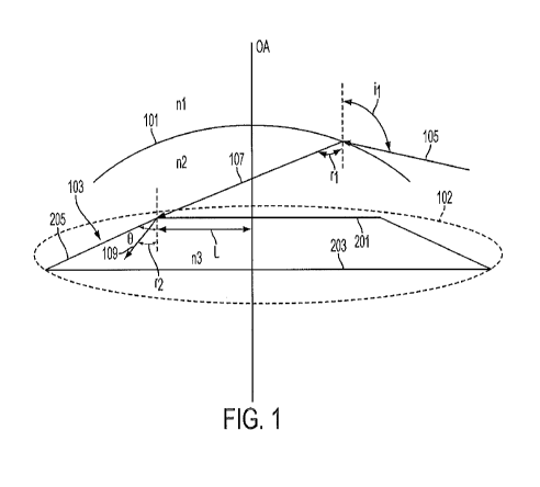
[0015] Figure 1 is a schematic representation of certain aspects of a
human eye
with an artificial IOL positioned therein configured such that the most
peripheral rays that
enter the eye are incident on and anterior optical surface of the IOL and not
incident on a
peripheral region, such as an edge of the IOL.
[0016] Figure 2 is a schematic perspective view of an implementation
of the IOL
depicted in Figure 1, showing a central optical axis.
[0017] Figure 3 is a cross-sectional view of another implementation of
an IOL in
which the most peripheral rays that enter the eye and are incident on and
refracted by the
IOL are not incident on an edge of the IOL.
[0018] Figures 4A through 4C are top plan views of substantially oval,
elliptical,
and rectangular preferred IOL embodiments.
DETAILED DESCRIPTION OF THE PREFERRED EMBODIMENT
[0019] A human eye includes a transparent crystalline biconvex lens
which can
focus light from objects over a wide range of distances on the retina. The
natural lens allows
the eye to focus on the objects at various distances by changing its shape
thereby changing its
focal length. The ability of the lens to change its shape to adjust the focal
length is known as
accommodation. The lens is housed in a structure known as the capsular bag
102. During
natural accommodation, the capsular bag is acted on by a ciliary muscle and
zonular fibers
(also known as zonules) in the eye, which can pull on the capsular bag to
change its shape.
-5-
CA 02882721 2014-10-07
WO 2014/005074 PCT/US2013/048682
The motion of the capsular bag generally deforms the lens in order to change
its power, so
that the eye can focus on objects at varying distances away from the eye.
[0020] In a healthy human eye ambient light is refracted into the eye
by the
cornea 101 and focused by the lens on the retina to form an image. The image
is produced
by the combination of the optical powers of the cornea 101, the capsular bag
102 and the
lens, all of which are generally disposed about a central optical axis OA. As
used herein, an
"anterior direction" is in the direction generally toward the cornea, while a
"posterior
direction" is generally in the direction toward the retina which is located
rearward of the
cornea 101. In a healthy human eye, an iris is disposed between the cornea 101
and the
capsular bag 102 which provides a variable pupil that dilates under lower
lighting conditions
(scotopic vision) and constricts under brighter lighting conditions (photopic
vision) to control
the amount of ambient light that enters the eye.
[0021] The average diameter of the cornea in a human eye is between
about 10
mm and 12 mm. The radius of curvature of the cornea is typically between about
6 mm and
about 11.5 mm. The average distance between the mid-point of the cornea and
the capsular
bag is between about 2.0 mm and 5.0 mm. In general, the average horizontal
diameter of the
natural lens is between 9 ¨ 10 mm and the average thickness of the natural
lens is about 4.5
mm. The pupil diameter can vary between about 1.0 mm and about 8 mm.
[0022] Figure 1 illustrates a cross-sectional view of a human eye in
which an IOL
103 is implanted in the capsular bag 102 to replace the natural lens. Figure 2
is a schematic
perspective view of the implementation of the IOL 103 illustrated in Figure 1.
Although, the
IOL 103 is illustrated as being implanted in the evacuated capsular bag 102,
it is understood
by a person having ordinary skill in the art that the IOL 103 can be a
phakic/piggy-back IOL
which acts as a secondary lens in a phakic eye that includes the natural lens.
Also, it will be
understood that the IOL 103 may have haptics to mechanically position the
optic in position
in the eye, and as further described below. The IOL 103 has an anterior
surface 201 and a
posterior surface 203 that is intersected by the central optical axis OA. In
use, the optical
axis OA may extend from the fovea of the retina to an object being viewed. The
central area
of the anterior and/or posterior surface (about a 3 mm radius from the central
optical axis)
may be monofocal, aspheric, toric, diffractive, or any combination of the
aforementioned.
The IOL 103 also includes a peripheral region 205 that is disposed between the
anterior
-6-
CA 02882721 2014-10-07
WO 2014/005074 PCT/US2013/048682
surface 201 and the posterior surface 203. The peripheral region 205 can join
the anterior
surface 201 and the posterior surface 203. In various implementations, the
peripheral region
205 may comprise a circular, elliptical or other regular shaped peripheral
zone that extends
posteriorly from the anterior surface 201.
[0023] The figures suggest that a very precise demarcation can be
provided
between discrete regions of the IOL 103, such as between the anterior zone 201
and the
peripheral region 205. However, in some embodiments, a gradual transition can
be provided
between these and other zones. For example, in various implementations, the
peripheral
region 205 can include an inflection region 207 (illustrated in Figure 3) that
forms a
transition area between the anterior surface and the peripheral region 205.
The inflection
region 207 may be inclined posteriorly with respect to the anterior surface
201 as discussed
above. In various implementations, the inflection region can include a
peripheral surface
which connects the anterior surface 201 to the peripheral region 205.
[0024] The IOL 103 is generally made of a transparent bio-compatible
material
that can be deformed. For example, in various implementations, the IOL 103 can
be made of
silicone or acrylic. The anterior and/or the posterior surface of the IOL 103
are curved such
that the IOL 103 has optical power. The anterior and/or posterior surface may
also be
comprised of a diffractive surface or an extended depth of focus structure.
Or, the lens may
be moveable with respect to the retina or other surface or deform to have
adjustable power,
as in an accommodating IOL.
[0025] The field-of-view of an average human eye is about 110 degrees
in the
horizontal direction. Accordingly, the most peripheral rays of light are
incident on the
cornea 101 at a maximum angle il of about 110 degrees with respect to the
central optical
axis OA, as illustrated by ray 105, and are refracted by the cornea 101 into
the eye, as
illustrated by ray 107. Peripheral rays that are incident on the cornea 101 at
an angle greater
than about 110 degrees with respect to the central optical axis OA will not
enter the eye,
which is the reason for the limited field-of-view of the human eye. Rather,
these rays will be
reflected by or pass through the opposite side of the cornea. If the geometry
of the cornea at
the incident point of ray 105 and the refractive index of the cornea 101 and
aqueous humor
are known, the angle of refraction r1 of the refracted ray of light 107 can be
determined from
Snell's law of refraction. Mathematically, Snell's law of refraction is
expressed as
-7-
CA 02882721 2014-10-07
WO 2014/005074 PCT/US2013/048682
sin i n2
__ = ,
where i is the angle of incidence of a ray of light that is incident from a
medium
sin r ni
having a refractive index n1 onto a medium having refractive index n2 and r is
the angle of
refraction. With reference to Figure 1, ni is the refractive index of air
which is considered to
be 1.0 and n2 is the combined refractive index of the cornea and the aqueous
humor which is
about 1.38. For a typical human eye, the most peripheral rays (e.g. ray 105)
that are incident
at an angle of about 110 degrees with respect to the central optical axis OA
are refracted by
the cornea 101 into the eye with an angle of about 80 degrees. In other words,
for a typical
human eye, r1 is about 80 degrees.
[0026] The
most peripheral rays that are refracted into the eye by the cornea (e.g.
ray 107) are incident on the anterior surface 201 of the IOL 103 and refracted
into IOL 103
in accordance with Snell's law of refraction, as illustrated by ray 109. The
angle r2 that ray
109 makes with respect to an axis parallel to the central optical axis OA,
intersecting the
peripheral region at the inflection point and extending in the posterior
direction from the
inflection point can be calculated from Snell's law of refraction if the
geometry of the IOL
103 at the incidence point of ray 107 and the refractive index of the material
of the IOL 103
is known. For the implementation illustrated in Figure 1, the angle r2 is
given by
i n
sin-1 sin
r , where n3 is the refractive index of the material of the IOL 103.
Generally,
\n3 1
for an acrylic or silicone IOL with a low refractive index, the angle r2 is
less than or equal to
about 40 degrees for a typical human eye having r1 of about 80 degrees.
[0027] In
the embodiment of Figure 1, the IOL 103 is configured such that the
peripheral region 205 is disposed laterally of the point of incidence of the
ray 107 with the
anterior surface 201 of the IOL 103. Additionally, the peripheral region 205
is disposed
away from the trajectory of the refracted ray 109. In other words, the ray 109
may be
refracted by the IOL 103 along a path therethrough but the path does not
intersect the
peripheral region 205. In one embodiment, the region 205 may be inclined
posteriorly from
the anterior surface 201 and is at an angle 0 greater than or equal to about
40 degrees and is
preferably greater than or equal to about 55 degrees, and more preferably
greater than or
equal to about 60 degrees. The peripheral region may be substantially straight
thus
maintaining this angle. Or if the peripheral region is comprised of a curved
portion, it may
-8-
CA 02882721 2014-10-07
WO 2014/005074 PCT/US2013/048682
be configured such that light rays will not strike the peripheral region of
the IOL. In other
words, the most peripheral rays that enter the eye and are refracted into the
IOL 103 would
not be incident on peripheral region 205 and also would not be refracted by
the IOL to pass
through the peripheral region 205. Thus, the interaction between the light
that is refracted
into the IOL 103 and the peripheral region 205 can be reduced or eliminated
which can
prevent aberrant optical effects such as positive and/or negative
dysphotopsia. Since, the
angle of inclination 0 of the peripheral region 205 depends on the phenomenon
of refraction,
in various implementations of the IOL 103, the angle of inclination 0 of the
peripheral region
205 is determined by the refractive index of the material of the IOL 103 and
the geometry of
the portion of the anterior surface 103 at which the most peripheral rays that
enter the eye are
incident. For a typical silicone or acrylic IOL, the angle 0 may be in the
range of about 40
degrees and 120 degrees, and is preferably in the range of about 40 degrees
and 60 degrees,
and more preferably in the range of about 55 degrees and 60 degrees.
[0028] As discussed above, one of the causes for negative dysphotopsia
in some
IOL designs is the creation of a shadow in the eye. The shadow can be in a
region of the
retina between two groups of rays that are incident on the retina. The first
group of rays pass
laterally of the IOL are not refracted at all by the lens. The second group of
rays, which are
immediately adjacent to the first group, are incident on the lens and are
refracted at an angle
away from the first group. This causes the two groups of rays to diverge, with
little or no
light being present in the region between the diverging rays. Thus, the region
between the
diverging rays is darker, i.e., a shadow is cast on the retina. The IOL 103 is
configured such
that the inflection region or the transition area 207 that is inclined
posteriorly from the
anterior surface 201 is disposed at a distance L from the central optical axis
OA. If angle 0 is
greater than 90 degrees, then at least a portion of the inflection region or
the transition area is
inclined anteriorly. The distance L is selected to be equal to or greater than
the outermost
point of incidence of the ray 107. This ensures that the most peripheral rays
that enter the
eye are incident on the anterior surface and not on the peripheral zone 205.
In such
implementations, negative dysphotopsia can also be reduced or mitigated since
all light that
enters the eye is incident on the anterior surface of the IOL 103 and
refracted in the preferred
-9-
CA 02882721 2014-10-07
WO 2014/005074 PCT/US2013/048682
way. In various implementations, the inflection region can be disposed at a
distance L of
about 2-5 mm from the central optical axis OA.
[0029] Although Figures 1 and 2 illustrate the IOL 103 to be polygonal
in shape,
a person having ordinary skill in the art would understand that the anterior
surface 201, the
posterior surface 203 and the peripheral region 205 can be curved to produce
the desired
power. In various implementations, the anterior surface 201 or the inflection
region can have
some curvature. In those implementations, where the peripheral region 205 is
arcuate, the
angle of the inclination of the peripheral region 205 can be taken as the
angle between an
anterior-posterior line parallel to the central optical axis and a line
connecting a point of
inflection of the peripheral region 205 closer to the anterior surface and a
point located at the
boundary between the peripheral zone 205 and the posterior surface 203. In
some
implementations, where the peripheral region 205 is arcuate, the angle of
inclination of the
peripheral region 205 can be taken as the angle between the largest chord of
the peripheral
region 205 and an axis that is parallel to the central optical axis and
extends posteriorly from
the point of inflection.
[0030] In various implementations, the IOL 103 can be designed by
selecting
parameters such as the lateral distance of the peripheral region 205 from the
central optical
axis, the curvature of the peripheral region 205, the angle of inclination of
the peripheral
region 205 such that the most peripheral rays that enter an average human eye
are incident on
the anterior surface of the IOL 103 and do not intersect the peripheral region
205 after being
refracted by the IOL 103. In some implementations, the IOL 103 can be designed
specifically for a patient's eye by taking the patient's pupil diameter, depth
of the capsular
bag from a mid-point of the cornea into consideration such that most
peripheral rays that
enter the patient's eye are incident on the anterior surface of the IOL and do
not intersect the
peripheral region 205 after being refracted by the IOL. In other
implementations, a set of
IOLs designed for different pupil diameters and different depth of the
capsular bag from a
mid-point of the cornea can be provided to suit the needs of the general
population.
[0031] Figures 4A and 4B depict the present invention (as seen from a
top plan
view) as an elliptical IOL with an optic which has a width W (measured in the
nasal-
temporal direction), that is greater than its height H (measured in the
superior-inferior
direction). Figure 4C depicts an optic that is generally rectangular in shape
with the width W
-10-
CA 02882721 2014-10-07
WO 2014/005074 PCT/US2013/048682
again greater than its height H. The width of the optic as measured on the
anterior side of the
optic is in the range of about 6.25 mm-10 mm and is preferably about 8-10 mm
or more
preferably between about 9-10 mm. Though the upper limit of width of the
capsular bag is
typically about 10 mm, it is envisioned that the width of the optic as
disclosed herein may be
configured to match the capsular bag. The height of the optic as measured on
the anterior
side of the optic is in the range of about 4.5 mm-9 mm and is preferably about
7-9 mm, or
more preferably about 7-8 mm. The thickness of the central area of the optic
will depend on
the optical characteristics (e.g. refractive versus diffractive) desired for
the central and as
known to those skilled in the art. The thickness of the noncentral area may be
between 0.01
mm and 1.0 mm. This thickness may be constant or may vary, for example, by
tapering
toward the peripheral zone. In certain preferred embodiments, the thickness at
the edge of
the IOL (as measured from the point of inflection to the posterior surface in
a line parallel to
the optical axis) on the nasal and/or temporal sides will be less than or
about 0.03 mm or
preferably less than or about 0.02 mm or even more preferably less than or
about 0.01 mm
(though the superior and inferior sides may also be so dimensioned). Such
embodiments
may allow the IOL to be folded/ compressed in such a manner to allow the IOL
through a
small diameter inserter using less force. While substantially oval, elliptical
and rectangular
embodiments are depicted in Figures 4A through 4C, other shapes encompassed
herein
include substantially poly-angle shapes such as a triangle, square, and other
poly-angle basic
shapes. At least two haptics are coupled to the optic at opposing ends, on the
superior and
inferior sides of the optic. As in the embodiments disclosed above, these
configurations
provide stability while substantially reducing or eliminating dysphotopsia and
increasing
field of view.
[0032] The above described design considerations can also be used to
design
implementations or contact lenses, spectacles or other ophthalmologic visual
aid devices to
avoid aberrant optical effects.
[0033] The description of the invention and its applications as set
forth herein is
illustrative and is not intended to limit the scope of the invention.
Variations and
modifications of the embodiments disclosed herein are possible, and practical
alternatives to
and equivalents of the various elements of the embodiments would be understood
to those of
ordinary skill in the art upon study of this patent document. These and other
variations and
-11-
CA 02882721 2014-10-07
WO 2014/005074 PCT/US2013/048682
modifications of the embodiments disclosed herein may be made without
departing from the
scope and spirit of the invention.
-12-