Note: Descriptions are shown in the official language in which they were submitted.
CA 02882759 2016-08-23
DETECTION OF THE NTRK1-MPRIP GENE FUSION FOR CANCER DIAGNOSIS
FIELD OF THE INVENTION
The present invention generally relates to markers, methods and assay kits for
the
identification of lung cancer patients predicted to respond to specific cancer
therapies.
BACKGROUND OF THE INVENTION
Lung cancer remains a leading cause of mortality in cancer worldwide and is
mostly
represented by non-small cell lung cancer (NSCLC). NSCLC is increasingly being
recognized as a
heterogeneous set of diseases based both upon histology as well as molecular
characteristics. The
identification of these molecular subsets is relevant as there is a growing
number of targeted
therapies that can effectively inhibit activated oncogenes leading to improved
clinical outcomes for
patients.
The first important oncogenic fusion in lung cancer was discovered in 2007 by
Soda and
colleagues. The anaplastic lymphoma kinase gene (ALK) was activated by fusion
with the
.. echinoderm microtubule-associated protein-like 4 (EML4). This fusion gene
resulted in
constitutive activation of the ALK tyrosine kinase domain with activation of
downstream signaling
pathways and transformed cell growth. Since this discovery, TFG and KIF5B have
also been
identified as fusion partners for ALK in NSCLC. Crizotinib, a tyrosine kinase
inhibitor (TKI) with
multiple targets, is currently approved by the US FDA for the treatment of
patients with advanced
NSCLC proven to be ALK positive (ALK+) and the only FDA approved method for
identifying
ALK positivity is fluorescence in situ hybridization (FISH) using break-apart
(BA) probes specific
for regions 5' and 3' of the common breakpoint in rearranged ALK. ROS1 is
another receptor
tyrosine kinase (RTK) recently found to be activated by gene fusions in NSCLC.
The first cancer-
related genomic rearrangement involving ROS1, an intra-chromosomal deletion on
chromosome
6q21 fusing the 5' region of GOPC to the 3' region of ROS1, was reported in
glioblastoma. In the
last five years, 7 different fusions activating ROS1 were identified.
Importantly, the ROS1 kinase
domain is retained in all of these fusion events and the expressed fusion
genes have been reported
to be oncogenic. FISH has been a technical platform commonly used to diagnosis
these
rearrangements, as the nature of the assay allows it to, in theory, detect any
all cases in which the
ROS1 gene has undergone rearrangement. Recent data also support that lung
cancer patients
harboring ROS1 gene fusions also respond to crizotinib supporting the clinical
utility in
identifying these patients.
Four recent studies describe the 3rd gene activated by fusion in lung cancer,
RET
(Rearranged during Transfection). RET is a well known RTK, with an oncogenic
role in papillary
.. and follicular thyroid carcinoma through activation by gene fusions (RET-
PTC). Patients whose
tumors harbor this fusion respond well to vandetanib, an oral TKI that targets
VEGFR, EGFR and
RET. A total of 33 patients were reported harboring KIF5B-RET fusions
involving 7 different
1
CA 02882759 2015-05-11
WO 2014/036387 PCT/US2013/057495
breakpoints. These molecular rearrangements were identified through multiple
technologies used
to screen more than 2,000 lung adenocarcinomas. In the 3 larger series
screened, likely with lower
level of pre-selection, frequency of KIF5B-RET fusion ranged from 1% to 2%.
There continues to be a need in the field for identification of further
molecular markers,
including oncogenic fusion markers, to facilitate more effective detection and
treatment of lung
cancer.
SUMMARY OF THE DISCLOSURE
The present invention is based on the discovery of a novel gene fusion
comprising the
NTRKI gene that is indicative of lung cancer and also indicative of which
patients may respond to
cancer therapy comprising therapeutic administration of tyrosine kinase
inhibitors.
Accordingly, in one embodiment, the invention comprises a method for
determining if a
lung cancer patient is predicted to respond to the administration of a
chemotherapeutic regimen.
The method comprises detecting in a sample of tumor cells from the patient the
presence or
absence of a marker, wherein the marker comprises a gene fusion comprising a
NTRKI-MPRIP
gene fusion (NTRK1 encodes the TRKA protein), and wherein the presence or
absence of the
marker is indicative of whether the cancer patient will respond to the
administration of the
chemotherapeutic regimen.
In various embodiments, the chemotherapeutic regimen may include
administration of one
or more of the following: a tyrosine kinase inhibitor, a HSP90 inhibitor (or
other chaperone
inhibitor), an inhibitor that targets tyrosine kinase downstream signalling
cascade, or combinations
thereof. Such inhibitors arc well known in the art and are commercially
available. All such
inhibitors are encompassed in the present invention. For instance, in some
embodiments, the
tyrosine kinase inhibitor may be a TrkA inhibitor, examples of which include,
but are not limited
to, crizotinib (PF-02340166), ponatinib (AP24534), dovitinib (TK-258), CEP-
701, or rebastinib
(DCC-2036). Examples of HSP90 inhibitors include, but are not limited to,
geldanamycin,
herbimycin, 17-AAG, PU24FC1, STA-9090, IPI-504, and AUY-922. Examples of
inhibitors that
target tyrosine kinase receptor downstream signalling cascade include, without
limitation,
elumetinib (AZD6244) and MK2206.
In some embodiments, a level of the marker is determined and compared to a
standard
level or reference range. In some embodiments, the standard level or reference
range is
determined according to a statistical procedure for risk prediction.
In some embodiments, the presence of the marker may be determined by detecting
the
presence of a polynucleotide or a polypeptide. In some embodiments, the method
may comprise
detecting the presence of the polypeptide using a reagent that specifically
binds to the polypeptide
or a fragment thereof. The reagent may be an antibody, an antibody derivative,
or an antibody
fragment.
In some embodiments, the presence of the marker may be determined by obtaining
RNA
2
CA 02882759 2015-05-11
WO 2014/036387 PCT/US2013/057495
from the sample; generating cDNA from the RNA; amplifying the cDNA with
primers specific for
the marker; and determining from the sequence of the amplified cDNA the
presence of absence of
the marker in the sample. In some embodiments, the presence of the marker may
be determined by
Fluorescent In Situ Hybridization (FISH).
The methods of the present invention may further comprise comparing the
expression
level of the marker in the sample to a control level of the marker selected
from the group
consisting of: a) a control level of the marker that has been correlated with
beneficial response to
the administration of a chemotherapeutic regimen including one or more kinase
inhibitor(s); and a
control level of the marker that has been correlated with lack of beneficial
response to the
administration of a chemotherapeutic regimen including one or more kinase
inhibitor(s); and
b)selecting the patient as being predicted to respond to the administration of
a chemotherapeutic
regimen including one or more kinase inhibitor(s), if the expression level of
the marker in the
sample is statistically similar to, or greater than, the control level of
expression of the marker that
has been correlated with sensitivity to the administration of a
chemotherapeutic regimen including
one or more kinase inhibitor(s), or c) selecting the patient as being
predicted to not respond to the
administration of a chemotherapeutic regimen including one or more kinase
inhibitor(s), if the
level of the marker in the sample is statistically less than the control level
of the marker that has
been correlated with beneficial response to the administration of a
chemotherapeutic regimen
including one or more kinase inhibitor(s).
In some embodiments, the methods may further comprise comparing the expression
level
of the marker in the sample to a level of the marker in a second patient
predicted to not respond to
the administration of a chemotherapeutic regimen including one or more kinase
inhibitor(s), and,
selecting the patient as being predicted to respond to the administration of a
chemotherapeutic
regimen including one or more kinase inhibitor(s), if the expression level of
the marker in the
sample is greater than the level of expression of the marker in the second
patient, or, selecting the
patient as being predicted to not respond to the administration of a
chemotherapeutic regimen
including one or more kinase inhibitor(s), if the level of the marker in the
sample is less than or
equal to the level of expression of the marker in the second patient. In some
embodiments the
patient is human.
In a further embodiment, the present invention includes an assay system for
predicting
patient response or outcome to tyrosine kinase anti-cancer therapy comprising
a means to detect at
least one of: a) the presence of a gene fusion comprising a NTRKI-MPRIP gene
fusion; b) the
level of expression of a gene transcript encoded by a NTRKI-MPRIP gene fusion;
c) the presence
of a protein encoded by a NTRKI-MPRIP gene fusion; d) the level of a protein
encoded by a
NTRKI-MPRIP gene fusion; and, e) the activity of a protein encoded by a NTRKI-
MPRIP gene
fusion. In some embodiments, the means to detect comprises nucleic acid probes
comprising at
least 10 to 50 contiguous nucleic acids of NTRK1 gene, or complementary
nucleic acid sequences
3
CA 02882759 2015-05-11
WO 2014/036387 PCT/US2013/057495
thereof. In some embodiments, the means to detect comprises binding ligands
that specifically
detect polypeptides encoded by a NTRK1-MPRIP gene fusion. In some embodiments,
a surface of
the assay system comprises a chip, array, or fluidity card. In some
embodiments, the assay system
further comprises: a control selected from the group consisting of:
information containing a
predetermined control level of a gene transcript encoded by a NTRK1-MPRIP gene
fusion that has
been correlated with response to the administration of a chemotherapeutic
regimen including one
or more kinase inhibitor(s); and information containing a predetermined
control level of a gene
transcript encoded by a NTRK1-MPRIP gene fusion that has been correlated with
a lack of
response to the administration of a chemotherapeutic regimen including one or
more kinase
inhibitor(s).
In another embodiment, the present invention includes a method of diagnosing a
specific
type of lung cancer in a subject, comprising detecting in a sample of cells
from the subject the
presence of a NTRK1-MPRIP gene fusion marker, wherein the presence of the
marker is indicative
of whether the subject has the specific type of lung cancer. In some
embodiments, the presence of
the gene fusion marker is detected by RT-PCR or FISH. In some embodiments, the
presence of the
gene fusion marker is detected by detecting the polypeptide encoded by the
gene fusion marker. In
some embodiments, the polypeptide is detected by using a reagent that
specifically binds to the
polypeptide or a fragment thereof.
Other features and advantages of the invention will become apparent to one of
skill in the
art from the following detailed description.
BRIEF DESCRIPTION OF THE DRAWINGS
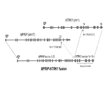
Figure 1 shows the chromosomal and exon maps of MPRIP gene.
Figure 2 shows the chromosomal and exon maps of NTRKI gene.
Figure 3 shows the arrangement and data confirming NTRK1 gene fusions in lung
cancer
samples. Figure 3A is a schematic of genomic rearrangement from tumor samples
harboring
NTRK1-MPRIP. Figure 3B shows RT-PCR demonstrating mRNA expression of the novel
fusion
transcripts. RNA extracted from formalin-fixed paraffin-embedded tumor sample
harboring the
NTRKI-MPRIP was subject to RT-PCR followed by agarose gel electrophoresis and
DNA
sequencing. The following abbreviates are used: MPRIP (M), CD74 (C), NTRK1
(N), and exon
(ex). Sanger sequencing chromatograms were obtained of RT-PCR product of RNA
isolated from
tumor samples with NTRK1-MPRIP fusion. SEQ ID NO:1 is the complete cDNA
sequence of
NTRK1-MPRIP fusion (M21;N14). The cDNA was cloned from a frozen tumor sample
from the
patient in which this fusion was first identified. Capital letters represent
nucleotides contained
within the open reading frame.
Figure 4 shows the RT-PCR primer read sequences for detection of NTRK1-MPRIP
gene
fusion. Figure 4A shows the forward read sequence with MPRIP CC3F1 primer.
Figure 4B shows
the reverse read sequence with NTRK1 Y490R1 primer.
4
CA 02882759 2016-08-23
Figure 5 shows the design of the NTRKI-MPRIP fusion FISH probes.
Figure 6 shows the design of the 5'-3' NTRKI Break-Apart FISH probe set
aligned
against the NTRK I encoding region of chromosome 1q23.1.
Figures 7A and 7B show FISH images obtained from the normal cell line GM09948
using
.. the NTRKI-MPRIP gene fusion probes, providing single clone validation in a
normal cell line (
Diploid,2N). Figure 7A: Clone RP11-1038N13 (3'NTRK1) and RP11-125116
(5'MPRIP). Figure
7B: Clone RP11-1059C21 (3'NTRK1) and Rp11-796J19 (5'MPRIP).
Figures 8A and 8B show FISH images obtained from tissue sections that are
negative for
NTRK1-MPRIP gene fusion using the NTRK1-MPRIP gene fusion probes. Figure 8A
shows
specimen S-12-047486 showing low copy number for both genes. Figure 8B shows
specimen 5-
12-047098, showing high copy number for 3' NTRK1 and Mid/low copy number for
5' MPRIP.
Figures 9A and 9B show FISH images obtained from tissue sections that are
positive for
NTRIC1-MPRIP gene fusion using the NTRK1-MPRIP gene fusion probes.
Figure 9A showing low copy number for both genes with approximately one
red/green fusion
(positive pattern) per tumor cell. Figure 9B showing gene amplification of the
fused red/green with
mid/low copy number of single reds and single greens per tu or cell.
Figure 10 shows the testing of NTRK I break-apart FISH probe. Figure 10a shows
cell line
GM09948 with a normal karyotype showing metaphase spread and interphase nuclei
demonstrating close proximity of the 5' and 3' signals indicating an intact
NTRKI gene. Figure
10b shows KM12 cells which harbor a TPM3-NTRK1 gene fusion showing clear
separation of the
5' and 3' signals indicating a rearrangement of the NTRKI gene. Figure 10c
shows a break-apart
FISH analysis of NTRKI-MPRIP samples showing clear separation of 5' and 3'
signals
corresponding to the NTRKI gene. Figure 10d shows break-apart FISH analysis of
a tumor sample
without an NTRK1 gene rearrangement showing close approximation of the
green/red signals
(indicated by arrow).
Figure 11 shows FISH images obtained from tissue sections that are positive
for NTRKI
gene rearrangement using the NTRK1 break-apart probes. Specimen S12-6889 BI
Hybridized with
the 5'NTRK1/3'NTRK1 Break Apart probe set. Cells show both the 'positive'
pattern of split and
the 'normal' pattern of fused signals.
Figure 12A and B show immunoblot analyses of cell lysates from 293T cells
expressing
TRKA. Figure 12A, Expression of TRKA (with HA tag) or empty vector
demonstrates expression
of a ¨115-120kD protein detected by an HA-specific antibody (left, Cell
Signaling) and a TRKA-
specific antibody (right, Santa Cruz, SC-118). Figure 12B, Immunoprecipation
using an HA-
specific antibody (Cell Signaling) followed by immunoblot using the same
antibody (left) or a
phosphotyrosine specific antibody (right, Miilipore, 4G10) following treatment
with 1 i.tM of the
indicated inhibitors or DMSO (control) for 5 hours. Figure 12C shows the
expression of NTRK I-
MPRIP yields a chimeric protein that is autophosphorylated. Immunoblot
analysis of 293T cells
5
CA 02882759 2016-08-23
transiently transfected with empty vector (EV), full length NTRKI cDNA, NTRK1-
MPRIP cDNA
compared to tumor cells from a frozen pleural fluid sample or early passage
cells in culture
(CUTO-3) from the index patient with the NTRKI-MPRIP fusion gene. Figure 12D
is a schematic
demonstrating fusion break-point and critical domains of predicted fusion
protein products.
Figure 13A shows immunoblot analyses of downstream signalling of TRKA
following
treatment with tyrosine kinase inhibitors. SDS-PAGE of 293T cell lysates with
expression of
TRKA-HA or empty vector in the presence or absence of NGF (10 minutes) and the
presence or
absence of the indicated tyrosine kinase inhibitors at li.tM for 5 hours.
Membranes were probed
with antibodies to TRKA phosphotyrosine 490, 674, and 675 (Cell Signaling),
total TRKA (anti-
HA, Cell Signaling), AKT phosphoserine 473 (Cell Signaling), total AKT (Cell
Signaling),
phosphorylated ERK p42/44 (Cell Signaling), total ERK p42/44 (Cell Signaling),
and gamma-
tubulin (Santa Cruz, SC-8035).
Figure 13B shows the expression of NTRK I -MPRIP induces activation of
downstream
MAPK, AKT, and STAT3 pathways. TRKA (NTRKI) fusions are autophosphorylated and
activate key downstream signaling pathways. Representative immunoblot analyses
(n = 3) of cell
lysates from Ba/F3 cells expressing RIP-TRKA, the protein product of NTRK1-
MPRIP but not its
kinase dead (KD) variant display phosphorylation of critical tyrosine residues
and activation of
pAKT, pERK and pSTAT3 in the absence of IL-3.
Figure 14 demonstrates that NTRKI gene fusions support cellular proliferation
of Ba/F3
cells in the absence of IL-3. MTS assay of Ba/F3 demonstrates that cells
expressing RIP-TRKA,
CD74-TRKA, EML4-ALK, or full length TRKA supplemented with NGF proliferate in
the
absence of IL-3, whereas Ba/F3 cells expressing EV or the kinase dead variant
of RIP-TRKA do
not proliferate (n = 3). Values are mean SEM.
Figure 15 demonstrates that NTRKI fusions support anchorage independent
growth.
Representative images (n = 4) from anchorage independent growth assays of
N1H3T3 cells
expressing EV, RIP-TRKA-kinase dead (KD), or RIP-TRKA in soft agar.
Figure 16 shows NTRKI-MPRIP fusion proteins induce tumorigenesis. NIH3T3 cells
expressing NTRK1-MPRIP ("RIP-TRKA"), NTRK1-MPRIP kinase dead ("RIP-TRKA-Kinase
Dead"), EML4-ALK or Empty Vector were injected into the flanks of nude mice
and observed for
tumor growth. The number of mice with tumors compared to the total number mice
injected are
indicated.
Figure 17 shows RNAi knockdown of NTRK1 inhibits cell proliferation in a cell
line
harboring TPM3-NTRK1. KM12 cells were analyzed by MTS proliferation assay 96hr
after
siRNA transfection (n = 3). ANOVA analysis followed by Bonferroni's multiple
comparison test
indicated a significant inhibition of proliferation induced by siRNA 1
(p<0.05). Values represent
the mean + SEM. KM12 cells were transfected with siRNAs targeting NTRKI and
then harvested
48hr later. Cell lysates were analyzed by immunoblot to detect TRKA, pERK1/2
and ERK1/2.
6
CA 02882759 2015-05-11
WO 2014/036387 PCT/US2013/057495
Figure 18 shows drug inhibition of activation of TRKA and downstream
signaling. Ba/F3
cells expressing NTRK1-MPRIP (RIP-TRKA) or empty vector (EV) were lysed after
5h of
treatment with the indicated doses of drugs (ARRY-470, crizotinib, CEP-701,
ARRY-772, or
ARRY-523) or DMSO control (C).
Figure 19 shows that drug treatment inhibits NTRK1 fusion-mediated Ba/F3 cell
proliferation and shows the treatment of index patient with crizotinib.
Treatment of Ba/F3 cells
expressing NTRK1 fusions with TRKA inhibitors inhibits cell proliferation as
measured by MTS
assay (n = 5). Figure 19(a) Values represent the mean SEM. Ba/F3 cells
expressing NTRK1-
MPRIP demonstrate inhibition of proliferation by the pan-TRK inhibitors, ARRY-
470, -523, and -
772 and the multi-kinase inhibitor, CEP-701, but not the EGFR inhibitor,
gefitinib. Figure 19(b)
Crizotinib leads to inhibition of Ba/F3 expressing NTRK1 fusions, similar to
Ba/F3 cells
expressing ALK or ROS1 fusion constructs. The half maximal inhibitory
concentration (IC50)
values are listed (nM).
Figure 20 shows the drug treatment of Ba/F3 cells in the presence of IL-3.
Ba/F3 cells
expressing empty vector were grown in the presence of IL-3 and treated with a
range of doses of
ARRY-470, CEP-701, crizotinib, or gefitinib. IC50 values are listed (n = 3).
Values represent the
mean + SEM.
Figure 21 shows the expression and drug inhibition of NTRK1 fusions in NIH3T3
cells.
N1H3T3 cells expressing RIP-TRKA were treated with the indicated doses of
drugs for 5h prior to
cell lysis and immunoblot analysis of pTRKA, TRKA, pAKT, AKT, pERKI/2, ERK1/2,
pSTAT3,
and STAT3 as indicated.
Figure 22 shows the inhibition of anchorage-independent growth by drugs with
TRKA
activity. Figure 22a, NIH3T3 cells expressing empty RIP-TRKA were seeded in
triplicate in soft
agar and treated with DMSO (control) or 200nM of ARRY-470, crizotinib, or CEP-
701 for 2
weeks (n = 4). Representative images are shown. Figure 22b, The total colony
area for each plate
was quantified using MetaMorph software and plotted for each condition. Values
represent the
mean SEM.
Figure 23 shows the short term cell culture from index patient showing the
NTRK1-
MPRIP fusion. Colorado University Thoracic Oncology (CUTO) 3 cells were
derived from a
pleural effusion from the index patient harboring the NTRK1-MPRIP gene fusion.
Left: NTRK1
FISH analysis of CUTO-3 cells showing a positive signal (split green/red
signals). Right:
Immunoblot analysis of CUTO-3 cells demonstrating inhibition of pTRKA and pERK
by the pan-
TRK inhibitor, ARRY-470.
Figure 24 shows drug treatment of KM12 cells. KM12 cells harboring the TPM3-
NTRK1
fusion were lysed following 5h treatment with the indicated doses of
inhibitors and subject to
immunoblot analysis (n = 3).
Figure 25 shows that drug treatment of KM12 cells inhibits proliferation.
Proliferation of
7
CA 02882759 2015-05-11
WO 2014/036387 PCT/US2013/057495
KM12 cells treated with the indicated drugs and doses were assayed for cell
proliferation by MTS
assay. KM12 cells are inhibited by ARRY-470, CEP-701, and crizotinib, but not
gefitinib.
Figure 26 shows that TRKA inhibition results in the accumulation of KMI2 cells
in GI
phase. KM12 cells were treated with the indicated doses of drugs for 24hr.
Cells were then stained
with propidium iodide and analyzed by flow cytometry. ModFit analysis was used
to quantify cell
cycle profiles (n = 3). Values are the mean SEM.
Figure 27 shows that drug treatment with TRKA inhibitors induces apoptosis in
KM12
cells. Figure 27A, KM12 cells were treated for 24h with the indicated drugs
and doses, trypsinized,
stained with YO-PRO and propidium iodide (PI), and analyzed by flow-
cytometry. The percent
of cells undergoing apoptosis (YO-PRO positive and PI negative) are plotted
(n = 4). Values
represent the mean SEM. Figure 27B TRKA inhibitors induce cleavage of PARP-
1. KM12
cells were treated for 24h with the indicated drugs and doses. Cells were
lysed, separated by
SDS-PAGE and subject to immunoblot analysis with the indicated antibodies.
Figure 28 shows histopathology from index patient harboring NTRKI-MPRIP
demonstrating lung adenocarcinoma. Figure 28 (a): Needle core biopsy of
primary lung left lower
lung mass showing adenocarcinoma. Figure 28(b): Cell block of fine needle
aspirate from the
same procedure showing tumor cells. Figure 28(c): TTF-1 immunohistochemistry
(IHC)
demonstrating strong nuclear staining in tumor cells. Figure 28(d):
Thyrogloblin IHC
demonstrating negative staining in tumor cells. Representative images are
shown.
Figure 29 shows the results of treatment when the index patient (NTRKI-MPRIP)
consented to treatment with crizotinib 250mg PO BID (off-protocol, off-label)
given lack of other
therapeutic options. Figure 29A: CT scan of the chest before and after 28d of
crizotinib and Figure
29B: serial CA125 tumor marker levels during crizotinib treatment.
Figure 30 shows the signal configuration "Dot," which is the typical round and
compact
signal and comparison with other signals.
DETAILED DESCRIPTION OF THE DISCLOSURE
The present inventors have discovered that fusion of the MPRIP and NTRKI genes
is
indicative of the presence of a specific type of lung cancer. This gene fusion
is also indicative of
patient clinical response to treatment with tyrosine kinasc inhibitors. This
gene fusion, and the
levels of the protein encoded by this gene fusion, along with clinical
parameters can be used as
biological markers to diagnose a specific type of lung cancer and to assess
cancer patient response
to treatment with tyrosine kinase inhibitors.
According to one definition, a biological marker is "a characteristic that is
objectively
measured and evaluated as an indicator of normal biologic processes,
pathogenic processes, or
pharmacological responses to therapeutic interventions." NIH Marker
Definitions Working Group
(1998). Biological markers can also include patterns or ensembles of
characteristics indicative of
particular biological processes ("panel of markers"). The marker measurement
can be increased or
8
CA 02882759 2015-05-11
WO 2014/036387 PCT/US2013/057495
decreased to indicate a particular biological event or process. In addition,
if a marker
measurement typically changes in the absence of a particular biological
process, a constant
measurement can indicate occurrence of that process.
Marker measurements may be of the absolute values (e.g., the molar
concentration of a
molecule in a biological sample) or relative values (e.g., the relative
concentration of two
molecules in a biological sample). The quotient or product of two or more
measurements also may
be used as a marker. For example, some physicians use the total blood
cholesterol as a marker of
the risk of developing coronary artery disease, while others use the ratio of
total cholesterol to
HDL cholesterol.
In the present invention, the markers may be used for diagnostic, prognostic,
therapeutic,
drug screening and patient stratification purposes (e.g., to group patients
into a number of
"subsets" for evaluation), as well as other purposes described herein,
including evaluation of the
effectiveness of a potential cancer therapeutic.
The practice of the invention employs, unless otherwise indicated,
conventional methods
of analytical biochemistry, microbiology, molecular biology and recombinant
DNA generally
known techniques within the skill of the art. Such techniques are explained
fully in the literature.
(See, e.g., Sambrook et al. Molecular Cloning: A Laboratory Manual. 3rd, ed.,
Cold Spring
Harbor Laboratory, Cold Spring Harbor Laboratory Press, Cold Spring Harbor,
NY, 2000; DNA
Cloning: A Practical Approach, Vol. I & II (Glover, ed.); Oligonucleotide
Synthesis (Gait, ed.,
Current Edition); Nucleic Acid Hybridization (Hames & Higgins, eds., Current
Edition);
Transcription and Translation (Hames & Higgins, eds., Current Edition); CRC
Handbook of
Parvoviruses, Vol. I & II (Tijessen, ed.); Fundamental Virology, 2nd Edition,
Vol. I & II (Fields
and Knipe, eds.)).
The terminology used herein is for describing particular embodiments and is
not intended
to be limiting. As used herein, the singular forms "a," "and" and "the"
include plural referents
unless the content and context clearly dictate otherwise. Thus, for example, a
reference to "a
marker" includes a combination of two or more such markers. Unless defined
otherwise, all
scientific and technical terms are to be understood as having the same meaning
as commonly used
in the art to which they pertain. For the purposes of the present invention,
the following terms are
defined below.
As used herein, the term "marker" includes polypeptide markers and
polynucleotide
markers. For clarity of disclosure, aspects of the invention will be described
with respect to
"polypeptide markers" and "polynucleotide markers." However, statements made
herein with
respect to "polypeptide markers" are intended to apply to other polypeptidcs
of the invention.
Likewise, statements made herein with respect to "polynucleotide" markers are
intended to apply
to other polynucleotides of the invention, respectively. Thus, for example, a
polynucleotide
described as encoding a "polypeptide marker" is intended to include a
polynucleotide that encodes:
9
CA 02882759 2015-05-11
WO 2014/036387 PCT/US2013/057495
a polypeptide marker, a polypeptide that has substantial sequence identity to
a polypeptide marker,
modified polypeptide markers, fragments of a polypeptide marker, precursors of
a polypeptide
marker and successors of a polypeptide marker, and molecules that comprise a
polypeptide
marker, homologous polypeptide, a modified polypeptide marker or a fragment,
precursor or
successor of a polypeptide marker (e.g., a fusion protein).
As used herein, the term "polypeptide" refers to a polymer of amino acid
residues that has
at least 5 contiguous amino acid residues, e.g., 5, 6, 7, 8, 9, 10, 11 or 12
or more amino acids long,
including each integer up to the full length of the polypeptide. A polypeptide
may be composed of
two or more polypeptide chains. A polypeptide includes a protein, a peptide,
an oligopeptide, and
an amino acid. A polypeptide can be linear or branched. A polypeptide can
comprise modified
amino acid residues, amino acid analogs or non-naturally occurring amino acid
residues and can be
interrupted by non-amino acid residues. Included within the definition are
amino acid polymers
that have been modified, whether naturally or by intervention, e.g., formation
of a disulfide bond,
glycosylation, lipidation, methylation, acetylation, phosphorylation, or by
manipulation, such as
conjugation with a labeling component. Also included are antibodies produced
by a subject in
response to overexpressed polypeptide markers.
As used herein, a "fragment" of a polypeptide refers to a single amino acid or
a plurality of
amino acid residues comprising an amino acid sequence that has at least 5
contiguous amino acid
residues, at least 10 contiguous amino acid residues, at least 20 contiguous
amino acid residues or
at least 30 contiguous amino acid residues of a sequence of the polypeptide.
As used herein, a
"fragment" of polynucicotide refers to a single nucleic acid or to a polymer
of nucleic acid
residues comprising a nucleic acid sequence that has at least 15 contiguous
nucleic acid residues,
at least 30 contiguous nucleic acid residues, at least 60 contiguous nucleic
acid residues, or at least
90% of a sequence of the polynucleotide. In some embodiment, the fragment is
an antigenic
fragment, and the size of the fragment will depend upon factors such as
whether the epitope
recognized by an antibody is a linear epitope or a conformational epitope.
Thus, some antigenic
fragments will consist of longer segments while others will consist of shorter
segments, (e.g. 5, 6,
7, 8, 9, 10, 11 or 12 or more amino acids long, including each integer up to
the full length of the
polypeptide). Those skilled in the art are well versed in methods for
selecting antigenic fragments
of proteins.
In some embodiments, a polypeptide marker is a member of a biological pathway.
As
used herein, the term "precursor" or "successor" refers to molecules that
precede or follow the
polypeptide marker or polynucleotide marker in the biological pathway. Thus,
once a polypeptide
marker or polynucleotide marker is identified as a member of one or more
biological pathways, the
present invention can include additional precursor or successor members of the
biological
pathway. Such identification of biological pathways and their members is
within the skill of one
in the art.
CA 02882759 2015-05-11
WO 2014/036387 PCT/US2013/057495
As used herein, the term "polynucleotide" refers to a single nucleotide or a
polymer of
nucleic acid residues of any length. The polynucleotide may contain
deoxyribonucleotides,
ribonucleotides, and/or their analogs and may be double-stranded or single
stranded. A
polynucleotide can comprise modified nucleic acids (e.g., methylated), nucleic
acid analogs or
non-naturally occurring nucleic acids and can be interrupted by non-nucleic
acid residues. For
example a polynucleotide includes a gene, a gene fragment, cDNA, isolated DNA,
mRNA, tRNA,
rRNA, isolated RNA of any sequence, recombinant polynucleotides, primers,
probes, plasmids,
and vectors. Included within the definition are nucleic acid polymers modified
either naturally, or
by intervention.
As used herein, a component (e.g., a marker) is referred to as "differentially
expressed" in
one sample as compared to another sample when the method used for detecting
the component
provides a different level or activity when applied to the two samples. A
component is referred to
as "increased" in the first sample if the method for detecting the component
indicates that the level
or activity of the component is higher in the first sample than in the second
sample (or if the
component is detectable in the first sample but not in the second sample).
Conversely, a
component is referred to as "decreased" in the first sample if the method for
detecting the
component indicates that the level or activity of the component is lower in
the first sample than in
the second sample (or if the component is detectable in the second sample but
not in the first
sample). In particular, marker is referred to as "increased" or "decreased" in
a sample (or set of
samples) obtained from a lung cancer subject (or a subject who is suspected of
having lung cancer,
or is at risk of developing lung cancer) if the level or activity of the
marker is higher or lower,
respectively, compared to the level of the marker in a sample (or set of
samples) obtained from a
non-lung cancer subject, or a reference value or range.
The novel gene fusion marker of the present invention was identified as
follows: the
presence of an oncogene driver gene abnormality was investigated in a non-
smoker patient with
lung adenocarcinoma. The patient showed no evidence of known mutations, gene
amplifications or
gene fusions associated with lung cancer. In order to pursue other possible
gene targets, genomic
DNA from a tumor biopsy sample was analyzed by targeted next generation
sequencing, which
identified the presence of a novel NTRK1 gene fusion. The gene fusion marker
was determined to
be a NTRKI-MPRIP gene fusion.
The NTRKI gene encodes the TRKA receptor tyrosine kinase. The NTRKI gene has
been
isolated from a number of species such as human, chimpanzee, dog, cow, mouse,
rat, chicken and
zebrafish and the sequence determined. All these gene sequences are known to
one skilled in the
art and are intended to be encompassed in the present invention. Gene fusions
involving NTRKI
have previously been reported in papillary thyroid cancer, but have not been
reported in lung
cancer or other malignancies.
The MPRIP gene encodes the Myosin phosphatase Rho-interacting Protein. The
MPRIP
11
CA 02882759 2015-05-11
WO 2014/036387 PCT/US2013/057495
gene has been isolated from a number of species such as human, chimpanzee,
dog, cow, mouse,
rat, chicken, zebrafish and C. elegans and the sequence determined. All these
gene sequences are
known to one skilled in the art and are intended to be encompassed in the
present invention.
This is believed to be the first instance identifying the NTRK1-MPRIP gene
fusion in any
malignancy. Customized RT-PCR assays, including novel primers, were developed
to detect the
mRNA transcript of the NTRKI-MPRIP gene fusion. The RT-PCR successfully
amplified a small
product containing sequences from both MPRIP and NTRK1, confirming expression
of a novel
gene fusion that included exon 1-21 of MPRIP and exons 14-20 of NTRKI. (See
Example 1.)
Novel FISH assays were also developed to detect the presence of the NTRKI-
MPRIP gene fusion
in clinical specimens. (See Examples 2 and 3.)
Additionally, FISH probes that would detect other NTRKI gene fusions,
regardless of the
specific 5' gene fusion partner, were also developed. (See Examples 2 and 4.)
The markers identified herein arc of significant biologic interest. Gene
fusions involving
NTRK1 have previously been reported in papillary thyroid cancer, but have not
been reported in
lung cancer or other malignancies. Thus, the NTRKI fusion gene serves as a
novel diagnostic
marker of cancer. NTRKI gene encodes the TRKA receptor tyrosine kinase. The
presence of the
gene fusion was examined in tumor samples obtained from various cancer models,
including lung
and colorectal cancers, and the sensitivity of the tumor to tyrosine kinase
inhibitors was
investigated. The objective was to use this gene fusion marker to identify a
clinically relevant
marker of cancer patient response to tyrosine kinase inhibitor treatment. The
methods used are
detailed in the Examples section of this disclosure. Several tyrosine kinase
inhibitors that are
currently in various stages of clinical development including, without
limitation, crizotinib,
ponatinib, dovitinib, rebastinib, CEP-701, AZD-7451, ARRY-470, ARRY-523, and
ARRY-772 as
well as other tyrosine kinase inhibitor compounds known in the art that are
predicted to inhibit
TRKA or oncogenic fusion proteins that contain the TRKA kinase domain, such as
NTRKI fusion
proteins. Data presented in Example 5 demonstrates that small molecule
tyrosine kinase inhibitors
inhibit activated TRKA.
In addition to the discovery of the NTRKI gene fusion marker that can be used
for the
diagnosis of, prognosis of, or other evaluation or study of cancer, the marker
may also be studied
in more detail and/or be used as target for the discovery of other modulators
of disease or
therapeutic agents.
It is believed that the NTRKI gene fusion markers, including the NTRK1-MPRIP
gene
fusion marker, are indicators of cancer patient response to tyrosine kinase
inhibitors. Accordingly,
in one aspect, the invention provides a marker, the presence or expression
level of which is
indicative of cancer patient response to tyrosine kinase inhibitors.
In another aspect, the gene fusion markers of the present invention can serve
as indicators
of cancer patient response to other targeted cancer therapies such as
administration of HSP90
12
CA 02882759 2015-05-11
WO 2014/036387 PCT/US2013/057495
inhibitors (or other chaperone inhibitors) or agents that target downstream
signalling cascades.
Such inhibitors are well known in the art and are commercially available. All
such inhibitors are
encompassed in the present invention. Examples of HSP90 inhibitors include
without limitation
geldanamycin, herbimycin, 17-AAG, PU24FC1, STA-9090, IPI-504, and AUY-922.
Examples of
-- agents that target downstream signalling cascades include selumetinib (AZD-
6244) and MK2206.
The presence of the marker may be detected by detecting a polynucleotide. In
one
embodiment, the polynucleotide may be a probe that specifically hybridizes
with the NTRK1 gene
sequences and identifies a chromosomal rearrangement involving the NTRK1 gene.
In another
embodiment, the polynucleotide may be a primer that specifically binds and
amplifies a
-- polynucleotide sequence that is indicative of the presence of the gene
fusion involving a NTRK1
gene, including the NTRKI-MPRIP gene fusion marker.
Some variation is inherent in the measurements of the physical and chemical
characteristics of the markers of the invention. The magnitude of the
variation depends to some
extent on the reproducibility of the separation means and the specificity and
sensitivity of the
-- detection means used to make the measurement. Preferably, the method and
technique used to
measure the markers is sensitive and reproducible.
The presence of the gene fusion marker may also be detected by detecting a
polynucleotide. Polypeptides corresponding to the NTRKI gene fusion markers
may include a
fragment, precursor, successor or modified version of the protein encoded by
the NTRKI- gene
-- fusion markers. In another embodiment, the invention includes a molecule
that comprises a
fragment, precursor, successor or modified polypcptide encoded by the NTRKI-
gene fusion
markers.
Another embodiment of the present invention relates to an assay system
including a
plurality of antibodies, or antigen binding fragments thereof, or aptamers for
the detection of the
-- expression of the NTRKI gene fusion markers of the invention. The plurality
of antibodies, or
antigen binding fragments thereof, or aptamers selectively bind to proteins
encoded by the NTRKI
gene fusion markers.
As used herein, the terms "patient," "subject," "a subject who has cancer" and
"cancer
patient" are intended to refer to subjects who have been diagnosed with a
cancer or are suspected
-- of having cancer. The terms "non-subject" and "a subject who does not have
cancer" are intended
to refer to a subject who has not been diagnosed with cancer, or who is cancer-
free as a result of
surgery to remove one or more tumors. A non-cancer subject may be healthy and
have no other
disease, or they may have a disease other than cancer. The NTRK1 gene has been
found to be
conserved in a number of species such as chimpanzee, dog, cow, mouse, rat,
chicken, and
-- zebrafish and their sequences are known. In some embodiments, the patient
or subject may be a
mammal. In a preferred embodiment, the patient or subject is human.
Polypeptides encoded by the NTRKI gene fusion may be isolated by any suitable
method
13
CA 02882759 2015-05-11
WO 2014/036387 PCT/US2013/057495
known in the art. Native polypeptides encoded by the NTRKI gene fusion can be
purified from
natural sources by standard methods known in the art (e.g., chromatography,
centrifugation,
differential solubility, immunoassay). In one embodiment, the polypeptides may
be isolated from
a tumor sample. In another embodiment, the polypeptides may be isolated from a
sample by
contacting the sample with substrate-bound antibodies or aptamers that
specifically bind to the
marker.
The present invention also includes polynucleotides related to the gene fusion
markers of
the present invention. In one aspect, the invention provides polynucleotides
that comprise the
NTRK1 gene fusion markers of the invention. These may be referred to as
polynucleotide markers.
The polynucleotide markers may be genomic DNA, cDNA, or mRNA transcripts. In
another
embodiment, the invention provides polynucleotides that have substantial
sequence similarity to a
polynucleotide that comprises the NTRK1 gene fusion markers or variants
thereof, including the
NTRK1- gene fusion markers.
In some embodiments, the polypeptides encoded by the NTRK1 gene fusion markers
i.e.
polypeptide markers may be used as surrogate markers of the NTRKI-MPRIP gene
fusion. Thus,
for example, if a polypeptide encoded by the NTRK1-MPRIP gene fusion markers
is present in
cancer patients, the presence or level or activity of the polypeptides may be
interrogated (e.g., to
identify cancer patients expected to respond to tyrosine kinase inhibitors).
Polynucleotide markers comprising the gene fusion markers may be isolated by
any
suitable method known in the art. Native polynucleotide markers may be
purified from natural
sources by standard methods known in the art (e.g., chromatography,
centrifugation, differential
solubility, immunoassay). In one embodiment, a polynucleotide marker may be
isolated from a
mixture by contacting the mixture with substrate bound probes that are
complementary to the
polynucleotide marker under hybridization conditions.
Alternatively, polynucleotide markers comprising the NTRK1 gene fusion may be
synthesized by any suitable chemical or recombinant method known in the art.
In one
embodiment, for example, the makers can be synthesized using the methods and
techniques of
organic chemistry. In another embodiment, a polynucleotide marker can be
produced by
polymerase chain reaction (PCR).
The present invention also encompasses molecules which specifically bind the
polypeptide
or polynucleotide markers of the present invention. In one aspect, the
invention provides
molecules that specifically bind to a polypeptide marker or a polynucleotide
marker. As used
herein, the term "specifically binding," refers to the interaction between
binding pairs (e.g., an
antibody and an antigen or aptamer and its target). In some embodiments, the
interaction has an
affinity constant of at most 10-6 moles/liter, at most 10-7 moles/liter, or at
most 10-8 moles/liter. In
other embodiments, the phrase "specifically binds" refers to the specific
binding of one protein to
another (e.g., an antibody, fragment thereof, or binding partner to an
antigen), wherein the level of
14
CA 02882759 2015-05-11
WO 2014/036387 PCT/US2013/057495
=
binding, as measured by any standard assay (e.g., an immunoassay), is
statistically significantly
higher than the background control for the assay. For example, when performing
an
immunoassay, controls typically include a reaction well/tube that contain
antibody or antigen
binding fragment alone (i.e., in the absence of antigen), wherein an amount of
reactivity (e.g., non-
specific binding to the well) by the antibody or antigen binding fragment
thereof in the absence of
the antigen is considered to be background. Binding can be measured using a
variety of methods
standard in the art including enzyme immunoassays (e.g., ELISA), immunoblot
assays, etc.).
The binding molecules include antibodies, aptamers and antibody fragments. As
used
herein, the term "antibody" refers to an immunoglobulin molecule capable of
binding an epitope
present on an antigen. The term is intended to encompasses not only intact
immunoglobulin
molecules such as monoclonal and polyclonal antibodies, but also bi-specific
antibodies,
humanized antibodies, chimeric antibodies, anti-idiopathic (anti-ID)
antibodies, single-chain
antibodies, Fab fragments, F(ab') fragments, fusion proteins and any
modifications of the
foregoing that comprise an antigen recognition site of the required
specificity. As used herein, an
aptamer is a non-naturally occurring nucleic acid having a desirable action on
a target. A desirable
action includes, but is not limited to, binding of the target, catalytically
changing the target,
reacting with the target in a way which modifies/alters the target or the
functional activity of the
target, covalently attaching to the target as in a suicide inhibitor,
facilitating the reaction between
the target and another molecule. In a preferred embodiment, the action is
specific binding affinity
for a target molecule, such target molecule being a three dimensional chemical
structure other than
a polynucleotide that binds to the nucleic acid ligand through a mechanism
which predominantly
depends on Watson/Crick base pairing or triple helix binding, wherein the
nucleic acid ligand is
not a nucleic acid having the known physiological function of being bound by
the target molecule.
Certain antibodies that specifically bind polypeptide markers polynucleotide
markers of
the invention already may be known and/or available for purchase from
commercial sources. In
any event, the antibodies of the invention may be prepared by any suitable
means known in the art.
For example, antibodies may be prepared by immunizing an animal host with a
marker or an
immunogenic fragment thereof (conjugated to a carrier, if necessary).
Adjuvants (e.g., Freund's
adjuvant) optionally may be used to increase the immunological response. Sera
containing
polyclonal antibodies with high affinity for the antigenic determinant can
then be isolated from the
immunized animal and purified.
Alternatively, antibody-producing tissue from the immunized host can be
harvested and a
cellular homogenate prepared from the organ can be fused to cultured cancer
cells. Hybrid cells
which produce monoclonal antibodies specific for a marker can be selected.
Alternatively, the
antibodies of the invention can be produced by chemical synthesis or by
recombinant expression.
For example, a polynucleotide that encodes the antibody can be used to
construct an expression
vector for the production of the antibody. The antibodies of the present
invention can also be
CA 02882759 2015-05-11
WO 2014/036387 PCT/US2013/057495
generated using various phage display methods known in the art.
Antibodies or aptamers that specifically bind markers of the invention can be
used, for
example, in methods for detecting protein products encoded by the NTRKI gene
fusion markers of
the invention. In one embodiment, antibodies or aptamers against a polypeptide
marker or
polynucleotide marker of the invention can be used to assay a tissue sample
(e.g., a thin cortical
slice) for the markers. The antibodies or aptamers can specifically bind to
the marker, if any,
present in the tissue sections and allow the localization of the marker in the
tissue. Similarly,
antibodies or aptamers labelled with a radioisotope may be used for in vivo
imaging or treatment
applications.
The present invention also provides methods of detecting the NTRK1 gene fusion
markers
of the present invention. The practice of the present invention employs,
unless otherwise
indicated, conventional methods of analytical biochemistry, microbiology,
molecular biology and
recombinant DNA techniques within the skill of the art. Such techniques are
explained fully in the
literature. (See, e.g., Sambrook, J. et al. Molecular Cloning: A Laboratory
Manual. 3rd, ed., Cold
Spring Harbor Laboratory, Cold Spring Harbor Laboratory Press, Cold Spring
Harbor, NY, 2000;
DNA Cloning: A Practical Approach, Vol. I & II (D. Glover, ed.);
Oligonucleotide Synthesis (N.
Gait, ed., Current Edition); Nucleic Acid Hybridization (B. Hames & S.
Higgins, eds., Current
Edition); Transcription and Translation (B. Hames & S. Higgins, eds., Current
Edition); CRC
Handbook of Parvoviruses, Vol. I & II (P. Tijessen, ed.); Fundamental
Virology, 2nd Edition,
Vol. I & II (B. N. Fields and D. M. Knipe, eds.)).
The markers of the invention may be detected by any method known to those of
skill in
the art, including without limitation LC-MS, GC-MS, immunoassays,
hybridization and enzyme
assays. The detection may be quantitative or qualitative. A wide variety of
conventional
techniques are available, including mass spectrometry, chromatographic
separations, 2-D gel
separations, binding assays (e.g., immunoassays), competitive inhibition
assays, and so on. Any
effective method in the art for measuring the presence/absence, level or
activity of a polypeptide or
polynucleotide is included in the invention. It is within the ability of one
of ordinary skill in the art
to determine which method would be most appropriate for measuring a specific
marker. Thus, for
example, an ELISA assay may be best suited for use in a physician's office
while a measurement
requiring more sophisticated instrumentation may be best suited for use in a
clinical laboratory.
Regardless of the method selected, it is important that the measurements be
reproducible.
For protein markers, quantification can be based on derivatization in
combination with
isotopic labelling, referred to as isotope coded affinity tags ("ICAT"). In
this and other related
methods, a specific amino acid in two samples is differentially and
isotopically labelled and
subsequently separated from peptide background by solid phase capture, wash
and release. The
intensities of the molecules from the two sources with different isotopic
labels can then be
accurately quantified with respect to one another. Quantification can also be
based on the isotope
16
CA 02882759 2015-05-11
WO 2014/036387 PCT/IJS2013/057495
dilution method by spiking in an isotopically labelled peptide or protein
analogous to those being
measured. Furthermore, quantification can also be determined without isotopic
standards using the
direct intensity of the analyte comparing with another measurement of a
standard in a similar
matrix.
In addition, one- and two-dimensional gels have been used to separate proteins
and
quantify gels spots by silver staining, fluorescence or radioactive labelling.
These differently
stained spots have been detected using mass spectrometry, and identified by
tandem mass
spectrometry techniques.
A number of the assays discussed above employ a reagent that specifically
binds to a
NTRK1 gene fusion marker of the invention. Any molecule that is capable of
specifically binding
to the NTRKI gene fusion markers of the invention is included within the
invention. In some
embodiments, the binding molecules are antibodies or antibody fragments. In
other embodiments,
the binding molecules are non-antibody species, such as aptamers or nucleotide
probes.
As described above, the binding molecules may be identified and produced by
any method
accepted in the art. Methods for identifying and producing antibodies and
antibody fragments
specific for an analyte are well known.
The markers of the invention also may be detected or measured using a number
of
chemical derivatization or reaction techniques known in the art. Reagents for
use in such
techniques are known in the art, and are commercially available for certain
classes of target
molecules.
Measurement of the relative amount of an RNA or protein marker of the
invention may be
by any method known in the art (see, e.g., Sambrook, J., Fritsh, E. F., and
Maniatis, T. Molecular
Cloning: A Laboratory Manual. 2nd, ed., Cold Spring Harbor Laboratory, Cold
Spring Harbor
Laboratory Press, Cold Spring Harbor, NY, 1989; and Current Protocols in
Molecular Biology,
eds. Ausubel et al. John Wiley & Sons: 1992). Typical methodologies for RNA
detection include
RNA extraction from a cell or tissue sample, followed by hybridization of a
labelled probe (e.g., a
complementary polynucleotide) specific for the target RNA to the extracted
RNA, and detection of
the probe (e.g., Northern blotting). Typical methodologies for protein
detection include protein
extraction from a cell or tissue sample, followed by hybridization of a
labelled probe (e.g., an
antibody) specific for the target protein to the protein sample, and detection
of the probe. The
label group can be a radioisotope, a fluorescent compound, an enzyme, or an
enzyme co-factor.
Detection of specific protein and polynucleotides may also be assessed by gel
electrophoresis,
column chromatography, direct sequencing, or quantitative PCR (in the case of
polynucleotides)
among many other techniques well known to those skilled in the art.
Detection of the presence or number of copies of all or a part of a marker
gene of the
invention may be performed using any method known in the art. Typically, it is
convenient to
assess the presence and/or quantity of a DNA or cDNA by Southern analysis, in
which total DNA
17
CA 02882759 2015-05-11
WO 2014/036387 PCT/US2013/057495
from a cell or tissue sample is extracted, is hybridized with a labelled probe
(e.g., a complementary
DNA molecule), and the probe is detected. The label group can be a
radioisotope, a fluorescent
compound, an enzyme, or an enzyme co-factor. Other useful methods of DNA
detection and/or
quantification include direct sequencing, gel electrophoresis, column
chromatography, and
quantitative PCR, as is known by one skilled in the art.
Polynucleotide similarity can be evaluated by hybridization between single
stranded
nucleic acids with complementary or partially complementary sequences. Such
experiments are
well known in the art. High stringency hybridization and washing conditions,
as referred to herein,
refer to conditions which permit isolation of nucleic acid molecules having at
least about 80%
nucleic acid sequence identity with the nucleic acid molecule being used to
probe in the
hybridization reaction (i.e., conditions permitting about 20% or less mismatch
of nucleotides).
Very high stringency hybridization and washing conditions, as referred to
herein, refer to
conditions which permit isolation of nucleic acid molecules having at least
about 90% nucleic acid
sequence identity with the nucleic acid molecule being used to probe in the
hybridization reaction
(i.e., conditions permitting about 10% or less mismatch of nucleotides). One
of skill in the art can
calculate the appropriate hybridization and wash conditions to achieve these
particular levels of
nucleotide mismatch. Such conditions will vary, depending on whether DNA:RNA
or DNA:DNA
hybrids are being formed. Calculated melting temperatures for DNA:DNA hybrids
are 10 C less
than for DNA:RNA hybrids. In particular embodiments, stringent hybridization
conditions for
DNA:DNA hybrids include hybridization at an ionic strength of 6X SSC (0.9 M
Na) at a
temperature of between about 20 C and about 35 C (lower stringency), more
preferably, between
about 28 C and about 40 C (more stringent), and even more preferably, between
about 35 C and
about 45 C (even more stringent), with appropriate wash conditions. In
particular embodiments,
stringent hybridization conditions for DNA:RNA hybrids include hybridization
at an ionic strength
of 6X SSC (0.9 M Nat) at a temperature of between about 30 C and about 45 C,
more preferably,
between about 38 C and about 50 C, and even more preferably, between about 45
C and about
55 C, with similarly stringent wash conditions. These values are based on
calculations of a
melting temperature for molecules larger than about 100 nucleotides, 0%
formamide and a G + C
content of about 40%. Alternatively, Trn can be calculated empirically as set
forth in Sambrook et
al., supra, pages 9.31 to 9.62. In general, the wash conditions should be as
stringent as possible,
and should be appropriate for the chosen hybridization conditions. For
example, hybridization
conditions can include a combination of salt and temperature conditions that
are approximately 20-
25 C below the calculated Tin of a particular hybrid, and wash conditions
typically include a
combination of salt and temperature conditions that are approximately 12-20 C
below the
calculated Tm of the particular hybrid. One example of hybridization
conditions suitable for use
with DNA:DNA hybrids includes a 2-24 hour hybridization in 6X SSC (50%
formamide) at about
42 C, followed by washing steps that include one or more washes at room
temperature in about 2X
18
CA 02882759 2015-05-11
WO 2014/036387 PCT/US2013/057495
SSC, followed by additional washes at higher temperatures and lower ionic
strength (e.g., at least
one wash as about 37 C in about 0.1X-0.5X SSC, followed by at least one wash
at about 68 C in
about 0.1X-0.5X SSC). Other hybridization conditions, and for example, those
most useful with
nucleic acid arrays, will be known to those of skill in the art.
Using the methods of the present invention, administration of a
chemotherapeutic drug or
drug combination can be evaluated or re-evaluated in light of the assay
results of the present
invention. For example, the tyrosine kinase inhibitor drug(s) can be
administered differently to
different subject populations, depending on the presence of the NTRKI-MPRIP
gene fusion
markers of the invention in tumor samples from the subjects tested. Results
from the different
drug regimens can also be compared with each other directly. Alternatively,
the assay results may
indicate the desirability of one drug regimen over another, or indicate that a
specific drug regimen
should or should not be administered to a cancer patient. In one preferred
embodiment, the finding
of the presence of the NTRKI-MPRIP gene fusion markers of the invention is
indicative of a good
prognosis for response to treatment with chemotherapeutic agents comprising
tyrosine kinase
inhibitors ("tyrosine kinase inhibitor chemotherapeutic agents"). In another
preferred embodiment,
the absence of the NTRKI-MPRIP gene fusion markers of the invention in a
cancer patient is
indicative of a poor prognosis for response to treatment with tyrosine kinase
inhibitor
chemotherapeutic agents, and may further recommend not administering tyrosine
kinase inhibitor
chemotherapeutic agent drug regimens.
In another aspect, the invention provides a kit for identifying cancer
patients predicted to
respond or not respond to tyrosine kinase inhibitor drugs, based on the
presence or absence of
NTRKI-MPRIP gene fusion markers of the disclosure.
The kits of the invention may comprise one or more of the following: an
antibody,
wherein the antibody specifically binds with a polypeptide marker, a labelled
binding partner to
the antibody, a solid phase upon which is immobilized the antibody or its
binding partner, a
polynucleotide probe that can hybridize to a polynucleotide marker, pairs of
primers that under
appropriate reaction conditions can prime amplification of at least a portion
of a gene fusion
polynucleotide marker (e.g., by PCR), instructions on how to use the kit, and
a label or insert
indicating regulatory approval for diagnostic or therapeutic use.
The invention further includes polynucleotide or polypeptide microarrays
comprising
polypeptides of the invention, polynucleotides of the invention, or molecules,
such as antibodies,
which specifically bind to the polypeptides or polynucleotides of the present
invention. In this
aspect of the invention, standard techniques of microarray technology are
utilized to assess
expression of the polypeptides markers and/or identify biological constituents
that bind such
polypeptides. Protein microarray technology is well known to those of ordinary
skill in the art and
is based on, but not limited to, obtaining an array of identified peptides or
proteins on a fixed
substrate, binding target molecules or biological constituents to the
peptides, and evaluating such
19
CA 02882759 2015-05-11
WO 2014/036387 PCT/US2013/057495
binding. Polynucleotide arrays, particularly arrays that bind polypeptides of
the invention, also
can be used for diagnostic applications, such as for identifying subjects that
have a condition
characterized by expression of polypeptide markers, e.g., cancer.
The assay systems of the present invention can include a means for detecting
in a sample
of tumor cells the presence of the NTRKI gene fusion markers of the invention,
and/or a level of
expression of the NTRKI-MPRIP gene fusion markers of the invention, and/or a
level of protein
product of the NTRK1-MPRIP gene fusion markers of the invention.
The assay system preferably also includes one or more controls. The controls
may
include: (i) a control sample for detecting sensitivity to tyrosine kinase
inhibitor
chemotherapeutics; (ii) a control sample for detecting resistance to tyrosine
kinase inhibitor
chemotherapeutics; (iii) information containing a predetermined control level
of markers to be
measured with regard to tyrosine kinase inhibitor sensitivity or resistance
(e.g., a predetermined
control level of a marker of the NTRKI gene fusion of the present invention
that has been
correlated with sensitivity to tyrosine kinase inhibitor chemotherapeutics or
resistance to tyrosine
kinase inhibitor chemotherapeutics).
In another embodiment, a means for detecting the NTRKI gene fusion markers of
the
disclosure can generally be any type of reagent that can include, but are not
limited to,
polynucleotides, hybridization probes, PCR primers, antibodies and antigen
binding fragments
thereof, peptides, binding partners, aptamers, enzymes, and small molecules.
Additional reagents
useful for performing an assay using such means for detection can also be
included, such as
reagents for performing immunohistochemistry, Fluorescent in situ
Hybridization (FISH) or a
preferred binding assay.
The means for detecting of the assay system of the present invention can be
conjugated to
a detectable tag or detectable label. Such a tag can be any suitable tag which
allows for detection
of the reagents used to detect the gene or protein of interest and includes,
but is not limited to, any
composition or label detectable by spectroscopic, photochemical, electrical,
optical or chemical
means. Useful labels in the present invention include: biotin for staining
with labeled streptavidin
conjugate, magnetic beads (e.g., DYNABEADSTm), fluorescent dyes (e.g.,
fluorescein, texas red,
rhodamine, green fluorescent protein, and the like), radiolabels (e.g., 3H,
125/, 35s, 14C, or
enzymes (e.g., horse radish peroxidase, alkaline phosphatase and others
commonly used in an
ELISA), and colorimetric labels such as colloidal gold or colored glass or
plastic (e.g.,
polystyrene, polypropylene, latex, etc.) beads.
In addition, the means for detecting of the assay system of the present
invention can be
immobilized on a substrate. Such a substrate can include any suitable
substrate for immobilization
of a detection reagent such as would be used in any of the previously
described methods of
detection. Briefly, a substrate suitable for immobilization of a means for
detecting includes any
solid support, such as any solid organic, biopolymer or inorganic support that
can form a bond
CA 02882759 2015-05-11
WO 2014/036387 PCT/US2013/057495
with the means for detecting without significantly affecting the activity
and/or ability of the
detection means to detect the desired target molecule. Exemplary organic solid
supports include
polymers such as polystyrene, nylon, phenol-formaldehyde resins, and acrylic
copolymers (e.g.,
polyacrylamide). The kit can also include suitable reagents for the detection
of the reagent and/or
for the labeling of positive or negative controls, wash solutions, dilution
buffers and the like. The
assay system can also include a set of written instructions for using the
system and interpreting the
results.
The assay system can also include a means for detecting a control marker that
is
characteristic of the cell type being sampled can generally be any type of
reagent that can be used
in a method of detecting the presence of a known marker (at the nucleic acid
or protein level) in a
sample, such as by a method for detecting the presence of a marker described
previously herein.
Specifically, the means is characterized in that it identifies a specific
marker of the cell type being
analyzed that positively identifies the cell type. For example, in a lung
tumor assay, it is desirable
to screen lung cancer cells for the level of the marker expression and/or
biological activity.
Therefore, the means for detecting a control marker identifies a marker that
is characteristic of, for
example, a lung cell, so that the cell is distinguished from other cell types,
such as a connective
tissue or inflammatory cell. Such a means increases the accuracy and
specificity of the assay of
the present invention. Such a means for detecting a control marker include,
but are not limited to: a
probe that hybridizes under stringent hybridization conditions to a nucleic
acid molecule encoding
a protein marker; PCR primers which amplify such a nucleic acid molecule; an
aptamer that
specifically binds to a conformationally-distinct site on the target molecule;
and/or an antibody,
antigen binding fragment thereof, or antigen binding peptide that selectively
binds to the control
marker in the sample. Nucleic acid and amino acid sequences for many cell
markers are known in
the art and can be used to produce such reagents for detection.
The assay systems and methods of the present invention can be used not only to
identify
patients that are predicted to be responsive to tyrosine kinase inhibitor
chemotherapeutic agents,
but also to identify treatments that can improve the responsiveness of cancer
cells which are
resistant to tyrosine kinase inhibitor chemotherapeutic agents, and to develop
adjuvant treatments
that enhance the response of cancer patients to tyrosine kinase inhibitor
chemotherapeutic agent(s).
The Examples that follow are illustrative of specific embodiments of the
invention, and
various uses thereof. They are set forth for explanatory purposes only, and
are not to be taken as
limiting the invention.
EXAMPLES
Example 1: This example illustrates the RT-PCR assay performed for detecting
the presence of
NTRK1-MPRIP fusion gene.
After RNA extraction of a formalin-fixed, paraffin-embedded (FFPE) tumor
section
sample, a gene specific RT-PCR method was used to identify the NTRKI-MPRIP
fusion gene.
21
CA 02882759 2015-05-11
WO 2014/036387 PCT/US2013/057495
The resulting RT-PCR reaction generated an approximately 280bp fragment (see
Figure 3B) that
upon sequencing confirmed the presence of a novel in-frame NTRKI-MPRIP fusion
gene in which
exon 21 of MPRIP is fused to exon 14 of NTRKI. The primer sequences for RT PCR
and cloning
are as follows:
Primer Name : Primer Sequence (5' to 3')
MPRIPStart: ACCATGTCGGCAGCCAAGGAGAACCCGTGC (SEQ ID NO:2)
MPRIP CC1FI : ACACACGAGCTGACCTCTCTGC (SEQ ID NO:3)
MPRIP CC2F1: GTGCCTGGAGAATGCCCATCTG (SEQ ID NO:4)
MPRIP CC3F1: GCGAAGGCTAAGGCTGACTGTG (SEQ ID NO:5)
MPRIP XhoRl: CCATTGCTGCAAACCCTCGCTC (SEQ ID NO:6)
EcoRI MPRIP ¨ Kozak ATG:GAATTCGCCGCCGCGCCGACCATGTCGG (SEQ ID NO:7)
NTRK1Y490R1: CGGCGCTTGATGTGGTGAAC (SEQ ID NO:8)
NTRK1stopR1: TATTCCGGCTAACCACTCCCAG (SEQ ID NO:9)
NTRK1stopR2: CCTAGCCCAGGACATCCAGG (SEQ ID NO:10)
NTRKI HAstop Not!:
CGCGGCCGCTTAAGCGTAGTCTGGGACGTCGTATGGGTAGCCCAGGACATCCA
GG (SEQ ID NO:11)
RNA extraction from FFPE and Frozen tissues: RNA from FFPE was processed using
the
RECOVERALLTM Total Nucleic Acid Isolation Kit [Ambion (Austin, Tx)]. Sections
were
initially deparaffinized in xylene and washed with 100% ethanol prior to the
Protease K digest.
After Protease K digest samples were processed for RNA isolation per
manufacture instructions.
NTRKI-MPRIP RT-PCR: To identify the fusion breakpoint of MPRIP to NTRK1 from
the
RNA sample, RT-PCR was carried out using the SUPERSCRIPTTm III First-Strand
Synthesis
System (Invitrogen) with a NTRKI primer located in exon 15 of NTRK1 (NTRK
Y490R1). For
first strand synthesis, RNA, dNTPs and NTRKI Y490F1 primer were initially
denatured at 65 C
for 5 mins and then placed on ice for 2 mins. SUPERSCRIPT' III reserve
transcriptase, RNasin,
DTT and reaction buffer was then added to the denatured samples and first
strand synthesis was
carried out in a PCR machine under the following conditions: 55 C, 10mins; 50
C 120mins;
70 C, 15mins; 4 C hold. Following first strand synthesis, the duplcxed RNA was
removed by an
RNase H digest at 37 C for 20mins. RT-PCR was then performed to amplify NTRKI-
MPRIP
fusion using the same NTRKI reverse primer, NTRKI Y490R1 and a primer to MPRIP
located in
its 3r1 coil-coiled domain (MPRIP CC3F1). PCR conditions for detecting the
NTRKI fusion:
initial denaturation at 95 C for 5mins; 40 cycles of PCR (95 C for 30 sec,
annealing at 58 C for 30
sec, and 30 sec extension at 72 C). PCR products were resolved on a 1.5%
agarose gel and the
.. fragments were treated with EXOSAPIT" (Affymetrix) to remove reaction
primers and
unincorporated dNTPs. The RT-PCR products were sequenced by the University of
Colorado
Cancer Center DNA Sequencing and Analysis Core using the same forward and
reverse primer in
22
CA 02882759 2015-05-11
WO 2014/036387 PCT/US2013/057495
the RT-PCR reaction. The reference sequences used for exon alignment are NCBI
Reference
Sequences: NM_002529. 3 (NTRK1) and NM_015134.3 (MPRIP).
Example 2: This example illustrates the design of FISH gene fusion probe sets
to detect
chromosomal rearrangements involving the NTRKland MPRIP genes generated by the
t(1;17)
(q23.1;p11.2) translocation and the break apart probe sets to detect
chromosomal rearrangements
involving the 5' and 3' ends of the NTRK1 gene mapping at 1q23.1. The FISH
NTRK1-MPRIP
fusion probes and NTRK1 break apart probe sets were developed and validated as
described below.
23
0
t.4
0-k
c.a
oo
co
co
Lri
Table 1. Detailed features of the reagents used for the NTRK1 beak-apart probe
set and the MPRIP/NTRK1 fusion probe set.
(xi
Chromosome Length
Probe Labeled
Probe BAC clones Start Point End point
Primers
Band (bp)
length (Kb) color Lri
1q23.1 RP11-
891L18 156,512,039 156,693,389 181,351 Forward TTCCCAGCTTCTAAGATTCCACCT
5'
Reverse TTTCCCGTGACATTTGGTCCCTTT
NTRK1
Forward TGCATCGAAGTTTGGTTACGGGTT 339.4 Green
1q23.1 RP11-711018 156,664,877 156,851,480
186,604
Reverse ACTGGAAATGCTTTGAGGTCAGGA
1q23.1 RP11-
1038N13 156,854,507 156,983,651 129,145 Forward TGAAAGCCTTCATAGGTGCCTCTT
3' Reverse
TGCAATCAGGGCTGTGAAAGATGT
NTRK1 Forward
AAACCCAGCCACGAATCTCTTCAA
¨ 331.8 Red
1q23.1 RP11-1059C21 156,972,753 157,186,313
213,561 cm
Reverse ACTTGGAAGGAGTGCTGTT GT GTA
17p11.2 RP11-
125116 16,753,286 16,915,525 162,240 Forward TCTAGTGCAAGGCTCTTCCTCACA
Reverse AGACAGCGGAGTGGAGAAGTTGAAJI
5' MPRIP
341.4 Green
4¨
17p11.2 RP11-
796.119 16,916,283 17,094,721 178,439 Forward AAGCATGACCTCCAGGGATCTTCA
Reverse ATGTGCTTCTGTCCGTGTTCCCTA
CA 02882759 2015-05-11
WO 2014/036387 PCT/US2013/057495
Probe Development: Clone selection, PCR verification, DNA extraction,
amplification,
and labelling: For the NTRK1-MPRIP fusion probe set, four BAC clones were
selected: two for the
3' NTRK1 probe (labeled with SpectrumRed, SR), recognizing sequences at and
downstream (3')
the exon 20 of the NTRK1 gene (RP11-1038N13 and RP I 1-1059C21), and two for
the 5' MPRIP
probe (labeled with SpectnunGreen, SG), recognizing sequences at and upstream
(5') of the exon
29 of the MPRIP gene (RP I 1-796J19, and RP11-125116). For the Break Apart
NTRK1 probe set,
two additional BACs (RP11-711018 and RP11-891L18) were selected mapped at and
downstream
of the 5' end of the NTRK1 breakpoint. Detailed information for all 6 clones
is listed in Table 1
and schematic representation of the fusion and the break-apart probe sets are
shown in Figures 5
and 6 respectively.
All BAC clones were purchased from BACPAC Resources (CHORI, Oakland CA). To
verify that the BAC clones encompassed the regions of interest, the specific
primers were designed
and synthesized by Integrated DNA Technologies. The glycerol stabs were plated
on agar plates
containing selected antibiotic and 10 single-cell colonies from each BAC clone
were selected for
PCR verification. Two PCR-validated single colonies per BAC clone had aliquots
frozen in
glycerol stocks at -86 C.
Mini-cultures of 1 or 2 validated single-cell colonies from each BAC clone
were grown in
antibiotic-containing LB medium, and genomic DNA was extracted and purified
using
QIAAMP'm DNA Mini Kit from Qiagen. The purified genomic DNAs from each BAC
clone were
subject to whole genomic amplification using the REPLI-g Midi Kit from Qiagen.
Amplified human DNA from each BAC clone was labeled in 1 g aliquots with
SpectrumRed conjugated dUTPs (all 3' NTRK1 probe clones), and SpectrumGreen
conjugated
dUTPs (all 5' MPRIP probe clones and all 5' NTRK1 probe clones), using the
Vysis Nick
translation kit (Cat# 32-801300), according to the manufacturer's
instructions. Each reaction was
then treated according to the planned use in validation assays for single
clones or combos. Labeled
DNAs were co-precipitated with herring sperm DNA as carrier (1:50) and human
Cot-1 DNA
(1:10) for blocking repetitive sequences and each pellet was diluted in 10 I
of TDENHYBTm-2
hybridization buffer from Insitus Biotechnologies for a final concentration of
100 ng/ 1.
Definition of Scoring System: Expected signal patterns: In the 5 'MPRIP- 3'
NTRK1 FISH
fusion probe, the 3' NTRK1 probe covered 331.8Kb of the 3'genomic region from
the break points
of NTRK1 gene in chromosome 1q23.1. The 5' MPRIP probe covered 341.4 Kb of the
5'genomic
region from the break points of 111/3RiP gene in chromosome 17. Normal diploid
(2N) cells should
have two copies of single Red (R), and two copies of single Green (G) signals.
In cells carrying a
translocation t(1;17)( q23.1;p11.2), the 5' MPRIP sequence recognized by the
(G) probe will be
molecularly fused to 3' NTRK1 sequence recognized by the (R) probe, generating
a fused RIG
signal. In a normal cell, due to eventual physical co-localization, one copy
of R signal and one
copy of G signal could be juxtaposed to each other generating a fused RIG
signal that mimics the
CA 02882759 2015-05-11
WO 2014/036387 PCT/US2013/057495
positive signal for the t(1;17). In the 5' NTRKI- 3' NTRKI FISH break-apart
probe, the 5' probe
covered 339.4 kb upstream the breakpoint and the 3' NTRKI probe covered
331.8Kb downstream
the break point of NTRKI in chromosome 1q23.1. Normal diploid (2N) cells
should have two
copies of fused RIG signals and cells carrying a translocation
t(1;17)(q23.1;p11.2) should display
at least one copy of single G and single R signals.
Analyses of Signal Configuration on Normal Specimens: Typical fluorescent
signals are
seen as a round and compact spot, named "dot" (see Figure 30). Atypical signal
patterns may be
seen in nuclei due to technical variations, probe quality and chromatin
stretching. Because the
fusion 5' MPRIP- 3' NTRKI and the break-apart 5' NTRKI- 3' NTRK1 probe sets
are designed for
differentiation between normal and rearranged genes in tumor cells, any
atypical configuration of
the signals or the relationship (signal pattern) between both probes in a set
should be evaluated in
detail in normal specimens.
The configuration "Dot" is the typical round and compact signal, as described
above (and
see Figure 30). The other categories of split, patchy, and stringy are
uncompact signals: split is a
divided signal, usually in 2 or 3 fragments; patchy is a diffuse signal with
irregular presentation
and multiple tiny spots; stringy is an elongated fibrous-like signal. All four
of these signal
configurations were presented in cell suspension assays. For signal scoring,
one dot was counted as
1 signal; a signal with split, patchy or stringy configuration was also
counted as 1, even if it
consists of 2 or more small spots.
Example 3: This example illustrates that the FISH fusion probe set for the
detection of the
MPRIP/NTRKI gene fusion works efficiently both in cell suspensions and FFPE
specimens.
Validation of DNA from single BAC clones and for each of the sets 5 'MPRIP, 5'
NTRKI
and 3' NTRK1: For validation of the single BAC clones for the Fusion probe
set, dual-color FISH
assays were performed using combinations of one 3' NTRKI probe and one 5'
MPRIP probe
[Clones (RP11-1038N13+ RP11-125116) and (RP 11-1059C21+ RP11-796J19)] in the
cell line
GM09948 (normal karyotype 46,XY). All of the probe mixtures were made up of
10Ong of each of
the clones involved and the total volume made up to 4.5 p.1 with CDENHYarm-2
hybridization
buffer for a hybridization area of 113mm2.
FISH assay: All FISH assays in cell suspensions were performed according to
standard
protocol. Briefly, the slides were treated in a solution of 70% acetic acid
for 20-30 sec, incubated
in 0.008% pepsin/0.01 M HCL at 37 C for 3-5 min, fixed in 1% formaldehyde
solution for 10 min
and dehydrated in graded ethanol series. Probe mix was applied to the selected
12 mm2 diameter
hybridization areas, which were covered with glass cover slips and sealed with
rubber cement.
DNA co-denaturation was performed for 8 minutes in an 85 C dry oven and
hybridization was
allowed to occur in a moist chamber at 37 C for 40 hours. Post-hybridization
washes were
performed with 2 x SSC/0.3% NP-40 at 72 C for 2 min, and 2x SSC for 2 min at
room temperature,
26
CA 02882759 2015-05-11
WO 2014/036387 PCT/US2013/057495
and dehydrated in graded ethanol series. Chromatin was counterstained with
DAPI (0.3 g/m1 in
Vectashield Mounting Medium, Vector Laboratories).
FISH assays in FFPE specimens were also performed according to standard lab
protocols.
Specimens were incubated at 56 C for 4 h, dewaxed in CitriSolv, dehydrated and
air-dried, then
slides were soaked in 2xSSC at 75 C for 13-14 min and digested in 0.6 mg/ml
proteinase K at c
for 14-16 min. After dehydration, the fusion probe set 5' MPRIP- 3' NTRKI
which contained
10Ong of each of the 4 BAC clones in 4.5 I of hybridization buffer was
applied to selected 113
mm2 hybridization areas and hybridization was allowed to occur for ¨40 h in a
humidified
chamber at 37 C. Post-hybridization washes were performed as described above
for the cell line.
Evaluation of Single BAG Clone probes in Non-Rearranged Cell Lines
Chromosome Mapping: The quality of the preparations and the intensity of the
fluorescence signal were excellent in all slides. Chromosomal mapping was
investigated in 25
karyotypically normal metaphase spreads and all of the individual BAG clones
mapped correctly:
the BAG clones RP11-1038N13 and RP11-1059C21 for 3' NTRKI mapped at 1q23.1,
the BAG
clones RP11-796J19, and RP11-125116 for 5' MPRIP mapped at 17p11.2 and the BAG
clones
RP11-711018/ RP11-891L18 for 5' NTRKI also mapped at 1q23.1. The GM09948 cell
line had
about 99% of the cells with diploid (2N) and 1% of the cells with tetraploid
(4N) chromosome
content. These cells had, respectively, 2 and 4 copies of each of the clones
tested. See Figures 7
and 8.
Analysis of Signal Configuration: The signal quality and pattern
classification were
investigated in 100 diploid interphase nuclei (2N) and the results are
summarized in the Table 2.
Table 2. Distribution of signals of the MPRIP/NTRK1 probe set in disomic
interphase cells
according to the configuration.
Signal Frequency 3'NTRKI 5' MPRIP (G)
Configuration (Red)
118 99
Dot
58.7 47.8
61 52
Split
30.4 25.1
12 37
Patchy
6.0 17.9
10 19
Stringy
5.0 9.2
201 207
Total
100 100
Evaluation in FFPE lung cancer sections
27
CA 02882759 2015-05-11
WO 2014/036387 PCT/US2013/057495
Three non-small cell lung cancer specimens were tested with the NTRK1-MPRIP
fusion
probe set and results of the analyses are shown in Table 3.
Two specimens were negative for the presence of fusion, with 8% and 9% of
cells
displaying a typical pattern for positivity. Specimen S-12-047486 (Figure 8A)
had low copy
number of each DNA target while specimen S-12-047098 (Figure 8B) had much
higher copy
number for both tested targets, albeit higher for NTRK1 than MPRIP.
Conversely, the third
specimen S-12-6988 B1 showed 88% of cells with typical pattern for NTRK1-MPRIP
gene fusion,
a clear support for a positive result. Interestingly, as illustrated in Figure
10, this specimen was
heterogeneous, with tumor nuclei ranging in size from medium to very large and
harboring,
respectively, few copies of each DNA target with one copy of the NTRK1-MPRIP
fusion to gene
amplification of the NTRK1-MPRIP fusion. These two extreme patterns are
illustrated in Figures
9A and 9B, respectively.
Example 4: This example illustrates that the FISH break apart probe set for
the detection of the
5 'NTRK1/3 'NTRK I gene rearrangement works efficiently both in cell
suspensions and FFPE
specimens.
The 5 'NTRK1/3 'NTRK1 Break Apart probe set was validated in the cell line
GM09948
(normal karyotype 46,XY) by using the two 3' NTRK1 probes and the two 5' NTRK1
probes,
(Clones RP11-1038N13/RP11-1059C21+ RP11-711018/ RP 11-891L18), because using
only one
clone of each set will generate split signals, using the same methodology as
described above for
gene fusion probes. Figures 10a-10d show the results. 5' NTRK1 (green signal)
is the proximal end
of the gene with respect to the centromere and 3 'NTRKI (red signal) is the
distal end of the gene
with respect to the centromere. Both probes mapped correctly at 1q23.1.
The 5 'NTRK1/3'NTRK1 Break Apart probe set was tested in the GM09948 cell line
and
specimen S12-6988 B1 as described in Example 3. Analysis was performed on
epifluorescence
microscope using single interference filters sets for green (FITC), red (Texas
red) and blue
(DAPI). For each interference filter, monochromatic images were acquired and
merged using
CytoVision (Leica Microsystems Inc).
The quality of the preparations and the intensity of the fluorescence signals
were adequate
in all the slides. Chromosomal mapping was investigated in metaphase spreads
and the individual
BAC clones mapped correctly at 1q23.1 (Figures 10a-10d). Evaluation in FFPE
lung cancer
section was done with specimen S12-6988 B1 (which was shown in Example 3 to
have the 5'
MPRIP- 3' NTRK1 rearrangement) and positive patterns were observed (Figure
11). As shown in
Figure 11 cells show both the 'positive' pattern of split red and green and
the 'normal' pattern of
Fused red and green signals.
28
0
co
co
====.1
In
Table 3. Results from hybridization of 3 lung cancer specimens with the
5'MPRIP-3' NTRK1 Fusion Probe Set n.)
Fused 3 NTRK1/5' MPRIP (RIG) Individual 3' NTRK1 (RI
Individual 5' MPRIP (6) %cells per category of
patterns U1
% cells
Fused +
9'. of % of % of % of %
of % of single Positive
% of cells % of cells % of cells 9's of cells % of
cells % of cells only only Fused Fused -f single
U1
Specimen ID FISH assay cells ells cells
cells cells cells only Red -f for
Mean SD with 0 with 1 with 33 Mean SD c
with 2 with 0 with 1 with 2
with 3.3 Mean SD
with 0 with 1
with 2 with Fused single single + single single Red
singlegle +
copies copy copies copies
Red Green Red Green single Rearrang
copies copies copy copies
copies copy copies copies Green ement
Green
5-12-047486 FISH12135.1 0.07 0.26 93% 7% 0%
0% 1.92 0.79 3% 21% 62% 1491 1.76 0.73 2% 32% 57% 9% 0% 0% 0% 2%
395 93% 2% 7%
S-12-047098 FI5H12137.2 0.11 0.37 91% 7% 2% 0% 5.34 2.79 0%
0% 7% 93% 2.26 1.00 0% 20% 50% 30% 0% 095 091 0% 0%
91% 9% 9%
5-12-6988 B1 FISH12137.1 2.54 3.53 12% 53% _ 9% 26%
1.64 1.34 17% 35% 3091 18% 1.48 1.10 12% 49% 26% 13%
8% _ 0% 095 4% 9% 12% 67% 88%
CID
Cit
Cit
=
CA 02882759 2015-05-11
WO 2014/036387 PCT/US2013/057495
Example 5: This example demonstrates that activated TRKA (the protein product
of the NTRK1
gene) can be inhibited by several small molecule tyrosine kinase inhibitors.
Gene fusion events lead to activation of the kinase domain encoded in the 3'
end of the
fusion gene by leading to increased expression via the promoter of the 5' gene
in the fusion and
often by inducing dimerization through domains (such as coiled-coil domains)
encoded within the
5' gene fusion partner. This enhanced dimerization leads to constitutive
activation of the kinase
domain, in this case the kinase domain of TRKA.
To determine whether small molecular tyrosine kinase inhibitors inhibit TRKA,
the
inventors expressed full length TRKA in 293T cells (Figures 12 and 13). The
NTRK1 gene was
cloned into pCDH-MCS1-EF1-puro with the addition of a hemagglutinin (HA) tag
at the 3' (C-
terminal) end. This vector was transiently transfected into 293T cells which
demonstrated
expression of TRKA compared to the empty vector control using an antibody to
both HA (Cell
Signaling, C29F4) and TRKA (Santa Cruz, C-118) in immunoblot analysis (Figure
12A). Figure
12A shows expression of an approximate 115-120kD protein detected by an HA-
specific antibody
(left, Cell Signaling) and a TRKA-specific antibody (right, Santa Cruz, SC-
118).
Immunoprecipation of TRKA using the HA-specific antibody followed by
immunobloting with an
anti-phosphotyrosine antibody (Millipore, 4G10) demonstrated significant
phosphorylation of
TRKA in the DMSO-treated (control) sample (Figure 12B). Phosphorylation of
TRKA was
significantly reduced after treatment of cells with 1 tiM of either K252A,
crizotinib, or CEP-701
(all purchased from Selleck Chemical) for 5 hours, indicating inhibition of
TRKA activity. Figure
12B shows immunoprecipation using an HA-specific antibody (Cell Signaling)
followed by
immunoblot using the same antibody (left) or a phosphotyrosine specific
antibody (right)
following treatment with 1 11M of the indicated inhibitors or DMSO (control)
for 5 hours. Figure
12C shows that expression of NTRK1-MPRIP yields a chimeric protein that is
autophosphorylated. Immunoblot analysis of 293T cells transiently transfected
with empty vector
(EV), full length NTRK1 cDNA, NTRK1-MPRIP cDNA compared to tumor cells from a
frozen
pleural fluid sample or early passage cells in culture (CUTO-3) from the index
patient with the
NTRK1-MPRIP fusion gene.
Expression of TRKA was detected in cell lysates and this protein was activated
in the
absence or presence of its ligand, nerve growth factor (NGF), as detected by
immunoblot analysis
using antibodies to phosphotyrosines at TRKA amino acid positions 490, 674 and
675 (Cell
Signaling) (Figure 13A). Further evidence of TRKA activity was seen by
increased
phosphorylation of downstream signaling pathways such as AKT (Cell Signaling,
S473) or ERK
(Cell Signaling). Treatment of cells with 1 WVI of either K252A or crizotinib
led to decreased
phosphorylation of TRKA and ERK, whereas CEP-701 also inhibited
phosphorylation of AKT in
addition to TRKA and ERK. Figure 13 shows SDS-PAGE of 293T cell lysates with
expression of
TRKA-HA or empty vector in the presence or absence of NGF (10 minutes) and the
presence or
CA 02882759 2016-08-23
absence of the indicated tyrosine kinase inhibitors at 1 [NI for 5 hours.
Membranes were probed
with antibodies to TRKA phosphotyrosine 490, 674, and 675 (Cell Signaling),
total TRKA (anti-
HA, Cell Signaling), AKT phosphoserine 473 (Cell Signaling), total AKT (Cell
Signaling),
phosphorylated ERK p42/44 (Cell Signaling), total ERK p42/44 (Cell Signaling),
and tubulin
(Santa Cruz, SC-8035).
Those skilled in the art will appreciate, or be able to ascertain using no
more than routine
experimentation, further features and advantages of the invention based on the
above-described
embodiments. Accordingly, the invention is not to be limited by what has been
particularly shown
and described, except as indicated by the appended Claims. All relevant
publications and
references are listed as follows:
1. Ferlay J, Shin HR, Bray F, Forman D, Mathers C, Parkin DM. Estimates of
worldwide
burden of cancer in 2008: GLOBOCAN 2008. Int J Cancer 2010;127:2893-917.
2. Wells SA, Jr., Gosnell JE, Gagel RF, et al. Vandetanib for the treatment
of patients with
locally advanced or metastatic hereditary medullary thyroid cancer. J Clin
Oncol 2010;28:767-72.
3. Mok TS, Wu YL, Thongprasert S, et al. Gefitinib or carboplatin-
paclitaxel in pulmonary
adenocarcinoma. N Eng) J Med 2009;361:947-57.
4. Kwak EL, Bang YJ, Camidge DR, et al. Anaplastic lymphoma kinase
inhibition in non-
small-cell lung cancer. N Engl J Med 2010;363:1693-703.
5. Soda M, Choi YL, Enomoto M, et al. Identification of the transforming
EML4-ALK
fusion gene in non-small-cell lung cancer. Nature 2007;448:561-6.
6. Rikova K, Guo A, Zeng Q, et al. Global survey of phosphotyrosine
signaling identifies
oncogenic kinases in lung cancer. Cell 2007;131:1190-203.
7. Takeuchi K, Choi YL, Togashi Y, et al. KIF5B-ALK, a novel fusion
oncokinase identified
by an immunohistochemistry-based diagnostic system for ALK-positive lung
cancer. Clin Cancer
Res 2009;15:3143-9.
8. Birchmeier C, Sharma S, Wigler M. Expression and rearrangement of the
ROS1 gene in
human glioblastoma cells. Proc Natl Acad Sci U S A 1987;84:9270-4.
9. Charest A, Lane K, McMahon K, et al. Fusion of FIG to the receptor
tyrosine kinase ROS
in a glioblastoma with an interstitial del(6)(q21q21). Genes Chromosomes
Cancer 2003;37:58-71.
10. Bergethon K, Shaw AT, Ou SH, et al. ROS1 rearrangements define a unique
molecular
class of lung cancers. J Clin Oncol 2012;30:863-70.
11. Rimkunas VM, Crosby K, Kelly M, et al. Analysis of Receptor
Tyrosine Kinase ROS1
Positive Tumors in Non-small Cell Lung Cancer: Identification of a FIG-ROS1
Fusion. Clin
Cancer Res 2012.
12. Takeuchi K, Soda M, Togashi Y, et al. RET, ROS1 and ALK fusions in lung
cancer. Nat
Med 2012;18:378-81.
13. Davies et al., "Identifying and targeting ROS I Gene Fusions in Non-
Small Cell Lung
31
CA 02882759 2016-08-23
cancer," Clin Cancer Res., 18(17): 4570-9 (2012).
14. Nikiforov YE, Nikiforova MN. Molecular genetics and diagnosis of
thyroid cancer. Nat
Rev Endocrinol 2011;7:569-80.
15. Herbst RS, Heymach JV, O'Reilly MS, Onn A, Ryan AJ. Vandetanib
(ZD6474): an orally
available receptor tyrosine kinase inhibitor that selectively targets pathways
critical for tumor
growth and angiogenesis. Expert Opin Investig Drugs 2007;16:239-49.
16. Doebele RC, Pilling AB, Aisner DL, et al. Mechanisms of resistance to
crizotinib in
patients with ALK gene rearranged non-small cell lung cancer. Clin Cancer Res
2012;18:1472-82.
17. Davies KD, Le AT, Theodoro MF, et al. Targeting ROS1 Gene Fusions in
Non-Small Cell
Lung Cancer. Clin Can Res Submitted.
18. Cam idge DR, Kono SA, Flacco A, et al. Optimizing the detection of lung
cancer patients
harboring anaplastic lymphoma kinase (ALK) gene rearrangements potentially
suitable for ALK
inhibitor treatment. Clin Cancer Res 2010;16:5581-90.
19. Camidge DR, Theodoro M, Maxson DA, et al. Correlations between the
percentage of
.. tumor cells showing an ALK (anaplastic lymphoma kinase) gene rearrangement,
ALK signal copy
number, and response to crizotinib therapy in ALK fluorescence in situ
hybridization-positive
nonsmall cell lung cancer. Cancer 2012.
20. Pierotti MA, Bongarzone 1, Borello MG, Greco A, Pilotti S, Sozzi G.
Cytogenetics and
molecular genetics of carcinomas arising from thyroid epithelial follicular
cells. Genes
Chromosomes Cancer 1996;16:1-14.
21. Hondermarck H. Neurotrophins and their receptors in breast cancer.
Cytokine Growth
Factor Rev 2012.
22. Knezevich SR, McFadden DE, Tao W, Lim JF, Sorensen PH. A novel ETV6-
NTRK3
gene fusion in congenital fibrosarcoma. Nat Genet 1998;18:184-7.
23. Tognon C, Knezevich SR, Huntsman D, et al. Expression of the ETV6-NTRK3
gene
fusion as a primary event in human secretory breast carcinoma. Cancer Cell
2002;2:367-76.
24. Kralik JM, Kranewitter W, Boesmueller H, et al. Characterization of a
newly identified
ETV6-NTRK3 fusion transcript in acute myeloid leukemia. Diagn Pathol
2011;6:19.
25. Cui JJ, Tran-Dube M, Shen H, et al. Structure based drug design of
crizotinib (PF-
02341066), a potent and selective dual inhibitor of mesenchymal-epithelial
transition factor (c-
MET) kinase and anaplastic lymphoma kinase (ALK). J Med Chem 2011;54:6342-63.
26. O'Hare T, Shakespeare WC, Zhu X, et al. AP24534, a pan-BCR-ABL
inhibitor for chronic
myeloid leukemia, potently inhibits the T3151 mutant and overcomes mutation-
based resistance.
Cancer Cell 2009;16:401-12.
27. Karaman MW, Herrgard S, Treiber DK, et al. A quantitative analysis of
kinase inhibitor
selectivity. Nat Biotechnol 2008;26:127-32.
28. George DJ, Dionne CA, Jani J, et al. Sustained in vivo regression of
Dunning H rat
32
CA 02882759 2015-05-11
WO 2014/036387 PCT/US2013/057495
prostate cancers treated with combinations of androgen ablation and Trk
tyrosine kinase inhibitors,
CEP-751 (KT-6587) or CEP-701 (KT-5555). Cancer Res 1999;59:2395-401.
29. Wang T, Yu D, Lamb ML. Trk kinase inhibitors as new treatments for
cancer and pain.
Expert Opin Ther Pat 2009;19:305-19.
33