Note: Descriptions are shown in the official language in which they were submitted.
INTRAMEDULLARY FIXATION ASSEMBLY
Technical Field
[0002] The present disclosure relates to intramedullary fixation devices,
methods for fixing
bone fractures and devices for inserting intramedullary fixation devices.
Background
[0003] The use of intramedullary fixation devices to fix bone fractures is
well known in the
orthopaedic field. With those nails known in the art, a surgeon will have to
make multiple skin
incisions, and drill multiple bone holes in order to implant the nails. This
results in a long,
complicated procedure requiring multiple instruments and resulting in multiple
traumas to the
patient.
[0004] Also, WO 02/024088 discloses an intramedullary interlocking fixation
rod to fix a bone
fracture comprising an intramedullary nail which requires anchoring of its
head by a screw in the
articular fragment of the bone and anchoring of its tail in the medullary
canal of the second
fragment of the bone with two further screws. Due to the requirement of screws
at multiple
positions on the fixation rod, multiple skin incisions and bone holes are
required for insertion of
the nail.
[0005] There is therefore a need in the art for nail devices for fixing bone
fractures that require
fewer skin incisions, fewer bone holes and that do not require a multitude of
instruments for
implantation.
1
CA 2883089 2020-02-21
CA 02883089 2015-02-25
WO 2014/035811 PCT/US2013/056356
Summary
[0006] In a first aspect, an intramedullary fixation assembly can include an
intramedullary
fixation device, such as an intramedullary nail, and at least one fixation
element, such as a
plurality of fixation elements. The intramedullary fixation device can be
dimensioned to lie in a
medullary canal of a bone when implanted, the intramedullary nail including:
a head from which a shaft extends defining an insertion axis, and
a body, and plurality of insertion channels arranged through the body, the
insertion
channels configured to receive the fixation elements, respectively,
therethrough, each
insertion channel defining an insertion point, an exit point and a channel
axis passing
through the insertion point and the exit point;
[0007] The head may comprise an insertion area in which each insertion point
of the plurality
of insertions channels is located. The insertion area may be dimensioned and
positioned to
remain accessible through a hole in a bone through which the intramedullary
nail has been
inserted.
[0008] The intramedullary fixation device may be completely inserted and fixed
in position
through a single hole in a bone. Fixation of the intramedullary fixation
device is possible with
only a single skin incision and through the making of a single bone hole.
Additional locking of
the end of the intramedullary fixation device opposed to the insertion area is
unnecessary.
[0009] All insertion channels of the intramedullary fixation device may have
their insertion
points located in the insertion area.
[0010] The insertion area may be the only area in the nail having insertion
points through
which fixation elements can be inserted.
[0011] The insertion axis and each of the plurality of channel axes may
diverge with respect to
each other away from the insertion area. The insertion axis and plurality of
channel axes
pyramidally may diverge with respect to each from the insertion area.
[0012] The plurality of insertion channels may include, and can be limited to,
two insertion
channels. When implanted, one of the insertion axis or one of the channel axes
of the two
insertion channels may extend in a direction from a first bone fragment to a
second bone
fragment, the bone fragments separated by a fracture, and the other two of the
insertion axis
and the channel axes of the two insertion channels may extend within the first
bone fragment.
2
CA 02883089 2015-02-25
WO 2014/035811 PCT/US2013/056356
[0013] Alternatively, the plurality of insertion channels may include, and can
be limited to,
three insertion channels. When implanted, two of the insertion axis or the
channel axes of the
three insertion channels may extend in a direction from a first bone fragment
to a second bone
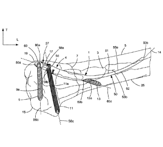
fragment, the bone fragments separated by a fracture, and the other two of the
insertion axis
and the channel axes of the three insertion channels may extend within the
first bone fragment.
[0014] The intramedullary fixation assembly, including the intramedullary
fixation device and
the fixation elements, may restrict motion in up to six dimensions and have
the effect of
ensuring that the bone fracture is stably reduced and thereby supporting bone
healing. In
particular, the combination of the intramedullary nail and the three insertion
channels has the
effect of restricting motion in six dimensions.
[0015] One of the channel axes may be a coaxial channel axis. A portion of the
coaxial
channel axis may be substantially coaxial with the insertion axis. The
intramedullary fixation
device can include a body, and the insertion channel having the coaxial
channel axis may run
through the body of the intramedullary fixation device from its insertion
point to its exit point, the
exit point being located in the shaft. The insertion axis may curve away from
the coaxial
channel axis in the vicinity of the exit point in a direction from the exit
point to the end of the
shaft.
[0016] At least one of the plurality of insertion channels may have a seating
area configured to
locking hold a portion of a fixation element therein. The seating area may be
located adjacent
an insertion point of one of the plurality of insertion channels, said one of
the plurality of
insertion channels has its exit point located in the shaft. Each one of the
plurality of insertion
channels may have a seating area configured to locking hold a portion of a
fixation element
therein, each seating area located adjacent respective insertion points.
[0017] The intramedullary fixation device can include a body, which can be a
nail body (for
instance, when the intramedullary fixation device is an intramedullary nail
such as a styloid nail)
that can have a curvilinear shape and the shaft may be configured to be
elastically deformable
to conform to the shape of the medullary canal during implantation. The shaft
of the nail body
can be substantially smooth and devoid of threads.
[0018] The shaft of the body of the intramedullary fixation device may body be
threaded.
[0019] The head may be shaped to reside within a head of a long bone, such as
the styloid
region of a long bone.
3
CA 02883089 2015-02-25
WO 2014/035811 PCT/US2013/056356
[0020] The head may be dimensioned to reside within a head of a long bone,
such as the
styloid region of a long bone.
[0021] The intramedullary fixation assembly can include the intramedullary
fixation device,
which can be a styloid nail device, for fixing a bone fracture. The styloid
nail device may
comprise a first longitudinal fixation element configured to pass across the
fracture line between
a first bone fragment and a second bone fragment. The intramedullary fixation
assembly can
further include a plurality of second fixation elements configured to anchor
the styloid nail device
in the first bone fragment.
[0022] The head of the first longitudinal fixation element may be configured
to accommodate
the plurality of second fixation elements further, and one of the second
fixation elements may be
configured to pass from a distal bone fragment to a proximal bone fragment.
[0023] The first longitudinal fixation element may be flexible and bowed. This
may improve the
anchoring of the styloid nail device in the medullary canal of the bone.
[0024] As used herein, a distal bone fragment is the fragment of a fractured
bone in which the
fracture line is closest to a joint. For example, the distal fragment is an
articular bone fragment,
and the fracture may be an extra articular fracture. An extra articular
fracture is a fracture where
the bone has not penetrated the skin, contains only one complete fracture
line, and the fracture
line does not intersect with part of the joint.
[0025] The second fixation elements may be screws or staples which have a
longitudinal core.
The longitudinal cores of the second fixation elements, and also the first
fixation element, may
be identical. Having fixation elements with the same core diameter provides
the advantage that
a reduced number of instruments are needed for implantation of the
intramedullary fixation
assembly (as compared to an assembly comprising elements with differing
diameters), thereby
reducing the complexity and costs of the implantation procedure.
[0026] As used herein, the "core" of a screw refers to the longitudinal shaft
of the screw upon
which the thread resides.
[0027] The intramedullary fixation assembly may have at least three second
fixation elements,
which may be screws, wherein the second fixation elements are mounted in the
head of the first
longitudinal fixation element so as to form a pyramidal engagement with a bone
fragment. The
pyramidal engagement prevents all rotation and separation of the bone
fragments, with the
exception of micromovements. Therefore, a stable fixation of the bone can be
achieved, whilst
4
CA 02883089 2015-02-25
WO 2014/035811 PCT/US2013/056356
minimal instrumentation is needed to insert the styloid nail device and
minimal trauma is caused
to the patient as few incisions in the skin and bone holes are required.
[0028] The head of the longitudinal first fixation element may have holes that
are threaded to
receive the second fixation elements. Advantageously, this increases the
stability of the
intramedullary fixation device.
[0029] The intramedullary fixation device may be an intramedullary nail or
screw. The term
"intramedullary" is known in the art and denotes that the nail resides at
least partly in the
medullary canal of the bone.
[0030] Two of the second fixation elements may be screws configured to be
located in a distal
bone fragment, and a tail of a third second fixation element is configured to
pass from the distal
bone fragment to a proximal bone fragment across a fracture line, and wherein
at least a portion
of the second fixation element that crosses the fracture line is configured to
extend longitudinally
through the first fixation element. The advantage associated with this
particular configuration,
especially where the second fixation elements form a pyramidal engagement with
the bone
fragment, is that a stable fixation of the bone is achieved with the
requirement of only one skin
incision and one bone hole to implant the styloid nail device.
[0031] In a second aspect, an intramedullary fixation device can be pass from
a first bone
fragment to a second bone fragment across a fracture line, wherein the
intramedullary fixation
device is threaded. Further, the second aspect also provides an intramedullary
fixation
assembly that includes the intramedullary fixation device and a first fixation
element, wherein
the intramedullary fixation device is adapted to be received in a head of the
first fixation
element, such that the intramedullary fixation device can be anchored in a
distal bone fragment,
wherein the head of the first fixation element is adapted to further
accommodate a plurality of
second fixation elements.
[0032] At least one of the second fixation elements of the second aspect may
be configured to
pass between a first bone fragment and a second bone fragment across a
fracture line, in use.
[0033] The first and second fixation elements of the second aspect may be
screws which may
have longitudinal cores, which may have the same core diameter. As with the
first aspect, this
has the advantage that less instrumentation is required for implantation.
[0034] In a third aspect, an intramedullary fixation assembly may have an
intramedullary
fixation device having a body dimensioned to lie in a medullary canal of a
bone when implanted,
the body having:
CA 02883089 2015-02-25
WO 2014/035811 PCT/1JS2013/056356
a head from which a shaft extends;
a first insertion channel for receiving a fixation element therethrough, the
first insertion
channel defining an insertion point and an exit point, and
a second insertion channel for receiving a fixation element therethrough, the
second
insertion channel defining an insertion point and an exit point.
[0035] The head may comprise an insertion area in which the insertion points
of the first and
second insertions channels are located. The insertion area may be dimensioned
and positioned
to remain accessible through a hole in a bone through which the nail body has
been inserted.
[0036] The intramedullary fixation assembly also has a first fixation element
for insertion in the
insertion point of the first insertion channel and a second fixation element
for insertion in the
insertion point of the second insertion channel.
[0037] When implanted at least one of the shaft, the first fixation element
and the second
fixation element is a bridging element arranged to span across a bone fracture
from a first bone
fragment to a second bone fragment, and thus is an intramedullary fixation
device, and at least
one of the shaft, the first fixation element and the second fixation element
is arranged to lie
within the first bone fragment.
[0038] The intramedullary fixation assembly of the third aspect has a bridging
element for
allowing a second bone fragment to be fixed to a first bone fragment through
insertion of the
bridging element in the vicinity of a single bone hole.
[0039] A fixation element separate from the intramedullary fixation assembly
may be
additionally inserted from the first to the second bone fragment to lock the
bone fragments
together and restrict motion in six dimensions.
[0040] The bridging element may have a multi-faceted outer surface for
engaging with a
medullary canal of the first and second bone fragments.
[0041] The body of the intramedullary fixation device, which can be a nail,
may have a third
insertion channel for receiving a fixation element therethrough. The third
insertion channel may
define an insertion point and an exit point. The head may have an insertion
area in which the
insertion points of the first, second and third insertions channels are
located. The insertion area
may be dimensioned and positioned to remain accessible through a hole in a
bone through
which the nail body has been inserted. The intramedullary fixation assembly
may have a third
fixation element. When implanted two of the shaft and the first, second and
third fixation
6
CA 02883089 2015-02-25
WO 2014/035811 PCT/US2013/056356
elements may be bridging elements and the other two of the shaft and the
first, second and third
fixation elements may lie within the first bone fragment.
[0042] The intramedullary nail may be inserted in a minimally invasive manner
through a
single incision in skin, and other soft tissue, and through making a single
hole in a bone to be
fixated. The combination of the intramedullary nail and first through third
fixation elements lock
the second bone fragment to the first bone fragment and restrict motion in six
dimensions to
support bone healing using minimally invasive techniques.
[0043] The insertion paths may be defined by the shaft and the plurality of
insertion channels
pyramidally diverging with respect to each other from the insertion area.
[0044] The fixation elements may form a pyramidal engagement with the bone,
the angle
between the elements at the vertex of the pyramid at the insertion area on the
head of the
intramedullary nail may all be different or equal and may be 109.5 , or 100 ,
or 900, or 80 , or
70 , or 60 . There may be a pair of fixation elements in which the angle
between them at the
vertex of the pyramid is, for example 60 and the third fixation element is
at an angle of 100
from each of the pair of fixation elements. For example, in aspect one, the
third second fixation
element (that may cross the bone fracture), is at an angle of about 100 from
the second
fixation elements that remain in the distal fragment of the bone, and the
second fixation
elements that remain in the distal fragment of the bone are at an angle of
about 60 .
[0045] The insertion channels may be configured according to the type of
fixation element
they are to receive. The fixation elements may be, but are not limited to,
being one of a locking
screw, a variable angle locking screw or a staple.
[0046] The intramedullary nail of the third aspect may have any of the
features of the
intramedullary nail of the first aspect.
[0047] In a fourth aspect, an intramedullary fixation system may include an
intramedullary
fixation device according to the first aspect or the second aspect. The
intramedullary fixation
system also has an aiming arm. The aiming arm may be connectable to the
intramedullary nail
and may define a plurality of guide channels therein. Each guide channel may
have a guide
axis aligned with a respective channel axis of an insertion channel, the
channel axes diverging
from an insertion area defined in a head of the intramedullary nail.
[0048] The intramedullary fixation system may further include a measuring
device for
measuring the depth of insertion of a fixation element.
7
CA 02883089 2015-02-25
WO 2014/035811 PCT/1JS2013/056356
[0049] The aiming arm may include, consist of or consist essentially of a
radiolucent material.
The radiolucent material is polyether ether ketone (PEEK). The aiming arm may
have an x-ray
visible mark.
[0050] In a fifth aspect, a first fixation element can be adapted to receive
an intramedullary
fixation device, such that the intramedullary fixation device can pass across
a bone fracture
between a first bone fragment and a second bone fragment in use, the first
fixation element
being threaded to anchor the first fixation element in a first bone fragment,
a head of the first
fixation element being further shaped to receive at least one second fixation
element. The head
of the first fixation element may be threaded to receive the second fixation
element.
[0051] The head of the fixation element of the fifth aspect may be shaped to
accommodate
second fixation elements such that they define a pyramidal anchor. The
advantage associated
with this particular configuration, especially where the second fixation
elements form a
pyramidal engagement with the bone fragment, is that a stable fixation of the
bone is achieved
with the requirement of only one skin incision and one bone hole to implant
the styloid nail
device.
[0052] The intramedullary fixation devices, including styloid nail devices,
styloid nails, and
fixation elements described herein may be used in the temporal bone of the
skull, and the ulna,
tibia and fibula styloid processes, or any suitable alternative long bone as
desired. In particular,
intramedullary fixation devices, including styloid nail devices, styloid
nails, and fixation elements
described herein are used to fix an extraarticular fracture of the distal
radius, and are inserted
through the styloid process of the distal radius.
[0053] The term "styloid process" is a term known in the art and refers to a
projection of bone
on the surface of a bone, that serves as a small attachment point for muscles.
[0054] The head of the first fixation element may have holes that are threaded
to receive the
intramedullary fixation device and second fixation elements. Advantageously,
this increases the
stability of the combination.
[0055] In a sixth aspect, a method of implanting an intramedullary fixation
device in a
medullary canal of a bone cam support bone healing of a bone fracture between
a first bone
fragment and a second bone fragment. The method may have the steps of:
aligning the first and second bone fragments;
making a hole in the cortical bone of the first bone fragment;
passing an intramedullary fixation device through the hole, the intramedullary
fixation
device having a head from which a shaft extends and a plurality of fixation
element
8
CA 02883089 2015-02-25
WO 2014/035811 PCT/US2013/056356
receiving channels, each one of the plurality of fixation element receiving
channels
having an insertion point located in an insertion area defined in the head;
inserting a first fixation element through an insertion point in the insertion
area; and
inserting a second fixation element through a different insertion point in the
insertion
area.
[0056] At least one of the shaft, the first fixation element and the second
fixation element so
inserted may be a bridging element arranged to span from the first bone
fragment to the second
bone fragment across the bone fracture and at least one of the shaft, the
first fixation element
and the second fixation element is arranged to lie within the first bone
fragment.
[0057] A measurement may be taken before insertion of each of the first and
the second
fixation elements for determining the length of the fixation element to be
inserted.
[0058] The first and second fixation elements may have the same core diameter.
[0059] The shaft may be threaded.
[0060] The fixation elements may be inserted in a manner so as to form a
stable pyramidal
construct with the bone.
[0061] The first bone fragment may be an articular fragment.
[0062] The fracture may be an extraarticular fracture.
[0063] In a seventh aspect, a method of fixing a bone fracture comprises
making a single skin
incision. The advantages associated with an implantation that only requires a
single skin
incision will be recognised by those skilled in the art. For example, minimal
trauma is caused to
the patient therefore minimising healing time and minimising the possibility
of complications
resulting from the procedure. The method may further comprise making only a
single bone hole,
with the same advantages associated with a single skin incision.
[0064] The method of fixing a bone fracture may comprise i) making one skin
incision, ii)
drilling a hole in a distal fragment of the bone; iii) inserting a first
fixation element into the distal
bone fragment; iv) inserting at least one second fixation element into the
distal bone fragment.
[0065] The first fixation element of the method may be inserted so as to pass
from the distal
bone fragment to the proximal bone fragment across a fracture line, and the
second fixation
element may be inserted through the first fixation element. This insertion may
be through the
9
CA 02883089 2015-02-25
WO 2014/035811 PCT/US2013/056356
head of the first fixation element and the second fixation element may be
inserted so as to
remain entirely in the distal bone fragment.
[0066] A measurement may be taken before the second fixation element is
inserted. This
measurement is used to determine the length required of the at least one
second fixation
element.
[0067] A further second fixation element may be inserted through the head of
the first fixation
element in a manner so as to pass from a distal bone fragment across a
fracture line to a
proximal bone fragment.
[0068] The advantage associated with this method is that a stable fixation can
be achieved
with only a single skin incision and single bone hole. Advantageously, this
method may be
carried out using the device for inserting an intramedullary fixation device
of the type described
herein. Therefore, a stable fixation of the bone is achieved using minimal
instrumentation.
[0069] The method of fixing a bone fracture may comprise, i) making an
incision in the skin; ii)
inserting a wire in a distal bone fragment, substantially parallel to a joint
in the distal fragment;
iii) measuring a depth of the distal bone fragment; iv) drilling a hole in a
distal bone fragment
substantially parallel to a joint in the distal fragment; v) placing a screw
in the hole drilled in the
bone fragment; vi) drilling one or more further holes in the distal fragment;
viii) inserting one or
more screws in the distal fragment, at least one of which passes from the
distal bone fragment
to a proximal bone fragment across a fracture line.
[0070] The fixation elements may be inserted so as to form a stable pyramidal
construct with
the bone, with the advantages previously discussed for this type of construct.
[0071] The depth of the distal bone fragment may be measured by applying a
measuring
device to the wire inserted in the distal bone fragment, which is calibrated
with the length of wire
employed. The wire may have a diameter of 1.1 mm, and may be a K-wire, and
where the bone
is the radius, the wire may be inserted into the volar-ulnar canal.
[0072] As used herein, K-wire is a shortened form of Kirschner wire, a
sterilised, sharpened,
smooth stainless steel pin used widely in the orthopaedic art.
[0073] The hole in the distal bone fragment may be drilled by applying a drill
over the inserted
wire, the drill may have a diameter of 2.0mm.
[0074] The screw may be inserted over the guide wire, after which point the
guide wire is
removed.
CA 02883089 2015-02-25
WO 2014/035811 PCT/1JS2013/056356
[0075] Before drilling one or more further holes in the distal fragment, a
second guide wire,
which may be a 1.1 mm K-wire, may be inserted. The drilling of the one or more
further holes
may then be performed over the second guide wire. A measurement may then be
taken to
measure the length required for the one or more screws in the distal fragment,
by applying a
measuring device to the second guide wire. The one or more screws may then be
inserted over
the guide wire, before removal of the guide wire.
[0076] The one or more further screws that pass from the distal bone fragment
to a proximal
bone fragment across a fracture line may be inserted over a guide wire, which
may be a 1.1mm
K-wire, after a hole has been drilled from the distal bone fragment to the
proximal bone
fragment across the fracture line, wherein the hole was drilled over the guide
wire.
[0077] The advantage of this method is that a stable fixation can be achieved
with the use of
minimal instrumentation.
[0078] In an eighth aspect, an insertion device is provided and is configured
to insert an
intramedullary fixation device in a bone having a fracture, the device
comprising; i) a first part
configured to insert a first fixation element longitudinally through a distal
fragment of the bone
and across a fracture line; ii) an aiming arm configured to insert a guide
wire; and iii) a
measuring device for measuring the depth of insertion of the guide wire.
[0079] Advantageously, the device for inserting an intramedullary fixation
assembly can be
used with certain aspects of the method of fixing a bone fracture.
[0080] The device of the eighth aspect may be configured to insert one or more
second
fixation elements into the distal fragment of the bone, through a head of the
first fixation
element. The device may be configured to insert the one or more second
fixation elements via
the aiming arm.
[0081] The device of the eighth aspect includes, consists of or consists
essentially of a
radiolucent material which may be polyether ether ketone. Advantageously, this
allows the
surgeon using the device to have a clear view of the wires, screws and nails
being used in the
device via x-ray imaging. The device may have an x-ray visible mark on the
aiming arm, to help
the surgeon aim the guide wire.
[0082] As used herein, radiolucent refers to a material that allows the
passage of x-rays with
little attenuation, thereby rendering the material not visible by x-ray
imaging.
11
CA 02883089 2015-02-25
WO 2014/035811 PCT/1JS2013/056356
[0083] Advantageously, the device of the eighth aspect can be used to insert a
styloid nail
device which provides a stable construct in the bone, preventing all rotation
and separation of
the bone fragments (except micromovements), with only one skin incision and
one bone hole.
Minimal instrumentation is also required.
[0084] The intramedullary fixation assemblies, including the intramedullary
fixation devices,
may be used in the temporal bone of the skull, and the ulna, tibia and fibula
styloid processes,
or in any suitable alternative long bone.
Brief Description of the Drawings
[0085] Embodiments will now be described in detail with reference to the
accompanying
drawings, in which:
[0086] Fig. 1A is a perspective view of an intramedullary fixation assembly in
accordance with
one embodiment, shown implanted into a bone;
[0087] Fig. 1B is a top plan view of an intramedullary fixation assembly
similar to Fig. 1A, but
constructed in accordance with an alternative embodiment;
[0088] Fig. 1C is a side elevation view of an intramedullary fixation assembly
similar to Figs.
1A-B, but constructed in accordance with an alternative embodiment;
[0089] Fig. 2 is a top plan view of an intramedullary fixation assembly in
accordance with
another embodiment;
[0090] Fig. 3A is a perspective view of an intramedullary fixation assembly in
accordance with
another embodiment;
[0091] Fig. 3B is a top plan view of an intramedullary fixation assembly
similar to the
intramedullary fixation assembly illustrated in Fig. 3A, but constructed in
accordance with an
alternative embodiment;
[0092] Fig. 4A is a perspective view of an intramedullary fixation assembly in
accordance with
one embodiment;
[0093] Fig. 48 is a side elevation view of an intramedullary fixation assembly
similar to Fig.
4A, but constructed in accordance with another embodiment;
[0094] Figs. 5A-J show an insertion assembly and associated steps inserting
and fixing an
intramedullary fixation assembly in accordance with one embodiment;
12
CA 02883089 2015-02-25
WO 2014/035811 PCT/US2013/056356
[0095] Fig. 6A shows a first step of a method of fixing a bone fracture in
accordance with one
embodiment; and
[0096] Fig. 6B shows a second step of the method of fixing a bone fracture
illustrated in Fig.
6A.
Detailed Description
[0097] Referring to Figs. 1A-1C generally, an intramedullary fixation assembly
4, can include
a bone fixation device, which can be configured as an intramedullary fixation
device 1. The
term "intramedullary" is known in the art and denotes that the nail resides at
least partly in the
medullary canal of a bone. The intramedullary fixation device 1 can be
elongate generally along
a central axis 14. For instance, the central axis 14, and thus the
intramedullary fixation device
1, can be bowed or generally curvilinear in shape along its direction of
elongation. The
intramedullary fixation device 1 is configured to reside in a medullary canal
3 of a long bone,
such as a radius 5 that includes a shaft 25, and a head or articular fragment
15 that extends
distally from the shaft 25.
[0098] In accordance with the illustrated embodiment, the intramedullary
fixation device 1 is
sized and configured to extend across a fracture location 7 disposed between
the shaft 25 and
the articular fragment 15. As used herein, a distal bone fragment can refer to
the fragment of a
fractured bone in which the fracture line 7 is closest to a joint. For
example, the distal fragment
is the articular bone fragment 15, and the fracture may be an extra articular
fracture. An extra
articular fracture is a fracture where the bone has not penetrated the skin,
contains only one
complete fracture line, and the fracture line does not intersect with part of
the joint. Thus, the
articular fragment 15 can define a first or distal bone fragment, the shaft 25
can define a second
or proximal bone fragment, and the fracture location 7 can separate the first
bone fragment from
the second bone fragment. As will be appreciated from the description below,
the
intramedullary fixation device 1 is configured to be inserted through a
styloid process 19 of the
articular fragment 15 and into the medullary canal 3 so as to extend across
the fracture 7, and is
further configured to be fixed to both the articular fragment 15 and the shaft
25, thereby
stabilizing the articular fragment and the shaft 25 with respect to each other
so as to promote
bone fixation across the fracture 7. Thus, the intramedullary fixation device
1 can be referred to
as a styloid fixation device. It should, of course, be appreciated that a bone
fixation device of
the type described herein is configured to be used in the temporal bone of the
skull, and the
ulna, tibia and fibula styloid processes, or any suitable alternative long
bone as desired.
13
[0099] The intramedullary fixation device 1 includes a body 50 that defines
the head 17 and a
shaft 52 that extends proximally from the head 17 so as to define an insertion
axis that can be
defined by the central axis 14 of the intramedullary fixation device 1. The
body 50 can be
curved within a plane defined by a longitudinal direction L and a transverse
direction T that is
oriented substantially perpendicular to the longitudinal direction L. The head
17 can define a
free end that defines a first or distal outermost end 50a of the body 50, and
the shaft 52 can
define a free end that defines a second or proximal outermost end 50b of the
body 50 opposite
the first outermost end 50a and spaced from the first outermost end 50a along
the central axis
14. The first and second ends 50a and 50b can be spaced apart a greater
distance along the
longitudinal direction L than along the transverse direction T. Thus, the
central axis 14 can
extend along both the longitudinal direction L and the transverse direction T,
Accordingly, the
body 50 can define an upper surface 53a at least a portion of which can be
concave in a plane
substantially defined by the longitudinal L and transverse T directions, and a
lower surface 53b
opposite the upper surface 53a along the transverse direction T, at least a
portion of which can
be convex in the plane substantially defined by the longitudinal L and
transverse T directions.
The body 50 further extends along a lateral direction A that is substantially
perpendicular to the
longitudinal direction L and the transverse direction T.
[01001 The head 17 can be shaped and dimensioned to reside within the bone
structure, such
as the styloid process 19 of the radius 5, when the intramedullary fixation
device 1 is disposed
within the medullary canal 3. In accordance with one embodiment, the shaft 52
may be
configured to be elastically deformable to conform to the shape of the
medullary canal 3 during
implantation. Thus the shaft 52 can be flexible and bowed, which may improve
the anchoring of
the intramedullary fixation device 1 in the medullary canal 3. The shaft 52 of
the intramedullary
fixation device 1 can define a longitudinal first fixation element configured
to pass across the
fracture 7 between the first and second bone fragments. The shaft 52 can be
substantially
smooth and devoid of threads in accordance with one embodiment, such that the
intramedullary
fixation device 1 is an intramedullary nail 51 (see Figs. 1A-C), or can be
threaded as desired
such that the intramedullary fixation device 1 is an intramedullary screw 29
(see Figs. 2-38). Of
course, it should be appreciated that any of the intramedullary fixation
devices 1 described
herein can be constructed as a nail or a screw unless otherwise indicated.
[0101] The intramedullary fixation assembly 4 can further include at least
one, such as a
plurality, of second bone fixation elements, such as screws, that are
configured to anchor the
intramedullary fixation device 1 to the radius 5, and in particular to the
articular fragment 15.
For instance, the intramedullary fixation assembly can include a first bone
fixation element or
screw 9, a second bone fixation element or screw 11, and a third bone fixation
element or screw
14
CA 2883089 2020-02-21
CA 02883089 2015-02-25
WO 2014/035811 PCT/1JS2013/056356
13. The intramedullary fixation device 1 defines a head 17 that is configured
to receive
respective heads of the first, second, and third screws 9, 11 and 13,
respectively, in the articular
fragment 15. The head 17 can be configured to accommodate the screws 9, 11,
13, such that a
select one of the screws, for instance the third screw 13, may be configured
to pass from the
articular fragment 15 to the shaft 25 so as to fix the shaft 25 to the
articular fragment 15.
[0102] It should be appreciated that the second bone fixation elements can be
configured as
screws (Figs. 1A-2) that have a longitudinal core, or staples (Figs. 3A-B), or
the like. The
longitudinal cores of the second fixation elements, and also the first
fixation element, may be
substantially identical in accordance with one embodiment. Having fixation
elements with the
same core diameter provides the advantage that a reduced number of instruments
are able to
implant the intramedullary fixation assembly 4 (as compared to an assembly
comprising
elements with differing diameters), thereby reducing the complexity and costs
of the
implantation procedure. As used herein, the "core" of a screw can refer to the
longitudinal shaft
of the screw upon which the thread resides.
[0103] The intramedullary fixation device 1 can define at least one channel
that is configured
to receive a corresponding at least one of the fixation elements that can be
configured as
screws 9,11, and 13. In accordance with the illustrated embodiment, the
intramedullary fixation
device 1, for instance the head 17, can include at least one, such as a
plurality, of insertion
channels that extends through the body 50, each configured to receive a
respective one of the
bone fixation elements. For instance, the intramedullary fixation device 1 can
define a first
insertion channel 56 that extends through the body 50 and is configured to
receive the first
screw 9, a second insertion channel 58 that is configured to receive the
second screw 11, and a
third insertion channel 60 that is configured to receive the third screw 13.
Each of the insertion
channels 56, 58, and 60, respectively, can define an insertion point 56a, 58a,
and 60a,
respectively, an exit point 56b, 58b, and 60b, respectively, and a channel
axis 56c, 58c, and
60c, respectively, that passes through the respective insertion point and the
exit point. The
screws 9-13 are configured to be inserted into the respective channels 56-60
through the
respective insertion point 56a-60a, along the channel axis 56c-60c, and exit
through the
respective exit point 56b-60b. The head 17 can define an insertion area 27 in
which the
insertion points 56a-60a, respectively, are located.
[0104] As will be described in more detail below with reference to Figs. 5A-J,
the insertion
area 27 may be dimensioned and positioned to remain accessible through a
single hole in a
bone through which the intramedullary nail has been inserted. For instance,
the single hole can
extend through the styloid process of the radius 5. Thus, the insertion points
56a-60a of all of
CA 02883089 2015-02-25
WO 2014/035811 PCT/US2013/056356
the insertion channels 56-60 may be located in the insertion area 27. Further,
intramedullary
fixation device 1 may be completely inserted and fixed in position through a
single hole in a
bone that can extend, for instance, through the styloid process. Fixation of
the intramedullary
fixation device 1 is possible with only a single skin incision and through the
making of a single
bone hole. Additional locking of the second outermost end 50b of the
intramedullary fixation
device 1 opposed to the insertion area 27 is unnecessary.
[0105] The first and second insertion channels 56 and 58 are configured to
receive the first
and second screws 9 and 11, respectively, and the third channel 60 is
configured to receive the
third screw 13. In accordance with the illustrated embodiment, the third
insertion point 60a
extends through the first end 50a of the body 50 substantially coextensive
with the central axis
14. Thus, the first and second insertion points 56a and 58a are spaced from
the second end
50b a distance that is less than the distance that the insertion point 60a is
spaced from the
second end 50b.
[0106] One of the channels, such as the third channel 60, may be a coextensive
channel,
such that the third channel axis 60c may be a coaxial channel axis. At least a
portion of the
coaxial channel axis 60c may be substantially coaxial with the insertion axis,
and thus the
central axis 14. The insertion channel 60c can extend through the body 50 of
the intramedullary
fixation device 1 from its insertion point 60a to its exit point 60b, the exit
point being 60b located
in the shaft 52. The central axis 14 may curve away from the coaxial channel
axis 60c in the
vicinity of the exit point 60b in a direction from the exit point 60b to the
50b end of the shaft 52.
Thus, it can be said that at least a portion of the corresponding third
channel axis 60c can be at
least partially coaxial, and thus extend longitudinally through (for instance
coaxial with,
tangential to or intersecting two points of) the central axis 14 of the
intramedullary fixation
device 1. The third exit point 60b is disposed on an opposite side of the
fracture line 7 with
respect to the third insertion point 60a. Thus, when the third screw 13 is
inserted into the
channel 60, a portion of the third screw 13 can across the fracture line 7.
Accordingly, two of
the screws 9-13, such as the first and second screws 9 and 11, respectively,
are configured to
be entirely located in the articular fragment 15, and at least a portion, for
instance a tail, of the
third screw 13 is configured to extend longitudinally through the
intramedullary fixation device 1,
and pass from the articular fragment 15 to the shaft 25 across the fracture 7.
[0107] At least one, and up to all of, the channels 56-60 can define holes in
the head 17 of the
intramedullary fixation device 1 that are threaded so as to receive threaded
heads of the
respective screws 9-13, thereby increasing the stability of the intramedullary
fixation device 1.
Thus, it can be said that at least one of the plurality of insertion channels,
such as the third
16
CA 02883089 2015-02-25
WO 2014/035811 PCT/US2013/056356
insertion channel 60, may define a seating area configured to lockingly hold a
portion of the
respective screws 13 therein. The seating area may be located adjacent the
respective
insertion point 60a of the at least one of the plurality of insertion channels
60, said at least one
of the insertion channels 60 has its exit point 60b located in the shaft 52.
Each one of the
plurality of insertion channels 56-60 may have a seating area configured to
locking hold a
portion of the respective fixation element therein 9-13, each seating area
located adjacent
respective insertion points 56a-60a. Further, the channels 56-60 can be
configured according
to the type of fixation element they are to receive. The fixation elements may
be, but are not
limited to, one of a locking screw, such as a fixed angle locking screw or a
variable angle
locking screw, or a staple.
[0108] The central axis 14 of the intramedullary fixation device 1 and at
least two, and up to
all, of the plurality of channel axes 56c-60c may diverge with respect to each
other away from
the insertion area 27. For instance, the central axis 14 and at least one up
to all of the plurality
of channel axes 56c-60c, and thus the respective screws 9-13 that are inserted
through the
channels 56-60, pyramidally may diverge with respect to each from the
insertion area 27. In
accordance with the illustrated embodiment, the first and second channels 56
and 58 diverge
from each other with respect to the central axis 14 along their respective
channel axes 56c and
58c, in a direction from the respective insertion points 56a and 58a to the
respective exit points
56h and 58b. Further, the third channel 60 diverges from least one such as
both of the first and
second channels 56 and 58 with respect to a lateral axis, along their
respective channel axes
56c-60c, in a direction from the respective insertion points to the respective
exit points. In one
embodiment, as shown, for example in Figs. 1A-C, at least one of channel axes
56c and 58c,
and preferably both, diverge from a vertical plane defined by the transverse
direction T and the
central axis 14 at the head 17 by an angle of at least 5 , preferably at least
10 , and more
preferably at least 150, and it is preferred that the two channels 56c and 58c
diverge in opposite
directions, one medially and one laterally. This angle is advantageously less
than 45 ,
preferably less than 350, and more preferably less than 30 for each channel
56c and 58c; the
angle is thus advantageously between 5 and 35 , more preferably between 5
and 30 . The
channel axis 60c can diverge from a vertical plane defined by the lateral
direction A and the
channel axis of one of the insertion channels 56c or 58c by an angle of at
least 20 , preferably
at least 25 , and more preferably at least 35, and is advantageously less than
55 , preferably
less than 50'; the angle is thus advantageously between 20 and 55 , more
preferably between
25 and 50 .
[0109] The screws 9-13 are thus mounted to the head 17 of the intramedullary
fixation device
1 so as to form a pyramidal engagement with the radius 5. The pyramidal
engagement of the
17
CA 02883089 2015-02-25
WO 2014/035811 PCT/US2013/056356
intramedullary fixation device and screws 9-13 with the radius 5 prevents
rotation and
separation of the articular fragment 15 and the shaft 25, with the exception
of micromovements.
For instance, the intramedullary fixation assembly 4, including the
intramedullary fixation device
1 and the screws 9-11, may restrict motion in up to six dimensions and have
the effect of
ensuring that the bone fracture 7 is stably reduced and thereby supporting
healing of the radius
5. Thus, the combination of the intramedullary fixation device 1 and the three
insertion channels
56-60 and corresponding screws 9-13 is configured to restrict motion of the
articular fragment
15 relative to the shaft 25 in six dimensions. It should therefore be
appreciated that a stable
fixation of the radius 5 is achieved with the requirement of only one skin
incision and one bone
hole to implant the intramedullary fixation device 1. As a result, a stable
fixation of the radius 5
can be achieved, while minimal instrumentation can insert the intramedullary
fixation device 1
and minimal trauma is caused to the patient as few incisions in the skin and
bone holes are
created.
[0110] As described above, the screws 9, 11 and 13 can have cores 9a, 11a and
13a of equal
diameter thereby requiring only one instrument to insert each distal screw.
The screws 9, 11
and 13 form a pyramidal engagement with bone 5, thereby providing a stable
fixation of the two
bone fragments separated by fracture line 7. The third screw 13 passes across
fracture line 7 in
order to increase the stability of the engagement. As can be seen clearly in
Fig. 1C, the head
17 of the intramedullary fixation device 1 can be configured to receive the
first, second, and third
screws 9, 11 and 13 in a manner so as to produce the desired pyramidal
engagement. The
intramedullary fixation device 1 and screws 9, 11 and 13 can be inserted
through the styloid
process 19 of the radius 5. Advantageously, an entirety of the intramedullary
fixation device 1
can be inserted through a single bone hole, advantageously using an insertion
device 30 (see
Fig. 5B) for inserting an intramedullary fixation assembly according to the
one embodiment.
[01111 In accordance with the embodiments illustrated in Figs. 1A-C, the
plurality of insertion
channels 56-60 may include, and can be limited to, three insertion channels.
When implanted,
two of the central axis 14 or the channel axes of the three insertion channels
may extend in a
direction from the articular fragment 15 to the shaft 25 that are separated by
the fracture 7, and
the other two of the central axis 14 and the channel axes of the three
insertion channels may
extend within the articular fragment 15.
[0112] As illustrated in Figs. 1A-C, a substantial entirety of the first and
second insertion
channels 56 and 58 are spaced from each other along the lateral direction A.
For instance, the
first and second insertion points 56a and 58a can be at least partially
aligned with each other
along the lateral direction A. Similarly, the first and second exit points 56b
and 58b can be at
18
CA 02883089 2015-02-25
WO 2014/035811 PCT/US2013/056356
least partially aligned with each other along the lateral direction A. Thus,
at least a portion of
the first and second channel axes 56c and 58c can be at least partially
aligned with each other
along the lateral direction A. The first and second insertion channels 56 and
58 can diverge
away from each other with respect to the central axis 14 along their
respective channel axes
56b and 58b along respective directions from the insertion points 56a and 58a
to their
respective exit points 56c and 58c.
[0113] At least a portion, for instance the third insertion point 60a, up to
an entirety of the third
insertion channel 60 can be disposed between the first and second insertion
channels 56 and
58 with respect to the lateral direction A. The third insertion point 60a can
further be displaced
from the first and second insertion points 56a and 58a proximally along the
longitudinal direction
L. For instance, the insertion point 60a can be spaced from the second end 50b
a distance that
is less than the distance that the insertion points 56a and 58a are spaced
from the second end
50b. Further, the insertion point 60a, along with the insertion points 56a and
58a, extends
through the concave upper surface of the body 50, for instance at the head 17.
The first and
second screws 9 and 11 are thus configured to extend through the respective
first and second
channels 56 and 58 and anchor to the articulation fragment 15, and the third
screw 13 is
configured to extend through the third channel 60 and anchor to the shaft 52.
It should thus be
appreciated that the intrannedullary fixation device 1 defines a region
between the third insertion
point 60a and the third exit point 60b that is configured to extend across the
fracture 7. The
third insertion point 60a can lie on the first end 50a, which can extend in a
plane that is
substantially defined by the transverse T and lateral A directions. As
illustrated in Fig. 1B, the
first end 50a can extend in a plane that is substantially defined by the
longitudinal L and lateral
A directions.
[0114] In accordance with the embodiment illustrated in Fig. 1C, at least a
portion up to all of
the first and second insertion channels 56 and 58, including the respective
first and second
insertion points 56a and 58a, the first and second exit points 56b and 58b,
and the first and
second channel axes 56c and 59c, can be further spaced from each other along
the longitudinal
direction L as desired. Furthermore, at least a portion up to all of the first
and second insertion
channels 56 and 58, including the respective first and second insertion points
56a and 58a, the
first and second exit points 56b and 58b, and the first and second channel
axes 56c and 59c,
can be aligned with each other along the longitudinal direction L as desired.
In accordance with
the illustrated embodiment, the third channel 60 can be disposed distal to the
first and second
channels 56 and 58. For instance, the third insertion point 60a, the third
channel axis 60b, and
the third exit point 60c can be disposed distal to the respective first and
second insertion points
56a and 58a, the first and second channel axes 56ba and 58b, and the first and
second exit
19
CA 02883089 2015-02-25
WO 2014/035811 PCT/US2013/056356
points 56c and 58c, respectively. Further, in accordance with the illustrated
embodiment, the
first channel 56 can be disposed between the second and third channels 58 and
60 with respect
to the longitudinal direction L. For instance, the first insertion point 56a,
the first channel axis
56b, and the first exit point 56c can be disposed between the respective
second and third
insertion points 58a and 60a, the second and third channel axes 58ba and 60b,
and the second
and third exit points 58c and 60c, respectively. As described above, the
channels 56-60, and
thus the respective retained screws 9-13, can diverge from each other so as to
define a
pyramidal construct.
[0115] Referring now to Fig. 2, the intramedullary fixation device 1 can be
configured
substantially as described above with respect to Figs. 1A-C, but wherein the
shaft 52 defines
externally threads 31 such that the intramedullary fixation device 1 defines
an intramedullary
screw 29. Accordingly, the shaft 52 can be configured as a bone fixation
element that is
configured to attach to bone so as to attach the intramedullary fixation
device 1 to the radius 5.
The shaft 52 can extend substantially linearly from the head 17, such that the
central axis 14 is
likewise substantially linear. The intramedullary fixation device 1 can define
first and second
insertion channels 56 and 58, and may be limited to two insertion channels.
Alternatively, the
intramedullary fixation device 1 illustrated in Fig. 2 can include any number
of channels as
described in any one of Figs. 1A-1C. In accordance with the illustrated
embodiment, at least a
portion up to all of the first and second insertion channels 56 and 58,
including the respective
first and second insertion points 56a and 58a, the first and second exit
points 56b and 58b, and
the first and second channel axes 56c and 59c, can be aligned with each other
along the
longitudinal direction L as desired.
[0116] The first and second channels 56 and 58 extend through the insertion
area 27 of the
head 17, and can diverge from each other with respect to the central axis 14
as described
above, and the shaft 52 can diverge from each of the first and second channels
56 and 58 with
respect to a lateral axis, as described above. For instance, the insertion
paths may be defined
by the shaft 52 and the plurality of insertion channels 56 and 58 pyramidally
diverging with
respect to each other from the insertion area 27. Thus, the shaft 52 and the
first and second
screws 9 and 11 that are inserted in the first and second channels 56 and 58
are configured to
define the pyramidal construct described above. The fixation elements,
including the shaft 52,
and the first and second screws 9 and 11, may form a pyramidal engagement with
the bone, the
angle between the elements at the vertex of the pyramid at the insertion area
on the head of the
intramedullary nail may all be different or equal and may be 109.5 , or 100 ,
or 900, or 80 , or
70 , or 60 . There may be a pair of fixation elements, such as screws 9 and
11, in which the
angle between them at the vertex of the pyramid is, for example 60 and the
third fixation
element, for instance the shaft 52, is at an angle of 100 from each of the
pair of fixation
elements.
[0117] When implanted in the radius 5, one of the central axis 14 or one of
the channel axes
56c and 58c of the two insertion channels 56 and 58, respectively, may extend
in a direction
from the articular fragment 15 to the shaft 25 that are separated by the
fracture 7, and the other
two of the central axis 14 and the channel axes 56c and 58c of the two
insertion channels 56
and 58 may extend within the articular fragment 15. For instance, the head 17
can be
configured to be disposed in the articular fragment 15, and the threaded shaft
52 is configured
to extend from the head 17, across the fracture 7, and into the medullary
canal 3 defined by the
shaft 25 of the radius 5. The threads 31 can be disposed in at least one or
both of the articular
fragment 15 and in the shaft 25.
[0118] It should thus be appreciated that intramedullary fixation assembly 4
can include a
bridging element that is configured to attach the shaft 25 to the articular
fragment 15 through
insertion of the bridging element in the vicinity of the single bone hole.
When the intramedullary
fixation device 1 is implanted in the radius 5, at least one of the
intramedullary fixation device 1,
such as the shaft 52, the first screw 9 and the second screw 11 is a bridging
element arranged
to span across a bone fracture 7 from the articular fragment 15 to the shaft
25, and at least one
of the intramedullary fixation device 1, such as the shaft 52, the first screw
9 and the second
screw 11 is arranged to lie within the articular fragment 15. The bridging
element may have a
multi-faceted outer surface for engaging with the medullary canal 3 of the
articular fragment 15
and the shaft 25. When implanted two of the shaft and the first, second and
third fixation
elements may be bridging elements and the other two of the shaft and the
first, second and third
fixation elements may lie within the first bone fragment. A fixation element
separate from the
intramedullary fixation assembly 4 may be additionally inserted from the
articular fragment 15 to
the shaft 25 so as to lock the articular fragment 15 and the shaft 25 together
and restrict motion
in six dimensions.
[0119] Referring now to Fig. 3A, as described above, the second fixation
elements of the
intramedullary fixation assembly 4 can be configured as staples, such as a
first staple 21 and a
second staple 22. For instance, each of the first and second staples 21 and 22
can wrap
around at least a portion of the body 50 of the intramedullary fixation device
1. In accordance
with the illustrated embodiment, each of the first and second staples 21 and
22 can wrap around
the head 17 of the intramedullary fixation device 1 so as to define at least a
partial revolution
about the head 17. For example, each of the first and second staples 21 and 22
can wrap
around the upper surface 53a of the head 17, such that opposed free ends of
the staples 21 and
21
CA 2883089 2020-02-21
22 are configured to anchor in the articular fragment 15, and an intermediate
portion that
extends between the free ends is wrapped about the head 17. The free ends of
each staple can
be disposed on laterally opposite sides of the intramedullary fixation device
1, such that the
intramedullary fixation device 1 is disposed between the free ends of each of
the staples with
respect to the lateral direction A. The first staple 21 can be spaced from the
second staple 22
along the longitudinal direction L, and the free ends of the staples 21 and 22
can be spaced
from the head 17 along at least the transverse direction T, for instance in
addition to the lateral
direction A as desired Further, the free ends of the first staple 21 can be
offset with respect to
the free ends of the second staple 22 along the lateral direction A, or can be
aligned with the
free ends of the second staple 22 along the lateral direction A as desired.
The staples 21 and
22 are configured to form an angular construct which provides a stable
fixation of the bone
fragments separated by fracture line 7.
[0120] It should be appreciated that the intramedullary fixation assembly 4
can include any
number of staples as desired. For instance, as illustrated in Fig. 3B, the
intramedullary fixation
assembly 4 can include a single staple 21 that can wrap around at least a
portion of the body 50
of the intramedullary fixation device 1. In accordance with the illustrated
embodiment, the
staple 21 can wrap around the head 17 of the intramedullary fixation device 1
so as to define at
a full revolution about the head 17. For example, the staple 21 can wrap
around the upper
surface 53a of the head 17 and the lower surface 53b of the head 17, such that
the opposed
free ends the staple 21 are configured to anchor in the articular fragment 15,
and an
intermediate portion of the staple 21 that extends between the free ends is
wrapped about the
head. The free ends of the staple 21 can be disposed on laterally opposite
sides of the
intramedullary fixation device 1, such that the intramedullary fixation device
1 is disposed
between the free ends the staple with respect to the lateral direction A. One
of the free end so
the staple 21 can be spaced from the other of the free ends of the staple 21
along the
longitudinal direction L, and the free ends of the 21 can be spaced from the
head 17 along at
least the transverse direction T, for instance in addition to the lateral
direction A as desired. The
free ends of the staple 21 are configured to form an angular construct which
provides a stable
fixation of the bone fragments separated by fracture line 7. Thus, it should
be appreciated that
the intramedullary fixation assembly 4 can include at least one staple, such
as a plurality of
staples that can be wrapped about the intramedullary fixation device 1 along
at least a partial
revolution. The shaft 52 of the intramedullary fixation device 1 illustrated
in Figs. 3A-B can be
constructed as described above, and can thus be curved and substantially
smooth, or can
alternatively be externally threaded, as described above. Furthermore, the
head 17 can define
the channel 60 that is configured to receive a screw that is configured to
extend coaxially from
the head 17 across the fracture 7 to the shaft 25 as described above.
22
CA 2883089 2020-02-21
[0121] Referring now to Fig. 4A, an intramedullary fixation assembly 4 can
include an
intramedullary fixation device 24, which can be in the form of a threaded
screw having a head
17 and a shaft 52 that extends distally from the head 17, that is configured
to pass from the
articular fragment 15 to the shaft 25 across the fracture 7. The
intramedullary fixation assembly
4 can further include a first fixation element 26, which can be a screw that
defines a head 28a
and a threaded shaft 28b that extends distally from the head 28a along a
central axis. The
intramedullary fixation device 24, and in particular the shaft 52, is adapted
to extend through an
aperture 28c that extends through the head 28a of the first fixation element
26 at an angle
oblique to the central axis of the threaded shaft 28b so as to be anchored in
the shaft 25 of the
radius 5. The head 17 of the intramedullary fixation device 24 can define
externally threads,
and the aperture 28c can define internal threads that mate with the external
threads of the head
17 so as to attach the head 17 of the intramedullary fixation device 24 to the
head 28a of the
first fixation element 26. The first fixation element 26, and in particular
the threaded shaft 28b,
is configured to anchor the first fixation element 26, and thus the
intramedullary device 24, for
instance the head 17 of the intramedullary device 24, in the articular
fragment 15 of the radius 5.
[0122] The intramedullary fixation assembly 4 can include at least one second
fixation
element, such as second fixation elements configured as screws 9 and 11 that
are also received
in the head 28a of first fixation element 26. For instance, the first fixation
element 26 can include
at least one auxiliary aperture 28d that are circumferentially spaced about
the head 28a. For
instance, the apertures 28c and 28d can be equidistantly spaced from each
other or spaced
from each other at variable distances. The auxiliary apertures 28d can be
configured to receive
respective second fixation elements, which can be configured as first and
second screws 9 and
11, respectively. In accordance with the illustrated embodiment, the heads of
the screws 9 and
11 can be externally threaded, and the auxiliary apertures 28d can be
internally threaded so as
to mate with the heads of the first and second screws 9 and 11 to thereby
attach the first and
second screws to the head 28a. The screws 9 and 11 are configured to anchor to
the articular
fragment 15. The shafts of the screws 9 and 11 are elongate along respective
central axes that
are angularly offset with respect to each other, the shaft 26b, and the shaft
52. Thus, the
screws 9 and 11, the shaft 28b, and the shaft 52 can define a pyramidal anchor
in the radius 5.
It should be appreciated that at least some up to all of the screws 9 and 11,
the shaft 28b, and
the shaft 52 can have the same core diameter.
[0123] As illustrated in Fig. 4A, the head 28a is configured to receive a pair
of screws 9 and
11. As illustrated in Fig. 4B, the intramedullary fixation assembly 4 can
include a single screw 9
that is configured to be attached to the head 28a. For instance, the first
fixation element 26 can
define a single auxiliary aperture 28d that extends through the head 28a and
is configured to
23
CA 2883089 2020-02-21
CA 02883089 2015-02-25
WO 2014/035811 PCT/US2013/056356
receive and attach to the head of the screw 9 in the manner described above.
The screw 9 is
configured to anchor to the articular fragment 15. The shaft of the screw 9
can be angularly
offset with respect to the central axis 14 of the intramedullary device 24,
such that the shaft of
the screw 9, the shaft 28b, and the shaft 52 can define a pyramidal construct
of the type
described above.
[0124] Referring to Fig. 5A, an intramedullary fixation system 33 can include
the
intramedullary fixation system of the type described above along with
insertion instrumentation
62 is configured to insert and fix an intramedullary fixation assembly 4 to
the radius 5. The
instrumentation 62 can include a drilling guide 64 that can be made from a
radiolucent material,
for instance polyetheretherketone (PEEK), and a drill 68. The drilling guide
64 can include one
or more radio-opaque markings 66 visible on an x-ray. As used herein,
radiolucent can refer to
a material that allows the passage of x-rays with little attenuation, thereby
rendering the material
substantially invisible by x-ray imaging. For instance, the one or more
markings 66 can include
first and second markings that are aligned with respective first and second
trajectories along
which the respective second and third fixation elements 11 and 13 are to be
inserted into the
styloid process 19. When the at least one marking 66 is aligned as desired, a
drill 68 can be
inserted through the drill guide 64 and guided along a desired trajectory so
as to create an
opening 70 through the styloid process 19, and a channel 72 that extends from
the opening 70
across the fracture 7, the channel configured to guide the intramedullary
fixation device 1 into
the medullary canal 3. The trajectory defined by the at least one marking 66
can be aligned with
the opening 70, such that the first and second screws 9 and 11 can be inserted
into the
respective first and second channels 56 and 58 through the opening 70.
[0125] For instance, referring now to Fig. 5B, the insertion instrumentation
62 can further
include an insertion device 30 having an aiming arm 34 and a part 32, such as
a pusher, that is
configured to be coupled to the aiming arm 34 and urge the intramedullary
fixation device 1
through the drilled opening 70 and into the channel 72, such that the
intramedullary fixation
device 1 can be inserted into the medullary canal. The intramedullary fixation
device 1 can be
constructed as desired, for instance as described and illustrated above. Thus,
the aiming arm
34 may be connectable to the intramedullary fixation device 1, and may define
a plurality of
guide channels 34a therein. Each guide channel 34a may have a guide axis
aligned with a
respective channel axis of an insertion channel of the intramedullary fixation
device 1, the
channel axes diverging from an insertion area defined in a head 17 of the
intramedullary fixation
device 1, as described above.
24
CA 02883089 2015-02-25
WO 2014/035811 PCT/1JS2013/056356
[0126] The aiming arm 34 can include, consist of or consist essentially of a
radiolucent
material. The radiolucent material is polyether ether ketone (PEEK). The
aiming arm 34 may
have an x-ray visible mark. For instance, the aiming arm 34 can include one or
more radio-
opaque markers 65 that define the same trajectory or trajectories as
previously defined by the
one or more radio-opaque markers 66 of the drill guide 64 as described above
with respect to
Fig. 5A. As illustrated in Fig. 50, a pair of k-wires 36 can be inserted
through aiming arm 34
along the first and second the trajectories as indicated by the radio-opaque
markers 65, and
thus through the drilled opening 70 and into the styloid process 19. The K-
wires are thus
inserted through the head 17 of intramedullary fixation device 1, and in
particular through the
first and second openings 56 and 58 of the type described above.
[0127] Referring to Fig. 5D, the intramedullary fixation system 33, for
instance the insertion
device 30, can further include a measuring device 38 configured to measure the
depth of
insertion of a fixation element, such as a screw. The measuring device 38 that
can be added to
the top of the aiming arm 34 and onto the K-wire 36, in order to establish the
depth of articular
fragment 15 (shown to be 28mm in accordance with the illustration) so that
appropriate length
screws such as the first and second screws 9 and 11 described above can be
selected.
Because the first and second screws 9 and 11 are configured to attach to the
articular fragment
15, the first and second screws 9 and 11 can be referred to as distal screws.
Next, as illustrated
in Fig. 5E, the distal screws such as first and second screws 9 and 11 can be
inserted over the
respective K-wires 36 and through the aiming arm 34, for instance after the
measuring device
38 has been removed. Next, as illustrated in Fig. 5F, the aiming arm 34 can be
removed.
[0128] After the addition of distal screws 9 and 11, a screw such as the third
screw 13 can be
inserted through the opening 70 and through at least a portion of the head 17
of the
intramedullary fixation device 1, as described above. For instance, referring
now to Fig. 5G, the
insertion instrumentation 62 can further include a K-wire sleeve 74 that also
defines a
measuring device 76, through which a K-wire 36 can be inserted along a
trajectory that defines
an insertion path for the third screw 13. The measuring device 76 can be
placed onto the K-
wire sleeve 74 so as to measure the insertion depth of the K-wire 36, such
that an appropriately
sized screw 13 can be selected as described above with respect to the
measuring device 38
illustrated in Fig. 5D. Next, as illustrated in Figs. 5H-5I, the third screw
13 can be inserted
through the opening 70 and into the third channel 60 so as to extend across
the fracture line as
described above. Advantageously, implantation of the intramedullary fixation
assembly 4 using
the insertion instrumentation 62 requires only one bone hole, and a single
skin incision and
minimal instruments, thereby reducing complexity of the procedure, cost and
trauma to the
patient.
CA 02883089 2015-02-25
WO 2014/035811 PCT/US2013/056356
[0129] An aspect of the method of the present disclosure embodies the method
of using the
insertion instrumentation 62 for inserting an intramedullary fixation assembly
4 according to any
of the embodiments described herein. For instance, a method of implanting an
intramedullary
fixation device in a medullary canal of a bone can support bone healing of a
bone fracture
between a first bone fragment and a second bone fragment. The method may have
the steps of
aligning the first and second bone fragments; making a hole in the cortical
bone of the first bone
fragment; passing an intramedullary fixation device through the hole, the
intramedullary fixation
device having a head from which a shaft extends and a plurality of fixation
element receiving
channels, each one of the plurality of fixation element receiving channels
having an insertion
point located in an insertion area defined in the head; inserting a first
fixation element through
an insertion point in the insertion area; and inserting a second fixation
element through a
different insertion point in the insertion area. A measurement may be taken
before insertion of
each of the first and the second fixation elements for determining the length
of the fixation
element to be inserted.
[0130] Figs. 6A and 6B illustrate certain steps of a method of fixing a bone
fracture, not using
the insertion device 30, for inserting an intramedullary fixation device of
the present disclosure.
The method may be used to insert an intramedullary fixation assembly of the
type disclosed
herein, such as that shown in Figs. 4A and 4B. A 1.1rnrn diameter K-wire 40 is
inserted into the
volar-ulnar canal 42 of the articular fragment 15 of the radius 5. A measuring
device of the type
described above can then placed on K-wire 40 to measure the depth of the
articular fragment in
order to determine the length required for first fixation element 26. The
measuring device is then
removed and a hole is drilled through the styloid process 19, over the K-wire
40, using a drill of
any size as desired, for instance with a 2.0 mm diameter. The first fixation
element 26 is then
inserted in the hole. After insertion of first fixation element 26 a second K-
wire 40 is inserted in
the articular fragment 15 and then a hole is drilled over said K-wire. Using a
measuring device
on the second K-wire, the desired length of a second fixation element, such as
the second
screw 11, is obtained. The measuring device is then removed and the second
screw 11 is
inserted into the drilled hole. Fig. 6B illustrates this step in the
procedure, viewed down the
length of radius 5 from the end of articular fragment 15. Following insertion
of the second screw
11, a hole for the intramedullary fixation device 24 is drilled through
styloid process 19.
Because the second screw 11 is configured to be anchored to the articular
fragment 15, the
second screw 11 can be referred to as a distal screw. A length is measured to
establish the
desired length of the intramedullary fixation device 24 using a measuring
device 44, which can
be configured as described above with respect to Figs. 5A-I. The measuring
part 44 is then
removed and the intramedullary fixation device 1 is inserted into the hole.
26
CA 02883089 2015-02-25
WO 2014/035811
PCT/US2013/056356
[0131] It will be appreciated that this description is by way of example only;
alterations and
modifications may be made to the described embodiments without departing from
the scope of
the invention as defined in the claims.
27