Note: Descriptions are shown in the official language in which they were submitted.
CA 02883536 2015-03-02
WO 2014/043794
PCT/CA2013/000800
A SEGMENTAL RECONSTRUCTIVE INTRAMEDULLARY NAIL
AND DELIVERY SYSTEM
CROSS-REFERENCE TO RELATED APPLICATION
This application claims priority benefit under 35 U.S.C. 119(e) to U.S.
provisional
application No. 61/704,546 filed September 23. 2012, which is incorporated
herein by reference
in its entirety.
FIELD OF THE INVENTION
The present invention relates generally to an intramedullary device and, in
particular, to
an intramedullary nail for delivering materials to the site of a fracture.
BACKGROUND
Intramedullary ("IM") nails are currently used in orthopaedics to reconstruct
bones without
major defects. Most of the IM nail implants used are static in their function
and provide mechanical
stability to bone that then heals around or under the implant. Generally IM
nails do not do well in the
presence of significant bone defects, where there is no mechanical continuity
and bone grafting is
necessary. Soft tissue injuries are now routinely treated with free flaps by
plastic surgeons, however
bone grafting is limited to what can be taken from the iliac wings and there
are usually inadequate
amounts available to fill large defects. Allograft bones are not usually used
in potentially infected
wounds, and cortical allografts take a long time to be incorporated and become
capable of physiologic
load bearing activity. In trauma situations with large bone defects,
particularly in the tibia,
surgeons are conditioned to amputation if there are also associated major soft
tissue defects.
Amputation is currently used on limbs with devastating soft tissue injuries
and segmental bone loss
even if there is an intact distal innervation. neurovascular bundle, or nerve
in the foot, allowing for
a sensate foot. There is a need for an 1M nail for use in bones with major
defects whether or not
there are major soft tissue defects. The major impediment to reconstructing
missing bone has been
stabilization of the injured limb, soft tissue reconstruction and the methods
to deliver and grow new
bone while maintaining mechanical stability of the injured limb.
SUMMARY
The present disclosure relates generally to an intramedullary device with a
delivery
system for delivering materials to the site of a bone deficiency, due to
cancer, significant trauma,
bone loss or weakness due to various different clinical conditions, to
stimulate bone formation and
provide a scaffold for bone formation.
In one aspect, provided herein is an intramedullary device including a nail
with a
proximal end and a distal end. The nail has a first segment proximate the
distal end, a second
CA 02883536 2015-03-02
WO 2014/043794
PCT/CA2013/000800
segment proximate the proximal end, and a delivery segment connecting the
first segment and
the second segment.
In another aspect, provided herein is a delivery system including a nail and a
dispersion
device. The nail has a proximal end and a distal end and includes at least one
first segment that
is proximate to the distal end, at least one second segment that is proximate
to the proximal end,
and a delivery segment connecting the at least one first segment and the at
least one second
segment. The dispersion device includes a proximal end and a distal end and is
configured to
slidingly engage the nail.
In yet another aspect, provided herein is an intramedullary device system that
has a nail
and a dispersion device. The nail with a proximal end and a distal end
includes a first segment
at the distal end, a second segment at the proximal end, and a delivery
segment positioned
medial to the first segment and the second segment. The nail also has a first
plurality of
extension segments connecting the first segment and the delivery segment and a
second plurality
of extension segments connecting the delivery segment and the second segment.
The dispersion
device is configured to transport biomedical material to be dispersed into a
bone through an
interior channel in the nail.
In another aspect, provided herein is a surgical method for implanting an
intramedullary
device. The surgical method includes obtaining an intramedullary device. The
intramedullary
device includes a nail with a proximal end and a distal end and a dispersion
device with a
proximal end and a distal end. The nail has a first segment at the distal end,
a second segment at
the proximal end, a delivery segment connecting the first and second segments,
and an interior
channel extending through the first segment, delivery segment, and second
segment. The
dispersion device is configured to engage the delivery segment. The nail of
the intramedullary
device is then inserted into a canal created within a bone. The delivery
segment is aligned with
a damaged portion of the bone. The dispersion device is then inserted into the
interior channel
of the nail until the distal end of the dispersion device is aligned with the
distal end of the
delivery segment. Then a biomedical material is dispensed through the
dispersion device and
delivery segment to the damaged portion of the bone.
In a further aspect of the present invention, a method of assembling the
intramedullary
device is disclosed. The method of assembling the intramedullary device
includes selecting a
first segment. Next a delivery segment is selected and secured to the proximal
end of the first
segment. A second segment then selected and the second segment is secured on a
proximal end
of the delivery segment opposite the first segment.
2
CA 02883536 2015-03-02
WO 2014/043794
PCT/CA2013/000800
These, and other objects, features and advantages of this invention will
become apparent
from the following detailed description of the various aspects of the
invention taken in
conjunction with the accompanying drawings.
BRIEF DESCRIPTION OF DRAWINGS
The accompanying drawings, which are incorporated in and constitute a part of
the
specification, illustrate embodiments of the invention and together with the
detailed description
herein, serve to explain the principles of the invention. The drawings are
only for purposes of
illustrating preferred embodiments and are not to be construed as limiting the
invention.
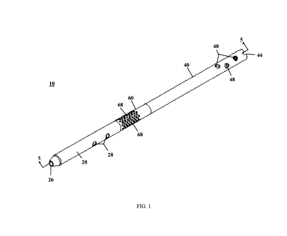
FIG. 1 is an isometric view of an intramedullary device from a distal end, in
accordance
with an aspect of the present invention;
FIG. 2 is an isometric view of the intramedullary device of FIG. 1 from a
proximal end,
in accordance with an aspect of the present invention;
FIG. 3 is a side view of the intramedullary device of FIG. 1, in accordance
with an aspect
of the present invention;
FIG. 4 is another side view of the intramedullary device of FIG. 1, in
accordance with an
aspect of the present invention;
FIG. 5 is a cross section of the intramedullary device of FIG. 1 taken along
line 5--5 of
FIG. I, in accordance with an aspect of the present invention;
FIG. 6 is a bottom view of the intramedullary device of FIG. 1, in accordance
with an
aspect of the present invention;
FIG. 7 is a partially exploded view of an intramedullary device, in accordance
with an
aspect of the present invention;
FIG. 8 is a side view of a dispersion device, in accordance with an aspect of
the present
invention;
FIG. 9 is a side view of the intramedullary device of FIG. 7 with the
dispersion device
partially inserted into the intramedullary nail, in accordance with an aspect
of the present
invention;
FIG. 10 is a cross section of the intramedullary device of FIG. 7 with the
dispersion
device partially inserted into the intramedullary nail taken along line l0--l0
of FIG. 9, in
accordance with an aspect of the present invention;
FIG. 11 is a side view of the intramedullary device of FIG. 7 with the
dispersion device
fully inserted into the intramedullary nail, in accordance with an aspect of
the present invention;
3
CA 02883536 2015-03-02
WO 2014/043794
PCT/CA2013/000800
FIG. 12 is a cross section of the intramedullary device of FIG. 7 with the
dispersion
device fully inserted into the intramedullary nail taken along line I2--12 of
FIG. 11, in
accordance with an aspect of the present invention;
FIG. 13 is an exploded view of another embodiment intramedullary device, in
accordance with an aspect of the present invention;
FIG. 14 is an isometric view of a first segment of the intramedullary device
of FIG. 1, in
accordance with an aspect of the present invention;
FIG. 15 is an isometric view from the top of the first segment of the
intramedullary
device of FIG. 1, in accordance with an aspect of the present invention;
FIG. 16 is an isometric view of a delivery segment of the intramedullary
device of FIG. 1
from the bottom, in accordance with an aspect of the present invention;
FIG. 17 is a side view of the delivery segment of the intramedullary device of
FIG. 1, in
accordance with an aspect of the present invention;
FIG. 18 is an isometric view of the delivery segment of the intramedullary
device of FIG.
1 from the top, in accordance with an aspect of the present invention;
FIG. 19 is a side view of a second segment of the intramedullary device of
FIG. 1, in
accordance with an aspect of the present invention;
FIG. 20 is an isometric view of the second segment of the intramedullary
device of FIG.
1 taken from the top, in accordance with an aspect of the present invention;
FIG. 21 is an isometric view of an extension segment of the intramedullary
device of
FIG. 13 from a proximal end, in accordance with an aspect of the present
invention;
FIG. 22 is an isometric view of the extension segment of the intramedullary
device of
FIG. 13 from a distal end, in accordance with an aspect of the present
invention;
FIG. 23 is a side view of a shuttle of the dispersion device of FIG. 8, in
accordance with
an aspect of the present invention;
FIG. 24 is a side isometric view of the shuttle of the intramedullary device
of FIG. 8, in
accordance with an aspect of the present invention;
FIG. 25 is an isometric view of the shuttle of the intramedullary device of
FIG. 8 taken
from the front, in accordance with an aspect of the present invention;
FIG. 26 is a side view of the shuttle of the intramedullary device of FIG. 8
including two
o-rings, in accordance with an aspect of the present invention;
FIG. 27 is an isometric view of a delivery tube of the dispersion device of
FIG. 8, in
accordance with an aspect of the present invention; and
FIG. 28 is a cross section of an intramedullary device inserted into a
patient's bone and
fixed with a fixation system, in accordance with an aspect of the present
invention.
4
CA 02883536 2015-03-02
WO 2014/043794
PCT/CA2013/000800
DETAILED DESCRIPTION
In this application, the words proximal, distal, anterior, posterior, medial
and lateral are
defined by their standard usage for indicating a particular part or portion of
a bone or prosthesis
coupled thereto, or directional terms of reference, according to the relative
disposition of the
natural bone. For example, "proximal" means the portion of a bone or
prosthesis nearest the
torso, while "distal" indicates the portion of the bone or prosthesis farthest
from the torso. As an
example of directional usage of the terms, "anterior" refers to a direction
towards the front side
of the body, "posterior" refers to a direction towards the back side of the
body, "medial" refers
to a direction towards the midline of the body and "lateral" refers to a
direction towards the sides
or away from the midline of the body.
Referring to the drawings, wherein like reference numerals are used to
indicate like or
analogous components throughout the several views, and with particular
reference to FIGS. 1-6,
there is illustrated an exemplary embodiment intramedullary device or nail 10.
The
intramedullary nail 10 may include a first non-delivery segment 20, a second
non-delivery
segment 40, and a delivery segment or dispersion segment 60 connecting the
first segment 20
and second segment 40. The first non-delivery segment 20, second non-delivery
segment 40,
and delivery segment 60 are made of a biomedical material, for example, a
metal, such as,
titanium, a composite, or bioabsorbable materials. The biomedical material may
be impregnated
with antimicrobial agents, for example, silver coatings, to help prevent
infection.
As best seen in FIGS. 14 and 15, the first segment 20 includes an interior
channel 22
along the longitudinal axis of the first segment 20. The channel 22 travels
from a first opening
24 to a second opening 26 (see FIG. 15). The first segment 20 also includes at
least one through
hole 28 perpendicular to the channel 22 for inserting at least one fastener to
secure the
intramedullary nail 10 to a bone. In the depicted embodiment there are two
through holes 28,
although it is also contemplated that the number of through holes 28 may range
from, for
example, two to four through holes. The proximal end of the first segment 20
may also include
a fastening mechanism 30. The fastening mechanism 30 may be, for example, a
female threaded
section as depicted in the present invention, alternative fastening mechanisms
30, for example, a
quick lock, snap fit, snap lock mechanisms, Morse tapers, and the like, are
also contemplated.
The fastening mechanism 30 may be reversible or non-reversible, in the present
invention the
fastening mechanism 30 is preferably non-reversible. The first segment 20 may
have a length
ranging from about, for example, 1 inch to about 6 inches with an inner
diameter ranging from,
for example, about 5 mm to about 10 mm, and an outer diameter ranging from,
for example,
about 9 mm to about 15 mm.
5
CA 02883536 2015-03-02
WO 2014/043794
PCT/CA2013/000800
Referring now to FIGS. 19 and 20, the second segment 40 is depicted and
includes an
interior channel 42 (see FIG. 20) along the longitudinal axis of the second
segment 40. The
channel 42 travels from a first opening 44 to a second opening 46. The second
segment 40 also
includes at least one through hole 48 perpendicular to the channel 42 for
inserting at least one
fastener to secure the intramedullary nail 10 into the bone. In the
illustrated embodiment there
are three through holes 48, although it is also contemplated that the number
of through holes 48
may range from, for example, two to four through holes. The distal end of the
second segment
40 may also include a first fastening mechanism 50. The first fastening
mechanism 50 may be,
for example, a male threaded section as depicted in the present invention,
although alternative
fastening mechanisms 50, for example, a quick lock, snap fit, snap lock
mechanisms, Morse
tapers, and the like, are also contemplated. The fastening mechanism 50 may be
reversible or
non-reversible, in the present embodiment, the fastening mechanism 50 is
preferably non-
reversible. The proximal end of the second segment 40 may include a second
fastening
mechanism 52. The second fastening mechanism 52 may be, for example, a female
threaded
section as depicted in the present invention. Alternative second fastening
mechanisms 52 are
also contemplated, such as quick lock, snap fit, snap lock mechanisms, Morse
tapers, and the
like. The second fastening mechanism 52 may be reversible or non-reversible
and second the
fastening mechanism 52 is preferably non-reversible in the illustrated
embodiment. The second
segment 40 may have a length ranging from, for example, about 1 inch to about
6 inches with an
inner diameter ranging from, for example, about 5 mm to about 10 mm and an
outer diameter
ranging from, for example, about 9 mm to about 15 mm.
The delivery segment 60 is best seen in FIGS. 16-18 and includes an interior
channel 62
along the longitudinal axis of the delivery segment 60. The channel 62 travels
from a first
opening 64 to a second opening 66. The delivery segment 60 also includes a
plurality of
through holes 68 passing from the channel 62 to an outer surface 69 of the
delivery segment 60.
The plurality of through holes 68 allow materials, for example, biomedical
materials, to exit the
intramedullary nail 10 into the location of a bone deficiency into the
surrounding tissues or
remaining bone. The distal end of the delivery segment 60 may also include a
first fastening
mechanism 70. The first fastening mechanism 70 may be, for example, a male
threaded section
as shown in the depicted embodiments. Alternative fastening mechanisms 70 are
also
contemplated, for example, quick lock, snap fit, snap lock mechanisms, Morse
tapers, and the
like. The fastening mechanism 70 is preferably non-reversible, although
reversible fastening
mechanisms 70 are also contemplated. The proximal end of the delivery segment
60 may
include a second fastening mechanism 72. In the illustrated embodiment the
second fastening
mechanism 72 may be, for example, a female threaded section, although
alternative
6
CA 02883536 2015-03-02
WO 2014/043794
PCT/CA2013/000800
embodiments are contemplated including, for example, quick lock, snap fit,
snap lock
mechanisms, Morse tapers, and the like. The fastening mechanism 72 is
preferably non-
reversible, although reversible fastening mechanisms 72 are also contemplated.
The delivery
segment 60 of the intramedullary nail 10 is modular and may be customized to
allow for
delivery of biomedical material to a deficiency in the bone at any location.
The delivery
segment 60 may have a length ranging from, for example, about 1 inch to about
8 inches with an
inner diameter ranging from, for example, about 3 mm to about 10 mm and more
preferably
from about 3 mm to about 7 mm, and an outer diameter ranging from, for
example, about 9 mm
to about 15 mm.
The intramedullary nail 10 may be assembled by the surgeon just prior to
implantation
and customized for the exact location of the site of a bone deficiency
specifically for each
patient, such as a deficiency due to cancer, significant trauma, bone loss or
weakness. The
surgeon may select a delivery segment 60 including the desired number and
desired size of
through holes 68 based on the material(s) to be injected into the bone
deficiency and the desired
rate of injection. Once the delivery segment 60 is selected the first non-
delivery segment 20 and
second non-delivery segment 40 may be selected to position the delivery
segment 60 at the
location of the bone deficiency or fracture. The first segment 20, second
segment 40, and
delivery segment 60 may be selected and secured together in any order. The
first non-delivery
segment 20 may be smaller than, larger than, or the same size as the second
non-delivery
segment 40 to allow for placement of the delivery segment 60 anywhere along
the
intramedullary nail 10. Further, additional delivery segments 60 may be placed
along the
intramedullary nail 10 if necessary to disperse biomedical materials to
multiple locations within
the bone.
After the segments 20, 40, and 60 are selected the first segment 20 may be
secured to the
delivery segment 60 at a distal end and the second segment 40 may be secured
to the delivery
segment 60 at the proximal end. By way of specific example, the male threaded
section of the
delivery segment's first fastening mechanism 70 will be inserted into the
female threaded
section of the first segment's fastening mechanism 30. Then the male threaded
section of the
second segment's fastening mechanism 50 will be inserted into the female
threaded section of
the delivery segment's second fastening mechanism 72.
In another embodiment, where the first segment 20, second segment 40, and
delivery
segment 60 are Morse tapers, the fastening mechanism 70 of the delivery
segment 60 will
include a tapered distal end (not shown). The tapered distal end (not shown)
of the delivery
segment 60 may be placed in the first opening 24 of the first segment 20 which
may also be
tapered from the first opening 24 to the second opening 26. In addition, the
second segment 40
7
CA 02883536 2015-03-02
WO 2014/043794
PCT/CA2013/000800
may be tapered from the first opening 44 to the second opening 46. The tapered
distal end (not
shown) of the fastening mechanism 50 of the second segment 40 may be inserted
into the first
opening 64 of the delivery segment 60. Once the delivery segment 60 is
inserted into the first
segment 20 and the second segment 40 is inserted into the delivery segment 60
to form an
intramedullary device 10 a force may be applied to the proximal and distal
ends of the
intramedullary device 10 to secure the first segment 20, second segment 40,
and delivery
segment 60 together. The force may be applied, for example, by a mechanical
press, a hammer,
or other known methods of securing Morse taper components together.
When multiple delivery segments 60 are placed along the intramedullary nail 10
a center
non-delivery segment, not shown, may be inserted between the multiple delivery
segments 60
and the first segment 20 will be attached to the delivery segment 60 located
at the distal end and
the second segment 40 will be attached to the delivery segment 60 located at
the proximal end of
the intramedullary nail 10. The resulting intramedullary nail 10 places the
delivery segments 60
precisely where the surgeon wants them for delivery of biomedical materials to
the site of the
bone deficiency.
If additional length is needed for the intramedullary nail 10 for the
embodiment depicted
in FIG. 1 or the nail is comprised of extension segments as illustrated in
FIG. 13, extension
segments 120, may be used. The extension segments 120, as shown in FIGS. 13,
21, and 22,
may be inserted between the first segment 20 and the delivery segment 60,
between the delivery
segment 60 and the second segment 40. These extension segments may range from,
for
example, approximately 1 inch to 6 inches and are more preferably about one
and a half inch
segments. The resulting intramedullary nail 10 will range from, for example,
approximately 10
inches to 40 inches. In alternative embodiments, extension segments 120 may
also be attached
at the proximal end of the second segment 40.
Referring now to FIGS. 21-22, the extension segments 120 are illustrated. The
extension
segments 120 include an interior channel 122 along the longitudinal axis of
the extension
segments 120. The channel 122 travels from a first opening 124 to a second
opening 126. The
distal end of the extension segments 120 may also include a first fastening
mechanism 128. The
first fastening mechanism 128 may be, for example, a male threaded section as
depicted in the
present invention, alternative fastening mechanisms 128, for example, a quick
lock, snap fit,
snap lock mechanisms, Morse tapers, and the like, are also contemplated. The
first fastening
mechanism 128 may be reversible or non-reversible, in the present invention
the fastening
mechanism 128 is preferably non-reversible. The proximal end of the extension
segments 120
may include a second fastening mechanism 130, as shown in FIG. 21. In the
illustrated
embodiment the second fastening mechanism 130 may be, for example, a female
threaded
8
CA 02883536 2015-03-02
WO 2014/043794
PCT/CA2013/000800
section, although alternative embodiments are contemplated including, for
example, quick lock,
snap fit, snap lock mechanisms, Morse tapers, and the like. The second
fastening mechanism
130 is preferably non-reversible, although reversible fastening mechanisms 130
are also
contemplated. The extension segments 120 of the intramedullary nail 10 are
modular and may
be inserted anywhere along the nail where additional length is needed. The
extension segments
120 are generally inserted between the first non-delivery segment 20 and the
delivery segment
60 and between the delivery segment 60 and the second non-delivery segment 40.
Any additional delivery segments 60 or extension segments of the
intramedullary nail 10
may also include a fastening or locking mechanism, not shown, that allows for
the locking and
unlocking of the segments 20, 40, and 60 of the intramedullary nail 10
relative to each other.
The locking mechanism may be, for example, a quick lock, snap fit, snap lock
mechanism,
Morse taper, and the like, which allows for the segments 20, 40, and 60 and
extension segments
120, if used, to be secured together to prevent the segments 20, 40, 60, and
120 from
disconnecting while implanted in a patient.
Referring now to FIGS. 7 and 9-12, a modular intramedullary device or nail
system 110
is shown. The modular intramedullary device system 110 includes a dispersion
device 80 and
the intramedullary nail 10. The intramedullary nail 10, described in greater
detail above,
provides for stabilization of the bone, while the dispersion device 80,
described in greater detail
below, allows for the precise placement of biomedical materials within the
bone to augment the
stabilization. The dispersion device 80 may also accept instrumentation to
assist a surgeon
determine movement and healing of the bone at the site of the bone deficiency.
An alternative modular intramedullary device system 110 is illustrated in FIG.
13. The
alternative modular intramedullary device system 110 includes a dispersion
device 80 and the
intramedullary nail 118. The intramedullary nail 118 may be assembled by the
surgeon just
prior to implantation and customized for the exact location of the site of a
bone deficiency
specifically for each patient, such as a deficiency due to cancer, significant
trauma, bone loss or
weakness. The surgeon may select a delivery segment 60 including the desired
number and
desired size of through holes 68 based on the biomedical material(s) to be
injected into the bone
deficiency and the desired rate of injection. Once the delivery segment 60 is
selected a first non-
delivery segment 20 and a second non-delivery segment 40, as well as the
desired number of
extension segments 120 may be selected to position the delivery segment 60 at
the location of
the bone deficiency or fracture. As illustrated in the depicted embodiment of
FIG. 13, two
extension segments 120 are connected to the distal end of the delivery segment
60 before the
first non-delivery segment 20 is attached. On the proximal end of the delivery
segment 60 three
extension segments 120 are connected prior to securing the second non-delivery
segment 40.
9
CA 02883536 2015-03-02
WO 2014/043794
PCT/CA2013/000800
Any number of extension segments 120 may be selected for insertion between the
first segment
20 and the delivery segment 60 and between the delivery segment 60 and the
second segment 40
to allow for placement of the delivery segment 60 anywhere along the
intramedullary nail 118.
Further, additional delivery segments 60 may be placed along the
intramedullary nail 118 if
necessary to disperse biomedical materials to multiple locations within the
bone.
With continued reference to FIGS. 7 and 9-12, after the segments 20, 40, and
60 are
selected the first segment 20 may be secured to the delivery segment 60 at a
distal end and the
second segment 40 may be secured to the delivery segment 60 at the proximal
end. Specifically,
the male threaded section of the delivery segment's first fastening mechanism
70 will be
inserted into the female threaded section of the first segment's fastening
mechanism 30. Then
the male threaded section of the second segment's fastening mechanism 50 will
be inserted into
the female threaded section of the delivery segment's second fastening
mechanism 72. When
multiple delivery segments 60 are placed along the intramedullary nail 10 a
center non-delivery
segment, not shown, may be inserted between the multiple delivery segments 60
and the first
segment 20 will be attached to the delivery segment 60 located at the distal
end and the second
segment 40 will be attached to the delivery segment 60 located at the proximal
end of the
intramedullary nail 10. The resulting intramedullary nail 10 places the
delivery segments 60
precisely where the surgeon wants them for delivering biomedical materials to
the site of the
bone deficiency.
As seen in FIG. 8, the dispersion device 80 includes a tube 82 and a shuttle
or dispensing
member 90. In order to deliver materials, for example, irrigation or cleaning
fluids, bone
regenerative materials, or bone cement, the dispersion device 80 is inserted
into the
intramedullary nail 10. The dispensing member 90 is inserted into the first
opening 44 and slid
into alignment with the delivery segment 60. The interior channel 62 may
include a stop
member (not shown) at its distal end to stop the dispensing member 90 in the
desired location
for release of the delivery materials into the bone fracture. Alternatively,
the channel 22 may
include a stop member (not shown) at its proximal end to stop the dispensing
member 90 in the
desired location for release of the delivery materials into the bone fracture.
The dispersion
device 80 may be made of, for example, a polymer or composite material. The
polymer material
used for the dispersion device 80 may be a long term polymer material or a
resorbable material.
As illustrated in FIGS. 23-26, the dispensing member 90 includes a proximal
end 92 and
a distal end 94 connected by at least one center member 96 with at least one
dispersion opening
98. In the depicted embodiment, there are four center members 96 connecting
the proximal end
92 and the distal end 94. The four center members 96 create four dispersion
openings 98 for
releasing the delivery materials into the bone fracture site. In the
illustrated embodiment, the
CA 02883536 2015-03-02
WO 2014/043794
PCT/CA2013/000800
center members 96 provide large dispersion openings 98 to enable a full 360
degree dispersion
of materials through the delivery segment 60. The dispensing member 90 also
includes an
attachment portion 100 at the proximal end 92 to connect the dispensing member
90 and the
tube 82. In the depicted embodiment, the attachment portion 100 is a stepped
up connector
section creating a tight fit when the tube 82 is inserted over the attachment
portion 100. The
attachment portion 100 includes an opening 102 (see FIG. 25) allowing for
delivery materials to
pass from the tube 82 into the dispensing member 90 for delivery out of the
dispersion openings
98. The distal end 94 may include an optional opening 104, as depicted in
FIGS. 24-25.
Further, the dispensing member 90 may also include at least one groove 106
(see FIG. 23) for
mating with at least one o-ring 108 (see FIG. 24). In the depicted embodiment,
there are two
grooves 106 and two o-rings 108 with a first groove 106 and o-ring 108 at the
proximal end 92
of the dispensing member 90 and a second groove 106 and o-ring 108 at the
distal end 94 of the
dispensing member 90. Further, a sealing mechanism (not shown) may be provided
to seal the
ends of the dispensing member 90. The sealing mechanism may be a ring locking
mechanism,
wherein the proximal and distal ends each include a ring of material which has
a slightly larger
diameter than the inner diameter of the nail 10. The dispensing member 90 with
the sealing
mechanism may be snapped into the nail 10 to prevent fluids and in some case
materials from
flowing past the proximal and distal ends including the ring locking
mechanisms.
Referring now to FIG. 27, the tube 82 is illustrated and includes a proximal
end 84 and a
distal end 86 with an interior channel 88. The channel 88 runs along the
longitudinal axis of the
tube 82 from the proximal end 84 to the distal end 86. The distal end 86 of
the tube 82 mates
with the attachment portion 100 (see FIG. 26) of the dispensing member 90 to
create the
dispersion device 80, as shown in FIG. 8. Once the tube 82 and dispensing
member 90 are
secured together, a material injection system, not shown, may be secured to
the proximal end 84
of the tube 82 for dispensing delivery materials into the dispersion device
80. The material
injection system may include, for example, a syringe system, a one-way pump, a
cement gun, an
external pump and suction or pump and valve system, or the like, for
dispensing delivery
materials into the tube 82. The syringe system, one-way pump system, and
cement gun are
preferably used for dispersion of bone regenerative materials and bone cement
through the
delivery segment 60. The external pump and suction or pump and valve system is
preferably
used for irrigating or cleaning the area of bone deficiency by pumping a fluid
to clean the wound
into the dispersion device 80 and the extracting the fluid from the wound
using suction to pull
the fluid out of the nail 10 between the channels 62 and 42 (see FIG. 5) and
the exterior of tube
82. The material injection system may be secured to the proximal end 84 of the
tube 82 by a
11
CA 02883536 2015-03-02
WO 2014/043794
PCT/CA2013/000800
locking mechanism (not shown). The locking mechanism may be a threaded system
or a quick
connect-disconnect system.
Alternative dispersion devices 80 are also contemplated such as using a
capsule system
(not shown) that would be inserted into the first opening 44 of the second
segment 40 (see FIG.
5). The capsule system would travel through the channel 42 and into the
channel 62 in the
delivery segment 60. Once in the desired position in the delivery segment 60
the capsule could
be pierced to release the delivery material. The capsule could be pierced by a
puncture tool (not
shown), such as a sharp protrusion, within the channel 62 or alternatively the
capsule could be
pierced manually by inserting a puncture tool or a sharp instrument down the
channel 42 of the
second segment 40 until the instrument contacted the capsule and pierced it to
release the
dispersion materials. Another alternative dispersion devise 80 may include a
tube 82 and an
instrument (not shown) for deploying the biomedical material in the tube 82.
The tube 82 may
be, for example, flexible or rigid. The tube 82 may be inserted into the first
opening 44 of the
second segment 40 (see FIG. 5) and pushed into the nail 10 along the interior
channel 42, 62
until the distal end 86 of the tube 82 engages at least a portion of the
delivery segment 60. Then
a delivery tool (not shown) may be inserted into the channel 88 at a proximal
end 84 of the tube
82 to dispense the delivery material through the through holes 68 to the bone.
Alternatively, the
delivery tool (not shown) may be connected to the tube 82 at a proximal end to
apply pressure
through the tube causing the delivery material to dispense from the tube 82
into the delivery
segment 60 and out the through holes 68 to the bone. The delivery tool may be,
for example, a
plunger, bougie, or the like. Each dispersion device 80 can prevent the
delivery materials that
are injected into the intramedullary device system 110 from flowing down the
nail where the
delivery materials are not needed. One method to prevent the delivery
materials from flowing
past the delivery segment 60 is by pressurizing the material injection system
thereby forcing the
delivery materials out of the plurality of holes 68. Another method to prevent
the materials from
flowing into channels 22 or 42 is to include a sealing mechanism (not shown)
which seals the
ends of the dispensing member 90.
The intramedullary device system 110 may be used to providing stabilization of
a bone
and limb, reconstruction of soft tissue defects, and the precise placement or
delivery of materials
within the bone to augment the stabilization by stimulating bone formation and
providing a
scaffold for bone formation. For example, bone cement may be delivered to
bones that have
been weakened or removed by cancer to fill the deficiencies in the bone.
Alternatively materials
to promote bone formation and healing may be delivered where bone is missing.
Additional
uses of the intramedullary device system 110 include but are not limited to
irrigating bones and
surrounding soft tissues when there are open and contaminated wounds, for
example, in high
12
CA 02883536 2015-03-02
WO 2014/043794
PCT/CA2013/000800
energy injuries such as blast injuries or due to other trauma. Further, the
intramedullary device
system 110 also allows for bone regeneration materials, such as growth
stimulators, to be placed
at a site of fracture or bone loss to stimulate bone formation around the nail
at that site. Growth
stimulators may include, for example, platelet derived growth factor ("PDGF"),
vascular
endothelial growth factor ("VEGF") and epidermal growth factor, which may be
used to initiate
healing by promoting cell replication and repair. The intramedullary device
system 110 may
also be used to deliver bone material, for example from the reamings, or from
allograft
preparations that stimulate bone formation from the surrounding tissue. Yet
further, the
intramedullary device system 110 may also be used to provide drugs or
chemicals to the bone or
tissues within the bone. The drugs or chemicals could be used to prevent or
treat infection or to
provide drugs or medically active chemicals to the entire body from a
reservoir within the
intramedullary device system 110. The intramedullary device system 110 may
also be used to
treat bones that have a regular bone fracture, as well as bones that are at
risk from fracturing by
allowing the placement of materials or substances that will strengthen or
improve the bone's
response to physiologic activities.
A surgical method for implanting an intramedullary device includes obtaining
an
intramedullary device system 110 for insertion into a patient's bone. The bone
is then prepared
for insertion of the intramedullary device system 110 by inserting a guidewire
into the bone then
drilling over the guidewire to create a canal for the nail 10. The nail 10 of
the intramedullary
device system 110 is then inserted into the canal created in the bone. The
nail 10 is positioned
so the delivery segment 60 is located at the bone deficiency. The dispersion
device 80 is then
inserted into an interior longitudinal channel created by channels 22, 42 and
62 (see FIG. 5) in
the nail 10 until the distal end of the dispensing member 90 is aligned with
the distal end of the
delivery segment 60 (see FIGS. 11-12). Then material is dispersed from the
dispersion device
80 at the dispensing member 90 through the delivery segment 60 to the damaged
portion of the
bone.
For example, once the intramedullary device system 110 is assembled by the
surgeon
and inserted into the bone of the patient, the device system 110 can be used
as an irrigation
device to the wound where both the bone and the surrounding soft tissue
envelope has been
injured.
It is accepted medical practice that the treatment of open wounds involving
bone
fractures requires the patient to be taken to an operating facility where the
wound can be
surgically treated to remove all visible foreign material, all dead tissues
and dead bone. The
wound may be washed with fluids during or after this debridement. The usual
practice is then to
13
CA 02883536 2015-03-02
WO 2014/043794
PCT/CA2013/000800
either close the wound or apply a sponge and covering to the wound and apply
suction to
remove fluids from the injured area.
The intramedullary device system 110 may provide, for example, ongoing fluid
lavage to
both washout the wound and remove microscopic foreign material, bacteria and
other noxious
organisms and blood clots that may harbor and encourage growth of bacteria.
The fluids that are
delivered to the injured area may also contain antibiotics and antiseptics
that will further inhibit
growth of bacteria. The use of a low pressure pulsatile system for fluid
delivery is unique in the
application of pressure allows for the soft tissues to be lightly distended so
that fluid flows to all
parts of the wound, and then allows for the fluid to be removed, so improving
the washout
ability for all materials. The pulsatile pressure is also beneficial to the
soft tissues as it may
prevent contractures of the soft tissues, keeping them pliable and elastic
while healing occurs.
The fluid management system here described may also speed the resolution and
prevention of
infection, which is the main early complication of traumatic open wounds to
long bone fractures.
An alternative embodiment of the dispersion device 80 allows for an early
irrigation
system such as a tubing apparatus (not shown) to be inserted through the
incision used to insert
the nail and then passed into the end of the nail closest to the skin wound.
The tubing apparatus
is inserted into the nail 10, in such a fashion that there is a water tight
seal at the distal or far end
of the delivery segment 60, and a watertight seal at the proximal or near end
of the delivery
segment 60. This allows for irrigation of only the injured part of the bone
and soft tissues. The
tubing apparatus consists of two passageways within the tube, one having a
large bore and the
second having smaller bore. The fluid pressure of the inlet and exit fluid of
the tube will be
monitored externally near the proximal end of the tube (given the low flow
rates, these pressure
readings outside of the nail will be close approximations of internal
pressures). The large bore
passageway is the outlet for the fluids, and the fluids flow from the delivery
segment 60 up the
tubing to a connector out of the patient. There the fluids may flow over or
through material
which gathers bacterial and fungal DNA and RNA for analysis at a laboratory to
determine the
type of infection that might be present within the patient. The fluids then
flow to a container for
disposal. Alternatively the fluids may flow from the delivery segment 60 up
the tubing to a
connector out of the patient and to a container for disposal. The tubing
apparatus of the
dispersion device 80 is a closed system.
The smaller bore passageway is the inflow for fluids and is connected to a
pump which
applies a pulsating pressure, with that pressure being adjustable by attending
health care
personnel. The source of fluids for the pump consists of a regular IV bag in
which different
chemicals or antibiotics can be placed on the orders of a medical doctor.
There is a closed loop
system of controls from the pressure monitors within the delivery segment 60
of the irrigation
14
CA 02883536 2015-03-02
WO 2014/043794
PCT/CA2013/000800
system to the pump which controls the pressure for the inlet line. Pressures
within the delivery
segment 60 cannot exceed pressure limits set by the attending health care
personnel. The health
care personnel can control the pressure and the amount of fluids dispensed by
the pump.
After irrigation of the wound or dispersion of desired fluids, the dispersion
device 80
may then be removed and the nail 10 secured to the bone by inserting pins or
other bone
fastening mechanisms through the through holes 28 of the first segment 20 (see
FIG. 1).
Alternatively, the first segment 20 may have been secured to the bone prior to
insertion of the
delivery segment 60 into the nail 10. Once all material has been inserted into
the bone for the
present procedure the second segment 40 may optionally be secured to the bone
by inserting
pills or other bone fastening mechanisms into through holes 48 (see FIG. 1).
If the surgeon intends to deliver additional materials to the bone deficiency
the surgeon
may decide not to secure the second segment 40 to the bone to provide
continued access to the
nail 10 through the healing process. However, if the surgeon will be allowing
the patient to
perform weight bearing activities on the bone which received the nail 10, the
second segment 40
should be secured to the bone. If the second segment 40 is secured to the bone
using bone
fastening mechanisms (not shown) which traverse the channel 42 and additional
material is to be
inserted into the nail 10 at a later date, the threaded pins or other bone
fastening mechanisms
would have to be removed prior to insertion of the dispersion device 80 into
the nail 10. After
the bone deficiencies have been completely stabilized or healed, the
intramedullary nail 10 may
be removed from the patient's bone.
As the locking or transfixion screws used to stabilize the nail 10 to the bone
described
above would occupy the inside of the nail 10 and interfere with the passage of
materials down
the nail 10 through channel 42, an alternative fixation system 112, shown in
FIG. 28, may be
used to maintain access to the delivery segment 60 throughout the healing
process without
having to insert and remove locking screws. The fixation system 112 allows for
temporary
locking screws 114 to be placed using standard guides that fit the proximal
end of the nail 10,
whereby the holes may be drilled through the bone towards the nail 10.
However, instead of the
holes being present in the nail 10, captured gimbals are provided with holes
in them or small
tapered detents in the outer surface of the nail 10. The temporary locking
screws 114 will
engage the gimbals or detents but do not enter the channel 42 of the nail 10,
thereby leaving the
inside channel 42 of the nail 10 free for passage by a dispersion device 80.
The temporary
locking screws 114 may be longer than the bones and may extend into the soft
tissue and muscle
surrounding the patient's bone, but are contained within the patient's skin.
After irrigation is
complete and new bone begins forming in the region of the fracture, the
temporary fixation
screws 114 can be removed and replaced with standard fixation screws as
described above
CA 02883536 2015-03-02
WO 2014/043794
PCT/CA2013/000800
which are inserted into the through holes 48 (see FIG. 1) in the proximal end
of the nail. The
standard fixation screws will traverse the through holes 48, which may be
tapped through holes.
After the bone deficiencies have been completely stabilized or healed, the
intramedullary nail 10
may be removed from the patient's bone.
The terminology used herein is for the purpose of describing particular
embodiments
only and is not intended to be limiting of the invention. As used herein, the
singular forms "a",
"an" and "the" are intended to include the plural forms as well, unless the
context clearly
indicates otherwise. It will be further understood that the terms "comprise"
(and any form of
comprise, such as "comprises" and "comprising"), "have" (and any form of have,
such as "has",
and "having"), "include" (and any form of include, such as "includes" and
"including"), and
"contain" (and any form of contain, such as "contains" and -containing") are
open-ended
linking verbs. As a result, a method or device that "comprises," "has,"
"includes," or "contains"
one or more steps or elements possesses those one or more steps or elements,
but is not limited
to possessing only those one or more steps or elements. Likewise, a step of a
method or an
element of a device that "comprises," "has," "includes," or "contains" one or
more features
possesses those one or more features, but is not limited to possessing only
those one or more
features. Furthermore, a device or structure that is configured in a certain
way is configured in
at least that way, but may also be configured in ways that are not listed.
The invention has been described with reference to the preferred embodiments.
It will be
understood that the architectural and operational embodiments described herein
are exemplary
of a plurality of possible arrangements to provide the same general features,
characteristics, and
general system operation. Modifications and alterations will occur to others
upon a reading and
understanding of the preceding detailed description. It is intended that the
invention be
construed as including all such modifications and alterations.
16