Note: Descriptions are shown in the official language in which they were submitted.
CA 02884468 2015-03-09
WO 2014/053913
PCT/IB2013/002672
NAVIGATION INSTRUMENTS FOR SUBCHONDRAL BONE TREATMENT
TECHNICAL FIELD
The present invention relates to tools for the surgical treatment of joints,
and more
particularly to instruments and associated methods for the surgical repair and
treatment of
bone tissue at these joints. Even more particularly, the present invention
relates to navigation
instruments for targeting an area near a subchondral bone defect using
anatomical landmarks.
BACKGROUND ART
Human joints, in particular the knee, hip and spine, are susceptible to
degeneration
from disease, trauma, and long-term repetitive use that eventually lead to
pain. Knee pain,
for example, is the impetus for a wide majority of medical treatments and
associated medical
costs. The most popular theory arising from the medical community is that knee
pain results
from bone-on-bone contact or inadequate cartilage cushioning. These conditions
are believed
to frequently result from the progression of osteoarthritis, which is measured
in terms of
narrowing of the joint space. Therefore, the severity of osteoarthritis is
believed to be an
indicator or precursor to joint pain. Most surgeons and medical practitioners
thus base their
treatments for pain relief on this theory. For example, the typical treatment
is to administer
pain medication, or more drastically, to perform some type of joint
resurfacing or joint
replacement surgery.
However, the severity of osteoarthritis, especially in joints such as the knee
and ankle,
has been found to correlate poorly with the incidence and magnitude of knee
pain. Because
of this, surgeons and medical practitioners have struggled to deliver
consistent, reliable pain
relief to patients especially if preservation of the joint is desired.
Whether by external physical force, disease, or the natural aging process,
structural
damage to bone can cause injury, trauma, degeneration or erosion of otherwise
healthy tissue.
The resultant damage can be characterized as a bone defect that can take the
form of a
fissure, fracture, microfracture, lesion, edema, tumor, or sclerotic
hardening, for example.
1
CA 02884468 2015-03-09
WO 2014/053913
PCT/1B2013/002672
Particularly in joints, the damage may not be limited to a bone defect, and
may also include
cartilage loss (especially articular cartilage), tendon damage, and
inflammation in the
surrounding area.
Patients most often seek treatment because of pain and deterioration of
quality of life
attributed to the osteoarthritis. The goal of surgical and non-surgical
treatments for
osteoarthritis is to reduce or eliminate pain and restore joint function. Both
non-surgical and
surgical treatments are currently available for joint repair.
Non-surgical treatments include weight loss (for the overweight patient),
activity
modification (low impact exercise), quadriceps strengthening, patellar taping,
analgesic and
.. anti-inflammatory medications, and with corticosteroid and/or
viscosupplements. Typically,
non-surgical treatments, usually involving pharmacological intervention such
as the
administration of non-steroidal anti-inflammatory drugs or injection of
hyaluronic acid-based
products, are initially administered to patients experiencing relatively less
severe pain or joint
complications. However, when non-surgical treatments prove ineffective, or for
patients with
severe pain or bone injury, surgical intervention is often necessary.
Surgical options include arthroscopic partial meniscectomy and loose body
removal.
Most surgical treatments conventionally employ mechanical fixation devices
such as screws,
plates, staples, rods, sutures, and the like are commonly used to repair
damaged bone. These
fixation devices can be implanted at, or around, the damaged region to
stabilize or
immobilize the weakened area, in order to promote healing and provide support.
Injectable
or fillable hardening materials such as bone cements, bone void fillers, or
bone substitute
materials are also commonly used to stabilize bone defects.
High tibial osteotomy (HTO) or total knee arthroplasty (TKA) is often
recommended
for patients with severe pain associated with osteoarthritis, especially when
other non-
invasive options have failed. Both procedures have been shown to be effective
in treating
knee pain associated with osteoarthritis.
However, patients only elect HTO or TKA with reluctance. Both HTO and TKA are
major surgical interventions and may be associated with severe complications.
HTO is a
painful procedure that may require a long recovery. TKA patients often also
report the
replaced knee lacks a "natural feel" and have functional limitations.
Moreover, both HTO
2
CA 02884468 2015-03-09
WO 2014/053913
PCT/1B2013/002672
and TKA have limited durability. Accordingly, it would be desirable to provide
a medical
procedure that addresses the pain associated with osteoarthritis and provides
an alternative to
a VITO or TKA procedure.
In current practice, surgeons typically "eyeball" (i.e., visually estimate)
the target site
on a bone to be repaired. Most conventional targeting and location methods are
relatively
crude and provide little guidance to a surgeon during the actual surgical
procedure.
Accordingly, it would be desirable to provide methods and instruments in which
the area near
a bone defect can be easily located and provide a reference framework that can
be used in a
surgical procedure irrespective of the approach. Furthermore, in some
situations where
pinpoint accuracy is not critical or necessary, a navigation system that can
indicate an area
sufficiently near the bone defect in a quick and reliable manner would be
highly beneficial to
the clinician.
Accordingly, it is desirable to provide instruments that allow fast, easy, and
repeatable
navigation to an area sufficiently near a bone defect to be treated. It is
further desirable to
provide instruments that do not obstruct access to the working area around the
target site, and
allow as clear a view as possible for the clinician.
3
CA 02884468 2015-03-09
WO 2014/053913
PCT/1B2013/002672
SUMMARY OF INVENTION
'Me present disclosure provides navigation instruments for targeting an area
sufficiently near a subchondral bone defect using anatomical landmarks. The
instruments
allow the surgeon to navigate to the area around the bone defect quickly and
easily, while
also facilitating proper insertion of a tool or other device into an
appropriate area near the
defect.
In one embodiment, an instrument for navigating to a target area near a
subchondral
defect of a bone is provided. The instrument may comprise a guide having a
plurality of
device portals, each portal defining a trajectory and configured to provide
accurate and
controlled delivery of a tool to the target area. Also provided is a handle
extending from the
guide. The handle may be detachable. The instrument may be configured to align
with an
anatomical landmark of the bone, and include visual markers to assist in
positioning the
instrument. In one embodiment, the guide is configured to target a subchondral
area of the
tibia. In another embodiment, the guide is configured to target a subchondral
area of the
femur, and may comprise a hinged pair of arms.
In some embodiments, the instrument may further include visual markers for
vertical
alignment of the instrument. In addition, the detachable handle may include a
guide
attachment end having a plurality of keyed slots. The guide component may
comprise a
shaped stem that is configured to engage one or more of the keyed slots of the
handle, thus
allowing the guide to be angularly adjustable relative to the detachable
handle. The
instrument may receive a tool such as an injection needle that may include a
depth gauge.
In another exemplary embodiment, an instrument for navigating to a target area
near a
subchondral defect of a bone is provided. The instrument may comprise a guide
having a
horizontal approach device portal and a distal approach device portal, each
portal defining a
trajectory and configured to provide accurate and controlled delivery of a
tool to the target
area. The instrument may also comprise a handle extending from the guide. The
guide and
handle may comprise a uniform body. In addition, the instrument may be
configured to align
with an anatomical landmark of the bone, and include visual markers to assist
in positioning
the instrument..
4
CA 02884468 2015-03-09
WO 2014/053913
PCT/1B2013/002672
In some embodiments, the handle may be configured to secure to a patient's
leg. The
instrument may also include a slot for insertion of a scalpel. The instrument
may be
configured to align with an anatomical landmark of the bone, and include
visual markers to
assist in positioning the instrument. In one embodiment, the guide is
configured to target a
subchondral area of the tibia. The instrument may receive a tool such as an
injection needle
that may include a depth gauge.
In still another exemplary embodiment, a system for navigating to a target
area near a
subchondral defect of a bone is provided. The system may comprise a handle
component
comprising a slot for receiving a guide component, a femoral guide component
comprising a
hinged pair of aims, and a tibial guide component. Each of the guide
components may
comprise a plurality of device portals, each portal defining a trajectory and
configured to
provide accurate and controlled delivery of a tool to the target area.
Furtheimore, each of the
guide components may be slidingly received in the slot of the handle
component. The slot of
the handle component may comprise a plurality of keyed sections or notches.
Each of the
guide components may comprise a shaped stem configured to engage one or more
of the
keyed sections or notches of the slot of the handle. The guide components may
be angularly
adjustable relative to the handle.
It is to be understood that both the foregoing general description and the
following
detailed description are exemplary and explanatory only and are not
restrictive of the
disclosure. Additional features of the disclosure will be set forth in part in
the description
which follows or may be learned by practice of the disclosure.
5
CA 02884468 2015-03-09
WO 2014/053913
PCT/IB2013/002672
BRIEF DESCRIPTION OF THE DRAWINGS
The accompanying drawings, which are incorporated in and constitute a part of
this
specification, illustrate several embodiments of the disclosure and together
with the
description, serve to explain the principles of the disclosure.
FIG. 1 is top-down perspective view of an exemplary embodiment of a navigation
instrument of the present disclosure relative to a tibia.
FIG. 2 is a perspective view of a tibial hone and anatomical landmark used
with
navigation instruments of the present disclosure.
FIG. 3 is another top-down perspective view of the navigation instrument of
FIG. 1
relative to a tibia.
FIG. 4 shows a front view of the navigation instrument of FIG. 1 relative to a
tibia.
FIGS. 5A and 5B show a right and left version of the navigation instrument of
FIG. 4,
respectively.
FIG. 6 shows a side view of the navigation instrument of FIG. 1 relative to a
tibia.
FIGS. 7A and 7B show a side perspective view of the navigation instrument of
FIG. 4
and a front perspective view of the navigation instrument of FIG. 4,
respectively.
FIG. 8 shows a partial cutaway view of the navigation instrument of FIG. 4
relative to
a tibia.
FIG. 9 shows a front perspective view of the navigation instrument of FIG. 4
relative
to a tibia.
FIG. 10 shows a side perspective view of the navigation instrument of FIG. 4
relative
to a tibia.
FIG. 11 shows a perspective view of the navigation instrument of FIG. 4 with
delivery pin relative to a tibia.
FIG. 12A shows a perspective view of another exemplary embodiment of a
navigation
instrument of the present disclosure relative to a femur.
6
CA 02884468 2015-03-09
WO 2014/053913
PCT/IB2013/002672
FIG. 12B shows another perspective view of the navigation instrument of FIG.
12A
relative to a femur.
FIGS. 13A ________ 13C show various perspective views of still another
exemplary
embodiment of a navigation instillment for use with a femur.
FIG. 14A shows a top-down view of still another exemplary embodiment of a
navigation instrument of the present disclosure relative to a femur.
FIG. 14B shows a perspective view of the navigation instrument of FIG. 14A
relative
to a tibia.
FIG. 14C shows an enlarged view of the navigation instrument of FIG. 14B
relative to
a tibia.
FIG. 14D shows a top-down perspective view of the navigation instrument of
FIG.
14B in use with a needle relative to a tibia.
FIG. 14E shows another perspective view of the navigation instrument and
needle of
FIG. 14D relative to a tibia.
FIG. 14F shows a perspective view of the navigation instrument of FIG. 14B in
use
with an injection needle relative to a tibia.
FIG. 14G shows the navigation instrument with needle of FIG. 14B relative to
the
patient's leg.
FIGS. 15A-15C show perspective views of another embodiment of a navigation
instrument of the present disclosure relative to a femur.
FIG. 16 shows an exemplary embodiment of a guide system comprising various
components for assembling a femoral or tibial guide instrument of the present
disclosure.
FIG. 17 shows a perspective view of an exemplary tibial guide instrument
assembled
from the system of FIG. 16 in use.
FIGS. 18A and 18B show a method of using an exemplary femoral guide instrument
assembled from the system of FIG. 16.
7
CA 02884468 2015-03-09
WO 2014/053913
PCT/IB2013/002672
FIG. 19A shows a perspective view of another exemplary embodiment of a
navigation
instrument relative to a tibia.
FIG. 19B shows a perspective view of another exemplary embodiment of a
navigation
instrument relative to a femur.
FIG. 20 shows an enlarged view of the handle of the navigation instrument of
FIGS.
19A and 19B.
FIGS. 21A-21C show an exemplary method of assembling the navigation
instrument of FIG. 19A.
8
DETAILED DESCRIPTION OF THE EMBODIMENTS
Methods, devices and instruments for treating joint pain to restore natural
joint
function and preserving, as much as possible, the joint's articular and
cartilage surface are
known. Treatments through the joint that violate the articular and cartilage
surface often
weaken the bone and have unpredictable results. Rather than focusing on
treatment of pain
through the joint, alternative treatments that diagnose and treat pain at its
source in the
subchondral region of a bone of a joint to relieve the pain are provided. Pain
associated with
joints, especially osteoarthritic joints, can be correlated to bone defects or
changes at the
subchondral level rather than, for example, the severity of osteoarthritic
progression or
defects at the articular surface level. In particular, bone defects, such as
bone marrow
lesions, edema, fissures, fractures, hardened bone, etc. near the joint
surface lead to a
mechanical disadvantage and abnormal stress distribution in the periarticular
bone, which
may cause inflammation and generate pain. By altering the makeup of the
periarticular bone
(which may or may not be sclerotic) in relation to the surrounding region, it
is possible to
change the structural integrity of the affected bone and restore normal
healing function, thus
leading to a resolution of the inflammation surrounding the defect.
Treatment of the bone by mechanical and biological means to restore the normal
physiologic stress distribution, and restore the healing balance of the bone
tissue at the
subchondral level, is a more effect way of treating pain than conventional
techniques. That
is, treatment can be effectively achieved by mechanically strengthening or
stabilizing the
defect, and biologically initiating or stimulating a healing response to the
defect. Methods,
devices, and systems for a subchondral procedure that achieve these goals are
disclosed in co-
owned U.S. Patent No. 8,062,364 entitled "OSTEGARTHRITIS TREATMENT AND
DEVICE" as well as in co-owned and co-pending U.S. Patent Application
Publication Nos.
2011/0125156 entitled "METHOD FOR TREATING JOINT PAIN AND ASSOCIATED
INSTRUMENTS" and 2011/0125157 entitled "SUBCIIONDRAL TREATMENT OF JOINT
PAIN," both of which were filed on November 19, 2010, the contents of which
are
This subchondral procedure, and its associated devices, instruments, etc. are
also marketed
under the registered trademark name of SUBCHONDROPLASTY(TM). The
SUBCHONDROPLASTY(TM) procedure is a response to a desire for an alternative to
patients facing partial or total knee replacement.
9
CA 2884468 2018-04-30
CA 02884468 2015-03-09
WO 2014/053913
PCT/1B2013/002672
In general, the SUBCHONDROPLASTY(TM) or SCP(TM) technique is intended to
both strengthen the bone and stimulate the bone. In an SCP(TM) procedure, bone
fractures
or non-unions are stabilized, integrated or healed, which results in reduction
of a bone defect,
such as a bone marrow lesion or edema. In addition, the SCP(TM) procedure
restores or
alters the distribution of forces in a joint to thereby relieve pain. The
SCP(TM) procedure
can be performed arthroscopically or percutaneously to treat pain by
stabilizing chronic stress
fracture, resolving any chronic bone marrow lesion or edema, and preserving,
as much as
possible, the articular surfaces of the joint. The SUBCHONDROPLASTY(TM)
procedure
generally comprises evaluating a joint, for example, by taking an image of the
joint, detecting
the presence of one or more subchondral defects, diagnosing, which of these
subchondral
defects is the source of pain, and determining an extent of treatment for the
subchondral
defect. The technique is particularly suited for treating chronic defects or
injuries, where the
patient's natural healing response has not resolved the defect. It should be
noted, however,
that the technique is equally applicable to treatment of defects in the
subchondral region of
bone where the defect is due to an acute injury or from other violations.
Several exemplary
treatment modalities for the SCP(TM) procedure for the different extents of
treatment needed
can be employed. Accordingly, a medical practitioner may elect to use the
techniques and
devices described herein to subchondrally treat any number of bone defects, as
he deems
appropriate.
Detection and identification of the relevant bone marrow lesion or bone marrow
edema (BML or BME) can be achieved by imaging, e.g., magnetic resonance
imaging (MRI),
X-ray, bone scans, manual palpation, chemical or biological assay, and the
like. A T1-
weighted MRI can be used to detect sclerotic bone, for example. Another
example is that a
T2-weighted MRI can be used to detect lesions, edemas, and cysts. X-ray
imaging may be
suitable for early-stage as well as end-stage arthritis. From the imaging,
certain defects may
be identified as the source of pain. In general, defects that are associated
with chronic injury
and chronic deficit of healing are differentiated from defects that result,
e.g., from diminished
bone density. SCP(TM) treatments are appropriate for a BML or BME that may be
characterized as a bone defect that is chronically unable to heal (or remodel)
itself, which
may cause a non-union of the bone, stress or insufficiency fractures, and
perceptible pain.
Factors considered may include, among other things, the nature of the defect,
size of the
defect, location of the defect, etc. For example, bone defects at the edge
near the articular
CA 02884468 2015-03-09
WO 2014/053913
PCT/IB2013/002672
surface of periphery of a joint may be often considered eligible for treatment
due to edge-
loading effects as well as the likelihood of bone hardening at these
locations. A bone defect
caused by an acute injury would generally be able to heal itself through the
patient's own
natural healing process. However, in such situations where the bone defect is
due to an acute
injury and either the defect does not heal on its own, or the medical
practitioner decides that
the present technique is appropriate, SCP(TM) treatment can be administered on
acute stress
fractures, BML or BME, or other subchondral defects, as previously mentioned.
The SCP(TM) treatment may continue after surgery. In particular, the patient
may be
monitored for a change in pain scores, or positive change in function. For
example, patients
are also checked to see when they are able to perform full weight-bearing
activity and when
they can return to normal activity. Of note, the SCP(TM) procedure can be
revised and thus
allows for optional further treatment in the event that a patient requires or
desires a joint
replacement or other type of procedure. The procedure does not exclude a
future joint repair
or replacement treatment to be applied, and thus may also be performed in
conjunction with
other procedures, such as cartilage resurfacing, regeneration or replacement,
if desired. In
those instances where additional treatment is desired, the SCP(TM) treated
area may remain
undisturbed while the additional treatment is performed, such as where
cartilage resurfacing
is desired. Alternatively, the SCP(TM) treated area can be removed, and not
create an
obstacle to the additional treatment, such as where a partial or total joint
replacement is
desired. Advantageously, the SCP(TM) treatment may be provided as a first or
initial
treatment, reserving for the future and possibly forestalling until a later
date than otherwise
might be the case more invasive treatments such as partial or total joint
replacement.
Various surgical treatments to address subchondral defects known as bone
marrow
lesions have previously been attempted. Between May and November 2008, three
(3)
surgeries were performed at Riddle Hospital in Media, Pennsylvania in the
United States. On
May 12, 2008, Dr. Peter F. Sharkey performed a right knee arthroscopy with
arthroscopically
assisted stabilization of a patient's right knee with a medial tibial plateau
fracture. During the
procedure, a cannulated bone biopsy needle was placed into the bone under
fluoroscopic
guidance, and augmentation material was injected. The injected augmentation
material was
Stryker Orthopedics Hydroset (Bone Substitute Material). The surgeon expressed
difficulty
in injecting the bone substitute material.
11
CA 02884468 2015-03-09
WO 2014/053913
PCT/1B2013/002672
On October 27, 2008, Dr. Steven B. Cohen performed a left knee arthroscopy,
partial
medial meniscectomy, drilling of osteochondral lesion using retrograde
technique, and
debridement chondroplasty of patellofemoral chondrosis on a patient's left
knee with medial
meniscus tear and left knee osteochondral defect with bone marrow lesion of
the medial
femoral condyle. During the procedure, an Anterior Cruciate Ligament (ACL)
portal-
creation device was repurposed for this surgery. The tibial probe was placed
on the medial
femoral condyle, with the tunnel guide secured proximally on the thigh. The
surgeon
expressed difficulty in positioning and stabilizing the guide. A cannulated
pin was placed
through the tunnel guide and placed distally into the medial femoral condyle.
No implantable
material was injected into the bone in this case.
On November 10, 2008, Dr. Steven B. Cohen performed a right knee arthroscopic-
assisted repair of a tibial plateau fracture bone marrow lesion with
subchondral fracture using
bone cement, partial medial and partial lateral meniscectomy to treat medial
meniscus tear,
and arthroscopic debridement and chondroplasty of medial, lateral, and
patellofemoral
compartments to treat compartment chondrosis. During the procedure, a guide
pin was
inserted into the medial tibial plateau, and an endo button drill bit was used
to expand the
drill hole. One (1) cubic centimeter (cc) of cement was inserted into the
bone. A second drill
hole was made from below, and a second cubic centimeter (cc) of cement was
inserted into
the bone.
The experiences gained from these previous surgeries helped to develop the
fundamental theories underlying the SUB CHONDROPLASTY(TM) procedure and the
number of treatment modalities, associated devices, instruments and related
methods of use
for performing the SUBCHONDROPLASTY(TM) procedure, which are disclosed in the
aforementioned publications. These treatment modalities may be used alone or
in
combination, as will be described in detail below.
In one treatment modality, the subchondral bone in the region of the bone
marrow
lesion or defect can be strengthened by introduction of a hardening material,
such as a bone
substitute, at the site. The bone substitute may be an injectable calcium
phosphate ensconced
in an optimized carrier material. In an SCP(TM) procedure, the injected
material may also
serve as a bone stimulator that reinvigorates the desired acute bone healing
activity.
12
CA 02884468 2015-03-09
WO 2014/053913
PCT/1B2013/002672
For example, polymethylmethacrylate (PMMA) or calcium phosphate (CaP) cement
injections can be made at the defect site. PMMA injection may increase the
mechanical
strength of the bone, allowing it to withstand greater mechanical stresses.
CaP cement
injection may also increase the mechanical strength of the bone, while also
stimulating the
localized region for bone fracture repair. In one embodiment, the injection
can be made
parallel to the joint surface. In another embodiment, the injection can be
made at an angle to
the joint surface. In yet another embodiment, the injection can be made below
a bone
marrow lesion. Preferably, the injection is made without disrupting the joint
surface.
In another treatment modality, the subchondral bone region can be stimulated
to
trigger or improve the body's natural healing process. For example, in one
embodiment of
this treatment modality, one or more small holes may be drilled at the region
of the defect to
increase stimulation (e.g., blood flow, cellular turnover, etc.) and initiate
a healing response
leading to bone repair. In another embodiment, after holes are drilled an
osteogenic,
osteoinductive, or osteoconductive agent may be introduced to the site. Bone
graft material,
for example, may be used to fill the hole. This treatment modality may create
a better load-
supporting environment leading to long term healing. Electrical or heat
stimulation may also
be employed to stimulate the healing process of a chronically injured bone.
Chemical,
biochemical and/or biological stimulation may also be employed in an SCP(TM)
procedure.
For instance, stimulation of bone tissue in an SCP(TM) procedure may be
enhanced via the
use of cytokines and other cell signaling agents to trigger osteogenesis,
chondrogenesis,
and/or angiogenesis to perhaps reverse progression of osteoarthritis.
In yet another treatment modality, an implantable device may be implanted into
the
subchondral bone to provide mechanical support to the damaged or affected bone
region,
such as where an insufficiency fracture or stress fracture has occurred. The
implant may help
create a better load distribution in the subchondral region. In the knees, the
implant may
support tibio-femoral compressive loads. In addition, the implant may
mechanically integrate
sclerotic bone with the surrounding healthy bone tissue. The implants may be
place in
cancellous bone, through sclerotic bone, or under sclerotic bone at the
affected bone region.
The implant may also be configured as a hi-cortical bone implant. In one
embodiment, one
side of the implant can be anchored to the peripheral cortex to create a
cantilever beam
support (i.e., a portion of the implant is inserted into bone but the second
end stays outside or
near the outer surface of the bone). The implant may be inserted using a guide
wire. In one
13
example, the implant may be inserted over a guide wire. In another example,
the implant
may be delivered through a guide instrument.
The implant may further be augmented with a PMMA or CaP cement injection,
other
biologic agent, or an osteoconductive, osteoinductive and/or osteogenic agent.
The
augmentation material may be introduced through the implant, around the
implant, and/or
apart from the implant but at the affected bone region, such as into the lower
region of a bone
marrow lesion or below the lesion. For example, the implant may act as a
portal to inject the
augmentation material into the subchondral bone region.
While each of the above-mentioned treatment modalities may be administered
independent of one another, it is contemplated that any combination of these
modalities may
be applied together and in any order so desired, depending on the severity or
stage of
development of the bone defect(s). Suitable implantable fixation devices for
the surgical
treatment of these altered bone regions or bone defects, especially at the
subchondral level,
are disclosed in co-pending and co-owned U.S. Patent Application Publication
No.
2011/0125265 entitled "IMPLANTABLE DEVICES FOR SUBCHONDRAL TREATMENT
OF JOINT PAIN," U.S. Patent Application Publication No. 2011/0125264 entitled
"IMPLANTABLE DEVICES FOR SLTBCHONDRAL TREATMENT OF JOINT PAIN,"
and U.S. Patent Application Publication No. 2011/0125272 entitled "BONE-
DERIVED
IMPLANTABLE DEVICES FOR SUBCIIONDRAL TREATMENT OF JOINT PAIN," all
of which were filed on November 19, 2010. These devices and instruments can be
use in
combination with cements or hardening materials commonly used to repair
damaged bone
by their introduction into or near the site of damage, either to create a
binding agent, cellular
scaffold or mechanical scaffold for immobilization, regeneration or remodeling
of the bone
tissue. As previously stated, treatment of the bone defect at the subchondral
level preferably
is performed without disrupting the joint surface.
In a healthy joint such as a tibio-femoral joint, the compressive load between
the
contact bones (i.e., the Mmur and the tibia) is properly distributed, thus
keeping the contact
stresses in the cartilage to a reasonably low level. As the cartilage starts
to wear out or
degenerate locally, the tibio-femoral contact area reduces and starts to get
localized at the site
of the cartilage defect. The localization of the stresses may also occur due
to varus or valgus
14
CA 2884468 2018-04-30
CA 02884468 2015-03-09
WO 2014/053913
PCT/1B2013/002672
deformity. Sometimes, the condition may occur because of osteoporosis, where
bone
becomes weak and is no longer able to support normal loads. This condition
leads to higher
localized contact stresses in the cartilage, and the subchondral region below
the cartilage.
Once the stresses reach beyond a certain threshold level, it leads to defects
like bone marrow
lesions and edema, and perhaps generates knee pain. If the problem persists,
the high contact
stresses can lead to sclerotic hone formation as well. The presence of
sclerotic bone can
compromise vascularization of the local area, and also create a mechanical
mismatch in the
bone tissue. This mismatch may start to expedite degeneration of all parts of
the joint leading
to increased levels of osteoarthritis.
Pain associated with osteoarthritic joints can be correlated to bone defects
or changes
at the subchondral level. In particular, bone defects such as bone marrow
lesions, edema,
fissures, fractures, etc. near the joint surface lead to abnormal stress
distribution in the
periarticular bone, which may or may not cause inflammation and generate pain.
By altering
the makeup of the periarticular bone (which may or may not be sclerotic) in
relation to the
surrounding region, it is possible to change the structural integrity of the
affected bone,
leading to a resolution of the inflammation. Treatment of the bone in an
effort to alter the
structural makeup of the affected periarticular bone leads to reduced
inflammation and pain
has proven to be successful. Over time, restoration of normal physiologic
stress distribution
can be achieved in load bearing joints such as the hip and knee, and
mechanical congruity
restored, thereby resulting in healing of the inflammation and reduction or
elimination of
pain.
In general, the present disclosure provides embodiments related to instruments
and
associated methods for the surgical treatment of a joint, and particularly a
bone defect at that
joint region. More specifically, the embodiments relate to instruments for
treating a bone
defect of a joint at the subchondral level and associated methods. These
instruments and
devices may be used in a manner consistent with the subchondral procedures
previously
described.
As previously mentioned, instruments and tools to carry out the SCP(TM)
techniques
mentioned above, such as navigation instruments and guides for targeting a
subchondral
region of bone and subchondral bone defects, have been disclosed by
applicants. Such
navigation or imaging tools or guides may be used to ascertain a desired
access path for
targeting the location of the subchondral region near the subchondral defect
to be treated. In
one example, this access path may be determined using a mapping system that
provides a set
of coordinates for targeting the location of the subchondral region. Such a
mapping system
may be similar to the one disclosed in co-pending and co-owned U.S. Patent
Application
Publication No. 2011/0125201, filed November 19, 2010 and entitled "COORDINATE
MAPPING SYSTEM FOR JOINT TREATMENT".
In addition to the mapping system described above, other navigation or imaging
tools
suitable for use with the systems and methods of the present disclosure may
include those
disclosed in co-pending and co-owned U.S. Patent Application Publication No.
2011/0125159, filed November 19, 2010 and entitled "INSTRUMENTS FOR A
VARIABI,E
ANGLE APPROACH TO A JOINT," U.S. Patent Application Publication No.
2011/0125200, filed November 19, 2010 and entitled "NAVIGATION AND POSITIONING
INSTRUMENTS FOR JOINT REPAIR AND METIIODS OF USE," and U.S. Patent
.. Application Publication No. 2012/0245645, filed February 22, 2012 and
entitled
"NAVIGATION AND POSITIONING SYSTEMS AND GUIDE INSTRUMENTS FOR
JOINT REPAIR".
The present disclosure provides alternative embodiments of these types of
navigation
instruments that are simpler and require fewer steps to implement. These
navigation
instruments eliminate the need to pin the instrument to the bone, are
compatible with more
injection systems, and may also provide depth control. In addition, the
navigation
instruments of the present disclosure eliminate the need for posterior edge
alignment, thus
enabling a faster procedure by reducing the time of surgery and time and
amount of C-arm
fluoroscopic or x-ray exposure to the patient. Additional, the navigation
instruments of the
present disclosure may be disposable. If desired, these navigation instruments
may also be
tied into a template system, similar to those previously described by
applicants.
The present disclosure provides at least two varieties of navigation
instruments: one
for frame navigation that may require MRI / template assisted targeting,
another for free hand
navigation that does not require MRI / template assisted targeting. Both
versions still employ
.. basic principles outlined in applicants' previous disclosures relating to
template and targeting
instruments. For instance, a template map is still used to target the SCP
target location in the
16
CA 2884468 2018-04-30
CA 02884468 2015-03-09
WO 2014/053913
PCT/1B2013/002672
bone, and anatomy, fluoroscopic, x-ray, or guide fixtures to align 3-
dimensionally and target
the injection system to the lesion are still applicable.
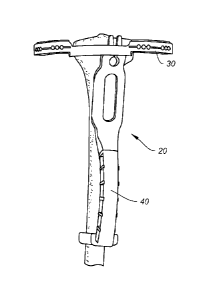
Turning now to the drawings, FIG. 1 shows a guide 30 of a navigation
instrument 20
relative to a tibia 2. Principles of targeting with a template as previously
disclosed by
applicants may still apply here. Similar to those previous techniques, the
tibia 2 is mapped
out into separate targeting zones correlating to the instrument 20. The
anatomical reference
is the tibial tuberosity, as shown here in FIG. 2. The tibial tuberosity 4 is
usually visible and
can be palpated below the skin. It is also visible on X-ray and on MRI. The
location is fairly
consistent and can be used as an anatomical landmark by the navigation
instruments of the
present disclosure. r[he template and guide are provided in both a Left Knee
and Right Knee
version so as to be specific to match the anatomical geometry of the tibia 2
and tuberosity 4.
FIG. 3 shows the top part of the guide 30 corresponding to the template zones
and tibia
plateau.
FIG. 4 shows that the handle 40 of the instrument 20 has a geometry that fits
securely
over the tibial tuberosity 4, allowing the instrument 20 to use the tibial
tuberosity 4 as an
anatomical reference point. The handle 40 aligns to the tibial axis and to the
tuberosity 4
creating a stable frame on the leg. 'Me axis angle of tibial axis to the
plateau is built into the
handle 40, as further shown in FIG. 10.
As mentioned, the handle 40 and guide 30 are separable components and are
attachable in two different positions for the Left or Right knee. This allows
for specific
anatomical alignment and secure fit to either Left or Right side being
treated. A Right Knee
and Left Knee version of the navigation instrument 20 are shown in FIGS. 5A
and 5B,
respectively.
FIG. 6 shows a side view of the fit of the guide 30 onto the tibia 2. The
anterior-
posterior slope angle of the tibial plateau is fixed and built into the guide
handle connection
40, as represented by the broken lines representing the plateau axis A¨A and
the tibial axis
B ____ B in FIGS. 6 and 10.
FIGS. 7A and 7B show side and front views of the guide 30 and handle 40 of the
instrument 20 without the tibia bone, respectively, while FIG. 8 shows the
side view of the
instrument and guide 30 positioned relative to the tibial bone 2. The guide
component 38
17
CA 02884468 2015-03-09
WO 2014/053913
PCT/IB2013/002672
may have internal radio-opaque lines/markers 38 to help vertically align the
guide 30 with the
top of the tibial plateau, as shown in FIG. 10. Further, as shown in FIG. 9,
the guide 30 and
handle 40 are attachable and detachable for Left and Right Knee orientation
via right slot 34a
and left slot 34b on the guide 30. Once the guide 30 is held in place with
alignment to the
.. tibial plateau, a depth control sleeve and injection needle 10 can be
inserted through the guide
30 into the targeted area of the bone 2, as shown in FIG. 11.
FIGS. 12A and 12B show an exemplary embodiment of the navigation instrument 20
configured for use with a femur 6. For treatment of the femur 6, a femoral
guide 60 can be
attached to the side of the guide 30 for direct lateral approach to the distal
femur 6, as shown.
The side profile would appear over the distal femur in x-ray and radio-opaque
markers would
aide the user in selecting the appropriate trajectory and corresponding portal
into the femur 6.
The tibial navigation instrument 20 includes a guide 30 that contours to an
average
tibial tuberosity 4. Further, the guide 30 already includes M/L tibia tilt;
A/P tibia tilt; and
axial rotation. There is no need for posterior edge alignment due to depth
gauge used with
.. injection needle 10. In an exemplary method of using the instrument, the
surgeon would
mark the joint line from a sagittal view, and rest the guide on the tibia,
defaulting to the
contoured shape. The surgeon would match the height of the joint line and,
under C-arm
visualization, make final adjustments. Then, the injection pin and depth gauge
(with optional
C-arm visualization to confimt depth) are drilled into the bone.
An exemplary method of using the femoral navigation instrument 20 with
attached
femoral guide 60 is similar to the tibial technique described above. The
femoral guide 60 is
initially put in its more comfortable position. The femoral guide position is
then aligned with
the femoral condyle in a lateral view. The delivery pin or needle with depth
gauge 10 is then
drilled into the target location through the portal of the guide 60 (with
optional C-arm
visualization to confirm depth).
It is contemplated that the delivery pin of the present disclosure may be
similar to
those disclosed in co-pending and co-owned U.S. Patent Application Publication
No.
2012/0316513, filed June 8, 2012 and entitled "INSTRUMENTS AND DEVICES FOR
SUBCHONDRAL JOINT REPAIR," and further include adapter and components as
.. described in this application. Likewise, as mentioned above, the pin may
incorporate various
depth control features or components. Additional depth control features or
components that
18
may be incorporated into the systems and methods of the present disclosure are
disclosed in
U.S. Patent Application No. 14/021,785 filed on September 9, 2013 and entitled
"INSTRUMENTS FOR CONTROLLED DELIVERY OF INJECTABLE MATERIALS
INTO BONE".
FIGS. 13A-13C show still another embodiment of a femoral navigation instrument
20 allowing easy adjustment and attachment of the femoral guide component 60
to the guide
30. As shown in FIG. 13A, the guide 30 may have a notch 34 for mating with a
slot 66 on
the femoral guide component 60. Both the notch 34 and slot 66 may be shaped
and keyed to
fit one another. The user may adjust the height of the femoral guide component
60 relative to
the guide 30 easily by sliding the component 60 on and off the notch 34. Once
the proper
height is achieved, the depth control sleeve and needle 10 may be drilled
through the femoral
guide component 60, as shown in FIGS. 13B and 13C.
The previous embodiments of navigation instruments, both tibial and femoral
versions, are directed to frame navigation. The next embodiments of navigation
instruments
are directed to free hand navigation. In this instance, instead of a guide
with multiple holes to
target the different zones or locations in the bone, the guide has a single
portal configuration
with multiple reference marks that correlate to an anatomical reference point.
A template is
used to map the tibia and determine which trajectory to use. By aligning the
chosen guide
mark with the anatomical reference, the portal trajectory is aligned to the
intended target
zone.
The template alignment zones or guide 130 would appear something similar to
what is
shown in FIG. 14A. FIG. 14B shows a full-length view of the navigation
instrument 120 with
a single component handle 140 and guide 130 with markings 132. The handle 140
is made to
fit securely at each end onto the leg surface to align with the tibia 2. The
guide 130 may
include horizontal radiopaque markers 138 that can be aligned with the center
notch of the
tibial plateau in the x-ray A/P view, as shown in FIG. 14C. Once the
instrument 120 is
aligned, a depth sleeve and injection needle 10 can be inserted into bone
through the guide
130, as shown in FIGS. 14D and 14E. As FIG. 14F illustrates, this guide 130
may also
include one or more slots 134 configured with a size and shape that allows for
insertion of a
scalpel; this eliminates the need to move the guide 130 to cut the skin with
the scalpel and
19
CA 2884468 2018-04-30
CA 02884468 2015-03-09
WO 2014/053913
PCT/IB2013/002672
decreases chances of error. Two different choices of needle trajectory are
provided in the
guide 130 for a direct horizontal and angled distal approach, as shown in FIG.
14F. FIG. 14G
shows the instrument positioned against a patient's leg and a needle inserted
through one of
the slots.
It is contemplated that the surgeon would use a template to choose a
trajectory to the
subchondral bone defect, such as a bone marrow lesion or edema, and under
fluoroscopic
visualization align the instrument 120 to the plateau. Next, the skin is cut
with a scalpel
through the port, and a delivery pin with desired depth gauge may be power
drilled into the
targeted location (with optional perpendicular view to confii III).
FIGS. 15A-15C show a femoral version of the navigation instrument 120.
Treatment of the femur 6 may employ a simple guide 160 with similar single
injection
trajectory, as shown in FIGS. 15A and 15C; radiopaque markers may be provided
with the
guide 160 to help position the guide 160 in the x-ray lateral view against the
side of the femur
6, as shown in FIG. 15B. In the femoral procedure, using a sagittal view, the
surgeon may
mark the surface tangent to the femur 6 where injection is desired. Then the
surgeon would
align the fluoroscopic markers to match, use a scalpel to cut the skin through
the port, and
power drill a delivery pin with desired depth gauge into the targeted location
(with optional
perpendicular view to confirm).
FIG. 16 shows a system 200 comprising various components for assembling a
tibial
guide instrument 220 (FIG. 17) or femoral guide instrument 260 (FIGS. 18A and
18B) of the
present disclosure. The system 200 may include a handle 240 that receives
either one of a
tibial guide component 232 or a femoral guide component 262. One will
recognize that the
guide components 232, 262 include many of the features already described above
for the
guide instruments 120, 160, such as slots for receiving a scalpel that can
also receive the
depth gauge sleeve and needle 10. The guides are interchangeable and easily
attachable to
the handle 240, making the system highly adaptable to different uses.
FIG. 17 shows a tibial guide instrument 220 assembled from the system 200 of
FIG.
16. The tibial guide instrument 220 includes the tibial guide 232 inserted
into the handle
240. The instrument 220 is shown braced against the leg of the patient.
CA 02884468 2015-03-09
WO 2014/053913
PCT/1B2013/002672
FIGS. 18A and 18B show a femoral guide instrument 260 assembled from the
system 200 of FIG. 16. As shown in FIG. 18A, the femoral guide 262 may
comprise hinged
arms and allow a pin or needle 10 to be placed into the femur 6 in the open
position of the
femoral guide 262. FIG. 18B shows the femoral guide instrument 260 in which
the femoral
guide 262 is inserted into the handle 240, with the instrument 260 braced
against the
patient's leg.
FIGS. 19A and 19B show other exemplary embodiments of guide instruments of the
present disclosure. FIG. 19A shows a tibial guide instrument 320 assembled by
inserting a
tibial guide 332 into handle 340 similar to the manner described above. The
tibial guide
instrument 320 may be braced against the patient's knee in a manner similar to
the one
shown in FIG. 17. FIG. 19B shows a femoral guide instrument 360 assembled by
inserting a
femoral guide component 362 into handle 340 similar to the manner described
above.
The guide instruments 320, 360 of FIGS. 19A and 19B have the ability to be
angularly adjustable. As shown in FIG. 20, the handle 340 may comprise an
attachment end
.. 342 within which are slots or cutaway portions 344A, 344B. Each of the
cutaway portions
344A, 34413 also includes keyed sections or shaped notches 346. This allows a
complementarily shaped or fluted stem 336 on the guide components 332, 362 to
be inserted
at an angle with respect to the handle 340 by engaging the stem 336 with
selected notches
346. For instance, as shown in FIG. 21A, the tibial guide component 332 may be
attached to
the handle 340 straight. As shown in FIGS. 21B and 21C, the tibial guide
component 332
can also be angled relative to the handle 340 by sliding the tibial guide
component 332 at an
angle into the slots 344A, 344B. The ability to angle the guide components
332, 362
relative to the handle 340 enables customization to the patient's anatomy, and
better
confoimity to a left or right knee.
The present navigation instruments simplify the process of mapping to an edema
while also keeping a pin subchondral with faster, simpler positioning
requirements using
easy anatomical reference points. For instance, the instruments can be
oriented to the tibial
tuberosity. The complexity is built into the guide with built in anatomical
angles specific to
the left or right tibia. That is, the frame navigation-focused instruments
provide preset
.. anatomical planes for the frame to be simple and save time. All that is
needed is to
deteimine the height relative to the tibial plateau.
21
CA 02884468 2015-03-09
WO 2014/053913
PCT/1B2013/002672
Alternatively the amount of precision is reduced to simplify the placement of
the
guide. For both guides the result is a "point and shoot" method that reduces
the time and
complexity in the technique. The new embodiments are intended to be simple one
or two-
piece designs. For instance, the free hand-focused instruments combine a
scalpel hole with
a drill hole, and allows "clocking" of the drill hole relative to the edema or
map target, can
be located off the mid-line, and can work in multiple planes of imaging
planes.
In some embodiments, the guide may be marked in terms of absolute numerical
values to additionally serve as a ruler-like function. This would allow the
guide to still be
used on many different sized knees, where the distance to the center of the
knee may be
deteimined along with the distance to the edema (such as from MR1 software
that can
calculate the distance of the edema from the knee's center). Then, using the
guide as a ruler,
the user is able to deteimine the location of the edema according to the
distance markers of
the guide.
In still other embodiments, the markings on the guide may be proportionally
.. distanced relative to the luiee size to allow for size fluctuations. Thus,
accounting for this
size differential in the markings along the guide would prove beneficial.
Other embodiments will be apparent to those skilled in the art from
consideration of
the specification and practice of the embodiment disclosed herein. It is
intended that the
specification and examples be considered as exemplary only, with a true scope
and spirit of
the embodiment being indicated by the following claims.
22