Note: Descriptions are shown in the official language in which they were submitted.
CA 02884606 2015-03-04
WO 2014/036638
PCT/CA2013/000761
SYSTEMS AND METHODS FOR DIAGNOSING STROKES
FIELD OF THE INVENTION
[0001] The invention relates to systems and methods for diagnosing strokes. In
particular, systems and methods for acquiring timely patient status
information are
described that enable a physician to make diagnostic and treatment decisions
relating to
ischemic and hemorrhagic strokes. The systems and methods enable the efficient
and
quantitative assessment of arterial collaterals within the brain for aiding
these decisions
in the case of ischemic strokes. In the case of hemorrhagic strokes, the
systems and
methods are effective in determining if there is a leak and what is the rate
of leaking.
BACKGROUND OF THE INVENTION
[0002] lschemic stroke is an acute disease where tissue death (infarction)
within the
brain of different patients will progress at different rates from the time of
the ischemic
event. The rate of infarction within a patient depends on a large number of
physiological
factors.
[0003] For the physician diagnosing and treating ischemic strokes, when a
stroke
patient arrives at a hospital, it is very important for the physician to
obtain as much
knowledge about the nature of the stroke as soon as possible in order to make
an
effective diagnosis and effective decisions regarding treatment. As is readily
understood,
time to effect diagnosis and treatment is very important as faster diagnoses
will impact
treatment decisions and can minimize the amount of brain tissue that is
ultimately
affected as a result of the stroke.
[0004] For example, in the case of an ischemic stroke, it is important for the
physician to
know where the vessel occlusion is, how big the occlusion is, where any dead
brain
tissue (termed "core") is and, how big and where is the brain tissue that may
have been
affected by the ischemic event but that may potentially be saved (this tissue
is termed
"penumbra").
-1-
CA 02884606 2015-03-04
WO 2014/036638 PCT/CA2013/000761
[0005] More specifically, the penumbra is tissue around the ischemic event
that can
potentially stay alive for a number of hours after the event due to perfusion
of this tissue
by collateral arteries. That is, the collateral arteries may provide
sufficient oxygen to the
penumbra tissue to prevent this tissue from dying for a period of time.
[0006] When the physician has good information about the collaterals and how
the
collaterals may be located in and around the penumbra, treatment decisions can
be
made that can significantly affect patient outcomes.
[0007] Importantly, in an emergency or acute situation, the process of making
a decision
will consider the amount of information at a given moment in time. That is, a
definitive
'yes' decision can be made to take action or a 'no' decision can be made to
take no
action based on the current information. In addition, a third decision choice
can be made
to wait for additional information. In the situation of acute stroke (and
other emergency
scenarios), time to make a definitive diagnostic/treatment decision must be
balanced
against the likelihood of a negative outcome that results simply from the
delay in making
a decision. In other words, the decision to wait for more information must
consider what
the effects of a delay in making a decision might be.
[0008] In the specific case of acute ischemic stroke, the pace or rate of
neural circuitry
loss in a typical large vessel supratentorial acute ischemic stroke is shown
in Table 1.
Table 1- Estimated Pace of Neural Circuitry Loss in Typical Large Vessel,
Supratentorial Acute Ischemic Stroke (3)
Estimated Pace of Neural Circuitry Loss in Typical Large Vessel,
Supratentorial Acute lschemic Stroke
Neurons Synapses Myelinated Accelerated
Lost Lost Fibers Lost Aging
Per Stroke 1.2 billion 8.3 trillion 7140 km/4470 36 yrs
miles
Per Hour 120 billion 830 billion 714/447 miles 3.6
yrs
-2-
CA 02884606 2015-03-04
WO 2014/036638 PCT/CA2013/000761
Per Minute 1.9 million 14 billion 12 km/7.5 miles 3.1 weeks
Per 32,000 230 200 meters/218 8.7 hours
Second million yards
[0009] As can be seen, delays in making a decision in the order of only a few
minutes
can have a significant impact on patient outcome in terms of neural circuitry
loss.
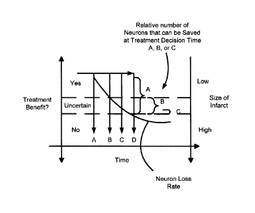
Moreover, and as shown in Figures 1 and 2, a better outcome is significantly
more likely
to occur when the decision to treat is made earlier. As shown in Figure 1,
whether or not
a treatment is ultimately beneficial or not may depend on when the decision to
treat is
made. As shown in Figure 1, treatment decision times A, B, C, D will each have
a
different affect on the relative number of neurons that could be saved. That
is, if a
treatment decision is made at time A (i.e. an earlier time), if it is assumed
that the pace
of neural circuitry loss is linear (assumed only for this example), a greater
number of
neurons can be saved. As the time of making the treatment decision is delayed,
the
likelihood of the treatment being beneficial will decrease until it is
uncertain whether the
treatment will be beneficial (i.e. at times B and C) or where there is a high
likelihood that
the treatment will be of no value (i.e. at time D).
[00010] Further, Figure 2 illustrates the effect of time to reperfusion
and good
clinical outcome for observed cases where the abscissa shows time from stroke
to
reperfusion and the ordinate shows the probability of the patient achieving a
post-
treatment mRS score of 0-2. Table 2 shows the time to reperfusion and good
clinical
outcome for the data of Figure 2 (1).
Table 2-Time to Reperfusion and Good Clinical Outcome
Risk Ratio 95% Cl p-value
Time to Reperfusion 0.86 0.78-0.95 P=0.0045
(every 30 minutes)
[00011]
-3-
CA 02884606 2015-03-04
WO 2014/036638
PCT/CA2013/000761
[00012] At the present time, in many treatment centers, when a stroke
patient
arrives, the assessment protocol is generally as follows:
a. Conduct a CT scan of the head to rule out or look for evidence of a
hemorrhagic stroke.
b. Conduct a CT angiogram (CTA) to locate the site of vessel occlusion.
c. Conduct a CT perfusion (CTP) study to create perfusion maps that
provide the physician with information about various parameters including
cerebral blood flow, cerebral blood volume and mean transit time.
[00013] As is known, each of these generalized steps will be affected by a
large
number of factors and the time to complete each of them will be variable from
patient to
patient and between different treatment centers. For example, such factors may
include
resource availability (eg. trained medical staff and equipment) as well as
processing
times required by CT scan equipment and other ancillary hardware and software
to
present data to physicians.
[00014] For the purposes of illustration, these factors are described in
terms of a
representative diagnosis and treatment scenario of a patient exhibiting
symptoms of a
stroke, the patient arriving at the emergency room of a treatment center and
who
thereafter receives the above CT procedures as part of the diagnostic
protocol. Table 3
summarizes a number of the key process steps and typical times that may be
required to
complete each step.
[00015] Upon arrival at the treatment center, an emergency room physician
conducts a preliminary assessment of the patient. If the preliminary
assessment
concludes a potential stroke, the patient is prepared for a CT scan. The time
taken to
initially assess a potential stroke patient upon arrival at the treatment
facility may be 3-5
minutes.
[00016] Preparing the patient for a CT scan involves a number of steps
including
transferring the patient to the CT imaging suite and connecting an intra-
venous line to
-4-
CA 02884606 2015-03-04
WO 2014/036638
PCT/CA2013/000761
the patient to enable the injection of contrast agent into the patient during
the various CT
procedures.
[00017] The CT scan includes conducting an x-ray scan of the patient
together
with a computerized analysis of the x-ray data collected. More specifically,
as is known,
during a CT scan, beams of x-rays are emitted from a rotating device through
the area of
interest in the patient's body from several different angles to receivers
located on the
opposite sides of the body. The received data is used to create projection
images, which
are then assembled by computer into a two or a three-dimensional picture of
the area
being studied. More specifically, the computer receives the x-ray information
and uses it
to create multiple individual images or slices which are displayed to the
physician for
examination.
[00018] CT scans require that the patient hold still during the scan
because
significant movement of the patient will cause blurred images. This is
sometimes difficult
in stroke patients and hence sometimes head restraints are used to help the
patient hold
still. Complete scans take only a few minutes.
[00019] Upon completion of the initial CT scan including the post-
processing time
to assemble the images, the physician interprets the images to determine a) if
a stroke
has occurred and, b) if so, to determine if the stroke is hemorrhagic or
ischemic. If the
stroke is hemorrhagic, different procedures may be followed. It will typically
take the
physician in the order of 1-2 minutes from the time the images are available
to make the
determination that the stroke is hemorrhagic or ischemic.
[00020] If the stroke is ischemic, the decision may be made to conduct a
CT
angiogram (CTA).
[00021] CT angiography procedures generally require that contrast agents
be
introduced into the body before the scan is started. Contrast is used to
highlight specific
areas inside the body, in this case the blood vessels. In addition because of
presence of
contrast in the very small vessels of the brain, overall the brain looks
brighter (has a
higher Hounsfield value) also known as contrast enhancement. Contrast agents
are
iodine based compounds that inhibit the passage of x-rays through the tissue.
As such,
-5-
CA 02884606 2015-03-04
WO 2014/036638
PCT/CA2013/000761
they can be effective in enhancing the distinction between tissues where the
contrast
agent is present compared to those tissues where it is not. The CT angiogram
requires
additional preparation time but will typically not require that the patient be
moved.
Generally, CT angiogram procedures involve the injection of a bolus of
contrast through
an IV line followed by the CT scan. A typical contrast bolus may be 70-100 ml
injected at
ml/second. The volume and injection rate of contrast is determined by the
procedure
being followed and is generally injected in a minimally sufficient volume to
be present in
the tissues of interest at the time the CT scan is conducted. Over a
relatively short time
period, the contrast becomes diffused within the body thereby providing only a
relatively
short window of time to conduct a CT procedure.
[00022] The CT angiogram data is substantially greater than what is
collected
from a basic scan and like a basic CT scan must be subjected to post-
processing to
create the images. The post-processing time is typically in the range of 3-5
minutes.
[00023] After processing, the physician interprets the data and makes a
decision
regarding treatment. Generally, the physician is looking to determine a) where
is the
occlusion? b) what is the size of the core? and c) obtain a qualitative feel
for penumbra
and collaterals.
[00024] Ultimately, and based on these factors, the physician is looking
to make a
decision on what brain tissue is worth fighting for. In other words, based on
the
combination of all these factors, the physician is looking to decide either
that very little or
no penumbra can be saved, or alternatively that it appears that penumbra can
be saved
and it is worthwhile to do so.
[00025] The CT angiogram provides relatively little data about collaterals
and
perfusion to the ischemic tissue as it is only a picture of the brain at one
instance in time.
That is, as it takes time for contrast agent to flow through the brain tissues
and such flow
will be very dependent on the ability of vessels to carry the contrast agent,
a single
snapshot in time does not give the physician enough information to make a
diagnostic
and/or treatment decision. Hence, CT perfusion (CTP) procedures may be
undertaken to
give the physician a more quantitative sense of brain perfusion. Like CT
angiogram, CT
perfusion procedures involve the injection of contrast agent into the patient.
It should
-6-
CA 02884606 2015-03-04
WO 2014/036638
PCT/CA2013/000761
also be noted that some centers may choose to do a CT perfusion study before
the CT
angiogram because they feel that the contrast injection from the CT angiogram
interferes
with the quality of data of the CT perfusion.
[00026] Perfusion computed tomography (CTP) allows qualitative and
quantitative evaluation of cerebral perfusion by generating maps of cerebral
blood flow
(CBF), cerebral blood volume (CBV), and mean transit time (MTT). The technique
is
based on the central volume principle (CBF = CBV/MTT) and requires the use of
complex software employing complex deconvolution algorithms to produce
perfusion
maps. Other maps such as Tmax maps may also be created.
[00027] CTP studies are acquired with repeated imaging through the brain
while
the contrast is injected. The technique varies significantly from vendor to
vendor and
also from center to center and hence requires specialized training with the
specific
equipment at each center. CTP typically involves imaging of the brain over
approximately 60-70 seconds (at 1-4 second intervals) in order to acquire
multiple
images. The technique is quite vulnerable to patient motion and also requires
the patient
to hold still for the period. Furthermore, CTP also involves substantial
radiation exposure
in the range of 5-10 mSv as the number of images taken over the time period is
significant.
[00028] The procedure generates a large dataset that must then be
transferred to
a dedicated workstation for post-processing. This step may take over 10
minutes in
order to produce separate maps of each of CBF, CBV, and MTT. The perfusion
maps
are typically color coded maps.
[00029] Importantly, the post-processing requires the use of specialized
and very
often proprietary software that must be run by trained individuals.
Ultimately, the time
taken to fully complete CTP acquisition and analysis is highly variable as the
above
factors including the vendor, the speed of data transfer, local expertise, the
time of day
the study is being undertaken (i.e. working hours vs. after hours) as well as
other factors
can all have an affect on the actual amount of time required to complete the
study.
-7-
CA 02884606 2015-03-04
WO 2014/036638
PCT/CA2013/000761
Table 3- Typical Diagnostic Steps and Completion Times
Procedure Time (minutes) Elapsed Comments
Total
Initial Assessment 3-5 3-5
Transfer and 20 23-25
Preparation for CT Scan
CT Scan 1 24-26
CT Scan Interpretation 2-3 26-30 CT Angiogram Preparation
and may be concurrent with
CT Angiogram CT Scan Interpretation
Preparation
CT Angiogram 1-3 27-33
Procedure
CT Angiogram Post 2 29-35
Processing
CT Angiogram 4 (minimum) 33-39 CT Perfusion Preparation
Interpretation and CT may be concurrent with
Perfusion Preparation CT Scan Interpretation
CT Perfusion Procedure 1 34-40
CT Perfusion Post Variable 44-60 Will depend on vendor
Processing 5-20 (minimum) specifics
CT Perfusion Variable 46-70 Will depend factors
Interpretation 2-10 (minimum) including: time of day;
center; vendor equipment
etc.
[00030] Thus, while perfusion CT is not a perfect technique, it has been
found to
be useful for noninvasive diagnosis of cerebral ischemia and infarction as it
does provide
some degree of quantitative determination of core and penumbra. However, as
noted
above, there are problems with these procedures. In summary, these problems
include:
a. CT perfusion takes time to complete (8-30+ minutes total).
b. Patient motion can affect results.
c. Significant post-processing time is required to complete a full perfusion
map.
d. Additional radiation exposure to the patient.
e. Need for additional contrast agents.
f. Non-standardized procedures for completing the perfusion map.
g. Variations in technique with different vendor equipment.
-8-
CA 02884606 2015-03-04
WO 2014/036638
PCT/CA2013/000761
h. Lack of consensus in the medical community regarding the interpretation
and best practices for treatments based on the CT perfusion maps.
i. Lack of information regarding rate of infarct growth.
j. Significant variability across vendors for the degree of coverage of the
brain (eg. 4 to 16 cms). Also some vendors have the option of covering 8
cm using a 'toggle table' technique that may introduce additional errors.
[00031] As a result, notwithstanding the benefits of CTP, there continues
to be a
need for improved procedures and systems that can address these problems that
provide the physician with the ability to make faster diagnoses. Most
importantly, there
has been a need for improved systems for assessing patient collaterals after
ischemic
stroke and, in particular, the need to create a fast and reproducible
collateral map as
opposed to a perfusion map. Further still, there has been a need for systems
and
methods that enable faster recanalization in order to increase the chances of
saving
penumbra tissue given the rate of neural death in a typical large vessel
ischemic stroke.
[00032] In addition, there has also been a need for systems and methods
that can
be consistently implemented at different treatment centers and across
different CT
machines (i.e. from different vendors) that reduce the level of specialized
and/or
advanced training that may be required to provide a consistent and accurate
diagnosis.
[00033] Further still, there has also been a need for systems and methods
that
enable the identification and quantification of parameters about the blood
clot/thrombus
causing an ischemic stroke. That is, in proximal artery occlusion it is
helpful to the
endovascular surgeon to understand more about the nature of the clot causing
the
stroke and more specifically know the exact length of the clot and its
relative
permeability and/or porosity which will aid in treatment decisions.
[00034] With regards to hemorrhagic strokes, there is similarly a need for
systems
and systems methods that enable faster diagnoses with enough information to
assist in
making treatment decisions.
-9-
CA 02884606 2015-03-04
WO 2014/036638
PCT/CA2013/000761
SUMMARY OF THE INVENTION
[00035] In accordance with the invention, systems and methods for
diagnosing
strokes are described. The systems and methods described herein enable faster
diagnoses and treatments of different types of strokes by providing a
physician with
effective and timely information.
[00036] In accordance with a first aspect of the invention, a method of
imaging the
brain within a patient diagnosed as potentially suffering a stroke is
described, the
method for deriving information about blood flow within the brain the method
comprising
the steps of:
a) injecting a bolus of contrast agent into the patient;
b) obtaining a set of computed tomography (CT) images of the patient's brain
at different levels at a specific time period, t, after step a);
c) repeating step b) n times to obtain at least one additional set of CT
images
of the patient's brain at different levels at time period t after step b),
wherein n
is at least one and each set of CT images is defined as a phase of images,
P1-Pn;
d) displaying each phase of CT images from steps b) and c) as a time-
sequenced series of images.
[00037] In various embodiments, the number of phases can be varied but
preferably n is 1-6. The time period, t, can also be varied and may be
selected based on
a number of factors including the anticipated flow rate of contrast agent
through the
patient. The time period, t, may also be selected based on an initial
diagnosis of the
patient having suffered an ischemic or hemorrhagic stroke. For example, if the
patient is
suspected as having suffered an ischemic stroke, t will typically be 6-18
seconds. If the
patient is suspected as having suffered a hemorrhagic stroke the time period
t, is
preferably 10-40 seconds.
[00038] In another embodiment, the method further comprises the step of:
enabling a user to mark at least one zone of interest within one phase of the
images to
create a marked zone of interest and wherein a marked zone of interest
represents any
-10-
CA 02884606 2015-03-04
WO 2014/036638
PCT/CA2013/000761
one of or a combination of asymptomatic tissue or symptomatic tissue. In one
embodiment, a corresponding zone of interest of a single image on an opposite
side of
the brain is automatically marked based on the area and location of the at
least one
marked zone of interest. In one embodiment, a corresponding zone of interest
in another
phase is automatically marked to create further marked zones of interest based
on the
area and location of each marked zone of interest.
[00039] In another embodiment, the method further comprises the step of:
calculating a contrast density value within each marked zone of interest. In
one
embodiment, contrast density values for each marked zone of interest are
tabulated
within a database.
[00040] In another embodiment, the method further comprises the step of:
calculating and displaying a contrast density trend value from P1 to Pn for
corresponding
zones of interest across P1 to Pn on a symptomatic side.
[00041] In another embodiment, the method further comprises the step of:
calculating and displaying a contrast density trend value from P1 to Pn for
corresponding zones of interest across P1 to Pn on an asymptomatic side.
[00042] In a still further embodiment, the method further comprises the
step of:
comparing the contrast density trend value against a database of trend values
to
ascertain a collateral value for the marked zones across all phases.
[00043] In another embodiment, the method further comprises the step of:
calculating and displaying a color code on at least one phase of images based
on the
collateral value or creating a colour coded map by summating the data from all
the
phases.
[00044] In another embodiment, the method further comprises the step of:
calculating and displaying a change in contrast density of the entire brain
from P1 to Pn.
[00045] In a still further embodiment, the method further comprises the
steps of:
identifying and marking one or more occlusions in one or more images in one or
phases
of the CT images and marking a downstream area relative to each marked
occlusion;
and, calculating and displaying a rate of pacification of vessels in the
downstream area
beyond each marked occlusion.
[00046] In yet another embodiment, the method further comprises the steps
of:
-11-
CA 02884606 2015-03-04
WO 2014/036638
PCT/CA2013/000761
identifying and marking corresponding symptomatic and asymptomatic regions of
the
brain; and calculating, comparing and displaying contrast density trends from
the marked
symptomatic and asymptomatic regions of the brain.
[00047] In yet another embodiment, the method further comprises the steps
of:
identifying and marking the location of an occlusion; calculating the diameter
of vessels
distal to the occlusion; identifying corresponding vessels on the
contralateral side;
calculating the diameter of vessels on the contralateral side; and comparing
and
displaying the differences in vessel diameter for each side for each of P1 to
Pn.
[00048] In one embodiment, if the patient is suspected as having suffered
an
ischemic stroke, a method of deriving information about the location and
properties of a
blood clot/thrombus is provided wherein after steps a) to d) are conducted,
the method
further comprising the steps of: enabling a user to mark a proximal end
position of a
suspected blood clot within at least one image of at least one phase of
images; enabling
a user to mark a distal end position of a suspected blood clot within at least
one image of
a later phase of images; and calculating and displayed a blood clot length
based on the
proximal and distal positions.
[00049] In one embodiment, if the patient is suspected as having suffered
an
ischemic stroke, a method of deriving information about the location and
properties of a
blood clot is provided wherein after steps a) to d) are conducted, the method
further
comprises the steps of: enabling a user to mark a proximal end area of a
suspected
blood clot/thrombus within at least one image of at least one phase of images;
enabling
a user to mark a distal end area of a suspected blood clot/thrombus within at
least one
image of a later phase of images; calculating and displayed a blood
clot/thrombus
volume based on the proximal and distal end areas.
[00050] In another embodiment, the method includes the step of calculating
a
rate of change of contrast density within an intravascular blood clot/thrombus
volume
across different phases and correlating the rate of change to a known rate of
change of
contrast density within a blood clot/thrombus volume to determine a blood
clot/thrombus
permeability.
[00051] In another embodiment, the method includes the step of calculating
a rate
of change of contrast density within a blood clot/thrombus volume across
different
-12-
CA 02884606 2015-03-04
WO 2014/036638
PCT/CA2013/000761
phases to a known rate of change of contrast density within a blood
clot/thrombus
volume to determine a blood clot/thrombus porosity.
[00052] In another embodiment, if the patient is suspected as having
suffered a
hemorrhagic stroke, the method includes deriving information about the
location of and
rate of leak within a patient wherein steps a) to d) are conducted where t is
10-40
seconds and the method further comprising the steps of: enabling a user to
mark a first
instance of a suspected leak within the hematoma within each image of at least
one
phase of images wherein the user marks a border of the leak within the
hematoma;
calculating a first volume of the leak within the hematoma based on marked
borders of
the leak from the earliest phase of images showing the leak; repeating steps
aa) and bb)
for subsequent phases to calculate successive volumes of the leak; displaying
each of
the first volume and successive volumes; and, calculating the rate of leak and
consequently the rate of increase of the hematoma over time
BRIEF DESCRIPTION OF THE DRAWINGS
[00053] The invention is described with reference to the accompanying
figures in
which:
Figure 1 is a schematic diagram showing the relative effect of the time of a
treatment decision to the benefit of a potential treatment with consideration
to
relative size of an infarct.
Figure 2 is a graph showing time to re-perfusion and good clinical outcome.
Figure 3 are images of a multiphase CT (mCTA) scan from a first case in
accordance with the invention where 3 sets (phases) of image data were
obtained over approximately 8 second intervals through the entire brain of the
patient; the first row (P1) being first phase data; the middle row (P2) being
second phase data and the third row (P3) being third phase data.
Figure 4 are images of a multiphase CT (mCTA) scan from a second case in
accordance with the invention where 3 sets (phases) of image data were
obtained over approximately 8 second intervals through the entire brain of the
-13-
CA 02884606 2015-03-04
WO 2014/036638
PCT/CA2013/000761
patient; the first row (P1) being first phase data; the middle row (P2) being
second phase data and the third row (P3) being third phase data.
Figure 5 are images of a multiphase CT (mCTA) scan from a third case in
accordance with the invention where 3 sets (phases) of image data were
obtained over approximately 8 second intervals through the entire brain of the
patient; the first row (P1) being first phase data; the middle row (P2) being
second phase data and the third row (P3) being third phase data.
Figure 5A are images of a multiphase CT (mCTA) scan from a fourth case in
accordance with the invention where 3 sets (phases) of image data were
obtained over approximately 8 second intervals through the entire brain of the
patient; the first row (P1) being first phase data; the middle row (P2) being
second phase data and the third row (P3) being third phase data.
Figure 6 is a flow-chart showing the steps in creation of a semi-quantitative
collateral map in accordance with one embodiment of the invention.
Figure 6A is a representative image showing how zones of interest may be
marked within an mCTA image.
Figure 7 is a schematic diagram showing segregation of regions of the brain
(MCA territory) divided into areas traditionally supplied by ACA collaterals
and by
PCA collaterals. lschemic tissue is marked B and healthy tissue marked A.
Figure 8 are images of a multiphase CT (mCTA) scan from a case where the
patient has suffered a hemorrhagic stroke. The image data were obtained over
approximately 12 second intervals through the entire brain of the patient; the
first
row (P1) being first phase data; the middle row (P2) being second phase data
and the third row (P3) being third phase data.
Figure 8A are initial pre-mCTA CT images (no contrast) from the patient of
Figure 6 showing that the patient has suffered a hemorrhagic stroke.
-14-
CA 02884606 2015-03-04
WO 2014/036638
PCT/CA2013/000761
Figure 8B are follow-up and post-mCTA CT images (no contrast) from the
patient of Figure 6 showing that the size of the hematoma has grown as
compared to the images of Figure 6A.
DETAILED DESCRIPTION OF THE INVENTION
[00054] With
reference to the figures, systems and methods for diagnosing
strokes are described. More specifically, multiphase CT (mCTA) angiogram
techniques
are described that can significantly improve the time required to effect an
accurate
diagnosis for a stroke patient. Importantly, the procedures described herein
allow for
faster diagnosis of the location and extent of blockages as well as faster and
semi-
quantitative determination of the extent of the collaterals which will aid the
physician in
determining the treatment protocol. The systems and methods of the invention
are
primarily discussed herein in relation to ischemic strokes but may also be
applied to the
diagnosis of hemorrhagic strokes as discussed below.
[00055] In a
first aspect, the invention involves conducting multiple CT
angiograms over a condensed period of time and at defined intervals. In a
second
aspect and from the image information obtained, the location and diameter of
collaterals,
the density of contrast and variance in the rate of filling of the collaterals
(i.e. the rate of
opacification) is assessed in both space and time which is used to create a
collateral
map or collateral score. The collateral map or collateral score can be used by
the
physician to make a diagnostic and/or treatment decision.
[00056]
Generally, in the context of this invention, and as explained in greater
detail below, a collateral map is a visual representation of multiple, time
varied images of
a section of the brain that show a variance in contrast over a period of time.
A collateral
score is a grading system that represents the relative "strength" of
collaterals.
[00057] In
accordance with the first aspect of the invention, mCTA is a multiple
image CT procedure conducted with a single bolus of contrast. It is conducted
as 3-5
phases of CTA at a 6-12 second (preferably about 8 seconds) interval between
each CT
scan; however, the time interval may be longer in some circumstances, for
example
-15-
CA 02884606 2015-03-04
WO 2014/036638
PCT/CA2013/000761
during the work up of hemorrhagic stroke or older patients or in patients with
atrial
fibrillation resulting in poor cardiac output, may suggest a greater interval.
In addition,
the time period may be varied between each CT scan. The mCTA procedure
produces a
series of time-sequenced or phases of images at different levels within the
brain that
provide information about the flow of contrast through areas of the brain from
which the
quality of perfusion and the quality of collaterals can be assessed and/or
calculated.
[00058] Initially, the mCTA methodology is described in comparison to past
procedures by way of example for typical cases to illustrate the distinctions
between past
procedures and some of the treatment scenarios where mCTA can provide
significant
advantages over these procedures. The following four examples are
representative of
various diagnostic scenarios that may occur at a treatment center and are
intended to
illustrate various time situations that could occur in the treatment of
typical patients. The
numbers and times discussed are not intended to be limiting.
Case 1-CT, CTA, CTP Procedure
[00059] A 72 year old man presents to the ER at 0820 hours. On
examination, he
has right hemiplegia and aphasia with an NIHSS of 19. As known, NIHSS is a
stroke
scale where the NIHSS number is derived from an examination of the patient.
The scale
range is from zero to 42 with 42 indicating that the patient is dead.
Generally, a score of
or larger usually means a large stroke.
[00060] A quick examination of the patient is performed (5 min to
complete). An IV
line is started, blood is withdrawn and the patient is transferred to CT scan.
Patient
arrives at CT scan at 0840 hours.
[00061] A non-contrast CT scan is performed at 0843 hours. This is
immediately
seen by the treating physician and it does not show a bleed. The CT
technologist
immediately sets up for doing a CT angiogram. A CT angiogram is performed (80
cc of
contrast is injected). The CTA is completed by 0846 hours.
[00062] The CT technologist gets set up to do a CT perfusion exam (CTP). A
localizer is performed and CTP is started (an additional 45 cc of contrast is
injected
along with 2000 DLP of radiation exposure). The CTP study is over by 0851.
-16-
CA 02884606 2015-03-04
WO 2014/036638
PCT/CA2013/000761
[00063] In the meantime, the CTA data is available for review (while the
CTP is
going on) by 0848 hours. The treating physician is able to make the following
assessments:
1. The patient has an ischemic stroke.
2. Approximate size of core (based on ASPECTS score).
3. Site of occlusion.
4. Quality and quantity of collaterals.
[00064] Going back to CTP, the data is transferred to a dedicated
workstation.
The data is available at the workstation at 0901 hours. An expert initiates
and
undertakes the required steps of post processing with it being noted that the
expert may
not be immediately available and may be an additional source of delay. The
post
processing takes 10 minutes. Finally there is a discussion of interpretation
of the final
CBF, CBV and MTT maps that takes another 5-7 minutes. The CTP data is finally
available at 0918 hours. Thus, the detailed CTP data is available
approximately 30 min
after the CTA data.
Case 2¨CT, mCTA
[00065] A 65 year old presents with slight right sided weakness and slight
difficulty in word finding. The NIHSS was 4.
[00066] Patient is taken for a CT, mCTA. The initial non-contrast CT scan
is
unremarkable. CT angiogram shows an ulcerated plaque at the origin of the left
internal
carotid artery. No obvious intracranial occlusion is seen. However on the mCTA
there is
hold up of contrast in one of the branches of middle cerebral artery (MCA)
which is
detected on the later phases. This allows for detection of an embolus in the
M4 branch
(one of the distal branches) of the MCA. This has the potential to alter
patient
management including prognostication, decision on admission as well as whether
or not
to give thrombolytics.
Case 3-CT, CTA, mCTA, CTP
[00067] A 75 year old woman presents with left hemiplegia at 1520 hours.
After
assessment the patient is shifted to the CT scan suite. The patient is not
cooperative
and is not able to hold perfectly still. There is slight amount of motion
artifact on the non-
-17-
CA 02884606 2015-03-04
WO 2014/036638
PCT/CA2013/000761
contrast CT scan. Some sections have to be repeated. Subsequently, the
multiphase
CTA is performed. There is again some degree of motion artifact that affects
the quality
of the scan at the level of the neck and in the second phase. However the
intracranial
examinations on the first and third phase are of good quality. Subsequently a
CTP is
performed. However due to patient motion the data is uninterpretable in spite
of attempts
at motion correction. In this scenario, it is important to note that the
uninterpretable data
(i.e. marginal data) was not available for consideration until the time the
post processing
was performed (which as in the example above took approx 30 min beyond the
multiphase CTA). The treating team has no choice but to depend on the
multiphase CTA
or to bring the patient back and do another CTP which is a less desirable
protocol as it
requires more contrast, more radiation and more time.
Case 4-CT, CTA, CTP
[00068] The patient presents at 0220 hours. All the imaging: non contrast
CT,
CTA and CTP are performed as above. However there is no one available at that
time
who knows how to do the post processing. The person is paged from home.
However
the person is not able to do this from home so has to come into the hospital.
It produces
a delay of over 45 minutes.
mCTA Procedures and Interpretation
[00069] As shown in Figure 3, representative images from mCTA are
described.
The top row of images shows a first phase CT scan. More specifically, the
first row of
images shows 5 different spatial slices of a patient's brain at a first time,
referenced
herein as phase 1 or P1. The second and third row of images also show 5
corresponding
spatial slices of a patient's brain at second and third times or P2 and P3
respectively at
the same levels that the P1 images.
[00070] From the P1 images, it can be seen that the right side vessels of
the brain
contralateral to the side causing the patient's symptoms, are unaffected as
they can be
seen as fully opacified (right middle cerebral artery branches) at P1 (arrow
1) whereas
the left side (ipsilateral) is not opacified (arrow 2). In addition, it can be
seen that
posteriorly (PCA circulation), both sides are unaffected as the vessels are
opacified.
That is, the P1 scan shows that within a few seconds of injecting a contrast
bolus, the
contrast has effectively flowed to the anterior right side and the posterior
regions of the
-18-
CA 02884606 2015-03-04
WO 2014/036638
PCT/CA2013/000761
brain and has otherwise been fully distributed as would be expected within
healthy
tissue. In comparison, at P1, arrow 2 shows that contrast has not fully
perfused an area
of the left side by the absence of a similar contrast density as compared to
the right side.
Thus, these P1 images are suggestive of a left side occlusion.
[00071] At P2, on the right side, contrast is passing through the
contralateral
vessels (arrow 3). Thus, the P2 images show a decreasing contrast density on
the
healthy right side. At P3, almost all of the contrast has passed and the
contrast density is
lower still on the right side (arrow 5).
[00072] At P2, on the anterior left side, the images show that some
collaterals are
filling due to an observed increase in contrast density at this level (arrow
4). At P3, the
contrast density is increasing further (arrow 6). In addition, at other
levels, a hold up of
contrast can be seen in the left middle cerebral artery (MCA) region (arrow
7).
[00073] From these images, it is determined that the perinsular region
(ie. the
region where the collaterals are weak (arrows 6, 7, 9)) is at a greater risk
to die, whereas
posteriorly (arrow 8), the brain may be salvageable.
[00074] Accordingly, from this series of time sequenced images, the
physician
has a basis on which to assess the quality of the collaterals. In this first
example,
collateral health is sufficiently robust to suggest potentially salvageable
tissue and thus
in conjunction with the patient's clinical symptoms may make the decision to
conduct an
intra-arterial recanalization treatment.
[00075] It should also be noted and as understood by those skilled in the
art that
the medical practitioner in making a diagnostic/treatment decision may also be
making
that decision based on a concurrent evaluation of the non-contrast CT scan
(and other
clinical data) which has already been performed and/or obtained from the
patient.
[00076] As shown in Figure 4, the series of images suggest a different
treatment.
In this case, the original CT scan and the patient's clinical presentation
suggested a left
side occlusion. The P1 images confirmed a small clot in the left MCA but the
P1 images
also show relatively robust contrast density in the anterior left side. The P1
right side
images similarly show good contrast density. The P2 and P3 right side images
show that
-19-
CA 02884606 2015-03-04
WO 2014/036638
PCT/CA2013/000761
contrast is clearing as expected for healthy tissue. However, the P2 and P3
images
show that contrast is clearing more slowly than on the right side (arrows X,
Y, Z). The
slow clearing rate shows that the area, while at risk, has excellent
collaterals, thus
suggesting that nearly all of the left MCA territory is salvageable.
[00077] A
further case is shown in Figure 5 where a distal occlusion on the left is
observed (arrow 10). Normally, a distal occlusion (i.e. an occlusion within
smaller
vessels and that cannot be treated by recanalization) is difficult to detect
on a routine CT
angiogram. However, from the P3 image, it can be observed the contrast is no
longer
seen in most of the intracranial arteries. However there is still contrast
visible in some of
the distal left MCA branches (arrow 11) suggesting retrograde filling through
collaterals
and also points to the site of occlusion. Thus, the mCTA procedure enables the
physician to confirm that the patient has had a stroke and may need to be
admitted to
the treatment facility for further monitoring and/or or treatment.
[00078] In
Figure 5A, the three rows represent the three phases P1, P2 and P3
with an approximate 8 second image interval. In the P1 images, the arrow
identifies an
area with poor opacification in comparison to the posterior regions where
there is strong
contrast density. These images, when interpreted along with the non-contrast
CT scan,
also helps in a more accurate and precise determination of infarct core.
[00079] In the
P3 images which are taken approximately 16 seconds after the P1
images, the arrows show a hold up of contrast in the left MCA territory thus
indicating
that contrast is filling in through collaterals.
[00080] It is
important to note that on the right side (normal side), the P3 images
show near complete clearing of contrast from the arterial vasculature by the
third phase
which would be expected as contrast flows through unaffected vessels
approximately 16
seconds after injection.
[00081] The
images collectively indicate that the peninsular region (i.e. the area
that shows poor collaterals) is at high risk to die; however further
posteriorly and
cranially, there are good collaterals likely representing salvageable brain.
Semi-Quantitative and Quantitative Assessment of Collateral Strength
-20-
CA 02884606 2015-03-04
WO 2014/036638
PCT/CA2013/000761
[00082] As can be appreciated, the foregoing mCTA methodology provides a
unique series of time-sequenced images that can allow the physician to effect
a timely
diagnosis of the nature of an ischemic stroke.
[00083] In a second aspect of the invention, methods of providing a
quantitative or
semi-quantitative assessment of collateral strength are described that are
built from the
mCTA images.
[00084] As described above, the mCTA procedures provide data that is
sequenced in time. The image data can be interpreted based on different input
functions
including:
a. Change in contrast density of the entire brain over time.
b. Change in contrast density of vessels over time.
c. Rate of opacification of vessels beyond the occlusion.
d. Comparison of contrast density to the opposite side of the brain (eg. not
an absolute change in contrast density but a comparison to a
corresponding area of the opposite side of the brain).
e. Location of the occlusion. For example, for an M1 occlusion (proximal
middle cerebral artery), collaterals come through leptomeningeal
connections from the anterior cerebral artery and posterior cerebral artery
while for an M2 occlusion (first order branch of the middle cerebral artery)
collaterals come from the other M2 branch.
f. Diameter of vessels distal to the occlusion compared to the
contralateral
side.
g. Understanding the information on the multiphase CTA taking into account
the patient's clinical information eg. a patient with minor stroke symptoms
with an MCA occlusion likely has excellent collaterals. However
assessment of these collaterals may help determine which patients are
likely to deteriorate.
-21-
CA 02884606 2015-03-04
WO 2014/036638
PCT/CA2013/000761
[00085] The creation of collaterals maps can in various embodiments take
combinations of these input functions into account.
[00086] For example, in one example, image data is processed to quantify
changes in density in both space and time. The rate of change of density is
quantified
that then becomes a quantitative measure of the normalcy of circulation (or
not).
[00087] As shown in Figure 6, a representative algorithm is described that
can be
used to provide a semi-quantitative assessment of collateral strength from the
mCTA
images. For each of the images from each of the phases, blood vessel (BV)
opacification
can be quantified for assisting in making a semi-quantitative assessment of
collateral
strength.
[00088] In one embodiment, mCTA software displays the mCTA images 51 to
the
physician. For the P1 images, the physician is prompted to mark zones of
interest
including contralateral (asymptomatic) and ipsilateral (symptomatic) regions
52. For the
ipsilateral regions, one or more areas from one or more levels showing
abnormal
perfusion are selected 53. Once marked for P1, the software may automatically
identify
corresponding areas on the P1-Pn images for the corresponding levels 54 for
each
phase. The software may enable that corresponding areas on the contralateral
side are
marked automatically based on the area and location marked for the ipsilateral
regions
or the physician may mark the ipsilateral zones of interest manually. As shown
in Figure
4A, three ipsilateral zones Z1, Z2, Z3 may be marked on the left side with
corresponding
areas on the right side, ZIA, Z2A, Z3A being marked for our example.
[00089] For the marked P1 areas (Z1-Z3 and Z1A-Z3A), a base measurement of
the contrast density is calculated 55. For example, the total area of the zone
of interest
may be calculated and within that area, the area of vessels containing
contrast may be
determined based on a color threshold value. That is, the total number of
pixels have a
threshold darkness is determined, thus providing a base value of contrast
density. For
the P2-Pn images, the same contrast measurements/calculations are made for the
corresponding areas. These values may be tabulated by the software 56.
-22-
CA 02884606 2015-03-04
WO 2014/036638
PCT/CA2013/000761
[00090] In healthy tissue, it would normally be expected that the degree
of
opacification would decrease from P1-Pn as contrast is passing through the
vessels for
the typical contrast injection volume and the time period between each phase.
Thus, a
rate of decrease in contrast can be calculated to provide a determination of
the behavior
of healthy tissue. In one embodiment, this comparison can be compared against
typical
or known rates of contrast as may be stored in a database.
[00091] Similarly, in the ipsilateral region, areas of interest can be
similarly
marked for each of the P1-Pn images. In the ipsilateral region, different
behaviors can be
quantified and thereafter compared to the contralateral region to determine a
score
representing collateral quality 57. It should be noted that it is more likely
that the
ipsilateral regions of interest are marked initially.
[00092] In an example of a case where there may be a severe blockage with
poor
collaterals, the area of interest may show a low value of contrast at P1 and
no change in
the calculated contrast density for each successive image. The combination of
low P1
contrast density and the absence of change may be indicative of no collateral
perfusion
in which case the software would flag the area with a low viability value.
[00093] For the case of a blockage with acceptable collaterals, the area
of interest
may show a low value of contrast at P1 but improved or increasing calculated
contrast
density for each successive image. Thus, in this case, the combination of low
P1
contrast density and a positive increase in calculated contrast density may be
indicative
of acceptable collateral perfusion in which case the software would flag the
area with a
higher viability value.
[00094] Table 4
shows representative values that the software may utilize in
calculating collateral scores after the practitioner has marked the zones of
interest. In
this example, the practitioner suspects a left side occlusion based on images
as shown
in Figure 4. As described above, the P1 images confirmed a small clot in the
left MCA
but the P1 images also show relatively robust contrast density in the anterior
left side.
The P1 right side images similarly show good contrast density. The P2 and P3
right side
images show that contrast is clearing as expected for healthy tissue. However,
the P2
-23-
CA 02884606 2015-03-04
WO 2014/036638
PCT/CA2013/000761
and P3 images show that contrast is clearly more slowly than on the right side
(arrows X,
Y, Z).
[00095] As shown in Table 4, the software may tabulate the data derived
from the
mCTA images and the areas that have been marked. These are representative
values
only as an indicator of relative numbers for the purposes of illustration
only.
Table 4-Representative Area and Contrast Density Values for Zones of Interest.
Zone of Area (mm2) P1 Contrast P2 Contrast P3 Comment
Interest Density (1- Density (1- Contrast
10) 10) Density (1-
10)
Z1 20 5 5 4 Primary
Area of
Interest
Z2 20 7 5 4 Secondary
Z3 10 8 5 4 Secondary
Z1A 20 8 6 2 Healthy
tissue
Z2A 20 8 6 2 Healthy
tissue
Z3A 10 8 6 2 Healthy
tissue
[00096] Table 5 shows how tabulated data may be used to calculate either a
qualitative or quantitative value related to contrast density in the various
zones of
interest. For the purposes of illustration below, qualitative values are
provided, however,
it is understood that specifically calculated values could be derived from the
data using
appropriate scaling factors. In addition, the parameters of clearance trend
rate,
contralateral density comparison and clearance time shift are only
representative of
parameters that may be utilized. For example, in one embodiment, vessel
diameter in a
zone of interest may be calculated.
Table 5-Representative Parameters derived from mCTA
Zones of Clearance Contralateral Clearance Time Comment
Interest Trend Rate Density Shift-
Comparison Contralateral v.
from P1 to lpsilateral?
Pn
Z1 Slow Lower Yes
Suggests retrograde
-24-
CA 02884606 2015-03-04
WO 2014/036638
PCT/CA2013/000761
filling of collaterals
Z2 Medium Slightly Lower Minimal
Z3 Fast Same No Healthy Tissue
[00097] As images are taken from different levels, the software may also
consider
the effects occurring at different levels.
[00098] Color coding of the rate of change of contrast density may be used
to
provide the physician with a readily identifiable visual indicator of the
relative tissue
health. For example, the contralateral region may be marked with shades of red
indicating healthy perfusion. The ipsilateral region may be marked with color
shades
ranging from blue (indicating ischemic tissue) to red or green (indicating
healthy tissue).
[00099] With reference to Figure 7, further details of a methodology of
assessing
collaterals is described by the mCTA technique and specifically the technique
being
used to identify retrograde filling pial arteries in the MCA territory distal
to the occlusion.
Pial arteries are distinguished from veins based on morphology, direction of
filling and
whether visualized early or late. These retrograde filling pial arteries are
divided into 2
groups based on origin from anterior or posterior circulation; namely Anterior
cerebral
artery (ACA) to MCA and Posterior cerebral artery (PCA) to MCA and assessed
for the
following 2 properties using a grading system:
a) Prominence of pial arteries when compared to similar vessels in the
opposite
MCA territory (Same or more prominent=2, thin=1, minimal or not visualized=0)
on any of the phases.
b) Rate of retrograde filling from parasagittal region to the sylvian sulcus.
(Sylvian
sulcus filling in first phase=2, in second phase=1, in third phase or not at
all=0).
[000100] In case of a proximal M2 MCA segment occlusion, the same scoring
template is used either in the anterior or in the posterior MCA regions
depending on
whether a dominant anterior or posterior M2 segment is occluded.
[000101] A scoring template as above results in a 4 point score for
collateral
assessment in the anterior and posterior MCA regions individually. A total
score of 0-1
will be considered poor collateral status, 2 will be considered moderate and 3
good and
4 excellent collateral status for M2 MCA +1- intracranial ICA occlusions. A
score of 0-2
-25-
CA 02884606 2015-03-04
WO 2014/036638
PCT/CA2013/000761
will be considered poor collateral status, 3-4 will be considered moderate and
5-6 good
collateral and 7-8 excellent status for patients with M1 MCA +/- intracranial
ICA
occlusions. For imaging selection, recanalization in any patient with poor
collateral status
in either anterior or posterior MCA regions (score 0-1) is likely futile.
[000102] Image quality may also be assessed. A good first phase is when
convexity pial arteries are well seen on the contralateral asymptomatic
hemisphere. If
patient factors like congestive cardiac failure, atrial fibrillation,
hypotension or
contralateral proximal ICA stenosis or technical factors like early triggering
of scan
acquisition relative to contrast bolus injection limit visualization of
convexity pial arteries
in the first phase on the contralateral asymptomatic hemisphere, then this
scan is
considered sub-optimal. However, collateral assessments may still be carried
out if the
third phase on the contralateral asymptomatic hemisphere is in the late venous
phase. If
not, this scan cannot be used for collateral assessment. One easy solution for
this is to
add additional phases.
[000103] Figure 7 also shows representative leptomeningeal collaterals
assessed
on multi-phase CT-angio at baseline by comparing size and rate of retrograde
backfilling
in the anterior (G, green) and posterior (B, blue) MCA regions. Any patients
with a score
0-1 in either region may not benefit from recanalization therapy. That is, the
green, G
territory is usually the area of the MCA territory that would be supplied by
the ACA when
M1 segment (proximal MCA) is blocked. The blue, B territory is the area that
would
usually be supplied by the PCA in a similar clinical situation.
[000104] When an area has a poor collateral score as discussed above, this
will
mean either the tissue is already dead or the tissue is about to die and would
be dead by
the time the vessel can be opened making it a case of futile recanalization.
[000105] The hardware and software to enable mCTA requires modification of
known CT imaging equipment to enable the display of the images to the
physician
(and/or technicians) and to enable practitioners to input appropriate markings
to the
images for subsequent calculations. That is, the system provides appropriate
computer
input systems for point, line or shape marking for the purposes of identifying
and/or
delineating points, areas or zones of interest. Appropriate scales are
supported to
-26-
CA 02884606 2015-03-04
WO 2014/036638
PCT/CA2013/000761
enable consistent comparison between marked areas on an images and comparisons
across patients. Back end computer systems, user interfaces and network
configurations
enable the effective support for the various computational algorithms and the
sharing or
distribution of data across both local and wide area networks.
Discussion
[000106] Importantly, the mCTA techniques as described above provide
numerous
advantages over currently used CTA and CTP procedures in the diagnosis of
ischemic
stroke.
[000107] mCTA can be done utilizing any CTA scanner (with appropriate
software
modifications as necessary) and thus significantly increases the number of
centers
where more efficient stroke diagnosis can be achieved. In addition, mCTA does
not
require the same degree of post-processing as currently required by CTP; does
not
require additional contrast to be injected into the body; and subjects the
body to less
radiation as compared to a CTA procedure that is followed by a CTP procedure.
[000108] That is, although mCTA may utilize an additional 2-4 phases of
radiation
(as compared to CTA alone) where the patient is subjected to an additional
¨150-200
dose length product (DLP) per phase, this is less than what the patient would
be
subjected to by a CTP procedure where the total amount of radiation may be
1800-4000
DLP. Generally, the additional phases of mCTA will add up to around 0.6-0.9 of
a head
CT scan dose or 600-900 DLP.
[000109] Importantly, the mCTA data that is collected over the typical 3-5
cycles
provides the physician will a sequential series of data that can reveal
changes in density
within the collateral network over a known period of time.
Intravascular Clot/Thrombus Identification and Quantification
[000110] In another aspect of the invention, blood clots causing an
ischemic stroke
and parameters describing the clot can be determined from appropriate
graphical user
interface and the addition of further processing algorithms as described
below.
[000111] That is, in proximal artery occlusion it is helpful to the
endovascular
surgeon to understand more about the nature of the clot causing the stroke. In
particular,
-27-
CA 02884606 2015-03-04
WO 2014/036638
PCT/CA2013/000761
it is useful to know the exact length of the clot and its relative
permeability. These
parameters can be difficult to determine using traditional CTA where only the
proximal
end of the clot can be identified. Moreover, this information cannot usually
be obtained
on the CTP images without a detailed study of the source images that be quite
time
consuming. The mCTA procedures allows for a quick determination of this length
(and/or
other dimensional parameters) which has implications in decision making such
as
choosing the length of the clot retrieving stents (eg. stentriever length) at
the time of the
recanalization procedure.
[000112] In addition, the degree of porosity or permeability of the clot
may have
implications on the response to intravenous thrombolytic therapy.
[000113] The porosity and permeability of a clot can be determined using
similar
marking procedures as described above. That is, as the contrast goes through
the body
it will penetrate the clot based on its porosity and permeability and result
in a change in
density of the clot. As with the other mCTA diagnostic methodologies discussed
above,
the clot length can be identified and its length determined on the sequential
phases of
the mCTA. More specifically, as the contrast agent encounters the clot,
depending on
the porosity and permeability of the clot, the contrast agent will begin to
permeate
through the clot. Over successive mCTA phases, the images will show an
increase in
contrast density at the clot site that will not clear due to the hold up of
contrast within the
clot. This will be likely be seen at different levels as the clot will likely
not be planar with
the plane of a CT image. Thus, the physician will likely see the growth of
contrast density
across different levels that is indicative of the clot size and density. As
above, the
physician may be able to mark the proximal and distal termini of the clot as
zones of
interests whereby the computational algorithms may utilize a Cartesian
coordinate
system within the software to estimate clot length and/or other dimensional
parameters.
Points, areas or zones of interest relating to a clot may be utilized.
[000114] In addition, to the extent that contrast permeates relatively
quickly
permeate through the clot, the rate of permeation may be quantifiable which
can be
helpful to the physician to the extent that the permeation rate correlates to
the ability of
the thrombolytic drug to penetrate the clot. This knowledge may be used to
effect faster
recanalization.
-28-
CA 02884606 2015-03-04
WO 2014/036638
PCT/CA2013/000761
Carotid Artery Occlusion
[000115] In another aspect, the systems and methods can be applied to the
diagnosis of carotid artery occlusions. Differentiating neck and intra-cranial
occlusions
can be difficult to diagnose using a CTA procedure as in a contrast CTA
procedure a
carotid artery occlusion may prevent the appearance of any contrast in the
brain from a
single series of images. However, by utilizing a mCTA procedure, the
successive series
of images may be helpful in determining the nature of the occlusion as being
neck or
intra-cranial as the mCTA procedure may show slow forward filling of the
carotid artery
in the neck if it is not occluded in successive phases that enables the
effective
determination of the location of the occlusion.
Hemorrhagic Stroke
[000116] In addition, while the foregoing has been described primarily as a
technique for obtaining information about ischemic stroke, the technique can
also be
used in patients with hemorrhagic stroke to determine if there is an active
leak from a
vessel, whether there is hematoma growth and/or determining the size of the
active leak.
In the case of hemorrhagic stroke, the mCTA procedure can be utilized to
obtain a series
of images specifically intended to provide the physician with information
about a
potential hemorrhagic stroke.
[000117] As shown in Figure 8, the P1 images are not unusual in that the
contrast
is seen to arrive as expected on both the contralateral and ipsilateral sides.
However, in
P2, the contrast is seen to diffuse from the leak side and thus is not
clearing as expected
in comparison to the contralateral side. The P3 images show that the gradual
disappearance of contrast on the ipsilateral side. These images, together with
any initial
pre-mCTA CT images (no contrast) taken to initially diagnose a hemorrhagic
stroke can
both confirm a hemorrhagic stroke has occurred but also provide quantitative
information
about the rate of change in the bleed and other parameters. Figures 8A and 8B
show
initial (no contrast) and follow-up CT images (no contrast; 10 hours later).
[000118] Thus, the mCTA methodology is also an effective diagnostic tool
for
hemorrhagic stroke.
-29-
CA 02884606 2015-03-04
WO 2014/036638
PCT/CA2013/000761
[000119] During the mCTA procedure, as noted above, if the patient is
suspected
of suffering a hemorrhagic stroke, a time t between successive phases of
imaging will be
selected and will generally be longer relative to an ischemic stroke
diagnosis. That is, in
a hemorrhagic stroke, the time period of interest is longer and therefore, the
multiphase
images are obtained over a longer time period. However, the number of phases
does not
need to be increased. Typically, if hemorrhagic stroke is suspected, each
phase will be
conducted at a 10-40 second interval, with 10-30 seconds as a more typical
interval.
[000120] Although the present invention has been described and illustrated
with
respect to preferred embodiments and preferred uses thereof, it is not to be
so limited
since modifications and changes can be made therein which are within the full,
intended
scope of the invention as understood by those skilled in the art.
-30-
CA 02884606 2015-03-04
WO 2014/036638
PCT/CA2013/000761
References
(1) Khatri P, Yeatts SD, Mazighi M, Broderick JP, Liebeskind D, Demchuk A,
Amarenco P, Foster LD, Goyal M, Hill MD, Palesch Y, Jauch E, Haley EC, Tomsick
TA. Time To Angiographic Reperfusion is Highly Associated with Good Clinical
Outcomein the IMS III Trial. Presented at the International Stroke Conference,
Honolulu,
Hawaii, 2013.
(2) Broderick JP, Palesch YY, Demchuk AM, Yeatts SD, Khatri P, Hill MD,
Jauch
EC, Jovin TG, Yan B, Silver FL, von Kummer R, Molina CA, Demaerschalk BM,
Budzik
R, Clark WM, Zaidat 00, Malisch TW, Goyal M, Schonewille WJ, Mazighi M,
Engelter
ST, Anderson C, Spilker J, Carrozzella J, R T R, Ryckborst KJ, Janis LS,
Martin RH,
Foster LD, Tomsick TA; the Interventional Management of Stroke (IMS) III
Investigators.
Endovascular Therapy after Intravenous t-PA versus t-PA Alone for Stroke. N
Engl J
Med. 2013 Feb 7.
(3) Time is brain--quantified. Stroke. 2006 Jan;37(1):263-6. Epub 2005 Dec
8.
-31-