Note: Descriptions are shown in the official language in which they were submitted.
WO 2014/039983
PCT/US2013/058785
VISTA MODULATORS FOR DIAGNOSIS AND TREATMENT OF CANCER
RELATED APPLICATION DISCLOSURE
[0001] This application claims priority to US provisional serial no.
61/698,003 filed September
7, 2012. In addition, this application is related to U.S. Provisional
Application Ser. No.
61/663,431, filed June 22, 2012. entitled "VISTA-IG FOR TREATMENT OF
AUTOIMMUNE
DISORDERS AND INFLAMMATORY DISORDERS" (Atty. Docket No. 76799.000600) and
U.S. Provisional Application Ser. No. 61/663,969, filed June 25, 2012,
entitled "VISTA-IG FOR
TREATMENT OF AUTOIMMUNE DISORDERS AND INFLAMMATORY DISORDERS"
(Atty. Docket No. 76799.000600).
Field
[0002] The present disclosure relates to compositions and therapeutic methods
for activating
an immune response in a patient in need thereof. In a preferred embodiment,
the subject methods
and compositions are able to antagonize the activity of VISTA, a naturally
occuring "checkpoint"
protein which contributes to immune tolerance, optionally in combination with
an antagonist of a
second checkpoint pathway such as PD-1. For example, such methods and
compositions may be
suitable for preventing and treating colon cancer or another cancer. An
exemplary VISTA
antagonist, specifically, an anti-VISTA antibody, is demonstrated herein to
activate an immune
response against cancer cells in vitro and in vivo, thereby conferring
protective anti-tumor
immunity which decreased tumor burden. Additionally, an additive benefit was
observed when a
VISTA antagonist was used in combination with a second checkpoint protein
antagonist,
specifically, an antibody against PD-1 ligand (PD-L1).
[0003] In another aspect, the disclosure relates to diagnostic methods
comprising measuring
the level of expression of VISTA to diagnose disease mediated by immune
tolerance. For
example, detection of high levels of VISTA expression (e.g., VISTA protein or
mRNA) in a
patient sample may indicate the presence of a cancer. Additionally, these
diagnostic tests may be
used to assign a treatment to a patient, for example by administering a VISTA
antagonist based
upon the detection of a high level of VISTA expression in the patient's
sample.
BACKGROUND
[0004] Immune responses against foreign pathogens and cancer are regulated by
multiple
checkpoints, including CTLA-4, PD-L1/PD-1 and B7-H4 pathways. They function as
"effector
Date Recue/Date Received 2021-01-07
CA 02884704 2015-03-10
WO 2014/039983
PCMJS2013/058785
molecules" on multiple immunosuppressive cells, including Tregs, myeloid-
derived suppressors
(MDSCs) and tolerogenic DCs, to disable tumour-specific T-cell responses.
[0005] CTLA-4 is induced on T cells upon activation, and constitutively
expressed on
Foxp3+CD4+CD25+ natural Tregs (nTreg). CTLA-4 critically regulates peripheral
tolerance,
suppresses T-cell responses, and contributes to Treg-mediated immune
suppression (refs. 6-10).
The critical role of CTLA-4 in suppressing tumour-specific immunity is
demonstrated when
antibody-mediated CTLA-4 blockade in combination with a cellular vaccine
induced regression
of established poorly immunogenic B16 melanoma (Ii). Ipilimumab, the human
aCTLA-4 mAb,
has been approved for treating advanced melanoma, although the survival
response in metastatic
melanoma is modest (12). It has also undergone early phase trials for other
cancers (13).
However, consistent with the severe autoimmune phenotypes in CTLA-4 knockout
(KO) mice,
aCTLA-4 therapy was associated with serious autoimmune toxicity in patients
(14).
[0006] Programmed Death-1 (PD-1) and its ligand PD-Li represent another immune
checkpoint pathway (refs. 15, 16). PD-1 KO mice developed autoimmune disease
(refs. 17, 18).
In cancer, aberrant PD-L1 expression is seen on tumour cells, which correlates
with poorer
prognosis in cancer patients (refs. 19, 20). PD-Ll/PD-1 axis downregulates
tumour-specific
immunity by inducing T-cell apoptosis, anergy, resistance to cytotoxic T-cell
mediated lysis,
functional exhaustion, and IL10 production (refs. 21-23). We and others
previously demonstrated
that PD-L1 expression on DCs promotes the induction of Foxp3+ adaptive Tregs
(aTregs), and
PD-Ll is a potent inducer of aTregs within the TME (2). Blocking the PD-Ll/PD-
1 pathway, in
conjunction with other immune therapies such as CTLA-4 blockade, inhibits
tumour progression
(refs. 24-29). MDX-1106, the human aPD-1 mAb has entered clinical trials
showing promising
anti-tumour effect, and reduced toxicity compared to Ipilumimab (30).
[0007] B7-H4 is a newer member of the B7 inhibitory ligand family (ref. 31-
33). B7-H4
expression is detected on many human cancers. In human ovarian cancer, B7-H4
expression is
induced on tumour associated macrophages (TAM), and its blockade restored
tumour-specific T-
cell responses and contributed to tumour regression (34). Human Tregs also
convey suppressive
activity to APCs by upregulating B7-1-14 expression through IL10 produced by
APCs (35).
[0008] In summary, immune-checkpoint blockade improved both endogenous and
vaccine-
elicited anti-tumour immune responses, yet only produced limited responses in
clinical trials.
[0009] Foxp3+ CD4+CD25+ regulatory T cells (Tregs) are critical in maintaining
peripheral
tolerance under normal physiological conditions, as well as suppressing anti-
tumour immune
2
CA 02884704 2015-03-10
WO 2014/039983
PCMJS2013/058785
responses in cancer (36-38). In human ovarian cancer, large infiltration of
Foxp3+ Tregs is
associated with reduced survival (39). Systemic removal of Tregs or
attenuation of their functions
enhances natural and vaccine-induced antitumor T-cell responses, resulting in
improved
therapeutic efficacy (37, 40). Tregs activated by IDO+ plasmacytoid DCs
upregulate B7-H1
expression on target DCs, and suppress T-cell responses in a PD-L1 dependent
manner (41).
[0010] Monocytes are precursors for tissue macrophages and monocyte-derived
DCs (mo-
DC), which play critical roles for both innate and adaptive immunity (42-46).
Murine monocytes
are identified as CD115+CD11b+F4/80+ (47), consisting of two subsets
LY6C+CX3CR1" and
LY6C-CX3CR1111 (48, 49). The human counterparts are CD14+CD16-CCR2+CX3CR1" and
CD1410CD16+CX3CR1h1 monocytes respectively. Murine Ly6C+ inflammatory
monocytes
(IMC) are recruited to inflammatory sites and differentiate to M1 macrophages
and inflammatory
mo-DCs, which produce high levels of TNF/iNOS (Tip DCs) and are critical for
microbial
c1earance43, 50-53. In contrast, resident LY6C0eg monocytes patrol blood
vessels in the steady
state, and differentiate into M2-like macrophages during infection and
inflammation (46).
[0011] IMC critically influence the adaptive immune response. In man, TLR
induces the
differentiation of monocytes into macrophages and mo-DCs, which are required
for optimal T-
cell responses (54, 55). In mouse models, monocyte-derived MI macrophages and
mo-DCs are
essential for the induction of T cell immunity against microbial infection or
vaccination, via the
production of inflammatory cytokines such as IL-12, and direct T-cell priming
(56-58).
[0012] In tumour-bearing mice and cancer patients, IMCs expand aberrantly and
contribute to
the mononuclear subset of myeloid-derived suppressor cells (MDSC) (59-61).
MDSCs are
collectively marked as CD11b+Gr1+, consisting of the mononuclear (Ly6G+/-LY6C)
and the
granulocytic (Ly6G+LY6CI0) subset (62). MDSCs suppress T cells responses and
impede the
efficacy of cancer immunotherapies (60, 62-64). Strategies to eliminate MDSCs,
or neutralize
their activity, or induce their differentiation have shown efficacy in cancer
immunotherapy (60,
63). The majority of tumour-associated DCs are monocyte-derived DCs. They are
typically
defective in antigen-presentation, lack costimulatory molecules, and
upregulate inhibitory
molecules such as PD-Ll (29, 65, 66). As such, these mo-DCs do not effectively
prime T-cell
responses, resulting in deletional tolerance, or the induction of functionally
inert T cells, and even
the expansion and induction of Tregs (40, 60, 62, 63, 67, 68). Therapeutic
targeting of tumour
DCs by PD-Li blockade, CD40/TLR stimulation, or immunotoxin-mediated depletion
significantly increased tumour-specific T-cell responses and enhanced survival
(29, 69-74).
3
WO 2014/039983
PCT/US2013/058785
[0013] We have recently discovered a novel Immunoglobulin (1g) family
ligand, designated V-
domain Immunoglobulin Suppressor of T cell Activation (VISTA) (Genbank:
1N602184)75. Key
features of VISTA include the following. VISTA bears limited homology to PD-
L1, but does not
belong to the B7 family due to its unique structure. VISTA is exclusively
expressed within the
hematopoietic compartment, with very high levels of expression on CD1lblugh
myeloid cells, and
lower expression levels on CD4+ and CD8+ T cells, and Tregs. A soluble VISTA-
Ig fusion
protein or VISTA expressed on APCs, acts as a ligand to suppress CD4+ and CD8+
T cell
proliferation and cytokine production, via an unidentified receptor
independent of PD-1. An anti-
VISTA mAb (13F3) reversed VISTA-mediated T cell suppression in vitro and
suppressed tumour
growth in multiple murine tumour models by enhancing the anti-tumour T cell
responses.
VISTA over-expression on tumour cells impaired protective anti-tumour immunity
in vaccinated
hosts. VISTA KO mice develop an inflammatory phenotype, which points towards a
loss of
peripheral tolerance. See U.S. Pat. Nos. 8,236,304 and 8,231,872, Published
International
Applications WO/2011/120013 and WO/2006/116181, U.S. Published Application
Nos.
2008/0287358, 2011/0027278, and 2012/0195894, and and U.S. Provisional Patent
Application
Ser Nos. 60/674,567, filed Apr. 25, 2005, 61/663,431, filed June 22, 2012,
Ser. No. 61(663,969,
filed June 25, 2012, 61/390,434, filed October 06, 2010, 61/436,379, filed
January 26, 2011, and
61/449,882, filed March 07, 2011 .
[0014] We therefore hypothesize that VISTA is a novel immune checkpoint
protein ligand that
critically regulates immune responses, and VISTA blockade will reverse the
suppressive
character of the tumour microenvironment (TME) and lead to the development of
protective anti-
tumour immunity.
[0015] The immune system is tightly controlled by co-stimulatory and co-
inhibitory ligands
and receptors. These molecules provide not only a second signal for T cell
activation but also a
balanced network of positive and negative signals to maximize immune responses
against
infection while limiting immunity to self.
[0016] Induction of an immune response requires T cell expansion,
differentiation, contraction
and establishment of T cell memory. T cells must encounter antigen presenting
cells (APCs) and
communicate via T cell receptor (TCR)/major histocompatibility complex (MHC)
interactions on
APCs. Once the TCR/MHC interaction is established, other sets of receptor-
ligand contacts
between the T cell and the APC are required, i.e. co-stimulation via
CD154/CD40 and
4
Date Recue/Date Received 2021-01-07
CA 02884704 2015-03-10
WO 2014/039983
PCMJS2013/058785
CD28/B7.1-B7.2. The synergy between these contacts results in a productive
immune response
capable of clearing pathogens and tumors, and may be capable of inducing
autoimmunity.
[0017] Another level of control has been identified, namely regulatory T
cells (Treg). This
specific subset of T cells is generated in the thymus, delivered into the
periphery, and is capable
of constant and inducible control of T cells responses. Sakaguchi (2000) Cell
101(5):455-8;
Shevach (2000) Annu. Rev. Immunol. 18:423-49; Bluestone and Abbas (2003) Nat.
Rev.
Immunol. 3(3):253-7. Treg are represented by a CD4+CD25+ phenotype and also
express high
levels of cytotoxic T lymphocyte-associated antigen-4 (CTLA-4), OX-40, 4-1BB
and the
glucocorticoid inducible TNF receptor-associated protein (GITR). McHugh, et
al. (2002)
Immunity 16(2):311-23; Shimizu, et al. (2002) Nat. Immun. 3(2):135 12.
Elimination of Treg
cells by 5 day neonatal thymectomy or antibody depletion using anti-CD25,
results in the
induction of autoimmune pathology and exacerbation of T cells responses to
foreign and self-
antigens, including heightened anti-tumor responses. Sakaguchi, et al. (1985)
J. Exp. Med.
161(1):72-87; Sakaguchi, et al. (1995) J. Immunol. 155(3):1151-64; Jones,
etal. (2002) Cancer
Immun. 2:1. In addition, Treg have also been involved in the induction and
maintenance of
transplantation tolerance, since depletion of Treg with anti-CD25 monoclonal
antibodies results in
ablation of transplantation tolerance and rapid graft rejection. Jarvinen,
etal. (2003)
Transplantation 76:1375-9. Among the receptors expressed by Treg GITR seems to
be an
important component since ligation of GITR on the surface of Treg with an
agonistic monoclonal
antibody results in rapid termination of Treg activity, resulting in
autoimmune pathology and
ablation of transplantation tolerance.
[0018] Costimulatory and co-inhibitory ligands and receptors not only provide
a "second
signal" for T cell activation, but also a balanced network of positive and
negative signal to
maximize immune responses against infection while limiting immunity to self.
The best
characterized costimulatory ligands are B7.1 and B7.2, which are expressed by
professional
APCs, and whose receptors are CD28 and CTLA-4. Greenwald, et al. (2005) Annu
Rev
Immunol 23, 515-548; Sharpe and Freeman (2002) Nat Rev Immunol 2, 116-126.
CD28 is
expressed by naïve and activated T cells and is critical for optimal T cell
activation. In contrast,
CTLA-4 is induced upon T cell activation and inhibits T cell activation by
binding to B7.1/B7.2,
thus impairing CD28-mediated costimulation. CTLA-4 also transduces negative
signaling
through its cytoplasmic ITIM motif. Teft, et al. (2006). Annu Rev Immunol 24,
65-97.
B7.1/B7.2 KO mice are impaired in adaptive immune response (Borriello, etal.
(1997) Immunity
CA 02884704 2015-03-10
WO 2014/039983
PCMJS2013/058785
6, 303-313; Freeman, et al. (1993) Science 262, 907-909), whereas CTLA-4 KO
mice can not
adequately control inflammation and develop systemic autoimmune diseases.
Chambers, et al.
(1997) Immunity 7,885-895; Tivol, etal. (1995) Immunity 3,541-547; Waterhouse,
etal. (1995)
Science 270, 985-988. The B7 family ligands have expanded to include
costimulatory B7-H2
(ICOS Ligand) and B7-H3, as well as co-inhibitory B7-H1 (PD-L1), B7-DC (PD-
L2), B7-H4
(B7S1 or B7x), and B7-H6. See Brandt, etal. (2009) J Exp Med 206, 1495-1503;
Greenwald, et
at. (2005) Annu Rev Immunol 23: 515-548.
[0019] Inducible
costimulatory (ICOS) molecule is expressed on activated T cells and binds to
B7-H2. See Yoshinaga, et at. (1999) Nature 402, 827-832. ICOS is important for
T cell
activation, differentiation and function, as well as essential for T-helper-
cell-induced B cell
activation, Ig class switching, and germinal center (GC) formation. Dong, et
at. (2001) Nature
409, 97-101; Tafuri, et at. (2001) Nature 409, 105-109; Yoshinaga, etal.
(1999) Nature 402,
827-832. Programmed Death 1 (PD-1) on the other hand, negatively regulates T
cell responses.
PD-1 KO mice develop lupus-like autoimmune disease, or autoimmune dilated
cardiomyopathy
depending upon the genetic background. Nishimura, etal. (1999) Immunity 11,
141-151.
Nishimura, etal. (2001) Science 291: 319-322. The autoimmunity most likely
results from the
loss of signaling by both ligands PD-L1 and PD-L2. Recently, CD80 was
identified as a second
receptor for PD-Ll that transduces inhibitory signals into T cells. Butte, et
al. (2007) Immunity
27: 111-122. The receptor for B7-H3 and B7-H4 still remain unknown.
[0020] The best characterized co-stimulatory ligands are B7.1 and B7.2, which
belong to the
Ig superfamily and are expressed on professional APCs and whose receptors are
CD28 and
CTLA-4. Greenwald, et al. (2005) Annu Rev. Immunol. 23: 515-548. CD28 is
expressed by
naive and activated T cells and is critical for optimal T cell activation. In
contrast, CTLA-4 is
induced upon T cell activation and inhibits T cell activation by binding to
B7.1/B7.2, impairing
CD28-mediated co-stimulation. B7.1 and B7.2 KO mice are impaired in adaptive
immune
response (Borriello, et al. (1997) Immunity 6: 303-313), whereas CTLA-4 KO
mice cannot
adequately control inflammation and develop systemic autoimmune diseases.
Tivol, et al. (1995)
Immunity 3: 541-547; Waterhouse, etal. (1995) Science 270: 985-988; Chambers,
etal. (1997)
Immunity 7: 885-895.
[0021] The B7 family ligands have expanded to include co-stimulatory B7-H2
(inducible T
cell co-stimulator [ICOSJ ligand) and B7-H3, as well as co-inhibitory B7-H1
(PD-L I), B7-DC
(PD-L2), B7-H4 (B7S1 or B7x), and B7-H6. Greenwald, etal. (2005) Annu Rev.
Immunol. 23:
6
CA 02884704 2015-03-10
WO 2014/039983
PCMJS2013/058785
515-548; Brandt, et al. (2009) J. Exp. Med. 206: 1495-1503. Accordingly,
additional CD28
family receptors have been identified. ICOS is expressed on activated T cells
and binds to B7-
H2. 1COS is a positive coregulator, which is important for T cell activation,
differentiation, and
function. Yoshinaga, etal. (1999) Nature 402: 827-832; Dong, et al. (2001) J.
Mol. Med. 81:
281-287. In contrast, PD-1 (programmed death 1) negatively regulates T cell
responses. PD-1
KO mice developed lupus-like autoimmune disease or autoimmune dilated
cardiomyopathy.
Nishimura, etal. (1999) Immunity 11: 141-151; Nishimura, etal. (2001) Science
291: 319-322.
The autoimmunity most likely results from the loss of signaling by both
ligands PD-Ll and PD-
L2. Recently, CD80 was identified as a second receptor for PD-Ll that
transduces inhibitory
signals into T cells. Butte, etal. (2007) Immunity 27: 111-122.
[0022] The two inhibitory B7 family ligands, PD-L I and PD-L2, have distinct
expression
patterns. PD-L2 is inducibly expressed on DCs and macrophages, whereas PD-Ll
is broadly
expressed on both hematopoietic cells and nonhematopoietic cell types. Okazaki
& Honjo (2006)
Trends Immunol. 27(4): 195-201; Keir, etal. (2008) Ann Rev Immunol. 26: 677-
704.
Consistent with the immune-suppressive role of PD-1 receptor, a study using PD-
L14" and PD-
L24" mice has shown that both ligands have overlapping roles in inhibiting T
cell proliferation
and cytokine production. Keir, etal. (2006) J Immunol. 175(11): 7372-9. PD-Li
deficiency
enhances disease progression in both the nonobese diabetic model of autoimmune
diabetes and
the mouse model of multiple sclerosis (experimental autoimmune
encephalomyelitis [EAE]).
Ansari, etal. (2003) J. Exp. Med. 198: 63-69; Salama, et al. (2003) J. Exp.
Med. 198: 71-78;
Latchman, etal. (2004) Proc. Natl. Acad. Sci. USA. 101: 10691-10696. PD-L14- T
cells produce
elevated levels of the proinflammatory cytokines in both disease models. In
addition, BM
chimera experiments have demonstrated that the tissue expression of PD-Li
(i.e., within
pancreas) uniquely contributes to its capacity of regionally controlling
inflammation. Keir, et al
(2006) J. Exp. Med. 203: 883-895; Keir, etal. (2007) J. Immunol. 179: 5064-
5070; Grabie, etal.
(2007) Circulation 116: 2062-2071. PD-Ll is also highly expressed on placental
syncytiotrophoblasts, which critically control the maternal immune responses
to allogeneic fetus.
Guleria, et al. (2005) J. Exp. Med. 202: 231-237.
[0023] Consistent with its immune-suppressive role, PD-Ll potently suppresses
antitumor
immune responses and helps tumors evade immune surveillance. PD-Li can induce
apoptosis of
infiltrating cytotoxic CD84. T cells, which express a high level of PD-1.
Dong, et al. (2002) Nat.
Med. 8:793-800; Dong and Chen (2003) J. Mol. Med. 81: 281-287. Blocking the PD-
Ll-PD-1
7
CA 02884704 2015-03-10
WO 2014/039983
PCMJS2013/058785
signaling pathway, in conjunction with other immune therapies, prevents tumor
progression by
enhancing antitumor CTL activity and cytokine production. Iwai, et al. (2002)
Proc. Natl. Acad.
Sci. USA 99: 12293-12297; Blank, et al. (2004) Cancer Res. 64: 1140-1145;
Blank, etal. (2005)
Cancer Immunol. Immunother. 54: 307-314; Geng, etal. (2006) Int. J. Cancer
118: 2657-2664.
PD-Ll expression on DCs promotes the induction of adaptive Foxp3+CD4+
regulatory T cells
(T,g cells), and PD-Li is a potent inducer of aTõg cells within the tumor
microenvironment.
Wang, etal. (2008) Proc Natl. Acad. Sci. USA 105: 9331-9336. Recent advances
in targeting B7
family regulatory molecules show promise in treating immune-related diseases
such as
autoimmunity and cancer. Keir, et al. (2008) Annu. Rev. Immunol. 26: 677-704;
Zou and Chen
(2008) Nat. Rev. Immunol. 8: 467-477.
SUMMARY
[0024] Cancer immunotherapies that target immune checkpoint proteins such as
CTLA-4 and
PD-1 have shown promising outcomes in clinical trials. This is especially
promising considering
the poor prognosis and treatment options for the patients involved. However,
the overall response
rate has been disappointingly low, with 6-21% patients in various ipilimumab
(aCTLA-4) trials
having objective responses3-5. Therefore, identifying novel checkpoint
proteins that play a non-
redundant role and synergize with the known checkpoint pathways is critically
needed. As a
novel immune checkpoint pathway, VISTA provides a new target for immune
intervention in
cancer. VISTA blockade reverses the suppressive character of the TME, and
leads to the
development of protective antitumour immunity. The results described herein
help show that
VISTA blockade is an effective therapeutic strategy for targeting prominent
immunosuppressive
cells, including Tregs and MDSCs in cancer such as colorectal cancer (CRC).
100251 In one aspect, the present disclosure provides a new paradigm in which
a novel immune
checkpoint pathway, VISTA, critically controls the anti-tumour immune
responses. This
paradigm builds a foundation for designing novel therapeutic strategies that
target the VISTA
pathway. The collaborative interaction between VISTA and another immune
checkpoint pathway
PD-Ll/PD-1 argues against "redundancy", and emphasizes the necessity to target
all of the
immunosuppressive pathways for maximal impact. Based thereon, the application
further
provides novel combinatorial strategies and changes the current regimes of
targeting a single
pathway in cancer immunotherapy. Moreover, the study of the role of VISTA
during natural
tumourigenesis will generate more clinically relevant information, and guide
the development of
better tomor therapies.
8
CA 02884704 2015-03-10
WO 2014/039983
PCMJS2013/058785
10011 In specific aspects the invention provides methods for treating a
subject having a
condition that would benefit from upregulation of an immune response
comprising: administering
a VISTA antagonist, thereby inhibiting the VISTA-mediated suppression of
immune responses,
such that a condition that would benefit from upregulation of an immune
response is treated.
[002] In other specific aspects the invention provides methods for treating
a subject having a
condition that would benefit from upregulation of an immune response
comprising: removing
immune cells from the subject, contacting said immune cells in vitro with a
VISTA antagonist,
thereby in vitro-stimulating said immune cells, and reintroducing said in
vitro-stimulated immune
cells into said subject e.g., CD4+ T cells and/or CD8+ T cells, which
population of immune cells
may be expanded in vitro.
[003] In another aspect the invention provides methods for treating a
subject having a
condition that would benefit from upregulation of an immune response
comprising: removing
immune cells from the subject, transfecting said immune cells with a nucleic
acid molecule
encoding a form of VISTA that cannot bind its natural binding partner(s), such
that the cells
express all or a portion of the VISTA molecule, and reintroducing said
transfected cells into the
subject, whereby said transfected cells prevent a VISTA-mediated an inhibitory
signal to immune
cells, and thereby upregulating an immune response, e.g., CD4+ T cells and/or
CD8+ T cells
which optionally may be expanedd in vitro.
1004] In another aspect the invention provides methods for treating cancer
in a subject
comprising: transfecting cancer cells from said subject with a nucleic acid
molecule that inhibits
VISTA (PD-L3) activity, whereby said transfected cells prevent a VISTA-
mediated an inhibitory
signal to immune cells, and thereby upregulating an immune response against
said cancer
wherein said immune cells optionally may comprise CD4+ T cells and/or CD8+ T
cells which
optionally may be expanded in vitro.
[005] In another aspect the invention any of the afore-described methods
may additionally
include transfecting said cancer cells with one or more additional
polypeptides which effect
immune system costimulation e.g., B7-I and/or B7-2 oand/or all or portion of
an MHC class I a
chain polypeptide, a beta2 microglobulin polypeptide, an MHC class II a chain
polypeptide,
and/or an MHC class II B chain polypeptide, thereby causing said cancer cells
to express MHC
class I or MHC class II polypeptides on the cell surface.
[006] In another aspect the invention these afore-mentioned methods may
additionally
include introducing an siRNA or siRNA-encoding gene which inhibits expression
of an MHC
9
CA 02884704 2015-03-10
WO 2014/039983
PCT/US2013/058785
class II-associated polypeptide, optionally the invariant chain, thereby
promoting presentation of
tumor associated antigens which optionally may be effected ex vivo and
reintroducing said
cancer cells into said subject or mau be effected in vivo.
[007] In another aspect the invention provides methods for treating cancer
in a subject
comprising: transfecting cancer cells from said subject with a nucleic acid
molecule that inhibits
VISTA activity, whereby said transfected cells prevent a VISTA-mediated an
inhibitory signals
to immune cells, removing immune cells from the subject, contacting said
transfected cells with
said immune cells, thereby upregulating an immune response against said
cancer, and and
reintroducing said in vitro-stimulated immune cells into said subject.
[0026] In another embodiment, the present disclosure provides a method for
detecting VISTA
in a sample may comprise contacting a sample with an anti-VISTA antibody or
antibody
fragment and detecting the anti-VISTA antibody-VISTA conjugates. In another
embodiment, the
sample may be a biological sample. In another embodiment, the anti-VISTA
antibody binds the
amino acid sequence of SEQ ID NO: 2, 3, or 5.
[0027] In another embodiment, compositions for therapeutic, diagnostic or
immune
modulatory usage may comprise an isolated soluble VISTA (PD-L3) protein or
VISTA fusion
protein (e.g., a soluble VISTA-Ig fusion protein or a multimeric VISTA
protein) may comprise
an amino acid sequence that preferably may be at least 70-90% identical to the
human or murine
VISTA (PD-L3) polypeptide set forth in SEQ ID NO: 2, 4 or 5 or an ortholog, or
fragment
thereof encoded by a gene that specifically hybridizes to SEQ ID NO:1 or 3
that modulates
VISTA in vivo and a pharmaceutically acceptable carrier. In some embodiments,
the soluble or
multimeric VISTA protein may be directly or indirectly linked to a
heterologous (non-VISTA)
protein or may be expressed by a viral vector or a cell containing (e.g., a
transfected immune cell
such as a T cell.)
[0028] In an embodiment, isolated or recombinant VISTA (PD-L3) polypeptides
(e.g.,
proteins, polypeptides, peptides, or fragments or portions thereof). In one
embodiment, an
isolated VISTA (PD-L3) polypeptide or VISTA (PD-L3) fusion protein comprises
at least one of
the following domains: a signal peptide domain, an IgV domain, an
extracellular domain, a
transmembrane domain, or a cytoplasmic domain.
[0029] In an embodiment, a VISTA (PD-L3) polypeptide comprises at least one of
the
following domains: a signal peptide domain, an IgV domain, an extracellular
domain, a
transmembrane domain, or a cytoplasmic domain, and comprises an amino acid
sequence at least
CA 02884704 2015-03-10
WO 2014/039983
PCT/US2013/058785
about 71%, 75%, 80%, 85%, 90%, 91%, 92%, 93%, 94%, 95%, 96%, 97%, 98%, 99%
identical
to the amino acid sequence of SEQ ID NO: 2, 4, or 5. In another embodiment, a
VISTA (PD-L3)
polypeptide comprises at least one of the following domains: a signal peptide
domain, an IgV
domain, an extracellular domain, a transmembrane domain, or a cytoplasmic
domain, and may
have a VISTA (PD-L3) activity (as described herein).
[00301 In one embodiment, an isolated VISTA protein may comprise a polypeptide
with at
least about 90% sequence identity to the extracellular domain of the
polypeptide sequence of
SEQ ID NO: 2,4, 5, 16-25, 36, or 37. In a further embodiment, the polypeptide
may have at
least about 95% sequence identity to the polypeptide sequence of SEQ ID NO: 2,
4, 5, 16-25, 36,
or 37.
[0031] In another embodiment. a VISTA polypeptide comprises at least one of
the following
domains: a signal peptide domain, an IgV domain, an extracellular domain, a
transmembrane
domain, or a cytoplasmic domain, and may be encoded by a nucleic acid molecule
having a
nucleotide sequence which hybridizes under stringent hybridization conditions
to a complement
of a nucleic acid molecule may comprise the nucleotide sequence of SEQ ID NO:
1 or 3.
[0032] In another embodiment, fragments or portions of the polypeptide may
comprise the
amino acid sequence of SEQ ID NO: 2, 4, or 5, wherein the fragment comprises
at least 15 amino
acids (i.e., contiguous amino acids) of the amino acid sequence of SEQ ID NO:
2 or 4. In another
embodiment, a VISTA (PD-L3) polypeptide comprises or consists of the amino
acid sequence of
SEQ ID NO: 1 4 or 5. In another embodiment, a VISTA (PD-L3) polypeptide may be
encoded
by a nucleic acid molecule may comprise a nucleotide sequence at least about
70%, 75%. 80%,
85%, 90%, 91%, 92%, 93%, 94%, 95%, 96%, 97%, 98%, 99% identical to a
nucleotide sequence
of SEQ ID NO: 1 or 3, or a complement thereof. A VISTA (PD-L3) polypeptide
which may be
encoded by a nucleic acid molecule consisting of a nucleotide sequence which
hybridizes under
stringent hybridization conditions to a complement of a nucleic acid molecule
may comprise the
nucleotide sequence of SEQ ID NO: 1 or 3.
[0033] In one embodiment, the VISTA polypeptides may be agonists wherein they
induce
suppression. In another embodiment, the VISTA polypeptides may be antagonists
wherein they
interfere with suppression.
[0034] The polypeptides of the present invention or portions thereof, e.g.,
biologically active
portions thereof, may be operatively linked to a non-VISTA (PD-L3) polypeptide
(e.g.,
heterologous amino acid sequences) to form fusion polypeptides.
11
CA 02884704 2015-03-10
WO 2014/039983
PCMJS2013/058785
[0035] In one embodiment, expression vectors may comprise an isolated
nucleic acid encoding
a VISTA protein that may be at least about 70-99% identical to the human or
murine VISTA
amino acid sequence set forth in SEQ ID NO: 2, 4 or 5 or a fragment or
ortholog thereof, which
optionally may be fused to a sequence encoding another protein such as an Ig
polypeptide (e.g.,
an Fe region) or a reporter molecule; and host cells containing said vectors.
[0036] In another embodiment, isolated nucleic acid molecules encoding VISTA
polypeptides,
preferably encoding soluble fusion proteins and multimeric VISTA proteins as
well as nucleic
acid fragments suitable as primers or hybridization probes for the detection
of VISTA (PD-L3)-
encoding nucleic acids. In one embodiment, a VISTA (PD-L3) nucleic acid
molecule of the
invention may be at least about 70%, 75%, 80%, 85%, 90%, 91%, 92%, 93%, 94%,
95%, 96%,
97%, 98%, 99% identical to the nucleotide sequence (e.g., to the entire length
of the nucleotide
sequence) encoding VISTA (PD-L3) in SEQ ID NO:1 or 3 or a complement thereof.
[0037] In another embodiment, a VISTA (PD-L3) nucleic acid molecule comprises
a
nucleotide sequence encoding a polypeptide having an amino acid sequence
having a specific
percent identity to the amino acid sequence of SEQ ID NO: 2, 4 or 5. In an
embodiment, a
VISTA (PD-L3) nucleic acid molecule comprises a nucleotide sequence encoding a
polypeptide
having an amino acid sequence at least about 71%, 75%, 80%, 85%, 90%, 95%,
96%, 97%, 98%,
99% identical to the entire length of the amino acid sequence of SEQ ID NO: 2,
4 or 5 or to the
extracellular domain thereof.
100381 In another embodiment, an isolated nucleic acid molecule encodes the
amino acid
sequence of human or murine or VISTA or a conserved region or functional
domain therein. In
yet another embodiment, the nucleic acid molecule comprises a nucleotide
sequence encoding a
polypeptide may comprise the amino acid sequence of SEQ ID NO: 2, 4 or 5. In
yet another
embodiment, the nucleic acid molecule may be at least about 50, 100, 150, 200,
250, 300, 350,
400, 450, 500, 550, 600, 650, 700, 750, 800, 850, 900, 950, 1000, 1050, 1100,
1150 nucleotides
in length. In a further embodiment, the nucleic acid molecule may be at least
about 50, 100, 150,
200, 250, 300, 350, 400, 450, 500, 550, 600, 650, 700, 750, 800, 850, 900,
950, 1000, 1050,
1100, 1150 nucleotides in length and encodes a polypeptide having a VISTA (PD-
L3) activity or
modulating VISTA (PD-L3) function.
[0039] Another embodiment features nucleic acid molecules, preferably VISTA
(PD-L3)
nucleic acid molecules, which specifically detect VISTA (PD-L3) nucleic acid
molecules relative
to nucleic acid molecules encoding non-VISTA (PD-L3) polypeptides. For
example, in one
12
CA 02884704 2015-03-10
WO 2014/039983
PCT/US2013/058785
embodiment, a nucleic acid molecule may be at least about 880, 900, 950, 1000,
1050, 1100,
1150 nucleotides in length and hybridizes under stringent conditions to a
nucleic acid molecule
encoding the polypeptide shown in SEQ ID NO: 2, 4 or 5, or a complement
thereof. In another
embodiment, a nucleic acid molecule may be at least 20, 30, 40, 50, 100, 150,
200, 250, 300
nucleotides in length and hybridizes under stringent conditions to a nucleic
acid molecule
encoding a fragment of VISTA (PD-L3), e.g., may comprise at least about 20,
30, 40, 50, 100,
150, 200, 250, 300, 350, 400, 450, 500, 550, 600, 650, 700, 750, 800, 850,
900, 950 nucleotides
in length, comprises at least 15 (i.e., 15 contiguous) nucleotides of the
disclosed nucleic acid
sequence in SEQ ID NO:1 and 3 encoding the VISTA (PD-L3) polypeptides in SEQ
ID NO: 2, 4
or 5, or a complement thereof, and hybridizes under stringent conditions to a
nucleic acid
molecule may comprise the nucleotide sequence shown in SEQ ID NO: 1 or 3 or a
complement
thereof.
[0040] In one embodiment, the nucleic acid molecule encodes a naturally
occurring allelic
variant of a polypeptide may comprise the amino acid sequence of SEQ ID NO: 2
or 4 or 5,
wherein the nucleic acid molecule hybridizes to a complement of a nucleic acid
molecule may
comprise SEQ ID NO: I or 3, or a complement thereof, under stringent
conditions.
[0041] Another embodiment of the invention provides an isolated antisense to a
VISTA (PD-
L3) nucleic acid molecule (e.g., antisense to the coding strand of a VISTA (PD-
L3) nucleic acid
molecule of SEQ ID NO: 1 or 3.)
[0042] Another aspect of the invention provides a vector may comprise a VISTA
(PD-L3)
nucleic acid molecule. In certain embodiments, the vector may be a recombinant
expression
vector.
[0043] In another embodiment, a host cell comprises a vector of the
invention. In yet another
embodiment, a host cell comprises a nucleic acid molecule of the invention.
The invention also
provides a method for producing a polypeptide, preferably a VISTA (PD-L3)
polypeptide, by
culturing in a suitable medium, a host cell, e.g., a mammalian host cell such
as a non-human
mammalian cell, of the invention containing a recombinant expression vector,
such that the
polypeptide may be produced.
[0044] In one embodiment, an siRNA molecule which targets VISTA mRNA
transcribed from
a VISTA DNA may comprise the nucleic acid sequence of SEQ ID NO: 1 or 3. In
another
embodiment, an siRNA molecule which targets VISTA mRNA transcribed from a
VISTA DNA
encoding the amino acid sequence set forth in SEQ ID NO: 2, 4 or 5. In a
further embodiment,
13
CA 02884704 2015-03-10
WO 2014/039983
PCMJS2013/058785
an siRNA molecule that targets VISTA may comprise the nucleic acid sequence of
any one of
SEQ ID NOs: 38-67. In another embodiment, an siRNA molecule that targets
either the ORF or
UTR region of VISTA may comprise the amino acid sequence of any one of SEQ ID
NO: 38-47.
In another embodiment, an siRNA molecule that targets the UTR region only of
VISTA may
comprise the amino acid sequence of any one of SEQ ID NO: 48-57. In another
embodiment, an
siRNA molecule that targets the ORF region only of VISTA may comprise the
amino acid
sequence of any one of SEQ ID NO: 58-67. In one embodiment, an siRNA molecule
that targets
VISTA may consist of the nucleic acid sequence of any one of SEQ ID NOs: 38-
67. In one
embodiment, an siRNA molecule that targets either the ORF or UTR region of
VISTA may
consist of the amino acid sequence of any one of SEQ ID NO: 38-47. In one
embodiment, an
siRNA molecule that targets the UTR region only of VISTA may consist the amino
acid
sequence of any one of SEQ ID NO: 48-57. In one embodiment, an siRNA molecule
that targets
the ORF region only of VISTA may consist the amino acid sequence of any one of
SEQ ID NO:
58-67.
[0045] In a further embodiment, a composition may comprise an siRNA molecule
comprising
the nucleic acid sequence of any one of SEQ ID NOs: 38-67. In a further
embodiment, a
composition may comprise an siRNA molecule consisting of the nucleic acid
sequence of any
one of SEQ ID NOs: 38-67. In a further embodiment, a composition may be a
pharmaceutical
composition.
[0046] In one embodiment, an antagonist may specifically binds to a VISTA (PD-
L3) protein
may comprise the amino acid sequence set forth in SEQ ID NO:2,4 or 5 or a
variant, fragment,
or ortholog thereof. In an embodiment, the binding agent modulates (agonizes
or antagonizes)
VISTA activity in vitro or in vivo.
101001 In one embodiment, the VISTA antagonist may be a VISTA ligand. In
another
embodiment, the VISTA ligand may be a protein. In another embodiment, the
VISTA antagonist
may be an antibody or an antibody fragment ther7eof, a peptide, a glycoalkoid,
an antisense
nucleic acid, a ribozyme, a retinoid, an avemir, a small molecule, or any
combination thereof.
[0101] In one embodiment, the VISTA antagonists may have functional properties
including
but not limited to modulating specific effects of VISTA (PD-L3) on immunity
such as the
suppressive effect of the protein on TCR activation, the suppressive effect of
the protein on CD4
T cell proliferative responses to anti-CD3, suppression of antigen specific
proliferative responses
14
CA 02884704 2015-03-10
WO 2014/039983
PCMJS2013/058785
of cognate CD4 T cells, the suppressive effects of VISTA (PD-L3) on the
expression of specific
cytokines (e.g., IL-2 and y interferon).
[0102] In one embodiment, an antagonist, optionally a proteinanceous
antagonist, that
specifically binds to a VISTA polypeptide, multimeric VISTA polypeptide, or
VISTA fusion
protein. In another embodiment, the antagonist, optionally a proteinanceous
antagonist, may
exhibit antitumor or antimetastatic activity. In another embodiment, the
antagonist, optionally a
proteinanceous antagonist may specifically bind an epitope comprised in
residues 1-20,20-40,
30-50,60-80,70-90,80-100, or 90-110. In another embodiment, the antagonist,
optionally a
proteinanceous antagonist, may bind an epitope comprised in the IgV, stalk
region, cytoplasmic
region or transmembrane region of said VISTA protein. In another embodiment,
the antagonist,
optionally a proteinanceous antagonist, may elicit at least one of the
following activities: (a)
upregulates cytokines; (b) induces expansion of T cells, (c) promotes
antigenic specific T cell
immunity: or (d) promotes CD4+ and/or CD8+ T cell activation.
[0103] In another embodiment, an isolated binding agent, preferably an
antibody or antibody
fragment, mayh specifically binds to a VISTA (PD-L3) protein may comprise the
amino acid
sequence set forth in SEQ ID NO:2,4 or 5 or a variant, fragment or ortholog
thereof. In an
embodiment, the binding agent modulates (agonizes or antagonizes) VISTA
activity in vitro or in
vivo. In one embodiment, the binding agent may be an agonistic or antagonistic
anti-VISTA
antibody.
[0104] In one embodiment, the anti-VISTA (PD-L3) antibodies may have
functional properties
including but not limited to modulating specific effects of VISTA (PD-L3) on
immunity such as
the suppressive effect of the protein on TCR activation, the suppressive
effect of the protein on
CD4 T cell proliferative responses to anti-CD3, suppression of antigen
specific proliferative
responses of cognate CD4 T cells, the suppressive effects of VISTA (PD-L3) on
the expression of
specific cytokines (e.g., IL-2 and y interferon).
[0105] In a further embodiment, antibodies, optionally monoclonal or
polyclonal antibodies,
may specifically bind VISTA (PD-L3) polypeptides including human VISTA
polypeptides.
[0106] In one embodiment, an isolated antibody, or antibody fragment thereof,
that
specifically binds to a VISTA polypeptide, multimeric VISTA polypeptide, or
VISTA fusion
protein. In another embodiment, the antibody or antibody fragment thereof may
exhibit
antitumor or antimetastatic activity. In another embodiment, the antibody or
antibody fragment
thereof may specifically bind an epitope comprised in residues 1-20,20-40,30-
50,60-80,70-
CA 02884704 2015-03-10
WO 2014/039983
PCMJS2013/058785
90, 80-100, or 90-110. In another embodiment, the antibody or antibody
fragment thereof may
specifically bind an epitope comprised in the IgV, stalk region, cytoplasmic
region or
transmembrane region of said VISTA protein. In another embodiment, the
antibody or antibody
fragment thereof may elicit at least one of the following activities: (a)
upregulates cytokines; (b)
induces expansion of T cells, (c) promotes antigenic specific T cell immunity;
or (d) promotes
CD4+ and/or CD8+ T cell activation. In another embodiment, the antibody or
fragment may be
recombinant. In another embodiment, the antibody or fragment may have anti-
tumor activity. In
another embodiment, the antibody fragment may be a Fab, Fab', F(ab')2, Fv,
CDR, paratope, or
portion of an antibody that is capable of binding the antigen. In another
embodiment, the
antibody may be chimeric, humanized, anti-idiotypic, single-chain,
bifunctional, or co-specific.
In another embodiment, the antibody or fragment may be directly or indirectly
conjugated to a
label, cytotoxic agent, therapeutic agent, or an immunosuppressive agent. In a
further
embodiment, the may be a chemiluminescent label, paramagnetic label, an MRI
contrast agent,
fluorescent label, bioluminescent label, or radioactive label.
[0107] In one embodiment, the invention provides anti-VISTA antibodies and
antibody
fragments thereof. In one embodiment, the antibody fragment is a Fab, Fab',
F(ab')2, Fv and
scFv fragment. In one embodiment, the antibody or antibody fragment thereof
may comprise a
Fab, Fab', F(ab')2, Fv, single-chain variable fragment (scFv), IgNAR. SMIP,
camelbody, or
nanobody. In another embodiment, a recombinant protein may comprise the
hypervariable region
of an anti-VISTA antibody and selectively bind VISTA. In another embodiment,
the antibody
fragment may selective bind VISTA may comprise the amino acid sequence of SEQ
ID NO:2, 4
or 5.
[0108] In addition, the VISTA (PD-L3) polypeptides (or biologically active
portions thereof)
or modulators of the VISTA (PD-L3) molecules (e.g., anti-VISTA antibodies) may
be
incorporated into pharmaceutical compositions, optionally may comprise a
pharmaceutically
acceptable carrier.
101091 In another embodiment, the invention provides a vaccine may comprise an
antigen and
an agent that modulates (enhances or inhibits) VISTA (PD-L3) activity. In an
embodiment, the
vaccine inhibits the interaction between VISTA (PD-L3) and its natural binding
partner(s). In
another embodiment, a vaccine may comprise an antigen and an agent that
inhibits the interaction
between VISTA (PD-L3) and its natural binding partner(s). In another
embodiment, a vaccine
may comprise an antigen and an agent that promotes the interaction between
VISTA (PD-L3) and
16
CA 02884704 2015-03-10
WO 2014/039983
PCMJS2013/058785
its natural binding partner(s). In one embodiment, the vaccine comprises an
excipient, adjuvant,
or a carrier.
101101 In one embodiment, a kit may comprise a VISTA fusion protein. In
another
embodiment, a kit may comprise a multimeric VISTA protein. In a further
embodiment, the
VISTA fusion protein or multimeric VISTA protein may be directly or indirectly
fixed to a solid
phase support. In a further embodiment, the solid phase support may be a bead,
test tube, sheet,
culture dish, or test strip. In another embodiment the solid phase support may
be an array.
[0111] In another embodiment, immune cells may be activated may comprise
contacting an
immune cell with a VISTA polypeptide, VISTA-Ig fusion protein, or anti-VISTA
antibody. In
another embodiment, the immune cell may be a T cell, B cell, or an antigen-
presenting cell.
Immune cells activated in accordance with the method of the instant invention
can subsequently
be expanded ex vivo and used in the treatment and prevention of a variety of
diseases; e.g., human
T cells which have been cloned and expanded in vitro maintain their regulatory
activity. Prior to
expansion, a source of T cells may be obtained from a subject (e.g., a mammals
such as a human,
dog, cat, mouse, rat, or transgenic species thereof). T cells can be obtained
from a number of
sources, including peripheral blood mononuclear cells, bone marrow, lymph node
tissue, cord
blood, thymus tissue, tissue from a site of infection, spleen tissue, tumors
or T cell lines. T cells
may be obtained from a unit of blood collected from a subject using any number
of techniques
known to the skilled artisan, such as FICOLL separation.
[0112] In another embodiment, a method for modulating VISTA (PD-L3) activity,
may
comprise contacting a cell capable of expressing VISTA (PD-L3) with an agent
that modulates
VISTA (PD-L3) activity, preferably an anti-VISTA (PD-L3) antibody such that
VISTA (PD-L3)
activity in the cell may be modulated. In one embodiment, the agent inhibits
VISTA (PD-L3)
activity. In another embodiment, the agent stimulates VISTA (PD-L3) activity.
In a further
embodiment, the agent interferes with or enhances the interaction between a
VISTA (PD-L3)
polypeptide and its natural binding partner(s). In one embodiment, the agent
may be an antibody
that specifically binds to a VISTA (PD-L3) polypeptide. In another embodiment,
the agent may
be a peptide, peptidomimetic, or other small molecule that binds to a VISTA
(PD-L3)
polypeptide.
101131 In another embodiment, the agent modulates expression of VISTA (PD-L3)
by
modulating transcription of a VISTA (PD-L3) gene, translation of a VISTA (PD-
L3) mRNA, or
post-translational modification of a VISTA (PD-L3) polypeptide. In another
embodiment, the
17
CA 02884704 2015-03-10
WO 2014/039983
PCT/US2013/058785
agent may be a nucleic acid molecule having a nucleotide sequence that may be
antisense to the
coding strand of a VISTA (PD-L3) mRNA or a VISTA (PD-L3) gene. In a further
embodiment,
the agent may be an siRNA molecule that targets VISTA (PD-L3) mRNA.
[0114] In one embodiment, a method for modulating the interaction of VISTA (PD-
L3) with
its natural binding partner(s) on an immune cell may comprise contacting an
antigen presenting
cell which expresses VISTA (PD-L3) with an agent selected from the group
consisting of a form
of VISTA (PD-L3), or an agent that modulates the interaction of VISTA (PD-L3)
and its natural
binding partner(s) such that the interaction of VISTA (PD-L3) with it natural
binding partner(s)
on an immune cell may be modulated and assessing the interaction of VISTA with
its natural
binding partner(s). In an embodiment, an agent that modulates the interaction
of VISTA (PD-L3)
and its natural binding partner(s) may be an antibody that specifically binds
to VISTA (PD-L3).
In one embodiment, the interaction of VISTA (PD-L3) with its natural binding
partner(s) may be
upregulated. In another embodiment, the interaction of VISTA (PD-L3) with its
natural binding
partner(s) may be downregulated. In one embodiment, the method further
comprises contacting
the immune cell or the antigen presenting cell with an additional agent that
modulates an immune
response. In one embodiment, the step of contacting may be performed in vitro.
In another
embodiment, the step of contacting may be performed in vivo. In one
embodiment, the immune
cell may be selected from the group consisting of a T cell, a monocyte, a
macrophage, a dendritic
cell, a B cell, and a myeloid cell.
[01151 In one
embodiment, a method for inhibiting activation in an immune cell may comprise
inhibiting the activity or expression of VISTA (PD-L3) in a cell such that
immune cell activation
may be inhibited. In one embodiment, a method for increasing activation in an
immune cell may
comprise increasing the activity or expression of VISTA (PD-L3) in a cell such
that immune cell
activation may be increased.
[01161 In another embodiment, a method for upregulating an immune response may
comprise
administering an agent that inhibits the interaction between VISTA (PD-L3) and
its natural
binding partner(s) on immune cells. In one embodiment, the agent comprises a
blocking antibody
or a small molecule that binds to VISTA (PD-L3) and inhibits the interaction
between VISTA
(PD-L3) and its natural binding partner(s). In another embodiment, the method
further comprises
administering a second agent that upregulates an immune response to the
subject. In another
embodiment, a method for dovvnregulating an immune response may comprise
administering an
18
CA 02884704 2015-03-10
WO 2014/039983
PCT/US2013/058785
agent that stimulates the interaction between VISTA (PD-L3) and its natural
binding partner(s)
on immune cells.
[0117] In one embodiment, a method for treating a condition selected from the
group
consisting of a tumor, a pathogenic infection, an inflammatory immune response
or condition,
preferably less pronounced inflammatory conditions, or an immunosuppressive
disease may
comprise administration of an effective amount of a VISTA polypeptide or VISTA-
Ig fusion
protein. Specific examples include multiple sclerosis, thyroiditis, rheumatoid
arthritis, diabetes
type II and type I and cancers, both advanced and early forms, including
metastatic cancers (e.g.,
bladder cancer, ovarian cancer, melanoma, lung cancer), wherein VISTA
suppresses an effective
anti-tumor response. The subject may be administered cells or a viral vector
that express a
nucleic acid that encodes an anti-VISTA antibody or VISTA fusion protein.
[0118] In one embodiment, a method for treating a condition selected from the
group
consisting of transplant, an allergy, infectious disease, cancer, and
inflammatory or autoimmune
disorders (e.g., an inflammatory immune disorder) may comprise administration
of an effective
amount of a VISTA (PD-L3) proteins, binding agents or VISTA (PD-L3)
antagonists or agonists.
In another embodiment, type I diabetes, multiple sclerosis, rheumatoid
arthritis, psoriatic
arthritis, systemic lupus erythematosus, rheumatic diseases, allergic
disorders, asthma, allergic
rhinitis, skin disorders, gastrointestinal disorders such as Crohn's disease
and ulcerative colitis,
transplant rejection, poststreptococcal and autoimmune renal failure, septic
shock, systemic
inflammatory response syndrome (SIRS), adult respiratory distress syndrome
(ARDS) and
envenomation; autoinflammatory diseases as well as degenerative bone and joint
diseases
including osteoarthritis, crystal arthritis and capsulitis and other
arthropathies may be treated may
comprise administration of an effective amount of a VISTA (PD-L3) proteins,
binding agents or
VISTA (PD-L3) antagonists or agonists. Further, the methods and compositions
may comprise an
effective amount of a VISTA (PD-L3) proteins, binding agents or VISTA (PD-L3)
antagonists or
agonists may be used for treating tendonitis, ligamentitis and traumatic joint
injury. In one
embodiment, an agent comprises an antibody or a small molecule that stimulates
the interaction
between VISTA (PD-L3) and its natural binding partner(s). In another
embodiment, the method
further comprises administering a second agent that downregulates an immune
response to the
subject such as a PD-L I, PD-L2 or CTLA-4 fusion protein or antibody specific
thereto.
[0119] In embodiments the subject VISTA (PD-L3) proteins, nucleic acids, and
ligands
specific to VISTA (PD-L3), preferably antibodies having desired effects on
VISTA (PD-L3)
19
CA 02884704 2015-03-10
WO 2014/039983
PCT/US2013/058785
functions may be used to treat conditions including but not limited to cancer,
autoimmune
diseases, allergy, inflammatory disorders or infection and more specifically
immune system
disorders such as severe combined immunodeficiency, multiple sclerosis,
systemic lupus
erythematosus, type I diabetes mellitus, lymphoproliferative syndrome,
inflammatory bowel
disease, allergies, asthma, graft-versus-host disease, and transplant
rejection; immune responses
to infectious pathogens such as bacteria and viruses; and immune system
cancers such as
lymphomas and leukemias. In one embodiment, an agent that modulates the
activity of VISTA
may relieve T cell exhaustion and enhance immunity to infectious disease.
[0120] In one embodiment, a method of treating a cancer in a patient in need
thereof may
comprise administering an effective amount of VISTA protein, multimeric VISTA
protein,
VISTA fusion protein, optionally a VISTA-Ig fusion protein, wherein said VISTA
protein,
multimeric VISTA protein, and/or VISTA fusion protein enhances antitumor
immunity by
suppressing the immunosuppressive activity of VISTA expressed by myeloid
dendritic
suppressor cells. In a further embodiment, the patient prior to treatment may
be found to express
elevated levels of VISTA protein on immune cells.
[0121] In one embodiment, a method of enhancing the efficacy of radiotherapy,
chemotherapy
or an anti-cancer biologic may comprise administering an effective amount of
VISTA protein,
multimeric VISTA protein, VISTA fusion protein, optionally a VISTA-Ig fusion
protein, in a
therapeutic regimen including the administration of radiotherapy, chemotherapy
or an anti-cancer
biologic. In a further embodiment, the patient prior to treatment may have a
cancer that does not
respond to said radiotherapy, chemotherapy or an anti-cancer biologic.
[0122] In one embodiment, a method of treating colorectal, bladder, ovarian,
or melanoma
cancer may comprise administering an effective amount of VISTA protein,
multimeric VISTA
protein, VISTA fusion protein, optionally a VISTA-Ig fusion protein, wherein
said cancer is in
early (non-metastatic) or metastatic fowl and the VISTA-Ig blocks interaction
with its receptor.
[0123] In one embodiment, a method for modulating an immune cell response may
comprise
contacting an immune cell with an effective amount of a VISTA protein,
multimeric VISTA
protein, VISTA fusion protein, optionally a VISTA-Ig fusion protein in the
presence of a primary
signal so that a response of the immune cell is modulated.
[0124] In one embodiment, a method of modulating Treg cells in a subject in
need thereof may
comprise administering an effective amount of VISTA protein, multimeric VISTA
protein,
VISTA fusion protein, optionally a VISTA-Ig fusion protein.
CA 02884704 2015-03-10
WO 2014/039983
PCMJS2013/058785
[0125] In one embodiment, a method of releasing the suppressive effect of
VISTA on
immunity may comprise administering an effective amount of a VISTA protein,
multimeric
VISTA protein, VISTA fusion protein, optionally a VISTA-Ig fusion protein. In
another
embodiment, the treated patient may be found to express elevated levels of
VISTA prior to
treatment. In another embodiment, the VISTA levels may be monitored after
treatment in order
to assess that the immune response may have been enhanced.
[0126] In one embodiment, a method of enhancing cell mediated immunity in a
subject in need
thereof may comprise administering an effective amount of a VISTA protein,
multimeric VISTA
protein, VISTA fusion protein, optionally a VISTA-Ig fusion protein,
[0127] In one embodiment, a method for modulating an immune cell response may
comprise
contacting an immune cell with may comprise administering an effective amount
of a VISTA
fusion protein, optionally a VISTA-Ig fusion protein, or a multimeric VISTA
protein in the
presence of a primary signal so that a response of the immune cell is
modulated. In another
embodiment, the contacting may be performed in vitro, in vivo, or ex vivo.
[0128] In one embodiment, a method of regulating T cell responses during
cognate interactions
between T cells and myeloid derived A PCs may comprise administering an
effective amount of a
VISTA fusion protein, optionally a VISTA-Ig fusion protein, or a multimeric
VISTA protein.
[0129] In one embodiment, a method of eliciting immunosuppression in an
individual in need
thereof may comprise administering an effective amount of a VISTA fusion
protein, optionally a
VISTA-Ig fusion protein, or a multimeric VISTA protein.
[0130] In another embodiment, a method for decreasing immune cell activation
may comprise
administering an effective amount of a VISTA (PD-L3) polypeptide or VISTA-Ig
fusion protein
to a subject, wherein said VISTA (PD-L3) polypeptide or VISTA-Ig fusion
protein acts as
inhibitory signal for decreasing immune cell activation. In one embodiment,
the immune cell
activation is inhibited. In another embodiment, the immune cell activation is
significantly
decreased. In one embodiment, the inhibitory signal binds to an inhibitory
receptor (e.g., CTLA-
4 or PD-I) on an immune cell thereby antagonizing the primary signal which
binds to an
activating receptor (e.g., via a TCR, CD3, BCR, or Fc polypeptide). In one
embodiment, the
VISTA polypeptide or VISTA-Ig fusion protein inhibits second messenger
generation; inhibits
immune cell proliferation; inhibits effector function in the immune cell
(e.g., reduced
phagocytosis, reduced antibody production, reduced cellular cytotoxicity, the
failure of the
21
CA 02884704 2015-03-10
WO 2014/039983
PCT/US2013/058785
immune cell to produce mediators, (cytokines (e.g., IL-2) and/or mediators of
allergic responses);
or the development of anergy.)
101311 In one embodiment, the primary signal may be a ligand (e.g., CD3 or
anti-CD3) that
binds TCR and initiates a primary stimulation signal. TCR ligands include but
are not limited to
anti-CD3 antibody OKT3 and anti-CD3 monoclonal antibody G19-4. In one
embodiment, a
primary signal may be delivered to a T cell through other mechanisms including
a protein kinase
C activator, such as a phorbol ester (e.g., phorbol myristate acetate), and a
calcium ionophore
(e.g., ionomycin, which raises cytoplasmic calcium concentrations). The use of
such agents
bypasses the TCR/CD3 complex but delivers a stimulatory signal to T cells.
Other agents acting
as primary signals may include natural and synthetic ligands. A natural ligand
may comprise
MHC with or without a peptide presented. Other ligands may include, but are
not limited to, a
peptide, polypeptide, growth factor, cytokine, chemokine, glycopeptide,
soluble receptor, steroid,
hormone, mitogen (e.g., PHA), or other superantigens, peptide-MHC tetramers
and soluble MHC
dimers.
[0132] In another embodiment, a method for detecting the presence of a VISTA
(PD-L3)
nucleic acid molecule, protein, or polypeptide in a biological sample
comprises contacting the
biological sample with an agent capable of detecting a VISTA (PD-L3) nucleic
acid molecule,
protein, or polypeptide, such that the presence of a VISTA (PD-L3) nucleic
acid molecule,
protein or polypeptide may be detected in the biological sample. This VISTA
(PD-L3)
expression may be used to detect certain disease sites such as inflammatory
sites.
101331 In another embodiment, a method for detecting the presence of VISTA (PD-
L3)
activity in a biological sample comprises contacting the biological sample
with an agent capable
of detecting an indicator of VISTA (PD-L3) activity, such that the presence of
VISTA (PD-L3)
activity may be detected in the biological sample. In a further embodiment, a
method for
detecting soluble VISTA in biological sample may comprise contacting the
biological sample
with an agent capable of detecting an indicator of VISTA (PD-L3) activity,
such that the presence
of VISTA (PD-L3) activity may be detected in the biological sample. In another
embodiment, a
method for detecting soluble VISTA in biological sample may comprise
contacting the biological
sample with an agent capable of binding VISTA (PD-L3), optionally an anti-
VISTA antibody or
antibody fragment, and detecting the presence of VISTA-antibody complexes. In
a further
embodiment, the measurement may be quantitative, optionally Western blot
densitometry,
colorimetric, or flourometic.
22
CA 02884704 2015-03-10
WO 2014/039983
PCMJS2013/058785
101341 In another embodiment, diagnostic assays for identifying the presence
or absence of a
genetic alteration in a VISTA gene comprises obtaining a sample may comprise a
nucleic acid
and analyzing the sample, wherein said genetic alteration is characterized by
at least one of (i)
aberrant modification or mutation of a gene encoding a VISTA (PD-L3)
polypeptide; (ii)
misregulation of the gene; and (iii) aberrant post-translational modification
of a VISTA (PD-L3)
polypeptide, wherein a wild-type form of the gene encodes a polypeptide with a
VISTA (PD-L3)
activity. In one embodiment, the nucleic acid may be DNA or mRNA.
101351 In one embodiment, a method of selecting for anti-VISTA antibodies
for having
potential use as a therapeutic or immune modulatory agent may comprise: (a)
immunizing
immune cells or a host with a VISTA protein, immunogenic fragment, or
conjugate thereof; (b)
selecting lymphoid cells which express antibodies that specifically bind to
VISTA; (c) selecting
anti-VISTA antibodies or antibody fragments thereof; (d) screening said anti-
VISTA antibodies
or antibody fragments thereof for the ability to inhibit or enhance at least
one of the following
activities of VISTA (PD-L3) or VISTA: (i) suppression of T cell activation or
differentiation; (ii)
suppression of CD4+ or CD8+ T cell proliferation, or suppression of cytokine
production by T
cells; (iii) wherein an antibody or antibody fragment thereof which has at
least one of the
activities in (d) has potential use as a therapeutic or immune modulatory
agents.
101361 In further embodiment, methods of selecting anti-VISTA (PD-L3)
antibodies having
desired functional properties may comprise screening panels of monoclonal
antibodies produced
against this protein or a VISTA (PD-L3)-Ig fusion protein based on desired
functional properties
including modulating specific effects of VISTA (PD-L3) on immunity such as the
suppressive
effect of the protein on TCR activation, the suppressive effect of the protein
on CD4 T cell
proliferative responses to anti-CD3, suppression of antigen specific
proliferative responses of
cognate CD4 T cells, the suppressive effects of VISTA (PD-L3) on the
expression of specific
cytokines (e.g., IL-2 and y interferon) and selecting the desired antibody.
101371 In another embodiment, methods for identifying a compound that binds to
or modulates
the activity of a VISTA (PD-L3) polypeptide may comprise providing an
indicator composition
may comprise a VISTA (PD-L3) polypeptide having VISTA (PD-L3) activity,
contacting the
indicator composition with a test compound, and determining the effect of the
test compound on
VISTA (PD-L3) activity in the indicator composition to identify a compound
that modulates the
activity of a VISTA (PD-L3) polypeptide.
23
CA 02884704 2015-03-10
WO 2014/039983
PCT/US2013/058785
[0138] In another embodiment, a cell-based assay for screening for compounds
which
modulate the activity of VISTA (PD-L3) may comprise contacting a cell
expressing a VISTA
(PD-L3) target molecule with a test compound and determining the ability of
the test compound
to modulate the activity of the VISTA (PD-L3) target molecule.
[0139] In another embodiment, a cell-free assay for screening for compounds
which modulate
the binding of VISTA (PD-L3) to a target molecule may comprise contacting a
VISTA (PD-L3)
polypeptide or biologically active portion thereof with a test compound and
determining the
ability of the test compound to bind to the VISTA (PD-L3) polypeptide or
biologically active
portion thereof.
[0140] In another embodiment, a method of identifying a compound, e.g. an anti-
VISTA (PD-
L3) antibody which modulates the effect of VISTA (PD-L3) on T cell activation
or cytokine
production at a first and second antigen concentration may comprise contacting
a T cell
expressing a VISTA (PD-L3) target molecule with a test compound at a first
antigen
concentration, determining the ability of the test compound to modulate T cell
proliferation or
cytokine production at the first antigen concentration, contacting a T cell
expressing a VISTA
(PD-L3) target molecule with the test compound at a second antigen
concentration, and
determining the ability of the test compound to modulate T cell proliferation
or cytokine
production at the second antigen concentration, thereby identifying a compound
which modulates
T cell activation or cytokine production at a first and second antigen
concentration.
[0141] In other embodiments panels of anti-VISTA (PD-L3) antibodies and VISTA
(PD-L3)
proteins may be screened and selected on the basis of which anti-VISTA
antibodies inhibit or
promote the effects of VISTA (PD-L3) on CD4+ and CD8+ T cell differentiation,
proliferation
and/or cytokine production. In a further embodiment, a mouse that has been
engineered to
express human VISTA may be used to test the function of anti-human VISTA
antibodies in
regulating immunity.
DETAILED DESCRIPTION OF THE DRAWINGS
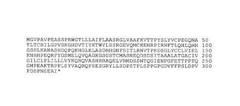
[0142] Figure 1 depicts sequence analysis. (A) Full length amino acid sequence
of murine
VISTA (PD-L3) (SEQ ID NO: 17). (B) Amino acid sequence alignment of
extracellular Ig
domains between murine VISTA (PD-L3) (SEQ ID NO: 25) and selected B7 family
ligands,
including B7-H1 (PD-L1) (SEQ ID NO: 26), B7-DC (PD-L2) (SEQ ID NO: 27), B7-H3
(CD276)
(SEQ ID NO: 28), and B7-H4 (B7S1) (SEQ ID NO: 29). (C) Alignment of VISTA (PD-
L3) (SEQ
ID NO: 30) Ig domain with B7 family receptors, including PD-1 (SEQ ID NO: 31),
CTLA-4
(SEQ ID NO: 32), CD28 (SEQ ID NO: 33), BTLA (SEQ ID NO: 34), and ICOS (SEQ ID
NO:
24
CA 02884704 2015-03-10
WO 2014/039983
PCMJS2013/058785
35). Ig-v domain, "...."; Ig-c domain, " ". Alignment was performed using
the MUSCLE
algorithm (Multiple Sequence Comparison by Log-Expectation). (D) Sequence
identity (%) of
the Ig-V domains between VISTA (PD-L3) and other B7 family ligands and
receptors is
calculated using ClustalW2 program. (E) Sequence alignment to show sequence
homology
between human (SEQ ID NO: 37) and murine VISTA (PD-L3) (SEQ ID NO: 36).
Identical
residues are shaded in black. Highly conserved and semi-conserved residues are
shaded in dark
and light shade of gray respectively.
[0143] Figure 2 depicts a hylogenic analysis of mouse VISTA (PD-L3) with other
Immunoglobulin (Ig) superfamily members. Full-length sequence of mouse VISTA
(PD-L3) and
other Ig superfamily members, including CD28, CTLA-4, ICOS, BTLA, PD-I, B7-H1
(PD-L1),
B7-DC (PD-L2), B7-H2, B7-H3, B7-H4, B7-1, B7-2, BTNL2, BTN3A3, BTN2A2, and
BTN1A1, were analyzed using PhyML algorithm (Phylogenetic Maximum Likelihood).
Branch
distances were shown at tree branch joints.
[0144] Figure 3 depicts the tissue expression and hematopoietic cell
expression patterns of
VISTA (PD-L3) A. RT-PCR of full length VISTA (PD-L3) from mouse tissues.
Lanes:
(1)muscle (2)heart (3)eye (4) thymus (5)spleen (6)small intestine (7)kidney
(8)liver (9)brain
(10)mammary gland (11)1ung (12)ovary (13)bone marrow. B. RT-PCR of full-length
VISTA
(PD-L3) from purified hematopoietic cell types. Lanes (1) peritoneal
macrophages (2) splenic
CDI lb+ monocytes (3) splenic CDI lc+ DCs (4) splenic CD4+ T cells (5) splenic
CD8+ T cells
(6) splenic B cells. C-E. Flow cytometry analysis of VISTA (PD-L3) expression
on splenic CD4+
and CD8+ T cells from thymus and spleen (C), on CD11b+ monocytes (D), and on
CD11c+ DC
subsets from spleen and peritoneal cavity (E). (F) Splenic B cells, NK cells
and granulocytes are
also analyzed. (G) The differential expression of VISTA (PD-L3) on
hematopoietic cells from
different tissue sites, including mesenteric LN, peripheral LN, spleen, blood
and peritoneal
cavity. Representative data from at least 3 independent experiments are shown.
101451 Figure 4 depicts a VISTA, novel and structurally-distinct, Ig-
superfamily inhibitory
ligand, whose extracellular domain bears highest homology to the B7 family
ligand PD-L1 as
displayed on an Antigen Presenting Cell along with other CDs and B7 family
members. VISTA
has a 93 aa cytoplasmic domain with no obvious signal transducing motifs,
except a possible
protein kinase C binding site.
[0146] Figure 5 depicts the specificity of VISTA (PD-L3) hamster monoclonal
antibodies.
Mouse EL4 cell lines over-expressing either PD-L I or VISTA (PD-L3) fused to
RFP were
CA 02884704 2015-03-10
WO 2014/039983
PCT/US2013/058785
stained using the supernatants from hybridoma cultures and analyzed by flow
cytometry. Two
representative positive clones are shown, 8D8 AND 6E7.
101471 Figure 6 depicts a comparison of VISTA (PD-L3) expression with other B7
family
ligands on in vitro cultured spleen cells. Expression of VISTA (PD-L3) and
other B7 family
ligands (i.e., PD-L I , PD-L2, B7-H3, and B7-H4) on hematopoietic cell types,
including CD4+ T
cells, CDI I bhi monocytes, and CDI lc+ DCs were compared. Cells were either
freshly isolated,
or in vitro cultured for 24 hrs, with and without activation. CD4+ T cells
were activated with
plate-bound aCD3 (5 ig/m1), CDIlbhi monocytes and CDI 1 c+ DCs were activated
with IFNot
(20 ng/ml) and LPS (200 ng/ml). Representative results from three independent
experiments are
shown.
[0148] Figure 7 depicts the comparison of in vivo expression patterns of VISTA
(PD-L3) and
other B7 family ligands during immunization. D011.10 TCR transgenic mice were
immunized
with chicken ovalbumin (OVA) emulsified in complete Freund's adjuvant (CFA) on
the flank.
Draining and non-draining lymph node cells were collected 24 hr post
immunization, and
analyzed by flow cytometry for the expression of VISTA (PD-L3), PD-Ll and PD-
L2. Shown are
representative results from at least four independent experiments. (A) A
population of CD11b+
cells expressing a high level of VISTA (PD-L3) was induced at 24 hr post
immunization with
CFA/OVA, but not with CFA alone within the draining lymph node. These cells
are of mixed
phenotype of F4/80+ macrophages and CDI1C+ dendritic cells. (B) Expression of
VISTA (PD-
L3), PD-L1 and PD-L2 on CD1lbhi monocytes, CD11c+ DCs and CD4+ T cells were
analyzed
at 24 hr post immunization.
101491 Figure 8 depicts the loss of VISTA (PD-L3) expression on activated CD4+
T cells,
CDI lb and CDI lc+ cells in response to immunization. D011.10 mice were
immunized with
chicken ovalbumin (OVA) emulsified in complete Freund's adjuvant (CFA) on the
flank.
Draining and non-draining lymph node cells were collected 48 hr post
immunization, and
analyzed for VISTA (PD-L3) expression by flow cytometry. Shown are
representative results
from 2 independent experiments.
101501 Figure 9 depicts that immobilized VISTA (PD-L3)-Ig fusion protein
inhibited CD4+
and CD8+ T cell proliferation. (A) CFSE labeled CD4+ and CD8+ T cells were
stimulated by
plate-bound aCD3 with or without co-absorbed VISTA (PD-L3)-lg. The percentage
of CFSE-low
cells was quantified and shown in (B). (C) CD4+ T cells from PD-1 ko mice were
also
suppressed by VISTA (PD-L3)-Ig. (D) VISTA (PD-L3)-Ig-mediated suppression is
persistent and
26
CA 02884704 2015-03-10
WO 2014/039983
PCMJS2013/058785
can act late. CD4+ T cells were activated in the presence of VISTA (PD-L3)-Ig
or control-Ig for
either 72 hrs (i), or for 24 hrs (ii, iii and iv). 24 hour-preactivated cells
were harvested and re-
stimulated under specified conditions for another 48 hours. Cell proliferation
was analyzed at the
end of the 72 hour culture. (ii) Pre-activation with VISTA (PD-L3)-Ig and re-
stimulation with
antiCD3; (iii) Pre-activation with antiCD3 and re-stimulation with VISTA (PD-
L3)-Ig. (iv) Pre-
activation with VISTA (PD-L3)-Ig and re-stimulation with VISTA (PD-L3)-Ig.
Duplicated wells
were analyzed for all conditions. Shown are representative results from four
experiments.
[0151] Figure 10 depicts the similar inhibitory effect of PD-L1-Ig and
VISTA (PD-L3)-Ig
fusion proteins on CD4+ T cell proliferation. Bulk purified CD4+ T cells were
CFSE labeled and
stimulated with plate-bound aCD3 together with titrated amount of PD-L I -Ig
or VISTA (PD-
L3)-Ig fusion proteins. CFSE dilution was analyzed at 72 hours and the
percentage of CFSElow
cells was quantified. Duplicated wells were analyzed for all conditions. Shown
are representative
results from 2 independent experiments.
[0152] Figure 11 depicts the suppressive impact of VISTA (PD-L3)-Ig on the
proliferation of
naïve and memory CD4+ T cells. (A) Naïve (CD25-CD44lowCD62Lhi) and memory
(CD25-
CD44hiCD62Llow) CD4+ T cell subsets were sorted, CFSE labeled, and stimulated
with plate-
bound anti-CD3 (2.5 tg/m1) together with VISTA (PD-L3)-Ig or control-Ig at
indicated ratios.
Cell proliferation was analyzed at 72 hours by examining the CFSE division
profile. The
percentage of proliferated cells, as determined by percentage of CFSElow
cells, is calculated and
shown in B. Duplicated wells were analyzed for all conditions. Shown are
representative results
from two independent experiments.
[0153] Figure 12 depicts VISTA (PD-L3)-Ig fusion protein suppressed early
TCR activation
and cell proliferation, but did not directly induce apoptosis. Bulk purified
CD4+ T cells were
stimulated with plate-bound anti-CD3 together with VISTA (PD-L3)-Ig or control-
Ig at 1-2 ratio
(2.5 tg/m1 and 5 tg/m1 respectively). Cells were analyzed at 24 hr and 48 hrs
for the expression
of CD69, CD62L, and CD44 by flow cytometry. Cells were also stained for early
apoptosis
marker annexin-V, and cell death marker 7-Aminoactinomycin D (7-AAD). Shown
are
representative results from two independent experiments.
[0154] Figure 13 depicts VISTA-Ig inhibited cytokine production by CD4+ and
CD8+ T cells.
(A-B) Bulk purified CD4+ T cells were stimulated with plate-bound anti-CD3,
and VISTA-Ig or
control-Ig at stated ratios. Culture supernatants were collected after 24 hrs
and 48 hrs. Levels of
IL-2 and IFNy were analyzed by ELISA. (C-D) CD4+ T cells were sorted into
naïve (CD25-
27
CA 02884704 2015-03-10
WO 2014/039983
PCMJS2013/058785
CD44lowCD62Lhi) and memory (CD25-CD44hiCD62Llow) cell populations. Cells were
stimulated with plate-bound aCD3 and VISTA (PD-L3)-Ig or control-Ig at a ratio
of 1:2. Culture
supernatants were collected at 48 hrs and analyzed for the level of IL-2 and
IFNy by ELISA. (E)
Bulk purified CD8+ T cells were stimulated with plate-bound aCD3, and VISTA
(PD-L3)-Ig or
control-Ig at indicated ratios. IFNy in the culture supernatant was analyzed
by ELISA. For all
conditions, supernatant for six duplicated wells were pooled for ELISA
analysis. Shown are
representative results from three experiments.
101541 Figure 14 depicts VISTA-Ig-mediated suppression may overcome a moderate
level of
costimulation provided by CD28, but was completely reversed by a high level of
costimulation,
as well as partially rescued by exogenous IL-2. A-B. Mouse CD4+ T cells were
activated by
plate-bound aCD3 together with either VISTA (PD-L3)-Ig or control-Ig at 1-1
ratio and 1-2
ratios. For cytokine rescue, soluble mIL-2, mIL7, mIL15 and ml L-23 (all at
40ng/m1) were added
to the cell culture (A). To examine the effects of costimulation, aCD28 (1
niml) was
immobilized together with aCD3 and Ig proteins at indicated ratios (B). Cell
proliferation was
analyzed at 72 hr by examining CFSE division profiles. C-D. To examine the
suppressive activity
of VISTA (PD-L3) in the presence of lower levels of costimulation, titrated
amounts of aCD28
were coated together with anti-CD3 (2.5 ug/m I) and VISTA-Ig fusion proteins
or control-Ig
fusion protein (10 rtg/ml) to stimulate mouse CD4+ T cell proliferation. Cell
proliferation was
analyzed at 72 hour. Percentages of proliferated CFSElow cells were quantified
and shown in D.
Duplicated wells were analyzed for all conditions. Representative CFSE
profiles from three
independent experiments are shown.
[0155] Figure 15 depicts that VISTA (PD-L3) expressed on antigen presenting
cells
suppressed CD4 T cell proliferation. A-C The CHO cell line that stably
expresses MHCII
molecule 1-Ad and costimulation molecule B7-2 was used as the parent cell
line. Cells were
transduced with retrovirus expressing either VISTA-RFP or RFP control
molecules. Transduced
cells were sorted to achieve homogenous level of expression. To test their
ability as antigen
presenting cells. CHO-VISTA or CHO-RFP cells were mitomycin C treated and
mixed with
OVA-specific transgenic CD4+ T cells D011.10, in the presence of titrated
amount of OVA
peptide. Proliferation of D011 cells was analyzed at 72 hrs, either by CFSE
division profiles (A-
B), or by tritium incorporation (C). (D) bone marrow derived dendritic cells
were transduced with
RFP or B7B-H5-RFP retrovirus during 10-day culture period. Transduced CD11c+
RFP+ DCs
and non-transduced CD11 c+ RFP- DCs were sorted and used to stimulate OVA-
specific
28
CA 02884704 2015-03-10
WO 2014/039983
PCMJS2013/058785
transgenic CD4+ T cells OTII in the presence of titrated amount of OVA
peptide. Cell
proliferation was analyzed on day 3 by examining CFSE division. For all
experiments, duplicated
wells were analyzed for all conditions, and representative results from three
independent
experiments are shown.
[0156] Figure 16 depicts the surface expression level of VISTA (PD-L3) in
retrovirally
transduced bone marrow derived DCs. Bone marrow derived DCs (BMDC) were
cultured in the
presence of GM-CSF (20 ng/mml) and transduced with either RFP or VISTA-RFP
retrovirus as
described herein. On day 10, surface expression level of VISTA were analyzed
on cultured
BMDCs, and compared to freshly-isolated peritoneal macrophages.
[0157] Figure 17 shows that anti-PDL3monoclonal antibody exhibits efficacy in
a passive
transfer EAE model. In this adoptive transfer EAE model, donor SJL mice were
immunized with
CFA and PLP peptide. On day 10, total lymphocytes from draining LN were
isolated, and
cultured in vitro with PLP peptide, IL-23 (20 ng/ml) and anti-IFNg (10 gimp
for 4 days.
Expanded CD4 T cells were then purified and adoptively transferred into naïve
recipient mice.
Disease progression was monitored and scored with: 0, no disease; 0.5 loss of
tail tone; I: limp
tail; 2: limp tail + hind limb paresis; 2.5: 1 hind limb paralysis; 3: both
hind limb paralysis; 3.5:
forelimb weakness; 4: hind limb paralysis+ unilateral forelimb paralysis. Mice
were sacrificed
when disease score reached 4. *, mice were sacrificed.
[0158] Figure 18 shows that VISTA expressed on antigen-presenting cells
suppressed CD4+ T
cell proliferation.
[0159] Figure 19 shows that an anti-VISTA antibody inhibited tumor growth in
mice
transplanted with MB49 tumor cells.
[0160] Figure 20 shows the antitumor effect of VISTA monoclonal antibodies in
four
different mouse anti-tumor models (A-D). Figure 21E shows the expression of
VISTA on
different cells in the ID8 model. Very high expression on the myeloid
dendritic cells in different
anatomic locations. As can be seen, very high levels on myeloid dendritic
cells in the ascites
cells, the site where the tumor grows and leukocytes infiltrate.
[0161] Figure 21 shows the potentiating effect of VISTA monoclonal antibodies
on the
efficacy of a CD40/TER agonist vaccine (consisting of using an agonistic aCD40
mab, TLR
agonist and OVA peptide).
[0162] Figure 22 shows VISTA expression on CNS cells in mice that are healthy
or in mice
that are developing EAE.
29
CA 02884704 2015-03-10
WO 2014/039983 PCMJS2013/058785
[0163] Figure 23 depicts a sequence and structural analysis of VISTA. (A) The
primary
amino acid sequence of mouse VISTA with the Ig-V domain, the stalk segment,
and the
transmembrane region highlighted in bold, italics, and Times New Roman,
respectively.
Cysteines in the ectodomain region are indicated by underlining. (B) Multiple
sequence
alignment of the Ig-V domains of several B7 family members and VISTA. The
predicted
secondary structure (using arrows, springs, and "1".s for strands, helices,
and B-turns,
respectively) is marked above the alignment and is based on the VISTA
structural model. VISTA
(SEQ ID NO: 15), PD1L1 (SEQ ID NO: 11), PD1L2 (SEQ ID NO: 12), B7H4 (SEQ ID
NO: 13),
and B7H3 (SEQ ID NO: 14). (C) Multiple sequence alignment of VISTA
orthologues. Invariant
residues are represented by the red background, and physico-chemically
conserved positions are
represented by red letters. Conserved amino acids are marked by blue boxes.
Conservation is
calculated on the basis of 36 VISTA orthologous proteins, but only 9
representatives are shown.
The canonical cysteine pair (within the "B" and "F" strands) that is conserved
in almost all Ig
superfamily members is highlighted by red circles, whereas cysteines that are
specific to VISTA
are marked by blue circles. The unique VISTA eysteine pattern is conserved in
all orthologues
from mouse (SEQ ID NO: 17), human (SEQ ID NO: 16), kangaroo (SEQ ID NO: 18),
dolphin
(SEQ ID NO: 19), chicken (SEQ ID NO: 20), xenopus (SEQ ID NO: 21), zebra finch
(SEQ ID
NO: 22), zebrafish, and fugu (SEQ ID NO: 23).
[0164] Figure 24 depicts that VISTA over expression on tumor cells overcomes
protective
antitumor immunity. MCA105 tumor cells over expressing VISTA or RFP control
protein were
generated by retroviral transduction and sorted to homogeneity. To generate
protective
immunity, naive mice were vaccinated with irradiated MCA105 tumor cells
subcutaneously on
the left flank. (A) Vaccinated mice were challenged 14 day later with live
MCA105VISTA or
MCA105RFP tumor cells subcutaneously on the right flank. Tumor growth was
monitored every
2 d. Tumor size is shown as mean SEM. Shown are representative results from
three
independent repeats. (B) Vaccinated mice were either untreated or depleted of
both CD44 and
CD8+ T cells by monoclonal antibodies before live tumor challenge. Tumor size
was monitored
as in A and shown as mean SEM. Shown are representative results from two
independent
repeats. For all experiments, ratios indicate the number of tumor-bearing mice
among total
number of mice per group. The statistical differences (p-values) were assessed
with an unpaired
Mann-Whitney test.
CA 02884704 2015-03-10
WO 2014/039983
PCMJS2013/058785
[0165] Figure 25A-F depicts that VISTA blockade using a specific monoclonal
antibody
enhanced CD4+ T cell response in vitro and in vivo. (A) A monoclonal antibody
clone 13F3
neutralized VISTA-mediated suppression in vitro. A20-RFP and A20-VISTA cells
were used to
stimulate CFSE-labeled D011.10 CD4+ T cells in the presence of cognate OVA
peptide. 20
ng/m1 VISTA-specific monoclonal antibody 13F3 or control-Ig was added as
indicated. CFSE
dilution was analyzed after 72 h, and percentages of CFSew cells are shown as
mean SEM.
Duplicated wells were analyzed for all conditions. (B and C) Total
CD1lbhimyeloid cells (B) or
CD11b-CD11c- monocytes (C) and CD1lbh1CD11c+ myeloid DCs (D) sorted from naive
splenocytes were irradiated and used to stimulate CFSE-labeled OT-II
transgenic CD4+ T cells in
the presence of OVA peptide. Cell proliferation was measured by incorporation
of tritiated
thymidine during the last 8 h of a 72-h culture period and shown as mean
SEM. Triplicate
wells were analyzed in all conditions.
[0166] Figure 26 depicts VISTA-IgG2a reduces Experimental Autoimmune
Encephalomyelitis (EAE) (a model of multiple sclerosis) progression. Mice were
immunized with
175 1.tg MOG/CFA and pertussis toxin (PT) 300 ng (day 0, 2) to induce active
EAE. On day 14,
17, and 20, 150 jig VISTA-IgG 2a (n=8) or 150 1.tg control IgG2a (n=8) was
administered. The
data is shown as the mean SEM.
[0167] Figure 27 depicts the therapeutic effect of VISTA-IgG1 and VISTA-IgG2a
on
Experimental Autoimmune Encephalomyelitis (EAE) progression. Mice were
immunized with
175 jig MOG/CFA and pertussis toxin (PT) 300 ng (day 0, 2) to induce active
EAE. On day 6,
mice were treated with 3 doses per week of 150 jig control IgG1 (n=3), 150 fig
control IgG2a
(n=6), 150 pig mVISTA-IgG1 (n=3), or 150 jig mVISTA IgG2a (n=6) (two weeks in
total). The
data is shown as the mean SEM.
[0168] Figure 28 depicts the therapeutic effect of VISTA-IgG2a fusion protein
on Experimental
Autoimmune Encephalomyelitis (EAE) progression. Mice were immunized with 175
jig
MOG/CFA and pertussis toxin (PT) 300 ng (day 0, 2) to induce active EAE. On
day 14, mice
were treated with 3 doses per week of PBS (n=6), 100 1.tg control IgG2a (n=6),
300 pig control
IgG2a (n=6), 100 ptgVISTA-IgG2a (n=6), or 300 l_tg mVISTA IgG2a (n=6) (two
weeks in total).
The data is shown as the mean SEM.
[0169] Figure 29 depicts the expression of VISTA healthy human tissues was
examined by real-
time PCR analysis of a eDNA tissue panel (Origene). (A) VISTA was
predominantly expressed
in haematopoietic tissues or in tissues that contain significant numbers of
haematopoietic tissues.
31
CA 02884704 2015-03-10
WO 2014/039983
PCMJS2013/058785
This is consistent with importance of VISTA in immune related functions. (B)
The expression
pattern of expression was found to follow a similar trend to that of VISTA's
closest homologue
PD-Ll.
[0170] Figure 30 depicts VISTA protein expression in monocytes, dendritic
cells and by
approximately 20% of CD4 and CD8 T cells (Fig. 30). VISTA expression was
observed within
both of the 'patrolling' (CD14thmCD16+) and 'inflammatory' (CD14+CD16+/)
subsets of blood
monocytes, and within both lymphoid and myeloid subsets of dendritic cell.
[0171] Figure 31 depicts the suppression of CFSE dilution of bulk purified CD4
(Fig. 31A) and
CD8 (Fig. 31B) T cells. An Ig fusion protein was created, consisting of the
extracellular domain
of VISTA and the Fe region of human IgG containing mutations for reduced Fc
receptor binding.
p.g/m1 of VISTA-Ig or control Ig was immobilized on plates along with
2.51.tg/m1 of anti-CD3
(OKT3) and then proliferation was measured by CFSE dilution.
[0172] Figure 32 depicts the titration of human VISTA-Ig and human VISTA-Ig
over different
concentrations of OKT3, showed that higher concentrations of OKT3 can be
overcome by higher
concentrations of VISTA (Fig. 32A and 32B).
[0173] Figure 33A-C depicts the status of cells was examined following
activation in the
presence or absence of VISTA-Ig. During 2 days of culture, upregulation by
anti-CD3 of the
early activation markers CD25 and CD69 was blocked by VISTA-Ig (Fig. 33A &
33B).
Similarly, after 5 days of culture, the shift from expression of CD45RA to
CD45RO, indicative of
antigen-experience was prevented (Fig. 33C). VISTA had no affect on cell
viability. Figure 34D
shows that VISTA-1g increased FoxP3 conversion.
[0174] Figure 34 depicts the suppression induced by VISTA where cells were
cultured on anti-
CD3 and VISTA-Ig for two days, and then moved onto anti-CD3 alone for 3 days.
This further
stimulation was unable to rescue suppression (Fig. 34A and 34B.)
[0175] Figure 35 shows that VISTA-Ig significantly reduced production of IL-
10, TNFa. and
IFN7 by CD4 (Fig. 35A) and CD8 (Fig. 35B) T cells, and there was a trend
towards a modest
decrease in IL-17 production.
[0176] Figure 36 shows that anti-CD28 agonistic antibody provides potent
costimulation to T
cells, and so titred into the cultures to challenge VISTA suppression (Fig.
36A-C).
[0177] Figure 37. VISTA mAb treatment reduced tumour growth. Mice were
injected with A.
MB49. B. MCA105. C. EG7 tumour cells, D. ID8-luciferase. E. B16F10. Mice were
treated with
VISTA mAb 13F3 every other day (300m) beginning on day 0 (A-D), or day-2 (E).
PD-Ll mAb
32
CA 02884704 2015-03-10
WO 2014/039983
PCMJS2013/058785
(MIH5) was also administered to Bl6F10. Subcutaneous tumour growth was
monitored with
calliper and recorded as mm2. For intraperitoneal ID8-luciferase tumour, mice
were imaged on
day 30 and 55 using Xenogen IVIS. For MB49 EL1SPOT analysis (A), tumour drain-
LN cells
were stimulated with irradiated tumour cells.
[0178] Figure 38. Human lamina propria DCs express VISTAa. EMPCs isolated from
healthy
colon were stained with biotin-conjugated anti-human VISTA (antibody clone
GA1) to identify
VISTA expression in Lin-HLA-DR+ LP dendritic cells.
[0179] Figure 39. Vista-Ig is suppressive to human CD4 T cells. CFSE-labeled
human CD4 T
cells were stimulated with plate-bound anti-CD3 at 2.5 ug/ml and VISTA-Ig at
the indicated
concentrations. (A) Representative CFSE dilution profiles. (B) The percentage
of CFSE-low
cells was quantified and shown as mean +/- SEM.
[0180] Figure 40. VISTA acts in concert with the PD-Li/PD-1 pathway. A.
Combinatorial
treatment (Day+4) with aVISTA and aPD-L1 mAbs inhibited B16F10 tumour growth.
B.
Synergy in vitro: VISTA-Ig and PD-L1-1g were immobilized together with
aCD3/CD28 to
stimulate CD4+ and CD8+ naïve T cells. Cell proliferation was assessed by CFSE
dilution at
72hrs. C. Differential expression pattern of PD-Ll and VISTA within the TME of
B16F10
tumour. VISTA is expressed only on tumour-infiltrating leukocytes (T1Ls),
whereas PD-Li is
expressed on both tumour cells and TILs.
[0181] Figure 41. Detection of VISTA on myeloid cells with ah VISTA mAb. PBL
were stained
in the absence (top) or presence of VISTA-1g (bottom) to confirm specificity.
DETAILED DESCRIPTION
[0182] We have discovered a novel inhibitory ligand, designated V-domain Ig
Suppressor of T
cell Activation (VISTA), that plays a key role in disrupting protective anti-
tumour immunity in
mice (1). VISTA bears limited homology to PDL1, and critically suppresses T
cell activation via
an unknown receptor, independent of PD-1. VISTA KO mice display inflammatory
phenotypes,
indicating an essential role for VISTA in maintaining peripheral tolerance.
VISTA is highly
expressed in the tumour microenvironment (TME) and directly impairs the
generation of optimal
anti-tumour immunity. VISTA mAb-mediated blockade significantly suppressed
tumour growth
in multiple mouse tumour models.
[0183] Based on these findings, we hypothesize that VISTA is expressed on
tumour-infiltrating
leukocytes, and that this expression is suppressive for T cell responses in
the TME. This
application extends our existing studies of VISTA in mouse models to human
patients.
33
CA 02884704 2015-03-10
WO 2014/039983
PCMJS2013/058785
Specifically, the application further describes examining VISTA expression in
patient samples
and testing how this influences T cell function. Based thereon, inhibition of
VISTA may be used
in the treatment of cancer (similar to the success with functionally related
proteins CTLA-4 and
PD-L1). In another aspect, the disclosure provides methods of identifying
suitable blocking
antibodies to be developed for this purpose.
[0184] In another aspect, the present invention relates to therapeutic methods
that modulate the
activity and/or which specifically bind or block the binding of a specific
regulatory T cell protein
to its counterreceptor. This protein, designated PD-L3 OR VISTA, is a novel
and structurally-
distinct, Ig-superfamily inhibitory ligand, whose extracellular domain bears
homology to the B7
family ligand PD-Li. This molecule is referred to interchangeably herein as PD-
L3 or VISTA or
as V-domain Immunoglobulin Suppressor of T cell Activation (VISTA). VISTA is
expressed
primarily within the hematopoietic compartment and is highly regulated on
myeloid APCs and T
cells. Therapeutic intervention of the VISTA inhibitory pathway represents an
exciting approach
to modulate T cell-mediated immunity for the treatment of a wide variety of
cancers.
[0185] The present invention in particular relates to the use of antibodies
specific to VISTA or
PD-L3 to treat specific cancers including colorectal cancer, bladder cancer,
ovarian cancer, and
melanoma.
[0186] As disclosed infra, the expression of VISTA appears to be exclusive to
the hematopoietic
compartment and this protein is highly expressed on mature myeloid cells
(CD11bbnght), with
lower levels of expression on CD4+ T cells, Treg and CD8+ T cells. Soluble
VISTA proteins, e.g.,
soluble VISTA-Ig fusion protein, or VISTA expression on APCs, suppresses in
vitro CD4+ and
CD8+ T cell proliferation and cytokine production. It is also observed that
anti-VISTA antibodies,
e.g., an anti-VISTA mab (13F3) blocked VISTA-induced suppression of T cell
responses by
VISTA + APCs in vitro. Also, it has been discovered that an anti-VISTA mab
exacerbated EAE
and increased the frequency of encephalitogenic Thl7s in vivo. Still further,
as disclosed in detail
infra, it has been found that an anti -VISTA mab induces tumor remission in
multiple (4) murine
tumor models. VISTA expression on myeloid derived suppressor cells (MDSC) in
these models
is extremely high, suggesting that VISTA+ MDSC suppress tumor specific
immunity. As shown
herein, VISTA exerts immunosuppressive activities on T cells both in vitro and
in vivo, in mouse
and in human (in vitro only) and is an important mediator in controlling the
development of
autoimmunity and the immune responses to cancer. Specifically, the data show
that:
34
CA 02884704 2015-03-10
WO 2014/039983
PCMJS2013/058785
[0187] (I) VISTA is a new member of the Ig superfamily and contains an Ig-V
domain with
distant sequence similarity to PD-Li. We disclose herein that when produced as
an Ig fusion
protein or when overexpressed on artificial APCs VISTA inhibits both mouse and
human CD4+
and CD8+ T cell proliferation and cytokine production.
[0188] (2) VISTA expression on myeloid APCs is inhibitory for T cell responses
in vitro.
[01891 (3) VISTA expression on MDSC in the tumor microenvironment is extremely
high.
Phenotypic and functional analysis of many cell surface molecules previously
suggested to be
involved in MDSC-mediated suppression of T cells: CD115, CD124, CD80, PD-L1,
and PD-L2
were expressed by MDSC but with no differences in the levels of their
expression or proportion
of positive cells were found between MDSC and cells from tumor-free mice that
lack immune
suppressive activity. Therefore, we predict that VISTA will be the primary B7
negative regulator
on MDSCs.
[0190] (4) Antibody-mediated VISTA blockade induces protective immunity to an
autologous
tumor.
[0191] Based thereon, VISTA appears to be a dominant, negative immune
regulatory molecule
on MDSCs that interferes with the development of protective anti-tumor
immunity. Therefore,
blocking the activity of this molecule with anti-VISTA antibodies will permit
the development of
protective anti-tumor immunity in humans and other mammals.
[0192] Therefore, the invention relates to methods of using soluble VISTA
proteins, e.g., fusion
proteins and multimeric VISTA proteins comprising multiple copies of the VISTA
extravcelular
domain or a fragment thereof, andVISTA binding agents, e.g., small molecules
and antibodies or
fragments theeof, which bind or modulate (agonize or antagonize) the activity
of VISTA as
immune modulators and for the treatment of different cancers, e.g., colorectal
cancer, bladder,
ovarian and lymphoma, autoimmune disease, allergy, infection and inflammatory
conditions, e.g.
multiple sclerosis and arthritis.
[0193] As described in detail infra, VISTA is a novel inhibitory ligand, which
extracellular Ig-V
domain bears homology to the two known B7 family ligands Programmed Death
Ligand 1 and 2
(PD-Ll and PD-L2) and exhibits unique sequence features and distinctive
expression patterns in
vitro and in vivo on subsets of APCs and T cells,(vvhich distinguishes PD-L3
or VISTA from
other B7 family ligands). This protein has been shown to have a functional
impact on CD4+ and
CD8+ T cell proliferation and differentiation (suppresses CD4 and CD8+ T cell
proliferation, as
well as cytokine production). Based on its expression pattern and inhibitory
impact on T cells,
CA 02884704 2015-03-10
WO 2014/039983
PCMJS2013/058785
PD-L3 OR VISTA apparently functions as a regulatory ligand that negatively
regulates T cell
responses during cognate interactions between T cells and myeloid derived
APCs.
[0194] While PD-L3 OR VISTA appears to be a member of the B7 family of
ligands, unlike
other B7 family ligands, this molecule contains only an lg-V domain without an
Ig-C domain,
and is phylogenically closer to the B7 family receptor Programmed Death-1 (PD-
1). Based
thereon, PD-L3 OR VISTA, and agonists or antagonists specific thereto can be
used to regulate T
cell activation and differentiation, and more broadly to modulate the
regulatory network that
controls immune responses. In particular PD-L3 or VISTA proteins and PD-L3 or
VISTA
agonists or antagonists, preferably antibodies specific to PD-L3 or VISTA are
useful in
modulating immune responses in autoimmunity, inflammatory responses and
diseases, allergy,
cancer, infectious disease and transplantation.
101951 Therefore, the present invention in part relates to compositions e.g.,
for therapeutic,
diagnostic or immune modulatory usage containing an isolated soluble PD-L3 OR
VISTA protein
or fusion protein, e.g., a soluble VISTA-Ig fusion protein or a multimeric
VISTA protein,
comprising an amino acid sequence that preferably is at least 70-90% identical
to the human or
murine PD-L3 OR VISTA polypeptide set forth in SEQ ID NO:2, 4 or 5 or an
ortholog, or
fragment thereof encoded by a gene that specifically hybridizes to SEQ ID NO:1
or 3 that
modulates VISTA in vivo and a pharmaceutically acceptable carrier. In some
embodiments, the
soluble or multimeric VISTA protein may be directly or indirectly linked to a
heterologous (non-
VISTA) protein or may be expressed by a viral vector or a cell containing,
e.g., a transfected
immune cell such as a T cell.
[01961 The present invention also provides expression vectors comprising an
isolated nucleic
acid encoding a VISTA protein that is at least 70-90% identical to the human
or murine VISTA
amino acid sequence set forth in SEQ ID NO:2, 4 or 5 or a fragment or ortholog
thereof, which
optionally is fused to a sequence encoding another protein such as an Ig
polypeptide, e.g., an Fc
region or a reporter molecule; and host cells containing said vectors.
101971 The present invention also specifically relates to an isolated binding
agent, preferably an
antibody or antibody fragment which specifically binds to a PD-L3 OR VISTA
protein
comprising the amino acid sequence set forth in SEQ ID NO:2, 4 or 5 or a
variant, fragment or
ortholog thereof. In a preferred embodiment, the binding agent modulates
(agonizes or
antagonizes) VISTA activity in vitro or in vivo. In most preferred
embodiments, the binding
agent is an agonistic or antagonistic antibody.
36
CA 02884704 2015-03-10
WO 2014/039983
PCMJS2013/058785
[01981 The present invention further provides methods for modulating an immune
cell response
by contacting an immune cell in vitro or in vivo with a VISTA protein, or
binding agent specific
thereto, in the presence of a primary signal so that a response of the immune
cell is modulated.
(Interaction of VISTA or a modulator thereof transmits a signal to immune
cells, regulating
immune responses. PD-L3 OR VISTA protein is expressed at high levels on
myeloid antigen
presenting cells, including myeloid dendritic cells (DCs) and macrophages, and
at lower densities
on CD4+ and CD8+ T cells. Upon immune activation, PD-L3 or VISTA expression is
upregulated on myeloid APCs, but downregulated on CD4+ T cells). Therefore,
the PD-L3 or
VISTA nucleic acids and polypeptides of the present invention, and agonists or
antagonists
thereof are useful, e.g., in modulating the immune response.
[0199] In addition, the PD-L3 or VISTA polypeptides (or biologically active
portions thereof) or
modulators of the PD-L3 or VISTA molecules, i.e., antibodies such as selected
using the
foregoing methods can be incorporated into pharmaceutical compositions, which
optionally
include pharmaceutically acceptable carriers.
[0200] Immune cells activated in accordance with the method of the instant
invention can
subsequently be expanded ex vivo and used in the treatment and prevention of a
variety of
diseases; e.g., human T cells which have been cloned and expanded in vitro
maintain their
regulatory activity (Groux, et al. (1997) Nature 389(6652):737-42). Prior to
expansion, a source
of T cells is obtained from a subject (e.g., a mammals such as a human, dog,
cat, mouse, rat, or
transgenic species thereof). T cells can be obtained from a number of sources,
including
peripheral blood mononuclear cells, bone marrow, lymph node tissue, cord
blood, thymus tissue,
tissue from a site of infection, spleen tissue, tumors or T cell lines. T
cells can be obtained from a
unit of blood collected from a subject using any number of techniques known to
the skilled
artisan, such as ficollTM separation. In another aspect, the present invention
provides a method for
detecting the presence of a PD-L3or VISTA nucleic acid molecule, protein, or
polypeptide in a
biological sample by contacting the biological sample with an agent capable of
detecting a PD-L3
OR VISTA nucleic acid molecule, protein, or polypeptide, such that the
presence of a PD-L3 OR
VISTA nucleic acid molecule, protein or polypeptide is detected in the
biological sample. This
PD-L3 OR VISTA expression can be used to detect certain disease sites,
including cancerous
sites.
[0201] In another aspect, the invention provides a method for modulating PD-L3
OR VISTA
activity, comprising contacting a cell capable of expressing PD-L3 OR VISTA
with an agent that
37
CA 02884704 2015-03-10
WO 2014/039983
PCMJS2013/058785
modulates PD-L3 OR VISTA activity, preferably an anti-PD-L3 OR VISTA antibody
such that
PD-L3 OR VISTA activity in the cell is modulated. In one embodiment, the agent
inhibits PD-L3
OR VISTA activity. In another embodiment, the agent stimulates PD-L3 OR VISTA
activity. In a
further embodiment, the agent interferes with or enhances the interaction
between a PD-L3 OR
VISTA polypeptide and its natural binding partner(s). In one embodiment, the
agent is an
antibody that specifically binds to a PD-L3 OR VISTA polypeptide. In another
embodiment, the
agent is a peptide, peptidomimetic, or other small molecule that binds to a PD-
L3 OR VISTA
polypeptide.
[0202] In still another embodiment, the agent modulates expression of PD-L3 OR
VISTA by
modulating transcription of a PD-L3 OR VISTA gene, translation of a PD-L3 OR
VISTA
mRNA, or post-translational modification of a PD-L3 OR VISTA polypeptide. In
another
embodiment, the agent is a nucleic acid molecule having a nucleotide sequence
that is antisense
to the coding strand of a PD-L3 OR VISTA mRNA or a PD-L3 OR VISTA gene.
[0203] In one embodiment, the methods of the present invention are used to
treat a subject having
a disorder or condition characterized by aberrant, insufficient, or unwanted
PD-L3 OR VISTA
polypeptide or nucleic acid expression or activity by administering an agent
which is a PD-L3
OR VISTA modulator to the subject. In one preferred embodiment, the PD-L3 OR
VISTA
modulator is a PD-L3 OR VISTA polypeptide, preferably a soluble fusion protein
or multimeric
VISTA protein or anti-VISTA antibody as described infra. In another embodiment
the PD-L3 OR
VISTA modulator is a PD-L3 OR VISTA nucleic acid molecule, e,g in an
adenoviral vector. In
another embodiment, the invention further provides treating the subject with
an additional agent
that modulates an immune response.
[0204] In still another embodiment, the invention provides a vaccine
comprising an antigen and
an agent that modulates (enhances or inhibits) PD-L3 OR VISTA activity. In a
preferred
embodiment, the vaccine inhibits the interaction between PD-L3 OR VISTA and
its natural
binding partner(s).
[0205] In another aspect the invention provides methods for identifying a
compound that binds
to or modulates the activity of a PD-L3 OR VISTA polypeptide, by providing an
indicator
composition comprising a PD-L3 OR VISTA polypeptide having PD-L3 OR VISTA
activity,
contacting the indicator composition with a test compound, and determining the
effect of the test
compound on PD-L3 OR VISTA activity in the indicator composition to identify a
compound
that modulates the activity of a PD-L3 OR VISTA polypeptide.
38
CA 02884704 2015-03-10
WO 2014/039983
PCMJS2013/058785
[0206] In one aspect, the invention features a method for modulating the
interaction of PD-L3
OR VISTA with its natural binding partner(s) on an immune cell comprising
contacting an
antigen presenting cell which expresses PD-L3 OR VISTA with an agent selected
from the group
consisting of: a form of PD-L3 OR VISTA, or an agent that modulates the
interaction of PD-L3
OR VISTA and its natural binding partner(s) such that the interaction of PD-L3
OR VISTA with
it natural binding partner(s) on an immune cell is modulated. In a preferred
embodiment, an agent
that modulates the interaction of PD-L3 OR VISTA and its natural binding
partner(s) is an
antibody that specifically binds to PD-L3 OR VISTA. In one embodiment, the
interaction of PD-
L3 OR VISTA with its natural binding partner(s) is upregulated. In another
embodiment, the
interaction of PD-L3 OR VISTA with its natural binding partner(s) is
downregulated. In one
embodiment, the method further comprises contacting the immune cell or the
antigen presenting
cell with an additional agent that modulates an immune response.
[0207] In one embodiment, the step of contacting is performed in vitro. In
another embodiment,
the step of contacting is performed in vivo. In one embodiment, the immune
cell is selected from
the group consisting of: a T cell, a monocyte, a macrophage, a dendritic cell,
a B cell, and a
myeloid cell.
[0208] In another aspect, the invention pertains to a method for inhibiting or
increasing activation
in an immune cell comprising increasing or inhibiting the activity or
expression of PD-L3 OR
VISTA in a cell such that immune cell activation is inhibited or increased.
[0209] In yet another aspect, the invention pertains to a vaccine comprising
an antigen and an
agent that inhibits the interaction between PD-L3 OR VISTA and its natural
binding partner(s).
[0210] In another aspect, the invention pertains to a method for treating a
subject having a
condition that would benefit from upregulation of an immune response
comprising administering
an agent that inhibits the interaction between PD-L3 OR VISTA and its natural
binding partner(s)
on immune cells of the subject such that a condition that would benefit from
upregulation of an
immune response is treated. In one preferred embodiment, the agent comprises a
blocking
antibody or a small molecule that binds to PD-L3 OR VISTA and inhibits the
interaction between
PD-L3 OR VISTA and its natural binding partner(s). In another embodiment, the
method further
comprises administering a second agent that upregulates an immune response to
the subject. In
another aspect, the invention pertains to a method for treating a subject
having a condition that
would benefit from downregulation of an immune response comprising
administering an agent
that stimulates the interaction between PD-L3 OR VISTA and its natural binding
partner(s) on
39
CA 02884704 2015-03-10
WO 2014/039983
PCMJS2013/058785
cells of the subject such that a condition that would benefit from
downregulation of an immune
response is treated.
[0211] For example the condition treated with the PD-L3 OR VISTA protein or
binding agents is
selected from the group consisting of: a tumor, a pathogenic infection, an
inflammatory immune
response or condition, preferably less pronounced inflammatory conditions, or
an
immunosuppressive disease. Specific examples include multiple sclerosis,
thyroiditis,
rheumatoid arthritis, diabetes type Ii and type I and cancers, both advanced
and early forms,
including metastatic cancers such as colorectal cancer, bladder cancer,
ovarian cancer, melanoma,
lung cancer, and other cancers wherein VISTA suppresses an effective anti-
tumor response. In
some case the individual may be administered cells or a viral vector that
express a nucleic acid
that encodes an anti-VISTA antibody or VISTA fusion protein.
[0212] Exemplary conditions treatable using PD-L3 OR VISTA proteins, binding
agents or PD-
L3 OR VISTA antagonists or agonists according to the invention include by way
of example
transplant, an allergy, infectious disease, cancer, and inflammatory or
autoimmune disorders,
e.g., an inflammatory immune disorder. Specific examples of the foregoing
include type 1
diabetes, multiple sclerosis, rheumatoid arthritis, psoriatic arthritis,
systemic lupus erythematosis,
rheumatic diseases, allergic disorders, asthma, allergic rhinitis, skin
disorders, gastrointestinal
disorders such as Crohn's disease and ulcerative colitis, transplant
rejection, poststreptococcal
and autoimmune renal failure, septic shock, systemic inflammatory response
syndrome (SIRS),
adult respiratory distress syndrome (ARDS) and envenomation; autoinflammatory
diseases as
well as degenerative bone and joint diseases including osteoarthritis, crystal
arthritis and
capsulitis and other arthropathies. Further, the methods and compositions can
be used for treating
tendonitis, ligamentitis and traumatic joint injury.
[0213] In preferred embodiments the subject PD-L3 OR VISTA proteins, nuclei
acids, and
ligands specific to PD-L3 OR VISTA, preferably antibodies having desired
effects on PD-L3 OR
VISTA functions are used to treat conditions such a cancer, autoimmune
diseases, allergy,
inflammatory disorders or infection and more specifically immune system
disorders such as
severe combined immunodeficiency, multiple sclerosis, systemic lupus
erythematosus, type I
diabetes mellitus, lymphoproliferative syndrome, inflammatory bowel disease,
allergies, asthma,
grail-versus-host disease, and transplant rejection; immune responses to
infectious pathogens
such as bacteria and viruses; and immune system cancers such as lymphomas and
leukemias)
CA 02884704 2015-03-10
WO 2014/039983
PCMJS2013/058785
102141 In addition to the infectious and parasitic agents mentioned above,
another area for
desirable enhanced immunogenicity to a non-infectious agent is in the area of
dysproliferative
diseases, including but not limited to cancer, in which cells expressing
cancer antigens are
desirably eliminated from the body. Tumor antigens which can be used in the
compositions and
methods of the invention include, but are not limited to, prostate specific
antigen (PSA), breast
cancer antigens, bladder cancer antigens, ovarian cancer antigens, testicular
cancer antigens,
melanoma antigens, colorectal cancer antigens, telomerase; multidrug
resistance proteins such as
P-glycoprotein; MAGE-1, alpha fetoprotein, carcinoembryonic antigen, mutant
p53,
papillomavirus antigens, gangliosides or other carbohydrate-containing
components of melanoma
or other tumor cells. It is contemplated by the invention that antigens from
any type of tumor cell
can be used in the compositions and methods described herein. The antigen may
be a cancer cell,
or immunogenic materials isolated from a cancer cell, such as membrane
proteins. Included are
survivin and telomerase universal antigens and the MAGE family of cancer
testis antigens.
Antigens which have been shown to be involved in autoimmunity and could be
used in the
methods of the present invention to induce tolerance include, but are not
limited to, myelin basic
protein, myelin oligodendrocyte glycoprotein and proteolipid protein of
multiple sclerosis and CII
collagen protein of rheumatoid arthritis.
Definitions
[0215] Unless defined otherwise, all technical and scientific terms used
herein have the same
meaning as those commonly understood by one of ordinary skill in the art to
which this invention
belongs. Although methods and materials similar or equivalent to those
described herein may be
used in the invention or testing of the present invention, suitable methods
and materials are
described herein. The materials, methods and examples are illustrative only,
and are not intended
to be limiting.
[0216] As used in the description herein and throughout the claims that
follow, the meaning of
"a," "an," and "the" includes plural reference unless the context clearly
dictates otherwise.
[0217] The terms "cancer" and "cancerous" refer to or describe the
physiological condition in
mammals that is typically characterized by unregulated cell growth. Examples
of cancer include
but are not limited to, carcinoma, lymphoma, blastoma, sarcoma, and leukemia.
More particular
examples of such cancers include squamous cell cancer, lung cancer (including
small-cell lung
cancer, non-small cell lung cancer, adenocarcinoma of the lung, and squamous
carcinoma of the
lung), cancer of the peritoneum, hepatocellular cancer, gastric or stomach
cancer (including
41
CA 02884704 2015-03-10
WO 2014/039983
PCMJS2013/058785
gastrointestinal cancer), pancreatic cancer, glioblastoma, cervical cancer,
ovarian cancer, liver
cancer, bladder cancer, hepatoma, breast cancer, colon cancer, colorectal
cancer, endometrial or
uterine carcinoma, salivary gland carcinoma, kidney or renal cancer, liver
cancer, prostate cancer,
vulval cancer, thyroid cancer, hepatic carcinoma and various types of head and
neck cancer, as
well as B-cell lymphoma (including low grade/follicular non-Hodgkin's lymphoma
(NHL); small
lymphocytic (SL) NHL; intermediate grade/follicular NIIL; intermediate grade
diffuse NHL;
high grade immunoblastic NHL; high grade lymphoblastic NHL; high grade small
non-cleaved
cell NHL; bulky disease NHL; mantle cell lymphoma; AIDS-related lymphoma; and
Waldenstrom's Macroglobulinemia); chronic lymphocytic leukemia (CLL); acute
lymphoblastic
leukemia (ALL); Hairy cell leukemia; chronic myeloblastic leukemia; multiple
myeloma and
post-transplant lymphoproliferative disorder (PTLD).
[0218] Exemplary cancers amenable for treatment by the present invention
include, but are not
limited to, carcinoma, lymphoma, blastoma, sarcoma, and leukemia or lymphoid
malignancies.
More particular examples of such cancers include colorectal, bladder, ovarian,
melanoma,
squamous cell cancer, lung cancer (including small-cell lung cancer, non-small
cell lung cancer,
adenocarcinoma of the lung, and squamous carcinoma of the lung), cancer of the
peritoneum,
hepatocellular cancer, gastric or stomach cancer (including gastrointestinal
cancer), pancreatic
cancer, glioblastoma, cervical cancer, ovarian cancer, liver cancer, bladder
cancer, hepatoma,
breast cancer, colon cancer, colorectal cancer, endometrial or uterine
carcinoma, salivary gland
carcinoma, kidney or renal cancer, liver cancer, prostate cancer, vulval
cancer, thyroid cancer,
hepatic carcinoma and various types of head and neck cancer, as well as B-cell
lymphoma
(including low grade/follicular non-Hodgkin's lymphoma (NHL); small
lymphocytic (SL) NHL;
intermediate grade/follicular NHL; intermediate grade diffuse NHL; high grade
immunoblastic
NHL; high grade lymphoblastic NHL; high grade small non-cleaved cell NHL;
bulky disease
NHL; mantle cell lymphoma; AIDS-related lymphoma; and Waldenstrom's
Macroglobulinemia);
chronic lymphocytic leukemia (CLL); acute lymphoblastic leukemia (ALL); Hairy
cell leukemia;
chronic myeloblastic leukemia; and post-transplant lymphoproliferative
disorder (PTLD), as well
as abnormal vascular proliferation associated with phakomatoses, edema (such
as that associated
with brain tumors), and Meigs' syndrome. Preferably, the cancer is selected
from the group
consisting of colorectal cancer, breast cancer, colorectal cancer, rectal
cancer, non-small cell lung
cancer, non-Hodgkins lymphoma (NHL), renal cell cancer, prostate cancer, liver
cancer,
pancreatic cancer, soft-tissue sarcoma, kaposi's sarcoma, carcinoid carcinoma,
head and neck
42
CA 02884704 2015-03-10
WO 2014/039983
PCMJS2013/058785
cancer, melanoma, ovarian cancer, mesothelioma, and multiple myeloma. In an
exemplary
embodiment the cancer is an early or advanced (including metastatic) bladder,
ovarian or
melanoma. In another embodiment the cancer is colorectal cancer. The cancerous
conditions
amenable for treatment of the invention include metastatic cancers wherein
VISTA expression by
myeloid derived suppressor cells suppress antitumor responses and anti-
invasive immune
responses. The method of the present invention is particularly suitable for
the treatment of
vascularized tumors.
[0219] The invention is also suitable for treating cancers in combination with
chemotherapy or
radiotherapy or other biologics and for enhancing the activity thereof, i.e.,
in individuals wherein
VISTA expression by myeloid derived suppressor cells suppress antitumor
responses and the
efficacy of chemotherapy or radiotherapy or biologic efficacy. Any
chemotherapeutic agent
exhibiting anticancer activity can be used according to the present invention.
Preferably, the
chemotherapeutic agent is selected from the group consisting of alkylating
agents,
antimetabolites, folic acid analogs, pyrimidine analogs, purine analogs and
related inhibitors,
vinca alkaloids, epipodopyyllotoxins, antibiotics, L-Asparaginase,
topoisomerase inhibitor,
interferons, platinum coordination complexes, anthracenedione substituted
urea, methyl
hydrazine derivatives, adrenocortical suppressant, adrenocorticosteroides,
progestins, estrogens,
antiestrogen, androgens, antiandrogen, and gonadotropin-releasing hormone
analog. More
preferably, the chemotherapeutic agent is selected from the group consisting
of 5-fluorouracil (5-
FU), leucovorin (LV), irenotecan, oxaliplatin, capecitabine, paclitaxel and
doxetaxel. Two or
more chemotherapeutic agents can be used in a cocktail to be administered in
combination with
administration of the anti-VEGF antibody. One preferred combination
chemotherapy is
fluorouracil-based, comprising 5-FU and one or more other chemotherapeutic
agent(s). Suitable
dosing regimens of combination chemotherapies are known in the art and
described in, for
example, Saltz et al. (1999) Proc ASCO 18:233a and Douillard et al. (2000)
Lancet 355:1041-7.
The bilogic may be another immune potentiators such as antibodies to PD-L1, PD-
L2, CTLA-4
and PD-L1, PD-L2, CTLA-4 fusion proteins as well as cytokines, growth factor
antagonists and
agonists, hormones and anti-cytokine antibodies.
[0220] "Activating receptor," as used herein, refers broadly to immune cell
receptors that bind
antigen, complexed antigen (e.g., in the context of MHC molecules), Ig-fusion
proteins, ligands,
or antibodies. Activating receptors but are not limited to T cell receptors
(TCRs), B cell receptors
(SCRs), cytokine receptors, LPS receptors, complement receptors, and Fc
receptors. For
43
CA 02884704 2015-03-10
WO 2014/039983
PCMJS2013/058785
example, T cell receptors are present on T cells and are associated with CD3
molecules. T cell
receptors are stimulated by antigen in the context of MHC molecules (as well
as by polyclonal T
cell activating reagents). T cell activation via the TCR results in numerous
changes, e.g., protein
phosphorylation, membrane lipid changes, ion fluxes, cyclic nucleotide
alterations, RNA
transcription changes, protein synthesis changes, and cell volume changes. For
example, T cell
receptors are present on T cells and are associated with CD3 molecules. T cell
receptors are
stimulated by antigen in the context of MHC molecules (as well as by
polyclonal T cell activating
reagents). T cell activation via the TCR results in numerous changes, e.g,
protein
phosphorylation, membrane lipid changes, ion fluxes, cyclic nucleotide
alterations, RNA
transcription changes, protein synthesis changes, and cell volume changes.
[0221] "Antigen presenting cell," as used herein, refers broadly to
professional antigen
presenting cells (e.g., B lymphocytes, monocytes, dendritic cells, and
Langerhans cells) as well as
other antigen presenting cells (e.g., keratinocytes, endothelial cells,
astrocytes, fibroblasts, and
oligodendrocytes).
[0222] "Amino acid," as used herein refers broadly to naturally occurring and
synthetic amino
acids, as well as amino acid analogs and amino acid mimetics that function in
a manner similar to
the naturally occurring amino acids. Naturally occurring amino acids are those
encoded by the
genetic code, as well as those amino acids that are later modified (e.g.,
hydroxyproline, y-
carboxyglutamate, and 0-phosphoserine.) Amino acid analogs refers to compounds
that have the
same basic chemical structure as a naturally occurring amino acid (i.e., an a
carbon that is bound
to a hydrogen, a carboxyl group, an amino group), and an R group (e.g.,
homoserine, norleucine,
methionine sulfoxide, methionine methyl sulfonium.) Analogs may have modified
R groups
(e.g., norleucine) or modified peptide backbones, but retain the same basic
chemical structure as a
naturally occurring amino acid. Amino acid mimetics refers to chemical
compounds that have a
structure that is different from the general chemical structure of an amino
acid, but that functions
in a manner similar to a naturally occurring amino acid.
[0223] "Anergy" or -tolerance," as used herein, refers broadly to refractivity
to activating
receptor-mediated stimulation. Refractivity is generally antigen-specific and
persists after
exposure to the tolerizing antigen has ceased. For example, anergy in T cells
(as opposed to
unresponsiveness) is characterized by lack of cytokine production, e.g., IL-2.
T cell anergy occurs
when T cells are exposed to antigen and receive a first signal (a T cell
receptor or CD-3 mediated
signal) in the absence of a second signal (a costimulatory signal). Under
these conditions,
44
CA 02884704 2015-03-10
WO 2014/039983
PCMJS2013/058785
reexposure of the cells to the same antigen (even if reexposure occurs in the
presence of a
costimulatory molecule) results in failure to produce cytokines and, thus,
failure to proliferate.
Anergic T cells can, however, mount responses to unrelated antigens and can
proliferate if
cultured with cytokines (e.g., IL-2). For example, T cell anergy can also be
observed by the lack
of IL-2 production by T lymphocytes as measured by ELISA or by a proliferation
assay using an
indicator cell line. Alternatively, a reporter gene construct can be used. For
example, anergic T
cells fail to initiate IL-2 gene transcription induced by a heterologous
promoter under the control
of the 5' 1L-2 gene enhancer or by a multimer of the AP1 sequence that can be
found within the
enhancer (Kang et al. (1992) Science 257:1134). Modulation of a costimulatory
signal results in
modulation of effector function of an immune cell. Thus, the term "PD-L3 OR
VISTA activity"
includes the ability of a PD-L3 OR VISTA polypeptide to bind its natural
binding partner(s), the
ability to modulate immune cell costimulatory or inhibitory signals, and the
ability to modulate
the immune response. Modulation of an inhibitory signal in an immune cell
results in modulation
of proliferation of and/or cytokine secretion by an immune cell.
102241 "Antibody", as used herein, refers broadly to an "antigen-binding
portion" of an antibody
(also used interchangeably with "antibody portion," "antigen-binding
fragment," "antibody
fragment"), as well as whole antibody molecules. The term "antigen-binding
portion", as used
herein, refers to one or more fragments of an antibody that retain the ability
to specifically bind to
an antigen (e g, VISTA (PD-L3)). The antigen-binding function of an antibody
can be performed
by fragments of a full-length antibody. Examples of antigen-binding fragments
encompassed
within the term "antigen-binding portion" of an antibody include (a) a Fab
fragment, a
monovalent fragment consisting of the VL, VH, CL and CH1 domains; (b) a
F(ab')2 fragment, a
bivalent fragment comprising two Fab fragments linked by a disulfide bridge at
the hinge region;
(c) a Fd fragment consisting of the VH and CH1 domains; (d) a Fv fragment
consisting of the VL
and VH domains of a single arm of an antibody; (e) a dAb fragment (Ward, et
al. (1989) Nature
341: 544-546), which consists of a VH domain; and (f) an isolated
complementarily determining
region (CDR). Furthermore, although the two domains of the Fv fragment, VL and
VH, are coded
for by separate genes, they can be joined, using recombinant methods, by a
synthetic linker that
enables them to be made as a single protein chain in which the VL and VH
regions pair to form
monovalent molecules (known as single chain Fv (scFv). See e.g., Bird, etal.
(1988) Science
242: 423-426; Huston, etal. (1988) Proc Natl. Acad. Sci. USA 85: 5879-5883;
and Osbourn, et
al. (1998) Nat. Biotechnol. 16: 778. Single chain antibodies are also intended
to be encompassed
CA 02884704 2015-03-10
WO 2014/039983
PCMJS2013/058785
within the term "antigen-binding portion" of an antibody. Any VH and VL
sequences of specific
scFv can be linked to human immunoglobulin constant region cDNA or genomic
sequences, in
order to generate expression vectors encoding complete IgG molecules or other
isotypes. VH and
VI can also be used in the generation of Fab, Fv, or other fragments of
immunoglobulins using
either protein chemistry or recombinant DNA technology. Other forms of single
chain antibodies,
such as diabodies are also encompassed. Diabodies are bivalent, bispecific
antibodies in which
VH and VL domains are expressed on a single polypeptide chain, but using a
linker that is too
short to allow for pairing between the two domains on the same chain, thereby
forcing the
domains to pair with complementary domains of another chain and creating two
antigen binding
sites. See e.g., Holliger, et al (1993) Proc Natl. Acad. Sci. USA 90: 6444-
6448; Poljak, etal.
(1994) Structure 2: 1121-1123.
102251 Still further, an antibody or antigen-binding portion thereof (antigen-
binding fragment,
antibody fragment, antibody portion) may be part of a larger immunoadhesion
molecules, formed
by covalent or noncovalent association of the antibody or antibody portion
with one or more
other proteins or peptides. Examples of immunoadhesion molecules include use
of the
streptavidin core region to make a tetrameric scFv molecule (Kipriyanov, etal.
(1995) Hum.
Antibodies Hybridomas 6: 93-101) and use of a cysteine residue, a marker
peptide and a C-
terminal polyhistidine tag to make bivalent and biotinylated scFv molecules.
Kipriyanov, etal.
(1994) Mol Immunol. 31: 1047-1058. Antibody portions, such as Fab and F(ab')2
fragments, can
be prepared from whole antibodies using conventional techniques, such as
papain or pepsin
digestion, respectively, of whole antibodies. Moreover, antibodies, antibody
portions and
immunoadhesion molecules can be obtained using standard recombinant DNA
techniques, as
described herein.
[0226] Antibodies may be polyclonal, monoclonal, xenogeneic, allogeneic,
syngeneic, or
modified forms thereof, e.g., humanized, chimeric. Preferably, antibodies of
the invention bind
specifically or substantially specifically to VISTA (PD-L3) molecules. The
terms "monoclonal
antibodies" and "monoclonal antibody composition", as used herein, refer to a
population of
antibody molecules that contain only one species of an antigen binding site
capable of
immunoreacting with a particular epitope of an antigen, whereas the term
"polyclonal antibodies"
and "polyclonal antibody composition" refer to a population of antibody
molecules that contain
multiple species of antigen binding sites capable of interacting with a
particular antigen. A
46
CA 02884704 2015-03-10
WO 2014/039983
PCMJS2013/058785
monoclonal antibody composition, typically displays a single binding affinity
for a particular
antigen with which it immunoreacts.
[0227] "Antigen," as used herein, refers broadly to a molecule or a portion of
a molecule capable
of being bound by an antibody which is additionally capable of inducing an
animal to produce an
antibody capable of binding to an epitope of that antigen. An antigen may have
one epitope, or
have more than one epitope. The specific reaction referred to herein indicates
that the antigen
will react, in a highly selective manner, with its corresponding antibody and
not with the
multitude of other antibodies which may be evoked by other antigens. In the
case of a desired
enhanced immune response to particular antigens of interest, antigens include,
but are not limited
to, infectious disease antigens for which a protective immune response may be
elicited are
exemplary.
[0228] "Allergic disease," as used herein, refers broadly to a disease
involving allergic reactions.
More specifically, an "allergic disease" is defined as a disease for which an
allergen is identified,
where there is a strong correlation between exposure to that allergen and the
onset of pathological
change, and where that pathological change has been proven to have an
immunological
mechanism. Herein, an immunological mechanism means that leukocytes show an
immune
response to allergen stimulation.
[0229] "Antisense nucleic acid molecule," as used herein, refers broadly to a
nucleotide sequence
which is complementary to a "sense" nucleic acid encoding a protein (e.g.,
complementary to the
coding strand of a double-stranded cDNA molecule) complementary to an mRNA
sequence or
complementary to the coding strand of a gene. Accordingly, an antisense
nucleic acid molecule
can hydrogen bond to a sense nucleic acid molecule.
[0230] "Asthma," as used herein, refers broadly to a disorder of the
respiratory system
characterized by inflammation, narrowing of the airways and increased
reactivity of the airways
to inhaled agents. Asthma is frequently, although not exclusively, associated
with atopic or
allergic symptoms.
[0231] "Apoptosis," as used herein, refers broadly to programmed cell death
which can be
characterized using techniques which are known in the art. Apoptotic cell
death can be
characterized by cell shrinkage, membrane blebbing, and chromatin condensation
culminating in
cell fragmentation. Cells undergoing apoptosis also display a characteristic
pattern of
intemucleosomal DNA cleavage.
47
CA 02884704 2015-03-10
WO 2014/039983
PCMJS2013/058785
[0232] "Autoimmunity" or "autoimmune disease or condition," as used herein,
refers broadly to a
disease or disorder arising from and directed against an individual's own
tissues or a co-segregate
or manifestation thereof or resulting condition therefrom.
[0233] "B cell receptor" (BCR) ," as used herein, refers broadly to the
complex between
membrane Ig (mIg) and other transmembrane polypeptides (e.g., Ig a and Ig 13)
found on B cells.
The signal transduction function of mIg is triggered by crosslinking of
receptor molecules by
oligomeric or multimeric antigens. B cells can also be activated by anti-
immunoglobulin
antibodies. Upon BCR activation, numerous changes occur in B cells, including
tyrosine
phosphorylation
[0234] "Cancer," as used herein, refers broadly to any neoplastic disease
(whether invasive or
metastatic) characterized by abnormal and uncontrolled cell division causing
malignant growth or
tumor (e.g., unregulated cell growth.)
[0235] "Chimeric antibody," as used herein, refers broadly to an antibody
molecule in which the
constant region, or a portion thereof, is altered, replaced or exchanged so
that the antigen binding
site (variable region) is linked to a constant region of a different or
altered class, effector function
and/or species, or an entirely different molecule which confers new properties
to the chimeric
antibody, e.g., an enzyme, toxin, hormone, growth factor, drug, the variable
region or a portion
thereof, is altered, replaced or exchanged with a variable region having a
different or altered
antigen specificity.
[0236] "Coding region," as used herein, refers broadly to regions of a
nucleotide sequence
comprising codons which are translated into amino acid residues, whereas the
term "noncoding
region" refers to regions of a nucleotide sequence that are not translated
into amino acids (e.g., 5'
and 3' untranslated regions).
[0237] "Conservatively modified variants," as used herein, applies to both
amino acid and
nucleic acid sequences, and with respect to particular nucleic acid sequences,
refers broadly to
conservatively modified variants refers to those nucleic acids which encode
identical or
essentially identical amino acid sequences, or where the nucleic acid does not
encode an amino
acid sequence, to essentially identical sequences. Because of the degeneracy
of the genetic code,
a large number of functionally identical nucleic acids encode any given
protein. "Silent
variations" are one species of conservatively modified nucleic acid
variations. Every nucleic acid
sequence herein which encodes a polypeptide also describes every possible
silent variation of the
nucleic acid. One of skill will recognize that each codon in a nucleic acid
(except AUG, which is
48
CA 02884704 2015-03-10
WO 2014/039983
PCMJS2013/058785
ordinarily the only codon for methionine, and TGG, which is ordinarily the
only codon for
tryptophan) may be modified to yield a functionally identical molecule.
[0238] "Complementarity determining region," "hypervariable region," or "CDR,"
as used
herein, refers broadly to one or more of the hyper-variable or complementarily
determining
regions (CDRs) found in the variable regions of light or heavy chains of an
antibody. See Kabat,
etal. (1987) "Sequences of Proteins of Immunological Interest" National
Institutes of Health,
Bethesda, MD. These expressions include the hypervariable regions as defined
by Kabat, et al.
(1983) "Sequences of Proteins of Immunological Interest" U.S. Dept. of Health
and Human
Services or the hypervariable loops in 3-dimensional structures of antibodies.
Chothia and Lesk
(1987) J Mol. Biol. 196: 901-917. The CDRs in each chain are held in close
proximity by
framework regions and, with the CDRs from the other chain, contribute to the
formation of the
antigen binding site. Within the CDRs there are select amino acids that have
been described as
the selectivity determining regions (SDRs) which represent the critical
contact residues used by
the CDR in the antibody-antigen interaction. Kashmiri (2005) Methods 36: 25-
34.
[0239] "Control amount," as used herein, refers broadly to a marker can be any
amount or a
range of amounts to be compared against a test amount of a marker. For
example, a control
amount of a marker may be the amount of a marker in a patient with a
particular disease or
condition or a person without such a disease or condition. A control amount
can be either in
absolute amount (e.g., microgram/ml) or a relative amount (e.g., relative
intensity of signals).
[0240] "Costimulatory receptor," as used herein, refers broadly to receptors
which transmit a
costimulatory signal to an immune cell, e.g., CD28 or ICOS. As used herein,
the term "inhibitory
receptors" includes receptors which transmit a negative signal to an immune
cell.
[0241] "Costimulate," as used herein, refers broadly to the ability of a
costimulatory molecule to
provide a second, non-activating, receptor-mediated signal (a "costimulatory
signal") that induces
proliferation or effector function. For example, a costimulatory signal can
result in cytokine
secretion (e.g., in a T cell that has received a T cell-receptor-mediated
signal.) Immune cells that
have received a cell receptor-mediated signal (e.g., via an activating
receptor) may be referred to
herein as "activated immune cells."
[0242] "Cytoplasmic domain," as used herein, refers broadly to the portion of
a protein which
extends into the cytoplasm of a cell.
[0243] "Diagnostic," as used herein, refers broadly to identifying the
presence or nature of a
pathologic condition. Diagnostic methods differ in their sensitivity and
specificity. The
49
CA 02884704 2015-03-10
WO 2014/039983
PCMJS2013/058785
"sensitivity" of a diagnostic assay is the percentage of diseased individuals
who test positive
(percent of "true positives"). Diseased individuals not detected by the assay
are "false
negatives." Subjects who are not diseased and who test negative in the assay
are termed "true
negatives." The "specificity" of a diagnostic assay is 1 minus the false
positive rate, where the
"false positive" rate is defined as the proportion of those without the
disease who test positive.
While a particular diagnostic method may not provide a definitive diagnosis of
a condition, it
suffices if the method provides a positive indication that aids in diagnosis.
[0244] "Diagnosing," as used herein refers broadly to classifying a disease or
a symptom,
determining a severity of the disease, monitoring disease progression,
forecasting an outcome of
a disease and/or prospects of recovery. The term "detecting" may also
optionally encompass any
of the foregoing. Diagnosis of a disease according to the present invention
may, in some
embodiments, be affected by determining a level of a polynucleotide or a
polypeptide of the
present invention in a biological sample obtained from the subject, wherein
the level determined
can be correlated with predisposition to, or presence or absence of the
disease. It should be noted
that a "biological sample obtained from the subject" may also optionally
comprise a sample that
has not been physically removed from the subject.
[0245] "Effective amount," as used herein, refers broadly to the amount of a
compound,
antibody, antigen, or cells that, when administered to a patient for treating
a disease, is sufficient
to effect such treatment for the disease. The effective amount may be an
amount effective for
prophylaxis, and/or an amount effective for prevention. The effective amount
may be an amount
effective to reduce, an amount effective to prevent the incidence of
signs/symptoms, to reduce the
severity of the incidence of signs/symptoms, to eliminate the incidence of
signs/symptoms, to
slow the development of the incidence of signs/symptoms, to prevent the
development of the
incidence of signs/symptoms, and/or effect prophylaxis of the incidence of
signs/symptoms. The
"effective amount" may vary depending on the disease and its severity and the
age, weight,
medical history, susceptibility, and pre-existing conditions, of the patient
to be treated. The term
"effective amount" is synonymous with "therapeutically effective amount" for
purposes of this
invention.
[0246] "Extracellular domain," as used herein refers broadly to the portion of
a protein that
extend from the surface of a cell.
[0247] "Expression vector," as used herein, refers broadly to any recombinant
expression system
for the purpose of expressing a nucleic acid sequence of the invention in
vitro or in vivo,
CA 02884704 2015-03-10
WO 2014/039983
PCMJS2013/058785
constitutively or inducibly, in any cell, including prokaryotic, yeast,
fungal, plant, insect or
mammalian cell. The term includes linear or circular expression systems. The
term includes
expression systems that remain episomal or integrate into the host cell
genome. The expression
systems can have the ability to self-replicate or not, i.e., drive only
transient expression in a cell.
The term includes recombinant expression cassettes which contain only the
minimum elements
needed for transcription of the recombinant nucleic acid.
[0248] "Family," as used herein, refers broadly to the polypeptide and nucleic
acid molecules of
the invention is intended to mean two or more polypeptide or nucleic acid
molecules having a
common structural domain or motif and having sufficient amino acid or
nucleotide sequence
homology as defined herein. Family members can be naturally or non-naturally
occurring and can
be from either the same or different species. For example, a family can
contain a first polypeptide
of human origin, as well as other, distinct polypeptides of human origin or
alternatively, can
contain homologues of non-human origin (e.g., monkey polypeptides.) Members of
a family may
also have common functional characteristics.
[0249] "Fc receptor" (FcRs) as used herein, refers broadly to cell surface
receptors for the Fc
portion of immunoglobulin molecules (Igs). Fc receptors are found on many
cells which
participate in immune responses. Among the human Felts that have been
identified so far are
those which recognize IgG (designated FcyR), IgE (FccR1), IgA (FcaR), and
polymerized IgM/A
(FcgaR). FcRs are found in the following cell types: FccRI (mast cells), Fcc
RII (many
leukocytes), FcaR (neutrophils), and FcgaR (glandular epithelium,
hepatocytes). Hogg (1988)
Immunol. Today 9: 185-86. The widely studied FcyRs are central in cellular
immune defenses,
and are responsible for stimulating the release of mediators of inflammation
and hydrolytic
enzymes involved in the pathogenesis of autoimmune disease. Unkeless (1988)
Annu. Rev.
Immunol. 6: 251-87. The FcyRs provide a crucial link between effector cells
and the
lymphocytes that secrete Ig, since the macrophage/monocyte, polymorphonuclear
leukocyte, and
natural killer (NK) cell Fc gamma Rs confer an element of specific recognition
mediated by IgG.
Human leukocytes have at least three different receptors for IgG: hFcyRI
(found on
monocytes/macrophages), hFcyRII (on monocytes, neutrophils, eosinophils,
platelets, possibly B
cells, and the K562 cell line), and FcylII (on NK cells, neutrophils,
eosinophils, and
macrophages).
[0250] With respect to T cells, transmission of a costimulatory signal to a T
cell involves a
signaling pathway that is not inhibited by cyclosporin A. In addition, a
costimulatory signal can
51
CA 02884704 2015-03-10
WO 2014/039983
PCMJS2013/058785
induce cytokine secretion (e.g., IL-2 and/or IL-10) in a T cell and/or can
prevent the induction of
unresponsiveness to antigen, the induction of anergy, or the induction of cell
death in the T cell.
[0251] "Framework region" or "FR," as used herein, refers broadly to one or
more of the
framework regions within the variable regions of the light and heavy chains of
an antibody. See
Kabat, et al. (1987) "Sequences of Proteins of Immunological Interest"
National Institutes of
Health, Bethesda, MD. These expressions include those amino acid sequence
regions interposed
between the CDRs within the variable regions of the light and heavy chains of
an antibody.
[0252] "Heterologous," as used herein, refers broadly to portions of a nucleic
acid indicates that
the nucleic acid comprises two or more subsequences that are not found in the
same relationship
to each other in nature. For instance, the nucleic acid is typically
recombinantly produced,
having two or more sequences from unrelated genes arranged to make a new
functional nucleic
acid (e.g., a promoter from one source and a coding region from another
source.) Similarly, a
heterologous protein indicates that the protein comprises two or more
subsequences that are not
found in the same relationship to each other in nature (e.g., a fusion
protein).
[0253] "High affinity," as used herein, refers broadly to an antibody having a
KD of at least 104
M, more preferably at least 10-9 M and even more preferably at least 10-10 M
for a target antigen.
However, "high affinity" binding can vary for other antibody isotypes. For
example, "high
affinity" binding for an IgM isotype refers to an antibody having a KD of at
least 10-7 M, more
preferably at least 10-8 M.
[0254] "Homology," as used herein, refers broadly to a degree of similarity
between a nucleic
acid sequence and a reference nucleic acid sequence or between a polypeptide
sequence and a
reference polypeptide sequence. Homology may be partial or complete. Complete
homology
indicates that the nucleic acid or amino acid sequences are identical. A
partially homologous
nucleic acid or amino acid sequence is one that is not identical to the
reference nucleic acid or
amino acid sequence. The degree of homology can be determined by sequence
comparison. The
term "sequence identity" may be used interchangeably with "homology."
[0255] "Host cell," as used herein, refers broadly to refer to a cell into
which a nucleic acid
molecule of the invention, such as a recombinant expression vector of the
invention, has been
introduced. Host cells may be prokaryotic cells (e.g., E. call), or eukaryotic
cells such as yeast,
insect (e.g., SF9), amphibian, or mammalian cells such as CHO, HeLa, HEK-293,
e.g., cultured
cells, explants, and cells in viva. The terms "host cell" and "recombinant
host cell" are used
interchangeably herein. It should be understood that such terms refer not only
to the particular
52
CA 02884704 2015-03-10
WO 2014/039983
PCMJS2013/058785
subject cell but to the progeny or potential progeny of such a cell. Because
certain modifications
may occur in succeeding generations due to either mutation or environmental
influences, progeny
may not, in fact, be identical to the parent cell, but are still included
within the scope of the term
as used herein.
[0256] "Humanized antibody," as used herein, refers broadly to include
antibodies made by a
non-human cell having variable and constant regions which have been altered to
more closely
resemble antibodies that would be made by a human cell. For example, by
altering the non-
human antibody amino acid sequence to incorporate amino acids found in human
germline
immunoglobulin sequences. The humanized antibodies of the invention may
include amino acid
residues not encoded by human germline immunoglobulin sequences (e.g.,
mutations introduced
by random or site-specific mutagenesis in vitro or by somatic mutation in
vivo), for example in
the CDRs. The term "humanized antibody", as used herein, also includes
antibodies in which
CDR sequences derived from the germline of another mammalian species, such as
a mouse, have
been grafted onto human framework sequences.
[0257] "Hybridization," as used herein, refers broadly to the physical
interaction of
complementary (including partially complementary) polynucleotide strands by
the formation of
hydrogen bonds between complementary nucleotides when the strands are arranged
antiparallel
to each other.
[0258] "IgV domain" and "IgC domain" as used herein, refer broadly to Ig
superfamily member
domains. These domains correspond to structural units that have distinct
folding patterns called Ig
folds. Ig folds are comprised of a sandwich of two beta sheets, each
consisting of antiparallel beta
strands of 5-10 amino acids with a conserved disulfide bond between the two
sheets in most, but
not all, domains. IgC domains of Ig, TCR, and MHC molecules share the same
types of sequence
patterns and are called the CI set within the Ig superfamily. Other IgC
domains fall within other
sets. IgV domains also share sequence patterns and are called V set domains.
IgV domains are
longer than C-domains and form an additional pair of beta strands.
[0259] "Immune cell," as used herein, refers broadly to cells that are of
hematopoietic origin and
that play a role in the immune response. Immune cells include lymphocytes,
such as B cells and T
cells; natural killer cells; and myeloid cells, such as monocytes,
macrophages, eosinophils, mast
cells, basophils, and granulocytes.
53
CA 02884704 2015-03-10
WO 2014/039983
PCMJS2013/058785
[0260] "Immunoassay," as used herein, refers broadly to an assay that uses an
antibody to
specifically bind an antigen. The immunoassay may be characterized by the use
of specific
binding properties of a particular antibody to isolate, target, and/or
quantify the antigen.
[0261] "Immune response," as used herein, refers broadly to T cell-mediated
and/or B cell-
mediated immune responses that are influenced by modulation of T cell
costimulation.
Exemplary immune responses include B cell responses (e.g., antibody
production) T cell
responses (e.g., cytokine production, and cellular cytotoxicity) and
activation of cytokine
responsive cells, e.g., macrophages. As used herein, the term "downmodulation"
with reference
to the immune response includes a diminution in any one or more immune
responses, while the
term "upmodulation" with reference to the immune response includes an increase
in any one or
more immune responses. It will be understood that upmodulation of one type of
immune response
may lead to a corresponding downmodulation in another type of immune response.
For example,
upmodulation of the production of certain cytokines (e.g., IL-10) can lead to
downmodulation of
cellular immune responses.
[0262] "Infectious agent" herein refers to any pathogen or agent that infects
mammalian cells,
preferably human cells and causes a disease condition. Examples thereof
include bacteria, yeast,
fungi, protozoans, mycoplasma, viruses, prions, and parasites. Examples of
such infectious
agents include by way of example those involved in (a) viral diseases such as,
for example,
diseases resulting from infection by an adenovirus, a herpesvirus (e.g., HSV-
I, HSV-II, CMV, or
VZV), a poxvirus (e.g., an orthopoxvirus such as variola or vaccinia, or
molluscum
contagiosum), a picomavirus (e.g., rhinovirus or enterovirus), an
orthomyxovirus (e.g.,
influenzavirus), a paramyxovirus (e.g., parainfluenzavirus, mumps virus,
measles virus, and
respiratory syncytial virus (RSV)), a coronavirus (e.g., SARS), a papovavirus
(e.g.,
papillomaviruses, such as those that cause genital warts, common warts, or
plantar warts), a
hepadnavirus (e.g., hepatitis B virus), a flavivirus (e.g., hepatitis C virus
or Dengue virus), or a
retrovirus (e.g., a lentivirus such as HIV); (b) bacterial diseases such as,
for example, diseases
resulting from infection by bacteria of, for example, the genus Escherichia,
Enterobacter,
Salmonella, Staphylococcus, Shigella, Listeria, Aerobacter, Helicobacter,
Klebsiella, Proteus,
Pseudomonas, Streptococcus, Chlamydia, Mycoplasma, Pneumococcus, Neisseria,
Clostridium,
Bacillus, Corynebacterium, Mycobacterium, Campylobacter, Vibrio, Serratia,
Providencia,
Chromobacterium, Brucel la, Yersinia, Haemophilus, or Bordetella; (c) other
infectious diseases,
such chlamydia, fungal diseases including but not limited to candidiasis,
aspergillosis,
54
CA 02884704 2015-03-10
WO 2014/039983
PCMJS2013/058785
histoplasmosis, cryptococcal meningitis, parasitic diseases including but not
limited to malaria,
pneumocystis carnii pneumonia, leishmaniasis, cryptosporidiosis,
toxoplasmosis, and
trypanosome infection and prions that cause human disease such as Creutzfeldt-
Jakob Disease
(CJD), variant Creutzfeldt-Jakob Disease (vCJD),
Gerstmann¨Straussler¨Scheinker syndrome,
Fatal Familial Insomnia and kuru.
[0263] "Infectious agent antigen" herein means a compound, e.g., peptide,
polypeptide,
glycopeptide, glycoprotein, and the like, or a conjugate, fragment or variant
thereof, which
compound is expressed by a specific infectious agent and which antigen may be
used to elicit a
specific immune response, e,g., antibody or cell-mediated immune response
against the
infectious agent such as a virus. Typically the antigen will comprise a
moiety, e.g., polypeptide
or glycoprotein expressd on the surface of the virus or other infectious
agent, such as a capsid
protein or other membrane protein.
[0264] Inflammatory conditions or inflammatory disease," as used herein,
refers broadly to
chronic or acute inflammatory diseases.
[0265] "Inhibitory signal," as used herein, refers broadly to a signal
transmitted via an inhibitory
receptor molecule on an immune cell. A signal antagonizes a signal via an
activating receptor
(e.g., via a TCR, CD3, BCR, or Fe molecule) and can result, e.g., in
inhibition of: second
messenger generation; proliferation; or effector function in the immune cell,
e.g., reduced
phagocytosis, antibody production, or cellular cytotoxicity, or the failure of
the immune cell to
produce mediators (e.g, cytokines (e.g., IL-2) and/or mediators of allergic
responses); or the
development of anergy.
[0266] "Isolated," as used herein, refers broadly to material removed from its
original
environment in which it naturally occurs, and thus is altered by the hand of
man from its natural
environment. Isolated material may be, for example, exogenous nucleic acid
included in a vector
system, exogenous nucleic acid contained within a host cell, or any material
which has been
removed from its original environment and thus altered by the hand of man
(e.g., "isolated
antibody"). For example, "isolated- or "purified," as used herein, refers
broadly to a protein,
DNA, antibody, RNA, or biologically active portion thereof, that is
substantially free of cellular
material or other contaminating proteins from the cell or tissue source from
which the biological
substance is derived, or substantially free from chemical precursors or other
chemicals when
chemically synthesized. The language "substantially free of cellular material"
includes
CA 02884704 2015-03-10
WO 2014/039983
PCMJS2013/058785
preparations of VISTA (PD-L3) protein in which the protein is separated from
cellular
components of the cells from which it is isolated or recombinantly produced.
[0267] An "isolated antibody", as used herein, is intended to refer to an
antibody that is
substantially free of other antibodies having different antigenic
specificities (e.g., an isolated
antibody that specifically binds PD-L3 OR VISTA is substantially free of
antibodies that
specifically bind antigens other than PD-L3 OR VISTA). Moreover, an isolated
antibody may be
substantially free of other cellular material and/or chemicals.
[0268] "K-assoc" or "Ka", as used herein, refers broadly to the association
rate of a particular
antibody-antigen interaction, whereas the term "Kdiss" or "Kd," as used
herein, refers to the
dissociation rate of a particular antibody-antigen interaction. The term "KD",
as used herein, is
intended to refer to the dissociation constant, which is obtained from the
ratio of Kd to Ka (i.e.,
Kd/Ka) and is expressed as a molar concentration (M). KD values for antibodies
can be
determined using methods well established in the art.
[0269] "Label" or a "detectable moiety" as used herein, refers broadly to a
composition
detectable by spectroscopic, photochemical, biochemical, immunochemical,
chemical, or other
physical means.
[0270] "Low stringency," "medium stringency," "high stringency," or "very high
stringency
conditions," as used herein, refers broadly to conditions for nucleic acid
hybridization and
washing. Guidance for performing hybridization reactions can be found in
Ausubel, et al. (2002)
Short Protocols in Molecular Biology (5th Ed.) John Wiley & Sons, NY.
Exemplary specific
hybridization conditions include but are not limited to: (1) low stringency
hybridization
conditions in 6X sodium chloride/sodium citrate (SSC) at about 45 C, followed
by two washes in
0.2XSSC, 0.1% SDS at least at 50 C (the temperature of the washes can be
increased to 55 C for
low stringency conditions); (2) medium stringency hybridization conditions in
6XSSC at about
45 C, followed by one or more washes in 0.2XSSC, 0.1% SDS at 60 C; (3) high
stringency
hybridization conditions in 6XSSC at about 45 C, followed by one or more
washes in 0.2XSSC,
0.1% SDS at 65 C; and (4) very high stringency hybridization conditions are
0.5M sodium
phosphate, 7% SDS at 65 C, followed by one or more washes at 0.2XSSC, 1% SDS
at 65 C.
[0271] "Mammal," as used herein, refers broadly to any and all warm-blooded
vertebrate animals
of the class Mammalia, including humans, characterized by a covering of hair
on the skin and, in
the female, milk-producing mammary glands for nourishing the young. Examples
of mammals
include but are not limited to alpacas, armadillos, capybaras, cats, camels,
chimpanzees,
56
CA 02884704 2015-03-10
WO 2014/039983
PCMJS2013/058785
chinchillas, cattle, dogs, goats, gorillas, hamsters, horses, humans, lemurs,
llamas, mice, non-
human primates, pigs, rats, sheep, shrews, squirrels, tapirs, and voles.
Mammals include but are
not limited to bovine, canine, equine, feline, murine, ovine, porcine,
primate, and rodent species.
Mammal also includes any and all those listed on the Mammal Species of the
World maintained
by the National Museum of Natural History, Smithsonian Institution in
Washington DC.
[02721 "Naturally-occurring nucleic acid molecule," as used herein, refers
broadly to refers to an
RNA or DNA molecule having a nucleotide sequence that occurs in nature (e.g.,
encodes a
natural protein).
[02731 "Nucleic acid" or "nucleic acid sequence," as used herein, refers
broadly to a deoxy-
ribonucleotide or ribonucleotide oligonucleotide in either single- or double-
stranded form. The
term encompasses nucleic acids, i.e., oligonucleotides, containing known
analogs of natural
nucleotides. The term also encompasses nucleic-acid-like structures with
synthetic backbones.
Unless otherwise indicated, a particular nucleic acid sequence also implicitly
encompasses
conservatively modified variants thereof (e.g., degenerate codon
substitutions) and
complementary sequences, as well as the sequence explicitly indicated. The
term nucleic acid is
used interchangeably with gene, cDNA, mRNA, oligonucleotide, and
polynucleotide.
[02741 "Oligomerization domain", as used herein, refers broadly to a domain
that when attached
to a VISTA extracellular domain or fragment thereof, facilitates
oligomerization. Said
oligomerization domains comprise self-associating a-helices, for example,
leucine zippers, that
can be further stabilized by additional disulfide bonds. The domains are
designed to be
compatible with vectorial folding across a membrane, a process thought to
facilitate in vivo
folding of the polypeptide into a functional binding protein. Examples thereof
are known in the
art and include by way of example coiled GCN4, and COMP.
[0275] The a-helical coiled coil is probably the most widespread subunit
oligomerization motif
found in proteins. Accordingly, coiled coils fulfill a variety of different
functions. In several
families of transcriptional activators, for example, short leucine zippers
play an important role in
positioning the DNA-binding regions on the DNA. Ellenberger, et al. (1992)
Cell 71: 1223-
1237. Coiled coils are also used to form oligomers of intermediate filament
proteins. Coiled-coil
proteins furthermore appear to play an important role in both vesicle and
viral membrane fusion.
Skehel and Wiley (1998) Cell 95: 871-874. In both cases hydrophobic sequences,
embedded in
the membranes to be fused, are located at the same end of the rod-shaped
complex composed of a
bundle of long a-helices. This molecular arrangement is believed to cause
close membrane
57
CA 02884704 2015-03-10
WO 2014/039983
PCMJS2013/058785
apposition as the complexes are assembled for membrane fusion. The coiled coil
is often used to
control oligomerization. It is found in many types of proteins, including
transcription factors
include, but not limited to GCN4, viral fusion peptides, SNARE complexes and
certain tRNA
synthetases, among others. Very long coiled coils are found in proteins such
as tropomyosin,
intermediate filaments and spindle-pole-body components. Coiled coils involve
a number of a-
helices that are supercoiled around each other in a highly organized manner
that associate in a
parallel or an antiparallel orientation. Although dimers and trimers are the
most common. The
helices may be from the same or from different proteins. The coiled-coil is
formed by component
helices coming together to bury their hydrophobic seams. As the hydrophobic
seams twist around
each helix, so the helices also twist to coil around each other, burying the
hydrophobic seams and
forming a supercoil. It is the characteristic interdigitation of side chains
between neighbouring
helices, known as knobs-into-holes packing, that defines the structure as a
coiled coil. The helices
do not have to run in the same direction for this type of interaction to
occur, although parallel
conformation is more common. Antiparallel conformation is very rare in trimers
and unknown in
pentamers, but more common in intramolecular dimers, where the two helices are
often
connected by a short loop. In the extracellular space, the heterotrimeric
coiled-coil protein
laminin plays an important role in the formation of basement membranes. Other
examples are the
thrombospondins and cartilage oligomeric matrix protein (COMP) in which three
(thrombospondins 1 and 2) or five (thrombospondins 3,4 and COMP) chains are
connected. The
molecules have a flower bouquet-like appearance, and the reason for their
oligomeric structure is
probably the multivalent interaction of the C-terminal domains with cellular
receptors. The yeast
transcriptional activator GCN4 is 1 of over 30 identified eukaryotic proteins
containing the basic
region leucine zipper (bZIP) DNA-binding motif. Ellenberger, etal. (1992) Cell
71: 1223-1237.
The bZIP dimer is a pair of continuous alpha helices that form a parallel
coiled-coil over their
carboxy-terminal 34 residues and gradually diverge toward their amino termini
to pass through
the major groove of the DNA binding site. The coiled-coil dimerization
interface is oriented
almost perpendicular to the DNA axis, giving the complex the appearance of the
letter T. bZIP
contains a 4-3 heptad repeat of hydrophobic and nonpolar residues that pack
together in a parallel
alpha-helical coiled-coil. Ellenberger, etal. (1992) Cell 71: 1223-1237. The
stability of the
dimer results from the side-by-side packing of leucines and nonpolar residues
in positions a and d
of the heptad repeat, as well as a limited number of intra- and interhelical
salt bridges, shown in a
crystal structure of the GCN4 leucine zipper peptide. Ellenberger, etal.
(1992) Cell 71: 1223-
58
CA 02884704 2015-03-10
WO 2014/039983
PCT/US2013/058785
1237. Another example is CMP (matrilin-1) isolated from bovine tracheal
cartilage as a
homotrimer of subunits of Mr 52,000 (Paulsson & Heinegard (1981) Biochem J.
197: 367-375),
where each subunit consists of a vWFA1 module, a single EGF domain, a vWFA2
module and a
coiled coil domain spanning five heptads. Kiss, et al. (1989) J. Biol. Chem.
264:8126-8134;
Hauser and Paulsson (1994) J. Biol. Chem. 269: 25747-25753. Electron
microscopy of purified
CMP showed a bouquet-like trimer structure in which each subunit forms an
ellipsoid emerging
from a common point corresponding to the coiled coil. Hauser and Paulsson
(1994) J. Biol.
Chem. 269: 25747-25753. The coiled coil domain in matrilin-1 has been
extensively studied. The
trimeric structure is retained after complete reduction of interchain
disulfide bonds under non-
denaturing conditions. Hauser and Paulsson (1994) J. Biol. Chem. 269: 25747-
25753. Yet
another example is Cartilage Oligomeric Matrix Protein (COMP). A non-
collagenous
glycoprotein, COMP, was first identified in cartilage. Hedbom, et al. (1992)
J. Biol. Chem.
267:6132-6136. The protein is a 524 kDa homopentamer of five subunits which
consists of an N-
terminal heptad repeat region (cc) followed by four epidermal growth factor
(EGF)-like domains
(EF), seven calcium-binding domains (T3) and a C-terminal globular domain
(TC). According to
this domain organization, COMP belongs to the family of thrombospondins.
Heptad repeats
(abcdefg)õ with preferentially hydrophobic residues at positions a and d form-
helical coiled-coil
domains. Cohen and Parry (1994) Science 263: 488-489. Recently, the
recombinant five-
stranded coiled-coil domain of COMP (COMPcc) was crystallized and its
structure was solved at
0.2 nm resolution. Malashkevich, el al. (1996) Science 274: 761-765.
[0276] "Operatively linked", as used herein, refers broadly to when two DNA
fragments are
joined such that the amino acid sequences encoded by the two DNA fragments
remain in-frame.
[0277] "Paratope," as used herein, refers broadly to the part of an antibody
which recognizes an
antigen (e.g., the antigen-binding site of an antibody.) Paratopes may be a
small region (e.g., 15-
22 amino acids) of the antibody's Fv region and may contain parts of the
antibody's heavy and
light chains. See Goldsby, et al. Antigens (Chapter 3) Immunology (5th Ed.)
New York: W.H.
Freeman and Company, pages 57-75.
[0278] "Patient," as used herein, refers broadly to any animal who is in need
of treatment either
to alleviate a disease state or to prevent the occurrence or reoccurrence of a
disease state. Also,
"Patient" as used herein, refers broadly to any animal who has risk factors, a
history of disease,
susceptibility, symptoms, signs, was previously diagnosed, is at risk for, or
is a member of a
patient population for a disease. The patient may be a clinical patient such
as a human or a
59
CA 02884704 2015-03-10
WO 2014/039983
PCT/US2013/058785
veterinary patient such as a companion, domesticated, livestock, exotic, or
zoo animal. The term
"subject" may be used interchangeably with the term "patient."
[0279] "Polypeptide," "peptide" and "protein," are used interchangeably and
refer broadly to a
polymer of amino acid residues. The terms apply to amino acid polymers in
which one or more
amino acid residue is an analog or mimetic of a corresponding naturally
occurring amino acid, as
well as to naturally occurring amino acid polymers. The terms apply to amino
acid polymers in
which one or more amino acid residue is an artificial chemical mimetic of a
corresponding
naturally occurring amino acid, as well as to naturally occurring amino acid
polymers and non-
naturally occurring amino acid polymer. Polypeptides can be modified, e.g., by
the addition of
carbohydrate residues to form glycoproteins. The terms "polypeptide,"
"peptide" and "protein"
include glycoproteins, as well as non-glycoproteins.
[0280] "Promoter." as used herein, refers broadly to an array of nucleic acid
sequences that direct
transcription of a nucleic acid. As used herein, a promoter includes necessary
nucleic acid
sequences near the start site of transcription, such as, in the case of a
polymerase II type
promoter, a TATA element. A promoter also optionally includes distal enhancer
or repressor
elements, which can be located as much as several thousand base pairs from the
start site of
transcription. A "constitutive" promoter is a promoter that is active under
most environmental
and developmental conditions. An "inducible" promoter is a promoter that is
active under
environmental or developmental regulation.
[0281] "Prophylactically effective amount," as used herein, refers broadly to
the amount of a
compound that, when administered to a patient for prophylaxis of a disease or
prevention of the
reoccurrence of a disease, is sufficient to effect such prophylaxis for the
disease or reoccurrence.
The prophylactically effective amount may be an amount effective to prevent
the incidence of
signs and/or symptoms. The "prophylactically effective amount" may vary
depending on the
disease and its severity and the age, weight, medical history, predisposition
to conditions,
preexisting conditions, of the patient to be treated.
[0282] "Prophylaxis," as used herein, refers broadly to a course of therapy
where signs and/or
symptoms are not present in the patient, are in remission, or were previously
present in a patient.
Prophylaxis includes preventing disease occurring subsequent to treatment of a
disease in a
patient. Further, prevention includes treating patients who may potentially
develop the disease,
especially patients who are susceptible to the disease (e.g., members of a
patent population, those
with risk factors, or at risk for developing the disease).
CA 02884704 2015-03-10
WO 2014/039983 PCMJS2013/058785
102831 "Recombinant" as used herein, refers broadly with reference to a
product, e.g., to a cell, or
nucleic acid, protein, or vector, indicates that the cell, nucleic acid,
protein or vector, has been
modified by the introduction of a heterologous nucleic acid or protein or the
alteration of a native
nucleic acid or protein, or that the cell is derived from a cell so modified.
Thus, for example,
recombinant cells express genes that are not found within the native (non-
recombinant) form of
the cell or express native genes that are otherwise abnormally expressed,
under expressed or not
expressed at all.
[0284] "Signal sequence" or "signal peptide," as used herein, refers broadly
to a peptide
containing about 15 or more amino acids which occurs at the N-terminus of
secretory and
membrane bound polypeptides and which contains a large number of hydrophobic
amino acid
residues. For example, a signal sequence contains at least about 10-30 amino
acid residues,
preferably about 15-25 amino acid residues, more preferably about 18-20 amino
acid residues,
and even more preferably about 19 amino acid residues, and has at least about
35-65%,
preferably about 38-50%, and more preferably about 40-45% hydrophobic amino
acid residues
(e.g., Valine, Leucine, Isoleucine or Phenylalanine). A "signal sequence,"
also referred to in the
art as a "signal peptide," serves to direct a polypeptide containing such a
sequence to a lipid
bilayer, and is cleaved in secreted and membrane bound polypeptides.
[0285] "Specifically (or selectively) binds" to an antibody or "specifically
(or selectively)
immunoreactive with," or "specifically interacts or binds," as used herein,
refers broadly to a
protein or peptide (or other epitope), refers, in some embodiments, to a
binding reaction that is
determinative of the presence of the protein in a heterogeneous population of
proteins and other
biologics. For example, under designated immunoassay conditions, the specified
antibodies bind
to a particular protein at least two times greater than the background (non-
specific signal) and do
not substantially bind in a significant amount to other proteins present in
the sample. Typically a
specific or selective reaction will be at least twice background signal or
noise and more typically
more than about 10 to 100 times background.
102861 "Specifically hybridizable" and "complementary" as used herein, refer
broadly to a
nucleic acid can form hydrogen bond(s) with another nucleic acid sequence by
either traditional
Watson-Crick or other non-traditional types. The binding free energy for a
nucleic acid molecule
with its complementary sequence is sufficient to allow the relevant function
of the nucleic acid to
proceed, e.g., RNAi activity. Determination of binding free energies for
nucleic acid molecules
is well known in the art. See, e.g., Turner, et al. (1987) CSH Symp. Quant.
Biol. LII: 123-33;
61
CA 02884704 2015-03-10
WO 2014/039983
PCT/US2013/058785
Frier, et al. (1986) PNAS 83: 9373-77; Turner, et al. (1987) J. Am. Chem. Soc.
109: 3783-85. A
percent complementarity indicates the percentage of contiguous residues in a
nucleic acid
molecule that can form hydrogen bonds (e.g, Watson-Crick base pairing) with a
second nucleic
acid sequence (e.g., about at least 5, 6, 7, 8, 9,10 out of 10 being about at
least 50%, 60%, 70%,
80%, 90%, and 100% complementary, inclusive). "Perfectly complementary" or
100%
complementarity refers broadly all of the contiguous residues of a nucleic
acid sequence
hydrogen bonding with the same number of contiguous residues in a second
nucleic acid
sequence. "Substantial complementarity" refers to polynucleotide strands
exhibiting about at
least 90% complementarity, excluding regions of the polynucleotide strands,
such as overhangs,
that are selected so as to be noncomplementary. Specific binding requires a
sufficient degree of
complementarity to avoid non-specific binding of the oligomeric compound to
non-target
sequences under conditions in which specific binding is desired, i.e., under
physiological
conditions in the case of in vivo assays or therapeutic treatment, or in the
case of in vitro assays,
under conditions in which the assays are performed. The non-target sequences
typically may
differ by at least 5 nucleotides.
[0287] "Signs" of disease, as used herein, refers broadly to any abnormality
indicative of disease,
discoverable on examination of the patient; an objective indication of
disease, in contrast to a
symptom, which is a subjective indication of disease.
[0288] "Solid support," "support," and "substrate," as used herein, refers
broadly to any material
that provides a solid or semi-solid structure with which another material can
be attached
including but not limited to smooth supports (e.g., metal, glass, plastic,
silicon, and ceramic
surfaces) as well as textured and porous materials.
[0289] "Subjects" as used herein, refers broadly to anyone suitable to be
treated according to the
present invention include, but are not limited to, avian and mammalian
subjects, and are
preferably mammalian. Any mammalian subject in need of being treated according
to the present
invention is suitable. Human subjects of both genders and at any stage of
development (i.e.,
neonate, infant, juvenile, adolescent, adult) can be treated according to the
present invention. The
present invention may also be carried out on animal subjects, particularly
mammalian subjects
such as mice, rats, dogs, cats, cattle, goats, sheep, and horses for
veterinary purposes, and for
drug screening and drug development purposes. "Subjects" is used
interchangeably with
"patients."
62
CA 02884704 2015-03-10
WO 2014/039983
PCT/US2013/058785
102901 "Substantially free of chemical precursors or other chemicals," as used
herein, refers
broadly to preparations of VISTA protein in which the protein is separated
from chemical
precursors or other chemicals which are involved in the synthesis of the
protein. In one
embodiment, the language "substantially free of chemical precursors or other
chemicals" includes
preparations of VISTA protein having less than about 30% (by dry weight) of
chemical
precursors or non-VISTA chemicals, more preferably less than about 20%
chemical precursors or
non-VISTA chemicals, still more preferably less than about 10% chemical
precursors or non-
VISTA chemicals, and most preferably less than about 5% chemical precursors or
non-VISTA
(PD-L3) chemicals.
[0291] "Symptoms" of disease as used herein, refers broadly to any morbid
phenomenon or
departure from the normal in structure, function, or sensation, experienced by
the patient and
indicative of disease.
[0292] "T cell," as used herein, refers broadly to CD4+ T cells and CD8+ T
cells. The term T cell
also includes both T helper 1 type T cells and T helper 2 type T cells.
[0293] "Treg cell" (sometimes also referred to as suppressor T cells) as used
herein refers to a
subpopulation of T cells which modulate the immune system and maintain
tolerance to self-
antigens and can abrogate autoimmune diseases. Foxp3+ CD4+CD25+ regulatory T
cells (Tregs)
are critical in maintaining peripheral tolerance under normal physiological
conditions, and
suppress anti-tumour immune responses in cancer.
[0294] "Therapy," "therapeutic," "treating," or "treatment", as used herein,
refers broadly to
treating a disease, arresting, or reducing the development of the disease or
its clinical symptoms,
and/or relieving the disease, causing regression of the disease or its
clinical symptoms. Therapy
encompasses prophylaxis, treatment, remedy, reduction, alleviation, and/or
providing relief from
a disease, signs, and/or symptoms of a disease. Therapy encompasses an
alleviation of signs
and/or symptoms in patients with ongoing disease signs and/or symptoms (e.g.,
inflammation,
pain). Therapy also encompasses "prophylaxis". The term "reduced", for purpose
of therapy,
refers broadly to the clinical significant reduction in signs and/or symptoms.
Therapy includes
treating relapses or recurrent signs and/or symptoms (e.g., inflammation,
pain). Therapy
encompasses but is not limited to precluding the appearance of signs and/or
symptoms anytime as
well as reducing existing signs and/or symptoms and eliminating existing signs
and/or symptoms.
Therapy includes treating chronic disease ("maintenance") and acute disease.
For example,
63
CA 02884704 2015-03-10
WO 2014/039983
PCMJS2013/058785
treatment includes treating or preventing relapses or the recurrence of signs
and/or symptoms
(e.g., inflammation, pain).
[0295] "Transmembrane domain," as used herein, refers broadly to an amino acid
sequence of
about 15 amino acid residues in length which spans the plasma membrane. More
preferably, a
transmembrane domain includes about at least 20. 25, 30, 35, 40, or 45 amino
acid residues and
spans the plasma membrane. Transmembrane domains are rich in hydrophobic
residues, and
typically have an alpha-helical structure. In an embodiment, at least 50%,
60%, 70%, 80%, 90%,
95% or more of the amino acids of a transmembrane domain are hydrophobic,
e.g., leucines,
isoleucines, tyrosines, or tryptophans. Transmembrane domains are described
in, for example,
Zagotta, et al. (1996) Annu. Rev, Neurosci. 19:235-263.
[0296] "Transgenic animal," as used herein, refers broadly to a non-human
animal, preferably a
mammal, more preferably a mouse, in which one or more of the cells of the
animal includes a
"transgene". The term "transgene" refers to exogenous DNA which is integrated
into the genome
of a cell from which a transgenic animal develops and which remains in the
genome of the
mature animal, for example directing the expression of an encoded gene product
in one or more
cell types or tissues of the transgenic animal.
[0297] "Tumor," as used herein, refers broadly to at least one cell or cell
mass in the form of a
tissue neoformation, in particular in the form of a spontaneous, autonomous
and irreversible
excess growth, which is more or less disinhibited, of endogenous tissue, which
growth is as a rule
associated with the more or less pronounced loss of specific cell and tissue
functions. This cell or
cell mass is not effectively inhibited, in regard to its growth, by itself or
by the regulatory
mechanisms of the host organism, e.g., colorectal cancer, melanoma or
carcinoma. Tumor
antigens not only include antigens present in or on the malignant cells
themselves, but also
include antigens present on the stromal supporting tissue of tumors including
endothelial cells
and other blood vessel components.
[0298] "Unresponsiveness," as used herein, refers broadly to refractivity of
immune cells to
stimulation, e.g., stimulation via an activating receptor or a cytokine.
Unresponsiveness can
occur, e.g., because of exposure to immunosuppressants or high doses of
antigen.
[0299] "Variable region- or "VR," as used herein, refers broadly to the
domains within each pair
of light and heavy chains in an antibody that are involved directly in binding
the antibody to the
antigen. Each heavy chain has at one end a variable domain (VH) followed by a
number of
constant domains. Each light chain has a variable domain (VI) at one end and a
constant domain
64
CA 02884704 2015-03-10
WO 2014/039983
PCMJS2013/058785
at its other end; the constant domain of the light chain is aligned with the
first constant domain of
the heavy chain, and the light chain variable domain is aligned with the
variable domain of the
heavy chain.
[0300] "Vector," as used herein, refers broadly to a nucleic acid molecule
capable of transporting
another nucleic acid molecule to which it has been linked. One type of vector
is a "plasmid",
which refers to a circular double stranded DNA loop into which additional DNA
segments may
be ligated. Another type of vector is a viral vector, wherein additional DNA
segments may be
ligated into the viral genome. Certain vectors are capable of autonomous
replication in a host cell
into which they are introduced (e.g., bacterial vectors having a bacterial
origin of replication and
episomal mammalian vectors). Other vectors (e.g., non-episomal mammalian
vectors) are
integrated into the genome of a host cell upon introduction into the host
cell, and thereby are
replicated along with the host genome. Moreover, certain vectors are capable
of directing the
expression of genes to which they are operatively linked. Vectors are referred
to herein as
"recombinant expression vectors" or simply "expression vectors". In general,
expression vectors
of utility in recombinant DNA techniques are often in the form of plasmids. In
the present
specification, "plasmid" and "vector" may be used interchangeably as the
plasmid is the most
commonly used form of vector. However, the invention is intended to include
such other forms of
expression vectors, such as viral vectors (e.g., replication defective
retroviruses, adenoviruses and
adeno-associated viruses), which serve equivalent functions. The techniques
and procedures are
generally performed according to conventional methods well known in the art
and as described in
various general and more specific references that are cited and discussed
throughout the present
specification. See, e.g., Sambrook, et al. (2001) Molec. Cloning: Lab. Manual
[31d Ed] Cold
Spring Harbor Laboratory Press. Standard techniques may be used for
recombinant DNA,
oligonucleotide synthesis, and tissue culture, and transformation (e.g.,
electroporation,
lipofection). Enzymatic reactions and purification techniques may be performed
according to
manufacturer's specifications or as commonly accomplished in the art or as
described herein.
[0301] The nomenclatures utilized in connection with, and the laboratory
procedures and
techniques of, analytical chemistry, synthetic organic chemistry, and
medicinal and
pharmaceutical chemistry described herein are those well known and commonly
used in the art.
Standard techniques may be used for chemical syntheses, chemical analyses,
pharmaceutical
preparation, formulation, and delivery, and treatment of patients.
VISTA
CA 02884704 2015-03-10
WO 2014/039983
PCMJS2013/058785
103021 This application relates to a novel, structurally-distinct, Ig-
superfamily inhibitory ligand
designated as V-region Immunoglobul in-containing Suppressor of T cell
Activation (VISTA) or
PD-L3 that is selectively expressed on hematopoietic cells. The extracellular
domain bears
homology to the B7 family ligand PD-L1, and like PD-L1, VISTA has a profound
impact on
immunity. However, unlike PD-L1, VISTA is selectively expressed within the
hematopoietic
compartment. Expression is most prominent on myeloid antigen-presenting cells
(APCs),
although expression on CD4+ T cells, CD8+ T cells and higher expression on on
a subset of
Foxp3+ regulatory T cells (Treg) is also of significant interest. A soluble
VISTA-Ig fusion
protein, or VISTA expression on APCs, potently inhibits in vitro T cell
proliferation, cytokine
production and induces Foxp3 expression in T cells. Conversely, a newly
developed anti-VISTA
monoclonal antibody interfered with VISTA-induced immune suppression of T cell
responses by
VISTA+ APCs in vitro. Furthermore, in vivo anti-VISTA intensified the
development of the T
cell mediated autoimmune disease experimental allergic encephalomyelitis
(EAE), and facilitated
the development of a protective, tumor-specific immune response with
subsequent tumor
remission. Initial studies of VISTA ¨/-- mice are revealing early indications
of spontaneous
inflammatory disease, and their ultimate pathologic fate will be determined.
Unlike all other PD-
Ligand-related molecules (e.g., B7-H3, H4, H6), VISTA is selectively expressed
in
hematopoietic cells, together with its profound suppressive activities and
unique structural
features, illustrates that VISTA is a novel, functionally non-redundant,
central negative regulator
of immunity, whose expression is primarily T cell and myeloid-restricted. See
WO 2011/120013.
[0303] The best characterized eostimulatory ligands are B7.1 and B7.2 and they
belong to the Ig
superfamily which consists of many critical immune regulators, such as the B7
family ligands
and receptors. Ig superfamily members are expressed on professional antigen-
presenting cells
(APCs), and their receptors are CD28 and CTLA-4. CD28 is expressed by naïve
and activated T
cells and is critical for optimal T-cell activation. In contrast, CTLA-4 is
induced following T-cell
activation and inhibits T-cell activation by binding to B7.1/B7.2, impairing
CD28-mediated
costimulation. B7.1 and B7.2 knockout (KO) mice are impaired in adaptive
immune response,
whereas CTLA-4 KO mice cannot adequately control inflammation and develop
systemic
autoimmune diseases. Over time the B7 family ligands have expanded to include
costimulatory
ligands such as B7-H2 (ICOS Ligand) and B7-H3, and coinhibitory ligands such
as B7-H1 (PD-
LI), B7-DC (PD-L2), B7-H4 (B7S1 or B7x), and B7-H6. Accordingly, additional
CD28 family
receptors have been identified. ICOS is expressed on activated T cells and
binds to B7-H2. ICOS
66
CA 02884704 2015-03-10
WO 2014/039983
PCT/US2013/058785
is a positive co-regulator, important for T-cell activation, differentiation
and function. On the
other hand, Programmed Death 1 (PD-1) negatively regulates T cell responses.
PD-1 KO mice
developed lupus-like autoimmune disease, or T dilated cardiomyopathy. In
contrast to VISTA,
the two inhibitory B7 family ligands, PD-L I and PD-L2, have distinct
expression patterns. PD-L2
is inducibly expressed on DCs and macrophages, whereas PD-Li is broadly
expressed on both
hematopoietic cells and nonhematopoietic cell types. Consistent with the
immune-suppressive
role of PD-1 receptor, studies using PD-L1 ¨/¨ and PD-L2 mice have
shown that both ligands
have overlapping roles in inhibiting T-cell proliferation and cytokine
production. PD-L1
deficiency enhances disease progression in both the non-obese diabetic (NOD)
model of
autoimmune diabetes and the murine model of multiple sclerosis (experimental
autoimmune
encephalomyelitis (EAE). PD-L1¨/¨ T cells produce elevated levels of the
proinflammatory
cytokines in both disease models. In addition, studies in NOD mice have
demonstrated that the
tissue expression of PD-L1 (i.e., within pancreas) uniquely contributes to its
capacity of
regionally controlling inflammation. PD-L1 is also highly expressed on
placental
syncytiotrophoblasts, which critically control the maternal immune responses
to allogeneic fetus.
[0304] Anti-CTLA-4 antibodies show an enhanced therapeutic benefit in murine
models and
clinical trials of melanoma. Mice vaccinated with B16-GM-CSF (Gvax) promote
the rejection of
B16 melanomas when combined with antibody blockade of CTLA-4. Antibodies to PD-
1 as well
as PD-L I also document enhanced anti-tumor immunity and host survival in a
wide range of
murine tumor models. Finally, although CTLA-4 and PD-1 belong to the same
family of co-
inhibitory molecules, evidence suggests they use distinct nonredundant
mechanisms to inhibit T-
cell activation, and there is synergy in the ability of anti-CTLA-4 and anti-
PD-1/L1 to enhance
host survival in murine melanoma when used in combination.
[0305] The immunoglobulin (Ig) superfamily consists of many critical immune
regulators,
including the B7 family ligands and receptors. VISTA is a novel and
structurally distinct Ig
superfamily inhibitory ligand, whose extracellular domain bears homology to
the B7 family
ligand PD-Ll. This molecule is designated V-domain Ig suppressor of T cell
activation (VISTA).
VISTA is primarily expressed on hematopoietic cells, and VISTA expression is
highly regulated
on myeloid antigen-presenting cells (APCs) and T cells. A soluble VISTA-Ig
fusion protein or
VISTA expression on APCs inhibits T cell proliferation and cytokine production
in vitro. A
VISTA-specific monoclonal antibody interferes with VISTA-induced suppression
of T cell
responses by VISTA-expressing APCs in vitro. Furthermore, anti-VISTA treatment
exacerbates
67
CA 02884704 2015-03-10
WO 2014/039983
PCMJS2013/058785
the development of the T cell-mediated autoimmune disease experimental
autoimmune
encephalomyelitis in mice. Finally, VISTA over expression on tumor cells
interferes with
protective antitumor immunity in vivo in mice. These findings show that VISTA,
a novel
immunoregulatory molecule, has functional activities that are nonredundant
with other Ig
superfamily members and may play a role in the development of autoimmunity and
immune
surveillance in cancer. See Wang, et al. (2011) The Journal of Experimental
Medicine 208(3):
577-92.
[0306] Human VISTA (PD-L3) or VISTA was identified as an upregulated molecule
in a T cell
transcriptional profiling screen. Our characterization of an identical 930 bp
gene product
recovered from a murine CD4+ T-cell cDNA library confirmed the size and
sequence. Silico-
sequence and structural analysis predicts a type I transmembrane protein of
309 amino acids upon
maturation. Its extracellular domain contains a single extracellular Ig-V
domain of 136 amino
acids, which is linked to a 23-amino acid stalk region, a 21-residue
transmembrane segment, and
a 97-amino acid cytoplasmic domain. The cytoplasmic tail of VISTA does not
contain any
signaling domains. A BLAST sequence search with the VISTA Ig-V domain
identified PD-L1 of
the B7 family as the closest evolutionarily related protein with a borderline
significant e-value
score. A structure based sequence alignment of VISTA with the B7 family
members PD-L1, PD-
L2, B7-H3, and B7-H4 highlights several amino acids that are systematically
conserved in all Ig-
V domain proteins.
[0307] The expression of VISTA appears to be selectively expressed in the
hematopoietic
compartment and this protein is highly expressed on mature myeloid cells
(CD1lbbright), with
lower levels of expression on CD4+ T cells, Treg and CD8+ T cells. Soluble
VISTA proteins, e.g.,
soluble VISTA-Ig fusion protein, or VISTA expression on APCs, suppresses in
vitro CD4+ and
CD8+ T cell proliferation and cytokine production. It is also observed that
anti-VISTA antibodies,
e.g., an anti-VISTA monoclonal antibody (13F3) blocked VISTA-induced
suppression of T cell
responses by VISTA+ APCs in vitro. Also, it has been discovered that an anti-
VISTA monoclonal
antibody exacerbated EAE and increased the frequency of encephalitogenic Th
I7s in vivo. Still
further, the inventors suprisingly discovered that an anti-VISTA monoclonal
antibody induces
tumor remission in multiple murine tumor models. VISTA expression on myeloid
derived
suppressor cells (MDSC) in these models is extremely high, suggesting that
VISTAf.MDSC
suppress tumor specific immunity. VISTA exerts immunosuppressive activities on
T cells both
in vitro and in vivo, in mouse and in human (in vitro only) and is an
important mediator in
68
CA 02884704 2015-03-10
WO 2014/039983
PCT/US2013/058785
controlling the development of autoimmunity and the immune responses to
cancer. Specifically,
the data show that VISTA is a new member of the Ig superfamily and contains an
Ig-V domain
with distant sequence similarity to PD-Ll. A VISTA-Ig fusion protein or when
over expressed on
artificial APCs VISTA inhibits both mouse and human CD4+ and CD8+ T cell
proliferation and
cytokine production. Further, VISTA expression on myeloid APCs is inhibitory
for T cell
responses in vitro.
[0308] VISTA expression on MDSC in the tumor microenvironment is extremely
high.
Phenotypic and functional analysis of many cell surface molecules previously
suggested to be
involved in MDSC-mediated suppression of T cells: CD115, CD124, CD80, PD-L I,
and PD-L2
were expressed by MDSC but with no differences in the levels of their
expression or proportion
of positive cells were found between MDSC and cells from tumor-free mice that
lack immune
suppressive activity. Therefore, VISTA is the primary B7 negative regulator on
MDSCs.
Antibody-mediated VISTA blockade induces protective immunity to an autologous
tumor.
[0309] VISTA is a dominant, negative immune regulatory molecule on MDSCs that
interferes
with the development of protective anti-tumor immunity. Therefore, blocking
the activity of this
molecule with anti-VISTA antibodies may be used to induce protective anti-
tumor immunity in
mammals (e.g., humans).
[0310] Methods of using soluble VISTA proteins, e.g., fusion proteins and
multimeric VISTA
proteins comprising multiple copies of the VISTA extracellular domain or a
fragment thereof,
and VISTA binding agents, e.g., small molecules and antibodies or fragments
thereof, which bind
or modulate (agonize or antagonize) the activity of VISTA as immune modulators
and for the
treatment of different cancers, e.g., bladder, ovarian and lymphoma,
autoimmune disease, allergy,
infection and inflammatory conditions, e.g. multiple sclerosis and arthritis.
[0311] VISTA is a novel inhibitory ligand, which extracellular Ig-V domain
bears homology to
the two known B7 family ligands Programmed Death Ligand 1 and 2 (PD-I.1 and PD-
L2) and
exhibits unique sequence features and distinctive expression patterns in vitro
and in vivo on
subsets of APCs and T cells,(which distinguishes PD-L3 or VISTA from other B7
family
ligands). VISTA has a functional impact on CD4 and CD8+ T cell proliferation
and
differentiation (suppresses CD4+ and CD8+ T cell proliferation, as well as
cytokine production).
Based on its expression pattern and inhibitory impact on T cells, PD-L3 or
VISTA apparently
functions as a regulatory ligand that negatively regulates T cell responses
during cognate
interactions between T cells and myeloid derived APCs.
69
CA 02884704 2015-03-10
WO 2014/039983
PCMJS2013/058785
[0312] Although VISTA (PD-L3) appears to be a member of the B7 family of
ligands, unlike
other B7 family ligands, this molecule contains only an Ig-V domain without an
Ig-C domain,
and is phylogenically closer to the B7 family receptor Programmed Death-1 (PD-
1). Based
thereon, VISTA (PD-L3), and agonists or antagonists specific thereto can be
used to regulate T
cell activation and differentiation, and more broadly to modulate the
regulatory network that
controls immune responses. In particular VISTA (PD-L3) proteins and VISTA (PD-
L3) agonists
or antagonists, preferably antibodies specific to VISTA (PD-L3) are useful in
modulating
immune responses in autoimmunity, inflammatory responses and diseases,
allergy, cancer,
infectious disease and transplantation.
[0313] Anergy in T cells (as opposed to unresponsiveness) is characterized by
lack of cytokine
production, e.g., IL-2. T cell anergy occurs when T cells are exposed to
antigen and receive a first
signal (a T cell receptor or CD-3 mediated signal) in the absence of a second
signal (a
costimulatory signal). Under these conditions, reexposure of the cells to the
same antigen (even if
reexposure occurs in the presence of a costimulatory molecule) results in
failure to produce
cytokines and, thus, failure to proliferate. Anergic T cells can, however,
mount responses to
unrelated antigens and can proliferate if cultured with cytokines (e.g., IL-
2). For example, T cell
anergy can also be observed by the lack of IL-2 production by T lymphocytes as
measured by
ELISA or by a proliferation assay using an indicator cell line. Alternatively,
a reporter gene
construct can be used. For example, anergic T cells fail to initiate IL-2 gene
transcription induced
by a heterologous promoter under the control of the 5' IL-2 gene enhancer or
by a multimer of
the API sequence that can be found within the enhancer. Kang, et al. (1992)
Science 257: 1134.
[0314] A VISTA (PD-L3) molecule of the present invention is identified based
on the presence
of a "extracellular domain" in the polypeptide or corresponding nucleic acid
molecule. In another
embodiment, a VISTA (PD-L3) molecule of the present invention is identified
based on the
presence of a "cytoplasmic domain" in the polypeptide or corresponding nucleic
acid molecule.
[0315] Methods for modulating an immune cell response by contacting an immune
cell in vitro or
in vivo with a VISTA protein, or binding agent specific thereto, in the
presence of a primary
signal so that a response of the immune cell is modulated. (Interaction of
VISTA or a modulator
thereof transmits a signal to immune cells, regulating immune responses. VISTA
(PD-L3) protein
is expressed at high levels on myeloid antigen presenting cells, including
myeloid dendritic cells
(DCs) and macrophages, and at lower densities on CD4+ and CD8+ T cells. Upon
immune
activation, VISTA (PD-L3) expression is upregulated on myeloid APCs, but
downregulated on
CA 02884704 2015-03-10
WO 2014/039983
PCT11JS2013/058785
CD4+ T cells). Therefore, the VISTA (PD-L3) nucleic acids and polypeptides of
the present
invention, and agonists or antagonists thereof are useful, e.g., in modulating
the immune
response.
[0316] As used interchangeably herein, "VISTA (PD-L3) activity", "biological
activity of
VISTA (PD-L3)" or "functional activity of VISTA (PD-L3)", refers to an
activity exerted by a
VISTA (PD-L3) protein, polypeptide or nucleic acid molecule on a VISTA (PD-L3)-
responsive
cell or tissue, or on a VISTA (PD-L3) polypeptide binding partner, as
determined in vivo, or in
vitro, according to standard techniques. These activities include modulating
CD4+ and CD8+ T
cell proliferation and cytokine production. In another embodiment, a VISTA (PD-
L3) activity is a
direct activity, such as an association with a VISTA (PD-L3) binding partner.
As used herein, a
"target molecule" or "binding partner" is a molecule with which a VISTA (PD-
L3) polypeptide
binds or interacts in nature, Le., expressed on a T cell, such that VISTA (PD-
L3)-mediated
function is achieved. Alternatively, a VISTA (PD-L3) activity is an indirect
activity, such as a
cellular signaling activity mediated by the VISTA (PD-L3) polypeptide. The
biological activities
of VISTA (PD-L3) are described herein. For example, the VISTA (PD-L3)
polypeptides and
VISTA (PD-L3) agonists or antagonists of the present invention can have one or
more of the
following activities: (1)suppresses or promotes CD4+ and CD8+ T cell
proliferation, (2)
suppresses or promotes cytokine production (3) functions as a regulatory
ligand that negatively
regulates T cell responses during cognate interactions between T cells and
myeloid derived APCs
(4) negatively regulates CD4+ T cell responses by suppressing early TCR
activation and arresting
cell division, but with minimum direct impact on apoptosis, (5) suppresses or
promotes antigen-
specific T cell activation during cognate interactions between APCs and T
cells and/or (6)
suppresses or promotes T cell-mediated immune responses; (7) modulate
activation of immune
cells, e.g., T lymphocytes, and (8) modulate the immune response, e.g.,
inflammatory immune
response of an organism, e.g., a mouse or human organism.
[0317] Isolated VISTA (PD-L3) proteins and polypeptides that modulate one or
more VISTA
(PD-L3) activities. These polypeptides will include VISTA (PD-L3) polypeptides
having one or
more of the following domains: a signal peptide domain, an IgV domain, an
extracellular domain,
a transmembrane domain, and a cytoplasmic domain, and, preferably, a VISTA (PD-
L3) activity.
[0318] Modulation of a costimulatory signal may result in modulation of
effector function of an
immune cell. Thus, the term "VISTA activity" includes the ability of a VISTA
polypeptide to
71
CA 02884704 2015-03-10
WO 2014/039983
PCT11JS2013/058785
bind its natural binding partner(s), the ability to modulate immune cell
costimulatory or inhibitory
signals, and the ability to modulate the immune response.
[0319] Modulation of an inhibitory signal in an immune cell results in
modulation of
proliferation of and/or cytokine secretion by an immune cell. For example, the
family of VISTA
(PD-L3) polypeptides of the present invention preferably comprises least one
"signal peptide
domain". As described infra a signal sequence was identified in the amino acid
sequence of
native human VISTA (PD-L3) and was also identified in the amino acid sequence
of native
mouse VISTA (PD-L3).
[0320] Stimulation of VISTA (PD-L3) activity is desirable in situations in
which VISTA (PD-
L3) is abnormally downregulated and/or in which increased VISTA (PD-L3)
activity is likely to
have a beneficial effect. Likewise, inhibition of VISTA (PD-L3) activity is
desirable in situations
in which VISTA (PD-L3) is abnormally upregulated and/or in which decreased
VISTA (PD-L3)
activity is likely to have a beneficial effect. Exemplary agents for use in
downmodulating VISTA
(PD-L3) (i.e., VISTA (PD-L3) antagonists) include, e.g., antisense nucleic
acid molecules,
antibodies that recognize and block VISTA (PD-L3), combinations of antibodies
that recognize
and block VISTA (PD-L3) and antibodies that recognize and block VISTA (PD-L3)
counter
receptors, and compounds that block the interaction of VISTA (PD-L3) with its
naturally
occurring binding partner(s) on an immune cell (e.g., soluble, monovalent
VISTA (PD-L3)
molecules; soluble forms of VISTA (PD-L3) molecules that do not bind Fe
receptors on antigen
presenting cells; soluble forms of VISTA (PD-L3) binding partners; and
compounds identified in
the subject screening assays). Exemplary agents for use in upmodulating VISTA
(PD-L3) (i.e.,
VISTA (PD-L3) agonists) include, e.g., nucleic acid molecules encoding VISTA
(PD-L3)
polypeptides, multivalent forms of VISTA (PD-L3), compounds that increase the
expression of
VISTA (PD-L3), compounds that enhance the interaction of VISTA (PD-L3) with
its naturally
occurring binding partners and cells that express VISTA (PD-L3).
[0321] Depending upon the form of the VISTA (PD-L3) molecule that binds to a
receptor, a
signal can be either transmitted (e.g., form of a VISTA (PD-L3) molecule that
results in
crosslinking of the receptor or by a soluble form of VISTA (PD-L3) that binds
to Fc receptors on
antigen presenting cells) or inhibited (e.g., by a soluble, monovalent form of
a VISTA (PD-L3)
molecule or a soluble form of VISTA (PD-L3) that is altered using methods
known in the art
such that it does not bind to Fe receptors on antigen presenting cells), e.g.,
by competing with
activating forms of VISTA (PD-L3) molecules for binding to the receptor.
However, there are
72
CA 02884704 2015-03-10
WO 2014/039983
PCT11JS2013/058785
instances in which a soluble molecule can be stimulatory. The effects of the
various modulatory
agents can be easily demonstrated using routine screening assays as described
herein.
Downregulation of Immune Responses
103221 Upregulating the inhibitory function of a VISTA (PD-L3) polypeptide may
be used to
downregulate immune responses. Downregulation can be in the form of inhibiting
or blocking an
immune response already in progress, or may involve preventing the induction
of an immune
response. The functions of activated immune cells can be inhibited by
downregulating immune
cell responses or by inducing specific anergy in immune cells, or both. For
example, VISTA
(PD-L3) may bind to an inhibitory receptor, forms of VISTA (PD-L3) that bind
to the inhibitory
receptor, e.g., multivalent VISTA (PD-L3) on a cell surface, can be used to
downmodu late the
immune response. An activating antibody may be used to stimulate VISTA (PD-L3)
activity is a
bispecific antibody. For example, such an antibody can comprise a VISTA (PD-
L3) binding site
and another binding site which targets a cell surface receptor on an immune
cell, e.g., a T cell, a
B cell, or a myeloid cell. Such an antibody, in addition to comprising a VISTA
(PD-L3) binding
site, can further comprise a binding site which binds to a B cell antigen
receptor, a T cell antigen
receptor, or an Fe receptor, in order to target the molecule to a specific
cell population. Selection
of this second antigen for the bispecific antibody provides flexibility in
selection of cell
population to be targeted for inhibition. Agents that promote a VISTA (PD-L3)
activity or which
enhance the interaction of VISTA (PD-L3) with its natural binding partners
(e.g., VISTA (PD-
L3) activating antibodies or VISTA (PD-L3) activating small molecules) can be
identified by
their ability to inhibit immune cell proliferation and/or effector function,
or to induce anergy
when added to an in vitro assay. For example, cells can be cultured in the
presence of an agent
that stimulates signal transduction via an activating receptor. A number of
art-recognized
readouts of cell activation can be employed to measure, e.g., cell
proliferation or effector function
(e.g., antibody production, cytokine production, phagocytosis) in the presence
of the activating
agent. The ability of a test agent to block this activation can be readily
determined by measuring
the ability of the agent to effect a decrease in proliferation or effector
function being measured. In
one embodiment, at low antigen concentrations, VISTA (PD-L3) immune cell
interactions inhibit
strong B7-CD28 signals. In another embodiment, at high antigen concentrations,
VISTA (PD-L3)
immune cell interactions may reduce cytokine production but not inhibit T cell
proliferation.
Accordingly, the ability of a test compound to block activation can be
determined by measuring
cytokine production and/or proliferation at different concentrations of
antigen.
73
CA 02884704 2015-03-10
WO 2014/039983
PCT11JS2013/058785
103231 Tolerance may be induced against specific antigens by co-administering
an antigen with a
VISTA (PD-L3) agonist. For example, tolerance may be induced to specific
polypeptides.
Immune responses to allergens or foreign polypeptides to which an immune
response is
undesirable can be inhibited. For example, patients that receive Factor VIII
frequently generate
antibodies against this clotting factor. Co-administration of an agent that
stimulates VISTA (PD-
L3) activity or interaction with its natural binding partner, with recombinant
factor VIII (or
physically linking VISTA (PD-L3) to Factor VIII, e.g., by cross-linking) can
result in immune
response downmodulation.
103241 A VISTA (PD-L3) agonist and another agent that can block activity of
costimulatory
receptors on an immune cell can be used to downmodulate immune responses.
Exemplary
molecules include: agonists forms of other PD ligands, soluble forms of CTLA-
4, anti-B7-1
antibodies, anti-B7-2 antibodies, or combinations thereof. Alternatively, two
separate peptides
(for example, a VISTA (PD-L3) polypeptide with blocking forms of B7-2 and/or
B7-1
polypeptides), or a combination of antibodies (e.g., activating antibodies
against a VISTA (PD-
L3) polypeptide with blocking anti-B7-2 and/or anti-B7-1 monoclonal
antibodies) can be
combined as a single composition or administered separately (simultaneously or
sequentially) to
dovvnregulate immune cell mediated immune responses in a subject. Furthermore,
a
therapeutically active amount of one or more peptides having a VISTA (PD-L3)
polypeptide
activity, along with one or more polypeptides having B7-1 and/or B7-1
activity, can be used in
conjunction with other downmodulating reagents to influence immune responses.
Examples of
other immunomodulating reagents include antibodies that block a costimulatory
signal (e.g.,
against CD28 or ICOS), antibodies that activate an inhibitory signal via
CTLA4, and/or
antibodies against other immune cell markers (e.g., against CD40, CD40 ligand,
or cytokines),
fusion proteins (e.g., CTLA4-Fc or PD-1-Fe), and immunosuppressive drugs
(e.g., rapamycin,
cyclosporine A, or FK506). The VISTA (PD-L3) polypeptides may also be useful
in the
construction of therapeutic agents which block immune cell function by
destruction of cells. For
example, portions of a VISTA (PD-L3) polypeptide can be linked to a toxin to
make a cytotoxic
agent capable of triggering the destruction of cells to which it binds.
103251 Infusion of one or a combination of such cytotoxic agents (e.g., VISTA
(PD-L3) ricin
(alone or in combination with PD-L1-ricin), into a patient may result in the
death of immune
cells, particularly in light of the fact that activated immune cells that
express higher amounts of
VISTA (PD-L3) binding partners. For example, because PD-1 is induced on the
surface of
74
CA 02884704 2015-03-10
WO 2014/039983
PCT11JS2013/058785
activated lymphocytes, a VISTA (PD-L3) polypeptide can be used to target the
depletion of these
specific cells by Fc-R dependent mechanisms or by ablation by conjugating a
cytotoxic drug
(e.g., ricin, saporin, or calicheamicin) to the VISTA (PD-L3) polypeptide to
kill cells that express
a receptor for VISTA. A toxin can be conjugated to an anti-VISTA (PD-L3)
antibody in order to
target for death VISTA (PD-L3)-expressing antigen-presenting cell. In a
further embodiment, the
VISTA (PD-L3)-antibody-toxin can be a bispecific antibody. Such bispecific
antibodies are
useful for targeting a specific cell population, e.g., using a marker found
only on a certain type of
cell, e.g., B lymphocytes, monocytes, dendritic cells, or Langerhans cells.
Downregulating
immune responses by activating VISTA (PD-L3) activity or the VISTA (PD-L3)-
immune cell
interaction (and thus stimulating the negative signaling function of VISTA (PD-
L3)) is useful in
downmodulating the immune response, e.g., in situations of tissue, skin and
organ
transplantation, in graft-versus-host disease (GVHD), or allergies, or in auto
immune diseases
such as systemic lupus erythematosus and multiple sclerosis. For example,
blockage of immune
cell function results in reduced tissue destruction in tissue transplantation.
Typically, in tissue
transplants, rejection of the transplant is initiated through its recognition
as foreign by immune
cells, followed by an immune reaction that destroys the transplant. The
administration of a
molecule which promotes the activity of VISTA (PD-L3) or the interaction of
VISTA (PD-L3)
with its natural binding partner(s), on immune cells (such as a soluble,
multimeric form of a
VISTA (PD-L3) polypeptide) alone or in conjunction with another downmodulatory
agent prior
to or at the time of transplantation can inhibit the generation of a
costimulatory signal. Moreover,
promotion of VISTA (PD-L3) activity may also be sufficient to anergize the
immune cells,
thereby inducing tolerance in a subject.
103261 To achieve sufficient immunosuppression or tolerance in a subject, it
may also be
desirable to block the costimulatory function of other molecules. For example,
it may be desirable
to block the function of B7-1 and B7-2 by administering a soluble form of a
combination of
peptides having an activity of each of these antigens or blocking antibodies
against these antigens
(separately or together in a single composition) prior to or at the time of
transplantation.
Alternatively, it may be desirable to promote inhibitory activity of VISTA (PD-
L3) and inhibit a
costimulatory activity of B7-1 and/or B7-2. Other downmodulatory agents that
can be used in
connection with the downmodulatory methods of the invention include, for
example, agents that
transmit an inhibitory signal via CTLA4, soluble forms of CTLA4, antibodies
that activate an
inhibitory signal via CTLA4, blocking antibodies against other immune cell
markers, or soluble
CA 02884704 2015-03-10
WO 2014/039983
PCT11JS2013/058785
forms of other receptor ligand pairs (e.g., agents that disrupt the
interaction between CD40 and
CD40 ligand (e.g., anti CD40 ligand antibodies)), antibodies against
cytokines, or
immunosuppressive drugs. For example, activating VISTA (PD-L3) activity or the
interaction of
VISTA (PD-L3) with its natural binding partner(s), is useful in treating
autoimmune disease.
Many autoimmune disorders are the result of inappropriate activation of immune
cells that are
reactive against self tissue and which promote the production of cytokines and
autoantibodies
involved in the pathology of the diseases. Preventing the activation of
autoreactive immune cells
may reduce or eliminate disease symptoms. Administration of agents that
promote activity of
VISTA (PD-L3) (PD-L3) or VISTA interaction with its natural binding
partner(s), may induce
antigen-specific tolerance of autoreactive immune cells which could lead to
long-term relief from
the disease. Additionally, co-administration of agents which block
costimulation of immune cells
by disrupting receptor-ligand interactions of B7 molecules with costimulatory
receptors may be
useful in inhibiting immune cell activation to prevent production of
autoantibodies or cytokines
which may be involved in the disease process. The efficacy of reagents in
preventing or
alleviating autoimmune disorders can be determined using a number of well-
characterized animal
models of human autoimmune diseases. Examples include murine experimental
autoimmune
encephalitis, systemic lupus erythematosus in MRL/Ipelpr mice or NZB hybrid
mice, murine
autoimmune collagen arthritis, diabetes mellitus in NOD mice and BB rats, and
murine
experimental myasthenia gravis. See Paul ed., Fundamental Immunology, Raven
Press, New
York, 1989. pages 840-856.
103271 Inhibition of immune cell activation is useful therapeutically in the
treatment of allergies
and allergic reactions, e.g., by inhibiting IgE production. An agent that
promotes VISTA (PD-L3)
activity or VISTA (PD-L3) interaction with its natural binding partner(s) can
be administered to
an allergic subject to inhibit immune cell-mediated allergic responses in the
subject. Stimulation
VISTA (PD-L3) activity or interaction with its natural binding partner(s), can
be accompanied by
exposure to allergen in conjunction with appropriate MHC molecules. Allergic
reactions can be
systemic or local in nature, depending on the route of entry of the allergen
and the pattern of
deposition of IgE on mast cells or basophils. Thus, immune cell-mediated
allergic responses can
be inhibited locally or systemically by administration of an agent that
promotes VISTA (PD-L3)
activity or VISTA (PD-L3)- immune cell interactions.
103281 Downregulation of an immune response via stimulation of VISTA (PD-L3)
activity or
VISTA (PD-L3) interaction with its natural binding partner(s), may also be
useful in treating an
76
CA 02884704 2015-03-10
WO 2014/039983
PCT11JS2013/058785
autoimmune attack of autologous tissues. Thus, conditions that are caused or
exacerbated by
autoimmune attack (e.g., heart disease, myocardial infarction or
atherosclerosis) may be
ameliorated or improved by increasing VISTA (PD-L3) activity or VISTA (PD-L3)
biding to its
natural binding partner. It is therefore within the scope of the invention to
modulate conditions
exacerbated by autoimmune attack, such as autoimmune disorders (as well as
conditions such as
heart disease, myocardial infarction, and atherosclerosis) by stimulating
VISTA (PD-L3) activity
or VISTA (PD-L3) interaction with its counter receptor.
Upregulation of Immune Responses
[0329] Inhibition of VISTA (PD-L3) activity or VISTA (PD-L3) interaction with
its natural
binding partner(s), as a means of upregulating immune responses is also useful
in therapy.
Upregulation of immune responses can be in the form of enhancing an existing
immune response
or eliciting an initial immune response. For example, enhancing an immune
response through
inhibition of VISTA (PD-L3) activity is useful in cases of infections with
microbes, e.g., bacteria,
viruses, or parasites, or in cases of immunosuppression. For example, an agent
that inhibits
VISTA (PD-L3) activity, e.g., a non-activating antibody (i.e., a blocking
antibody) against
VISTA (PD-L3), or a soluble form of VISTA (PD-L3), is therapeutically useful
in situations
where upregulation of antibody and cell-mediated responses, resulting in more
rapid or thorough
clearance of a virus, bacterium, or parasite, would be beneficial. These
conditions include viral
skin diseases such as Herpes or shingles, in which case such an agent can be
delivered topically
to the skin. In addition, systemic viral diseases such as influenza, the
common cold, and
encephalitis might be alleviated by the administration of such agents
systemically. In certain
instances, it may be desirable to further administer other agents that
upregulate immune
responses, for example, forms of B7 family members that transduce signals via
costimulatory
receptors, in order further augment the immune response.
[0330] Immune responses may be enhanced in an infected patient by removing
immune cells
from the patient, contacting immune cells in vitro with an agent that inhibits
the VISTA (PD-L3)
activity or VISTA (PD-L3) interaction with its natural binding partner(s), and
reintroducing the in
vitro-stimulated immune cells into the patient. In another embodiment, a
method of enhancing
immune responses involves isolating infected cells from a patient, e.g.,
virally infected cells,
transfecting them with a nucleic acid molecule encoding a form of VISTA (PD-
L3) that cannot
bind its natural binding partner(s), such that the cells express all or a
portion of the VISTA (PD-
L3) molecule on their surface, and reintroducing the transfected cells into
the patient. The
77
CA 02884704 2015-03-10
WO 2014/039983
PCT11JS2013/058785
transfected cells may be capable of preventing an inhibitory signal to, and
thereby activating,
immune cells in vivo.
[03311 A agent that inhibits VISTA (PD-L3) activity or VISTA (PD-L3)
interaction with its
natural binding partner(s), can be used prophylactically in vaccines against
various polypeptides,
e.g., polypeptides derived from pathogens. Immunity against a pathogen, e.g.,
a virus, can be
induced by vaccinating with a viral polypeptide along with an agent that
inhibits VISTA (PD-L3)
activity, in an appropriate adjuvant. Alternately, a vector comprising genes
which encode for both
a pathogenic antigen and a form of VISTA (PD-L3) that blocks VISTA (PD-L3)
interaction with
immune cells can be used for vaccination. Nucleic acid vaccines can be
administered by a variety
of means, for example, by injection (e.g., intramuscular, intradermal, or the
biolistic injection of
DNA-coated gold particles into the epidermis with a gene gun that uses a
particle accelerator or a
compressed gas to inject the particles into the skin. Haynes, et al. (1996) J.
Biotechnol. 44:37.
Alternatively, nucleic acid vaccines can be administered by non-invasive
means. For example,
pure or lipid-formulated DNA can be delivered to the respiratory system or
targeted elsewhere,
e.g., Peyers patches by oral delivery of DNA. Schubbert (1997) Proc Natl.
Acad. Sci. USA 94:
961. Attenuated microorganisms can be used for delivery to mucosal surfaces.
Sizemore et al.
(1995) Science 270:29.
[03321 The antigen in the vaccine may be a self-antigen. Such a vaccine is
useful in the
modulation of tolerance in an organism. Immunization with a self antigen and
an agent that
blocks VISTA (PD-L3) activity or VISTA (PD-L3) interaction with its natural
binding partner
can break tolerance (i.e., interfere with tolerance of a self antigen). Such a
vaccine may also
include adjuvants such as alum or cytokines (e.g., GM-CSF, IL-12, B7-1, or B7-
2). In one
embodiment, an agent which inhibits VISTA (PD-L3) activity or VISTA (PD-L3)
interaction
with its natural binding partner(s), can be administered with class I
polypeptides by, for
example, a cell transfected to coexpress a VISTA (PD-L3) polypeptide or
blocking antibody and
MI-IC class I a chain polypeptide and f:12 microglobulin to result in
activation of T cells and
provide immunity from infection. For example, viral pathogens for which
vaccines are useful
include: hepatitis B, hepatitis C, Epstein-Barr virus, cytomegalovirus, HIV-1,
HIV-2,
tuberculosis, malaria and schistosomiasis.
(03331 Inhibition of VISTA (PD-L3) activity or VISTA (PD-L3) interaction with
its natural
binding partner(s), can be useful in the treatment of tumor immunity. Tumor
cells (e.g., colorectal
cancer, sarcoma, melanoma, lymphoma, leukemia, neuroblastoma, or carcinoma)
can be
78
CA 02884704 2015-03-10
WO 2014/039983
PCT11JS2013/058785
transfected with a nucleic acid molecule that inhibits VISTA (PD-L3) activity.
These molecules
can be, e.g., nucleic acid molecules which are antisense to VISTA (PD-L3), or
can encode non-
activating anti-VISTA (PD-L3) antibodies. These molecules can also be the
variable region of an
anti-VISTA (PD-L3) antibody. If desired, the tumor cells can also be
transfected with other
polypeptides which activate costimulation (e.g., B7-1 or B7-2). The
transfected tumor cells are
returned to the patient, which results in inhibition (e.g., local inhibition)
of VISTA (PD-L3)
activity Alternatively, gene therapy techniques can be used to target a tumor
cell for transfection
in vivo.
103341 Stimulation of an immune response to tumor cells can also be achieved
by inhibiting
VISTA (PD-L3) activity or VISTA (PD-L3) interaction with its natural binding
partner(s), by
treating a patient with an agent that inhibits VISTA (PD-L3) activity or VISTA
(PD-L3)
interaction with its natural binding partner(s). Preferred examples of such
agents include, e.g.,
antisense nucleic acid molecules, antibodies that recognize and block VISTA
(PD-L3), and
compounds that block the interaction of VISTA (PD-L3) with its naturally
occurring binding
partner(s) on an immune cell (e.g., soluble, monovalent VISTA (PD-L3)
molecules; soluble
forms of VISTA (PD-L3) molecules that do not bind to Fe receptors on antigen
presenting cells;
soluble forms of VISTA (PD-L3) binding partner(s); and compounds identified in
the subject
screening assays). In addition, tumor cells which lack MHC class I or MHC
class II molecules,
or which fail to express sufficient amounts of MHC class I or MHC class II
molecules, can be
transfected with nucleic acid encoding all or a portion of (e.g., a
cytoplasmic-domain truncated
portion) of an MHC class I a chain polypeptide and beta2 microglobulin
polypeptide or an MHC
class II a chain polypeptide and an MHC class 1113 chain polypeptide to
thereby express MHC
class! or MHC class II polypeptides on the cell surface. Expression of the
appropriate class I or
class II MHC in conjunction with an VISTA (PD-L3) inhibiting polypeptide or
antisense nucleic
acid induces a T cell mediated immune response against the transfected tumor
cell. Optionally, a
gene encoding an antisense construct which blocks expression of an MHC class
II-associated
polypeptide, such as the invariant chain, can also be cotransfected with a DNA
encoding a
VISTA (PD-L3) inhibiting polypeptide or antisense nucleic acid to promote
presentation of tumor
associated antigens and induce tumor specific immunity. Expression of B7-1 by
B7-negative
murine tumor cells has been shown to induce T cell mediated specific immunity
accompanied by
tumor rejection and prolonged protection to tumor challenge in mice. Chen, et
al. (1992) Cell 71:
1093-1102; Townsend & Allison (1993) Science 259: 368-370; Baskar, et a/.
(1993) Proc Natl.
79
CA 02884704 2015-03-10
WO 2014/039983 PCT11JS2013/058785
Acad. Sci. 90: 5687-5690. Thus, the induction of an immune cell-mediated
immune response in a
human subject can be sufficient to overcome tumor-specific tolerance in the
subject. In another
embodiment, the immune response can be stimulated by the inhibition of VISTA
(PD-L3)
activity or VISTA (PD-L3) interaction with its natural binding partner(s),
such that preexisting
tolerance is overcome. For example, immune responses against antigens to which
a subject
cannot mount a significant immune response, e.g., tumor-specific antigens, can
be induced by
administering an agent that inhibits the activity of VISTA (PD-L3) activity or
the ability of
VISTA (PD-L3) to bind to its natural binding partner, can be used as adjuvants
to boost responses
to foreign antigens in the process of active immunization.
[0335] Immune cells may be obtained from a subject and cultured ex vivo in the
presence of an
agent that that inhibits VISTA (PD-L3) activity or VISTA (PD-L3) interaction
with its natural
binding partner(s), to expand the population of immune cells. In a further
embodiment the
immune cells are then administered to a subject. Immune cells can be
stimulated to proliferate in
vitro by, for example, providing the immune cells with a primary activation
signal and a
costimulatory signal, as is known in the art. Various forms of VISTA (PD-L3)
polypeptides or
agents that inhibit VISTA (PD-L3) activity can also be used to costimulate
proliferation of
immune cells. In one embodiment, immune cells are cultured ex vivo according
to the methods
described in WO 94/29436. The costimulatory molecule can be soluble, attached
to a cell
membrane or attached to a solid surface, such as a bead.
[0336] In performing any of the methods described herein, it is within the
scope of the invention
to upregu late an immune response by administering one or more additional
agents. For example,
the use of other agents known to stimulate the immune response, such as
cytokines, adjuvants, or
stimulatory forms of costimulatory molecules or their I igands can be used in
conjunction with an
agent that inhibits VISTA (PD-L3) activity or VISTA (PD-L3) interaction with
its natural
binding partner(s).
Identification of Cytokines Modulated by Modulation of VISTA (PD-L3) Activity
or VISTA
(PD-L3)-Interactions with its Counter Receptor on T cells
[0337] The VISTA (PD-L3) molecules described herein may be used to identify
cytokines which
are produced by or whose production is enhanced or inhibited in immune cells
in response to
modulation of VISTA (PD-L3) activity or VISTA (PD-L3) interaction with its
natural binding
partner(s), Immune cells may be suboptimally stimulated in vitro with a
primary activation
signal, for example, T cells can be stimulated with phorbol ester, anti-CD3
antibody or
CA 02884704 2015-03-10
WO 2014/039983
PCMJS2013/058785
preferably, antigen, in association with an MHC class II molecule, and given a
costimulatory
signal, e.g., by a stimulatory form of B7 family antigen, for instance by a
cell transfected with
nucleic acid encoding a B7 polypeptide and expressing the peptide on its
surface, or by a soluble,
stimulatory form of the peptide. The cells can then be contacted with cells
expressing VISTA
(PD-L3) (e.g., antibodies against VISTA (PD-L3) Known cytokines released into
the media can
be identified by ELISA or by the ability of an antibody which blocks the
cytokine to inhibit
immune cell proliferation or proliferation of other cell types that are
induced by the cytokine. For
example, an IL-4 ELISA kit is available from Genzyme (Cambridge, MA.), as is
an IL-7
blocking antibody. Blocking antibodies against IL-9 and IL-12 are available
from Genetics
Institute (Cambridge, MA.). The effect of stimulating or blocking VISTA (PD-
L3) activity or the
interaction of VISTA (PD-L3) and its binding partner(s)on the cytokine profile
can then be
determined. As noted supra and shown in the examples VISTA (PD-L3) apparently
suppresses
the expression of IL-2 and gamma interferon by immune cells.
[0338] An in vitro immune cell costimulation assay as described above can also
be used in a
method for identifying novel cytokines which can be modulated by modulation of
VISTA (PD-
L3) activity. For example, where stimulation of the CD28/CTLA4 pathway seems
to enhance IL-
2 secretion, stimulation of the ICOS pathway seems to enhance IL-10 secretion.
Hutloff, et al.
(1999) Nature 397: 263. If a particular activity induced upon costimulation,
e.g., immune cell
proliferation, cannot be inhibited by addition of blocking antibodies to known
cytokines, the
activity may result from the action of an unknown cytokine. Following
costimulation, this
cytokine can be purified from the media by conventional methods and its
activity measured by its
ability to induce immune cell proliferation.
[0339] To identify cytokines which may play a role the induction of tolerance,
an in vitro T cell
costimulation assay as described above can be used. In this case, T cells
would be given the
primary activation signal and contacted with a selected cytokine, but would
not be given the
costimulatory signal. After washing and resting the immune cells, the cells
would be rechallenged
with both a primary activation signal and a costimulatory signal. If the
immune cells do not
respond (e.g., proliferate or produce cytokines) they have become tolerized
and the cytokine has
not prevented the induction of tolerance. However, if the immune cells
respond, induction of
tolerance has been prevented by the cytokine. Those cytokines which are
capable of preventing
the induction of tolerance can be targeted for blockage in vivo in conjunction
with reagents which
block B lymphocyte antigens as a more efficient means to induce tolerance in
transplant
81
CA 02884704 2015-03-10
WO 2014/039983
PCMJS2013/058785
recipients or subjects with autoimmune diseases. For example, one could
administer a cytokine
blocking antibody to a subject along with an agent that promotes VISTA (PD-L3)
activity or
VISTA (PD-L3) interaction with a binding partner.
[0340] Thus, to summarize a novel member of the Programmed Death Ligand (PDL)
family has
now been identified which is expressed by Treg cells. This novel protein has
been designated
VISTA (PD-L3). The receptors of this PD-L family are type I transmembrane
proteins containing
a single IgV domain, while the ligands are type I transmembrane proteins
expressing both an IgV
and an IgC extracellular domains. Like other members of the PDL family, VISTA
(PD-L3) co-
stimulates aCD3 proliferation of T cells in vitro. In addition, the expression
of VISTA (PD-L3) is
increased in aCD3 activated Treg and reduced in the presence of aGITR.
[0341] A second, TNF-like, protein has also been identified as being
upregulated upon
aCD3/aGITR stimulation. This protein has been designated Treg-sTNF. These
proteins may be
involved in contact-dependent and paracrine suppression of immunity and
therefore are useful for
modulating (e.g., inhibiting or stimulating) an immune response and in the
treatment of diseases
and conditions involving Treg signaling. For example, the VISTA (PD-L3)
protein can be used as
a co-stimulatory signal for stimulating or enhancing immune cell activation.
VISTA (PD-L3)
proteins and VISTA (PD-L3) binding agents and VISTA (PD-L3) agonists and
antagonists are
especially useful in treating immune conditions wherein regulation of T cell
immunity is desired,
e.g., modulation of T cell activation, differentiation and proliferation, and
in particular
modulation of CD4+ and CD8+ T cell proliferation, cytokine production, and T
cell responses
during cognate interactions between T cells and myeloid derived APCs.
VISTA AND VISTA CONJUGATE POLYPEPTIDES
[0342] The invention provides VISTA and VISTA conjugate polypeptides. The
inventors
surprisingly discovered that VISTA and VISTA conjugate polypeptides act as
negative immune
modulators. Exemplary VISTA polypeptides are provided in SEQ ID NO: 2, 4, and
5. VISTA
(PD-L3) molecules of the invention include at least one or more of the
following domains: a
signal peptide domain, an IgV domain, an extracellular domain, a transmembrane
domain, or a
cytoplasmic domain. Isolated polypeptides of the present invention, preferably
VISTA (PD-L3)
polypeptides, may comprise an amino acid sequence sufficiently identical to
the amino acid
sequence of SEQ ID NO: 2 or 4, or 5 or are encoded by a nucleotide sequence
sufficiently
identical to SEQ ID NO: 1 or 3 or fragment or complement thereof. As used
herein, the term
"sufficiently identical" refers to a first amino acid or nucleotide sequence
which contains a
82
CA 02884704 2015-03-10
WO 2014/039983
PCMJS2013/058785
sufficient or minimum number of identical or equivalent (e.g, an amino acid
residue which has a
similar side chain) amino acid residues or nucleotides to a second amino acid
or nucleotide
sequence such that the first and second amino acid or nucleotide sequences
share common
structural domains or motifs and/or a common functional activity. For example,
amino acid or
nucleotide sequences which share common structural domains have at least 30%,
40%, or 50%
homology, preferably 60% homology, more preferably 70-80%, and even more
preferably 90-
95% homology across the amino acid sequences of the domains and contain at
least one and
preferably two structural domains or motifs, are defined herein as
sufficiently identical.
Furthermore, amino acid or nucleotide sequences which share at least 30%, 40%,
or 50%,
preferably 60%, more preferably 70-80%, or 90-95% homology and share a common
functional
activity are defined herein as sufficiently identical. An extracellular domain
of the VISTA
polypeptide may comprise an IgV domain and may include a signal peptide
domain. See Figures
land 23.
[0343] VISTA (PD-L3) polypeptides may have at least one extracellular domain,
and one or
more of a signal peptide domain, an IgV domain, an transmembrane domain, and a
cytoplasmic
domain, and are, preferably, encoded by a nucleic acid molecule having a
nucleotide sequence
which hybridizes under stringent hybridization conditions to a nucleic acid
molecule comprising
a complement of the nucleotide sequence of SEQ ID NO: I or 3 herein. The
nucleotide and
amino acid sequences sequence of the exemplified isolated human and murine
VISTA (PD-L3)
cDNA and the predicted amino acid sequence of the human VISTA (PD-L3)
polypeptide are
contained in the sequence listing herein.
[0344] A VISTA (PD-L3) polypeptide of the present invention may be identified
based on the
presence of a "transmembrane domain". The transmembrane domain region of PDL3
are
identified herein. See e.g., Figures 1 and 23. A VISTA (PD-L3) molecule of the
present
invention may be identified based on the absence of an "IgC domain" and the
presence of an
"IgV domain" in the polypeptide or corresponding nucleic acid molecule. The
amino acid
residues of the native human and murine VISTA (PD-L3) polypeptide,
constituting the IgV
domain can be seen in Figures 1 and 23. The presence of an IgV domain is
likely required for
binding of VISTA (PD-L3) to its natural binding partner(s).
[0345] Nucleic acids encoding VISTA polypeptides may be modified using
standard molecular
biological techniques that result in variants polypeptides comprising at least
one VISTA and
VISTA conjugate including but not limited to deletions, additions and
substitutions in the amino
83
CA 02884704 2015-03-10
WO 2014/039983
PCT/US2013/058785
acid sequence, that retain the specific antigenicity of the VISTA and VISTA
conjugate (e.g., the
VISTA polypeptides is bound by an anti-VISTA antibody). Additionally, variant
polypeptides
comprising at least one VISTA polypeptide may also retain the antigenicity of
the VISTA
polypeptide (e.g., raise a specific immune response against the VISTA
polypeptide and variant
VISTA polypeptide, respectively, upon immunization in a subject). The VISTA
and VISTA
conjugate polypeptides may be formulated with a pharmaceutical carrier to
manufacture an
antigen composition useful as a "cancer vaccine" (e.g., a pharmaceutical
composition that elicits
a specific immune response against the VISTA and VISTA conjugate, that
produces anti-tumor
antibodies after immunization in a subject). The VISTA polypeptides and VISTA
conjugates
described herein may be used to treat autoimmune disorders and inflammatory
diseases.
Polypeptide Derivatives and Analogs
[0346] It will be appreciated that polypeptides described herein may be
degradation products,
synthetic peptides or recombinant peptides as well as peptidomimetics,
synthetic peptides,
peptoids, and semipeptoids (e.g., peptide analogs, which may have, for
example, modifications
rendering the peptides more stable while in a body or more capable of
penetrating into cells.)
Modifications of the VISTA and VISTA conjugate polypeptides described herein
include, but are
not limited to N-terminus modification, C-terminus modification, peptide bond
modification
(e.g., CH2-NH, CH2-S, CH2-S=0, 0=C-NH, CH2-0, CH2-CH2, S=C-NH, CH¨CH or CF=C1-
1),
backbone modifications, and residue modification. Methods for preparing
peptidomimetic
compounds are well known in the art. Martin, (2010) Quantitative Drug Design:
A Critical
Introduction [2nd Ed.] CRC Press.
[0347] Peptide bonds (-CO-NH-) within the peptide may be substituted, for
example, by N-
methylated bonds (-N(CH3)-00-), ester bonds (-C(R)H-C-0-0-C(R)-N-),
ketomethylen bonds (-
CO-CH2-), a-aza bonds (-NH-N(R)-00-), wherein R is any alkyl, e.g., methyl,
carba bonds (-
CH2-NH-), hydroxyethylene bonds (-CH(OH)-CH2-), thioamide bonds (-CS-NH-),
olefinic
double bonds (-CH=CH-), retro amide bonds (-NH-00-), peptide derivatives (-
N(R)-CH2-00-),
wherein R is the "normal" side chain, naturally presented on the carbon atom.
These
modifications can occur at any of the bonds along the peptide chain and even
at several (2-3) at
the same time.
[0348] Natural aromatic amino acids, Trp, Tyr and Phe, may be substituted by
synthetic non-
natural acid such as phenylglycine, TIC, naphthylelanine (Nol), ring-
methylated derivatives of
phenylalanine, halogenated derivatives of phenylalanine or o-methyl-tyrosine.
In addition to the
84
CA 02884704 2015-03-10
WO 2014/039983
PCMJS2013/058785
above, the polypeptides of the present invention may also include one or more
modified amino
acids or one or more non-amino acid monomers (e.g. fatty acids, complex
carbohydrates), for
example, hydroxyproline, phosphoserine and phosphothreonine; and other unusual
amino acids
including, but not limited to, 2-aminoadipic acid, hydroxylysine,
isodesmosine, nor-valine, nor-
leucine and omithine. Furthermore, the term "amino acid" includes both D- and
L-amino acids.
[0349] Since the polypeptides of the present invention are preferably utilized
in therapeutics
which requires the peptides to be in soluble form, the polypeptides of the
present invention may
comprise one or more non-natural or natural polar amino acids, including but
not limited to serine
and threonine which are capable of increasing peptide solubility due to their
hydroxyl-containing
side chain.
[0350] The polypeptides of the present invention may be in a linear form,
although it will be
appreciated that in cases may also be utilized.
[0351] The VISTA and VISTA conjugate polypeptides described herein may be
purified from
cells that have been altered to express it (e.g., recombinant). DNA sequences
encoding the
VISTA and VISTA conjugate polypeptides may be inserted into an expression
vector and then
transformed (or transfected) in an appropriate host cell and/or expressed in a
transgenic animal.
The VISTA and VISTA conjugate polypeptides so expressed may then be isolated
by methods
known in the art. See, e.g., Maniatis, et al. (2001) Molecular Cloning: A
Laboratory Manual [3rd
Ed.] Cold Spring Harbor Laboratory Press.
[0352] The polypeptides of the present invention may be biochemically
synthesized such as by
using standard solid phase techniques. These methods include exclusive solid
phase synthesis,
partial solid phase synthesis methods, fragment condensation, classical
solution synthesis. These
methods are preferably used when the peptide is relatively short (i.e., 10
kDa) and/or when it
cannot be produced by recombinant techniques (i.e., not encoded by a nucleic
acid sequence) and
therefore involves different chemistry. Solid phase peptide synthesis
procedures are well known
in the art and further described by Stewart (1984) Solid Phase Peptide
Syntheses [2nd Ed.] Pierce
Chemical Company and Benoiton (2005) Chemistry of Peptide Synthesis CRC Press.
Synthetic
peptides may be purified by preparative high performance liquid chromatography
and the
composition of which may be confirmed via amino acid sequencing. See Creighton
(1992) [2nd
Ed.] Proteins, Structures and Molecular Principles W.H. Freeman and Company;
Aguilar (2004)
[Ed.] HPLC of Peptides and Proteins: Methods and Protocols Humana Press;
Simpson (2002)
Protein Sequencing Protocols [20d Ed.] Humana Press.
CA 02884704 2015-03-10
WO 2014/039983
PCT/ITS2013/058785
[0353] In cases where large amounts of the polypeptides of the present
invention are desired, the
polypeptides of the present invention may be generated using recombinant
techniques such as
described by Invitrogen (2002) "Guide to Baculovirus Expression Vector Systems
(BEVs) and
Insect Culture Techniques" Instruction Manual; Hatti-Kaul and Mattiasson
(2003) [Eds] Isolation
and Purification of Proteins; Ahmed (2004) Principles and Reactions of Protein
Extraction,
Purification and Characterization CRC Press. Further recombinant techniques
such as described
by, for example, Bitter, etal. (1987) Methods in Enzymol. 153: 516-544,
Studier, et al. (1990)
Methods in Enzymol. 185: 60-89, Brisson, etal. (1984) Nature 310: 511-514,
Takamatsu, et al.
(1987) EMBO J. 6: 307-311. Coruzzi, et al. (1984) EMBO J. 3: 1671-1680 and
Brogli, et at.
(1984) Science 224: 838-843, Gurley, et at. (1986) Mol. Cell. Biol. 6: 559-565
and Weissbach &
Weissbach (1988) Methods for Plant Molecular Biology, Academic Press, NY,
Section VIII,
pages 421-463.
Polypeptide Sequence Variants
[0354] For any VISTA and VISTA conjugate sequence described herein, further
characterization
or optimization may be achieved by systematically either adding or removing
amino acid residues
to generate longer or shorter peptides, and testing those and sequences
generated by walking a
window of the longer or shorter size up or down the antigen from that point.
Coupling this
approach to generating new candidate targets with testing for effectiveness of
antigenic molecules
based on those sequences in an immunogenicity assay, as known in the art or as
described herein,
may lead to further manipulation of the antigen. Further still, such optimized
sequences may be
adjusted by, e.g., the addition, deletions, or other mutations as known in the
art and/or discussed
herein to further optimize the VISTA and VISTA conjugate (e.g., increasing
serum stability or
circulating half-life, increasing thermal stability, enhancing delivery,
enhance immunogenicity,
increasing solubility, targeting to a particular in vivo location or cell
type).
[0355] The VISTA and VISTA conjugate polypeptides described herein may
comprise
conservative substitution mutations, (i.e., the substitution of one or more
amino acids by similar
amino acids). For example, conservative substitution refers to the
substitution of an amino acid
with another within the same general class, e.g., one acidic amino acid with
another acidic amino
acid, one basic amino acid with another basic amino acid, or one neutral amino
acid by another
neutral amino acid.
[0356] VISTA and VISTA conjugate polypeptide sequences may have at least about
60, 65, 70,
75, 80, 81, 82, 83, 84, 85, 86, 87, 88, 89, 90, 91, 92, 93, 94, 95, 96, 97,
98, 99, or 100% sequence
86
CA 02884704 2015-03-10
WO 2014/039983
PCT/US2013/058785
homology to any one or more of the polypeptide sequences of SEQ ID NO: 2, 4,
or 5. More
preferably, the invention contemplates polypeptide sequences having at least
about 95% sequence
homology, even more preferably at least about 98% sequence homology, and still
more
preferably at least about 99% sequence homology to any one or more of the
polypeptide
sequences of VISTA and VISTA conjugate polypeptide sequences of SEQ ID NO:
2,4, or 5.
Methods for determining homology between amino acid sequences, as well as
nucleic acid
sequences, are well known to those of ordinary skill in the art. See, e.g.,
Nedelkov & Nelson
(2006) New and Emerging Proteomic Techniques Humana Press.
[0357] Thus, a VISTA and VISTA conjugate polypeptide may have at least about
80%, 81%,
82%, 83%, 84%, 85%, 86%, 87%, 88%, 89%, 90%, 91%, 92%, 93%, 94%, 95%, 96%,
97%,
98%, 99%, or 100% sequence homology with a polypeptide sequence. For example,
a VISTA
and VISTA conjugate polypeptide may have at least about 80%, 81%, 82%, 83%,
84%, 85%,
86%, 87%, 88%, 89%, 90%, 91%, 92%, 93%, 94%, 95%, 96%, 97%, 98%, 99%, or 100%
sequence homology with SEQ ID NO: 2, 4, or 5.
[0358] The term homology, or identity, is understood as meaning the number of
agreeing amino
acids (identity) with other proteins, expressed in percent. The identity is
preferably determined
by comparing a given sequence with other proteins with the aid of computer
programs. If
sequences which are compared with each other are different in length, the
identity is to be
determined in such a way that the number of amino acids which the short
sequence shares with
the longer sequence determines the percentage identity. The identity can be
determined routinely
by means of known computer programs which are publicly available such as, for
example,
ClustalW. Thompson, et al. (1994) Nucleic Acids Research 22: 4673-4680.
ClustalW is
publicly available from the European Molecular Biology Laboratory and may be
downloaded from
various intemet pages, inter alio the IGBMC (Institut de Genetique et de
Biologic Moleculaire et
Cellulaire) and the EBI and all mirrored EBI intemet pages (European
Bioinformatics Institute).
If the ClustalW computer program Version 1.8 is used to determine the identity
between, for
example, the reference protein of the present application and other proteins,
the following
parameters are to be set: KTUPLE=1, TOPDIAG=5, WINDOW=5, PAIRGAP=3,
GAPOPEN=10, GAPEXTEND=0.05, GAPDIST=8, MAXDIV=40, MATRIX=GONNET,
ENDGAPS(OFF), NOPGAP, NOHGAP. See also European Bioinformatics Institute (EBI)
toolbox available on-line and Smith (2002) Protein Sequencing Protocols [2nd
Ed.] Humana
Press.
87
CA 02884704 2015-03-10
WO 2014/039983
PCMJS2013/058785
[0359] One possibility of finding similar sequences is to carry out sequence
database researches.
Here, one or more sequences may be entered as what is known as a query. This
query sequence
is then compared with sequences present in the selected databases using
statistical computer
programs. Such database queries (blast searches) are known to the skilled
worker and may be
carried out at different suppliers. If, for example, such a database query is
carried out at the NCBI
(National Center for Biotechnology Information), the standard settings for the
respective
comparison query should be used. For protein sequence comparisons (blastp),
these settings are:
Limit entrez = not activated; Filter = low complexity activated; Expect value
= 10; word size = 3;
Matrix = BLOSUM62; Gap costs: Existence = II, Extension = 1. The result of
such a query is,
among other parameters, the degree of identity between the query sequence and
the similar
sequences found in the databases.
[0360] VISTA and VISTA conjugates include functional fragments of said
polypeptides. A
-functional fragment" of said polypeptide includes a fragment of the gene or
cDNA encoding
said VISTA and VISTA conjugate, which fragment is capable of eliciting an
immune response
(e.g., humoral or cellular immune response.) Thus, for example, fragments of
the VISTA and
VISTA conjugate according to the invention which correspond to amino acid
residues that
contribute to the immunogenicity of the antigen and which fragments may serve
to function as
antigens to elicit an immune response (e.g., humoral or cellular immune
response.) This aspect
of the invention also includes differentially spliced isoforms and
transcriptional starts of the
polypeptides according to the invention. The polypeptides according to the
invention also may
comprise fragments, derivatives and allelic variants of the VISTA and VISTA
conjugates.
Methods and materials for making fragments of VISTA and VISTA conjugate
polypeptides are
well known in the art. See, e.g., Maniatis, et al. (2001) Molecular Cloning: A
Laboratory Manual
[3rd Ed.] Cold Spring Harbor Laboratory Press.
[0361] Variant VISTA and VISTA conjugate polypeptides may retain their
antigenic specificity
to bind their respective antibodies (e.g., a variant VISTA polypeptide will be
bound by an anti-
VISTA antibody.) Fully antigenic variants may contain only conservative
variations or variations
in non-critical residues or in non-critical regions. Antigenic variants may
also contain
substitution of similar amino acids that result in no change or an
insignificant change in
antigenicity. Alternatively, such substitutions may positively or negatively
affect antigenicity to
some degree. Non-antigenic variants typically contain one or more non-
conservative amino acid
substitutions, deletions, insertions, inversions, or truncation or a
substitution, insertion, inversion,
88
CA 02884704 2015-03-10
WO 2014/039983
PCMJS2013/058785
or deletion in a critical residue or critical region of an epitope. Molecular
biology and
biochemistry techniques for modifying VISTA and VISTA conjugate polypeptides
while
preserving specific antigenicity of the polypeptides for their respective
antibodies are well known
in the art. See, e.g., Ho, et al. (1989) Gene 77(1): 51-59; Landt, etal.
(1990) Gene 96(1): 125-
128; Hopp & Woods (1991) Proc. Natl. Acad. Sci. USA 78(6): 3824-3828; Kolaskar
&
Tongaonkar (1990) FEBS Letters 276(1-2): 172-174; and Welling, et al. (1985)
FEBS Letters
188(2): 215-218.
[0362] Variants of the VISTA polypeptides which function as either VISTA
agonists (mimetics)
or as VISTA antagonists. Variants of the VISTA polypeptides can be generated
by mutagenesis,
e.g., discrete point mutation or truncation of a VISTA polypeptide. An agonist
of the VISTA
polypeptides can retain substantially the same, or a subset, of the biological
activities of the
naturally occurring form of a VISTA polypeptide. An antagonist of a VISTA
polypeptide can
inhibit one or more of the activities of the naturally occurring form of the
VISTA polypeptide by,
for example, competitively modulating a VISTA-mediated activity of a VISTA
polypeptide.
Thus, specific biological effects can be elicited by treatment with a variant
of limited function.
For example, a subject may be treated with a variant having a subset of the
biological activities of
the naturally occurring form of the polypeptide has fewer side effects in a
subject relative to
treatment with the naturally occurring form of the VISTA polypeptide.
[0363] Variants of a VISTA polypeptide which function as either VISTA agonists
(mimetics) or
as VISTA antagonists may be identified by screening combinatorial libraries of
mutants, e.g.,
truncation mutants, of a VISTA polypeptide for VISTA polypeptide agonist or
antagonist
activity. Diseases treatable with the subject VISTA (PD-L3) binding agents are
identified
previously and include various inflammatory, autoimmune, cancer, allergic and
infectious
disorders. A particularly preferred indication is multiple sclerosis.
Peptidomimetics
[0364] In addition to VISTA polypeptides consisting only of naturally-
occurring amino acids.
VISTA peptidomimetics are also provided. Peptide analogs are commonly used in
the
pharmaceutical industry as non-peptide drugs with properties analogous to
those of the template
peptide. These types of non-peptide compounds are termed "peptide mimetics" or
"peptidomimetics" (Fauchere (1986) Adv. Drug Res. 15: 29; Advances in Amino
Acid Mimetics
and Peptidomimetics (Volume 2) Andrew Abell (Ed.) (1999) JAI Press, Inc. and
Evans et al.
(1987) J. Med. Chem 30: 1229) and are usually developed with the aid of
computerized
89
CA 02884704 2015-03-10
WO 2014/039983
PCT/US2013/058785
molecular modeling. Peptide mimetics that are structurally similar to
therapeutically useful
peptides can be used to produce an equivalent therapeutic or prophylactic
effect. Generally,
peptidomimetics are structurally similar to a paradigm polypeptide (i e., a
polypeptide that has a
biological or pharmacological activity), such as human or mouse VISTA, but
have one or more
peptide linkages optionally replaced by a linkage selected from the group
consisting of
CH2NH __ , ¨CHS __ , ________ CH2 CH2¨, __________ CH=CH (cis and trans),
COCH2--, ¨
CH(OH)CH2¨, and ¨CH2S0 __ , by methods known in the art and further described
in the
following references: Spatola in Chemistry and Biochemistry of Amino Acids,
Peptides, and
Proteins Weinstein, B., ed., Marcel Dekker, New York, p. 267 (1983); Spatola,
Vega Data
(March 1983), Vol. 1, Issue 3, "Peptide Backbone Modifications"; Morley (1980)
Trends. Pharm.
Sci. pp.463-468; Hudson, et al. (1979) Int. J. Pept. Prot. Res. 14:177-185 (
CH2NH¨,
CH2CH2¨); Spatola, et al. (1986) Life. Sci. 38:1243-1249 (¨CH2 ____ S); Hann,
(1982) J. Chem.
SoC Perkin. Trans. I 307-314 ( CH CH¨, cis and trans); Almquist, et al.
(1980) J. Med.
Chem. 23:1392-1398 ( ______________________________________________ COCH2
); Jennings-White, et al. (1982) Tetrahedron Lett. 23:2533 (¨
COCH2¨); ( __ CH(OH)C FI2-) ; Holladay, et al. (1983) Tetrahedron. Lett.
24:4401-4404 (¨
C(OH)CH,¨); and Hruby (1982) Life Sci. 31:189-199 (¨CH2¨S ). A particularly
preferred
non-peptide linkage is CH2NH¨. Such peptide mimetics may have significant
advantages over
polypeptide embodiments, including, for example: more economical production,
greater chemical
stability, enhanced pharmacological properties (half-life, absorption,
potency, efficacy), altered
specificity (e.g., a broad-spectrum of biological activities), reduced
antigenicity, and others.
Labeling of peptidomimetics usually involves covalent attachment of one or
more labels, directly
or through a spacer (e.g., an amide group), to non-interfering position(s) on
the peptidomimetic
that are predicted by quantitative structure-activity data and/or molecular
modeling. Such non-
interfering positions generally are positions that do not form direct contacts
with the
macromolecules(s) to which the peptidomimetic binds to produce the therapeutic
effect.
Derivitization (e.g., labeling) of peptidomimetics should not substantially
interfere with the
desired biological or pharmacological activity of the peptidomimetic.
[0365] Systematic substitution of one or more amino acids of a VISTA amino
acid sequence with
a D-amino acid of the same type (e.g., D-lysine in place of L-lysine) can be
used to generate
more stable peptides. In addition, constrained peptides comprising a VISTA
amino acid sequence
or a substantially identical sequence variation can be generated by methods
known in the art
(Rizo and Gierasch (1992) Annu. Rev. Biochem. 61:387); for example, by adding
internal
CA 02884704 2015-03-10
WO 2014/039983
PCMJS2013/058785
cysteine residues capable of forming intramolecular disulfide bridges which
cyclize the peptide.
The amino acid sequences of the VISTA polypeptides identified herein will
enable those of skill
in the art to produce polypeptides corresponding to VISTA peptide sequences
and sequence
variants thereof. Such polypeptides can be produced in prokaryotic or
eukaryotic host cells by
expression of polynucleotides encoding a VISTA peptide sequence, frequently as
part of a larger
polypeptide. Alternatively, such peptides can be synthesized by chemical
methods. Methods for
expression of heterologous polypeptides in recombinant hosts, chemical
synthesis of
polypeptides, and in vitro translation are well known in the art. Certain
amino-terminal and/or
carboxy-terminal modifications and/or peptide extensions to the core sequence
can provide
advantageous physical, chemical, biochemical, and pharmacological properties,
such as:
enhanced stability, increased potency and/or efficacy, resistance to serum
proteases, desirable
pharmacokinetic properties, and others. Peptides can be used therapeutically
to treat disease, e.g.,
by altering costimulation in a patient.
[0366] Amino acids that are essential for function may be identified by
methods known in the art,
such as site-directed mutagenesis or alanine-scanning mutagenesis. Cunningham,
etal. (1989)
Sci. 244: 1081-85. The latter procedure introduces single alanine mutations at
every residue in
the molecule. The resulting mutant molecules are then tested for biological
activity such as
epitope binding or in vitro ADCC activity. Sites that are critical for ligand-
receptor binding may
also be determined by structural analysis such as crystallography, nuclear
magnetic resonance, or
photoaffinity labeling. Smith, etal. (1992) J. Mol. Biol. 224: 899-904; de
Vos, et al. (1992)
Sci. 255: 306-12.
[0367] For example, one class of substitutions is conserved amino acid
substitutions. Such
substitutions are those that substitute a given amino acid in a VISTA and
VISTA conjugate
polypeptide with another amino acid of like characteristics. Typically seen as
conservative
substitutions are the replacements, one for another, among the aliphatic amino
acids Ala, Val,
Leu, and Ile; interchange of the hydroxyl residues Ser and Thr, exchange of
the acidic residues
Asp and Glu, substitution between the amide residues Asn and Gln, exchange of
the basic
residues Lys and Arg, replacements among the aromatic residues Phe, Tyr.
Guidance concerning
which amino acid changes are likely to be phenotypically silent is found in,
for example, Bowie,
etal. (1990) Sci. 247: 1306-10. Hence, one of ordinary skill in the art
appreciates that the
inventors possess peptide variants without delineation of all the specific
variants. As to amino
acid sequences, one of skill will recognize that individual substitutions,
deletions or additions to a
91
CA 02884704 2015-03-10
WO 2014/039983
PCMJS2013/058785
nucleic acid, peptide, polypeptide, or protein sequence which alters, adds or
deletes a single
amino acid or a small percentage of amino acids in the encoded sequence is a
"conservatively
modified variant" where the alteration results in the substitution of an amino
acid with a
chemically similar amino acid. Conservative substitution tables providing
functionally similar
amino acids are well known in the art. Such conservatively modified variants
are in addition to
and do not exclude polymorphic variants, interspecies homologs, and alleles of
the invention.
See, e.g., Creighton (1992) Proteins: Structures and Molecular Properties [2nd
Ed.] W.H.
Freeman.
[0368] Moreover, polypeptides often contain amino acids other than the twenty
"naturally
occurring" amino acids. Further, many amino acids, including the terminal
amino acids, may be
modified by natural processes, such as processing and other post-translational
modifications, or
by chemical modification techniques well known in the art. Known modifications
include, but
are not limited to, acetylation, acylation, ADP-ribosylation, amidation,
covalent attachment of
flavin, covalent attachment of a heme moiety, covalent attachment of a
nucleotide or nucleotide
derivative, covalent attachment of a lipid or lipid derivative, covalent
attachment of
phosphotidylinositol, cross-linking, cyclization, disulfide bond formation,
demethylation,
formation of covalent crosslinks, formation of cystine, formation of
pyroglutamate, formylation,
g-carboxylation, glycosylation. GPI anchor formation, hydroxylation,
iodination, methylation,
myristoylation, oxidation, proteolytic processing, phosphorylation,
prenylation, racemization,
selenoylation, sulfation, transfer-RNA mediated addition of amino acids to
proteins such as
arginylation, and ubiquitination. See Creighton (1992) Proteins: Structure and
Molecular
Properties [2nd Ed.] and Lundblad (1995) Techniques in Protein Modification
[1st Ed.] Many
detailed reviews are available on this subject. See, e.g., Wold (1983)
Posttranslational Covalent
Modification of Proteins Acad. Press, NY; Seifter, et al. (1990) Meth.
Enzymol. 182: 626-46;
and Rattan, etal. (1992) Ann. NY Acad. Sci. 663: 48-62.
Fragments
[0369] A biologically active portion of a VISTA polypeptide includes a
fragment of a VISTA
polypeptide which participates in an interaction between a VISTA molecule and
a non-VISTA
molecule, e.g., a natural ligand of VISTA. Biologically active portions of a
VISTA polypeptide
include peptides comprising amino acid sequences sufficiently identical to or
derived from the
amino acid sequence of the VISTA polypeptide, e.g., the amino acid sequence
shown in SEQ ID
NO: 2,4 or 5, which include fewer amino acids than the full length VISTA
polypeptides, and
92
CA 02884704 2015-03-10
WO 2014/039983
PCMJS2013/058785
exhibit at least one activity of a VISTA polypeptide. Typically, biologically
active portions
comprise a domain or motif with at least one activity of the VISTA
polypeptide, e.g., modulating
(suppressing) CD4 T cell proliferative responses to anti-CD3, suppression of
the proliferative
response of cognate CD4 T cells in an antigen specific manner, effects on the
expression of
specific cytokines. A biologically active portion of a VISTA polypeptide can
be a polypeptide
which is, for example, 25, 50, 75, 100, 125, 150, 175, 200, 225 or more amino
acids in length.
Biologically active portions of a VISTA polypeptide can be used as targets for
developing agents
which modulate a VISTA-mediated activity, e.g., immune cell activation.
[0370] A biologically active portion of a VISTA polypeptide may comprise at
least a portion of
an extracellular domain. A biologically active portion of a VISTA polypeptide
may contain at
least a portion of an extracellular domain (e.g., comprising an IgV), and one
or more of the
following domains: a signal peptide domain, a transmembrane domain, or a
cytoplasmic domain.
Moreover, other biologically active portions, in which other regions of the
polypeptide are
deleted, can be prepared by recombinant techniques and evaluated for one or
more of the
functional activities of a native VISTA polypeptide.
103711 The VISTA polypeptide may have the amino acid sequence shown in SEQ ID
NO: 2, 4 or
5. The VISTA polypeptide may be substantially identical to SEQ ID NO: 2, 4 or
5, and retains
the functional activity of the polypeptide of SEQ ID NO: 2, 4 or 5, yet
differs in amino acid
sequence due to natural allelic variation or mutagenesis, as described herein.
Fusion Proteins
103721 Fusions comprising the VISTA and VISTA conjugate polypeptides are also
within the
scope of the present invention. For example, the fusion protein may be linked
to a GST fusion
protein in which the VISTA and VISTA conjugate polypeptide sequences are fused
to the C-
terminus of the GST sequences. Such fusion proteins may facilitate the
purification of the
recombinant VISTA and VISTA conjugate polypeptides. Alternatively, VISTA and
VISTA
conjugate polypeptides may be fused with a protein that binds B-cell
follicles, thus initiating both
a humoral immune response and activation of T cells. Berney, et al. (1999) J.
Exp. Med. 190:
851-60. Alternatively, for example, the VISTA and VISTA conjugate polypeptides
may be
genetically coupled with and anti-dendritic cell antibody to deliver the
antigen to the immune
system and stimulate a cellular immune response. He, etal. (2004) Clin. Cancer
Res. 10: 1920-
27. A chimeric or fusion protein of the invention may be produced by standard
recombinant
DNA techniques. For example, DNA fragments coding for the different
polypeptide sequences
93
CA 02884704 2015-03-10
WO 2014/039983
PCMJS2013/058785
are ligated together in-frame in accordance with conventional techniques,
e.g., by employing
blunt-ended or stagger-ended termini for ligation, restriction enzyme
digestion to provide for
appropriate termini, filling-in of cohesive ends as appropriate, alkaline
phosphatase treatment to
avoid undesirable joining, and enzymatic ligation. The fusion gene may be
synthesized by
conventional techniques including automated DNA synthesizers.
[0373] Fusion proteins may include C-terminal or N-terminal translocation
sequences. Further,
fusion proteins can comprise additional elements, e.g., for protein detection,
purification, or other
applications. Detection and purification facilitating domains including but
not limited to metal
chelating peptides such as polyhistidine tracts, histidine-tryptophan modules,
or other domains
that allow purification on immobilized metals; maltose binding protein;
protein A domains that
allow purification on immobilized immunoglobulin; or the domain utilized in
the FLAG
extension/affinity purification system (Immunex Corp, Seattle WA.)
[0374] A fusion protein may be prepared from a protein of the invention by
fusion with a portion
of an immunoglobulin comprising a constant region of an immunoglobulin. More
preferably, the
portion of the immunoglobulin comprises a heavy chain constant region which is
optionally and
more preferably a human heavy chain constant region. The heavy chain constant
region is most
preferably an IgG heavy chain constant region, and optionally and most
preferably is an Fc chain,
most preferably an IgG Fc fragment that comprises CH2 and CH3 domains.
Although any IgG
subtype may optionally be used, the IgG1 subtype is preferred. The Fc chain
may optionally be a
known or "wild type" Fe chain, or alternatively may be mutated. See, e.g.,
U.S. Patent
Application Publication No. 2006/0034852. The term "Fe chain" also optionally
comprises any
type of Fc fragment. Several of the specific amino acid residues that are
involved in antibody
constant region-mediated activity in the IgG subclass have been identified.
Inclusion,
substitution or exclusion of these specific amino acids therefore allows for
inclusion or exclusion
of specific immunoglobulin constant region-mediated activity. Furthermore,
specific changes
may result in aglycosylation for example and/or other desired changes to the
Fc chain. At least
some changes may optionally be made to block a function of Fc which is
considered to be
undesirable, such as an undesirable immune system effect. See McCafferty, et
al. (2002)
Antibody Engineering: A Practical Approach (Eds.) Oxford University Press.
[0375] The inclusion of a cleavable linker sequences such as Factor Xa (See,
e.g., Ottavi, (1998)
Biochimie 80: 289-93), subtilisin protease recognition motif (See, e.g.,
Polyak (1997) Protein
Eng. 10: 615-19); enterokinase (Invitrogen, San Diego, CA.), between the
translocation domain
94
CA 02884704 2015-03-10
WO 2014/039983
PCMJS2013/058785
(for efficient plasma membrane expression) and the rest of the newly
translated polypeptide may
be useful to facilitate purification. For example, one construct can include a
polypeptide
encoding a nucleic acid sequence linked to six histidine residues followed by
a thioredoxin, an
enterokinase cleavage site (See, e.g., Williams (1995) Biochemistry 34: 1787-
97), and an C-
terminal translocation domain. The histidine residues facilitate detection and
purification while
the enterokinase cleavage site provides a means for purifying the desired
protein(s) from the
remainder of the fusion protein. Technology pertaining to vectors encoding
fusion proteins and
application of fusion proteins are well described in the scientific and patent
literature. See, e.g.,
Kroll (1993) DNA Cell. Biol. 12: 441-53.
103761 A fusion protein may be a GST-VISTA fusion protein in which the VISTA
sequences are
fused to the C-terminus of the GST sequences. Such fusion proteins can
facilitate the purification
of recombinant VISTA. In another embodiment, the fusion protein is a VISTA
polypeptide
containing a heterologous signal sequence at its N-terminus. In certain host
cells (e.g.,
mammalian host cells), expression and/or secretion of VISTA can be increased
through use of a
heterologous signal sequence. In an embodiment, the fusion protein is an Ig-
VISTA fusion
protein in which the VISTA sequences are fused to a portion of an Ig molecule.
The Ig portion of
the fusion protein can include and immunoglobulin constant region, e.g., a
human Cyl domain or
a Cy4 domain (e.g., the hinge, CI12, and CH3 regions of human IgCyl or human
IgC74 (see, e.g.,
U.S. Pat. Nos. 5,116,964; 5,580,756; 5,844,095). A resulting fusion protein
may have altered
VISTA solubility, binding affinity, stability and/or valency (i.e., the number
of binding sites per
molecule) and may increase the efficiency of protein purification.
[0377] Particularly preferred VISTA Ig fusion proteins include an
extracellular domain portion of
VISTA coupled to an immunoglobulin constant region (e.g, the Fc region). The
immunoglobulin
constant region may contain genetic modifications which reduce or eliminate
effector activity
inherent in the immunoglobulin structure. For example, DNA encoding an
extracellular portion of
a VISTA polypeptide can be joined to DNA encoding the hinge, CH2, and CH3
regions of human
IgGyl and/or IgGy4 modified by site-directed mutagenesis, e.g., as taught in
WO 97/28267. The
VISTA fusion proteins of the invention can be incorporated into pharmaceutical
compositions
and administered to a subject in vivo. The VISTA fusion proteins can be used
to affect the
bioavailability of a VISTA binding partner. Use of VISTA fusion proteins may
be useful
therapeutically for the treatment of conditions or disorders that would
benefit from modulation of
the immune response. Moreover, the VISTA-fusion proteins of the invention can
be used as
CA 02884704 2015-03-10
WO 2014/039983
PCMJS2013/058785
immunogens to produce anti-VISTA antibodies in a subject, to purify VISTA-
binding proteins,
and in screening assays to identify molecules which inhibit the interaction of
VISTA with its
natural binding partner.
Conjugates
[0378] The VISTA and VISTA conjugate, antibodies that bind the VISTA and VISTA
conjugate
and fragments thereof, may be conjugated to other moieties. Such conjugates
are often used in
the preparation of vaccines. The VISTA and VISTA conjugate polypeptide may be
conjugated to
a carbohydrate (e.g., mannose, fucose, glucose, GIcNAs, maltose), which is
recognized by the
mannose receptor present on dendritic cells and macrophages. The ensuing
binding, aggregation,
and receptor-mediated endocytosis and phagocytosis functions provide enhanced
innate and
adaptive immunity. See Mahnke, et al. (2000) J. Cell Biol. 151: 673-84; Dong,
et al. (1999)1.
Immonol. 163: 5427-34.
[0379] Other moieties suitable for conjugation to elicit an immune response
includes but not
limited to Keyhole Limpit Hemocyannin (KLH), diphtheria toxoid, cholera
toxoid, Pseudomonas
exoprotein A, and microbial outer membrane proteins (OMPS).
Polypeptide Isolation
[0380] The present invention also provides methods for isolation of the VISTA
and VISTA
conjugate polypeptides. For example, relevant cell lines or tumor samples may
be obtained from
a cancer patient. After homogenization and solubilization in a detergent, the
antigen is
chromatographically purified. Size-exclusion or affinity chromatography may be
used for this,
and may be used in conjunction with anti-VISTA and anti-VISTA-Ig conjugate
antibodies. For
example, anti-VISTA or anti-VISTA-Ig conjugate antibody may be immobilized on
a solid
support (e.g., coupled to resins, magnetic beads) for simple antigen
adsorption, washing, and
elution from the solid support. The eluted protein is then studied further for
antigen presence,
characterization, and identification. See Walker (2002) Protein Protocols
Handbook [2nd Ed.]
Humana Press and Culture (2003) [Ed.] Protein Purification Protocols Humana
Press.
[0381] The antigen isolated in this way may be used for preparing a
pharmaceutical using the
conventional pharmaceutical excipient and carrier substance. For example, in-
vivo administration
of the purified antigen in a physiological NaCI solution.
[0382] Additionally, the VISTA and VISTA conjugate polypeptides according to
the invention
may serve as an antigen in the identification of activities as part of a high-
throughput screening.
96
CA 02884704 2015-03-10
WO 2014/039983
PCT/US2013/058785
High-throughput screening methods are known to persons skilled in the art.
Wells (2002) High
Throughout Bioanalytical Sample Preparation Elsevier Health Sciences.
POLVNUCLEOTIDES ENCODING VISTA AND VISTA CONJUGATE
[0383] The present invention also provides nucleotides which encode VISTA and
VISTA
conjugates. The present invention also provides polynucleotides comprising the
nucleic acid
sequences of SEQ ID NOs: 1 and 3 which encode VISTA polypeptides. The present
invention
also provides for fragments, sequences hybridizable with, and sequences
homologous to the
polynucleotide sequences described herein which are at least about 80%, 81%,
82%, 83%, 84%,
85%, 86%, 87%, 88%, 89%, 90%, 91%, 92%, 93%. 94%, 95%, 96%, 97%, 98%, 99%, or
100%.
[0384] The invention also provides polynucleotides comprising at least one
VISTA and VISTA
conjugate sequence encoding similar polypeptides with different codon usage,
altered sequences
characterized by mutations, such as deletion, insertion or substitution of one
or more nucleotides,
either naturally occurring or man induced, either randomly or in a targeted
fashion. The present
invention also encompasses homologous nucleic acid sequences (e.g., which form
a part of a
polynucleotide sequence of the present invention), which include sequence
regions unique to the
polynucleotides of the present invention.
[0385] The present invention also encompasses nucleic acids encoding
homologues of VISTA
and VISTA conjugate polypeptides, such homologues can be at least about 80%,
81%, 82%,
83%, 84%, 85%, 86%, 87%, 88%, 89%, 90%, 91%, 92%, 93%, 94%, 95%, 96%, 97%,
98%,
99%, or 100% identical homologous to the amino acid sequences set forth
herein, as may be
determined using BlastP software of the National Center of Biotechnology
Information (NCBI)
using default parameters. The present invention also encompasses fragments of
the above
described polynucleotides and polypeptides having mutations, such as
deletions, insertions or
substitutions of one or more nucleic acids, either naturally occurring or man
induced, either
randomly or in a targeted fashion.
[0386] Nucleic acid molecules may encode a VISTA and VISTA conjugate, or a
functional
fragment of said nucleic acid molecule. A "functional fragment" of said
nucleic acid includes a
fragment of the gene or cDNA encoding said VISTA and VISTA conjugate, which
fragment is
capable of being expressed to produce a VISTA and VISTA conjugate capable of
eliciting an
immune response (e.g., antibodies which selectively bind the VISTA and VISTA
conjugate)
Thus, for example, fragments of the VISTA and VISTA conjugate according to the
invention
which correspond to amino acid residues that contribute to the immunogenicity
of the antigen and
97
CA 02884704 2015-03-10
WO 2014/039983
PCMJS2013/058785
which fragments may serve to function as antigens to elicit an immune response
(e.g., humoral or
cellular immune response.) This aspect of the invention also includes
differentially spliced
isoforms and transcriptional starts of the nucleic acids according to the
invention. The nucleic
acid molecules according to the invention also comprise fragments, derivatives
and allelic
variants of the nucleic acid molecules described above that encodes a VISTA
and VISTA
conjugate according to the invention. Methods and materials for making nucleic
acids encoding
fragments of VISTA and VISTA conjugate are well known in the art. See, e.g.,
Maniatis, etal.
(2001) Molecular Cloning: A Laboratory Manual 13rd Ed.] Cold Spring Harbor
Laboratory Press.
[0387] A nucleic acid molecule encompassing all or a portion of SEQ ID NO: 1,
3, or an ortholog
or variant can be isolated by the polymerase chain reaction (PCR) using
synthetic oligonucleotide
primers designed based upon the sequence of SEQ ID NO: 1,2, 3, 4 or 5.
[0388] A nucleic acid molecule of the invention can be amplified using cDNA,
mRNA or,
alternatively, genomic DNA as a template and appropriate oligonucleotide
primers according to
standard PCR amplification techniques. The nucleic acid molecule so amplified
can be cloned
into an appropriate vector and characterized by DNA sequence analysis.
Furthermore,
oligonucleotides corresponding to VISTA (PD-L3) nucleotide sequences can be
prepared by
standard synthetic techniques, e.g., using an automated DNA synthesizer.
[0389] In an embodiment, an isolated VISTA encoding nucleic acid molecule of
the invention
comprises the nucleotide sequence shown in SEQ ID NO: 1 or 3, or a fragment
thereof. In
another embodiment the nucleic acid molecule of the invention comprises a
nucleic acid
molecule which is a complement of the nucleotide sequence shown in SEQ ID NO:
1 or 3, or a
portion of any of these nucleotide sequences. A nucleic acid molecule which is
complementary to
the nucleotide sequence shown in SEQ ID NO: 1 or 3, is one which is
sufficiently complementary
to the nucleotide sequence shown in SEQ ID NO: 1 or 3 such that it can
hybridize to the
nucleotide sequence shown in SEQ ID NO: 1 or 3 respectively, thereby forming a
stable duplex.
[0390] In another embodiment, an isolated nucleic acid molecule of the present
invention
comprises a nucleotide sequence which is at least about 70%, 75%, 80%, 85%,
90%, 91%, 92%,
93%, 94%, 95%, 96%, 97%, 98%, 99%, or 99.5% identical to the entire length of
the nucleotide
sequence shown in SEQ ID NO: 1 or 3, or a portion of any of these nucleotide
sequences.
[0391] Moreover, the nucleic acid molecule of the invention can comprise only
a portion of the
nucleic acid sequence of SEQ ID NO: 1 or 3, for example, a fragment which can
be used as a
probe or primer or a fragment which encodes a portion of a VISTA polypeptide,
e.g., a
98
CA 02884704 2015-03-10
WO 2014/039983
PCMJS2013/058785
biologically active portion of a VISTA-polypeptide. The nucleotide sequences
determined from
the cloning of the human PD-L2 gene allow for the generation of probes and
primers designed for
use in identifying and/or cloning other PD-L2 family members, as well as VISTA
homologues
from other species. The probe/primer typically comprises substantially
purified oligonucleotide.
The oligonucleotide typically comprises a region of nucleotide sequence that
hybridizes under
stringent conditions to at least about 12 or 15, preferably about 20 or 25,
more preferably about
30, 35, 40, 45, 50, 55, 60, 65, or 75 consecutive nucleotides of a sense
sequence of SEQ ID NO: 1
or 3; of an anti-sense sequence of SEQ ID NO: 1, 3, or a naturally occurring
allelic variant or
mutant of SEQ ID NO: 1 or 3.
[0392] In one embodiment, a nucleic acid molecule of the present invention
comprises a
nucleotide sequence which is greater than about 50-100, 100-150, 150-200, 200-
250, 250-300,
300-350, 350-400, 400-450, 450-500, 500-550, 550-600, 600-650, 650-700, 700-
750, 750-
800, 800-850, 850-900, 900-950, or more nucleotides in length and hybridizes
under stringent
hybridization conditions to a nucleic acid molecule of SEQ ID NO: 1 or 3, or
the complement
thereof. In a further embodiment, a nucleic acid molecule of the present
invention comprises a
nucleotide sequence which is greater than about 880-900, 900-950, 950-1000,
1000-1050,
1050-1100, 1100-1150, or more nucleotides in length and hybridizes under
stringent
hybridization conditions to a nucleic acid molecule of SEQ ID NO: 1 or 3, or
the complement
thereof. In yet another embodiment, a nucleic acid molecule of the present
invention comprises a
nucleotide sequence which is greater than 50-100, 100-150, 150-200, 200-250,
250-300 or
more nucleotides in length and hybridizes under stringent hybridization
conditions to a nucleic
acid molecule comprising the coding region in SEQ ID NO: 1 or 3, or a
complement thereof. In
yet a further embodiment, a nucleic acid molecule of the present invention
comprises a nucleotide
sequence which is greater than about 50-100, 100-150, 150-200, 200-250, 250-
300, 300-350,
350 __ 100, 400-450, 450-500, 500-550, 550-600, 600-650, 650-700, 700-750,
750-800, 850-
900, 900-950, or more nucleotides in length, includes at least about 15 (i.e.,
15 contiguous)
nucleotides of the sequence comprising the coding region of SEQ ID NO: 1 or 3,
or a
complement thereof, and hybridizes under stringent conditions to a nucleic
acid molecule
comprising the nucleotide sequence shown in SEQ ID NO: 1 or 3 a complement
thereof.
[0393] Probes based on the VISTA nucleotide sequences can be used to detect
transcripts or
genomic sequences encoding the same or homologous polypeptides. In
embodiments, the probe
further comprises a label group attached thereto, e.g., the label group can be
a radioisotope, a
99
CA 02884704 2015-03-10
WO 2014/039983 PCMJS2013/058785
fluorescent compound, an enzyme, or an enzyme co-factor. Such probes can be
used as a part of a
diagnostic test kit for identifying cells or tissue which misexpress a VISTA
polypeptide, such as
by measuring a level of a VISTA-encoding nucleic acid in a sample of cells
from a subject, e.g.,
detecting VISTA mRNA levels or determining whether a genomic VISTA gene has
been mutated
or deleted.
103941 In addition to the VISTA nucleotide sequences of SEQ ID NO: 1 and 3, it
will be
appreciated by those skilled in the art that DNA sequence polymorphisms that
lead to changes in
the amino acid sequences of the VISTA polypeptides may exist within a
population (e.g., the
human population). Such genetic polymorphism in the VISTA genes may exist
among
individuals within a population due to natural allelic variation. As used
herein, the terms "gene"
and "recombinant gene" refer to nucleic acid molecules which include an open
reading frame
encoding a VISTA polypeptide, preferably a mammalian VISTA polypeptide, and
can further
include non-coding regulatory sequences, and introns.
[0395] Allelic variants of human or mouse VISTA include both functional and
non-functional
VISTA polypeptides. Functional allelic variants are naturally occurring amino
acid sequence
variants of the human or mouse VISTA polypeptide that maintain the ability to
bind natural
VISTA binding partner(s)and/or modulate CD4+ and CD8+ T cell proliferation and
cytokine
production and lymphocyte activation. Functional allelic variants will
typically contain only
conservative substitution of one or more amino acids of SEQ ID NO: 2, 4 or 5,
or substitution,
deletion or insertion of non-critical residues in non-critical regions of the
polypeptide.
[0396] Non-functional allelic variants are naturally occurring amino acid
sequence variants of the
human or mouse VISTA polypeptide that do not have the ability to either bind
natural VISTA
binding partners, and/or modulate any of the VISTA activities described
herein. Non-functional
allelic variants will typically contain a non-conservative substitution,
deletion, or insertion or
premature truncation of the amino acid sequence of SEQ ID NO: 2, 4 or 5, or a
substitution,
insertion or deletion in critical residues or critical regions of the
polypeptide, e.g., in an IgV
domain.
[0397] The present invention further provides non-human, non-mouse orthologs
of the human or
mouse VISTA polypeptide. Orthologs of the human or mouse VISTA polypeptide are
polypeptides that are isolated from non-human, non-mouse organisms and possess
the same
binding activity and/or lymphocyte activation-modulating activity, and ability
to modulate CD4+
and CD8+ T cell proliferation and cytokine production as the human and murine
VISTA
100
CA 02884704 2015-03-10
WO 2014/039983
PCMJS2013/058785
polypeptides disclosed herein. Orthologs of the human or mouse PD-L3
polypeptide can readily
be identified as comprising an amino acid sequence that is substantially
identical to SEQ ID NO:
2,4 or 5.
[0398] A mutant VISTA polypeptide may be assayed for the ability to bind to
and/or modulate
the activity of a natural VISTA binding partner, to modulate intra- or
intercellular signaling,
modulate activation of T lymphocytes, and/or modulate the immune response of
an organism.
[0399] Isolated nucleic acid molecules encoding a VISTA or VISTA fusion
proteins. Such
nucleic acid molecules, comprising at least a first nucleotide sequence
encoding a VISTA or
VISTA protein, polypeptide or peptide operatively linked to a second
nucleotide sequence
encoding a non-VISTA protein, polypeptide or peptide, can be prepared by
standard recombinant
DNA techniques.
[0400] Furthert [tore, identity refers broadly to the that functional
and/or structural equivalence
that exists between the nucleic acid molecules concerned or the proteins coded
by them. The
nucleic acid molecules, which are homologous to the molecules described above
and constitute
derivatives of these molecules, are generally variations of these molecules,
which constitute
modifications, which execute the same biological function. At the same time,
the variations may
occur naturally, for example they may be sequences from other species, or they
may be mutants,
wherein these mutants may have occurred in a natural manner or have been
introduced by
objective mutagenesis. The variations may also be synthetically manufactured
sequences. The
allelic variants may be both naturally occurring variants and also
synthetically manufactured
variants or variants produced by recombinant DNA techniques. Nucleic acid
molecules, which
deviate from nucleic acid molecules according to the invention due to
degeneration of the genetic
code, constitute a special form of derivatives.
[0401] Included also within the scope of the invention is any nucleotide
sequence that encodes
the amino acid sequence of VISTA and VISTA conjugate thereof. Because the
genetic code is
degenerate, more than one codon may be used to encode a particular amino acid.
Using the
genetic code, one or more different nucleotides may be identified, each of
which would be
capable of encoding the amino acid. The probability that a particular
nucleotide will, in fact,
constitute the actual codon encoding sequence may be estimated by considering
abnormal base
pairing relationships and the frequency with which a particular codon is
actually used (to encode
a particular amino acid) in eukaryotic or prokaryotic cells expressing a VISTA
and VISTA
101
CA 02884704 2015-03-10
WO 2014/039983
PCMJS2013/058785
conjugate thereof. Such "codon usage rules" are disclosed by Lathe, et al.
(1985) J. Molec. Biol.
183: 1-12.
Modified VISTA and VISTA conjugate polynucleotides
[0402] The nucleotides of the present invention may be modified
polynucleotides. Unmodified
nucleotide are often less optimal in some applications, e.g., prone to
degradation by cellular
nucleases. Chemical modifications to one or more of the subunits of
oligonucleotide may confer
improved properties, e.g., may render polynucleotides more stable to
nucleases. Typical
oligonucleotide modifications are well-known in the art and may include one or
more of:
(i) alteration, e.g., replacement, of one or both of the non-linking phosphate
oxygens and/or of
one or more of the linking phosphate oxygens in the phosphodiester intersugar
linkage;
(ii) alteration, e.g., replacement, of a constituent of the ribose sugar,
e.g., of the modification or
replacement of the 2' hydroxyl on the ribose sugar; (iii) wholesale
replacement of the phosphate
moiety; (iv) modification or replacement of a naturally occurring base with a
non-natural base;
(v) replacement or modification of the ribose-phosphate backbone, e.g. with
peptide nucleic acid
(PNA); (vi) modification of the 3' end or 5' end of the oligonucelotide; and
(vii) modification of
the sugar, e.g., six membered rings. Polynucleotides used in accordance with
this invention may
be synthesized by any number of means well-known in the art, or purchased from
a variety of
commercial vendors (LC Sciences, Houston, TX; Promega, Madison, WI;
Invitrogen,
Carlsbad, CA).
Antisense
[0403] In addition to the nucleic acid molecules encoding VISTA polypeptides
described above,
another embodiment of the invention pertains to isolated nucleic acid
molecules which are
antisense thereto. An "antisense" nucleic acid comprises a nucleotide sequence
which is
complementary to a "sense" nucleic acid encoding a polypeptide, e.g.,
complementary to the
coding strand of a double-stranded cDNA molecule or complementary to an mRNA
sequence.
Accordingly, an antisense nucleic acid can hydrogen bond to a sense nucleic
acid. The antisense
nucleic acid can be complementary to an entire VISTA coding strand, or to only
a portion
thereof. In one embodiment, an antisense nucleic acid molecule is antisense to
a "coding region"
of the coding strand of a nucleotide sequence encoding a VISTA. The term
"coding region" refers
to the region of the nucleotide sequence comprising codons which are
translated into amino acid
residues. In another embodiment, the antisense nucleic acid molecule is
antisense to a "noncoding
region" of the coding strand of a nucleotide sequence encoding PD-L. The term
"noncoding
102
CA 02884704 2015-03-10
WO 2014/039983
PCMJS2013/058785
region" refers to 5' and 3' sequences which flank the coding region that are
not translated into
amino acids (also referred to as 5' and 3' untranslated regions). Given the
coding strand
sequences encoding human or mouse VISTA or VISTA disclosed herein, antisense
nucleic acids
of the invention can be designed according to the rules of Watson and Crick
base pairing. The
antisense nucleic acid molecule can be complementary to the entire coding
region of VISTA
mRNA, but more preferably is an oligonucleotide which is antisense to only a
portion of the
coding or noncoding region of VISTA mRNA. For example, the antisense
oligonucleotide can be
complementary to the region surrounding the translation start site of VISTA or
VISTA mRNA.
An antisense oligonucleotide can be, for example, about 5, 10, 15, 20, 25, 30,
35, 40, 45 or 50
nucleotides in length. An antisense nucleic acid molecule of the invention can
be constructed
using chemical synthesis and enzymatic ligation reactions using procedures
known in the art. For
example, an antisense nucleic acid molecule (e.g., an antisense
oligonucleotide) can be
chemically synthesized using naturally occurring nucleotides or variously
modified nucleotides
designed to increase the biological stability of the molecules or to increase
the physical stability
of the duplex formed between the antisense and sense nucleic acids, e.g.,
phosphorothioate
derivatives and acridine substituted nucleotides can be used. Examples of
modified nucleotides
which can be used to generate the antisense nucleic acid include 5-
fluorouracil, 5-bromouracil, 5-
chlorouracil, 5-iodouracil, hypoxanthine, xantine, 4-acetylcytosine, 5-
(carboxyhydroxylmethyl)
uracil, 5-earboxymethylaminomethy1-2-thiouridin- e, 5-
carboxymethylaminomethyluracil,
dihydrouracil, beta-D-galactosylqueosine, inosine, N6-isopentenyladenine, 1-
methylguanine, 1-
methylinosine, 2,2-dimethylguanine. 2-methyladenine, 2-methylguanine, 3-
methylcytosine, 5-
methylcytosine, N6-adenine, 7-methylguanine, 5-methylaminomethyluracil, 5-
methoxyaminomethy1-2-thiour- acii, beta-D-mannosylqueosine, 5'-
methoxycarboxymethyluracil,
5-methoxyuracil, 2-methylthio-N6-isopentenyladenine, uracil-5-oxyacetic acid
(v),
wybutoxosine, pseudouracil, queosine, 2-thiocytosine, 5-methy1-2-thiouracil, 2-
thiouracil, 4-
thiouracil, 5-methyluracil, uracil-5-oxyacetic acid methylester, uracil-5-
oxyacetic acid (v), 5-
methy1-2-thiouracil, 3-(3-amino-3-N-2-carboxypropyl) uracil, (acp3)w, and 2,6-
diaminopurine.
Alternatively, the antisense nucleic acid can be produced biologically using
an expression vector
into which a nucleic acid has been subcloned in an antisense orientation
(i.e., RNA transcribed
from the inserted nucleic acid will be of an antisense orientation to a target
nucleic acid of
interest, described further in the following subsection).
103
CA 02884704 2015-03-10
WO 2014/039983
PCMJS2013/058785
[0404] The antisense nucleic acid molecules of the invention are typically
administered to a
subject or generated in situ such that they hybridize with or bind to cellular
mRNA and/or
genomic DNA encoding a VISTA or VISTA polypeptide to thereby inhibit
expression of the
polypeptide, e.g, by inhibiting transcription and/or translation. The
hybridization can be by
conventional nucleotide complementarity to form a stable duplex, or, for
example, in the case of
an antisense nucleic acid molecule which binds to DNA duplexes, through
specific interactions in
the major groove of the double helix. An example of a route of administration
of antisense
nucleic acid molecules of the invention include direct injection at a tissue
site. Alternatively,
antisense nucleic acid molecules can be modified to target selected cells and
then administered
systemically. For example, for systemic administration, antisense molecules
can be modified such
that they specifically bind to receptors or antigens expressed on a selected
cell surface, e.g., by
linking the antisense nucleic acid molecules to peptides or antibodies which
bind to cell surface
receptors or antigens. The antisense nucleic acid molecules can also be
delivered to cells using
the vectors described herein. To achieve sufficient intracellular
concentrations of the antisense
molecules, vector constructs in which the antisense nucleic acid molecule is
placed under the
control of a strong pot II or pol III promoter are preferred.
[0405] The VISTA antisense nucleic acid molecule may be an a-anomeric nucleic
acid molecule.
An a-anomeric nucleic acid molecule forms specific double-stranded hybrids
with
complementary RNA in which, contrary to the usual 13-units, the strands run
parallel to each
other. Gaultier, etal. (1987) Nucleic Acids Res. 15: 6625-6641. The antisense
nucleic acid
molecule can also comprise a 2'-0-methylribonucleotide (Inoue, etal. (1987)
Nucleic Acids Res.
15: 6131-6148) or a chimeric RNA-DNA analogue (Inoue, etal. (1987) FEBS Lett.
215: 327-
330).
[0406] A VISTA antisense nucleic acid may be a ribozyme. Ribozymes are
catalytic RNA
molecules with ribonuclease activity which are capable of cleaving a single-
stranded nucleic acid,
such as an mRNA, to which they have a complementary region. Thus, ribozymes
(e.g.,
hammerhead ribozymes (described in Haseloff and Gerlach (1988) Nature 334:585-
591)) can be
used to catalytically cleave VISTA mRNA transcripts to thereby inhibit
translation of VISTA
mRNA. A ribozyme having specificity for a VISTA-encoding nucleic acid can be
designed based
upon the nucleotide sequence of a VISTA cDNA disclosed herein (i.e., SEQ ID
NO: 1 or 3). For
example, a derivative of a Tetrahymena L-19 IVS RNA can be constructed in
which the
nucleotide sequence of the active site is complementary to the nucleotide
sequence to be cleaved
104
CA 02884704 2015-03-10
WO 2014/039983
PCMJS2013/058785
in a VISTA-encoding mRNA. See, e.g., U.S. Patent No. 4,987,071 and U.S. Patent
No.
5,116,742. Alternatively, VISTA mRNA can be used to select a catalytic RNA
having a specific
ribonuclease activity from a pool of RNA molecules. See, e.g., Bartel and
Szostak (1993) Science
261:1411-1418.
[0407] Alternatively, VISTA gene expression can be inhibited by targeting
nucleotide sequences
complementary to the regulatory region of the VISTA (e.g., the VISTA promoter
and/or
enhancers; to form triple helical structures that prevent transcription of the
PD-L3 gene in target
cells. See generally, Helene (1991) Anticancer Drug Des. 6(6):569-84; Helene,
et al. (1992) Ann.
N.Y. Acad. Sci. 660:27-36; and Maher, L. J. (1992) Bioessays 14(12):807-15.
Peptide Nucleic Acid
[0408] In yet another embodiment, the VISTA nucleic acid molecules of the
present invention
can be modified at the base moiety, sugar moiety or phosphate backbone to
improve, e.g., the
stability, hybridization, or solubility of the molecule. For example, the
deoxyribose phosphate
backbone of the nucleic acid molecules can be modified to generate peptide
nucleic acids. See
Hyrup and Nielsen (1996) Bioorg. Med. Chem. 4(1): 5-23. As used herein, the
terms "peptide
nucleic acids" or "PNAs" refer to nucleic acid mimics, e.g, DNA mimics, in
which the
deoxyribose phosphate backbone is replaced by a pseudopeptide backbone and
only the four
natural nucleobases are retained. The neutral backbone of PNAs has been shown
to allow for
specific hybridization to DNA and RNA under conditions of low ionic strength.
The synthesis of
PNA oligomers can be performed using standard solid phase peptide synthesis
protocols as
described in Hyrup and Nielsen (1996) supra and Perry-O'Keefe et al. (1996)
Proc Natl. Acad.
Sci. USA 93:14670-675.
[0409] PNAs of VISTA nucleic acid molecules can be used in therapeutic and
diagnostic
applications. For example, PNA scan be used as antisense or antigene agents
for sequence-
specific modulation of gene expression by, for example, inducing transcription
or translation
arrest or inhibiting replication. PNAs of VISTA nucleic acid molecules can
also be used in the
analysis of single base pair mutations in a gene (e.g., by PNA-directed PCR
clamping); as
'artificial restriction enzymes' when used in combination with other enzymes
(e.g., SI nucleases
(Hyrup and Nielsen (1996) supra)); or as probes or primers for DNA sequencing
or hybridization
(Hyrup and Nielsen (1996) supra; Perry-O'Keefe et al. (1996) supra).
[0410] PNAs of VISTA can be modified (e.g., to enhance their stability or
cellular uptake), by
attaching lipophilic or other helper groups to PNA, by the formation of PNA-
DNA chimeras, or
105
CA 02884704 2015-03-10
WO 2014/039983
PCMJS2013/058785
by the use of liposomes or other techniques of drug delivery known in the art.
For example, PNA-
DNA chimeras of VISTA nucleic acid molecules can be generated which may
combine the
advantageous properties of PNA and DNA. Such chimeras allow DNA recognition
enzymes (e.g.,
RNAse H and DNA polymerases), to interact with the DNA portion while the PNA
portion
would provide high binding affinity and specificity. PNA-DNA chimeras can be
linked using
linkers of appropriate lengths selected in terms of base stacking, number of
bonds between the
nucleobases, and orientation (Hyrup and Nielsen (1996) supra). The synthesis
of PNA-DNA
chimeras can be performed as described in Hyrup and Nielsen (1996) supra and
Finn P. J. et al.
(1996) Nucleic Acids Res. 24 (17):3357-63. For example, a DNA chain can be
synthesized on a
solid support using standard phosphoramidite coupling chemistry and modified
nucleoside
analogs, e.g., 5 '-(4-methoxytrityl)amino-5'-deoxy-thymidine phosphoramidite,
can be used as a
bridge between the PNA and the 5' end of DNA (Mag, M. et al. (1989) Nucleic
Acids Res.
17:5973-88). PNA monomers are then coupled in a stepwise manner to produce a
chimeric
molecule with a 5' PNA segment and a 3' DNA segment (Finn P. J. et al. (1996)
supra).
Alternatively, chimeric molecules can be synthesized with a 5' DNA segment and
a 3' PNA
segment (Peterser, et al. (1975) Bioorganic Med. Chem. Lett. 5:1119-11124).
Oligonucleotide
10411] The oligonucleotide may include other appended groups such as peptides
(e.g., for
targeting host cell receptors in vivo), or agents facilitating transport
across the cell membrane
(See, e.g., Letsinger et al. (1989) Proc Natl. Acad. Sci. USA 86:6553-6556;
Lemaitre etal.
(1987) Proc Natl. Acad. Sci. USA 84:648-652; PCT Publication No. WO 88/09810)
or the blood-
brain barrier (see, e.g., PCT Publication No. WO 89/10134). In addition,
oligonucleotides can be
modified with hybridization-triggered cleavage agents (See, e.g., Krol et al.
(1988) Biotechniques
6:958-976) or intercalating agents (See, Zon (1988) Pharm. Res. 5:539-549).
To this end,
the oligonucleotide may be conjugated to another molecule (e.g., a peptide,
hybridization
triggered cross-linking agent, transport agent, or hybridization-triggered
cleavage agent).
siRNA
[0412] Small interfering RNA (siRNA) are a class of double-stranded RNA
molecules usually
about 20-25 nucleotides in length that bind to a specific mRNA and direct it
to mRNA
degradation, thus suppressing the transcrioption (e.g., expression) of the
gene. See Hamilton &
Bauleombe (1999) Science 286(5441): 950-2 and Elbashir, et al. (2001) Nature
411(6836): 494-
8. It is also possible to take advantage of ribozyme or RNA interference
(siRNA) technology,
106
CA 02884704 2015-03-10
WO 2014/039983
PCMJS2013/058785
which prevents a gene from producing a functional protein by destroying the
messenger RNA.
An siRNA molecule may bind to VISTA mRNA transcribed from a VISTA DNA
comprising the
nucleic acid sequence of SEQ ID NO: 1 or 3. An siRNA molecule may bind to
VISTA mRNA
transcribed from a VISTA DNA encoding the amino acid sequence set forth in SEQ
ID NO:2,4
or 5.
[0413] An siRNA molecule which targets VISTA mRNA transcribed from a VISTA DNA
may
comprise the nucleic acid sequence of SEQ ID NO: 1 or 3. An siRNA molecule
which targets
VISTA mRNA transcribed from a VISTA DNA encoding the amino acid sequence set
forth in
SEQ ID NO:2,4 or 5. The siRNA molecule that targets VISTA may comprise the
nucleic acid
sequence of any one of SEQ ID NOs: 38-67. An siRNA molecule that targets
either the ORF or
UTR region of VISTA may comprise the amino acid sequence of any one of SEQ ID
NO: 38-47.
An siRNA molecule that targets the UTR region only of VISTA may comprise the
amino acid
sequence of any one of SEQ ID NO: 48-57. An siRNA molecule that targets the
ORF region only
of VISTA may comprise the amino acid sequence of any one of SEQ ID NO: 58-67.
An siRNA
molecule that targets VISTA may consist of the nucleic acid sequence of any
one of SEQ ID
NOs: 38-67. An siRNA molecule that targets either the ORF or UTR region of
VISTA may
consist of the amino acid sequence of any one of SEQ ID NO: 38-47. An siRNA
molecule that
targets the UTR region only of VISTA may may consist the amino acid sequence
of any one of
SEQ ID NO: 48-57. An siRNA molecule that targets the ORF region only of VISTA
may may
consist the amino acid sequence of any one of SEQ ID NO: 58-67.
Table 1: siRNA for human VISTA
siRNA sequence Target region of VISTA SEQ II) NO:
GGGCACGATGTGACC'TTCTACAAGA ORF 38
CAGATGCCAAATGACTTACATCTTA UTR3 39
GAGATGGATTGTAAGAGCCAG it IA UTR3 40
GGGC fil GAGGAGAGGGTAAACATA UTR3 41
CCTATCTCCTGACA'TTCACAG ITI A UTR3 42
CAG ITI AATAGAGACTTCCTGCCTT UTR3 43
CAGGGAGAGGCTGAAGGAATGGAAT UTR3 44
GGAATGTGTTGAGAGGGAI'ICTGAA UTR3 45
GAGAGGGATTCTGAATGATCAATAT UTR3 46
CACAGAGGGCAATAGAGGTTCTGAA UTR3 47
CAGATGCCAAATGAC'TTACATCTTA UTR3 48
GAGATGGA'TTGTAAGAGCCAG ITI A UTR3 49
GGTGAGTCCTCTGTGGAATTGTGAT UTR3 50
GGGC nil GAGGAGAGGGTAAACATA UTR3 51
CCTATCTCCTGACATTCACAG ill A UTR3 52
107
CA 02884704 2015-03-10
WO 2014/039983
PCMJS2013/058785
CAGTTTAATAGAGACTTCCTGCCTT UTR3 53
CAGGGAGAGGCTGAAGGAATGGAAT UTR3 54 ______
GGAATGTGTTGAGAGGGATTCTGAA UTR3 55
GAGAGGGATTCTGAATGATCAATAT UTR3 56
CACAGAGGGCAATAGAGGTTCTGAA UTR3 57
ACAAAGGGCACGATGTGACCTTCTA ORF 58
GGGCACGATGTGACCTTCTACAAGA ORF 59
GACCACCATGGCAACTTCTCCATCA ORF 60
CAGACAGGCAAAGATGCACCATCCA ORF 61
GGCAAAGATGCACCATCCAACTGTG ORF 62
CCATCCAACTGTGTGGTGTACCCAT ORF 63
GGATGGACAGCAACATTCAAGGGAT ORF 64
GACAGCAACATTCAAGGGATTGAAA ORF 65
CCCTGTCCCTGACTCTCCAAAC ili ORF 66
CCTGACTCTCCAAAC1T1GAGGTCA ORF 67
Expression
[0414] Isolation and expression of the VISTA and VISTA conjugate of the
invention may be
effected by well-established cloning procedures using probes or primers
constructed based on the
VISTA and VISTA conjugate nucleic acids sequences disclosed in the
application. Related
VISTA and VISTA conjugate sequences may also be identified from human or other
species
genomic databases using the sequences disclosed herein and known computer-
based search
technologies, e.g., BLAST sequence searching. The pseudogenes disclosed herein
may be used
to identify functional alleles or related genes.
[0415] Expression vectors can then be used to infect or transfect host cells
for the functional
expression of these sequences. These genes and vectors can be made and
expressed in vitro or in
vivo. One of skill will recognize that desired phenotypes for altering and
controlling nucleic acid
expression can be obtained by modulating the expression or activity of the
genes and nucleic
acids (e.g., promoters, enhancers) within the vectors of the invention. Any of
the known methods
described for increasing or decreasing expression or activity can be used.
[0416] In another embodiment, the recombinant mammalian expression vector is
capable of
directing expression of the nucleic acid preferentially in a particular cell
type (e.g., tissue-specific
regulatory elements are used to express the nucleic acid). Tissue-specific
regulatory elements are
known in the art. Non-limiting examples of suitable tissue-specific promoters
include the albumin
promoter (liver-specific; Pinkert et al. (1987) Genes Dev. 1:268-277),
lymphoid-specific
promoters (Calame and Eaton (1988) Adv. Immunol. 43:235-275), particular
promoters of T cell
receptors (Winoto and Baltimore (1989) EMBO J. 8:729-733) and immunoglobulins
(Banerji et
108
CA 02884704 2015-03-10
WO 2014/039983
PCMJS2013/058785
al. (1983) Cell 33:729-740; Queen and Baltimore (1983) Cell 33:741-748),
neuron-specific
promoters (e.g., the neurofilament promoter; Byrne and Ruddle (1989) Proc
Natl. Acad. Sci.
USA 86:5473-5477), pancreas-specific promoters (Edlund et al. (1985) Science
230:912-916),
and mammary gland-specific promoters (e.g., milk whey promoter; U.S. Pat. No.
4,873,316 and
European Application Publication No. 264,166). Developmentally-regulated
promoters are also
encompassed, for example by the murine hox promoters (Kessel and Gruss (1990)
Science
249:374-379) and the a-fetoprotein promoter (Campes and Tilghman (1989) Genes
Dev. 3: 537-
546).
104171 The polynucleotide sequences provided herein may be generated according
to any
oligonucleotide synthesis method known in the art such as enzymatic synthesis
or solid phase
synthesis. Equipment and reagents for executing solid-phase synthesis are
commercially
available from, for example, Applied Biosystems. Any other means for such
synthesis may also
be employed; the actual synthesis of the polynucleotides is well within the
capabilities of one
skilled in the art. See, e.g., Maniatis, et al. (2001) Molecular Cloning: A
Laboratory Manual [3rd
Ed.] Cold Spring Harbor Laboratory Press; Swamy (2008) Laboratory Manual on
Biotechnology
Rastogi Publications; Herdewijn (2005) [Ed.] Methods in Molecular Biolog:
Oligonucleotide
Synthesis: Methods and Applications Volume 288 Humana Press; and Rapley (2000)
[Ed.] The
Nucleic Acid Protocols Handbook Humana Press. Double-stranded DNA fragments
may then be
obtained either by synthesizing the complementary strand and annealing the
strands together
under appropriate conditions, or by adding the complementary strand using DNA
polymerase
with an appropriate primer sequence.
10418] Techniques for the manipulation of nucleic acids, such as, for example,
for generating
mutations in sequences, subcloning, labeling probes, sequencing, hybridization
are well described
in the scientific and patent literature. See, e.g., Sambrook, et al. (2001)
(Eds.) Molecular
Cloning: A Laboratory Manual (3rd Ed.) Cold Spring Harbor Laboratory; Ausubel,
et al. (2011)
Ed., Current Protocols in Molecular Biology, John Wiley & Sons, Inc., New
York; Tijssen (1993)
[Ed.] Laboratory Techniques in Biochemistry and Molecular Biology:
Hybridization With
Nucleic Acid Probes, Part I, Theory and Nucleic Acid Preparation, Elsevier,
NY.
104191 Hybridization and the strength of hybridization (e.g., the strength of
the association
between polynucleotides) is impacted by many factors well known in the art
including the degree
of complementarity between the polynucleotides, and the stringency of the
conditions involved,
which is affected by such conditions as the concentration of salts, the
presence of other
109
CA 02884704 2015-03-10
WO 2014/039983
PCMJS2013/058785
components (e.g., the presence or absence of polyethylene glycol), the
molarity of the hybridizing
strands and the G-I-C content of the polynucleotide strands, all of which
results in a characteristic
melting temperature (TO of the formed hybrid. Techniques of nucleic acid
hybridization are
disclosed by Sambrook, et al. (2001) (Eds.) Molecular Cloning: A Laboratory
Manual [3rd Ed.]
Cold Spring Harbor Laboratory, and by Hayrnes, et al. (1985) in NUCLEIC ACID
HYBRIDIZATION,
A PRACTICAL APPROACH (IRL Press, DC). Hybridization wash conditions may
include wash
solution of 0.2 x SSC/0.1% SDS and incubation with rotation for 10 minutes at
room
temperature. (low stringency wash), wash solution of prewarmed (42 C) 0.2 x
SSC/0.1% SDS
and incubation with rotation for 15 minutes at 42 C (medium stringency wash)
and wash solution
of prewarmed (68 C) 0.1 x SSC/0.1% SDS and incubation with rotation for 15
minutes at 68 C
(high stringency wash). See Ausubel, et al. (2011) [Ed.] Current Protocols in
Molecular Biology
John Wiley & Sons, Inc.
[0420] Oligonucleotide primers may be used to amplify nucleic acids encoding a
VISTA and
VISTA conjugate. The nucleic acids described herein can also be cloned or
measured
quantitatively using amplification techniques. Amplification methods are also
well known in the
art, and include, e g , polymerase chain reaction (PCR) (Innis (1990) [Ed.]
PCR Protocols, a
Guide to Methods and Applications, Academic Press, NY.; Innis (1995) [Ed.] PCR
Strategies,
Academic Press, Inc., NY.); ligase chain reaction (LCR) (Wu (1989) Genomics 4:
560;
Landegren (1988) Science 241: 1077; Barringer (1990) Gene 89: 117);
transcription
amplification (Kwoh (1989) PNAS 86: 1173); self-sustained sequence replication
(Guatelli
(1990) PNAS 87: 1874); Q Beta replicase amplification (Smith (1997) J. Clin.
Microbiol. 35:
1477-91)); automated Q-beta replicase amplification assay (Burg (1996) Mol.
Cell. Probes 10:
257-71); and other RNA polymerase mediated techniques (e.g., NASBA. Cangene,
Mississauga,
Ontario). See, also, Berger (1987) Methods Enzymol. 152: 307-16; Sambrook,
etal. (2001)
(Eds.) Molecular Cloning: A Laboratory Manual (3rd Ed.) Cold Spring Harbor
Laboratory;
Ausubel, et al. (2011) [Ed.] Current Protocols in Molecular Biology, John
Wiley & Sons, Inc.,
New York; Maniatis, et al. (2001) Molecular Cloning: A Laboratory Manual [3rd
Ed.] Cold
Spring Harbor Laboratory Press; U.S. Patent Nos. 4,683,195 and 4,683,202;
Sooknanan (1995)
Biotechnology 13: 563-64.
[0421] Paradigms to design degenerate primer pairs are well known in the art.
For example, a
COnsensus-DEgenerate Hybrid Oligonucleotide Primer (CODEHOP) strategy computer
program
is readily accessible and is directly linked from the BlockMaker multiple
sequence alignment site
110
CA 02884704 2015-03-10
WO 2014/039983
PCMJS2013/058785
for hybrid primer prediction beginning with a set of related protein
sequences, such as the VISTA
and VISTA conjugate sequences provided herein. See, e.g., Rose (1998) Nucleic
Acids Res. 26:
1628-35; Singh (1998) Biotechniques 24: 318-19.
[04221 Polymorphic variants, alleles, and interspecies homologs that are
substantially identical to
VISTA and VISTA conjugate disclosed herein may be isolated using the nucleic
acid probes
described above. Alternatively, expression libraries can be used to clone
VISTA and VISTA
conjugates and polymorphic variants, alleles, and interspecies homologs
thereof, by detecting
expressed homologs immunologically with antisera or purified antibodies made
against a VISTA
and VISTA conjugate, which also recognize and selectively bind to the VISTA or
VISTA
conjugate homolog.
[04231 Nucleic acids that encode VISTA and VISTA conjugate may be generated by
amplification (e.g., PCR) of appropriate nucleic acid sequences using
appropriate (perfect or
degenerate) primer pairs. The amplified nucleic acid can be genomic DNA from
any cell or
tissue or mRNA or cDNA derived from VISTA or VISTA conjugate expressing cells.
Methods
for expression of heterologous sequences in host cells are well known in the
art. See, e.g.,
Maniatis, et al. (2001) Molecular Cloning: A Laboratory Manual [3rd Ed.] Cold
Spring Harbor
Laboratory Press.
Fusion Proteins comprising a VISTA and VISTA conjugate
104241 Hybrid protein-coding sequences comprising nucleic acids encoding VISTA
and VISTA
conjugate fused to a translocation sequences may be constructed. Also provided
are hybrid
VISTA and VISTA conjugate comprising the motifs and antigenic regions. These
nucleic acid
sequences may be operably linked to transcriptional or translational control
elements, e g ,
transcription and translation initiation sequences, promoters and enhancers,
transcription and
translation terminators, polyadenylation sequences, and other sequences useful
for transcribing
DNA into RNA. In construction of recombinant expression cassettes, vectors,
and transgenics, a
promoter fragment can be employed to direct expression of the desired nucleic
acid in all desired
cells or tissues.
[0425] Fusion proteins may comprise C-terminal or N-terminal translocation
sequences. Further,
fusion proteins can comprise additional elements, e.g., for protein detection,
purification, or other
applications. Detection and purification facilitating domains include, e.g.,
metal chelating
peptides such as polyhistidine tracts, histidine-tryptophan modules, or other
domains that allow
purification on immobilized metals; maltose binding protein; protein A domains
that allow
111
CA 02884704 2015-03-10
WO 2014/039983
PCT[US2013/058785
purification on immobilized immunoglobulin; or the domain utilized in the
FLAGS
extension/affinity purification system (Immunex Corp, Seattle WA.)
10426] The inclusion of a cleavable linker sequences such as Factor Xa (see,
e.g., Ottavi, (1998)
Biochimie 80: 289-93), subtilisin protease recognition motif (see, e.g.,
Polyak (1997) Protein
Eng. 10: 615-19); enterokinase (Invitrogen, San Diego, CA.), between the
translocation domain
(for efficient plasma membrane expression) and the rest of the newly
translated polypeptide may
be useful to facilitate purification. For example, one construct can include a
polypeptide
encoding a nucleic acid sequence linked to six histidine residues followed by
a thioredoxin, an
enterokinase cleavage site (see, e.g., Williams (1995) Biochemistry 34: 1787-
97), and an C-
terminal translocation domain. The histidine residues facilitate detection and
purification while
the enterokinase cleavage site provides a means for purifying the desired
protein(s) from the
remainder of the fusion protein. Technology pertaining to vectors encoding
fusion proteins and
application of fusion proteins are well described in the scientific and patent
literature. See, e.g.,
Kroll (1993) DNA Cell. Biol. 12: 441-53.
Systems for Recombinant Expression of the VISTA and VISTA conjugate
[04271 Expression vectors, either as individual expression vectors or as
libraries of expression
vectors, comprising the ligand-binding region encoding sequences may be
introduced into a
genome or into the cytoplasm or a nucleus of a cell and expressed by a variety
of conventional
techniques, well described in the scientific and patent literature. See, e.g.,
Sambrook, etal.
(2001) [Eds.] Molecular Cloning: A Laboratory Manual (3rd Ed.) Cold Spring
Harbor Laboratory;
Ausubel, et al. (2011) [Ed.] Current Protocols in Molecular Biology John Wiley
& Sons, Inc.
10428] The nucleic acids can be expressed in expression cassettes, vectors or
viruses which are
stably or transiently expressed in cells (e.g., episomal expression systems).
Selection markers
can be incorporated into expression cassettes and vectors to confer a
selectable phenotype on
transformed cells and sequences. For example, selection markers can code for
episomal
maintenance and replication such that integration into the host genome is not
required. For
example, the marker may encode antibiotic resistance (e.g., chloramphenicol,
kanamycin, G418,
bleomycin, hygromycin) or herbicide resistance (e.g., chlorosulfurone or
Basta) to permit
selection of those cells transformed with the desired DNA sequences. See,
e.g., Ausubel, et al.
(2011) [Ed.] Current Protocols in Molecular Biology John Wiley & Sons, Inc.;
and Walker &
Papley (2009) Molecular Biology and Biotechnology [5th Ed.] Royal Society of
Chemistry.
Because selectable marker genes conferring resistance to substrates like
neomycin or hygromycin
112
CA 02884704 2015-03-10
WO 2014/039983
PCMJS2013/058785
can only be utilized in tissue culture, chemoresistance genes are also used as
selectable markers
in vitro and in vivo.
[0429] To enable cellular expression of the polynucleotides of the present
invention, a nucleic
acid construct according to the present invention may be used, which includes
at least a coding
region of one of the above nucleic acid sequences, and further includes at
least one cis acting
regulatory element. Preferably, the promoter utilized by the nucleic acid
construct of the present
invention is active in the specific cell population transformed. Examples of
cell type-specific
and/or tissue-specific promoters are well-known in the art. See Bernardi
(2003) [Ed.] Gene
Transfer and Expression in Mammalian Cells Volume 38 Elsevier Science B.V. The
nucleic acid
construct of the present invention can further include an enhancer, which can
be adjacent or
distant to the promoter sequence and can function in up regulating the
transcription therefrom.
[0430] The nucleic acid construct of the present invention preferably further
includes an
appropriate selectable marker and/or an origin of replication. Preferably, the
nucleic acid
construct utilized is a shuttle vector, which can propagate both in E. coli
(wherein the construct
comprises an appropriate selectable marker and origin of replication) and be
compatible for
propagation in cells, or integration in a gene and a tissue of choice. The
construct according to
the present invention can be, for example, a plasmid, a bacmid, a phagemid, a
cosmid, a phage, a
virus or an artificial chromosome.
[0431] Examples of suitable constructs include, but are not limited to,
pcDNA3, pcDNA3.1 (+/-),
pGL3, PzeoSV2 (+/-), pDisplay, pEF/myc/cyto, pCMV/myc/cyto each of which is
commercially
available from lnvitrogen Co. (Carlsbad, CA.) Examples of retroviral vector
and packaging
systems are those sold by Clontech (San Diego, CA.), including Retro-X vectors
pLNCX and
pLXSN, which permit cloning into multiple cloning sites and the transgene is
transcribed from
CMV promoter. Vectors derived from Mo-MuLV are also included such as pBabe,
where the
transgene will be transcribed from the 5' LTR promoter.
[0432] The recombinant expression vectors of the invention comprise a nucleic
acid of the
invention in a form suitable for expression of the nucleic acid in a host
cell, which means that the
recombinant expression vectors include one or more regulatory sequences,
selected on the basis
of the host cells to be used for expression, that is operatively-linked to the
nucleic acid sequence
to be expressed. Within a recombinant expression vector. "operably-linked" is
intended to mean
that the nucleotide sequence of interest is linked to the regulatory
sequence(s) in a manner that
113
CA 02884704 2015-03-10
WO 2014/039983
PCMJS2013/058785
allows for expression of the nucleotide sequence (e.g., in an in vitro
transcription/translation
system or in a host cell when the vector is introduced into the host cell).
10433] The term "regulatory sequence" is intended to includes promoters,
enhancers and other
expression control elements (e.g., polyadenylation signals). Such regulatory
sequences are
described, for example, in Goeddel (1990) Gene Expression Technology: Methods
in
Enzymology 185, Academic Press, San Diego, CA. Regulatory sequences include
those that
direct constitutive expression of a nucleotide sequence in many types of host
cell and those that
direct expression of the nucleotide sequence only in certain host cells (e.g.,
tissue-specific
regulatory sequences). It will be appreciated by those skilled in the art that
the design of the
expression vector can depend on such factors as the choice of the host cell to
be transformed, the
level of expression of protein desired. The expression vectors of the
invention can be introduced
into host cells to thereby produce proteins or peptides, including fusion
proteins or peptides,
encoded by nucleic acids as described herein.
104341 The recombinant expression vectors of the invention may be designed for
production of
variant proteins in prokaryotic or eukaryotic cells. For example, proteins of
the invention can be
expressed in bacterial cells such as Escherichia coli, insect cells (e.g.,
using baculovirus
expression vectors), yeast cells, or mammalian cells. Suitable host cells are
discussed further in
Goeddel (1990) Gene Expression Technolo_gy: Methods in Enzymology 185,
Academic Press,
San Diego. CA. Alternatively, the recombinant expression vector can be
transcribed and
translated in vitro, for example using T7 promoter regulatory sequences and T7
polymerase.
104351 Expression of proteins in prokaryotes is most often carried out in
Escherichia call with
vectors containing constitutive or inducible promoters directing the
expression of either fusion or
non-fusion proteins. Fusion vectors add a number of amino acids to a protein
encoded therein, to
the amino or C terminus of the recombinant protein. Such fusion vectors
typically serve three
purposes: (i) to increase expression of recombinant protein; (ii) to increase
the solubility of the
recombinant protein; and (iii) to aid in the purification of the recombinant
protein by acting as a
ligand in affinity purification. Often, in fusion expression vectors, a
proteolytic cleavage site is
introduced at the junction of the fusion moiety and the recombinant protein to
enable separation
of the recombinant protein from the fusion moiety subsequent to purification
of the fusion
protein. Such enzymes, and their cognate recognition sequences, include Factor
Xa, thrombin,
PreScission, TEV and enterokinase. Typical fusion expression vectors include
pGEX (Pharmacia
Biotech Inc; Smith and Johnson (1988) Gene 67: 31-40), pMAL (New England
Biolabs, Beverly,
114
CA 02884704 2015-03-10
WO 2014/039983
PCMJS2013/058785
MA.) and pRIT5 (Pharmacia, Piscataway, N.J.) that fuse glutathione S-
transferase (GST),
maltose E binding protein, or protein A, respectively, to the target
recombinant protein.
[0436] The recombinant mammalian expression vector is capable of directing
expression of the
nucleic acid may be in a particular cell type (e.g., tissue-specific
regulatory elements are used to
express the nucleic acid). Tissue-specific regulatory elements are known in
the art. For efficient
production of the protein, it is preferable to place the nucleotide sequences
encoding the protein
of the invention under the control of expression control sequences optimized
for expression in a
desired host. For example, the sequences may include optimized transcriptional
and/or
translational regulatory sequences (e.g., altered Kozak sequences).
[0437] One strategy to maximize recombinant protein expression in E. coil is
to express the
protein in a host bacterium with an impaired capacity to proteolytically
cleave the recombinant
protein. See, e.g., Gottesman (1990) Gene Expression Technology: Methods in
Enzymology
Academic Press, San Diego, CA. 185: 119-128. Another strategy is to alter the
nucleic acid
sequence of the nucleic acid to be inserted into an expression vector so that
the individual codons
for each amino acid are those preferentially utilized in E. colt. See, e.g.,
Wada, et al. (1992)
Nucl. Acids Res. 20: 2111-2118. Such alteration of nucleic acid sequences of
the invention can
be carried out by standard DNA synthesis techniques. Another strategy to solve
codon bias is by
using BL21-codon plus bacterial strains (Invitrogen) or Rosetta bacterial
strain (Novagen), these
strains contain extra copies of rare E.coli tRNA genes.
[0438] The expression vector encoding for the protein of the invention may be
a yeast expression
vector. Examples of vectors for expression in yeast Saccharomyces cerevisiae
include pYepSecl
(Baldari, et al. (1987) EMBO J. 6: 229-234), pMFa (Kurjan and Herskowitz
(1982) Cell 30:
933-943), pJRY88 (Schultz, etal. (1987) Gene 54: 113-123), pYES2 (Invitrogen
Corporation,
San Diego, CA.), and picZ (Invitrogen Corp, San Diego, CA.)
[0439] Alternatively, polypeptides of the present invention can be produced in
insect cells using
baculovirus expression vectors. Baculovirus vectors available for expression
of proteins in
cultured insect cells (e.g., SF9 cells) include the pAc series (Smith, etal.
(1983) Mol. Cell. Biol.
3: 2156-2165) and the pVL series (Lucklow and Summers (1989) Virology 170: 31-
39). In yet
another embodiment, a nucleic acid of the invention is expressed in mammalian
cells using a
mammalian expression vector. Examples of mammalian expression vectors include
pCDM8
(Seed (1987) Nature 329: 840) and pMT2PC (Kaufman, etal. (1987) EMBO J. 6: 187-
195),
pIRESpuro (Clontech), pUB6 (Invitrogen), pCEP4 (Invitrogen) pREP4
(Invitrogen), pcDNA3
115
CA 02884704 2015-03-10
WO 2014/039983
PCT/US2013/058785
(Invitrogen). When used in mammalian cells, the expression vector's control
functions are often
provided by viral regulatory elements. For example, commonly used promoters
are derived from
polyoma, adenovirus 2, cytomegalovirus, Rous Sarcoma Virus, and simian virus
40. For other
suitable expression systems for both prokaryotic and eukaryotic cells see,
e.g., Sambrook, et al.
(2001) (Eds.) Molecular Cloning: A Laboratory Manual (3rd Ed.) Cold Spring
Harbor Laboratory.
[0440] A host cell can be any prokaryotic or eukaryotic cell. For example,
protein of the
invention can be produced in bacterial cells such as E. coli, insect cells,
yeast, plant or
mammalian cells (e.g., Chinese hamster ovary cells (CHO), COS, HEK293 cells).
Other suitable
host cells are known to those skilled in the art.
[0441] Vector DNA can be introduced into prokaryotic or eukaryotic cells via
conventional
transformation or transfection techniques. As used herein, the terms
"transformation" and
"transfection" are intended to refer to a variety of art-recognized techniques
for introducing
foreign nucleic acid (e.g., DNA) into a host cell, including calcium phosphate
or calcium chloride
co-precipitation, DEAE-dextran-mediated transfection, lipofection, or
electroporation. Suitable
methods for transforming or transfecting host cells can be found in Sambrook,
et al. (2001) [Eds.]
Molecular Cloning: A Laboratory Manual (3th Ed.) Cold Spring Harbor Laboratory
and other
laboratory manuals.
104421 Any of the well-known procedures for introducing foreign nucleotide
sequences into host
cells may be used. These include the use of calcium phosphate transfection,
polybrene,
protoplast fusion, electroporation, liposomes, microinjection, plasma vectors,
viral vectors and
any of the other well known methods for introducing cloned genomic DNA, cDNA,
synthetic
DNA or other foreign genetic material into a host cell. See, e.g., Sambrook,
et al. (2001) (Eds.)
Molecular Cloning: A Laboratory Manual (3rd Ed.) Cold Spring Harbor Laboratory
and Walker &
Papley (2009) Molecular Biology and Biotechnology [5th Ed] Royal Society of
Chemistry. It is
only necessary that the particular genetic engineering procedure used be
capable of successfully
introducing at lest one nucleic acid molecule into the host cell capable of
expressing the VISTA
and VISTA conjugate, fragment, or variant of interest.
[0443] For stable transfection of mammalian cells, it is known that, depending
upon the
expression vector and transfection technique used, only a small fraction of
cells may integrate the
foreign DNA into their genome. In order to identify and select these
integrants, a gene that
encodes a selectable marker (e.g., resistance to antibiotics) is generally
introduced into the host
cells along with the gene of interest. Various selectable markers include
those that confer
116
CA 02884704 2015-03-10
WO 2014/039983
PCMJS2013/058785
resistance to drugs, such as G418, hygromycin, puromycin, blasticidin and
methotrexate. Nucleic
acids encoding a selectable marker can be introduced into a host cell on the
same vector as that
encoding protein of the invention or can be introduced on a separate vector.
Cells stably
transfected with the introduced nucleic acid can be identified by drug
selection (e.g., cells that
have incorporated the selectable marker gene will survive, while the other
cells die).
[0444] A host cell of the invention, such as a prokaryotic or eukaryotic host
cell in culture, can be
used to produce (i.e., express) protein of the invention. Accordingly, the
invention further
provides methods for producing proteins of the invention using the host cells
of the invention. In
one embodiment, the method comprises culturing the host cell of the present
invention (into
which a recombinant expression vector encoding protein of the invention has
been introduced) in
a suitable medium such that the protein of the invention is produced. In
another embodiment, the
method further comprises isolating protein of the invention from the medium or
the host cell.
[0445] After the expression vector is introduced into the cells, the
transfected cells are cultured
under conditions favoring expression of the receptor, fragment, or variant of
interest, which is
then recovered from the culture using standard techniques. Examples of such
techniques are well
known in the art. See, e.g., WO 00/06593.
ANTIBODIES WHICH BIND VISTA OR VISTA CONJUGATES
[0446] The present invention also provides antibodies which selectively bind
the VISTA and
VISTA conjugate including but not limited monoclonal and humanized monoclonal
antibodies.
The antibodies which selectively bind the VISTA and VISTA conjugate may be
admixed in
compositions with pharmaceutical carriers and additional antibodies (e.g.,
anti-PD-L1, PD-L2 or
CTLA-4 antibodies).
[0447] An isolated VISTA polypeptide, or a portion or fragment thereof, can be
used as an
immunogen to generate antibodies that bind VISTA using standard techniques for
polyclonal and
monoclonal antibody preparation. A full-length VISTA polypeptide can be used
or, alternatively,
the invention provides antigenic peptide fragments of VISTA for use as
immunogens. In one
embodiment, an antigenic peptide of VISTA comprises at least 8 amino acid
residues of the
amino acid sequence shown in SEQ ID NO: 2, 4 or 5 and encompasses an epitope
of VISTA such
that an antibody raised against the peptide forms a specific immune complex
with the VISTA
polypeptide. Preferably, the antigenic peptide comprises at least 10 amino
acid residues, more
preferably at least 15 amino acid residues, even more preferably at least 20
amino acid residues,
and most preferably at least 30 amino acid residues. Preferred epitopes
encompassed by the
117
CA 02884704 2015-03-10
WO 2014/039983
PCT/US2013/058785
antigenic peptide are regions of VISTA that are located in the extracellular
domain of the
polypeptide, e.g., hydrophilic regions, as well as regions with high
antigenicity.
[0448] A VISTA immunogen typically is used to prepare antibodies by immunizing
a suitable
subject (e.g., rabbit, goat, mouse, or other mammal) with the immunogen. An
appropriate
immunogenic preparation can contain, for example, recombinantly expressed
VISTA polypeptide
or a chemically synthesized VISTA polypeptide. The preparation can further
include an adjuvant,
such as Freund's complete or incomplete adjuvant, or similar immunostimulatory
agent.
Immunization of a suitable subject with an immunogenic VISTA preparation
induces a
polyclonal anti-VISTA antibody response.
[0449] Antibodies may comprise of two identical light polypeptide chains of
molecular weight
approximately 23,000 daltons ("light chain"), and two identical heavy chains
of molecular weight
53,000-70,000 ("heavy chain"). See Edelman (1971) Ann. NY. Acad. Sci. 190: 5.
The four
chains are joined by disulfide bonds in a "Y" configuration wherein the light
chains bracket the
heavy chains starting at the mouth of the "Y" configuration. The "branch"
portion of the "Y"
configuration is designated the Fab region; the stem portion of the "Y"
configuration is designated
the Fc region. The amino acid sequence orientation runs from the N-terminal
end at the top of
the "Y" configuration to the C-terminal end at the bottom of each chain. The N-
terminal end
possesses the variable region having specificity for the antigen that elicited
it, and is about 100
amino acids in length, there being slight variations between light and heavy
chain and from
antibody to antibody.
[0450] The variable region is linked in each chain to a constant region that
extends the remaining
length of the chain and that within a particular class of antibody does not
vary with the specificity
of the antibody (i.e., the antigen eliciting it). There are five known major
classes of constant
regions that determine the class of the immunoglobulin molecule (e.g., IgG,
IgM, IgA, IgD, and
IgE corresponding to y, t, a, 6, and & heavy chain constant regions). The
constant region or class
determines subsequent effector function of the antibody, including activation
of complement
(Kabat (1976) Structural Concepts in Immunology and Immunochemistry [2nd Ed.]
pages 413-
436; Holt, Rinehart, Winston) and other cellular responses (Andrews, et al.
(1980) Clinical
Immunobiology 1-18; Kohl, et al. (1983) Immunology 48: 187) while the variable
region
determines the antigen with which it will react. Light chains are classified
as either x (kappa) or
k (lambda). Each heavy chain class may be prepared with either kappa or lambda
light chain.
The light and heavy chains are covalently bonded to each other, and the "tail"
portions of the two
118
CA 02884704 2015-03-10
WO 2014/039983
PCT/US2013/058785
heavy chains are bonded to each other by covalent disulfide linkages when the
immunoglobulins
are generated either by hybridomas or by B cells.
[0451] Specific binding to an antibody under such conditions may require an
antibody that is
selected for its specificity for a particular protein. For example, polyclonal
antibodies raised to
seminal basic protein from specific species such as rat, mouse, or human can
be selected to obtain
only those polyclonal antibodies that are specifically immunoreactive with
seminal basic protein
and not with other proteins, except for polymorphic variants and alleles of
seminal basic protein.
This selection may be achieved by subtracting out antibodies that cross-react
with seminal basic
protein molecules from other species. A variety of immunoassay formats may be
used to select
antibodies specifically immunoreactive with a particular protein. For example,
solid-phase
ELISA immunoassays are routinely used to select antibodies specifically
immunoreactive with a
protein. See, e.g., Harlow & Lane (1998) USING ANTIBODIES: A LABORATORY MANUAL
Cold Spring Harbor Laboratory, for a description of immunoassay formats and
conditions that
can be used to determine specific immunoreactivity. Typically a specific or
selective reaction
will be at least twice background signal or noise and more typically more than
about 10 to 100
times background.
[0452] Antibodies may be screened to identify those that bind to specific
epitopes of VISTA, e.g.
in the IgV domain or other specific domains and/or to select antibodies
possessing high affinity
and avidity to VISTA protein. In addition these antibodies are screened to
identify those of
which modulate specific functions and effects of VISTA on immunity and immune
cells in vitro
and in vivo. For example assays can be conducted to ascertain the modulatory
effect, if any, of a
particular anti-VISTA antibody on immune functions negatively regulated by
VISTA including
cytokine production by CD4+ or CD8+ T cells, CD28 costimulation, CD4+ T cell
proliferation,
and the proliferation of naïve and memory CD4+ T cells, et al. In an
embodiment assays are
conducted to identify potential therapeutic anti-VISTA antibodies which in
vitro, when the
presence of VISTA-Ig enhance the suppression by VISTA-Ig as these anti-VISTA
antibodies
behave oppositely in vivo, i.e., they are immunosuppressive. The invention
encompasses anti-
VISTA antibodies and use thereof that specifically bind to the 136 amino acid
extracellular
domain, e.g., to amino acids 1-50,50-100,100-136, antibodies that specifically
bind the IgV,
antibodies that specifically bind the stalk region, antibodies that
specifically bind the
transmembrane region and antibodies that specifically bind the cytoplasmic
region of VISTA.
These specific regions are identified in the application.
119
CA 02884704 2015-03-10
WO 2014/039983
PCMJS2013/058785
104531 In another embodiment, the recombinant mammalian expression vector is
capable of
directing expression of the nucleic acid preferentially in a particular cell
type (e.g., tissue-specific
regulatory elements are used to express the nucleic acid). Tissue-specific
regulatory elements are
known in the art. Non-limiting examples of suitable tissue-specific promoters
include the albumin
promoter (liver-specific; Pinkert et al. (1987) Genes Dev. 1:268-277),
lymphoid-specific
promoters (Calame and Eaton (1988) Adv. Immunol. 43:235-275), particular
promoters of T cell
receptors (Winoto and Baltimore (1989) EMBO J. 8:729-733) and immunoglobulins
(Banerji et
al. (1983) Cell 33:729-740; Queen and Baltimore (1983) Cell 33:741-748),
neuron-specific
promoters (e.g., the neurofilament promoter; Byrne and Ruddle (1989) Proc
Natl, Acad. Sci.
USA 86:5473-5477), pancreas-specific promoters (Edlund etal. (1985) Science
230:912-916),
and mammary gland-specific promoters (e.g., milk whey promoter; U.S. Pat. No.
4,873,316 and
European Application Publication No. 264,166). Developmentally-regulated
promoters are also
encompassed, for example by the murine hox promoters (Kessel and Gruss (1990)
Science
249:374-379) and the a-fetoprotein promoter (Campes and Tilghman (1989) Genes
Dey. 3: 537-
546).
Polyclonal Antibody
104541 Polyclonal antibodies are heterogeneous populations of antibody
molecules derived from
the sera of animals immunized with an antigen. Polyclonal antibodies which
selectively bind the
VISTA and VISTA conjugate may be made by methods well-known in the art. See,
e.g., Howard
& Kaser (2007) Making and Using Antibodies: A Practical Handbook CRC Press.
Monoclonal Antibody
104551 A monoclonal antibody contains a substantially homogeneous population
of antibodies
specific to antigens, which population contains substantially similar epitope
binding sites.
Monoclonal antibodies may be obtained by methods known to those skilled in the
art. See, e.g.
Kohler and Milstein (1975) Nature 256: 495-497; U.S. Patent No. 4,376,110;
Ausubel, etal.
[Eds.] (2011) CURRENT PROTOCOLS IN MOLECULAR BIOLOGY, Greene Publishing
Assoc. and Wiley Interscience, NY.; and Harlow & Lane (1998) USING ANTIBODIES:
A
LABORATORY MANUAL Cold Spring Harbor Laboratory; Colligan, et al. (2005)
[Eds.]
Current Protocols in Immunology Greene Publishing Assoc. and Wiley
lnterscience, NY. Such
antibodies may be of any immunoglobulin class including IgG, IgM, IgE, IgA,
GILD and any
subclass thereof. A hybridoma producing an antibody of the present invention
may be cultivated
in vitro, in situ, or in vivo.
120
CA 02884704 2015-03-10
WO 2014/039983
PCMJS2013/058785
Chimeric Antibody
[0456] Chimeric antibodies are molecules different portions of which are
derived from different
animal species, such as those having variable region derived from a murine
antibody and a human
immunoglobulin constant region, which are primarily used to reduce
immunogenicity in
application and to increase yields in production, for example, where murine
monoclonal
antibodies have higher yields from hybridomas but higher immunogenicity in
humans, such that
human murine chimeric monoclonal antibodies are used. Chimeric antibodies and
methods for
their production are known in the art. See Cabilly, et al. (1984) Proc. Natl.
Acad. Sci. USA 81:
3273-3277; Morrison, etal. (1994) Proc. Natl. Acad. Sci. USA 81: 6851-6855,
Boulianne, et al.
(1984) Nature 312: 643-646; Neuberger, et al. (1985) Nature 314: 268-270;
European Patent
Application 173494 (1986); WO 86/01533 (1986); European Patent Application
184187 (1986);
European Patent Application 73494 (1986); Sahagan, etal. (1986) J. Immunol.
137: 1066-1074;
Liu, eta!, (1987) Proc. Natl. Acad. Sci. USA 84: 3439-3443; Sun, etal. (1987)
Proc. Natl. Acad.
Sci. USA 84: 214-218; Better, etal. (1988) Science 240: 1041-1043; and Harlow
& Lane (1998)
USING ANTIBODIES: A LABORATORY MANUAL Cold Spring Harbor Laboratory; U.S.
Patent No. 5,624,659.
Humanized Antibody
[0457] Humanized antibodies are engineered to contain even more human-like
immunoglobulin
domains, and incorporate only the complementarity-determining regions of the
animal-derived
antibody. This may be accomplished by examining the sequence of the hyper-
variable loops of
the variable regions of the monoclonal antibody, and fitting them to the
structure of the human
antibody chains. See, e.g., U.S. Patent No. 6,187,287. Likewise, other methods
of producing
humanized antibodies are now well known in the art. See, e.g., U.S. Patent
Nos. 5,225,539;
5,530,101; 5,585,089; 5,693,762; 6,054,297; 6,180,370; 6,407,213; 6,548,640;
6,632,927; and
6,639,055; Jones, etal. (1986) Nature 321: 522-525; Reichmann, etal. (1988)
Nature 332: 323-
327; Verhoeyen, etal. (1988) Science 239: 1534-36; and Zhiqiang An (2009)
[Ed.] Therapeutic
Monoclonal Antibodies: From Bench to Clinic John Wiley & Sons, Inc.
Antibody Fragments
[0458] In addition to entire immunoglobulins (or their recombinant
counterparts),
immunoglobulin fragments comprising the epitope binding site (e.g, Fab',
F(ab')2, or other
fragments) may be synthesized. "Fragment," or minimal immunoglobulins may be
designed
utilizing recombinant immunoglobulin techniques. For instance "Fv"
immunoglobulins for use in
121
CA 02884704 2015-03-10
WO 2014/039983
PCT/US2013/058785
the present invention may be produced by synthesizing a fused variable light
chain region and a
variable heavy chain region. Combinations of antibodies are also of interest,
e.g. diabodies,
which comprise two distinct Fv specificities. Antigen-binding fragments of
immunoglobulins
include but are not limited to SMIPs (small molecule immunopharmaceuticals),
camelbodies,
nanobodies, and IgNAR.
Anti-idiotypic Antibody
[0459] An anti-idiotypic (anti-Id) antibody is an antibody which recognizes
unique determinants
generally associated with the antigen-binding site of an antibody. An Id
antibody may be
prepared by immunizing an animal of the same species and genetic type (e.g.,
mouse strain) as
the source of the antibody with the antibody to which an anti-Id is being
prepared. The
immunized animal will recognize and respond to the idiotypic determinants of
the immunizing
antibody by producing an antibody to these idiotypic determinants (the anti-Id
antibody). See
e g., U.S. Patent No. 4,699,880. The anti-Id antibody may also be used as an
"immunogen" to
induce an immune response in yet another animal, producing a so-called anti-
anti-Id antibody.
The anti-anti-Id may be epitopically identical to the original antibody which
induced the anti-Id.
Thus, by using antibodies to the idiotypic determinants of an antibody it is
possible to identify
other clones expressing antibodies of identical specificity.
Engineered And Modified Antibodies
[0460] An antibody of the invention further may be prepared using an antibody
having one or
more of the VH and/or VL sequences derived from an antibody starting material
to engineer a
modified antibody, which modified antibody may have altered properties from
the starting
antibody. An antibody may be engineered by modifying one or more residues
within one or both
variable regions (L e., VH and/or VL), for example within one or more CDR
regions and/or within
one or more framework regions. Additionally or alternatively, an antibody may
be engineered by
modifying residues within the constant region(s), for example to alter the
effector function(s) of
the antibody.
[0461] One type of variable region engineering that may be performed is CDR
grafting.
Antibodies interact with target antigens predominantly through amino acid
residues that are
located in the six heavy and light chain complementarity determining regions
(CDRs). For this
reason, the amino acid sequences within CDRs are more diverse between
individual antibodies
than sequences outside of CDRs. Because CDR sequences are responsible for most
antibody-
antigen interactions, it is possible to express recombinant antibodies that
mimic the properties of
122
CA 02884704 2015-03-10
WO 2014/039983
PCT/US2013/058785
specific naturally occurring antibodies by constructing expression vectors
that include CDR
sequences from the specific naturally occurring antibody grafted onto
framework sequences from
a different antibody with different properties. See, e.g., Riechmann, et al.
(1998) Nature 332:
323-327; Jones, et al. (1986) Nature 321: 522-525; Queen, et al. (1989) Proc.
Natl. Acad. U.S.A.
86: 10029-10033; U.S. Patent Nos. 5,225,539; 5,530,101; 5,585,089; 5,693,762;
and 6,180,370.
[0462] Suitable framework sequences may be obtained from public DNA databases
or published
references that include germline antibody gene sequences. For example,
germline DNA
sequences for human heavy and light chain variable region genes may be found
in the "VBase"
human germline sequence database (available on the Internet), as well as in
Kabat, E. A., et al
(1991) Sequences of Proteins of Immunological Interest [5th Ed.]U.S.
Department of Health and
Human Services, NIH Publication No. 91-3242; Tomlinson, etal. (1992) "The
Repertoire of
Human Germline VH Sequences Reveals about Fifty Groups of VH Segments with
Different
Hypervariable Loops" J. Mol. Biol. 227: 776-798; and Cox, etal. (1994) Eur. J
1mmunol. 24:
827-836.
[04631 Another type of variable region modification is to mutate amino acid
residues within the
VH and/or VL CDR 1, CDR2 and/or CDR3 regions to thereby improve one or more
binding
properties (e.g., affinity) of the antibody of interest. Site-directed
mutagenesis or PCR-mediated
mutagenesis may be performed to introduce the mutation(s) and the effect on
antibody binding,
or other functional property of interest, may be evaluated in appropriate in
vitro or in vivo assays.
Preferably conservative modifications (as discussed herein) may be introduced.
The mutations
may be amino acid substitutions, additions or deletions, but are preferably
substitutions.
Moreover, typically no more than one, two, three, four or five residues within
a CDR region are
altered.
[0464] Engineered antibodies of the invention include those in which
modifications have been
made to framework residues within VH and/or VL, e.g. to improve the properties
of the antibody.
Typically such framework modifications are made to decrease the immunogenicity
of the
antibody. For example, one approach is to "backmutate" one or more framework
residues to the
corresponding germline sequence. More specifically, an antibody that has
undergone somatic
mutation may contain framework residues that differ from the germline sequence
from which the
antibody is derived. Such residues may be identified by comparing the antibody
framework
sequences to the germline sequences from which the antibody is derived.
123
CA 02884704 2015-03-10
WO 2014/039983
PCT/US2013/058785
[0465] In addition or alternative to modifications made within the framework
or CDR regions,
antibodies of the invention may be engineered to include modifications within
the Fc region,
typically to alter one or more functional properties of the antibody, such as
serum half-life,
complement fixation, Fc receptor binding, and/or antigen-dependent cellular
cytotoxicity.
Furthermore, an antibody of the invention may be chemically modified (e.g.,
one or more
chemical moieties may be attached to the antibody) or be modified to alter its
glycosylation,
again to alter one or more functional properties of the antibody. Such
embodiments are described
further below. The numbering of residues in the Fc region is that of the EU
index of Kabat.
[0466] The hinge region of CHI may be modified such that the number of
cysteine residues in
the hinge region is altered, e.g., increased or decreased. See U.S. Patent No.
5,677,425. The
number of cysteine residues in the hinge region of CH1 may be altered to, for
example, facilitate
assembly of the light and heavy chains or to increase or decrease the
stability of the antibody.
[0467] The Fc hinge region of an antibody may be mutated to decrease the
biological half life of
the antibody. More specifically, one or more amino acid mutations may be
introduced into the
CII2-C}13 domain interface region of the Fc-hinge fragment such that the
antibody has impaired
Staphylocoecyl protein A (SpA) binding relative to native Fc-hinge domain SpA
binding. See,
e.g., U.S. Patent No. 6,165,745.
[0468] The antibody may be modified to increase its biological half life.
Various approaches are
possible. For example, one or more of the following mutations may be
introduced: T252L,
1254S, T256F. See U.S. Patent No. 6,277,375. Alternatively, to increase the
biological half life,
the antibody may be altered within the CHI or CL region to contain a salvage
receptor binding
epitope taken from two loops of a CH2 domain of an Fc region of an IgG. See
U.S. Patent Nos.
5,869,046 and 6,121,022.
[0469] The Fc region may be altered by replacing at least one amino acid
residue with a different
amino acid residue to alter the effector function(s) of the antibody. For
example, one or more
amino acids selected from amino acid residues 234, 235, 236, 237, 297, 318,
320 and 322 may be
replaced with a different amino acid residue such that the antibody has an
altered affinity for an
effector ligand but retains the antigen-binding ability of the parent
antibody. The effector ligand
to which affinity may be altered may be, for example, an Fc receptor or the Cl
component of
complement. See U.S. Patent Nos. 5,624,821 and 5,648,260.
[0470] The glycosylation of an antibody may be modified. For example, an
aglycoslated
antibody may be made (i.e., the antibody lacks glycosylation). Glycosylation
may be altered to,
124
CA 02884704 2015-03-10
WO 2014/039983
PCT/US2013/058785
for example, increase the affinity of the antibody for antigen. Such
carbohydrate modifications
may be accomplished by, for example, altering one or more sites of
glycosylation within the
antibody sequence. For example, one or more amino acid substitutions may be
made that result
in elimination of one or more variable region framework glycosylation sites to
thereby eliminate
glycosylation at that site. Such aglycosylation may increase the affinity of
the antibody for
antigen. See, e.g., U.S. Patent Nos. 5,714,350 and 6,350,861.
104711 Additionally or alternatively, an antibody may be made that has an
altered type of
glycosylation, such as a hypofucosylated antibody having reduced amounts of
fucosyl residues or
an antibody having increased bisecting GIcNac structures. Such altered
glycosylation patterns
have been demonstrated to increase the ADCC ability of antibodies. Such
carbohydrate
modifications may be accomplished by, for example, expressing the antibody in
a host cell with
altered glycosylation machinery. Cells with altered glycosylation machinery
have been described
in the art and may be used as host cells in which to express recombinant
antibodies of the
invention to thereby produce an antibody with altered glycosylation. See U.S.
Patent Application
Publication No. 2004/0110704 and Yamane-Ohnuki, etal. (2004) Biotechnol
Bioeng. 87: 614-
22; EP 1,176,195; WO 2003/035835; Shields, etal. (2002) J. Biol. Chem. 277:
26733-26740;
WO 99/54342; Umana, etal. (1999) Nat. Biotech. 17: 176-180; and Tarentino,
etal. (1975)
Biochem. 14: 5516-23.
104721 An antibody may be Pegylated to, for example, increase the biological
(e.g., serum) half
life of the antibody. To pegylate an antibody, the antibody, or fragment
thereof, typically is
reacted with polyethylene glycol (PEG), such as a reactive ester or aldehyde
derivative of PEG,
under conditions in which one or more PEG groups become attached to the
antibody or antibody
fragment. Preferably, the pegylation is carried out via an acylation reaction
or an alkylation
reaction with a reactive PEG molecule (or an analogous reactive water-soluble
polymer).
[04731 The invention also provides variants and equivalents that are
substantially homologous to
the antibodies, antibody fragments, diabodies, SMIPs, camelbodies, nanobodies,
IgNAR,
polypeptides, variable regions and CDRs set forth herein. These may contain,
e.g., conservative
substitution mutations, (i.e., the substitution of one or more amino acids by
similar amino acids).
For example, conservative substitution refers to the substitution of an amino
acid with another
within the same general class, e.g., one acidic amino acid with another acidic
amino acid, one
basic amino acid with another basic amino acid, or one neutral amino acid by
another neutral
amino acid.
125
CA 02884704 2015-03-10
WO 2014/039983
PCT/US2013/058785
ANTIBODY CONJUGATES
104741 Further, an antibody (or fragment thereof) may be conjugated to a
therapeutic moiety such
as a cytotoxin, a therapeutic agent or a radioactive metal ion. A cytotoxin or
cytotoxic agent
includes any agent that is detrimental to cells. Examples include taxol,
cytochalasin B, gramicidin
D, ethidium bromide, emetine, mitomycin, etoposide, tenoposide, vincristine,
vinblastine,
colchicin, doxorubicin, daunorubicin, dihydroxy anthracin dione, mitoxantrone,
mithramycin,
actinomycin D, 1-dehydrotestosterone, glucocorticoids, procaine, tetracaine,
lidocaine,
propranolol, and puromycin and analogs or homologs thereof. Therapeutic agents
include, but are
not limited to, antimetabolites (e.g., methotrexate, 6-mercaptopurine, 6-
thioguanine, cytarabine,
5-fluorouracil decarbazine), alkylating agents (e.g., mechlorethamine, thioepa
chlorambucil,
melphalan, carmustine (BSNU) and lomustine (CCNU), cyclothosphamide, busulfan,
dibromomannitol, streptozotocin, mitomycin C, and cis-dichlorodiamine platinum
(II) (DDP)
cisplatin), anthracyclines (e.g., daunorubicin (formerly daunomycin) and
doxorubicin), antibiotics
(e.g., dactinomycin (formerly actinomycin), bleomycin, mithramycin, and
anthramycin (AMC)),
and anti-mitotic agents (e.g., vincristine and vinblastine).
Methods of Engineering Antibodies
[0475] Antibodies having VH and VL sequences disclosed herein may be used to
create new
variant antibodies by modifying the VH and/or VL sequences, or the constant
region(s) attached
thereto. Thus, the structural features of an variant antibody of the
invention, are used to create
structurally related variant antibodies that retain at least one functional
property of the antibodies
of the invention, such as binding to VISTA and VISTA conjugate. For example,
one or more
CDR regions of one Anti-VISTA variant antibody or anti-VISTA conjugate variant
antibody, or
mutations thereoff, may be combined recombinantly with known framework regions
and/or other
CDRs to create additional, recombinantly-engineered, anti-VISTA or anti-VISTA
conjugate
antibodies (e.g., antibodies which bind the VISTA and VISTA conjugate) of the
invention, as
discussed herein. The starting material for the engineering method may be one
or more of the
VH and/or VK sequences provided herein, or one or more CDR regions thereof. To
create the
engineered antibody, it is not necessary to actually prepare (i.e., express as
a protein) an antibody
having one or more of the VH and/or VK sequences provided herein, or one or
more CDR
regions thereof. Rather, the information contained in the sequence(s) is used
as the starting
material to create a "second generation" sequence(s) derived from the original
sequence(s) and
126
CA 02884704 2015-03-10
WO 2014/039983
PCT/US2013/058785
then the "second generation" sequence(s) is prepared and expressed as a
protein. Standard
molecular biology techniques may be used to prepare and express altered
antibody sequence.
[0476] The antibody encoded by the altered antibody sequence(s) may retain
one, some or all of
the functional properties of the anti-VISTA or anti-VISTA conjugate antibodies
produced by
methods and with sequences provided herein, which functional properties
include binding to
variant VISTA or variant VISTA conjugate with a specific KD level or less
and/or modulating
immune cell activity, and/or selectively binding to desired target cells such
as, for example,
colorectal carcinoma, lung cancer, prostate cancer, pancreas cancer, ovarian
cancer, gastric
cancer, and liver cancer. The functional properties of the altered antibodies
may be assessed
using standard assays available in the art and/or described herein.
[0477] Mutations may be introduced randomly or selectively along all or part
of an anti-VISTA
or anti-VISTA conjugate antibody coding sequence and the resulting modified
anti-VISTA or
anti-VISTA conjugate antibodies may be screened for binding activity and/or
other desired
functional properties. See WO 2011/120013.
Nucleic Acids Encoding Antibodies that Selectively Bind VISTA or VISTA
conjugate
104781 Another embodiment of the invention pertains to nucleic acid molecules
that encode the
antibodies of the invention which bind the VISTA and VISTA conjugate. The
nucleic acids may
be present in whole cells, in a cell lysate, or in a partially purified or
substantially pure form. A
nucleic acid may be isolated by purification away from other cellular
components or other
contaminants (e.g., other cellular nucleic acids or proteins) by standard
techniques, including
alkaline/SDS treatment, CsC1 banding, column chromatography, agarose gel
electrophoresis and
others well known in the art. See Ausubel, et al. (2011) Current Protocols in
Molecular Biology
John Wiley & Sons, Inc. A nucleic acid of the invention may be, for example,
DNA or RNA and
may or may not contain intronic sequences. The nucleic acid may be a cDNA
molecule.
[0479] Nucleic acids of the invention may be obtained using standard molecular
biology
techniques. For antibodies expressed by hybridomas (e.g., hybridomas prepared
from transgenic
mice carrying human immunoglobulin genes as described further below), cDNAs
encoding the
light and heavy chains of the antibody made by the hybridoma may be obtained
by standard PCR
amplification or cDNA cloning techniques. For antibodies obtained from an
immunoglobulin
gene library (e.g., using phage display techniques), nucleic acid encoding the
antibody may be
recovered from the library.
104801 Specifically, degenerate codon substitutions may be achieved by
generating, e.g.,
sequences in which the third position of one or more selected codons is
substituted with mixed-
127
CA 02884704 2015-03-10
WO 2014/039983
PCMJS2013/058785
base and/or deoxyinosine residues. Batzer, et al. (1991) Nucleic Acid Res. 19:
5081; Ohtsuka, et
al. (1985) J. Biol. Chem. 260: 2605-08; Rossolini, et al. (1994) Mol. Cell.
Probes 8: 91-98.
[0481] Once DNA fragments encoding VH and VL segments are obtained, these DNA
fragments
may be further manipulated by standard recombinant DNA techniques, for example
to convert the
variable region genes to full-length antibody chain genes, to Fab fragment
genes or to a scFv
gene. In these manipulations, a VL- or VH-encoding DNA fragment is operatively
linked to
another DNA fragment encoding another protein, such as an antibody constant
region or a
flexible linker.
[0482] The isolated DNA encoding the VH region may be converted to a full-
length heavy chain
gene by operatively linking the VH-encoding DNA to another DNA molecule
encoding heavy
chain constant regions (CHI, CH2 and CH3). The sequences of human heavy chain
constant
region genes are known in the art (see, e.g., Kabat, et al. (1991) Sequences
of Proteins of
Immunological Interest, Fifth Edition, U.S. Department of Health and Human
Services, NIH
Publication No. 91-3242) and DNA fragments encompassing these regions may be
obtained by
standard PCR amplification. The heavy chain constant region may be an IgG I,
IgG2, IgG3,
IgG4, IgA, IgE, IgM, or IgD constant region, but most preferably is an IgG1 or
IgG4 constant
region. For a Fab fragment heavy chain gene, the VH-encoding DNA may be
operatively linked
to another DNA molecule encoding only the heavy chain CHI constant region.
[0483] The isolated DNA encoding the VL region may be converted to a full-
length light chain
gene (as well as a Fab light chain gene) by operatively linking the VL-
encoding DNA to another
DNA molecule encoding the light chain constant region, CL. The sequences of
human light
chain constant region genes are known in the art (see, e.g., Kabat, et al.
(1991) Sequences of
Proteins of Immunological Interest Fifth Edition, U.S. Department of Health
and Human
Services, NIH Publication No. 91-3242) and DNA fragments encompassing these
regions may be
obtained by standard PCR amplification. The light chain constant region may be
a kappa or
lambda constant region, but most preferably is a kappa constant region.
[0484] To create a scFv gene, the VH- and VL-encoding DNA fragments are
operatively linked
to another fragment encoding a flexible linker, e.g., encoding the amino acid
sequence (Gly4-
Ser)3, such that the VH and VL sequences may be expressed as a contiguous
single-chain protein,
with the VL and VH regions joined by the flexible linker. See, e.g., Bird, et
al. (1988) Science
242: 423-426; Huston, et al. (1988) Proc. Natl. Acad. Sci. USA 85: 5879-5883;
McCafferty, et
al. (1990) Nature 348: 552-554.
128
CA 02884704 2015-03-10
WO 2014/039983
PCT/US2013/058785
Methods of Producing Antibodies and Fragments thereof
104851 The present invention also provides methods for producing antibodies
and fragments
thereof. Methods of producing antibodies are well known to those of ordinary
skill in the art.
For example, methods of producing chimeric antibodies are now well known in
the art. See, e.g.,
U.S. Patent No. 4,816,567; Morrison, et al. (1984) PNAS USA 81: 8651-55;
Neuberger, et al.
(1985) Nature 314: 268-270; Boulianne, et al. (1984) Nature 312: 643-46.
[0486] For example, antibodies or antigen binding fragments may be produced by
genetic
engineering. In this technique, as with other methods, antibody-producing
cells are sensitized to
the desired antigen or immunogen. The messenger RNA isolated from antibody
producing cells
is used as a template to make cDNA using PCR amplification. A library of
vectors, each
containing one heavy chain gene and one light chain gene retaining the initial
antigen specificity,
is produced by insertion of appropriate sections of the amplified
immunoglobulin cDNA into the
expression vectors. A combinatorial library is constructed by combining the
heavy chain gene
library with the light chain gene library. This results in a library of clones
which co-express a
heavy and light chain (resembling the Fab fragment or antigen binding fragment
of an antibody
molecule). The vectors that carry these genes are co-transfected into a host
cell. When antibody
gene synthesis is induced in the transfected host, the heavy and light chain
proteins self-assemble
to produce active antibodies that may be detected by screening with the
antigen or immunogen.
[0487] Antibodies, and fragments thereof, of the invention may also be
produced by constructing,
using conventional techniques well known to those of ordinary skill in the
art, an expression
vector containing an operon and a DNA sequence encoding an antibody heavy
chain in which the
DNA sequence encoding the CDRs required for antibody specificity is derived
from a non-human
cell source, while the DNA sequence encoding the remaining parts of the
antibody chain is
derived from a human cell source. Furthermore, the invention relates to
vectors, especially
plasmids, cosmids, viruses, bacteriophages and other vectors common in genetic
engineering,
which contain the above-mentioned nucleic acid molecules of the invention. The
nucleic acid
molecules contained in the vectors may be linked to regulatory elements that
ensure the
transcription in prokaryotic and eukaryotic cells.
[0488] Vectors contain elements that facilitate manipulation for the
expression of a foreign
protein within the target host cell. Conveniently, manipulation of sequences
and production of
DNA for transformation is first performed in a bacterial host (e.g., E. eon)
and usually vectors
will include sequences to facilitate such manipulations, including a bacterial
origin of replication
129
CA 02884704 2015-03-10
WO 2014/039983
PCMJS2013/058785
and appropriate bacterial selection marker. Selection markers encode proteins
necessary for the
survival or growth of transformed host cells grown in a selective culture
medium. Host cells not
transformed with the vector containing the selection gene will not survive in
the culture medium.
Typical selection genes encode proteins that confer resistance to antibiotics
or other toxins,
complement auxotrophic deficiencies, or supply critical nutrients not
available from complex
media. Exemplary vectors and methods for transformation of yeast are described
in the art. See,
e.g., Burke, et al. (2000) Methods in Yeast Genetics Cold Spring Harbor
Laboratory Press.
[0489] The polypeptide coding sequence of interest may be operably linked to
transcriptional and
translational regulatory sequences that provide for expression of the
polypeptide in yeast cells.
These vector components may include, but are not limited to, one or more of
the following: an
enhancer element, a promoter, and a transcription termination sequence.
Sequences for the
secretion of the polypeptide may also be included (e.g., a signal sequence).
104901 Nucleic acids are "operably linked" when placed into a functional
relationship with
another nucleic acid sequence. For example, DNA for a signal sequence is
operably linked to
DNA for a polypeptide if it is expressed as a preprotein that participates in
the secretion of the
polypeptide; a promoter or enhancer is operably linked to a coding sequence if
it affects the
transcription of the sequence. Generally, "operably linked" refers broadly to
contiguous linked
DNA sequences, and, in the case of a secretory leader, contiguous and in
reading frame.
However, enhancers do not have to be contiguous.
[0491] Promoters are untranslated sequences located upstream (5') to the start
codon of a
structural gene (generally within about 100 to 1000 bp) that control the
transcription and
translation of particular nucleic acid sequences to which they are operably
linked. Such
promoters fall into several classes: inducible, constitutive, and repressible
promoters (e.g., that
increase levels of transcription in response to absence of a repressor).
Inducible promoters may
initiate increased levels of transcription from DNA under their control in
response to some
change in culture conditions (e.g., the presence or absence of a nutrient or a
change in
temperature.)
[0492] A second expression vector may be produced using the same conventional
means well
known to those of ordinary skill in the art, said expression vector containing
an operon and a
DNA sequence encoding an antibody light chain in which the DNA sequence
encoding the CDRs
required for antibody specificity is derived from a non-human cell source,
preferably a rabbit B-
130
CA 02884704 2015-03-10
WO 2014/039983
PCMJS2013/058785
cell source, while the DNA sequence encoding the remaining parts of the
antibody chain is
derived from a human cell source.
[0493] The expression vectors are transfected into a host cell by convention
techniques well
known to those of ordinary skill in the art to produce a transfected host
cell, said transfected host
cell cultured by conventional techniques well known to those of ordinary skill
in the art to
produce said antibody polypeptides.
[0494] The host cell may be co-transfected with the two expression vectors
described above, the
first expression vector containing DNA encoding an operon and a light chain-
derived polypeptide
and the second vector containing DNA encoding an operon and a heavy chain-
derived
polypeptide. The two vectors contain different selectable markers, but
preferably achieve
substantially equal expression of the heavy and light chain polypeptides.
Alternatively, a single
vector may be used, the vector including DNA encoding both the heavy and light
chain
polypeptides. The coding sequences for the heavy and light chains may comprise
cDNA,
genomic DNA, or both.
[0495] The host cells used to express the antibodies, and fragments thereof,
may be either a
bacterial cell such as E. coli, or a eukaryotic cell. A mammalian cell of a
well-defined type for
this purpose, such as a myeloma cell, a Chinese hamster ovary (CHO), a NSO, or
a HEK293 cell
line may be used.
[0496] The general methods by which the vectors may be constructed,
transfection methods
required to produce the host cell and culturing methods required to produce
the antibodies, and
fragments thereof, from said host cells all include conventional techniques.
Although preferably
the cell line used to produce the antibody is a mammalian cell line, any other
suitable cell line,
such as a bacterial cell line such as an E. coll-derived bacterial strain, or
a yeast cell line, may be
used.
[0497] Similarly, once produced the antibodies may be purified according to
standard procedures
in the art, such as for example cross-flow filtration, ammonium sulphate
precipitation, and
affinity column chromatography.
Generation of Antibodies that bind a VISTA or VISTA conjugate using Animals
[0498] The antibodies of the invention that selectively bind the VISTA and
VISTA conjugate
may be human monoclonal antibodies. Such human monoclonal antibodies directed
against a
VISTA and VISTA conjugate may be generated using transgenic or
transchromosomic mice
carrying parts of the human immune system rather than the mouse system. These
transgenic and
131
CA 02884704 2015-03-10
WO 2014/039983
PCMJS2013/058785
transchromosomic mice include mice referred to herein as the HuMAb Mouse and
KM
Mouse respectively, and are collectively referred to herein as "human Ig
mice." The HuMAb
Mouse $14 (Medarex, Inc.) contains human immunoglobulin gene miniloci that
encode
unrearranged human heavy OA and 7) and lc light chain immunoglobulin
sequences, together with
targeted mutations that inactivate the endogenous t and lc chain loci. See,
e.g., Lonberg, et al.
(1994) Nature 368(6474): 856-859. Accordingly, the mice exhibit reduced
expression of mouse
IgM or K, and in response to immunization, the introduced human heavy and
light chain
transgenes undergo class switching and somatic mutation to generate high
affinity human Igac
monoclonal. Lonberg (1994) Handbook of Experimental Pharmacology 113: 49-101;
Lonberg
and Huszar (1995) Intern. Rev. Immunol. 13: 65-93, and Harding and Lonberg
(1995) Ann. NY.
Acad. Sci. 764: 536-546. The preparation and use of the HuMab Mouse , and the
genomic
modifications carried by such mice, is further described in Taylor, et al.
(1992) Nucleic Acids
Research 20: 6287-6295; Chen, et al. (1993) International Immunology 5: 647-
656; Tuaillon, et
al. (1993) Proc. Natl. Acad. Sci. USA 90: 3720-3724; Choi, etal. (1993) Nature
Genetics 4:
117-123; Chen, et al. (1993) EMBO J. 12: 821-830; Tuaillon, et aL (1994) J.
Immunol. 152:
2912-2920; Taylor, etal. (1994) International Immunology 6: 579-591; and
Fishwild, etal.
(1996) Nature Biotechnology 14: 845-851. See further, U.S. Patent Nos.
5,545,806; 5,569,825;
5,625,126; 5,633,425; 5,789,650; 5,877,397; 5,661,016; 5,814,318; 5,874,299;
5,770,429; and
5,545,807; WO 92/03918, WO 93/12227, WO 94/25585; WO 97/13852; WO 98/24884; WO
99/45962; and WO 01/14424.
[0499] Human anti-VISTA and anti-VISTA-Ig conjugate antibodies (e.g.,
antibodies which
selectively bind the VISTA and VISTA conjugate) of the invention may be raised
using a mouse
that carries human immunoglobulin sequences on transgenes and
transchromosomes, such as a
mouse that carries a human heavy chain transgene and a human light chain
transchromosome.
Such mice, referred to herein as "KM micee", are described in detail in WO
02/43478.
[0500] Still further, alternative transgenic animal systems expressing human
immunoglobulin
genes are available in the art and may be used to raise anti-VISTA and anti-
VISTA-Ig conjugate
antibodies of the invention. For example, an alternative transgenic system
referred to as the
Xenomouse (Abgenix, Inc.) may be used; such mice are described in, for
example, U.S. Patent
Nos. 5,939,598; 6,075,181; 6,114,598; 6,150,584 and 6.162,963.
[0501] Moreover, alternative transchromosomic animal systems expressing human
immunoglobulin genes are available in the art and may be used to raise anti-
VISTA and anti-
132
CA 02884704 2015-03-10
WO 2014/039983
PCMJS2013/058785
VISTA-Ig conjugate antibodies of the invention. For example, mice carrying
both a human
heavy chain transchromosome and a human light chain transchromosome, referred
to as "TC
mice" may be used. See Tomizuka, et al. (2000) Proc. Natl. Acad. Sci. USA 97:
722-727.
Furthermore, cows carrying human heavy and light chain transchromosomes have
been described
in the art (Kuroiwa, et al. (2002) Nature Biotechnology 20: 889-894) and may
be used to raise
anti-VISTA and anti-VISTA-Ig conjugate antibodies of the invention.
[0502] Human monoclonal antibodies of the invention may also be prepared using
phage display
methods for screening libraries of human immunoglobulin genes. Such phage
display methods
for isolating human antibodies are established in the art. See, for example,
U.S. Patent Nos.
5,223,409; 5,403,484; 5,571,698; 5,427,908 5,580,717; 5,969,108; 6,172,197;
5,885,793;
6,521,404; 6,544,731; 6,555,313; 6,582,915 and 6,593,081.
[0503] Human monoclonal antibodies of the invention may also be prepared using
SCID mice
into which human immune cells have been reconstituted such that a human
antibody response
may be generated upon immunization. See, e.g., U.S. Patent Nos. 5,476,996 and
5,698,767.
[0504] When human Ig mice are used to raise human antibodies of the invention,
such mice may
be immunized with a purified or enriched preparation of VISTA and VISTA
conjugate
polypeptide, as described by Lonberg, etal. (1994) Nature 368(6474): 856-859;
Fishwild, etal.
(1996) Nature Biotechnology 14: 845-851; WO 98/24884 and WO 01/14424.
Preferably, the
mice will be 6-16 weeks of age upon the first infusion. For example, a
purified or recombinant
preparation (5-50 itg) of VISTA and VISTA conjugate may be used to immunize
the human Ig
mice intraperitoneally.
[0505] Prior experience with various antigens by others has shown that the
transgenic mice
respond when initially immunized intraperitoneally (IP) with antigen in
complete Freund's
adjuvant, followed by every other week IP immunizations (up to a total of 6)
with antigen in
incomplete Freund's adjuvant. However, adjuvants other than Freund's are also
found to be
effective. In addition, whole cells in the absence of adjuvant are found to be
highly
immunogenic. The immune response may be monitored over the course of the
immunization
protocol with plasma samples being obtained by retroorbital bleeds. The plasma
may be screened
by ELISA (as described below), and mice with sufficient titers of anti-VISTA
or anti-VISTA-Ig
human immunoglobulin may be used for fusions. Mice may be boosted
intravenously with
antigen 3 days before sacrifice and removal of the spleen. It is expected that
2-3 fusions for each
immunization may need to be performed. Between 6 and 24 mice are typically
immunized for
133
CA 02884704 2015-03-10
WO 2014/039983
PCT/US2013/058785
each antigen. Usually both HCo7 and HCo12 strains are used. In addition, both
HCo7 and
HCol2 transgene may be bred together into a single mouse having two different
human heavy
chain transgenes (HCo7/1-1Co12). Alternatively or additionally, the KM Mouse
strain may be
used.
Generation of Hybridomas Producing Human Monoclonal Antibodies of the
Invention
[0506] To generate hybridomas producing human monoclonal antibodies of the
invention,
splenocytes and/or lymph node cells from immunized mice may be isolated and
fused to an
appropriate immortalized cell line, such as a mouse myeloma cell line. The
resulting hybridomas
may be screened for the production of antigen-specific antibodies. For
example, single cell
suspensions of splenic lymphocytes from immunized mice may be fused to one-
sixth the number
of P3X63-Ag8.653 nonsecreting mouse myeloma cells (ATCC, CRL 1580) with 50%
PEG.
Cells may be plated at approximately 2 X 10⁻⁵ in flat bottom microtiter plate,
followed by a two
week incubation in selective medium containing 20% fetal Clone Serum, 18%
"653" conditioned
media, 5% origen (IGEN), 4 mM L-glutamine, 1 mM sodium pyruvate, 5 mM HEPES,
0.055
mM 2-mercaptoethanol, 50 units/ml penicillin, 50 mg/ml streptomycin, 50 mg/ml
gentamycin
and 1X HAT (Sigma: the HAT is added 24 hours after the fusion). After
approximately two
weeks, cells may be cultured in medium in which the HAT is replaced with HT.
Individual wells
may then be screened by ELISA for human monoclonal IgM and IgG antibodies.
Once extensive
hybridoma growth occurs, medium may be observed usually after 10-14 days. The
antibody
secreting hybridomas may be replated, screened again, and if still positive
for human IgG, the
monoclonal antibodies may be subcloned at least twice by limiting dilution.
The stable subclones
may then be cultured in vitro to generate small amounts of antibody in tissue
culture medium for
characterization.
[0507] To purify human monoclonal antibodies, selected hybridomas may be grown
in two-liter
spinner-flasks for monoclonal antibody purification. Supernatants may be
filtered and
concentrated before affinity chromatography with protein A-Sepharose
(Pharmacia, Piscataway,
N.J.) Eluted IgG may be checked by gel electrophoresis and high performance
liquid
chromatography to ensure purity. The buffer solution may be exchanged into
PBS, and the
concentration may be determined by 0D280 using 1.43 extinction coefficient.
The monoclonal
antibodies may be aliquoted and stored at ¨80 C.
134
CA 02884704 2015-03-10
WO 2014/039983
PCMJS2013/058785
TRANSGENIC ANIMALS
[0508] The host cells of the invention can also be used to produce non-human
transgenic animals.
For example, in one embodiment, a host cell of the invention is a fertilized
oocyte or an
embryonic stem cell into which VISTA-coding sequences have been introduced.
Such host cells
can then be used to create non-human transgenic animals in which exogenous
VISTA sequences
have been introduced into their genome or homologous recombinant animals in
which
endogenous VISTA sequences have been altered. Such animals are useful for
studying the
function and/or activity of a VISTA and for identifying and/or evaluating
modulators of VISTA
activity. As used herein, a "transgenic animal" is a non-human animal,
preferably a mammal,
more preferably a rodent such as a rat or mouse, in which one or more of the
cells of the animal
includes a transgene. Other examples of transgenic animals include non-human
primates, sheep,
dogs, cows, goats, chickens, amphibians, and the like. A transgene is
exogenous DNA which is
integrated into the genome of a cell from which a transgenic animal develops
and which remains
in the genome of the mature animal, thereby directing the expression of an
encoded gene product
in one or more cell types or tissues of the transgenic animal. As used herein,
a "homologous
recombinant animal" is a non-human animal, preferably a mammal, more
preferably a mouse, in
which an endogenous VISTA gene has been altered by homologous recombination
between the
endogenous gene and an exogenous DNA molecule introduced into a cell of the
animal, e.g., an
embryonic cell of the animal, prior to development of the animal. A transgenic
animal of the
invention can be created by introducing a VISTA-encoding nucleic acid into the
male pronuclei
of a fertilized oocyte, e.g., by microinjection, retroviral infection, and
allowing the oocyte to
develop in a pseudopregnant female foster animal. The VISTA cDNA sequence of
SEQ ID NO: 1
or 4 can be introduced as a transgene into the genome of a non-human animal.
Alternatively, a
nonhuman homologue of a human VISTA gene, such as a monkey or rat VISTA gene,
can be
used as a transgene. Alternatively, a VISTA gene homologue, such as another
VISTA family
member, can be isolated based on hybridization to the VISTA cDNA sequences of
SEQ ID NO: 1
or 3 and used as a transgene. Intronic sequences and polyadenylation signals
can also be included
in the transgene to increase the efficiency of expression of the transgene. A
tissue-specific
regulatory sequence(s) can be operably linked to a VISTA transgene to direct
expression of a
VISTA polypeptide to particular cells. Methods for generating transgenic
animals via embryo
manipulation and microinjection, particularly animals such as mice, have
become conventional in
the art and are described, for example, in U.S. Pat. Nos. 4,736.866 and
4,870,009, both by Leder,
135
CA 02884704 2015-03-10
WO 2014/039983
PCMJS2013/058785
et al. U.S. Pat. No. 4,873,191 by Wagner etal. and in Hogan, B., Manipulating
the Mouse
Embryo, (Cold Spring Harbor Laboratory Press, Cold Spring Harbor, N.Y., 1986).
Similar
methods are used for production of other transgenic animals. A transgenic
founder animal can be
identified based upon the presence of a VISTA transgene in its genome and/or
expression of
VISTA mRNA in tissues or cells of the animals. A transgenic founder animal can
then be used to
breed additional animals carrying the transgene. Moreover, transgenic animals
carrying a
transgene encoding a VISTA polypeptide can further be bred to other transgenic
animals carrying
other transgenes.
[0509] To create a homologous recombinant animal, a vector is prepared which
contains at least
a portion of a VISTA gene into which a deletion, addition or substitution has
been introduced to
thereby alter, e.g., functionally disrupt, the VISTA gene. The VISTA gene can
be a human or
murine gene (e.g., the cDNA of SEQ ID NO: 1 or 3)
[0510] In another embodiment, transgenic non-human animals can be produced
which contain
selected systems which allow for regulated expression of the transgene. One
example of such a
system is the cre/loxP recombinase system of bacteriophage Pl. For a
description of the cre/loxP
recombinase system, see, e.g., Lakso et al. (1992) Proc Natl. Acad. Sci. USA
89:6232-6236.
Another example of a recombinase system is the FLP recombinase system of S.
cerevisiae
(O'Gorman et al. (1991) Science 251:1351-1355. If a cre/loxP recombinase
system is used to
regulate expression of the transgene, animals containing transgenes encoding
both the Cre
recombinase and a selected polypeptide are required. Such animals can be
provided through the
construction of "double" transgenic animals, e.g., by mating two transgenic
animals, one
containing a transgene encoding a selected polypeptide and the other
containing a transgene
encoding a recombinase.
[0511] Clones of the non-human transgenic animals described herein can also be
produced
according to the methods described in Wilmut, et al. (1997) Nature 385: 810-
813; WO 97/07668;
and WO 97/07669. In brief, a cell, e.g., a somatic cell, from the transgenic
animal can be isolated
and induced to exit the growth cycle and enter GO phase. The quiescent cell
can then be fused,
e.g., through the use of electrical pulses, to an enucleated oocyte from an
animal of the same
species from which the quiescent cell is isolated. The reconstructed oocyte is
then cultured such
that it develops to the morula or blastocyst stage and then transferred to
pseudopregnant female
foster animal. The offspring borne of this female foster animal will be a
clone of the animal from
which the cell, e.g., the somatic cell, is isolated.
136
CA 02884704 2015-03-10
WO 2014/039983
PCMJS2013/058785
LABELS
[0512] The polypeptides, conjugates, and antibodies described herein may be
modified post-
translational ly to add effector moieties such as chemical linkers, detectable
moieties such as for
example fluorescent dyes, enzymes, substrates, bioluminescent materials,
radioactive materials,
chemiluminescent moieties, a cytotoxic agent, radioactive materials, or
functional moieties.
[0513] A wide variety of entities, e.g., ligands, may be coupled to the
oligonucleotides as known
in the art. Ligands may include naturally occurring molecules, or recombinant
or synthetic
molecules. Exemplary ligands include, but are not limited to, avadin, biotin,
peptides,
peptidomimetics, polylysine (PLL), polyethylene glycol (PEG), mPEG, cationic
groups,
spermine, spermidine, polyamine, thyrotropin, melanotropin, lectin,
glycoprotein, surfactant
protein A, mucin, glycosylated polyaminoacids, transferrin, aptamer,
immunoglobulins (e.g.,
antibodies), insulin, transferrin, albumin, sugar, lipophilic molecules (e.g.,
steroids, bile acids,
cholesterol, cholic acid, and fatty acids), vitamin A, vitamin E, vitamin K,
vitamin B, folic acid,
B12, riboflavin, biotin, pyridoxal, vitamin cofactors, lipopolysaccharide,
hormones and hormone
receptors, lectins, carbohydrates, multivalent carbohydrates, radiolabeled
markers, fluorescent
dyes, and derivatives thereof. See, e.g., U.S. Patent Nos. 6,153, 737;
6,172,208; 6,300,319;
6,335,434; 6,335,437; 6,395,437; 6,444,806; 6,486,308; 6,525,031; 6,528,631;
and 6,559, 279.
[0514] Additionally, moieties may be added to the antigen or epitope to
increase half-life in vivo
(e.g., by lengthening the time to clearance from the blood stream. Such
techniques include, for
example, adding PEG moieties (also termed pegilation), and are well-known in
the art. See U.S.
Patent Application Publication No. 2003/0031671.
[0515] An antigen, antibody or antigen binding fragment thereof, described
herein may be
"attached" to a substrate when it is associated with the solid label through a
non-random chemical
or physical interaction. The attachment may be through a covalent bond.
However, attachments
need not be covalent or permanent. Materials may be attached to a label
through a "spacer
molecule" or "linker group." Such spacer molecules are molecules that have a
first portion that
attaches to the biological material and a second portion that attaches to the
label. Thus, when
attached to the label, the spacer molecule separates the label and the
biological materials, but is
attached to both. Methods of attaching biological material (e.g., label) to a
label are well known
in the art, and include but are not limited to chemical coupling.
137
CA 02884704 2015-03-10
WO 2014/039983
PCMJS2013/058785
Detectable Labels
[0516] The VISTA and VISTA conjugate described herein may be modified post-
translationally
to add effector labels such as chemical linkers, detectable labels such as for
example fluorescent
dyes, enzymes, substrates, bioluminescent materials, radioactive materials,
and chemiluminescent
labels, or functional labels such as for example streptavidin, avidin, biotin,
a cytotoxin. a
cytotoxic agent, and radioactive materials. Further exemplary enzymes include,
but are not
limited to, horseradish peroxidase, acetylcholinesterase, alkaline
phosphatase. P-galactosidase
and luciferase. Further exemplary fluorescent materials include, but are not
limited to,
rhodamine, fluorescein, fluorescein isothiocyanate, umbelliferone,
dichlorotriazinylamine,
phycoerythrin and dansyl chloride. Further exemplary chemiluminescent labels
include, but are
not limited to, luminol. Further exemplary bioluminescent materials include,
but are not limited
to, luciferin, luciferase, and aequorin. Further exemplary radioactive
materials include, but are
not limited to, bismuth-213 (213Bs), carbon-14 (I4C), carbon-11 (11C),
chlorine-18 (CI18),
chromium-51 (5ICr), cobalt-57 (57Co), cobalt-606(
) copper-64 (64Cu), copper-67 (67Cu),
dysprosium-165 (165Dy), erbium-169 (169Er), fluorine-18 (18F), gallium-67
(67Ga), gallium-68
(68Ga), germanium-68 (68-e),
holmium-166 (166Ho),
indium-111 ("In), iodine-125 (1251), iodine-
123 (1241) iodine-124 (1241), iodine-131 (1310, iridium-192 (192Ir), iron-59
(59Fe), krypton-81
(81Kr), lead-212 (212-, sp), tilutetium-177 (177Lu). molybdenum-99 (99Mo),
nitrogen-13 (I3N),
oxygen-15 (150), palladium-103 (103
Pd), phosphorus-32 (32P), potassium-42 (42K), rhenium-I86
(186-e),
rhenium-188 (188-K es
)rubidium-8I (81Rb), rubidium-82 (82Rb), samarium-153 (1535m),
selenium-75 (755e), sodium-24 (24Na), strontium-82 (82Sr). strontium-89
(89Sr), sulfur 35 (35S),
technetium-99m (99Tc), thallium-201 (201T1), tritium (3H), xenon-133 (133Xe),
ytterbium-169
(169y, so),
ytterbium-177 (I77Yb), and yttrium-90 (90Y).
Cytotoxic Agents
[0517] For making cytotoxic agents, VISTA polypeptides and VISTA conjugates of
the invention
may be linked, or operatively attached, to toxins using techniques that are
known in the art. A
wide variety of toxins are known that may be conjugated to polypeptides or
antibodies of the
invention. Examples include: numerous useful plant-, fungus- or even bacteria-
derived toxins,
which, by way of example, include: various A chain toxins, particularly ricin
A chain; ribosome
inactivating proteins such as saporin or gelonin; alpha-sarcin; aspergillin;
restrictocin; and
ribonucleases such as placental ribonuclease, angiogenic, diphtheria toxin, or
pseudomonas
exotoxin. A preferred toxin moiety for use in connection with the invention is
toxin A chain
138
CA 02884704 2015-03-10
WO 2014/039983
PCMJS2013/058785
which has been treated to modify or remove carbohydrate residues,
deglycosylated A chain. U.S.
Patent No. 5,776,427.
[0518] The VISTA and VISTA conjugates described herein may be conjugated to
cytotoxic
agents including, but are not limited to, methotrexate, aminopterin, 6-
mercaptopurine, 6-
thioguanine, cytarabine. 5-fluorouracil decarbazine; alkylating agents such as
mechlorethamine,
thioepa chlorambucil, melphalan, carmustine (BSNU), mitomycin C, lomustine
(CCNU), 1-
methylnitrosourea, cyclothosphamide, mechlorethamine, busulfan,
dibromomannitol,
streptozotocin, mitomycin C, cis-dichlorodiamine platinum (II) (DDP) cisplatin
and carboplatin
(paraplatin); anthracyclines include daunorubicin (formerly daunomycin),
doxorubicin
(adriamycin), detorubicin, carminomycin, idarubicin, epirubicin, mitoxantrone
and bisantrene;
antibiotics include dactinomycin (actinomycin D), bleomycin, calicheamicin,
mithramycin, and
anthramycin (AMC); and antimytotic agents such as the vinca alkaloids,
vincristine and
vinblastine. Other cytotoxic agents include paclitaxel (TAXOLI1), ricin,
pseudomonas exotoxin,
gemcitabine, cytochalasin B, gramicidin D, ethidium bromide, emetine,
etoposide, tenoposide,
colchicin, dihydroxy anthracin dione, 1-dehydrotestosterone, glucocorticoids,
procaine,
tetracaine, lidocaine, propranolol, puromycin, procarbazine, hydroxyurea,
asparaginase,
corticosteroids, mytotane (0,P'-(DDD)), interferons, and mixtures of these
cytotoxic agents.
[0519] Further cytotoxic agents include, but are not limited to,
chemotherapeutic agents such as
carboplatin, cisplatin, paclitaxel, gemcitabine, calicheamicin, doxorubicin, 5-
fluorouracil,
mitomycin C, actinomycin D, cyclophosphamide, vincristine, bleomycin, VEGF
antagonists,
EGFR antagonists, platins, taxols. irinotecan, 5-fluorouracil, gemcytabine,
leucovorine, steroids,
cyclophosphamide, melphalan, vinca alkaloids (e.g., vinblastine, vincristine,
vindesine and
vinorelbine), mustines, tyrosine kinase inhibitors, radiotherapy, sex hormone
antagonists,
selective androgen receptor modulators, selective estrogen receptor
modulators, PDGF
antagonists, TNF antagonists, IL-1 antagonists, interleukins (e.g.. IL-12 or
IL-2), IL-12R
antagonists, Toxin conjugated monoclonal antibodies, tumor antigen specific
monoclonal
antibodies, Erbitux , Avastin , Pertuzumab, anti-CD20 antibodies, Rituxan ,
ocrelizumab,
ofatumumab, DXL625, Herceptinf , or any combination thereof. Toxic enzymes
from plants and
bacteria such as ricin, diphtheria toxin and Pseudomonas toxin may be
conjugated to the
humanized antibodies, or binding fragments thereof, to generate cell-type-
specific-killing
reagents. Youle, et al. (1980) Proc. Nat'l Acad. Sci. USA 77: 5483; Gilliland,
et al. (1980) Proc.
139
CA 02884704 2015-03-10
WO 2014/039983
PCMJS2013/058785
Nat'l Acad. Sci. USA 77: 4539; Krolick, etal. (1980) Proc. Nat'l Acad. Sci.
USA 77: 5419.
Other cytotoxic agents include cytotoxic ribonucleases. See U.S. Patent No.
6,653,104.
[0520] The VISTA protein described herein may be conjugated to a radionuclide
that emits alpha
or beta particles (e.g., radioimmunoconjuagtes). Such radioactive isotopes
include but are not
limited to beta-emitters such as phosphorus-32 (32P), scandium-47 (47Sc),
copper-67 (67Cu),
gallium-67 (67Ga), yttrium-88 (88Y), yttrium-90 (90Y), iodine-125 (1251),
iodine-131 (1311),
samarium-153 (153Sm), lutetium-177 (177Lu), rhenium-186 (186''e),
rhenium-188 (188Re), and
,
alpha-emitters such as astatine-211 (211At), lead-212 (212pb)bismuth-212
(212Bi), bismuth-213
(213Bi) or actinium-225 (225Ac).
[0521] Methods are known in the art for conjugating a VISTA and VISTA
conjugate described
herein to a label, such as those methods described by Hunter, eta! (1962)
Nature 144: 945;
David, etal. (1974) Biochemistry 13: 1014; Pain, etal. (1981) J. Immunol.
Meth. 40: 219; and
Nygren (1982) Histochem and Cytochem, 30: 407.
SUBSTRATES
[0522] The VISTA and VISTA conjugate described herein may be attached to a
substrate. A
number of substrates (e.g., solid supports) known in the art are suitable for
use with the VISTA
and VISTA conjugate described herein. The substrate may be modified to contain
channels or
other configurations. See Fung (2004) [Ed.] Protein Arrays: Methods and
Protocols Humana
Press and Kambhampati (2004) [Ed.] Protein Microarray Technology John Wiley ez
Sons.
[0523] Substrate materials include, but are not limited to acrylics, agarose,
borosilicate glass,
carbon (e.g., carbon nanofiber sheets or pellets), cellulose acetate,
cellulose, ceramics, gels, glass
(e.g., inorganic, controlled-pore, modified, soda-lime, or functionalized
glass), latex, magnetic
beads, membranes, metal, metalloids, nitrocellulose, NYLON , optical fiber
bundles, organic
polymers, paper, plastics, polyacryloylmorpholide, poly(4-methylbutene),
poly(ethylene
terephthalate), poly(vinyl butyrate), polyacrylamide, polybutylene,
polycarbonate, polyethylene,
polyethyleneglycol terephthalate, polyforrnaldehyde, polymethacrylate,
polymethylmethacrylate,
polypropylene, polysaccharides, polystyrene, polyurethanes, polyvinylacetate,
polyvinylchloride,
polyvinylidene difluoride (PVDF), polyvinylpyrrolidinone, rayon, resins,
rubbers, semiconductor
materials, SEPHAROSE , silica, silicon, styrene copolymers, TEFLON', and
variety of other
polymers.
[0524] Substrates need not be flat and can include any type of shape including
spherical shapes
(e.g., beads) or cylindrical shapes (e.g., fibers). Materials attached to
solid supports may be
140
CA 02884704 2015-03-10
WO 2014/039983
PCMJS2013/058785
attached to any portion of the solid support (e.g., may be attached to an
interior portion of a
porous solid support material).
[0525] The substrate body may be in the form of a bead, box, column, cylinder,
disc, dish (e.g.,
glass dish, PETRI dish), fiber, film, filter, microtiter plate (e.g., 96-well
microtiter plate), multi-
bladed stick, net, pellet, plate, ring, rod, roll, sheet, slide, stick, tray,
tube, or vial. The substrate
may be a singular discrete body (e.g., a single tube, a single bead), any
number of a plurality of
substrate bodies (e.g, a rack of 10 tubes, several beads), or combinations
thereof (e.g., a tray
comprises a plurality of microtiter plates, a column filled with beads, a
microtiter plate filed with
beads).
[0526] An VISTA and VISTA conjugate may be "attached" to a substrate when it
is associated
with the solid substrate through a non-random chemical or physical
interaction. The attachment
may be through a covalent bond. However, attachments need not be covalent or
permanent.
Materials may be attached to a substrate through a "spacer molecule" or
"linker group." Such
spacer molecules are molecules that have a first portion that attaches to the
biological material
and a second portion that attaches to the substrate. Thus, when attached to
the substrate, the
spacer molecule separates the substrate and the biological materials, but is
attached to both.
Methods of attaching biological material (e.g., label) to a substrate are well
known in the art, and
include but are not limited to chemical coupling.
[0527] Plates, such as microtiter plates, which support and contain the solid-
phase for solid-phase
synthetic reactions may be used. Microtiter plates may house beads that are
used as the solid-
phase. By "particle" or "microparticle" or "nanoparticle or "bead" or
"microbead" or
"microsphere" herein is meant microparticulate matter having any of a variety
of shapes or sizes.
The shape may be generally spherical but need not be spherical, being, for
example, cylindrical or
polyhedral. As will be appreciated by those in the art, the particles may
comprise a wide variety
of materials depending on their use, including, but not limited to, cross-
linked starch, dextrans,
cellulose, proteins, organic polymers including styrene polymers such as
polystyrene and
methylstyrene as well as other styrene co-polymers, plastics, glass, ceramics,
acrylic polymers,
magnetically responsive materials, colloids, thoriasol, carbon graphite,
titanium dioxide, nylon,
latex, and TEFLON. See e.g., "Microsphere Detection Guide" from Bangs
Laboratories,
Fishers, IN.
[0528] The VISTA and VISTA conjugate described herein may be attached to on
any of the
forms of substrates described herein (e.g., bead, box, column, cylinder, disc,
dish (e.g., glass dish,
141
CA 02884704 2015-03-10
WO 2014/039983
PCT/US2013/058785
PETRI dish), fiber, film, filter, microtiter plate (e.g., 96-well microtiter
plate), multi-bladed stick,
net, pellet, plate, ring, rod, roll, sheet, slide, stick, tray, tube, or
vial). In particular, particles or
beads may be a component of a gelling material or may be separate components
such as latex
beads made of a variety of synthetic plastics (e.g., polystyrene). The label
(e.g., streptavidin)
may be bound to a substrate (e.g., bead).
PHARMACEUTICAL COMPOSITIONS
[0529] A "pharmaceutical composition" refers to a chemical or biological
composition suitable
for administration to a mammal. Such compositions may be specifically
formulated for
administration via one or more of a number of routes, including but not
limited to buccal,
epicutaneous, epidural, inhalation, intraarterial, intracardial,
intracerebroventricular, intradermal,
intramuscular, intranasal, intraocular, intraperitoneal, intraspinal,
intrathecal, intravenous, oral,
parenteral, rectally via an enema or suppository, subcutaneous, subdermal,
sublingual,
transdermal, and transmucosal. In addition, administration may occur by means
of injection,
powder, liquid, gel, drops, or other means of administration.
[0530] As noted such compositions may additionally comprise a desired antigen,
e.g., a tumor
antigen or another immune modulatory compounds such as Toll like receptor
agonists, type 1
interferon such as alpha and beta interferons and CD40 agonists such as
agonistic CD40
antibodies and antibody fragments, preferably anti-human CD40 agonistic
antibodies and
antibody fragments or other immune enhancers or suppressors such as PD-L1, PD-
L2, CTLA4
fusion proteins and antibodies specific thereto.
[0531] In one embodiment, the antigen may be a cancer antigen or a tumor
antigen. The terms
cancer antigen and tumor antigen are used interchangeably and refer to an
antigen that is
differentially expressed by cancer cells. Therefore, cancer antigens can be
exploited to
differentially target an immune response against cancer cells. Cancer antigens
may thus
potentially stimulate tumor-specific immune responses. Certain cancer antigens
are encoded,
though not necessarily expressed, by normal cells. Some of these antigens may
be characterized
as normally silent (i.e., not expressed) in normal cells, those that are
expressed only at certain
stages of differentiation, and those that are temporally expressed (e.g.,
embryonic and fetal
antigens). Other cancer antigens can be encoded by mutant cellular genes such
as, for example,
oncogenes (e.g., activated ras oncogene), suppressor genes (e.g., mutant p53),
or fusion proteins
resulting from internal deletions or chromosomal translocations. Still other
cancer antigens can be
encoded by viral genes such as those carried by RNA and DNA tumor viruses.
142
CA 02884704 2015-03-10
WO 2014/039983
PCMJS2013/058785
105321 Examples of tumor antigens include MAGE, MART-1/Melan-A, gp100,
Dipeptidyl
peptidase IV (DPPUV), adenosine deaminase-binding protein (ADAbp), cyclophilin
b, Colorectal
associated antigen (CRC)-0017-1A/GA733, Carcinoembryonic Antigen (CEA) and its
antigenic
epitopes CAP-1 and CAP-2, etv6, am Ii, Prostate Specific Antigen (PSA) and its
antigenic
epitopes PSA-1, PSA-2, and PSA-3, prostate-specific membrane antigen (PSMA), T-
cell
receptor/CD3-.zeta. chain, MAGE-family of tumor antigens (e.g., MAGE-Al, MAGE-
A2,
MAGE-A3, MAGE-A4, MAGE-A5, MAGE-A6, MAGE-A7, MAGE-A8, MAGE-A9, MAGE-
A10, MAGE-Al I, MAGE-Al2, MAGE-Xp2 (MAGE-B2), MAGE-Xp3 (MAGE-B3), MAGE-
Xp4 (MAGE-B4), MAGE-C1, MAGE-C2, MAGE-C3, MAGE-C4, MAGE-05), GAGE-family
of tumor antigens (e.g., GAGE-1, GAGE-2, GAGE-3, GAGE-4, GAGE-5, GAGE-6, GAGE-
7,
GAGE-8, GAGE-9), BAGE, RAGE, LAGE-1, NAG, GnT-V, MUM-1, CDK4, tyrosinase, p53,
MUC family, HER2/neu, p2lras, RCAS1, a-fetoprotein, e-cadherin, a-catenin,13-
catenin, y-
catenin, p120ctn, gp10Pme1117, PRAME, NY-ESO-1, cdc27, adenomatous
polyposis coli
protein (APC), fodrin, Connexin 37, Ig-idiotype, p15, gp75, GM2 and GD2
gangliosides, viral
products such as human papilloma virus proteins, Smad family of tumor
antigens, Imp-1, PIA,
EBV-encoded nuclear antigen (EBNA)-1, brain glycogen phosphorylase, SSX-1, SSX-
2 (HOM-
MEL-40), SSX-3, SSX-4, SSX-5, SCP-1 and CT-7, and c-erbB-2.
105331 Cancers or tumors and specific tumor antigens associated with such
tumors (but not
exclusively), include acute lymphoblastic leukemia (etv6, amll, cyclophilin
b), B cell lymphoma
(Ig-idiotype), glioma (E-cadherin, a-catenin, B-catenin, y-catenin, pl20ctn),
bladder cancer
(p2lras), biliary cancer (p21ras), breast cancer (MUC family, HER2/neu, c-erbB-
2), cervical
carcinoma (p53, p2 I ras), colon carcinoma (p2lras, HER2/neu, c-erbB-2, MUC
family),
colorectal cancer (Colorectal associated antigen (CRC)-0017-1A/GA733, APC),
choriocarcinoma (CEA), epithelial cell cancer (cyclophilin b), gastric cancer
(HER2/neu, c-erbB-
2, ga733 glycoprotein), hepatocellular cancer (a-fetoprotein), Hodgkins
lymphoma (Imp-1,
EBNA-1), lung cancer (CEA, MAGE-3, NY-ESO-1), lymphoid cell-derived leukemia
(cyclophilin b), melanoma (p5 protein, gp75, oncofetal antigen, GM2 and GD2
gangliosides,
Melan-A/MART-1, cdc27, MAGE-3, p21ras, gp100Pme1117), myeloma (MUC
family,
p2lras), non-small cell lung carcinoma (HER2/neu, c-erbB-2), nasopharyngeal
cancer (Imp-1,
EBNA-1), ovarian cancer (MUC family, HER2/neu, c-erbB-2), prostate cancer
(Prostate Specific
Antigen (PSA) and its antigenic epitopes PSA-1, PSA-2, and PSA-3, PSMA,
HER2/neu, c-erbB-
2, ga733 glycoprotein), renal cancer (HER2/neu, c-erbB-2), squamous cell
cancers of the cervix
143
CA 02884704 2015-03-10
WO 2014/039983
PCMJS2013/058785
and esophagus (viral products such as human papilloma virus proteins),
testicular cancer (NY-
ESO-1), and T cell leukemia (HTLV-1 epitopes).
[0534] A "pharmaceutical excipient" or a "pharmaceutically acceptable
excipient" is a carrier,
usually a liquid, in which an active therapeutic agent is formulated. In one
embodiment of the
invention, the active therapeutic agent is a humanized antibody described
herein, or one or more
fragments thereof. The excipient generally does not provide any
pharmacological activity to the
formulation, though it may provide chemical and/or biological stability, and
release
characteristics. Exemplary formulations may be found, for example, in Grennaro
(2005) [Ed.]
Remington: The Science and Practice of Pharmacy [21st Ed.]
[0535] Pharmaceutical compositions typically must be sterile and stable under
the conditions of
manufacture and storage. The invention contemplates that the pharmaceutical
composition is
present in lyophilized form. The composition may be formulated as a solution,
microemulsion,
liposome, or other ordered structure suitable to high drug concentration. The
carrier may be a
solvent or dispersion medium containing, for example, water, ethanol, polyol
(for example,
glycerol, propylene glycol, and liquid polyethylene glycol), and suitable
mixtures thereof. The
invention further contemplates the inclusion of a stabilizer in the
pharmaceutical composition.
[0536] The polypeptides, conjugates, and antibodies described herein may be
formulated into
pharmaceutical compositions of various dosage forms. To prepare the
pharmaceutical
compositions of the invention, at least one VISTA and VISTA conjugate as the
active ingredient
may be intimately mixed with appropriate carriers and additives according to
techniques well
known to those skilled in the art of pharmaceutical formulations. See Grennaro
(2005) [Ed.]
Remington: The Science and Practice of Pharmacy [214' Ed.] For example, the
antibodies
described herein may be formulated in phosphate buffered saline pH 7.2 and
supplied as a 5.0
mg/mL clear colorless liquid solution.
[0537] Similarly, compositions for liquid preparations include solutions,
emulsions, dispersions,
suspensions, syrups, and elixirs, with suitable carriers and additives
including but not limited to
water, alcohols, oils, glycols, preservatives, flavoring agents, coloring
agents, and suspending
agents. Typical preparations for parenteral administration comprise the active
ingredient with a
carrier such as sterile water or parenterally acceptable oil including but not
limited to
polyethylene glycol, polyvinyl pyrrolidone, lecithin, arachis oil or sesame
oil, with other
additives for aiding solubility or preservation may also be included. In the
case of a solution, it
may be lyophilized to a powder and then reconstituted immediately prior to
use. For dispersions
144
CA 02884704 2015-03-10
WO 2014/039983
PCT/US2013/058785
and suspensions, appropriate carriers and additives include aqueous gums,
celluloses, silicates, or
oils.
[0538] For each of the recited embodiments, the VISTA and VISTA conjugate may
be
administered by a variety of dosage forms. Any biologically-acceptable dosage
form known to
persons of ordinary skill in the art, and combinations thereof, are
contemplated. Examples of
such dosage forms include, without limitation, reconstitutable powders,
elixirs, liquids, solutions,
suspensions, emulsions, powders, granules, particles, microparticles,
dispersible granules,
cachets, inhalants, aerosol inhalants, patches, particle inhalants, implants,
depot implants,
injectables (including subcutaneous, intramuscular, intravenous, and
intradermal), infusions, and
combinations thereof
[0539] In many cases, it will be preferable to include isotonic agents, e.g.,
sugars, polyalcohols
such as mannitol, sorbitol, or sodium chloride in the composition. Prolonged
absorption of the
injectable compositions may be brought about by including in the composition
an agent which
delays absorption, e.g., monostearate salts and gelatin. Moreover, the
compounds described
herein may be formulated in a time release formulation, e.g. in a composition
that includes a slow
release polymer. The VISTA and VISTA conjugate may be prepared with carriers
that will
protect the compound against rapid release, such as a controlled release
formulation, including
implants and microencapsulated delivery systems. Biodegradable, biocompatible
polymers may
be used, such as ethylene vinyl acetate, polyanhydrides, polyglycolic acid,
collagen,
polyorthoesters, polylactic acid and polylactic, polyglycolic copolymers
(PLG). Many methods
for the preparation of such formulations are known to those skilled in the
art.
[0540] Supplementary active compounds can also be incorporated into the
compositions.
[0541] For example, compositions may further comprise a desired antigen, e.g.,
a tumor antigen
or another immune modulatory compounds such as Toll like receptor agonists,
type 1 interferon
such as alpha and beta interferons and CD40 agonists such as agonistic CD40
antibodies and
antibody fragments, preferably anti-human CD40 agonistic antibodies and
antibody fragments or
other immune enhancers or suppressors such as PD-L1, PD-L2, CTLA4 fusion
proteins and
antibodies specific thereto..
[0542] Compositions comprising VISTA may further comprise an antigen or other
immune
agonist. The antigen may be administered in an amount that, in combination
with the other
components of the combination, is effective to generate an immune response
against the antigen.
For example, the antigen may be administered in an amount from about 100 ug/kg
to about 100
145
CA 02884704 2015-03-10
WO 2014/039983
PCMJS2013/058785
mg/kg. In some embodiments, the antigen may be administered in an amount from
about 10
pg/kg to about 10 mg/kg. In some embodiments, the antigen may be administered
in an amount
from about 1 mg/kg to about 5 mg/kg. The particular amount of antigen that
constitutes an
amount effective to generate an immune response, however, depends to some
extent upon certain
factors such as, for example, the particular antigen being administered; the
particular agonist
being administered and the amount thereof; the particular agonist being
administered and the
amount thereof; the state of the immune system; the method and order of
administration of the
agonist and the antigen; the species to which the formulation is being
administered; and the
desired therapeutic result. Accordingly, it is not practical to set forth
generally the amount that
constitutes an effective amount of the antigen. Those of ordinary skill in the
art, however, can
readily determine the appropriate amount with due consideration of such
factors.
[0543] The antigen can be any material capable of raising a Thl immune
response, which may
include one or more of, for example, a CD8+ T cell response, an NK T cell
response, a 7/6 T cell
response, or a Thl antibody response. Suitable antigens include but are not
limited to peptides;
polypeptides; lipids; glycolipids; polysaccharides; carbohydrates;
polynucleotides; prions; live or
inactivated bacteria, viruses or fungi; and bacterial, viral, fungal,
protozoal, tumor-derived, or
organism-derived antigens, toxins or toxoids.
105441 Furthermore, certain currently experimental antigens, especially
materials such as
recombinant proteins, glycoproteins, and peptides that do not raise a strong
immune response, can
be used in connection with adjuvant combinations of the invention. Exemplary
experimental
subunit antigens include those related to viral disease such as adenovirus,
AIDS, chicken pox,
cytomegalovirus, dengue, feline leukemia, fowl plague, hepatitis A, hepatitis
B, HSV-1, HSV-2,
hog cholera, influenza A, influenza B, Japanese encephalitis, measles,
parainfluenza, rabies,
respiratory syncytial virus, rotavirus, wart, and yellow fever.
105451 The antigen may be a cancer antigen or a tumor antigen. The terms
cancer antigen and
tumor antigen are used interchangeably and refer to an antigen that is
differentially expressed by
cancer cells. Therefore, cancer antigens can be exploited to differentially
target an immune
response against cancer cells. Cancer antigens may thus potentially stimulate
tumor-specific
immune responses. Certain cancer antigens are encoded, though not necessarily
expressed, by
normal cells. Some of these antigens may be characterized as normally silent
(i.e., not expressed)
in normal cells, those that are expressed only at certain stages of
differentiation, and those that are
temporally expressed (e.g., embryonic and fetal antigens). Other cancer
antigens can be encoded
146
CA 02884704 2015-03-10
WO 2014/039983
PCT/US2013/058785
by mutant cellular genes such as, for example, oncogenes (e.g., activated ras
oncogene),
suppressor genes (e.g., mutant p53), or fusion proteins resulting from
internal deletions or
chromosomal translocations. Still other cancer antigens can be encoded by
viral genes such as
those carried by RNA and DNA tumor viruses.
105461 Examples of tumor antigens include MAGE, MART-1/Melan-A, gp100,
Dipeptidyl
peptidase IV (DPPUV), adenosine deaminase-binding protein (ADAbp), cyclophilin
b, Colorectal
associated antigen (CRC)-0017-1A/GA733, Carcinoembryonic Antigen (CEA) and its
antigenic
epitopes CAP-1 and CAP-2, etv6, am ii, Prostate Specific Antigen (PSA) and its
antigenic
epitopes PSA-1, PSA-2, and PSA-3, prostate-specific membrane antigen (PSMA), T-
cell
receptor/CD3-c chain, MAGE-family of tumor antigens (e.g., MAGE-Al, MAGE-A2,
MAGE-
A3, MAGE-A4, MAGE-A5, MAGE-A6, MAGE-A7, MAGE-A8, MAGE-A9, MAGE-Al 0,
MAGE-All, MAGE-Al2, MAGE-Xp2 (MAGE-B2), MAGE-Xp3 (MAGE-B3), MAGE-Xp4
(MAGE-B4), MAGE-C1, MAGE-C2, MAGE-C3, MAGE-C4, MAGE-05), GAGE-family of
tumor antigens (e.g., GAGE-1, GAGE-2, GAGE-3, GAGE-4, GAGE-5, GAGE-6, GAGE-7,
GAGE-8, GAGE-9), BAGE, RAGE, LAGE-1, NAG, GnT-V, MUM-1, CDK4, tyrosinase, p53,
MUC family, HER2/neu, p2lras, RCAS1, oc-fetoprotein, s-cadherin, oc-catenin,
13-catenin, . y-
catenin, p120ctn, gp10Pme1117, PRAME, NY-ESO-1, cdc27, adenomatous
polyposis coli
protein (APC), fodrin, Connexin 37, lg-idiotype, p15, gp75, GM2 and GD2
gangliosides, viral
products such as human papilloma virus proteins, Smad family of tumor
antigens, Imp-1, PIA,
EBV-encoded nuclear antigen (EBNA)-1, brain glycogen phosphorylase, SSX-1, SSX-
2 (HOM-
MEL-40), SSX-3, SSX-4, SSX-5, SCP-1 and CT-7, and c-erbB-2.
105471 Cancers or tumors and specific tumor antigens associated with such
tumors (but not
exclusively), include acute lymphoblastic leukemia (etv6, amll, cyclophilin
b), B cell lymphoma
(Ig-idiotype), glioma (E-cadherin, a-catenin, 13-catenin, . y-catenin,
pl2Octn), bladder cancer
(p2lras), biliary cancer (p2lras), breast cancer (MUC family, HER2/neu, c-erbB-
2), cervical
carcinoma (p53, p2lras), colon carcinoma (p21ras, HER2/neu, c-erbB-2, MUC
family),
colorectal cancer (Colorectal associated antigen (CRC)-0017-1A/GA733, APC),
choriocarcinoma (CEA), epithelial cell cancer (cyclophilin b), gastric cancer
(HER2/neu, c-erbB-
2, ga733 glycoprotein), hepatocellular cancer (a-fetoprotein), Hodgkins
lymphoma (Imp-1,
EBNA-1), lung cancer (CEA, MAGE-3, NY-ES0-1), lymphoid cell-derived leukemia
(cyclophilin b), melanoma (p5 protein, gp75, oncofetal antigen, GM2 and GD2
gangliosides,
Melan-A/MART-1, cdc27, MAGE-3, p21ra5, gp100Pme1117), myeloma (MUC
family,
147
CA 02884704 2015-03-10
WO 2014/039983
PCMJS2013/058785
p2 lras), non-small cell lung carcinoma (HER2/neu, c-erbB-2), nasopharyngeal
cancer (Imp-1,
EBNA-1), ovarian cancer (MUC family, HER2/neu, c-erbB-2), prostate cancer
(Prostate Specific
Antigen (PSA) and its antigenic epitopes PSA-1, PSA-2, and PSA-3, PSMA,
HER2/neu, c-erbB-
2, ga733 glycoprotein), renal cancer (HER2/neu, c-erbB-2), squamous cell
cancers of the cervix
and esophagus (viral products such as human papilloma virus proteins),
testicular cancer (NY-
ESO-1), and T cell leukemia (HTLV-1 epitopes).
[0548] A person of skill in the art would be able to determine an effective
dosage and frequency
of administration through routine experimentation, for example guided by the
disclosure herein
and the teachings in Goodman, et al. (2011) Goodman & Gilman's The
Pharmacological Basis of
Therapeutics [12th Ed.]; Howland, etal. (2005) Lippincott's Illustrated
Reviews: Pharmacology
[2nd Ed.]; and Golan, (2008) Principles of Pharmacology: The Pathophysiologic
Basis of Drug
Therapy [2nd Ed.] See, also, Grennaro (2005) [Ed.] Remington: The Science and
Practice of
Pharmacy [214t Ed.]
Routes of Administration
[0549] The compositions described herein may be administered in any of the
following routes:
buccal, epicutaneous, epidural, infusion, inhalation, intraarterial,
intracardial,
intracerebroventricular, intradermal, intramuscular, intranasal, intraocular,
intraperitoneal,
intraspinal, intrathecal, intravenous, oral, parenteral, pulmonary, rectally
via an enema or
suppository, subcutaneous, subdermal, sublingual, transdermal, and
transmucosal. The preferred
routes of administration are intravenous injection or infusion. The
administration can be local,
where the composition is administered directly, close to, in the locality,
near, at, about, or in the
vicinity of, the site(s) of disease, e.g., tumor, or systemic, wherein the
composition is given to the
patient and passes through the body widely, thereby reaching the site(s) of
disease. Local
administration (e.g., injection) may be accomplished by administration to the
cell, tissue, organ,
and/or organ system, which encompasses and/or is affected by the disease,
and/or where the
disease signs and/or symptoms are active or are likely to occur (e.g., tumor
site). Administration
can be topical with a local effect, composition is applied directly where its
action is desired (e.g.,
tumor site).
[0550] For each of the recited embodiments, the compounds can be administered
by a variety of
dosage forms as known in the art. Any biologically-acceptable dosage form
known to persons of
ordinary skill in the art, and combinations thereof, are contemplated.
Examples of such dosage
forms include, without limitation, chewable tablets, quick dissolve tablets,
effervescent tablets,
148
CA 02884704 2015-03-10
WO 2014/039983 PCMJS2013/058785
reconstitutable powders, elixirs, liquids, solutions, suspensions, emulsions,
tablets, multi-layer
tablets, bi-layer tablets, capsules, soft gelatin capsules, hard gelatin
capsules, caplets, lozenges,
chewable lozenges, beads, powders, gum, granules, particles, microparticles,
dispersible granules,
cachets, douches, suppositories, creams, topicals, inhalants, aerosol
inhalants, patches, particle
inhalants, implants, depot implants, ingestibles, injectables (including
subcutaneous,
intramuscular, intravenous, and intradermal), infusions, and combinations
thereof.
[0551] Other compounds which can be included by admixture are, for example,
medically inert
ingredients (e.g., solid and liquid diluent), such as lactose,
dextrosesaccharose, cellulose, starch
or calcium phosphate for tablets or capsules, olive oil or ethyl oleate for
soft capsules and water
or vegetable oil for suspensions or emulsions; lubricating agents such as
silica, talc, stearic acid,
magnesium or calcium stearate and/or polyethylene glycols; gelling agents such
as colloidal
clays; thickening agents such as gum tragacanth or sodium alginate, binding
agents such as
starches, arabic gums, gelatin, methylcellulose, carboxymethylcellulose or
polyvinylpyrrolidone;
disintegrating agents such as starch, alginic acid, alginates or sodium starch
glycolate;
effervescing mixtures; dyestuff; sweeteners; wetting agents such as lecithin,
polysorbates or
laurylsulphates; and other therapeutically acceptable accessory ingredients,
such as humectants,
preservatives, buffers and antioxidants, which are known additives for such
formulations.
[0552] Liquid dispersions for oral administration can be syrups, emulsions,
solutions, or
suspensions. The syrups can contain as a carrier, for example, saccharose or
saccharose with
glycerol and/or mannitol and/or sorbitol. The suspensions and the emulsions
can contain a
carrier, for example a natural gum, agar, sodium alginate, pectin,
methylcellulose,
carboxymethylcellulose, or polyvinyl alcohol.
[0553] In further embodiments, the present invention provides kits including
one or more
containers comprising pharmaceutical dosage units comprising an effective
amount of one or
more antibodies and fragments thereof of the present invention. Kits may
include instructions,
directions, labels, marketing information, warnings, or information pamphlets.
Dosages
[0554] The amount of VISTA or VISTA conjugate in a therapeutic composition
according to any
embodiments of this invention may vary according to factors such as the
disease state, age,
gender, weight, patient history, risk factors, predisposition to disease,
administration route, pre-
existing treatment regime (e.g., possible interactions with other
medications), and weight of the
individual. Dosage regimens may be adjusted to provide the optimum therapeutic
response. For
149
1
CA 02884704 2015-03-10
WO 2014/039983
PCMJS2013/058785
example, a single bolus may be administered, several divided doses may be
administered over
time, or the dose may be proportionally reduced or increased as indicated by
the exigencies of
therapeutic situation.
[0555] It is especially advantageous to formulate parenteral compositions in
dosage unit form for
ease of administration and uniformity of dosage. Dosage unit form as used
herein refers to
physically discrete units suited as unitary dosages for the mammalian subjects
to be treated; each
unit containing a predetermined quantity of antibodies, and fragments thereof,
calculated to
produce the desired therapeutic effect in association with the required
pharmaceutical carrier.
The specification for the dosage unit forms of the invention are dictated by
and directly
dependent on the unique characteristics of the antibodies, and fragments
thereof, and the
particular therapeutic effect to be achieved, and the limitations inherent in
the art of compounding
such an antibodies, and fragments thereof, for the treatment of sensitivity in
individuals. In
therapeutic use for treatment of conditions in mammals (e.g., humans) for
which the antibodies
and fragments thereof of the present invention or an appropriate
pharmaceutical composition
thereof are effective, the antibodies and fragments thereof of the present
invention may be
administered in an effective amount. The dosages as suitable for this
invention may be a
composition, a pharmaceutical composition or any other compositions described
herein.
105561 The dosage may be administered as a single dose, a double dose, a
triple dose, a
quadruple dose, and/or a quintuple dose. The dosages may be administered
singularly,
simultaneously, and sequentially.
[0557] The dosage form may be any form of release known to persons of ordinary
skill in the art.
The compositions of the present invention may be formulated to provide
immediate release of the
active ingredient or sustained or controlled release of the active ingredient.
In a sustained release
or controlled release preparation, release of the active ingredient may occur
at a rate such that
blood levels are maintained within a therapeutic range but below toxic levels
over an extended
period of time (e.g., 4 to 24 hours). The preferred dosage forms include
immediate release,
extended release, pulse release, variable release, controlled release, timed
release, sustained
release, delayed release, long acting, and combinations thereof, and are known
in the art.
[05581 As defined herein, a therapeutically effective amount of protein or
polypeptide (i.e., an
effective dosage) ranges from about 0.001 to 30 mg,/kg body weight, preferably
about 0.01 to 25
mg/kg body weight, more preferably about 0.1 to 20 mg/kg body weight, and even
more
preferably about 1 to 10 mg/kg, 2 to 9 mg/kg, 3 to 8 mg/kg, 4 to 7 mg/kg, or 5
to 6 mg/kg body
150
CA 02884704 2015-03-10
WO 2014/039983
PCMJS2013/058785
weight. The skilled artisan will appreciate that certain factors may influence
the dosage required
to effectively treat a subject, including but not limited to the severity of
the disease or disorder,
previous treatments, the general health and/or age of the subject, and other
diseases present.
Moreover, treatment of a subject with a therapeutically effective amount of a
protein,
polypeptide, or antibody can include a single treatment or, preferably, can
include a series of
treatments.
[0559] In a preferred example, a subject is treated with antibody, protein, or
polypeptide in the
range of between about 0.1 to 20 mg/kg body weight, one time per week for
between about I to
weeks, preferably between 2 to 8 weeks, more preferably between about 3 to 7
weeks, and
even more preferably for about 4, 5, or 6 weeks. It will also be appreciated
that the effective
dosage of antibody, protein, or polypeptide used for treatment may increase or
decrease over the
course of a particular treatment. Changes in dosage may result and become
apparent from the
results of diagnostic assays as described herein.
[0560] It will be appreciated that the pharmacological activity of the
compositions may be
monitored using standard pharmacological models that are known in the art.
Furthermore, it will
be appreciated that the compositions comprising a VISTA and VISTA conjugate,
antibody or
antigen-binding fragment thereof, may be incorporated or encapsulated in a
suitable polymer
matrix or membrane for site-specific delivery, or may be functionalized with
specific targeting
agents capable of effecting site specific delivery. These techniques, as well
as other drug
delivery techniques are well known in the art. Determination of optimal
dosages for a particular
situation is within the capabilities of those skilled in the art. See, e.g.,
Grennaro (2005) [Ed.]
Remington: The Science and Practice of Pharmacy [214t Ed.]
METHODS OF TREATMENT
[0561] The VISTA and VISTA conjugates described herein may be used in methods
for treating
inflammatory disorders, autoimmune diseases, suppress CD4+ T cell
proliferation, suppress CD8+
T cell proliferation, suppress CD4+ T cell cytokine production, and suppress
CD8+ T cell cytokine
production comprising administering an effective amount of a VISTA and VISTA
conjugate to a
subject in need thereof. Further, the VISTA and VISTA conjugates described
herein may be used
to manufacture medicaments for use in treating autoimmune diseases, suppress
CD4+ T cell
proliferation, suppress CD8 T cell proliferation, suppress CD4+ T cell
cytokine production, and
suppress CD8+ T cell cytokine production comprising an effective amount of a
VISTA and
VISTA conjugate described herein. The VISTA and VISTA conjugates described
herein may be
151
CA 02884704 2015-03-10
WO 2014/039983
PCMJS2013/058785
admixed with a pharmaceutically acceptable carrier to manufacture a
composition for treating
autoimmune diseases, suppress CD4f T cell proliferation, suppress CD8+ T cell
proliferation,
suppress CD4+ T cell cytokine production, and suppress CD8+ T cell cytokine
production
comprising an effective amount of a VISTA or VISTA conjugate described herein.
[0562] The therapeutic methods described herein may comprise administration of
PD-L3 or
VISTA, is a novel and structurally-distinct, Ig-superfamily inhibitory ligand,
whose extracellular
domain bears homology to the B7 family ligand PD-L I. This molecule is
referred to
interchangeably herein as PD-L3 or VISTA or as V-domain Immunoglobulin
Suppressor of T
cell Activation (VISTA). VISTA is expressed primarily within the hematopoietic
compartment
and is highly regulated on myeloid APCs and T cells. Therapeutic intervention
of the VISTA
inhibitory pathway represents a novel approach to modulate T cell-mediated
immunity for the
treatment of a wide variety of cancers. VISTA polypeptides, conjugates,
nucleic acids, ligands,
and modulators thereof, may be useful in regulating immunity, especially T
cell immunity, for the
treatment of autoimmune disorders and inflammatory disorders.
[0563] The use of VISTA, VISTA-conjugates (e.g., VISTA-Ig), and anti-VISTA
antibodies to
treat cancers including but not limited to colorectal cancer, bladder cancer,
ovarian cancer, and
melanoma, autoimmune disorders, and inflammatory disorders. In addition, the
present invention
in particular relates to the use of VISTA proteins, especially multimeric
VISTA proteins and viral
vectors (e.g., adenoviral) that express same to treat conditions wherein
immunosupression is
therapeutically desired such as allergy, autoimmune disorders, and
inflammatory conditions.
[0564] The patient may express symptoms of an autoimmune disease or a patient
without
symptoms. The methods described herein may be used on cells, e.g., human
cells, in vitro or ex
vivo. Alternatively, the method may be performed on cells present in a subject
as part of an in
vivo (e.g., therapeutic) protocol.
[0565] The present invention provides for both prophylactic and therapeutic
methods of treating a
subject at risk of (or susceptible to) a disorder characterized by
insufficient or excessive
production of VISTA (PD-L3) protein or production of VISTA (PD-L3) protein
forms which
have decreased or aberrant activity compared to VISTA (PD-L3) wild type
protein. Moreover,
the anti-VISTA (PD-L3) antibodies of the invention can be used to detect and
isolate VISTA
(PD-L3) proteins, regulate the bioavailability of VISTA (PD-L3) proteins, and
modulate VISTA
(PD-L3) activity, e.g., by modulating the interaction of VISTA (PD-L3) with
its counter receptor.
152
CA 02884704 2015-03-10
WO 2014/039983 PCT/US2013/058705
Uses and Methods of the Invention
[0566) The VISTA molecules, e.g., the VISTA nucleic acid molecules,
polypeptides, polypeptide
homologues, and antibodies and antibody fragments described herein can be used
in one or more
of the following methods: a) screening assays; b) predictive medicine (e.g.,
diagnostic assays,
prognostic assays, and monitoring clinical trials); and c) methods of
treatment (e.g., therapeutic
and prophylactic, e.g., by up- or down-modulating the immune response). As
described herein, a
VISTA (PD-L3) polypeptide of the invention has one or more of the following
activities: 1) binds
to and/or modulates the activity of its natural binding partner(s), 2)
modulates intra- or
intercellular signaling, 3) modulates activation of T lymphocytes, 4)
modulates the immune
response of an organism, e.g., a mammalian organism, such as a mouse or human.
The isolated
nucleic acid molecules of the invention can be used, for example, to express
VISTA (PD-L3)
polypeptide (e.g., via a recombinant expression vector in a host cell in gene
therapy applications),
to detect VISTA (PD-L3) mRNA (e.g., in a biological sample) or a genetic
alteration in a VISTA
(PD-L3) gene, and to modulate VISTA (PD-L3) activity, as described further
below. The VISTA
(PD-L3) polypeptides can be used to treat conditions or disorders
characterized by insufficient or
excessive production of a VISTA (PD-L3) polypeptide or production of VISTA (PD-
L3)
inhibitors. In addition, the VISTA (PD-L3) polypeptides can be used to screen
for naturally
occurring VISTA (PD-L3) binding partner(s), to screen for drugs or compounds
which modulate
VISTA (PD-L3) activity, as well as to treat conditions or disorders
characterized by insufficient
or excessive production of VISTA (PD-L3) polypeptide or production of VISTA
(PD-L3)
polypeptide forms which have decreased, aberrant or unwanted activity compared
to VISTA (PD-
L3) wild-type polypeptide (e.g., immune system disorders such as severe
combined
immunodeficiency, multiple sclerosis, systemic lupus erythematosus, type I
diabetes mellitus,
lymphoproliferative syndrome, inflammatory bowel disease, allergies, asthma,
graft-versus-host
disease, and transplant rejection; immune responses to infectious pathogens
such as bacteria and
viruses; and immune system cancers such as lymphomas and leukemias). Moreover,
the anti-
VISTA (PD-L3) antibodies of the invention can be used to detect and isolate
VISTA (PD-L3)
polypeptides, regulate the bioavailability of VISTA (PD-L3) polypeptides, and
modulate VISTA
(PD-L3) activity, e.g., by modulating the interaction between VISTA (PD-L3)
and its natural
binding partner(s).
[0567] Anti-VISTA (PD-L3) antibodies for use as therapeutics may be selected
based on the fact
that in the presence of soluble VISTA (PD-L3)-proteins (e.g., VISTA (PD-L3)-Ig
fusion protein),
153
1
CA 02884704 2015-03-10
WO 2014/039983
PCMJS2013/058785
the anti-VISTA antibodies enhance the suppressive effects of VISTA (PD-L3) on
VISTA (PD-
L3) related immune functions. This is quite unexpected as these anti-VISTA
antibodies behave
in vivo opposite to what would be expected from their in vitro effect on
immunity (i.e., these anti-
VISTA monoclonal antibodies are immunosuppressive.)
105681 An important aspect of the invention pertains to methods of modulating
VISTA (PD-L3)
expression or activity or interaction with its natural binding partners,
Relevant to therapy VISTA
(PD-L3) has been demonstrated to inhibit CD28 costimulation, to inhibit TCR
activation of
immune cells, to inhibit proliferation of activated immune cells (CD4+ and
CD8+ T cells), to
inhibit cytokine production by T cells (IL-2, gamma interferon) and to
transmit an inhibitory
signal to immune cells. Accordingly, the activity and/or expression of VISTA
(PD-L3), as well as
the interaction between VISTA (PD-L3) and its binding partners) on T cells can
be modulated in
order to modulate the immune response. Because VISTA (PD-L3) binds to
inhibitory receptors
(on T cells), upregulation of VISTA (PD-L3) activity should result in
downregulation of immune
responses, whereas downregulation of VISTA (PD-L3) activity should results in
upregulation of
immune responses. In an embodiment, VISTA (PD-L3) binds to inhibitory
receptors. As noted
previously, counterintuitively VISTA (PD-L3) specific antibodies produced by
Applicant which
in vitro (in the presence of VISTA (PD-L3)-Ig) enhance the suppressive
activities of VISTA (PD-
L3)-Ig fusion proteins (i.e., these antibodies enhance the suppression of
VISTA (PD-L3) related
activities such as effects of VISTA (PD-L3) on cytokine production, T cell
proliferation,
differentiation or activation and other functions noted previously), behave
oppositely to what
would be expected in vivo, i.e., these antibodies have been found to be
immunosuppressive in
vivo.
[05691 Modulatory methods of the invention involve contacting a cell with a
VISTA (PD-L3)
polypeptide or agent that modulates one or more of the activities of VISTA (PD-
L3) polypeptide
activity associated with the cell, e.g., an agent that modulates expression or
activity of VISTA
(PD-L3) and/or modulates the interaction of VISTA (PD-L3) and its natural
binding partner(s).
An agent that modulates VISTA (PD-L3) polypeptide activity can be an agent as
described
herein, such as a nucleic acid or a polypeptide, a naturally-occurring binding
partner of a VISTA
(PD-L3) polypeptide a VISTA (PD-L3) antibody, a VISTA (PD-L3) agonist or
antagonist, a
peptidomimetic of a VISTA (PD-L3) agonist or antagonist, a VISTA (PD-L3)
peptidomimetic, or
other small molecule. Soluble forms of VISTA (PD-L3) may also be used to
interfere with the
binding of VISTA (PD-L3) to any of its natural binding partner(s) or ligands.
154
CA 02884704 2015-03-10
WO 2014/039983
PCMJS2013/058785
105701 An agent that modulates the expression of VISTA (PD-L3) is, e.g., an
antisense nucleic
acid molecule, triplex oligonucleotide, ribozyme, or recombinant vector for
expression of a
VISTA (PD-L3) polypeptide. For example, an oligonucleotide complementary to
the area around
a VISTA (PD-L3) polypeptide translation initiation site can be synthesized.
One or more
antisense oligonucleotides can be added to cell media, typically at 200 g/ml,
or administered to
a patient to prevent the synthesis of a VISTA (PD-L3) polypeptide. The
antisense oligonucleotide
is taken up by cells and hybridizes to a VISTA (PD-L3) mRNA to prevent
translation.
Alternatively, an oligonucleotide which binds double-stranded DNA to form a
triplex construct to
prevent DNA unwinding and transcription can be used. As a result of either,
synthesis of VISTA
(PD-L3) polypeptide is blocked. When VISTA (PD-L3) expression is modulated,
preferably,
such modulation occurs by a means other than by knocking out the VISTA (PD-L3)
gene.
[05711 Agents which modulate expression, by virtue of the fact that they
control the amount of
VISTA (PD-L3) in a cell, also modulate the total amount of VISTA (PD-L3)
activity in a cell. In
one embodiment, the agent the modulates VISTA (PD-L3) stimulates one or more
VISTA (PD-
L3) activities. Examples of such stimulatory agents include active VISTA (PD-
L3) polypeptide
and a nucleic acid molecule encoding VISTA (PD-L3) that has been introduced
into the cell. In
another embodiment, the agent inhibits one or more VISTA (PD-L3) activities.
Examples of such
inhibitory agents include antisense VISTA (PD-L3) nucleic acid molecules, anti-
VISTA (PD-L3)
antibodies, VISTA (PD-L3) inhibitors, and compounds identified in the subject
screening assays.
In a further embodiment, an inhibitory agent is a combination of an anti-VISTA
(PD-L3)
antibody and an anti-PD-L I or anti-PD-L2 antibody. These modulatory methods
can be
performed in vitro (e.g., by contacting the cell with the agent) or,
alternatively, by contacting an
agent with cells in vivo (e.g., by administering the agent to a subject). As
such, the present
invention provides methods of treating an individual afflicted with a
condition or disorder that
would benefit from up- or down-modulation of a VISTA (PD-L3) polypeptide,
e.g., a disorder
characterized by unwanted, insufficient, or aberrant expression or activity of
a VISTA (PD-L3)
polypeptide or nucleic acid molecule. In one embodiment, the method involves
administering an
agent (e.g., an agent identified by a screening assay described herein), or
combination of agents
that modulates (e.g., upregulates or downregulates) VISTA (PD-L3) expression
or activity. In
another embodiment, the method involves administering a VISTA (PD-L3)
polypeptide or
nucleic acid molecule as therapy to compensate for reduced, aberrant, or
unwanted VISTA (PD-
L3) expression or activity.
155
CA 02884704 2015-03-10
WO 2014/039983
PCMJS2013/058785
[0572] The invention provides a method for preventing in a subject, a disease
or condition
associated with an aberrant or unwanted VISTA (PD-L3) expression or activity,
by administering
to the subject a VISTA (PD-L3) polypeptide or an agent which modulates VISTA
(PD-L3)
expression or at least one VISTA (PD-L3) activity. Subjects at risk for a
disease or disorder
which is caused or contributed to by aberrant or unwanted VISTA (PD-L3)
expression or activity
can be identified by, for example, any or a combination of diagnostic or
prognostic assays as
described herein. Administration of a prophylactic agent can occur prior to
the manifestation of
symptoms characteristic of the VISTA (PD-L3) aberrancy, such that a disease or
disorder is
prevented or, alternatively, delayed in its progression. Depending on the type
of VISTA (PD-L3)
aberrancy, for example, a VISTA (PD-L3) polypeptide, VISTA (PD-L3) agonist or
VISTA (PD-
L3) antagonist (e.g., an anti-VISTA (PD-L3) antibody) agent can be used for
treating the subject.
The appropriate agent can be determined based on screening assays described
herein.
[0573] The VISTA and VISTA conjugate, may be admixed with additional
chemotherapeutic
agents, cytotoxic agent, antibodies (e.g., anti-PD-L1, PD-L2 or CTLA-4
antibodies), lymphokine,
or hematopoietic growth factor. The VISTA and VISTA conjugate, may also be
administered in
combination with another antibody, a lymphokine, cytotoxic agent (e.g., a
moiety that inhibits
DNA, RNA, or protein synthesis, a radionuclide, or ribosomal inhibiting
protein, e.g.,212Bi, 1311,
188Re, 90Y, vindesine, methotrexate, adriamycin, cisplatin, pokeweed antiviral
protein,
Pseltdomonas exotoxin A, ricin, diphtheria toxin, ricin A chain, or cytotoxic
phospholipase
enzyme), immunosuppressive agent (e.g., cyclosporine, leflunomide,
methotrexate, azothiprine,
mercaptopurine, dactinomycin, tacrolimus, or sirolimus) or a hematopoietic
growth factor. The
VISTA and VISTA conjugate, may be label with a chemiluminescent label,
paramagnetic label
(e.g., aluminum, manganese, platinum, oxygen, lanthanum, lutetium, scandium,
yttrium, or
gallium), an MRI contrast agent, fluorescent label, bioluminescent label, or
radioactive label. In
the methods described herein, the second agent may be administered
simultaneously or
sequentially with the antibody. For example, the second agent may be an agent
that
downregulates an immune response (e.g., PD-L1, PD-L2 or CTLA-4 fusion protein
or antibody
specific thereto.)
[0574] In one embodiment, methods of treating a subject with an autoimmune
disease comprising
administering a VISTA and VISTA conjugate, to a subject who may be receiving
secondary
therapy. Examples of secondary therapy include chemotherapy, radiotherapy,
immunotherapy,
phototherapy, cryotherapy, toxin therapy, hormonal therapy, or surgery. Thus,
the invention
156
CA 02884704 2015-03-10
WO 2014/039983
PCMJS2013/058785
contemplates use of the methods and compositions in conjunction with standard
anti-cancer
therapies. The patient to be treated may be of any age. One of skill in the
art will recognize the
presence and development of other anticancer therapies which may be used in
conjugation with
the VISTA or VISTA conjugate.
[05751 Determination of dose is within the level of ordinary skill in the art.
The VISTA and
VISTA conjugate, may be administered for acute treatment, over one week or
less, often over a
period of one to three days or may be used in chronic treatment, over several
months or years. In
general, a therapeutically effective amount of the VISTA and VISTA conjugate
is an amount
sufficient to produce a clinically significant change in the autoimmune
disease.
105761 An inhibitory signal as transduced by an inhibitory receptor can occur
even if a
costimulatory receptor (e.g., CD28 or ICOS) in not present on the immune cell
and, thus, is not
simply a function of competition between inhibitory receptors and
costimulatory receptors for
binding of costimulatory molecules (Fallarino, et al. (1998) J. Exp. Med. 188:
205). Transmission
of an inhibitory signal to an immune cell can result in unresponsiveness,
anergy or programmed
cell death in the immune cell. Preferably, transmission of an inhibitory
signal operates through a
mechanism that does not involve apoptosis.
AUTOIMMUNE DISEASES
[05771 The VISTA polypeptides, multimeric VISTA polypeptides, VISTA fusion
proteins (e.g.,
VISTA-Ig), and anti-VISTA antibodies described herein may be used in
compositions, uses, and
methods for the treatment of autoimmune diseases.
[05781 V-domain Immunoglobulin containing Suppressor of T cell Activation
(VISTA) is a
member of a family related to the Immunoglobulin (Ig) superfamily, which
exerts profound
impact on the immune system. The Ig superfamily consists of many critical
immune regulators,
such as the B7 family ligands and receptors. The best characterized
costimulatory ligands are
B7.1 and B7.2 that belong to the Ig superfamily and are expressed on
professional APCs and
whose receptors are CD28 and CTLA-4.
[0579] The B7 family ligands have expanded to include co-stimulatory B7-H2
(ICOS Ligand)
and B7-H3, as well as co-inhibitory B7-Hl (PD-L1), B7-DC (PD-L2), B7-H4 (B751
or B7x), and
B7-H6. Brandt, et al. (2009) J Exp Med 206, 1495-1503; Greenwald, et al.
(2005) Annu Rev
Immunol 23: 515-548. Accordingly, additional CD28 family receptors have been
identified.
ICOS is expressed on activated T cells and binds to B7-H2. ICOS is a positive
co-regulator,
important for T-cell activation, differentiation and function. Dong, et oL
(2001) Nature 409, 97-
157
CA 02884704 2015-03-10
WO 2014/039983
PCMJS2013/058785
101. On the other hand, programmed death 1 (PD-I) negatively regulates T cell
responses. PD-F
mice develop lupus-like autoimmune disease, or autoimmune dilated
cardiomyopathy.
Nishimura, etal. (2001) Science 291: 319-322. Recently, CD80 was identified as
a second
receptor for PD-L1 that transduces inhibitory signals into T cells. Butte, et
al. (2007) Immunity
27,111-122. The two inhibitory B7 family ligands, PD-L I and PD-L2, have
distinct expression
patterns. PD-L2 is expressed inducibly on DCs and macrophages, whereas PD-L1
is broadly
expressed on both hematopoietic cells and non-hematopoietic cell types.
Consistent with the
immune-suppressive role of PD-1 receptor, studies using PD-LI-/- and PD-L2-'-
mice have
shown that both ligands have overlapping roles in inhibiting T-cell
proliferation and cytokine
production. At this time, VISTA appears to be selectively expressed
hematopoietic cells, which
distinguishes it from PD-L I in distribution, and likely plays a critical role
in negatively regulating
the development of autoimmune disease.
[0580] A novel and structurally-distinct, Ig-superfamily inhibitory ligand,
whose extracellular
domain bears highest homology to the B7 family ligand PD-Ll. Although its
closest relative
phylogenetically is PD-L I, it was not designated a PD-L name due to its
modest level of
similarity (20%). It has a 93 aa cytoplasmic domain with no obvious signal
transducing motifs,
except a possible protein kinase C binding site. See FIG. 4. VISTA is a
negative, regulatory
ligand and that is based on the following facts:
[0581] A soluble VISTA-Ig fusion protein suppresses in vitro CD4+ and CD8+ T
cell
proliferation and cytokine production. Suppression is observed with PD-I-1- T
cells indicating that
PD- I is not the VISTA receptor.
[0582] Overexpression of VISTA on APCs suppresses in vitro CD4+ and CD8+ T
cell
proliferation.
[0583] VISTA over-expression on tumor cells impaired protective anti-tumor
immunity in tumor-
vaccinated hosts.
[0584] VISTA-/- mice develop an inflammatory phenotype, establishing that
VISTA has an
immunosuppressive function. VISTA-I- DC stimulate more T cell proliferation
then WT DCs.
[0585] Anti-VISTA monoclonal antibody (13F3) blocked VISTA-induced suppression
of T cell
responses by VISTA+ APCs in vitro to enhance T cell activation.
[0586] Anti-VISTA monoclonal antibody exacerbated EAE and increased the
frequency of
encephalitogenic Thl7s in vivo.
158
CA 02884704 2015-03-10
WO 2014/039983
PCT/US2013/058785
[0587] Anti-VISTA monoclonal antibody induces tumor remission in multiple (6)
murine tumor
models and VISTA expression on myeloid derived suppressor cells (MDSC) in
these models is
extremely high, suggesting that VISTA MDSC suppress tumor specific immunity.
TREATMENT OF CANCER
[0588] The VISTA polypeptides, multimeric VISTA polypeptides, VISTA fusion
proteins (e.g.,
VISTA-Ig), siRNA molecules consisting of any one of the nucleic acid sequences
of SEQ ID NO:
38-67, and anti-VISTA antibodies described herein may be used in compositions,
uses, and
methods for the treatment of cancer (e.g., tumors).
[0589] Examples of cancer include but are not limited to, carcinoma, lymphoma,
blastoma,
sarcoma, and leukemia. More particular examples of such cancers include
squamous cell cancer,
lung cancer (including small-cell lung cancer, non-small cell lung cancer,
adenocarcinoma of the
lung, and squamous carcinoma of the lung), cancer of the peritoneum,
hepatocellular cancer,
gastric or stomach cancer (including gastrointestinal cancer), pancreatic
cancer, glioblastoma,
cervical cancer, ovarian cancer, liver cancer, bladder cancer, hepatoma,
breast cancer, colon
cancer, colorectal cancer, endometrial or uterine carcinoma, salivary gland
carcinoma, kidney or
renal cancer, liver cancer, prostate cancer, vulval cancer, thyroid cancer,
hepatic carcinoma and
various types of head and neck cancer, as well as B-cell lymphoma (including
low
grade/follicular non-Hodgkin's lymphoma (NHL); small lymphocytic (SL) NHL;
intermediate
grade/follicular NHL; intermediate grade diffuse NHL; high grade immunoblastic
NHL; high
grade lymphoblastic NHL; high grade small non-cleaved cell NHL; bulky disease
NHL; mantle
cell lymphoma; AIDS-related lymphoma; and Waldenstrom's Macroglobulinemia);
chronic
lymphocytic leukemia (CLL); acute lymphoblastic leukemia (ALL); Hairy cell
leukemia; chronic
myeloblastic leukemia; multiple myeloma and post-transplant
lymphoproliferative disorder
(PTLD).
[0590] The term cancer amenable for treatment by the present invention
include, but not limited
to, colorectal cancer, carcinoma, lymphoma, blastoma, sarcoma, and leukemia or
lymphoid
malignancies. More particular examples of such cancers include bladder,
ovarian, melanoma,
squamous cell cancer, lung cancer (including small-cell lung cancer, non-small
cell lung cancer,
adenocarcinoma of the lung, and squamous carcinoma of the lung), cancer of the
peritoneum,
hepatocellular cancer, gastric or stomach cancer (including gastrointestinal
cancer), pancreatic
cancer, glioblastoma, cervical cancer, ovarian cancer, liver cancer, bladder
cancer, hepatoma,
breast cancer, colon cancer, colorectal cancer, endometrial or uterine
carcinoma, salivary gland
159
CA 02884704 2015-03-10
WO 2014/039983
PCMJS2013/058785
carcinoma, kidney or renal cancer, liver cancer, prostate cancer, vulval
cancer, thyroid cancer,
hepatic carcinoma and various types of head and neck cancer, as well as B-cell
lymphoma
(including low grade/follicular non-Hodgkin's lymphoma (NHL); small
lymphocytic (SL) NHL;
intermediate grade/follicular NHL; intermediate grade diffuse NHL; high grade
immunoblastic
NHL; high grade lymphoblastic NHL; high grade small non-cleaved cell NHL;
bulky disease
NHL; mantle cell lymphoma; AIDS-related lymphoma; and Waldenstrom's
Macroglobulinemia);
chronic lymphocytic leukemia (CLL); acute lymphoblastic leukemia (ALL); Hairy
cell leukemia;
chronic myeloblastic leukemia; and post-transplant lymphoproliferative
disorder (PTLD), as well
as abnormal vascular proliferation associated with phakomatoses, edema (such
as that associated
with brain tumors), and Meigs' syndrome. Preferably, the cancer is selected
from the group
consisting of colorectal cancer, breast cancer, colorectal cancer, rectal
cancer, non-small cell lung
cancer, non-Hodgkins lymphoma (NHL), renal cell cancer, prostate cancer, liver
cancer,
pancreatic cancer, soft-tissue sarcoma, kaposi's sarcoma, carcinoid carcinoma,
head and neck
cancer, melanoma, ovarian cancer, mesothelioma, and multiple myeloma. The
cancer may be an
early advanced (including metastatic) colorectal cancer, bladder cancer,
ovarian cancer or
melanoma. The cancer may be colorectal cancer. The cancerous conditions
amenable for
treatment of the invention include metastatic cancers wherein VISTA expression
by myeloid
derived suppressor cells suppress antitumor responses and anti-invasive immune
responses. The
method of the present invention is particularly suitable for the treatment of
vascularized tumors.
105911 The invention is also suitable for treating cancers in combination with
chemotherapy or
radiotherapy or other biologics and for enhancing the activity thereof, i.e.,
in individuals wherein
VISTA expression by myeloid derived suppressor cells suppress antitumor
responses and the
efficacy of chemotherapy or radiotherapy or biologic efficacy. Any
chemotherapeutic agent
exhibiting anticancer activity can be used according to the present invention.
Preferably, the
chemotherapeutic agent may be selected from the group consisting of alkylating
agents,
antimetabolites, folic acid analogs, pyrimidine analogs, purine analogs and
related inhibitors,
vinca alkaloids, epipodopyyllotoxins, antibiotics, L-Asparaginase,
topoisomerase inhibitor,
interferons, platinum coordination complexes, anthracenedione substituted
urea, methyl
hydrazine derivatives, adrenocortical suppressant, adrenocorticosteroides,
progestins, estrogens,
antiestrogen, androgens, antiandrogen, and gonadotropin-releasing hormone
analog. More
preferably, the chemotherapeutic agent may be selected from the group
consisting of 5-
fluorouracil (5-FU), leucovorin (LV), irenotecan, oxaliplatin, capecitabine,
paclitaxel and
160
CA 02884704 2015-03-10
WO 2014/039983
PCMJS2013/058785
doxetaxel. Two or more chemotherapeutic agents can be used in a cocktail to be
administered in
combination with administration of the anti-VEGF antibody. One preferred
combination
chemotherapy is fluorouracil-based, comprising 5-FU and one or more other
chemotherapeutic
agent(s). Suitable dosing regimens of combination chemotherapies are known in
the art and
described in, for example, Saltz, etal. (1999) Proc ASCO 18:233a and
Douillard, et at. (2000)
Lancet 355: 1041-7. The biologic may be another immune potentiators such as
antibodies to PD-
L I, PD-L2, CTLA-4 and PD-L1, PD-L2, CTLA-4 fusion proteins as well as
cytokines, growth
factor antagonists and agonists, hormones and anti-cytokine antibodies.
GRAFT VERSUS HOST DISEASE
[0592] The VISTA polypeptides, multimeric VISTA polypeptides, VISTA fusion
proteins (e.g.,
VISTA-Ig), siRNA molecules consisting of any one of the nucleic acid sequences
of SEQ ID NO:
38-67, and anti-VISTA antibodies described herein may be used in compositions,
uses, and
methods for the treatment of graft-versus-host disease (GVHD).
105931 The invention also provides a method of treaint graft-versus-host-
disease (GVHD)
comprising administration of an effective amount of a VISTA fusion protein,
optionally a
VISTA-Ig fusion protein, or the multimeric VISTA protein. A method for
treating graft-versus-
host disease (GVHD), acute graft-versus-host disease, chronic graft-versus-
host disease, acute
graft-versus-host disease associated with stem cell transplant, chronic graft-
versus-host disease
associated with stem cell transplant, acute graft-versus-host disease
associated with bone marrow
transplant, acute graft-versus-host disease associated with allogeneic
hemapoetic stem cell
transplant (HSCT), or chronic graft-versus-host disease associated with bone
marrow transplant
may comprise administering of an effective amount of a VISTA fusion protein,
optionally a
VISTA-Ig fusion protein, or the multimeric VISTA protein.
[05941 The graft-versus-host disease (GVHD) may be graft-versus-host disease
(GVHD),
acute graft-versus-host disease, chronic graft-versus-host disease, acute
graft-versus-host disease
associated with stem cell transplant, chronic graft-versus-host disease
associated with stem cell
transplant, acute graft-versus-host disease associated with bone marrow
transplant, acute graft-
versus-host disease associated with allogeneic hemapoetic stem cell transplant
(HSCT), or
chronic graft-versus-host disease associated with bone marrow transplant. The
patient treated to
be treated may have at least one symptom of graft-versus-host disease (GVHD),
optionally
wherein the patient exhibits acute GVHD includes but is not limited to
abdominal pain,
abdominal cramps, diarrhea, fever, jaundice, skin rash, vomiting, and weight
loss. The patient
161
CA 02884704 2015-03-10
WO 2014/039983
PCMJS2013/058785
may have at least one symptom of chronic graft-versus-host disease (GVHD)
includes but is not
limited to dry eyes, dry mouth, hair loss, hepatisis, lung disorder,
gastrointestinal tract disorders,
skin rash, and skin thickening. The patient may have or may be to receive
allogeneic stem cell or
bone marrow transplant. The patient may have or may be to receive autologous
stem cell or bone
marrow transplant.
DIAGNOSTIC METHODS
[0595] The anti-VISTA and anti-VISTA conjugate antibodies which selectively
bind the VISTA
and VISTA conjugate, siRNA molecules consisting of any one of the nucleic acid
sequences of
SEQ ID NO: 38-67, and antigen-binding fragments thereof, may be used in
diagnostic methods
for detecting the presence or absence of an VISTA and VISTA conjugate. Anti-
VISTA and anti-
VISTA conjugate antibodies may be used in methods comprising (a) contacting a
test sample
with an antibody, or fragment thereof, that binds a VISTA or VISTA conjugate,
and (b) assaying
for antibody-epitope complexes. The antibody-epitope complex may be detected
by Western
blot, radioimmunoassay, ELISA (enzyme linked immunosorbent assay), "sandwich"
immunoassay, immunoprecipitation assay, precipitation reaction, gel diffusion
precipitation
reaction, immunodiffusion assay, agglutination assay, complement-fixation
assay,
immunohistochemical assay, fluorescent immunoassay, and protein A immunoassay.
The sample
may be sample is a tissue biopsy, lymph, urine, cerebrospinal fluid, amniotic
fluid, inflammatory
exudate, blood, serum, stool, or liquid collected from the colorectal tract.
[0596] The antibodies which selectively bind a VISTA and VISTA conjugate may
be
recombinant. The fragments of antibodies which selectively bind a VISTA and
VISTA conjugate
may be a Fab, Fab', F(ab')2, Fv, CDR, paratope, or portion of an antibody that
is capable of
binding the antigen. The antibodies which selectively bind a VISTA and VISTA
conjugate may
be chimeric, humanized, anti-idiotypic, single-chain, bifunctional, or co-
specific. The antibodies
which selectively bind a VISTA and VISTA conjugate may be or fragment is
conjugated to a
label, including but not limited to a chemiluminescent label, paramagnetic
label (e.g., aluminum,
manganese, platinum, oxygen, lanthanum, lutetium, scandium, yttrium, or
gallium), an MRI
contrast agent, fluorescent label, bioluminescent label, or radioactive label.
[0597] Additionally, VISTA and VISTA conjugate, antibody which selectively
bind a VISTA
and VISTA conjugate, and antigen-binding fragments thereof, may be attached to
a solid support
(e.g., bead, test tube, sheet, culture dish, or test strip) such as an array.
162
CA 02884704 2015-03-10
WO 2014/039983
PCMJS2013/058785
10598] The method may comprise imaging a VISTA polypeptide or VISTA conjugate
by
positron emission tomography (PET), CCD low-light monitoring system, x-ray, CT
scanning,
scintigraphy, photo acoustic imaging, single photon emission computed
tomography (SPECT),
magnetic resonance imaging (MRI), ultrasound, paramagnetic imaging, and
endoscopic optical
coherence tomography.
Screening Assays
[0599] The invention provides a method for identifying modulators ("screening
assay"), i.e.,
candidate or test compounds or agents (e.g., peptides, peptidomimetics, small
molecules or other
drugs) which bind to VISTA polypeptides, have a stimulatory or inhibitory
effect on, for
example, VISTA expression or VISTA activity, or have a stimulatory or
inhibitory effect on the
interaction between VISTA and its natural binding partner(s).
[0600] Assays for screening candidate or test compounds which bind to the
VISTA polypeptide
or biologically active portion thereof, e.g., modulate the ability of the
VISTA polypeptide to
interact with its natural binding partner(s) may comprise contacting a
candidate compound with a
VISTA polypeptide and testing for the modulating of the ability of the VISTA
polypeptide to
interact with its natural binding partner. Assays for screening candidate or
test compounds which
bind to or modulate the activity of a VISTA protein or polypeptide or
biologically active portion
thereof may comprise contacting a VISTA polypeptide and testing for binding
between the
VISTA polypeptide and the candidate agent. Assays for screening candidate or
test compounds
which have a stimulatory or inhibitory effect on immune functions negatively
regulated by
VISTA such as are identified herein or based on its effect on the interaction
of between VISTA
and its natural binding partner(s). These VISTA related functions include by
way of example
inhibiting cytokine production (e.g., 11-2, gamma interferon by T cells,
suppressing moderate
CD28 costimulation, inhibiting CD4+ and CD8+ T cell proliferation, suppressing
proliferation of
naïve and memory CD4+ T cells, and suppressing TCR activation without inducing
apoptosis.)
The test compounds of the present invention can be obtained using any of the
numerous
approaches in combinatorial library methods known in the art, including:
biological libraries;
spatially addressable parallel solid phase or solution phase libraries;
synthetic library methods
requiring deconvolution; the "one-bead one-compound" library method; and
synthetic library
methods using affinity chromatography selection. The biological library
approach is limited to
peptide libraries, while the other four approaches are applicable to peptide,
non-peptide oligomer
or small molecule libraries of compounds. Lam (1997) Anticancer Drug Des. 12:
145.
163
CA 02884704 2015-03-10
WO 2014/039983
PCMJS2013/058785
[0601] An assay may be a cell-based assay in which a cell which expresses a
VISTA polypeptide
or biologically active portion thereof comprising contacting a VISTA
polypeptide or biologically
active portion thereof with a test compound, and determining the ability of
the test compound to
modulate VISTA activity. Determining the ability of the test compound to
modulate VISTA
activity can be accomplished by monitoring, for example, the ability of VISTA
to bind to its
natural binding partner(s), and modulate immune cell activity. The immune cell
can be a T cell, a
B cell, or a myeloid cell. Determining the ability of the test compound to
modulate VISTA
binding to its counter-receptor can be accomplished, for example, by coupling
VISTA with a
radioisotope or enzymatic label to monitor the ability of a test compound to
modulate VISTA
binding to T cells which express the VISTA counter-receptor. Determining the
ability of the test
compound to bind VISTA can be accomplished, for example, by coupling the
compound with a
radioisotope or enzymatic label such that binding of the compound to VISTA can
be determined
by detecting the labeled VISTA compound in a complex.
[0602] Assays may be used to determine the ability of a compound to interact
with VISTA
without the labeling of any of the interactants. For example, a
microphysiometer can be used to
detect the interaction of a compound with VISTA without the labeling of either
the compound or
the VISTA. McConnell, H. M. etal. (1992) Science 257:1906-1912. A
microphysiometer (e.g.,
Cytosensor) is an analytical instrument that measures the rate at which a cell
acidifies its
environment using a light-addressable potentiometric sensor (LAPS). Changes in
this
acidification rate can be used as an indicator of the interaction between a
compound and VISTA.
[0603] An assay may be a cell-based assay comprising contacting a T cell
expressing a VISTA
binding partner with a test compound and determining the ability of the test
compound to
modulate (e.g., stimulate or inhibit) the activity of the VISTA binding
partner. Determining the
ability of the test compound to modulate the activity of a VISTA binding
partner can be
accomplished, for example, by determining the ability of the VISTA polypeptide
to bind to or
interact with the VISTA binding partner.
[0604] Determining the ability of the VISTA polypeptide, or a biologically
active fragment
thereof, to bind to or interact with a VISTA binding partner, can be
accomplished by one of the
methods described above for determining direct binding. In an embodiment,
determining the
ability of the VISTA polypeptide to bind to or interact with a VISTA binding
partner can be
accomplished by determining the activity of the binding partner. For example,
the activity of the
binding partner can be determined by detecting induction of a cellular second
messenger (e.g.,
164
CA 02884704 2015-03-10
WO 2014/039983
PCMJS2013/058785
tyrosine kinase or phosphatase activity), detecting catalytic/enzymatic
activity of an appropriate
substrate, detecting the induction of a reporter gene (comprising a target-
responsive regulatory
element operatively linked to a nucleic acid encoding a detectable marker,
e.g., luciferase), or
detecting a target-regulated cellular response. For example, determining the
ability of the VISTA
polypeptide to bind to or interact with a natural VISTA binding partner, can
be accomplished by
measuring the ability of a compound to modulate immune cell costimulation or
inhibition in a
proliferation assay, or by interfering with the ability of a VISTA polypeptide
to bind to antibodies
that recognize a portion of the VISTA polypeptide. In one embodiment,
compounds that
modulate T cell activation can be identified by determining the ability of a
compound to
modulate T cell proliferation or cytokine production. In an embodiment,
compounds that
modulate T cell activation can be identified by determining the ability of a
compound to
modulate T cell proliferation or cytokine production at more than one antigen
concentration.
[0605] An assay may be a cell-free assay in which a VISTA polypeptide or
biologically active
portion thereof is contacted with a test compound and the ability of the test
compound to bind to
the VISTA polypeptide or biologically active portion thereof is determined.
Preferred
biologically active portions of the VISTA polypeptides to be used in assays of
the present
invention include fragments which participate in interactions with non-VISTA
molecules, e.g., at
least a portion of an extracellular domain which binds to a VISTA binding
partner. Binding of the
test compound to the VISTA polypeptide can be determined either directly or
indirectly as
described above.
[0606] The assay may be a cell-free assay in which a VISTA polypeptide or
biologically active
portion thereof is contacted with a test compound and the ability of the test
compound to
modulate (e.g., stimulate or inhibit) the activity of the VISTA polypeptide or
biologically active
portion thereof is determined. Determining the ability of the test compound to
modulate the
activity of a VISTA polypeptide can be accomplished, for example, by
determining the ability of
the VISTA polypeptide to bind to a VISTA binding partner by one of the methods
described
above for determining direct binding. The cell-free assays of the present
invention are amenable
to use of both soluble and/or membrane-bound forms of polypeptides (e.g.,
VISTA polypeptides
or biologically active portions thereof, or binding partners to which VISTA
binds). In the case of
cell-free assays in which a membrane-bound form a polypeptide is used (e.g., a
cell-surface
VISTA), it may be desirable to utilize a solubilizing agent such that the
membrane-bound form of
the polypeptide is maintained in solution. Examples of such solubilizing
agents include non-ionic
165
CA 02884704 2015-03-10
WO 2014/039983
PCT/1JS2013/058785
detergents such as n-octylglucoside, n-dodecylglucoside, n-dodecylmaltoside,
octanoyl-N-
methylglucamide, decanoyl-N-methylglucamide, Triton X-100, Triton X-114,
Thesit,
Isotridecypoly(ethylene glycol ether)n, 3{(3-cholamidopropyl)dimethylamminio1-
1-propane
sulfonate (CHAPS), 34(3-cholamidopropyl)dimethylamminio]-2-hydroxy-1-propane
sulfonate
(CHAPSO), or N-dodecyl=N,N-dimethy1-3-ammonio-l-propane sulfonate.
[0607] In assay methods, it may be desirable to immobilize either VISTA or its
binding partner to
facilitate separation of complexed from uncomplexed forms of one or both of
the polypeptides, as
well as to accommodate automation of the assay. Binding of a test compound to
a VISTA
polypeptide, or interaction of a VISTA polypeptide with its binding partner in
the presence and
absence of a candidate compound, can be accomplished in any vessel suitable
for containing the
reactants. Examples of such vessels include microtitre plates, test tubes, and
micro-centrifuge
tubes. In one embodiment, a fusion protein can be provided which adds a domain
that allows one
or both of the polypeptides to be bound to a matrix. For example, glutathione-
S-
transferase/VISTA fusion proteins or glutathione-S-transferase/binding partner
fusion proteins
can be adsorbed onto glutathione SEPHAROSE beads (Sigma Chemical, St. Louis,
Mo.) or
glutathione derivatized microtitre plates, which are then combined with the
test compound or the
test compound and either the non-adsorbed binding partner polypeptide or VISTA
polypeptide,
and the mixture incubated under conditions conducive to complex formation
(e.g., at
physiological conditions for salt and pH). Following incubation, the beads or
microtitre plate
wells are washed to remove any unbound components, the matrix is immobilized
in the case of
beads, and complex formation is determined either directly or indirectly, for
example, as
described above. Alternatively, the complexes can be dissociated from the
matrix, and the level
of VISTA binding or activity determined using standard techniques. Other
techniques for
immobilizing polypeptides on matrices can also be used in the screening assays
of the invention.
Determining the ability of the test compound to modulate the activity of a
VISTA polypeptide
may be accomplished by determining the ability of the test compound to
modulate the activity of
a molecule that functions downstream of VISTA, e.g., by interacting with the
cytoplasmic
domain of a VISTA binding partner. For example, levels of second messengers,
the activity of the
interacting molecule on an appropriate target, or the binding of the
interactor to an appropriate
target can be determined as previously described.
[0608] Modulators of VISTA expression may be identified in a method wherein a
cell is
contacted with a candidate compound and the expression of VISTA mRNA or
polypeptide in the
166
CA 02884704 2015-03-10
WO 2014/039983 PCMJS2013/058785
cell is determined. The level of expression of VISTA mRNA or polypeptide in
the presence of
the candidate compound is compared to the level of expression of VISTA mRNA or
polypeptide
in the absence of the candidate compound. The candidate compound can then be
identified as a
modulator of VISTA expression based on this comparison if the change is
statistically significant.
[0609] The VISTA polypeptides may be used as "bait proteins" in a two-hybrid
assay or three-
hybrid assay (See, e.g., U .S. Patent No. 5,283,317; Zervos, et al. (1993)
Cell 72:223-232;
Madura, et al. (1993) J. Biol. Chem. 268:12046-12054; Bartel, etal. (1993)
Biotechniques
14:920-924; lwabuchi, etal. (1993) Oncogene 8:1693-1696; and WO 94/10300), to
identify
other polypeptides which bind to or interact with VISTA ("VISTA-binding
proteins", "VISTA
binding partners", or "VISTA-bp") and are involved in VISTA activity. Such
VISTA-binding
proteins are also likely to be involved in the propagation of signals by the
VISTA polypeptides or
VISTA targets as, for example, downstream elements of a VISTA-mediated
signaling pathway.
Alternatively, such VISTA-binding polypeptides may be VISTA inhibitors. The
two-hybrid
system is based on the modular nature of most transcription factors, which
consist of separable
DNA-binding and activation domains. Briefly, the assay utilizes two different
DNA constructs. In
one construct, the gene that codes for a VISTA polypeptide is fused to a gene
encoding the DNA
binding domain of a known transcription factor (e.g, GAL-4). In the other
construct, a DNA
sequence, from a library of DNA sequences, that encodes an unidentified
polypeptide ("prey" or
-sample") is fused to a gene that codes for the activation domain of the known
transcription
factor. If the "bait" and the "prey" polypeptides are able to interact, in
vivo, forming a VISTA-
dependent complex, the DNA-binding and activation domains of the transcription
factor are
brought into close proximity. This proximity allows transcription of a
reporter gene (e.g, LacZ)
which is operably linked to a transcriptional regulatory site responsive to
the transcription factor.
Expression of the reporter gene can be detected and cell colonies containing
the functional
transcription factor can be isolated and used to obtain the cloned gene which
encodes the
polypeptide which interacts with the VISTA polypeptide.
[0610] A combination of two or more of the assays described herein. For
example, a modulating
agent may be identified using a cell-based or a cell-free assay, and the
ability of the agent to
modulate the activity of a VISTA polypeptide can be confirmed in vivo, e.g.,
in an animal such as
an animal model for cellular transformation and/or tumorigenesis.
[0611] This invention further pertains to novel agents identified by the above-
described screening
assays. An agent as identified in the methods described herein in an
appropriate animal model.
167
CA 02884704 2015-03-10
WO 2014/039983
PCMJS2013/058785
For example, an agent identified as described herein (e.g., a VISTA modulating
agent, an
antisense VISTA nucleic acid molecule, a VISTA-specific antibody, or a VISTA
binding partner)
can be used in an animal model to determine the efficacy, toxicity, or side
effects of treatment
with such an agent. Alternatively, an agent identified as described herein can
be used in an animal
model to determine the mechanism of action of such an agent. Furthermore, this
invention
pertains to uses of novel agents identified by the above-described screening
assays for treatments
as described herein.
Detection Assays
[0612] Portions or fragments of the cDNA sequences identified herein (and the
corresponding
complete gene sequences) can be used in numerous ways as polynucleotide
reagents. For
example, these sequences can be used to: (i) map their respective genes on a
chromosome; and,
thus, locate gene regions associated with genetic disease; (ii) identify an
individual from a minute
biological sample (tissue typing); and (iii) aid in forensic identification of
a biological sample.
These applications are described in the subsections below.
Chromosome Mapping
[0613] Once the sequence (or a portion of the sequence) of a gene has been
isolated, this
sequence can be used to map the location of the gene on a chromosome. This
process is called
chromosome mapping. Accordingly, portions or fragments of the VISTA nucleotide
sequences,
described herein, can be used to map the location of the VISTA genes on a
chromosome. The
mapping of the VISTA sequences to chromosomes is an important first step in
correlating these
sequences with genes associated with disease. Briefly, VISTA genes can be
mapped to
chromosomes by preparing PCR primers (preferably 15-25 bp in length) from the
VISTA
nucleotide sequences. Computer analysis of the VISTA sequences can be used to
predict primers
that do not span more than one exon in the genomic DNA, thus complicating the
amplification
process. These primers can then be used for PCR screening of somatic cell
hybrids containing
individual human chromosomes. Only those hybrids containing the human gene
corresponding to
the VISTA sequences will yield an amplified fragment. Somatic cell hybrids are
prepared by
fusing somatic cells from different mammals (e.g., human and mouse cells). As
hybrids of human
and mouse cells grow and divide, they gradually lose human chromosomes in
random order, but
retain the mouse chromosomes. By using media in which mouse cells cannot grow,
because they
lack a particular enzyme, but human cells can, the one human chromosome that
contains the gene
encoding the needed enzyme will be retained. By using various media, panels of
hybrid cell lines
168
CA 02884704 2015-03-10
WO 2014/039983
PCT/US2013/058785
can be established. Each cell line in a panel contains either a single human
chromosome or a
small number of human chromosomes, and a full set of mouse chromosomes,
allowing easy
mapping of individual genes to specific human chromosomes. D'Eustachio, et al.
(1983) Science
220: 919-924. Somatic cell hybrids containing only fragments of human
chromosomes can also
be produced by using human chromosomes with translocations and deletions.
[0614] PCR mapping of somatic cell hybrids is a rapid procedure for assigning
a particular
sequence to a particular chromosome. Three or more sequences can be assigned
per day using a
single thermal cycler. Using the VISTA nucleotide sequences to design
oligonucleotide primers,
subloealization can be achieved with panels of fragments from specific
chromosomes. Other
mapping strategies which can similarly be used to map a VISTA sequence to its
chromosome
include in situ hybridization (described in Fan, et al. (1990) Proc Natl.
Acad. Sci. USA 87:6223-
27), pre-screening with labeled flow-sorted chromosomes, and pre-selection by
hybridization to
chromosome specific cDNA libraries.
[0615] Fluorescence in situ hybridization (FISH) of a DNA sequence to a
metaphase
chromosomal spread can further be used to provide a precise chromosomal
location in one step.
Chromosome spreads can be made using cells whose division has been blocked in
metaphase by
a chemical such as colcemid that disrupts the mitotic spindle. The chromosomes
can be treated
briefly with trypsin, and then stained with Giemsa. A pattern of light and
dark bands develops on
each chromosome, so that the chromosomes can be identified individually. The
FISH technique
can be used with a DNA sequence as short as 500 or 600 bases. However, clones
larger than
1,000 bases have a higher likelihood of binding to a unique chromosomal
location with sufficient
signal intensity for simple detection. Preferably 1,000 bases, and more
preferably 2,000 bases
will suffice to get good results in a reasonable amount of time. For a review
of this technique, see
Verma, et al. Human Chromosomes: A Manual of basic Techniques (Pergamon Press,
New York
1988). Reagents for chromosome mapping can be used individually to mark a
single
chromosome or a single site on that chromosome, or panels of reagents can be
used for marking
multiple sites and/or multiple chromosomes. Reagents corresponding to
noncoding regions of the
genes actually are preferred for mapping purposes. Coding sequences are more
likely to be
conserved within gene families, thus increasing the chance of cross
hybridization during
chromosomal mapping.
[0616] Once a sequence has been mapped to a precise chromosomal location, the
physical
position of the sequence on the chromosome can be correlated with genetic map
data Ultimately,
169
CA 02884704 2015-03-10
WO 2014/039983
PCMJS2013/058785
complete sequencing of genes from several individuals can be performed to
confirm the presence
of a mutation and to distinguish mutations from polymorphisms.
Tissue Typing
[0617] The VISTA sequences of the present invention can also be used to
identify individuals
from minute biological samples. Furthermore, the sequences of the present
invention can be used
to provide an alternative technique which determines the actual base-by-base
DNA sequence of
selected portions of an individual's genome. Thus, the VISTA nucleotide
sequences described
herein can be used to prepare two PCR primers from the 5' and 3' ends of the
sequences. These
primers can then be used to amplify an individual's DNA and subsequently
sequence it.
[0618] Panels of corresponding DNA sequences from individuals, prepared in
this manner, can
provide unique individual identifications, as each individual will have a
unique set of such DNA
sequences due to allelic differences. The sequences of the present invention
can be used to obtain
such identification sequences from individuals and from tissue. The VISTA
nucleotide sequences
of the invention uniquely represent portions of the human genome. Allelic
variation occurs to
some degree in the coding regions of these sequences, and to a greater degree
in the noncoding
regions. It is estimated that allelic variation between individual humans
occurs with a frequency
of about once per each 500 bases. Each of the sequences described herein can,
to some degree, be
used as a standard against which DNA from an individual can be compared for
identification
purposes. Because greater numbers of polymorphisms occur in the noncoding
regions, fewer
sequences are necessary to differentiate individuals. The noncoding sequences
of SEQ ID NO: 1
or 4 can comfortably provide positive individual identification with a panel
of perhaps 10 to
1,000 primers which each yield a noncoding amplified sequence of 100 bases. If
predicted coding
sequences, such as those in SEQ ID NO: 3 or 6 are used, a more appropriate
number of primers
for positive individual identification would be 500-2000.
[0619] If a panel of reagents from VISTA nucleotide sequences described herein
is used to
generate a unique identification database for an individual, those same
reagents can later be used
to identify tissue from that individual. Using the unique identification
database, positive
identification of the individual, living or dead, can be made from extremely
small tissue samples.
Use of VISTA Sequences in Forensic Biology
[0620] DNA-based identification techniques can also be used in forensic
biology. The sequences
of the present invention can be used to provide polynucleotide reagents, e.g.,
PCR primers,
targeted to specific loci in the human genome, which can enhance the
reliability of DNA-based
170
CA 02884704 2015-03-10
WO 2014/039983
PCMJS2013/058785
forensic identifications by, for example, providing another "identification
marker" (i.e., another
DNA sequence that is unique to a particular individual). As mentioned above,
actual base
sequence information can be used for identification as an accurate alternative
to patterns formed
by restriction enzyme generated fragments. Sequences targeted to noncoding
regions of SEQ ID
NO: 1 or 3 are particularly appropriate for this use as greater numbers of
polymorphisms occur in
the noncoding regions, making it easier to differentiate individuals using
this technique.
Examples of polynucleotide reagents include the VISTA nucleotide sequences or
portions
thereof, e.g., fragments derived from the noncoding regions of SEQ ID NO: 1 or
3 having a
length of at least 20 bases, preferably at least 30 bases. The VISTA
nucleotide sequences
described herein can further be used to provide polynucleotide reagents, e.g.,
labeled or labelable
probes which can be used in, for example, an in situ hybridization technique,
to identify a specific
tissue, e.g., lymphocytes. This can be very useful in cases where a forensic
pathologist is
presented with a tissue of unknown origin. Panels of such VISTA probes can be
used to identify
tissue by species and/or by organ type. In a similar fashion, these reagents,
e.g., VISTA primers
or probes can be used to screen tissue culture for contamination (i.e., screen
for the presence of a
mixture of different types of cells in a culture).
Diagnostic Assays
[0621] An exemplary method for detecting the presence or absence of VISTA
polypeptide or
nucleic acid in a biological sample involves obtaining a biological sample
from a test subject and
contacting the biological sample with a compound or an agent capable of
detecting VISTA
polypeptide or nucleic acid (e.g., mRNA or genomic DNA) that encodes VISTA
polypeptide
such that the presence of VISTA polypeptide or nucleic acid is detected in the
biological sample.
A preferred agent for detecting VISTA mRNA or genomic DNA is a labeled nucleic
acid probe
capable of hybridizing to VISTA mRNA or genomic DNA. The nucleic acid probe
can be, for
example, the VISTA nucleic acid set forth in SEQ ID NO: 1 or 3, or a portion
thereof, such as an
oligonucleotide of at least 15, 30, 50, 100, 250 or 500 nucleotides in length
and sufficient to
specifically hybridize under stringent conditions to VISTA mRNA or genomic
DNA. Other
suitable probes for use in the diagnostic assays of the invention are
described herein. A preferred
agent for detecting VISTA polypeptide is an antibody capable of binding to
VISTA polypeptide,
preferably an antibody with a detectable label. Antibodies can be polyclonal,
or more preferably,
monoclonal. An intact antibody, or a fragment thereof (e.g, Fab or F(ab')2)
can be used. The term
-labeled", with regard to the probe or antibody, is intended to encompass
direct labeling of the
171
CA 02884704 2015-03-10
WO 2014/039983
PCMJS2013/058785
probe or antibody by coupling (i.e., physically linking) a detectable
substance to the probe or
antibody, as well as indirect labeling of the probe or antibody by reactivity
with another reagent
that is directly labeled. Examples of indirect labeling include detection of a
primary antibody
using a fluorescently labeled secondary antibody and end-labeling of a DNA
probe with biotin
such that it can be detected with fluorescently labeled streptavid in. The
term "biological sample"
is intended to include tissues, cells, and biological fluids isolated from a
subject, as well as
tissues, cells, and fluids present within a subject. That is, the detection
method of the invention
can be used to detect VISTA mRNA, polypeptide, or genomic DNA in a biological
sample in
vitro as well as in vivo. For example, in vitro techniques for detection of PD-
L2 mRNA include
Northern hybridizations and in situ hybridizations. in vitro techniques for
detection of VISTA
polypeptide include enzyme linked immunosorbent assays (ELISAs), Western
blots,
immunoprecipitations and immunofluorescence. in vitro techniques for detection
of VISTA
genomic DNA include Southern hybridizations. Furthermore, in vivo techniques
for detection of
VISTA polypeptide include introducing into a subject a labeled anti-VISTA
antibody. For
example, the antibody can be labeled with a radioactive marker whose presence
and location in a
subject can be detected by standard imaging techniques. In one embodiment, the
biological
sample contains polypeptide molecules from the test subject. Alternatively,
the biological sample
can contain mRNA molecules from the test subject or genomic DNA molecules from
the test
subject. A preferred biological sample is a serum sample isolated by
conventional means from a
subject. In another embodiment, the methods further involve obtaining a
control biological
sample from a control subject, contacting the control sample with a compound
or agent capable
of detecting VISTA polypeptide, mRNA, or genomic DNA, such that the presence
of VISTA
polypeptide, mRNA or genomic DNA is detected in the biological sample, and
comparing the
presence of VISTA polypeptide, mRNA or genomic DNA in the control sample with
the
presence of VISTA polypeptide, mRNA or genomic DNA in the test sample.
106221 The invention also encompasses kits for detecting the presence of VISTA
in a biological
sample. For example, the kit can comprise a labeled compound or agent capable
of detecting
VISTA polypeptide or mRNA in a biological sample; means for determining the
amount of
VISTA in the sample; and means for comparing the amount of VISTA in the sample
with a
standard. The compound or agent can be packaged in a suitable container. The
kit can further
comprise instructions for using the kit to detect VISTA polypeptide or nucleic
acid.
172
CA 02884704 2015-03-10
WO 2014/039983
PCMJS2013/058785
Prognostic Assays
[0623] The diagnostic methods described herein can furthermore be utilized to
identify subjects
having or at risk of developing a disease or disorder associated with aberrant
or unwanted VISTA
expression or activity. As used herein, the term "aberrant" includes a VISTA
expression or
activity which deviates from the wild type VISTA expression or activity.
Aberrant expression or
activity includes increased or decreased expression or activity, as well as
expression or activity
which does not follow the wild type developmental pattern of expression or the
subcellular
pattern of expression. For example, aberrant VISTA expression or activity is
intended to include
the cases in which a mutation in the VISTA gene causes the VISTA gene to be
under-expressed
or over-expressed and situations in which such mutations result in a non-
functional VISTA
polypeptide or a polypeptide which does not function in a wild-type fashion,
e.g., a polypeptide
which does not interact with a VISTA binding partner, or one which interacts
with a non-VISTA
binding partner. As used herein, the term "unwanted" includes an unwanted
phenomenon
involved in a biological response such as immune cell activation. For example,
the term
unwanted includes a VISTA expression or activity which is undesirable in a
subject.
[0624] The assays described herein, such as the preceding diagnostic assays or
the following
assays, can be utilized to identify a subject having or at risk of developing
a disorder associated
with a misregulation in VISTA polypeptide activity or nucleic acid expression,
such as an
autoimmune disorder, an immunodeficiency disorder, an immune system disorder
such as
autoimmunity, allergic or inflammatory disorder or cancer. Thus, the present
invention provides a
method for identifying a disease or disorder associated with aberrant or
unwanted VISTA
expression or activity in which a test sample is obtained from a subject and
VISTA polypeptide
or nucleic acid (e.g., mRNA or genomic DNA) is detected, wherein the presence
of VISTA
polypeptide or nucleic acid is diagnostic for a subject having or at risk of
developing a disease or
disorder associated with aberrant or unwanted VISTA expression or activity. As
used herein, a
"test sample" refers to a biological sample obtained from a subject of
interest. For example, a test
sample can be a biological fluid (e.g., cerebrospinal fluid or serum), cell
sample, or tissue.
[0625] Furthermore, the prognostic assays described herein can be used to
determine whether a
subject can be administered an agent (e.g., an agonist, antagonist,
peptidomimetic, polypeptide,
peptide, nucleic acid, small molecule, or other drug candidate) to treat a
disease or disorder
associated with aberrant or unwanted VISTA expression or activity. For
example, such methods
can be used to determine whether a subject can be effectively treated with an
agent for an
173
CA 02884704 2015-03-10
WO 2014/039983
PCMJS2013/058785
autoimmune disorder, immunodeficiency disorder, immune system cancer, or
allergic or
inflammatory disorder. Thus, the present invention provides methods for
determining whether a
subject can be effectively treated with an agent for a disorder associated
with aberrant or
unwanted VISTA expression or activity in which a test sample is obtained and
VISTA
polypeptide or nucleic acid expression or activity is detected (e.g., wherein
the abundance of
VISTA polypeptide or nucleic acid expression or activity is diagnostic for a
subject that can be
administered the agent to treat a disorder associated with aberrant or
unwanted VISTA expression
or activity). The methods of the invention can also be used to detect genetic
alterations in a
VISTA gene, thereby determining if a subject with the altered gene is at risk
for a disorder
characterized by misregulation in VISTA polypeptide activity or nucleic acid
expression, such as
an autoimmune disorder, an immunodeficiency disorder, an immune system cancer,
an allergic
disorder, or an inflammatory disorder. The methods described herein may be
performed, for
example, by utilizing pre-packaged diagnostic kits comprising at least one
probe nucleic acid or
antibody reagent described herein, which may be conveniently used, e.g., in
clinical settings to
diagnose patients exhibiting symptoms or family history of a disease or
illness involving a
VISTA gene. Furthermore, any cell type or tissue in which VISTA is expressed
may be utilized
in the prognostic assays described herein.
IMMUNOASSAYS
[0626] The VISTA and VISTA conjugate, antibodies and antigen-binding fragments
that bind the
VISTA and VISTA conjugate, may be used in immunoassays to qualitatively or
quantitatively
detect and analyze markers in a sample. This method comprises providing an
antibody
specifically binds to a VISTA or VISTA conjugate; contacting a sample with the
antibody; and
detecting the presence of a complex of the antibody bound to the marker in the
sample.
106271 VISTA and VISTA conjugate may be detected and/or quantified using any
of a number of
well recognized immunological binding assays. Useful assays include, for
example, an enzyme
immune assay (ETA) such as enzyme-linked immunosorbent assay (ELISA), a
radioimmunoassay
(RIA), a Western blot assay, or a slot blot assay. See, e.g., U.S. Patent Nos.
4,366,241;
4,376,110; 4,517,288; and 4,837,168. Generally, a sample obtained from a
subject can be
contacted with the antibody specifically binds the VISTA or VISTA conjugate.
[0628] Optionally, the antibody can be fixed to a solid support to facilitate
washing and
subsequent isolation of the complex, prior to contacting the antibody with a
sample. Examples of
174
CA 02884704 2015-03-10
WO 2014/039983
PCMJS2013/058785
solid supports include but are not limited to glass or plastic in the form of,
e.g., a microtiter plate,
a stick, a bead, or a microbead. Antibodies may be attached to a solid
support.
[0629] After incubating the sample with antibodies, the mixture is washed and
the antibody-
marker complex formed may be detected. This can be accomplished by incubating
the washed
mixture with a detection reagent. Alternatively, the marker in the sample can
be detected using
an indirect assay, wherein, for example, a second, labeled antibody is used to
detect bound
marker-specific antibody, and/or in a competition or inhibition assay wherein,
for example, a
monoclonal antibody which binds to a distinct epitope of the marker are
incubated
simultaneously with the mixture.
[0630] Throughout the assays, incubation and/or washing steps may be required
after each
combination of reagents. Incubation steps can vary from about 5 seconds to
several hours,
preferably from about 5 minutes to about 24 hours. However, the incubation
time will depend
upon the assay format, marker, volume of solution, concentrations. Usually the
assays will be
carried out at ambient temperature, although they can be conducted over a
range of temperatures
(e.g., I 0 C-40 C).
[0631] The immunoassay can be used to determine a test amount of a marker in a
sample from a
subject. First, a test amount of a marker in a sample may be detected using
the immunoassay
methods described above. If a marker is present in the sample, it will form an
antibody-marker
complex with an antibody specifically binds the marker under suitable
incubation conditions
described above. The amount of an antibody-marker complex can optionally be
determined by
comparing to a standard. As noted above, the test amount of marker need not be
measured in
absolute units, as long as the unit of measurement can be compared to a
control amount and/or
signal. Several immunoassays are known in the art and the VISTA polypeptide or
VISTA
conjugate described herein may used in such immunoassays including but not
limited to radio-
immunoassay (RIA), enzyme linked immunosorbent assay (ELISA), magnetic
immunoassay,
immunoblot, Western blot, immunoprecipitation assays, immunohistochemical
analysis, and
fluorescence activated cell sorting (FACS). See Wild, (2008) [Ed.] The
Immunoassay Handbook
[3rd Ed.] Elsevier.
RADIO-IMAGING METHODS
[0632] The VISTA and VISTA conjugate may be used in radio-imaging methods to
diagnosis
cancer including pancreatic and colorectal cancer, or monitor the progression
of tumors. These
methods include but are not limited to, positron emission tomography (PET)
single photon
175
CA 02884704 2015-03-10
WO 2014/039983
PCMJS2013/058785
emission computed tomography (SPECT). Both of these techniques are non-
invasive, and can be
used to detect and/or measure a wide variety of tissue events and/or
functions, such as detecting
cancerous cells for example. SPECT may optionally be used with two labels
simultaneously.
See U.S. Patent No. 6,696,686.
COMMERCIAL APPLICATIONS AND METHODS
[0633] The present invention further provides for the production of VISTA and
VISTA conjugate
to reach commercial quantities. The VISTA and VISTA conjugate may be produced
on a large
scale, stored if necessary, and supplied to hospitals, clinicians or other
healthcare facilities.
[0634] Methods of production, storage, and distribution of VISTA and VISTA
conjugate may be
produced by the methods disclosed herein. Following production, the VISTA and
VISTA
conjugate may be harvested, purified, and optionally stored prior to a
patient's treatment. For
example, once a patient presents with an indication such as, for example,
cancer, autoimmune
disease, or inflammatory condition, VISTA and VISTA conjugate may be ordered
and provided
in a timely manner. Accordingly, the present invention relates to methods of
producing VISTA
and VISTA conjugate to attain antibodies on a commercial scale, pharmaceutical
compositions
comprising antibodies and antigen binding fragments thereof which selectively
bind to VISTA
and VISTA conjugate, as well as methods of providing (i.e., producing,
optionally storing, and
selling) the VISTA and VISTA conjugate to hospitals and clinicians. The
production of VISTA
and VISTA conjugate may be scaled up for commercial use.
[0635] The present invention also provides for methods of conducting a
pharmaceutical business
comprising establishing a distribution system for distributing the preparation
for sale or may
include establishing a sales group for marketing the pharmaceutical
preparation.
LIBRARY OF NUCLEIC ACIDS
[0636] A variegated library of VISTA (PD-L3) variants may be generated by
combinatorial
mutagenesis at the nucleic acid level and is encoded by a variegated gene
library. A variegated
library of VISTA (PD-L3) variants may be produced by, for example,
enzymatically ligating a
mixture of synthetic oligonucleotides into gene sequences such that a
degenerate set of potential
VISTA (PD-L3) sequences expressible as individual polypeptides, or
alternatively, as a set of
larger fusion proteins (e.g., for phage display) containing the set of VISTA
(PD-L3) sequences
therein. There are a variety of methods which can be used to produce libraries
of potential VISTA
(PD-L3) variants from a degenerate oligonucleotide sequence. Chemical
synthesis of a
degenerate gene sequence can be performed in an automatic DNA synthesizer, and
the synthetic
176
CA 02884704 2015-03-10
WO 2014/039983
PCMJS2013/058785
gene then ligated into an appropriate expression vector. Use of a degenerate
set of genes allows
for the provision, in one mixture, of all of the sequences encoding the
desired set of potential
VISTA (PD-L3) sequences. Methods for synthesizing degenerate oligonucleotides
are known in
the art. See, e.g., Narang (1983) Tetrahedron 39:3; Itakura, et al. (1984)
Annu. Rev. Biochem.
53:323; Itakura, et al. (1984) Science 198:1056; Ike, et al. (1983) Nucleic
Acids Res. 11:477.
[0637] In addition, libraries of fragments of a VISTA (PD-L3) polypeptide
coding sequence may
be used to generate a variegated population of VISTA (PD-L3) fragments for
screening and
subsequent selection of variants of a VISTA (PD-L3) polypeptide. A library of
coding sequence
fragments can be generated by treating a double stranded PCR fragment of a
VISTA (PD-L3)
coding sequence with a nuclease under conditions wherein nicking occurs only
about once per
molecule, denaturing the double stranded DNA, renaturing the DNA to form
double stranded
DNA which can include sense/antisense pairs from different nicked products,
removing single
stranded portions from reformed duplexes by treatment with SI nuclease, and
ligating the
resulting fragment library into an expression vector. By this method, an
expression library can be
derived which encodes N-terminal, C-terminal and internal fragments of various
sizes of the
VISTA (PD-L3) polypeptide.
[0638] Several techniques are known in the art for screening gene products of
combinatorial
libraries made by point mutations or truncation, and for screening cDNA
libraries for gene
products having a selected property'. Such techniques are adaptable for rapid
screening of the
gene libraries generated by the combinatorial mutagenesis of VISTA (PD-L3)
polypeptides. The
most widely used techniques, which are amenable to high through-put analysis,
for screening
large gene libraries typically include cloning the gene library into
replicable expression vectors,
transforming appropriate cells with the resulting library of vectors, and
expressing the
combinatorial genes under conditions in which detection of a desired activity
facilitates isolation
of the vector encoding the gene whose product was detected. Recursive ensemble
mutagenesis
(REM), a new technique which enhances the frequency of functional mutants in
the libraries, can
be used in combination with the screening assays to identify VISTA (PD-L3)
variants. Arkin and
Youvan (1992) Proc Natl. Acad. Sci. USA 89:7811-7815; Delagrave et al. (1993)
Protein Eng.
6(3):327-33 I.
Predictive Medicine
[0639] The present invention also pertains to the field of predictive medicine
in which diagnostic
assays, prognostic assays, and monitoring clinical trials are used for
prognostic (predictive)
1 77
CA 02884704 2015-03-10
WO 2014/039983
PCT/US2013/058785
purposes to thereby treat an individual prophylactically. Accordingly, one
aspect of the present
invention relates to diagnostic assays for determining VISTA polypeptide
and/or nucleic acid
expression as well as VISTA activity, in the context of a biological sample
(e.g., blood, serum,
cells, or tissue) to thereby determine whether an individual is afflicted with
a disease or disorder,
or is at risk of developing a disorder, associated with aberrant or unwanted
VISTA expression or
activity. The invention also provides for prognostic (or predictive) assays
for determining
whether an individual is at risk of developing a disorder associated with
VISTA polypeptide,
nucleic acid expression or activity. For example, mutations in a VISTA gene
can be assayed in a
biological sample. Such assays can be used for prognostic or predictive
purpose to thereby
prophylactically treat an individual prior to the onset of a disorder
characterized by or associated
with VISTA polypeptide, nucleic acid expression or activity.
[0640] Another embodiment of the invention pertains to monitoring the
influence of agents (e.g.,
drugs, compounds) on the expression or activity of VISTA in clinical trials.
These and other
agents are described in further detail in the following sections.
Monitoring of Effects During Clinical Trials
[0641] Monitoring the influence of agents (e.g., drugs) on the expression or
activity of a VISTA
polypeptide (e.g., the modulation of cell proliferation and/or migration) can
be applied not only in
basic drug screening, but also in clinical trials. For example, the
effectiveness of an agent
determined by a screening assay as described herein to increase VISTA gene
expression,
polypeptide levels, or upregulate VISTA activity, can be monitored in clinical
trials of subjects
exhibiting decreased VISTA gene expression, polypeptide levels, or
downregulated VISTA
activity. Alternatively, the effectiveness of an agent determined by a
screening assay to decrease
VISTA gene expression, polypeptide levels, or downregulate VISTA activity, can
be monitored
in clinical trials of subjects exhibiting increased VISTA gene expression,
polypeptide levels, or
VISTA activity. As noted VISTA is expressed on many hematopoietic cell types
including APCs
(macrophages and myeloid dendritic cells), and CD4+ T cells, and more
specifically is expressed
on CD I lc DCs, CD4+ T cells (including both Foxp3- effector T cells and
Foxp3+ nTregs), CD8'
T cells, and Gr I granulocytes, and expressed at low levels on B cells and
NK cells In such
clinical trials, the expression or activity of a VISTA gene, and preferably,
other genes that have
been implicated in, for example, a VISTA-associated disorder can be used as a
"read out" or
marker of the phenotype of a particular cell.
178
CA 02884704 2015-03-10
WO 2014/039983
PCT/US2013/058785
[0642] For example, and not by way of limitation, genes, including VISTA, that
are modulated in
cells by treatment with an agent (e.g., compound, drug or small molecule)
which modulates
VISTA activity (e.g., identified in a screening assay as described herein) can
be identified. Thus,
to study the effect of agents on VISTA-associated disorders, for example, in a
clinical trial, cells
can be isolated and RNA prepared and analyzed for the levels of expression of
VISTA and other
genes implicated in the VISTA-associated disorder, respectively. The levels of
gene expression
(e.g., a gene expression pattern) can be quantified by Northern blot analysis
or RT-PCR, as
described herein, or alternatively by measuring the amount of polypeptide
produced, by one of
the methods as described herein, or by measuring the levels of activity of
VISTA or other genes.
In this way, the gene expression pattern can serve as a marker, indicative of
the physiological
response of the cells to the agent. Accordingly, this response state may be
determined before, and
at various points during treatment of the individual with the agent. In an
embodiment, the present
invention provides a method for monitoring the effectiveness of treatment of a
subject with an
agent (e.g., an agonist, antagonist, peptidomimetic, polypeptide, peptide,
nucleic acid, small
molecule, or other drug candidate identified by the screening assays described
herein) including
the steps of (i) obtaining a pre-administration sample from a subject prior to
administration of the
agent; (ii) detecting the level of expression of a VISTA polypeptide, mRNA, or
genomic DNA in
the preadministration sample; (iii) obtaining one or more post-administration
samples from the
subject; (iv) detecting the level of expression or activity of the VISTA
polypeptide, mRNA, or
genomic DNA in the post-administration samples; (v) comparing the level of
expression or
activity of the VISTA polypeptide, mRNA, or genomic DNA in the pre-
administration sample
with the VISTA polypeptide, mRNA, or genomic DNA in the post administration
sample or
samples; and (vi) altering the administration of the agent to the subject
accordingly. For example,
increased administration of the agent may be desirable to increase the
expression or activity of
VISTA to higher levels than detected, i.e., to increase the effectiveness of
the agent.
Alternatively, decreased administration of the agent may be desirable to
decrease expression or
activity of VISTA to lower levels than detected, i.e., to decrease the
effectiveness of the agent.
According to such an embodiment, VISTA expression or activity may be used as
an indicator of
the effectiveness of an agent, even in the absence of an observable phenotypic
response.
[06431 All publications (e.g., Non-Patent Literature), patents, patent
application publications, and
patent applications mentioned in this specification are indicative of the
level of skill of those
skilled in the art to which this invention pertains. All such publications
(e.g., Non-Patent
179
WO 2014/039983
PCT/US2013/058785
Literature) .
[0644] In order that the invention herein described may be fully understood,
the foregoing
detailed description is set forth. Various embodiments of the invention are
described in detail and
may be further illustrated by the provided examples.
EXAMPLES
[06451 The invention now being generally described, it will be more readily
understood by
reference to the following examples, which are included merely for purposes of
illustration of
certain aspects and embodiments of the present invention, and are not intended
to limit the
invention.
EXAMPLE 1
CLONING AND SEQUENCE ANALYSIS OF VISTA (PD-L3)
[0646] VISTA (PD-L3) and Treg-sTNF were identified by global transcriptional
profiling of
resting Treg, Treg activated with aCD3, and Treg activated with aCD3/aGITR.
aGITR was
selected for this analysis as triggering of GITR on Treg has been shown to
extinguish their
contact-dependent suppressive activity (Shimizu, et al. (2002) supra). VISTA
(PD-L3) and Treg-
sTNF were identified on AFFIMETRIX DNA arrays based on their unique
expression patterns
(Table 2). VISTA (PD-L3) exhibited an increase in expression in aCD3 activated
Treg and
reduced expression in the presence of aGITR; and Treg-sTNF exhibited a
aCD3/aGITR-
dependent increase in expression.
[0647] Purified CD4+CD25+ T cells were stimulated in culture overnight with
none, aCD3, or
aCD3/aGITR, and RNA isolated for real-time PCR analysis. Expression listed is
relative to actin.
TABLE 2
Relative Expression
mRNA
None aCD3 aCD3/aGITR
VISTA (PD-L3) 6 10 7
Vg-sTNF 0.2 0.3 1.5
[06481 AFFIMETRIX analysis of activated vs. resting CD25+ CD4+ nTregs
revealed the
expression of a gene product (RIKEN cDNA 4632428N05, or 4632428N05Rik) with
unknown
function but with sequence homology to the Ig superfamily.
180
Date Recue/Date Received 2021-01-07
CA 02884704 2015-03-10
WO 2014/039983
PCMJS2013/058785
[0649] More specifically, a 930 bp gene product was cloned from the CD4+ T
cell cDNA library,
which matched the predicted size and sequence. Silico-sequence and structural
analysis predicts a
transmembrane protein of 309 amino acids upon maturation, with an
extracellular domain of 159
amino acids, a transmembrane domain of 22 amino acids and a cytoplasmic tail
of 95 amino acids
(Figure IA). Amino acid sequence alignment reveals an extracellular
Immunoglobulin (Ig)-V like
domain homologous to B7 family ligands such as PD-L1, PD-L2, B7-H3 and B7-H4,
as well as
to the B7 family receptors (Le., PD-I, CTLA-4, CD28, BTLA, ICOS) (Figure IB-
C). Although
the sequence identity of the Ig-V domains between B7 family ligands and
receptors in general is
not very high (<40%), the Ig-V domain of 4632428N05Rik bears the highest
homology with B7
family ligands PD-L1 and PD-L2. Sequence alignment also reveals several highly
conserved
cysteines (Figure 1B) that are important for intra-chain disulfide bond
formation, which is
characteristic of the B7 family ligands. See also Figure 23; Sica, et al.
(2003) Immunity 18:
849-861.
[0650] The extracellular domain of 4632428NO5Rik contains only the Ig-V domain
but lacks the
Ig-C domain (Figure IB-C). This unique feature is characteristic of the B7
family receptors, and
distinguishes 4632428N05Rik from all other B7 family ligands, which contain
both Ig-V and Ig-
C domains. Freeman (2008) Proc Natl Acad Sci USA 105: 10275-10276; Lazar-
Molnar, et at
(2008) Proc Natl Acad Sci USA 105: 10483-10488; Lin, et at (2008) Proc Natl
Acad Sci USA
105: 3011-3016; Schwartz, et al. (2001) Nature 410: 604-608; Stamper, et al.
(2001) Nature 410:
608-61. Consistently, the phylogenic analysis using PhyML algorithm
(Phylogenetic Maximum
Likelihood) placed 4632428N05Rik in a closer evolutionary distance with B7
family receptors, in
particular with PD-1, than the B7 family ligands (Figure 2). Guindon & Gascuel
(2003) Syst Biol
52: 696-704. However, the cytoplasmic tail of VISTA (PD-L3) does not contain
any signaling
domains (e.g. ITIM, ITAM or ITSM), which are the signature domains of B7
family receptors.
Sharpe & Freeman (2002) Nat Rev Immunol. 2: 116-126. Despite its close
evolutionary
relationship with the inhibitory receptor PD-I,4632428NO5Rik represents a
novel member of the
B7 ligand family. Based on these structural and phylogenic characteristics,
this molecule was
named PD-I -eXpressed as Ligand (VISTA (PD-L3)). VISTA (PD-L3) is also highly
conserved
between the mouse and human orthologs, sharing 77% sequence identity (Figure
ID).
[0651] The nucleic acid sequence encoding mouse VISTA (PD-L3) is set forth
herein as SEQ ID
NO:I and the mouse VISTA (PD-L3) protein sequence is set forth as SEQ ID NO:2.
181
CA 02884704 2015-03-10
WO 2014/039983
PCMJS2013/058785
[0652] The human homolog of VISTA (PD-L3) is located on chromosome 10 (72.9
Mb) and
composed of 6 exons thereby generating a transcript of 4689 bases in length
coding for a 311
residue protein. The human homolog mRNA coding sequence is provided in GENBANK
accession number NM_022153 and protein sequence give as NP_071436. The nucleic
acid
sequence encoding human VISTA (PD-L3) is set forth herein as SEQ ID NO: 3 and
the human
VISTA (PD-L3) protein sequence is set forth as SEQ ID NO:4. Mouse and human
genes share
74% homology and are 68% identical at the protein level. Homologs were also
identified in
Rattus norvegicus on chromosome 20 (27.7 Mb; GENBANK accession number
BC098723), as
well as Fugu rubripes and Danio rerio. In one embodiment, VISTA (PD-L3)
proteins of the
present share the common amino acid sequence set forth in SEQ ID NO: 5.
Additional
orthologues of VISTA have been identified and are shown in Figure 23D, e.g.,
(SEQ ID NO: 17),
human (SEQ ID NO: 16), kangaroo (SEQ ID NO: 18), dolphin (SEQ ID NO: 19),
chicken (SEQ
ID NO: 20), xenopus (SEQ ID NO: 21), zebra finch (SEQ ID NO: 22), zebrafish,
and fugu (SEQ
ID NO: 23).
EXAMPLE 2
EXPRESSION STUDIES OF VISTA (PD-L3) BY RT-PCR ANALYSIS AND FLOW
CYTOMETRY
[0653] RT-PCR analysis was used to determine the mRNA expression pattern of
VISTA (PD-L3)
in mouse tissues (Figure 3A). VISTA (PD-L3) is mostly expressed on
hematopoietic tissues
(spleen, thymus, bone marrow), or tissues with ample infiltration of
leukocytes (i.e. lung). Weak
expression was also detected in non-hematopoietic tissues (i.e. heart, kidney,
brain, and ovary).
Analysis of several hematopoietic cell types reveals expression of VISTA (PD-
L3) on peritoneal
macrophages, splenic CDT I b+ monocytes, CD1 I c+ DCs, CD4+ 1 cells and CD8+ T
cells, but
lower expression level on B cells (Figure 3B). This expression pattern is also
largely consistent
with the GNF (Genomics Institute of Novartis Research Foundation) gene array
database, as well
as NCBI GEO (gene expression omnibus) database (Figure 4A-D). See Su, et al.
(2002) Proc
Natl Acad Sci USA 99: 4465-4470.
[0654] In order to study the protein expression, VISTA (PD-L3) specific
hamster 8D8 and 6E7
monoclonal antibodies were produced. The specificity is demonstrated by
positive staining on
VISTA (PD-L3)-overexpressing murine EL4 T cells, but negative staining on PD-L
I -
overexpressing EL4 cells (Figure 5).
[0655] Both polyclonal and monoclonal antibodies were raised against VISTA (PD-
L3). Using a
rabbit anti-VISTA (PD-L3) antibody, VISTA (PD-L3) protein was localized to
lymphoid organs
182
CA 02884704 2015-03-10
WO 2014/039983
PCMJS2013/058785
and prominently found in brain tissue. Of the monoclonal antibodies
identified, the specificity of
aVISTA (PD-L3) clone 8D8 was further evaluated. In this analysis, clone 8D8
was tested for
binding against a panel of PD-L like-Ig fusion protein molecules including
CTLA-4, PD-1, PD-
L1, PD-L2, B7-1, B7-2, VISTA (PD-L3) and hlg. The results of this analysis
indicated that 8D8
aPDL-3 was highly specific for VISTA (PD-L3).
[0656] Specifically, using the anti-VISTA (PD-L3) monoclonal antibody clone
8D8, VISTA
(PD-L3) expression was analyzed on hematopoietic cells by flow cytometry.
Foxp3GFP knock-in
reporter mice were used to distinguish CD4+ nTregs. In peripheral lymphoid
organs (spleen and
lymph nodes), significant expression is seen on all CD4+ T cell subsets (see
total CD4+ T cells,
or Foxp3- naïve T cells and Foxp3+ nTreg cells, and memory CD4+ T cells),
whereas CD8+ T
cells express markedly lower amount of surface VISTA (PD-L3) (Figure 3C). In
thymus, VISTA
(PD-L3) expression is negative on CD4+CD8+ double positive thymocytes, low on
CD4 single
positive cells, and detectable on CD8 single positive cells. Next, a strong
correlation of high
VISTA (PD-L3) expression with CD1 lb marker can be seen for both splenic and
peritoneal cells,
including both F4/80 macrophages and myeloid CDL lc+ DCs (Figure 3D-E). On the
other hand,
B cells and NK cells are mostly negative for VISTA (PD-L3) expression. A small
percentage of
Gr-l+ granulocytes also express VISTA (PD-L3) (Figure 3F).
[06571 A differential expression pattern is shown on the same lineage of cells
from different
lymphoid organs (Figure 3G). For CD4+ T cells and CD1 lb intermediate
monocytes, the
expression level follows the pattern of mesenteric lymph node > peripheral LN
and spleen >
peritoneal cavity and blood. This pattern is less pronounced for CD1lbhi
cells. This data suggests
that VISTA (PD-L3) expression on certain cell types might be regulated by cell
maturity and/or
tissue microenvironment.
[0658] In addition to freshly isolated cells, VISTA (PD-L3) expression was
analyzed on splenic
CD4+ T cells, CD1lbhi monocytes and CD I lc+ DCs upon in vitro culture with
and without
activation (Figure 6). Spleen cells were either cultured with medium, or with
anti-CD3 (for
activating T cells), or with IFNy and LPS (for activating monocytes and DCs)
for 24 hrs before
being analyzed for the expression of VISTA (PD-L3) and other B7 family ligands
(e.g. PD-L1,
PD-L2, B7-H3 and B7-H4). This comparison revealed distinctive expression
patterns between
these molecules. VISTA (PD-L3) expression is quickly lost on all cell types
upon in vitro culture,
regardless of the activation status. In contrast, PD-Li expression is
upregulated on CD4+ T cells
upon stimulation, or on CD1lbhi monocytes and CD11 c+ DCs upon culture in
medium alone,
183
CA 02884704 2015-03-10
WO 2014/039983
PCMJS2013/058785
and further enhanced in the face of stimulation. The expression of PD-L2, B7-
H3 and B7-H4 are
not prominent under the culture conditions used. The loss of VISTA (PD-L3)
expression in vitro
is unique when compared to other B7 family ligands, but might reflect non-
optimal culture
conditions that fail to mimic the tissue microenvironment.
10659] To address how VISTA (PD-L3) expression might be regulated in vivo, CD4
TCR
transgenic mice D011.10 were immunized with the cognate antigen chicken
ovalbumin (OVA)
emulsified in complete Freund's adjuvant (CFA). At 24 hrs after immunization,
cells from the
draining lymph node were analyzed for VISTA (PD-L3) expression (Figure 7A).
Immunization
with antigen (CFA/OVA) but not the adjuvant alone drastically increased the
CD11b+ VISTA
(PD-L3)+ myeloid cell population, which contained a mixed population of F4/80+
macrophages
and CD11c+ DCs. Further comparison with PD-L I and PD-L2 reveals that even
though PD-Li
has the highest constitutive expression level, VISTA (PD-L3) is the most
highly upregulated
during such an inflammatory immune response (Figure 7B). Collectively, these
data strongly
suggest that the expression of VISTA (PD-L3) on myeloid APCs is tightly
regulated by the
immune system, which might contribute to its role in controlling immune
responses and
regulating T cell immunity.
[0660] In contrast to its increased expression on APCs, VISTA (PD-L3)
expression is diminished
on activated D011.10 CD4+ T cells at a later time point upon immunization
(i.e. at 48 hr but not
at 24 hr) (Figure 8). This result suggests that VISTA (PD-L3) expression on
CD4 T cells in vivo
may be regulated by its activation status and cytokine microenvironment during
an active
immune response.
EXAMPLE 3
FUNCTIONAL IMPACT OF VISTA (PD-L3) SIGNALING
ON CD4+ AND CD8+ T CELL RESPONSES
[0661] A VISTA (PD-L3)-Ig fusion proteins were was produced to examine the
regulatory roles
of VISTA (PD-L3) on CD4+ T cell responses. The VISTA (PD-L3)-1g fusion protein
contains
the extracellular domain of VISTA (PD-L3) fused to the human IgG1 Fe region.
When
immobilized on the microplate, VISTA (PD-L3)-Ig but not control Ig suppressed
the proliferation
of bulk purified CD4+ and CD8+ T cells in response to plate-bound anti-CD3
stimulation, as
determined by arrested cell division (Figure 9A-B). The VISTA (PD-L3) Ig
fusion protein did not
affect the absorption of anti-CD3 antibody to the plastic wells, as determined
by ELISA, thus
excluding the possibility of non-specific inhibitory effects. PD-1 KO CD4+ T
cells were also
suppressed (Figure 9C), indicating that PD-1 is not the receptor for VISTA (PD-
L3). The
184
CA 02884704 2015-03-10
WO 2014/039983
PCT/US2013/058785
inhibitory effect of PD-Ll-Ig and VISTA (PD-L3)-Ig was also directly compared
(Figure 10).
When titrated amounts of Ig fusion proteins were absorbed to the microplates
together with aCD3
to stimulate CD4+ T cells, VISTA (PD-L3)-Ig showed similar inhibitory efficacy
as PD-L1-Ig
fusion protein.
[0662] Since bulk purified CD4+ T cells contain various subsets, the impact of
VISTA (PD-L3)-
Ig on sorted naïve (CD25-CD44lowCD62Lhi) and memory (CD25-CD44hiCD62Llow) CD4+
T
cell subsets was evaluated (Figure 11). VISTA (PD-L3) can suppresses the
proliferation of both
subsets, albeit with much less efficacy on the memory cells.
[0663] To further understand the mechanism of VISTA (PD-L3)-mediated
suppression, the
expression of early TCR activation markers and apoptosis were measured
following T cell
activation in the presence or absence of VISTA (PD-L3)-1g. Consistent with the
negative impact
on cell proliferation, there is a global suppression on the expression of the
early activation
markers CD69, CD44, and CD62L (supplemental Figure 12A). On the other hand,
the VISTA
(PD-L3)-Ig fusion protein did not induce apoptosis. On the contrary, less
apoptosis (as
determined by the percentage of annexin V+ 7AAD- cells) was seen in the
presence of VISTA
(PD-L3) or VISTA-Ig than the control-Ig, at both early (24 hr) and later stage
(48 hr) of TCR
activation (Figure 12B). For example, at 24 hr time point, on total "ungated'
population, ¨27%
cells were apoptotic in the presence of VISTA (PD-L3) or VISTA-Ig, but ¨39%
control cells
were apoptotiC When examining the cells within the live cell RI gate, it is
apparent that VISTA
(PD-L3) or VISTA-Ig strongly inhibited activation-induced-cell-death (ACID),
because about
72.6% control cells became apoptotic whereas only 43.5% cells were apoptotic
when treated with
VISTA (PD-L3) or VISTA-Ig. Similar results were seen for the 48 hr time point.
Therefore, it
appears that VISTA (PD-L3) or VISTA negatively regulates CD4+ T cell responses
by
suppressing early TCR activation and arresting cell division, but with minimum
direct impact on
apoptosis. This mechanism of suppression is similar to that of B7-H4. Sica, et
al. (2003)
Immunit 18: 849-861.
[0664] A 2-step assay was developed to determine whether VISTA (PD-L3) or
VISTA-Ig can
suppress pre-activated CD4 T cells, and how persistent its suppressive effect
is. It is shown that
the suppressive effect of VISTA (PD-L3) or VISTA-1g fusion protein persists
after its removal at
24hr post activation (Figure 9D). In addition, both naïve and pre-activated
CD4+ T cells could be
suppressed by VISTA (PD-L3) or VISTA-Ig. See Figure 9D(i), 9D(iii), and
9D(iv).
185
CA 02884704 2015-03-10
WO 2014/039983
PCT/US2013/058785
[0665] Next, the impact of VISTA (PD-L3) or VISTA-Ig on CD4+ T cell cytokine
production
was analyzed. VISTA (PD-L3) or VISTA-Ig suppressed the production of Thl
cytokines IL-2
and IFNa from bulk purified CD4+ T cell culture (Figure 13A-B). The impact of
VISTA (PD-
L3) or VISTA was further tested on separate naïve (CD25-CD44lowCD62Lhi) and
memory
(CD25-CD44hiCD62Llow) CD4+ T cell populations. It is shown that memory CD4+ T
cells are
the major source for cytokine production within the CD4+ T cell compartment,
and VISTA (PD-
L3) or VISTA can suppress this production (Figure 13C-D). Similar inhibitory
effect of VISTA
(PD-L3) or VISTA on IFNcc production from CD8+ T cells was also shown (Figure
13E). This
inhibitory effect of VISTA (PD-L3) or VISTA on cytokine production by CD4+ and
CD8+ T
cells is consistent with the hypothesis that VISTA (PD-L3) or VISTA is an
inhibitory ligand that
down-regulates immune responses.
[0666] Next, studies were designed to determine the factors that are able to
overcome the
inhibitory effect of VISTA (PD-L3) or VISTA. Given that VISTA (PD-L3) or VISTA
suppressed
IL-2 production, and IL-2 is critical for T cell survival and proliferation,
IL-2 might circumvent
the inhibitory activity of VISTA (PD-L3) or VISTA. As shown in Figure 14A,
exogenous IL-2,
but not IL-15, IL-7, or IL-23, partially reversed the suppressive effect of
VISTA (PD-L3) or
VISTA-Ig on cell proliferation. The incomplete rescue by high levels of IL-2
indicates that
VISTA (PD-L3) or VISTA signaling targets broader T cell activation pathways
than simply IL-2
production. On the other hand, potent co-stimulation signal provided by anti-
CD28 agonistic
antibody completely reversed VISTA (PD-L3) or VISTA-Ig mediated suppression
(Figure 14B),
whereas intermediate levels of costimulation is still suppressed by VISTA (PD-
L3) or VISTA
signaling (Figure 14C). This result suggests that VISTA (PD-L3) or VISTA-
mediated immune
suppression would be more effective under less inflammatory conditions, but
will be inevitably
overwhelmed by strong positive costimulatory signals. In this regard, VISTA
(PD-L3) or VISTA
shares this feature with other suppressive B7 family ligands such as PD-Li and
137-H4. Sica, et
al. (2003) Immunity 18: 849-861; Carter, et al. (2002) Fur J lmmunol. 32: 634-
643.
[0667] In addition to VISTA (PD-L3) or VISTA-Ig fusion protein, it is
necessary to confirm that
VISTA (PD-L3) or VISTA expressed on APCs can suppress antigen-specific T cell
activation
during cognate interactions between APCs and T cells. For this purpose, VISTA
(PD-L3) or
VISTA-RFP or RFP control protein was over-expressed via retroviral
transduction in an artificial
antigen presenting cell line (CHO-APC) that stably expresses MI-ICII and B7-2
molecules
Latchman, et (2001) Nat Immunol 2:261-268. One problem in expressing VISTA (PD-
L3)
186
CA 02884704 2015-03-10
WO 2014/039983
PCMJS2013/058785
or VISTA in CHO is that the majority of VISTA (PD-L3) or VISTA failed to
localize to the cell
surface, perhaps due to the alien environment that lacks support for VISTA (PD-
L3) or VISTA
surface localization. Although there are no clear motifs present on the
cytoplasmic tail of VISTA
(PD-L3) or VISTA to suggest the mode of regulation, the tail might play a role
for its
intracellular localization. Consequently, a tail-less VISTA (PD-L3) or VISTA
mutant was
designed and was found to successfully localize to CHO cell surface.
[0668] To stimulate T cell response, CHO-VISTA (PD-L3) or VISTA or CHO-RFP
cells were
incubated together with D011.10 CD4+ T cells in the presence of antigenic OVA
peptide. As
shown in Figure I5A-C, CHO-VISTA (PD-L3) or VISTA induced less proliferation
of D011.10
cells than CHO-RFP cells. This suppressive effect is more pronounced at lower
peptide
concentrations, consistent with the notion that a stronger stimulator)/ signal
would overcome the
suppressive impact of VISTA (PD-L3) or VISTA.
[0669] In addition, the inhibitory effect of full-length VISTA (PD-L3) or
VISTA on natural
APCs was confirmed. in vitro cultured bone marrow derived dendritic cells
(BMDC) do not
express high level of VISTA (PD-L3) or VISTA (Figure 16). VISTA (PD-L3) or
VISTA-RFP or
RFP was expressed in BMDCs by retroviral transduction during the 10 day
culture period.
Transduced cells were sorted to homogeneity based on RFP expression. The
expression level of
VISTA (PD-L3) or VISTA on transduced DCs was estimated by staining with anti-
VISTA (PD-
L3) or VISTA monoclonal antibody, and found to be similar to the level on
freshly isolated
peritoneal macrophages, thus within the physiological expression range (Figure
16). Sorted
BMDCs were then used to stimulate OVA-specific transgenic CD4+ T cells (0T11)
in the
presence of OVA peptide (Figure 15D). Expression of VISTA (PD-L3) or VISTA on
BMDCs
suppressed the cognate CD4+ T cell proliferative responses. This result is
consistent with
previous data using VISTA (PD-L3) or VISTA-Ig fusion protein and CHO-APC
cells, suggesting
that VISTA (PD-L3) or VISTA can suppress T cell-mediated immune responses.
EXAMPLE 4
VISTA (PD-L3) OR VISTA TRANSGENIC AND KNOCK-OUT MICE
[0670] Using Lentiviral infection of embryos, four transgenic mice
ubiquitously expressing
VISTA (PD-L3) or VISTA have been produced. These mice express full-length
VISTA (PD-L3)
or VISTA under the control of the human elongation factor I promoter. These
mice were
generated using lentiviral vector pWPT. Similar to other PD-L I family members
(Appay, et al.
(2002) J. Immunol. 168: 5954-8), it is contemplated that VISTA (PD-L3) or
VISTA will function
as a negative regulator in vivo while functioning to co-stimulate aCD3 T cell
proliferation in
187
CA 02884704 2015-03-10
WO 2014/039983
PCT/US2013/058785
vitro. In this respect, these mice are expected to spontaneously develop
autoimmunity and in vivo
immune responses in the VISTA (PD-L3) or VISTA transgenic mice (i.e., humoral
immune
responses, T cell priming) are evaluated to assess systemic autoimmune disease
development.
[0671] For knock-out mice, VISTA (PD-L3) or VISTA is inactivated by homologous
recombination. A BAC clone containing full-length VISTA (PD-L3) or VISTA
sequence was
purchased from INVITROGEN (Carlsbad, CA). A VISTA (PD-L3) or VISTA targeting
vector
was generated by inserting a 1.6 kb fragment located at the 5' side of the
second exon of VISTA
(PD-L3) or VISTA gene upstream the neomycin gene and the 5 kb fragment located
at the 3' side
of the third exon of VISTA (PD-L3) or VISTA gene downstream the neomycin gene.
I36-derived
embryonic stem (ES) cells are electroporated with VISTA (PD-L3) or VISTA
targeting vector
and recombined clones are selected. Selected clones are then injected into
C57BL/6 blastocysts
and the resulting chimeric male offspring are mated to FLP-deleter mice to
remove the neomycin
cassette. Transmission of the targeted allele in the offspring is determined
by PCR from genomic
DNA. The second and the third exon contain the VISTA (PD-L3) or VISTA domain,
therefore,
the resulting mice have only the inactivated form of the VISTA (PD-L3) or
VISTA molecule.
[0672] The overall immune capacity of VISTA (PD-L3) or VISTA deficient mice is
determined
as with other PD-L ¨/-- mice, including assessment of T cell responses to
antigen, humoral
immune responses, overt autoimmunity (e.g., Systemic Lupus Erythematosus,
inflammatory
bowel disease), and increased susceptibility to induced autoimmune disease
(experimental
autoimmune encephalomyelitis) (Chen (2004) supra).
EXAMPLE 6
VISTA BLOCKADE BY A SPECIFIC VISTA
MONOCLONAL ANTIBODY ENHANCES T CELL RESPONSES IN VITRO.
[0673] A VISTA-specific monoclonal antibody (13F3) was identified which
neutralizes VISTA-
mediated suppression (Fig. 18). CD1lbhl myeloid APCs were purified from naïve
mice to
stimulate OT-II transgenic CD4+ T cells in the presence or absence of 13F3.
Consistent with its
neutralizing effect, 13F3 enhanced T cell proliferation stimulated by CD I I
bh myeloid cells,
which were shown to express high levels of VISTA.
EXAMPLE 7
ANTI-VISTA ENHANCES ANTI-TUMOR IMMUNITY
[0674] Because of the capacity of anti-VISTA to enhance T cell activation,
whether anti-V1STA
would enhance the protective immune response to an immunogenic tumor was
assessed. A model
in which we have a great deal of experience is the bladder carcinoma, MB49.
MB49 expresses
188
CA 02884704 2015-03-10
WO 2014/039983 PCMJS2013/058785
male antigen, and thus it is modestly immunogenic in female mice, although, it
will grow and kill
female mice if there is no immune intervention. To test the efficacy of aVISTA
therapy, female
mice were administered MB49 tumor cells subcutaneously (sq) and treated with
aVISTA. Days
thereafter, the size of the tumor was measured until the mice had to be
euthanized. Figure 19
shows that anti-VISTA therapy greatly impairs tumor growth. This is due to the
ability of anti-
VISTA to intensify cell-mediated immune (CMI) responses.
EXAMPLE 8
EFFECT OF AVISTA ON TUMOR REGRESSION IN 4 MURINE TUMOR MODELS
[0675] Experiments in the immunogenic bladder carcinoma tumor MB49 have shown
that
neutralization of VISTA using monoclonal antibody 13F3 and protects host from
tumor growth.
The data indicates that VISTA has a considerable negative immunoregulatory
role in the
microenvironment of a tumor because of its extremely high expression of MDSCs.
Studies
examining the effect of anti-mouse VISTA on the growth of immunogenic (MB49)
and highly
non-immunogenic (B 16) tumor models will further confirm the efficacy of
aVISTA therapy, shed
light on the mechanism of action, and provide the basis for selecting the
optimal dose and timing.
The rationale for each tumor model is detailed below.
TABLE 3
Tumor Tumor Type Host Groups ASSAYS
Name
Bladder Tumor growth
MB49 B6 Female
_____________ Carcinoma ________________
MB49
Bladder B6 Male aVISTA Survival
Carcinoma
B16.F10 Melanoma B6 Male or female Control Ig Immune/
Autoimmune
1D8 Ovarian Cancer B6 Female
Assays
[0676] MB49 in female mice: Efficacy in this murine model has been
demonstrated. MDSCs in
this model also express elevated levels of VISTA. In this model, due to the
presence of H-Y
antigen, the MB49 tumor is modestly immunogenic. Since we know anti-VISTA
therapy is
effective, this model will serve as a "positive" control to determine dosing
(1-100 1..tg/mouse; and
timing (day of tumor inoculation, or 4, 7, 10 days after tumor; therapeutic
intervention) of anti-
VISTA therapy.
[0677] MB49 in male mice: Using doses and timing effective in female mice, the
efficacy of anti-
VISTA therapy in male mice (in which the tumor is less immunogenic) is
determined.
189
CA 02884704 2015-03-10
WO 2014/039983
PCMJS2013/058785
[0678] B16 melanoma: Anti-CTLA-4 monoclonal antibody was shown highly
effective in this
model, and represents a non-immunogenic tumor where the mouse model has been
valuable for
predicting success in humans. Dosing regimes and timing will be similar to
those shown to be
effective in the MB49 model.
[0679] 1D8 Ovarian carcinoma: It is in this model, that VISTA expression has
been shown to be
extremely high on MDSCs. Mice bearing 1D8 tumor are treated with aVISTA at the
time of
tumor inoculum or at day 5, 15, 25 post inoculation.
[0680] Methods. B6 WT mice are used to determine the optimal dose and timing
of anti-VISTA
treatment for the remission of all murine tumor models noted. The models to be
used are listed in
the Table 3.
[0681] The readout for this dose and timing assay are tumor growth kinetics.
For MB49 and B16
studies, all tumor studies are done via intradermal (i.d.) inoculation and
therefore tumor size can
be readily measured. Tumor measurements is collected every 2-3 days using a
caliper. In each of
these models, the impact of anti-VISTA or control antibody will be tested for
its ability to slow
tumor growth or facilitate tumor regression. Growth of ID8 will be followed
using a luciferase
transduced ID8 and whole body imaging using an IVIS Workstation. In addition,
host survival
will also be determined.
[0682] Data on tumor growth is expressed as mean tumor volume SEM and
differences
between groups will be analyzed by two-tailed ANOVA. Probability (p) values
less than 0.05 is
considered statistically significant. Survival data is analyzed using the
Kaplan-Meier method with
the Wilcoxon rank test and the log-rank test used to verify the significance
of the difference in
survival between groups. In the B16 models, frequencies of mice that develop
vitiligo is
determined.
[0683] Using these methods slowed tumor growth and/or tumor regression in mice
treated with
anti-VISTA monoclonal antibody is obtained as compared with mice treated with
control ab in
several of the non-immunogenic tumor models. It has already been shown that
anti-VISTA
treatment delays tumor growth in an immunogenic tumor model. As each of these
tumor models
have their own specific growth kinetics and, anticipated dependency on VISTA
to confer tumor
growth and suppress immunity, mice will be administered monoclonal antibody
either at the time
of tumor inoculum or at times thereafter. Additionally, at least 3 different
concentrations of anti-
VISTA monoclonal antibodies are tested to determine the optimal dose for
therapeutic benefit.
190
CA 02884704 2015-03-10
WO 2014/039983
PCMJS2013/058785
[0684] As shown in Figure 20A-E, VISTA monoclonal antibody treatment reduced
tumor growth
in all 4 of these tumor models wherein mice were inoculated either
subcutaneously (sq) with (A)
MB49, (B) MCAI05, (C) EG7 tumor cells, or (D) intraperitoneal (ip) with ID8-
luciferase tumor
cells, and treated with VISTA monoclonal antibody 13F3 every other day (300
pg) beginning on
day +1. Subcutaneous tumor growth was monitored. For 1D8-luciferase tumor,
mice were imaged
on day 30 using Xenogen IVIS. (E) VISTA expression on myeloid leukocytes in
tumor-bearing
mice was also determined. Draining LN and tumor tissues (ascites) were
analyzed for VISTA
expression. These findings show that VISTA expressed on MDSC is a major
suppressive
molecule that interferes with the development of protective anti-tumor
immunity, and aVISTA
relieves this suppressive activity allowing immune intervention and slowing
growth of tumor.
These findings also support the conclusion that VISTA on myeloid cells in
autoimmune disease
plays a pivotal function in regulating the extent of inflammation.
EXAMPLE 9
SYNTHESIS OF OLIGOMERIC VISTA AND VISTA
FUSION PROTEINS
[0685] Soluble VISTA-Ig in vitro is not suppressive nor can its binding to
cells be readily
detected. By contrast, this molecule bound to plastic is profoundly
suppressive. In addition,
studies using VISTA-Ig in vivo did not show overt activity. With respect to
these studies the
VISTA-Ig that was created has mutations in the CH2-CI-13 domain precluding FcR
binding, and
therefore is not cytophilie in vivo. Recent studies have shown that tetrameric
PD-Li bound 100X
higher (Kd 6x10⁻⁸ M) than monomeric PD-L126 to PD-1, and that binding to cells
was readily
detectable. Tetrameric PD-L1 was not tested in vivo, but in vitro it was shown
to block the
functional suppression by native PD-L1. Using similar methods oligomers are
made that will
target the VISTA pathway and elicit potent immunosuppressive activity in vitro
ad in vivo.
106861 Such oligomers are constructed using the monomeric extracellular domain
of VISTA or a
fragment thereof, e.g., at least 50, 75, 100, 125, 150, 175 or 200 amino acids
long which
extracellular domain or a portion thereof is used as the building blocks for
oligomer. In these
methods the inventors take advantage of the well-established MHC tetramer
technologies. In
these methods the VISTA ectodomain construct or a fragment is linked to the N-
terminus of a
variety of oligomerization domains (identified herein) in order to generate a
series of VISTA
complexes with valencies that span from divalent to heptavalent.
[0687] Thereby, a series of non-covalent oligomers is created based on high
affinity coiled-coil
domains that direct the stable formation of dimeric, trimeric, tetrameric,
pentameric and
191
CA 02884704 2015-03-10
WO 2014/039983
PCMJS2013/058785
heptameric assemblie. These oligomeric constructs are expressed in a host cell
(e.g., E. coli).
When expression is effected in E coli the expressed oligomers are then
refolded and purified from
inclusion bodies using standard laboratory protocols. This approach has
routinely produced high
quality material for biological and structural analysis, including MHC-peptide
complexes and
trimeric GITRL66. The isolated oligomeric proteins are then assessed by SDS-
PAGE, analytical
gel filtration, analytical ultracentrifugation and mass spectrometry. These
quality control
measures ensure the availability of homogeneous, well-characterized materials
for in vitro and in
vivo studies. The parallel organization of these constructs results in
molecules in which the
valency is equal to the oligomeric state since each individual VISTA complex
is positioned to
productively interact with cell surface bound VISTA receptor. The above
constructs possess
extreme stability and homogeneitiy of oligomeric state. (Non-covalent coiled-
coil
oligomerization domains typically exhibit melting temperatures that exceed 100
C, except for
the heptamer sequence which exhibits a melting temperature of 95 C.
[0688] In addition dimeric VISTA-1g is tetramerized that is either cytophilic
or not cytophilic.
The Fc fusion constructs of VISTA in frame with the IgG1 Fc (both wild-type
IgG1 and the
existing non-FcR-binding IgG1) are modified with an N-terminal BirA site for
enzymatic
biotinylation and cloned into the pIRES2-EGFP vector. Enzymatic biotinylation
will allow
specific, single residue modification and orientation upon avidin
multimerization. This approach
has been used for the generation of numerous Ig-fusion proteins, including B7-
I, PD-L1, PD-L2
and TIM-3. The expressed proteins are then enzymatically biotinylated in
vitro, purified by size
exclusion HPLC, and tetramerized using PE-Avidin. The resulting tetramers
which are cytophilic
or not, are assessed in vivo.
[0689] These engineered multimeric VISTA proteins are useful in treating
autoimmunity and
other conditions wherein intervention in the VISTA pathway and
immunosuppression is
therapeutically warranted.
EXAMPLE 10
VISTA ADENOVIRAL VECTORS FOR INDUCING IMMUNE SUPPRESSION
[0690] Gene transfer using recombinant adeno-associated virus (AAV) has seen
great
technological development in gene therapy Specifically, AAV-mediated gene
delivery of PD-L I
gene, or CTLA4-Ig and CD40-Ig has achieved therapeutic efficacy in autoimmune
disease
models of lupus (Kyttaris et al., 2005) and cardiac transplantation. These
methods will be used to
deliver either full length VISTA, or oligomeric VISTA ectodomains, and their
therapeutic effects
are assessed in the EAE model. Recombinant adenovirus vector expressing either
full-length
192
CA 02884704 2015-03-10
WO 2014/039983
PCMJS2013/058785
murine VISTA, or oligomeric VISTA ectodomain, is created using the Adeno-XTM
Expression
System (Clontech) according to the manufacturer's instructions. Briefly, VISTA
is cloned into an
El and E3-deleted, pAdDEST-based expression vector, under the control of the
human
cytomegalovirus (CMV) promoter. VISTA and control lacZ expressing adenovirus
are then
purified from cell lysates. For systemic overexpression of VISTA, adenovirusis
administered to
mice by intravenous tail vein injection (1x10⁹ plaque-forming units [Pfu])
either prior to or
shortly after disease induction via immunization, or after disease onset. The
control mice will
receive 100 pl PBS. Disease development and alterations are monitored in both
SJL mice and
C57BL/6 mice, which exhibit different disease progression pattern, and which
represent two
distinct forms of clinical manifestation of human MS patients.
EXAMPLE 11
STRUCTURAL STUDIES ON VISTA AND
DETERMINING MOLECULAR DETERMINANTS OF VISTA FUNCTION
[0691] Affinity, specificity, oligomeric state, and the formation and
localization of organized
signaling complexes are critical contributors to immune function. All of these
features impact
signaling and immune regulation, as the organization of the receptor-ligand
ectodomains directly
controls the recruitment, organization and function of non-covalently
associated cytoplasmic
signaling and scaffolding molecules. The high resolution crystal structure of
VISTA is
determined using techniques including bacterial, insect and mammalian
expression systems, as
well as high-throughput crystallization and structure determination
approaches. To validate the
crystallographically-observed disulfide bonding pattern, high resolution mass
spectrometry using
approaches that successfully supported studies of TIM-3 and human DcR359 will
be used. Based
on these structural results, a series of mutants with altered oligomeric
properties is designed, as
well as mutants in the vicinity of any perturbed regions of the VISTA IgV
domain. These mutant
proteins will provide additional direct mechanistic insight into VISTA
function and should be
useful in therapeutics wherein immunosuppression is desired such as the
autoimmune, allergic
and inflammatory diseases identified herein. These mutants, especially
oligomers are tested in in
vitro systems and are assessed in animal autoimmune and inflammatory disease
models in order
to assess the immunosuppressive effect on disease progression, disease
remission or in protecting
the animal from developing the autoimmune or inflammatory condition.
[0692] These oligomeric VISTA proteins will activate the VISTA pathway and
function as a
target of immune intervention in autoimmunity. This intervention will suppress
immunity and
exert a therapeutic benefit on autoimmune disease and other conditions wherein
autoimmune
193
CA 02884704 2015-03-10
WO 2014/039983
PCMJS2013/058785
suppression is desired. This is accomplished by administering the oligomerized
VISTA proteins
in different autoimmune and inflammatory models such as the EAE and collagen-
induced
arthritis animal models. In addition, as discussed above, adenoviral vectors
that over-express
full-length VISTA or VISTA oligomers are constructed and tested in vivo. These
studies will
confirm the immunosuppressive effects of VISTA oligomers.
EXAMPLE 12
EXPERIMENTS USING CONDITIONAL OVER-EXPRESSING VISTA
TRANSGENIC MOUSE STRAIN (VISTA TRANSGENIC MOUSE STRAIN:
R26STOPFLVISTA (VISTA)
[0693] A targeting construct containing the full-length cDNA of VISTA preceded
by a loxP-
flanked STOP cassette, has been targeted into the ubiquitously expressed
ROSA26 locus.
Multiple correctly targeted R26StopFL/-VISTA pups were born, and bred onto the
CMV-Cre
deleter strain60. Preliminary data in the VISTA x CMV-cre confirm GFP and
heightened VISTA
expression. Studies on the immune status of these mice (T cell responses to
antigen, antibody
titers) will confirm a suppressed phenotype. The VISTA strain will be
interbred with CD4-cre,
CD I I c-cre, and Lys-Cre to determine if the lineage location of VISTA
expression influences
suppression. The phenotype and function of the T cells is also determined and
it is determined if
over-expression of VISTA results in the generation of aTreg. In these studies
Tregs from OVA-
immune cre x VISTA strain are adoptively transferred into WT hosts, to see if
antigen
immunization in the presence of over-expressed VISTA induces antigen-specific
Tregs. This
should verify that VISTA impacts Treg differentiation.
[0694] In addition, studies are effected in the EAE model whereby the impact
of VISTA
proteins on different lineages (by interbreeding with CD4-, CDI lc-, Lys-cre)
with respect to
disease development is assessed. Assuming that disease can be suppressed by
lineage restricted
overexpression of VISTA mutants or in the CMV x VISTA mutant the temporal
control of
disease development is also using Cre-ERT2x VISTAII. Through the
administration of tamoxifen
we can induce overexpression of VISTA prior to, or at disease initiation or at
peak disease to
determine if VISTA can impact on the induction and/or effector phases of
immunity. Using BM
chimeric mice, temporally-restricted overexpression of VISTA can be restricted
to the
hematopoietic compartment. For an appreciation of controlling the window of
time VISTA is
overexpressed, VISTA is genetically turned on, then serologically turned off
with the
administration of anti-VISTA monoclonal antibody. These studies will determine
where and
when VISTA has to act to control the development and progression of autoimmune
disease.
194
CA 02884704 2015-03-10
WO 2014/039983
PCMJS2013/058785
EXAMPLE 13
EFFECT OF ANTI-VISTA ANTIBODIES ON EFFICACY OF CD40/TLR AGONIST
VACCINE
[0695] As shown in Figure 21, experiments were conducted that assayed the
effect of anti-
VISTA antibodies on vaccine efficacy. These results show that anti-VISTA
enhances the
therapeutic efficacy of a CD40/TLR vaccine. C57BL/6 mice were challenged with
1X10⁵
metastatic B16.F10 melanoma cells s.q. Four days later, mice were vaccinated
with 100 ug of the
tumor associated antigen AV, 100 pg aCD40 FGK45 (CD40 agonistic antibody) and
100 p,g S-
27609(TLR7 agonist) with or without anti-VISTA (200 ug x 3/week). Growth of
tumor was
monitored by caliper measurements.
EXAMPLE 14
EXPRESSION PROFILING
[0696] To facilitate comparisons with established expression profiles of Treg
cells, standard
growth and activation conditions were employed (Mcl lugh, et al. (2002)
supra). Briefly, fresh
isolated Treg cells (-96% positive) were inoculated at 106/mL into complete
RPM' medium
supplemented with 10% fetal bovine serum and 100 units IL-2 in a 24-well plate
precoated with
anti-CD3 with or without anti-GITR (DTA-1)(Shimizu, et al. (2002) supra). The
cells were
cultured at 37 C for 0 and 12 hours, RNA was purified and subsequently
analyzed using an
AFFYMETRIX mouse genome A430 oligonucleotide array.
[0697] By comparing the data from resting or activated CD4+CD25+ T cell
groups, gene
expression patterns were found to be similar to those established in the art
(Gavin, et al. (2002)
supra; McHugh, et al. (2002) supra). To identify genes regulated by GITR
signaling, gene
expression profiles were compared between the different cell populations with
or without anti-
GITR treatment. A list of known as well as unknown genes were compiled
including the
previously uncharacterized VISTA and Treg-sTNF.
EXAMPLE 15
MOLECULAR CLONING OF VISTA, RETROVIRUS PRODUCTION
AND RETROVIRAL TRANSDUCTION OF CELLS
[0698] Full length VISTA was cloned from purified murine CD4+ T cells. Total
RNA was
isolated from CD4+ T cells using Qiagen RNAmini kit. cDNA was generated using
Bio-Rad
iScriptTM cDNA synthesis kit. Full-length VISTA was amplified and cloned into
the ECorI ¨
XhoI site of a retroviral vector pMSCV-IRES-GFP (Zhang & Ren (1998) Blood 92:
3829-3840)
in which the IRES-GFP fragment was replaced by RFP, thus resulting in a fusion
protein of
195
CA 02884704 2015-03-10
WO 2014/039983
PCT/US2013/058785
VISTA fused to the N-terminus of RFP. Helper free retroviruses were generated
in HEK293T
cells by transient transfection of the VISTA-RFP retroviral vector together
with an ecotrophic
packaging vector pCL-Eco (IMGENEX Corp.) Retroviral transduction of murine T
cell line EL4
cells, or bone marrow derived DCs were carried out by spin infection at 2000
rpm at RT for 45
min in the presence of 8 Rg/m1 polybrene (Sigma).
EXAMPLE 16
PRODUCTION OF VISTA-IG FUSION PROTEIN
[0699] The extracellular domain of VISTA (amino acid 32-190) was amplified and
cloned into
the Spel-BamHI sites of the parental vector CDM7B. Hollenbaugh, etal. (1995) J
Immunol
Methods 188: 1-7. This vector contains the mutant form of constant and hinge
regions of human
IgGl, which has much reduced binding to Fe receptors. The resulting vector
CDM7B-VISTA
was co-transfected with a DHFR expression vector pSV-dhfr (McIvor & Simonsen
(1990)
Nucleic Acids Res 18: 7025-7032) into the CHO (dhfr-) cell line (ATCC #CRL-
9096). Stable
CHO cell clones that express VISTA-Ig were selected in medium MEM-alpha
without
nucleotides (INVITROGENt). Further amplification with 0.5-1 p.114 methotrexate
(SIGMA
M9929) yielded clones expressing high levels of soluble VISTA-Ig fusion
protein. The fusion
protein was further purified from culture supernatant using standard protein-G
column affinity
chromatography.
EXAMPLE 17
GENERATION OF VISTA MONOCLONAL ANTIBODIES
[0700] Armenian hamsters were immunized 4x times with EL4 cells over-
expressing VISTA-
REP weekly, then boosted with VISTA-Ig fusion protein emulsified in CFA. Four
weeks after the
boost, hamsters were boosted again with soluble VISTA-Ig fusion protein. Four
days after the last
boost, hamster spleen cells were harvested and fused to the myeloma cell line
SP2/0¨Ag14
(ATCC #CRL-1581) using standard hybridoma fusion techniques Shulman, etal.
(1978) Nature
276: 269-270. Hybridoma clones that secret VISTA specific antibodies were
selected after
limiting dilution and screened by both ELISA and flow cytometric methods.
EXAMPLE 18
INHIBITORY ACTIVITY OF VISTA
[0701] The inhibitory activity of PD-L I was revealed by using antigen
presenting cells over-
expressing PD-L1 in vitro with CD4+ and CD8+ T cell antigen receptor
transgenic T cells and
antigen stimulation (Carter, et al. (2002) Eur. J. lmmunol. 32:634-43).
Similarly, the lentivector
disclosed herein, which expresses the full-length VISTA, is transduced into
cell lines expressing
196
CA 02884704 2015-03-10
WO 2014/039983
PCMJS2013/058785
class II major histocompatibility complex (MHC) and class I MHC. The response
of TEa Tg or
the 2C transgenic T cells to antigen presented by empty vector-transduced or
VISTA-transduced
antigen presenting cells is determined according to established methods.
EXAMPLE 19
MONOCLONAL ANTIBODY PRODUCTION
[0702] VISTA was overexpressed in the murine B cell line A20, and the
recombinant cell line
was used to immunize Armenian hamsters. After 5x cell immunization, hamsters
were boosted
with purified VISTA-Ig fusion protein emulsified in CFA. Four weeks later, a
final boost was
provided with soluble VISTA-Ig. Subsequently, fusions of hamster splenocytes
with SP2/0 cells
were performed on day 4. Sixteen different clones were identified that
recognized VISTA-Ig
fusion protein by ELISA, as well as stained VISTA but not PD-Li overexpressed
on the murine
T cell line EL4. Eleven of the clones were successfully subcloned and prepared
for evaluation of
their ability to stain endogenous VISTA on cells and tissues, and to block
VISTA functions.
EXAMPLE 20
VISTA-IG CONJUGATES NEGATIVELY REGULATES T CELL RESPONSES
[0703] The immunoglobulin (Ig) superfamily consists of many critical immune
regulators,
including the B7 family ligands and receptors. VISTA, a novel and structurally
distinct Ig
superfamily inhibitory ligand, whose extracellular domain bears homology to
the B7 family
ligand PD-L I . This molecule is designated V-domain Ig suppressor of T cell
activation (VISTA).
VISTA is primarily expressed on hematopoietic cells, and VISTA expression is
highly regulated
on myeloid antigen-presenting cells (APCs) and T cells. A soluble VISTA-Ig
fusion protein or
VISTA expression on APCs inhibits T cell proliferation and cytokine production
in vitro. A
VISTA-specific monoclonal antibody interferes with VISTA-induced suppression
of T cell
responses by VISTA-expressing APCs in vitro. Furthermore, anti-VISTA treatment
exacerbates
the development of the T cell-mediated autoimmune disease experimental
autoimmune
encephalomyelitis in mice. Finally, VISTA overexpression on tumor cells
interferes with
protective antitumor immunity in vivo in mice. These findings show that VISTA,
a novel
immunoregulatory molecule, has functional activities that are nonredundant
with other Ig
superfamily members and may play a role in the development of autoimmunity and
immune
surveillance in cancer. See Wang, etal. (2011) The Journal of Experimental
Medicine 208(3):
577-592. In this Example, VISTA may also be referred to as "PD-XL."
MATERIALS AND METHODS
197
CA 02884704 2015-03-10
WO 2014/039983
PCMJS2013/058785
[0704] Mice. C57BL/6 mice, OT-II CD4 transgenic mice, and SJL/J mice were
purchased from
the Jackson Laboratory. FoxP3-GFP reporter mice were as previously described
(Fontenot, et al.
2005) and were provided by A. Rudensky (University of Washington School of
Medicine,
Seattle, WA). PD-I KO mice were provided by T. Honjo (Kyoto University, Kyoto,
Japan;
Nishimura, etal. 1999, 2001). All animals were maintained in a pathogen-free
facility at
Dartmouth Medical School. All animal protocols were approved by the
Institutional Animal Care
and Use Committee of Dartmouth College.
[0705] Antibodies, cell lines, and reagents. Antibodies ct-CD3 (2C11), a-CD28
(PV-1), a-
CD4 (GK1.5), a-CD8 (53-6.7), a-CDI lb (M1/70), cc-F4/80 (BM8), a-CD1 1 c
(N418), a-NK1.1
(PK136), a-Gr 1 (RB6-8C5), a-PD-Ll (MINS), a-PD-L2 (TY25), a-B7-H3 (M3.2D7),
and
a-B7-H4 (188) were purchased from eBioscience. LPS (Sigma-Aldrich),
recombinant mouse
IEN-7 (PeproTech), human IL-2 (PeproTech), and soluble PD-L I-1g fusion
protein (R&D
Systems) were used at the indicated concentrations. CFA and chicken OVA were
purchased
from Sigma-Aldrich. The B cell lymphoma cell line A20 (BALB/c origin) was
obtained from the
American Type Culture Collection.
[0706] Molecular cloning of VISTA, retrovirus production, and retroviral
transduction of
cells. Full-length VISTA was cloned from purified mouse CD4+ T cells. Total
RNA was
isolated from CD4+ T cells using an RNAmini kit (QIAGEN). cDNA was generated
using an
iScript cDNA synthesis kit (Bio-Rad Laboratories). Full-length VISTA was
amplified and
cloned into the ECORI-Xhol site of a retroviral vector pMSCV-IRES-GFP (Zhang
and Ren,
1998), in which the IRES-GFP fragment was replaced by RFP, thus resulting in a
fusion protein
of VISTA fused to the N terminus of RFP. Helper free retroviruses were
generated in HEK293T
cells by transient transfection of the VISTA-RFP retroviral vector together
with an ecotrophic
packaging vector pCL-Eco (Imgenex Corp.). Retroviral transduction of mouse T
cell line EL4
cells or BMDCs was performed by spin infection at 2,000 rpm at room
temperature for 45 min in
the presence of 8 ps/m1 polybrene (Sigma-Aldrich).
[0707] Bioinformatics analysis of VISTA. Proteins that are evolutionarily
related to the VISTA
Ig-V sequence were identified by the BLAST algorithm (Altschul, et al. 1990).
The most
suitable structural templates from the Protein Data Bank (Berman, et al. 2000)
were identified
with the mGenTHREADER algorithm (Lobley, etal. 2009). PD-Li (Protein Data Bank
accession no. 3BIS), one of the top scoring hits, was selected as the template
for comparative
protein structure modeling. The structural model of VISTA was constructed with
the MMM
198
CA 02884704 2015-03-10
WO 2014/039983
PCMJS2013/058785
server using the optimal combination of two alignment methods, MUSCLE and
HHalign (Rai and
Fiser, 2006; Rai, et at. 2006). 36 VISTA orthologous proteins were collected
from the
ENSEMBL database (Flicek, et at. 2008). Structure and sequence alignments were
calculated
with DALI (Holm and Park, 2000) and Clustalw (Larkin, et at. 2007),
respectively, and were
presented using the ESPript 2.2 server (Gouet, et at. 1999). The BLAST
pairwise comparison
network was constructed as described previously (Atkinson, et al. 2009) and
analyzed using
Cytoscape (Shannon, et at. 2003).
[0708] Production of VISTA-Ig fusion protein. The extracellular domain of
VISTA (aa 32-
190) was amplified and cloned into the Spel-BamHI sites of the parental vector
CDM7B
(Hollenbaugh, et at. 1995). This vector contains the mutant form of constant
and hinge regions
of human IgG I, which has much reduced binding to Fc receptors. The resulting
vector CDM7B-
VISTA was cotransfected with a dihydrofolate reductase expression vector pSV-
dhfr (McIvor
and Simonsen, 1990) into the Chinese hamster ovary (dhfr") cell line (#CRL-
9096; American
Type Culture Collection). Stable Chinese hamster ovary cell clones that
express VISTA-Ig were
selected in medium MEM-a without nucleotides (Invitrogen). Further
amplification with 0.5-1
1.t11/1 methotrexate (M9929; Sigma-Aldrich) yielded clones expressing high
levels of soluble
VISTA-Ig fusion protein. The fusion protein was further purified from culture
supernatant using
standard protein G column affinity chromatography.
[0709] Generation of VISTA monoclonal antibodies (mAb). Armenian hamsters were
immunized with EL4 cells overexpressing VISTA-RFP and then boosted with VISTA-
Ig fusion
protein emulsified in CFA. 4 wk after the boost, hamsters were boosted again
with soluble
VISTA-Ig fusion protein. 4 d after the last boost, hamster spleen cells were
harvested and fused
to the myeloma cell line SP2/0-Ag14 (#CRL-1581; American Type Culture
Collection) using
standard hybridoma fusion techniques (Shulman, etal. 1978). Hybridoma clones
that secret
VISTA-specific antibodies were selected after limiting dilution and screened
by both ELISA and
flow cytometry methods.
[0710] RNA and RT-PCR. Total RNA from various mouse tissue samples or purified
hematopoietic cell types were collected by using TRIZOL (Invitrogen)
according to the
company's instructions. cDNAs were prepared by using the iScript cDNA
synthesis kit (Bio-Rad
Laboratories). Equal amounts of tissue cDNAs (10 ng) were used for RT-PCR
reactions to
amplify full-length VISTA. PCR products were viewed after running through a 1%
agarose gel.
199
CA 02884704 2015-03-10
WO 2014/039983
PCMJS2013/058785
[0711] Flow cytometry and analysis. Flow cytometry analysis was performed on
FACScan
using CellQuest software (BD). Data analysis was performed using FlowJo
software (Tree Star,
Inc.). To quantify cell proliferation, the histogram profile of CFSE divisions
was analyzed, and
the percentage of proliferative CFSE' cells was graphed using Prism 4
(GraphPad Software,
Inc.).
[0712] Cell preparation. Total CD4+ T cells were isolated from naive mice
using a total CD4+
T cell isolation kit (Miltenyi Biotec). When indicated, enriched CD4+ T cells
were flow sorted
into naive (CD4410wCD25-CD62LI") and memory (CD441"CD25-CD62LI0) populations.
For in
vitro proliferation assays, CD4+ T cells were labeled with 5 iM CFSE
(Invitrogen) for 10 min at
37 C and washed twice before being stimulated.
[0713] For A20 assay, A20-RFP or A20-PD-XL cells (20,000) were pretreated with
100 ug/m1
mitomycin C (1 h) and then incubated with CFSE-labeled D011.10 CD4+ T cells
(100,000) in the
presence of OVA peptide. Control-Ig or 13F3 monoclonal antibody was added as
indicated. Cell
proliferation was analyzed at 72 h by CFSE dilution. For sorting CD1lblu
myeloid APCs,
CD1 1 b+ monocytes were enriched from naive splenocytes using CDI 1 b magnetic
beads
(Miltenyi Biotec). Total CD11b1" myeloid APCs, or CD11bh1CD11c- monocytes and
CD Ilbh1CD1 lc myeloid DCs were sorted, irradiated (2,500 rad), and used to
stimulate OT-II
transgenic CD4+ T cells in the presence of OVA peptide. Control-Ig or 13F3
monoclonal
antibody was added as indicated. Cell proliferation was measured by tritium
incorporation during
the last 8 h of a 72-hour assay.
[0714] In vitro plate-bound T cell activation assay. Purified CD4+ T cells
(100,000 cells per
well) were cultured in 96-well flat-bottom plates in the presence of anti-CD3
(clone 2C11) and
either VISTA-Ig or control-Ig at the indicated concentration ratios. For
example, for a full-range
titration, the 96-well plates were coated with 2.5 fig/m1 of a-CD3 mixed
together with 1.25 fig/m1
(ratio 2:1), 2.5 ug/m1 (ratio 1:1), 5 ug/m1 (ratio 1:2), or 10 ug/m1 (ratio
1:4) VISTA-Ig or control-
Ig protein in PBS at 4 C overnight. Wells were washed three times with PBS
before adding
CD4+ T cells. Replicate cultures were in complete RPM! 1640 medium
supplemented with 10%
FBS, 10 mM Hepes, 50 tiM B-ME, and penicillin/streptomycin/L-glutamine. When
indicated,
either 100 U/ml human IL-2 (PeproTech) or a titrated amount of a-CD28 (clone
PV-1; Bio X
Cell) was coated together with a-CD3 to rescue the inhibitory effects of VISTA-
Ig. Cultures
were analyzed on day 3 for CFSE profiles or according to a time course as
indicated.
200
CA 02884704 2015-03-10
WO 2014/039983
PCMJS2013/058785
[0715] Culture of BMDCs, retroviral transduction, and stimulation of
transgenic CD4+ T
cells. BMDCs were generated as described previously (Lutz, et al. 1999; Son,
et al. 2002), with
some modifications. In brief, on day 0, BM cells were isolated from tibia and
femur by flushing
with a 27-gauge needle. After red blood cell lysis, 1-2 x 10⁶ BM cells were
resuspended in 1 ml
complete RPMI 1640 medium containing 20 ng/ml GM-CSF (PeproTech). Cells were
infected
with RFP or VISTA-RFP retrovirus in the presence of 8 g/m1Polybrene (Sigma-
Aldrich).
Infection was performed by spinning the plate at 2,000 rpm for 45 min at room
temperature.
Cells were then cultured for another 2 h before fresh medium was added.
Similar infection
procedure was repeated on days 1, 3, and 5. Loosely adherent cells (90% were
CD I lc+) were
collected on day 10, and CDI1c+RFP+-double positive cells were sorted and used
to stimulate
OT-II transgenic CD41 T cells. For OT-II T cell proliferation assays, 100,000
CFSE-labeled OT-
11 T cells were cultured in 96-well round-bottom plates with 30,000 sorted
RFP' or
V1STA-RFP+ BMDCs, with a titrated amount of synthetic 0VA323-339 peptide
(AnaSpec).
Proliferation of OT-II T cells was analyzed at 72 h by examining CFSE
profiles.
[0716] Tumor experiment. Parent MCA105 tumor cells were retrovirally
transduced with
VISTA-RFP or RFP control and sorted to homogeneity based on RFP expression.
For tumor
vaccination, naive C57BL/6 mice were immunized with 1,000,000 irradiated
MCA105 (10,000
rad) cells that were inoculated subcutaneously into the left flank. On day 14,
vaccinated mice
were challenged with live MCA105 tumor cells that were inoculated
subcutaneously into the
right flank. Tumor growth was monitored every 2 d. Mice were euthanized when
tumor size
reached 150 mm2. For T cell depletion, vaccinated mice were pretreated
intraperitoneally (250
fig) with monoclonal antibody specific for CD4+ T cells (clone GK1.5) and CD8+
T cells (clone
53.6.72) 2 d before live tumor cell challenge, and the treatment was repeated
every 3-4 d until the
end of the experiment. Mice were euthanized when tumor size reached 160 mm2.
[0717] Passive induction of EAE and characterization of central nervous system-
infiltrating
CD4+ T cells. For passive transfer EAE, female SJL mice (6 wk old) were
immunized
subcutaneously with 200 1 of emulsion containing 400 g Mycobacterium
tuberculosis H37Ra
and 100 fig PLP peptide. Draining LN cells were harvested on day 10 for in
vitro stimulation.
Red blood cells were lysed. Single cell suspensions (10,000,000 per
microliter) were cultured in
complete IMDM medium with 10% FBS, 50 M 2-ME, 1 mM glutamine, 1%
penicillin/streptavidin, 1 mM nonessential amino acids, 20 ng/ml IL-23, 10
ng/ml IL-6, 10 ng/ml
IL-1 3, 20 g/m1 anti- IFN-y, and 20 g/m1PLP peptide. On day 4, cells were
harvested, and live
201
CA 02884704 2015-03-10
WO 2014/039983
PCMJS2013/058785
CD4 T cells were purified using CD4 magnetic beads (Miltenyi Biotec).
1,500,000-2,000,000
purified live CD4 T cells were adoptively transferred into naive SJL mice to
induce EAE. Mice
were treated with either nonspecific hamster control-Ig or 400 ug VISTA-
specific monoclonal
antibody every 3 days. Disease was scored as the following: 0, no disease; 1,
hind limb weakness
or loss of tail tone; 2, flaccid tail and hind limb paresis; 2.5, one hind
limb paralysis; 3, both hind
limb paralysis; 4, front limb weakness; 5, moribund. Mice were euthanized at a
score of 4.
Cloning and sequence and structural analysis of VISTA
[0718] Affymetrix analysis of activated versus resting mouse CD25+ CD4+
natural Treg cells
(nTõg cells) revealed the expression of a gene product (RIKEN cDNA 4632428N05
or
4632428N05Rik) with unknown function but with sequence homology to the Ig
superfamily. A
930-bp gene product was cloned from the mouse CD4+ T cell cDNA library, which
matched the
predicted size and sequence. Silico sequence and structural analysis predicts
a type I
transmembrane protein of 309 aa upon maturation. Its extracellular domain
contains a single
extracellular Ig-V domain of 136 aa, which is linked to a 23-aa stalk region,
a 21-residue
transmembrane segment, and a 97-aa cytoplasmic domain (Fig. 23A). The
cytoplasmic tail of
4632428N05Rik does not contain any signaling domains. Based on the structural
feature of the
Ig-V domain and its immune-suppressive function that is shown herein, this
molecule was named
VISTA.
[0719] A BLAST (Altschul, et al. 1990) sequence search with the VISTA Ig-V
domain identified
PD-Ll of the B7 family as the closest evolutionarily related protein with a
borderline significant
e-value score of 10-4 and with a sequence identity of 24%.
[0720] A structure-based sequence alignment of VISTA with the B7 family
members PD-L1,
PD-L2, B7-H3, and B7-H4 highlights several amino acids that are known to be
systematically
conserved in all Ig-V domain proteins and are thought to be important for the
stability of the Ig-V
fold (Fig. 23C). Examples include the two cysteines in the B and the F B
strands that form a
disulfide bond between the two B sheets, which is a hallmark feature of Ig
superfamily proteins
(Fig. 23C). This multiple sequence alignment also reveals additional sequence
features that are
unique to VISTA.
[0721] Expression experiments of VISTA by RT-PCR analysis and flow cytometiy.
RT-PCR
analysis was used to determine the messenger RNA expression pattern of VISTA
in mouse
tissues (Fig. 3A). VISTA is mostly expressed on hematopoietic tissues (spleen,
thymus, and BM)
or tissues with ample infiltration of leukocytes (i.e., lung). Weak expression
was also detected in
202
CA 02884704 2015-03-10
WO 2014/039983
PCMJS2013/058785
nonhematopoietic tissues (i.e., heart, kidney, brain, and ovary). Analysis of
several
hematopoietic cell types revealed expression of VISTA on peritoneal
macrophages, splenic
CD! 1b monocytes, CD11c4 DCs, CD44 T cells, and CD8+ T cells but a lower
expression level
on B cells (Fig. 3B). This expression pattern is also largely consistent with
the GI\IF (Genomics
Institute of the Novartis Research Foundation) gene array database (symbol
4632428N05Rik; Su,
et al. 2002), as well as the National Center for Biotechnology Information GEO
(Gene
Expression Omnibus) database (Accession No. GDS868).
[0722] To study the protein expression, VISTA-specific hamster monoclonal
antibodies were
produced. The specificity is demonstrated by positive staining on VISTA-
overexpressing mouse
EL4 T cells but negative staining on PD-Ll-overexpressing EL4 cells.
[0723] Using an a-VISTA monoclonal antibody clone 8D8, VISTA expression was
analyzed on
hematopoietic cells by flow cytometry. Foxp3-GFP knockin reporter mice were
used to
distinguish CD4+ nT,,g cells (Fontenot, et al. 2005). In peripheral lymphoid
organs (spleen and
LNs), significant expression was seen on all CD4+ T cell subsets (see total
CD4+ T cells or
Foxp3- naive T cells and Foxp3+ nTreg cells and memory CD4+ T cells), whereas
CD8+ T cells
expressed a markedly lower amount of surface VISTA (Fig. 3C). In thymus, VISTA
expression
was negative on CD4+CD8+¨double positive thymocytes, low on CD4¨single
positive cells, and
detectable on CD8¨single positive cells. Next, a strong correlation of high
VISTA expression
with CD! lb marker was seen for both splenic and peritoneal cells, including
both F4/80
macrophages and myeloid CD! 1c DCs (Fig. 3D and 3E). In contrast, B cells and
NK cells were
mostly negative for VISTA expression. A small percentage of Gr-14 granulocytes
also expressed
VISTA (Fig. 3F).
[0724] A differential expression pattern was shown on the same lineage of
cells from different
lymphoid organs (Fig. 3G). For CD44 T cells and CD11bIntate monocytes, the
expression
level followed the pattern of mesenteric LN > peripheral LN and spleen >
peritoneal cavity and
blood. This pattern was less pronounced for CD1lbh1 cells. These data suggest
that VISTA
expression on certain cell types might be regulated by cell maturity and/or
tissue
microenvironment.
[0725] In addition to freshly isolated cells, VISTA expression was analyzed on
splenic CD4+ T
cells, CD! lb1 monocytes, and CD11c+ DCs upon in vitro culture with and
without activation
(Fig. 6). Spleen cells were cultured with medium, with a-CD3 (for activating T
cells), or with
IFN-7 and LPS (for activating monocytes and DCs) for 24 h before expression
analysis of VISTA
203
CA 02884704 2015-03-10
WO 2014/039983 PCMJS2013/058785
and other B7 family ligands (e.g., PD-L1, PD-L2, B7-H3, and B7-H4). This
comparison revealed
distinctive expression patterns between these molecules. VISTA expression was
quickly lost on
all cell types upon in vitro culture, regardless of the activation status. In
contrast, PD-Li
expression was up-regulated on activated CD44 T cells or on CD111P monocytes
and CD1 1 c+
DCs after culture in medium alone and further enhanced upon stimulation. The
expression of
PD-L2, B7-H3, and B7-H4 was not prominent under the culture conditions used.
The loss of
VISTA expression in vitro is unique when compared with other B7 family ligands
but might
reflect nonoptimal culture conditions that fail to mimic the tissue
microenvironment.
[0726] To address how VISTA expression might be regulated in vivo, CD4 TCR
transgenic mice
D011.10 were immunized with the cognate antigen chicken OVA emulsified in CFA.
At 24 h
after immunization, cells from the draining LN were analyzed for VISTA
expression (Fig. 7A).
Immunization with antigen (CFA/OVA) but not the adjuvant alone drastically
increased the
CD11b+ VISTA + myeloid cell population, which contained a mixed population of
F4/80+
macrophages and CD1 ic DCs. Further comparison with PD-Li and PD-L2 revealed
that even
though PD-Ll had the highest constitutive expression level, VISTA was the most
highly
up-regulated during such an inflammatory immune response (Fig. 7B).
Collectively, these data
strongly suggest that the expression of VISTA on myeloid APCs is tightly
regulated by the
immune system, which might contribute to its role in controlling immune
responses. In contrast
to its increased expression on APCs, VISTA expression was diminished on
activated D011.10
CD4+ T cells at a later time point upon immunization (i.e., at 48 h but not at
24 h).
[0727] Functional impact of VISTA signaling on CD4+ and CD8+ T cell responses
in vitro. A
VISTA Ig fusion protein (VISTA-Ig) was produced to examine the regulatory
roles of VISTA on
CD4+ T cell responses. VISTA-Ig contained the extracellular domain of VISTA
fused to the
human IgGi Fc region. When immobilized on the microplate, VISTA-Ig but not
control-Ig
suppressed the proliferation of bulk purified CD4+ and CD8+ T cells in
response to a-CD3
stimulation (Fig. 9A and 9B). The VISTA-Ig did not affect the absorption of
anti-CD3 antibody
to the plastic wells, as determined by ELISA , thus excluding the possibility
of nonspecific
inhibitory effects. The inhibitory effect of PD-L1-Ig and VISTA-Ig was
directly compared.
When titrated amounts of Ig fusion proteins were absorbed to the microplates
together with a-
CD3 to stimulate CD4+ T cells, VISTA-Ig showed potent inhibitory efficacy
similar to the PD-
LI-Ig fusion protein. PD-1 KO CD4+ T cells were also suppressed (Fig. 9C),
indicating that PD-
1 is not the receptor for VISTA.
204
j
CA 02884704 2015-03-10
WO 2014/039983 PCMJS2013/058785
[0728] Because bulk purified CD44 T cells contain various subsets, the impact
of VISTA-Ig on
sorted naive (CD25-CD4410wCD621,111) and memory (CD25-CD44h1CD62L10w) CD44 T
cell subsets
was evaluated. VISTA suppressed the proliferation of both subsets, albeit with
less efficacy on
the memory cells.
[0729] To further understand the mechanism of VISTA-mediated suppression, the
expression of
early TCR activation markers and apoptosis were measured after T cell
activation. Consistent
with the negative effect on cell proliferation, there was a global suppression
on the expression of
the early activation markers CD69, CD44, and CD62L (Fig. 12A). In contrast,
VISTA-Ig did not
induce apoptosis. Less apoptosis (as determined by the percentage of annexin
V+ 7AAD- cells)
was seen in the presence of VISTA-Ig than the control-1g at both early (24
hours) and later (48
hours) stages of TCR activation (Fig. 12B). For example, at 24 h, of the total
ungated population,
¨27% of cells were apoptotic in the presence of VISTA-Ig, but ¨39% of cells
were apoptotic in
the presence of control-Ig. Similarly, of the cells within the live cell R1
gate, ¨72.6% cells
became apoptotic in the presence of control-Ig, whereas only ¨43.5% cells were
apoptotic in the
presence of VISTA-Ig. Similar results were seen at the 48-h time point.
Therefore, it appears
that VISTA negatively regulates CD4+ T cell responses by suppressing early TCR
activation and
arresting cell division but with minimum direct impact on apoptosis. This
mechanism of
suppression is similar to that of B7-H4 (Sica, et al. 2003).
[0730] A two-step assay was developed to determine whether VISTA-Ig can
suppress
preactivated CD4 T cells and how persistent its suppressive effect is. The
suppressive effect of
VISTA-Ig fusion protein persisted after its removal at 24 hours after
activation (Fig. 9D, ii). In
addition, both naive and preactivated CD4+ T cells were suppressed by VISTA-Ig
(Fig. 9D,
and iv).
[0731] Next, the effect of VISTA-Ig on CD44 T cell cytokine production was
analyzed.
VISTA-Ig suppressed the production of Thl cytokines IL-2 and IFN-y from bulk
purified CD44 T
cell culture (Fig. 13A and 13B). The impact of VISTA was further tested on
separate naive
(CD25-CD4410\vC D62Lhi) and memory (CD25 CD44hICD62LI') CD44 T cell
populations.
Memory CD44 T cells were the major source for cytokine production within the
CD44 T cell
compartment, and VISTA suppressed this production (Fig. 13C and 13D). IFN-y
production
from CDS+ T cells was also inhibited by VISTA-Ig (Fig. 13E). This inhibitory
effect of VISTA
on cytokine production by CD44 and CD84 T cells is consistent with the
hypothesis that VISTA
is an inhibitory ligand that down-regulates T cell-mediated immune responses.
205
CA 02884704 2015-03-10
WO 2014/039983
PCMJS2013/058785
[0732] Further experiments were designed to determine the factors that are
able to overcome the
inhibitory effect of VISTA. Given that VISTA suppressed IL-2 production and IL-
2 is critical for
T cell survival and proliferation, we hypothesized that IL-2 might circumvent
the inhibitory
activity of VISTA. As shown in Fig. 14A, exogenous IL-2 but not IL-15, IL-7,
or IL-23 partially
reversed the suppressive effect of VISTA-Ig on cell proliferation. The
incomplete rescue by high
levels of IL-2 indicates that VISTA signaling targets broader T cell
activation pathways than
simply IL-2 production. In contrast, potent co-stimulatory signals provided by
a-CD28 agonistic
antibody completely reversed VISTA-Ig-mediated suppression (Fig. 14B), whereas
intermediate
levels of co-stimulation continued to be suppressed by VISTA signaling (Fig.
14C). In this
regard, VISTA shares this feature with other suppressive B7 family ligands
such as PD-Li and
B7-H4 (Carter, et al 2002; Sica, et al. 2003).
[0733] In addition to the VISTA-Ig fusion protein, it was necessary to confirm
that VISTA
expressed on APCs can suppress antigen-specific T cell activation during
cognate interactions
between APCs and T cells. We have used two independent cell systems to address
this question.
First, VISTA-RFP or RFP control protein was overexpressed via retroviral
transduction in a B
cell line A20. The correct cells surface localization of VISTA-RFP fusion
protein was confirmed
by fluorescence microscopy. To stimulate T cell response, A20-VISTA or A20-RFP
cells were
incubated together with D011.10 CD4+ T cells in the presence of antigenic OVA
peptide. As
shown in Fig. 15A and C), A20-VISTA induced less proliferation of D011.10
cells than A20-
RFP cells. This suppressive effect is more pronounced at lower peptide
concentrations, which is
consistent with the notion that a stronger stimulatory signal would overcome
the suppressive
impact of VISTA.
[0734] Second, the inhibitory effect of full-length VISTA on natural APCs was
confirmed. in
vitro cultured BM-derived DCs (BMDCs) did not express high levels of VISTA
(Fig. 16).
VISTA-RFP or RFP was expressed in BMDCs by retroviral transduction during the
10-day
culture period. Transduced cells were sorted to homogeneity based on RFP
expression. The
expression level of VISTA on transduced DCs was estimated by staining with a-
VISTA
monoclonal antibody and found to be similar to the level on freshly isolated
peritoneal
macrophages, thus within the physiological expression range (Fig. 16). Sorted
BMDCs were then
used to stimulate OVA-specific transgenic CD4+ T cells (0T-II) in the presence
of OVA peptide.
The expression of VISTA on BMDCs suppressed the cognate CD4+ T cell
proliferation (Fig.
15D). This result is consistent with data (Fig. 5) using VISTA-Ig fusion
protein or VISTA-
206
CA 02884704 2015-03-10
WO 2014/039983
PCMJS2013/058785
expressing A20 cells, suggesting that VISTA expressed on APCs can suppress T
cell-mediated
immune responses.
[0735] To validate the impact of VISTA expression in vivo, whether VISTA
overexpression on
tumor cells could impair the antitumor immune response was examined. MCA105
(methylcholanthrene 105) fibrosarcoma does not express VISTA. Two MCA105 tumor
lines
were established by retroviral transduction with either VISTA-RFP or RFP
control virus.
Because MCA105 tumor is immunogenic and can be readily controlled in hosts
preimmunized
with irradiated MCA105 cells (Mackey, et al. 1997), we examined the effect of
tumor VISTA
expression on such protective immunity. As shown in Fig. 26A, VISTA-expressing
MCA105
grew vigorously in vaccinated hosts, whereas the control tumors failed to
thrive. To confirm that
there is no intrinsic difference in tumor growth rate in the absence of T cell-
mediated antitumor
immunity, tumors were inoculated in vaccinated animals in which both CD4 and
CD84 T cells
were depleted using monoclonal antibodies. As shown in Fig. 26B, upon T cell
depletion, both
MCA105RFP and MCA105VISTA tumors grew at an equivalent rate and much more
rapidly
than non-T-depleted hosts. Together, these data indicate that VISTA expression
on tumor cells
can interfere with the protective antitumor immunity in the host.
VISTA blockade by a specific monoclonal antibody enhanced T cell responses in
vitro and
in vivo.
[0736] A VISTA-specific monoclonal antibody (13F3) was identified to
neutralize VISTA-
mediated suppression in the A20-D011.10 assay system (Fig. 25A). To further
confirm the
impact of 13F3 on T cell responses, CD1 1 b"' myeloid APCs were purified from
naive mice to
stimulate OT-II transgenic CD44 T cells in the presence or absence of 13F3
(Fig. 25B).
Consistent with its neutralizing effect, 13F3 enhanced T cell proliferation
stimulated by CD1 1 b"'
myeloid cells, which were shown to express high levels of VISTA (Fig. 3). A
similar effect of
13F3 could be seen on both CD11 b"ICD11c+ myeloid DCs and CD1lb"ICD11 c-
monocytes (Fig.
25C-D).
[0737] Next, the impact of VISTA blockade by monoclonal antibody was examined
in a passive
transfer model of EAE, which is a mouse autoimmune inflammatory disease model
for human
multiple sclerosis (Stromnes and Goverman, 2006). Encephalitogenic CD4+ T
cells were primed
in the donor mice by active immunization with proteolipid protein (PLP)
peptide and adoptively
transferred into naive mice. So as to carefully evaluate the ability of a-
VISTA to exacerbate
disease, tittered numbers of activated encephalitogenic T cells were passively
transferred into
207
CA 02884704 2015-03-10
WO 2014/039983 PCMJS2013/058785
naive hosts treated with a-VISTA or control-Ig, and the development of EAE was
monitored.
13F3 was found to significantly accelerate disease onset, as well as
exacerbate disease severity
under the suboptimal T cell transfer dosage. The 13F3-treated group reached
100% disease
incidence by day 14, whereas those mice treated with control antibody did not
reach 100%
disease incidence during the experimental duration. The mean disease score was
significantly
higher in the 13F3-treated group than the control group throughout the disease
course. Consistent
with the higher disease score, analysis of the central nervous system at the
end of disease course
confirmed significantly more IL-17A-producing CD4+ T cell infiltration in the
13F3-treated
group.
DISCUSSION
[0738] VISTA is as a novel member of the Ig superfamily network, which exerts
immunosuppressive activities on T cells both in vitro and in vivo and is an
important mediator in
controlling the development of autoimmunity and the immune responses to
cancer. The data
presented suggests that (a) VISTA is a new member of the Ig superfamily that
contains an Ig-V
domain with distant sequence similarity to PD-L1, (b) when produced as an Ig
fusion protein or
overexpressed on artificial APCs, it inhibits both CD4 and CD8- T cell
proliferation and cytokine
production, (c) VISTA expression on myeloid APCs is inhibitory for T cell
responses in vitro, (d)
overexpression on tumor cells impairs protective antitumor immunity in
vaccinated mice, and (e)
antibody-mediated VISTA blockade exacerbates the development of a T cell-
mediated
autoimmune disease, EAE.
[0739] Bioinformatics analysis of the VISTA Ig-V domain suggests that the B7-
butyrophilin
family members PD-Li, PD-L2, and MOG, as well as the non-B7 family CAR and
VCBP3 are
the closest evolutionary relatives of VISTA (Fig. 23). However, close
examination of primary
sequence signatures suggests that all VISTA orthologues share unique and
conserved sequence
motifs and that VISTA possibly represents a structurally and functionally
novel member of the Ig
superfamily. Specifically, the presence of four invariant cysteines that are
unique to the VISTA
ectodomain (three in the Ig-V domain and one in the stalk) may contribute to
novel structural
features that impact its function. Given their strict invariance, it is
plausible that all four VISTA-
specific cysteines participate in disulfide bonds. This observation suggests
several possibilities,
including that the four cysteines (a) form two intramolecular disulfide bonds,
(b) form four
intermolecular disulfide bonds at a dimer interface, and (c) form one
intramolecular and two
intermolecular disulfide bonds. Any of these scenarios would represent a novel
disulfide bonding
208
CA 02884704 2015-03-10
WO 2014/039983
PCMJS2013/058785
pattern and would lead to unique tertiary and/or quaternary structures
relative to typical Ig
superfamily members. In addition, a global sequence comparison suggests that
VISTA is not a
member of any known functional groups within the Ig superfamily.
[0740] The expression pattern of VISTA further distinguishes VISTA from other
B7 family
ligands. This data has contrasted mostly with PD-Li and PD-L2 because of the
higher sequence
homology between these two ligands and VISTA and their similar inhibitory
function on T cell
activation. The steady-state expression of VISTA is selectively expressed on
hematopoietic cells
and most highly expressed on both APCs (macrophages and myeloid DCs) and CD4+
T
lymphocytes. In this context, PD-Li has broad expression on both hematopoietic
and
nonhematopoietic cells, whereas PD-L2 is restricted on DCs and macrophages
(Keir, et al. 2006,
2008). Although both PD-Li and PD-L2 are up-regulated on APCs upon in vitro
culture and
upon activation (Yamazaki, et al. 2002; Liang, et al. 2003; Keir, et al.
2008), VISTA expression
on myeloid cells and T cells is lost after short-term in vitro culture,
regardless of whether any
stimuli were present (Fig. 6). Such loss might reflect the necessary role of
lymphoid tissue
microenvironment to maintain or regulate VISTA expression in vivo. Consistent
with this
hypothesis, even at steady-state, VISTA is differentially expressed at
different tissue sites (i.e.,
higher at mesenteric LN than peripheral lymphoid tissues and lowest in blood).
We speculate
that such different expression levels might reflect the differential
suppressive function of VISTA
at particular tissue sites.
[0741] VISTA expression in vivo is highly regulated during active immune
response.
Immunization with adjuvant plus antigen (OVA/CFA) but not adjuvant alone (CFA)
in TCR
transgenic mice induced a population of VISTAhi myeloid APCs within the
draining LN (Fig. 7).
The need for antigen suggests that VISTA up-regulation on APCs might be a
result of T cell
activation. Compared with VISTA, PD-Ll and PD-L2 were also up-regulated on
myeloid APCs
in response to immunization but to a much lesser degree. We speculate that the
induction of
VISTA + myeloid APCs constitutes a self-regulatory mechanism to curtail an
ongoing immune
response. Consistent with this hypothesis, a neutralizing VISTAmonoclonal
antibody enhanced
T cell proliferative response in vitro when stimulated by VISTA-expressing
myeloid APCs (Fig.
25).
[0742] In contrast to the expression pattern on myeloid cells, VISTA
expression is diminished on
in vivo activated CD4+ T cells. This result suggests that VISTA expression on
CD4 T cells in
vivo may be regulated by its activation status and cytokine microenvironment
during an active
209
CA 02884704 2015-03-10
WO 2014/039983
PCMJS2013/058785
immune response. Such down-regulation is unique and has not been seen for
other inhibitory B7
family ligands such as PD-L1, PD-L2, and B7-H4. Although the functional
significance of
VISTA expression on CD4+ T cells is currently unknown, the possibility of
reverse signaling
from T cells to APCs during their cognate interaction will be investigated in
future studies.
[0743] The inhibitory ligand function of VISTA was delineated by using the
VISTA-Ig fusion
protein, APCs expressing VISTA, and tumors overexpressing VISTA, as well as
the
neutralizingmonoclonal antibody both in vitro and in vivo. VISTA-
overexpressing tumor could
overcome a potent protective immunity in vaccinated hosts. The strong
enhancing effect of
VISTAmonoclonal antibody in the EAE model further validates the hypothesis
that VISTA is an
inhibitory ligand in vivo. Similar approaches have been used to characterize
the functions of
other B7 family ligands (Sica, et al. 2003; Keir, etal. 2008). It is important
to note that VISTA
exerts its suppressive function by engaging a different receptor than PD-I
(Fig. 9). The fact that
blockade of the VISTA pathway exacerbates EAE confirms that its function is
not redundant with
PD-L1 or PD-L2. On the contrary, we speculate that VISTA controls immune
response in a
manner that is reflected by its unique structural features, expression
pattern, and dynamics.
Identification of its unknown receptor will further shed light on the
mechanisms of VISTA-
mediated suppression.
[0744] In summary, VISTA was identified as a novel immune-suppressive ligand.
Expression of
VISTA on APCs suppresses T cell responses by engaging its yet to be identified
counter-receptor
on T cells during cognate interactions between T cells and APCs. VISTA
blockade enhanced T
cell-mediated immunity in an autoimmune disease model, suggesting its unique
and
nonredundant role in controlling autoimmunity when compared with other
inhibitory B7 family
ligands such as PD-L1 and PD-L2. Its highly regulated expression pattern at
early stages of
immune activation might also indicate a feedback control pathway to down-
regulate T cell
immunity and attenuate inflammatory responses. In this regard, therapeutic
intervention of the
VISTA inhibitory pathway represents a novel approach to modulate T cell-
mediated immunity
for treating diseases such as viral infection and cancer.
EXAMPLE 21
THE VISTA PATHWAY AS A TARGET OF
IMMUNE INTERVENTION IN AUTOIMMUNITY
[0745] The purpose of these studies is to determine if soluble VISTA-Ig
proteins can suppress
immune responses in vivo. Studies using a murine VISTA-mIGg2a in vivo showed
that
therapeutic treatment as late as day 14 had a beneficial effect on Clinical
Disease Score in EAE.
210
CA 02884704 2015-03-10
WO 2014/039983
PCMJS2013/058785
These ongoing, experiments look very exciting in that we may have identified a
new axis in
autoimmune disease intervention (Fig. 26). With this success we have extended
our studies using
murine VISTA on a murine IgG1 or IgG2a backbone to exploit their cytophilic
capacity. The Fc
fusion constructs of VISTA in frame with the IgG1 Fc (both wild-type IgG1 and
the existing non-
FcR-binding IgG1) have been produced. Each of these soluble VISTA molecules
was tested to
determine if they can suppress EAE and it is shown that both VISTA-IgG1 and
VISTA-IgG2a
suppress the development and progression of EAE (Fig 27). While these early
results suggest that
a dimeric, cytophilic VISTA will have activity in vivo, we will also be
prepared to tetramerize
using site specific biotinylation and complexing with avidin for
multimerization. The proposed
studies leverage this expertise for the systematic generation and analysis of
a set of multivalent
reagents to modulate T cell function in vitro and, in particular, in the
context of EAE. Finally, we
believe that the efforts described in this proposal hold substantial promise
for the development of
new therapeutic strategies and will be of considerable benefit to the entire
community interested
in autoimmunity and T cell function in general.
EXAMPLE 22
VISTA-Ig CONJUGATE REDUCES EAE PROGRESSION
[0746] Experimental Autoimmune Encephalomyelitis (EAE) is a model of multiple
sclerosis.
EAE was induced by immuizing mice with 175 ug MOG/CFA and pertussis toxin (PT)
300 ng
(day 0, 2). On day 14, 17, and 20, 150 ug VISTA-IgG 2a (n=8) or 150 vtg
control IgG2a (n=8)
was administered. The data is shown in Fig. 26 as the mean SEM. In another
experiment, on
day 6, mice were treated with 3 doses per week of 150 1.tg control IgG1 (n=3),
150 ug control
IgG2a (n=6), 150 ug mVISTA-IgG1 (n=3), or 150 Kg mVISTA IgG2a (n=6) (two weeks
in
total). The data is shown in Fig. 27 as the mean SEM. In another experiment,
on day 14, mice
were treated with 3 doses per week of PBS (n=6), 100 virg control IgG2a (n=6),
300 lig control
IgG2a (n=6), 100 mgVISTA-IgG2a (n=6), or 300 ug mVISTA IgG2a (n=6) (two weeks
in total).
The data is shown in Fig. 28 as the mean SEM. Thus, a VISTA-Ig fusion
protein has a
therapeuitic effect on an inflammatory condition, e.g., multiple sclerosis.
EXAMPLE 23
Analysis of VISTA Expression in Human Cells
and Suppression by VISTA-Ig
211
CA 02884704 2015-03-10
WO 2014/039983
PCMJS2013/058785
[0747] The expression pattern of VISTA and its suppression by administration
of a VISTA-Ig
fusion protein was examined in human cell samples.
Materials and Methods
[0748] Production of VISTA-Ig fusion protein A fusion protein was created
consisting of
amino acids 16-194 from the extracellular IgV domain of human VISTA and a form
of human
IgG1 mutated for low binding of Fc receptors. The VISTA sequence was cloned
into the SpeI¨
BamH1 sites of the vector CDM7B. Protein was produced by transient
transfection of Freestyle
CHO cells using Freestyle transfection reagent and protein-free Freestyle
Expression Media
according to manufacturer instructions (Invitrogen). Supernatant was harvested
after 5 days of
growth and purified by protein G affinity columns. Protein was concentrated
using 10K MWCO
spin columns (Amicon).
[0749] Cell Preparation¨ Human apheresis samples were obtained from
unidentified healthy
human donors. For culture experiments, blood was layered onto Lymphoprep (PAA)
and isolated
by density-gradient centrifugation. Interface cells were washed twice in PBS,
then once in MACS
buffer before undergoing magnetic bead selection with Miltenyi CD4 Negative
selection kit II,
CD8 Negative Selection Kit, or the CD4 Memory T cell selection kit according
to manufacturer
instructions. For effector cell isolation, CD4 T cells were subsequently
depleted of CD27+ cell
types with Miltenyi CD27 positive selection beads.
[0750] Culture T cells were plated at 2 x 10⁵ cells per well in 96-well
flat-bottom plates
coated with anti-CD3 (clone OKT3, BioXCell) and either VISTA-Ig or control-Ig
(ZZ, R&D
biosystems). Unless otherwise indicated, anti-CD3 was coated at 2.5 g/m1
mixed together with
jig/m1 (ratio 1:4) VISTA-Ig or control-Ig protein in PBS at 4 C overnight.
Wells were washed
twice with complete media before adding cells. When indicated, a titrated
amount of anti-CD28
(Miltenyi Biotech) was included in the coating mix, or 50 ng/ml of IL-2, IL-4,
IL-7 or IL-15
(Peprotech) was added to the culture media. Cultures were analyzed on day 2
for early activation
markers, and on day 5 for late activation markers or CFSE profiles.
[0751] Flow Cytometry¨ For staining following culture, cells were harvested
and transferred
into V-bottomed 96-well plates. Cells were washed and stained in HBSS/5%BCS
staining buffer
containing antibodies (CD4, CD8, CD25, CD69, CD45RA; BD biosciences) and near-
infrared
fixable live-dead dye (Invitrogen). Cells were washed and fixed with BD
fixation buffer before
analysis.
212
CA 02884704 2015-03-10
WO 2014/039983 PCMJS2013/058785
[0752] For staining for VISTA expression, whole blood was washed and stained
with PBA buffer
(PBS/0.1%BSA/0.1% sodium azide) containing antibodies for extracellular
markers. Antibodies
against CD4, CD8, CD3, CD45RA, CD56, CD11 b, CD11 c, CD123, HLA-DR, CD14 and
CD16
were purchased from BD biosciences and anti-VISTA was produced as described
herein. To stain
FoxP3 intracellularly, Foxp3 Fixation/Permeabilization Concentrate and Diluent
kit from
eBiosciences and anti-FoxP3 antibody from BD biosciences were used. See Figure
33D.
[0753] Samples were acquired on a LSRII Fortessa (Becton & Dickinson, San
Jose, CA, USA)
with FACSDiva software v6.1.2 (Becton & Dickinson) and analysed with FlowJo
software (Tree
Star, Inc.). Graphs were created using graphed using Prism 5 (GraphPad
Software, Inc.)
Results
[0754] The human VISTA protein¨ A BLAST of the mouse VISTA sequence against
the
human genome identifies chromosome 10 open reading frame 54 (C100rf54 or
platelet receptor
Gi24 precursor, GENE ID: 64115) with an e-value of 8e-165 and 77% identity.
Common with
mouse VISTA, this protein is predicted to encode a type I transmembrane
protein with a single
extracellular IgV domain. Human VISTA is a 311 amino acid (aa) long,
consisting of a 32-aa
signal peptide, a 130-aa extracellular IgV domain, 33-aa stalk region, 20-aa
transmembrane
domain and a long 96-aa cytoplasmic tail. See amino acid sequence of SEQ ID
NO: 16.
107551 VISTA expression analysis The expression of VISTA healthy human
tissues was
examined by real-time PCR analysis of a cDNA tissue panel (Origene) Fig. 29A).
Similar to
mouse tissues, VISTA was predominantly expressed in haematopoietic tissues or
in tissues that
contain significant numbers of haematopoietic tissues. This is consistent with
importance of
VISTA in immune related functions. Interestingly, expression of VISTA was
particularly high in
human placenta, which may be indicative of a functional role for VISTA in
maintenance of
tolerance to the allogeneic environment of pregnancy. This pattern of
expression was found to
follow a similar trend to that of VISTA's closest homologue PD-L1 (Fig. 29B).
[0756] Next, VISTA protein expression was examined within the haematopoietic
compartment
by flow cytometry. PBMCs were isolated from peripheral blood and stained with
the anti-VISTA
monoclonal antibody GA1. VISTA was highly expressed by the majority of
monocytes, dendritic
cells and by approximately 20% of CD4 and CD8 T cells (Fig. 30). VISTA
expression was
observed within both of the 'patrolling' (CD14d"CD164) and 'inflammatory'
(CD14f-CD16 /-)
subsets of blood monocytes, and within both lymphoid and myeloid subsets of
dendritic cell.
213
CA 02884704 2015-03-10
WO 2014/039983
PCMJS2013/058785
[0757] Functional effect of VISTA on T cell function VISTA has previously
been
demonstrated to have a negative impact on mouse Tee!! immune responses (Wang,
el al. (2011)
J. Exp. Med. pages 1-16). Whether VISTA had the same role in the human cell-
mediated
immune response was examined. An Ig fusion protein was created, consisting of
the extracellular
domain of VISTA and the Fe region of human IgG containing mutations for
reduced Fe receptor
binding. 10ug/m1 of VISTA-Ig or control Ig was immobilized on plates along
with 2.5 ug/m1 of
anti-CD3 (OKT3) and then proliferation was measured by CFSE dilution. VISTA
was found to
suppress CFSE dilution of bulk purified CD4 (Fig. 31A) and CD8 (Fig. 3IB) T
cells. The
suppression by VISTA is comparable to that induced by PD-L1-Ig (R&D
biosystems).
Additionally, VISTA-Ig was effective at suppression of memory (CD45R0+, Fig.
31C) and
effector (CD27-, Fig. 31D) subsets. Comparison of mouse VISTA and human VISTA
on human
CD4 T cells demonstrated that VISTA is cross-reactive across species.
Titration of human
VISTA-Ig and human VISTA-Ig over different concentrations of OKT3, showed that
higher
concentrations of OKT3 can be overcome by higher concentrations of VISTA (Fig.
32A and
32B).
[0758] To gain some insight into the mechanism of suppression, the status of
cells was examined
following activation in the presence or absence of VISTA-Ig. During 2 days of
culture,
upregulation by anti-CD3 of the early activation markers CD25 and CD69 was
blocked by
VISTA-Ig (Fig. 33A & 33B). Similarly, after 5 days of culture, the shift from
expression of
CD45RA to CD45RO, indicative of antigen-experience was prevented (Fig. 33C).
VISTA had no
affect on cell viability. Consistent with a block in proliferation, cells
treated with VISTA-Ig had
forward and side-scatter profiles similar to unstimulated cells rather than
blasting cells seen with
OKT3 alone. To determine if the suppression induced by VISTA is stable, cells
were cultured on
anti-CD3 and VISTA-Ig for two days, and then moved onto anti-CD3 alone for 3
days. This
further stimulation was unable to rescue suppression as shown in Fig. 34A and
34B.
[0759] Next, the effect of VISTA-Ig on cytokine production was examined. Cells
were
stimulated with plate-bound OKT3 for 5 days in the presence of increasing
amounts of VISTA-
Ig, and then the concentration of various cytokines was measured in culture
supernatants by
cytometric bead array. Only trace levels of IL-2, IL-4 or IL-6 were detected
(<5pg/m1) and no
differences were observed. However, VISTA-Ig significantly reduced production
of IL-10, TNFa
and IFN7 by CD4 (Fig. 35A) and CD8 (Fig. 35B) T cells, and there was a trend
towards a modest
decrease in IL-17 production.
214
CA 02884704 2015-03-10
WO 2014/039983
PCMJS2013/058785
[0760] Factors that were able to overcome the VISTA-induced suppression of T
cells were also
examined. Anti-CD28 agonistic antibody provides potent costimulation to T
cells, and so titred
into the cultures to challenge VISTA suppression (Fig. 36A-C). Although lower
amounts of anti-
CD28 were unable to overcome VISTA, when anti-CD28 was included at a coating
concentration
of 1 pg/m1 VISTA was unable to block proliferation. Similarly, while low
concentrations of
VISTA could be overcome by the addition of cytokines such as IL-2, IL-7 and IL-
15, higher
concentrations of VISTA were still suppressive even with a physiologically
high concentration of
cytokine at 50ng/ml.
[0761] Thus, VISTA-Ig fusion protein may be used as a negative regulator of
inflammation
because it may significantly reduce production of IL-10, TNFcc and IFNy by CD4
and CD8 T
cells. This, in turn, may lead to a thearpoeutic downregulation of the immune
response and
provide relief from autoimmune or inflammatory disorders.
EXAMPLE 24
VISTA expression on Tumour Infiltrating Leukocytes (TILs) in human colorectal
carcinoma and relationship to disease stage and prognosis
[0762] We previously demonstrated that murine TILs express very high levels of
VISTA, and
blocking antibody to VISTA reduces tumour growth (2). We have also
demonstrated VISTA
expression in both peripheral blood mononuclear cells (PBMCs) and healthy
colonic lamina
propria mononuclear cells (LPMCs) in humans and hypothesize that VISTA is
expressed on
tumour-infiltrating leukocytes in human colorectal carcinoma (CRC). This
example describes
characterization of VISTA expression in CRC, adjacent "healthy" mucosa and
paired peripheral
blood by immunofluorescence microscopy and flow cytometry. Tissue sections
give valuable
information about the architecture of VISTA expression within the TME. Flow
cytometry allows
more extensive characterization of VISTA-expressing and non-expressing cells,
including the
frequency and activation status of TILs such as myeloid-derived suppressor
cells (MDSCs),
tumour-associated macrophages (TAMs), dendritic cells (DCs) and regulatory T
cells (Tregs).
VISTA expression in CRC is also related to clinical and pathological data to
demonstrate the
association between VISTA expression and prognostic markers, such as tumour
stage.
[0763] Antibody-mediated VISTA blockade inhibits tumour growth:
[0764] Previous studies in the lab have established that VISTA is a potent
immune suppressive
ligand that binds to an unknown receptor on T cells independently of PD-1 (1).
VISTA
suppresses T-cell proliferation and cytokine production when expressed on
APCs. VISTA
215
CA 02884704 2015-03-10
WO 2014/039983
PCMJS2013/058785
overexpression on tumour cells impaired protective anti-tumour immunity in
vaccinated hosts.
Anti-VISTA clone I3F3 functionally blocked the suppressive activity of VISTA
in vitro, and
exacerbated disease progression of Experimental Autoimmune Encephalomyelitis
(EAE) (75).
Furthermore, 13F3 administration significantly reduced tumour growth in
multiple transplantable
tumour systems including a bladder tumour MB49, a Methylcholanthrene (MCA)-105
fibrosarcoma, a thymoma EG7, an ovarian tumour ID8 (FIG. 37), and B16F10
melanoma. In
addition, we have confirmed that aVISTA-mediated tumour rejection is
correlated with enhanced
regional anti-tumour T-cell response, as measured by IFNg ELISPOT from MB49
tumour-
draining lymph node (LN) lymphocytes.
[0765] To further demonstrate the direct translational relevance of the above
murine data to the
pathogenesis of cancer in man, our group has initiated a multi-site, NIHR-
funded, CLRN-
registered observational study investigating defects in mucosal immunology in
inflammatory
bowel disease and CRC (REC 10/H0804/65, NIHR CRN 9929). Next, we perfected the
extraction
of LPMCs from intestinal resection specimens, for use in multi-colour flow
cytometry and
functional assays. Using anti-VISTA clones GA1 and HC1 (APS Biotech Ltd), we
subsequently
identified VISTA expression in human peripheral blood and lamina propria
monocytes and Lin-
HLA-DR+ DCs and monocytes, which has not previously been described (Fig. 38).
Consequently, in addition to being expressed in healthy LPMCs, we hypothesise
that VISTA is
expressed in TILs in adjacent CRC.
[0766] We have optimised 8-10 colour Fluorescence Assisted Cell Sorting (FACS)
antibody
panels to determine the frequency and activation status of immune cells, in
addition to cytokine
and transcription factor expression, and applied these panels to PBMCs and
LPMCs. FACS is
then used to distinguish populations of TILs in the colon tumour samples based
on population-
specific surface markers. For example human MDSCs are found to be CD11 b+,
CD33+, HLA-
DR- and are further divided into CD14+ and CDI4- subsets. The VISTA positive
and negative
sub-populations of these are further distinguished with the help of the 2
antibody clones our lab
has previously developed for the molecule (Fig. 41; GA1 and HC1). Activation
markers like
CD69 on the T cells, CD64, CD62L on tumour associated macrophages, etc. are
compared
between the VISTA positive and negative subpopulations. These can then be
further studied, as in
Example 29, in in vitro conditions.
[0767] Sections of tumour samples are frozen in OCT, then analysed for VISTA
expression.
These are be stained by immunofluorescence, or by immunohistochemistry with
haematoxylin
216
CA 02884704 2015-03-10
WO 2014/039983
PCMJS2013/058785
counterstain. Immunofluorescence staining will use anti-VISTA antibodies along
with antibodies
specific for other cells in the TME (e.g. epithelial cells, MDSCs, tolerogenic
DCs, T cells, B cells
and other immune cells. This demonstrates the micro-anatomical localisation of
VISTA-
expressing cells and their spatial interaction with other cells in the TME.
Immunohistochemistry
with a haematoxylin counterstain elucidates the pathology and morphology of
the tumour, and
how VISTA is associated with different pathology (e.g. inflamed or necrotic
regions).
[0768] Further, VISTA expression is compared to clinical and pathological data
such as Duke's
stage, pathological TNM staging and histological features such as neural
invasion and degree of
differentiation, to determine how VISTA expression relates to proxies for
clinical outcome. Using
the paradigm of COX-2 expression in CRC ("High" COX-2 expression in Duke's A
66% vs.
Duke's D 100%), a sample size of 80 (n=20 in each Duke's staging group) has an
84% power
(a=0.05) to detect a 33% difference between any two groups (76).
[0769] By classifying the samples in a globally and clinically accepted
manner, the study
provides clinically relatable data. Characterization of the TILs in patient
samples will outline
which cells express VISTA in humans and how expression within each subset
changes with each
stage of cancer development and is associated with outcome. For example, it
has been found that
in human ovarian cancer, large infiltration of FoxP3+ Tregs is associated with
a poorer prognosis.
This aim will also drive further in vitro studies, as in Example 29.
EXAMPLE 25
The functional role of VISTA expression on Tumour Infiltrating Leukocytes
(TIL)
[0770] Our murine data suggest that VISTA-positive Tregs are more suppressive
than VISTA-
negative Tregs. Consequently, we hypothesized that VISTA-expressing TILs will
be more
suppressive on effector immune responses than VISTA-negative TILs. This
hypothesis is tested
using two complementary methods. First, TAMs, MDSCs, DCs and Tregs from
colonic cancer
and healthy control samples are sorted for VISTA positive versus negative
subsets. The
suppressive or stimulatory nature of these cells towards T cells is determined
in vitro, followed
by further mechanistic experiments. Second, VISTA expression is knocked down
by retroviral
RNAi to determine how this influences the suppressive nature of different TIL
cell types in vitro.
This approach helps further our understanding of how potential treatments may
impact the TME.
[0771] We have demonstrated that VISTA profoundly suppresses CD4+ and CD8+ T
cell
responses (FIG. 39). Specifically, FIG. 39 shows the proflieration suppression
in an in vitro
assay using plate-bound VISTA-Ig in an aCD3 proliferation assay. This example
further
examines how VISTA influences the function of different cell types within the
TME.
217
CA 02884704 2015-03-10
WO 2014/039983
PCMJS2013/058785
Understanding the functional role of VISTA in colon cancer further provides
the basis for
anticancer treatment made possible by the use of anti-VISTA antibodies.
[0772] Fluorescence Assisted Cell Sorting is used to obtain individual
populations of myeloid or
lymphoid derived TILs (tumour associated macrophages, myeloid derived
suppressor cells, T
cells, B cells, and DC's) from tumour samples based on cell-specific and
activation markers.
They are then transfected with lentiviruses expressing iRNA to knockdown VISTA
or PD-L1
expression, or retroviruses expressing high-level VISTA, PD-L I or 'empty'. PD-
Ll will be used
as a positive control and to give data context within the B7-CD28 family.
Constructs for four
unique 29mer shRNAs for hVISTA and a control non-functional 29mer scrambled
shRNA in a
retroviral vector are commercially available (Origene, Rockville, MD). These
are guaranteed to
achieve greater than 70% knockdown. Following transfection, TILs are tested
for suppressive /
stimulatory ability in standard mixed lymphocyte reaction (MLR) cultures with
CFSE labelled
responder T cells. Cytokine production is also measured to determine how VISTA
impacts
differentiation.
EXAMPLE 26
Screening a panel of anti-VISTA antibodies and comparing activity with
existing anti-PD-
Li mAb and identification of VISTA blocking antibodies
[0773] Anti-VISTA antibodies previously produced in our lab have proved to be
effective in
diminishing tumour growth in mice, and closest homologue to VISTA, PD-L I is
already
performing well in clinical trials for treatment of melanoma. This example
describes testing a
panel of anti-VISTA antibodies for efficacy in enhancing T cell proliferation
in vitro, compared
to the efficacy of either anti-PD-Ll alone, or the combination of VISTA and PD-
Li blockade.
[0774] The role of immune checkpoint pathways such as PD-Ll/PD-1 and CTLA-4
has been
well documented in human patients, and CTLA-4 blockade (Ipilumimab) the first
immunological
treatment with any efficacy against late-stage melanoma (77). The use of anti-
VISTA treatment
in combination with blockade of another immunological checkpoint protein may
enhance anti-
tumour responses still further. Our lab tested this theory using the cell line
B 16F10 in C57BL/6
mice. Due to its poor immunogenicity, B16F10 also represents a very
challenging murine tumour
system for immune-interventions against cancer. Prophylactic treatment (day-2)
with aVISTA
halted B16F10 tumour progression significantly, whereas blockade of another
immune
checkpoint protein, PD-L1, failed to have any impact (Fig. 40). When aVISTA
was administered
therapeutically (day+4), efficacy as a single reagent was not detected.
However, an additive
impact on tumour growth was seen when VISTA blockade was combined with PD-L I
blockade
218
CA 02884704 2015-03-10
WO 2014/039983
PCT/US2013/058785
(Fig. 40). Greater synergistic effect was observed in vitro when VISTA-Ig and
PD-L I-1g were
used together to suppress T cell activation.
[0775] Our results lead us to the belief that VISTA synergizes with PD-Li to
maximally suppress
T-cell responses. In this example, we test a panel of anti-VISTA antibodies
for efficacy in
enhancing T cell proliferation in vitro, and contrast this with either anti-PD-
Li alone, or the
combination of VISTA and PD-L1 blockade.
[07761 We have previously generated clones of anti-VISTA, GA1 and HC1.
Staining with GA1
mAb is shown in Fig. 41. GA1 was selected by its ability to bind hVISTA-Ig but
not Ig in an
ELISA and then selected by specific staining of GFP-VISTA transduced cells but
not control
transduced cells. As shown (Figure 41) using mAb supernatant, GA I binds over
50% of
monocytes in peripheral blood and a small percentage of lymphocytes, all which
are blocked by
VISTA-Ig. A more extensive panel of anti-human VISTA antibodies is generated
and tested for
efficacy in blocking the VISTA pathway. To generate further antibodies, we
will mice with an
irradiated VISTA-expressing EL4 cell line, followed by monthly boosts with
VISTA-Ig. Titres
are confirmed by ELISA using VISTA-Ig against human IgG to control for
responses against the
Fe region. Spleens are then used to generate myelomas. Supernatants are then
screened for
binders using human PBMCs and also VISTA-expressing cell lines. Positive
clones are sub-
cloned and cryopreserved. Based on initial data, from each fusion, we expect 5-
10 candidate
binders, and will therefore immunize 10 mice in the first instance.
[0777] Antibodies are tested for efficacy in blocking T cell responses induced
by VISTA
expressing APCs. Human apheresis leukocyte-enriched cones (e.g., available
from King's
College Hospital). From each sample, we typically can obtain about 109 PBMCs.
Large numbers
of CD4 T cells, and from mismatched donors CD 14+ monocytes, are both isolated
by Miltenyi
bead selection and cryopreserved. This gives a relatively consistent cell
population to screen
antibodies with in mixed lymphocyte reactions. Human blood monocytes express
high levels of
VISTA (Fig. 41), and therefore are a suitable stimulatory population to use.
Proliferative
responses of T cells are measured by CFSE dilution in the presence of anti-
vista antibodies
relative to control Ig. Additionally, anti-PD-L1 antibody is also used, as it
is a well-characterized
agent useful as a positive control and for comparison. We also test the effect
of using both
reagents together for synergy, and envision using the two reagents together as
treatment.
[0778] Using similar methods, anti-VISTA antibodies are tested for efficacy in
blocking T cell
responses induced by the VISTA-expressing APC cell lines such as K-562.
219
WO 2014/039983
PCT/US2013/058785
[07791 Additionally, anti-VISTA antibodies are tested for the ability to block
VISTA-Ig
suppression of anti-CD3 stimulation. As described in Example 3 above, VISTA
(PD-L3)-Ig
suppressed the proliferation of bulk purified CD4+ and CD8+ T cells in
response to plate-bound
anti-CD3 stimulation, as determined by arrested cell division (see Figure 9A-
B).
[0780] VISTA-inhibitory antibodies identified in these assays are further
tested in vivo for
promoting anti-tumor responses in animal models of cancer, e.g., as further
described in the
preceding examples. Additionally, antibodies that show efficacy may be
humanized and further
developed for potential therapeutic use.
[0782] Those skilled in the art will recognize, or be able to ascertain using
no more than routine
experimentation, many equivalents to the specific embodiments of the invention
described herein.
Such equivalents are intended to be encompassed by the following claims.
References:
1. Wang, L., et al., VISTA, a novel Ig-superfamily ligand that negatively
regulates T cell
responses. J. Exp. Med., 2011. 208(3): p.577-92.
2. Wang, L., et al., Programmed death 1 ligand signaling regulates the
generation of adaptive
Foxp3+CD4+ regulatory T cells. Proc Natl Acad Sci USA, 2008. 105(27): p.9331-
6.
3. Hodi, F.S., Overcoming immunological tolerance to melanoma: Targeting CTLA-
4. Asia Pac J
Clin Oncol, 2010.6 Suppl 1: p.516-23.
4. Tarhini, A., E. Lo, and D.R. Minor, Releasing the brake on the immune
system: ipilimumab in
melanoma and other tumors. Cancer Biother Radiopharm, 2010. 25(6): p. 601-13.
5. Kaehler, K.C., et at., Update on immunologic therapy with anti-CTLA-4
antibodies in
melanoma: identification of clinical and biological response patterns, immune-
related adverse
events, and their management. Semin Oncol, 2010. 37(5): p.485-98.
6. Wing, K., et al., CTLA-4 control over Foxp3+ regulatory T cell function.
Science, 2008.
322(5899): p. 271-5.
7. Chambers, C.A., T.J. Sullivan, and J.P. Allison, Lymphoproliferation in
CTLA-4-deficient
mice is mediated by costimulation-dependent activation of CD4+ T cells.
Immunity, 1997. 7(6):
p. 885-95.
220
Date Recue/Date Received 2021-01-07
CA 02884704 2015-03-10
WO 2014/039983
PCMJS2013/058785
8. Waterhouse, P., et al., Lymphoproliferative disorders with early lethality
in mice deficient in
Ctla-4. Science, 1995. 270(5238): p.985-8.
9. Tivol, E.A., et al., Loss of CTLA-4 leads to massive lymphoproliferation
and fatal multiorgan
tissue destruction, revealing a critical negative regulatory role of CTLA-4.
Immunity, 1995. 3(5):
p. 541-7.
10. Zheng, S.G., et al., TGF-beta requires CTLA-4 early after T cell
activation to induce FoxP3
and generate adaptive CD4+CD25+ regulatory cells. J Immunol, 2006. 176(6): p.
3321-9.
11. van Elsas, A., A.A. Hurwitz, and J.P. Allison, Combination immunotherapy
of 616
melanoma using anticytotoxic T lymphocyte-associated antigen 4 (CTLA-4) and
granulocyte/macrophage colony-stimulating factor (GM-CSF)-producing vaccines
induces
rejection of subcutaneous and metastatic tumors accompanied by autoimmune
depigmentation. J
Exp Med, 1999. 190(3): p. 355-66.
12. Hoos, A., et al., Development of ipilimumab: contribution to a new
paradigm for cancer
immunotherapy. Semin Oncol, 2010. 37(5): p. 533-46.
13. Calabro, L., et al., Clinical studies with anti-CTLA-4 antibodies in non-
melanoma
indications. Semin Oncol, 2010. 37(5): p. 460-7.
14. Attia, P., et al., Autoimmunity correlates with tumor regression in
patients with metastatic
melanoma treated with anti-cytotoxic T-lymphocyte antigen-4. J Clin Oncol,
2005. 23(25): p.
6043-53.
15. Freeman, G.J., et al., Engagement of the PD-1 immunoinhibitory receptor by
a novel B7
family member leads to negative regulation of lymphocyte activation. J Exp
Med, 2000. 192(7):
p. 1027-34.
16. Butte, M.J., et al., Programmed death-I ligand 1 interacts specifically
with the b7-1
costimulatory molecule to inhibit T cell responses. Immunity, 2007. 27(1): p.
111-22.
17. Nishimura, H., et al., Development of lupus-like autoimmune diseases by
disruption of the
PD-1 gene encoding an ITIM motif-carrying immunoreceptor. Immunity, 1999.
11(2): p. 141-51.
18. Nishimura, H., et al., Autoimmune dilated cardiomyopathy in PD-1 receptor-
deficient mice.
Science, 2001. 291(5502): p.319-22.
19. Keir, M.E., et al., PD-1 and its ligands in tolerance and immunity. Annu
Rev Immunol, 2008.
26: p. 677-704.
221
CA 02884704 2015-03-10
WO 2014/039983
PCT/US2013/058785
20. Gao, Q., et al., Overexpression of PD-L1 significantly associates with
tumor aggressiveness
and postoperative recurrence in human hepatocellular carcinoma. Clin Cancer
Res, 2009. 15(3):
p.971-9.
21. Dong, H. and L. Chen, B7-H1 pathway and its role in the evasion of tumor
immunity. J Mol
Med, 2003. 81(5): p. 281-7.
22. Zou, W. and L. Chen, Inhibitory B7-family molecules in the tumour
microenvironment. Nat
Rev Immunol, 2008. 8(6): p. 467-77.
23. Dong, H., et al., Tumor-associated B7-H1 promotes T-cell apoptosis: a
potential mechanism
of immune evasion. Nat Med, 2002. 8(8): p. 793-800.
24. Blank, C., et al., PD-Ll/B7H-1 inhibits the effector phase of tumor
rejection by T cell
receptor (TCR) transgenic CD8+ T cells. Cancer Res, 2004. 64(3): p. 1140-5.
25. Blank, C., T.F. Gajewski, and A. Mackensen, Interaction of PD-L1 on tumor
cells with PD-1
on tumorspecific T cells as a mechanism of immune evasion: implications for
tumor
immunotherapy. Cancer Immunol Immunother, 2005. 54(4): p. 307-14.
26. lwai, Y., et al., Involvement of PD-Ll on tumor cells in the escape from
host immune system
and tumor immunotherapy by PD-Ll blockade. Proc Natl Acad Sci U S A, 2002.
99(19): p.
12293-7.
27. Geng, H., et al., HSP70 vaccine in combination with gene therapy with
plasmid DNA
encoding sPD-1 overcomes immune resistance and suppresses the progression of
pulmonary
metastatic melanoma. Int J Cancer, 2006. 118(11): p. 2657-64.
28. Hirano, F., et al., Blockade of B7-H1 and PD-1 by monoclonal antibodies
potentiates cancer
therapeutic immunity. Cancer Res, 2005. 65(3): p. 1089-96.
29. Curie!, T.J., et al., Blockade of B7-H1 improves myeloid dendritic cell-
mediated antitumor
immunity. Nat Med, 2003. 9(5): p. 562-7.
30. Brahmer, J.R., et al., Phase I study of single-agent anti-programmed death-
1 (MDX-1106) in
refractory solid tumors: safety, clinical activity, pharmacodynamics, and
immunologic correlates.
J Clin Oncol, 2010. 28(19): p. 3167-75.
31. Sica, G.L., et al., B7-H4, a molecule of the B7 family, negatively
regulates T cell immunity.
Immunity, 2003. 18(6): p. 849-61.
32. Prasad, D.V., et al., B7S1, a novel B7 family member that negatively
regulates T cell
activation. Immunity, 2003. 18(6): p. 863-73.
222
CA 02884704 2015-03-10
WO 2014/039983
PCT/US2013/058785
33. Yi, K.I1. and L. Chen, Fine tuning the immune response through B7-H3 and
B7-H4. Immunol
Rev, 2009. 229(1): p. 145-51.
34. Kryczek, I., et al., B7-H4 expression identifies a novel suppressive
macrophage population in
human ovarian carcinoma. J Exp Med, 2006. 203(4): p. 871-81.
35. Kryczek, I., et al., Cutting edge: induction of B7-H4 on APCs through IL-
10: novel
suppressive mode for regulatory T cells. J lmmunol, 2006. 177(1): p. 40-4.
36. Shevach, E.M., CD4+ CD25+ suppressor T cells: more questions than answers.
Nat Rev
Immunol, 2002. 2(6): p. 389-400.
37. Nishikawa, H. and S. Sakaguchi, Regulatory T cells in tumor immunity. Int
J Cancer, 2010.
127(4): p. 759- 67.
38. Yamaguchi, T. and S. Sakaguchi, Regulatory T cells in immune surveillance
and treatment of
cancer. Semin Cancer Biol, 2006. 16(2): p. 115-23.
39. Curiel, T.J., et al., Specific recruitment of regulatory T cells in
ovarian carcinoma fosters
immune privilege and predicts reduced survival. Nat Med, 2004. 10(9): p. 942-
9.
40. 7ou, W., Regulatory T cells, tumour immunity and immunotherapy. Nat Rev
Immunol, 2006.
6(4): p. 295- 307.
41. Sharma, M.D., et al., Plasmacytoid dendritic cells from mouse tumor-
draining lymph nodes
directly activate mature Tregs via indoleamine 2,3-dioxygenase. J Clin Invest,
2007. 117(9): p.
2570-82.
42. Tacke, F. and G.J. Randolph, Migratory fate and differentiation of blood
monocyte subsets.
Immunobiology, 2006. 211(6-8): p.609-18.
43. Shortman, K. and S.H. Naik, Steady-state and inflammatory dendritic-cell
development. Nat
Rev Immunol, 2007. 7(1): p. 19-30.
44. Leon, B. and C. Ardavin, Monocyte-derived dendritic cells in innate and
adaptive immunity.
Immunol Cell Biol, 2008. 86(4): p. 320-4.
45. Auffray, C., M.H. Sieweke, and F. Geissmann, Blood monocytes: development,
heterogeneity, and relationship with dendritic cells. Annu Rev Immunol, 2009.
27: p. 669-92.
46. Geissmann, F., et al., Development of monocytes, macrophages, and
dendritic cells. Science,
2010. 327(5966): p. 656-61.
47. Qu, C., et al., Role of CCR8 and other chemokine pathways in the migration
of monocyte-
derived dendritic cells to lymph nodes. J Exp Med, 2004. 200(10): p. 1231-41.
223
CA 02884704 2015-03-10
WO 2014/039983
PCMJS2013/058785
48. Geissmann, F., S. Jung, and D.R. Littman, Blood monocytes consist of two
principal subsets
with distinct migratory properties. Immunity, 2003. 19(1): p. 71-82.
49. Sunderkotter, C., et al., Subpopulations of mouse blood monocytes differ
in maturation stage
and inflammatory response. J Immunol, 2004. 172(7): p. 4410-7.
50. Randolph, G.J., et al., Differentiation of phagocytic monocytes into lymph
node dendritic
cells in vivo. Immunity, 1999. 11(6): p. 753-61.
51. Robben, P.M., etal., Recruitment of Gr-1+ monocytes is essential for
control of acute
toxoplasmosis. J Exp Med, 2005. 201(11): p. 1761-9.
52. Serbina, N.V., et al., TNF/iNOS-producing dendritic cells mediate innate
immune defense
against bacterial infection. Immunity, 2003. 19(1): p. 59-70.
53. Copin, R., et al., MyD88-dependent activation of B220-CD11b+LY-6C+
dendritic cells
during Brucella melitensis infection. J Immunol, 2007. 178(8): p. 5182-91.
54. Geissmann, F., et al., Blood monocytes: distinct subsets, how they relate
to dendritic cells,
and their possible roles in the regulation of T-cell responses. Immunol Cell
Biol, 2008. 86(5): p.
398-408.
55. Krutzik, SR., et al.. TLR activation triggers the rapid differentiation of
monocytes into
macrophages and dendritic cells. Nat Med, 2005. 11(6): p. 653-60.
56. Leon, B., M. Lopez-Bravo, and C. Ardavin, Monocyte-derived dendritic cells
formed at the
infection site control the induction of protective T helper 1 responses
against Leishmania.
Immunity, 2007. 26(4): p. 519-31.
57. Le Borgne, M., et al., Dendritic cells rapidly recruited into epithelial
tissues via CCR6/CCL20
are responsible for CD8+ T cell crosspriming in vivo. Immunity, 2006. 24(2):
p. 191-201.
58. Nakano, H., et al., Blood-derived inflammatory dendritic cells in lymph
nodes stimulate acute
T helper type 1 immune responses. Nat Immunol, 2009. 10(4): p. 394-402.
59. Rabinovich, G.A., D. Gabrilovich, and E.M. Sotomayor, Immunosuppressive
strategies that
are mediated by tumor cells. Annu Rev Immunol, 2007. 25: p. 267-96.
60. Gabrilovich, D.I. and S. Nagaraj, Myeloid-derived suppressor cells as
regulators of the
immune system. Nat Rev Immunol, 2009. 9(3): p. 162-74.
61. Wilcox, R.A., Cancer-associated myeloproliferation: old association, new
therapeutic target.
Mayo Clin Proc, 2010. 85(7): p. 656-63.
62. Ostrand-Rosenberg, S. and P. Sinha, Myeloid-derived suppressor cells:
linking inflammation
and cancer. J Immunol, 2009. 182(8): p. 4499-506.
224
CA 02884704 2015-03-10
WO 2014/039983
PCMJS2013/058785
63. Mango, I., et al., Tumor-induced tolerance and immune suppression by
myeloid derived
suppressor cells. Immunol Rev, 2008. 222: p. 162-79.
64. Corzo, C.A., et al., HIF-lalpha regulates function and differentiation of
myeloid-derived
suppressor cells in the tumor microenvironment. J Exp Med, 2010. 207(11): p.
2439-53.
65. Gabrilovich, D., Mechanisms and functional significance of tumour-induced
dendritic-cell
defects. Nat Rev Immunol, 2004. 4(12): p. 941-52.
66. Melief, C.J., Cancer immunotherapy by dendritic cells. Immunity, 2008.
29(3): p. 372-83.
67. Steinman, R.M., D. Hawiger, and M.C. Nussenzweig, Tolerogenic dendritic
cells. Annu Rev
Immunol, 2003. 21: p.685-711.
68. Ghiringhelli, F., et al., Tumor cells convert immature myeloid dendritic
cells into TGF-beta-
secreting cells inducing CD4+CD25+ regulatory T cell proliferation. J Exp Med,
2005. 202(7): p.
919-29.
69. Conejo-Garcia, J.R., et al., Tumor-infiltrating dendritic cell precursors
recruited by a beta-
defensin contribute to vasculogenesis under the influence of Vegf-A. Nat Med,
2004. 10(9): p.
950-8.
70. Huarte, E., et al., Depletion of dendritic cells delays ovarian cancer
progression by boosting
antitumor immunity. Cancer Res, 2008. 68(18): p.7684-91.
71. Cubillos-Ruiz, J.R., et al., Polyethylenimine-based siRNA nanocomplexes
reprogram tumor-
associated dendritic cells via TLR5 to elicit therapeutic antitumor immunity.
J Clin Invest, 2009.
119(8): p. 2231-44.
72. Bak, S.P., et al., Murine ovarian cancer vascular leukocytes require
arginase-1 activity for T
cell suppression. Mol Immunol, 2008. 46(2): p. 258-68.
73. Scarlett, U.K., et al., In situ stimulation of CD40 and Toll-like receptor
3 transforms ovarian
cancerinfiltrating dendritic cells from immunosuppressive to immunostimulatory
cells. Cancer
Res, 2009. 69(18): p. 7329-37.
74. Nesbeth, Y.C., et al., CD4+ T cells elicit host immune responses to MHC
class II-negative
ovarian cancer through CCL5 secretion and CD40-mediated licensing of dendritic
cells. J
Immunol, 2010. 184(10): p. 5654-62.
75. Wang, L., et al., VISTA, a novel mouse Ig-superfamily ligand that
negatively regulates T cell
responses. J Exp Med, 2010.
76. Sheehan, K., K. Sheehan, and D. O'Donoghue, The relationship between
cyclooxygenase-2
expression and colorectal cancer. JAMA, 1999. 282: p. 1254-7.
225
CA 02884704 2015-03-10
WO 2014/039983
PCT/US2013/058785
77. Weber, J., Immune checkpoint proteins: a new therapeutic paradigm for
cancer--preclinical
background: CTLA-4 and PD-1 blockade. Semin Oncol, 2010. 37(5): p. 430-9.
226