Note: Descriptions are shown in the official language in which they were submitted.
DRUG DELIVERY DEVICE INCLUDING INSERTION MEMBER AND
RESERVOIR
Background
100011 This patent is directed to a drug delivery device, and, in
particular, to a
drug delivery device for use with a blunt cannula or rigid needle.
100021 Drug delivery devices can administer a bolus at high flow
rates. Such
drug delivery devices include, but are not limited to, autoinjectors, infusion
pumps
and microinfusers. A microinfuser can be an on-body pump that may be worn
continuously. At such high flow rates, the flow of a drug can become
interrupted
when a buildup of pressure occurs at the tip of the needle or cannula used to
administer the bolus. The buildup of pressure may occur when, for example, the
opening at the end of the needle or cannula is occluded or blocked. The
interruption
of the flow through the needle or cannula can have negative effects, such as
preventing delivery of the correct amount of drug product or preventing
delivery of
the drug product at the desired rate (i.e., a lower rate must be used).
Summary
100031 According to an aspect of the present disclosure, a drug
delivery device
includes a blunt cannula and a reservoir. The blunt cannula has a cylindrical
wall that
defines an axial passage between a first end and a second end of the blunt
cannula.
The wall has at least a first tapered region at the first end to define an
opening in fluid
communication with the axial passage and adapted at the first end to resist
interruption of fluid flow through the axial passage and out of the first end
of the blunt
cannula. The reservoir is connected to the second end of the blunt cannula.
100041 According to another aspect of the present disclosure, a
drug delivery
device includes a blunt cannula and a reservoir. The blunt cannula has a
cylindrical
wall that defines an axial passage between a closed first end and a second end
of the
blunt cannula. The wall has a first tapered region at the first end with at
least one side
opening in fluid communication with the axial passage adapted to resist
interruption
of fluid flow through the axial passage and out of the first end of the blunt
cannula.
The reservoir is connected to the second end of the blunt cannula.
100051 According to a further aspect of the present disclosure, a
drug delivery
device includes a blunt cannula, a vibration generator, and a reservoir. The
blunt
- 1 -
CA 2884887 2019-08-23
CA 02884887 2015-03-12
WO 2014/081780 PCT/US2013/070929
cannula has a cylindrical wall that defines an axial passage between a first
end and a
second end of the blunt cannula. The wall has in a first tapered region at the
first end
to define an opening in fluid communication with the axial passage. The
vibration
generator is coupled to the blunt cannula, the generator being actuated to
resist
interruption of fluid flow through the axial passage and out of the first end
of the blunt
cannula. The reservoir connected to the second end of the blunt cannula.
[0006] According to a still further aspect of the present disclosure, a
drug
delivery device includes a rigid needle and a reservoir. The rigid needle has
a
cylindrical wall that defines an axial passage between a first end and a
second end of
the rigid needle. The wall has an opening at the first end in fluid
communication with
the axial passage and adapted at the first end to resist interruption of fluid
flow
through the axial passage and out of the first end of the rigid needle. The
adaptation
at the first end includes at least one of a pattern of openings disposed about
the
opening in the first tapered region, and at least one external recessed region
recessed
toward the axial passage relative to adjoining surface regions. The reservoir
is
connected to the second end of the rigid needle.
[0007] According to yet another aspect of the present disclosure, a drug
delivery
device includes a rigid needle, a vibration generator and a reservoir. The
rigid needle
has a cylindrical wall that defines an axial passage between a first end and a
second
end of the rigid needle. The wall has an opening at the first end in fluid
communication with the axial passage. The vibration generator is coupled to
the rigid
needle, the generator being actuated to resist interruption of fluid flow
through the
axial passage and out of the first end of the rigid needle. The reservoir is
connected to
the second end of the rigid needle.
Brief Description of the Drawings
[0008] It is believed that the disclosure will be more fully understood
from the
following description taken in conjunction with the accompanying drawings.
Some of
the figures may have been simplified by the omission of selected elements for
the
purpose of more clearly showing other elements. Such omissions of elements in
some
figures are not necessarily indicative of the presence or absence of
particular elements
in any of the exemplary embodiments, except as may be explicitly delineated in
the
corresponding written description. None of the drawings are necessarily to
scale.
- 2 -
CA 02884887 2015-03-12
WO 2014/081780 PCT/US2013/070929
[0009] Fig. 1 is a schematic view of a drug delivery device according to
the
present disclosure, including a blunt cannula adapted to resist interruption
of fluid
flow through the cannula;
[0010] Fig. 2 is a perspective view of an embodiment of a blunt cannula
according to the present disclosure that may be used with a drug delivery
device, such
as is illustrated in Fig. 1, with at least one pair of side ports;
[0011] Fig. 3 is a side view of the blunt cannula of Fig. 2;
[0012] Fig. 4 is an enlarged side view of a first end of the blunt
cannula of Fig. 2;
[0013] Fig. 5 is a cross-sectional view of the blunt cannula of Fig. 4
taken along
line 5-5;
[0014] Fig. 6 is an enlarged side view of a first end of another
embodiment of a
blunt cannula according to the present disclosure with at least one pair of
side ports
having a different shape than those of Figs. 2-5;
[0015] Fig. 7 is an enlarged side view of a first end of another
embodiment of a
blunt cannula according to the present disclosure with a single bevel;
[0016] Fig. 8 is an enlarged side view of a first end of another
embodiment of a
blunt cannula according to the present disclosure with a single offset bevel;
[0017] Fig. 9 is an enlarged side view of a first end of another
embodiment of a
blunt cannula according to the present disclosure with a pair of positive,
intersecting
bevels;
[0018] Fig. 10 is an enlarged side view of a first end of another
embodiment of a
blunt cannula according to the present disclosure with a pair of negative,
intersecting
bevels;
[0019] Fig. 11 is an enlarged side view of a first end of another
embodiment of a
blunt cannula according to the present disclosure with a first pattern of
recesses
disposed about an axial opening;
[0020] Fig. 12 is an enlarged side view of a first end of another
embodiment of a
blunt cannula according to the present disclosure with a second pattern of
recesses
disposed about an axial opening;
[0021] Fig. 13 is an enlarged side view of a first end of another
embodiment of a
blunt cannula according to the present disclosure with a pattern of transverse
openings
disposed about a capped first end;
- 3 -
CA 02884887 2015-03-12
WO 2014/081780 PCT/US2013/070929
[0022] Fig. 14 is an enlarged side view of a first end of another
embodiment of a
blunt cannula according to the present disclosure with having recessed surface
regions
on an external surface of the cannula defined by a pattern of ribs formed on
the
external surface;
[0023] Fig. 15 is an enlarged side view of a first end of another
embodiment of a
blunt cannula according to the present disclosure with having recessed surface
regions
on an external surface of the cannula defined by a pattern of grooves formed
in the
external surface;
[0024] Fig. 16 is a schematic view of a drug delivery device according to
the
present disclosure, including a blunt cannula (which may or may not have
features of
the blunt cannulas illustrated in Figs. 2-15) and a vibration generator;
[0025] Fig. 17 is an enlarged schematic view of a drug delivery device
according
to the present disclosure and a region of skin to which the drug delivery
device is
applied;
[0026] Fig. 18 is an enlarged plan view of the region of skin illustrated
in Fig. 17,
with a point of introduction or insertion of the cannula or needle illustrated
relative to
a region of skin to which the drug delivery device is applied with adhesive;
[0027] Fig. 19 is an enlarged schematic view of the drug delivery device
of Fig.
17 in an intermediate state or configuration as the cannula or needle is
introduced into
the skin; and
[0028] Fig. 20 is an enlarged schematic view of the drug delivery device
of Fig.
17 in a final state or configuration with the cannula or needle fully
introduced or
inserted into the skin.
Detailed Description of Various Embodiments
[0029] Fig. 1 is a schematic diagram of a drug delivery device 50
according to
the present disclosure, which drug delivery device 50 may be in the form of an
auto-
injector, infuser or microinfuser, for example. For the purpose of
clarification,
reference to one of these devices does not preclude use of other drug delivery
devices.
The drug delivery device 50 includes a reservoir 52 and a blunt cannula 54.
The blunt
cannula 54 has a first end and a second end, the reservoir 52 connected to the
second
end of the blunt cannula 54 and the first end used for subcutaneous delivery
of a drug
product from the reservoir to a patient. The reservoir can be any primary
container,
e.g. a prefilled syringe or a cartridge. The drug delivery device 50 also
includes a
- 4 -
CA 02884887 2015-03-12
WO 2014/081780 PCT/US2013/070929
controller 56 that is operatively coupled to the reservoir 52, and may even
preferably
be formed integrally with the reservoir 52. The controller 56 may include a
drive,
which may be mechanical, electromechanical, or electrical, that is operatively
coupled
to the reservoir 52 to force fluid from the reservoir 52 through the blunt
cannula 54.
For example, where the reservoir 52 is defined by a barrel and a plunger
disposed
within the barrel, the drive may incorporate a mechanical element that
advances the
plunger along the barrel to force a drug product from the reservoir 52. The
controller
56 may also include a microprocessor which is operatively coupled to the drive
to
cause the drive to actuate. In some embodiments, the controller 56 may simply
be a
plunger rod in a syringe.
[0030] Figs. 2-14 illustrate a variety of blunt cannulas according to the
present
disclosure. In particular, Figs. 2-5 illustrate one embodiment of a blunt
cannula, the
details of which are discussed so that the structure and function of the
remaining
embodiments illustrated in Figs. 6-15 might be appreciated without repeating
the
details of structure and function common with the embodiment of Figs. 2-5 for
each
of these additional embodiments. Instead, only the details of structure and
function
that differentiate the embodiments illustrated in Figs. 6-15 from the
embodiment of
Figs. 2-5 will be discussed in relation to Figs. 6-15.
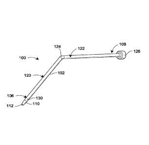
[0031] Fig. 2 illustrates a blunt cannula 100 having a cylindrical wall
102 that
defines an axial passage 104 best seen in Fig. 5. The axial passage 104
extends
between a first end 106 and a second end 108 of the blunt cannula 100. The
wall 102
has at least a first tapered region 110 at the first end 106 to define an
opening 112 in
fluid communication with an axial passage 104.
[0032] As regards the specific embodiment of the blunt cannula 100
illustrated in
Fig. 2, it will be recognized that the cannula 100 includes a first segment
120 and a
second segment 122 that are connected by a joint or transition 124. The first
segment
120 and the second segment 122 are disposed at an angle to each other, as best
seen in
Fig. 3. According to the illustrated embodiment, the first segment 120 and the
second
segment 122 are disposed at an obtuse angle relative to each other. Even
though the
segments 120, 122 are disposed at an angle to each other, the passage 104 may
still be
referred to as the axial passage. Moreover, while Figs. 1 and 2 show a cannula
with
segments disposed at an angle to each other, there may be additional
embodiments
- 5 -
CA 02884887 2015-03-12
WO 2014/081780 PCT/US2013/070929
where the segments are not at an angle to each other and the cannula could in
fact be a
single straight segment.
[0033] In some embodiments, a hub 126 is disposed at the second end 108
of the
cannula 100, while in other embodiments a hub may not be present. The hub 126
may
surround a needle or other connector used to connect the cannula 100 with a
reservoir
such that the cannula 100 and the reservoir are in fluid communication with
each
other (e.g., by having the needle pierce a rubber septum or the like). While
the
structure has been explained in regard to the illustrated embodiment, the
cannula
according to the present disclosure is not so limited, and variations may
exist to the
cannula illustrated in Figs. 2 and 3.
[0034] The first end 106 also is adapted to resist interruption of fluid
flow
through the axial passage 104 and out of the first end 106 of the blunt
cannula 100. As
will be discussed in regards to the remaining embodiments, the manner in which
the
first end 106 is adapted to resist interruption of fluid flow may vary. In
fact, while a
variety of adaptations are discussed individually in regards to Fig. 2-15, it
will be
appreciated that it is possible to combine the specific embodiments discussed
in each
of the illustrations of Figs. 2-15 with each other. For example, the
embodiments of
Figs. 2-5 or Fig. 6 may be combined with the embodiment of Fig. 7. Still
further
combinations will be apparent to those skilled in the art, and discussed
below.
[0035] As to the adaptation illustrated in Figs. 2-5, this embodiment has
at least
one pair of side ports 130 formed in the wall 102 of the blunt cannula 100 at
the first
end 106, although the present disclosure would also encompass an embodiment
that
includes at least one side port (e.g., only one side port). The at least one
pair of side
ports 130 illustrated in Figs. 2-5 are in the form of circular openings formed
in the
wall 102 of the blunt cannula 100 at the first end 106. Further, as best seen
in Fig. 5,
the circular openings formed in the wall 102 of the blunt cannula 100 are
aligned with
each other across the axial passage 104 (i.e., the circular openings lie along
a
transverse axis 132), however such alignment is not necessary. For example,
the side
ports 130 may be at different distances from the first end 106 of the cannula
100.
[0036] In regard to the two side ports 130, it will be recognized that
the wall 102
of the blunt cannula 100 has an internal surface 134 and an external surface
136. The
side ports 130 have a first opening on the internal surface 134 and a second
opening
- 6 -
CA 02884887 2015-03-12
WO 2014/081780 PCT/US2013/070929
on the external surface 136, and a passage connecting the first and second
openings.
Consequently, the side ports 130 depend through the wall 102 of the cannula
100.
[0037] As will be further recognized, the side ports 130 of the
embodiment
illustrated in Figs. 2-5 are formed in the first end 106 of the blunt cannula
100 distal
from the first tapered region 110 that defines the opening 112. In other
embodiments,
the side ports 130 may be disposed within the first tapered region 110. In
still other
embodiments, the side ports 130 are disposed further from the first tapered
region
110.
[0038] According to a specific embodiment of the present disclosure, the
side
ports may have a diameter of 0.006 inches (0.15 mm), and may be disposed 0.06
inches (1.5 mm) from the opening 112 for a 24 gauge cannula. In an alternative
embodiment, the side ports may have a diameter of 0.006 inches (0.15 mm), and
may
be disposed 0.08 inches (2 mm) from the opening 112 for a 24 gauge cannula. In
yet
other embodiments the diameter and the distance from the opening 112 may be
different than the values set forth immediately above. The ports may be formed
using
a drill, for example.
[0039] Fig. 6 illustrates a related embodiment of the present disclosure
wherein
at least one pair of side ports is formed in the wall 102 of the blunt cannula
100.
However, unlike the embodiment illustrated in Figs. 2-5, the embodiment of the
side
port illustrated in Fig. 6 is in the form of an elongated slit or an elongated
slot 140
(wherein the slit has a narrow width relative to the slot). Specifically, the
elongated
slit 140 may lie along an axis 142 that runs parallel to the axial passage
104. Further,
the elongated slit 140 may have first and second rounded ends 144. As was the
case
relative to the side ports 130, the slits 140 may have in a first opening in
the internal
surface 134 and a second opening in the external surface 136 of the blunt
cannula 100,
and a passage connecting the first and second openings. The slit may be formed
using
laser cutting tools, for example.
[0040] Figs. 7-9 illustrate a different adaptation for resisting the
interruption of
fluid flow through the axial passage 104. In particular, the embodiments
illustrated in
Figs. 7-9 involve the formation of at least one bevel in the first end 106 of
the cannula
100. In particular, the at least one bevel is formed in the first tapered
region 110 that
defines the opening 112. As a consequence, whereas the opening 112 illustrated
in the
embodiments of Figs. 2-6 may be circular in cross-section, the opening 112 of
the
- 7 -
CA 02884887 2015-03-12
WO 2014/081780
PCT/US2013/070929
embodiments illustrated in Figs. 7-10 may have at least one oblong or oval
face, and
may in fact have oval faces in multiple planes inclined relative to the axial
passage
104.
[0041] In
particular, the embodiments illustrated in Figs. 7 and 8 include only a
single bevel 150, 151, although the embodiment of Fig. 7 has the bevel across
the
entire tapered region 110 while the embodiment of Fig. 8 has the bevel across
only a
portion of the tapered region 110 (which may make the end of the embodiment of
Fig.
8 more resistant to buckling or bending as a consequent). The bevel 150. 151
may be
referred to as a positive bevel herein. In a similar vein, Fig. 9 illustrates
the use of two
positive bevels 152, 154 intersecting each other. However, it is also possible
to use an
inverted or negative bevel, as is done in Fig. 10 wherein an inverted double
bevel 156.
158 is used. As mentioned previously, any one of the embodiments illustrated
in Figs.
7-10 may be used with any one of the embodiments illustrated in Figs. 2-6 in
so far as
the side ports do not interfere with the function of the bevel or bevels
formed in the
tapered region 110.
[0042] As
illustrated, the bevel may be inclined at 45 degrees. However, it will
be recognized that other angles of bevel are possible. It will also be
recognized that
shallow angles resist buckling of the blunt cannula when the cannula is
inserted into
or through the skin of the patient.
[0043] Figs. 11-13
illustrate further adaptations for resisting the interruption of
fluid flow through the axial passage 104. In particular, the embodiments
illustrated in
Figs. 11-13 involve a pattern of openings disposed about the opening 112 in
the first
tapered region 110. The pattern of openings may be regular and periodic as
illustrated,
or the pattern of openings may be irregular (i.e., the spacing may not be
equal relative
to the openings and the intervening wall sections).
[0044] For example,
the embodiment of Fig. 11 includes a regular and periodic
pattern of openings 160 and wall sections 162. The resulting pattern may be
referred
to as castellated. Similarly, a second pattern of openings 164 is illustrated
in Fig. 12,
which openings 164 may be defined as recesses in the tapered region 110 formed
by a
series of grooves transverse to the axial passage 104. In fact, a still
further pattern of
openings 166 is illustrated in Fig. 13, wherein the first end 106 is capped
such that
fluid flow is only possible transverse to the axial passage 104 through the
openings
166. The openings 166 may be referred to as windows.
- 8 -
CA 02884887 2015-03-12
WO 2014/081780 PCT/US2013/070929
[0045] As was the case relative to the embodiments illustrated in Figs. 7-
10, the
embodiments illustrated in Figs. 11-13 may be used in combination with any of
the
embodiments illustrated in Figs. 2-5. Alternatively, the embodiments
illustrated in
Figs. 11-13 may be used in combination with any of the embodiments illustrated
in
Figs. 7-10. Still further, the embodiments illustrated in Figs. 11-13 may be
used in
combination with the embodiments of Figs. 2-6 and 7-10.
[0046] Figs 14 and 15 illustrate still further adaptations for minimizing
or
reducing the interruption of fluid flow through the axial passage 104 as well
as
resisting buckling of the cannula 100. In particular, the wall 102 of the
blunt cannula
100 may have at least one external recessed region formed thereon, the recess
being
recessed towards the axial passage 104 relative to adjoining surface regions.
This at
least one external recessed region may be in a pattern that is both regular
and periodic,
or the pattern of openings may be irregular (i.e., the spacing may not be
equal relative
to the first tapered region 110).
[0047] According to the embodiment illustrated in Fig. 14, the at least
one
recessed region 170 may be defined by a pattern of ribs 172 formed on the
external
surface 136. Alternatively, the least one recessed region 174 in Fig. 15 may
be
defined by a pattern of grooves 176 formed in the external surface 136. It may
also be
possible to define the least one recessed region using the combination of a
pattern of
ribs and a pattern of grooves in combination. It is preferred that the ribs or
grooves
terminate before the portion of the cannula 100 that depends from the
insertion site so
as to limit the possibility of leakage from the site. It is also preferred
that the ribs or
grooves be used with at least one side port or opening along the perimeter of
the
opening 112, although the ribs or grooves may be used without providing a side
port
or other opening (e.g., with a beveled end).
[0048] As was the case relative to the embodiments illustrated above, the
embodiments illustrated in Figs. 14 and 15 may be used in combination any one
of the
embodiments illustrated in Figs. 2-13. It is the case that the embodiments of
Figs. 14
and 15 may be most useful with an embodiment where a radial flow path is
provided,
such as with the provision of a side port, tip groove, or the like.
Alternatively, the
embodiments illustrated in Figs. 14 and 15 may be used in combination with any
of
the embodiments illustrated any one or more of the groups of adaptations in
Figs. 2-6,
7-10 or 11-13. Consequently, the embodiments illustrated in Figs. 14 and 15
may be
- 9 -
CA 02884887 2015-03-12
WO 2014/081780 PCT/US2013/070929
used in combination with embodiments section from each of the groups
illustrated in
Figs. 2-6, 7-10, and 11-13.
[0049] A still further adaptation according to the present disclosure is
illustrated
in Fig. 16. According to this embodiment, in addition to the reservoir 52.
cannula 54,
and controller 56, the drug delivery device 60 includes a vibration generator
62. The
operation of the vibration generator 62 may be controlled by the controller 56
to
which it is operatively coupled. In addition, the output of the vibration
generator 62
may be operatively coupled to the cannula 54. The vibration generator may be
in the
form of a motor having a shaft with an eccentric weight attached to the shaft.
Alternatively, the vibration generator may be in the form of a piezoelectric
vibrator.
Still further alternative embodiments are possible. Actuation of the vibration
generator 62 may dislodge an obstruction or blockage from the end of the
cannula 54,
move the tip of the cannula 54 away from the occluding structure (e.g.,
tissue), or
create a pocket of micro-fractured tissue increasing the surface area for
lower pressure
threshold, thereby permitting flow to resume
[0050] As was the case with the adaptations recited above, the adaptation
illustrated in Fig. 16 may be used with any one or more of the adaptations
illustrated
in Figs. 2-15.
[0051] In addition, while the previous embodiments have been discussed in
regard to a blunt cannula, certain of the above-mentioned adaptations may also
be
used with a rigid needle as well, which needle may be made of metal and have a
point
defined by one or more bevels made at a first end of the needle. The rigid
needle may
have a cylindrical wall that defines a passage between the first end and a
second end,
and the first end may have an opening that is in fluid communication with the
axial
passage. Moreover a reservoir may be connected to the second end of the rigid
needle.
[0052] In particular, the adaptations according to Figs. 11, 12 and 14-16
may be
used with the rigid needle.
[0053] As will be recognized, the devices according to the present
disclosure
may have one or more advantages relative to conventional technology, any one
or
more of which may be present in a particular embodiment in accordance with the
features of the present disclosure included in that embodiment. In particular.
each of
the embodiments illustrated in Figs. 2-15 provides alternative flow paths for
the drug
- 10 -
CA 02884887 2015-03-12
WO 2014/081780 PCT/US2013/070929
product should the opening in the first end of the blunt cannula be obstructed
or
blocked, thereby limiting the possibility of the interruption of the flow
through the
cannula. The embodiments also increase the surface area exposed, lowering the
pressure threshold required to displace tissue. The embodiment illustrated in
Fig. 16
uses a different mechanism by which to limit the interruption of flow through
the
cannula, wherein the vibrations may dislodge an obstruction or blockage from
the end
of the cannula, move the cannula tip away from the occluding structure (e.g.,
tissue),
or create a pocket of micro-fractured tissue increasing the surface area for
lower
pressure threshold, thereby permitting flow to resume. Other advantages not
specifically listed herein may also be recognized as well.
[0054] Figs. 17-20 illustrate how the systematic approach to lowering the
pressure required to pass fluid through a cannula or needle provided by the
embodiments according to the present disclosure may operate in a particular
application of this technology. In particular, this discussion is in relation
to a
wearable drug delivery device, which is one that is attached to the patient's
skin and
includes its own mechanism(s) for deploying the cannula or needle and
delivering
fluid through that cannula or needle. Because a wearable drug delivery device
(e.g.,
autoinjector, infuser or microinfuser) has a generally limited ability to
increase the
pressure at which a fluid is delivered to the patient, a variable increase in
the pressure
required to pass fluid through the cannula or needle into the tissue may
unpredictably
reduce the amount of fluid (e.g., drug) delivered to the patient or may
require that the
device be engineered to provide higher pressures, which pressures may not
always be
required. Consequently, a systematic approach to reducing the pressure
required to
deliver fluid through the cannula or needle may not only limit the need to
engineer
such a delivery device to provide higher pressures, it may ensure that a
specific
amount of fluid is consistently provided to the patient, improving the
reliability of the
device.
[0055] In particular, the resistance to fluid flow in such a wearable
device may
come about as a consequence of an effect referred to herein as "tenting" As
illustrated in Fig. 17, a wearable drug delivery device has a housing 200 with
an
adhesive layer 202 disposed on an outer surface 204 of the housing 200, which
adhesive layer 202 may include a fabric backing 201 and an adhesive 203
according
to certain embodiments. See Fig. 17 (while not specifically illustrated in
Figs. 19 and
- 11-
CA 02884887 2015-03-12
WO 2014/081780 PCT/US2013/070929
20, the adhesive layer 202 in these Figs. may be defined as illustrated in
Fig. 17 as
well). In particular, the adhesive layer 202 is disposed on the housing 200
except for
a region 206 disposed about the cannula or needle 100 (which, as illustrated,
is similar
to the embodiment illustrated in Figs. 2-5). This region 206 may be referred
to as
adhesive-free, and according to those embodiments wherein the adhesive layer
202
includes both a fabric backing 201 and an adhesive 203, the region 206 would
be free
of both the backing and the adhesive while still termed "adhesive-free."
[0056] For example, the region 206 may be adhesive-free in that an
opening 208
is formed in the housing 200. According to certain embodiments, the opening
208
may be made as small as is possible while still providing for the free passage
of the
cannula 100. For example, according to one embodiment, the opening 208 is
circular
and has a diameter that is not more than twice the diameter of the cannula 100
(by
which phrase it is also understood that the opening 208 must be greater than
the
diameter of the cannula 100 or rigid needle so that the cannula or rigid
needle may be
disposed through the opening 208 in an operative state of the wearable device
and the
cannula 100).
[0057] While the skin 210 (and associated subcutaneous tissue) to which
the drug
delivery device is attached has some degree of elasticity (which elasticity
may vary
from person to person), the adhesive layer 202 disposed about the region 206
attaches
the device (and in particular, the housing 200) to the skin surface 212 so as
to hold the
skin surface 212 at a boundary 214 substantially fixed relative to the housing
200 (see
also Fig. 18). While there may be some movement at or along this boundary 214,
it is
not substantial. As a consequence, when the cannula or needle 100 is
automatically
advanced or deployed by the action of the drug delivery device in the
direction of the
skin surface 212, the skin surface 212 starts to stretch relative to the fixed
boundary
214. Furthermore, as the cannula or needle 100 passes through the skin 210,
the
insertion of the cannula 100 causes dragging of the skin tissue 210 because of
friction
between the cannula 100 and the skin 210. This causes a "tenting" action to
occur
about the cannula or needle 100 as illustrated in Fig. 19 with reference to
numeral
216.
[0058] As a brief aside, it will be recognized that the cannula 100 in
Figs. 17-20
is illustrated disposed about a structure that is used to insert the cannula
into the skin,
and which may be removed thereafter to permit fluid to pass through the
cannula
- 12 -
CA 02884887 2015-03-12
WO 2014/081780 PCT/US2013/070929
(compare Figs. 19 and 20). This structure, which may be referred to as an
introducer
needle 220, need not be present in all embodiments according to the present
disclosure. However, according to certain embodiments, the introducer needle
220 is
disposed in the cannula 100 and is used to introduce the cannula 100 into the
skin
210, after which it may be removed from cannula 100 (see Fig. 20).
[0059] As illustrated in Fig. 20, the cannula 100 has now been inserted
into the
skin 210, and the surface 212 of the skin 210 has generally attempted to
return to its
original position relative to the outer surface 204 of the drug delivery
device housing
200. However, the friction between the cannula 100 and the skin tissue 210
prevents
the tissue from completely returning to its initial, or rest, state. As such,
the residual
forces of the skin and subcutaneous tissues against the tip of the cannula 100
increases
the pressure required to deliver fluid through the tip of the cannula 100, and
may
completely obstruct the tip of the cannula 100 as well.
[0060] According to any of the embodiments of the cannula or needle in
Figs. 2-
15, the pressure required to pass fluid into the tissue may be reduced by, for
example,
increasing the overall surface area of tissue exposed to the fluid, through
the
introduction of side ports, etc. In addition, by vibrating the tissue,
according to the
mechanism of Fig. 16, some of the residual force in the skin tissue 210 may be
released, thereby decreasing the pressure and potential for occlusion. The
embodiments of Figs. 2-15 may be used in combination with the vibration
provided
by the embodiment of Fig. 16, or the embodiments of Figs. 2-15 and 16 may be
used
separately. In any event, it is believed that the embodiments disclosed herein
may
provide a solution to the tenting of the skin caused at the time the cannula
or needle is
automatically introduced or deployed into the patient by a wearable drug
delivery
device. It is also believed that by reducing the size of the opening
(fenestration) 208
in the adhesive layer 202. there will be a reduction in tenting and associated
residual
forces.
[0061] As also mentioned above, the reservoir 52 may be filled with a
drug or
pharmaceutical product. For example, the reservoir may be filled with colony
stimulating factors, such as G-CSF. Such G-CSF agents include, but are not
limited
to. Neupogen (filgrastim) and Neulasta (pegfilgrastim).
[0062] In various other embodiments, the drug delivery device may be used
with
various pharmaceutical products, which use may or may not occur under the same
- 13 -
conditions as described above for G-CSF. These products may include, for
example,
an erythropoiesis stimulating agent (ESA), which may be in a liquid or a
lyophilized
form. An ESA is any molecule that stimulates erythropoiesis, such as Epogen
(epoetin alfa), Aranesp (darbepoetin alfa), Dynepo (epoetin delta), Mircera
(methyoxy polyethylene glycol-epoetin beta), Hematide , MRK-2578, INS-22,
Retacrit (epoetin zeta), Neorecormon (epoetin beta), Silapo (epoetin zeta),
Binocrit (epoetin alfa), epoetin alfa Hexal, Abseamed (epoetin alfa),
Ratioepo
(epoetin theta), Eporatiot (epoetin theta), Biopoin (epoetin theta), epoetin
alfa,
epoetin beta, epoetin zeta, epoetin theta, and epoetin delta, as well as the
molecules or
variants or analogs thereof as disclosed in the following patents or patent
applications:
U.S. Pat. Nos. 4,703,008; 5,441,868; 5,547,933; 5,618,698; 5,621,080;
5,756,349;
5,767,078; 5,773,569; 5,955,422; 5,986,047; 6,583,272; 7,084,245; and
7,271,689;
and PCT Publ. Nos. WO 91/05867; WO 95/05465; WO 96/40772; WO 00/24893;
WO 01/81405; and WO 2007/136752.
[0063] An ESA can be an erythropoiesis stimulating protein. As
used herein,
"erythropoiesis stimulating protein" means any protein that directly or
indirectly
causes activation of the erythropoietin receptor, for example, by binding to
and
causing dimerization of the receptor. Erythropoiesis stimulating proteins
include
erythropoietin and variants, analogs, or derivatives thereof that bind to and
activate
erythropoietin receptor; antibodies that bind to erythropoietin receptor and
activate the
receptor; or peptides that bind to and activate erythropoietin receptor.
Erythropoiesis
stimulating proteins include, but are not limited to, epoetin alfa, epoetin
beta, epoetin
delta, epoetin omega, epoetin iota, epoetin zeta, and analogs thereof,
pegylated
erythropoietin, carbamylated erythropoietin, mimetic peptides (including
EMPI/hematide), and mimetic antibodies. Exemplary erythropoiesis stimulating
proteins include erythropoietin, darbepoetin, erythropoietin agonist variants,
and
peptides or antibodies that bind and activate erythropoietin receptor (and
include
compounds reported in U.S. Publ. Nos. 2003/0215444 and 2006/0040858) as well
as
erythropoietin molecules or variants or analogs thereof as disclosed in the
following
patents or patent applications:U.S. Pat. Nos. 4,703,008; 5,441,868; 5,547,933;
5,618,698;
,
- 14 -
CA 2884887 2019-08-23
CA 02884887 2015-03-12
WO 2014/081780 PCT/US2013/070929
5,621,080; 5,756,349; 5,767,078; 5,773,569; 5,955,422; 5,830,851; 5,856,298;
5,986,047; 6,030,086; 6,310,078; 6,391,633; 6,583,272; 6,586,398; 6,900,292;
6,750,369; 7,030,226; 7,084,245; and 7,217.689; US Publ. Nos. 2002/0155998;
2003/0077753; 2003/0082749; 2003/0143202; 2004/0009902; 2004/0071694;
2004/0091961; 2004/0143857; 2004/0157293; 2004/0175379; 2004/0175824;
2004/0229318; 2004/0248815; 2004/0266690; 2005/0019914: 2005/0026834;
2005/0096461; 2005/0107297; 2005/0107591; 2005/0124045; 2005/0124564;
2005/0137329; 2005/0142642; 2005/0143292; 2005/0153879; 2005/0158822;
2005/0158832; 2005/0170457; 2005/0181359; 2005/0181482: 2005/0192211;
2005/0202538; 2005/0227289; 2005/0244409; 2006/0088906; and 2006/0111279;
and PCT Publ. Nos. WO 91/05867; WO 95/05465; WO 99/66054; WO 00/24893;
WO 01/81405; WO 00/61637; WO 01/36489; WO 02/014356; WO 02/19963; WO
02/20034; WO 02/49673; WO 02/085940; WO 03/029291; WO 2003/055526; WO
2003/084477; WO 2003/094858; WO 2004/002417; WO 2004/002424; WO
2004/009627; WO 2004/024761; WO 2004/033651; WO 2004/035603; WO
2004/043382; WO 2004/101600; WO 2004/101606; WO 2004/101611; WO
2004/106373; WO 2004/018667; WO 2005/001025; WO 2005/001136; WO
2005/021579; WO 2005/025606; WO 2005/032460; WO 2005/051327; WO
2005/063808; WO 2005/063809; WO 2005/070451; WO 2005/081687; WO
2005/084711; WO 2005/103076; WO 2005/100403; WO 2005/092369; WO
2006/50959; WO 2006/02646; and WO 2006/29094.
[0064] Examples of other pharmaceutical products for use with the device
may
include, but are not limited to, antibodies such as Vectibix0 (panitumumab),
XgevaTM
(denosumab) and ProliaTM (denosamab); other biological agents such as Enbrel0
(etanercept, TNF-receptor /Fc fusion protein, TNF blocker), Neulasta0
(pegfilgrastim, pegylated filgastrim, pegylated G-CSF, pegylated hu-Met-G-
CSF),
Neupogen (filgrastim , G-CSF, hu-MetG-CSF), and Nplate0 (romiplostim); small
molecule drugs such as Sensipar0 (cinacalcet). The device may also be used
with a
therapeutic antibody, a polypeptide, a protein or other chemical, such as an
iron, for
example, ferumoxytol, iron dextrans, ferric glyconate, and iron sucrose. The
pharmaceutical product may be in liquid form, or reconstituted from
lyophilized form.
[0065] Among particular illustrative proteins are the specific proteins
set forth
below, including fusions, fragments, analogs, variants or derivatives thereof:
- 15 -
100661 OPGL specific antibodies, peptibodies, and related proteins,
and the like
(also referred to as RANKL specific antibodies, peptibodies and the like),
including
fully humanized and human OPGL specific antibodies, particularly fully
humanized
monoclonal antibodies, including but not limited to the antibodies described
in PCT
Publ. No. WO 03/002713 and the OPGL specific antibodies and antibody related
proteins disclosed therein, particularly those having the sequences set forth
therein,
particularly, but not limited to, those denoted therein: 9H7; 18B2; 2D8; 2E11;
16E1;
and 22B3, including the OPGL specific antibodies having either the light chain
of
SEQ ID NO: 2 as set forth therein in Figure 2 and/or the heavy chain of SEQ ID
NO:4, as set forth therein in Figure 4.
100671 Myostatin binding proteins, peptibodies, and related
proteins, and the like,
including myostatin specific peptibodies, particularly those described in US
Publ. No.
2004/0181033 and PCT Publ. No. WO 2004/058988 in particular in the parts
pertinent to myostatin specific peptibodies, including but not limited to
peptibodies of
the mTN8-19 family, including those of SEQ ID NOS: 305-351, including TN8-19-1
through TN8-19-40, TN8-19 conl and TN8-19 con2; peptibodies of the mL2 family
of SEQ ID NOS: 357-383; the mL15 family of SEQ ID NOS: 384-409; the mL17
family of SEQ ID NOS: 410-438; the mL20 family of SEQ ID NOS: 439-446; the
mL21 family of SEQ ID NOS: 447-452; the mL24 family of SEQ ID NOS: 453-454;
and those of SEQ ID NOS: 615-631.
100681 IL-4 receptor specific antibodies, peptibodies, and related
proteins, and
the like, particularly those that inhibit activities mediated by binding of 1L-
4 and/or
IL-13 to the receptor, including those described in PCT Publ. No. WO
2005/047331
or PCT Appl. No. PCT/US2004/03742 and in US Publ. No. 2005/112694, in
particular in the parts pertinent to IL-4 receptor specific antibodies,
particularly such
antibodies as are described therein, particularly, and without limitation,
those
designated therein: L1H1; L1H2; L1H3; L1H4; L1H5; L1H6; L1H7; L1H8; L1H9;
L I H10; L1H11; L2H1; L2H2; L2H3; L2H4; L2H5; L2H6; L2H7; L2H8; L2H9;
L2H10; L2H11; L2H12; L2H13; L2H14;
- 16 -
CA 2884887 2019-08-23
L3H1; L4H1; L5H1; L6H1.
100691 Interleukin 1-receptor 1 ("IL1-R1") specific antibodies,
peptibodies, and
related proteins, and the like, including but not limited to those described
in U.S.
Publ. No. 2004/097712A1, in particular in the parts pertinent to ILl-R1
specific
binding proteins, monoclonal antibodies in particular, especially, without
limitation,
those designated therein: 15CA, 26F5, 27F2, 24E12, and 10H7.
[0070] Ang2 specific antibodies, peptibodies, and related
proteins, and the like,
including but not limited to those described in PCT Publ. No. WO 03/057134 and
U.S. Publ No. 2003/0229023, in particular in the parts pertinent to Ang2
specific
antibodies and peptibodies and the like, especially those of sequences
described
therein and including but not limited to: LI(N); Ll(N) WT; Ll(N) 1K WT;
2xL1(N);
2xL1(N) WT; Con4 (N), Con4 (N) 1K WT, 2xCon4 (N) 1K; L1C; Ll C 1K; 2xL IC;
Con4C; Con4C 1K; 2xCon4C IK; Con4-L1 (N); Con4-L1C; TN-12-9 (N); C17 (N);
TN8-8(N); TN8-14 (N); Con 1 (N), also including anti-Ang 2 antibodies and
formulations such as those described in PCT Publ. No. WO 2003/030833,
particularly
Ab526; Ab528; Ab531; Ab533; Ab535; Ab536; Ab537; Ab540; Ab543; Ab544;
Ab545; Ab546; A551; Ab553; Ab555; Ab558; Ab559; Ab565; AbFlAbFD; AbFE;
AbFJ; AbFK; AbG1D4; AbGC1E8; AbH1C12; AblAl; AblF; Ab1K, AblP; and AblP,
in their various permutations as described therein.
100711 NGF specific antibodies, peptibodies, and related proteins,
and the like
including, in particular, but not limited to those described in US Publ. No.
2005/0074821 and US Patent No. 6,919,426, in particular as to NGF-specific
antibodies and related proteins in this regard, including in particular, but
not limited
to, the NGF-specific antibodies therein designated 4D4, 4G6, 6H9, 7H2, 14D10
and
14D11.
- 17 -
CA 2884887 2019-08-23
100721 CD22 specific antibodies, peptibodies, and related proteins,
and the like,
such as those described in US Patent No. 5,789,554, in particular as to CD22
specific
antibodies and related proteins, particularly human CD22 specific antibodies,
such as
but not limited to humanized and fully human antibodies, including but not
limited to
humanized and fully human monoclonal antibodies, particularly including but
not
limited to human CD22 specific IgG antibodies, such as, for instance, a dimer
of a
human-mouse monoclonal hLL2 gamma-chain disulfide linked to a human-mouse
monoclonal hLL2 kappa-chain, including, but limited to, for example, the human
CD22 specific fully humanized antibody in Epratuzumab, CAS registry number
501423-23-0;
100731 IGF-I receptor specific antibodies, peptibodies, and related
proteins, and
the like, such as those described in PCT Publ. No. WO 06/069202, in particular
as to
IGF-1 receptor specific antibodies and related proteins, including but not
limited to
the IGF-1 specific antibodies therein designated L1H1, L2H2, L3H3, L4H4, L5H5,
L6H6, L7H7, L8H8, L9H9, LIOH10, LI1H11, Ll2H12, Ll3H13, Ll4H14, L15H15,
Ll6H16, Ll7H17, L18H18, L19H19, L20H20, L21H21, L22H22, L23H23, L24H24,
L25H25, L26H26, L27H27, L28H28, L29H29, L30H30, L31H31, L32H32, L33H33,
L34H34, L35H35, L36H36, L37H37, L38H38, L39H39, L40H40, L41H41, L42H42,
L43H43, L44H44, L45H45, L46H46, L47H47, L48H48, L49H49, L50H50, L51H51,
L52H52, and IGF-1R-binding fragments and derivatives thereof.
100741 Also among non-limiting examples of anti-IGF-1R antibodies
for use in
the methods and compositions of the present invention are each and all of
those
described in:
(i) US Publ. No. 2006/0040358 (published February 23, 2006),
2005/0008642 (published January 13, 2005), 2004/0228859 (published November
18,
2004), including but not limited to, for instance, antibody lA (DSMZ Deposit
No.
DSM ACC 2586), antibody 8 (DSMZ Deposit No. DSM ACC 2589), antibody 23
(DSMZ Deposit No. DSM ACC 2588) and antibody 18 as described therein;
- 18 -
CA 2884887 2019-08-23
CA 02884887 2015-03-12
WO 2014/081780 PCT/US2013/070929
(ii) PCT Publ. No. WO 06/138729 (published December 28, 2006) and
WO 05/016970 (published February 24, 2005), and Lu et al., 2004, J Biol. Chem.
279:2856-65, including but not limited to antibodies 2F8, Al2, and IMC-Al2 as
described therein;
(iii) PCT Publ. No. WO 07/012614 (published February 1, 2007), WO
07/000328 (published January 4, 2007), WO 06/013472 (published February 9,
2006),
WO 05/058967 (published June 30, 2005). and WO 03/059951 (published July 24,
2003);
(iv) US Publ. No. 2005/0084906 (published April 21, 2005), including but
not limited to antibody 7C10, chimaeric antibody C7C10, antibody h7C10,
antibody
7H2M, chimaeric antibody *7C10, antibody GM 607, humanized antibody 7C10
version 1, humanized antibody 7C10 version 2, humanized antibody 7C10 version
3,
and antibody 7H2HM, as described therein;
(v) US Publ. Nos. 2005/0249728 (published November 10, 2005),
2005/0186203 (published August 25, 2005), 2004/0265307 (published December 30,
2004), and 2003/0235582 (published December 25, 2003) and Maloney et al.,
2003,
Cancer Res. 63:5073-83, including but not limited to antibody EM164.
resurfaced
EM164, humanized EM164, huEM164 v1.0, huEM164 v1.1, huEM164 v1.2, and
huEM164 v1.3 as described therein;
(vi) US Pat. No. 7,037,498 (issued May 2, 2006), US Publ. Nos.
2005/0244408 (published November 30, 2005) and 2004/0086503 (published May 6,
2004), and Cohen, et al., 2005, Clinical Cancer Res. 11:2063-73, e.g.,
antibody CP-
751,871, including but not limited to each of the antibodies produced by the
hybridomas having the ATCC accession numbers PTA-2792, PTA-2788, PTA-2790,
PTA-2791, PTA-2789, PTA-2793, and antibodies 2.12.1, 2.13.2, 2.14.3, 3.1.1.
4.9.2,
and 4.17.3, as described therein;
(vii) US Publ. Nos. 2005/0136063 (published June 23, 2005) and
2004/0018191 (published January 29, 2004), including but not limited to
antibody
19D12 and an antibody comprising a heavy chain encoded by a polynucleotide in
plasmid 15H12/19D12 HCA (y4), deposited at the ATCC under number PTA-5214,
and a light chain encoded by a polynucleotide in plasmid 15H12/19D12 LCF (lc),
deposited at the ATCC under number PTA-5220, as described therein; and
- 19 -
(viii) US Publ. No. 2004/0202655 (published October 14, 2004),
including but not limited to antibodies PINT-6A1, PINT-7A2, PINT-7A4, PINT-
7A5,
PINT-7A6, PINT-8A1, PINT-9A2, PINT-11A1, PINT-11A2, PINT-11A3, PINT-
11A4, PINT-11A5, PINT-11A7, PINT-11Al2, PINT-12A1, PINT-12A2, PINT-
12A3, PINT-12A4, and PINT-12A5, as described therein, particularly as to the
aforementioned antibodies, peptibodies, and related proteins and the like that
target
IGF-1 receptors;
[0075] B-7 related protein 1 specific antibodies, peptibodies,
related proteins and
the like ("B7RP-1,- also is referred to in the literature as B7H2, ICOSL, B7h,
and
CD275), particularly B7RP-specific fully human monoclonal IgG2 antibodies,
particularly fully human IgG2 monoclonal antibody that binds an epitope in the
first
immunoglobulin-like domain of B7RP-1, especially those that inhibit the
interaction
of B7RP-1 with its natural receptor, ICOS, on activated T cells in particular,
especially, in all of the foregoing regards, those disclosed in U.S. Publ. No.
2008/0166352 and PCT Publ. No. WO 07/011941, in particular as to such
antibodies
and related proteins, including but not limited to antibodies designated
therein as
follow: 16H (having light chain variable and heavy chain variable sequences
SEQ ID
NO:1 and SEQ ID NO:7 respectively therein); 5D (having light chain variable
and
heavy chain variable sequences SEQ ID NO:2 and SEQ ID NO:9 respectively
therein); 214 (having light chain variable and heavy chain variable sequences
SEQ ID
NO:3 and SEQ ID NO:10 respectively therein); 43H (having light chain variable
and
heavy chain variable sequences SEQ ID NO:6 and SEQ ID NO:14 respectively
therein); 41H (having light chain variable and heavy chain variable sequences
SEQ ID
NO:5 and SEQ ID NO:13 respectively therein); and 15H (having light chain
variable
and heavy chain variable sequences SEQ ID NO:4 and SEQ ID NO:12 respectively
therein);
[0076] IL-15 specific antibodies, peptibodies, and related
proteins, and the like,
such as, in particular, humanized monoclonal antibodies, particularly
antibodies such
as those disclosed in U.S. Publ. Nos. 2003/0138421; 2003/023586; and
2004/0071702; and US Patent No. 7,153,507, in particular as to IL-15 specific
- 20 -
CA 2884887 2019-08-23
antibodies and related proteins, including peptibodies, including
particularly, for
instance, but not limited to, HuMax IL-15 antibodies and related proteins,
such as, for
instance, 146B7;
[0077] IFN
gamma specific antibodies, peptibodies, and related proteins and the
like, especially human IFN gamma specific antibodies, particularly fully human
anti-
IFN gamma antibodies, such as, for instance, those described in US Pub!. No.
2005/0004353, in particular as to IFN gamma specific antibodies, particularly,
for
example, the antibodies therein designated 1118; 1118*; 1119; 1121; and 1121*.
The
entire sequences of the heavy and light chains of each of these antibodies, as
well as
the sequences of their heavy and light chain variable regions and
complementarity
determining regions, are each individually and specifically described in the
foregoing
US Publication and in Thakur etal., Mol. Immunol. 36:1107-1115 (1999). In
addition, description of the properties of these antibodies provided in the
foregoing
US publication. Specific antibodies include those having the heavy chain of
SEQ ID
NO: 17 and the light chain of SEQ ID NO:18; those having the heavy chain
variable
region of SEQ ID NO:6 and the light chain variable region of SEQ ID NO:8;
those
having the heavy chain of SEQ ID NO:19 and the light chain of SEQ ID NO:20;
those
having the heavy chain variable region of SEQ ID NO:10 and the light chain
variable
region of SEQ ID NO:12; those having the heavy chain of SEQ ID NO:32 and the
light chain of SEQ ID NO:20; those having the heavy chain variable region of
SEQ
ID NO:30 and the light chain variable region of SEQ ID NO:12; those having the
heavy chain sequence of SEQ ID NO:21 and the light chain sequence of SEQ ID
NO:22; those having the heavy chain variable region of SEQ ID NO:14 and the
light
chain variable region of SEQ ID NO:16; those having the heavy chain of SEQ ID
NO:21 and the light chain of SEQ ID NO:33; and those having the heavy chain
variable region of SEQ ID NO:14 and the light chain variable region of SEQ ID
NO:31, as disclosed in the foregoing US Publication. A specific antibody
contemplated is antibody 1119 as disclosed in foregoing US Publication and
having a
complete heavy chain of SEQ ID NO:17 as disclosed therein and having a
complete
light chain of SEQ ID NO:18 as disclosed therein;
-21 -
CA 2884887 2019-08-23
100781 TALL-1 specific antibodies, peptibodies, and the related
proteins, and the
like, and other TALL specific binding proteins, such as those described in
U.S. Publ.
Nos. 2003/0195156 and 2006/0135431, in particular as to TALL-1 binding
proteins,
particularly the molecules of Tables 4 and 5B, as disclosed in the foregoing
US
Publications;
[0079] Parathyroid hormone ("PTH") specific antibodies,
peptibodies, and
related proteins, and the like, such as those described in US Patent No.
6,756,480, in
particular in the parts pertinent to proteins that bind PTH;
[0080] Thrombopoietin receptor ("TPO-R") specific antibodies,
peptibodies, and
related proteins, and the like, such as those described in US Patent No.
6,835,809, in
particular in the parts pertinent to proteins that bind TPO-R;
[0081] Hepatocyte growth factor ("HGF") specific antibodies,
peptibodies, and
related proteins, and the like, including those that target the HGF/SF:cMet
axis
(HGF/SF:c-Met), such as the fully human monoclonal antibodies that neutralize
hepatocyte growth factor/scatter (HGF/SF) described in US Publ. No.
2005/0118643
and PCT Publ. No. WO 2005/017107, huL2G7 described in US Patent No. 7,220,410
and 0A-5d5 described in US Patent Nos. 5,686,292 and 6,468,529 and in PCT
Publ.
No. WO 96/38557, in particular in the parts pertinent to proteins that bind
HGF;
100821 TRAIL-R2 specific antibodies, peptibodies, related proteins
and the like,
such as those described in US Patent No. 7,521,048, in particular in the parts
pertinent
to proteins that bind TRAIL-R2;
[0083] Activin A specific antibodies, peptibodies, related
proteins, and the like,
including but not limited to those described in US Publ. No. 2009/0234106, in
particular in the parts pertinent to proteins that bind Activin A;
100841 TGF-beta specific antibodies, peptibodies, related proteins,
and the like,
including but not limited to those described in US Patent No. 6,803,453 and US
Publ.
- 22 -
CA 2884887 2019-08-23
No. 2007/0110747, in particular in the parts pertinent to proteins that bind
TGF-beta;
100851 Amyloid-beta protein specific antibodies, peptibodies,
related proteins,
and the like, including but not limited to those described in PCT Pub!. No. WO
2006/081171, in particular in the parts pertinent to proteins that bind
amyloid-beta
proteins. One antibody contemplated is an antibody having a heavy chain
variable
region comprising SEQ ID NO: 8 and a light chain variable region having SEQ ID
NO: 6 as disclosed in the International Publication;
100861 c-Kit specific antibodies, peptibodies, related proteins,
and the like,
including but not limited to those described in Publ. No. 2007/0253951, in
particular
in the parts pertinent to proteins that bind c-Kit and/or other stem cell
factor receptors;
100871 OX4OL specific antibodies, peptibodies, related proteins,
and the like,
including but not limited to those described in US Appl. No. 11/068,289, in
particular
in the parts pertinent to proteins that bind OX4OL and/or other ligands of the
0X040
receptor; and
100881 Other exemplary proteins, including Activase (alteplase,
tPA);
Aranesp (darbepoetin alfa); Epogene (epoetin alfa, or erythropoietin); GLP-1,
Avonex (interferon beta-la); Bexxar0 (tositumomab, anti-CD22 monoclonal
antibody); Betaseron (interferon-beta); Campath (alemtuzumab, anti-CD52
monoclonal antibody); Dynepo (epoetin delta); Velcade (bortezomib); MLN0002
(anti- a4f37 mAb); MLN1202 (anti-CCR2 chemokine receptor mAb); Enbrel
(etanercept, 'TNF-receptor /Fc fusion protein, TNF blocker); Eprex0 (epoetin
alfa);
Erbitux (cetuximab, anti-EGFR / HER1 / c-ErbB-1); Genotropin (somatropin,
Human Growth Hormone); Herceptin (trastuzumab, anti-HER2/neu (erbB2)
receptor mAb); Humatrope (somatropin, Human Growth Hormone); Humira
(adalimumab); insulin in solution; Infergen (interferon alfacon-1); Natrecor
(nesiritide; recombinant human B-type natriuretic peptide (hBNP); Kineret
(anakinra); Leukine (sargamostim, rhuGM-CSF); LymphoCide (epratuzumab,
anti-CD22 mAb); BenlystaTM (lymphostat B, belimumab, anti-BlyS mAb);
Metalyse (tenecteplase, t-PA analog); Mircera0 (methoxy polyethylene glycol-
epoetin beta); Mylotarg (gemtuzumab ozogamicin); Raptiva (efalizumab);
- 23 -
CA 2884887 2019-08-23
CA 02884887 2015-03-12
WO 2014/081780 PCT/US2013/070929
Cimzia0 (certolizumab pegol, CDP 870); SolirisTM (eculizumab); pexelizumab
(anti-
05 complement); Numax (MEDI-524); Lucentis (ranibizumab); Panorex (17-
1A, edrecolomab); Trabio0 (lerdelimumab); TheraCim hR3 (nimotuzumab);
Omnitarg (pertuzumab, 2C4); Osidem (IDM-1); OvaRex (B43.13); Nuvion
(visilizumab); cantuzumab mertansine (huC242-DM1); NeoRecormon0 (epoetin
beta); Neumega0 (oprelvekin, human interleukin-11); Neulasta0 (pegylated
filgastrim, pegylated G-CSF, pegylated hu-Met-G-CSF); Neupogen0 (filgrastim .
G-
CSF. hu-MetG-CSF); Orthoclone OKT30 (muromonab-CD3, anti-CD3 monoclonal
antibody); Procrit (epoetin alfa); Remicade0 (infliximab, anti-TNFa
monoclonal
antibody); Reopro0 (abciximab, anti-GP 1Ib/Ilia receptor monoclonal antibody);
Actemra0 (anti-IL6 Receptor mAb); Avastin0 (bevacizumab), HuMax-CD4
(zanolimumab); Rituxan0 (rituximab, anti-CD20 mAb); Tarceva0 (erlotinib);
Roferon-A0-(interferon alfa-2a); Simulect0 (basiliximab); Prexige0
(lumiracoxib);
Synagis0 (palivizumab); 146B7-CHO (anti-IL15 antibody, see US Patent No.
7,153,507); Tysabri (natalizumab, anti-a4integrin mAb); Valortim (MDX-1303,
anti-B. anthracis protective antigen mAb); ABthraxTM; Vectibix (panitumumab);
Xolair (omalizumab); ETI211 (anti-MRSA mAb); IL-1 trap (the Fc portion of
human IgG1 and the extracellular domains of both IL-1 receptor components (the
Type I receptor and receptor accessory protein)); VEGF trap (12 domains of
VEGFR1
fused to IgG1 Fe); Zenapax0 (daclizumab); Zenapax (daclizumab, anti-IL-2Ra
mAb); Zevalin0 (ibritumomab tiuxetan); Zetia0 (ezetimibe): Orencia0
(atacicept,
TACI-Ig); anti-CD80 monoclonal antibody (galiximab); anti-CD23 mAb
(lumiliximab); BR2-Fc (huBR3 / huFc fusion protein, soluble BAFF antagonist);
CNTO 148 (golimumab, anti-TNFa mAb); HGS-ETR1 (mapatumumab; human anti-
TRAIL Receptor-1 mAb); HuMax-CD20 (ocrelizumab, anti-CD20 human mAb);
HuMax-EGFR (zalutumumab); M200 (volociximab, anti-a5131 integrin mAb); MDX-
010 (ipilimumab, anti-CTLA-4 mAb and VEGFR-1 (IMC-18F1); anti-BR3 mAb;
anti-C. difficile Toxin A and Toxin B C mAbs MDX-066 (CDA-1) and MDX-1388);
anti-CD22 dsFv-PE38 conjugates (CAT-3888 and CAT-8015); anti-CD25 mAb
(HuMax-TAC); anti-CD3 mAb (NI-0401); adecatumumab; anti-CD30 mAb (MDX-
060); MDX-1333 (anti-IFNAR); anti-CD38 mAb (HuMax CD38); anti-CD4OL mAb;
anti-Cripto mAb; anti-CTGF Idiopathic Pulmonary Fibrosis Phase I Fibrogen (FG-
3019); anti-CTLA4 mAb; anti-eotaxinl mAb (CAT-213); anti-FGF8 mAb; anti-
- 24 -
ganglioside GD2 mAb; anti-ganglioside GM2 mAb; anti-GDF-8 human mAb (MY0-
029); anti-GM-CSF Receptor mAb (CAM-3001); anti-HepC mAb (HuMax HepC);
anti-IFNa mAb (MEDI-545, MDX-1103); anti-IGF1R mAb; anti-IGF-1R mAb
(HuMax-Inflam); anti-IL12 mAb (ABT-874); anti-IL12/1L23 mAb (CNTO 1275);
anti-IL13 mAb (CAT-354); anti-IL2Ra mAb (HuMax-TAC); anti-IL5 Receptor mAb;
anti-integrin receptors mAb (MDX-018, CNTO 95); anti-IP10 Ulcerative Colitis
mAb
(MDX-1100); anti-LLY antibody; BMS-66513; anti-Mannose Receptor/hCGB mAb
(MDX-1307); anti-mesothelin dsFv-PE38 conjugate (CAT-5001); anti-PD1mAb
(MDX-1106 (ONO-4538)); anti-PDGFRa antibody (IMC-3G3); anti-TGFB mAb
(GC-1008); anti-TRAIL Receptor-2 human mAb (HGS-ETR2); anti-TWEAK mAb;
anti-VEGFR/Flt-1 mAb; anti-ZP3 mAb (HuMax-ZP3); NVS Antibody #1; and NVS
Antibody #2.
100891 Also included can be a sclerostin antibody, such as but not
limited to
romosozumab, blosozumab, or BPS 804 (Novartis). Further included can be
therapeutics such as rilotumumab, bixalomer, trebananib, ganitumab,
conatumumab,
motesanib diphosphate, brodalumab, vidupiprant, panitumumab, denosumab,
NPLATE, PROLIA, VECTIBIX or XGEVA. Additionally, included in the device
can be a monoclonal antibody (IgG) that binds human Proprotein Convertase
Subtilisin/Kexin Type 9 (PCSK9), e.g. US 8,030,547, US13/469,032,
W02008/057457, W02008/057458, W02008/057459, W02008/063382,
W02008/133647, W02009/100297, W02009/100318, W02011/037791,
W02011/053759, W02011/053783, W02008/125623, W02011/072263,
W02009/055783, W02012/0544438, W02010/029513, W02011/111007,
W02010/077854, W02012/088313, W02012/101251, W02012/101252,
W02012/101253, W02012/109530, and W02001/031007.
100901 Although the preceding text sets forth a detailed
description of different
embodiments of the invention, it should be understood that the legal scope of
the
invention is defined by the words of the claims set forth at the end of this
patent. The
detailed description is to be construed as exemplary only and does not
describe every
possible embodiment of the invention because describing every possible
embodiment
would be impractical, if not impossible. Numerous alternative embodiments
could be
implemented, using either current technology or technology developed after the
filing
- 25 -
CA 2884887 2019-08-23
date of this patent.
100911 It should also be understood that, unless a term is
expressly defined in this
patent using the sentence "As used herein, the term ' __ ' is hereby defined
to
mean..." or a similar sentence, there is no intent to limit the meaning of
that term,
either expressly or by implication, beyond its plain or ordinary meaning, and
such
term should not be interpreted to be limited in scope based on any statement
made in
any section of this patent (other than the language of the claims). To the
extent that
any term recited in the claims at the end of this patent is referred to in
this patent in a
manner consistent with a single meaning, that is done for sake of clarity only
so as to
not confuse the reader, and it is not intended that such claim term be
limited, by
implication or otherwise, to that single meaning.
- 26 -
CA 2884887 2019-08-23