Note: Descriptions are shown in the official language in which they were submitted.
- 1 -
SURGICAL TRAINING MODEL FOR TRANSLUMINAL LAPAROSCOPIC
PROCEDURES
[0001]
FIELD OF THE INVENTION
[0002] This application is generally related to surgical training
tools, and in
particular, to simulated tissue structures and models for teaching and
practicing
various surgical techniques and procedures related but not limited to
laparoscopic,
endoscopic and minimally invasive surgery.
BACKGROUND OF THE INVENTION
[0003] Medical students as well as experienced doctors learning new
surgical techniques must undergo extensive training before they are qualified
to
perform surgery on human patients. The training must teach proper techniques
employing various medical devices for cutting, penetrating, clamping,
grasping,
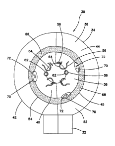
stapling, cauterizing and suturing a variety of tissue types. The range of
possibilities
that a trainee may encounter is great. For example, different organs and
patient
anatomies and diseases are presented. The thickness and consistency of the
various tissue layers will also vary from one part of the body to the next and
from one
patient to another. Different procedures demand different skills. Furthermore,
the
trainee must practice techniques in various anatomical environs that are
influenced
by factors such as the size and condition of the patient, the adjacent
anatomical
landscape and the types of targeted tissues and whether they are readily
accessible
or relatively inaccessible.
[0004] Numerous teaching aids, trainers, simulators and model organs
are
available for one or more aspects of surgical training. However, there is a
need for
CA 2885314 2020-02-26
CA 02885314 2015-03-18
WO 2014/052808
PCT/US2013/062269
- 2 -
model organs or simulated tissue elements that are likely to be encountered in
and that
can be used for practicing endoscopic and laparoscopic, minimally invasive
surgical
procedures. In laparoscopic surgery, a trocar or cannula is inserted to access
a body
cavity and to create a channel for the insertion of a camera such as a
laparoscope. The
camera provides a live video feed capturing images that are then displayed to
the
surgeon on one or more monitors. Another trocar/cannula is inserted to create
a
pathway through which surgical instruments are passed for performing
procedures
observed on the monitor. The targeted tissue location such as the abdomen is
typically
enlarged by delivering carbon dioxide gas to insufflate the body cavity and
create a
working space large enough to accommodate the scope and instruments used by
the
surgeon. The insufflation pressure in the tissue cavity is maintained by using
specialized trocars. Laparoscopic surgery offers a number of advantages when
compared with an open procedure. These advantages include reduced pain,
reduced
blood and shorter recovery times.
[0005]
Laparoscopic or endoscopic minimally invasive surgery requires an
increased level of skill compared to open surgery because the target tissue is
not
directly observed by the clinician. The target tissue is observed on monitors
displaying
a portion of the surgical site that is accessed through a small opening.
Therefore,
clinicians need to practice visually determining tissue planes, three-
dimensional depth
perception on a two-dimensional viewing screen, hand-to-hand transfer of
instruments,
suturing, precision cutting and tissue and instrument manipulation. Typically,
models
simulating a particular anatomy or procedure are placed in a simulated pelvic
trainer
where the anatomical model is obscured from direct visualization by the
practitioner.
Ports in the trainer are employed for passing instruments to practice
techniques on the
anatomical model hidden from direct visualization. Simulated pelvic trainers
provide a
functional, inexpensive and practical means to train surgeons and residents
the basic
skills and typical techniques used in endoscopic and laparoscopic minimally
invasive
surgery such as grasping, manipulating, cutting, tying knots, suturing,
stapling,
cauterizing as well as how to perform specific surgical procedures that
utilized these
basic skills. Simulated pelvic trainers are also effective sales tools for
demonstrating
medical devices required to perform these laparoscopic procedures.
CA 02885314 2015-03-18
WO 2014/052808 PCT/US2013/062269
- 3 -
[0006] Some procedures are required to be performed within small
confines,
such as a rectum, and substantially along an axis such as in transanal
endoscopic
micro-surgery (TEMS) also known as transanal minimally invasive surgery
(TAMIS) or
other transluminal surgeries generally performed to resect benign and
malignant lesions
in the distal to proximal rectum using transanal access platforms and standard
laparoscopic instrumentation. These procedures require the clinician to
develop skills
such as three-dimensional depth perception along the lumen, determining tissue
planes
and hand-to-hand transfer, in addition to suturing, cauterizing, stapling,
tying knots,
cutting, grasping, manipulating instruments and moving tissue all performed
within the
small confines of elongate tubular region while observing such procedures on a
two-
dimensional video monitor. Therefore, it is desirable to present a model
suitable for
practicing these skills and that also isolates a particular step of a
procedure for the
trainee such as the passing of sutures for the clinician to practice in a
simulated
laparoscopic environment. The laparoscopic training model is removably placed
inside
a simulated laparoscopic environment such as a laparoscopic trainer in which
it is at
least partially obscured from direct visualization. A camera and monitor
provide
visualization to the practitioner. After a technique is practiced, it is
furthermore
desirable that such a model permits repeatable practice with ease, speed and
cost
savings. In view of the above, it is an object of this invention to provide a
surgical
training device that realistically simulates an anatomy and isolates a
particular stage or
step of a procedure that also enables repeatable practice. It has been
demonstrated
that the use of simulation trainers greatly enhances the skill levels of new
laparoscopists
and are a great tool to train future surgeons in a non-surgical setting. There
is a need
for such improved, realistic and effective surgical training models.
SUMMARY OF THE INVENTION
[0007] According to one aspect of the invention, a surgical training
device is
provided. The device includes a top cover connected to and spaced apart from
the
base to define an internal cavity between the top cover and the base. At least
one
aperture, side opening, or a penetrable tissue simulation region is provided
for
accessing the internal cavity. A camera is disposed inside the cavity and
configured to
CA 02885314 2015-03-18
WO 2014/052808 PCT/US2013/062269
- 4 -
display video images on a video monitor connected to the camera. The video
monitor is
located outside of the cavity. A model is removably disposed inside the cavity
such that
the model is substantially obscured from view yet observable via the camera
displaying
images of the model on the video monitor. The model includes a body having an
outer
surface and an inner surface. The inner surface defines an elongate lumen
having an
open proximal end. A plurality of eyelets is connected to the inner surface of
the lumen
and distributed along the longitudinal axis. The plurality of eyelets forms at
least one
pathway for practicing the passing of at least one needle and suture through
the eyelets
of the pathway.
[0008] According to another aspect of the invention, a surgical training
device
is provided. The device includes an elongate body having an inner surface and
an
outer surface. The inner surface defines a lumen with a proximal opening and a
longitudinal axis. A plurality of eyelets is connected to the inner surface of
the lumen.
The eyelets extend inwardly from the inner surface into the lumen and are
spaced apart
circumferentially and longitudinally along the lumen. Each eyelet has a head
portion
with an aperture sized for passing a suture needle and suture. The aperture of
each
eyelet defines an aperture plane.
[0009] According to another aspect of the invention, a surgical training
device
for the practice of laparoscopic suture passing along an enclosed lumen is
provided. A
practice model is disposed inside a cavity of a laparoscopic trainer. The
model includes
a body with an elongate sidewall having an inner surface defining an internal
lumen with
an open proximal end. A plurality of eyelets is connected to the inner surface
of the
lumen. The eyelets extend into the lumen from the inner surface. Each eyelet
includes
a head portion connected to a neck portion. The neck portion is connected to
the inner
surface. The head portion has an aperture defining an aperture plane. The open
proximal end is configured for inserting a suture and suture needle into the
lumen and
through one or more apertures of the plurality of eyelets that are spaced
longitudinally
and circumferentially along the lumen.
[0010] According to another aspect of the invention, a surgical training
device
is provided. The device includes a body having an outer surface and an inner
surface.
The inner surface defines an elongate lumen having an open proximal end. A
plurality
CA 02885314 2015-03-18
WO 2014/052808 PCT/US2013/062269
- 5 -
of eyelets is connected to and distributed longitudinally along the inner
surface of the
lumen. At least one of the eyelets includes a hook-like feature. A staging
area at one
end of the lumen is provided. The staging area has at least one object
removably
located in the staging area. The at least one object includes an aperture
sized to fit
over the hook-like feature of at least one of the eyelets. The at least one
object is
configured to be removable from the staging area and movable along a length
inside the
lumen and onto the hook-like feature.
BRIEF DESCRIPTION OF THE DRAWINGS
[0011] FIG. 1 illustrates a top perspective view of a surgical training
device
according to the present invention.
[0012] FIG. 2 illustrates a top perspective view of a model according to
the
present invention.
[0013] FIG. 3 illustrates a proximal end view of a model according to
the
present invention.
[0014] FIG. 4 illustrates a distal end view of a model according to the
present
invention.
[0015] FIGs. 5A-5D illustrate various eyelets according to the present
invention.
DETAILED DESCRIPTION OF THE INVENTION
[0016] A surgical training device 10 that is configured to mimic the
torso of a
patient such as the abdominal region is shown in FIG. 1. The surgical training
device
provides a body cavity 12 substantially obscured from the user for receiving
simulated or live tissue or model organs or training model of the like
described in this
invention. The body cavity 12 is accessed via a tissue simulation region 14
that is
penetrated by the user employing devices to practice surgical techniques on
the tissue
or organ model found located in the body cavity 12. Although the body cavity
12 is
shown to be accessible through a tissue simulation region, a hand-assisted
access
device or single-site port device may be alternatively employed to access the
body
cavity 12. An exemplary surgical training device is described in U.S. Patent
Application
- 6 -
Serial No. 13/248,449 entitled "Portable Laparoscopic Trainer" filed on
September 29,
2011. The surgical training device 10 is particularly well suited for
practicing
laparoscopic or other minimally invasive surgical procedures.
[0017] Still referencing FIG. 1, the surgical training device 10
includes a top
cover 16 connected to and spaced apart from a base 18 by at least one leg 20.
FIG. 1
shows a plurality of legs 20. The surgical training device 10 is configured to
mimic the
torso of a patient such as the abdominal region. The top cover 16 is
representative of
the anterior surface of the patient and the space between the top cover 16 and
the base
18 is representative of an interior of the patient or body cavity where organs
reside. The
surgical trainer 10 is a useful tool for teaching, practicing and
demonstrating various
surgical procedures and their related instruments in simulation of a patient
undergoing a
surgical procedure. Surgical instruments are inserted into the cavity 12
through the
tissue simulation region 14 as well as through pre-established apertures 22 in
the top
cover 16. Various tools and techniques may be used to penetrate the top cover
16 to
perform mock procedures on model organs or training tools placed between the
top
cover 16 and the base 18. The base 18 includes a model-receiving area 24 or
tray for
staging or holding a simulated tissue model. The model-receiving area 24 of
the base
18 includes frame-like elements for holding the model (not shown) in place. To
help
retain the simulated tissue model on the base 18, a clip attached to a
retractable wire is
provided at locations 26. The retractable wire is extended and then clipped to
hold the
tissue model in position substantially beneath the tissue simulation region
14. Other
means for retaining the tissue model include a patch of hook-and-loop type
fastening
material (VELCRO ) affixed to the base 18 in the model receiving area 24 such
that it is
removably connectable to a complementary piece of hook-and-loop type fastening
material (VELCRO ) affixed to the model.
[0018] A video display monitor 28 that is hinged to the top cover 16
is shown
in a closed orientation in FIG. 1. The video monitor 62 is connectable to a
variety Of
visual systems for delivering an image to the monitor. For example, a
laparoscope
inserted through one of the pre-established apertures 22 or a webcam located
in the
cavity and used to observe the simulated procedure can be connected to the
video
CA 2885314 2020-02-26
CA 02885314 2015-03-18
WO 2014/052808 PCT/US2013/062269
- 7 -
monitor 28 and/or a mobile computing device to provide an image to the user.
Also,
audio recording or delivery means may also be provided and integrated with the
trainer
to provide audio and visual capabilities. Means for connecting a portable
memory
storage device such as a flash drive, smart phone, digital audio or video
player, or other
digital mobile device is also provided, to record training procedures and/or
play back
pre-recorded videos on the monitor for demonstration purposes. Of course,
connection
means for providing an audio visual output to a larger screen other than the
monitor is
provided. In another variation, the top cover 10 does not include a video
display but
includes means for connecting with a laptop computer, a mobile digital device
or tablet
such as an RAD and connecting it by wire or wirelessly to the trainer.
[0019] When assembled, the top cover 16 is positioned directly above the
base 18 with the legs 20 located substantially around the periphery and
interconnected
between the top cover 16 and base 18. The top cover 16 and base 18 are
substantially
the same shape and size and have substantially the same peripheral outline.
The
internal cavity is partially or entirely obscured from view. In the variation
shown in FIG.
1, the legs include openings to allow ambient light to illuminate the internal
cavity as
much as possible and also to advantageously provide as much weight reduction
as
possible for convenient portability. The top cover 16 is removable from the
legs 20
which in turn are removable or collapsible via hinges or the like with respect
to the base
18. Therefore, the unassembled trainer 10 has a reduced height that makes for
easier
portability. In essence, the surgical trainer 10 provides a simulated body
cavity 12 that
is obscured from the user. The body cavity 12 is configured to receive at
least one
surgical model accessible via at least one tissue simulation region 14 and/or
apertures
22 in the top cover 16 through which the user may access the models to
practice
laparoscopic or endoscopic minimally invasive surgical techniques. The model
may
also be accessed through the side openings of the trainer.
[0020] A model 30 for the practice of passing sutures in laparoscopic,
endoscopic or other minimally invasive procedures according to the present
invention is
shown in FIG. 2. The model 30 is configured to be placed inside the body
cavity 12 of
the surgical training device 10 described above or other similar surgical
trainer. The
model 30 includes a base 32 connected to a body 34.
CA 02885314 2015-03-18
WO 2014/052808 PCT/US2013/062269
- 8 -
[0021] The base 32 of the model 30 is a platform that serves as a bottom
support for the rest of the model 30 and it is sized and configured such that
the model
30 does not tip over. The platform is made of any material such as metal or
plastic.
The base 32 is of sufficient heft to maintain the stability of the model 30 in
the upright
position while being manipulated by a user. The model 30 is sized and
configured to be
placed into the body cavity 12 of the surgical trainer 10 in the location of
the model
receiving area 24. The underside of the base 32 is provided with means to
affix the
model 30 inside the surgical trainer 10. Such means to affix the model 30
inside the
trainer 10 include but are not limited to adhesive, suction cup, magnet, snap-
fit, and a
hook-and-loop type fastener material attached to the bottom surface of the
base 32 and
configured to connect with a complementary hook-and-loop type fastener
material or
adhesive attached to the base 18 of the surgical trainer 30. Alternatively,
the model 30
may be used as a stand alone trainer without and outside of the trainer 10.
[0022] Referencing FIGs. 2-4, the body 34 of the model 30 is connected
to the
base 32 or integrally formed with the base 32 such that the body 34 is
supported with
the base 32 in contact with a supporting surface such as a table top and may
be
configured to have an adjustable angle with respect to the table top or base
32. The
body 34 is substantially cylindrical in shape defining an inner lumen 36. In
one
variation, the inner lumen 36 has a diameter of approximately 3-4 inches. The
body 34
includes a sidewall 38 having an inner surface 40 and an outer surface 42
defining a
thickness therebetween and extending along a longitudinal axis between a
proximal end
44 having a proximal opening 45 and a distal end 46 having a distal opening
47. The
tubular body 34 is made of rigid plastic material that may be translucent or
transparent
permitting light to enter into the lumen through the wall 38 to illuminate the
interior
lumen 36. The tubular body 34 may also be a flexible shell and opaque as well
as
translucent. The body 34 has a cylindrical lumen 36 with a circular cross-
section,
however, the invention is not so limited and the lumen may be any cross-
sectional
shape. The body 34 may consist of two halves which may snap together or be
hinged
together such that the inner lumen 36 is easily accessible. The body 34 may
include
structures and shapes or additional base elements that allow the body 34 to be
placed
upright or supported at any angle. The body 34 defines a reduced cross-
sectional area
- 9 -
at a proximal end portion 48 and at a distal end portion 50. These end
portions 48, 50
are configured to allow the addition of various end caps and closures, such as
the
GELPOINT by Applied Medical Resources Corporation in California, and to
provide a
smaller entryway into the lumen 36. In one variation, the body 34 includes a
removable
sphincter insert 52 to simulate an anus which is attached to the proximal end
portion 48.
The sphincter insert 52 is typically made of silicone to provide a realistic
soft and flexible
tissue-like interface or entryway to the lumen 36. The sphincter insert 52 is
insertable
into the proximal opening 45 and includes an aperture 54 generally coaxial
with the
lumen 36 of the body 34. An access device (not shown) may also be provided and
connected to the body 34 at the proximal end 44 by being inserted into the
sphincter
insert 52 if is one is used or directly into the proximal opening 45 of the
body 34. The
access device seals the proximal opening of the lumen 36 and provides a
simulated
insufflation port. Hand-access devices, single-port devices and retraction
devices all of
which can be used with the model 30 are disclosed in greater detail in U.S.
Patent No.
7,473,221, U.S. Patent No. 6,958,037, U.S. Patent No. 7,650,887, U.S.
Published
Patent Application No. 2009-0187079, and U.S. Published Patent Application No.
2010-
0094227. Also, other simulated tissue structures made of silicone may be
placed at the
proximal end 44 or distal end 46 covering at least partially or
circumferentially the
proximal opening 45 or distal opening 47 or along the inner lumen 36 such that
the
simulated tissue structures in the form of membranes, for example, would have
to be
retracted in order to access other parts of the inner lumen 36.
[0023] The model 30 includes a plurality of eyelets 56 connected to
and
spaced around and along the inner surface 40 of the body 34 such that the
eyelets 56
are configured to reside above the inner surface 40 of the body 34 as shown in
FIGs. 3
and 4. An exemplary eyelet 56 is shown in FIG. 5A. In general, the eyelet 56
is
configured to provide an opening through which a clinician can practice
passing a needle
and suture. The eyelet 56 includes a neck portion 58 connected to a head
portion 60.
The head portion 60 includes at least one aperture 62 defining an aperture
plane in
which it lies. Although the aperture 62 is shown to have an elliptical shape,
the invention
is not so limited and the aperture 62 can have any shape such as a circle,
CA 2885314 2020-02-26
CA 02885314 2015-03-18
WO 2014/052808 PCT/US2013/062269
- 10 -
polygon or closed curve. While FIG. 5A depicts a closed aperture 62, an open
hook-like
aperture 64 is within the scope of the present invention as shown in FIG. 5B.
An open
aperture 64 is an aperture that is open and only partially enclosed by
surrounding
material of the head portion 60 leaving an opening or entry into the aperture
60 that is
anywhere from approximately 1/8 to 1/4 of the aperture perimeter in size
forming a
hook-like configuration. In one variation, the aperture 62 of the eyelet 56 is
covered
with at least one layer of silicone or other material that may include a mesh
or fabric
reinforcement such that passing a needle and suture through the aperture 62
requires
piercing the covering of the aperture 62 with the needle and suture. The
covering
mimics real tissue and thus contributes to the realism of the exercise.
[0024] In one variation, the eyelet 56 is rigid. In another variation,
the neck
portion 58 of the eyelet 56 is flexible while the head portion 60 is rigid and
in another
variation both the neck portion 58 and head portion 60 are flexible or capable
of being
deflected. A deflectable or flexible eyelet 56 increases the difficult of
performing suture
passing. In another variation, the eyelet 56 is pre-bent or angled with
respect to the
neck portion 58 as shown in FIG. 5C and 5D. In general, the eyelet 56 provides
an
aperture 62 for the surgeon to practice passing a surgical needle and suture
through.
The neck 58 of the eyelet 56 is configured to space the aperture 62 from the
inner
surface 40 of the body 34. Also, the neck 58 is configured to connect to the
body 34
and as such, the neck 58 may include threads, adhesive or other means for
connection
to the body. Also, the eyelet 56 may be mounted to the body 34 such that the
entire
eyelet 56 rotates or is rotatable with respect to the body 34 and, in another
variation, the
eyelet 56 is configured such that the head 60 of the eyelet 56 rotates with
respect to the
neck portion 58. Such resulting rotatability of the aperture 62, 64 with
respect to the
body 34 increases the difficulty of passing sutures. In one variation, the
inner surface
40 of the body 34 is pliable or includes a pliable coating to represent tissue
into which
the eyelets 56 are implanted. A pliable inner surface 40 results in the
eyelets 56 moving
as real tissue when the eyelets 56 are manipulated by the user.
[0025] A plurality of eyelets 56 is connected to the inner surface 40 of
the
body 34. Each eyelet 56 may be the same or the plurality of eyelets 56 may
include a
mixture of eyelets 56 having different features described above such as
eyelets with
CA 02885314 2015-03-18
WO 2014/052808 PCT/US2013/062269
- 1 1 -
apertures 62, 64 of different sizes and shapes, flexible eyelets, rotatable
eyelets,
deflectable eyelets, plastically deformable eyelets which when deflected
remain
deflected and deflectable eyelets that resume their previous position after
being
deflected. As can be seen in FIGs. 3 and 4, some eyelets 56 are connected to
the body
34 such that only the head portion 60 is above the inner surface 40 whereas
other
eyelets 56 are raised such that both the head portion 60 and neck portion 58
are above
the inner surface 40. The plurality of eyelets 56 may include eyelets of
different colors
including colors that blend in against the background or color of the inner
surface 40 of
the body 34 for increased difficulty in visualizing the eyelet aperture 62, 64
on a camera
viewing monitor. Also, at least one of the eyelets 56 attached to the body 34
may also
be colored such that the eyelet 56 visually stands out or is in contrast when
viewed
against the background or inner surface 40 of the body with a scope.
Furthermore, the
plurality of eyelets 56 may include one or more groups of eyelets that have
the same
color, thus being color-coded so that a predetermined path along which a
suture must
be passed is defined by the color of the eyelets 56. For example, a set of
green colored
eyelets 56 may define either a predetermined path that is particular to a
surgical
procedure or may define a relatively easy skill level define by eyelets 56
with relatively
large apertures 62, 64 for example. Alternatively, the predetermined path may
be
marked not with the coloring of the eyelets 56 but with interconnecting lines
or other
markings on the inner surface 40 of the body 34. Such markings on the inner
surface
40 can include anatomical landmarks from which the user can deduct the correct
pathway to follow for passing sutures. Alternatively, the markings are lines
drawn with
ink on the inner surface 40 interconnecting the eyelets 56. The line markings
are
contrast colored against the body 34 and may be color coded to indicate a
predetermined pathway. Also, among the plurality of eyelets 56 attached to the
body
34, groups of eyelets 56 may be interconnected with markings such as lines
drawn on
the body 34 that connect the eyelets 56 within a certain group. The certain
group of
eyelets can define a predetermined pathway to follow for testing the skill of
the user
making sure that all eyelets 56 of a particular group lying along a particular
pathway
have been passed. Hence, the arrangement and choice of eyelets 56 in a subset
of
eyelets 56 among a plurality attached to the body, can be used to improve the
skill of
CA 02885314 2015-03-18
WO 2014/052808
PCT/US2013/062269
- 12 -
passing a needle and suture through an aperture and as such the pathways and
eyelets
selected in each pathway can vary in difficulty from relatively easy eyelets,
for example,
ones having large apertures, standing upright and being rigid and located in
relatively
flat areas of the outer surface and being starkly contrasted against the
background to
more difficult eyelets, for example ones comprising smaller apertures,
flexible eyelets,
deflectable eyelets and eyelets colored so as to blend in with the background.
[0026] A predetermined pathway for passing sutures may be predefined
based on the surgical procedure to be practiced. For example, the practice of
particular
procedure may require a generally circular pathway with eyelets having small
apertures.
Accordingly, such a pathway may be defined and marked by colored eyelets or
markings on the inner surface 40 for the surgeon to practice. Hence, the
surgical
procedure to be practiced may determine the types of eyelets used and their
arrangement and the markings indicating the particular pathway to the user.
[0027] The eyelets 56 are embedded within the body 34 and extend inwardly
into the lumen 36 in a variety of patterns and configurations creating
patterns and
pathways. Some pathways may be aimed at making sure the clinician visualizes
all the
eyelets and successfully passes through all within a set without missing ones
that are
difficult to visualize or to pass a suture through. Of course, the eyelets are
placed at
differing heights and angles with the objective being for the surgeon to pass
an actual
suture needle or simulated suture needle through each eyelet and in a specific
order to
complete each pathway. The aperture planes are angled with respect to at least
one
other aperture plane of the plurality of eyelets. There are multiple pathways
with
different sized eyelets for different skill levels which allows for skill
advancement within
the same platform.
[0028] The model
30 may include interchangeable eyelets 56 in which the
user accesses the inner lumen 36 by opening the body 34 made of two parts
hinged or
otherwise connected together. The user may personally select certain eyelets
or a
predetermined set of eyelets that correspond to a pathway of a surgical
procedure for
practicing certain skills, difficulty levels or procedures before closing the
body of the
model to reform the lumen. The lumen may include obstructions or tumors
projecting
into the lumen that increase the difficulty in navigating the lumen. In one
variation, the
CA 02885314 2015-03-18
WO 2014/052808 PCT/US2013/062269
- 13 -
central lumen 36 is provided with a core extending axially along the
longitudinal axis of
the model 30. The core may be made of a polymer that may be rigid or soft and
pliable
such as silicone. The core obstructs the lumen reducing the accessible area to
an
annular space that extends longitudinally along the model 30 increasing the
difficulty
level of performing exercises. The model 30 is advantageously challenging and
adjustable for all skill levels and effective in that the user must use both
hands equally
to complete the path. The suture needle must also be manipulated to be facing
the
proper direction for each pass in order to successfully pass it through the
aperture.
Hence, the model is particularly useful for the practice of laparoscopic
suture passing,
determining and visualizing tissue planes, the practice of depth perception
and
visualization of eyelets, hand-to-hand transfer of instruments and needles,
suturing and
tissue manipulation all within the confines a small tubular structure. This
model allows
the clinician to keep their skills sharp or to "warm-up" beforehand for
successful
outcomes in real surgery.
[0029] The body 34 further includes a staging area 66 located inside the
lumen 36 and circumferentially attached to the inner surface 40 as can be seen
in FIG.
3. The staging area 66 includes a location for holding objects 70. The staging
area 66
may consist of a pocket or compartment. In one variation, it comprises a strip
of hook-
and-loop-type fastener such as VELCRO . In this variation, the objects 70
comprise at
least a portion of hook-and-loop-type fastener such as VELCRO that is
complimentary
to the hook-and-loop-type fastener of the strip in the staging area 66 such
that the
object 70 is removably attachable to the staging area 66. Other means for
removably
attaching the at least one object 70 includes adhesive on the object 70 or in
the staging
area 66. FIG. 3 illustrates a strip of hook-and-loop type fastener located
circumferentially around the inner surface 40 of the lumen 36 with objects 70
having
hook-and-loop-type fastener surfaces that are attached to the strip at a
proximal end 44
of the body 34. The objects 70 are flat or disc-like having a surface area
with hook-and-
loop type fastener configured for attachment to the complimentary hook-and-
loop-type
fastener location on the inner surface. The objects 70 can be of any shape and
are
circular in one variation. The objects 70 include apertures 72. The objects 70
are
temporarily placed in or otherwise removably fixed to the staging area 66. The
user
- 14 -
practices laparoscopic techniques such as hand-to-hand transfer and depth
perception by removing at least one object 70 and transporting it
longitudinally along
the tubular lumen 36 to one of the eyelets 56 having an open aperture 64 that
forms
a hook-like feature. The user practices placing or hanging the object 70 onto
the
hook-like feature of the eyelet 56 which involves moving the eyelet 56 through
the
aperture 72 on the object 70. The user can continue practice by removing the
object
70 from the hook-like eyelet, moving it longitudinally back along the lumen 36
to the
staging area 66 and placing or attaching the at least one object 70 to the
staging
area 66. Proper orientation of an object 70 is required to hook the aperture
72 onto a
hook-like eyelet 56 in order to complete the training exercise. The exercise
is
effective in that it requires the user to use hand-to-hand transfer techniques
and
depth perception in a laparoscopic environment.
[0030] The practice model 30 is placed inside a laparoscopic trainer
10 and
a laparoscope is inserted into the cavity 12 to observe the model 30. A suture
needle
and suture are passed through one of the apertures 22 or tissue simulation
region 14
or side openings between the top cover 16 and the base 18 into the cavity 12
and the
procedure of passing the suture through the eyelets 56 is observed on the
video
display monitor 28 providing a two-dimensional video representation to the
practitioner of the three-dimensional model 30 inside the laparoscopic trainer
10 and
obscured from direct visualization. The model 30 and trainer 10 combination
advantageously allow the user to practice identifying a desired surgical
pathway for
the suture, moving the needle and passing the suture through a number of
eyelets 34
laparoscopically.
[0031] While certain embodiments have been particularly shown and
described with reference to exemplary embodiments thereof, it will be
understood by
those of ordinary skill in the art that various changes in form and details
may be
made therein without departing from the spirit and scope thereof.
CA 2885314 2020-02-26