Note: Descriptions are shown in the official language in which they were submitted.
,
METHODS AND DEVICES FOR PROCESSING SAMPLES AND COUNTING CELLS
Inventors:
Ulrich Schaff
Gregory Sommer
Christopher Tomkins-Tinch
BACKGROUND
[0002] This invention relates generally to fluidic processing of
biological samples for diagnostic
purposes, sedimentation or centrifugal pelleting of suspended particulate
matter, such as cells, separating
particulate matter based on density, and enumerating particulates or cells by
measurement of packed
volume. More specifically, this invention relates to male fertility testing,
and, in particular, sperm cell
counting.
100031 Worldwide, 10-20% of couples that attempt to conceive a new
child have sub-optimal
fertility. Difficulty in conceiving may be due to defects in either the male
or the female reproduction
system or a combination of the two, or due to other contributing factors. hi
approximately 40% of cases
of infertility, the male partner is a contributing factor. The primary metrics
available to evaluate male
fertility are sperm count and motility. Sperm count is a concentration of
sperm cells in semen and
motility is a percentage of sperm cells capable of movement.
[0004] Conventional methods of evaluating male fertility comprise
conducting clinical tests
including microscopic examination to measure sperm count and motility. Semen
samples for the clinical
tests must be provided at the site of examination leading to privacy concerns
for male subjects.
Furthermore, providing a semen sample at the site of examination or in a
clinical setting is widely
perceived as awkward or embarrassing. This perception can deter male fertility
testing for couples with
difficulty conceiving despite the high prevalence of male fertility issues. A
semen analysis test suitable
for use in the home may be useful in cases where aversion to clinical
conditions would otherwise deter
testing. A few semen analysis test kits have been developed for use in the
home, such as those in which
1
CA 2885845 2019-01-15
CA 02885845 2015-03-24
WO 2014/074737 PCT/US2013/068991
a colored line is displayed when the concentration of sperm cells in a sample
exceeds a
particular number (e.g., 20 million per mL) or a color change is displayed
when concentration
of viable sperm cells in a sample exceeds a particular number (e.g., 10
million per mL). In
these examples of test kits, the semen analysis tests provide a non-
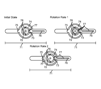
quantitative evaluation of
sperm count. In cases where a low sperm count is correctable or sperm count
varies over
time, it may be desirable to have a quantitative estimate of the absolute
sperm count and
motility.
SUMMARY
[0005] The disclosed device and method is for estimation of particulate
content in a
biological sample, including estimation of cell, such as sperm cell,
concentration by
centrifugal sedimentation of cells in fluid, such as seminal fluid. The
estimation is performed
using an enclosed sedimentation column of defined cross-sectional area and by
measuring
height of a pellet of compacted cells within the sedimentation column with aid
of a scale bar
along the sedimentation column. In one embodiment, the device includes a
cartridge
containing the sedimentation column as well as channels and cavities for
directing fluid and
sedimenting particulates or cells. In other embodiments, the sedimentation
column contains
fluid of defined density to further separate cell populations by density. The
sedimentation
column may also include portions of variable cross-sectional area allowing for
a visual of the
height of sedimented cells in the sedimentation column to resemble
measurements of cell
concentration in the sedimented cells over a wider range of cell
concentrations than otherwise
possible. The device also includes or can be used with an instrument for
rotating the
cartridge at specified rotational rates for intervals of time.
[0006] Embodiments of the device can be used at home as home use test kits
to estimate
sperm cell concentration and motile sperm cell concentration, aiding in
diagnosis and
monitoring of male fertility disorders and allowing users to avoid having to
provide samples
in a clinical setting. When used in a fertility context, the device and method
allow for a
quantitative evaluation of sperm count and motility. The user can get a more
accurate
estimate of the actual sperm count rather than just determining whether the
sperm count is
above or below a certain threshold. The user can, for example, determine if
sperm count and
motility is only somewhat low, and so may be more readily correctable.
Similarly, the user
can determine if the sperm counts vary over time, possibly allowing the user
to identify
causative factors for sperm count, and otherwise track times when sperm counts
are higher.
2
CA 02885845 2015-03-24
WO 2014/074737 PCT/US2013/068991
BRIEF DESCRIPTION OF THE FIGURES
[0007] Figure (FIG.) 1 is a representation of sedimentation before and
after rotation, in
accordance with an embodiment of the invention.
[0008] FIG. 2 is a top view of a cartridge, in accordance with an
embodiment of the
invention.
[0009] FIG. 3 is a side cross-section view of a cartridge, in accordance
with an
embodiment of the invention.
[0010] FIG. 4 illustrates a device as a kit, in accordance with an
embodiment of the
invention.
[0011] FIG. 5 is a top view of a cartridge, in accordance with an
embodiment of the
invention.
[0012] FIG. 6 is a side cross-section view of a cartridge, in accordance
with an
embodiment of the invention.
[0013] FIG. 7 illustrates an initial state, rotation rate 1 state, and
rotation rate 2 state of a
cartridge during operation, in accordance with an embodiment of the invention.
[0014] FIG. 8 is a top view of a cartridge, in accordance with an
embodiment of the
invention.
[0015] FIG. 9 is a side cross-section view of a cartridge, in accordance
with an
embodiment of the invention.
[0016] FIG. 10 illustrates an initial rotation state and rotation rate 1
state of a cartridge
during operation, in accordance with an embodiment of the invention.
[0017] FIG. 11 illustrates a top view of a cartridge, in accordance with an
embodiment of
the invention.
[0018] FIG. 12 illustrates a side cross-section view of a cartridge, in
accordance with an
embodiment of the invention.
[0019] FIG. 13 illustrates an initial state, rotation rate 1 state,
rotation rate 2 state, and
final state of a cartridge during operation, in accordance with an embodiment
of the
invention.
[0020] FIG. 14 illustrates a device as a kit, in accordance with an
embodiment of the
invention.
[0021] FIG. 15 is a flowchart of a method for preparing fluid for an
estimation of cell
count, in accordance with an embodiment of the invention.
[0022] FIG. 16 is a flowchart of a method for preparing fluid for an
estimation of cell
count with enzymes, in accordance with an embodiment of the invention.
3
CA 02885845 2015-03-24
WO 2014/074737 PCT/US2013/068991
[0023] FIG. 17 is a flowchart of a method for preparing fluid for an
estimation of cell
count, in accordance with an embodiment of the invention.
[0024] FIG. 18 illustrates a top view and a side view of a sedimentation
column, in
accordance with an embodiment of the invention.
[0025] FIG. 19 illustrates a top view and a side view of a sedimentation
column with a
lens, in accordance with an embodiment of the invention.
[0026] FIG. 20 illustrates a system for analyzing reflected light of fluid
in a
sedimentation column with lenses along the sedimentation column, in accordance
with an
embodiment of the invention.
[0027] FIG. 21 illustrates a system for analyzing reflected light of fluid
in a
sedimentation column with lenses on the end of the sedimentation column, in
accordance
with an embodiment of the invention.
[0028] FIG. 22 illustrates a top view and side cross-section view of a
tapered
sedimentation column, in accordance with an embodiment of the invention.
[0029] FIG. 23 illustrates a system for analyzing reflected light from
fluid in a
sedimentation column comprising a light source opposite of a detector, in
accordance with an
embodiment of the invention.
[0030] FIG. 24 illustrates a system for analyzing reflected light from
fluid in a
sedimentation column comprising a light source illuminating a transparent face
of the
cartridge, in accordance with an embodiment of the invention.
[0031] FIG. 25 illustrates a top view and side cross-section view of a
sedimentation
column with a density gradient, in accordance with an embodiment of the
invention.
[0032] FIG. 26 illustrates a side view of a cartridge, an instrument, and a
cavity-shaft
configuration, in accordance with an embodiment of the invention.
[0033] FIG. 27 illustrates a side view and bottom view of a cartridge, an
instrument, and
a cavity-shaft configuration, in accordance with an embodiment of the
invention.
[0034] FIG. 28 illustrates a side cross-section view and a front view of a
cartridge and an
instrument, in accordance with an embodiment of the invention.
[0035] FIG. 29 illustrates a top view and a side view of a cartridge and an
instrument and
a configuration 1 state and configuration 2 state of the cartridge, in
accordance with an
embodiment of the invention.
[0036] FIG. 30 illustrates an embodiment of a cartridge containing dense
objects, in
accordance with an embodiment of the invention.
4
CA 02885845 2015-03-24
WO 2014/074737 PCT/US2013/068991
[0037] FIG. 31 illustrates a cartridge, an enclosure for the cartridge, and
an instrument
with a tapered form factor, in accordance with an embodiment of the invention,
[0038] FIG. 32 illustrates an alternative configuration of a cartridge and
an instrument, in
accordance with an embodiment of the invention.
[0039] FIG. 33 illustrates an open enclosure of a cartridge, in accordance
with an
embodiment of the invention.
[0040[ FIG. 34 illustrates a sedimentation column with a density medium, in
accordance
with an embodiment of the invention.
[0041] FIG. 35 illustrates a sedimentation column with two density media,
in accordance
with an embodiment of the invention.
[0042] FIG. 36 illustrates two sedimentation columns with a density medium
in each, in
accordance with an embodiment of the invention.
[0043] FIG. 37 illustrates a cartridge configured to discern motility of
cells, in accordance
with an embodiment of the invention.
[0044] The figures depict various embodiments of the present invention for
purposes of
illustration only. One skilled in the art will readily recognize from the
following discussion
that alternative embodiments of the structures and methods illustrated herein
may be
employed without departing from the principles of the invention described
herein.
DETAILED DESCRIPTION
[0045] Various embodiments of estimation of sperm count and motility based
on volume
occupied by sperm cells packed into a column of defined cross-section
following
centrifugation is disclosed. The method is similar in principle to the
hematocrit technique
wherein concentration of red blood cells in a sample volume of blood is
estimated by volume
of packed red blood cells in a capillary following centrifugation. The
hematocrit technique is
a well-established technique for estimating red blood cell count in a blood
sample based on
packed volume of red blood cells following centrifugation from a known sample
volume of
blood. Estimation of cell count based on packed volume has also been applied
to nucleated
cell types such as leukocytes and leukocyte sub-types. For example, some
hematology
analyzers estimate red cell, granulocyte, and lymphocyte cell count from a
sample volume of
blood centrifuged in a capillary. In many cases, a scalebar incorporated in
the hematocrit
capillary provides a visual reference and aids in estimation of cell
concentration. Thus,
packed volume sedimentation provides an easy-to-read method for estimating
cell
concentration, which can be applied to counting sperm cells.
CA 02885845 2015-03-24
WO 2014/074737 PCT/US2013/068991
[0046] However, previous implementations used for blood analysis are wholly
impractical for direct application to semen analysis. For humans, the average
concentration of
red blood cells in blood is approximately 100 times higher than the average
sperm
concentration in semen. Also, the considerably higher viscosity of semen
prevents uptake of a
defined volume of sample by capillary action as is necessary for operation of
hematocrit
tubes and retards or prevents sedimentation of sperm cells upon
centrifugation. Semen is also
highly heterogeneous in composition (i.e. initially contains regions of high
and low sperm
concentration) unlike blood, and therefore requires homogenization to achieve
reproducible
measurements of concentration. For these reasons, different fluidic structures
and modified
sample processing steps are necessary to form a sedimented pellet of sperm
cells that can be
measured. Furthermore, the previously described hematocrit and blood analysis
techniques
require heavy and expensive centrifuges or dedicated analyzers to spin and
contain the
sedimentation capillaries, making them impractical for the general public.
Nonetheless, if a
means of mitigating the considerable challenges listed above was developed,
packed volume
sedimentation could provide a simple-to-use means of estimating sperm count.
[0047] In one embodiment, the estimation of cell count is provided for
through use of a
device that can be included in a kit. The device comprises a cartridge
including a packed
volume column and a motorized instrument for spinning the cartridge. The
cartridge may
attach to the motorized instrument, for example, using a frictional press fit
between a motor
shaft of the motorized instrument and the cartridge, or using a plurality of
magnets. In
addition, the device, when prepared as a kit, may also comprise a fluid
transfer device and a
sample collection cup to assist with transferring the sample to the cartridge.
The cartridge
may be a disposable cartridge, and a user can use a new cartridge for each
sample.
[0048] Throughout this description, the disclosed method and device is
presented in terms
of a method and device for manipulating semen samples for fertility analysis.
However,
these examples are provided for the purpose of illustration only. The method
and device can
also be used with other suitable fluids or samples for this method comprising
packed volume
sedimentation. For instance, the device may also be applied to examining
packed volume of
particulates in motor oil or to automated quantification of red blood cells or
leukocytes in a
sample volume of blood. Other types of particulates or solids in other types
of samples can
also be quantified or otherwise analyzed with the devices and methods
described throughout.
In some embodiments, the samples are food, soil or other materials, and in
other
embodiments, the samples are biological samples, such as blood, stool, semen,
and other
samples that might come from an organism, such as a human.
6
CA 02885845 2015-03-24
WO 2014/074737 PCT/US2013/068991
[0049] An embodiment of the device for estimating concentration of cells 11
based on
volume occupied by cells 11 in a packed volume is illustrated in FIG. 1. The
cells 11 are
initially suspended in a fluid 12. In one embodiment, the cells 11 are sperm
cells and the
fluid 12 is seminal fluid. Following rotation, the cells 11 are packed at the
bottom of a
sedimentation chamber 13 and the sedimented cells occupy a volume proportional
to a
number of cells 11 initially suspended in the fluid 12.
[0050] A top view and a side cross-section view of an embodiment of a
cartridge 21 are
illustrated in FIGS. 2 and 3, respectively. A top view of the cartridge 21 is
shown in FIG. 2
and a cross-sectional side view in FIG. 3. The cartridge 21 can be constructed
from a variety
of materials including a polymer or other similar material. All cartridges
described
throughout the detailed description may be constructed in the same manner. In
general,
features or materials described for any cartridge included herein can be
included or used in
any of the other cartridges described herein, as well. Cartridges described
herein can be the
cartridge 21 or embodiments of the cartridge as described per figure.
[0051] The cartridge 21 may comprise a sedimentation column 22 that
comprises
metering marks 23. The metering marks 23 aid a user in determining volume of
sedimented
cells. The cartridge may also comprise a central sample entry cavity 24 and a
sample
directing cavity 25 with a defined volume, the sample directing cavity 25 in
fluid
communication with the central sample entry cavity 24. In other words, the
fluid is capable
of moving between the central sample entry cavity 24 and the sample directing
cavity 25.
For example, when a volume of fluid equal to the volume of the sample
directing cavity 25 is
added to the central sample entry cavity 24 and the cartridge 21 is rotated
clockwise or
counterclockwise about a central axis 33 of the cartridge 21, the fluid
collects in the sample
directing cavity 25. With further rotation, for example at 2000-10000 RPM for
2-10 minutes,
cells from the fluid are packed at the bottom of the sedimentation column 22
and the
sedimented cells can be read by the user using the metering marks 23. The
cartridge 21 may
additionally include a hub attachment 31 configured to securely connect the
cartridge 21 to a
motorized instrument for spinning the cartridge 21. In one embodiment, the
cartridge 21 may
connect or attach to the motorized instrument for spinning the cartridge 21
using a plurality
of magnets. For all cartridge and instrument descriptions herein, a plurality
of magnets can
be used to attach the cartridge and the instrument.
[0052] In addition, the cartridge 21 may hold a reagent pellet 32 or be
coated with
chemical reagents, such as digestive enzymes, to provide fluorescent cell
labels, contrast
7
CA 02885845 2015-03-24
WO 2014/074737
PCT/US2013/068991
dyes, specific density beads, or, in the embodiment of sperm cells, reduce
semen viscosity to
facilitate easier reading by the user. These reagents may be freeze dried.
[0053] An
embodiment of the device as a kit is shown in FIG. 4. The device comprises
items used for counting cells including a cartridge 41 such as a disposable
cartridge, a fluid
transfer device 42 such as a bulb pipette, other type of pipette, or a
syringe, a collection cup
43 for collecting fluid, and an instrument 45 configured to rotate the
cartridge 41. The user
transfers a defined sample volume into the cartridge 41 using the transfer
device 42. The
transfer device 42 may have a level mark configured to assist in measuring the
defined
sample volume. For the cartridge designs embodied in FIGS. 5-13, a non-precise
amount of
sample may be transferred to the cartridge by the user. The collection cup 43
may comprise a
reagent pellet 44 or be coated with chemical reagents, such as those described
above
regarding reagent pellet 32, and the reagents or pellets may be freeze dried.
Similarly, for all
collection cups comprising a reagent pellet herein, the collection cup may
comprise the
reagent pellet or may be coated with chemical reagents, such as those
described above
regarding regent pellet 32, and the reagents and/or pellets may be freeze
dried. To rotate or
spin the cartridge 41, the user can attach the cartridge 41 to the instrument
45. In one
embodiment, the cartridge 41 and instrument 45 comprise additional components
to securely
attach the cartridge 41 to the instrument 45.
[0054] A top
view and a side cross-section view of an embodiment of a cartridge 51 are
illustrated in FIGS. 5 and 6, respectively. The cartridge 51 may comprise a
sedimentation
column 52 that contains metering marks 53. The metering marks 53 are
configured to aid the
user in determining volume of sedimented cells. The cartridge 51 may also
comprise a
central sample entry cavity 54 and a sample directing cavity 55 with a defined
volume. The
cartridge 51 may also comprise an overflow chamber 56 with a counterbalance
cavity 57
intended to counterbalance the sample directing cavity 55. The overflow
chamber 56 is
connected to the central sample entry cavity 54 by shallow channels 58. The
shallow
channels 58 allow the sample in the cartridge 51 to move from the central
sample entry cavity
54 to the overflow chamber 56 during rotation, for example during a second
round of
rotation, as further described in FIG. 7. The sample directing cavity 55 is
connected to the
central sample entry cavity 54 by additional shallow channels 59. The
additional shallow
channels 59 in one embodiment include a larger depth or diameter than the
depth or diameter,
respectively, of the shallow channels 58. The shallow channels 59 allow the
sample to move
from the central sample entry cavity 54 to the sample directing cavity 55, for
example during
a first round of rotation, further described in FIG. 7. The shallow channels
58 and additional
8
CA 02885845 2015-03-24
WO 2014/074737 PCT/US2013/068991
shallow channels 59 have a depth or diameter configured such that fluid
wetting and surface
tension forces prevent movement of the sample through the channels 58 and 59
unless a
threshold rotation rate is exceeded by the cartridge 51, for example, during
centrifugation.
The threshold rotation rate necessary for causing movement of the sample
through shallow
channels 59 is based at least in part on diameter or depth of the shallow
channels. For
example, the threshold rotation rate will increase as diameter or depth of
shallow channels
decrease. The cartridge 51 may additionally include a hub attachment 61
configured to
securely connect the cartridge 51 to a motorized instrument for spinning the
cartridge 51
during, for example, centrifugation. The cartridge 51 may hold a reagent
pellet 62 or be
coated with chemical reagents, as described above regarding the reagent pellet
32.
[0055] An initial state, first rotation rate state and second rotation rate
state of an
embodiment of fluid movement within a cartridge 71 are described in FIG. 7,
respectively.
Fluid 77 is initially loaded into a central sample entry cavity 72. In the
initial state, the
shaded portion of the cartridge 72 represents the fluid 77. Upon rotation at a
first rotation
rate, for example in a range of 100-4000 RPM, preferably in a range of 500-
2000 RPM, the
fluid enters a sample directing cavity 73 with a defined volume during a first
time period until
the sample directing cavity 73 is full as seen in the rotation rate 1 state.
The overflow
chamber 74 may comprise a counterbalance cavity 75. The cartridge 71 comprises
a
sedimentation column 76.
[0056] As seen in the rotation rate 1 state, since the cross-sectional area
of shallow
connecting channels 78 is smaller than the cross-sectional area of an overflow
chamber 74,
the fluid 77 is prevented from entering the overflow chamber 74 during the
first rotation rate.
Balance of fluid surface tension and wetting forces overcoming effective
gravitational force
prevents entry of the fluid 77 into the overflow chamber 74. In addition, the
counterbalance
cavity 75 assists in counterbalancing the sample directing cavity 73, which
fluid 77 can also
enter during the first rotation rate. Upon rotation of the cartridge 71 at a
second rotation rate
(e.g. 2000-10000 RPM, 2-10 minutes) during a second time period, the fluid
remaining in the
central sample entry cavity 72 enters the overflow chamber 74 and
counterbalance cavity 75,
as shown in the rotation rate 2 state. With centrifugation for the second time
period at the
second rotation rate, cells in the sample directing cavity 73 become compacted
in the
sedimentation column 76. Then, the pellet of sedimented and compacted cells
can be read by
the user with a cell pellet height proportional to amount of cells initially
contained in the
sample directing cavity 73 and sedimentation column 76 during rotation rate 1
state. Due to
excess fluid being directed to the overflow channels 74 during the second time
period, cells
9
CA 02885845 2015-03-24
WO 2014/074737 PCT/US2013/068991
can be measured from a precise amount of fluid. In some embodiments,
additional rotations
can be performed for additional time periods. This can be true for any
embodiments
described herein. In further embodiments, only a single rotation is performed
for an interval
of time, and, in some cases, this single rotation provides compacting of the
cells. This can be
true for any embodiments described herein.
[0057] A top view and a side cross-section view of another embodiment of a
cartridge 81
are illustrated in FIGS. 8 and 9, respectively. The cartridge 81 may comprise
a sedimentation
column 82 that contains metering marks 83. The metering marks 83 are
configured to aid the
user in deteimining volume of sedimented cells. The cartridge 81 may also
comprise a
central sample entry cavity 84 and a sample directing cavity 85 with a defined
volume. The
cartridge 81 may also comprise an overflow chamber 86 with a counterbalance
cavity 87
intended to counterbalance the sample directing cavity 85. The overflow
chamber 86 is
connected to the central sample entry cavity 84 by shallow connecting channels
88. Unlike
the sample directing cavity 85 being connected to the central sample entry
cavity 84 by
shallow channels such as the shallow channels 59 in FIG. 5, the sample
directing cavity 85
can be in direct fluid communication with the central sample entry cavity 84.
The sample
directing cavity 85 and the central sample entry cavity 84 are connected in a
manner similar
to that illustrated in FIGS. 2 and 3. The cartridge 81 may also comprise a hub
attachment 91
configured to securely connect the cartridge 81 to a motorized instrument for
spinning during
centrifugation. The cartridge 81 may comprise a reagent pellet 92 or be coated
with chemical
reagents, as described above regarding the reagent pellet 32.
[0058] An initial rotation state and a first rotation rate state of an
embodiment of the fluid
movement within a cartridge 101 are described in FIG. 10. Fluid 107 is
initially loaded into a
central sample entry cavity 102, as shown in the initial state. The cartridge
is intended to be
rotated at a first rotation rate (e.g. 2000-10000 RPM) for a time interval
(e.g. 2-10 minutes)
and distribute fluid in one step, allowing a simplified instrument design.
Upon rotation at the
single rate, fluid enters a sample directing cavity 103 and a sedimentation
column 106, the
sample directing cavity 103 and the sedimentation column 106 comprising a
defined volume
and being in direct fluid communication with the central sample entry cavity
102, unlike in
FIGS. 5 and 7 where the sample directing cavity and central sample entry
cavity are
connected by channels. As the rotation rate of the cartridge 101 accelerates
and reaches the
first rotation rate, a rate necessary to overcome surface tension and wetting
forces in shallow
channels 108 connecting the central sample entry cavity 102 and an overflow
chamber 104 is
exceeded, and the fluid remaining in the central sample entry cavity 102 will
enter the
CA 02885845 2015-03-24
WO 2014/074737 PCT/US2013/068991
overflow chamber 104 and a counterbalance cavity 105, as illustrated in the
rightmost
illustration of FIG. 10. Therefore the fluid remaining in the sample directing
cavity 103 is the
only fluid that contributes to the volume of sedimented cells in the
sedimentation column 106
following centrifugation. With continued centrifugation (e.g., 2-10 minutes),
cells within the
sample directing cavity 103 become compacted in the sedimentation column 106
where a
sedimented cell pellet can be read by the user with a pellet height
proportional to amount of
cells initially contained in the fluid inside the sample directing cavity 103
and sedimentation
column 106 during the rotation 1 state.
[0059] A top view and a side cross-section view of another embodiment of a
cartridge
111 are illustrated in FIGS. 11 and 12, respectively. The cartridge 111 may
comprise a
sedimentation column 112 that comprises metering marks 113. The metering marks
113 are
configured to aid the user in determining volume of sedimented cells. The
cartridge 111 may
also comprise a central sample entry cavity 114 and a sample directing cavity
115. The
cartridge 111 may also comprise a counterbalance cavity 116 for
counterbalancing the sample
directing cavity 115. The sample directing cavity 115 is connected to the
central sample
entry cavity 114 by angled shallow channels 117, wherein the angled shallow
channels 117
comprise an extension 118 for retaining or storing sedimented cells. For
example, the angled
shallow channels 117 are angled radially outward from the center of the
cartridge 111 with
respect to the sedimentation column 112. While the sedimentation column 112 is
located
radially outward from the center of the cartridge 111 along a first radial
axis, the angled
shallow channels 117 are also located radially outward from the center of the
cartridge 111
along a second radial axis and a third radial axis, where the second radial
axis and the third
radial axis are not the first radial axis. The cartridge 111 may additionally
include a hub
attachment 121 configured to securely connect the cartridge 111 to a motorized
instrument
for spinning the cartridge 111 during, for example, centrifugation. The
cartridge 111 may
comprise one or more reagent pellets 122 or be coated with chemical reagents,
as described
above regarding the reagent pellet 32.
[0060] An initial state, first rotation state, second rotation state, and
final state of an
embodiment of a fluid movement within a cartridge 131 are described in FIG.
13. Fluid 139
is loaded into a central sample entry cavity 132, as seen in the initial
state. Upon rotation,
fluid travels from central sample entry cavity 132 into channel extensions
137, connecting
channels 133, and into a sample directing cavity 134 with a defined volume.
The first
rotation rate state is shown in the rotation rate 1 state. When rotation rate
of the cartridge
exceeds a rate necessary to overcome surface tension, the fluid remaining in
the central
11
CA 02885845 2015-03-24
WO 2014/074737 PCT/US2013/068991
sample entry cavity 132 enters an optional counterbalance chamber 135. The
second rotation
rate state is shown in the rotation rate 2 state. With further rotation of the
cartridge 131, for
example at 2000-10000 RPM for 2-10 minutes, cells within the directing cavity
134 become
compacted in a sedimentation column 136 where the sedimented pellet can be
read by the
user with a pellet height proportional to amount of cells contained in the
sample directing
cavity 134 and sedimentation column 136 during the rotation rate 1 state. The
final state is
shown in the final state. Cells initially contained in channel extensions 137
and central
sample entry cavity during rotation rate 1 state are trapped in channel
extensions 137 and
therefore do not contribute to volume of sedimented cells in the sedimentation
column 136.
Locations where compacted cells will collect in the shown cartridge design of
FIG. 13 are
marked as 138. An advantage of the design of the cartridge 131 is that less
material and less
complexity is required for manufacturing the cartridge 131 than previously
described
cartridge designs due to lack of overflow chambers. Measurement of volume of
cells from
only a volume of interest of fluid can be achieved by capturing sedimented
cells from excess
fluid in an alternate location from the sedimentation column 136 rather than
physically
removing excess fluid from the central sample entry cavity.
[0061] FIG. 14 demonstrates a kit for cell, sperm cell or other particle
measurement
including a cartridge 141, the cartridge 141 an alternative embodiment of the
cartridge
described in FIGS. 11 and 12 (though cartridges of the other Figures can be
used too). The
sample inlet cavity 142 is increased in size to accommodate a fluid 148 in its
entirety. In one
embodiment, the fluid is seminal fluid and the cavity comprises a volume that
exceeds a
maximum fluid volume produced by a human male, where the maximum fluid volume
is
about 5 milliliters. This design is configured to directly collect a fluid for
analysis by the
central cavity 142. Optionally, the fluid may be collected in a collection cup
143. The
collection cup 143 may comprise a spout 144 for pouring the fluid into the
sample inlet cavity
142 of the cartridge 141. Upon centrifugation, such as at 2000-10000 RPM for 2-
10 minutes,
cells within a sample directing cavity 145 become compacted in a sedimentation
column 146
while cells in the fluid remaining in the sample inlet cavity 142 are retained
therein. Either
the sample inlet cavity 142 or the collection cup 143 may contain chemical
reagents for
enzymatic digestion, contrast enhancement, or other assay enhancing functions.
A lid 147
that is configured to attach to the cartridge 141 during centrifugation to
prevent fluid spillage
may be included in the kit. The design of FIGS. 5 and 6 or the design of FIGS.
8 and 9 may
comprise sufficiently large overflow cavities, allowing for analysis of a
greater volume of
fluid, such as up to 5 milliliters.
12
CA 02885845 2015-03-24
WO 2014/074737 PCT/US2013/068991
[0062] FIG. 15 is a flowchart of an embodiment of a method for estimating
sperm count
based on volume occupied by sperm cells packed into a column of defined cross-
section
following centrifugation. Different embodiments may perform the steps in the
method in a
different order, omit certain steps, and/or perform additional steps. In one
embodiment, the
method is performed using a kit comprising a collection cup, a cartridge, a
transfer device
and an instrument. Any cartridge or instrument design described herein may be
used in the
method.
[0063] The user collects 151 a sample or fluid in the collection cup. In
one embodiment,
the collection cup comprises digestive enzymes such as chymotrypsin, trypsin,
bromelain, or
papain for accelerating liquefaction of the fluid. The fluid is swirled or
agitated 152, for
example by the user, in the collection cup (or the sample can be otherwise
agitated, such as
agitated by the instrument once it is placed in the instrument). Swirling or
agitating the fluid
accelerates dissolution of the enzyme into the fluid. An interval of time
(e.g., of 1-30
minutes) elapses to allow the enzyme to liquefy the fluid. A portion of the
fluid is then
transferred 153 to the cartridge using a transfer device, such as a syringe or
bulb transfer
pipette. In one embodiment, the cartridge is capped with a lid or sticker
following input of
the fluid. The cartridge is attached 154 to the instrument, wherein the
instrument comprises a
motor configured to rotate the cartridge. Optionally, the instrument may
accelerate the
cartridge in one direction and then an opposite direction for an interval of
time, mechanically
agitating the fluid, encouraging homogenization and reduced viscosity for more
consistent
measurements. The instrument may also accelerate the cartridge in one
direction, allow it to
come to a stop, then repeat for an interval of time to provide mechanical
agitation. The
instrument spins 155 or rotates the cartridge at a rotation rate (e.g., for 2-
10 minutes at 2000-
10000 RPM). Optionally, the cartridge is spun at a reduced rotation rate for
an interval of
time (e.g., for 1-5 minutes) to allow for controlled expansion of compacted
cells in a
sedimentation column of the cartridge. After rotation, the cartridge is halted
by the
instrument and the user reads 156 the result by estimating cell count or
concentration in the
fluid based on height of compacted cell pellet in the sedimentation column of
the cartridge.
In some embodiments, the instrument comprises a digital reading the user can
read (e.g.,
digital reading on a user interface of the instrument). All embodiments of the
instrument
described herein may comprise a digital reading on a user interface of the
instrument. In one
embodiment, the instrument comprises a lid, wherein the lid comprises one or
more magnets
and the instrument comprises one or more sensors configured to detect magnetic
fields. The
one or more magnets and one or more sensors are placed within the lid and the
instrument
13
CA 02885845 2015-03-24
WO 2014/074737
PCT/US2013/068991
such that, when the lid is closed on the instrument, the one or more magnets
in the lid and the
one or more sensors in the instrument are a distance away, where the distance
is less than a
threshold distance necessary for the one or more sensors to detect a magnetic
field of the one
or more magnets in the lid and thus detect that the lid is closed on the
instrument. In another
embodiment, the one or more magnets can be in the instrument and the one or
more sensors
in the lid of the instrument. The magnet and sensor configuration described
here can be
applied to any instrument described herein.
[0064] FIG. 16 is a flowchart of an embodiment of a method for estimating
sperm
concentration based on volume occupied by speiiii cells packed into a column
of defined
cross-section following centrifugation. Different embodiments may perform the
steps in the
method in a different order, omit certain steps, and/or perform additional
steps. In one
embodiment, the method is performed using a kit comprising a collection cup, a
cartridge, a
transfer device and an instrument. Any cartridge or instrument design
described herein may
be used in the method.
[0065] The fluid or sample is collected 161 by a user in the collection
cup. The user may
swirl or agitate the fluid to aid in homogenization. A portion of the fluid is
then transferred
162 to the cartridge using the transfer device immediately or before
coagulation of the fluid.
The transfer device may be a syringe or bulb transfer pipette. The cartridge
may optionally
comprise a lid or sticker configured to securely cap the cartridge following
input of the fluid
in the cartridge. The cartridge is attached 163 to the instrument. The
instrument comprises a
motor and the motor is configured to rotate, spin, or reciprocate 164 the
cartridge for an
interval of time to liquefy the fluid. The instrument may alternately
accelerate the cartridge
in one direction and then the other for an interval of time to mechanically
agitate the fluid,
encouraging homogenization and reduced viscosity of the fluid for more
consistent
measurements. Enzymes enclosed in the cartridge can act on the agitated fluid
(e.g., for 1-30
minutes) to promote liquefaction of the fluid. The cartridge is spun 165 by
the instrument for
an interval of time at a specified rate (e.g., for 2-10 minutes at 2000-10000
RPM).
Optionally, the cartridge may then be spun 165 at a reduced RPM (e.g., for 1-5
minutes) to
allow for controlled expansion of compacted cells in the sedimentation column.
After this
spin is done, the cartridge is halted by the instrument and the user may read
166 a result of an
estimate of the cell concentration in the fluid from the height of a compacted
cell pellet in the
sedimentation column.
[0066] FIG. 17 is a flowchart of an embodiment of a method for estimating
sperm
concentration based on volume occupied by sperm cells packed into a column of
defined
14
CA 02885845 2015-03-24
WO 2014/074737 PCT/US2013/068991
cross-section following centrifugation. Different embodiments may perform the
steps in the
method in a different order, omit certain steps, and/or perform additional
steps. In one
embodiment, the method is performed using a kit comprising a collection cup, a
cartridge,
and an instrument. Any cartridge or instrument design described herein may be
used in the
method.
[0067] The fluid or sample is collected 171 in the cartridge or collected
172 in the
collection cup. In the case the sample is collected 172 in the collection cup,
the entire fluid is
poured 173 into the cartridge. The cartridge may optionally comprise a lid
configured to
securely cap the cartridge following input of the fluid in the cartridge. The
cartridge is
attached 174 to the instrument. The instrument comprises a motor and the motor
is
configured to rotate, spin or reciprocate 175 the cartridge. The instrument
may alternately
accelerate the cartridge in one direction and then the other for an interval
of time to
mechanically agitate the fluid, encouraging homogenization and reduced
viscosity of the fluid
for more consistent measurements. Enzymes enclosed in the cartridge can act on
the agitated
fluid (e.g., for 1-30 minutes) to promote liquefaction of the fluid. The
cartridge is spun 176
for an interval of time at a specified rate (e.g., for 2-10 minutes at 2000-
10000 RPM).
Optionally, the cartridge may then be spun 176 at a reduced RPM (e.g., 100-
2000 RPM for 1-
minutes) to allow for controlled expansion of compacted cells in the
sedimentation column.
After this spin is done, the cartridge is halted by the instrument and the
user may read 177 a
result of an estimate of the cell concentration in the fluid from the height
of compacted cells
in the sedimentation column.
[0068] For each of the methods described in FIGS. 15-17, the user may
perform all of the
steps himself at home using a cartridge and/or kit as described throughout
this description and
using an instrument for rotating the cartridge, such as those described
herein. In other
embodiments, the user provides the sample in the cartridge, but then the
cartridge is delivered
to a clinic, such as a fertility center, that performs the
rotation/centrifugation of the sample
using an instrument at the clinic, such as those described herein. In this
case, the user
performs the collection 151, 161, 172, 171 steps and possibly other steps,
such as the
swirl/incubate 152, transfer 153, 162, and pour 173 steps, but the clinic may
perform the
attachment 154, 163, 174 of the cartridge to the instrument along with the
steps that follow.
In another embodiment, the user provides the sample in a holding device and it
is transferred
to the cartridge at the clinic. In this case, the clinic performs the
transfers and pour and
possibly the swirl/incubate steps. Thus, the method can include just the
subset of steps
performed by the user or the subset of steps performed by the clinic.
CA 02885845 2015-03-24
WO 2014/074737 PCT/US2013/068991
[0069] FIGS. 18-25 describe various embodiments of configurations of the
sedimentation
column. Any of the described various embodiments may be incorporated into the
cartridge
designs described in FIGS. 3-14.
[0070] FIG. 18 shows an enlargement of a top view and a side view of a
sedimentation
column, the sedimentation column comprising metering marks 182 and numbers
183. After
cells are compacted by centrifugation, the height of a resulting pellet 184
may be determined
visually by differences in reflectance between cells in the pellet 184 and
fluid 185 or by other
means including fluorescent cell labels. The user can estimate initial
concentration of cells in
the fluid by reading the number 183 closest to a metering mark 182 closest to
the interface
between the cells 184 and the fluid 185.
[0071] FIG. 19 shows an enlargement of a top view and a side view of a
sedimentation
column, the sedimentation column comprising metering marks 192 and numbers
193. The
sedimentation column comprises a lens 196 configured to magnify the
sedimentation column
and size of the sedimentation column. The lens 196 can be integrated into the
sedimentation
column during fabrication, for example, by injection molding of polymer. The
presence of
the lens 196 may allow the user to visualize an interface between a pellet 194
and fluid 195
more easily. In one embodiment, the lens 196 is cylindrical in shape. Other
types and shapes
of lenses can also be used. After cells are compacted by centrifugation, the
height of the
pellet 194 may be determined visually by differences in reflectance between
the cells and
fluid 195 or by other means including fluorescent cell labels. The user can
estimate the initial
concentration of cells in the fluid by reading the number 193 closest to a
metering mark 192
closest to the interface between the cells 194 and the fluid 195.
[0072] FIG. 20 shows a side view of an alternate embodiment of a
sedimentation column
intended for use with fluorescent analysis. A top lens 201 and a bottom lens
202 are
integrated into a top surface and a bottom surface of the sedimentation
column, respectively.
In one embodiment, the lenses are cylindrical in shape. Other types and shapes
of lenses can
also be used. A fluorescent excitation light source 203, such as an LED,
filtered lamp, or
laser, emits light such that it is focused on the sedimentation column by the
bottom lens 202.
Labeled cells in a pellet 204 are excited by the impinging light and emit a
light 205 of a
wavelength longer than a threshold wavelength. The light 205 is focused by the
top lens 201
onto a detector 206. The detector 206 may be a CCD camera, photodiode,
photomultiplier, or
human eye. A selective filter 207 may be placed between the detector 206 and
the
sedimentation column to selectively pass the wavelengths of the light 205
emitted by the
16
CA 02885845 2015-03-24
WO 2014/074737
PCT/US2013/068991
excited cells. The detector 206 may determine the height of the pellet 204 by
scanning along
the sedimentation column and can be based on total fluorescent signal.
[0073] FIG. 21
shows an alternate embodiment of a sedimentation column 211 intended
for use with fluorescent analysis. Geometry of the sedimentation column 211
causes cells to
be compacted into a pellet 212 with small surface area following
centrifugation. A top lens
213 and a bottom lens 214 are integrated into a top surface and a bottom
surface of the
sedimentation column, respectively. The top lens 213 and the bottom lens 214
may be
spherical or aspheric lenses. A fluorescent excitation light source 215, such
as an LED,
filtered lamp, or laser emits light such that the light is focused on the
pellet 212 by the bottom
lens 214. Cells in the pellet 212 may be labeled with fluorescent dyes, such
as acrinidine
orange which are active only within nucleic acid containing cells. Labeled
cells in the pellet
212 are excited by the impinging light and emit light 217 of a wavelength
longer than a
threshold wavelength. The light 217 is focused by the top lens 213 onto a
detector 218 which
may be a CCD camera, CMOS sensor, photodiode, photomultiplier, or human eye. A
selective filter 219 may be placed between the detector 218 and the
sedimentation column
211 to selectively pass the light 217 emitted by the excited cells 217. The
detector 218 may
determine a total number of cells present based on total integrated
fluorescence emitted by
the cells in the pellet 212.
[0074] FIG. 22
shows an alternate embodiment of a sedimentation column intended for
use in estimating a wide range of cell concentrations. The sedimentation
column comprises
metering marks 221 and numbers 222. After cells are compacted by
centrifugation, height of
a resulting pellet 223 may be determined visually by differences in light
scattering and
reflectance between the cells in the pellet 223 and fluid 224 or by other
means including
fluorescent cell labels. The user can estimate initial concentration of cells
in the fluid by
reading the number 222 closest to a metering mark 221 closest to an interface
between the
cells in the pellet 223 and the fluid 224. In this embodiment, the
sedimentation column is
tapered comprising a section of a high cross-sectional area 225 exceeding a
reference cross-
sectional area and a low cross-sectional area 226 not exceeding a reference
cross-sectional
area with a transition area 227 in between the sections 225 and 226. In this
embodiment, a
pellet comprising low cell concentration will be accommodated by a portion
comprising low
cross-sectional area 226, while a pellet comprising substantially higher cell
concentrations
will be accommodated by a portion comprising high cross-sectional area 225.
The metering
marks 221 and numbers 222 are adjusted for the different cross-sectional
areas, allowing a
user to accurately estimate cell concentration. To one skilled in the art, it
is apparent that
17
CA 02885845 2015-03-24
WO 2014/074737 PCT/US2013/068991
many variations of sedimentation column taper are possible. For instance more
than one
transition area 227 may be integrated into the sedimentation column to create
multiple
sections with varying cross-sectional area. For example, the multiple sections
may comprise
sections with sequentially increasing or decreasing cross-sectional areas. In
another example,
cross-sectional area may continuously increase or decrease along the
sedimentation column
to accommodate a wide range of cell concentrations and metering marks 221 and
numbers
222 can be adjusted accordingly. The multiple sections may also comprise
varying cross-
sectional areas such that a visual of the height of the pellet 223 in the
sedimentation column
corresponds to cell concentration of the pellet 223. For example, if a user
sees a pellet 223
with a height of 4 mm, there are 4 metering marks 221, each metering mark
equidistant from
each other along the pellet 223. In addition, there may be the numbers 222 per
metering
mark. In an embodiment where the visual of the height does not correspond to
cell
concentration of the pellet 223, there may be 4 metering marks 221 not
equidistant from each
other along the pellet 223.
[0075] FIG. 23 shows an embodiment of a sedimentation column 231 enclosed
by an
upper layer 232 of polymer and a lower layer 233 of polymer. The upper layer
232 and lower
layer 233 may be joined by processes including ultrasonic welding, laser
welding, or thermal
bonding. A fluorescent excitation light source 234, such as an LED, filtered
lamp, or laser,
emits light such that the light impinges a pellet 235 in the sedimentation
column 231. The
lower layer 233 may be dyed with filtering agents configured to selectively
pass light emitted
by the light source 234 to enhance contrast. Cells in the pellet 235 may be
labeled with
fluorescent dyes, such as acrinidine orange which are active only within
nucleic acid
containing cells. Labeled cells in the pellet 235 are excited by the impinging
light and emit
light 236 of a wavelength longer than a threshold wavelength. The light 236
impinges onto a
detector 237. Embodiments of the detector 237 comprise a CCD camera,
photodiode,
photomultiplier, or human eye. The upper layer 232 may be dyed with filtering
agents
configured to selectively pass light 236 longer than a threshold wavelength
emitted by the
cells in the pellet 235 to improve accuracy of cell detection. The detector
237 may determine
estimated cell concentration by scanning along the sedimentation column and
can be based
on total fluorescent signal. The embodiment of the sedimentation column 231
described here
using dyes with filtering agents may be combined with lenses as described in
FIGS. 19, 20
and 21 to enhance accuracy of cell count estimations. The features described
with respect to
the sedimentation column 231 may also be used to enhance detection in particle-
based
18
CA 02885845 2015-03-24
WO 2014/074737 PCT/US2013/068991
immunoassays. The features described with respect to the sedimentation column
231 may
also be incorporated into the sedimentation column 211 of FIG. 21.
[0076] FIG. 24 shows an embodiment of a sedimentation column 241 enclosed
by an
upper layer 242 of polymer and a lower layer 243 of polymer. The upper layer
242 and the
lower layer 243 may be joined by processes including ultrasonic welding or
thermal bonding.
A light source 244, such as an LED, sunlight, or room lighting, emits light
that impinges a
pellet 245 through the upper layer 242, wherein the polymer of the upper layer
242 is
transparent. The pellet 245 scatters light 247 back towards the light source
244 due to the
pellet's particulate nature while fluid 246 transmits light. If the lower
layer 243 is doped or
covered with light absorbing material, such as carbon black or other light
absorbing
pigments, part of the light transmitted by the fluid 246 will be absorbed,
enhancing optical
contrast between the pellet 245 and the fluid 246. The scattered light 247 may
be detected by
a detector, such as a CCD camera, mobile communication device, or human eye,
to estimate
cell concentration. In this embodiment, the light source 244 is perpendicular
to the upper
layer 242. The light source 244 may also be placed in parallel with or in the
upper layer 242
and still scatter light 247 toward a viewer or detector. This configuration
may further
increase optical contrast of the pellet 245 by avoiding or minimizing
interfering reflection off
of planar surfaces of the layers 242 and 243.
[0077] FIG. 25 illustrates an example of a sedimentation column of a fluid
following
centrifugation, wherein the fluid comprises particles or materials with a
density higher than
the fluid's density but lower than density of certain cells or particulates in
the fluid. The
sedimentation column comprises metering marks 252 and numbers 253. Following
centrifugation, the intermediate density particles or materials form an
intermediate layer 254
between the pellet 255 of compacted cells and the fluid 256. The intermediate
layer 254 may
comprise distinctively colored particles or materials such as dyed polystyrene
or another
polymer in order to enhance optical contrast of an interface between the
pellet 255,
intermediate layer 254, and fluid 256. The user can estimate initial
concentration of cells in
the sample by reading a number 253 closest to a metering mark 252 closest to
an interface
between the pellet 255 and the intermediate layer 254.
[0078] FIG. 25 alternately may represent an example of the sedimentation
column of a
fluid following centrifugation, wherein the fluid was mixed with a dye prior
to centrifugation
that identifies dead cells. For example, the dye can selectively partition
into dead cells but
not living cells. It is known in the art that dead or immotile sperm cells
have a density lower
than the density of living and motile sperm cells. The sedimentation column
comprises
19
CA 02885845 2015-03-24
WO 2014/074737 PCT/US2013/068991
metering marks 252 and numbers 253. Following centrifugation, the fluid
separates into
layers with a fluid layer 256 closest to the center of the centrifugation, a
live cells layer or a
pellet 255 furthest from the center of the centrifugation, and a dead cells
layer 254 with
intermediate density in between the two layers 256 and 255. Living cells
exclude the dye and
therefore are visually distinct from the dead cells layer 254 and fluid layer
256 which also
exhibit the color of the dye. The user can estimate initial concentration of
living cells in the
fluid by reading a number 253 closest to a metering mark 252 closest to an
interface between
the pellet 255 and the dead cells layer 254. The user can also estimate number
of dead cells
from the visually distinct dead cell layer 254. As described previously, an
intermediate
density layer formed from polymer fragments or particles may be mixed into the
fluid prior to
centrifugation to enhance the contrast between the dead cells layer 254 and
pellet 255.
[0079] FIGS. 26 and 27 illustrate various embodiments of mechanisms for
attaching a
cartridge to a motor of an instrument. These embodiments may be used with any
of the
cartridges or instruments described herein, or may be used to attach the
cartridge to other
instruments outside of those described herein. In any embodiment, the
cartridge, the motor
shaft or an adaptor configured to attach to the motor may comprise a magnetic
material,
providing a mechanism for attaching the cartridge to the motor.
[0080] FIG. 26 shows an embodiment of a schematic for attaching a cartridge
261 to a
motor 262 of an instrument. The cartridge comprises a cavity 263. The cavity
263 comprises
a first diameter less than a second diameter of a shaft 264 of the motor 262.
To attach the
cartridge 261 to the motor 262, the shaft 264 is press-fit into the cavity
263. Material used in
the first diameter of the cavity 263 and elastic modulus of material used for
the cartridge 261
may be selected such that a tight friction fit is established between the
shaft 264 and cavity
263 of the cartridge 261 allowing rotation of the cartridge 261 when attached
to the
instrument.
[0081] FIG. 27 shows a second embodiment of a schematic for attaching a
cartridge 271
to a motor 275 of an instrument 272. The cartridge comprises a cavity 273. The
cavity 273
comprises a shape and the adaptor 274 comprises the same shape and is
configured to fit in
the cavity 273. The adaptor 274 is attached to the motor 275. To attach the
cartridge to the
motor, the adaptor 274 is press-fit into the cavity 273. Material used in the
first diameter of
the cavity 273 and elastic modulus of material used for the cartridge 271 may
be selected
such that a tight friction fit is established between the motor 275 and cavity
273 of the
cartridge 271 allowing rotation of the cartridge 271 when attached to the
instrument. The
material of the adaptor 274 may comprise notches configured to allow the
adaptor to flex
CA 02885845 2015-03-24
WO 2014/074737 PCT/US2013/068991
creating a secure fit between the cartridge and adaptor. In another
embodiment, the motor
272 may comprise a cavity-containing socket and the cartridge 271 may comprise
a
corresponding projection. Additional projections may be added to the adaptor
274 of the
motor 272 in order to create a "snap" fit with the cartridge.
[0082] FIGS. 28-33 show various embodiments of instruments. These
embodiments may
be used with any of the cartridges described throughout and any of the
attachment
mechanisms described throughout. In addition, the cartridges can be attached
to other
instruments outside of those described here.
[0083] FIG. 28 shows an embodiment of a schematic of an instrument
configured to
rotate a cartridge 287. This embodiment may be used with any of the cartridges
described
throughout. The instrument comprises an enclosure 281, a printed circuit board
282, a motor
283, a lid 284, a switch 285, indicator LEDs 286, or any combination thereof.
In alternative
embodiments, the printed circuit board 282 may be any suitable controller
coupled to the
motor 283, the switch 285, or the indicator LEDs 286. The printed circuit
board 282 may
comprise one or more microcontrollers, an oscillator crystal, motor control
transistors, power
regulating circuitry, LEDs, user switches, and other circuitry necessary to
operate the
instrument or provide feedback to users. The printed circuit board 282 may be
configured to
detect whether or not a cartridge is attached to the instrument. In one
embodiment, the
printed circuit board 282 detects whether or not the cartridge is attached by
differences in
voltage (such as by back EMF) generated by the motor when rotating with and
without the
attached cartridge. For example, the instrument comprises a plurality of
reference points
from which the printed circuit board 282 can measure voltage among the
plurality of
reference points. Such detection may be advantageous because an additional
switch to
activate the instrument will not be necessary, reducing the instrument cost
and making the
instrument easier to use. The printed circuit board 282 can be configured to
provide variable
power to the motor for specified intervals of time in order to mix and spin
the cartridge as
described above. Furthermore, the printed circuit board 282 can be configured
to control
illumination of indicator LEDs 286 to notify a user of significant events
including completion
of an assay. The lid may comprise a structure 288 which closes on the
cartridge 287 during
lid closure, the structure 288 configured to ensure secure attachment of the
cartridge 287 to
the motor 283 during operation. The instrument may be powered by an external
AC-DC
power converter 289 or other suitable power mechanism configured to plug into
an electrical
socket or may contain alternately or additionally a set of batteries
configured to provide
electrical power. If electrical power is provided by batteries, the printed
circuit board 282
21
CA 02885845 2015-03-24
WO 2014/074737
PCT/US2013/068991
may be configured to adjust power provided to the motor to maintain consistent
spin rates
and compensate for variations in battery voltage. In addition, the printed
circuit board 282
can be configured to terminate rotation and display warning signs such as
flashing LEDs if
battery voltage decreases below a threshold level. The lid or enclosure may
also comprise a
latch configured to prevent rotation of the cartridge when the lid is open,
ensuring safety for
the user. The lid or enclosure may also include a switch or other mechanism
configured to
trigger instrument operation. In some embodiments, the lid may include one or
more
magnets that trigger activation of the motor or other operations of the
instrument when the
one or more magnets are brought a threshold distance away from one or more
sensors in the
instrument. Such embodiments may be advantageous because an additional switch
to
activate the instrument is not required and instrument cost is reduced, making
it easier and/or
more intuitive to use.
[0084] FIG. 29 shows a top view, a side view, a configuration 1 view and a
configuration
2 view of another embodiment of a configuration of a cartridge 291 and
instrument 292
intended for use in fluorescent detection assays. The configuration can also
be used for
visual inspection methods. Any of the cartridges described throughout can be
used with the
configuration. The instrument comprises an impinging element 293 that
comprises flexible
material and intersects with a portion of the cartridge or a catch feature on
the cartridge 294
configured to stop the cartridge at a specified location within the
instrument. The flexible
material of the impinging element 293 or material of the cartridge catch
feature 294 may be
selected such that, due to the flexible material, the cartridge is configured
to rotate freely
while the motor provides sufficient power. However, the cartridge is
configured to stop at a
specified location when power is reduced or withdrawn from the motor.
Therefore, a portion
295 of the cartridge to be analyzed can be aligned with a light source 296
and/or
photodetector 297 for static analysis without additional control inputs from
the instrument.
The photodetector and light source may be positioned on opposite sides of the
cartridge as
shown in configuration 1 of FIG. 29 or at an angle from each other on the same
side, such as
top or bottom, of the cartridge as shown in configuration 2 of FIG. 29.
[0085] FIG. 30 demonstrates mechanical agitation of fluid, such as semen,
or other
viscous or particulate containing samples. A cartridge 301 contains a central
cavity 302
which receives the fluid 305. This may be used with any of the cartridges
described
throughout. The central cavity may contain dense objects 303 which comprise a
diameter
larger than diameter or width of fluidic channels 304, the fluidic channels
304 directed
radially outward from the central cavity. The cartridge may be accelerated
first in a first
22
CA 02885845 2015-03-24
WO 2014/074737 PCT/US2013/068991
direction 306, then in a second direction 307 and then continuing this
alternating motion for a
defined interval of time. Alternately, the cartridge may be accelerated in the
first direction
306 and allowed to come to a stop, with this pattern of accelerating in one
direction and
stopping repeated for a defined interval of time. These motion patterns causes
agitation of
the fluid and may cause any enclosed dense objects 303 to move relative to the
fluid, aiding
in mechanical agitation and configured to break down fluid viscosity or break
up clumps of
particles in the fluid.
[0086] FIG. 31 illustrates a side view of an instrument 311 and cartridge
312, the
instrument 311 and the cartridge 312, the cartridge and instrument for fluid,
semen or
particulate analysis. The instrument may be used with any of the cartridges
described
throughout. The instrument comprises a motor 313, a printed circuit board 314,
a motor
enclosure 315, a cartridge enclosure 316, and power supply 317 such as a
battery. In one
embodiment, the motor enclosure 315 is tapered. In one embodiment, the
cartridge enclosure
316 is form-fitting to the cartridge and openable. A user switch and indicator
LEDs may also
be included (not shown). The cartridge and cartridge enclosure are configured
to be fully
detachable from the instrument and may both be disposable. Once closed, the
cartridge
enclosure can be configured to be irreversibly bond together a first side and
a second side of
the cartridge, preventing the user from opening the cartridge during operation
or following
processing of the fluid. To prevent excessive noise, vibration, and movement
of the
instrument during operation, such as centrifugation, the instrument may
comprise a securing
mechanism 318, such as a suction cup or rubber feet, attached to the bottom
surface of the
instrument. The instrument may comprise weighted ballast 319 in the form of
dense material,
such as metal plates, configured to prevent the instrument from tipping over
during operation.
[0087] FIG. 32 illustrates another embodiment of an instrument 321 and a
cartridge 322
for fluid, semen or particulate analysis in which the instrument is plugged
into the cartridge
and cartridge enclosure 326 from above. This may be used with any of the
cartridges
described throughout. The instrument comprises a motor 323, control board 324,
motor
enclosure 325, cartridge enclosure 326, and power supply 327 such as a
battery. In one
embodiment, the motor enclosure 325 is tapered. In one embodiment, the
cartridge enclosure
316 is form-fitting to the cartridge. A user switch and indicator LEDs may
also be included
(not shown). The instrument may comprise weighted ballast in the form of dense
material,
such as metal plates, configured to prevent the instrument from tipping over
during operation.
[0088] FIG. 33 diagrams an embodiment of a configuration of a cartridge
enclosure 331
which is detachable from the instrument 311 (of FIG. 31) and comprises a
cartridge 333.
23
CA 02885845 2015-03-24
WO 2014/074737 PCT/US2013/068991
This may be used with any of the cartridges described throughout. The
enclosure 331 may
comprised a bottom half 334, top half 335, and a living hinge 336. The
enclosure 331 can be
opened, as shown in FIG. 33, configured to allow a user to add fluid to the
cartridge 333.
Following fluid addition, the user may close the cartridge enclosure 331. The
combined
cartridge 333 and closure 331 may be connected to the instrument 311 to rotate
the cartridge
333 during centrifugation. In one embodiment, the cartridge and enclosure may
be made
from polymer and be disposable.
[0089] FIGS. 34-37 illustrate embodiments in which additional liquid
reagents are added
to a cartridge to separate different types of particles from cells, such as
sperm cells. Any of
these embodiments may be used with any of the cartridges or instruments
described
throughout.
[0090] FIG. 34 illustrates an embodiment in which a liquid medium of
defined density is
used to separate particulates based on unique physical characteristics of the
particulates. In
one embodiment, the cartridge 341 is loaded with a volume of a density medium
342; the
density medium 342 comprises a fluid medium of a defined density. The density
medium
342 occupies a defined volume of a sedimentation column 343 integrated into
the cartridge
341. The sedimentation column, the sample directing cavity, and the sample
entry cavity of
all cartridges described herein are configured to be able to hold the density
medium. The
density medium may be stored within a cartridge or included as part of a kit.
The sample
fluid is loaded through a central cavity 344 of the cartridge and the
cartridge is spun at a
specified rotation rate for an interval of time such that a defined volume of
the sample fluid
layers upon the density medium 342 in the sedimentation column 343. During
centrifugation,
particulates in the sample fluid that comprise a higher density than density
of the density
medium 342 will sediment to the end of the sedimentation column during
centrifugation,
forming a pellet 345. The height of the pellet 345 may be measured to estimate
initial
concentration of higher density particulates as described previously. Excess
fluid and
particulates comprising a density less than density of the density medium will
remain
suspended as a supernatant 346. The sample fluid may comprise semen, and the
particulates
may comprise sperm cells. The unique physical characteristics of the sperm
cells may
comprise a density, the density characteristic of cell motility, viability, or
morphology. In
some embodiments, the density medium may comprise a fluid of specified density
configured
to separate sperm cells from other particulates found in semen such as cell
fragments and
leukocytes (i.e. the density medium is less dense than the sperm cells and
denser than the
other particulates). In this embodiment, the pellet 345 may be measured to
estimate the
24
CA 02885845 2015-03-24
WO 2014/074737 PCT/US2013/068991
concentration of sperm cells without interference from other particulates in
semen. In some
embodiments, the density medium 342 may comprise a fluid of specified density
configured
to separate motile from non-motile sperm cells (i.e. the density medium is
more dense than
non-motile sperm cells and less dense than motile sperm cells). In this
embodiment, the
pellet 345 may be measured to estimate the concentration of motile sperm
cells. In another
embodiment, the density medium 342 may comprise a specified density configured
to isolate
X-chromosome containing sperm cells from Y-chromosome containing sperm cells
(X-
chromosome containing sperm cells are on average denser than Y-chromosome
containing
sperm cells).
[0091] FIG. 35 diagrams an embodiment of a cartridge 351 in which two or
more liquid
density media comprising defined densities are loaded into a sedimentation
column 352,
forming a continuous or discontinuous density gradient. Particulates in the
sample may be
separated based on unique physical characteristics of the particulates and
separate into the
density gradient. In one embodiment, the density media comprise a first
density medium
353, first loaded into the sedimentation column 352, and a second density
medium 354,
loaded into the sedimentation column 352 following the first density medium
353. The
sample fluid is loaded through a central cavity 356 of the cartridge 351 and
the cartridge is
spun at a specified rotation rate for an interval of time such that a defined
volume of the
sample fluid layers upon the density media 353 and 354 in the sedimentation
column. During
centrifugation, particulates comprising a density higher than density of the
first density
medium 353 and density of the second density medium 354 will form a pellet 357
at the
bottom of the sedimentation column. Particulates comprising a density lower
than density of
the first density medium 353 but higher than density of the second density
medium 354 will
concentrate at an interface 358 of the two density media 353 and 354. Excess
sample fluid
and particulates comprising a density less than the density media 353 and 354
will remain
suspended as supernatant 359.
[0092] FIG. 36 illustrates an embodiment of a cartridge 361 in which two or
more liquid
density media comprising defined densities are loaded into two sedimentation
columns 362
and 364 on the cartridge. A first sedimentation column 362 is filled with a
first density
medium 363, while a second sedimentation column 364 is filled with a second
density
medium 365. For example, the clinical fluid is loaded through a central cavity
366 and the
cartridge is centrifuged at a specified rotation rate for an interval of time
such that a defined
volume of the sample layers upon each pre-loaded density media 363 and 365 in
the
individual sedimentation columns 362 and 364, respectively. During
centrifugation,
CA 02885845 2015-03-24
WO 2014/074737 PCT/US2013/068991
particulates in the clinical fluid are isolated based on density in each
sedimentation column,
forming pellets 367 and 368 at the end of each sedimentation column 362 and
364
respectively. Excess clinical fluid and particulates comprising a density less
than density of
either density media will remain suspended as supernatants 369 and 3610. This
embodiment
may be extended to a plurality of sedimentation columns and density media. For
example,
certain embodiments may comprise a cartridge with three or more sedimentation
columns,
each containing zero, one, or more density media.
[0093] FIG. 37 illustrates an embodiment of a cartridge in which diffusion
is used to
isolate motile from immotile cells, such as sperm cells, prior to
centrifugation, thereby
enabling quantification of total cells and percent of cell motility. The fluid
is loaded to the
sample inlet channel 371 and a sheath fluid is loaded into the corresponding
inlet channel 372
on the other side of the central section 373 of the cartridge. The fluid and
sheath fluid flow
via gravity-driven flow or other pressure source from one side to the other
side of the central
section 373. Within the joint central channel 374, motile cells are able to
swim from the
sample inlet fluid stream including inlets 371 and 372 to the sheath fluid
stream including
outlets 377 and 388, thereby separating from immotile cells which remain in
the lower
channel. Following separation, the cartridge is rotated at a specified
rotation rate for an
interval of time such that motile cells in the upper channel are transported
to the upper
sedimentation channel, while cells in the lower channel are transported to the
lower
sedimentation channel. Following rotation, volumes of pellets 375 and 376 in
the upper and
lower sedimentation channels, respectively, are used to quantify total cell
count and percent
motility.
[0094] The above description and figures provide embodiments of different
designs that
can be incorporated in the devices, systems, and methods described herein.
More, fewer,
and/or different components and steps than those described herein may be used
with the
devices, systems, and methods. Different cartridges described throughout can
be used in any
of the different kits described throughout, and components of the kits
described throughout
can vary.
26