Note: Descriptions are shown in the official language in which they were submitted.
CA 02886364 2015-03-26
Temperature Measurement in Catheter
CROSS-REFERENCE TO RELATED APPLICATIONS
[0001]
This Application claims the benefit of U.S. Provisional Application No.
61/971,135,
which is herein incorporated by reference.
BACKGROUND OF THE INVENTION
1. Field of the Invention.
[0002]
This invention relates to invasive medical devices. More particularly, this
invention
relates to ablation of tissue using such devices.
2. Description of the Related Art.
[0003] Ablation of
body tissue using electrical energy is known in the art. The ablation is
typically performed by applying alternating currents, for example
radiofrequency energy, to the elec-
trodes, at a sufficient power to destroy target tissue. Typically, the
electrodes are mounted on the
distal tip of a catheter, which is inserted into a subject. The distal tip may
be tracked in a number of
different ways known in the art, for example by measuring magnetic fields
generated at the distal tip
by coils external to the subject.
[0004] A
known difficulty in the use of radiofrequency energy for cardiac tissue
ablation is
controlling local heating of tissue. There are tradeoffs between the desire to
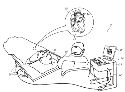
create a sufficiently large
lesion to effectively ablate an abnormal tissue focus, or block an aberrant
conduction pattern, and the
undesirable effects of excessive local heating. If the radiofrequency device
creates too small a lesion,
then the medical procedure could be less effective, or could require too much
time. On the other
hand, if tissues are heated excessively then there could be local charring
effects, coagulum, and or
steam pops due to overheating. Such overheated areas can develop high
impedance, and may form a
functional barrier to the passage of heat. The use of slower heating provides
better control of the
ablation, but unduly prolongs the procedure.
[0005] Self-
regulating tissue ablators have been proposed to achieve the desired control.
For
example, PCT International Publication W09600036 discusses
1
CA 02886364 2015-03-26
ablation of body tissue in which ablating energy is conveyed individually to
multiple emitters in a se-
quence of power pulses. The temperature of each emitter is periodically sensed
and compared to a
desired temperature established for all emitters to generate a signal
individually for each emitter
based upon the comparison. The power pulse to each emitter is individually
varied, based upon the
signal for that emitter to maintain the temperatures of all emitters
essentially at the desired tempera-
ture during tissue ablation.
[0006]
Commonly assigned U.S. Patent Application Publication No. 2012/0157890, which
is
herein incorporated by reference, discloses performing tissue ablation out by
determining a measured
temperature of the tissue and a measured power level of transmitted energy to
a probe, and control-
ling the power output level responsively to a function of the measured
temperature and the meas-
ured power level.
SUMMARY OF THE INVENTION
[0007]
According to disclosed embodiments of the invention, temperature is measured
ac-
cording to the changes in impedance between a pair of irrigated electrodes on
a catheter. The usual
temperature sensor found on such catheters can be omitted.
[0008]
There is provided according to embodiments of the invention a method of
ablation,
which is carried out by inserting a probe having an ablation electrode and a
plurality of microelec-
trodes into a body of a living subject. The method is further carried out by
establishing a contacting
relationship between two of the microelectrodes and a target tissue, and
energizing the ablation elec-
trade. While the ablation electrode is energized the method is further carried
out by measuring an
impedance between the two microelectrodes, and responsively to the impedance
adjusting the power
level of the ablation electrode.
[0009] A
further aspect of the method includes iteratively measuring the impedance, and
estimating a tissue temperature from a change between two measurements of the
impedance.
[oolo] Yet another
aspect of the method includes making a determination that the tissue
temperature exceeds a predetermined limit, and responsively to the
determination reducing the pow-
er of the ablation electrode. The power may be reduced to zero to deactivate
the ablation electrode.
[owl]
According to still another aspect of the method, measuring an impedance is per-
formed by polling the microelectrodes to determine pairwise impedances
therebetween. The pair of
the selected microelectrodes may have the highest and the second highest
measured impedance.
[0012]
According to a further aspect of the method, measuring an impedance includes
measuring a bipolar impedance between the selected pair of the
microelectrodes.
[0013]
According to an additional aspect of the method, establishing a contacting
relation-
ship includes determining a location and orientation of the tip of the probe
with respect to the target
tissue with six degrees of freedom.
2
CA 02886364 2015-03-26
[0014]
According to another aspect of the method, measuring an impedance includes
poll-
ing the microelectrodes to determine impedances between the microelectrodes
and an indifferent
electrode.
[0015]
One aspect of the method includes deploying an inflatable balloon through a
lumen
of the probe, wherein the microelectrodes are disposed circumferentially about
the longitudinal axis
of the balloon on its exterior wall.
[0016]
Another aspect of the method the balloon includes a subassembly comprising a
plu-
rality of strips extending longitudinally on the exterior wall of the balloon,
and the microelectrodes
are disposed on the strips.
[0017] There is
further provided according to embodiments of the invention an apparatus
for carrying out the above-described method.
BRIEF DESCRIPTION OF THE SEVERAL VIEWS OF 'THE DRAWINGS
[0018]
For a better understanding of the present invention, reference is made to the
de-
tailed description of the invention, by way of example, which is to be read in
conjunction with the
following drawings, wherein like elements are given like reference numerals,
and wherein:
[0019]
Fig. 1 is a pictorial illustration of a system for performing ablative
procedures, which
is constructed and operative in accordance with a disclosed embodiment of the
invention;
[0020]
Fig. 2 is a schematic diagram of a distal portion of a catheter, in accordance
with an
embodiment of the invention;
[0021] Fig. 3 is a
sectional view through line 3-3 of Fig. 2, in accordance with an embodi-
ment of the invention;
[0022]
Fig. 4 is an electrical schematic of circuitry for impedance measurement
during abla-
tion, in accordance with an embodiment of the invention;
[0023]
Fig. 5 is a schematic diagram of a distal portion of a catheter, in accordance
with an
embodiment of the invention'
[0024]
Fig. 6 is a sectional view through line 6-6 of Fig. 5, in accordance with an
embodi-
ment of the invention;
[0025]
Fig. 7 is a schematic sectional view of a portion of an ablation electrode, in
accord-
ance with an embodiment of the invention;
[0026] Fig. 8 is a
pictorial view of a balloon assembly for a cardiac catheter in accordance
with an alternate embodiment of the invention;
[0027]
Fig. 9 is a tracing of bipolar impedance measured between two microelectrodes
of a
catheter, in accordance with an embodiment of the invention; and
[0028]
Fig. 10 is a flow chart of a method of tissue temperature determination during
a
catheterization procedure, in accordance with an embodiment of the invention.
3
CA 02886364 2015-03-26
DETAILED DESCRIPTION OF THE INVENTION
[0029] In
the following description, numerous specific details are set forth in order to
provide a thorough understanding of the various principles of the present
invention. It will be
apparent to one skilled in the art, however, that not all these details are
necessarily needed for
practicing the present invention. In this instance, well-known circuits,
control logic, and the details of
computer program instructions for conventional algorithms and processes have
not been shown in
detail in order not to obscure the general concepts unnecessarily.
[0030]
Turning now to the drawings, reference is initially made to Fig. 1, which is a
pictorial
illustration of a system 10 for performing ablative procedures on a heart 12
of a living subject, which
is constructed and operative in accordance with a disclosed embodiment of the
invention. The system
comprises a catheter 14, which is percutaneously inserted by an operator 16
through the patient's
vascular system into a chamber or vascular structure of the heart. The
operator 16, who is typically a
physician, brings the catheter's distal tip 18 into contact with the heart
wall at an ablation target site.
Electrical activation maps may then be prepared, according to the methods
disclosed in U.S. Patent
Nos. 6,226,542, and 6,301,496, and in commonly assigned U.S. Patent No.
6,892,091, whose disclo-
sures are herein incorporated by reference. Although the embodiment described
with respect to Fig. 1
is concerned primarily with cardiac ablation, the principles of the invention
may be applied, mutatis
mutandis, to other catheters and probes and to body tissues other than the
heart.
[0031]
Areas determined to be abnormal by evaluation of the electrical activation
maps can
be ablated by application of thermal energy, e.g., by passage of
radiofrequency electrical current
through wires in the catheter to one or more electrodes at the distal tip 18,
which apply the radiofre-
quency energy to the myocardium. The energy is absorbed in the tissue, heating
it to a point (typical-
ly above 60 C) at which it permanently loses its electrical excitability. When
successful, this proce-
dure creates non-conducting lesions in the cardiac tissue, which disrupt the
abnormal electrical
pathway causing the arrhythmia. Alternatively, other known methods of applying
ablative energy can
be used, e.g., ultrasound energy, as disclosed in U.S. Patent Application
Publication No. 2004/0102769,
whose disclosure is herein incorporated by reference. The principles of the
invention can be applied
to different heart chambers, when many different cardiac arrhythmias are
present.
[0032]
The catheter 14 typically comprises a handle 20, having suitable controls on
the
handle to enable the operator 16 to steer, position and orient the distal end
of the catheter as desired
for the ablation. To aid the operator 16, the distal portion of the catheter
14 contains position sensors
(not shown) that provide signals to a positioning processor 22, located in a
console 24. The con-
sole 24 typically contains an ablation power generator 25. The catheter 14 may
be adapted to conduct
ablative energy to the heart using any known ablation technique, e.g.,
radiofrequency energy, ultra-
sound energy, and laser energy. Such methods are disclosed in commonly
assigned U.S. Patent
Nos. 6,814,733, 6,997,924, and 7,156,816, which are herein incorporated by
reference.
4
CA 02886364 2015-03-26
=
[0033]
The positioning processor 22 is an element of a positioning sub-system of the
sys-
tem 10 that measures location and orientation coordinates of the catheter 14.
[0034] In
one embodiment, the positioning sub-system comprises a magnetic position
tracking arrangement that determines the position and orientation of the
catheter 14 by generating
magnetic fields in a predefined working volume and sensing these fields at the
catheter. The magnetic
position tracking arrangement typically comprises a set of external radiators,
such as field generating
coils 28, which are located in fixed, known positions external to the patient.
The field generating
coils 28 are driven by field generators (not shown), which are typically
located in the console 24, and
generate fields, typically electromagnetic fields, in the vicinity of the
heart 12.
[0035] In an
alternative embodiment, a radiator in the catheter 14, such as a coil,
generates
electromagnetic fields, which are received by sensors (not shown) outside the
patient's body.
[0036]
Some position tracking techniques that may be used for this purpose are
described,
for example, in the above-noted U.S. Patents 6,690,963, and in commonly
assigned U.S. Patent
Nos. 6,618,612 and 6,332,089, and U.S.
Patent Application Publications 2004/0147920,
and 2004/0068178, whose disclosures are all incorporated herein by reference.
Although the position-
ing sub-system shown in Fig. 1 uses magnetic fields, the methods described
below may be implement-
ed using any other suitable positioning system, such as systems based on
electromagnetic fields,
acoustic or ultrasonic measurements.
[0037] As
noted above, the catheter 14 is coupled to the console 24, which enables the
op-
erator 16 to observe and regulate the functions of the catheter 14. Console 24
includes a processor,
preferably a computer with appropriate signal processing circuits. The
processor is coupled to drive a
monitor 30. The signal processing circuits typically receive, amplify, filter
and digitize signals from
the catheter 14, including signals generated by the above-noted sensors and a
plurality of sensing
electrodes (not shown) located distally in the catheter 14. The digitized
signals are received and used
by the console 24 to compute the position and orientation of the catheter 14
and to analyze the elec-
trical signals from the electrodes. The information derived from this analysis
may be used to generate
an electrophysiological map of at least a portion of the heart 12 or
structures such as the pulmonary
venous ostia, for diagnostic purposes such as locating an arrhythmogenic area
in the heart or to facil-
itate therapeutic ablation.
[0038] Typically,
the system 10 includes other elements, which are not shown in Fig. 1 for
the sake of simplicity. For example, the system 10 may include an
electrocardiogram (ECG) monitor,
coupled to receive signals from one or more body surface electrodes, to
provide an ECG synchroniza-
tion signal to the console 24. The system 10 typically also includes a
reference position sensor, either
on an externally-applied reference patch attached to the exterior of the
subject's body, or on an in-
ternally-placed catheter, which is inserted into the heart 12 maintained in a
fixed position relative to
the heart 12. Conventional pumps and lines for circulating liquids through the
catheter 14 for cooling
the ablation site are provided.
5
CA 02886364 2015-03-26
[0039]
One system that embodies the above-described features of the system 10 is the
CARTO 3 System, available from Biosense Webster, Inc., 3333 Diamond Canyon
Road, Diamond Bar,
CA 91765. This system may be modified by those skilled in the art to embody
the principles of the
invention described herein.
[0040] Reference
is now made to Fig. 2, which is a schematic diagram of a distal portion of
a catheter 32, in accordance with an embodiment of the invention, which is
suitable for use in the
system 10 (Fig. 1). An ablation electrode 34 is disposed at the tip of the
catheter 32. A hydraulic
line 36 supplies irrigation fluid to cool an ablation site when the ablation
electrode 34 is active.
Pores 38 provide egress for the irrigation fluid. While the pores 38 may be
placed through the abla-
tion electrode 34, this is not essential, so long as the irrigation fluid
exiting the pores 38 is able to
bathe the ablation site. Mapping electrodes 40 may be provided for purpose of
conventional electro-
physiological mapping.
[0041] A
series of microelectrodes 42 are positioned distally on the external surface
of the
catheter 32, They are disposed circumferentially its longitudinal axis 44 and
close to the ablation elec-
trode 34 such that at least two of the microelectrodes 42 and the ablation
electrode 34 can be con-
currently in firm contact with the target tissue when ablation is carried out.
The inventors have
found that measurements of bipolar impedance between the two contacting
microelectrodes 42 is
useful in determining the temperature of the target tissue.
[0042]
One way of identifying a pair of contacting microelectrodes 42 is to determine
their
pairwise impedances, e.g., by polling. Either or both the magnitude and the
phase of the impedance
can be used. An additional way, due to the microelectrodes' small size, is to
measure the impedance
between a microelectrode and a back patch (indifferent electrode) to identify
contact. Alternatively,
the identification of a contacting pair of microelectrodes 42 can be achieved
by exploiting the ability
of a position tracking system (Fig. i) such as the aforementioned CARTO system
to determine the
position and orientation of the catheter 32 with six degrees of freedom.
Contact between a particular
pair of the microelectrodes 42 can be determined by reference to the location
and orientation of the
tip of the catheter with respect to the target tissue.
[0043]
Reference is now made to Fig. 3, which is a sectional view through line 3-3 of
Fig. 2,
in accordance with an embodiment of the invention. The microelectrodes 42 are
distributed generally
evenly in perforations distributed about the circumference of the catheter 32.
They microelec-
trodes 42 may be bonded within the perforations by suitable glues or bonding
material. A flat profile
of the outer surface exposed to the tissue is shown in this example. However,
the profile of the mi-
croelectrodes 42 may be convex or sinusoidal. The profile of the
microelectrodes 42 may be level with
or raised above the external surface of the catheter 32. Wires 46 electrically
connect the microelec-
trodes 42 to impedance measuring circuitry (not shown) via a cable 48.
Hydraulic conduit 50 con-
ducts irrigation fluid to the pores 38 (Fig. i).
6
CA 02886364 2015-03-26
=
[0044]
The microelectrodes 42 are composed of an electrically conductive material,
such as
platinum, palladium, gold, stainless steel, silver or silver chloride, all of
which tend to maximize the
coupling between the microelectrodes and the target tissue. The
microelectrodes 42 are substantially
solid, but may include a bore 52 that can receive and assure electrical
connection between the
wires 46 and the microelectrodes 42. The wires 46 may be secured to the
microelectrodes 42 e.g., by
solder 54, glue, or other convenient methods. Further details of the
manufacture of the microelec-
trodes 42 are shown in U.S. Patent Application Publication No. 2014/0058375
and U.S. Patent No.
8,414,579, the disclosures of which are herein incorporated by reference.
[0045]
The microelectrodes 42 are dimensioned such that a desired number of them can
be
accommodated about the circumference of the catheter 32. The diameter of the
microelectrodes 42
should be no greater than half the length of the ablation electrode 34,
preferably no greater than
one-fourth the length of the ablation electrode 34. The microelectrodes 42
should be spaced apart
from one another by no more than one-half the diameter of the microelectrodes
42 (or one-half the
shortest dimension in the case of non-circular embodiments).
[0046] Reference is
now made to Fig. 4, which is an electrical schematic of circuitry 56 for
impedance measurement during ablation for temperature determination, in
accordance with an em-
bodiment of the invention. Multiple microelectrodes 58 are connected by
respective lead wires 60 via
the catheter handle (not shown). A signal generator 62 (SG) sends a high
frequency test signal, e.g.,
an alternating current (AC) signal at about 2 pamps, in the frequency range of
about 10 kHz to about
100 kHz, preferably about 50 kHz, to a multiplexer 64 via a high output
impedance buffer 66 (113).
[0047]
The multiplexer 64 has multiple channels 68, each of which is in communication
one of the microelectrodes 58, which receive the same current.
[0048] A
return electrode 70 is also driven by the signal generator 102. The signal to
the
return electrode 70 is first inverted in phase by an inverter 72 and
conditioned by high output im-
pedance buffer 74.
[0049]
Impedance measurement circuitry 76 (IMC) measures the impedance of each of the
microelectrodes 58 as an indicator of the extent of its respective tissue
contact and the condition of
the tissue being ablated. The impedance measurement circuitry 76 includes a
differential amplifier 78
(DA), an amplifier 80 (AMP) and a synchronous detector 82 (SD). The
differential amplifier 78
measures a difference signal, specifically the voltage across a selected
microelectrode 58 and the re-
turn electrode 70. The difference signal is further amplified by the amplifier
80 whose output is sent
to the synchronous detector 82, which transforms the AC signal into a direct
current (DC) signal and
decreases the sensitivity of the circuitry 56 to external noise. The signal
from the synchronous detec-
tor 82 is then used by a microcontroller 84 to control the multiplexer 64. To
that end, the microcon-
troller 84 continuously stores in a memory 86 a plurality of different
impedance signals from the
synchronous detector 82 that equals the plurality of channels 68 in the
multiplexer 64 (which is at
7
CA 02886364 2015-03-26
least the plurality of microelectrodes 58 on the catheter), along with
identification information on the
channels 68 associated with each impedance value stored.
[0050] As
such, the microcontroller 84 is at any time capable of identifying the
channels 68
(and hence the microelectrodes 58) exhibiting the highest impedance value,
which should be the ml-
croelectrode with the greatest tissue contact. Further details of the
circuitry 56 are found in com-
monly assigned U.S. Patent Application Publication No. 2011/0106075, which is
herein incorporated by
reference.
[0051]
Appropriate bipolar impedances between two microelectrodes can then be
measured.
This may be done by selecting the microelectrodes with the highest and second
highest impedance,
and providing signals from the microcontroller 84 to configure one of the two
microelectrodes as the
return electrode 70.
First Alternate Embodiment.
[0052]
Reference is now made to Fig. 5, which is a schematic diagram of a distal
portion of
a catheter 88, in accordance with an embodiment of the invention. Mounted on
an ablation elec-
trode 90 is a series of microelectrodes 92. The microelectrodes 92 are
elongated in the longitudinal
direction of the catheter 88, which allows a larger number to be accommodated
than is the case with
round microelectrodes having the same surface area. The elongated
configuration is not essential,
and, other configurations of the microelectrodes may be used. The
microelectrodes 92 are thermally
and electrically isolated from the ablation electrode 90 by an insulation
layer 94.
[0053] Reference is
now made to Fig. 6, which is a sectional view through line 6-6 of Fig. 5,
in accordance with an embodiment of the invention. The microelectrodes 92 are
disposed within per-
forations through the ablation electrode 90. The insulation layer 94 surrounds
the microelectrodes 92
and separates the microelectrodes 92 from the ablation electrode 90. As
described in the above-noted
U.S. Patent Application Publication No. 2014/0058375, the insulation layer 94
may be composed of
the suitable electrically and thermally insulative material, such as a high
temperature thermoset plas-
tic with high dielectric properties, e.g., polyimide or plastics from the
phenolic group, such as Bake-
lite or Ultem plastics. The insulation layer 94 and microelectrodes 92 may
be bonded within the
perforations using a suitable bonding material, such as epoxy.
Second Alternate Embodiment.
[0054] This
embodiment is similar to the embodiments of Figs. 5 and 6, except that it is
unnecessary to place large perforations in ablation electrode. Reference is
now made to Fig. 7, which
is a schematic sectional view of a portion of an ablation electrode 96, in
accordance with an embod-
iment of the invention. A microelectrode 98 is embedded in a recess 100 formed
in the wall of the
ablation electrode 96 and separated from the ablation electrode 96 by a
thermally and electrically
insulative layer 102. A relatively small perforation 104 extending from the
base of the recess 100
8
CA 02886364 2015-03-26
through the wall of the ablation electrode 96 carries a wire 106 into the
interior of the catheter to
ultimately connect to impedance measuring circuitry (not shown).
Third Alternate Embodiment.
[0055] In
this embodiment electrodes of a lasso, or loop, catheter having capabilities
for ab-
lation may be configured for bipolar impedance measurement. Such a catheter is
known, for example
from commonly assigned U.S. Patent Application Publication No. 2010/0168548,
which is hereby in-
corporated by reference. Electrodes in contact with the tissue may be
determined as described above.
Fourth Alternate Embodiment.
[0056] in
this embodiment the microelectrodes are disposed on a flexible circuit
substrate
and adhered to the exterior of a balloon that can be inserted through a
catheter and applied to the
target as described in copending Application No 14/578,807, entitled Balloon
for Ablation around
Pulmonary Veins, which is herein incorporated by reference. Reference is now
made to Fig. 8, which
is a pictorial view of a balloon assembly for a cardiac catheter in accordance
with an alternate em-
bodiment of the invention. A subassembly, e.g., a flexible circuit board 101
is configured as multiple
strips or bands radiating from shaft 103, extending longitudinally and
adhering to the exterior wall of
balloon 105 Arrays of microelectrodes 107 are disposed on the circuit board
101.
Operation.
[0057]
Reference is now made to Fig. 9, which is a prospective example of a tracing
108
that indicates bipolar impedance measured between two microelec-trodes of a
catheter during an ab-
lation procedure, in accordance with an embodiment of the invention. Prior to
time TO the microelec-
trodes are out of contact with tissue, as evidenced by a relatively low
impedance. At time TO, the
electrodes have come into tissue contact, and the bipolar impedance rises. At
time Ti, the ablator is
energized. Tissue temperature rises during the interval between times Ti, T2,
as evidenced by gradu-
ally decreasing bipolar impedance. At time T2, the ablator power is reduced,
as the impedance is ap-
proaching a threshold indicated by broken line 110. Nevertheless, during the
time interval T2-T3, im-
pedance continues to decrease, albeit at a slower rate than prior to time T2.
At time T3, the thresh-
old of line 110 has been reached, and the ablator is deactivated. Actual
impedance values vary accord-
ing to the surface area of the microelectrodes, and are typically in the order
of several hundred
Ohms.
[0058] Reference is
now made to Fig. 10, which is a flow-chart of a method of tissue tem-
perature determination during a catheterization procedure, in accordance with
an embodiment of the
invention. At initial step 112, a catheter in accordance with any of the above
embodiments is inserted
into contact with target tissue of a subject. The target is typically the
endocardial surface of a heart
chamber.
[0059] Next, at
step 114 two microelectrodes of the catheter are determined to be in contact
with the target. This determination may be made, for example, using the
position processor of the
9
CA 02886364 2015-03-26
CARTO system as noted above, by polling the microelectrodes pairwise until an
impedance level con-
sistent with tissue contact is identified, or measuring the impedance between
a microelectrode and a
backpatch (indifferent electrode), or a combination of the above.
[0060]
Next, at step 116 the ablator is energized and its power level set. Irrigation
fluid is
caused to flow onto the ablation electrode and the target tissue.
[0061]
Next, at step 118, while the ablator is active, bipolar impedance measurements
are
taken between the pair of electrodes identified in step 114.
[0062]
Next, at step 120, tissue temperature is estimated based on the change in the
imped-
ance measurements, either absolute or as a percentage, and using, for example,
empirical data from
simulations that reveals a correlation similar to the plot in Fig. 9.
[0063]
Next, at decision step 122, it is determined if the temperature is too high
for contin-
ued ablation. If the determination at decision step 122 is negative, then
control returns to step 116.
[0064] If
the determination at decision step 122 is affirmative then control proceeds to
final
step 124, where the power level of the ablator is lowered. The power level of
the ablator may be ad-
justed manually or automatically by a controller in accordance with known
algorithms, for example as
taught in commonly assigned U.S. Patent Application Publication No.
2012/0157890, which is herein
incorporated by reference. The process iterates until the time set for the
ablation expires, at which
the power to the ablator is reduced or the ablator deactivated entirely by
reducing the power to zero.
[0065] it
will be appreciated by persons skilled in the art that the present invention
is not
limited to what has been particularly shown and described hereinabove. Rather,
the scope of the
present invention includes both combinations and sub-combinations of the
various features described
hereinabove, as well as variations and modifications thereof that are not in
the prior art, which would
occur to persons skilled in the art upon reading the foregoing description.
10