Note: Descriptions are shown in the official language in which they were submitted.
CA 02886769 2015-03-31
1
DESCRIPTION
METHOD FOR MEASURING HEMAGGLUTININ FROM INFLUENZA VIRUS
TECHNICAL FIELD
[0001]
The present invention relates to a method for measuring haemagglutinin
which is an antigen of an influenza virus.
BACKGROUND ART
[0002]
Influenza viruses belong to Orthomyxoviridae and are classified into type A,
type B and type C depending on the antigenicity of the nucleoprotein and the
matrix
protein located in the virus. The type A and type B viruses have been
prevalent
every year, and in particular, the type A virus is classified into 16 subtypes
of
haemagglutinin and 9 subtypes of neuraminidase depending on the glycoproteins
which are surface antigens on the particles, and undergoes antigenic variation
easily.
Therefore, the vaccine strain has to be selected based on the prediction of
the
prevalence in each season, and it is necessary to prepare a reagent for
measuring
haemagglutinin which is a main antigen of vaccine in accordance with the
change of
the strain.
PRIOR ART DOCUMENTS
[Non-patent Documents]
[0003]
[Non-patent Document 1] Mahmood N, Hay AJ., "An ELISA utilizing
immobilised snowdrop lectin GNA for the detection of envelope glycoproteins of
HIV and Sly" J Immunol Methods.,151 (1992) 9-13
[Non-patent Document 2] Legastelois I. et al., "Avian glycan-specific IgM
monoclonal antibodies for the detection and quantitation of type A and B
CA 02886769 2015-03-31
2
haemagglutinins in egg-derived influenza vaccines", J Virol Methods., 178
(2011)
129-136
SUMMARY OF THE INVENTION
PROBLEMS TO BE SOLVED BY THE INVENTION
[0004]
Although examples of the method for measuring haemagglutinin with high
sensitivity and high throughput include a sandwich ELISA using monoclonal
antibodies and polyclonal antibodies, two kinds of antibodies specific to the
strain
are necessary, and it is difficult to provide monoclonal antibodies for
measuring
haemagglutinin of new vaccine strains or pandemic viruses in view of the
period to
prepare the antibodies. Further, the measuring method using monoclonal
antibodies
specific to sugar chains of birds as described in Non-patent Document 2 is not
widely
used because of the problems in supply and the like due to the unordinary
antibody,
and the method cannot be used for measuring haemagglutinin of an influenza
virus
amplified in mammalian cells.
[0005]
Accordingly, an object of the present invention is to provide a novel method
for measuring haemagglutinin of an influenza virus, which can construct an
assay
system in a shorter period of time than a sandwich immunoassay method using
two
kinds of anti-haemagglutinin antibodies.
MEANS FOR SOLVING THE PROBLEMS
[0006]
As a result of an intensive research, the present inventors found that a
lectin
bound to haemagglutinin of an influenza virus but did not bind to an antibody,
and
inferred that when the haemagglutinin is sandwiched between the lectin and an
anti-
haemagglutinin antibody, only one kind of anti-haemagglutinin antibody is
necessary,
and an assay system can be constructed in a shorter period of time than a
sandwich
81787074.
3
immunoassay method using two kinds of anti-haemagglutinin antibodies, thereby
completing the
present invention.
[0007]
That is, the present invention provides a method for measuring haemagglutinin
of an influenza
virus by a sandwich immunoassay method, the method comprising sandwiching the
haemagglutinin
between a lectin which binds to the haemagglutinin but does not bind to an
antibody, and an anti-
haemagglutinin antibody which undergoes antigen-antibody reaction with the
haemagglutinin.
[0007a]
In one aspect, the present invention provides a method for measuring
haemagglutinin of an
influenza virus by a sandwich immunoassay method, said method comprising
sandwiching the
haemagglutinin between a lectin which binds to the haemagglutinin but does not
bind to an antibody,
the lectin being at least one selected from the group consisting of Ricinus
communis agglutinin,
Datura stramonium lectin and Erythrina cristagalli lectin, and an anti-
haemagglutinin antibody which
undergoes antigen-antibody reaction with the haemagglutinin.
EFFECT OF THE INVENTION
[0008]
By the present invention, haemagglutinin of an influenza virus can be measured
with high
accuracy by a sandwich immunoassay method by using one kind of anti-
haemagglutinin antibody.
BRIEF DESCRIPTION OF THE DRAWINGS
[0009]
Fig. 1-1 shows Western blotting diagrams illustrating fraction analysis
results by sucrose
density gradient centrifugation after treating inactivated whole particle
virus with each surfactant
below. A: no treatment, B: treatment with 1.0% Triton X100TM, C: treatment
with 1.0% NP40TM, D:
treatment with 1.0% Tween 80TM E: treatment with 1.0% Brij 35TM and F:
treatment with 1.0%
CHAPS.
Fig. 1-2 shows the following diagrams similarly to Fig. 1-1: G: treatment with
1.0%
Zwittergent 3-14', H: treatment with 1.0% CTAB, I: treatment with 0.3% SDS, J:
treatment with 4.0
M Urea, and K: treatment with 3.0 M of guanidine hydrochloride.
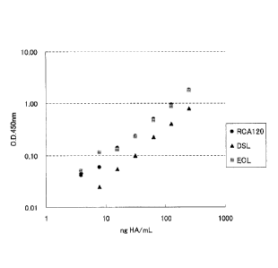
Fig. 2 shows the relationship between the concentrations of haemagglutinin and
absorbances,
which was measured by a sandwich immunoassay method in Examples below.
MODE FOR CARRYING OUT THE INVENTION
Date Recue/Date Received 2021-07-14
CA 02886769 2015-03-31
4
[0010]
As described above, the method of the present invention is a method for
measuring haemagglutinin by a sandwich immunoassay method, the method
comprising sandwiching the haemagglutinin between a lectin which binds to the
haemagglutinin of an influenza virus (hereinafter referred to as simply
"haemagglutinin") but does not bind to an antibody, and an anti-haemagglutinin
antibody which undergoes antigen-antibody reaction with the haemagglutinin.
[0011]
The lectin used in the method of the present invention binds to
haemagglutinin. The existence of binding with the haemagglutinin can be
confirmed by lectin blot analysis (a labeled lectin is reacted with
haemagglutinin
transferred on a PVDF membrane to determine whether the label is detected or
not)
which is specifically described in Reference Example 2 below. The term "does
not
bind to an antibody" means that the lectin does not bind to an immunoglobulin
which
belongs to the same species and same class as an immunoglobulin constituting
an
antibody to be used for the immunoassays (usually, IgG of mouse, rabbit, sheep
or
the like). The fact that the lectin does not bind to IgG derived from each
animal
such as mouse, rabbit, sheep or the like can be confirmed by determining
whether the
absorbance measured by ELISA with HRP-labeled IgG of each animal is less than
twice, preferably less than 1.5 times the mean value of those of negative
controls
(blank) as described in Reference Example 3 below. Preferred examples of the
lectin which binds to haemagglutinin but does not bind to an antibody include
Datum
stramonium lectin (DSL), Erythrina cristagalli lectin (ECL) and Ricinus
communis
agglutinin (RCA 120). The lectin which binds to haemagglutinin but does not
bind
to an antibody may be used individually, or two or more of these may be used
in
combination.
[0012]
CA 02886769 2015-03-31
In the method of the present invention, the lectin is preferably immobilized
on
a solid phase to carry out sandwich immunoassays. As the solid phase, any of
those
used in well-known sandwich immunoassays such as sandwich ELISA may be
employed, and examples thereof include plates, tubes, beads, membranes and
gels.
5 The examples of materials of the solid phase include polystyrene,
polypropylene,
nylon, latex, glass, cross-linked dextrin, agarose, cross-linked agarose and
poly
acrylamide.
[0013]
As the method for absorbing the lectin on the solid phase, covalent method,
physical adsorption method, ionic bonding method, biochemically specific
binding
method (for example, a biotinylated lectin is allowed to bind to a solid phase
on
which streptavidin is immobilized) or the like may be employed. In particular,
physical adsorption method and biochemically specific binding method are
preferred
from the viewpoint of simplicity of the operation.
[0014]
Examples of the physical adsorption method herein include a method in
which a lectin dissolved in a buffer, pH 7 to 9, containing 0.05% Tween 20
(trade
name) (for example, Tris-HCl buffer physiological saline, phosphate buffer
physiological saline and carbonate buffer) is added to a solid phase (for
example,
wells of microplate), and the resultant is left to stand at room temperature
for about 1
to 2 hours, or at about 4 C over night to attain the adsorption. As for the
biochemically specific binding method, since solid phases on which
streptavidin is
immobilized (plates, beads or the like) are commercially available, examples
thereof
include a method in which a lectin dissolved in a buffer, pH 7 to 9,
containing 0.05%
Tween 20 (trade name) (for example, Tris-HCl buffer physiological saline,
phosphate buffer physiological saline and carbonate buffer) is added to the
commercially available solid phase on which streptavidin is immobilized, and
the
CA 02886769 2015-03-31
6
resultant is left to stand at room temperature for about 1 to 2 hours, or at
about 4 C
over night to attain the adsorption (see Examples below). In any of the
physical
adsorption method and biochemically specific binding method, although the
concentration of lectin to be reacted with the solid phase is not restricted,
the final
concentration thereof is usually about 10 pig,/mL to about 60 gg/mL.
[0015]
In some cases, surface areas on which lectin was not adsorbed may remain on
the surface of the solid phase after the adsorption of the lectin, and in
cases where
haemagglutinin or other molecular species in a sample are adsorbed to the
areas,
accurate measurement results may not be obtained. Therefore, it is preferable
that a
blocking substance is added thereto prior to contacting the sample with the
solid
phase, to block the areas on which the lectin is not adsorbed. Examples of the
blocking substances include serum albumin, casein, milk protein, lactic acid
fermentation product, collagen and the decomposed products thereof which can
be
collected from mammal such as bovine, and those which are commercially
available
as blocking substances used in immunoassays may also be employed.
[0016]
The anti-haemagglutinin antibody used in method of the present invention
undergoes antigen-antibody reaction with haemagglutinin of an influenza virus,
and
may be a monoclonal antibody or polyclonal antibody. In the type-specific
assay of
an influenza virus, an anti-haemagglutinin monoclonal antibody which undergoes
antigen-antibody reaction specifically with each type of haemagglutinin is
usually
employed.
[0017]
The anti-haemagglutinin antibody is usually labeled. Examples of labeling
substances to be used for label include enzymes (peroxidase, alkaline
phosphatase, 13-
galactosidase, luciferase, acetylcholinesterase and the like), isotopes (1 2 5
1 3 11 3 H
CA 02886769 2015-03-31
7
and the like), fluorescent dyes (luminol, fluorescein isothiocyanate,
umbelliferone, 7-
amino-4-methylcoumarin-3-acetic acid and the like), chemiluminescent
substance,
hapten, biotin, avidin (for example, streptavidin and the like), but the
labeling
substances are not restricted as long as they can be commonly used for
labeling
proteins. The labeling substance herein includes a substance whichper se is
not
detected directly, like biotin, and which is used in a method in which a
material (for
example, avidin) capable of specifically binding to the substance, which
material is
bound to a detectable label, is used in combination.
[0018]
The above-described method for labeling an antibody can be appropriately
selected form known methods suitable to the labeling substance, for example,
in the
case of labeling enzymes, glutaraldehyde method, periodate crosslinking
method,
maleimide crosslinking method, carbodiimide method, activated ester method and
the like; and in case of label with radioisotopes, chloramine T method,
lactoperoxidase method and the like. Since the labeled anti-haemagglutinin
antibodies against various types are commercially available, a commercially
available one may be employed.
[0019]
In the method of the present invention, usually, the above-described lectin
immobilized on the solid phase, the labeled anti-haemagglutinin antibody and a
sample containing haemagglutinin of an influenza virus are reacted, and after
washing the solid phase, the labels bound to the solid phase are measured.
[0020]
The sample to which the method of the present invention is applied may
contain influenza virus particles or may be obtained by extracting
haemagglutinin
from the particles as long as the sample contains haemagglutinin of the
influenza
virus. It is preferable to extract haemagglutinin because haemagglutinin can
be
CA 02886769 2015-03-31
8
assayed regardless of the manner of existence, whether haemagglutinin exists
in
virus particles or in a free state, and accuracy of the assay can be
increased. The
extraction of haemagglutinin can be carried out by using a cationic or an
anionic
surfactant (both are collectively hereinafter referred to as "ionic
surfactant").
Preferred examples of the ionic surfactant include sodium dodecyl sulfate
(SDS),
lithium dodecyl sulfate (LiDS), hexadecyltrimethylammonium bromide (CTAB),
hexadecyltrimethylammonium chloride (CTAC) and hexadecylpyridinium chloride
(HPC); and hexadecyltrimethylammonium bromide (CTAB) is particularly
preferable. The ionic surfactant can be used individually, and two or more of
these
can be used in combination.
[0021]
Although the treatment conditions with the ionic surfactant is not restricted,
the ionic surfactant is preferably added to a sample to a final concentration
of 0.1 to
2.0%, and the resultant is left to stand or stirred at 37 C for about 1 to
about 2 hours
to extract haemagglutinin from virus particles.
[0022]
The lectin immobilized on the solid phase, a sample and the labeled anti-
haemagglutinin antibody may be reacted at the same time; or the lectin
immobilized
on the solid phase and a sample may be firstly reacted, and after washing the
solid
phase, the labeled anti-haemagglutinin antibody is reacted therewith; or a
sample and
the labeled anti-haemagglutinin antibody may be firstly reacted to form an
immune
complex, and the lectin immobilized on the solid phase is reacted therewith.
The
reaction can be carried out at room temperature for about 30 minutes to about
120
minutes. The final concentration of haemagglutinin in reaction system is
usually
about 1 ng/mL to about 1 i.tg/mL, and the final concentration of the labeled
anti-
haemagglutinin antibody is usually about 0.2 ng/mL to about 50 pz/mL.
[0023]
CA 02886769 2015-03-31
9
After the reaction, the solid phase is washed, and the labels bound to the
solid
phase are measured. Examples of washing solutions include buffers to which
surfactants such as Tween (trade name) type surfactant are added (for example,
phosphate buffer, phosphate buffer physiological saline, Tris-HC1 buffer, Tris-
HC1
buffer physiological saline). The method for detecting the labeled substance
are
different depending on the labeling substance to be used, and in the case of
using
biotin as a labeling substance, examples thereof include a method in which an
enzyme such as peroxidase is allowed to be bound to a complex containing
biotin as
a labeling substance through streptavidin or the like, a chromogenic substance
such
as tetramethylbenzidine and hydrogen peroxide solution as substrates of the
enzyme
are added thereto measure the degree of coloring of the product caused by
enzyme
reaction based on the change in absorbance. In the case of using a fluorescent
substance or a chemiluminescence substance as a labeling substance, examples
thereof include a method for measuring fluorescence or luminescence of the
solution
obtained after the reaction.
[0024]
Instead of the labeled anti-haemagglutinin antibody, a non-labeled anti-
haemagglutinin antibody is reacted therewith, a labeled anti-immunoglobulin
antibody is further reacted therewith, and after washing, the labels bound to
the solid
phase may be measured (indirect antibody technique). Since the number of
antigen-
antibody reaction is increased by one time in the indirect antibody technique,
when
quick test is necessary, the above-described direct technique using a labeled
anti-
haemagglutinin antibody is preferred.
[0025]
In the measuring method of the present invention, the relationship between
the concentration of haemagglutinin and the detection results of the labeled
substances is plotted using a standard solution containing a known
concentration of
CA 02886769 2015-03-31
haemagglutinin to prepare a calibration curve, and the concentration of
haemagglutinin in a sample may be quantified by using the detection result of
the
sample having unknown concentration and the above-described calibration curve.
[0026]
5 An preferable embodiment of the measuring method of the present
invention
will now be described. Firstly, the lectin is adsorbed (coated) on the solid
phase.
The preferable adsorption method is as described above.
[0027]
After the adsorption, it is preferred to block the areas to which the lectin
is
10 not adsorbed by adding a buffer containing a blocking substance such as
skim milk,
and leaving the resultant to stand at room temperature for about 30 minutes to
about
2 hours.
[0028]
In cases where the haemagglutinin in a sample exists in virus particles, CTAB
is added to the sample to a final concentration of 0.1 to 2.0%, and the
resultant is left
to stand or stirred at 37 C for about 1 to about 2 hours to extract the
haemagglutinin
form the virus particles.
[0029]
Then, a sample or a sample obtained by carrying out the extraction treatment
of haemagglutinin is added to the solid phase on which the lectin was
adsorbed, and
the resultant is left to stand or stirred, for example, at room temperature
for an
appropriate time of 30 to 120 minutes to bind the haemagglutinin to the
lectin.
[0030]
Thereafter, the solid phase to which this complex is bound is washed with a
washing solution such as a buffer containing Tween type surfactant or the like
(for
example, Tris-HC1 buffer physiological saline, phosphate buffer physiological
saline
or the like). Further, the anti-haemagglutinin antibody labeled with a
labeling
CA 02886769 2015-03-31
11
substance; or the anti-haemagglutinin antibody and the anti-anti-
haemagglutinin
antibody labeled with a labeling substance is(are) added to the solid phase,
and the
resultant is left to stand or stirred, for example, at room temperature for 30
to 120
minutes to bind the anti-haemagglutinin antibody (or the anti-haemagglutinin
antibody-anti-anti-haemagglutinin antibody) to the haemagglutinin. By this
procedure, the complex composed of the solid phase-lectin-haemagglutinin-anti-
haemagglutinin antibody (or the solid phase-lectin-haemagglutinin-anti-
haemagglutinin antibody-anti-anti-haemagglutinin antibody) is allowed to be
formed.
Next, the labeled substance of the complex is detected to measure the
haemagglutinin.
[0031]
The relationship between the concentrations of haemagglutinin standards and
the detection results of the labeled substances (for example, absorbances) is
plotted
to prepare a calibration curve, and the concentration of haemagglutinin in an
unknown sample may be quantified by using the detection result of the unknown
sample and the above-described calibration curve.
Examples
[0032]
The present invention will now be described more concretely by way of
Examples thereof; however, the present invention is not restricted at all to
the
following Examples.
[0033]
Reference Example 1 Study on Surfactant for Extracting Haemagglutinin
To inactivated whole particle virus of A/Brisbane/59/2007 strain, which was
amplified in an embryonated egg, and purified and inactivated by
ultrafiltration,
sucrose density gradient centrifugation and P-propiolactone,
Triton X-100 (trade name, produced by Sigma-Aldrich Japan), NP-40 (trade name,
produced by Nacalai Tesque), Tween 80 (trade name, produced by Wako Pure
CA 02886769 2015-03-31
12
Chemicals), Brij 35 (trade name, produced by Wako Pure Chemicals), CHAPS
(trade
name, produced by Dojindo Laboratories), Zwittergent 3-14 (trade name,
produced
by Calbiochem) and CTAB (produced by Wako Pure Chemicals) were each added to
a final concentration of 1.0%. SDS was added to a final concentration of 0.3%;
and
Urea (produced by M P Bio Japan) and guanidine hydrochloride (produced by M P
Bio Japan) were added to a final concentration of 4.0 M and 3.0 M
respectively.
The resultant was left to stand at 37 C for 60 minutes to allow reaction.
Sucrose
was added to the reaction solution to a final concentration of 20%, and the
resultant
was fractionated into 17 fractions by sucrose density gradient centrifugation
having a
fraction density of 20 to 50 w/w%. Equal amounts of each fraction solution and
a
sample buffer for SDS-PAGE (8% SDS, 40% glycerol/250 mM Tris-HC1 Buffer, pH
6.8) were mixed and the resultant was left to stand at 100 C for 5 minutes to
allow
reaction. The reaction solution was electrophoresed on 12.5% polyacrylamide
gel
(e-PAGEL produced by ATTO), and transferred to a PVDF membrane with a
semidry transfer apparatus (produced by ATTO). The PVDF membrane after the
transfer was immersed in 75 mL of blocking buffer (TBS containing 10% skim
milk),
and masking reaction was carried out at room temperature for 4 hours. After
the
reaction, the PVDF membrane was washed with an appropriate amount of TBS three
times, and the PVDF membrane was then immersed in anti-HA antibody solution
(antiserum for SRD (Single radial immunodiffusion)), followed by reaction at 4
C
for about 16 hours (reaction with primary antibody). After the reaction with
primary antibody, the PVDF membrane was washed with TBS containing Tween 20
(trade name) five times, and HRP-labeled anti-sheep antibody (produced by
Bethyl)
solution was added thereto, followed by reaction at room temperature for 60
minutes
(reaction with secondary antibody). After the reaction with secondary
antibody, the
PVDF membrane was washed with TBS containing Tween 20 (trade name) five
times, and the haemagglutinin was detected by Super Signal West Pico
CA 02886769 2015-03-31
13
Chemiluminescent Substrate (trade name, produced by Thermo Scientific). For
the
detection, LAS-3000(trade name, produced by GE Healthcare) was used.
[0034]
By this, as shown in Fig. 1, in case of the untreated inactivated whole
particle
virus or in case of the treatment with Urea or guanidine hydrochloride which
is a
protein-denaturant, the haemagglutinin was detected only in high-density
regions;
and in case of the treatments with Triton X-100 (trade name), NP-40 (trade
name),
Tween 80 (trade name) and Brij 35 (trade name) as non-ionic surfactants and in
case
of the treatments with CHAPS (trade name) and Zwittergent 3-14 (trade name) as
amphoteric surfactants, the haemagglutinin was detected in various density
regions
from high-density to low-density. On the other hand, since the treatment with
CTAB or SDS as ionic surfactants causes the bands of haemagglutinin to
transfer to
low-density regions, it can be seen that the treatment with ionic surfactants
is most
suitable to the solubilization and extraction treatment of the haemagglutinin.
[0035]
Reference Example 2 Binding between Various Lectins and Haemagglutinin
In MDCK cells and an embryonated egg, six virus solutions each containing a
strain of A/California/7/2009 (H1N1), A/Brisbane/59/2007 (H1N1),
ANictoria/210/2009 (H3N2), A/Uruguay/716/2007 (H3N2), B/Brisbane/60/2008 (B
type Victoria lineage) and B/Florida/4/2006 (B type Yamagata lineage)
respectively
were prepared. Equal amounts of the prepared virus and a sample buffer for SDS-
PAGE (8% SDS, 40% glycerol/250 mM Tris-HC1 Buffer, pH 6.8) were mixed and
the resultant was left to stand at 100 C for 5 minutes to allow reaction. The
reaction solution was electrophoresed on 12.5% polyacrylamide gel (e-PAGEL
produced by ATTO), and transferred to a PVDF membrane with a semidry transfer
apparatus (produced by ATTO). The PVDF membrane after the transfer was
immersed in 75 mL of blocking buffer (TBS containing 10% skim milk), and
CA 02886769 2015-03-31
14 .
masking reaction was carried out at room temperature for 4 hours. After the
reaction, the PVDF membrane was washed with an appropriate amount of TBS three
times and the reaction with various biotinylated lectins (produced by VECTOR
LABORATORIES) was carried out at room temperature for 4 hours. After the
reaction with lectin, the PVDF membrane was washed with TBS containing Tween
20 (trade name) five times, and HRP labelled-streptavidin (produced by Thermo
Scientific) solution was added thereto, followed by reaction at room
temperature for
60 minutes. After the reaction, the PVDF membrane was washed with TBS
containing Tween 20 (trade name) five times, and the complex composed of
haemagglutinin and the lectin was detected by Super Signal West Pico
Chemiluminescent Substrate (trade name, produced by Thermo Scientific). For
the
detection, LAS-3000 (trade name, produced by E Healthcare) was used.
[0036]
As a result, it was confirmed that any of RCA 120, DSL and ECL can bind to
haemagglutinin derived from viruses which were prepared by using both MDCK
cells and an embryonated egg as a base for expression.
[0037]
Reference Example 3 Binding between Various Lectins and IgG
To streptavidin-coated microplate (produced by Nunc), each biotinylated
lectin, which was diluted with 0.05% Tween 20 (trade name)/ Tris-HC1 buffer
physiological saline (TBST) to a final concentration of 30 ug/mL, was added in
an
amount of 1001.tUwell, and the resultant was reacted at 25 C for 2 hours.
After the
reaction with the lectin, each well was washed with 300 uL of Wash buffer
(TBST)
five times. Then, 300 uL of 2.5% skim milk/TBST was added to each well to
carry
out blocking at 25 C for 1 hour, and each well was then washed with 300 tL of
Wash buffer five times. A HRP-labeled IgG antibody diluted with 0.5% skim
milk/TBST or 0.5% BSA/TBST (mouse antibody: Mouse Anti-Rabbit IgG
CA 02886769 2015-03-31
15.
Secondary Antibody (H+L), HRP Conjugated, produced by BioSS; rabbit antibody:
Sheep IgG-heavy and light chain antibody, produced by Bethyl Laboratories;
sheep
antibody: Rabbit IgG-heavy and light chain antibody, produced by Bethyl
Laboratories) was added to each well in an amount of 100 uL, and the resultant
was
reacted at 25 C for 1 hour. After the antibody reaction, each well was washed
with
300 ut of Wash buffer five times, and 200 tL of TMB solution (produced by Wako
Pure Chemicals) was added to each well to allow reaction at 25 C for 20
minutes.
Then, 50 L of 1 mol/L sulfuric acid (produced by Wako Pure Chemicals) was
added
to each well to stop the reaction. Thereafter, the absorbances were measured
at 450
nm.
[0038]
The results are shown in Table 1. As shown in Table 1, it can be seen that
in any of RCA 120, DSL and ECL, the absorbances measured after being subjected
to the reaction with mouse, rabbit and sheep IgGs are less than twice of the
absorbances in the negative control (blank), which indicates that RCA 120, DSL
and
ECL do not react with mouse, rabbit and sheep IgGs.
[0039]
Since it is confirmed in the Reference Example 2 that any of RCA 120, DSL
and ECL bind to haemagglutinin, any of these lectins were thought to measure
haemagglutinin by sandwich immunoassays.
[0040]
Table 1
Lectin BLANK Mouse IgG Rabbit IgG Sheep IgG
1 2 3 mean 1 2 3 1 2 3 1 2 3
RCA120 0.057 0.051 0.055 0.054 0.065 0.072 0.070 0.079 0.079 0.077 0.065 0.063
0.067
DSL 0.046 0.047 0.050 0.048 0.055 0.061 0.061 0.055 0.056 0.058 0.054 0.053
0.052
ECL 0.049 0.051 0.047 0.049 0.053 0.061 0.058 0.056 0.058 0.057 0.052 0.055
0.051
[0041]
Example 1 Sandwich Immunoassay
To a vial containing a standardized antigen for SRD Test purchased from
CA 02886769 2015-03-31
16
NIBSC (The National Institute for Biological Standards and Control), 1 mL of
water
was added, and after leaving the vial to stand for 5 minutes, the solution was
well
stirred (50 ,g HA/mL). A NIBSC standard product (50 jig HA/mL) in an amount
of 50 u.L and 1.0% CTAB in an amount of 50 L were mixed and stirred well (25
jig
HA/mL), followed by leaving the mixture to stand at 37 C for 2 hours. Then,
the
mixture was diluted 10-fold to prepare a 2.5 jig HA/mL of haemagglutinin
solution.
This standard solution was diluted to prepare 3.13, 6.25, 12.5, 25, 50, 100
and 250 ng
HA/mL of haemagglutinin solutions.
[0042]
To streptavidin-coated microplate (produced by Nunc), each biotinylated
lectin diluted with 0.05% Tween 20 (trade name)/ Tris-HC1 buffer physiological
saline (TBST) was added to a final concentration of 30 [tg/mL in an amount of
100
L/well, and the resultant was reacted at 25 C for 2 hours. After the reaction
with
the lectin, each well was washed with 300 1, of Wash buffer (TBST) five
times.
Then, 300 !IL of 2.5% skim milk/TBST was added to each well to carry out
blocking
at 25 C for 1 hour, and each well was then washed with 300 L of Wash buffer
five
times. The prepared haemagglutinin solution having each concentration was
added
to each well in an amount of 100 L, and the resultant was reacted at 25 C for
1 hour.
After the reaction, each well was washed with 300 I, of Wash buffer five
times, and
a HRP-labeled anti-haemagglutinin antibody (attached with 2009H IN! Influenza
(Swine Flu) Haemagglutinin ELISA kit, produced by Sino Biological) solution
diluted with 0.5% skim milk/TBST to a final concentration of 2 ug/mL was added
to
each well in an amount of 100 pt, and the resultant was reacted at 25 C for 1
hour.
After the reaction with the anti-haemagglutinin antibody, each well was washed
with
300 pL of Wash buffer five times, and 200 I, of TMB solution (produced by
Wako
Pure Chemicals) was added to each well to allow reaction at 25 C for 20
minutes.
Then, 50 jiL of 1 mol/L sulfuric acid (produced by Wako Pure Chemicals) was
added
CA 02886769 2015-03-31
17
to each well to stop the reaction. Thereafter, the absorbances were measured
at 450
nm.
[0043]
Although Table 2 and Fig. 2 show the measurement results of haemagglutinin
by ELISA using RCA 120, DSL and ECL, it was confirmed that there is a good
correlation between haemagglutinin (HA) concentrations and the absorbances in
any
of these lectins. Therefore, the method for measuring haemagglutinin with high
sensitivity can be attained by using the lectin bound to haemagglutinin and
one kind
of anti-haemagglutinin antibody.
[0044]
Table 2
HA concentration
RCA120 DSL ECL
(ng/mL)
250 1.936 0.850 1.808
125 1.040 0.462 0.916
62.5 0.589 0.285 0.506
31.3 0.322 0.158 0.272
15.6 0.223 0.112 0.170
7.81 0.144 0.083 0.158
3.91 0.125 0.106 0.093
BLANK 0.084 0.058 0.042
[0045]
Example 2 Confirmation of Correlation with Measured Values of SRD Test
To each vial containing standard influenza HA antigen (for single radial
immunodiffusion test, National Institute of Infectious Diseases) of
A/California/07/2009 (X-179A), ANictoria/361/2011 (IVR-165) or
B/Wisconsin/01/2010 (BX-41A), 1 mL of water was added, and after leaving the
vial
to stand for 5 minutes, the solution was well stirred. A standard influenza HA
antigen solution in an amount of 50 pd., and 1.0% CTAB in an amount of 50 pi
were
mixed and the mixture was stirred well, followed by leaving the mixture to
stand at
37 C for 2 hours. Then, the mixture was diluted 10-fold to prepare a
CA 02886769 2015-03-31
18,
haemagglutinin solution. This standard solution was diluted to prepare 1.95,
3.91,
7.81, 31.3, 62.5 and 125 ng HA/mL of haemagglutinin solutions to obtain
standard
solutions for a calibration curve. The solutions from the preparation step
(A/California/07/2009 (X-179A) strain, A/Victoria/361/2011 (IVR-165) strain
and
B/Wisconsin/01/2010 (BX-41A) strain) each of which HA concentration was
determined by single radial immunodiffusion test (SRD Test) were also treated
with
CTAB and diluted in the same manner as the standard solutions for a
calibration
curve to prepare test samples.
[0046]
To streptavidin-coated microplate (produced by Nunc), biotinylated ECL
diluted with 0.05% Tween 20 (trade name)/ Tris-IIC1 buffer physiological
saline
(TBST) to a final concentration of 30 g/mL was added in an amount of 100
uL/well,
and the resultant was reacted at 25 C for 2 hours. After the reaction with the
lectin,
each well was washed with 300 L of Wash buffer (TBST) five times. Then, 300
1_, of 2.5% skim milk/TB ST was added to each well to carry out blocking at 25
C
for 1 hour, and each well was then washed with 300 ptI., of Wash buffer five
times.
The prepared standard solutions for a calibration curve and test samples were
added
to each well in an amount of 100 uL, and the resultant was reacted at 25 C for
2 hour.
After the reaction, each well was washed with 300 III, of Wash buffer five
times, and
100111, of Influenza antiserum reagent (for single radial immunodiffusion
test,
National Institute of Infectious Diseases) solution of each strain diluted
2500-fold
with 0.5% skim milk/TBST was added as an anti-haemagglutinin antibody to each
well to allow reaction at 25 C for 1 hour. After the reaction with the anti-
haemagglutinin antibody, each well was washed with 300 j.tL of Wash buffer
five
times. A HRP-labeled anti-sheep IgG antibody (Bethyl Laboratories) diluted
2500-
fold with 0.5% skim milk/TBST was added to each well in an amount of 100 uL,
and
the resultant was reacted at 25 C for 1 hour. After the reaction with the anti-
sheep
CA 02886769 2015-03-31
19
IgG antibody, 200 jiL of TMB solution (produced by Wako Pure Chemicals) was
added to each well to allow reaction at 25 C for 20 minutes, and 50 I., of 1
mol/L
sulfuric acid (produced by Wako Pure Chemicals) was then added to each well to
stop the reaction. Thereafter, the absorbances were measured at 450 nm.
[0047]
Table 3, Table 4 and Table 5 show measurement results of SRD Test,
measurement results of ELISA using lectin bound to haemagglutinin, and ratios
of
the measured values of ELISA to the measured values of SRD Test, for subtype
A/H1N1 (A/California/07/2009 X-179A), subtype A/H3N2 (A/Victoria/361/2011
IVR-165) and type B (B/Wisconsin/01/2010 BX-41A). As shown in Tables, ratios
of the measured values of ELISA to the measured values of SRD Test were 92.2
to
111% for subtype AJH1N1, 96.9% to 132% for subtype A/II3N2 and 80.4 to 91.6%
for type B, and good correlations could be confirmed. Therefore, by the ELISA
using the lectin bound to haemagglutinin of the present invention, the
measured
values correlating with the results of SRD Test which is a potency test of
vaccine can
be obtained.
[0048]
Table 3
A/H1N1: A/California/07/2009 (X-179A)
ELISA SRD Ratio (%)
Sample
(ugHA/mL) (ugHA/mL) (ELISA/ SRI))
H1-1 458 412 111
H1-2 441 425 104
H1-3 451 436 103
H1-4 445 430 103
HI -5 425 416 102
H1-6 432 405 107
H1-7 422 433 97.5
Hi-8 389 422 92.2
H1-9 411 418 98.3
H1-10 421 410 103
[0049]
CA 02886769 2015-03-31
,
Table 4
A/H3N2: A/Victoria/361/2011 (1VR-165)
Sample ELISA SRD Ratio (/0)
(ugHA/mL) (ugHA/mL) (ELISA/ SRD)
H3-1 467 361 129
H3-2 482 398 121
H3-3 440 370 119
H3-4 435 372 117
H3-5 411 366 112
H3-6 436 387 113
H3-7 475 361 132
H3-8 442 386 115
H3-9 428 377 114
H3-10 373 384 97.1
[0050]
Table 5
5 B: B/Wisconsin/01/2010 (BX-41A)
Sample ELISA SRD Ratio (%)
(ugHA/mL) (ugHA/mL) (ELISA/ SRD)
B-1 395 431 91.6
11-2 362 423 85.6
B-3 365 412 88.6
B-4 353 422 83.6
B-5 353 439 80.4
B-6 375 451 83.1
B-7 391 446 87.7
B-8 365 437 83.5
B-9 384 461 83.3
B-10 363 436 83.3